EP0797762B1 - Verfahren zur schnellen und gleichzeitigen analyse nukleierter roter blutzellen - Google Patents
Verfahren zur schnellen und gleichzeitigen analyse nukleierter roter blutzellen Download PDFInfo
- Publication number
- EP0797762B1 EP0797762B1 EP95940838A EP95940838A EP0797762B1 EP 0797762 B1 EP0797762 B1 EP 0797762B1 EP 95940838 A EP95940838 A EP 95940838A EP 95940838 A EP95940838 A EP 95940838A EP 0797762 B1 EP0797762 B1 EP 0797762B1
- Authority
- EP
- European Patent Office
- Prior art keywords
- nrbc
- signals
- scatter
- wbc
- signal
- Prior art date
- Legal status (The legal status is an assumption and is not a legal conclusion. Google has not performed a legal analysis and makes no representation as to the accuracy of the status listed.)
- Expired - Lifetime
Links
- 238000000034 method Methods 0.000 title claims abstract description 55
- 210000003743 erythrocyte Anatomy 0.000 title claims abstract description 34
- 238000004458 analytical method Methods 0.000 title description 14
- 210000000265 leukocyte Anatomy 0.000 claims abstract description 85
- 210000004369 blood Anatomy 0.000 claims abstract description 56
- 239000008280 blood Substances 0.000 claims abstract description 54
- 210000004027 cell Anatomy 0.000 claims abstract description 38
- 238000000684 flow cytometry Methods 0.000 claims abstract description 3
- 239000003153 chemical reaction reagent Substances 0.000 claims description 21
- 230000002934 lysing effect Effects 0.000 claims description 7
- XJMOSONTPMZWPB-UHFFFAOYSA-M propidium iodide Chemical compound [I-].[I-].C12=CC(N)=CC=C2C2=CC=C(N)C=C2[N+](CCC[N+](C)(CC)CC)=C1C1=CC=CC=C1 XJMOSONTPMZWPB-UHFFFAOYSA-M 0.000 claims description 7
- 230000003287 optical effect Effects 0.000 claims description 6
- BGWLYQZDNFIFRX-UHFFFAOYSA-N 5-[3-[2-[3-(3,8-diamino-6-phenylphenanthridin-5-ium-5-yl)propylamino]ethylamino]propyl]-6-phenylphenanthridin-5-ium-3,8-diamine;dichloride Chemical compound [Cl-].[Cl-].C=1C(N)=CC=C(C2=CC=C(N)C=C2[N+]=2CCCNCCNCCC[N+]=3C4=CC(N)=CC=C4C4=CC=C(N)C=C4C=3C=3C=CC=CC=3)C=1C=2C1=CC=CC=C1 BGWLYQZDNFIFRX-UHFFFAOYSA-N 0.000 claims description 4
- RPNUMPOLZDHAAY-UHFFFAOYSA-N Diethylenetriamine Chemical compound NCCNCCN RPNUMPOLZDHAAY-UHFFFAOYSA-N 0.000 claims description 4
- ZMMJGEGLRURXTF-UHFFFAOYSA-N ethidium bromide Chemical compound [Br-].C12=CC(N)=CC=C2C2=CC=C(N)C=C2[N+](CC)=C1C1=CC=CC=C1 ZMMJGEGLRURXTF-UHFFFAOYSA-N 0.000 claims description 3
- 229960005542 ethidium bromide Drugs 0.000 claims description 3
- QTANTQQOYSUMLC-UHFFFAOYSA-O Ethidium cation Chemical compound C12=CC(N)=CC=C2C2=CC=C(N)C=C2[N+](CC)=C1C1=CC=CC=C1 QTANTQQOYSUMLC-UHFFFAOYSA-O 0.000 claims description 2
- 230000024245 cell differentiation Effects 0.000 claims 1
- 230000000638 stimulation Effects 0.000 claims 1
- 238000005259 measurement Methods 0.000 abstract description 9
- 210000000805 cytoplasm Anatomy 0.000 abstract description 6
- 230000009089 cytolysis Effects 0.000 abstract description 3
- 230000005764 inhibitory process Effects 0.000 abstract 1
- 239000000523 sample Substances 0.000 description 52
- 210000004940 nucleus Anatomy 0.000 description 18
- 238000001514 detection method Methods 0.000 description 12
- 238000009826 distribution Methods 0.000 description 11
- 239000000975 dye Substances 0.000 description 11
- 230000008033 biological extinction Effects 0.000 description 8
- 210000001772 blood platelet Anatomy 0.000 description 8
- 210000004698 lymphocyte Anatomy 0.000 description 7
- 239000012634 fragment Substances 0.000 description 6
- 239000000243 solution Substances 0.000 description 6
- 230000002159 abnormal effect Effects 0.000 description 5
- 239000003085 diluting agent Substances 0.000 description 5
- 238000010186 staining Methods 0.000 description 5
- NLXLAEXVIDQMFP-UHFFFAOYSA-N Ammonia chloride Chemical compound [NH4+].[Cl-] NLXLAEXVIDQMFP-UHFFFAOYSA-N 0.000 description 4
- XKRFYHLGVUSROY-UHFFFAOYSA-N Argon Chemical compound [Ar] XKRFYHLGVUSROY-UHFFFAOYSA-N 0.000 description 4
- 210000002109 erythrocyte inclusion Anatomy 0.000 description 4
- 239000000203 mixture Substances 0.000 description 4
- 210000001995 reticulocyte Anatomy 0.000 description 4
- WSFSSNUMVMOOMR-UHFFFAOYSA-N Formaldehyde Chemical compound O=C WSFSSNUMVMOOMR-UHFFFAOYSA-N 0.000 description 3
- 238000000149 argon plasma sintering Methods 0.000 description 3
- 210000003651 basophil Anatomy 0.000 description 3
- 230000006378 damage Effects 0.000 description 3
- 210000003979 eosinophil Anatomy 0.000 description 3
- 150000007523 nucleic acids Chemical group 0.000 description 3
- 102000039446 nucleic acids Human genes 0.000 description 3
- 108020004707 nucleic acids Proteins 0.000 description 3
- 238000012545 processing Methods 0.000 description 3
- 238000004445 quantitative analysis Methods 0.000 description 3
- KCXVZYZYPLLWCC-UHFFFAOYSA-N EDTA Chemical compound OC(=O)CN(CC(O)=O)CCN(CC(O)=O)CC(O)=O KCXVZYZYPLLWCC-UHFFFAOYSA-N 0.000 description 2
- 241000287828 Gallus gallus Species 0.000 description 2
- 239000008351 acetate buffer Substances 0.000 description 2
- 235000019270 ammonium chloride Nutrition 0.000 description 2
- 208000007502 anemia Diseases 0.000 description 2
- 229910052786 argon Inorganic materials 0.000 description 2
- 210000000601 blood cell Anatomy 0.000 description 2
- 210000000170 cell membrane Anatomy 0.000 description 2
- 230000000875 corresponding effect Effects 0.000 description 2
- 238000010586 diagram Methods 0.000 description 2
- 230000004069 differentiation Effects 0.000 description 2
- 239000000815 hypotonic solution Substances 0.000 description 2
- 238000005286 illumination Methods 0.000 description 2
- 210000001616 monocyte Anatomy 0.000 description 2
- 229910000028 potassium bicarbonate Inorganic materials 0.000 description 2
- 235000015497 potassium bicarbonate Nutrition 0.000 description 2
- 239000011736 potassium bicarbonate Substances 0.000 description 2
- TYJJADVDDVDEDZ-UHFFFAOYSA-M potassium hydrogencarbonate Chemical compound [K+].OC([O-])=O TYJJADVDDVDEDZ-UHFFFAOYSA-M 0.000 description 2
- 239000001397 quillaja saponaria molina bark Substances 0.000 description 2
- 229930182490 saponin Natural products 0.000 description 2
- 150000007949 saponins Chemical class 0.000 description 2
- 230000003595 spectral effect Effects 0.000 description 2
- 206010029825 Nucleated red cells Diseases 0.000 description 1
- DPXHITFUCHFTKR-UHFFFAOYSA-L To-Pro-1 Chemical compound [I-].[I-].S1C2=CC=CC=C2[N+](C)=C1C=C1C2=CC=CC=C2N(CCC[N+](C)(C)C)C=C1 DPXHITFUCHFTKR-UHFFFAOYSA-L 0.000 description 1
- MZZINWWGSYUHGU-UHFFFAOYSA-J ToTo-1 Chemical compound [I-].[I-].[I-].[I-].C12=CC=CC=C2C(C=C2N(C3=CC=CC=C3S2)C)=CC=[N+]1CCC[N+](C)(C)CCC[N+](C)(C)CCC[N+](C1=CC=CC=C11)=CC=C1C=C1N(C)C2=CC=CC=C2S1 MZZINWWGSYUHGU-UHFFFAOYSA-J 0.000 description 1
- ULHRKLSNHXXJLO-UHFFFAOYSA-L Yo-Pro-1 Chemical compound [I-].[I-].C1=CC=C2C(C=C3N(C4=CC=CC=C4O3)C)=CC=[N+](CCC[N+](C)(C)C)C2=C1 ULHRKLSNHXXJLO-UHFFFAOYSA-L 0.000 description 1
- GRRMZXFOOGQMFA-UHFFFAOYSA-J YoYo-1 Chemical compound [I-].[I-].[I-].[I-].C12=CC=CC=C2C(C=C2N(C3=CC=CC=C3O2)C)=CC=[N+]1CCC[N+](C)(C)CCC[N+](C)(C)CCC[N+](C1=CC=CC=C11)=CC=C1C=C1N(C)C2=CC=CC=C2O1 GRRMZXFOOGQMFA-UHFFFAOYSA-J 0.000 description 1
- JSBNEYNPYQFYNM-UHFFFAOYSA-J YoYo-3 Chemical compound [I-].[I-].[I-].[I-].C12=CC=CC=C2C(C=CC=C2N(C3=CC=CC=C3O2)C)=CC=[N+]1CCC(=[N+](C)C)CCCC(=[N+](C)C)CC[N+](C1=CC=CC=C11)=CC=C1C=CC=C1N(C)C2=CC=CC=C2O1 JSBNEYNPYQFYNM-UHFFFAOYSA-J 0.000 description 1
- 238000010521 absorption reaction Methods 0.000 description 1
- 239000002253 acid Substances 0.000 description 1
- 238000013459 approach Methods 0.000 description 1
- 210000003969 blast cell Anatomy 0.000 description 1
- 238000004820 blood count Methods 0.000 description 1
- 239000000872 buffer Substances 0.000 description 1
- 210000003855 cell nucleus Anatomy 0.000 description 1
- 230000001413 cellular effect Effects 0.000 description 1
- 150000001875 compounds Chemical class 0.000 description 1
- 238000011109 contamination Methods 0.000 description 1
- 230000002596 correlated effect Effects 0.000 description 1
- 230000001086 cytosolic effect Effects 0.000 description 1
- 238000003745 diagnosis Methods 0.000 description 1
- 239000012895 dilution Substances 0.000 description 1
- 238000010790 dilution Methods 0.000 description 1
- 230000008034 disappearance Effects 0.000 description 1
- 230000009977 dual effect Effects 0.000 description 1
- 230000008030 elimination Effects 0.000 description 1
- 238000003379 elimination reaction Methods 0.000 description 1
- 238000002474 experimental method Methods 0.000 description 1
- 239000000834 fixative Substances 0.000 description 1
- 238000009472 formulation Methods 0.000 description 1
- 210000003714 granulocyte Anatomy 0.000 description 1
- 159000000011 group IA salts Chemical class 0.000 description 1
- 238000002847 impedance measurement Methods 0.000 description 1
- 230000002452 interceptive effect Effects 0.000 description 1
- 238000012538 light obscuration Methods 0.000 description 1
- 210000002751 lymph Anatomy 0.000 description 1
- 238000004519 manufacturing process Methods 0.000 description 1
- QSHDDOUJBYECFT-UHFFFAOYSA-N mercury Chemical compound [Hg] QSHDDOUJBYECFT-UHFFFAOYSA-N 0.000 description 1
- 229910052753 mercury Inorganic materials 0.000 description 1
- WSFSSNUMVMOOMR-NJFSPNSNSA-N methanone Chemical compound O=[14CH2] WSFSSNUMVMOOMR-NJFSPNSNSA-N 0.000 description 1
- 210000000440 neutrophil Anatomy 0.000 description 1
- 239000011824 nuclear material Substances 0.000 description 1
- 244000045947 parasite Species 0.000 description 1
- 210000005259 peripheral blood Anatomy 0.000 description 1
- 239000011886 peripheral blood Substances 0.000 description 1
- 238000006862 quantum yield reaction Methods 0.000 description 1
- 238000010561 standard procedure Methods 0.000 description 1
- 238000012360 testing method Methods 0.000 description 1
- 230000001960 triggered effect Effects 0.000 description 1
- XJCQPMRCZSJDPA-UHFFFAOYSA-L trimethyl-[3-[4-[(e)-(3-methyl-1,3-benzothiazol-2-ylidene)methyl]pyridin-1-ium-1-yl]propyl]azanium;diiodide Chemical compound [I-].[I-].S1C2=CC=CC=C2N(C)\C1=C\C1=CC=[N+](CCC[N+](C)(C)C)C=C1 XJCQPMRCZSJDPA-UHFFFAOYSA-L 0.000 description 1
- 239000002699 waste material Substances 0.000 description 1
- 229910052724 xenon Inorganic materials 0.000 description 1
- FHNFHKCVQCLJFQ-UHFFFAOYSA-N xenon atom Chemical compound [Xe] FHNFHKCVQCLJFQ-UHFFFAOYSA-N 0.000 description 1
Images


Classifications
-
- G—PHYSICS
- G01—MEASURING; TESTING
- G01N—INVESTIGATING OR ANALYSING MATERIALS BY DETERMINING THEIR CHEMICAL OR PHYSICAL PROPERTIES
- G01N15/00—Investigating characteristics of particles; Investigating permeability, pore-volume or surface-area of porous materials
- G01N15/10—Investigating individual particles
- G01N15/14—Optical investigation techniques, e.g. flow cytometry
- G01N15/1456—Optical investigation techniques, e.g. flow cytometry without spatial resolution of the texture or inner structure of the particle, e.g. processing of pulse signals
-
- Y—GENERAL TAGGING OF NEW TECHNOLOGICAL DEVELOPMENTS; GENERAL TAGGING OF CROSS-SECTIONAL TECHNOLOGIES SPANNING OVER SEVERAL SECTIONS OF THE IPC; TECHNICAL SUBJECTS COVERED BY FORMER USPC CROSS-REFERENCE ART COLLECTIONS [XRACs] AND DIGESTS
- Y10—TECHNICAL SUBJECTS COVERED BY FORMER USPC
- Y10T—TECHNICAL SUBJECTS COVERED BY FORMER US CLASSIFICATION
- Y10T436/00—Chemistry: analytical and immunological testing
- Y10T436/10—Composition for standardization, calibration, simulation, stabilization, preparation or preservation; processes of use in preparation for chemical testing
- Y10T436/101666—Particle count or volume standard or control [e.g., platelet count standards, etc.]
-
- Y—GENERAL TAGGING OF NEW TECHNOLOGICAL DEVELOPMENTS; GENERAL TAGGING OF CROSS-SECTIONAL TECHNOLOGIES SPANNING OVER SEVERAL SECTIONS OF THE IPC; TECHNICAL SUBJECTS COVERED BY FORMER USPC CROSS-REFERENCE ART COLLECTIONS [XRACs] AND DIGESTS
- Y10—TECHNICAL SUBJECTS COVERED BY FORMER USPC
- Y10T—TECHNICAL SUBJECTS COVERED BY FORMER US CLASSIFICATION
- Y10T436/00—Chemistry: analytical and immunological testing
- Y10T436/10—Composition for standardization, calibration, simulation, stabilization, preparation or preservation; processes of use in preparation for chemical testing
- Y10T436/107497—Preparation composition [e.g., lysing or precipitation, etc.]
-
- Y—GENERAL TAGGING OF NEW TECHNOLOGICAL DEVELOPMENTS; GENERAL TAGGING OF CROSS-SECTIONAL TECHNOLOGIES SPANNING OVER SEVERAL SECTIONS OF THE IPC; TECHNICAL SUBJECTS COVERED BY FORMER USPC CROSS-REFERENCE ART COLLECTIONS [XRACs] AND DIGESTS
- Y10—TECHNICAL SUBJECTS COVERED BY FORMER USPC
- Y10T—TECHNICAL SUBJECTS COVERED BY FORMER US CLASSIFICATION
- Y10T436/00—Chemistry: analytical and immunological testing
- Y10T436/25—Chemistry: analytical and immunological testing including sample preparation
- Y10T436/25125—Digestion or removing interfering materials
Definitions
- This invention relates to a method of differentiating and accurately counting nucleated red cells (“NRBC”)in a whole blood sample.
- NRBC nucleated red cells
- this invention relates to a method for the simultaneous differentiation and counting of NRBC and white blood cell (“WBC”) sub-populations in a whole blood sample by the use of two light scattering parameters and fluorescence.
- NRBC counts are conventionally determined by means of blood smear morphology. A stained blood smear is examined under the microscope and the NRBC are manually counted. In general, an NRBC concentration is reported as number of NRBC per 100 white blood cells ("WBC").
- NRBC red blood cell
- NRBC populations are not easily distinguished from WBC populations since NRBC do not form a well defined cluster among the WBC in the usual two dimensional space differentiation methods utilized on flow cytometers.
- One is usually not able to separate NRBC populations from the lymphocyte populations when the detected signals are viewed on the generally accepted, two-dimensional light scatter (forward vs. side) or light scatter vs. absorption, dot plots.
- the signals from the majority of the NRBC population is usually mixed in with the signals for RBC stroma and platelets ("PLT”), and the upper-end of NRBC cluster most often will extend into the space occupied by the lymphocyte population.
- PLT RBC stroma and platelets
- the method does not separate or distinguish the fluorescent signals of the NRBC nuclei from that of other nuclear remnants such as Howell-Jolly Bodies, Basophilic Stippling, RNA from lysed reticulocytes and reticulated platelets, and DNA from WBC and Megakaryocytic fragments.
- the Inami et al . method requires that the sample be pretreated, off-line, using several reagents to "prep" the sample before the prepped sample/reagent solution can be loaded into the instrument.
- European Patent Application 0105 614 discloses a method for differentiating blood cells which is similar to the one set forth in claim 1.
- this known method requires treating the blood sample with a surface or cytoplasmic stain and obtains signals for two parameters only, i.e. scattered light at a first and at a second range of scatter angles.
- a method for the simultaneous and quantitative, flow cytometric analysis of nucleated red blood cells and white blood cells in a whole blood sample comprises the destruction of RBC and NRBC cytoplasm from an aliquot of a whole blood sample to expose the NRBC nuclei to a vital nuclear stain and minimizing the permeation of the vital nuclear stain into the WBC, subjecting the stained aliquot to flow cytometric light measurements, obtaining at least one signal for parameters including scattered light at a first and a second range of scatter angles and fluorescence (FL), qualifying the obtained signals by using the combination logic wherein a qualified signal must be greater than the second scatter signal threshold, while at the same time it must be greater than either the first scatter signal threshold or the FL threshold ⁇ [(first scatter angle signal OR FL signals) AND second scatter angle signal] ⁇ , constructing a three-dimensional plot of qualified intensity signals of fluorescence and scattered light from the detected signals, and differentiating the NRBC and WBC from the constructed three-dimensional plot and determining the number of cells of each.
- a device for the simultaneous and quantitative analysis of NRBC and WBC in a whole blood sample.
- the device comprises a flow cytometer for obtaining at least one signal for parameters including scattered light at a first and a second range of scatter angles and fluorescence (FL or Fl), and a triple triggering circuit that qualifies signals obtained by the flow cytometer for digitation by means of AND/OR logic wherein a qualified signal must be greater than the second scatter signal threshold, while at the same time it must be greater than either the first scatter signal threshold or the FL threshold ⁇ [(first scatter angle signal OR FL signals) AND second scatter angle signal]).
- a method for the simultaneous and quantitative analysis of nucleated red blood cells and white blood cells in a whole blood sample comprises the elimination of the red blood cells ("RBC") and the cytoplasm of NRBC from an aliquot of a blood sample to expose the NRBC nuclei, staining of the NRBC nuclei with a nuclear stain while minimizing the staining of WBC, subjecting the aliquot to flow cytometric light measurements, obtaining at least one signal for parameters including scattered light extinction at from about 0° to about 1° (ALL), scattered light from about 3°-10° (IAS) and fluorescence (Fl), qualifying the signals obtained by using AND/OR logic wherein the logic comprises [(ALL signals) OR (Fl signals) AND (3°-10° scatter signals)], constructing a three-dimensional plot of qualified intensity signals of fluorescence and scattered light from the detected signals, and differentiating the NRBC and WBC from the constructed three-dimensional plot and determining the number of cells of each.
- RBC red blood cells
- a flow cytometric device for the quantitative analysis of nucleated red blood cells and white blood cells in a whole blood sample.
- the device comprises a flow cytometer for obtaining at least one signal for parameters including scattered light at from about 0° to about 1° and from about 3°-10° and fluorescence (Fl) and a triple triggering circuit that qualifies signals obtained by the flow cytometer for digitation by means of AND/OR logic wherein the logic comprises [(0° to about 1° scatter signals) OR (Fl signals) AND (3°-10° scatter signals)] to validate signals for further processing.
- the present invention relates to an automated method for simultaneous analysis of WBC/Diff and NRBC in a whole blood sample using a unique triple triggering method.
- This method enables accurate NRBC counts and WBC/Diff data, simultaneously from a whole blood sample containing NRBC.
- An important aspect of the present invention is that the signals from debris (both fluorescent and non-fluorescent) are blocked by the triple triggering method and the signals which fall below the ALL trigger but above the FL3 trigger can be identified and counted as NRBC. Therefore, accurate NRBC counts, which are essentially free of contamination from fluorescent nuclear debris, are obtained. Fragile blast cells and dead cells may also be detected utilizing the methods of this invention.
- the diluent/sample mixture is then passed, essentially a cell at a time through an illuminated optical flow cell. This causes the cells to scatter the illuminating light and any stained nuclei present to fluoresce.
- the scattered and fluorescent light signals are detected by known means and, by using the triple triggering method in conjunction with the processing of the detected signals it is possible to identify and quantify WBC, WBC/Diff and NRBC.
- a hematology analyzer which has been found to be particularly compatible with the triple trigger method of this invention is disclosed herein.
- the triple trigger method is unique in that the simultaneous analysis of WBC/Diff/NRBC can be carried out automatically, accurately, and rapidly without interference from other cellular debris such as RNA from lysed reticulocytes, Howell Jolly Bodies, reticulated platelets, giant platelets, DNA from WBC and Megakaryocytic fragments, parasites, and RBC fragments.
- Further advantage of the present invention is that it has a very high clinical value in that the method can be incorporated into a clinical hematology analyzer which routinely calibrate for WBC, RBC, and Platelet counts. Such a system is capable of producing an accurate WBC/Diff /NRBC data (percent as well as absolute counts) in clinical blood samples. This has not previously been possible.
- the triple trigger method also permits accurate WBC/Diff analysis in a blood sample that contains NRBC by subtracting signals identified as NRBC from the total WBC signals before WBC/Diff analysis is performed. Only one dye is needed for NRBC staining and the WBC/Diff analysis can be performed by the difference of light scattering characteristics of the WBC subclasses.
- the present invention achieves all of the objectives described above by a unique triple triggering method in the three dimensional space of Axial Light Loss (ALL), Intermediate Angle Scatter (IAS) and Red Fluorescence (FL3).
- ALL Axial Light Loss
- IAS Intermediate Angle Scatter
- FL3 Red Fluorescence
- one or more detectors are preferably placed in the forward light path for measuring forward intermediate angle scattering (IAS) and either small angle forward scattering (SAS) or axial light loss (ALL, also known as forward extinction).
- IAS forward intermediate angle scattering
- SAS small angle forward scattering
- ALL axial light loss
- ALL is generally the decrease in light energy due to a cell passing in front of a laser beam and being detected by a photodiode.
- the light loss is generally due to scattering and defined as the decrease in light energy reaching a detector in the path of a laser beam due to the passage of a cell through that beam (generally ALL is detected at an angle of from about 0° to about 1°.)
- Small angle forward scatter (SAS) is light energy that reaches a detector outside (but within a narrow angle of about 1° to 3°) the incident laser beam due to scattering from a cell passing through the beam.
- a beam stop is generally provided to keep the laser beam from getting into the detector.
- ALL measuring systems collect light within the incident cone of laser illumination, while small angle scatter systems collect light outside this cone.
- IAS Intermediate angle forward scattering
- ALL is collected in the angles less than about 0.3° horizontally and less than about 1.2° vertically from the laser axis, and IAS is collected at angles between about 3° and 10° from the laser axis.
- Another technical advantage of the disclosed system is that it requires much lower concentration of the dye to effectively and rapidly stain NRBC for accurate detection and counting because of complete lysis of the cytoplasm of NRBC making their nuclei more accessible to the stain. This condition permits high signal to noise (S/N) ratio, greater than 100, in NRBC detection.
- S/N signal to noise
- the concentration of a vital dye required this system to rapidly perform the simultaneous analysis of WBC/Diff/NRBC is only 1 to 2 ⁇ g/ml which is at least 50 fold less than that in the previous art.
- Vital stains (nuclear stains which stain only dead or damaged cells) that can be used in the present invention can be any vital stain with relatively high extinction coefficient and low fluorescence intensity when they are not bound to nucleic acid.
- the spectral characteristics, i.e. Extinction (EX) max. (nm) / Emission (EM) max. (nm), of the vital dyes must be compatible with the laser light source used in the system.
- nuclear dyes there are a number of nuclear dyes qualified for use in the disclosed system with appropriate light source.
- Some of the commercially available dyes that can be used in the disclosed system are YOYO-1, YOYO-3, TOTO-1, TOTO-3, BO-PRO-1, YO-PRO-1, TO-PRO-1, and many more. It is known to those who are familiar in the art that the dyes with different EX max. can be excited with appropriate light source such as He-Ne, Xenon or Mercury lamps.
- Qualified dyes which can be used with an Argon laser which are also commercially available are Propidium iodide (PI), ethidium bromide (EBr), ethidium homodimer-1 (EthD-1), ethidium homodimer-2 (EthD-2) or diethylene triamine (DTA).
- the vital stain used is PI.
- a multipurpose reagent system US Application Serial No.
- 08/297,662 having a pH of about 6.5, osmolarity of about 260, and comprises an acetate buffer (about 15 mM), ammonium chloride (about 5.0 g/L), potassium bicarbonate (about 2 g/L), saponin (about 100 mgs/L) and formaldehyde (about 0.07 %) is used to carry-out a one-step simultaneous analysis of WBC/Diff/NRBC at about 42°C within one minute.
- a known automated Hematology analyzer utilizes light scatter signals ALL and IAS, among others, as well as various fluorescent signals to differentiate cells and subclasses of cells from a whole blood sample.
- the following description is defined by this hematology analyzer. Such a description-is merely for convenience and by no means is the present invention limited to only one instrument.
- a portion of a whole blood sample, about 25 microliters, is deposited by means of a sample aspiration probe into the WBC cup which contains about 850 microliters of an isotonic lysing reagent.
- a lysing reagent is used to lyse the erythrocyte fraction of the blood sample and to lyse the cytoplasm of NRBC to expose the nuclei of any NRBC present.
- the reagent In addition to lysing the erythrocyte fraction of the blood, the reagent must be gentle enough to protect or not damage the WBC fraction.
- One such lysing reagent system is disclosed in US. Patent 5,516,695.
- This reagent system is characterized in that it embodies a one reagent/one step process that achieves multipurpose goals.
- This reagent is gentle enough to preserve the morphology of all fragile white cells, and at the same time efficiently lyse all of the red cells. Both of these goals are accomplished even in hemaglobinophathic samples, which may'require that the lysing time be extended.
- the reagent will additionally contain, or be combined with, a small concentration of a vital nuclear stain which effectively labels any NRBC which might be present in the peripheral blood.
- the lysis chemistry will be configured such that the refractive index matches that of a sheath solution to substantially less than 0.1%.
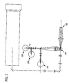
- the mixture of lyse reagent and sample will normally remain in the above referenced WBC cup only for 11 seconds. There it is lysed and mixed at 42°C ⁇ 3°C. At this point, the contents of the WBC cup are piped directly to an optical flowcell 100 for detection, see Figure 1.
- the measurement process begins as the cells stream passes through the flowcell 100, having been diluted with the addition of lyse so that the cells pass through the laser illuminated volume single file, in a laminar flowing sample stream surrounded by diluent/sheath solution.
- the illuminated volume is bounded in the two dimensions normal to the flow axis by the hydrodynamically focused cell stream, and in the dimension parallel to the flow axis by the vertical beam waist of the laser beam which is about 17 microns.
- the sample flow rate is about 2.5 microliters per second, and the corresponding illuminated sensing volume of the WBC and NRBC cells approximates an elliptical cylinder with dimension of about 80 x 5 x 17 microns.
- the 17 micron dimension is measured along the axis of the cylinder.
- the presence of a cell is detected by a compound photodiode 102 detecting axial light loss (ALL) and intermediate angle scatter (IAS), photomultiplier tube 104 which detects red fluorescence, and a unique triple trigger circuit, shown in Figure 2, in the three dimensional feature space of ALL, IAS, and FL3 (red fluorescence).
- the triple trigger circuit qualifies signals for digitization using AND/OR logic. A qualified signal must be greater than the IAS trigger, while at the same time it must be greater than either the ALL trigger or the FL3 trigger.
- this unique triggering circuit and the lysing properties which include a balanced fixative, allow the exposed NRBC nuclei to be rapidly stained, and clearly and non ambiguously counted and excluded from the WBC differential cell count without the usual interference from background, both fluorescent and non-fluorescent, such as DNA fragments, RBC stroma, and platelets.
- One or more detectors are preferably placed in the forward light path for measuring forward intermediate angle scattering (IAS) and either small angle forward scattering (SAS) or axial light loss (ALL, also known as forward extinction).
- IAS forward intermediate angle scattering
- SAS small angle forward scattering
- ALL axial light loss
- ALL is generally the decrease in light energy due to a cell passing in front of a laser beam and being detected by a photodiode.
- the light loss is generally due to scattering and defined as the decrease in light energy reaching a detector in the path of a laser beam due to the passage of a cell through that beam (generally ALL is detected at an angle of from about 0° to about 1°.)
- Small angle forward scatter (SAS) is light energy that reaches a detector outside the incident laser beam (but within a narrow angle of from about 1° to 3°) due to scattering from a cell passing through the beam.
- a beam stop is generally provided to keep the laser beam from getting into the detector. ALL measuring systems collect light within the incident cone of laser illumination, while small angle scatter systems collect light outside this cone.
- IAS Intermediate angle forward scattering
- ALL is collected in the angles less than about 0.3° horizontally and less than about 1.2° vertically from the laser axis, and IAS is collected at angles between about 3° and 10° from the laser axis.
- pulses are generated at detectors 102, 104, 106 and 108.
- the amplitudes of these pulses are then filtered, amplified, digitized, and stored in list mode in the corresponding five dimensional feature space of ALL, IAS, FL3, PSS (polarized side scatter), and DSS (depolarized side scatter).
- the normal counting time through flowcell 100 is 10 seconds. At the flow rate and dilution ratio described above, with a normal patient WBC count of 7000 cells per microliter of blood volume, the resulting event count rate would be 5000. In low count samples, this counting time can be automatically extended in order to improve the statistics of the measurement.
- the sample stream is piped to waste, and probe is cleaned and dried and prepared to process a subsequent sample.
- Algorithms are then applied to the list mode data of the aforementioned feature space of ALL, IAS, FL3, PSS, and DSS, and the following cell types are enumerated and/or flagged within less than 30 seconds of processing time: CELL TYPES ENUMERATED PERCENTAGES FLAGGED OR ENUMERATED White Cell concentration (WBC) Neutrophil concentration %N of WBC Lymphocyte concentration %LYMPH of WBC Monocyte concentration %MONO of WBC Eosinophil concentration % EOS of WBC Basophil concentration %BASO of WBC NRBC %NRBC of WBC Band concentration (BAND) Blast concentration (BLST) Immature gran. conc. (IG) Variant-lymph conc. (VARL)
- ALL and IAS signals are detected and collected for the WBC/Diff analysis and FL3 signals from stained NRBC nuclei are collected for NRBC analysis, as will be described below.
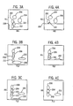
- the triple trigger circuit shown in Figure 2, qualifies these signals for digitization using AND/OR logic. To be qualified a signal must be greater than the IAS trigger, while at the same time it must be greater than either the ALL trigger or the FL3 trigger.
- Voltage comparators 300, 314 and 322 function by comparing the "+ inputs" (302, 310 and 318) to the "- inputs" (304, 312 and 320) to resultant outputs (306, 316, 324). If the "+ input” is of a higher voltage than the "- input” the output will be high. If the "+ input” is of a lower voltage than the "-input” the output will be low.
- the threshold voltages are independent voltages which are determined by system parameters.
- comparators 300 and 314 are inputs to OR gate 326 to give resultant OR gate output 328.
- the OR gate functions by comparing its inputs. The output will be high if either, or both, inputs are high.
- the output of the OR gate 328 and the output of comparators 322 and 324 are inputs to AND gate 330.
- the AND gate functions by comparing its inputs to derive its output 332 which is also also the valid trigger output. The output will be high only if both inputs are high.
- the valid trigger output 332) will only be high if the IAS signal 318 is greater than its threshold voltage 320, and either or both, the ALL signal 302 is greater than its threshold voltage 304 or the FL3 signal 310 is greater than its threshold voltage 312.
- the NRBC's form a unique cluster in the aforementioned three dimensional space, see Figures 14 and 15, which can be easily counted during the Optical WBC Differential analysis, and exclude non ambiguously from the WBC count.
- a count of NRBC per 100 WBC, and an absolute NRBC per ⁇ l of patient blood is reported. Consequently, NRBC are subtracted from total WBC counts permitting accurate total WBC and Differential analysis in the presence of NRBC in a blood sample.
- the multipurpose reagent system is comprised of about 95 mM ammonium chloride (5g/l), about 0.075% by volume of formaldehyde, from about 10 mM to about 20 mM acetate buffer, about 10 mM potassium bicarbonate, and about 0.01% by weight volume (i.e., grams per 100 ml) of.saponin.
- the pH of the reagent system is adjusted to a range of from about 6.2 to about 7.0 and the osmolality of the reagent system is from about 215 to about 270 mOsm/L.
- the reagent is pre-warmed at 42°C ⁇ 3° in the instrument's heated mixing chamber, where the sample and reagent are mixed and incubated for 11 seconds. This mixture was then transported to the flow cell (which takes 8 and 1/2 seconds) for the WBC/Diff/NRBC analysis.
- the optical configuration of the system is presented in Figure 1. The analysis was performed without implementing the triple triggering circuit; using only ALL and FL3 dual triggers as is common in the art. See all Figures from Fifure 3A through 10C.
- the upper dot plot display maps the light scatter signals (ALL vs. IAS) obtained from the sample and show 3 distinct populations of WBC.
- the Basophil cluster is not apparent here because normal bloods do not contain many Basophils.
- the Eosinophil cluster is not shown here since Eosinophils are separated via a DSS vs. PSS dot plot (not shown) and the middle cytogram, Figure 11B is a dot plot display of ALL and FL3 signals as labeled. Note that normal bloods do not contain any NRBCs.
- the lower bottom FL3+ cluster, Figures 11B and 11C is apparently cell debris containing RNA or DNA, as described earlier.
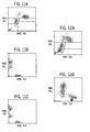
- Figures 12A and 12B top and bottom cytograms respectively, are dot plot displays of an abnormal blood with NRBC (47 NRBC/100 WBC) analyzed as described in EXAMPLE 1 utilizing a standard detection method.
- the cluster right below the lymphocyte population in the top cytogram belongs to NRBC and the small cluster at the bottom, left corner belongs to the origin noise which include RBC stroma (reticula, Howell Jolly Bodies and etc.), platelets and WBC debris.
- Figure 12B shows that the origin noise cluster of this sample stained with the nuclear dye brightly, following the stained NRBC cluster very closely in FL3 channel, thereby making it impossible to set the FL3 trigger to count NRBC accurately.
- the cytograms for Figures 13A and 13B are dot plot displays of an abnormal blood with NRBC (51 NRBC/100 WBC) analyzed as described in EXAMPLE 1 utilizing a standard detection method.
- the cluster right below the lymphocyte population in the top display, Figure 13A belongs to NRBC.
- An increased FL3+ origin noise of this sample can be seen.
- the noise cluster is located very close to the NRBC cluster in the FL3 channel.
- the FL3 noise is interfering with the position of the FL3 trigger.
- the FL3 trigger was set high enough to eliminate all the origin noise, a part of the NRBC population was also lost below the FL3 trigger as shown in Figure 13B.
- the disclosed triple trigger circuit (ALL/IAS/FL3) of the present invention was incorporated into the same instrument used in EXAMPLES 1 through 3 and utilized during this procedure.
- Figures 15A and 15B show the dot plot displays of the NRBC distribution of another clinical whole blood sample which contained 140 NRBC/100 WBC, also post triple trigger (ALL, FL3 and IAS) implementation.
- ALL, FL3 and IAS post triple trigger
- the origin noise is not visible and the total NRBC population is recovered above the FL3 trigger.
- Linearity samples were prepared by adding various concentrations of unfixed chicken erythrocytes to a EDTA, anti-coagulated normal human blood. The samples were processed as described in EXAMPLE 1 utilizing the triple trigger detection method of the present invention.
- the cytoplasm of chicken erythrocytes lyse in the method of present invention leaving only naked nuclei (CEN).
- CEN stained very rapidly with the vital nuclear stain (PI) in the diluent and become fluorescent (FL3).
- the FL3+ CEN are counted as NRBC and reported as number of NRBC/100 WBC and as absolute counts per ⁇ L of the whole blood sample in the method of the present invention.
- the results are presented in Figures 16A and 16B.
- the linearity plots of NRBC/100 WBC and NRBC in absolute numbers in the figure demonstrate that the method of the current invention generate a linear NRBC counts.
Landscapes
- Chemical & Material Sciences (AREA)
- Dispersion Chemistry (AREA)
- Physics & Mathematics (AREA)
- Health & Medical Sciences (AREA)
- Life Sciences & Earth Sciences (AREA)
- Analytical Chemistry (AREA)
- Biochemistry (AREA)
- General Health & Medical Sciences (AREA)
- General Physics & Mathematics (AREA)
- Immunology (AREA)
- Pathology (AREA)
- Investigating Or Analysing Biological Materials (AREA)
- Investigating, Analyzing Materials By Fluorescence Or Luminescence (AREA)
- Investigating Or Analysing Materials By Optical Means (AREA)
Claims (9)
- Ein Verfahren zur Differenzierung von Normoblastenzellen (nucleated red blood cells = NRBC) von anderen Zellen durch Durchfluss-Zytometrie, das folgendes umfasst:a) Mischen eines Aliquots einer Blutprobe mit einem Reagenzsystem, worin das Reagenzsystem eine rote Blutzellen (RBC) -lysierende Komponente, eine Leukozyten-fixierende Komponente und eine nukleare Vitalfarbstoffkomponente umfasst, um die Nuklei der NRBC zu färben;b) durch eine Röhre leiten des gemischten Aliquots, im wesentlichen eine Zelle pro Zeiteinheit, durch einen optischen Anregungsbereich (100);c) Erhalten von mindestens einem Signal für jeden Parameter, einschließlich gestreutem Licht in einem ersten Bereich von Streuwinkeln und gestreutem Licht in einem zweiten Bereich von Streuwinkeln, und weiterhin einschließlich Fluoreszenz (FL);d) Bewerten der Signale, die erhalten werden, indem die Signale (302, 310, 318) einer logischen Prüfung unterworfen werden, worin ein bewertetes Signal (332) größer sein muss als eine zweite Streusignalschwelle (320), während es zur selben Zeit größer sein muss als entweder eine erste Streusignalschwelle (304) oder eine FL-Schwelle (312), worin die Schwellen durch System-Parameter bestimmt werden;e) Konstruieren eines dreidimensionalen Plots von Intensitätssignalen von FL, gestreutem Licht in einem ersten Bereich von Streuwinkeln und gestreutem Licht in einem zweiten Bereich von Streuwinkeln, anhand eines detektierten Signals; undf) Differenzieren der NRBC-Cluster und der weißen Blutzellen (WBC) in dem konstruierten dreidimensionalen Plot und den bewerteten Signalen, und Bestimmen der jeweiligen Anzahl an Zellen.
- Das Verfahren von Anspruch 1, worin der erste Bereich von Streuwinkeln von ungefähr 0° bis ungefähr 1° ist.
- Das Verfahren von Anspruch 1, worin ein erhaltener Signal-Parameter axialen Licht-Verlust (Axial Light Loss, ALL) umfasst.
- Das Verfahren von Anspruch 1, worin ein erhaltener Signal-Parameter Kleinwinkelvorwärtsstreuung (Small Angle forward Scatter, SAS) umfasst.
- Das Verfahren von Anspruch 3, worin der ALL bei einem Winkel von ungefähr 0° bis ungefähr 1° erhalten wird.
- Das Verfahren von Anspruch 4, worin die SAS bei einem Winkel von ungefähr 1° bis ungefähr 3° erhalten wird.
- Das Verfahren von Anspruch 1, worin der zweite Bereich von Streuwinkeln von ungefähr 3° - 10° ist.
- Das Verfahren von Anspruch 1, worin das nukleare Färbemittel gewählt ist aus der Gruppe von Vitalfarbstoffen bestehend aus Propidiumiodid (PI), Ethidiumbromid (Ebr), Ethidium Homodimer-1 (EthD-1), Ethidium Homodimer-2 (EthD-2) und Diethylentriamin (DTA) .
- Ein Durchfluss-Zytometer für die Differenzierung von Zellen aus einer Blutprobe, das eine Schaltung umfasst, die folgendes enthält:wobei die Schaltung so angeordnet ist, um eine logische Formel zu implementieren, um die Signale zu bewerten, dadurch gekennzeichnet, dass die Signale 0° bis ungefähr 1° Streusignale (302), 3° - 10° Streusignale (318) und Fluoreszenz-Signale (310) sind, wobei die logische Formel definiert ist als [(0° bis ungefähr 1° Streusignale) ODER (Fluoreszenz-Signale)] UND (3° -10° Streusignale).eine Vielzahl an Spannungskomparatoren (300, 314, 322), die die Signale mit den entsprechenden Schwellensignalen (304, 312, 320) vergleichen; undeine Vielzahl an logischen Gattern (326, 330),
Applications Claiming Priority (3)
| Application Number | Priority Date | Filing Date | Title |
|---|---|---|---|
| US08/356,932 US5559037A (en) | 1994-12-15 | 1994-12-15 | Method for rapid and simultaneous analysis of nucleated red blood cells |
| US356932 | 1994-12-15 | ||
| PCT/US1995/015371 WO1996018878A1 (en) | 1994-12-15 | 1995-11-28 | Method for rapid and simultaneous analysis of nucleated red blood cells |
Publications (2)
| Publication Number | Publication Date |
|---|---|
| EP0797762A1 EP0797762A1 (de) | 1997-10-01 |
| EP0797762B1 true EP0797762B1 (de) | 2004-01-28 |
Family
ID=23403561
Family Applications (1)
| Application Number | Title | Priority Date | Filing Date |
|---|---|---|---|
| EP95940838A Expired - Lifetime EP0797762B1 (de) | 1994-12-15 | 1995-11-28 | Verfahren zur schnellen und gleichzeitigen analyse nukleierter roter blutzellen |
Country Status (8)
| Country | Link |
|---|---|
| US (1) | US5559037A (de) |
| EP (1) | EP0797762B1 (de) |
| JP (1) | JP2938976B2 (de) |
| AT (1) | ATE258677T1 (de) |
| CA (1) | CA2207396C (de) |
| DE (1) | DE69532511T2 (de) |
| ES (1) | ES2215181T3 (de) |
| WO (1) | WO1996018878A1 (de) |
Families Citing this family (56)
| Publication number | Priority date | Publication date | Assignee | Title |
|---|---|---|---|---|
| US6509192B1 (en) * | 1992-02-24 | 2003-01-21 | Coulter International Corp. | Quality control method |
| US5879900A (en) * | 1994-12-15 | 1999-03-09 | Abbott Laboratories | Method for simultaneous analysis of cell viability, nucleated red blood cells and white blood cell differentials |
| DE69638209D1 (de) * | 1995-12-15 | 2010-08-12 | Abbott Lab | Verfahren für gleichzeitige analyse von zelllebensfähigkeit, nukleierten roten blutkörperchen und leukozytendifferential |
| US5817519A (en) * | 1995-12-28 | 1998-10-06 | Bayer Corporation | Automated method and device for identifying and quantifying platelets and for determining platelet activation state using whole blood samples |
| US6025201A (en) * | 1995-12-28 | 2000-02-15 | Bayer Corporation | Highly sensitive, accurate, and precise automated method and device for identifying and quantifying platelets and for determining platelet activation state using whole blood samples |
| US5858790A (en) * | 1996-06-26 | 1999-01-12 | Abbott Laboratories | Hematology reference control and method of preparation |
| JP2001506005A (ja) * | 1996-12-11 | 2001-05-08 | ナイコメッド・アマーシャム・ピーエルシー | 細胞の選択的溶解 |
| US5874311A (en) * | 1997-11-21 | 1999-02-23 | Coulter International Corp. | Method for differentiation of reticulocytes in blood |
| US5874310A (en) * | 1997-11-21 | 1999-02-23 | Coulter International Corp. | Method for differentiation of nucleated red blood cells |
| US5917584A (en) * | 1997-11-21 | 1999-06-29 | Coulter International Corp. | Method for differentiation of nucleated red blood cells |
| US6911313B2 (en) | 1998-02-06 | 2005-06-28 | Sysmex Corporation | Process for discriminating and counting erythroblasts |
| FR2813891B1 (fr) * | 2000-09-14 | 2005-01-14 | Immunotech Sa | Reactif multifonctionnel pour erythrocytes mettant en jeu des carbamates et applications |
| US7049093B2 (en) * | 2000-11-08 | 2006-05-23 | Sysmex Corporation | Method of classifying and counting nucleated bone marrow cells |
| US6448085B1 (en) * | 2001-04-13 | 2002-09-10 | Sysmex Corporation | Quality control material and calibrator for nucleated red blood cell tested on hematology analyzer |
| US6630990B2 (en) * | 2001-06-05 | 2003-10-07 | Abbott Laboratories | Optical method and apparatus for red blood cell differentiation on a cell-by-cell basis, and simultaneous analysis of white blood cell differentiation |
| US6916658B2 (en) * | 2001-07-27 | 2005-07-12 | Beckman Coulter, Inc. | Method for measurement of immature granulocytes |
| US6472215B1 (en) | 2001-07-27 | 2002-10-29 | Coulter International Corp. | Method of analyzing nucleated red blood cells in a blood sample |
| US6573102B2 (en) | 2001-07-27 | 2003-06-03 | Coulter International Corp. | Lytic reagent composition for determination of nucleated blood cells |
| CN1276252C (zh) | 2001-07-27 | 2006-09-20 | 贝克曼库尔特有限公司 | 有核红细胞的测量方法 |
| US6410330B1 (en) | 2001-07-27 | 2002-06-25 | Coulter International Corp. | Method for measurement of nucleated red blood cells |
| JP4850711B2 (ja) * | 2003-10-02 | 2012-01-11 | ベックマン コールター, インコーポレイテッド | 血液サンプルの有核赤血球の光学測定のリファレンスコントロール |
| US7198953B2 (en) * | 2003-10-12 | 2007-04-03 | Beckman Coulter, Inc. | Method of using a reference control composition for measurement of nucleated red blood cells |
| US7195919B2 (en) * | 2003-12-19 | 2007-03-27 | Beckman Coulter, Inc. | Hematology controls for reticulocytes and nucleated red blood cells |
| US7008792B2 (en) * | 2004-02-10 | 2006-03-07 | Beckman Coulter, Inc. | Method of measurement of nucleated red blood cells |
| US7208319B2 (en) * | 2004-02-10 | 2007-04-24 | Beckman Coulter, Inc. | Method of measurement of nucleated red blood cells |
| US7135341B2 (en) * | 2004-04-07 | 2006-11-14 | Beckman Coulter, Inc. | Reference control containing a nucleated red blood cell component |
| US7625712B2 (en) * | 2004-05-21 | 2009-12-01 | Beckman Coulter, Inc. | Method for a fully automated monoclonal antibody-based extended differential |
| US7390662B2 (en) * | 2005-11-09 | 2008-06-24 | Beckman Coulter, Inc. | Method and apparatus for performing platelet measurement |
| US7354767B2 (en) * | 2006-03-16 | 2008-04-08 | Beckman Coulter, Inc. | Reference control composition containing a nucleated red blood cell component made of non-nucleated blood cells |
| CN1945326A (zh) * | 2006-10-13 | 2007-04-11 | 江西特康科技有限公司 | 基于视觉形态的五分类全血细胞分析方法 |
| US7674622B2 (en) * | 2006-12-22 | 2010-03-09 | Abbott Laboratories, Inc. | Method for determination of nucleated red blood cells and leukocytes in a whole blood sample in an automated hematology analyzer |
| CN101349644B (zh) * | 2007-07-20 | 2012-06-27 | 深圳迈瑞生物医疗电子股份有限公司 | 一种白细胞分类试剂和其使用方法 |
| US8102161B2 (en) * | 2007-09-25 | 2012-01-24 | Tdk Corporation | Stable output in a switching power supply by smoothing the output of the secondary coil |
| CN101475754A (zh) * | 2008-01-04 | 2009-07-08 | 深圳迈瑞生物医疗电子股份有限公司 | 不对称菁类荧光染料,组合物及在生物样品染色中的用途 |
| CN101602762B (zh) * | 2008-06-10 | 2013-10-16 | 深圳迈瑞生物医疗电子股份有限公司 | 不对称菁类化合物、其制备方法及应用 |
| US8148101B2 (en) * | 2008-07-22 | 2012-04-03 | Abbott Laboratories | Method for classifying and counting bacteria in body fluids |
| CN101726579B (zh) * | 2008-10-17 | 2014-06-18 | 深圳迈瑞生物医疗电子股份有限公司 | 血液检测试剂和方法 |
| CN101723874B (zh) * | 2008-10-31 | 2013-09-11 | 深圳迈瑞生物医疗电子股份有限公司 | 花菁类化合物及其在生物样品染色中的用途 |
| CN101750476B (zh) * | 2008-12-08 | 2015-06-03 | 深圳迈瑞生物医疗电子股份有限公司 | 血液分析试剂及其使用方法 |
| CN101750274B (zh) * | 2008-12-17 | 2014-06-25 | 深圳迈瑞生物医疗电子股份有限公司 | 白细胞分类计数试剂、试剂盒以及白细胞分类计数的方法 |
| JP5871792B2 (ja) | 2009-04-27 | 2016-03-01 | アボット・ラボラトリーズAbbott Laboratories | 自動血液分析器において、レーザーからの前方散乱を用いることによる、赤血球細胞を白血球細胞から判別する方法 |
| CN101988082B (zh) * | 2009-07-31 | 2015-04-08 | 深圳迈瑞生物医疗电子股份有限公司 | 白细胞分类计数试剂、试剂盒及其制备方法和白细胞分类计数的方法 |
| CN102115456B (zh) * | 2009-12-30 | 2014-08-20 | 深圳迈瑞生物医疗电子股份有限公司 | 花菁类化合物,包含所述化合物的组合物及其在细胞检测中的用途 |
| US9097704B2 (en) | 2010-05-05 | 2015-08-04 | Abbott Laboratories | Method for hematology analysis |
| US20120274925A1 (en) * | 2011-04-26 | 2012-11-01 | Yong Chen | Axial light loss sensor system for flow cytometery |
| CN103917868B (zh) | 2011-05-04 | 2016-08-24 | 雅培制药有限公司 | 嗜碱性粒细胞分析系统和方法 |
| ES2927316T3 (es) | 2011-05-04 | 2022-11-04 | Abbott Lab | Sistema y método de análisis de glóbulos blancos |
| ES2902648T3 (es) * | 2011-05-04 | 2022-03-29 | Abbott Lab | Método de análisis de glóbulos rojos nucleados y analizador hematológico automatizado |
| DE102014107837B4 (de) * | 2014-06-04 | 2021-09-02 | Presens Precision Sensing Gmbh | Optischer Sensor zum quantitativen Nachweis eines Analyten in einer Probe und Verfahren zur Herstellung des Sensors |
| JP6318026B2 (ja) * | 2014-06-26 | 2018-04-25 | アズビル株式会社 | 粒子検出装置及び粒子の検出方法 |
| JP6228085B2 (ja) | 2014-08-27 | 2017-11-08 | シスメックス株式会社 | 検体分析装置及び検体分析方法 |
| CN106687810B (zh) * | 2014-12-31 | 2019-10-22 | 深圳迈瑞生物医疗电子股份有限公司 | 一种非诊断目的的有核红细胞报警方法、装置及流式细胞分析仪 |
| EP3943912B1 (de) * | 2015-02-12 | 2025-08-20 | Shenzhen Mindray Bio-Medical Electronics Co., Ltd. | Zellanalysator und teilchensortierungsverfahren und -vorrichtung |
| LT3150988T (lt) * | 2015-10-01 | 2021-06-25 | Nanotemper Technologies Gmbh | Dalelių stabilumo ir agregacijos optinio matavimo sistema bei būdas |
| WO2018081880A1 (en) * | 2016-11-07 | 2018-05-11 | Kertesz Geza | Equipment for carrying out hemograms |
| EP4462126A4 (de) * | 2021-12-31 | 2025-07-23 | Shenzhen Mindray Biomedical Electronics Co Ltd | Blutzellenanalysegerät, verfahren und verwendung von infektionsmarkerparametern |
Family Cites Families (15)
| Publication number | Priority date | Publication date | Assignee | Title |
|---|---|---|---|---|
| US4284412A (en) * | 1979-07-13 | 1981-08-18 | Ortho Diagnostics, Inc. | Method and apparatus for automated identification and enumeration of specified blood cell subclasses |
| US4544546A (en) * | 1980-04-21 | 1985-10-01 | Abbott Laboratories | Fluorescent nucleic acid stains |
| US4492752A (en) * | 1982-09-03 | 1985-01-08 | Ortho Diagnostics Systems Inc. | Method for discriminating between unstained and absorbing dye stained cells |
| JPS59184841A (ja) * | 1983-04-05 | 1984-10-20 | ベクトン・デイツキンソン・アンド・カンパニ− | サンプル中の白血球のサブクラスを識別する方法および装置 |
| US5188935A (en) * | 1984-05-31 | 1993-02-23 | Coulter Electronics, Inc. | Reagent system and method for identification, enumeration and examination of classes and subclasses of blood leukocytes |
| US4751179A (en) * | 1984-05-31 | 1988-06-14 | Coulter Electronics, Inc. | Method and reagents for differential determination of four populations of leukocytes in blood |
| US4727020A (en) * | 1985-02-25 | 1988-02-23 | Becton, Dickinson And Company | Method for analysis of subpopulations of blood cells |
| US4978624A (en) * | 1985-09-06 | 1990-12-18 | Technicon Instruments Corporation | Reagent for the determination of a differential white blood cell count |
| KR970007077B1 (ko) * | 1987-03-13 | 1997-05-02 | 코울터 일렉트로닉스 인커퍼레이티드 | 광산란 기술을 이용한 다중-부분식별 분석 방법 |
| US4882284A (en) * | 1987-04-13 | 1989-11-21 | Ortho Pharmaceutical Corporation | Method for quantitating and differentiating white blood cells |
| JPS6435345A (en) * | 1987-07-31 | 1989-02-06 | Canon Kk | Particle analyzing device |
| US4987086A (en) * | 1987-11-30 | 1991-01-22 | Becton, Dickinson And Company | Method for analysis of subpopulations of cells |
| US5047321A (en) * | 1988-06-15 | 1991-09-10 | Becton Dickinson & Co. | Method for analysis of cellular components of a fluid |
| JP2927979B2 (ja) * | 1991-02-22 | 1999-07-28 | シスメックス株式会社 | フローサイトメトリーによる赤芽球の分類方法 |
| EP0685994B1 (de) * | 1993-02-25 | 2007-03-21 | Abbott Laboratories | Mehrzweckreagenzsystem zur schnellen lysierung von vollblutproben |
-
1994
- 1994-12-15 US US08/356,932 patent/US5559037A/en not_active Expired - Lifetime
-
1995
- 1995-11-28 WO PCT/US1995/015371 patent/WO1996018878A1/en not_active Ceased
- 1995-11-28 EP EP95940838A patent/EP0797762B1/de not_active Expired - Lifetime
- 1995-11-28 AT AT95940838T patent/ATE258677T1/de not_active IP Right Cessation
- 1995-11-28 CA CA002207396A patent/CA2207396C/en not_active Expired - Fee Related
- 1995-11-28 DE DE69532511T patent/DE69532511T2/de not_active Expired - Lifetime
- 1995-11-28 ES ES95940838T patent/ES2215181T3/es not_active Expired - Lifetime
- 1995-11-28 JP JP8518976A patent/JP2938976B2/ja not_active Expired - Lifetime
Also Published As
| Publication number | Publication date |
|---|---|
| JPH10504398A (ja) | 1998-04-28 |
| CA2207396A1 (en) | 1996-06-20 |
| ES2215181T3 (es) | 2004-10-01 |
| CA2207396C (en) | 2007-08-07 |
| ATE258677T1 (de) | 2004-02-15 |
| DE69532511D1 (de) | 2004-03-04 |
| EP0797762A1 (de) | 1997-10-01 |
| WO1996018878A1 (en) | 1996-06-20 |
| US5559037A (en) | 1996-09-24 |
| JP2938976B2 (ja) | 1999-08-25 |
| DE69532511T2 (de) | 2004-11-11 |
Similar Documents
| Publication | Publication Date | Title |
|---|---|---|
| EP0797762B1 (de) | Verfahren zur schnellen und gleichzeitigen analyse nukleierter roter blutzellen | |
| US5879900A (en) | Method for simultaneous analysis of cell viability, nucleated red blood cells and white blood cell differentials | |
| EP1250585B1 (de) | Verfahren und vorrichtung zur analyse von zellen in einer vollblutprobe | |
| US6524858B1 (en) | Single channel, single dilution detection method for the identification and quantification of blood cells and platelets in a whole blood sample using an automated hematology analyzer | |
| US5917584A (en) | Method for differentiation of nucleated red blood cells | |
| JP4366478B2 (ja) | 赤芽球の識別方法 | |
| US8148101B2 (en) | Method for classifying and counting bacteria in body fluids | |
| CA1309328C (en) | Method of classifying leukocytes by flow cytometry and reagents used in the method | |
| US7008792B2 (en) | Method of measurement of nucleated red blood cells | |
| US7208319B2 (en) | Method of measurement of nucleated red blood cells | |
| US5840515A (en) | Erythrocyte lysis reagent, and its use in methods for isolating and differentiating leucocytes | |
| EP0866960B1 (de) | Verfahren für gleichzeitige analyse von zelllebensfähigkeit, nukleierten roten blutkörperchen und leukozytendifferential | |
| WO1997021994A9 (en) | Method for simultaneous analysis of cell viability, nucleated red blood cells and white blood cell differential |
Legal Events
| Date | Code | Title | Description |
|---|---|---|---|
| PUAI | Public reference made under article 153(3) epc to a published international application that has entered the european phase |
Free format text: ORIGINAL CODE: 0009012 |
|
| 17P | Request for examination filed |
Effective date: 19970703 |
|
| AK | Designated contracting states |
Kind code of ref document: A1 Designated state(s): AT BE CH DE ES FR GB IT LI NL |
|
| 17Q | First examination report despatched |
Effective date: 20020214 |
|
| GRAP | Despatch of communication of intention to grant a patent |
Free format text: ORIGINAL CODE: EPIDOSNIGR1 |
|
| GRAS | Grant fee paid |
Free format text: ORIGINAL CODE: EPIDOSNIGR3 |
|
| GRAA | (expected) grant |
Free format text: ORIGINAL CODE: 0009210 |
|
| AK | Designated contracting states |
Kind code of ref document: B1 Designated state(s): AT BE CH DE ES FR GB IT LI NL |
|
| REG | Reference to a national code |
Ref country code: GB Ref legal event code: FG4D |
|
| REG | Reference to a national code |
Ref country code: CH Ref legal event code: EP |
|
| REF | Corresponds to: |
Ref document number: 69532511 Country of ref document: DE Date of ref document: 20040304 Kind code of ref document: P |
|
| REG | Reference to a national code |
Ref country code: CH Ref legal event code: NV Representative=s name: A. BRAUN, BRAUN, HERITIER, ESCHMANN AG PATENTANWAE |
|
| REG | Reference to a national code |
Ref country code: ES Ref legal event code: FG2A Ref document number: 2215181 Country of ref document: ES Kind code of ref document: T3 |
|
| ET | Fr: translation filed | ||
| PG25 | Lapsed in a contracting state [announced via postgrant information from national office to epo] |
Ref country code: AT Free format text: LAPSE BECAUSE OF NON-PAYMENT OF DUE FEES Effective date: 20041128 |
|
| PG25 | Lapsed in a contracting state [announced via postgrant information from national office to epo] |
Ref country code: LI Free format text: LAPSE BECAUSE OF NON-PAYMENT OF DUE FEES Effective date: 20041130 Ref country code: CH Free format text: LAPSE BECAUSE OF NON-PAYMENT OF DUE FEES Effective date: 20041130 Ref country code: BE Free format text: LAPSE BECAUSE OF NON-PAYMENT OF DUE FEES Effective date: 20041130 |
|
| PLBE | No opposition filed within time limit |
Free format text: ORIGINAL CODE: 0009261 |
|
| STAA | Information on the status of an ep patent application or granted ep patent |
Free format text: STATUS: NO OPPOSITION FILED WITHIN TIME LIMIT |
|
| 26N | No opposition filed |
Effective date: 20041029 |
|
| BERE | Be: lapsed |
Owner name: *ABBOTT LABORATORIES Effective date: 20041130 |
|
| PG25 | Lapsed in a contracting state [announced via postgrant information from national office to epo] |
Ref country code: NL Free format text: LAPSE BECAUSE OF NON-PAYMENT OF DUE FEES Effective date: 20050601 |
|
| REG | Reference to a national code |
Ref country code: CH Ref legal event code: PL |
|
| NLV4 | Nl: lapsed or anulled due to non-payment of the annual fee |
Effective date: 20050601 |
|
| BERE | Be: lapsed |
Owner name: *ABBOTT LABORATORIES Effective date: 20041130 |
|
| PGFP | Annual fee paid to national office [announced via postgrant information from national office to epo] |
Ref country code: GB Payment date: 20131028 Year of fee payment: 19 |
|
| PGFP | Annual fee paid to national office [announced via postgrant information from national office to epo] |
Ref country code: ES Payment date: 20131118 Year of fee payment: 19 Ref country code: IT Payment date: 20131121 Year of fee payment: 19 |
|
| PGFP | Annual fee paid to national office [announced via postgrant information from national office to epo] |
Ref country code: DE Payment date: 20141201 Year of fee payment: 20 Ref country code: FR Payment date: 20141027 Year of fee payment: 20 |
|
| GBPC | Gb: european patent ceased through non-payment of renewal fee |
Effective date: 20141128 |
|
| PG25 | Lapsed in a contracting state [announced via postgrant information from national office to epo] |
Ref country code: GB Free format text: LAPSE BECAUSE OF NON-PAYMENT OF DUE FEES Effective date: 20141128 |
|
| REG | Reference to a national code |
Ref country code: DE Ref legal event code: R071 Ref document number: 69532511 Country of ref document: DE |
|
| REG | Reference to a national code |
Ref country code: ES Ref legal event code: FD2A Effective date: 20151229 |
|
| PG25 | Lapsed in a contracting state [announced via postgrant information from national office to epo] |
Ref country code: IT Free format text: LAPSE BECAUSE OF NON-PAYMENT OF DUE FEES Effective date: 20141128 |
|
| PG25 | Lapsed in a contracting state [announced via postgrant information from national office to epo] |
Ref country code: ES Free format text: LAPSE BECAUSE OF NON-PAYMENT OF DUE FEES Effective date: 20141129 |