EP0804454B1 - HYBRIDISIERUNGS- UND ABDRUCKVERFAHREN ZUR CHARAKTERISIERUNG ODER INTERAKTIONEN VON 16S-rRNS-ANALOGEN ZUR IDENTIFIZIERUNG NEUER ANTIBIOTIKA - Google Patents
HYBRIDISIERUNGS- UND ABDRUCKVERFAHREN ZUR CHARAKTERISIERUNG ODER INTERAKTIONEN VON 16S-rRNS-ANALOGEN ZUR IDENTIFIZIERUNG NEUER ANTIBIOTIKA Download PDFInfo
- Publication number
- EP0804454B1 EP0804454B1 EP95930926A EP95930926A EP0804454B1 EP 0804454 B1 EP0804454 B1 EP 0804454B1 EP 95930926 A EP95930926 A EP 95930926A EP 95930926 A EP95930926 A EP 95930926A EP 0804454 B1 EP0804454 B1 EP 0804454B1
- Authority
- EP
- European Patent Office
- Prior art keywords
- analog
- test compound
- assay
- binding
- binding complex
- Prior art date
- Legal status (The legal status is an assumption and is not a legal conclusion. Google has not performed a legal analysis and makes no representation as to the accuracy of the status listed.)
- Expired - Lifetime
Links
- 108020004465 16S ribosomal RNA Proteins 0.000 title claims description 39
- 238000000034 method Methods 0.000 title description 37
- 230000003993 interaction Effects 0.000 title description 28
- 239000003242 anti bacterial agent Substances 0.000 title description 16
- 229940088710 antibiotic agent Drugs 0.000 title description 15
- 238000009396 hybridization Methods 0.000 title 1
- 150000001875 compounds Chemical class 0.000 claims description 83
- 239000002773 nucleotide Substances 0.000 claims description 82
- 125000003729 nucleotide group Chemical group 0.000 claims description 82
- 238000009739 binding Methods 0.000 claims description 77
- 230000027455 binding Effects 0.000 claims description 73
- 229920002477 rna polymer Polymers 0.000 claims description 65
- 238000012360 testing method Methods 0.000 claims description 65
- 108091027075 5S-rRNA precursor Proteins 0.000 claims description 55
- 150000007523 nucleic acids Chemical group 0.000 claims description 49
- 238000003556 assay Methods 0.000 claims description 42
- 239000003446 ligand Substances 0.000 claims description 33
- VAYGXNSJCAHWJZ-UHFFFAOYSA-N dimethyl sulfate Chemical group COS(=O)(=O)OC VAYGXNSJCAHWJZ-UHFFFAOYSA-N 0.000 claims description 31
- 239000003153 chemical reaction reagent Substances 0.000 claims description 24
- 108020004418 ribosomal RNA Proteins 0.000 claims description 22
- 238000006243 chemical reaction Methods 0.000 claims description 19
- 229940126575 aminoglycoside Drugs 0.000 claims description 17
- 230000000694 effects Effects 0.000 claims description 16
- 108091028043 Nucleic acid sequence Proteins 0.000 claims description 14
- 230000011987 methylation Effects 0.000 claims description 14
- 238000007069 methylation reaction Methods 0.000 claims description 14
- 239000000126 substance Substances 0.000 claims description 13
- 230000001225 therapeutic effect Effects 0.000 claims description 13
- 230000015572 biosynthetic process Effects 0.000 claims description 12
- 238000012544 monitoring process Methods 0.000 claims description 12
- 238000002156 mixing Methods 0.000 claims description 8
- 238000011065 in-situ storage Methods 0.000 claims description 7
- YRCRRHNVYVFNTM-UHFFFAOYSA-N 1,1-dihydroxy-3-ethoxy-2-butanone Chemical compound CCOC(C)C(=O)C(O)O YRCRRHNVYVFNTM-UHFFFAOYSA-N 0.000 claims description 6
- 108091034117 Oligonucleotide Proteins 0.000 claims description 5
- 229950001103 ketoxal Drugs 0.000 claims description 5
- 238000012875 competitive assay Methods 0.000 claims description 4
- 230000004570 RNA-binding Effects 0.000 claims description 2
- 239000002253 acid Substances 0.000 claims description 2
- 230000002265 prevention Effects 0.000 claims description 2
- 108020004566 Transfer RNA Proteins 0.000 description 31
- 108020004999 messenger RNA Proteins 0.000 description 25
- 108020004707 nucleic acids Proteins 0.000 description 25
- 102000039446 nucleic acids Human genes 0.000 description 25
- 210000003705 ribosome Anatomy 0.000 description 25
- 229930193140 Neomycin Natural products 0.000 description 24
- 230000003115 biocidal effect Effects 0.000 description 24
- 229960004927 neomycin Drugs 0.000 description 23
- 238000002474 experimental method Methods 0.000 description 14
- 238000012216 screening Methods 0.000 description 14
- 108020005098 Anticodon Proteins 0.000 description 11
- 230000004048 modification Effects 0.000 description 11
- 238000012986 modification Methods 0.000 description 11
- 230000014616 translation Effects 0.000 description 11
- ULGZDMOVFRHVEP-RWJQBGPGSA-N Erythromycin Chemical compound O([C@@H]1[C@@H](C)C(=O)O[C@@H]([C@@]([C@H](O)[C@@H](C)C(=O)[C@H](C)C[C@@](C)(O)[C@H](O[C@H]2[C@@H]([C@H](C[C@@H](C)O2)N(C)C)O)[C@H]1C)(C)O)CC)[C@H]1C[C@@](C)(OC)[C@@H](O)[C@H](C)O1 ULGZDMOVFRHVEP-RWJQBGPGSA-N 0.000 description 10
- 238000001243 protein synthesis Methods 0.000 description 10
- UCSJYZPVAKXKNQ-HZYVHMACSA-N streptomycin Chemical compound CN[C@H]1[C@H](O)[C@@H](O)[C@H](CO)O[C@H]1O[C@@H]1[C@](C=O)(O)[C@H](C)O[C@H]1O[C@@H]1[C@@H](NC(N)=N)[C@H](O)[C@@H](NC(N)=N)[C@H](O)[C@H]1O UCSJYZPVAKXKNQ-HZYVHMACSA-N 0.000 description 10
- 241000588724 Escherichia coli Species 0.000 description 8
- 210000004027 cell Anatomy 0.000 description 8
- 239000003814 drug Substances 0.000 description 8
- 239000000499 gel Substances 0.000 description 8
- 241000725303 Human immunodeficiency virus Species 0.000 description 7
- 102100034343 Integrase Human genes 0.000 description 7
- UOZODPSAJZTQNH-UHFFFAOYSA-N Paromomycin II Natural products NC1C(O)C(O)C(CN)OC1OC1C(O)C(OC2C(C(N)CC(N)C2O)OC2C(C(O)C(O)C(CO)O2)N)OC1CO UOZODPSAJZTQNH-UHFFFAOYSA-N 0.000 description 7
- 108010092799 RNA-directed DNA polymerase Proteins 0.000 description 7
- 230000009918 complex formation Effects 0.000 description 7
- 229960001914 paromomycin Drugs 0.000 description 7
- UOZODPSAJZTQNH-LSWIJEOBSA-N paromomycin Chemical compound N[C@@H]1[C@@H](O)[C@H](O)[C@H](CN)O[C@@H]1O[C@H]1[C@@H](O)[C@H](O[C@H]2[C@@H]([C@@H](N)C[C@@H](N)[C@@H]2O)O[C@@H]2[C@@H]([C@@H](O)[C@H](O)[C@@H](CO)O2)N)O[C@@H]1CO UOZODPSAJZTQNH-LSWIJEOBSA-N 0.000 description 7
- 238000007423 screening assay Methods 0.000 description 7
- 102000004169 proteins and genes Human genes 0.000 description 6
- 108090000623 proteins and genes Proteins 0.000 description 6
- 150000003839 salts Chemical class 0.000 description 6
- 239000004098 Tetracycline Substances 0.000 description 5
- 230000008901 benefit Effects 0.000 description 5
- 238000001514 detection method Methods 0.000 description 5
- 238000010586 diagram Methods 0.000 description 5
- 229960003276 erythromycin Drugs 0.000 description 5
- 238000012869 ethanol precipitation Methods 0.000 description 5
- 238000002372 labelling Methods 0.000 description 5
- 239000000203 mixture Substances 0.000 description 5
- 108090000765 processed proteins & peptides Proteins 0.000 description 5
- 230000009257 reactivity Effects 0.000 description 5
- 210000004708 ribosome subunit Anatomy 0.000 description 5
- 150000003384 small molecules Chemical class 0.000 description 5
- 229960005322 streptomycin Drugs 0.000 description 5
- 229960002180 tetracycline Drugs 0.000 description 5
- 229930101283 tetracycline Natural products 0.000 description 5
- 235000019364 tetracycline Nutrition 0.000 description 5
- 150000003522 tetracyclines Chemical class 0.000 description 5
- 238000005406 washing Methods 0.000 description 5
- YBJHBAHKTGYVGT-ZKWXMUAHSA-N (+)-Biotin Chemical compound N1C(=O)N[C@@H]2[C@H](CCCCC(=O)O)SC[C@@H]21 YBJHBAHKTGYVGT-ZKWXMUAHSA-N 0.000 description 4
- YQYJSBFKSSDGFO-UHFFFAOYSA-N Epihygromycin Natural products OC1C(O)C(C(=O)C)OC1OC(C(=C1)O)=CC=C1C=C(C)C(=O)NC1C(O)C(O)C2OCOC2C1O YQYJSBFKSSDGFO-UHFFFAOYSA-N 0.000 description 4
- 239000004793 Polystyrene Substances 0.000 description 4
- 239000002647 aminoglycoside antibiotic agent Substances 0.000 description 4
- 230000008878 coupling Effects 0.000 description 4
- 238000010168 coupling process Methods 0.000 description 4
- 238000005859 coupling reaction Methods 0.000 description 4
- 239000000463 material Substances 0.000 description 4
- NLXLAEXVIDQMFP-UHFFFAOYSA-N Ammonia chloride Chemical compound [NH4+].[Cl-] NLXLAEXVIDQMFP-UHFFFAOYSA-N 0.000 description 3
- 241000894006 Bacteria Species 0.000 description 3
- 108020004414 DNA Proteins 0.000 description 3
- 108090000279 Peptidyltransferases Proteins 0.000 description 3
- 108010081734 Ribonucleoproteins Proteins 0.000 description 3
- 102000004389 Ribonucleoproteins Human genes 0.000 description 3
- 108010090804 Streptavidin Proteins 0.000 description 3
- 101710137500 T7 RNA polymerase Proteins 0.000 description 3
- 238000000137 annealing Methods 0.000 description 3
- 229960002685 biotin Drugs 0.000 description 3
- 239000011616 biotin Substances 0.000 description 3
- 239000000872 buffer Substances 0.000 description 3
- 230000000295 complement effect Effects 0.000 description 3
- OPTASPLRGRRNAP-UHFFFAOYSA-N cytosine Chemical compound NC=1C=CNC(=O)N=1 OPTASPLRGRRNAP-UHFFFAOYSA-N 0.000 description 3
- 238000013461 design Methods 0.000 description 3
- 229940079593 drug Drugs 0.000 description 3
- 238000001962 electrophoresis Methods 0.000 description 3
- 239000007850 fluorescent dye Substances 0.000 description 3
- -1 for example Substances 0.000 description 3
- 238000010348 incorporation Methods 0.000 description 3
- MYWUZJCMWCOHBA-VIFPVBQESA-N methamphetamine Chemical compound CN[C@@H](C)CC1=CC=CC=C1 MYWUZJCMWCOHBA-VIFPVBQESA-N 0.000 description 3
- 230000003278 mimic effect Effects 0.000 description 3
- 150000004713 phosphodiesters Chemical group 0.000 description 3
- 229920002223 polystyrene Polymers 0.000 description 3
- 125000002924 primary amino group Chemical group [H]N([H])* 0.000 description 3
- 238000010839 reverse transcription Methods 0.000 description 3
- 238000012163 sequencing technique Methods 0.000 description 3
- 238000003786 synthesis reaction Methods 0.000 description 3
- 230000029812 viral genome replication Effects 0.000 description 3
- 230000003612 virological effect Effects 0.000 description 3
- 108091032973 (ribonucleotides)n+m Proteins 0.000 description 2
- 102000040650 (ribonucleotides)n+m Human genes 0.000 description 2
- 108020004463 18S ribosomal RNA Proteins 0.000 description 2
- 108020004565 5.8S Ribosomal RNA Proteins 0.000 description 2
- 108020005075 5S Ribosomal RNA Proteins 0.000 description 2
- PAYRUJLWNCNPSJ-UHFFFAOYSA-N Aniline Chemical compound NC1=CC=CC=C1 PAYRUJLWNCNPSJ-UHFFFAOYSA-N 0.000 description 2
- LFQSCWFLJHTTHZ-UHFFFAOYSA-N Ethanol Chemical compound CCO LFQSCWFLJHTTHZ-UHFFFAOYSA-N 0.000 description 2
- 241000206602 Eukaryota Species 0.000 description 2
- TWRXJAOTZQYOKJ-UHFFFAOYSA-L Magnesium chloride Chemical compound [Mg+2].[Cl-].[Cl-] TWRXJAOTZQYOKJ-UHFFFAOYSA-L 0.000 description 2
- XNPOFXIBHOVFFH-UHFFFAOYSA-N N-cyclohexyl-N'-(2-(4-morpholinyl)ethyl)carbodiimide Chemical compound C1CCCCC1N=C=NCCN1CCOCC1 XNPOFXIBHOVFFH-UHFFFAOYSA-N 0.000 description 2
- 229920002352 Peptidyl-tRNA Polymers 0.000 description 2
- 108091027981 Response element Proteins 0.000 description 2
- 108010034396 Streptogramins Proteins 0.000 description 2
- RYYWUUFWQRZTIU-UHFFFAOYSA-N Thiophosphoric acid Chemical class OP(O)(S)=O RYYWUUFWQRZTIU-UHFFFAOYSA-N 0.000 description 2
- NSFFHOGKXHRQEW-UHFFFAOYSA-N Thiostrepton B Natural products N1C(=O)C(C)NC(=O)C(=C)NC(=O)C(C)NC(=O)C(C(C)CC)NC(C(C2=N3)O)C=CC2=C(C(C)O)C=C3C(=O)OC(C)C(C=2SC=C(N=2)C2N=3)NC(=O)C(N=4)=CSC=4C(C(C)(O)C(C)O)NC(=O)C(N=4)CSC=4C(=CC)NC(=O)C(C(C)O)NC(=O)C(N=4)=CSC=4C21CCC=3C1=NC(C(=O)NC(=C)C(=O)NC(=C)C(N)=O)=CS1 NSFFHOGKXHRQEW-UHFFFAOYSA-N 0.000 description 2
- 108020000999 Viral RNA Proteins 0.000 description 2
- OIRDTQYFTABQOQ-KQYNXXCUSA-N adenosine Chemical compound C1=NC=2C(N)=NC=NC=2N1[C@@H]1O[C@H](CO)[C@@H](O)[C@H]1O OIRDTQYFTABQOQ-KQYNXXCUSA-N 0.000 description 2
- 239000011324 bead Substances 0.000 description 2
- 235000020958 biotin Nutrition 0.000 description 2
- 238000004113 cell culture Methods 0.000 description 2
- 108091092328 cellular RNA Proteins 0.000 description 2
- 230000001413 cellular effect Effects 0.000 description 2
- 238000001212 derivatisation Methods 0.000 description 2
- 230000002401 inhibitory effect Effects 0.000 description 2
- 239000003120 macrolide antibiotic agent Substances 0.000 description 2
- 229940041033 macrolides Drugs 0.000 description 2
- FPYJFEHAWHCUMM-UHFFFAOYSA-N maleic anhydride Chemical compound O=C1OC(=O)C=C1 FPYJFEHAWHCUMM-UHFFFAOYSA-N 0.000 description 2
- 230000001404 mediated effect Effects 0.000 description 2
- 230000035772 mutation Effects 0.000 description 2
- GBCAVSYHPPARHX-UHFFFAOYSA-M n'-cyclohexyl-n-[2-(4-methylmorpholin-4-ium-4-yl)ethyl]methanediimine;4-methylbenzenesulfonate Chemical compound CC1=CC=C(S([O-])(=O)=O)C=C1.C1CCCCC1N=C=NCC[N+]1(C)CCOCC1 GBCAVSYHPPARHX-UHFFFAOYSA-M 0.000 description 2
- SYJXFKPQNSDJLI-HKEUSBCWSA-N neamine Chemical compound N[C@@H]1[C@@H](O)[C@H](O)[C@@H](CN)O[C@@H]1O[C@H]1[C@H](O)[C@@H](O)[C@H](N)C[C@@H]1N SYJXFKPQNSDJLI-HKEUSBCWSA-N 0.000 description 2
- 230000008520 organization Effects 0.000 description 2
- 150000008300 phosphoramidites Chemical class 0.000 description 2
- 238000003752 polymerase chain reaction Methods 0.000 description 2
- 102000004196 processed proteins & peptides Human genes 0.000 description 2
- 230000001105 regulatory effect Effects 0.000 description 2
- 239000000523 sample Substances 0.000 description 2
- 239000007787 solid Substances 0.000 description 2
- 238000010561 standard procedure Methods 0.000 description 2
- 229940041030 streptogramins Drugs 0.000 description 2
- 229930188070 thiostrepton Natural products 0.000 description 2
- NSFFHOGKXHRQEW-AIHSUZKVSA-N thiostrepton Chemical compound C([C@]12C=3SC=C(N=3)C(=O)N[C@H](C(=O)NC(/C=3SC[C@@H](N=3)C(=O)N[C@H](C=3SC=C(N=3)C(=O)N[C@H](C=3SC=C(N=3)[C@H]1N=1)[C@@H](C)OC(=O)C3=CC(=C4C=C[C@H]([C@@H](C4=N3)O)N[C@H](C(N[C@@H](C)C(=O)NC(=C)C(=O)N[C@@H](C)C(=O)N2)=O)[C@@H](C)CC)[C@H](C)O)[C@](C)(O)[C@@H](C)O)=C\C)[C@@H](C)O)CC=1C1=NC(C(=O)NC(=C)C(=O)NC(=C)C(N)=O)=CS1 NSFFHOGKXHRQEW-AIHSUZKVSA-N 0.000 description 2
- 229940063214 thiostrepton Drugs 0.000 description 2
- NSFFHOGKXHRQEW-OFMUQYBVSA-N thiostrepton A Natural products CC[C@H](C)[C@@H]1N[C@@H]2C=Cc3c(cc(nc3[C@H]2O)C(=O)O[C@H](C)[C@@H]4NC(=O)c5csc(n5)[C@@H](NC(=O)[C@H]6CSC(=N6)C(=CC)NC(=O)[C@@H](NC(=O)c7csc(n7)[C@]8(CCC(=N[C@@H]8c9csc4n9)c%10nc(cs%10)C(=O)NC(=C)C(=O)NC(=C)C(=O)N)NC(=O)[C@H](C)NC(=O)C(=C)NC(=O)[C@H](C)NC1=O)[C@@H](C)O)[C@](C)(O)[C@@H](C)O)[C@H](C)O NSFFHOGKXHRQEW-OFMUQYBVSA-N 0.000 description 2
- 238000013519 translation Methods 0.000 description 2
- VQTBINYMFPKLQD-UHFFFAOYSA-N (2,5-dioxopyrrolidin-1-yl) 2-(3-hydroxy-6-oxoxanthen-9-yl)benzoate Chemical compound C=12C=CC(=O)C=C2OC2=CC(O)=CC=C2C=1C1=CC=CC=C1C(=O)ON1C(=O)CCC1=O VQTBINYMFPKLQD-UHFFFAOYSA-N 0.000 description 1
- JKMHFZQWWAIEOD-UHFFFAOYSA-N 2-[4-(2-hydroxyethyl)piperazin-1-yl]ethanesulfonic acid Chemical compound OCC[NH+]1CCN(CCS([O-])(=O)=O)CC1 JKMHFZQWWAIEOD-UHFFFAOYSA-N 0.000 description 1
- FWMNVWWHGCHHJJ-SKKKGAJSSA-N 4-amino-1-[(2r)-6-amino-2-[[(2r)-2-[[(2r)-2-[[(2r)-2-amino-3-phenylpropanoyl]amino]-3-phenylpropanoyl]amino]-4-methylpentanoyl]amino]hexanoyl]piperidine-4-carboxylic acid Chemical compound C([C@H](C(=O)N[C@H](CC(C)C)C(=O)N[C@H](CCCCN)C(=O)N1CCC(N)(CC1)C(O)=O)NC(=O)[C@H](N)CC=1C=CC=CC=1)C1=CC=CC=C1 FWMNVWWHGCHHJJ-SKKKGAJSSA-N 0.000 description 1
- YBJHBAHKTGYVGT-ZXFLCMHBSA-N 5-[(3ar,4r,6as)-2-oxo-1,3,3a,4,6,6a-hexahydrothieno[3,4-d]imidazol-4-yl]pentanoic acid Chemical compound N1C(=O)N[C@H]2[C@@H](CCCCC(=O)O)SC[C@H]21 YBJHBAHKTGYVGT-ZXFLCMHBSA-N 0.000 description 1
- LELMRLNNAOPAPI-UFLZEWODSA-N 5-[(3as,4s,6ar)-2-oxo-1,3,3a,4,6,6a-hexahydrothieno[3,4-d]imidazol-4-yl]pentanoic acid;aminophosphonous acid Chemical compound NP(O)O.N1C(=O)N[C@@H]2[C@H](CCCCC(=O)O)SC[C@@H]21 LELMRLNNAOPAPI-UFLZEWODSA-N 0.000 description 1
- 208000030507 AIDS Diseases 0.000 description 1
- HRPVXLWXLXDGHG-UHFFFAOYSA-N Acrylamide Chemical compound NC(=O)C=C HRPVXLWXLXDGHG-UHFFFAOYSA-N 0.000 description 1
- 229930024421 Adenine Natural products 0.000 description 1
- GFFGJBXGBJISGV-UHFFFAOYSA-N Adenine Chemical compound NC1=NC=NC2=C1N=CN2 GFFGJBXGBJISGV-UHFFFAOYSA-N 0.000 description 1
- 108091032955 Bacterial small RNA Proteins 0.000 description 1
- 239000002126 C01EB10 - Adenosine Substances 0.000 description 1
- 108090000695 Cytokines Proteins 0.000 description 1
- 102000004127 Cytokines Human genes 0.000 description 1
- 102000053602 DNA Human genes 0.000 description 1
- 238000001712 DNA sequencing Methods 0.000 description 1
- 102000004163 DNA-directed RNA polymerases Human genes 0.000 description 1
- 108090000626 DNA-directed RNA polymerases Proteins 0.000 description 1
- 108010006637 Edeine Proteins 0.000 description 1
- 229930193846 Edeine Natural products 0.000 description 1
- 102000004190 Enzymes Human genes 0.000 description 1
- 108090000790 Enzymes Proteins 0.000 description 1
- 229930182566 Gentamicin Natural products 0.000 description 1
- CEAZRRDELHUEMR-URQXQFDESA-N Gentamicin Chemical compound O1[C@H](C(C)NC)CC[C@@H](N)[C@H]1O[C@H]1[C@H](O)[C@@H](O[C@@H]2[C@@H]([C@@H](NC)[C@@](C)(O)CO2)O)[C@H](N)C[C@@H]1N CEAZRRDELHUEMR-URQXQFDESA-N 0.000 description 1
- 108091027874 Group I catalytic intron Proteins 0.000 description 1
- 241000724709 Hepatitis delta virus Species 0.000 description 1
- 108091092195 Intron Proteins 0.000 description 1
- ONIBWKKTOPOVIA-BYPYZUCNSA-N L-Proline Chemical compound OC(=O)[C@@H]1CCCN1 ONIBWKKTOPOVIA-BYPYZUCNSA-N 0.000 description 1
- 108091026898 Leader sequence (mRNA) Proteins 0.000 description 1
- OJMMVQQUTAEWLP-UHFFFAOYSA-N Lincomycin Natural products CN1CC(CCC)CC1C(=O)NC(C(C)O)C1C(O)C(O)C(O)C(SC)O1 OJMMVQQUTAEWLP-UHFFFAOYSA-N 0.000 description 1
- 239000004677 Nylon Substances 0.000 description 1
- 108700020796 Oncogene Proteins 0.000 description 1
- WVIUOSJLUCTGFK-UHFFFAOYSA-N Pactamycin Natural products CC=1C=CC=C(O)C=1C(=O)OCC1(O)C(O)(C)C(C(O)C)(NC(=O)N(C)C)C(N)C1NC1=CC=CC(C(C)=O)=C1 WVIUOSJLUCTGFK-UHFFFAOYSA-N 0.000 description 1
- 108010067902 Peptide Library Proteins 0.000 description 1
- 241000425347 Phyla <beetle> Species 0.000 description 1
- 241000709664 Picornaviridae Species 0.000 description 1
- ONIBWKKTOPOVIA-UHFFFAOYSA-N Proline Natural products OC(=O)C1CCCN1 ONIBWKKTOPOVIA-UHFFFAOYSA-N 0.000 description 1
- 229940123573 Protein synthesis inhibitor Drugs 0.000 description 1
- 230000007022 RNA scission Effects 0.000 description 1
- 230000006819 RNA synthesis Effects 0.000 description 1
- 108091028664 Ribonucleotide Proteins 0.000 description 1
- 102000002278 Ribosomal Proteins Human genes 0.000 description 1
- 108010000605 Ribosomal Proteins Proteins 0.000 description 1
- 240000004808 Saccharomyces cerevisiae Species 0.000 description 1
- 241000566107 Scolopax Species 0.000 description 1
- 108010022394 Threonine synthase Proteins 0.000 description 1
- 102000005497 Thymidylate Synthase Human genes 0.000 description 1
- 239000007983 Tris buffer Substances 0.000 description 1
- XSQUKJJJFZCRTK-UHFFFAOYSA-N Urea Chemical compound NC(N)=O XSQUKJJJFZCRTK-UHFFFAOYSA-N 0.000 description 1
- 150000007513 acids Chemical class 0.000 description 1
- 229960000643 adenine Drugs 0.000 description 1
- 229960005305 adenosine Drugs 0.000 description 1
- 150000001413 amino acids Chemical class 0.000 description 1
- 235000019270 ammonium chloride Nutrition 0.000 description 1
- 238000004458 analytical method Methods 0.000 description 1
- 230000000844 anti-bacterial effect Effects 0.000 description 1
- 230000001093 anti-cancer Effects 0.000 description 1
- 230000003110 anti-inflammatory effect Effects 0.000 description 1
- 230000000845 anti-microbial effect Effects 0.000 description 1
- 230000001028 anti-proliverative effect Effects 0.000 description 1
- 230000000840 anti-viral effect Effects 0.000 description 1
- 238000002802 antimicrobial activity assay Methods 0.000 description 1
- 238000013459 approach Methods 0.000 description 1
- XZNUGFQTQHRASN-XQENGBIVSA-N apramycin Chemical compound O([C@H]1O[C@@H]2[C@H](O)[C@@H]([C@H](O[C@H]2C[C@H]1N)O[C@@H]1[C@@H]([C@@H](O)[C@H](N)[C@@H](CO)O1)O)NC)[C@@H]1[C@@H](N)C[C@@H](N)[C@H](O)[C@H]1O XZNUGFQTQHRASN-XQENGBIVSA-N 0.000 description 1
- 229950006334 apramycin Drugs 0.000 description 1
- 150000001491 aromatic compounds Chemical class 0.000 description 1
- 230000002238 attenuated effect Effects 0.000 description 1
- 239000012148 binding buffer Substances 0.000 description 1
- 238000007413 biotinylation Methods 0.000 description 1
- 230000006287 biotinylation Effects 0.000 description 1
- 239000004202 carbamide Substances 0.000 description 1
- 150000001718 carbodiimides Chemical class 0.000 description 1
- 150000001768 cations Chemical class 0.000 description 1
- 229920002678 cellulose Polymers 0.000 description 1
- 239000001913 cellulose Substances 0.000 description 1
- 239000003795 chemical substances by application Substances 0.000 description 1
- 238000004132 cross linking Methods 0.000 description 1
- 210000004748 cultured cell Anatomy 0.000 description 1
- 229940104302 cytosine Drugs 0.000 description 1
- 239000005547 deoxyribonucleotide Substances 0.000 description 1
- 125000002637 deoxyribonucleotide group Chemical group 0.000 description 1
- 230000001419 dependent effect Effects 0.000 description 1
- 238000006073 displacement reaction Methods 0.000 description 1
- 229940000406 drug candidate Drugs 0.000 description 1
- 238000007878 drug screening assay Methods 0.000 description 1
- 239000000975 dye Substances 0.000 description 1
- POSJIVSSSYDSLQ-UHFFFAOYSA-N edeine Chemical compound NCCCNC(=O)CNC(=O)CC(O)C(N)CCCC(C(O)=O)NC(=O)C(CN)NC(=O)C(O)CNC(=O)CC(N)C1=CC=C(O)C=C1 POSJIVSSSYDSLQ-UHFFFAOYSA-N 0.000 description 1
- ZMMJGEGLRURXTF-UHFFFAOYSA-N ethidium bromide Chemical compound [Br-].C12=CC(N)=CC=C2C2=CC=C(N)C=C2[N+](CC)=C1C1=CC=CC=C1 ZMMJGEGLRURXTF-UHFFFAOYSA-N 0.000 description 1
- 229960005542 ethidium bromide Drugs 0.000 description 1
- 210000003002 eukaryotic large ribosome subunit Anatomy 0.000 description 1
- 210000004265 eukaryotic small ribosome subunit Anatomy 0.000 description 1
- 238000011156 evaluation Methods 0.000 description 1
- 238000013401 experimental design Methods 0.000 description 1
- 239000000284 extract Substances 0.000 description 1
- 238000000605 extraction Methods 0.000 description 1
- 238000004108 freeze drying Methods 0.000 description 1
- 238000013537 high throughput screening Methods 0.000 description 1
- 239000001257 hydrogen Substances 0.000 description 1
- 229910052739 hydrogen Inorganic materials 0.000 description 1
- 230000002209 hydrophobic effect Effects 0.000 description 1
- 238000000338 in vitro Methods 0.000 description 1
- 238000000099 in vitro assay Methods 0.000 description 1
- 238000011534 incubation Methods 0.000 description 1
- 230000001939 inductive effect Effects 0.000 description 1
- 230000005764 inhibitory process Effects 0.000 description 1
- 230000002452 interceptive effect Effects 0.000 description 1
- 230000009878 intermolecular interaction Effects 0.000 description 1
- 230000008863 intramolecular interaction Effects 0.000 description 1
- 230000000670 limiting effect Effects 0.000 description 1
- 229940041028 lincosamides Drugs 0.000 description 1
- 239000012160 loading buffer Substances 0.000 description 1
- 229910001629 magnesium chloride Inorganic materials 0.000 description 1
- 230000014759 maintenance of location Effects 0.000 description 1
- 238000004519 manufacturing process Methods 0.000 description 1
- 239000012528 membrane Substances 0.000 description 1
- 238000006011 modification reaction Methods 0.000 description 1
- 239000003607 modifier Substances 0.000 description 1
- 230000004001 molecular interaction Effects 0.000 description 1
- ZIUHHBKFKCYYJD-UHFFFAOYSA-N n,n'-methylenebisacrylamide Chemical compound C=CC(=O)NCNC(=O)C=C ZIUHHBKFKCYYJD-UHFFFAOYSA-N 0.000 description 1
- 230000007935 neutral effect Effects 0.000 description 1
- 229920001778 nylon Polymers 0.000 description 1
- 229940041024 other aminoglycosides in atc Drugs 0.000 description 1
- 239000006174 pH buffer Substances 0.000 description 1
- WVIUOSJLUCTGFK-JUJPXXQGSA-N pactamycin Chemical compound N([C@H]1[C@H](N)[C@@]([C@@]([C@]1(COC(=O)C=1C(=CC=CC=1C)O)O)(C)O)(NC(=O)N(C)C)[C@@H](O)C)C1=CC=CC(C(C)=O)=C1 WVIUOSJLUCTGFK-JUJPXXQGSA-N 0.000 description 1
- 239000008188 pellet Substances 0.000 description 1
- INAAIJLSXJJHOZ-UHFFFAOYSA-N pibenzimol Chemical compound C1CN(C)CCN1C1=CC=C(N=C(N2)C=3C=C4NC(=NC4=CC=3)C=3C=CC(O)=CC=3)C2=C1 INAAIJLSXJJHOZ-UHFFFAOYSA-N 0.000 description 1
- 239000013612 plasmid Substances 0.000 description 1
- 239000004033 plastic Substances 0.000 description 1
- 229920003023 plastic Polymers 0.000 description 1
- 229920002401 polyacrylamide Polymers 0.000 description 1
- 239000003910 polypeptide antibiotic agent Substances 0.000 description 1
- 150000003141 primary amines Chemical class 0.000 description 1
- 238000012545 processing Methods 0.000 description 1
- 230000001681 protective effect Effects 0.000 description 1
- 239000000007 protein synthesis inhibitor Substances 0.000 description 1
- 230000005855 radiation Effects 0.000 description 1
- 230000002285 radioactive effect Effects 0.000 description 1
- 230000035484 reaction time Effects 0.000 description 1
- 238000011160 research Methods 0.000 description 1
- 230000004044 response Effects 0.000 description 1
- 230000002441 reversible effect Effects 0.000 description 1
- 238000012552 review Methods 0.000 description 1
- 239000002336 ribonucleotide Substances 0.000 description 1
- 229910000033 sodium borohydride Inorganic materials 0.000 description 1
- 239000012279 sodium borohydride Substances 0.000 description 1
- 239000000243 solution Substances 0.000 description 1
- 241000894007 species Species 0.000 description 1
- 230000007019 strand scission Effects 0.000 description 1
- 238000006467 substitution reaction Methods 0.000 description 1
- 230000006918 subunit interaction Effects 0.000 description 1
- 108010057210 telomerase RNA Proteins 0.000 description 1
- 229940124597 therapeutic agent Drugs 0.000 description 1
- 238000013518 transcription Methods 0.000 description 1
- 230000035897 transcription Effects 0.000 description 1
- 238000011282 treatment Methods 0.000 description 1
- LENZDBCJOHFCAS-UHFFFAOYSA-N tris Chemical compound OCC(N)(CO)CO LENZDBCJOHFCAS-UHFFFAOYSA-N 0.000 description 1
- 241000701161 unidentified adenovirus Species 0.000 description 1
- 230000006656 viral protein synthesis Effects 0.000 description 1
Images


Classifications
-
- C—CHEMISTRY; METALLURGY
- C12—BIOCHEMISTRY; BEER; SPIRITS; WINE; VINEGAR; MICROBIOLOGY; ENZYMOLOGY; MUTATION OR GENETIC ENGINEERING
- C12Q—MEASURING OR TESTING PROCESSES INVOLVING ENZYMES, NUCLEIC ACIDS OR MICROORGANISMS; COMPOSITIONS OR TEST PAPERS THEREFOR; PROCESSES OF PREPARING SUCH COMPOSITIONS; CONDITION-RESPONSIVE CONTROL IN MICROBIOLOGICAL OR ENZYMOLOGICAL PROCESSES
- C12Q1/00—Measuring or testing processes involving enzymes, nucleic acids or microorganisms; Compositions therefor; Processes of preparing such compositions
- C12Q1/68—Measuring or testing processes involving enzymes, nucleic acids or microorganisms; Compositions therefor; Processes of preparing such compositions involving nucleic acids
- C12Q1/6876—Nucleic acid products used in the analysis of nucleic acids, e.g. primers or probes
- C12Q1/6888—Nucleic acid products used in the analysis of nucleic acids, e.g. primers or probes for detection or identification of organisms
- C12Q1/689—Nucleic acid products used in the analysis of nucleic acids, e.g. primers or probes for detection or identification of organisms for bacteria
-
- C—CHEMISTRY; METALLURGY
- C12—BIOCHEMISTRY; BEER; SPIRITS; WINE; VINEGAR; MICROBIOLOGY; ENZYMOLOGY; MUTATION OR GENETIC ENGINEERING
- C12Q—MEASURING OR TESTING PROCESSES INVOLVING ENZYMES, NUCLEIC ACIDS OR MICROORGANISMS; COMPOSITIONS OR TEST PAPERS THEREFOR; PROCESSES OF PREPARING SUCH COMPOSITIONS; CONDITION-RESPONSIVE CONTROL IN MICROBIOLOGICAL OR ENZYMOLOGICAL PROCESSES
- C12Q1/00—Measuring or testing processes involving enzymes, nucleic acids or microorganisms; Compositions therefor; Processes of preparing such compositions
- C12Q1/68—Measuring or testing processes involving enzymes, nucleic acids or microorganisms; Compositions therefor; Processes of preparing such compositions involving nucleic acids
- C12Q1/6811—Selection methods for production or design of target specific oligonucleotides or binding molecules
-
- C—CHEMISTRY; METALLURGY
- C12—BIOCHEMISTRY; BEER; SPIRITS; WINE; VINEGAR; MICROBIOLOGY; ENZYMOLOGY; MUTATION OR GENETIC ENGINEERING
- C12Q—MEASURING OR TESTING PROCESSES INVOLVING ENZYMES, NUCLEIC ACIDS OR MICROORGANISMS; COMPOSITIONS OR TEST PAPERS THEREFOR; PROCESSES OF PREPARING SUCH COMPOSITIONS; CONDITION-RESPONSIVE CONTROL IN MICROBIOLOGICAL OR ENZYMOLOGICAL PROCESSES
- C12Q1/00—Measuring or testing processes involving enzymes, nucleic acids or microorganisms; Compositions therefor; Processes of preparing such compositions
- C12Q1/68—Measuring or testing processes involving enzymes, nucleic acids or microorganisms; Compositions therefor; Processes of preparing such compositions involving nucleic acids
- C12Q1/6876—Nucleic acid products used in the analysis of nucleic acids, e.g. primers or probes
- C12Q1/6888—Nucleic acid products used in the analysis of nucleic acids, e.g. primers or probes for detection or identification of organisms
Definitions
- RNA-interacting therapeutics for example, antibiotics that bind to 16S rRNA and inhibit protein synthesis.
- Ribosomes are large, multisubunit ribonucleoproteins (RNPs) responsible for protein synthesis, and are highly conserved across phyla, both structurally and functionally. They include large (50S) and small (30S) subunits assembled from ribosomal RNAs (rRNAs) and proteins bound to the rRNA. The 30S ribosomal subunit contains 16S rRNA, while the 50S subunit contains 23S rRNA. Ribosomes synthesize proteins when correctly bound to messenger RNA (mRNA) and transfer RNA (tRNA).
- mRNA messenger RNA
- tRNA transfer RNA
- binding sites on the rRNAs Two binding sites of importance on the rRNA for protein synthesis are the so-called A and P sites, which accommodate the incoming aminoacyl-tRNA (A site) and the peptidyl-tRNA (P site), respectively. In prokaryotes, these sites are composed in part of highly ordered structures of the 16S rRNA, probably in the cleft of the 30S subunit.
- Ribosomes are structurally similar in all species (including eukaryotes), although the primary nucleotide sequences of rRNA molecules differ.
- ribosomal RNA function see Noller In The RNA World, Gesteland and Atkins (eds.), 137-84 (CSHL Press, NY, 1993); Noller et al., In The Ribosome: Structure, Function, and Evolution, Hill, et al. (eds.), 73-92 (American Soc. for Microbiol., Washington, DC, 1990).
- 16S and 23S rRNAs (found in the 50S ribosomal subunit; analogous eukaryotic rRNAs are 28S, 5.8S, and 5S rRNA in the 60S ribosomal subunit, and 18S rRNA in the 40S ribosomal subunit) play important, if not critical, roles in the decoding and peptidyl transferase activities of ribosomes (Noller et al. , 1990, supra, Noller, supra ).
- nucleotides in 16S rRNA are the binding targets of aminoglycosides such as neomycin, streptomycin, hygromycin, gentamycin, and tetracycline.
- specific nucleotides in 23S rRNA are targeted by numerous MLS compounds (macrolides, lincomycins, and streptogramins), including erythromycin.
- antibiotics e.g., edeine, pactamycin, apramycin, and neamine
- Some antibiotics inhibit protein synthesis by interfering with binding between tRNA and the A- or P-sites on the ribosome during translation (Woodcock et al. (1991) EMBO J. 10 :3099). Interactions of many of these compounds with 16S rRNA in the 30S ribosomal subunit have been mapped to various functional sites, primarily by chemical footprinting assays.
- Other antibiotic compounds such as the peptide antibiotic, thiostrepton, have been shown to similarly interact with 23S rRNA in 50S subunits (Thompson and Cundliffe (1991) supra).
- RNA molecules are complex targets for such drug screening assays, because they support multiple binding sites for small molecules.
- the polyanionic phosphodiester backbone of a nucleic acid presents an attractive binding target for many compounds carrying a positive charge, as does the hydrophobic helical core for many aromatic compounds.
- compounds that bind to nucleic acids moieties not directly related to the targeted function are less likely to be useful drugs.
- oligoribonucleotide analogs that are derived from portions of larger parental RNA structures (e.g., RNPs), surprisingly retain important parental structure even without the rest of the parental RNA sequence(s) and associated proteins. Oligoribonucleotide analogs are useful in new methods of screening candidate compounds for antibiotic activity.
- Purohit & Stern (Nature, 370, 1994, 659-662) describes designed oligoribonucleotide analogues which are used in interaction assays, but which consist of different structural features.
- the invention features an oligoribonucleotide analog of a region of a parental ribonucleic acid (RNA) including (i) a first nucleic acid structure having one or more nucleotide sequences, the first structure being derived from the region of parental RNA, e.g., 16S ribosomal RNA (rRNA), wherein in its native state, the region binds to a ligand with a parental RNA ligand binding pattern, and (ii) a second nucleic acid structure having one or more nucleotide sequences combined with the first nucleic acid structure to form the analog and provide the analog with a conformation that binds the ligand with a ligand binding pattern that is substantially identical to the parental RNA ligand binding pattern.
- RNA ribonucleic acid
- oligoribonucleotide analog or “analog” mean a single-stranded molecule composed of ribonucleic acids that is smaller than the parental RNA (e.g., having a total of approximately 10 to several hundred nucleotides) that folds into a discreet three-dimensional conformation that mimics the structure of a subdomain of a larger parental RNA molecule.
- modified RNAs e.g., phosphorothioates, deoxynucleotides, or 2'-O-methyl substitutions, can also be used to confer stability to the analogs.
- RNA polymerase e.g., T7 RNA polymerase
- chemical synthesis with a commercially available DNA/RNA synthesizer.
- the second nucleic acid structure can contain heterologous (artificial) nucleotide sequences, i.e., sequences that do not exist in the parental RNA, that, when combined with the first nucleic acid structure, stabilize the structure of the analog:
- the term "combined” means that the nucleotide sequences of the first and second nucleic acid structures are linked, e.g., by covalent and non-covalent bonds (e.g., hydrogen bonds, ionic (electrostatic) interactions, and/or van der Walls forces), in such a way that the complete analog has a ligand binding pattern that is substantially identical to the ligand binding pattern of the parental RNA in the native state, e.g., in the intact ribosome.
- covalent and non-covalent bonds e.g., hydrogen bonds, ionic (electrostatic) interactions, and/or van der Walls forces
- parental RNA molecule or structure refers to the naturally occurring (or “native"), intact RNA molecule or structure (including associated proteins and other components of the structure) from which the first nucleic acid structure of the oligoribonucleotide is derived.
- the parental RNA can be ribosomal RNA, viral RNA such as HIV RNA, messenger RNA, or specific cellular RNA regulatory elements.
- Preferred HIV RNA oligoribonucleotide analogs are derived from the Tat binding site (TAR) or the Rev Response Element (RRE), more preferably from a portion of the RRE having the sequence GCACUAUGGGCGCAGCGUCAAUGACGCU GACGGUACAGGCCAGACAAUUAUUGUCUGGUAUAGUGC (SEQ ID NO:8).
- the parental RNA-binding "ligand" is an aminoglycoside, such as neomycin
- the rRNA binding pattern can also be referred to as the "aminoglycoside protection profile" of the parental RNA.
- the region of parental RNA is a decoding region of 16S rRNA, e.g., including nucleotides 1398-1410 and 1490-1505 of 16S rRNA (of Escherichia coli ).
- the region is the A site subdomain of the decoding region of 16S rRNA, e.g., including nucleotides 1404-1410 and 1490-1497 of 16S rRNA (of E. coli ).
- the "decoding region” is the portion of the 16S rRNA which, in the intact ribosome, accurately aligns tRNA with mRNA for correct codon-anticodon base pairing during protein synthesis.
- the decoding region consists of the "A site,” which in the intact ribosome accommodates the incoming aminoacyl-tRNA, and the "P site,” which contains the peptidyl-tRNA complex (the tRNA still linked to all the amino acids added to the chain thus far).
- Other possible rRNAs which can be used in the invention are 23S rRNAs of prokaryotes; or 28S, 5.8S, 5S and 18S rRNAs of eukaryotes.
- the second nucleic acid structure of the analog can include a stable stem loop such as tetraloop, e.g., having the nucleotide sequence 5'-CCUUCGGG-3', in which the nucleotides UUCG form the loop, and the nucleotides CC and GG are paired.
- the second nucleic acid structure can also include two nucleotide sequences forming a based-paired stable helix, also known as a "nucleotide clamp.”
- a clamp can have the nucleotide sequence:
- Specific analogs of the invention include a first nucleic acid structure derived from a decoding region of 16S rRNA, and a second nucleic acid structure including a tetraloop and a base-paired nucleotide clamp.
- the decoding region can include nucleotides 1398-1410 and 1490-1505 of 16S rRNA
- the tetraloop can have nucleotide sequence 5'-CCUUCGGG-3'
- the base-paired nucleotide clamp can have the nucleotide sequence: and the complete linear nucleotide sequence of the combined first and second nucleotide structures of the analog would be 5'-GCACAGACCGCCCGUCACACCUUCGGGUGAAGUCGUAACAAGGCUGUGC-3' (SEQ ID NO: 1).
- the analog can be derived from a decoding region including nucleotides 1404-1410 and 1490-1497 of 16S rRNA, the tetraloop can have nucleotide sequence 5'-CCUUCGGG-3', the base-paired nucleotide clamp can have the nucleotide sequence: and the complete linear nucleotide sequence of the combined first and second nucleotide structures of the analog would be 5'-GGCGUCACACCUUCGGGUGAAGUCGCC-3' (SEQ ID NO:11).
- the invention features an affinity assay for determining the potential antibiotic or therapeutic activity of a test compound, the assay including the steps of (i) mixing a test compound with an oligoribonucleotide analog of the invention under conditions that allow formation of a binding complex between the analog and the test compound, and (ii) detecting the formation of a binding complex, wherein the presence of a binding complex indicates that the test compound has potential antibiotic activity.
- test compound show specific affinity, i.e., that its particular interaction with the oligoribonucleotide analog is highly repeatable and affects the same nucleotide(s) in the analog.
- affinity i.e., that its particular interaction with the oligoribonucleotide analog is highly repeatable and affects the same nucleotide(s) in the analog.
- affinity of the test molecule for the oligoribonucleotide the higher its potential usefulness as an therapeutic/ antibiotic compound.
- the analog is labelled, e.g., with a fluorescent or radioactive label, and immobilized on a surface, and the binding complex is detected by monitoring changes in the signal of the label when a test compound is bound to the analog, or the analog is immobilized on a surface, the test compound is labelled, and the binding complex is detected by detecting any of the label bound to the surface via the analog.
- the affinity assay is used to identify potentially useful compounds from a mixture of compounds. It includes the steps of contacting the analog with numerous test compounds existing in a mixture, isolating the analog-test compound complexes, and determining the identity of the test compounds that bind with the analog.
- a mixture of compounds is an encoded library of small molecules (see , e.g., Needels et al. (1993) PNAS 90 :10700-04).
- an encoded library the molecular structure of synthetic small molecules, e.g., peptides or other organic molecules, are encoded, e.g., by a DNA strand.
- synthetic small molecules e.g., peptides or other organic molecules
- the specific DNA sequences for each peptide are attached to beads (with a unique sequence encoding the peptide sequence attached to each bead), as are the small peptide molecules encoded by the DNA sequences.
- test molecules shows an affinity for the labelled oligoribonucleotide analog of the invention, it can be isolated, e.g., by biotinylation/ streptavidin interaction or fluorescently activated cell sorting of labelled molecules, and identified by standard means, e.g., polymerase chain reaction (PCR) or dideoxy sequencing.
- PCR polymerase chain reaction
- the invention also features a competitive binding assay for determining the potential antibiotic or therapeutic activity of a test compound, the assay including the steps of (i) mixing an analog with an analog-binding ligand under conditions that allow formation of a first binding complex between the analog and the ligand, (ii) mixing a test compound with the first binding complex under conditions that allow the test compound to disrupt the first binding complex to form a second binding complex between the analog and the test compound, and (iii) detecting the disruption of the first binding complex, wherein the disruption of the first binding complex indicates that the test compound has potential antibiotic activity.
- the ligand is labelled, e.g., fluorescently or radioactively
- the analog is immobilized on a surface, and the disruption of the first binding complex is detected by monitoring any decrease in the signal of the label when a test compound displaces the ligand from the first binding complex.
- the analog can be labelled, the ligand immobilized on a surface, and the disruption of the first binding complex detected by monitoring any decrease in the signal of the label when a test compound displaces the analog from the first binding complex.
- the ligand is an aminoglycoside, and is immobilized, e.g., covalently cross-linked, to a solid support, e.g., a nylon or cellulose-derived membrane, microtiter plate, or other plastic support, and then incubated with a labelled (fluorescently tagged or radiolabeled) oligoribonucleotide analog.
- a solid support e.g., a nylon or cellulose-derived membrane, microtiter plate, or other plastic support
- a labelled (fluorescently tagged or radiolabeled) oligoribonucleotide analog e.g., fluorescently tagged or radiolabeled
- the aminoglycoside-analog complexes are then challenged competitively with test compounds, or mixtures of compounds, cell extracts, etc., and those compounds that can effectively bind to the analog will displace the analog from the aminoglycoside-analog complex.
- Other embodiments include cross-linking the analog to
- the invention features an in situ footprinting assay for determining the potential antibiotic or therapeutic activity of a test compound, the assay including the steps of (i) mixing an oligoribonucleotide analog with a test compound under conditions that allow formation of a binding complex between the analog and the test compound, (ii) incubating the binding complex with a chemical probing reagent and monitoring for an effect of the reagent on the analog in the complex, (iii) in a separate control reaction, incubating the analog unbound to any test compound with the chemical probing reagent and monitoring for an effect of the reagent on the unbound analog, and (iv) comparing any effects of the probing reagent on the analog in the binding complex and on the unbound analog, wherein prevention of an effect of the reagent on the analog in the binding complex caused by the reagent on the unbound analog indicates that the test compound has potential antibiotic activity.
- the chemical probing reagent is dimethyl sulfate (DMS), kethoxal (KE), or carbodiimmide, e.g., 1-cyclohexyl-3-(2-morpholinoethyl) carbodiimide metho- p -toluene sulfonate (CMCT).
- DMS dimethyl sulfate
- KE kethoxal
- CMCT 1-cyclohexyl-3-(2-morpholinoethyl) carbodiimide metho- p -toluene sulfonate
- the effect of the probing reagent can be monitored by use of a labelled oligonucleotide, e.g., having the sequence CAGUGU, that is complementary to a portion of the analog, and thus hybridizes to the analog when the analog is protected by the test compound from modification, and does not hybridize to the analog when modified, e.g., methylated by the reagent, the presence of the label after completion of the assay indicating that the test compound has potential antibiotic activity.
- a labelled oligonucleotide e.g., having the sequence CAGUGU, that is complementary to a portion of the analog, and thus hybridizes to the analog when the analog is protected by the test compound from modification, and does not hybridize to the analog when modified, e.g., methylated by the reagent, the presence of the label after completion of the assay indicating that the test compound has potential antibiotic activity.
- the effect of the probing reagent is monitored by use of an oligonucleotide primer, e.g., having the sequence TTCACCCGGAAGGTG (SEQ ID NO:12), that is complementary to a portion of the analog, a labelled nucleotide, and reverse transcriptase, wherein extension of the primer on the analog with the labelled nucleotide does not occur when the analog is methylated by the reagent, the presence of the label after completion of the assay indicating that the test compound has potential antibiotic or therapeutic activity.
- an oligonucleotide primer e.g., having the sequence TTCACCCGGAAGGTG (SEQ ID NO:12), that is complementary to a portion of the analog, a labelled nucleotide, and reverse transcriptase, wherein extension of the primer on the analog with the labelled nucleotide does not occur when the analog is methylated by the reagent, the presence of the label after completion of the assay indicating that the test compound has potential
- HIV human immunodeficiency virus
- TAR Tat binding site
- RRE Rev Response Element
- protection e.g., by an aminoglycoside or thiostrepton, refers to the characteristic footprint or "profile," i.e., a pattern of bands on a polyacrylamide gel which results following various chemical treatments (e.g., dimethyl sulfate, kethoxal, 1-cyclohexyl-3-(3-morpholinoethyl)-carbodiimide metho-p-toluene sulfonate) of RNA previously exposed to the protective molecule.
- profile i.e., a pattern of bands on a polyacrylamide gel which results following various chemical treatments (e.g., dimethyl sulfate, kethoxal, 1-cyclohexyl-3-(3-morpholinoethyl)-carbodiimide metho-p-toluene sulfonate) of RNA previously exposed to the protective molecule.
- a major advantage of the methods described herein is that these methods allow the discovery of new antibiotic compounds or molecules without the labor and expense of standard antimicrobial activity assays.
- oligoribonucleotide analogs that mimic the aminoglycoside interacting site (ligand binding site) of a parent RNA, compounds which are likely to have antibiotic, protein synthesis-inhibiting, or viral-inhibiting properties can be quickly and inexpensively identified, even if very large-scale screening protocols are used with hundreds of test molecules.
- the assays are amenable to automation, which increases ease of large-scale screening methods.
- oligoribonucleotide analogs that mimic small domains of parental RNAs and that can fold and function autonomously for purposes of the screening assays described herein.
- the invention includes all such autonomously functioning oligoribonucleotide analogs that can be used to identify novel therapeutic agents for antibiotic, antiviral, anti-cancer, anti-proliferative, and anti-inflammatory use, particularly those specifically described below.
- Other oligoribonucleotide analogs useful as screening probes can be identified in cellular RNA, including mRNA, and viral RNA.
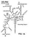
- One domain for use to derive the first nucleic acid structure of the analogs is the decoding region of 16S rRNA, which is located near the 3' end of 16S rRNA of E. coli (Figs. 1A and 1B).
- the experiments described below show that an oligoribonucleotide analog of the decoding region interacts with both antibiotic and RNA ligands of the 30S subunit in a manner that correlates with normal subunit function.
- the activities of the decoding region analog suggest that the intimidating structural complexity of the ribosome can be, to some degree, circumvented.
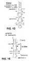
- a first oligoribonucleotide analog RNA derived from the decoding region of 16S rRNA, was made by transcribing, with T7 RNA polymerase (Milligan et al. , 1989, Meth. Enzymol., 180 :51-62), a linearized pGEM3 plasmid (Promega) as described in Zapp et al., 1993, Cell, 74 :969-978, containing the analog sequence shown in Fig. 1D (SEQ ID NO:1) flanked by EcoRI and BamHI restriction sites, and a 15 mer reverse primer annealing site immediately 3' of the BamHI site.
- This sequence includes both the first and second nucleic acid structures to form the complete analog.
- the first nucleic acid structure includes the nucleotide sequences 5'-ACCGCCCGUCACA-3' (SEQ ID NO:12) and 5'-UGAAGUCGUAACAAGG-3' (SEQ ID NO:13) derived from the complete decoding region.
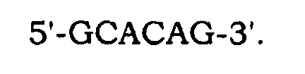
- the second nucleic acid structure includes a tetraloop 5'-CCUUCGGG-3', which is the minimum size of a tetraloop found to be sufficiently stable, and a nucleotide clamp made of the sequences 5'-GCACAG-3' and 3-CGUGUC-5'. Any other stable helix can be used in place of this particular nucleotide clamp. Likewise, any other stable stem loop can be used in place of this particular tetraloop.
- the second analog shown in Fig. 1E, was made in the same way, but is derived from the A-site subdomain of the decoding region, and has the nucleotides sequences 5'-CGUCACA-3' and 5'-UGAAGGUCG-3'.
- the second analog has the same tetraloop as in the first analog, but includes a shorter nucleotide clamp made of two nucleotide sequences 5'-GG-3' and 3'-CC-5'.
- the complete nucleotide sequence of the second analog is 5'-GGCGUCACACCUUCGGGUGAAGUCGCC-3' (SEQ ID NO:11).
- Interaction (or "binding") reactions (12.5 ⁇ l) containing 125 ng oligoribonucleotide analog RNA and antibiotics in 80 mM K-Hepes (pH 7.9), 50mM NH 4 Cl, and 5% PEG buffer were annealed at 37°C for 15 minutes and incubated on ice for 1 hour.
- DMS (1 ⁇ l of 1:5 in ethanol) was added, and modification reactions were incubated for 40 minutes on ice. Reactions were stopped with DMS Stop (Peattie et al. , 1980, Proc. Natl. Acad. Sci. USA, 77 :4679-82) and the RNA was purified by ethanol precipitation.
- DMS/N7 reactions were performed according to standard protocols (Peattie et al., supra ) except that lyophilization steps were replaced by acid-phenol extraction and ethanol precipitation.
- 10 ng analog RNA was annealed to 0.75 ng end-labeled primer and extended with 10-15 U MoMuLV reverse transcriptase for 1 hour. Reactions were stopped by ethanol precipitation, pellets resuspended in 10 ⁇ l 8M urea, 0.05X TBE loading buffer, and 2 ⁇ l was loaded onto 8%, 19:1 acrylamide:bisacrylamide, 0.5X TBE sequencing gels.
- Binding reactions were performed as described above for aminoglycoside protection experiments, except that binding buffer contained 200 mM NH 4 Cl and 80 mM MgCl 2 .
- E. coli tRNA phe anticodon stem-loop (GGGGAUUGAAAAUCCCC; SEQ ID NO:3) was transcribed with T7 RNA polymerase, gel purified, concentrated by ethanol precipitation, and annealed with a brief heat step (80°C/1 minute followed by 10 minutes on ice).
- tRNA pro anticodon stem-loop (GGUCAUCUUGGGG UGAUGACC; SEQ ID NO:4), scrambled tRNA phe (GGGAGCGUCAU CACAUA; SEQ ID NO:5), tetraloop element (GGGACUUGGGUCCC; SEQ ID NO:6), and triUloop element (GGCGCUUUGCGCC; SEQ ID NO:7) were transcribed and treated as described for the E. coli tRNA phe anticodon stem-loop. The assay and results are described below and in Fig. 4B.
- RNA structure analysis with chemical probes is a well established and powerful technique that allows individual atoms of nucleotide bases, or of the phosphodiester backbone, to be monitored for inter- or intra-molecular interactions.
- DMS dimethyl sulfate
- KE kethoxal
- Nucleotide base modifications that interfere with formation of Watson-Crick base pairs are typically monitored via reverse transcription, as the progress of the reverse transcriptase enzyme is strongly impeded when it encounters such modifications. This leads to production of a truncated reverse transcription product whose length effectively maps the position of the modified base in the RNA chain when the products are run on standard DNA sequencing gels.
- RNA strand scission For example, methylation of N7 of G by DMS can be monitored by treating the methylated RNA with sodium borohydride and aniline to induce strand scission at the positions of N7 methylation. Reverse transcriptase simply falls off the template at these points, again leading to a truncated product that maps the position of N7 methylation.
- oligoribonucleotide analog of the decoding region shown in Figs. 1C and 1D SEQ ID NO:1 as described above, and asked if it could interact with aminoglycoside antibiotics such as neomycin, which have previously been shown to protect N1 of A1408 (this notation indicates the adenine at location 1408) and N7 of G1491 and G1494 within the A-site subdomain of the decoding region (Moazed et al. , 1987, Nature , supra; Woodcock et al., supra ); similar interactions with the analog would be a strong indicator of its proper folding and functional potential.
- aminoglycoside antibiotics such as neomycin
- Figs. 1C, 1D, and 1E the interactions of aminoglycosides such as neomycin and paromomycin with decoding region oligoribonucleotide analogs bear a striking similarity to their counterpart interactions with the decoding region in subunits of complete 16S rRNA in ribosomes.
- the decoding region of ribosomes (Fig. 1A) and a small analog containing just the A-site subdomain (Fig. 1E) exhibit virtually identical aminoglycoside interactions, which are abolished by a single G to U transversion at position 1491.
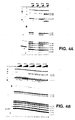
- RNA samples were probed with dimethyl sulfate (DMS; Stern et al. , 1988, Meth. Enzymol. , 164 :481-89) alone as naked RNA, or in the presence of increasing concentrations of neomycin, paromomycin, hygromycin, streptomycin, tetracycline, and erythromycin.
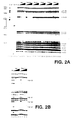
- Fig. 2A shows that neomycin (lanes 7-10) and the closely related aminoglycoside paromomycin (lanes 11-14) strongly protected N1 of A1408 at concentrations of 10 and 1 ⁇ M, respectively.
- hygromycin (lanes 15-18), a structurally dissimilar aminoglycoside, weakly enhanced the reactivity of N1 of A1408.
- streptomycin (lanes 19-22), tetracycline (lanes 23-26), and erythromycin (lanes 27-30), antibiotics that do not interact with the decoding region in 16S rRNA, did not detectably interact with the analogs.
- Fig. 2B shows that, in addition to N1 of A1408, N7 of G1405 and G1494 were strongly protected by neomycin and paromomycin, while N7 of G1491 and G1497 were weakly protected.
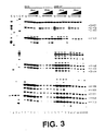
- N3 of C1402 and C1403 shows that the anticodon stem-loop transcript protected N3 of C1402 and C1403 (compare lane 6 with lanes 7-10) and N7 of G1401 (compare lane 16 with lanes 17-20), while poly U protected N1 of A1408, A1492, A1493, A1499, and A1502 strongly, N1 of A1500, A1503 weakly, and N7 of G1494 weakly (compare lane 6 with lanes 11-15 and lane 16 with lanes 21-25).
- nucleotides protected by mRNA are identical to those associated with the mRNA-dependent interaction of tRNA with the A-site of ribosomes (Moazed et al., 1986, Cell, supra ; Moazed et al. , 1990, J. Mol. Biol., supra ).
- the other nucleotides, protected by mRNA (A1499, A1500, A1502, and A1503), have not been previously identified with either A- or P-site function in chemical probing experiments.
- C1402 and C1403 are unreactive in 16S rRNA (Moazed et al. , 1986, J. Mol. Biol., 187 :399-416); their reactivity in the analog and their protection by the tRNA anticodon stem-loop transcript suggest that the structure of the P-site subdomain of the oligoribonucleotide analog differs from that of ribosomes.
- sctRNA phe coli tRNA phe with little potential for secondary structure formation (sctRNA phe ) (GGGAGCGUCAUCACAUA; SEQ ID NO:5), a highly stable tetraloop stem-loop element (tetraloop) (GGGACUUCGGUCCC; SEQ ID NO: 6) , and another, smaller stem-loop element with a loop composed of only three U residues (triUloop) (GGCGCUUUGCGCC; SEQ ID NO: 7) were each titrated into a binding reaction with the analog.
- Fig. 4B shows that only the tRNA phe and tRNA pro anticodon stem-loop transcripts interacted, each strongly protecting C1402 and C1403.
- the failure of the scrambled tRNA phe and other stem-loop elements to interact detectably supports the hypothesis that, despite its altered conformation, the P-site subdomain interacts selectively with RNA ligands.
- the oligoribonucleotides of the invention are diverse, in that they can be derived from any ribosomal RNA (e.g., 16S, 23S), human immunodeficiency virus (HIV) RNA (see Green (1993) AIDS Res. Rev. 3 :41-55 for examples of desirable regions such as Tat or Rev interaction sites), or any other RNA which can be manipulated to retain a functionally relevant structure.
- ribosomal RNA e.g., 16S, 23S
- HCV human immunodeficiency virus
- the decoding region of 16S rRNA and aminoglycoside antibiotics are featured in the experiments described herein, they are not the only region or chemical class which can be used in this invention.
- messenger RNA regulatory elements telomerase RNA (see, e.g., Bahattacharyya and Blackburn, EMBO J. , 13 :5721-31, 1994), oncogene mRNAs, cytokine.and lymphokine mRNAs, thymidylate synthase mRNAs, and various viral elements such as the adenovirus late mRNA tripartite leader, hepatitis delta virus RNA, and picornavirus "ribosomal landing pads," or "internal ribosomal entry sites,” located in the 5' untranslated region, can also be used to design oligoribonucleotide analogs of the invention.
- telomerase RNA see, e.g., Bahattacharyya and Blackburn, EMBO J. , 13 :5721-31, 1994
- oncogene mRNAs cytokine.and lymphokine mRNAs
- self-splicing group I introns including the HDV group I intron, are inhibited by the binding of aminoglycosides, and thus can be used to design RNA oligoribonucleotide analogs.
- the HIV Rev-Response-Element (RRE) is bound by some of the same antibiotics that bind to the decoding region of 16S rRNA. Neomycin binding to the RRE inhibits Rev function and viral replication in model cell culture systems.
- the RRE is a suitable target for designing oligoribonucleotide analogs that are useful to assay antibiotics effective against the RRE.
- RNA structural motif can be used as parental RNA to design an oligonucleotide analog that can be used as a screening target, and because the transport, processing, and translation of mRNA depend on such structures and their interactions with various binding partners, a large array of potential targets in viral and cellular mRNA exist. Extending these ideas even further, if one assumes that mutant cellular mRNA sequences produce altered mRNA structures, it may even be possible to selectively target specific mutant mRNA sequences with small molecules.
- the oligoribonucleotide analogs of the invention also contain second, heterologous sequences (e.g., artificial stem loops and nucleotide clamps) that promote the appropriate three-dimensional conformation of the first nucleotide sequences for normal molecular interactions with candidate drugs, nucleic acid molecules, etc.
- second, heterologous sequences e.g., artificial stem loops and nucleotide clamps
- the small size of these candidate drugs and molecules makes them amenable to numerous inexpensive in vitro assays that use a variety of oligoribonucleotide analogs.
- Protein synthesis inhibitors have two main ribosomal targets: the decoding region of 16S rRNA, and the peptidyl transferase region (Domain V) of 23S rRNA. Aminoglycosides interact with the decoding region while MLS antibiotics (macrolides, lincosamides, and streptogramins) interact with the peptidyl transferase region. Peptides, nucleic acid molecules, and a variety of other chemical compounds and molecules may be useful as therapeutics, and can easily be screened using the oligoribonucleotide analogs and methods of this invention.
- the oligoribonucleotide analogs enable the use of improved methods to screen for novel therapeutic compounds, particularly antibiotic compounds that inhibit protein synthesis.
- Standard methods to screen antibiotics involve detecting antimicrobial or bactericidal activity in cultured cells over long time periods (up to several days). Screening many potential antibiotic compounds under these conditions can be expensive and time consuming.
- screening assays using the oligoribonucleotide analogs of the invention have several distinct advantages over these known screening methods.
- the new screening assays are much more rapid (an assay according to the invention can be carried out in about an hour), the individual assays require only small amounts of materials (volumes of about 100 ⁇ l), and numerous reactions can be carried out in parallel (e.g., in a 96 well microtiter plate).
- likely candidate compounds can be rapidly identified, and only the likely candidates then could be further tested with cell culture assays.
- oligoribonucleotide analogs are small enough to be easily produced, either enzymatically as T7 polymerase transcripts, or chemically by automated RNA synthesis.
- the analogs are far more stable than ribosomes because they are radically simpler.
- their labile ribophosphodiester backbone can be stabilized by incorporation of 2'-O-methyl ribonucleotides, deoxyribonucleotides, or phosphorothioates, either uniformly or at selected positions.
- analogs can be readily derivatized during automated chemical synthesis.
- fluorescent moieties can be introduced at either termini (5' or 3'), or internally, and coupling moieties, such as primary amines or biotin, can be introduced at either termini (see discussion below).
- coupling moieties such as primary amines or biotin
- Aminoglycosides are polycations and charge-charge interactions with the negatively charged phosphodiester backbone of the oligoribonucleotide analogs are likely important determinants of their binding specificity and affinity.
- important factors governing the formation and stability of aminoglycoside-RNA interactions include mono- and divalent-cation (salt) concentrations, and the presence or absence of nonspecific nucleic acids. Accordingly, low salt concentrations ( ⁇ 100 mM K+, NH4+, Na+, ⁇ 5 mM Mg++) favor complex formation, and the presence of nonspecific nucleic acids disfavor it.
- Candidate compounds to be tested should, therefore, be prepared in low salt buffers largely free of contaminating nucleic acids.
- a standard microtiter plate format with a standard fluorescence or radiation detector is used.
- a robotic system is used to load, wash, and detect a signal in each well.
- Cation concentrations, nonspecific nucleic acids, and other variables including temperature and time can be adjusted during binding and washing stages to adjust background binding levels.
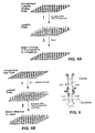
- Figs. 5A and 5B illustrate two specific screening assays based on 1) binding of test compounds to an immobilized (naked) oligoribonucleotide analog functioning as a reporter ("affinity assay"), and 2) displacement of a prebound reporter molecule from an immobilized analog by test compounds ("competitive binding assay”), respectively. These two methods, while distinct, are related in their overall strategy and organization.
- a third method (Figs. 7A, 7B, 8A, and 8B), based on oligomer binding to an in situ footprinted analog, differs in its organization and strategy from the first two methods, and will be discussed separately.
- This screening method can be implemented with an immobilized, fluorescently labeled oligoribonucleotide analog, as shown in Fig. 5A.
- the method consists of (1) incubation of the immobilized analog with test compounds, (2) wash step(s), and subsequent detection of complex formation relying on changes in the fluorescence properties of the immobilized RNA analog.
- the RNA analog can be derivatized, e.g., during automated chemical synthesis, both for immobilization and for labelling. Possible positions for derivatization on an oligoribonucleotide analog are indicated by boxes around the nucleotides in Fig. 6.
- the analog can be derivatized with either terminal biotin (using biotin phosphoramidite (5'-biotin) or CPG (3'-biotin)), or a free primary amino group (using amino modifier phosphoramidite (5'-NH2) or CPG (3'-NH2)).
- Immobilization in microtiter plate wells can then be accomplished with polystyrene plates activated with streptavidin or maleic anhydride, respectively. After coupling, plates should be thoroughly washed to eliminate soluble analog RNA.
- analogs are derivatized to allow the addition of a fluorophore at one of the RNA termini with fluorescein phosphoramidites (3' labeling) or CPG (5' labeling), or the incorporation of fluorescent adenosine or cytosine nucleotides (with EthenoA and EthenoC phosphoramidites, respectively) at specific internal positions in the RNA.
- fluorescein phosphoramidites 3' labeling
- CPG 5' labeling
- fluorescent adenosine or cytosine nucleotides with EthenoA and EthenoC phosphoramidites, respectively
- a fluorescent dye such as ethidium bromide, or possibly Hoechst 33258, can be used with underivatized RNA. This labelling allows the detection of any changes in either the binding or fluorescent properties of the dye, or both, upon test compound-analog complex formation.
- Typical (solution) binding reactions contain 25 mM neutral pH buffer (Tris or Hepes) and 50 mM monovalent salt (KCl, NH4Cl, etc). It is advantageous to carry out binding reactions at 30-37°C for approximately 30 minutes to facilitate the simultaneous annealing of the RNA. washes can utilize the same buffer.
- Test compound and salt concentrations should be adjusted empirically to optimize the assay.
- the test compound concentration should be in the micromolar range, and salt concentrations should be in the range of 10 to 500 mM, but these numbers must be adjusted depending on the particular assay conditions.
- nonspecific nucleic acids can be added to the binding step to reduce the effects of any contaminating compounds with nonspecific affinity for nucleic acids.
- the nonspecific nucleic acid could be total yeast tRNA, poly 1-C, etc.
- Another potentially useful nonspecific nucleic acid would be (soluble) A-site analog carrying the G1491U point mutation.
- this screening method is based on the hypothesis that useful test compounds will displace prebound soluble reporter ligands from their immobilized analog binding sites.
- immobilized neomycin an antibiotic ligand known to bind to analogs derived from the decoding region of 16S rRNA
- test compounds can be (1) pre-loaded with a labeled analog, and (2) test compounds added and allowed to compete with immobilized neomycin for the labeled analog, which serves as the reporter. After a wash step to remove any displaced analog, still complexed with the test compound, detection of the absence of label in specific wells would indicate that specific test compounds bound the analog RNA efficiently, a positive result.
- the reciprocal configuration, with immobilized analog RNA and labeled neomycin can also be used.
- Neomycin and other aminoglycosides contain numerous primary amino groups that are ideal targets for coupling to either amine-reactive fluorescent probes or activated polystyrene.
- a soluble neomycin reporter molecule can be produced by coupling neomycin to NHS-Fluorescein.
- neomycin can be immobilized on maleic anhydride-activated polystyrene microtiter plates. As discussed above, the oligoribonucleotide analog can be similarly labeled or immobilized.
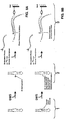
- Figs. 7A and 7B, and 8A and 8B illustrate a third screening method based directly on our chemical probing experience which has shown that the N1 atom of A1408 is strongly methylated by DMS in the absence of neomycin, and conversely, is strongly protected from DMS methylation in the presence of neomycin.
- this differential modification provides a method for detection of complex formation (monitored by inhibition of primer extension).
- Method 3 similarly exploits differential methylation, but monitors this methylation, e.g., via the differential annealing of a labeled complementary oligomer (e.g., CAGUGU) with a binding site overlapping the site of methylation, A1408 (Figs. 7A and 7B).
- a labeled complementary oligomer e.g., CAGUGU
- the N1 atom of A1408 is strongly methylated by dimethyl sulfate (DMS) in the absence of neomycin (Fig. 7A) and, conversely, strongly protected from DMS methylation in the presence of neomycin ("bound small molecule") (Fig. 7B).
- DMS dimethyl sulfate
- the mechanics of the assay are similar to Method 1 in that the first step involves forming complexes between the immobilized (but here unlabeled) analog and test compounds (Step 1). After complex formation and washing steps, the plate is treated with DMS to differentially methylate the RNA (Step 2). DMS reactions are then stopped with a 1M ⁇ ME wash, and labeled reporter oligo added and annealed (Step 3).
- Figs. 7A and 7B relies on the inability of N1-methylated A to form Watson-Crick base pairs with a short labeled oligomer.
- the short oligo fails to anneal to the analog and is washed away, leaving no label in that well.
- Fig. 7B with complexed analog and unmethylated N1, the oligo anneals to the analog and is effectively immobilized on it, leaving label in the well.
- Wells that retain the reporter oligo indicate that A1408 was not methylated, which in turn indicates that A1408 was protected by a bound antibiotic ligand.
- the presence of label in a well constitutes a positive result.
- RNA target analog
- a biotinylated primer e.g., having the nucleotide sequence TTCACCCGGAAGGTG; SEQ ID NO:12
- primer extension is initiated in the presence of labeled TTP (e.g., ⁇ - 32 P-TTP) and reverse transcriptase.
- Fig. 8A primer extension is inhibited by methylation of N1 of A1408 and no label is incorporated.
- Fig. 8B primer extension is successful, resulting in the incorporation of label into the primer.
- the biotinylated primer is immobilized via interaction with streptavidin-coated wells. After a wash step to remove unincorporated labeled TTP, only the scenario in Fig. 8B will allow retention of the label in the well, thus reporting the presence of a bound ligand in step 1.
- the reverse-transcriptase-based reporter methodology superficially resembles standard chemical probing methods.
Landscapes
- Chemical & Material Sciences (AREA)
- Life Sciences & Earth Sciences (AREA)
- Organic Chemistry (AREA)
- Proteomics, Peptides & Aminoacids (AREA)
- Analytical Chemistry (AREA)
- Zoology (AREA)
- Wood Science & Technology (AREA)
- Health & Medical Sciences (AREA)
- Engineering & Computer Science (AREA)
- Microbiology (AREA)
- Bioinformatics & Cheminformatics (AREA)
- Molecular Biology (AREA)
- Biotechnology (AREA)
- Biophysics (AREA)
- Physics & Mathematics (AREA)
- Biochemistry (AREA)
- Immunology (AREA)
- General Engineering & Computer Science (AREA)
- General Health & Medical Sciences (AREA)
- Genetics & Genomics (AREA)
- Measuring Or Testing Involving Enzymes Or Micro-Organisms (AREA)
- Pharmaceuticals Containing Other Organic And Inorganic Compounds (AREA)
- Saccharide Compounds (AREA)
Claims (28)
- Künstliches Oligoribonukleotid-Analog, das sowohl natürliche als auch heterologe Sequenzen darin enthält, wobei das Analog eine dreidimensionale Struktur aufweist, welche eine Ligandenbindungsregion eines größeren Stamm-Ribonukleinsäure (RNA)-Moleküls imitiert, wobei das Analog umfasst:eine erste Nukleinsäurestruktur, dessen Sequenz sich von dem Stamm-RNA-Molekül ableitet, und eine zweite Nukleinsäurestruktur, die eine heterologe Sequenz umfasst, die, wenn sie mit der ersten Nukleinsäurestruktur verbunden ist, das Analog bildet, und das Analog mit einer Konformation ausstattet, die den Liganden mit einem Ligandenbindungsmuster bindet, welches im Wesentlichen identisch mit dem Stamm-RNA-Bindungsmuster ist.
- Analog nach Anspruch 1, wobei die Sequenz der ersten Nukleinsäurestruktur identisch mit einer Ligandenbindungsregion einer 16S-ribosomalen RNA (rRNA) ist.
- Analog nach Anspruch 2, wobei die Region die dekodierende Region von 16S-rRNA umfasst.
- Analog nach Anspruch 3, wobei die Region die Nukleotide 1398-1410 und 1490-1505 von 16S-rRNA umfasst.
- Analog nach Anspruch 3, wobei die Region die A-Stellen-Subdomäne der dekodierenden Region von 16S-rRNA umfasst.
- Analog nach Anspruch 5, wobei die Region die Nukleotide 1404-1410 und 1490-1497 von 16S-rRNA umfasst.
- Analog nach Anspruch 1, wobei die zweite Nukleinsäurestruktur eine Tetra-Schleife umfasst.
- Analog nach Anspruch 7, wobei die Tetra-Schleife die Nukleotidsequenz umfasst: 5'-CCUUCGGG-3'.
- Analog nach Anspruch 1, wobei die zweite Nukleinsäurestruktur zwei Nukleotidsequenzen umfasst, die eine basengepaarte Nukleotidklemme bilden.
- Analog nach Anspruch 9, wobei die basengepaarte Nukleotidklemme die Nukleotidsequenz umfasst:

- Analog nach Anspruch 9, wobei die basengepaarte Nukleotidklemme die Nukleotidsequenz umfasst:


- Analog nach Anspruch 1, wobei die Sequenz der ersten Oligoribonukleinsäurestruktur sich von einer dekodierenden Region von 16S-rRNA ableitet und die zweite Nukleinsäurestruktur eine Tetra-Schleife und eine basengepaarte Nukleotidklemme umfasst.
- Analog nach Anspruch 12, wobei die dekodierende Region die Nukleotide 1398-1410 und 1490-1505 von 16S-rRNA umfasst, die Tetra-Schleife die Nukleotidsequenz 5'-CCUUCGGG-3' umfasst, die basengepaarte Nukleotidklemme die Nukleotidsequenz umfasst:
 und die vollständige lineare Nukleotidsequenz der kombinierten ersten und zweiten Nukleotidstrukturen des Analogs
 ist.
ist.
- Analog nach Anspruch 12, wobei die dekodierende Region die Nukleotide 1404-1410 und 1490-1497 von 16S-rRNA umfasst, die Tetra-Schleife die Nukleotidsequenz 5'-CCUUCGGG-3' umfasst, die basengepaarte Nukleotidklemme die Nukleotidsequenz umfasst:
 und die vollständige lineare Nukleotidsequenz der kombinierten ersten und zweiten Nukleotidstrukturen des Analogs
 ist.
ist.
- Affinitäts-Assays zum Bestimmen der potenziellen therapeutischen Aktivität einer Testverbindung, wobei der Assay die Schritte umfasst:Vermischen einer Testverbindung mit einem Analog nach Anspruch 1 unter Bedingungen, welche die Bildung eines Bindungskomplexes zwischen dem Analog und der Testverbindung ermöglichen undNachweisen der Bildung eines Bindungskomplexes, wobei das Vorliegen eines Bindungskomplexes anzeigt, dass die Testverbindung eine potenzielle therapeutische Aktivität besitzt.
- Assay nach Anspruch 15, wobei das Analog auf einer Oberfläche markiert und immobilisiert ist und der Bindungskomplex durch Beobachten von Veränderungen bei dem Signal der Markierung nachgewiesen wird, wenn eine Testverbindung an das Analog gebunden ist.
- Assay nach Anspruch 15, wobei das Analog auf einer Oberfläche immobilisiert ist, die Testverbindung markiert ist und der Bindungskomplex durch Nachweisen der Markierung, die an das immobilisierte Analog gebunden ist, nachgewiesen wird.
- Assay nach Anspruch 16, wobei das Analog Fluoreszenz- oder radioaktiv markiert ist.
- Kompetitiver Bindungs-Assay zum Bestimmen der potenziellen therapeutischen Aktivität einer Testverbindung, wobei der Assay die Schritte umfasst:Vermischen eines Analogs nach Anspruch 1 mit einem Analog-bindenden Liganden unter Bedingungen, welche die Bildung eines ersten Bindungskomplexes zwischen dem Analog und dem Liganden ermöglichen,Vermischen einer Testverbindung mit dem ersten Bindungskomplex unter Bedingungen, welche es der Testverbindung ermöglichen, den ersten Bindungskomplex zu trennen, um einen zweiten Bindungskomplex zwischen dem Analog und der Testverbindung zu bilden, undNachweisen der Trennung des ersten Bindungskomplexes, wobei die Trennung des ersten Bindungskomplexes anzeigt, dass die Testverbindung eine potenzielle therapeutische Aktivität besitzt.
- Assay nach Anspruch 19, wobei der Ligand markiert ist, das Analog auf einer Oberfläche immobilisiert ist und die Trennung des ersten Bindungskomplexes durch Beobachtung eines beliebigen Abfalls beim Signal der Markierung nachgewiesen wird, wenn eine Testverbindung den Liganden von dem ersten Bindungskomplex verdrängt.
- Assay nach Anspruch 19, wobei das Analog markiert ist, der Ligand auf einer Oberfläche immobilisiert ist und die Trennung des ersten Bindungskomplexes durch Beobachtung eines beliebigen Abfalls beim Signal der Markierung nachgewiesen wird, wenn eine Testverbindung das Analog von dem ersten Bindungskomplex verdrängt.
- Assay nach Anspruch 21, wobei das Analog Fluoreszenz- oder radiaktiv markiert ist.
- Assay nach Anspruch 21, wobei der Ligand Fluoreszenz- oder radioaktiv markiert ist.
- In situ-Footprinting-Assay zum Bestimmen der potenziellen therapeutischen Aktivität einer Testverbindung, wobei der Assay die Schritte umfasst:Vermischen eines Analogs nach Anspruch 1 mit einer Testverbindung unter Bedingungen, welche die Bildung eines Bindungskomplexes zwischen dem Analog und der Testverbindung ermöglichen,Inkubieren des Bindungskomplexes mit einem chemischen Sonden-Reagenz und Beobachten, ob das Reagenz auf das Analog in dem Komplex eine Wirkung hat,Inkubieren des Analogs, das an keine Testverbindung gebunden ist, mit dem chemischen Sonden-Reagenz und Beobachten, ob das Reagenz auf das ungebundene Analog in einer getrennten Kontrollreaktion eine Wirkung hat, undVergleichen von jeder der Wirkungen des Sonden-Reagenzes auf das Analog in dem Bindungskomplex und auf das ungebundene Analog, wobei eine Verhinderung einer Wirkung des Reagenz auf das Analog in dem Bindungskomplex, die durch das Reagenz auf dem ungebundenen Analog verursacht wird, anzeigt, dass die Testverbindung eine potenzielle therapeutische Aktivität besitzt.
- In situ-Footprinting-Assay nach Anspruch 24, wobei das chemische Sonden-Reagenz Dimethylsulfat, Kethoxal oder Carbodiimid ist.
- In situ-Footprinting-Assay nach Anspruch 24, wobei das Sonden-Reagenz ein Nukleotid in dem Analog kovalent modifiziert, wenn es an eine beliebige Testverbindung ungebunden ist.
- In situ-Footprinting-Assay nach Anspruch 26, wobei die Wirkung des Sonden-Reagenzes durch Verwendung eines markierten Oligonukleotids beobachtet wird, das an das Analog hybridisiert, wenn es durch die Testverbindung gegen Methylierung geschützt ist und das nicht an das Analog hybridisiert, wenn es durch das Reagenz methyliert ist, wobei das Vorliegen der Markierung nach Abschluss des Assays anzeigt, dass die Testverbindung eine potenzielle therapeutische Aktivität besitzt.
- Analog nach Anspruch 1, wobei der Ligand ein Aminoglykosid ist.
Applications Claiming Priority (5)
| Application Number | Priority Date | Filing Date | Title |
|---|---|---|---|
| US498402 | 1983-05-26 | ||
| US29445094A | 1994-08-23 | 1994-08-23 | |
| US294450 | 1994-08-23 | ||
| US08/498,402 US5712096A (en) | 1994-08-23 | 1995-07-05 | Oligoribonucleotide assays for novel antibiotics |
| PCT/US1995/010721 WO1996006106A1 (en) | 1994-08-23 | 1995-08-23 | HYBRIDIZATION AND FOOTPRINTING METHODS TO CHARACTERIZE THE INTERACTIONS OF 16S rRNA ANALOGUES FOR IDENTIFICATION OF NOVEL ANTIBIOTICS |
Publications (3)
| Publication Number | Publication Date |
|---|---|
| EP0804454A1 EP0804454A1 (de) | 1997-11-05 |
| EP0804454A4 EP0804454A4 (de) | 1999-12-22 |
| EP0804454B1 true EP0804454B1 (de) | 2004-05-26 |
Family
ID=26968533
Family Applications (1)
| Application Number | Title | Priority Date | Filing Date |
|---|---|---|---|
| EP95930926A Expired - Lifetime EP0804454B1 (de) | 1994-08-23 | 1995-08-23 | HYBRIDISIERUNGS- UND ABDRUCKVERFAHREN ZUR CHARAKTERISIERUNG ODER INTERAKTIONEN VON 16S-rRNS-ANALOGEN ZUR IDENTIFIZIERUNG NEUER ANTIBIOTIKA |
Country Status (6)
| Country | Link |
|---|---|
| US (1) | US5712096A (de) |
| EP (1) | EP0804454B1 (de) |
| AU (1) | AU3413495A (de) |
| CA (1) | CA2198261C (de) |
| DE (1) | DE69533092T2 (de) |
| WO (1) | WO1996006106A1 (de) |
Families Citing this family (26)
| Publication number | Priority date | Publication date | Assignee | Title |
|---|---|---|---|---|
| US6337183B1 (en) * | 1995-09-08 | 2002-01-08 | Scriptgen Pharmaceuticals, Inc. | Screen for compounds with affinity for nucleic acids |
| CA2282418A1 (en) | 1997-03-05 | 1998-09-11 | Scriptgen Pharmaceuticals, Inc. | Screen employing fluorescence anisotropy to identify compounds with affinity for nucleic acids |
| US6461815B1 (en) * | 1998-05-22 | 2002-10-08 | North Carolina State University | Antibacterial agents and methods of screening for the same |
| US6329145B1 (en) * | 1999-02-09 | 2001-12-11 | Gilead Science, Inc. | Determining non-nucleic acid molecule binding to target by competition with nucleic acid ligand |
| WO2001025188A1 (en) * | 1999-10-04 | 2001-04-12 | University Of Medicine And Dentistry Of New Jersey | Novel carbamates and ureas |
| US6316194B1 (en) * | 1999-12-16 | 2001-11-13 | Ribotargets | Methods and kits for discovery of RNA-binding antimicrobials |
| WO2001044505A2 (en) * | 1999-12-16 | 2001-06-21 | Ribotargets Limited | Methods and kits for discovery of rna-binding antimicrobials |
| US20030224375A1 (en) * | 2001-01-26 | 2003-12-04 | Pyle Anna Marie | Assays for evaluating the function of rna helicases |
| US6743586B2 (en) | 2001-01-29 | 2004-06-01 | The United States Of America As Represented By The Secretary Of The Commerce | Rapid fluorescence detection of binding to nucleic acid drug targets labelled with highly fluorescent nucleotide base analogs |
| US20050142545A1 (en) * | 2001-04-11 | 2005-06-30 | Conn Michael M. | Methods for identifying small molecules that bind specific rna structural motifs |
| US20060228730A1 (en) * | 2001-04-11 | 2006-10-12 | Rando Robert F | Methods for identifying small molecules that bind specific RNA structural motifs |
| CA2381627C (en) * | 2001-04-13 | 2010-06-22 | Mpl Innovations, Inc. | Solid lubricant and composition |
| US20050250133A1 (en) * | 2001-08-21 | 2005-11-10 | Ecker David J | Molecular interaction sites of 16S ribosomal RNA and methods of modulating the same |
| US20030092662A1 (en) * | 2001-08-21 | 2003-05-15 | Ecker David J. | Molecular interaction sites of 16S ribosomal RNA and methods of modulating the same |
| AU2002356187A1 (en) * | 2001-08-22 | 2003-03-10 | Isis Pharmaceuticals, Inc. | Molecular interaction sites of hepatitis c virus rna and methods of modulating the same |
| US20030130169A1 (en) * | 2001-10-01 | 2003-07-10 | The Board Of Trustees Of The University Of Illinois | Methods of treating drug-resistant bacterial infections |
| WO2004001010A2 (en) * | 2002-06-21 | 2003-12-31 | Ptc Therapeutics, Inc. | METHODS FOR IDENTIFYING SMALL MOLECULES THAT MODULATE PREMATURE TRANSLATION TERMINATION AND NONSENSE MEDIATED mRNA DECAY |
| EP1539944A4 (de) | 2002-07-01 | 2005-12-28 | Univ Wayne State | Verfahren und zusammensetzungen zur identifizierung von gegenüber antibiotikaresistenz unempfindlichen antibiotika |
| AU2003252136A1 (en) * | 2002-07-24 | 2004-02-09 | Ptc Therapeutics, Inc. | METHODS FOR IDENTIFYING SMALL MOLEDULES THAT MODULATE PREMATURE TRANSLATION TERMINATION AND NONSENSE MEDIATED mRNA DECAY |
| WO2005060573A2 (en) * | 2003-12-15 | 2005-07-07 | University Of Medicine And Dentistry Of New Jersey | Pna-neamine conjugates and methods for producing and using the same |
| CA2608093A1 (en) | 2005-05-11 | 2006-11-16 | Wayne State University | Novel targets for the identification of antibiotics that are not susceptible to antibiotic resistance |
| US8293517B2 (en) | 2005-05-11 | 2012-10-23 | Wayne State University | Methods and compositions for the identification of antibiotics that are not susceptible to antibiotic resistance in Pseudomonas aeruginosa |
| US7598040B2 (en) * | 2006-11-22 | 2009-10-06 | Trana Discovery, Inc. | Compositions and methods for the identification of inhibitors of protein synthesis |
| CA2699370A1 (en) * | 2007-09-14 | 2009-03-19 | Trana Discovery | Compositions and methods for the identification of inhibitors of retroviral infection |
| EP2342339A4 (de) * | 2008-09-29 | 2012-06-06 | Trana Discovery Inc | Screening-verfahren zur identifizierung spezifischer staphylococcus aureus-inhibitoren |
| US8362203B2 (en) | 2009-02-10 | 2013-01-29 | Wayne State University | Non-natural peptides as models for the development of antibiotics |
Family Cites Families (5)
| Publication number | Priority date | Publication date | Assignee | Title |
|---|---|---|---|---|
| US4330621A (en) * | 1980-08-25 | 1982-05-18 | Wisconsin Alumni Research Foundation | Assay for aminoglycosides |
| WO1981001419A1 (en) * | 1979-11-13 | 1981-05-28 | Wisconsin Alumni Res Found | Method for in activating and monitoring antibiotics and assaying for aminoglycosides |
| US5223618A (en) * | 1990-08-13 | 1993-06-29 | Isis Pharmaceuticals, Inc. | 4'-desmethyl nucleoside analog compounds |
| US5270170A (en) * | 1991-10-16 | 1993-12-14 | Affymax Technologies N.V. | Peptide library and screening method |
| WO1993023532A1 (en) * | 1992-05-14 | 1993-11-25 | Ribozyme Pharmaceuticals, Inc. | VIRUS RESISTANT PLANTS CONTAINING INDUCIBLE CYTOTOXIC mRNAs |
-
1995
- 1995-07-05 US US08/498,402 patent/US5712096A/en not_active Expired - Fee Related
- 1995-08-23 AU AU34134/95A patent/AU3413495A/en not_active Abandoned
- 1995-08-23 DE DE69533092T patent/DE69533092T2/de not_active Expired - Fee Related
- 1995-08-23 CA CA002198261A patent/CA2198261C/en not_active Expired - Fee Related
- 1995-08-23 WO PCT/US1995/010721 patent/WO1996006106A1/en not_active Ceased
- 1995-08-23 EP EP95930926A patent/EP0804454B1/de not_active Expired - Lifetime
Also Published As
| Publication number | Publication date |
|---|---|
| CA2198261A1 (en) | 1996-02-29 |
| AU3413495A (en) | 1996-03-14 |
| DE69533092T2 (de) | 2005-06-09 |
| CA2198261C (en) | 2003-10-28 |
| EP0804454A1 (de) | 1997-11-05 |
| EP0804454A4 (de) | 1999-12-22 |
| US5712096A (en) | 1998-01-27 |
| WO1996006106A1 (en) | 1996-02-29 |
| DE69533092D1 (de) | 2004-07-01 |
Similar Documents
| Publication | Publication Date | Title |
|---|---|---|
| US5712096A (en) | Oligoribonucleotide assays for novel antibiotics | |
| CA2366231C (en) | One step sample preparation and detection of nucleic acids in complex biological samples | |
| Famulok et al. | In vitro selection of specific ligand‐binding nucleic acids | |
| EP1778867B1 (de) | Verfahren und zusammensetzungen zum nachweis von nukleinsäuren in einer biologischen probe | |
| Green et al. | Mutations at nucleotides G2251 and U2585 of 23 S rRNA perturb the peptidyl transferase center of the ribosome | |
| US20210198732A1 (en) | Method | |
| CA2377567A1 (en) | High throughput assay system | |
| JPH06510201A (ja) | ダブルdループ形成の診断応用 | |
| JPH0437720B2 (de) | ||
| JPH0568597A (ja) | ミデイバリアントdna鋳型の増幅 | |
| JPH09506255A (ja) | 核酸の保護方法および分析方法 | |
| JP2001095590A (ja) | 核酸ハイブリダイゼーションアッセイのためのプローブ | |
| CN106460065A (zh) | 用于基因组应用和治疗应用的核酸分子的克隆复制和扩增的系统和方法 | |
| JPH09508268A (ja) | 核酸の配列決定 | |
| WO1998036100A1 (en) | Compositions, methods, kits and apparatus for determining the presence or absence of target molecules | |
| US8680257B2 (en) | Kits for comparative transcript analysis and mutant sequence enrichment | |
| EP1579005A1 (de) | Verfahren und zusammensetzungen zur analyse regulatorischer sequenzen | |
| EP1041160A1 (de) | Verfahren zur detektion einer mutation innerhalb einer basensequenz | |
| JPH04234999A (ja) | Qベータレプリカーゼを使用する選択的増幅系 | |
| WO1997044488A2 (en) | Compositions and methods for the detection of mycobacterium kansasii | |
| WO2004011606A2 (en) | Macromolecular protection assay | |
| Hershkovitz et al. | Ligation-mediated PCR for chromatin-structure analysis of interphase and metaphase chromatin | |
| Miao et al. | Yeast tRNA-splicing endonuclease cleaves precursor tRNA in a random pathway. | |
| EP0618983B1 (de) | Insertionselemente und amplifizierbare nukleinsäuren | |
| EP1002878A2 (de) | Hepatitis C Viren RNS-bindendes oligo DNS und Verfahren für dessen Herstellung |
Legal Events
| Date | Code | Title | Description |
|---|---|---|---|
| PUAI | Public reference made under article 153(3) epc to a published international application that has entered the european phase |
Free format text: ORIGINAL CODE: 0009012 |
|
| 17P | Request for examination filed |
Effective date: 19970321 |
|
| AK | Designated contracting states |
Kind code of ref document: A1 Designated state(s): CH DE FR GB IT LI |
|
| A4 | Supplementary search report drawn up and despatched |
Effective date: 19991110 |
|
| AK | Designated contracting states |
Kind code of ref document: A4 Designated state(s): CH DE FR GB IT LI |
|
| 17Q | First examination report despatched |
Effective date: 20010123 |
|
| GRAP | Despatch of communication of intention to grant a patent |
Free format text: ORIGINAL CODE: EPIDOSNIGR1 |
|
| GRAS | Grant fee paid |
Free format text: ORIGINAL CODE: EPIDOSNIGR3 |
|
| GRAA | (expected) grant |
Free format text: ORIGINAL CODE: 0009210 |
|
| AK | Designated contracting states |
Kind code of ref document: B1 Designated state(s): CH DE FR GB IT LI |
|
| REG | Reference to a national code |
Ref country code: GB Ref legal event code: FG4D |
|
| REG | Reference to a national code |
Ref country code: CH Ref legal event code: NV Representative=s name: RITSCHER & PARTNER AG PATENTANWAELTE Ref country code: CH Ref legal event code: EP |
|
| REF | Corresponds to: |
Ref document number: 69533092 Country of ref document: DE Date of ref document: 20040701 Kind code of ref document: P |
|
| PGFP | Annual fee paid to national office [announced via postgrant information from national office to epo] |
Ref country code: GB Payment date: 20040818 Year of fee payment: 10 |
|
| PGFP | Annual fee paid to national office [announced via postgrant information from national office to epo] |
Ref country code: FR Payment date: 20040819 Year of fee payment: 10 |
|
| PGFP | Annual fee paid to national office [announced via postgrant information from national office to epo] |
Ref country code: CH Payment date: 20040823 Year of fee payment: 10 |
|
| PGFP | Annual fee paid to national office [announced via postgrant information from national office to epo] |
Ref country code: DE Payment date: 20040930 Year of fee payment: 10 |
|
| ET | Fr: translation filed | ||
| PLBE | No opposition filed within time limit |
Free format text: ORIGINAL CODE: 0009261 |
|
| STAA | Information on the status of an ep patent application or granted ep patent |
Free format text: STATUS: NO OPPOSITION FILED WITHIN TIME LIMIT |
|
| 26N | No opposition filed |
Effective date: 20050301 |
|
| PG25 | Lapsed in a contracting state [announced via postgrant information from national office to epo] |
Ref country code: IT Free format text: LAPSE BECAUSE OF NON-PAYMENT OF DUE FEES Effective date: 20050823 Ref country code: GB Free format text: LAPSE BECAUSE OF NON-PAYMENT OF DUE FEES Effective date: 20050823 |
|
| PG25 | Lapsed in a contracting state [announced via postgrant information from national office to epo] |
Ref country code: LI Free format text: LAPSE BECAUSE OF NON-PAYMENT OF DUE FEES Effective date: 20050831 Ref country code: CH Free format text: LAPSE BECAUSE OF NON-PAYMENT OF DUE FEES Effective date: 20050831 |
|
| PG25 | Lapsed in a contracting state [announced via postgrant information from national office to epo] |
Ref country code: DE Free format text: LAPSE BECAUSE OF NON-PAYMENT OF DUE FEES Effective date: 20060301 |
|
| REG | Reference to a national code |
Ref country code: CH Ref legal event code: PL |
|
| GBPC | Gb: european patent ceased through non-payment of renewal fee |
Effective date: 20050823 |
|
| PG25 | Lapsed in a contracting state [announced via postgrant information from national office to epo] |
Ref country code: FR Free format text: LAPSE BECAUSE OF NON-PAYMENT OF DUE FEES Effective date: 20050831 |
|
| REG | Reference to a national code |
Ref country code: FR Ref legal event code: ST Effective date: 20111125 |