EP0807460A1 - Membrane cellulosique pour dialyse - Google Patents
Membrane cellulosique pour dialyse Download PDFInfo
- Publication number
- EP0807460A1 EP0807460A1 EP97107174A EP97107174A EP0807460A1 EP 0807460 A1 EP0807460 A1 EP 0807460A1 EP 97107174 A EP97107174 A EP 97107174A EP 97107174 A EP97107174 A EP 97107174A EP 0807460 A1 EP0807460 A1 EP 0807460A1
- Authority
- EP
- European Patent Office
- Prior art keywords
- acid
- membranes
- cellulose
- membrane
- water
- Prior art date
- Legal status (The legal status is an assumption and is not a legal conclusion. Google has not performed a legal analysis and makes no representation as to the accuracy of the status listed.)
- Withdrawn
Links
Images














Classifications
-
- B—PERFORMING OPERATIONS; TRANSPORTING
- B01—PHYSICAL OR CHEMICAL PROCESSES OR APPARATUS IN GENERAL
- B01D—SEPARATION
- B01D71/00—Semi-permeable membranes for separation processes or apparatus characterised by the material; Manufacturing processes specially adapted therefor
- B01D71/06—Organic material
- B01D71/08—Polysaccharides
- B01D71/12—Cellulose derivatives
- B01D71/14—Esters of organic acids
-
- B—PERFORMING OPERATIONS; TRANSPORTING
- B01—PHYSICAL OR CHEMICAL PROCESSES OR APPARATUS IN GENERAL
- B01D—SEPARATION
- B01D71/00—Semi-permeable membranes for separation processes or apparatus characterised by the material; Manufacturing processes specially adapted therefor
- B01D71/06—Organic material
- B01D71/08—Polysaccharides
- B01D71/10—Cellulose; Modified cellulose
-
- B—PERFORMING OPERATIONS; TRANSPORTING
- B01—PHYSICAL OR CHEMICAL PROCESSES OR APPARATUS IN GENERAL
- B01D—SEPARATION
- B01D71/00—Semi-permeable membranes for separation processes or apparatus characterised by the material; Manufacturing processes specially adapted therefor
- B01D71/06—Organic material
- B01D71/08—Polysaccharides
- B01D71/12—Cellulose derivatives
-
- B—PERFORMING OPERATIONS; TRANSPORTING
- B01—PHYSICAL OR CHEMICAL PROCESSES OR APPARATUS IN GENERAL
- B01D—SEPARATION
- B01D71/00—Semi-permeable membranes for separation processes or apparatus characterised by the material; Manufacturing processes specially adapted therefor
- B01D71/06—Organic material
- B01D71/08—Polysaccharides
- B01D71/12—Cellulose derivatives
- B01D71/22—Cellulose ethers
Definitions
- the invention relates to cellulosic dialysis membranes, processes for their production and their use in particular for hemodialysis, hemofiltration and hemodiafiltration, cellulosic being understood to mean consisting of cellulose or modified cellulose, mixtures of cellulose and modified cellulose, optionally in a mixture with synthetic polymers .
- Dialysis membranes made of synthetic or natural polymers can very easily cause blood to clot when used in artificial kidneys, but this is largely prevented by appropriate medication.
- Leukopenia is a decrease in the number of leukocytes (white blood cells) in the bloodstream.
- the number of white blood cells in humans is approximately 4,000 to 12,000 cells / mm 3 .
- Leukopenia is most pronounced in dialysis 15 to 20 minutes after the start of treatment, whereby the neutrophils can disappear almost completely. After that, the number of leukocytes recovers to almost the original value or exceeds it within about an hour. If a new dialyzer is connected after the leukocytes have recovered, leukopenia occurs again to the same extent.
- the complement system within the blood serum is a complex, multi-component plasma enzyme system that serves in various ways to ward off damage caused by the invasion of foreign cells (bacteria, etc.). If antibodies against structures of the foreign surface are present, the complement system can be activated in the classic way, otherwise the complement activation takes place in an alternative way through special features of the foreign surface.
- the complement system consists of a large number of plasma proteins. After activation, these proteins react with each other specifically in a certain order, and in the end a cell-damaging complex is formed that destroys the foreign cell.
- Peptides are released from individual components, which trigger inflammation symptoms and can occasionally also have undesirable pathological consequences for the organism. It is believed that activation in regenerated cellulose hemodialysis membranes occurs via the alternative route. These complement activations are objectively determined by determining the complement fragments C3a and C5a.
- complement activation was assessed on the basis of fragments C5a.
- 300 ml of heparinized blood were recirculated in vitro over a period of 3 hours at a flow of 250 ml / min through a dialyzer with an effective exchange surface of 1 to 1.3 m 2 .
- the C5a fragments were determined using the ELISA method developed by Behring-Werke Marburg (enzyme-linked-immunosorbent assay).
- the relative complement activation for the respective time of measurement was calculated by forming the ratio from the concentration at the time of sampling to the concentration at the beginning in percent.
- the measured value after 3 hours of recirculation was used for the evaluation.
- Flat membranes are incubated with heparinized blood plasma for 3 hours and then the C5a fragments are determined.
- Thrombogenicity was assessed using TAT (thrombin-antithrombin) and PC (platelet count).
- beta-2 microglobulin level is observed in long-term dialysis patients after using membranes made from regenerated cellulose and is attributed to the fact that these membranes are less permeable in the molecular weight range of 1000 to 20,000 and the microglobulins are therefore not removed to a sufficient extent during dialysis will.
- the beta-2-microglobulin is not adsorbed to any appreciable extent on the usual membranes made from regenerated cellulose.
- the cellulose derivatives used according to the invention can contribute to this in an unexpected manner.
- the average degree of polymerization (DP) of the cellulose was determined in a Cuen solution according to DIN 54270.
- cellulosic dialysis membranes which are to be used in particular in hemodialysis.
- the membrane is said to be biocompatible, ie in particular blood compatible. Complement activation should be as low as possible, and the membrane should also have no thrombogenicity.
- UFR ultrafiltration rate
- the membranes should be resistant to aging, ie they should not change their properties during storage, they should be sterilizable using known methods such as steam, ethylene oxide and radiation treatment Manufacturing processes that are environmentally friendly and economical are interested, ie processes in which the environment is not polluted by the disposal of chemicals and which enable high productivity, in particular high production speed.
- ultrafiltration rate UFR
- sieving coefficient SK
- DE-C2-3 021 943 discloses a process for producing a dialysis membrane made of cellulose, in which a spinning solution is prepared from a mixture of cellulose, a tertiary amine oxide and, if appropriate, the cellulose-free diluents and other customary additives, and this is carried out by extruding a nozzle into a precipitation bath. Coagulation forms a dialysis membrane, which is still washed and, after the addition of plasticizer, is dried and wound up with shrinkage hindrance between 50 and 110 ° C.
- the membranes have a very dense structure, the ultrafiltration rate is very low, so they can at best be used as a so-called "low flux" membrane.
- the membranes described there must be kept in moisture-tight containers, since they have a very labile structure and very quickly undergo irreversible structural changes.
- the object of the invention is therefore to provide a process for the production of cellulosic dialysis membranes which has high productivity and is economically feasible, which provides membranes which are storable under normal conditions and resistant to aging and have a stable structure with which targeted Membranes for the low, middle and high flux range are available, with which cellulose derivatives, mixtures of cellulose and cellulose derivatives, possibly in a mixture with synthetic polymers, can be processed to biocompatible membranes, which compete with many properties with favorable properties of synthetic membranes can, with which it is also possible to produce membranes which have an adsorption capacity or an affinity for certain substances present in the blood, such as, for example, ⁇ -2 microglobulin, LDL (low density lipoprotein) or toxins which are not thrombogenic and which are sterile sizable, and with the targeted and reproducible by choosing the polymer composition and concentration and the process parameters, the structure of the membrane, such as porosity and surface quality, can be influenced.
- Another object is to provide a process in which the basic structure of the cellulose is not subjected to oxidative damage and thus the UV spectrum of the cellulosic polymer material of the membrane produced, which is dissolved in cadoxes, largely in the range of wavelengths from 245 to 320 nm UV Spectrum of the starting material corresponds, ie the difference between the extinction of the material of the membrane produced and the extinction of the starting material is low and is preferably between 0 and 15%.
- This object is achieved by a process for the production of stable and storable cellulose dialysis membranes for the low, middle or high flux range with UFR values of 4 to 15, 15 to 50 and over 50 ml / hm 2 mm Hg in the form of flat, tubular or hollow fiber membranes by producing a spinning solution from 3 to 40 parts of cellulose with a degree of polymerization (DP)> 300 and / or modified cellulose with a degree of polymerization (DP)> 200, 97 to 60 parts of a mixture of tertiary amine oxide and one or more other liquids such as water, dimethylformamide, dimethylacetamide, dimethyl sulfoxide, N-methylpyrrolidone, one or more stabilizers, and optionally one or more pore formers, other spinning solution additives and, if appropriate, low molecular weight organic or inorganic compounds and / or synthetic polymers, extruding this solution by a nozzle in a precipitation bath at a temperature of 10-95 ° C,
- the invention further relates to dialysis membranes according to claims 36 to 47.
- Another object of the invention is the use of the dialysis membrane as a low-, middle- or high-flux membrane, in particular for hemodialysis, hemofiltration and hemodiafiltration, as specified in claim 48.
- Stable and storable in the context of the present invention means that the membranes under normal conditions, as are usually the case in rooms such as sick rooms, medical treatment rooms, storage rooms and the like. rule, do not undergo any significant changes. So they do not need to be stored in moisture-proof containers or in conditioned rooms.
- the structure of the membrane is so fixed after its manufacture that the membrane is resistant to aging and can be stored for a long time.
- the process can be carried out at high production speeds; it works very economically because, among other things, can be worked with high polymer concentrations.
- the use of a cosolvent can be advantageous for the preparation of the spinning solutions.
- Cellulose or cellulose derivatives with a lower DP dissolve more easily and allow high concentrations.
- the solubility and thus the concentration of the solutions can also be increased by modifying the cellulose or by using mixtures or blends of cellulosic compounds of different molecular weights.
- the process allows the properties and structure of the membranes to be influenced in a targeted and reproducible manner by varying individual or multiple parameters. For example, increase the ultrafiltration rate (UFR) with increasing precipitation bath temperature.
- UFR ultrafiltration rate
- the structure and of course the UFR can be influenced by the composition of the precipitation medium.
- the structure of the surface of the membranes can be controlled by means of the hydrophilicity of the precipitant.
- more hydrophilic precipitants, in particular water result in denser skin, whereas more hydrophobic materials such as isopropyl myristate lead to open-pore, more porous surfaces.
- the composition can be used of the cavity-forming fluid influence the structure of the inner surface of hollow fiber membranes.
- the structure of the membranes can also be influenced by the concentration of the spinning solution, as is shown in various examples.
- the conditioning of the air gap also has a means in hand to influence the formation of the structures.
- the avivage i.e. in particular their composition, contribute to the structure formation and fixation of the same.
- dialysis membranes of excellent quality can be produced in a simple manner according to the invention, which can be set as desired for the low, middle or high flux range. They have a stable structure and therefore do not need to be stored under special conditions, which is a great advantage especially during transport and storage. Since the membranes do not change their structure for a long time, the UFR or dialysis performance remains constant even after prolonged storage.
- the cellulose and the cellulose derivatives can be mixed very well with synthetic polymers Process membranes. Furthermore, low-molecular compounds such as fatty alcohols, for example stearyl alcohol, fatty oils and the like, can be incorporated in an outstanding manner according to the invention, as a result of which the compatibility with blood can be improved. Such components do not need to be extracted during washing, since they are non-toxic and hardly soluble in blood.
- the membranes have a favorable porosity and can be set with a sharp separation limit. For example, it is possible to produce dialysis membranes that still allow beta-2 microglobulin to pass through, but are impermeable to higher molecular weight proteins.
- Mixtures of celluloses or cellulose derivatives of different molecular weights can also be processed according to the invention.
- modified membranes with very good biocompatibility within the scope of the invention, i.e. such as cause less leukopenia and less complement activation. Furthermore, these membranes are less thrombogenic.
- the melt was degassed in the degassing zones at a pressure of 100 mbar, air and a total of 1,796 kg of water being removed.
- the resulting spinning solution was filtered and fed by means of a measuring pump to a hollow fiber spinneret heated to 120 ° C., which was arranged at a distance of 70 mm above the surface of the precipitation bath.
- the outer diameter of the nozzle bore was 1140 ⁇ m, that of the core needle 830 ⁇ m.
- Isopropyl myristate was used as the cavity-forming liquid.
- Demineralized water at 40 ° C was used as the precipitation bath.
- the take-off speed was 200 m / min and the delay was 6.68.
- the hollow fiber was first washed with demineralized water, then treated with a solution of 30% by weight glycerol, 20% by weight isopropanol and 50% by weight water and dried at 50 ° C.
- the hollow fiber obtained by this procedure had the following properties: 1 N-methylmorpholine-N-oxide 2 fully desalinated
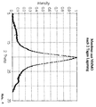
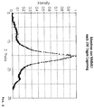
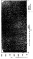
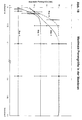
- X-ray wide-angle spectra on isotropic samples were after the 2nd or 100th day storage in transmission (Fig. 1 and 2). It can be seen from the spectra that the membranes are stable to aging with regard to the parameters mentioned above.
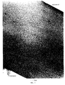
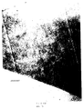
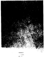
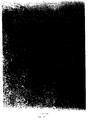
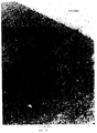
- the microscopic examinations (see Fig. 3, 4, 5, 6, 7) indicate a porous membrane with skin formation on the outside.
- the samples were embedded in nanoplast to determine the porosity and pore size of capillary membranes. Sections about 90 nm thick were then made on the ultramicrotome.
- the sections were examined in a transmission microscope (Philips CM12) and photographed at a magnification of 55000: 1 over the entire membrane cross section.
- the mean free path ( ⁇ ) in the pore structure was determined, which is defined as the mean of the distances between the pore boundaries.
- a large number of parallel measurement lines (Fig. 9) were created for their measurement with the image analysis and their length distribution within the areas identified as pores was determined.
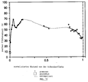
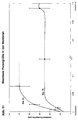
- Fig. 10 shows the course of the pore size in a hollow fiber membrane according to the invention.
- the skin on the outer surface has only tiny pores directly on its surface, the size of which is below 10 nm.
- the pore size quickly increases to over 50 nm and then moves according to the curve to about 30 nm on the inner wall.
- the pores on the surface of the inner wall are significantly larger than those on the skin of the outer wall.
- a large part of the pores, calculated from the inner wall, i.e. more than 75% is larger than the pores on the inner wall.
- Fig. 11 shows the same curve, plotted on a membrane section as the background.
- Fig. 12 shows the porous area.
- the porosity on the outer surface is extremely low, then increases quickly, similarly to the pore size, and then drops back to about 40%. It can be seen from this that the inner surface is porous, whereas the outer surface is considerably denser.
- Fig. 13 shows the same curve on a membrane section as a background.
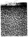
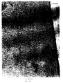
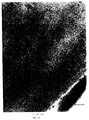
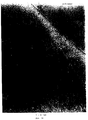
- the microscopic examination (see Fig. 14, 15, 16, 17) also confirms that this is a very dense membrane.
- the pore sizes can only be estimated and are far below 10 nm.
- the porosity cannot be determined by means of computer image analysis due to the very small pores.
- the resulting spinning solution was filtered and fed by means of a measuring pump to a hollow fiber spinneret heated to 120 ° C., which was arranged at a distance of 110 mm above the surface of the precipitation bath.
- the outer diameter of the nozzle bore was 1140 ⁇ m, that of the core needle 830 ⁇ m.
- Isopropyl myristate was used as the cavity-forming liquid.
- Demineralized water at 40 ° C was used as the precipitation bath.
- the withdrawal speed was 200 m / min and the delay 9.43.
- the hollow fiber was first washed with demineralized water, then treated with a solution of 15% by weight glycerol, 35% by weight isopropanol and 50% by weight water and dried at 50 ° C.
- the hollow fiber obtained by this procedure had the following properties: Inside diameter ( ⁇ m) 195 Wall thickness ( ⁇ m) 20th UFR water (ml / hm 2 mm Hg) 33 Sieving coefficient cytochrome C
- the melt was degassed in the degassing zones at a pressure of 100 mbar, air and a total of 1.796 kg of water being removed.
- the resulting spinning solution was filtered and fed by means of a measuring pump to a hollow fiber spinneret heated to 120 ° C., which was arranged at a distance of 110 mm above the surface of the precipitation bath.
- the outer diameter of the nozzle bore was 1140 ⁇ m, that of the core needle 830 ⁇ m.
- Isopropyl myristate was used as the cavity-forming liquid.
- Demineralized water at 40 ° C was used as the precipitation bath.
- the withdrawal speed was 200 m / min and the delay 9.43.
- the hollow fiber was first washed with demineralized water, then treated with a solution of 15% by weight glycerol, 35% by weight isopropanol and 50% by weight water and dried at 50 ° C.
- the hollow fiber obtained by this procedure had a porous structure with a maximum pore size of 15 to 20 nm and a somewhat denser one However, the outer and inner surfaces were still open (see Fig. 18, 19, 20) and showed the following properties: 3 Degree of modification or degree of substitution
- the melt was degassed in the degassing zones at a pressure of 100 mbar, air and a total of 1,796 kg of water being removed.
- the resulting spinning solution was filtered and fed by means of a measuring pump to a hollow fiber spinneret heated to 120 ° C., which was arranged at a distance of 110 mm above the surface of the precipitation bath.
- the outer diameter of the nozzle bore was 1140 ⁇ m, that of the core needle 830 ⁇ m.
- Isopropyl myristate was used as the cavity-forming liquid.
- Demineralized water at 40 ° C was used as the precipitation bath.
- the withdrawal speed was 200 m / min and the delay 9.43.
- the hollow fiber was first washed with demineralized water, then treated with a solution of 15% by weight glycerol, 35% by weight isopropanol and 50% by weight water and dried at 50 ° C.
- the hollow fiber obtained in this way had a porous structure with a maximum pore size of 15 to 20 nm and a denser but still open outer and inner surface (see Fig. 18, 21, 22) and showed the following properties: Inside diameter ( ⁇ m) 200 Wall thickness ( ⁇ m) 13 UFR water (ml / hm 2 mm Hg) 61 Sieving coefficient cytochrome C 0.70 Sieve coefficient albumin 0.06
- the melt was degassed in the degassing zones at a pressure of 100 mbar, air and a total of 1,796 kg of water being removed.
- the resulting spinning solution was filtered and fed by means of a measuring pump to a hollow fiber spinneret heated to 120 ° C., which was arranged at a distance of 110 mm above the surface of the precipitation bath.
- the outer diameter of the nozzle bore was 1140 ⁇ m, that of the core needle 830 ⁇ m.
- Isopropyl myristate was used as the cavity-forming liquid.
- Demineralized water at 40 ° C was used as the precipitation bath.
- the withdrawal speed was 200 m / min and the delay 9.43.
- the hollow fiber was first washed with demineralized water, then treated with a solution of 15% by weight glycerol, 35% by weight isopropanol and 50% by weight water and dried at 50 ° C.
- the hollow fiber obtained by this procedure had a clearly recognizable porosity with a maximum pore size of 10 to 15 nm (see Fig. 18) and showed the following properties: Inside diameter ( ⁇ m) 200 Wall thickness ( ⁇ m) 13 UFR water (ml / hm 2 mm Hg) 37 Sieving coefficient cytochrome C 0.57 Sieve coefficient albumin 0.004
- the melt was degassed in the degassing zones at a pressure of 100 mbar, air and a total of 2.280 kg of water being removed.
- the resulting spinning solution was filtered and fed by means of a measuring pump to a hollow fiber spinneret heated to 120 ° C., which was arranged at a distance of 110 mm above the surface of the precipitation bath.
- the outer diameter of the nozzle bore was 1140 ⁇ m, that of the core needle 830 ⁇ m.
- As cavity-forming liquid was used isopropyl myristate. Demineralized water at 40 ° C was used as the precipitation bath.
- the withdrawal speed was 120 m / min and the delay was 5.2.
- the hollow fiber was first washed with demineralized water, then treated with a solution of 15% by weight glycerol, 35% by weight isopropanol and 50% by weight water and dried at 50 ° C.
- the hollow fiber obtained by this procedure had a porous structure with a maximum pore size of ⁇ 5 nm (see Fig. 18) and showed the following properties: Inside diameter ( ⁇ m) 225 Wall thickness ( ⁇ m) 25th UFR water (ml / hm 2 mm Hg) 17th Sieving coefficient cytochrome C 0.17 Sieve coefficient albumin 0.005
- a hollow fiber was spun from the spinning solution of Example 1 according to the same procedure, precipitated with water at 85 ° C., then washed with water, with a mixture of glycerol (30% by weight), isopropanol (20% by weight) and water (50% by weight) ) finished and dried at 50 ° C.
- the hollow fiber shows the following dialysis properties: UFR water (ml / hm 2 mm Hg): 160 Sieving coefficient cytochrome C: 0.95 Sieve coefficient albumin: 0.22
- the C 5a activation is reduced by more than 98%.
- Thrombogenicity and heparin absorption are of the same order of magnitude as with Cuprophan.
- Isopropyl myristate was used as the cavity-forming liquid.
- the take-off speed was 250 m / min.
- the hollow fiber was first washed with demineralized water, then treated with a solution of 15% by weight glycerol, 35% by weight isopropanol and 50% by weight water and dried at 50 ° C.
- the hollow fiber obtained by this procedure had the following properties: Inside diameter: 200 ⁇ m Wall thickness: 10 ⁇ m UFR water (ml / hm 2 mm Hg): 98 Sieving coefficient cytochrome C: 0.82 Sieve coefficient albumin: 0.08
- the membranes were washed thoroughly with water, treated with a solution of 10% by weight glycerin, 40% by weight ethanol and 50% by weight water, stretched on a frame and dried at 22 ° C.
- the flat membrane produced using this procedure was 22 ⁇ m thick and had the following properties: UFR water (ml / hm 2 mm Hg): 55 Sieving coefficient cytochrome C: 0.76 Sieve coefficient albumin: 0.04
- C 5a activation is reduced by over 95%.
- Thrombogenicity and heparin absorption are of the same order of magnitude as with Cuprophan.
- Thrombogenicity and heparin absorption are of the same order of magnitude as with Cuprophan.
- the membranes were washed thoroughly with water, ethanol and water, treated with a solution of 40% by weight glycerol, 10% by weight isopropanol and 50% by weight water, stretched on a frame and dried at 22 ° C.
- the flat membrane produced by this procedure had a DS of 0.11, was 15 ⁇ m thick and showed an 81% reduction in C 5a activation compared to the unmodified membrane. Thrombogenicity and heparin absorption are of the same order of magnitude as with Cuprophan.
- NMMO solutions with 12% cellulose content were prepared with the addition of a water-soluble additive and processed into flat membranes. Water at 20 ° C. served as the precipitation bath. The membranes were washed with deionized water and treated with a mixture of glycerol, ethanol and water (5:45:50% by weight) before drying. The following UFR water values were measured (Table 4).
- the membranes from Example 49 were treated with a glycerol / ethanol / water mixture (20:30:50% by weight) which contained 5% by weight glycidyl ether for one hour at 20 ° C., stretched on a frame, only when Pre-dried at 22 ° C and then cured at 60 ° C for 24 hours.
- the membrane thickness was 19 ⁇ m and the UFR water value 500 - 600 ml / hm 2 mm Hg.
- Example 2 Analogously to Example 1, a spinning solution was prepared from Linter's cellulose and an additive, each 20% based on cellulose, and processed to form capillary membranes.
- the membranes produced by this procedure had the dialysis properties listed in Table (6).
- Table 6 example Additives (20% by weight) UFR water ml / hm 2 mm Hg Siebk cc Siebk albumin 51 Sorbitol 84 0.83 0.08 52 Glycerin 77 0.80 0.07 53 Polyvinyl pyrrolidone 77 0.80 0.09 54 Microcrystalline cellulose 40 0.67 0.04 55 PEG 1000 66 0.89 0.09
- an NMMO spinning solution with 6% polymer content which consisted of 75% by weight of Linter's cellulose and 25% by weight of benzyl cellulose with a DS of 0.24, was prepared and spun to form capillary membranes.
- Precipitation media of different compositions were used for coagulation (see Table 7).
- the distance between the spinneret and the precipitation bath surface was 100 mm.
- Isopropyl myristate was used as the cavity-forming liquid.
- the capillary membranes were first washed with demineralized water, then treated with a solution of 20% by weight glycerol, 30% by weight isopropanol and 50% by weight water and dried at 50 ° C.
- the membranes produced by this procedure had the dialysis properties listed in Table 7.
- Table 7 example Precipitation medium Falling bath temp. (° C) Trigger speed (m / min) UFR water ml / hm 2 mm Hg Siebk cc Siebk albumin 56 Water / NMMO 80/20 40 150 85 0.81 0.09 57 Water / NMMO 90/10 40 200 55 0.82 0.08 58 Ethanol 22 200 67 0.70 0.01 59 Isopropanol 22 100 120 0.92 0.20 60 Water / NMP 90/10 21 100 64 0.85 0.09 61 Water / DMSO 90/10 29 100 61 0.84 0.08
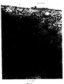
- Example 58, 59 and 60 Microscopic examination of the membranes of Examples 58, 59 and 60 shows that they are porous, the maximum pore size of Example 59 between 30 and 40 nm, that of Example 58 between 20 and 30 nm and that of Example 60 between 15 and 20 nm lies (see Fig. 23). Accordingly, the membrane structure is significantly influenced by the precipitant. With increasing hydrophilicity of the precipitating agent, a skin thickens or forms in the area of the outer surface, as is shown by the membrane from example 58 (see Fig. 24). Due to the lipophilic lumen filling, the inner wall is more open and large-pored (see Fig. 25).
- Example 3 an NMMO spinning solution with 6% polymer content, which consisted of 75% by weight of Linter's cellulose and 25% by weight of benzyl cellulose with a DS of 0.24, was prepared and spun to form capillary membranes. Liquids with different compositions were used to form cavities (lumen filling). With the exception of Examples 65 (20 mm), 68 (5 mm) and 69 (5 mm), the distance between the spinneret and the precipitation bath surface was 100 mm. Demineralized water at 24 to 60 ° C was used as the precipitation bath. The take-off speed was 100 to 200 m / min.
- the capillary membranes were first washed with demineralized water, then treated with a solution of 20% by weight glycerin, 30% by weight isopropanol and 50% by weight water (exception example 68; finishing composition: 10% by weight glycerin, 40% by weight isopropanol and 50% by weight). Water) and dried at 50 ° C.
- the membranes produced by this procedure had the dialysis properties listed in Table 8.
- Table 8 example Lumen filling Falling bath temp.
- the membranes from Examples 67 and 69 are porous.
- the membranes spun with an inner filling of glycerol / water / NaCl (example 67)
- a compaction can be observed in the area of the outer surface and skin formation in the area of the inner surface (see Fig. 26, 27).
- the maximum pore size is 30 to 40 nm (see Fig. 28).
- the membranes spun with PEG 400 (Example 69) have a compression on the outer wall and a slight compression on the inner wall compared to the directly adjacent coarse-pored structure (see Fig. 29,30).
- the maximum pore size is between 25 and 55 nm (see Fig. 28).
- capillary membranes were spun from an NMMO spinning solution with 6% polymer content, which consisted of 75% by weight Linters cellulose and 25% by weight benzyl cellulose with a DS of 0.24. Demineralized water at 77 ° C was used as the precipitation bath. The capillary membranes were treated with finishing agents of different compositions and their dialysis performance was checked. The results are shown in Table 9.
- NMMO spinning solutions with different polymer contents were prepared and spun into capillary membranes.
- the polymer consisted of 75% by weight of Linters cellulose and 25% by weight of benzyl cellulose with a DS of 0.24.
- the distance from the spinneret to the precipitation bath surface was 110 mm.
- Isopropyl myristate was used as the cavity-forming liquid.
- Demineralized water from 26 to 80 ° C was used as the precipitation bath.
- the take-off speed was 200 m / min.
- the capillary membranes were first washed with demineralized water, then treated with a solution of 20% by weight glycerol, 30% by weight isopropanol and 50% by weight water and dried at 50 ° C.
- the membranes had the dialysis properties listed in Table 10.
- Table 10 example Polymer content of the spinning solution (%) Falling bath temp. (° C) UFR water (ml / hm 2 mmHg) Sieve cc Sieve albumin 75 6 26 49 0.67 0.013 76 6 40 59 0.81 0.07 77 6 60 113 0.85 0.21 78 8th 40 28 0.45 0.005 79 8th 60 39 0.59 0.009 80 8th 80 51 0.70 0.10 81 10th 60 9 0.30 0.003 82 10th 80 12th 0.45 0.03
- Example 78 and 80 Microscopic examinations of the membranes of Examples 78 and 80 also show that the membranes are porous and that the pore size increases significantly with increasing temperature of the precipitation bath (see Fig. 31).
- the maximum pore size of the membranes of Example 80 is between 45 and 50 nm and that of Example 78 is between 20 and 30 nm.
- an NMMO spinning solution with 6% polymer content which consisted of 75% by weight of Linter's cellulose and 25% by weight of benzyl cellulose, was prepared and spun to form capillary membranes.
- the take-off speed was varied between 50 and 300 m / min.
- the distance between the spinneret and the precipitation bath surface was 70 mm.
- Isopropyl myristate was used as the cavity-forming liquid.
- Demineralized water at 40 to 80 ° C was used as the precipitation bath.
- the membranes were first washed with demineralized water, then treated with a solution of 20% by weight glycerol, 30% by weight isopropanol and 50% by weight water and dried at 50 ° C.
- the membranes produced by this procedure had the dialysis properties listed in Table 11.
- capillary membranes were spun from an NMMO spinning solution with 5% polymer content, which consisted of 95% by weight of Linters cellulose and 5% by weight of benzyl cellulose with a DS of 0.31. Demineralized water at 85 ° C was used as the precipitation medium. The capillary membranes were first washed with demineralized water and then with acetone. The acetone-moist membranes were treated with a 7% acetone tolylene diisocyanate solution and in a drying cabinet heated at 40 ° C. for 3 hours under nitrogen and with the exclusion of air humidity.
- the membranes were then washed thoroughly with cold and warm ethanol, treated with a solution of 30% by weight glycerol, 20% by weight isopropanol and 50% by weight water and dried at 50 ° C.
- the inside diameter of the membrane was 200 ⁇ m, the wall thickness was 16 ⁇ m and the UFR water value was 350 ml / hm 2 mm Hg.
- an NMMO spinning solution with 30% polymer content which consisted of 65% by weight of Linters cellulose with a DP of 350 and 35% by weight of benzyl cellulose with a DS of 0.20 and a DP of 270, was prepared and spun into capillary membranes .
- the distance between the spinneret and the precipitation bath surface was 180 mm.
- Demineralized water at 85 ° C was used as the precipitation bath.
- the take-off speed was 200 m / min.
- the capillary membranes were washed with demineralized water at 70 ° C., treated with a solution of 35% by weight glycerol, 15% by weight isopropanol and 50% by weight water and dried at 50 ° C.
- the inside diameter of the membrane was 230 ⁇ m, the wall thickness 35 ⁇ m and the UFR water value 9 ml / hm 2 mm Hg.
- an NMMO spinning solution with 7% 3-butoxy-2-hydroxypropyl cellulose of DS 0.20 was prepared and spun into capillary membranes.
- Demineralized water of 60 ° C was used as the precipitation bath.
- the distance between the spinneret and the precipitation bath surface was 160 mm.
- the take-off speed was 100 m / min.
- Isospropyl myristate served as the inner filling.
- the capillary membranes were passed through a series of water baths at 80 ° C, with a solution treated from 10% by weight glycerol, 40% by weight isopropanol and 50% by weight water, undulated, dried and processed with the aid of a flyer into bundles of 1.3 m 2 area.
- the bundles were extracted with isopropanol, finished with a mixture of glycerol and isopropanol (10:90) and dried in a stream of nitrogen. According to the analytical investigations, the capillary membranes produced in this way contained neither NMMO nor isopropyl myristate or other extractable components apart from glycerol.
- Demineralized water at 70 ° C was used as the precipitation bath.
- the distance between the spinneret and the precipitation bath surface was 185 mm and the spinning speed was 200 m / min.
- Isopropyl myristate was used as the inner filling.
- the capillary membranes were prewashed in countercurrent with demineralized water at 70 ° C., dried to a residual moisture of 140%, undulated and processed into bundles of 1.3 m 2 .
- the bundles were extracted with isopropanol, finished with a mixture of glycerol and isopropanol (25:75) and dried in a stream of nitrogen. After extraction, neither NMMO nor isopropyl myristate was analytically detectable in the bundles.
- Example 3 an NMMO spinning solution with 9% polymer content, which consisted of 70% by weight of Linter's cellulose and 30% by weight of 3-phenoxy-2-hydroxypropyl cellulose, was prepared and spun to form capillary membranes. Ethanol of 25 ° C. was used as the precipitation bath. The distance between the spinneret and the precipitation bath surface was 140 mm and the take-off speed was 200 m / min.
- a solution of 60% by weight NMMO and 40 °% by weight water served as the cavity-forming liquid.
- the capillary membranes were passed through an ethanol bath at 25 ° C, wound up on a winding wheel and then processed into bundles of 1.3 m 2 .
- the bundles were extracted with ethanol, cultivated with a mixture of glycerol and ethanol (15:85) and dried in a stream of nitrogen. After the extraction, no NMMO was detectable in the bundles.
- Demineralized water at 80 ° C was used as the precipitation bath.
- the distance between the spinneret and the precipitation bath surface was 120 mm.
- the spinneret was in a closed room in which a rel. Humidity of 87% and a temperature of 40 ° C prevailed.
- Isopropyl myristate was used as the inner filling.
- the take-off speed was 200 m / min.
- the capillary membranes were washed in countercurrent with demineralized water, dried to a residual moisture of 100%, undulated and processed into bundles.
- the bundles were extracted with isopropanol, finished with a mixture of glycerin and isopropanol (5:95) and dried in a stream of nitrogen. The bundles no longer contained NMMO or isopropyl myristate after extraction.
- capillary membranes were spun from an NMMO spinning solution with 6% polymer content, which consisted of 75% by weight of Linters cellulose and 25% by weight of benzyl cellulose with a DS of 0.28.
- Demineralized water at 40 ° C was used as the precipitation bath.
- the distance between the spinneret and the precipitation bath surface was 100 mm, the spinning speed 200 m / min and the draft 9.43.
- Isopropyl myristate served as the lumen filling.
- the capillary membranes were washed with demineralized water, then treated with a solution of 20% by weight glycerol, 30% by weight isopropanol and 50% by weight water and dried at 50 ° C.
- the membranes produced using this procedure had the following properties: Crystalline orientation: 0.78 Crystal percentage (%): 60 Ratio of crystallite length to crystallite width: 3.21 Inner diameter ( ⁇ ): 175 Wall thickness ( ⁇ m): 25th UFR water (ml / hm 2 mm Hg): 88.1 Sieving coefficient cytochrome C: 0.85 Sieve coefficient albumin: 0.10
- Capillary membranes were produced and tested according to the same procedure as in Example 95, but with the difference that the delay was 10.75. They had the following characteristics: Crystalline orientation: 0.83 Crystal percentage (%): 60 Ratio of crystallite length to crystallite width: 3.78 Inner diameter ( ⁇ ): 177 Wall thickness ( ⁇ m): 16 UFR water (ml / hm 2 mm Hg): 99 Sieving coefficient cytochrome C: 0.83 Sieve coefficient albumin: 0.10
- Capillary membranes were produced and tested using the same procedure as in Example 95, but with the difference that the warpage was 7.55. They had the following characteristics: Crystal percentage (%): 58 Inner wall ( ⁇ m): 177 Wall thickness ( ⁇ m): 25th UFR water (ml / hm 2 mmHg): 76 Sieving coefficient cytochrome C: 0.84 Sieve coefficient albumin: 0.07
- NMMO solutions with a flatness of 6% polymer content and their biochemical properties.
- Thrombogenicity and heparin absorption are of the same order of magnitude as with Cuprophan.
- the membranes were washed with deionized water and treated with a mixture of glycerol, ethanol and water (5:45:50% by weight) before drying.
- the flat membrane produced using this procedure was 30 ⁇ m thick and had a very porous structure (see Fig. 32 outside, max. Pore size 10 - 20 nm; 33 inside, max. Pore size 20 - 30 nm).
Landscapes
- Chemical & Material Sciences (AREA)
- Chemical Kinetics & Catalysis (AREA)
- Separation Using Semi-Permeable Membranes (AREA)
- External Artificial Organs (AREA)
Applications Claiming Priority (2)
| Application Number | Priority Date | Filing Date | Title |
|---|---|---|---|
| DE19619611 | 1996-05-15 | ||
| DE19619611 | 1996-05-15 |
Publications (1)
| Publication Number | Publication Date |
|---|---|
| EP0807460A1 true EP0807460A1 (fr) | 1997-11-19 |
Family
ID=7794410
Family Applications (1)
| Application Number | Title | Priority Date | Filing Date |
|---|---|---|---|
| EP97107174A Withdrawn EP0807460A1 (fr) | 1996-05-15 | 1997-04-30 | Membrane cellulosique pour dialyse |
Country Status (3)
| Country | Link |
|---|---|
| US (1) | US6019925A (fr) |
| EP (1) | EP0807460A1 (fr) |
| JP (1) | JPH1052630A (fr) |
Cited By (12)
| Publication number | Priority date | Publication date | Assignee | Title |
|---|---|---|---|---|
| GB2337990A (en) * | 1998-06-03 | 1999-12-08 | Courtaulds Fibres | Process for shaping cellulose |
| WO1999062986A1 (fr) * | 1998-06-04 | 1999-12-09 | Lenzing Aktiengesellschaft | Procede de production de corps moules cellulosiques |
| WO2000014148A1 (fr) * | 1998-09-03 | 2000-03-16 | Lenzing Aktiengesellschaft | Utilisation d'un corps moule en cellulose |
| WO2000014147A1 (fr) * | 1998-09-03 | 2000-03-16 | Lenzing Aktiengesellschaft | Procede de fabrication de membrane |
| WO2000023249A3 (fr) * | 1998-10-21 | 2000-08-24 | Devro Plc | Extrusion de cellulose |
| US6165401A (en) * | 1997-04-25 | 2000-12-26 | Lenzing Aktiengesellschaft | Process for the production of cellulosic moulded bodies |
| US6177035B1 (en) | 1997-04-25 | 2001-01-23 | Lenzing Aktiengesellschaft | Method for producing cellulose shaped bodies |
| US6517758B1 (en) * | 1998-06-04 | 2003-02-11 | Lenzing Aktiengesellschaft | Process for the production of cellulosic moulded bodies |
| USRE38583E1 (en) * | 1997-04-25 | 2004-09-14 | Lenzing Aktiengesellschaft | Process for the production of cellulosic moulded bodies |
| US6821591B2 (en) | 2000-07-22 | 2004-11-23 | Kalle Gmbh & Co. Kg | Seamless tubular film and methods and devices for producing a seamless tubular film materials |
| WO2005110585A1 (fr) * | 2004-05-12 | 2005-11-24 | Fraunhofer-Gesellschaft zur Förderung der angewandten Forschung e.V. | Membrane de detoxification sanguine, procede de production correspondant et utilisation de ladite membrane |
| DE19882319B4 (de) * | 1997-04-11 | 2008-09-25 | Tencel Ltd., Spondon | Fasern, Folien und ihre Herstellung |
Families Citing this family (21)
| Publication number | Priority date | Publication date | Assignee | Title |
|---|---|---|---|---|
| US7670491B2 (en) * | 1998-10-20 | 2010-03-02 | Advanced Renal Technologies | Buffered compositions for dialysis |
| US6610206B1 (en) | 1998-10-20 | 2003-08-26 | Advanced Renal Technologies | Buffered compositions for dialysis |
| US6322703B1 (en) * | 1999-04-20 | 2001-11-27 | Asahi Kasei Kabushiki Kaisha | Method for purifying aqueous suspension |
| DK1218039T3 (da) * | 1999-09-22 | 2009-06-15 | Advanced Renal Technologies | Anvendelse af dialysat med höjt citratindhold |
| TW581709B (en) | 1999-10-22 | 2004-04-01 | Asahi Kasei Corp | Heat-resistant microporous film |
| GB0020852D0 (en) * | 2000-08-24 | 2000-10-11 | Ucb Sa | Cellulosic films and uses thereof |
| FR2815886B1 (fr) * | 2000-10-30 | 2002-12-06 | Hospal Ind | Faisceau de fibres creuses pour un appareil pour le traitement extracorporel du sang et du plasma et son procede de fabrication |
| WO2003026779A1 (fr) * | 2001-08-01 | 2003-04-03 | Asahi Kasei Kabushiki Kaisha | Film microporeux multicouches |
| DE10248332A1 (de) * | 2002-10-17 | 2004-04-29 | Kalle Gmbh & Co. Kg | Nahtloser Folienschlauch, Verfahren und Vorrichtung zur Herstellung eines nahtlosen Folienschlauches |
| KR100668573B1 (ko) * | 2002-10-18 | 2007-01-16 | 아사히 카세이 메디칼 가부시키가이샤 | 친수성 미다공막 |
| US20110146492A1 (en) * | 2009-12-18 | 2011-06-23 | Honeywell International Inc. | Air separation module fiber material formulation |
| KR101223952B1 (ko) | 2011-08-29 | 2013-01-21 | 동아대학교 산학협력단 | 셀룰로오스 필름의 제조방법 및 이 방법에 의해 제조된 셀룰로오스 필름 |
| TWI525103B (zh) | 2014-12-29 | 2016-03-11 | 財團法人工業技術研究院 | 改質纖維素與及應用其之複合材料 |
| CN108883379B (zh) * | 2016-04-08 | 2022-04-05 | 株式会社大赛璐 | 半透膜 |
| CN108884171B (zh) | 2016-04-08 | 2022-02-18 | 株式会社大赛璐 | 纤维素酯及其成型体 |
| US20210170702A1 (en) * | 2018-04-16 | 2021-06-10 | Technische Universität München | Method for treatment of elements obtained by an additive manufacturing process |
| JP2019198816A (ja) * | 2018-05-15 | 2019-11-21 | 株式会社ダイセル | 半透膜 |
| JP2019217461A (ja) * | 2018-06-20 | 2019-12-26 | 株式会社ダイセル | 中空糸膜 |
| JP2020019854A (ja) * | 2018-07-31 | 2020-02-06 | 株式会社ダイセル | セルロース誘導体とその成形体 |
| US20220176326A1 (en) * | 2019-05-27 | 2022-06-09 | Conopco, Inc., D/B/A Unilever | Fibre comprising organosilane for purification of liquids |
| CN119656898B (zh) * | 2024-12-16 | 2025-12-09 | 重庆圆中圆生物材料有限公司 | 一种改性聚醚砜中空纤维血液透析膜及其制备方法 |
Citations (3)
| Publication number | Priority date | Publication date | Assignee | Title |
|---|---|---|---|---|
| EP0042517A1 (fr) * | 1980-06-12 | 1981-12-30 | Akzo GmbH | Procédé pour la fabrication d'une membrane de dialyse en cellulose |
| EP0459293A2 (fr) * | 1990-06-01 | 1991-12-04 | Akzo Nobel N.V. | Membrane pour dialyse à base d'éthers de polysaccharides |
| EP0550879A1 (fr) * | 1992-01-07 | 1993-07-14 | Akzo Nobel N.V. | Procédé pour le revêtement de membranes cellulosiques |
Family Cites Families (9)
| Publication number | Priority date | Publication date | Assignee | Title |
|---|---|---|---|---|
| DE3034685C2 (de) * | 1980-09-13 | 1984-07-05 | Akzo Gmbh, 5600 Wuppertal | Cellulose-Form- und Spinnmasse mit geringen Anteilen an niedermolekularen Abbauprodukten |
| FR2578865B1 (fr) * | 1985-03-14 | 1987-04-10 | Centre Nat Rech Scient | Procede pour la preparation de solutions d'un materiau ligno-cellulosique et solutions obtenues. |
| DE3723897A1 (de) * | 1987-07-18 | 1989-01-26 | Akzo Gmbh | Cellulosederivate und daraus hergestellte fasern und membranen |
| ES2048191T3 (es) * | 1987-12-11 | 1994-03-16 | Akzo Nv | Membrana de dialisis biocompatible a base de celulosa con adsorcion incrementada de beta-2-microglobulina. |
| US4806291A (en) * | 1988-02-22 | 1989-02-21 | Ionics, Incorporated | Process for preparing microporous polyvinylidene fluoride membranes |
| US5658524A (en) * | 1992-01-17 | 1997-08-19 | Viskase Corporation | Cellulose article manufacturing method |
| EP0648808B1 (fr) * | 1993-02-16 | 2002-05-22 | Mitsubishi Rayon Co., Ltd. | Solution de cellulose pour moulage et procede de moulage l'utilisant |
| AT401393B (de) * | 1994-09-05 | 1996-08-26 | Chemiefaser Lenzing Ag | Verfahren zur herstellung von cellulosefasern |
| AT402410B (de) * | 1995-04-19 | 1997-05-26 | Chemiefaser Lenzing Ag | Verfahren zur herstellung einer cellulosesuspension |
-
1997
- 1997-04-30 EP EP97107174A patent/EP0807460A1/fr not_active Withdrawn
- 1997-05-15 JP JP9139145A patent/JPH1052630A/ja active Pending
- 1997-05-15 US US08/856,861 patent/US6019925A/en not_active Expired - Fee Related
Patent Citations (4)
| Publication number | Priority date | Publication date | Assignee | Title |
|---|---|---|---|---|
| EP0042517A1 (fr) * | 1980-06-12 | 1981-12-30 | Akzo GmbH | Procédé pour la fabrication d'une membrane de dialyse en cellulose |
| DE3021943A1 (de) * | 1980-06-12 | 1982-01-21 | Akzo Gmbh, 5600 Wuppertal | Dialysemembran aus cellulose |
| EP0459293A2 (fr) * | 1990-06-01 | 1991-12-04 | Akzo Nobel N.V. | Membrane pour dialyse à base d'éthers de polysaccharides |
| EP0550879A1 (fr) * | 1992-01-07 | 1993-07-14 | Akzo Nobel N.V. | Procédé pour le revêtement de membranes cellulosiques |
Cited By (12)
| Publication number | Priority date | Publication date | Assignee | Title |
|---|---|---|---|---|
| DE19882319B4 (de) * | 1997-04-11 | 2008-09-25 | Tencel Ltd., Spondon | Fasern, Folien und ihre Herstellung |
| US6165401A (en) * | 1997-04-25 | 2000-12-26 | Lenzing Aktiengesellschaft | Process for the production of cellulosic moulded bodies |
| US6177035B1 (en) | 1997-04-25 | 2001-01-23 | Lenzing Aktiengesellschaft | Method for producing cellulose shaped bodies |
| USRE38583E1 (en) * | 1997-04-25 | 2004-09-14 | Lenzing Aktiengesellschaft | Process for the production of cellulosic moulded bodies |
| GB2337990A (en) * | 1998-06-03 | 1999-12-08 | Courtaulds Fibres | Process for shaping cellulose |
| WO1999062986A1 (fr) * | 1998-06-04 | 1999-12-09 | Lenzing Aktiengesellschaft | Procede de production de corps moules cellulosiques |
| US6517758B1 (en) * | 1998-06-04 | 2003-02-11 | Lenzing Aktiengesellschaft | Process for the production of cellulosic moulded bodies |
| WO2000014148A1 (fr) * | 1998-09-03 | 2000-03-16 | Lenzing Aktiengesellschaft | Utilisation d'un corps moule en cellulose |
| WO2000014147A1 (fr) * | 1998-09-03 | 2000-03-16 | Lenzing Aktiengesellschaft | Procede de fabrication de membrane |
| WO2000023249A3 (fr) * | 1998-10-21 | 2000-08-24 | Devro Plc | Extrusion de cellulose |
| US6821591B2 (en) | 2000-07-22 | 2004-11-23 | Kalle Gmbh & Co. Kg | Seamless tubular film and methods and devices for producing a seamless tubular film materials |
| WO2005110585A1 (fr) * | 2004-05-12 | 2005-11-24 | Fraunhofer-Gesellschaft zur Förderung der angewandten Forschung e.V. | Membrane de detoxification sanguine, procede de production correspondant et utilisation de ladite membrane |
Also Published As
| Publication number | Publication date |
|---|---|
| US6019925A (en) | 2000-02-01 |
| JPH1052630A (ja) | 1998-02-24 |
Similar Documents
| Publication | Publication Date | Title |
|---|---|---|
| US6019925A (en) | Method of making cellulosic dialysis membrane | |
| DE69212046T2 (de) | Dialyse Modul mit selektiv-durchlässiger Polysulfon-Hohlfasermembran und Verfahren zur Herstellung | |
| DE60034416T2 (de) | Dialysatoren zur Blutbehandlung sowie ihr Herstellungsverfahren | |
| EP0168783B1 (fr) | Fibre creuse microporeuse asymétrique en particulier pour l'hémodialyse et procédé pour sa préparation | |
| EP1718400B1 (fr) | Membrane de dialyse a debit eleve presentant un meilleur comportement de separation | |
| EP0300250B1 (fr) | Membranes biocompatibles pour l'hémodialyse à base de cellulose modifiée | |
| EP0319862B1 (fr) | Membrane biocompatible de cellulose pour la dialyse, à adsorption élevée de bêta-2-microglobuline | |
| EP0330106A1 (fr) | Cellulose modifiée, son utilisation comme membranes biocompatibles II et procédé pour sa préparation | |
| EP0042517B1 (fr) | Procédé pour la fabrication d'une membrane de dialyse en cellulose | |
| EP0339200A1 (fr) | Cellulose modifiée, son utilisation comme membranes biocompatibles II pour la dialyse, et procédé pour sa préparation | |
| DE69623196T3 (de) | Permselektive Membranen und Verfahren zu ihrer Herstellung | |
| EP1718399B1 (fr) | Membrane asymetrique d'un seul tenant, son procede de production, et son utilisation | |
| DE19750527C2 (de) | Cellulosische Trennmembran | |
| EP0339502B1 (fr) | Procédé de modification de membranes cellulosiques pour modifier leur biocompatibilité et appareil pour la mise en oeuvre de ce procédé | |
| EP0416377A2 (fr) | Membranes cellulosiques | |
| EP0155534A2 (fr) | Membrane de dialyse de cellulose avec une biocompatibilité améliorée | |
| EP0570826A1 (fr) | Membrane en acétate de cellulose | |
| EP0319938B1 (fr) | Cellulose modifiée pour des membranes de dialyse biocompatibles | |
| DE3629925C2 (de) | Celluloseester-Hohlfaser-Membran zur Plasma-Abtrennung | |
| DE4017745A1 (de) | Dialysemembran aus polysaccharidether | |
| EP0376069B1 (fr) | Membrane biocompatible pour la dialyse à partir d'un ester mixte de polysaccharide | |
| EP0572910B1 (fr) | Membrane pour dialyse à base d'éthers de polysaccharides | |
| EP0353561B1 (fr) | Membranes de dialyse en esters et carbamates de cellulose stables au cuoxame | |
| DE3840175A1 (de) | Modifizierte cellulose fuer biocompatible dialysemembranen | |
| DE4218568A1 (de) | Dialysemembran aus Polysaccharidether II |
Legal Events
| Date | Code | Title | Description |
|---|---|---|---|
| PUAI | Public reference made under article 153(3) epc to a published international application that has entered the european phase |
Free format text: ORIGINAL CODE: 0009012 |
|
| AK | Designated contracting states |
Kind code of ref document: A1 Designated state(s): AT DE FR GB IT SE |
|
| 17P | Request for examination filed |
Effective date: 19980115 |
|
| RAP1 | Party data changed (applicant data changed or rights of an application transferred) |
Owner name: MEMBRANA GMBH |
|
| 17Q | First examination report despatched |
Effective date: 20050411 |
|
| STAA | Information on the status of an ep patent application or granted ep patent |
Free format text: STATUS: THE APPLICATION IS DEEMED TO BE WITHDRAWN |
|
| 18D | Application deemed to be withdrawn |
Effective date: 20050823 |