EP0807814B1 - Two-photon molecular excitation in a laser scanning microscopy - Google Patents
Two-photon molecular excitation in a laser scanning microscopy Download PDFInfo
- Publication number
- EP0807814B1 EP0807814B1 EP97108581A EP97108581A EP0807814B1 EP 0807814 B1 EP0807814 B1 EP 0807814B1 EP 97108581 A EP97108581 A EP 97108581A EP 97108581 A EP97108581 A EP 97108581A EP 0807814 B1 EP0807814 B1 EP 0807814B1
- Authority
- EP
- European Patent Office
- Prior art keywords
- microscope
- target material
- excitation
- photons
- photon
- Prior art date
- Legal status (The legal status is an assumption and is not a legal conclusion. Google has not performed a legal analysis and makes no representation as to the accuracy of the status listed.)
- Expired - Lifetime
Links
- 230000005284 excitation Effects 0.000 title claims abstract description 49
- 238000004621 scanning probe microscopy Methods 0.000 title description 4
- 230000003287 optical effect Effects 0.000 claims abstract description 15
- 238000000034 method Methods 0.000 claims abstract description 12
- 230000001427 coherent effect Effects 0.000 claims abstract 6
- 239000013077 target material Substances 0.000 claims description 28
- 238000010521 absorption reaction Methods 0.000 claims description 15
- 238000005286 illumination Methods 0.000 claims description 12
- 230000000694 effects Effects 0.000 claims description 7
- 239000000463 material Substances 0.000 claims description 5
- 239000003153 chemical reaction reagent Substances 0.000 claims description 4
- 239000000126 substance Substances 0.000 claims description 4
- 150000001875 compounds Chemical class 0.000 claims description 2
- 239000011149 active material Substances 0.000 claims 1
- 230000001419 dependent effect Effects 0.000 claims 1
- 230000001939 inductive effect Effects 0.000 claims 1
- 238000006243 chemical reaction Methods 0.000 abstract 1
- 230000015654 memory Effects 0.000 abstract 1
- 239000011324 bead Substances 0.000 description 13
- 238000006303 photolysis reaction Methods 0.000 description 9
- 230000015843 photosynthesis, light reaction Effects 0.000 description 9
- 210000004027 cell Anatomy 0.000 description 8
- 239000004816 latex Substances 0.000 description 6
- 229920000126 latex Polymers 0.000 description 6
- 239000000975 dye Substances 0.000 description 5
- 238000004061 bleaching Methods 0.000 description 4
- 230000006378 damage Effects 0.000 description 4
- 238000000386 microscopy Methods 0.000 description 4
- 230000008901 benefit Effects 0.000 description 3
- 230000005281 excited state Effects 0.000 description 3
- 238000000799 fluorescence microscopy Methods 0.000 description 3
- 230000004807 localization Effects 0.000 description 3
- 238000001228 spectrum Methods 0.000 description 3
- UOLVQBSMMHANLG-XZNUSECASA-N [[(2r,3s,4r,5r)-5-(6-aminopurin-9-yl)-3,4-dihydroxyoxolan-2-yl]methoxy-hydroxyphosphoryl] [hydroxy-[1-(2-nitrophenyl)ethoxy]phosphoryl] hydrogen phosphate Chemical compound C([C@@H]1[C@H]([C@@H](O)[C@@H](O1)N1C2=NC=NC(N)=C2N=C1)O)OP(O)(=O)OP(O)(=O)OP(O)(=O)OC(C)C1=CC=CC=C1[N+]([O-])=O UOLVQBSMMHANLG-XZNUSECASA-N 0.000 description 2
- 230000004888 barrier function Effects 0.000 description 2
- 210000000349 chromosome Anatomy 0.000 description 2
- ZYGHJZDHTFUPRJ-UHFFFAOYSA-N coumarin Chemical compound C1=CC=C2OC(=O)C=CC2=C1 ZYGHJZDHTFUPRJ-UHFFFAOYSA-N 0.000 description 2
- 238000005516 engineering process Methods 0.000 description 2
- 238000002073 fluorescence micrograph Methods 0.000 description 2
- PNDZEEPOYCVIIY-UHFFFAOYSA-N indo-1 Chemical compound CC1=CC=C(N(CC(O)=O)CC(O)=O)C(OCCOC=2C(=CC=C(C=2)C=2N=C3[CH]C(=CC=C3C=2)C(O)=O)N(CC(O)=O)CC(O)=O)=C1 PNDZEEPOYCVIIY-UHFFFAOYSA-N 0.000 description 2
- 239000003068 molecular probe Substances 0.000 description 2
- 239000002858 neurotransmitter agent Substances 0.000 description 2
- 230000008569 process Effects 0.000 description 2
- 230000003595 spectral effect Effects 0.000 description 2
- YGTNHTPZQUQMKP-UHFFFAOYSA-N 1-[(1e)-1-diazoethyl]-4,5-dimethoxy-2-nitrobenzene Chemical compound COC1=CC(C(C)=[N+]=[N-])=C([N+]([O-])=O)C=C1OC YGTNHTPZQUQMKP-UHFFFAOYSA-N 0.000 description 1
- PRDFBSVERLRRMY-UHFFFAOYSA-N 2'-(4-ethoxyphenyl)-5-(4-methylpiperazin-1-yl)-2,5'-bibenzimidazole Chemical compound C1=CC(OCC)=CC=C1C1=NC2=CC=C(C=3NC4=CC(=CC=C4N=3)N3CCN(C)CC3)C=C2N1 PRDFBSVERLRRMY-UHFFFAOYSA-N 0.000 description 1
- SQAKQVFOMMLRPR-IWGRKNQJSA-N 2-[(e)-2-[4-[4-[(e)-2-(2-sulfophenyl)ethenyl]phenyl]phenyl]ethenyl]benzenesulfonic acid Chemical compound OS(=O)(=O)C1=CC=CC=C1\C=C\C1=CC=C(C=2C=CC(\C=C\C=3C(=CC=CC=3)S(O)(=O)=O)=CC=2)C=C1 SQAKQVFOMMLRPR-IWGRKNQJSA-N 0.000 description 1
- PDDJAJCJQXFQCW-UHFFFAOYSA-N 2-[4-[bis(carboxymethyl)amino]-3-(carboxymethoxy)phenyl]-1h-indole-6-carboxylic acid Chemical compound C1=C(N(CC(O)=O)CC(O)=O)C(OCC(=O)O)=CC(C=2NC3=CC(=CC=C3C=2)C(O)=O)=C1 PDDJAJCJQXFQCW-UHFFFAOYSA-N 0.000 description 1
- YOQMJMHTHWYNIO-UHFFFAOYSA-N 4-[6-[16-[2-(2,4-dicarboxyphenyl)-5-methoxy-1-benzofuran-6-yl]-1,4,10,13-tetraoxa-7,16-diazacyclooctadec-7-yl]-5-methoxy-1-benzofuran-2-yl]benzene-1,3-dicarboxylic acid Chemical compound COC1=CC=2C=C(C=3C(=CC(=CC=3)C(O)=O)C(O)=O)OC=2C=C1N(CCOCCOCC1)CCOCCOCCN1C(C(=CC=1C=2)OC)=CC=1OC=2C1=CC=C(C(O)=O)C=C1C(O)=O YOQMJMHTHWYNIO-UHFFFAOYSA-N 0.000 description 1
- KPQYDVAFRDWIBW-UHFFFAOYSA-N 5-(dimethylamino)naphthalene-1-sulfonohydrazide Chemical compound C1=CC=C2C(N(C)C)=CC=CC2=C1S(=O)(=O)NN KPQYDVAFRDWIBW-UHFFFAOYSA-N 0.000 description 1
- 101150047137 ABF1 gene Proteins 0.000 description 1
- 108010001857 Cell Surface Receptors Proteins 0.000 description 1
- IGXWBGJHJZYPQS-SSDOTTSWSA-N D-Luciferin Chemical compound OC(=O)[C@H]1CSC(C=2SC3=CC=C(O)C=C3N=2)=N1 IGXWBGJHJZYPQS-SSDOTTSWSA-N 0.000 description 1
- 108020004414 DNA Proteins 0.000 description 1
- XPDXVDYUQZHFPV-UHFFFAOYSA-N Dansyl Chloride Chemical compound C1=CC=C2C(N(C)C)=CC=CC2=C1S(Cl)(=O)=O XPDXVDYUQZHFPV-UHFFFAOYSA-N 0.000 description 1
- CYCGRDQQIOGCKX-UHFFFAOYSA-N Dehydro-luciferin Natural products OC(=O)C1=CSC(C=2SC3=CC(O)=CC=C3N=2)=N1 CYCGRDQQIOGCKX-UHFFFAOYSA-N 0.000 description 1
- BJGNCJDXODQBOB-UHFFFAOYSA-N Fivefly Luciferin Natural products OC(=O)C1CSC(C=2SC3=CC(O)=CC=C3N=2)=N1 BJGNCJDXODQBOB-UHFFFAOYSA-N 0.000 description 1
- 101100025200 Homo sapiens MSC gene Proteins 0.000 description 1
- DDWFXDSYGUXRAY-UHFFFAOYSA-N Luciferin Natural products CCc1c(C)c(CC2NC(=O)C(=C2C=C)C)[nH]c1Cc3[nH]c4C(=C5/NC(CC(=O)O)C(C)C5CC(=O)O)CC(=O)c4c3C DDWFXDSYGUXRAY-UHFFFAOYSA-N 0.000 description 1
- 102100038169 Musculin Human genes 0.000 description 1
- 101100438011 Oryza sativa subsp. japonica BZIP12 gene Proteins 0.000 description 1
- 239000002250 absorbent Substances 0.000 description 1
- 230000002745 absorbent Effects 0.000 description 1
- 239000006096 absorbing agent Substances 0.000 description 1
- 230000009471 action Effects 0.000 description 1
- 230000004075 alteration Effects 0.000 description 1
- 238000013459 approach Methods 0.000 description 1
- 238000003556 assay Methods 0.000 description 1
- 230000029918 bioluminescence Effects 0.000 description 1
- 238000005415 bioluminescence Methods 0.000 description 1
- 230000015556 catabolic process Effects 0.000 description 1
- 210000003855 cell nucleus Anatomy 0.000 description 1
- 108091092356 cellular DNA Proteins 0.000 description 1
- 238000001218 confocal laser scanning microscopy Methods 0.000 description 1
- 229960000956 coumarin Drugs 0.000 description 1
- 235000001671 coumarin Nutrition 0.000 description 1
- GLNDAGDHSLMOKX-UHFFFAOYSA-N coumarin 120 Chemical compound C1=C(N)C=CC2=C1OC(=O)C=C2C GLNDAGDHSLMOKX-UHFFFAOYSA-N 0.000 description 1
- 238000006731 degradation reaction Methods 0.000 description 1
- 239000006185 dispersion Substances 0.000 description 1
- 230000009977 dual effect Effects 0.000 description 1
- 230000007613 environmental effect Effects 0.000 description 1
- 238000001914 filtration Methods 0.000 description 1
- 239000007850 fluorescent dye Substances 0.000 description 1
- 239000003269 fluorescent indicator Substances 0.000 description 1
- YFHXZQPUBCBNIP-UHFFFAOYSA-N fura-2 Chemical compound CC1=CC=C(N(CC(O)=O)CC(O)=O)C(OCCOC=2C(=CC=3OC(=CC=3C=2)C=2OC(=CN=2)C(O)=O)N(CC(O)=O)CC(O)=O)=C1 YFHXZQPUBCBNIP-UHFFFAOYSA-N 0.000 description 1
- 230000036541 health Effects 0.000 description 1
- 238000003384 imaging method Methods 0.000 description 1
- 239000004615 ingredient Substances 0.000 description 1
- 230000001678 irradiating effect Effects 0.000 description 1
- 210000003292 kidney cell Anatomy 0.000 description 1
- 238000002372 labelling Methods 0.000 description 1
- 238000013507 mapping Methods 0.000 description 1
- 238000005259 measurement Methods 0.000 description 1
- 230000007246 mechanism Effects 0.000 description 1
- 102000006240 membrane receptors Human genes 0.000 description 1
- 239000013081 microcrystal Substances 0.000 description 1
- 238000001000 micrograph Methods 0.000 description 1
- 230000004048 modification Effects 0.000 description 1
- 238000012986 modification Methods 0.000 description 1
- 230000007935 neutral effect Effects 0.000 description 1
- 239000002773 nucleotide Substances 0.000 description 1
- 125000003729 nucleotide group Chemical group 0.000 description 1
- 230000002186 photoactivation Effects 0.000 description 1
- 230000008832 photodamage Effects 0.000 description 1
- INAAIJLSXJJHOZ-UHFFFAOYSA-N pibenzimol Chemical compound C1CN(C)CCN1C1=CC=C(N=C(N2)C=3C=C4NC(=NC4=CC=3)C=3C=CC(O)=CC=3)C2=C1 INAAIJLSXJJHOZ-UHFFFAOYSA-N 0.000 description 1
- 102000005962 receptors Human genes 0.000 description 1
- 108020003175 receptors Proteins 0.000 description 1
- 230000004044 response Effects 0.000 description 1
- 239000000523 sample Substances 0.000 description 1
- 230000001360 synchronised effect Effects 0.000 description 1
- 230000002123 temporal effect Effects 0.000 description 1
- 230000007704 transition Effects 0.000 description 1
Images


Classifications
-
- G—PHYSICS
- G01—MEASURING; TESTING
- G01N—INVESTIGATING OR ANALYSING MATERIALS BY DETERMINING THEIR CHEMICAL OR PHYSICAL PROPERTIES
- G01N21/00—Investigating or analysing materials by the use of optical means, i.e. using sub-millimetre waves, infrared, visible or ultraviolet light
- G01N21/62—Systems in which the material investigated is excited whereby it emits light or causes a change in wavelength of the incident light
- G01N21/63—Systems in which the material investigated is excited whereby it emits light or causes a change in wavelength of the incident light optically excited
- G01N21/64—Fluorescence; Phosphorescence
- G01N21/6402—Atomic fluorescence; Laser induced fluorescence
-
- G—PHYSICS
- G01—MEASURING; TESTING
- G01N—INVESTIGATING OR ANALYSING MATERIALS BY DETERMINING THEIR CHEMICAL OR PHYSICAL PROPERTIES
- G01N21/00—Investigating or analysing materials by the use of optical means, i.e. using sub-millimetre waves, infrared, visible or ultraviolet light
- G01N21/62—Systems in which the material investigated is excited whereby it emits light or causes a change in wavelength of the incident light
- G01N21/63—Systems in which the material investigated is excited whereby it emits light or causes a change in wavelength of the incident light optically excited
- G01N21/64—Fluorescence; Phosphorescence
- G01N21/6428—Measuring fluorescence of fluorescent products of reactions or of fluorochrome labelled reactive substances, e.g. measuring quenching effects, using measuring "optrodes"
-
- G—PHYSICS
- G01—MEASURING; TESTING
- G01N—INVESTIGATING OR ANALYSING MATERIALS BY DETERMINING THEIR CHEMICAL OR PHYSICAL PROPERTIES
- G01N21/00—Investigating or analysing materials by the use of optical means, i.e. using sub-millimetre waves, infrared, visible or ultraviolet light
- G01N21/62—Systems in which the material investigated is excited whereby it emits light or causes a change in wavelength of the incident light
- G01N21/63—Systems in which the material investigated is excited whereby it emits light or causes a change in wavelength of the incident light optically excited
- G01N21/64—Fluorescence; Phosphorescence
- G01N21/645—Specially adapted constructive features of fluorimeters
- G01N21/6456—Spatial resolved fluorescence measurements; Imaging
- G01N21/6458—Fluorescence microscopy
-
- G—PHYSICS
- G02—OPTICS
- G02B—OPTICAL ELEMENTS, SYSTEMS OR APPARATUS
- G02B21/00—Microscopes
- G02B21/0004—Microscopes specially adapted for specific applications
- G02B21/002—Scanning microscopes
- G02B21/0024—Confocal scanning microscopes (CSOMs) or confocal "macroscopes"; Accessories which are not restricted to use with CSOMs, e.g. sample holders
- G02B21/0052—Optical details of the image generation
- G02B21/0076—Optical details of the image generation arrangements using fluorescence or luminescence
-
- G—PHYSICS
- G11—INFORMATION STORAGE
- G11B—INFORMATION STORAGE BASED ON RELATIVE MOVEMENT BETWEEN RECORD CARRIER AND TRANSDUCER
- G11B7/00—Recording or reproducing by optical means, e.g. recording using a thermal beam of optical radiation by modifying optical properties or the physical structure, reproducing using an optical beam at lower power by sensing optical properties; Record carriers therefor
- G11B7/004—Recording, reproducing or erasing methods; Read, write or erase circuits therefor
-
- G—PHYSICS
- G11—INFORMATION STORAGE
- G11B—INFORMATION STORAGE BASED ON RELATIVE MOVEMENT BETWEEN RECORD CARRIER AND TRANSDUCER
- G11B7/00—Recording or reproducing by optical means, e.g. recording using a thermal beam of optical radiation by modifying optical properties or the physical structure, reproducing using an optical beam at lower power by sensing optical properties; Record carriers therefor
- G11B7/12—Heads, e.g. forming of the optical beam spot or modulation of the optical beam
- G11B7/135—Means for guiding the beam from the source to the record carrier or from the record carrier to the detector
- G11B7/1362—Mirrors
-
- G—PHYSICS
- G01—MEASURING; TESTING
- G01N—INVESTIGATING OR ANALYSING MATERIALS BY DETERMINING THEIR CHEMICAL OR PHYSICAL PROPERTIES
- G01N21/00—Investigating or analysing materials by the use of optical means, i.e. using sub-millimetre waves, infrared, visible or ultraviolet light
- G01N21/62—Systems in which the material investigated is excited whereby it emits light or causes a change in wavelength of the incident light
- G01N21/63—Systems in which the material investigated is excited whereby it emits light or causes a change in wavelength of the incident light optically excited
- G01N21/64—Fluorescence; Phosphorescence
- G01N21/6408—Fluorescence; Phosphorescence with measurement of decay time, time resolved fluorescence
- G01N2021/6415—Fluorescence; Phosphorescence with measurement of decay time, time resolved fluorescence with two excitations, e.g. strong pump/probe flash
-
- G—PHYSICS
- G01—MEASURING; TESTING
- G01N—INVESTIGATING OR ANALYSING MATERIALS BY DETERMINING THEIR CHEMICAL OR PHYSICAL PROPERTIES
- G01N21/00—Investigating or analysing materials by the use of optical means, i.e. using sub-millimetre waves, infrared, visible or ultraviolet light
- G01N21/62—Systems in which the material investigated is excited whereby it emits light or causes a change in wavelength of the incident light
- G01N21/63—Systems in which the material investigated is excited whereby it emits light or causes a change in wavelength of the incident light optically excited
- G01N21/631—Systems in which the material investigated is excited whereby it emits light or causes a change in wavelength of the incident light optically excited using photolysis and investigating photolysed fragments
-
- G—PHYSICS
- G01—MEASURING; TESTING
- G01N—INVESTIGATING OR ANALYSING MATERIALS BY DETERMINING THEIR CHEMICAL OR PHYSICAL PROPERTIES
- G01N21/00—Investigating or analysing materials by the use of optical means, i.e. using sub-millimetre waves, infrared, visible or ultraviolet light
- G01N21/62—Systems in which the material investigated is excited whereby it emits light or causes a change in wavelength of the incident light
- G01N21/63—Systems in which the material investigated is excited whereby it emits light or causes a change in wavelength of the incident light optically excited
- G01N21/64—Fluorescence; Phosphorescence
- G01N21/645—Specially adapted constructive features of fluorimeters
- G01N21/6456—Spatial resolved fluorescence measurements; Imaging
Definitions
- the resolution along the optical axis of a confocal scanning microscope provides useful discrimination against background scattering or fluorescence arising above and below the plane of focus in a transparent object. It is also very helpful in constructing three dimensional fluorescent images from a series of sections and for the use of quantitative fluorescence indicators or for mapping of fluorescent markers of cell surface receptors on nonplanar surfaces. Such devices provide slightly better lateral resolution, much better depth field discrimination, and orders of magnitude better background discrimination under ideal conditions than was available with prior devices, under ideal conditions.
- Scanning can be carried out either by moving the specimen stage under a stationary beam or by precisely synchronized optical scanning of both the illumination and the fluorescent response signals.
- the moving .stage solution is preferable from an optical point of view, it puts limits on sample access and mounting, the use of environmental chambers, and electrical recording with microelectrodes. Accordingly, the moving spot approach is often favored.
- Such a moving spot may be produced by the use of mirrors mounted on galvanometer scanners, although this limits the obtainable frame frequency.
- the use of accousto-optical deflectors interferes with the confocal spatial filtering in fluorescence microscopy because of their strong dispersion.
- polygonal mirrors are faster than galvonometer scanners, one alone does not allow a vector mode of operation.
- a conventional arc light source can be used for many applications of a confocal scanning microscope which utilizes a rotating disc illuminator, but apparently inescapable intensity modulations limit its use for quantitative applications.
- the image is formed either through a dual set of confocal pin holes in the disc, or, in recent versions, through the illumination pinholes themselves.
- Fluorescence microscopy is further limited, in all of its manifestations, by the photobleaching of fluorophores in the target material, for the exciting light slowly photobleaches the fluorophores while it is exciting fluorescence. Even in laser scanning confocal fluorescence microscopy, essentially the same photobleaching is incurred as happens in wide field , microscopy, because the focused exciting light still illuminates the full depth of the target specimen uniformly, in a time average, as it scans the plane of focus. Photobleaching is particularly troublesome in a three-dimensional image reconstruction because many two-dimensional images are required for this purpose, and the acquisition of each two-dimensional image produces photobleaching throughout the specimen.
- Two-photon excitation is made possible, in accordance with the present invention, by the combination of (a) the very high, local, instantaneous intensity provided by the tight focusing available in a laser scanning microscope, wherein the laser can be focused to a diffraction-limited waist of less than 1 micron in diameter, and (b) the temporal concentration of a pulsed laser.
- a high intensity, long wavelength, monochromatic light source which is focusable to the diffraction limit such as a colliding-pulse, mode-locked dye laser, produces a stream of pulses, with each pulse having a during of about 100 femtoseconds (100x10 -15 seconds) at a repetition rate of about 80 MHz.
- These subpicosecond pulses are supplied to the microscope, for example by way of a dichroic mirror, and are directed through the microscope optics to a specimen, or target material, located at the object plane of the microscope.
- the target material normally excitable by a single high energy photon having a short wavelength, typically ultraviolet, will absorb two long wavelength photons from the laser source simultaneously. This absorption combines the energy of the two photons in the molecule, thereby raising the targetto its excited state.
- the target material is a biological cell or a photon-activatable reagent.
- the two-photon excitation of those targets by highly intense, short pulses of light constitutes a general technique for microscopy which provides improved background discrimination, reduces photobleaching of the target, and minimizes the photo damage to living cell specimens. This is because the focused illumination produced in the microscope fills a converging cone as it passes into the specimen. All of the light which reaches the plane of focus at the apex of the converging cone, except the tiny fraction which is absorbed in the target, then passes out the opposite side of the specimen through a diverging cone.
- the two-photon excitation of the present invention allows accurate spatial discrimination.
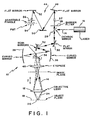
- Fig. 1 in diagrammatic form a conventional laser scanning microscope 10 which includes an objective lens 12 for focusing incident light 14 from a source 16 such as a laser onto an object plane 18.
- the object plane may lie on, or in, a specimen or target material 20 which may be carried on a movable stage 22.
- the illumination provided by incident light beam 14 fills a converging cone generally indicated at 24, the cone passing into the specimen 20 to reach the plane of focus at object plane 18 and, except for the tiny fraction of light absorbed by the specimen, passing out through a diverging cone 25.
- the incident light forms a waist, or focal point, 26 on the object plane 18.
- the diameter of the focal point 26 is limited by diffraction in the optical path, but preferably is less than 1 micron.
- the vertical location of the focal point in the specimen 20 can be selected.
- the stage 22 may be movable in a horizontal plane, as in a raster motion along X and Y axes, to position the incident light at selected locations in the specimen in the horizontal plane, so that three-dimensional scanning of the specimen can be obtained.
- mechanically scanned stages present difficulties, it is preferred to use a stationary stage, and to scan the incident beam in the X-Y plane optically, as by means of scanning mirrors in the optical path of the microscope.
- the optical path from laser 16 to the object plane 18 includes a dichroic mirror 28 onto which the light from the laser 16 is directed.
- the output from the laser consists of short intense pulses of light having a relatively long wavelength, preferably in the visible red or near infrared spectral range.
- the mirror 28 deflects this long wavelength light downwardly to a mirror 30 which in turn directs the light to a pair of scanning mirrors 32 and 34 by way of curved mirrors 36 and 38.
- the mirrors 32 and 34 are rotatable about mutually perpendicular axes in order to move the incident light 14 along perpendicular X and Y axes on the object plane so that the stationary specimen is scanned by the incident beam.
- the light from the scanning mirrors passes through eyepiece 40 and is focused through the objective lens 12 to the object plane 18.
- the light emitted by fluorescent material in the specimen is at a wavelength that is specific to the fluorophore contained in the specimen, and thus is a different wavelength than the incident light 14. This fluorescent light is able to pass through the dichroic mirror 28, rather than being reflected back toward the laser 16, and follows the light path indicated generally at 44.
- the fluorescent light 42 thus passes through a barrier filter 46 and is reflected by flat mirrors 48, 50 and 52 to a suitable detector such as a photomultiplier tube 54.
- a confocal laser scanning microscope is preferred, and accordingly such a microscope is illustrated in the drawings. However, it will be understood that other laser scanning microscopes may be used.
- an adjustable confocal pin hole 56 is provided in the collection optics 44 to minimize background fluorescence excited in the converging and diverging cones 24 and 25 above and below the plane of focus. This confocal pinhole is useful, but is not necessary in the two photon fluorescence excitation of the present invention, since excitation is essentially limited to the region of the focal point 26 on the object plane.
- the visible light fluorescence photons 42 are produced by molecules that are excited by absorbing a single photon from incident light 14 that has higher energy; that is, a shorter wavelength, than the fluorescence 42 generated during relaxation of the molecule from its excited state.
- the number of fluorescence photons released per molecule in such prior devices is ordinarily linearly proportional to the number of exciting photons absorbed. Because only a single photon need be absorbed in such devices, photolysis of molecules that absorb the exciting light 14 can occur all along the double cone beam 24 and 25 within the specimen 20, although this process is not necessarily linear with intensity.
- the present invention utilizes two-photon excitation of a fluorophore which has a one-photon absorption peak at a wavelength which overlaps one-half that of the exciting light.
- the laser 16 produces a very short pulsed laser beam of high instantaneous power and of a relatively long wavelength, for example in the visible red or the infrared range.
- This light is directed to a specimen containing a fluorophore normally excited by a single photon in the short wavelength, for example ultraviolet, range so that two low energy (red) photons must combine their energy to provide the same excitation of the specimen that would be provided by a single high energy (ultraviolet) photon.
- Both the excitation and hence the fluorescence rates in the specimen are proportional to the square of the intensity of the incident light.
- the intensity of the long wavelength incident light becomes high enough to excite the fluorophores in the specimen only in the region of the focal point 26 of the microscope optics.
- This focal point may be adjustably positioned within the specimen, so that fluorescence and/or photolysis of the specimen are produced only in a selected ellipsoidal volume around the focus.
- this long wavelength light is focused to produce sufficient intensity to excite fluorescence only in a very small region. This fluorescence is produced even if the fluorophore normally absorbs only in the ultraviolet. Since the focal point can be selectively positioned in the specimen, three-dimensional resolution is provided in both scanning fluorescence microscopy and in photolysis, including photolysis of photon-activatable reagents which can be released by photolysis.
- the necessary excitation intensity is provided at the focal point of the microscope 10 from a light source 16 which may be, for example, a colliding pulse, mode-locked dye laser generating pulses of light having a wavelength in the red region of the spectrum, for example about 630 nm, with the pulses having less than 100 fsec. duration at about 80 MHz repetition rate.
- a light source 16 which may be, for example, a colliding pulse, mode-locked dye laser generating pulses of light having a wavelength in the red region of the spectrum, for example about 630 nm, with the pulses having less than 100 fsec. duration at about 80 MHz repetition rate.
- Other bright pulsed lasers may also be used to produce light at different relatively long wavelengths in the infrared or visible red region of the spectrum, for example, to generate the necessary excitation photon energies which will add up to the appropriate absorption energy band required by the fluorophores in the specimen which normally would be excited by absorption of a single photon in the spectral region having wavelengths about one-half the wavelength of the incident light.
- two photons in the visible red region at 630 nm would combine to excite a fluorophore which normally absorbs light in the ultraviolet region at 315 nm, while two photons in the infrared region of, for example, 1070 nm, would excite a fluorophore which absorbs at 535 nm in the visible light region.
- the single wavelength light source 16 can be replaced by two different long wavelength laser sources so that the incident light beam 14 consists of two superimposed pulsed light beams of high instantaneous power and of different wavelengths.
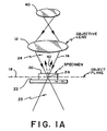
- Fig. 2 illustrates the depth discrimination achieved by the two photon technique of the present invention.
- a stereo pair of images 60 and 62 was generated from a stack of images of a cluster of fluorescent 9 micrometer diameter latex beads which are normally excited by ultraviolet light having a wavelength of about 365 nm. These images were obtained using a standard laser scanning microscope, but with its continuous-wave argon-ion laser illuminator 16 replaced by a 25 mw colliding-pulse mode-locked dyelaser producing output pulses at a wavelength of about 630 nm. Measurements made on the microscope 10 indicated that about 3 mw reached the object plane. An emission filter, passing wavelengths from 380 to 445 nm, was provided at the barrier filter 46, and the detector aperture 54 was opened to its limit in order to reduce the optical sectioning effect that would result from a small confocal aperture.
- the intensity of the incident beam 14 from laser 16 was adjusted by placing neutral density filters in the excitation beam between laser 16 and the dichroic mirror 28 and the blue fluorescence produced by the individual latex beads was measured. As illustrated in Fig. 3 by the graph 64, the detected intensity of fluorescence from the latex beads making up the specimen increased with the square of the excitation laser power, clearly indicating two-photon excitation in the beads.
- the excitation cross section of the beads which were "fluoresbrite BB" beads produced by Polysciences corporation, was estimated to be 5x10 -58 M 4 s/photon, accurate within a factor of 3, by taking into account the dye concentration in the beads, the optical throughput of the laser scanning microscope, the pulse duration, the repetition rate, the numerical aperture and the incident power. This value was found to be comparable to previously measured values for similar dyes.
- Fig. 4 is a scanned image of chromosomes in dividing cells (LLC-PK1; ATTC), using cellular DNA labeling with an ultraviolet excitable fluorescent stain (33258; Hoechst) the image acquisition time of 13 seconds was short compared to the bleaching time of several minutes. Furthermore, no degradation was apparent in these live cells even after illumination by the scanning laser for several minutes.
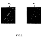
- Photobleaching during protracted scanning of a fluorescent bead occurred only in a slice about 2 micrometers thick around the focal plane, as demonstrated by the horizontal section 70 of reduced brightness bleached out of the bead 72 illustrated in Fig. 5. This bead was scanned for six minutes at a constant focal plane position. Similar localization of bleaching was observed in the fluorescently stained cell nuclei. This localization illustrates a distinct advantage over the use of single-photon excitation, where the entire specimen is bleached even when only a single plane is imaged. This is because for one-photon excitation, bleaching in both scanning and broad field microscopy depends on the time averaged excitation intensity, which does not vary along the axial, or Z-direction indicated in Fig. 1. For two-photon excitation, on the other hand, bleaching depends on the time averaged square of the intensity, which falls off strongly above and below the focal plane.
- the dependence of the fluorescent signal on the square of the excitation intensity is responsible for another advantage of two-photon excitation; that is, such excitation provides an optical sectioning effect through the specimen, even when using a detector, such as a CCD array, which views the whole field, without a pinhole being used as a spatial filter.
- This sectioning effect which is illustrated in Fig. 5, avoids the serious problems associated with chromatic aberration in the objective lens and some of the throughput losses in conventional confocal laser scanning microscopes.
- Two-photon photolysis can also be used for fast and localized release of biologically active chemicals such as caged Ca++, H+, nucleotides and neurotransmitters.
- biologically active chemicals such as caged Ca++, H+, nucleotides and neurotransmitters.
- caged neurotransmitters are released by a scanning beam
- the whole-cell transmembrane current so produced is usable as the contrast-generating mechanism to map the distribution of receptor activity for those transmitters on the cell surface.
- the feasibility of two-photon cage photolysis was demonstrated, in accordance with the present invention, by irradiating DMNPE caged ATP (33mM) [from Molecular Probes, Eugene Oregon], by the colliding pulse mode locked dyelaser 16 focused to a beam waist diameter at the object plane of about 10 micrometers.
- Photolysis yields of about 10 -11 moles of ATP were measured using a luciferin bioluminescence assay from Calbiochem, San Diego, CA. Typically, about 10% of the caged ATP in an aliquot volume of about 10 7 ( ⁇ m) 3 was photolyzed in the illumination volume of about 10 4 ( ⁇ m) 3 during about 600 seconds.
- two-photon excitation in accordance with the present invention provides access by visible light to excitation energies corresponding to single-ultraviolet-photon excitation
- a whole new class of fluorophores and fluorescent indicators becomes accessible to three-dimensionally resolved laser scanning microscopy.
- Such indicators may be Indo-1 for Ca +2 , Mag-Indo-1 for Mg +2 , ABF1 for Na + and PBFI for K + .
- the microscope allows sharp localization of photochemical processes such as photolysis and photoactivation within the focal volume.
- the invention is principally described as utilizing two photons from a single laser, but it should be understood that excitation of the target material can also be accomplished by two photons from two sources, as long as the two different wavelengths add up to the excitation wavelength of the target material.
- two different laser sources could be used, with their output beams being directed coaxially into the optical path of the microscope.
- two different wavelengths could be derived from a single source, as by means of a frequency doubler.
Landscapes
- Physics & Mathematics (AREA)
- Health & Medical Sciences (AREA)
- Optics & Photonics (AREA)
- Chemical & Material Sciences (AREA)
- Immunology (AREA)
- General Physics & Mathematics (AREA)
- Analytical Chemistry (AREA)
- General Health & Medical Sciences (AREA)
- Biochemistry (AREA)
- Life Sciences & Earth Sciences (AREA)
- Nuclear Medicine, Radiotherapy & Molecular Imaging (AREA)
- Pathology (AREA)
- Chemical Kinetics & Catalysis (AREA)
- Microscoopes, Condenser (AREA)
- Investigating, Analyzing Materials By Fluorescence Or Luminescence (AREA)
- Lasers (AREA)
Applications Claiming Priority (3)
| Application Number | Priority Date | Filing Date | Title |
|---|---|---|---|
| US436045 | 1989-11-14 | ||
| US07/436,045 US5034613A (en) | 1989-11-14 | 1989-11-14 | Two-photon laser microscopy |
| EP90917225A EP0500717B2 (en) | 1989-11-14 | 1990-11-13 | Two-photon laser scanning microscopy |
Related Parent Applications (2)
| Application Number | Title | Priority Date | Filing Date |
|---|---|---|---|
| EP90917225A Division EP0500717B2 (en) | 1989-11-14 | 1990-11-13 | Two-photon laser scanning microscopy |
| EP90917225.6 Division | 1991-06-14 |
Publications (2)
| Publication Number | Publication Date |
|---|---|
| EP0807814A1 EP0807814A1 (en) | 1997-11-19 |
| EP0807814B1 true EP0807814B1 (en) | 2003-11-05 |
Family
ID=23730872
Family Applications (2)
| Application Number | Title | Priority Date | Filing Date |
|---|---|---|---|
| EP97108581A Expired - Lifetime EP0807814B1 (en) | 1989-11-14 | 1990-11-13 | Two-photon molecular excitation in a laser scanning microscopy |
| EP90917225A Expired - Lifetime EP0500717B2 (en) | 1989-11-14 | 1990-11-13 | Two-photon laser scanning microscopy |
Family Applications After (1)
| Application Number | Title | Priority Date | Filing Date |
|---|---|---|---|
| EP90917225A Expired - Lifetime EP0500717B2 (en) | 1989-11-14 | 1990-11-13 | Two-photon laser scanning microscopy |
Country Status (8)
| Country | Link |
|---|---|
| US (1) | US5034613A (da) |
| EP (2) | EP0807814B1 (da) |
| JP (1) | JP2848952B2 (da) |
| AT (2) | ATE170636T1 (da) |
| DE (2) | DE69032621T3 (da) |
| DK (1) | DK0807814T3 (da) |
| ES (2) | ES2210414T3 (da) |
| WO (1) | WO1991007651A1 (da) |
Families Citing this family (362)
| Publication number | Priority date | Publication date | Assignee | Title |
|---|---|---|---|---|
| US5260578A (en) * | 1991-04-10 | 1993-11-09 | Mayo Foundation For Medical Education And Research | Confocal imaging system for visible and ultraviolet light |
| US6111645A (en) | 1991-04-29 | 2000-08-29 | Massachusetts Institute Of Technology | Grating based phase control optical delay line |
| US6564087B1 (en) | 1991-04-29 | 2003-05-13 | Massachusetts Institute Of Technology | Fiber optic needle probes for optical coherence tomography imaging |
| US6485413B1 (en) | 1991-04-29 | 2002-11-26 | The General Hospital Corporation | Methods and apparatus for forward-directed optical scanning instruments |
| US5289407A (en) * | 1991-07-22 | 1994-02-22 | Cornell Research Foundation, Inc. | Method for three dimensional optical data storage and retrieval |
| JP3082346B2 (ja) * | 1991-09-12 | 2000-08-28 | 株式会社ニコン | 蛍光コンフォーカル顕微鏡 |
| US5487080A (en) * | 1991-10-30 | 1996-01-23 | University Of New Mexico | Principle and applications of multiphoton pumped upconverted lasers |
| US5296703A (en) * | 1992-04-01 | 1994-03-22 | The Regents Of The University Of California | Scanning confocal microscope using fluorescence detection |
| EP0627643B1 (en) * | 1993-06-03 | 1999-05-06 | Hamamatsu Photonics K.K. | Laser scanning optical system using axicon |
| US5479252A (en) * | 1993-06-17 | 1995-12-26 | Ultrapointe Corporation | Laser imaging system for inspection and analysis of sub-micron particles |
| US5923430A (en) * | 1993-06-17 | 1999-07-13 | Ultrapointe Corporation | Method for characterizing defects on semiconductor wafers |
| USH1530H (en) * | 1993-06-17 | 1996-05-07 | Ultrapointe Corporation | Surface extraction from a three-dimensional data set |
| DE4324681C2 (de) * | 1993-07-22 | 1997-09-04 | Hell Stefan | Verfahren zur optischen Anregung eines Energiezustands einer Probe in einem Probenpunkt und Vorrichtung zur Durchführung des Verfahrens |
| DE4326181A1 (de) * | 1993-08-04 | 1995-02-09 | Europ Lab Molekularbiolog | Verfahren und Vorrichtung zur Lumineszenzspektroskopie und Materialmikrobearbeitung von fixierten und bewegten Molekülen, Partikeln und Objekten |
| DE4331570C2 (de) * | 1993-08-17 | 1996-10-24 | Hell Stefan | Verfahren zum optischen Anregen einer Probe |
| FI96452C (fi) * | 1994-01-26 | 1996-06-25 | Pekka Haenninen | Menetelmä väriaineiden virittämiseksi |
| US5731588A (en) * | 1994-02-01 | 1998-03-24 | Hell; Stefan | Process and device for optically measuring a point on a sample with high local resolution |
| DE4414940C2 (de) * | 1994-04-28 | 1998-07-02 | Pekka Haenninen | Lumineszenz-Rastermikroskop mit zwei Photonen Anregung |
| US5686988A (en) * | 1994-06-28 | 1997-11-11 | Lockheed Martin Energy Systems, Inc. | Gas concentration measurement instrument based on the effects of a wave-mixing interference on stimulated emissions |
| US6259104B1 (en) | 1994-07-15 | 2001-07-10 | Stephen C. Baer | Superresolution in optical microscopy and microlithography |
| US5952668A (en) * | 1994-07-15 | 1999-09-14 | Baer; Stephen C. | Resolution in microscopy and microlithography |
| US5866911A (en) * | 1994-07-15 | 1999-02-02 | Baer; Stephen C. | Method and apparatus for improving resolution in scanned optical system |
| US6903347B2 (en) | 1994-07-15 | 2005-06-07 | Stephen C. Baer | Superresolution in microlithography and fluorescence microscopy |
| US20050111089A1 (en) * | 1994-07-15 | 2005-05-26 | Baer Stephen C. | Superresolving microscopy apparatus |
| US7071477B2 (en) * | 1994-07-15 | 2006-07-04 | Baer Stephen C | Superresolution in microlithography and fluorescence microscopy |
| US5710046A (en) * | 1994-11-04 | 1998-01-20 | Amoco Corporation | Tagging hydrocarbons for subsequent identification |
| US5843783A (en) * | 1994-11-04 | 1998-12-01 | Amoco Corporation | Tagging hydrocarbons for subsequent identification |
| US5723338A (en) * | 1994-11-04 | 1998-03-03 | Amoco Corporation | Tagging hydrocarbons for subsequent identification |
| US5835262A (en) * | 1994-12-28 | 1998-11-10 | Research Development Corporation Of Japan | Multi-wavelength optical microscope |
| FI101829B1 (fi) * | 1995-03-07 | 1998-08-31 | Erkki Juhani Soini | Biospesifinen määritysmenetelmä |
| WO1996022531A1 (en) * | 1995-01-16 | 1996-07-25 | Erkki Soini | A biospecific multiparameter assay method |
| FI98765C (fi) | 1995-01-16 | 1997-08-11 | Erkki Soini | Virtaussytometrinen menetelmä ja laite |
| US5863504A (en) * | 1995-03-16 | 1999-01-26 | Bio-Rad Laboratories, Inc. | Fluorescence imaging instrument utilizing fish |
| US5786560A (en) * | 1995-03-31 | 1998-07-28 | Panasonic Technologies, Inc. | 3-dimensional micromachining with femtosecond laser pulses |
| US6104945A (en) * | 1995-08-01 | 2000-08-15 | Medispectra, Inc. | Spectral volume microprobe arrays |
| US5713364A (en) * | 1995-08-01 | 1998-02-03 | Medispectra, Inc. | Spectral volume microprobe analysis of materials |
| US5813987A (en) * | 1995-08-01 | 1998-09-29 | Medispectra, Inc. | Spectral volume microprobe for analysis of materials |
| CA2231114A1 (en) | 1995-09-06 | 1997-03-13 | The Research Foundation Of State University Of New York | Two-photon upconverting dyes and applications |
| DE19533092A1 (de) * | 1995-09-07 | 1997-03-13 | Basf Ag | Vorrichtung zur parallelisierten Zweiphotonen-Fluoreszenz-Korrelations-Spektroskopie (TPA-FCS) und deren Verwendung zum Wirkstoff-Screening |
| EP0852716B1 (en) * | 1995-09-19 | 2005-11-30 | Cornell Research Foundation, Inc. | Multi-photon laser microscopy |
| US5812308A (en) * | 1995-12-20 | 1998-09-22 | Spectra Physics Lasers, Inc. | Mode locked laser and amplifier |
| US5814820A (en) * | 1996-02-09 | 1998-09-29 | The Board Of Trustees Of The University Of Illinois | Pump probe cross correlation fluorescence frequency domain microscope and microscopy |
| US6545240B2 (en) | 1996-02-16 | 2003-04-08 | Huron Valley Steel Corporation | Metal scrap sorting system |
| US5761111A (en) * | 1996-03-15 | 1998-06-02 | President And Fellows Of Harvard College | Method and apparatus providing 2-D/3-D optical information storage and retrieval in transparent materials |
| JP2000512744A (ja) * | 1996-05-16 | 2000-09-26 | アフィメトリックス,インコーポレイテッド | 標識材料を検出するシステムおよび方法 |
| DE19622359B4 (de) * | 1996-06-04 | 2007-11-22 | Carl Zeiss Jena Gmbh | Vorrichtung zur Einkopplung der Strahlung von Kurzpulslasern in einem mikroskopischen Strahlengang |
| DE19744302B4 (de) * | 1996-06-04 | 2008-04-17 | Carl Zeiss Jena Gmbh | Vorrichtung zur Einkopplung der Strahlung von Kurzpulslasern in einem mikroskopischen Strahlengang |
| US5754291A (en) * | 1996-09-19 | 1998-05-19 | Molecular Dynamics, Inc. | Micro-imaging system |
| US6745067B1 (en) | 1998-09-14 | 2004-06-01 | Lucid, Inc. | System for marking the locations of imaged tissue with respect to the surface of the tissue |
| US7353829B1 (en) * | 1996-10-30 | 2008-04-08 | Provectus Devicetech, Inc. | Methods and apparatus for multi-photon photo-activation of therapeutic agents |
| US5829448A (en) * | 1996-10-30 | 1998-11-03 | Photogen, Inc. | Method for improved selectivity in photo-activation of molecular agents |
| US7036516B1 (en) * | 1996-10-30 | 2006-05-02 | Xantech Pharmaceuticals, Inc. | Treatment of pigmented tissues using optical energy |
| US20060095097A1 (en) * | 1996-10-30 | 2006-05-04 | Provectus Devicetech, Inc. | Treatment of pigmented tissue using optical energy |
| US5832931A (en) * | 1996-10-30 | 1998-11-10 | Photogen, Inc. | Method for improved selectivity in photo-activation and detection of molecular diagnostic agents |
| US6525862B2 (en) | 1996-10-30 | 2003-02-25 | Photogen, Inc. | Methods and apparatus for optical imaging |
| WO1998021521A1 (en) * | 1996-11-12 | 1998-05-22 | California Institute Of Technology | Two-photon or higher-order absorbing optical materials and methods of use |
| US6267913B1 (en) * | 1996-11-12 | 2001-07-31 | California Institute Of Technology | Two-photon or higher-order absorbing optical materials and methods of use |
| US6608228B1 (en) | 1997-11-07 | 2003-08-19 | California Institute Of Technology | Two-photon or higher-order absorbing optical materials for generation of reactive species |
| JP3917731B2 (ja) * | 1996-11-21 | 2007-05-23 | オリンパス株式会社 | レーザ走査顕微鏡 |
| US6148114A (en) * | 1996-11-27 | 2000-11-14 | Ultrapointe Corporation | Ring dilation and erosion techniques for digital image processing |
| DE19653413C2 (de) * | 1996-12-22 | 2002-02-07 | Stefan Hell | Rastermikroskop, bei dem eine Probe in mehreren Probenpunkten gleichzeitig optisch angeregt wird |
| DE19723873B4 (de) * | 1997-06-06 | 2004-02-05 | Evotec Oai Ag | Verfahren und Vorrichtung zur Bewegungserfassung eines sich zumindest zeitweilig periodisch bewegenden Objekts |
| US6847490B1 (en) | 1997-01-13 | 2005-01-25 | Medispectra, Inc. | Optical probe accessory device for use in vivo diagnostic procedures |
| US6826422B1 (en) | 1997-01-13 | 2004-11-30 | Medispectra, Inc. | Spectral volume microprobe arrays |
| ATE298084T1 (de) | 1997-01-31 | 2005-07-15 | Horticulture & Food Res Inst | Optische vorrichtung und methode |
| US5836877A (en) * | 1997-02-24 | 1998-11-17 | Lucid Inc | System for facilitating pathological examination of a lesion in tissue |
| US5995867A (en) * | 1997-03-19 | 1999-11-30 | Lucid Inc | Cellular surgery utilizing confocal microscopy |
| US5762607A (en) * | 1997-03-19 | 1998-06-09 | Schotland; John Carl | Emission tomography system and method using direct reconstruction of scattered radiation |
| US6208886B1 (en) * | 1997-04-04 | 2001-03-27 | The Research Foundation Of City College Of New York | Non-linear optical tomography of turbid media |
| US5995281A (en) * | 1997-04-09 | 1999-11-30 | Carl Zeiss Jena Gmbh | Device for coupling the radiation of short-pulse lasers in an optical beam path of a microscope |
| US6316950B1 (en) | 1997-05-15 | 2001-11-13 | Lucent Technologies Inc. | Method and apparatus for imaging semiconductor devices |
| RO114383B1 (ro) * | 1997-05-21 | 2000-02-28 | Storex Technologies S.R.L. | Dispozitiv pentru înscrierea tridimensională a informaţiei digitale într-o memorie optică de tip worm |
| US6096496A (en) * | 1997-06-19 | 2000-08-01 | Frankel; Robert D. | Supports incorporating vertical cavity emitting lasers and tracking apparatus for use in combinatorial synthesis |
| US6020591A (en) * | 1997-07-11 | 2000-02-01 | Imra America, Inc. | Two-photon microscopy with plane wave illumination |
| US6071748A (en) | 1997-07-16 | 2000-06-06 | Ljl Biosystems, Inc. | Light detection device |
| US6469311B1 (en) | 1997-07-16 | 2002-10-22 | Molecular Devices Corporation | Detection device for light transmitted from a sensed volume |
| US6466040B1 (en) * | 1997-08-01 | 2002-10-15 | Carl Zeiss Jena Gmbh | Three dimensional optical beam induced current (3-D-OBIC) |
| DE19733195B4 (de) * | 1997-08-01 | 2006-04-06 | Carl Zeiss Jena Gmbh | Hoch-Kompaktes Laser Scanning Mikroskop mit integriertem Kurzpuls Laser |
| DE19733194B4 (de) * | 1997-08-01 | 2005-06-16 | Carl Zeiss Jena Gmbh | Laser-Scanning-Mikroskop |
| US6771417B1 (en) * | 1997-08-01 | 2004-08-03 | Carl Zeiss Jena Gmbh | Applications of adaptive optics in microscopy |
| DE19733193B4 (de) * | 1997-08-01 | 2005-09-08 | Carl Zeiss Jena Gmbh | Mikroskop mit adaptiver Optik |
| US5978695A (en) * | 1997-08-18 | 1999-11-02 | Lucid Inc. | System for imaging mechanically stabilized tissue |
| US6982431B2 (en) | 1998-08-31 | 2006-01-03 | Molecular Devices Corporation | Sample analysis systems |
| US6326605B1 (en) | 1998-02-20 | 2001-12-04 | Ljl Biosystems, Inc. | Broad range light detection system |
| US6992761B2 (en) | 1997-09-20 | 2006-01-31 | Molecular Devices Corporation | Broad range light detection system |
| US6825921B1 (en) | 1999-11-10 | 2004-11-30 | Molecular Devices Corporation | Multi-mode light detection system |
| US6576476B1 (en) | 1998-09-02 | 2003-06-10 | Ljl Biosystems, Inc. | Chemiluminescence detection method and device |
| US6297018B1 (en) | 1998-04-17 | 2001-10-02 | Ljl Biosystems, Inc. | Methods and apparatus for detecting nucleic acid polymorphisms |
| US6121603A (en) * | 1997-12-01 | 2000-09-19 | Hang; Zhijiang | Optical confocal device having a common light directing means |
| US8974363B2 (en) * | 1997-12-11 | 2015-03-10 | Provectus Pharmatech, Inc. | Topical medicaments and methods for photodynamic treatment of disease |
| US6071689A (en) | 1997-12-31 | 2000-06-06 | Xy, Inc. | System for improving yield of sexed embryos in mammals |
| US6149867A (en) | 1997-12-31 | 2000-11-21 | Xy, Inc. | Sheath fluids and collection systems for sex-specific cytometer sorting of sperm |
| US6132643A (en) * | 1998-01-06 | 2000-10-17 | Pavel; Eugen | Fluorescent photosensitive vitroceramics and process for the production thereof |
| DE19801139B4 (de) * | 1998-01-14 | 2016-05-12 | Till Photonics Gmbh | Punktabtastendes Luminiszenz-Mikroskop |
| WO1999037999A1 (en) | 1998-01-27 | 1999-07-29 | Wisconsin Alumni Research Foundation | Signal enhancement for fluorescence microscopy |
| AU2786099A (en) | 1998-02-26 | 1999-09-15 | Lucid, Inc. | Confocal microscope for facilitating cryosurgery of tissue |
| US6855941B1 (en) * | 1998-03-11 | 2005-02-15 | Olympus Optical Co., Ltd. | Laser microscope |
| KR100618502B1 (ko) * | 1998-03-16 | 2006-09-01 | 지이 헬스케어 바이오-사이언시즈 코프. | 공초점 마이크로스코피 영상 시스템에서 사용하기 위한 전자기 방출의 집속 시스템 및 방법 |
| US20030036855A1 (en) | 1998-03-16 | 2003-02-20 | Praelux Incorporated, A Corporation Of New Jersey | Method and apparatus for screening chemical compounds |
| US5936728A (en) * | 1998-04-14 | 1999-08-10 | Noran Instruments, Inc. | Flash photolysis method and apparatus |
| US6316153B1 (en) | 1998-04-21 | 2001-11-13 | The University Of Connecticut | Free-form fabricaton using multi-photon excitation |
| US5880006A (en) | 1998-05-22 | 1999-03-09 | Vlsi Technology, Inc. | Method for fabrication of a semiconductor device |
| GB9811483D0 (en) * | 1998-05-29 | 1998-07-29 | Photonic Research Systems Limi | Luminescence assay using cyclical excitation wavelength sequence |
| DE19829944C2 (de) * | 1998-07-04 | 2002-03-28 | Zeiss Carl Jena Gmbh | Verfahren und Anordnung zur Gerätekonfiguration eines Fluoreszenz-Laserscanmikroskops |
| US6228787B1 (en) | 1998-07-27 | 2001-05-08 | Eugen Pavel | Fluorescent photosensitive glasses and process for the production thereof |
| AU5223899A (en) | 1998-07-27 | 2000-02-21 | Ljl Biosystems, Inc. | Apparatus and methods for spectroscopic measurements |
| AU5667599A (en) | 1998-07-27 | 2000-02-21 | Ljl Biosystems, Inc. | Apparatus and methods for time-resolved spectroscopic measurements |
| EP2283848A1 (en) | 1998-07-30 | 2011-02-16 | Xy, Llc | Equine system for non-surgical articificial insemination |
| US20090117199A1 (en) * | 1998-08-06 | 2009-05-07 | Scott Timothy C | Method of treatment of cancer |
| US8557298B2 (en) * | 1998-08-06 | 2013-10-15 | Provectus Pharmatech, Inc. | Medicaments for chemotherapeutic treatment of disease |
| AU750633B2 (en) * | 1998-08-06 | 2002-07-25 | Provectus Pharmatech, Inc. | Improved method for targeted topical treatment of disease |
| US7227630B1 (en) | 1998-09-14 | 2007-06-05 | Lucid, Inc. | Imaging of surgical biopsies |
| WO2000015021A2 (en) | 1998-09-14 | 2000-03-23 | Lucid, Inc. | Imaging of surgical biopsies |
| DE19851240C1 (de) * | 1998-11-06 | 2000-03-02 | Europ Lab Molekularbiolog | Fluoreszenzmikroskop mit Mehrphotonenanregung |
| US9192780B2 (en) | 1998-11-30 | 2015-11-24 | L'oreal | Low intensity light therapy for treatment of retinal, macular, and visual pathway disorders |
| US6887260B1 (en) | 1998-11-30 | 2005-05-03 | Light Bioscience, Llc | Method and apparatus for acne treatment |
| US20060212025A1 (en) * | 1998-11-30 | 2006-09-21 | Light Bioscience, Llc | Method and apparatus for acne treatment |
| US6936044B2 (en) * | 1998-11-30 | 2005-08-30 | Light Bioscience, Llc | Method and apparatus for the stimulation of hair growth |
| US6283956B1 (en) * | 1998-11-30 | 2001-09-04 | David H. McDaniels | Reduction, elimination, or stimulation of hair growth |
| CA2356623C (en) | 1998-12-23 | 2005-10-18 | Medispectra, Inc. | Systems and methods for optical examination of samples |
| JP2002532181A (ja) | 1998-12-23 | 2002-10-02 | メディスペクトラ, インコーポレイテッド | 頚部のスクリーニングのための光学的方法およびシステム |
| JP3099063B2 (ja) * | 1998-12-28 | 2000-10-16 | 大阪大学長 | 多光子顕微鏡 |
| DE19901381A1 (de) * | 1999-01-15 | 2000-07-20 | Joerg Enderlein | Verfahren und Vorrichtung zur optischen Detektion eines Partikels |
| IL128519A0 (en) * | 1999-02-14 | 2000-06-01 | Aaron Lewis | Deconvolving far-field optical images beyond the diffraction limit by using scanned-probe optical and non-optical data as the constraint in mathematical constraint algorithms |
| ES2520140T3 (es) * | 1999-02-17 | 2014-11-11 | Lucid, Inc. | Portador de muestras de tejido |
| EP1169630B1 (en) * | 1999-02-17 | 2017-02-01 | Lucid, Inc. | Cassette for facilitating optical sectioning of a retained tissue specimen |
| AU3005400A (en) | 1999-02-23 | 2000-09-14 | Ljl Biosystems, Inc. | Frequency-domain light detection device |
| DE19908883A1 (de) | 1999-03-02 | 2000-09-07 | Rainer Heintzmann | Verfahren zur Erhöhung der Auflösung optischer Abbildung |
| DE19919091C2 (de) * | 1999-04-27 | 2002-01-17 | Zeiss Carl Jena Gmbh | Anordnung zur Einstellung der Laserleistung und/oder der Pulslänge eines Kurzpulslasers in einem Mikroskop |
| ATE235686T1 (de) * | 1999-04-29 | 2003-04-15 | Univ Erasmus | Bestimmung von analytbeweglichkeit |
| US6449039B1 (en) | 1999-07-28 | 2002-09-10 | Thermo Noran Inc. | Laser scanning fluorescence microscopy with compensation for spatial dispersion of fast laser pulses |
| DE19935766A1 (de) * | 1999-07-29 | 2001-02-01 | Friedrich Schiller Uni Jena Bu | Verfahren zur optischen Anregung von Fluorophor-markierter DNA und RNA |
| WO2001009592A1 (en) * | 1999-07-30 | 2001-02-08 | California Institute Of Technology | System and method for monitoring cellular activity |
| US6445939B1 (en) | 1999-08-09 | 2002-09-03 | Lightlab Imaging, Llc | Ultra-small optical probes, imaging optics, and methods for using same |
| DE19939706C2 (de) * | 1999-08-18 | 2002-09-05 | Forschungsverbund Berlin Ev | Fluorophor für die Multi-Photonen-Laser-Scanning-Mikroskopie |
| JP4680337B2 (ja) * | 1999-09-20 | 2011-05-11 | オリンパス株式会社 | 走査型レーザ顕微鏡 |
| US7024316B1 (en) | 1999-10-21 | 2006-04-04 | Dakocytomation Colorado, Inc. | Transiently dynamic flow cytometer analysis system |
| US7167615B1 (en) | 1999-11-05 | 2007-01-23 | Board Of Regents, The University Of Texas System | Resonant waveguide-grating filters and sensors and methods for making and using same |
| AU1476801A (en) * | 1999-11-10 | 2001-06-06 | Lucid, Inc. | System for optically sectioning and mapping surgically excised tissue |
| US7208265B1 (en) | 1999-11-24 | 2007-04-24 | Xy, Inc. | Method of cryopreserving selected sperm cells |
| DE19956620A1 (de) * | 1999-11-25 | 2001-05-31 | Zeiss Carl Jena Gmbh | Verfahren zur Erfassung von Fluoreszenzerscheinungen in einem Mikroskop |
| US7187810B2 (en) | 1999-12-15 | 2007-03-06 | Medispectra, Inc. | Methods and systems for correcting image misalignment |
| US7260248B2 (en) | 1999-12-15 | 2007-08-21 | Medispectra, Inc. | Image processing using measures of similarity |
| US20020007122A1 (en) * | 1999-12-15 | 2002-01-17 | Howard Kaufman | Methods of diagnosing disease |
| JP2004500197A (ja) | 2000-02-08 | 2004-01-08 | コーネル リサーチ ファンデーション インコーポレーテッド | 光ファイバーを通じた蛍光分光用の多光子励起 |
| WO2001067176A1 (en) | 2000-03-09 | 2001-09-13 | Xerox Corporation | Three dimensional optical memory storage |
| WO2001079821A1 (de) * | 2000-04-14 | 2001-10-25 | Zeptosens Ag | Gitter-wellenleiter-struktur zur verstärkung eines anregungsfeldes und deren verwendung |
| US20020001089A1 (en) * | 2000-04-18 | 2002-01-03 | Price Jeffrey H. | Multiparallel three dimensional optical microscopy system |
| EP2258173A3 (en) | 2000-05-09 | 2012-06-13 | Xy, Llc | High purity x-chromosome bearing and y-chromosome bearing populations of spermatozoa |
| DE10027726A1 (de) * | 2000-06-03 | 2001-12-06 | Bundesdruckerei Gmbh | Sensor für die Echtheitserkennung von Signets auf Dokumenten |
| DE10027323B4 (de) * | 2000-06-05 | 2013-09-26 | Leica Microsystems Cms Gmbh | Verfahren zum Generieren eines dreidimensionalen Objekts |
| AU2001266918A1 (en) * | 2000-06-15 | 2001-12-24 | 3M Innovative Properties Company | Multidirectional photoreactive absorption method |
| WO2001096958A2 (en) * | 2000-06-15 | 2001-12-20 | 3M Innovative Properties Company | Process for producing microfluidic articles |
| EP1295179B1 (en) * | 2000-06-15 | 2013-05-22 | 3M Innovative Properties Company | Multiphoton curing to provide encapsulated optical elements |
| KR100754813B1 (ko) * | 2000-06-15 | 2007-09-04 | 쓰리엠 이노베이티브 프로퍼티즈 캄파니 | 다중통과 다광자 흡수 방법 및 장치 |
| DE60114820T2 (de) * | 2000-06-15 | 2006-09-14 | 3M Innovative Properties Co., St. Paul | Mikroherstellungsverfahren für organische optische bauteile |
| WO2001096952A2 (en) * | 2000-06-15 | 2001-12-20 | 3M Innovative Properties Company | Multicolor imaging using multiphoton photochemical processes |
| US6852766B1 (en) | 2000-06-15 | 2005-02-08 | 3M Innovative Properties Company | Multiphoton photosensitization system |
| DE20122783U1 (de) * | 2000-06-17 | 2007-11-15 | Leica Microsystems Cms Gmbh | Anordnung zum Untersuchen mikroskopischer Präparate mit einem Scanmikroskop und Beleuchtungseinrichtung für ein Scanmikroskop |
| EP1164401B1 (de) | 2000-06-17 | 2005-03-09 | Leica Microsystems Heidelberg GmbH | Verschränkte-Photonen-Mikroskop |
| DE10115486A1 (de) * | 2000-06-17 | 2001-12-20 | Leica Microsystems | Verschränkte-Photonen-Mikroskop |
| US6898367B2 (en) | 2000-06-17 | 2005-05-24 | Leica Microsystems Heidelberg Gmbh | Method and instrument for microscopy |
| US6687000B1 (en) | 2000-06-26 | 2004-02-03 | Wisconsin Alumni Research Foundation | Photon-sorting spectroscopic microscope system |
| DE10035190C5 (de) * | 2000-07-20 | 2009-07-16 | MAX-PLANCK-Gesellschaft zur Förderung der Wissenschaften e.V. | Verfahren und Vorrichtung zur Fluoreszenzmessung |
| US20070196815A1 (en) * | 2000-08-02 | 2007-08-23 | Jason Lappe | Positive Selection Procedure for Optically Directed Selection of Cells |
| DE10039520A1 (de) | 2000-08-08 | 2002-02-21 | Leica Microsystems | Vorrichtung zur Untersuchung und Manipulation von mikroskopischen Objekten |
| DE10044308A1 (de) | 2000-09-07 | 2002-03-21 | Leica Microsystems | Verfahren und Vorrichtung zur Detektion von Fluoreszenzlicht bei der konfokalen Rastermikroskopie |
| JP4693972B2 (ja) | 2000-09-29 | 2011-06-01 | オリンパス株式会社 | レーザ顕微鏡 |
| US7321394B1 (en) * | 2000-09-29 | 2008-01-22 | Lucid, Inc. | Automatic gain control for a confocal imaging system |
| US6295123B1 (en) | 2000-10-13 | 2001-09-25 | Taiwan Semiconductor Manufacturing Company | Multiple photon absorption for high resolution lithography |
| US7003345B1 (en) | 2000-10-17 | 2006-02-21 | Lucid, Inc. | System and method for enhancing microscope images of tissue using citric acid and agents of the like |
| US6369928B1 (en) | 2000-11-01 | 2002-04-09 | Optical Biopsy Technologies, Inc. | Fiber-coupled, angled-dual-illumination-axis confocal scanning microscopes for performing reflective and two-photon fluorescence imaging |
| AU2002251682A1 (en) * | 2000-11-02 | 2002-08-28 | Cornell Research Foundation, Inc. | In vivo multiphoton diagnostic detection and imaging of a neurodegenerative disease |
| US7194118B1 (en) | 2000-11-10 | 2007-03-20 | Lucid, Inc. | System for optically sectioning and mapping surgically excised tissue |
| CA2468774C (en) | 2000-11-29 | 2015-06-30 | George E. Seidel | System for in-vitro fertilization with spermatozoa separated into x-chromosome and y-chromosome bearing populations |
| US7713687B2 (en) | 2000-11-29 | 2010-05-11 | Xy, Inc. | System to separate frozen-thawed spermatozoa into x-chromosome bearing and y-chromosome bearing populations |
| US6414779B1 (en) | 2000-11-30 | 2002-07-02 | Opeical Biopsy Technologies, Inc. | Integrated angled-dual-axis confocal scanning endoscopes |
| JP3999662B2 (ja) * | 2000-12-14 | 2007-10-31 | オリンパス株式会社 | 蛍光分析装置および蛍光分析方法 |
| CA2328684A1 (en) * | 2000-12-15 | 2002-06-15 | Yahia Gawad | Photon-triggered luminescent assay |
| US6804000B2 (en) | 2000-12-15 | 2004-10-12 | Sloan-Kettering Institute For Cancer Research | Beam-steering of multi-chromatic light using acousto-optical deflectors and dispersion-compensatory optics |
| US6839661B2 (en) | 2000-12-15 | 2005-01-04 | Medispectra, Inc. | System for normalizing spectra |
| WO2002061799A2 (en) * | 2001-01-30 | 2002-08-08 | Board Of Trustees Operating Michigan State University | Control system and apparatus for use with laser excitation or ionization |
| US7609731B2 (en) * | 2001-01-30 | 2009-10-27 | Board Of Trustees Operating Michigan State University | Laser system using ultra-short laser pulses |
| US7973936B2 (en) * | 2001-01-30 | 2011-07-05 | Board Of Trustees Of Michigan State University | Control system and apparatus for use with ultra-fast laser |
| US7583710B2 (en) * | 2001-01-30 | 2009-09-01 | Board Of Trustees Operating Michigan State University | Laser and environmental monitoring system |
| US7450618B2 (en) * | 2001-01-30 | 2008-11-11 | Board Of Trustees Operating Michigan State University | Laser system using ultrashort laser pulses |
| US8208505B2 (en) * | 2001-01-30 | 2012-06-26 | Board Of Trustees Of Michigan State University | Laser system employing harmonic generation |
| US7567596B2 (en) | 2001-01-30 | 2009-07-28 | Board Of Trustees Of Michigan State University | Control system and apparatus for use with ultra-fast laser |
| EP1372552B1 (de) * | 2001-03-27 | 2017-03-01 | WaveLight GmbH | Vorrichtung zur bearbeitung und diagnose von augengewebe |
| WO2002079691A1 (en) | 2001-03-30 | 2002-10-10 | The Arizona Board Of Regents On Behalf Of The University Of Arizona | Materials, methods, and uses for photochemical generation of acids and/or radical species |
| US20040052489A1 (en) * | 2001-04-02 | 2004-03-18 | Duveneck Gert Ludwig | Optical structure for multi-photon excitation and the use thereof |
| JP3885511B2 (ja) * | 2001-04-11 | 2007-02-21 | ソニー株式会社 | レーザー光発生装置及び方法 |
| DE10120425C2 (de) * | 2001-04-26 | 2003-12-18 | Leica Microsystems | Scanmikroskop |
| US20030211009A1 (en) * | 2001-05-18 | 2003-11-13 | Buchanan Kris S. | Rapid multi-material sample input system |
| US20040012872A1 (en) * | 2001-06-14 | 2004-01-22 | Fleming Patrick R | Multiphoton absorption method using patterned light |
| US6998214B2 (en) * | 2001-07-13 | 2006-02-14 | The Trustees Of Boston College | Methods for three-dimensional optical data storage and retrieval |
| US7336988B2 (en) * | 2001-08-08 | 2008-02-26 | Lucent Technologies Inc. | Multi-photon endoscopy |
| US6643071B2 (en) | 2001-12-21 | 2003-11-04 | Lucent Technologies Inc. | Graded-index lens microscopes |
| DE10206980A1 (de) * | 2002-02-20 | 2003-08-21 | Leica Microsystems | Mikroskop, Detektor und Verfahren zur Mikroskopie |
| US20040133112A1 (en) * | 2002-03-08 | 2004-07-08 | Milind Rajadhyaksha | System and method for macroscopic and confocal imaging of tissue |
| CA2484730A1 (en) * | 2002-05-03 | 2003-11-13 | Immunivest Corporation | Device and method for analytical cell imaging |
| JP4175833B2 (ja) * | 2002-05-23 | 2008-11-05 | オリンパス株式会社 | レーザ顕微鏡 |
| DE10228374A1 (de) * | 2002-06-25 | 2004-01-15 | Leica Microsystems Heidelberg Gmbh | Verfahren zur Mikroskopie und Mikroskop |
| US6818903B2 (en) * | 2002-07-09 | 2004-11-16 | Medispectra, Inc. | Method and apparatus for identifying spectral artifacts |
| US6933154B2 (en) | 2002-07-09 | 2005-08-23 | Medispectra, Inc. | Optimal windows for obtaining optical data for characterization of tissue samples |
| US7309867B2 (en) | 2003-04-18 | 2007-12-18 | Medispectra, Inc. | Methods and apparatus for characterization of tissue samples |
| US7282723B2 (en) | 2002-07-09 | 2007-10-16 | Medispectra, Inc. | Methods and apparatus for processing spectral data for use in tissue characterization |
| US7459696B2 (en) | 2003-04-18 | 2008-12-02 | Schomacker Kevin T | Methods and apparatus for calibrating spectral data |
| US7136518B2 (en) | 2003-04-18 | 2006-11-14 | Medispectra, Inc. | Methods and apparatus for displaying diagnostic data |
| US7469160B2 (en) | 2003-04-18 | 2008-12-23 | Banks Perry S | Methods and apparatus for evaluating image focus |
| US7103401B2 (en) | 2002-07-10 | 2006-09-05 | Medispectra, Inc. | Colonic polyp discrimination by tissue fluorescence and fiberoptic probe |
| US6768918B2 (en) | 2002-07-10 | 2004-07-27 | Medispectra, Inc. | Fluorescent fiberoptic probe for tissue health discrimination and method of use thereof |
| US8486618B2 (en) | 2002-08-01 | 2013-07-16 | Xy, Llc | Heterogeneous inseminate system |
| MXPA05001100A (es) | 2002-08-01 | 2005-04-28 | Xy Inc | Sistema de separacion de baja presion para celulas de esperma. |
| MXPA05001654A (es) | 2002-08-15 | 2005-10-18 | Xy Inc | Citometro de flujo de alta resolucion. |
| US7169548B2 (en) | 2002-09-13 | 2007-01-30 | Xy, Inc. | Sperm cell processing and preservation systems |
| US8227256B2 (en) * | 2002-09-27 | 2012-07-24 | Saloma Caesar A | Two-color (two-photon) excitation with focused excitation beams and a raman shifter |
| DE10250012B4 (de) * | 2002-10-25 | 2005-06-23 | Universität Kassel | Verfahren zur Bestimmung der Oberflächenstruktur einer Materialprobe mit ultrakurzen Laserpulsen und Vorrichtung zur Durchführung des Verfahrens |
| DE10250568A1 (de) * | 2002-10-28 | 2004-05-13 | Carl Zeiss Jena Gmbh | Verfahren zur Verbesserung der Tiefendiskriminierung optisch abbildender Systeme |
| US7258687B2 (en) * | 2002-12-11 | 2007-08-21 | The Regents Of The University Of California | Device and method for inducing vascular injury and/or blockage in an animal model |
| US7141801B2 (en) * | 2002-12-26 | 2006-11-28 | Applied Precision, Llc | System and method of illuminating living cells for imaging |
| DE10300157B4 (de) * | 2003-01-07 | 2016-08-25 | Leica Microsystems Cms Gmbh | Konfokales 4-Pi-Mikroskop und Verfahren zur konfokalen 4-Pi-Mikroskopie |
| US7130042B2 (en) * | 2003-03-06 | 2006-10-31 | Board Of Trustees Of The Leland Stanford Junior University | Dual axis fluorescence microscope with modulated input |
| EP1608963B1 (en) | 2003-03-28 | 2009-12-30 | Inguran, LLC | Apparatus and methods for providing sex-sorted animal sperm |
| DE10314750A1 (de) * | 2003-03-31 | 2004-11-04 | Leica Microsystems Heidelberg Gmbh | Rastermikroskop zur Detektion eines Objekts |
| JP2006522660A (ja) | 2003-04-10 | 2006-10-05 | ライト バイオサイエンス,エルエルシー | 細胞増殖及び遺伝子発現を調節するためのフォトモジュレーション方法及び装置 |
| US7151270B2 (en) * | 2003-05-02 | 2006-12-19 | Leica Microsystems Cms Gmbh | Method for classifying object image regions of an object to be detected using a scanning microscope |
| CA2566749C (en) | 2003-05-15 | 2017-02-21 | Xy, Inc. | Efficient haploid cell sorting for flow cytometer systems |
| US7091500B2 (en) | 2003-06-20 | 2006-08-15 | Lucent Technologies Inc. | Multi-photon endoscopic imaging system |
| US7545494B2 (en) * | 2003-07-23 | 2009-06-09 | Bayer Technology Services Gmbh | Analytical system and method for analyzing nonlinear optical signals |
| EP1648385B1 (en) * | 2003-07-31 | 2016-05-04 | Gentlewaves Llc | System and method for the photodynamic treatment of skin |
| JP4823906B2 (ja) * | 2003-08-19 | 2011-11-24 | コーネル リサーチ ファンデーション インコーポレーティッド | 多光子顕微鏡観察、分光法、および内視鏡観察などの生物学的用途のための光ファイバー送集光システム |
| US20050056193A1 (en) * | 2003-09-11 | 2005-03-17 | Chep International, Inc | Pallet |
| US20050142608A1 (en) * | 2003-09-22 | 2005-06-30 | Yokogawa Electric Corporation | Screening method and device, and new drug screening method and device |
| DE10351414A1 (de) * | 2003-10-30 | 2005-06-23 | Carl Zeiss Jena Gmbh | Laser-Scanning-Mikroskop mit einem non-descannten Detektions- und/oder Beobachtungsstrahlengang |
| US7706863B2 (en) | 2004-01-21 | 2010-04-27 | University Of Washington | Methods for assessing a physiological state of a mammalian retina |
| JP2005275199A (ja) * | 2004-03-26 | 2005-10-06 | Yokogawa Electric Corp | 3次元共焦点顕微鏡システム |
| MXPA06011345A (es) | 2004-03-29 | 2006-12-15 | Monsanto Technology Llc | Suspensiones de espermatozoides para usar en inseminacion. |
| EP1582858A1 (de) * | 2004-03-29 | 2005-10-05 | Max-Planck-Gesellschaft zur Förderung der Wissenschaften e.V. | Verfahren zur Anregung der Moleküle von einem ersten Zustand in einen zweiten Zustand mit einem optischen Signal |
| US7170675B2 (en) * | 2004-05-19 | 2007-01-30 | Celloptic, Inc. | Method and system for wide-field multi-photon microscopy having a confocal excitation plane |
| US7355702B2 (en) * | 2004-06-21 | 2008-04-08 | Olympus Corporation | Confocal observation system |
| TWI261605B (en) * | 2004-06-30 | 2006-09-11 | Ind Tech Res Inst | Dye compositon of the optical recording medium |
| BRPI0513685A (pt) | 2004-07-22 | 2008-05-13 | Monsanto Technology Llc | processo para enriquecimento de uma população de células de esperma |
| ES2555126T3 (es) * | 2004-07-27 | 2015-12-29 | Beckman Coulter, Inc. | Mejora de la discriminación en citometría de flujo con transformación geométrica |
| US7816654B2 (en) * | 2005-01-16 | 2010-10-19 | Baer Stephen C | Single wavelength stimulated emission depletion microscopy |
| HU227859B1 (en) * | 2005-01-27 | 2012-05-02 | E Szilveszter Vizi | Real-time 3d nonlinear microscope measuring system and its application |
| EP1851532A1 (en) * | 2005-02-14 | 2007-11-07 | Board of Trustees of Michigan State University | Ultra-fast laser system |
| US7897638B2 (en) * | 2005-04-12 | 2011-03-01 | Philadelphia Health & Education Corporation | Synthesis of nitrodibenzylfuran chromophore for photodeprotection of organic molecules |
| DE112006002099A5 (de) * | 2005-05-31 | 2008-05-21 | W.O.M. World Of Medicine Ag | Verfahren und Vorrichtung zur optischen Charakterisierung von Gewebe |
| US7618770B2 (en) | 2005-07-29 | 2009-11-17 | Xy, Inc. | Methods and apparatus for reducing protein content in sperm cell extenders |
| JP4621893B2 (ja) * | 2005-08-22 | 2011-01-26 | 独立行政法人産業技術総合研究所 | 物体の調査方法及び調査装置 |
| US8618470B2 (en) | 2005-11-30 | 2013-12-31 | Board Of Trustees Of Michigan State University | Laser based identification of molecular characteristics |
| US7864996B2 (en) * | 2006-02-17 | 2011-01-04 | Lucid, Inc. | System for macroscopic and confocal imaging of tissue |
| US7525724B2 (en) * | 2006-03-16 | 2009-04-28 | The University Of Kansas | Laser system for photonic excitation investigation |
| JP4759425B2 (ja) | 2006-03-28 | 2011-08-31 | オリンパス株式会社 | 多光子励起型観察装置 |
| US9018562B2 (en) | 2006-04-10 | 2015-04-28 | Board Of Trustees Of Michigan State University | Laser material processing system |
| US7773300B2 (en) * | 2006-05-12 | 2010-08-10 | Semrock, Inc. | Multiphoton fluorescence filters |
| DE102006029809B3 (de) * | 2006-06-28 | 2007-11-08 | Ltb Lasertechnik Berlin Gmbh | Ortsaufgelöstes Messverfahren für die Detektion von Melanin in Fluorophorgemischen in einer Festkörperprobe |
| EP2033047B1 (en) * | 2006-06-29 | 2020-02-19 | Agency for Science, Technology and Research | Shg quantification of matrix-related tissue dynamic and disease |
| DE102006034914A1 (de) * | 2006-07-28 | 2008-01-31 | Carl Zeiss Microimaging Gmbh | Verfahren und Anordnung zur Ansteuerung eines Mikroskops, insbesondere eines Laser-Scanning-Mikroskopes |
| DE102006034906A1 (de) | 2006-07-28 | 2008-01-31 | Carl Zeiss Microimaging Gmbh | Laser-Scanning-Mikroskop und Verfahren zu seinem Betrieb |
| DE102006034910B4 (de) * | 2006-07-28 | 2019-05-02 | Carl Zeiss Microscopy Gmbh | Mikroskop umfassend einen Strahlvereiniger |
| AU2007281902A1 (en) * | 2006-08-04 | 2008-02-14 | The Government Of The United States Of America, As Represented By The Secretary, Department Of Health | Wide-area fluorescence detection system for multi-photon microscopy |
| GB0617945D0 (en) * | 2006-09-12 | 2006-10-18 | Ucl Business Plc | Imaging apparatus and methods |
| DE102006046925A1 (de) * | 2006-09-28 | 2008-04-03 | Jenlab Gmbh | Verfahren und Anordnung zur Laser-Endoskopie für die Mikrobearbeitung |
| US7973927B2 (en) * | 2006-09-29 | 2011-07-05 | Uwm Research Foundation, Inc. | Two-photon microscope with spectral resolution |
| US7480045B2 (en) * | 2006-10-31 | 2009-01-20 | Academia Sinica | Controlling pulses in optical microscopy |
| US20080116392A1 (en) * | 2006-11-20 | 2008-05-22 | Celloptic, Inc. | Method and system for wide-field multi-photon microscopy having a confocal excitation plane |
| US7936503B2 (en) * | 2007-02-19 | 2011-05-03 | Olympus Corporation | Laser scanning microscope |
| US7867778B2 (en) * | 2007-02-23 | 2011-01-11 | Visiongate, Inc. | Fluid focusing for positional control of a specimen for 3-D imaging |
| CN101254091B (zh) * | 2007-02-28 | 2010-08-18 | 深圳大学 | 一种视网膜成像的方法 |
| DE102007021378A1 (de) * | 2007-05-04 | 2008-11-06 | Ape Angewandte Physik Und Elektronik Gmbh | Verfahren und optische Anordnung zum Erzeugen eines nicht-linearen optischen Signals an einem durch ein Anregungsfeld angeregten Material sowie Verwendung des Verfahrens und der optischen Anordnung |
| US8958156B1 (en) | 2007-05-30 | 2015-02-17 | Semrock, Inc. | Interference filter for non-zero angle of incidence spectroscopy |
| DE102007025821A1 (de) | 2007-06-02 | 2008-12-04 | Carl Zeiss Microimaging Gmbh | Anordnung und Verfahren zur zeitlichen Einstellung der Pulse eines Kurzpulslasers |
| WO2009009630A1 (en) * | 2007-07-11 | 2009-01-15 | Clemson University | Photoluminescent materials for multiphoton imaging |
| WO2009016806A1 (ja) * | 2007-07-27 | 2009-02-05 | Nikon Corporation | マルチフォトンレーザ走査顕微鏡装置 |
| DE102007039111B4 (de) | 2007-08-18 | 2014-11-20 | MAX-PLANCK-Gesellschaft zur Förderung der Wissenschaften e.V. | STED-Fluoreszenzmikroskopie mit Zweiphotonen-Anregung |
| EP2198294A4 (en) * | 2007-09-06 | 2012-10-31 | Univ California | METHOD FOR IN VIVO MEASUREMENT OF NEUROTRANSMITTERS |
| US7961764B2 (en) * | 2007-09-12 | 2011-06-14 | Howard Hughes Medical Institute | Nonlinear imaging using passive pulse splitters and related technologies |
| US9354370B1 (en) | 2007-09-25 | 2016-05-31 | Semrock, Inc. | Optical thin-film notch filter with very wide pass band regions |
| US7767441B2 (en) | 2007-10-25 | 2010-08-03 | Industrial Technology Research Institute | Bioassay system including optical detection apparatuses, and method for detecting biomolecules |
| US7811810B2 (en) * | 2007-10-25 | 2010-10-12 | Industrial Technology Research Institute | Bioassay system including optical detection apparatuses, and method for detecting biomolecules |
| DE102007055530A1 (de) * | 2007-11-21 | 2009-05-28 | Carl Zeiss Ag | Laserstrahlbearbeitung |
| US8921826B2 (en) * | 2007-11-27 | 2014-12-30 | Technion Research & Development Foundation Limited | Light source based on simultaneous two-photon emission |
| WO2009086122A2 (en) | 2007-12-21 | 2009-07-09 | Board Of Trustees Of Michigan State University | Control in ultrashort laser systems by a deformable mirror in the stretcher |
| JP5202971B2 (ja) * | 2008-01-28 | 2013-06-05 | オリンパス株式会社 | 測定装置及び測定方法 |
| US8143600B2 (en) | 2008-02-18 | 2012-03-27 | Visiongate, Inc. | 3D imaging of live cells with ultraviolet radiation |
| US8090183B2 (en) * | 2009-03-12 | 2012-01-03 | Visiongate, Inc. | Pattern noise correction for pseudo projections |
| FR2930031A1 (fr) * | 2008-04-14 | 2009-10-16 | Centre Nat Rech Scient | Dispositif et procede d'analyse exaltee d'un echantillon de particules. |
| US8675699B2 (en) * | 2009-01-23 | 2014-03-18 | Board Of Trustees Of Michigan State University | Laser pulse synthesis system |
| JP5605992B2 (ja) * | 2009-01-30 | 2014-10-15 | 株式会社東芝 | 顕微鏡および観測方法 |
| FR2941787B1 (fr) | 2009-02-04 | 2011-04-15 | Ecole Polytech | Procede et dispositif d'acquisition de signaux en microscopie laser a balayage. |
| US8254023B2 (en) * | 2009-02-23 | 2012-08-28 | Visiongate, Inc. | Optical tomography system with high-speed scanner |
| US8861075B2 (en) | 2009-03-05 | 2014-10-14 | Board Of Trustees Of Michigan State University | Laser amplification system |
| US9778188B2 (en) | 2009-03-11 | 2017-10-03 | Industrial Technology Research Institute | Apparatus and method for detection and discrimination molecular object |
| US8432546B2 (en) | 2009-03-18 | 2013-04-30 | Robert D Frankel | Method and system for stimulated Raman microscopy beyond the diffraction limit |
| US8879150B1 (en) | 2009-03-20 | 2014-11-04 | Semrock, Inc. | Optical thin-film polarizing bandpass filter |
| DE102009029831A1 (de) | 2009-06-17 | 2011-01-13 | W.O.M. World Of Medicine Ag | Vorrichtung und Verfahren für die Mehr-Photonen-Fluoreszenzmikroskopie zur Gewinnung von Informationen aus biologischem Gewebe |
| EP2454569B1 (en) * | 2009-07-10 | 2014-12-24 | The U.S.A. As Represented By The Secretary, Department Of Health And Human Services | Emission detection for multi-photon microscopy |
| EP3667391B1 (de) | 2009-10-28 | 2026-01-14 | Carl Zeiss Microscopy GmbH | Mikroskopisches verfahren und mikroskop mit gesteigerter auflösung |
| WO2011059826A2 (en) * | 2009-10-29 | 2011-05-19 | California Institute Of Technology | Multiple-photon excitation light sheet illumination microscope |
| US20110147615A1 (en) * | 2009-12-23 | 2011-06-23 | Kintz Gregory J | Method and apparatus for microscopic imaging system with wide field of view and high collection efficiency |
| US8441710B2 (en) * | 2010-01-08 | 2013-05-14 | Semrock, Inc. | Tunable thin-film filter |
| US8630322B2 (en) | 2010-03-01 | 2014-01-14 | Board Of Trustees Of Michigan State University | Laser system for output manipulation |
| US9482615B2 (en) * | 2010-03-15 | 2016-11-01 | Industrial Technology Research Institute | Single-molecule detection system and methods |
| DE102010013829A1 (de) | 2010-03-26 | 2011-09-29 | Carl Zeiss Microlmaging Gmbh | Mikroskop und Verfahren zur Erfassung von Probenlicht |
| GB201006679D0 (en) | 2010-04-21 | 2010-06-09 | Ucl Business Plc | Methods and apparatus to control acousto-optic deflectors |
| DE102010018967B4 (de) | 2010-04-29 | 2021-11-04 | Carl Zeiss Microscopy Gmbh | Anordnungen und Verfahren zur nichtlinearen Mikroskopie |
| US8865077B2 (en) | 2010-06-11 | 2014-10-21 | Industrial Technology Research Institute | Apparatus for single-molecule detection |
| US8865078B2 (en) | 2010-06-11 | 2014-10-21 | Industrial Technology Research Institute | Apparatus for single-molecule detection |
| JP2012008261A (ja) * | 2010-06-23 | 2012-01-12 | Hamamatsu Photonics Kk | 画像生成装置 |
| WO2012027542A2 (en) | 2010-08-25 | 2012-03-01 | California Institute Of Technology | Simultaneous orthogonal light sheet microscopy and computed optical tomography |
| US8432543B2 (en) * | 2010-09-20 | 2013-04-30 | Robert D Frankel | Method and system for raman, fluorescence, lithographic, stimulated emission and photochemical imaging beyond the diffraction limit |
| DE102011013613A1 (de) | 2010-10-01 | 2012-04-05 | Carl Zeiss Microimaging Gmbh | Mikroskop und Mikroskopierverfahren |
| DE102010047353A1 (de) | 2010-10-01 | 2012-04-05 | Carl Zeiss Microimaging Gmbh | Laser-Scanning-Mikroskop mit umschaltbarer Betriebsweise |
| US9494781B2 (en) * | 2011-01-19 | 2016-11-15 | California Institute Of Technology | Plane-projection multi-photon microscopy |
| WO2012154333A1 (en) | 2011-04-07 | 2012-11-15 | The Uwm Research Foundation, Inc. | High speed microscope with spectral resolution |
| CA2832162C (en) * | 2011-04-08 | 2019-05-14 | British Columbia Cancer Agency Branch | Apparatus and methods for multiphoton microscopy |
| GB201106787D0 (en) | 2011-04-20 | 2011-06-01 | Ucl Business Plc | Methods and apparatus to control acousto-optic deflectors |
| DE102011115944B4 (de) | 2011-10-08 | 2013-06-06 | Jenlab Gmbh | Flexibles nichtlineares Laserscanning-Mikroskop zur nicht-invasiven dreidimensionalen Detektion |
| DE102011122230B8 (de) | 2011-12-23 | 2023-07-06 | Menlo Systems Gmbh | Optikanordnung und Verfahren zum Untersuchen oder Bearbeiten eines Objekts |
| BR112014020937B1 (pt) | 2012-02-26 | 2021-11-09 | Caliber Imaging & Diagnostics, Inc | Estágio de espécime de tecido para um microscópio tendo uma lente objetiva para realizar imagiologia de tecido, receptáculo de espécime de tecido, método para apresentar um espécime de tecido excisado a uma lente objetiva de um microscópio e aparelho para apresentar um espécime de tecido excisado a uma lente objetiva de um microscópio |
| US9109879B2 (en) | 2012-02-29 | 2015-08-18 | Corning Incorporated | Systems for and methods of characterizing the thickness profile of laminated glass structures |
| DE102012010207B4 (de) | 2012-05-15 | 2024-02-29 | Carl Zeiss Microscopy Gmbh | Mikroskop und Mikroskopieverfahren |
| US9709791B2 (en) | 2012-08-15 | 2017-07-18 | Lucid, Inc. | Systems and methods for imaging tissue |
| DE102012016346B4 (de) | 2012-08-16 | 2023-01-05 | Carl Zeiss Microscopy Gmbh | Laser-Scanning-Mikroskop |
| US9304237B1 (en) | 2012-12-10 | 2016-04-05 | Semrock, Inc. | Tunable band-pass filter |
| US9267893B2 (en) | 2013-10-01 | 2016-02-23 | Wisconsin Alumni Research Foundation | Triple sum frequency coherent multidimensional imaging |
| US10317347B2 (en) * | 2013-11-01 | 2019-06-11 | Kla-Tencor Corp. | Determining information for defects on wafers |
| JP6307903B2 (ja) * | 2014-02-04 | 2018-04-11 | 株式会社Ihi | 物質特定方法および物質特定システム |
| JP2017513211A (ja) | 2014-02-28 | 2017-05-25 | イムラ アメリカ インコーポレイテッド | 顕微鏡に適用される多波長超短パルスの生成及び放出 |
| CN106687036B (zh) | 2014-05-05 | 2021-06-08 | 凯利博成像和诊断公司 | 用于映射皮肤组织中的损伤的所捕获的共焦图像的位置的系统和方法 |
| TWI700473B (zh) | 2014-06-04 | 2020-08-01 | 美商康寧公司 | 用於量測玻璃物品厚度的方法及系統 |
| WO2016007954A1 (en) * | 2014-07-11 | 2016-01-14 | The University Of North Carolina At Chapel Hill | Steering devices for two-photon excitation imaging systems |
| GB201414631D0 (en) * | 2014-08-18 | 2014-10-01 | Univ Singapore | Apparatus and methods for simultaneous multimodal nonlinear optical microscopy for label-free bio-imaging |
| CN104198458B (zh) * | 2014-09-26 | 2017-02-22 | 哈尔滨工业大学 | 一种飞秒激光双光子荧光生物显微成像系统及其成像方法 |
| WO2016141339A1 (en) | 2015-03-04 | 2016-09-09 | Aramco Services Company | Adaptive optics for imaging through highly scattering media in oil reservoir applications |
| US10514532B1 (en) | 2015-09-27 | 2019-12-24 | Caliber Imaging & Diagnostics, Inc. | Confocal microscope having a positionable imaging head mounted on a boom stand |
| US11069054B2 (en) | 2015-12-30 | 2021-07-20 | Visiongate, Inc. | System and method for automated detection and monitoring of dysplasia and administration of immunotherapy and chemotherapy |
| TWI619937B (zh) * | 2016-01-15 | 2018-04-01 | 奇美視像科技股份有限公司 | 以多光子激發技術檢查物體之方法以及量測物體之裝置 |
| FI20165148A7 (fi) | 2016-02-25 | 2017-08-26 | Arcdia Int Oy Ltd | Kaksoisfotoniviritteistä fluoresenssia hyödyntävä bioaffiniteettimääritysmenetelmä |
| WO2017157950A1 (en) | 2016-03-15 | 2017-09-21 | Johann Wolfgang Goethe-Universität | Light inducible antisense oligonucleotides for in vivo application |
| DE102016108745A1 (de) * | 2016-05-11 | 2017-11-16 | Hydro Aluminium Rolled Products Gmbh | Verfahren und Vorrichtung für das legierungsabhängige Sortieren von Metallschrott, insbesondere Aluminiumschrott |
| US9785851B1 (en) | 2016-06-30 | 2017-10-10 | Huron Valley Steel Corporation | Scrap sorting system |
| US10082657B2 (en) * | 2016-08-17 | 2018-09-25 | The United States Of America, As Represented By The Secretary Of The Navy | Dual magnification apparatus and system for examining a single objective in a scanning optical microscope using two wavelengths of light |
| WO2018075562A1 (en) * | 2016-10-19 | 2018-04-26 | The Regents Of The University Of California | Imaging platform based on nonlinear optical microscopy for rapid scanning large areas of tissue |
| CN110268299A (zh) | 2016-11-12 | 2019-09-20 | 凯利博成像和诊断公司 | 带有可定位成像头的共焦显微镜 |
| US11293876B2 (en) * | 2017-06-29 | 2022-04-05 | Tokai University Education System | Fluid measurement method, fluid measurement device, and measurement system |
| RU2680664C1 (ru) * | 2017-12-18 | 2019-02-25 | Федеральное Государственное Бюджетное Образовательное Учреждение Высшего Образования "Казанский Национальный Исследовательский Технический Университет Им. А.Н. Туполева-Каи", Книту-Каи | Способ регистрации быстрых флуоресцентных сигналов при помощи лазерного сканирующего конфокального микроскопа |
| WO2019148024A1 (en) | 2018-01-26 | 2019-08-01 | Park Jong Kang | Systems and methods to reduce scattering in temporal focusing multiphoton microscopy |
| US11280990B2 (en) | 2018-02-26 | 2022-03-22 | Caliber Imaging & Diagnostics, Inc. | System and method for macroscopic and microscopic imaging ex-vivo tissue |
| EP3542710A1 (de) | 2018-03-23 | 2019-09-25 | JenLab GmbH | Multimodales bildgebungssystem und verfahren zur nicht-invasiven untersuchung eines untersuchungsobjekts |
| GB201804951D0 (en) | 2018-03-27 | 2018-05-09 | Pxyl Ltd | Improved non-linear optical microscope |
| GB201804952D0 (en) | 2018-03-27 | 2018-05-09 | Pxyl Ltd | Improved scanning optical microscope |
| US10823664B2 (en) | 2018-06-22 | 2020-11-03 | Wisconsin Alumni Research Foundation | Ultrafast, multiphoton-pump, multiphoton-probe spectroscopy |
| US11808702B2 (en) * | 2019-01-31 | 2023-11-07 | The Rockefeller University | Hybrid multi-photon microscopy |
| US11486818B2 (en) | 2020-05-26 | 2022-11-01 | Wisconsin Alumni Research Foundation | Methods and systems for coherent multidimensional spectroscopy |
| CN113049561A (zh) * | 2021-03-24 | 2021-06-29 | 雷振东 | 一种压缩光共聚焦探测装置及方法 |
| CN116300310B (zh) * | 2023-01-06 | 2024-04-16 | 之江实验室 | 一种利用光引发剂实现超分辨刻写与成像的方法和装置 |
| CN116609304A (zh) * | 2023-04-12 | 2023-08-18 | 华中光电技术研究所(中国船舶集团有限公司第七一七研究所) | 一种双光子跃迁原子束荧光收集系统 |
| WO2025073938A1 (en) * | 2023-10-04 | 2025-04-10 | Trince bv | System for photoporation of cells, method for photoporation of cells, and use of system for photoporation of cells |
| WO2025235781A1 (en) * | 2024-05-10 | 2025-11-13 | Element Biosciences, Inc. | Single color fluorescent optical systems |
Family Cites Families (18)
| Publication number | Priority date | Publication date | Assignee | Title |
|---|---|---|---|---|
| US3508208A (en) * | 1967-12-27 | 1970-04-21 | Bell Telephone Labor Inc | Optical organic memory device |
| US4466080A (en) * | 1975-01-27 | 1984-08-14 | Formigraphic Engine Corporation | Three-dimensional patterned media |
| US4471470A (en) * | 1977-12-01 | 1984-09-11 | Formigraphic Engine Corporation | Method and media for accessing data in three dimensions |
| US4288861A (en) * | 1977-12-01 | 1981-09-08 | Formigraphic Engine Corporation | Three-dimensional systems |
| DE3037983C2 (de) * | 1980-10-08 | 1983-03-31 | Fa. Carl Zeiss, 7920 Heidenheim | Verfahren und Vorrichtung zur lichtinduzierten rastermikroskopischen Darstellung von Probenparametern in ihrer räumlichen Verteilung |
| US4405237A (en) * | 1981-02-04 | 1983-09-20 | The United States Of America As Represented By The Secretary Of The Navy | Coherent anti-Stokes Raman device |
| SE455736B (sv) * | 1984-03-15 | 1988-08-01 | Sarastro Ab | Forfaringssett och anordning for mikrofotometrering och efterfoljande bildsammanstellning |
| DE3422144A1 (de) * | 1984-06-14 | 1985-12-19 | Josef Prof. Dr. 6900 Heidelberg Bille | Geraet zur darstellung flaechenhafter bereiche des menschlichen auges |
| DE3610165A1 (de) * | 1985-03-27 | 1986-10-02 | Olympus Optical Co., Ltd., Tokio/Tokyo | Optisches abtastmikroskop |
| US4877965A (en) * | 1985-07-01 | 1989-10-31 | Diatron Corporation | Fluorometer |
| DD254998A1 (de) * | 1985-07-26 | 1988-03-16 | Zeiss Jena Veb Carl | Anordnung zur bildlichen darstellung und analyse von fluoreszenzsignalen |
| US4792341A (en) * | 1986-06-19 | 1988-12-20 | Clairol Incorporated | Hair photobleaching |
| US4791310A (en) * | 1986-10-02 | 1988-12-13 | Syracuse University | Fluorescence microscopy |
| JPS63131116A (ja) * | 1986-11-21 | 1988-06-03 | Hitachi Ltd | 共焦点顕微鏡 |
| NL8700612A (nl) * | 1987-03-13 | 1988-10-03 | Tno | Confocale laserscanning microscoop. |
| US4827125A (en) * | 1987-04-29 | 1989-05-02 | The United States Of America As Represented By The Secretary Of The Department Of Health And Human Services | Confocal scanning laser microscope having no moving parts |
| US4887721A (en) * | 1987-11-30 | 1989-12-19 | The United States Of America As Represented By The United States Department Of Energy | Laser particle sorter |
| JPH0315746A (ja) * | 1988-05-13 | 1991-01-24 | Hitachi Ltd | 光検出型dna検出方式 |
-
1989
- 1989-11-14 US US07/436,045 patent/US5034613A/en not_active Expired - Lifetime
-
1990
- 1990-11-13 AT AT90917225T patent/ATE170636T1/de not_active IP Right Cessation
- 1990-11-13 DK DK97108581T patent/DK0807814T3/da active
- 1990-11-13 ES ES97108581T patent/ES2210414T3/es not_active Expired - Lifetime
- 1990-11-13 JP JP3500520A patent/JP2848952B2/ja not_active Expired - Fee Related
- 1990-11-13 ES ES90917225T patent/ES2123501T5/es not_active Expired - Lifetime
- 1990-11-13 DE DE69032621T patent/DE69032621T3/de not_active Expired - Lifetime
- 1990-11-13 AT AT97108581T patent/ATE253727T1/de not_active IP Right Cessation
- 1990-11-13 WO PCT/US1990/006482 patent/WO1991007651A1/en not_active Ceased
- 1990-11-13 EP EP97108581A patent/EP0807814B1/en not_active Expired - Lifetime
- 1990-11-13 DE DE69034117T patent/DE69034117T2/de not_active Expired - Lifetime
- 1990-11-13 EP EP90917225A patent/EP0500717B2/en not_active Expired - Lifetime
Also Published As
| Publication number | Publication date |
|---|---|
| ATE253727T1 (de) | 2003-11-15 |
| DE69032621T3 (de) | 2004-08-19 |
| ES2123501T3 (es) | 1999-01-16 |
| EP0807814A1 (en) | 1997-11-19 |
| JPH05503149A (ja) | 1993-05-27 |
| US5034613A (en) | 1991-07-23 |
| EP0500717B1 (en) | 1998-09-02 |
| DE69032621T2 (de) | 1999-03-25 |
| EP0500717A4 (en) | 1992-12-09 |
| DE69032621D1 (de) | 1998-10-08 |
| DK0807814T3 (da) | 2004-03-15 |
| DE69034117T2 (de) | 2004-08-19 |
| EP0500717B2 (en) | 2003-11-19 |
| ES2210414T3 (es) | 2004-07-01 |
| HK1010411A1 (en) | 1999-06-17 |
| EP0500717A1 (en) | 1992-09-02 |
| ES2123501T5 (es) | 2004-07-16 |
| JP2848952B2 (ja) | 1999-01-20 |
| WO1991007651A1 (en) | 1991-05-30 |
| ATE170636T1 (de) | 1998-09-15 |
| DE69034117D1 (de) | 2003-12-11 |
Similar Documents
| Publication | Publication Date | Title |
|---|---|---|
| EP0807814B1 (en) | Two-photon molecular excitation in a laser scanning microscopy | |
| US6344653B1 (en) | Multi-photon laser microscopy | |
| US7675045B1 (en) | 3-dimensional imaging at nanometer resolutions | |
| Denk et al. | Two-photon molecular excitation in laser-scanning microscopy | |
| Semwogerere et al. | Confocal microscopy | |
| US5952668A (en) | Resolution in microscopy and microlithography | |
| USRE41666E1 (en) | Process and arrangement for confocal microscopy | |
| US20050046936A1 (en) | Imaging system having a fine focus | |
| WO1996006369A2 (en) | Improving resolution in scanned optical systems | |
| JP2003028795A (ja) | 試料検査方法及び走査型顕微鏡 | |
| EP1584918A2 (de) | Verfahren und Vorrichtung zur Fluoreszenz-Lebensdauer-Imaging-Nanoskopie | |
| Ulrich et al. | Compact multiphoton/single photon laser scanning microscope for spectral imaging and fluorescence lifetime imaging | |
| US20040156102A1 (en) | Method for generating a multicolor image, and microscope | |
| Kubitscheck et al. | Two‐photon scanning microphotolysis for three‐dimensional data storage and biological transport measurements | |
| JP2000227556A (ja) | 顕微鏡 | |
| US20110186754A1 (en) | Device for the Optical Imaging of a Sample | |
| Stimson et al. | A unique optical arrangement for obtaining spectrally resolved confocal images | |
| HK1010411B (en) | Two-photon laser scanning microscopy | |
| JP2836859B2 (ja) | 3次元空間分解・時間分解吸収スペクトル測定装置 | |
| JP2000180728A (ja) | 二方位蛍光測光多光子励起レ―ザ顕微鏡 | |
| Wurm et al. | STED Nanoscopy | |
| Werner et al. | 3-dimensional imaging at nanometer resolutions | |
| Munro et al. | Confocal imaging using synchrotron radiation | |
| Schellenberg et al. | Two-photon time-resolved confocal microscopy using a digital micromirror device | |
| Sandison | Three-dimensional biological imaging by confocal and two-photon laser scanning microscopy |
Legal Events
| Date | Code | Title | Description |
|---|---|---|---|
| PUAI | Public reference made under article 153(3) epc to a published international application that has entered the european phase |
Free format text: ORIGINAL CODE: 0009012 |
|
| 17P | Request for examination filed |
Effective date: 19970616 |
|
| AC | Divisional application: reference to earlier application |
Ref document number: 500717 Country of ref document: EP |
|
| AK | Designated contracting states |
Kind code of ref document: A1 Designated state(s): AT BE CH DE DK ES FR GB GR IT LI LU NL SE |
|
| 111L | Licence recorded |
Free format text: 970911 0100 BIO-RAD LABORATORIES, INC. |
|
| 17Q | First examination report despatched |
Effective date: 19980923 |
|
| RTI1 | Title (correction) |
Free format text: TWO-PHOTON MOLECULAR EXCITATION IN A LASER SCANNING MICROSCOPY |
|
| GRAH | Despatch of communication of intention to grant a patent |
Free format text: ORIGINAL CODE: EPIDOS IGRA |
|
| GRAS | Grant fee paid |
Free format text: ORIGINAL CODE: EPIDOSNIGR3 |
|
| GRAA | (expected) grant |
Free format text: ORIGINAL CODE: 0009210 |
|
| AC | Divisional application: reference to earlier application |
Ref document number: 0500717 Country of ref document: EP Kind code of ref document: P |
|
| AK | Designated contracting states |
Kind code of ref document: B1 Designated state(s): AT BE CH DE DK ES FR GB GR IT LI LU NL SE |
|
| REG | Reference to a national code |
Ref country code: GB Ref legal event code: FG4D |
|
| REG | Reference to a national code |
Ref country code: CH Ref legal event code: EP |
|
| REF | Corresponds to: |
Ref document number: 69034117 Country of ref document: DE Date of ref document: 20031211 Kind code of ref document: P |
|
| REG | Reference to a national code |
Ref country code: SE Ref legal event code: TRGR |
|
| REG | Reference to a national code |
Ref country code: GR Ref legal event code: EP Ref document number: 20040400339 Country of ref document: GR |
|
| REG | Reference to a national code |
Ref country code: DK Ref legal event code: T3 |
|
| PLBQ | Unpublished change to opponent data |
Free format text: ORIGINAL CODE: EPIDOS OPPO |
|
| PLBI | Opposition filed |
Free format text: ORIGINAL CODE: 0009260 |
|
| 26 | Opposition filed |
Opponent name: LEICA MICROSYSTEMS HEIDELBERG GMBH Effective date: 20040407 |
|
| REG | Reference to a national code |
Ref country code: ES Ref legal event code: FG2A Ref document number: 2210414 Country of ref document: ES Kind code of ref document: T3 |
|
| NLR1 | Nl: opposition has been filed with the epo |
Opponent name: LEICA MICROSYSTEMS HEIDELBERG GMBH |
|
| ET | Fr: translation filed | ||
| PLAX | Notice of opposition and request to file observation + time limit sent |
Free format text: ORIGINAL CODE: EPIDOSNOBS2 |
|
| PLAX | Notice of opposition and request to file observation + time limit sent |
Free format text: ORIGINAL CODE: EPIDOSNOBS2 |
|
| PLBB | Reply of patent proprietor to notice(s) of opposition received |
Free format text: ORIGINAL CODE: EPIDOSNOBS3 |
|
| RAP4 | Party data changed (patent owner data changed or rights of a patent transferred) |
Owner name: CORNELL RESEARCH FOUNDATION, INC. |
|
| PLCK | Communication despatched that opposition was rejected |
Free format text: ORIGINAL CODE: EPIDOSNREJ1 |
|
| APBP | Date of receipt of notice of appeal recorded |
Free format text: ORIGINAL CODE: EPIDOSNNOA2O |
|
| PLAB | Opposition data, opponent's data or that of the opponent's representative modified |
Free format text: ORIGINAL CODE: 0009299OPPO |
|
| APAH | Appeal reference modified |
Free format text: ORIGINAL CODE: EPIDOSCREFNO |
|
| PLAB | Opposition data, opponent's data or that of the opponent's representative modified |
Free format text: ORIGINAL CODE: 0009299OPPO |
|
| R26 | Opposition filed (corrected) |
Opponent name: LEICA MICROSYSTEMS HEIDELBERG GMBH Effective date: 20040407 |
|
| R26 | Opposition filed (corrected) |
Opponent name: LEICA MICROSYSTEMS HEIDELBERG GMBH Effective date: 20040407 |
|
| APBQ | Date of receipt of statement of grounds of appeal recorded |
Free format text: ORIGINAL CODE: EPIDOSNNOA3O |
|
| NLR1 | Nl: opposition has been filed with the epo |
Opponent name: LEICA MICROSYSTEMS HEIDELBERG GMBH |
|
| PLAB | Opposition data, opponent's data or that of the opponent's representative modified |
Free format text: ORIGINAL CODE: 0009299OPPO |
|
| R26 | Opposition filed (corrected) |
Opponent name: LEICA MICROSYSTEMS CMS GMBH Effective date: 20040407 |
|
| NLR1 | Nl: opposition has been filed with the epo |
Opponent name: LEICA MICROSYSTEMS CMS GMBH |
|
| APBU | Appeal procedure closed |
Free format text: ORIGINAL CODE: EPIDOSNNOA9O |
|
| PLBN | Opposition rejected |
Free format text: ORIGINAL CODE: 0009273 |
|
| STAA | Information on the status of an ep patent application or granted ep patent |
Free format text: STATUS: OPPOSITION REJECTED |
|
| PLAB | Opposition data, opponent's data or that of the opponent's representative modified |
Free format text: ORIGINAL CODE: 0009299OPPO |
|
| 27O | Opposition rejected |
Effective date: 20080316 |
|
| NLR2 | Nl: decision of opposition |
Effective date: 20080316 |
|
| PGFP | Annual fee paid to national office [announced via postgrant information from national office to epo] |
Ref country code: LU Payment date: 20100203 Year of fee payment: 20 Ref country code: ES Payment date: 20100126 Year of fee payment: 20 Ref country code: DK Payment date: 20100126 Year of fee payment: 20 Ref country code: CH Payment date: 20100125 Year of fee payment: 20 |
|
| PGFP | Annual fee paid to national office [announced via postgrant information from national office to epo] |
Ref country code: IT Payment date: 20100128 Year of fee payment: 20 Ref country code: FR Payment date: 20100205 Year of fee payment: 20 |
|
| PGFP | Annual fee paid to national office [announced via postgrant information from national office to epo] |
Ref country code: GB Payment date: 20100125 Year of fee payment: 20 Ref country code: DE Payment date: 20100127 Year of fee payment: 20 Ref country code: BE Payment date: 20100121 Year of fee payment: 20 Ref country code: AT Payment date: 20100104 Year of fee payment: 20 |
|
| PGFP | Annual fee paid to national office [announced via postgrant information from national office to epo] |
Ref country code: NL Payment date: 20100124 Year of fee payment: 20 |
|
| REG | Reference to a national code |
Ref country code: CH Ref legal event code: PL |
|
| REG | Reference to a national code |
Ref country code: NL Ref legal event code: V4 Effective date: 20101113 |
|
| REG | Reference to a national code |
Ref country code: DK Ref legal event code: EUP |
|
| BE20 | Be: patent expired |
Owner name: *CORNELL RESEARCH FOUNDATION INC. Effective date: 20101113 |
|
| PGFP | Annual fee paid to national office [announced via postgrant information from national office to epo] |
Ref country code: SE Payment date: 20100127 Year of fee payment: 20 |
|
| REG | Reference to a national code |
Ref country code: GB Ref legal event code: PE20 Expiry date: 20101112 |
|
| PGFP | Annual fee paid to national office [announced via postgrant information from national office to epo] |
Ref country code: GR Payment date: 20100127 Year of fee payment: 20 |
|
| PG25 | Lapsed in a contracting state [announced via postgrant information from national office to epo] |
Ref country code: NL Free format text: LAPSE BECAUSE OF EXPIRATION OF PROTECTION Effective date: 20101113 |
|
| EUG | Se: european patent has lapsed | ||
| PG25 | Lapsed in a contracting state [announced via postgrant information from national office to epo] |
Ref country code: GB Free format text: LAPSE BECAUSE OF EXPIRATION OF PROTECTION Effective date: 20101112 |
|
| PG25 | Lapsed in a contracting state [announced via postgrant information from national office to epo] |
Ref country code: DE Free format text: LAPSE BECAUSE OF EXPIRATION OF PROTECTION Effective date: 20101113 |
|
| REG | Reference to a national code |
Ref country code: ES Ref legal event code: FD2A Effective date: 20130719 |
|
| PG25 | Lapsed in a contracting state [announced via postgrant information from national office to epo] |
Ref country code: ES Free format text: LAPSE BECAUSE OF EXPIRATION OF PROTECTION Effective date: 20101114 |