EP0824104A2 - Procede d'immunodosage - Google Patents
Procede d'immunodosage Download PDFInfo
- Publication number
- EP0824104A2 EP0824104A2 EP97306125A EP97306125A EP0824104A2 EP 0824104 A2 EP0824104 A2 EP 0824104A2 EP 97306125 A EP97306125 A EP 97306125A EP 97306125 A EP97306125 A EP 97306125A EP 0824104 A2 EP0824104 A2 EP 0824104A2
- Authority
- EP
- European Patent Office
- Prior art keywords
- antibodies
- antigen
- diacyl
- sample
- antibody
- Prior art date
- Legal status (The legal status is an assumption and is not a legal conclusion. Google has not performed a legal analysis and makes no representation as to the accuracy of the status listed.)
- Withdrawn
Links
Images


Classifications
-
- G—PHYSICS
- G01—MEASURING; TESTING
- G01N—INVESTIGATING OR ANALYSING MATERIALS BY DETERMINING THEIR CHEMICAL OR PHYSICAL PROPERTIES
- G01N33/00—Investigating or analysing materials by specific methods not covered by groups G01N1/00 - G01N31/00
- G01N33/48—Biological material, e.g. blood, urine; Haemocytometers
- G01N33/50—Chemical analysis of biological material, e.g. blood, urine; Testing involving biospecific ligand binding methods; Immunological testing
- G01N33/53—Immunoassay; Biospecific binding assay; Materials therefor
- G01N33/5308—Immunoassay; Biospecific binding assay; Materials therefor for analytes not provided for elsewhere, e.g. nucleic acids, uric acid, worms, mites
-
- A—HUMAN NECESSITIES
- A61—MEDICAL OR VETERINARY SCIENCE; HYGIENE
- A61P—SPECIFIC THERAPEUTIC ACTIVITY OF CHEMICAL COMPOUNDS OR MEDICINAL PREPARATIONS
- A61P37/00—Drugs for immunological or allergic disorders
- A61P37/02—Immunomodulators
-
- C—CHEMISTRY; METALLURGY
- C07—ORGANIC CHEMISTRY
- C07K—PEPTIDES
- C07K16/00—Immunoglobulins [IG], e.g. monoclonal or polyclonal antibodies
- C07K16/44—Immunoglobulins [IG], e.g. monoclonal or polyclonal antibodies against material not provided for elsewhere, e.g. haptens, metals, DNA, RNA, amino acids
Definitions
- the present invention relates to an immunoassay method for measuring diacyl hydrazine compounds.
- Diacyl hydrazine compounds find particular use as pesticides. Specifically, as caterpillar molt accelerating compounds (MAC). As with all potentially hazardous chemicals, such pesticides are regulated by governmental entities. During the process of obtaining regulatory approval residue analysis may be required under the Federal Insecticide, Fungicide and Rodenticide Act (FIFRA) Registration Guidelines, Subdivision O which necessitates an assay which has suitable sensitivity for such use. Furthermore, the assay must have adequate cross reactivity to the major metabolic products of the diacyl hydrazine pesticides so as to be suitable for use in metabolic and environmental fate studies as may be required in FIFRA Registration Guidelines, Subdivision N. Assays may also be required for monitoring usage of the pesticides.
- FIFRA Federal Insecticide, Fungicide and Rodenticide Act
- the present inventors have developed an immunochemical assay which is sufficiently sensitive and has sufficient cross-reactivity to qualify for usage under the FIFRA Guidelines. Moreover, the assay is simple and inexpensive to use.
- an immunogen comprising: a compound of the formula wherein R, R 1 , R 2 , R 3 , and R 4 are each independently, hydrogen, (C 1 -C 6 ) alkyl and substituted (C 1 -C 6 ) alkyl, (C 2 -C 6 ) alkenyl and substituted (C 2 -C 6 ) alkenyl, (C 1 -C 6 ) alkoxy, (C 1 -C 6 ) alkyloxy, and halide; conjugated to a carrier material.
- a method of measuring diacyl hydrazines or derivatives thereof comprising the steps of (A) providing a sample comprising an unknown amount of diacyl hydrazines; (B) contacting the sample with antibodies having binding affinity to diacyl hydrazine antigens in the presence of immobilized diacyl hydrazine antigen so as to form bound and unbound antibody-antigen complexes; (C) separating the unbound antibody-antigen complexes from the bound antibody-antigen complexes; (D) labeling the bound antibody complex with a detectable label; (E) effecting a measurable change in the label; and (F) measuring the diacyl hydrazines in the sample.
- a method of measuring diacyl hydrazines or derivatives thereof comprising the steps of (A) providing a sample comprising an unknown amount of diacyl hydrazines; (B) contacting the sample with immobilized antibodies having binding affinity to diacyl hydrazine antigen to form immobilized antibody-antigen complexes; (C) contacting diacyl hydrazine antigen labeled with a detectable label with uncomplexed immobilized antibodies to form labeled antibody-antigen complexes; (D) effecting a measurable change in the label; and (E) measuring the diacyl hydrazines in the sample.
- kits for measuring diacyl hydrazines comprising (A) antibodies having binding affinity to diacyl hydrazines; (B) a label capable of binding to the antibodies; and (C) a substrate capable of producing a measurable change in the presence of the label.
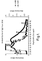
- Figure (1) depicts a protein elution profile from the DEAE ion exchange column.
- Figure (2) depicts an inhibition curve for RH2703.
- Figure (3) depicts an inhibition curve for RH2651.
- Figure (4) depicts an inhibition curve for RH5992.
- Figure (5) depicts an inhibition curve for RH0345.
- Figure (6) depicts an inhibition curve for RH2485.
- Figure (7) depicts external standard curves obtained in PBST and matrix blank samples.
- Figure (8) depicts a calculated linear regression line for Broccoli sample 20139-01.
- Figure (9) depicts a calculated linear regression line for Broccoli sample 20139-02.
- Figure (10) depicts a calculated linear regression line for Broccoli sample 20139-04.
- Figure (11) depicts a calculated linear regression line for Broccoli sample 20139-05.
- Figure (12) depicts a calculated linear regression line for soil sample 151.
- Figure (13) depicts a calculated linear regression line for soil sample 157.
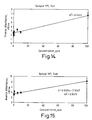
- Figure (14) depicts a calculated linear regression line for soil sample 175.
- Figure (15) depicts a calculated linear regression line for soil sample 187.
- Figure (16) a and b depicts a direct assay curve and direct assay standard curve.
- the term "antigen” is understood to be any substance capable of stimulating antibody production.
- diacyl hydrazine antigen is understood to be any diacyl hydrazine compound capable of stimulating antibody production wherein the produced antibodies have binding affinity to diacyl hydrazines and derivatives thereof.
- the term “immunogen” is understood to include any substance used to induce an immune response.
- the immunogen generally is comprised of a carrier molecule and a another constituent (hapten).
- measuring is understood to encompass both qualitative analysis, i.e., identification of the presence of an analyte in a sample, as well as quantitative analysis, i.e., the determination of the amount of an analyte in a sample. It is also understood that such term may include the preparation of standards and standard curves, such preparation being well known in the art.
- diacyl hydrazine compounds is understood to include within its scope diacyl hydrazines as well as metabolites, synthetic analogs, environmental degradates and photochemical degradates which are derived therefrom.
- % Cross-reactivity (IC 50.5992 / IC 50,analog ) x 100 where IC 50,5992 is the IC 50 concentration for RH5992 as determined above and IC 50,analog is the IC 50 concentration of the analog, i.e., RH2703, RH2651, and RH0345.
- an immunogen is provided which is useful in preparing antibodies to diacyl hydrazines.
- the immunogen includes a hapten and a carrier molecule.
- the hapten is generally a diacyl hydrazine molecule or derivative thereof.
- the hapten is a benzoyl hydrazine.
- the benzoyl hydrazine compound is a compound according to formula (1) above.
- Suitable examples of (C 1 -C 6 ) alkyl include, but are not limited to, methyl, ethyl, n-propyl, isopropyl, n-butyl, isobutyl, tert-butyl, n-pentyl, isopentyl, neopentyl, n-hexyl, isohexyl, etc.
- Suitable examples of (C 1 -C 6 ) substituted alkyl include, but are not limited to, methyl, ethyl, n-propyl, isopropyl, n-butyl, isobutyl, tert-butyl, n-pentyl, isopentyl, neopentyl, n-hexyl, etc., substituted with hydroxy, halide, (C 1 -C 4 ) alkoxy, and nitro.
- Suitable examples of (C 2 -C 6 ) alkenyl include, but are not limited to, ethenyl, n-propenyl, isopropenyl, n-butenyl, isobutenyl, tert-butenyl, n-pentenyl, isopentenyl, neopentenyl, n-hexenyl, etc.
- Suitable examples of (C 2 -C 6 ) substituted alkenyl include, but are not limited to, ethenyl, n-propenyl, isopropenyl, n-butenyl, isobutenyl, tert-butenyl, n-pentenyl, isopentenyl, neopentenyl, n-hexenyl, etc., substituted with hydroxy, halide, or nitro groups.
- Suitable examples of (C 1 -C 6 ) alkoxy include, but are not limited to, methoxy, ethoxy, n-propoxy, isopropoxy, n-butoxy, isobutoxy, tert-butoxy, n-pentoxy, isopentoxy, neopentoxy and n-hexoxy.
- Suitable examples of (C 1 -C 6 ) alkyloxy include, but are not limited to, carboxy (-COOH), acetyloxy (-CH 2 COOH), propyloxy (-CH 2 CH 2 COOH), and n-butyloxy (-CH 2 CH 2 CH 2 COOH).
- Suitable examples of halides include, but are not limited to Cl-, Br-, F-, and I-.
- R is -CH 2 COOH, R 1 and R 2 are each methyl and R 3 and R 4 are each hydrogen. In a more preferred embodiment, R is -COOH, R 1 and R 2 are each methyl and R 3 and R 4 are each hydrogen.
- the carrier material may be a protein, for example without limitation, bovine serum albumin, ovalbumin, or keyhole limpet hemocyanin; a polysaccharide, for example without limitation, dextran, sepharose, agarose or cellulose; or a synthetic polymer or copolymer, for example without limitation, polyacrolein, polyamide, polyacrylamide, polybutyrate, polyurea, polyureamide, or polystyrene.
- a protein for example without limitation, bovine serum albumin, ovalbumin, or keyhole limpet hemocyanin
- a polysaccharide for example without limitation, dextran, sepharose, agarose or cellulose
- a synthetic polymer or copolymer for example without limitation, polyacrolein, polyamide, polyacrylamide, polybutyrate, polyurea, polyureamide, or polystyrene.
- the carrier material is a carrier protein, more preferably bovine serum albumin (BSA) or keyhole limpet hemocyanin (KLH), most preferably keyhole limpet hemocyanin.
- BSA bovine serum albumin
- KLH keyhole limpet hemocyanin
- the immunogen of the present invention may be used to prepare antibodies to diacyl hydrazine compounds.
- the immunogen is introduced into a suitable animal and the antibodies collected, isolated and purified.
- the resultant antibodies are polyclonal antibodies having a high binding affinity and cross reactivity to diacyl hydrazine compounds and derivatives thereof, particularly benzoyl hydrazines and derivatives thereof.
- the immunogen may be used to prepare monoclonal antibodies through use of hybridoma technology well known in the art.
- antibodies are raised to an immunogen of formula (1) conjugated to a carrier material.
- antibodies are raised to an immunogen of formula (1) wherein R is -CH 3 COOH, R 1 and R 2 are methyl, R 3 and R 4 are hydrogen, conjugated to a carrier material.
- antibodies are raised to an immunogen of formula (1) wherein R is - COOH, R 1 and R 2 are methyl, R 3 and R 4 are hydrogen, conjugated to a carrier material.
- the present invention provides a method of measuring diacyl hydrazines or derivatives thereof. Initially, the method includes in step (A) providing a sample comprising an unknown amount of diacyl hydrazine.
- the sample is generally any material which may contain diacyl hydrazines. Suitable examples, include air, water, soil, and biological materials.
- the sample may be an air sample or a sample derived from an air sample.
- the sample may be a water sample or a sample derived from a water sample.
- the sample may be a soil sample or a sample derived from a soil sample.
- the sample may be a biological material or a sample containing a biological material or a sample derived from a biological material.
- suitable examples of biological materials include without limitation, plant material; biological fluids such as blood, serum, plasma, lymphatic fluid, gastric lavage, bile, vitreous humor; biological tissues such as plant and animal tissues, and microbiological specimens such as bacteria and viruses.
- the sample is a soil or plant material or a sample derived therefrom.
- the diacyl hydrazine compound is as described above in Formula 1 as well as derivatives thereof. Such derivatives may be, without limitation, metabolites, synthetic analogs, environmental degradates, photochemical degradates derived therefrom. Particularly useful diacyl hydrazines are benzoyl hydrazines according to Formula 1 which are depicted in Table 1 following.
- the sample of step (A) is contacted in Step (B) with antibodies having binding affinity to diacyl hydrazines in the presence of an immobilized diacyl hydrazine antigen.
- the antibodies are as described above, with antibodies raised from the immunogen comprising RH2651 conjugated to KLH being preferred.
- the antigen of the immobilized diacyl hydrazine antigen may be any of the diacyl hydrazines described above conjugated to any of the carrier molecules described above.
- the immobilized antigen is RH2651 or RH2703 conjugated to a carrier protein, more preferably RH2703 conjugated to BSA.
- the combination of the antigen and the carrier molecule is referred to as the coating antigen.
- the coating antigen is generally immobilized on a solid support having a surface(s) which are amenable to attachment of such an antigen. Suitable examples include without limitation microtiter plates; membrane material such as nitrocellulose, cellulose, cellulose acetate, polycarbonate, etc.; test tubes; particles such as beads, column packing materials, etc.; and derivatized surfaces having binding domains capable of binding the antigen attached thereon. Generally, the coating antigen is applied to a solid support and adsorbed thereto.
- the coating antigen is adsorbed onto a microtiter plate.
- step (B) competition for binding sites on the antibodies having a high affinity for diacyl hydrazines occurs between the free antigen, i.e., the unknown quantity of benzoyl hydrazines in the sample, and the immobilized, i.e., bound, antigen. As a result bound and unbound antigen-antibody complexes are formed.
- step (C) the bound complex is separated from the unbound complex. Separation may be by any means known in the art, such as without limitation gravity separation, magnetic separation, and washing to remove unbound complex.
- the bound complex is labeled with a detectable label in step (D).
- the label may be any material which can be fixed to the bound antigen-antibody complex and be detected.
- the label may be detected directly or indirectly through interaction with a substrate. Suitable examples include without limitation enzymes, colored dyes, fluorescent materials, chemiluminescent materials, bioluminescent materials, and radioactive isotopes.
- the detectable label is an enzyme.
- the enzyme is conjugated with a material which has a binding affinity to the bound antibody-antigen complex.
- the binding of the label to the complex may be by antibody-antigen, protein-ligand, or avidin(strepavidin)-biotin binding.
- the binding is antibody-antigen binding wherein the label is bound to an antibody having binding affinity for the bound antibody-antigen complex.
- the antibody bound to the label may be polyclonal or monoclonal.
- an antibody fragment of any of the above recited antibodies which contains the binding region of the antibody such as Fab fragments may be used.
- the particular antibody conjugated to the label will, of course, depend on the type of antibody conjugated to the antigen. That is, the antibody conjugated to the label will be chosen to have binding affinity to the immobilized antibody-antigen complex.
- the antibody conjugated to the antigen in the present invention is raised in rabbits. Consequently, the antibody conjugated to the label is an anti-rabbit antibody having binding affinity to the rabbit antibodies.
- a measurable change is effected in the label in step (E).
- the measurable change may be effected by irradiation with uv-vis light, fluorescent, an electrical waveform, or introduction of a substrate which interacts with the label to produce a measurable change. It is understood that the measurable change may be inherent in the label, for instance, the label may be a radioactive isotope wherein radioactivity, e.g., gamma-rays, which may be effected simply by introducing means to measure the radioactivity.
- the measurable change is effected by contacting the labeled complex with a substrate.
- a substrate are well known in the art and of course are dependent on the type of label used.
- the label is an enzyme and the substrate is a compound capable of interacting with the enzyme to produce a measurable change.
- the enzyme is a phosphatase and the substrate is a phosphate compound. The interaction of the enzyme and the substrate generally results in a compound having a measurable property. For instance, a compound which is fluorescent, has a strong absorbance, etc.
- step (F) the diacyl hydrazines are measured in step (F).
- measurement is by methods known in the art, e.g., uv-vis spectrophotometry, fluorometry, gamma counting, etc.
- the above recited assay is generally known as an indirect assay. Also, understood to be included within the scope of the present invention is a direct assay.
- the direct method includes in step (A) providing a sample comprising an unknown amount of diacyl hydrazines.
- the sample and diacyl hydrazines are as described above.
- the sample of step (A) is contacted in Step (B) with immobilized antibodies having binding affinity to diacyl hydrazines to form immobilized antibody-diacyl hydrazine complexes.
- the antibodies are as described above with antibodies raised to RH2651 being preferred.
- the antibodies are generally immobilized on a solid support having a surface(s) which are amenable to attachment of such an antibody. Suitable examples include without limitation microtiter plates; membrane material such as nitrocellulose, cellulose, cellulose acetate, polycarbonate, etc.; test tubes; particles such as beads, column packing materials, etc.; and derivatized surfaces having binding domains capable of binding the antibody attached thereon.
- the antibody is a coating antibody which is fixed to a solid support as described above with reference to the coating antigen.
- step (B) binding sites on the immobilized polyclonal antibodies are filled by free antigen, i.e., the unknown quantity of benzoyl hydrazines in the sample, to form immobilized antibody-antigen complexes.
- step (C) diacyl hydrazine antigen labeled with a detectable label is contacted with sites on the immobilized antibodies which were not bound to benzoyl hydrazine from the sample. Accordingly, the labeled antigen will fill the remaining binding sites on the antibodies. So that the immobilized antibodies will be conjugated to labeled and unlabeled antigen.
- the diacyl hydrazine antigen and label are as described above. However, in a preferred embodiment, the antigen is RH2651 and the label is an enzyme, preferably horseradish peroxidase (HRP).
- step (D) a measurable change is effected in the label and diacyl hydrazines are measured in step (E). Effecting of the measurable change and measuring of the diacyl hydrazines is as described above for the indirect assay.
- the assays described herein for diacyl hydrazines such as RH5992 and its derivatives RH2703, RH2651, RH0345 and RH2485 are sufficiently sensitive for application to field residue studies as required by FIFRA registration. Moreover the assay has sufficient cross-reactivity with structurally dissimilar analogs such as RH0345 that it is believed that the assay will be applicable for the entire class of substituted N-t-butyl-N, N'-benzoyl hydrazine pesticides.
- the greatest advantage may be the increased laboratory efficiency. For example, it has been shown that a single analyst can easily process twenty samples per day. This estimation includes sample preparation and results calculation. Assuming a ten hour day, only about 30 man-minutes per sample labor cost is incurred.
- antibodies of the present invention include binding the antibodies to a solid support to concentrate residues from samples matrices so they may be labeled with a fluorescent tag and be used in tissue studies to identify the location of pesticide residues in cellular compartments. This information provides important information as to the mode of action or mechanisms of toxicity and metabolism of the particular diacyl hydrazine compound. Also, the direct immunoassay method may be incorporated into a "dip-stick" test for field analysis and survey types of applications.
- Carrier Protein, KLH (Imject®, Pierce Chemical Co., 20mg of protein in PBS (pH 7.2 when reconstituted) was reconstituted in 5.4 ml deionized water.
- the activated hapten was transferred to a microcentrifuge (Microcentrifuge Model 235B, Fisher Scientific) and centrifuged for 5 minutes to remove the substituted urea precipitate. The supernatant was transferred to the reconstituted carrier protein solution and stirring was continued at room temperature for four hours.
- the protein reaction mixture was centrifuged @ 14000 rpm for 5 minutes to remove precipitated protein. The entire volume of the supernatant was applied to a Swift® polyacrylamide desalting column (Pierce Chemical Co.) and eluted with 0.01M PBS ( pH 7.3) in 3ml fractions. Fractions were collected after the sample had been completely loaded onto the column. Ten three (3) ml fractions were taken and the absorbance at 280 nm was measured for each fraction. The fractions containing the immunogen were pooled and the protein concentration was measured by the BCA method (See BCA Protein Assay Reagent Instructions - Pierce Chemical Co.).
- the hapten/protein binding ratio was determined as follows. A standard curve of the absorbance of KLH at 254 nm over the range of 0 ⁇ g/ml to 1200 ⁇ g/ml was established. The protein concentration of the immunogen fraction measured by the BCA method was converted to absorbance at 254 nm from the standard curve. A standard curve of the absorbance of the haptens RH2651 and RH2703 at 254 nm over the concentration range of 26 to 260 nmole/ml for RH2703 and 6.5 to 210 nmole for RH2651 was established.
- the absorbance of the immunogen was measured at 254 nm (A conj ) and the calculated contribution to the absorbance at 254 nm from the KLH (A KLH ) was subtracted to give the absorbance arising from the hapten (A Hap ). This value, A Hap , was converted to concentration (nmole/ml) from the standard curve for the hapten.
- the molar binding ratio of hapten to protein was calculated from the calculated concentration of hapten (nmole/ml) divided by the measured concentration of immunogen. An average molecular weight for KLH of 6.7 x 10 6 dalton was used. The average number of hapten molecules per 10,000 amu of KLH was calculated by dividing the molar binding ratio by 670. The results are presented in Table 2.
- An immunogen was prepared according to the procedure of Example 1, except that the hapten used was RH2703. Results are shown in Table 2.
- Coating antigens were prepared according to the procedure for preparing the immunogen in Example 1 except that BSA (Imject®, Pierce Chemical Co.) replaced KLH as the carrier protein.
- BSA Imject®, Pierce Chemical Co.
- the average molecular weight of BSA used in the calculation of molar binding ratio was 68,000 dalton.
- the average number of hapten molecules per 10,000 amu of BSA was calculated by dividing the molar binding ratio by 6.8. Results for the coating antigens prepared are shown in Table 2.
- a primary inoculum was prepared from 2.5 ml of the immunogen (product of Example 2 reconstituted to 1 mg/ml in 0.01 M PBS), 25 mg M. tuberculosis suspension and 2.5 ml of Freund's adjuvant. The mixture was homogenized until thick and creamy.
- New Zealand White rabbits (5) were used to produce antibodies.
- the rabbits were disease free females weighing between 4.75 to 5.5 pounds.
- the rabbits were first inoculated intramuscularly against Bordetella pertussis (B. pertussis inoculum prepared to contain 6 x 10 10 cells suspended in 0.6 ml saline) and bled to provide a background titer.
- Bordetella pertussis B. pertussis inoculum prepared to contain 6 x 10 10 cells suspended in 0.6 ml saline
- the dose of primary inoculum per rabbit was 1.0 ml by subcutaneous injection.
- the rabbits were inoculated and received booster immunizations every 21 days. Eight days after the fourth booster injection, the rabbits were bled to collect antibodies. After an additional time period, booster injections were reinitiated and a production bleed was done again after the fourth booster injection.
- Coating antigen was reconstituted in coating buffer (PBS, pH 7.2) to an initial concentration of approximately 10 ⁇ g/ml.
- the wells of columns 1 and 2 of 96 well microtiter plates were reserved for control samples. Beginning with the wells in column 3, 200 ⁇ l of the coating antigen was added at the initial concentration.
- the wells in columns 4 through 12 contained 100 ⁇ l of coating buffer. A portion, 100 ⁇ l, of the coating antigen in the wells of column 3 were transferred and mixed with the coating buffer in the wells of column 4 and in this manner the coating antigen is serially diluted 1:2 with coating buffer in each subsequent column of wells. The dilution process was continued through column 11 which was a 1:256 dilution of the coating antigen.
- Wells in column 12 contained only coating buffer with no antigen as negative controls.
- Wells A1 and A2 were air blanks and did not receive reagents.
- Wells B1 and B2 were coated with coating antigen and were background wells.
- Wells C1 and C2 were coated with 100 ⁇ l of KLH in coating buffer (about 10 ⁇ g/ml) as positive controls. The remaining wells D1 through H2 were untreated.
- the plate thus prepared was sealed with sealing tape, covered, wrapped in plastic wrap and placed in the refrigerator for overnight incubation at 4°C.
- BSA blocking solution 1% BSA, pH 7.2 prepared from 1.0 g BSA dissolved in 100 ml PBS. This solution was incubated at room temperature for at least 10 minutes.
- Test samples were diluted 1:1000 with blocking solution and 200 ⁇ l were applied to the wells in Row A beginning with well A3.
- the wells in rows B3 through H3 contained 100 ⁇ l blocking solution.
- the test samples (100 ⁇ l) in row A wells were transferred to row B wells and mixed thoroughly. In this manner samples were serially diluted 1:2 through the wells of row G3 (1:64000) and the final 100 ⁇ l was discarded.
- the wells of row H3 contained no antibody and were negative controls.
- the background wells B1 and B2 were incubated with 100 ⁇ l of pre-bleed sera that was diluted 1:500 with blocking solution.
- the positive control wells C1 and C2 were incubated with a 1:1000 dilution of the test sample. Plates were incubated with the test sample for approximately one hour.
- test samples were removed and the wells were washed with washing buffer.
- the wells were filled 4-5 times with buffer.
- the buffer was allowed to remain in the wells for a 3-5 minute soak before discarding.
- Immune serum collected over the course of the injections in Example 3 were pooled and assayed for total protein by the BCA method and found to be 66 mg/ml.
- the volume of immune serum was measured and divided into two equal aliquots of 90 ml each. A single aliquot was diluted with two equivalent volumes of deionized water. The resulting solution volume was diluted in turn with an equal volume of 4M ammonium sulfate to give a final 2M ammonium sulfate solution.
- the suspension was allowed to stir overnight at room temperature.
- the protein precipitate was collected by centrifugation at 10,000 g-av. for thirty minutes.
- the protein pellet was dissolved in a minimum volume of 0.02 M sodium phosphate buffer (pH 8.0) that had been prepared from dilution of 0.1 M sodium phosphate buffer.
- the reconstituted protein solution was dialyzed for four hours against 2 L of 0.02 M sodium phosphate buffer (pH 8) at room temperature, then overnight against 4 L at 4°C, and finally for 4 hours against 4 L at room temperature.
- the ion exchange purification of IgG from the pooled antisera was conducted with two separate aliquots utilizing DEAE Cellulose (DE52, Whatman, Inc.), 150 ml wet volume, which was packed to a bed height of approximately 26 cm in a 2.6 cm (i.d.) column.
- the column was equilibrated to 0.02 M sodium phosphate buffer (pH 8.0).
- the column had a protein capacity of approximately 1.5 g (10 mg per ml wet volume).
- the entire volume of the dialyzed protein solution was pumped onto the column and elution begun with 0.02 M sodium phosphate buffer, pH 8.0. The flow rate was adjusted to approximately 5 ml/min and 5 ml fractions were taken.
- the first 150 ml effluent contained any unretained protein.
- the molarity was continuously changed in a linear gradient to 0.3 M.
- the gradient was formed in a two chamber gradient former containing 250 ml of 0.02 M buffer in the first chamber and 250 ml of 0.3 ml buffer in the second chamber.
- the first chamber was continuously stirred during elution.
- the next 500 ml of effluent was collected in 5 ml fractions.
- the elution of protein was monitored by measuring the absorbance at 280 nm which was plotted against elution volume (fraction #) to give a chromatogram.
- the DEAE purified IgG (1 mg/ml) was affinity purified using Protein A AffinityPak® Columns and the ImmunPure® IgG Buffers (ImmunoPure® IgG Purification Kit, Pierce Chemical Co.). The protein A column and buffers were allowed to come to room temperature and the column was washed with 5 ml of Binding Buffer. The DEAE purified IgG was diluted 1:9 to give a protein concentration of 6 mg/ml and a 1 ml aliquot was applied to the column. The column was washed with another 15 ml of binding buffer.
- Elution of the IgG was carried out with 5 ml of ImmunoPure® IgG Elution buffer and 1 ml fractions were taken. The absorbance of each fraction was measured at 280 nm and the fraction with significant absorbance (fraction 3) was desalted using a 5 ml Excellulose® column which had been previously conditioned with 10 ml of 0.02 M PBS (20 mM sodium phosphate, 100mM NaCl, pH 7.4). Ten one (1) ml aliquots of 0.02 M PBS were used for elution and 1 ml fractions were collected. The absorbance of each fraction was assayed for total protein by the BCA method.
- the Protein A affinity chromatography purified IgG was diluted 1:5 to give a protein concentration of 1 mg/ml. Given an estimated 9 mg/ml IgG concentration from an average total serum protein of 66 mg/ml the estimated recovery of IgG was 67 percent.
- RH2703-BSA was used as the coating antigen.
- concentration required to give a readable response (0.3-0.5 absorbance units @ 410 nm) in the presence of the minimum quantity of antigen (RH2703) was optimized in the presence of a constant amount of protein A purified IgG (10 ⁇ l of a 20 ⁇ g/ml solution, 100 ng/well) by a checkerboard type assay ( see Voller et al, Bull of World Health Organization, 53, 35 (1976) ) .
- the concentration of the Protein A purified IgG had been previously optimized in the presence of a constant amount of coating antigen (RH2703-BSA, 100 ⁇ l of a 10 ⁇ g/ml solution) to give a readable response to the minimum amount of RH5992.
- Antibody concentration was optimized to be approximately 20 ⁇ g/ml (100 ng per micro well) and the coating antigen was optimized to be 25 ng/ml or 2.5 ng per micro well.
- the hapten is in a three-fold molar excess of the IgG added.
- the percent inhibition for RH2703, RH2651, RH5992, RH0345 and RH2485 were determined to the protein IgG in the presence of the optimized concentration of RH2703-BSA coating antigen.
- Each test substance was assayed at seven concentrations ranging from no test substance 0 ng/ml to 1000 ng/ml. The assay at each concentration was replicated seven times.
- Antigen negative and antibody negative wells provided negative controls while KLH/antiKLH wells were used as positive controls.
- test substance stock solutions were made with coating buffer containing 0.1% Tween 20 to give 1000 ng/ml, 100 ng/ml, 10 ng/ml, 1 ng/ml, 0.1 ng/ml, and 0.01 ng/ml assay concentrations.
- Coating buffer w/0.1% Tween 20 was used as the 0 ng/ml assay concentration.
- the test substance assay concentrations (190 ⁇ l each) were incubated with 10 ⁇ l of the protein A IgG (20 ⁇ l/ml) for one hour at room temperature. After the incubation period, 100 ⁇ l of the test substance-IgG mixture was transferred to the appropriate microtiter well.
- the microtiter wells had been previously coated with the optimized concentration of RH2703-BSA and the excess protein binding sites were blocked with 0.3 ml of a 1% BSA solution in coating buffer. The plates were incubated for an additional hour at room temperature.
- Percent cross-reactivity was calculated from IC 50 's as recited above.
- a plot of the average absorbances of the seven replicate analysis obtained from the seven concentrations of the inhibitors was made using Microsoft Excel ⁇ , Ver 5.0 graphics (Microsoft Corporation, Copyright 1993).
- the linear dynamic range was visualized as the linear portion of the curve obtained from a plot of absorbances at 410 nm from the various inhibitor concentrations. Sensitivity was calculated as described above.
- the limit of detection was defined as three times the measured background noise. To assess the background noise the standard deviations of the replicate analyses at each concentration of inhibitor were calculated and the least squares method of regression analysis was performed. The y-intercept of the regression line was taken as the background absorbance arising from noise. The background absorbance was multiplied by a factor of three and converted to the concentration of the inhibitor by division with the slope of the standard curve obtained over the linear range. The resulting concentration was defined as the detection limit of the inhibitor.
- Microtiter plates were coated with 100 ⁇ l/well of RH2703-BSA coating antigen (10 ⁇ g/ml) to give a 1 ⁇ g per well coating. The plates were incubated overnight at 4°C. The unbound protein binding sites were blocked prior to use with 300 ⁇ l/well of a 1% BSA solution in coating buffer.
- Broccoli sample # 03 1.0191 g
- soil sample # 49 1.0180 g
- the sample identification and sample mass used are identified in Table 6 below.
- the sample aliquots were placed in 25 ml conical centrifuge tubes and the recovery sample was spiked with 50 or 500 ng of RH5992 which was prepared from dilution of RH5992 stock standard (1 mg/ml in acetonitrile) with PBST.
- Samples were extracted with 5ml aliquots of PBST by sonication with a sonic cell disruptor (Sonicator®,Heat Systems, Inc. Model W-225R with a Model H-1 microtip probe) for three minutes at 80% power.
- the extraction mixture was centrifuged for five minutes at 4,000 g and the PBST layer was decanted.
- An aliquot of each sample was diluted with PBST to give a 1:100 dilution (broccoli samples) and a 1:500 dilution (soil samples) of the original sample extract.
- Sample concentrations were obtained by solving the linear equation for x when y was set equal to the inverse absorbance measured with no added inhibitor. The result was converted to total ng by multiplication with the corresponding dilution factor. The final result in ng/g was calculated by dividing the total ng by the mass of sample taken.
- RH2651 A 4.6 mg (10.5 ⁇ mole) quantity of RH2651 was placed in a 5 ml pear shaped flask and dissolved in 100 ml of DMF. In separate test tubes, 4.6 mg NHS and 8.8 ⁇ l DCC were dissolved in 100 ⁇ l each of DMF. The NHS and DCC were added to the RH2651 and stirred at room temperature for about one hour. Stirring was continued overnight at 4°C. The following day 10 mg HRP was dissolved in 2.7 ml 0.01M PBS, pH 7.2. The RH2651/NHS/DCC reaction mixture was centrifuged for three minutes in a Beckman Microfuge E (Beckman Instruments). The supernatant was added dropwise with stirring to the HRP.
- the mixture was stirred for 4 hours at room temperature and then centrifuged for 3 minutes in the microfuge.
- the supernatant containing the conjugated protein was desalted with a Swift® desalting column (Pierce Chemical Co.) equilibrated with PBS.
- the protein was eluted with PBS and 3ml fractions were collected. Fractions containing the protein conjugate, as determined by absorbance at 280 nm, were pooled and the protein concentration was determined by the BCA Protein Assay (Pierce Chemical Co.).
- Binding of RH2651 to HRP was confirmed as follows. Microplate wells were coated with 100 ⁇ l of 10 ⁇ g/ml 2651-HRP and stored at 4°C overnight. After emptying the plate the wells were blocked with 1 % BSA in PBS. After blocking, 1:2 serial dilutions of protein A purified 2651 IgG, prepared according to Example 6, (starting with 1:500) were added to the wells to probe for the RH2651. The plate was washed with 0.01% Tween 20 PBS after a 1 hour room temperature incubation. Alkaline phosphatase labeled goat anti-rabbit IgG was added and incubated for an additional one hour at room temperature. The plate was washed and p-nitrophenylphosphate substrate added. After 15 minutes development time, the absorbance was measured at 410 nm with a Dynatech MR5000 Microplate Reader.
- a microplate was coated with 2651 IgG.
- 100 ⁇ l of 10 ⁇ g/ml 2651 IgG were added to each well and 1:2 serial dilutions made across the plate.
- the plate was sealed and stored at 4°C until ready to use. After emptying, the plate was washed with 0.01 % Tween 20 in PBS and blocked with 1% BSA in PBS for about 1 hour at room temperature. After blocking, the plate was emptied and 150 ⁇ l PBS added to each well. Then 50 ⁇ l of 2651-HRP dilutions were added down the plate starting with 10 ⁇ g/ml and continuing with 1:2 dilutions down the plate.
- Standard solutions of RH5992, RH2651, RH2703, and RH2485 were made from 1 mg/ml stock solutions.
- Standard solutions of RH0345 were prepared from a 0.1 ng/ml, 0.5 ng/ml, 1 ng/ml, 5 ng/ml, 10 ng/ml, 50 ng/ml, and 100 ng/ml.
- 150 ⁇ l of the standards were added to the appropriate wells.
- 150 ⁇ l of PBS was added.
- 50 ⁇ l of 1.25 ⁇ g/ml 2651-HRP were added to each well and the plate agitated to mix the contents.
- One column containing antibody and standards did not receive 2651-HRP but rather 50 ⁇ l PBS as a negative control.
- the plate was washed as before and 150 ⁇ l 2651-HRP was added to each well and the plate agitated to mix the contents.
- One column containing antibody and standards did not receive 2651-HRP but rather 50 ⁇ l PBS as a negative control.
- Figure 16a illustrates a typical curve generated from the assay.
- the linear portion of the curve is visualized between 0.01 ng/ml and 10 ng/ml.
- a typical standard curve in the linear range of the assay is illustrated by Figure 16b using inverse absorbances to generate a positive slope.
- the limit of detection was calculated as 0.096 ng/ml and the sensitivity calculated as 0.005 ng for RH5992.
- Data for RH5992 and analogs is given in Table 7.
Landscapes
- Health & Medical Sciences (AREA)
- Life Sciences & Earth Sciences (AREA)
- Chemical & Material Sciences (AREA)
- Immunology (AREA)
- Molecular Biology (AREA)
- Engineering & Computer Science (AREA)
- Hematology (AREA)
- General Health & Medical Sciences (AREA)
- Biomedical Technology (AREA)
- Medicinal Chemistry (AREA)
- Urology & Nephrology (AREA)
- Biochemistry (AREA)
- Organic Chemistry (AREA)
- Cell Biology (AREA)
- Tropical Medicine & Parasitology (AREA)
- Physics & Mathematics (AREA)
- Analytical Chemistry (AREA)
- Microbiology (AREA)
- Biotechnology (AREA)
- General Physics & Mathematics (AREA)
- Pathology (AREA)
- Food Science & Technology (AREA)
- Genetics & Genomics (AREA)
- Proteomics, Peptides & Aminoacids (AREA)
- Biophysics (AREA)
- Bioinformatics & Cheminformatics (AREA)
- Chemical Kinetics & Catalysis (AREA)
- General Chemical & Material Sciences (AREA)
- Nuclear Medicine, Radiotherapy & Molecular Imaging (AREA)
- Pharmacology & Pharmacy (AREA)
- Animal Behavior & Ethology (AREA)
- Public Health (AREA)
- Veterinary Medicine (AREA)
- Peptides Or Proteins (AREA)
Applications Claiming Priority (4)
| Application Number | Priority Date | Filing Date | Title |
|---|---|---|---|
| US2390096P | 1996-08-12 | 1996-08-12 | |
| US23900 | 1996-08-12 | ||
| US907318 | 1997-08-06 | ||
| US08/907,318 US5948406A (en) | 1996-08-12 | 1997-08-06 | Immunogens, antibodies formed therefrom, and coating antigens useful in a diacylhydrazine immunoassay method |
Publications (2)
| Publication Number | Publication Date |
|---|---|
| EP0824104A2 true EP0824104A2 (fr) | 1998-02-18 |
| EP0824104A3 EP0824104A3 (fr) | 1999-03-24 |
Family
ID=26697755
Family Applications (1)
| Application Number | Title | Priority Date | Filing Date |
|---|---|---|---|
| EP97306125A Withdrawn EP0824104A3 (fr) | 1996-08-12 | 1997-08-12 | Procede d'immunodosage |
Country Status (6)
| Country | Link |
|---|---|
| US (2) | US5948406A (fr) |
| EP (1) | EP0824104A3 (fr) |
| KR (1) | KR19980018623A (fr) |
| AU (1) | AU745546B2 (fr) |
| CA (1) | CA2212711A1 (fr) |
| IL (1) | IL121534A (fr) |
Cited By (2)
| Publication number | Priority date | Publication date | Assignee | Title |
|---|---|---|---|---|
| GB2329247A (en) * | 1997-08-12 | 1999-03-17 | Univ Sunderland | Method for monitoring airborne chemicals |
| US8727246B2 (en) | 2009-10-14 | 2014-05-20 | Dow Global Technologies Llc | Process for dry-grinding a polysaccharide derivative |
Families Citing this family (7)
| Publication number | Priority date | Publication date | Assignee | Title |
|---|---|---|---|---|
| US7304161B2 (en) * | 2003-02-10 | 2007-12-04 | Intrexon Corporation | Diaclhydrazine ligands for modulating the expression of exogenous genes in mammalian systems via an ecdysone receptor complex |
| US7456315B2 (en) | 2003-02-28 | 2008-11-25 | Intrexon Corporation | Bioavailable diacylhydrazine ligands for modulating the expression of exogenous genes via an ecdysone receptor complex |
| US8449842B2 (en) * | 2009-03-19 | 2013-05-28 | Thermo Scientific Portable Analytical Instruments Inc. | Molecular reader |
| US9127024B2 (en) | 2013-03-15 | 2015-09-08 | Intrexon Corporation | Boron-containing diacylhydrazines |
| AU2015317862A1 (en) | 2014-09-17 | 2017-04-06 | Intrexon Corporation | Boron-containing diacylhydrazine compounds |
| US20160136790A1 (en) | 2014-11-17 | 2016-05-19 | Snap-On Incorporated | Ratchet Mechanism Spring |
| CN109444412A (zh) * | 2018-10-26 | 2019-03-08 | 成都普利泰生物科技有限公司 | 一种用于微型化学发光免疫分析系统的宠物d-二聚体检测试剂盒 |
Family Cites Families (6)
| Publication number | Priority date | Publication date | Assignee | Title |
|---|---|---|---|---|
| US5354762A (en) * | 1986-07-14 | 1994-10-11 | Rohm And Haas Company | Six-membered heterocyclic derivatives of N'-substituted N,N'-diacylhydrazines |
| US5424333A (en) * | 1985-10-21 | 1995-06-13 | Rohm And Haas Company | Anthelmintic N'-substituted-N,N'-disubstitutedhydrazines |
| US5530028A (en) * | 1992-11-23 | 1996-06-25 | Rohm And Haas Company | Insecticidal N'-substituted-N,N'-diacylhydrazines |
| US5552298A (en) * | 1992-10-23 | 1996-09-03 | Lumigen, Inc. | Enzyme-catalyzed chemiluminescence from hydroxyaryl cyclic diacylhydrazide compounds |
| US5358966A (en) * | 1993-06-08 | 1994-10-25 | Rohm And Haas Company | Turfgrass insecticides |
| DE29614127U1 (de) * | 1996-08-14 | 1996-09-26 | Kessler, Sigurd, Dr., 82178 Puchheim | Bandage zur Fixierung des Sprunggelenks |
-
1997
- 1997-08-06 US US08/907,318 patent/US5948406A/en not_active Expired - Fee Related
- 1997-08-11 CA CA002212711A patent/CA2212711A1/fr not_active Abandoned
- 1997-08-12 IL IL12153497A patent/IL121534A/en not_active IP Right Cessation
- 1997-08-12 KR KR1019970038464A patent/KR19980018623A/ko not_active Ceased
- 1997-08-12 EP EP97306125A patent/EP0824104A3/fr not_active Withdrawn
- 1997-08-12 AU AU33265/97A patent/AU745546B2/en not_active Ceased
-
1998
- 1998-09-09 US US09/150,425 patent/US5981196A/en not_active Expired - Fee Related
Cited By (2)
| Publication number | Priority date | Publication date | Assignee | Title |
|---|---|---|---|---|
| GB2329247A (en) * | 1997-08-12 | 1999-03-17 | Univ Sunderland | Method for monitoring airborne chemicals |
| US8727246B2 (en) | 2009-10-14 | 2014-05-20 | Dow Global Technologies Llc | Process for dry-grinding a polysaccharide derivative |
Also Published As
| Publication number | Publication date |
|---|---|
| CA2212711A1 (fr) | 1998-02-12 |
| US5981196A (en) | 1999-11-09 |
| IL121534A0 (en) | 1998-02-22 |
| US5948406A (en) | 1999-09-07 |
| AU3326597A (en) | 1998-02-19 |
| AU745546B2 (en) | 2002-03-21 |
| IL121534A (en) | 2001-09-13 |
| EP0824104A3 (fr) | 1999-03-24 |
| KR19980018623A (ko) | 1998-06-05 |
Similar Documents
| Publication | Publication Date | Title |
|---|---|---|
| CA1255216A (fr) | Dosage immunologique pour la detection de ligands | |
| US5223441A (en) | Receptors for immune complexes | |
| AU596562B2 (en) | Polyclonal antibodies, preparation and use | |
| US5919641A (en) | Homogeneous immunoassays using enzyme inhibitors | |
| US4298593A (en) | Reagents and methods utilizing labeled Fab bound to antigens | |
| WO2014158864A1 (fr) | Analyses de vitamine d | |
| US4410634A (en) | Method of passively adsorbing immuno-reactive haptens to solid phases | |
| CA1146853A (fr) | Methode pour adsorber passivement des haptenes immuno-reactifs a des supports solides | |
| US5981196A (en) | Immunossay method | |
| US5780243A (en) | Methods for the quantitative analysis of organic compounds | |
| EP0538053B1 (fr) | Séparation et analyse | |
| US5639627A (en) | Method for assaying specific antibody | |
| US5990274A (en) | Cyclosporine derivatives and uses thereof | |
| EP0396570B1 (fr) | Procede de determination de quantite, utilisation et composants | |
| JP3502497B2 (ja) | 複合化被検物質誘導体を用いた競合イムノアッセイ | |
| AU588111B2 (en) | Solid phase analysis method | |
| US4727023A (en) | Preparations for use in solid phase immunoassays comprising monoclonal antibodies covalently embedded in their immobilized hybridoma cells | |
| WO2017039574A1 (fr) | Dosages de vitamine d | |
| JPH10310536A (ja) | イムノアッセイ法 | |
| US4732848A (en) | Process for the determination of an immunologically-bindable substance involving a Fab fragment | |
| JP3709078B2 (ja) | ジアセチルポリアミンの測定法及びキット | |
| US4954433A (en) | Method for the immunolocalization of antigens with the use of antibodies directed against epitopes of non-glucidic nature | |
| Akman et al. | An Enzymeimmunoassay for total Thyroxine using avidin-biotin separation system and Thyroxine-Peroxidase conjugate | |
| HOSODA et al. | Bound/free separation methods in steroid enzyme immunoassay with monoclonal antibody | |
| CN1189618A (zh) | 免疫测定方法 |
Legal Events
| Date | Code | Title | Description |
|---|---|---|---|
| PUAI | Public reference made under article 153(3) epc to a published international application that has entered the european phase |
Free format text: ORIGINAL CODE: 0009012 |
|
| 17P | Request for examination filed |
Effective date: 19970820 |
|
| AK | Designated contracting states |
Kind code of ref document: A2 Designated state(s): BE CH DE DK FR GB IT LI NL |
|
| RAP1 | Party data changed (applicant data changed or rights of an application transferred) |
Owner name: ROHM AND HAAS COMPANY |
|
| RIN1 | Information on inventor provided before grant (corrected) |
Inventor name: WU, SHUGUANG Inventor name: STAVINSKI, STANLEY STEPHEN Inventor name: CASALE, ELLEN SCHALK Inventor name: THACKER, JAMES DOUGLAS |
|
| PUAL | Search report despatched |
Free format text: ORIGINAL CODE: 0009013 |
|
| AK | Designated contracting states |
Kind code of ref document: A3 Designated state(s): AT BE CH DE DK ES FI FR GB GR IE IT LI LU MC NL PT SE |
|
| AKX | Designation fees paid |
Free format text: BE CH DE DK FR GB IT LI NL |
|
| RAP1 | Party data changed (applicant data changed or rights of an application transferred) |
Owner name: DOW AGROSCIENCES LLC |
|
| 17Q | First examination report despatched |
Effective date: 20040114 |
|
| STAA | Information on the status of an ep patent application or granted ep patent |
Free format text: STATUS: THE APPLICATION IS DEEMED TO BE WITHDRAWN |
|
| 18D | Application deemed to be withdrawn |
Effective date: 20040525 |