EP0835643B1 - Thérapie avec ultrasons - Google Patents
Thérapie avec ultrasons Download PDFInfo
- Publication number
- EP0835643B1 EP0835643B1 EP97117456A EP97117456A EP0835643B1 EP 0835643 B1 EP0835643 B1 EP 0835643B1 EP 97117456 A EP97117456 A EP 97117456A EP 97117456 A EP97117456 A EP 97117456A EP 0835643 B1 EP0835643 B1 EP 0835643B1
- Authority
- EP
- European Patent Office
- Prior art keywords
- scanner
- therapy
- therapy device
- sonic
- coupling
- Prior art date
- Legal status (The legal status is an assumption and is not a legal conclusion. Google has not performed a legal analysis and makes no representation as to the accuracy of the status listed.)
- Expired - Lifetime
Links
Images

Classifications
-
- A—HUMAN NECESSITIES
- A61—MEDICAL OR VETERINARY SCIENCE; HYGIENE
- A61B—DIAGNOSIS; SURGERY; IDENTIFICATION
- A61B17/00—Surgical instruments, devices or methods
- A61B17/22—Implements for squeezing-off ulcers or the like on inner organs of the body; Implements for scraping-out cavities of body organs, e.g. bones; for invasive removal or destruction of calculus using mechanical vibrations; for removing obstructions in blood vessels, not otherwise provided for
- A61B17/225—Implements for squeezing-off ulcers or the like on inner organs of the body; Implements for scraping-out cavities of body organs, e.g. bones; for invasive removal or destruction of calculus using mechanical vibrations; for removing obstructions in blood vessels, not otherwise provided for for extracorporeal shock wave lithotripsy [ESWL], e.g. by using ultrasonic waves
- A61B17/2251—Implements for squeezing-off ulcers or the like on inner organs of the body; Implements for scraping-out cavities of body organs, e.g. bones; for invasive removal or destruction of calculus using mechanical vibrations; for removing obstructions in blood vessels, not otherwise provided for for extracorporeal shock wave lithotripsy [ESWL], e.g. by using ultrasonic waves characterised by coupling elements between the apparatus, e.g. shock wave apparatus or locating means, and the patient, e.g. details of bags, pressure control of bag on patient
-
- A—HUMAN NECESSITIES
- A61—MEDICAL OR VETERINARY SCIENCE; HYGIENE
- A61B—DIAGNOSIS; SURGERY; IDENTIFICATION
- A61B17/00—Surgical instruments, devices or methods
- A61B17/22—Implements for squeezing-off ulcers or the like on inner organs of the body; Implements for scraping-out cavities of body organs, e.g. bones; for invasive removal or destruction of calculus using mechanical vibrations; for removing obstructions in blood vessels, not otherwise provided for
- A61B17/225—Implements for squeezing-off ulcers or the like on inner organs of the body; Implements for scraping-out cavities of body organs, e.g. bones; for invasive removal or destruction of calculus using mechanical vibrations; for removing obstructions in blood vessels, not otherwise provided for for extracorporeal shock wave lithotripsy [ESWL], e.g. by using ultrasonic waves
- A61B17/2256—Implements for squeezing-off ulcers or the like on inner organs of the body; Implements for scraping-out cavities of body organs, e.g. bones; for invasive removal or destruction of calculus using mechanical vibrations; for removing obstructions in blood vessels, not otherwise provided for for extracorporeal shock wave lithotripsy [ESWL], e.g. by using ultrasonic waves with means for locating or checking the concrement, e.g. X-ray apparatus, imaging means
-
- A—HUMAN NECESSITIES
- A61—MEDICAL OR VETERINARY SCIENCE; HYGIENE
- A61B—DIAGNOSIS; SURGERY; IDENTIFICATION
- A61B8/00—Diagnosis using ultrasonic, sonic or infrasonic waves
- A61B8/42—Details of probe positioning or probe attachment to the patient
- A61B8/4272—Details of probe positioning or probe attachment to the patient involving the acoustic interface between the transducer and the tissue
- A61B8/4281—Details of probe positioning or probe attachment to the patient involving the acoustic interface between the transducer and the tissue characterised by sound-transmitting media or devices for coupling the transducer to the tissue
-
- G—PHYSICS
- G01—MEASURING; TESTING
- G01S—RADIO DIRECTION-FINDING; RADIO NAVIGATION; DETERMINING DISTANCE OR VELOCITY BY USE OF RADIO WAVES; LOCATING OR PRESENCE-DETECTING BY USE OF THE REFLECTION OR RERADIATION OF RADIO WAVES; ANALOGOUS ARRANGEMENTS USING OTHER WAVES
- G01S15/00—Systems using the reflection or reradiation of acoustic waves, e.g. sonar systems
- G01S15/88—Sonar systems specially adapted for specific applications
- G01S15/89—Sonar systems specially adapted for specific applications for mapping or imaging
- G01S15/8906—Short-range imaging systems; Acoustic microscope systems using pulse-echo techniques
- G01S15/899—Combination of imaging systems with ancillary equipment
Definitions
- the present invention relates to a sound therapy device for contactless crushing of concrements in the body a patient according to the preamble of claim 1.
- EP B 301360 describes a shock wave generator for non-contact Smash one in the body of a living being Concrete known by shock waves, where he a shock wave source based on the electromagnetic principle has a bobbin, a flat coil and a metallic membrane insulated from the flat coil includes.
- the shock wave source is based on a coupling medium the stone can be aligned and with an ultrasound head an ultrasound transmitting and receiving device for transmitting an acoustic signal and to receive echo signals provided by interaction of the acoustic transmission signal in the body of the living being for the purpose of location and Observation of the concretion.
- the membrane is a central opening into which the ultrasound head of the ultrasound transmitter and receiving device can be inserted such that he is in contact with the coupling medium. This is supposed to can be achieved that the ultrasonic waves and the shock waves always have the same direction of incidence to guarantee the same centering of the two systems, to continuously observe the stone disintegration can.
- the central arrangement of the ultrasound head in Shock wave generator is designed to symmetrize the shock waves to care. The maximum occurring in the middle of the Shock wave generator is reduced.
- EP A 355175 describes a device for contactless Smashing concretions in the body of a living being with a shock wave source to generate in a Focus zone converging shock waves with an ultrasound locating device, which has a B-scan applicator, by means of which at least the focus zone can be scanned and with means for acoustically coupling the shock wave source and the B-scan applicator with the body of the living being, the B-scan applicator being multiple ultrasound transducers contains, by means of which at least quasi-simultaneously one of the number of ultrasonic transducers corresponding number of approximations parallel to each other extending layers is scanned, in the area of Sections located in the focus zone are directly adjacent to one another are.
- the object of the present invention is a particular to create a simple sound therapy device that is accurate Observation and / or localization of the calculus with any commercially available shock wave source.
- the invention has the advantage that the lateral Introduce the scanner into the coupling bellows (or through the Structure of the shock wave source) an optimal adaptation the scanner parameter ensures that the The easiest way to locate your scanner brought respective position within the coupling bellows can be, and that if necessary the scanner out of the way of Therapy waves can be removed to remove any shade to avoid the concrement to be treated, or to be able to carry out an undisturbed X-ray location.
- the scanner (or several scanners) through the side locks arranged in the coupling bellows introduced, the bracket of the actual scanner either perpendicular to the axis of the therapy waves or can run at an angle to it.
- This bracket also takes the necessary cables for the scanner.
- the inlet openings i.e. H. the Locks
- the openings are each offset by 90 ° to one another in such a way three-dimensional orientation and observation of the calculus to enable.
- the scanner is used with a suitable one Bracket installed in a fixed place. He can be adjusted so that the image axis exactly with the axis the therapy waves coincide, so that a clear Orientation even without showing the conventional Crosshairs is enabled if, as is common today, distance marks are shown in the ultrasound image and a fade-in central axis is available.
- the scanner can by means of a movable bracket to be moved so that it in the actual therapy from the side of the axis Therapy waves are swung out and thus the therapy waves shadows only minimally while locating and / or observation is brought into an axial position.
- a targeted adjustment outside the axis of the therapy waves is also possible if e.g. B. in renal lithotripsy a suitable sound window between the ribs is to be visited.
- the scanner can either brought in through one of the staggered windows or the shock wave source can around its axis an angle, e.g. B. pivoted between 90 ° and 110 °.
- Another possibility for exact position determination of the scanner consists in the use of position sensors on its holder, to then position in the ultrasound image a brand, e.g. B. a crosshair for marking show the focus position of the therapy waves.
- the scanner can largely move freely be, in which case either a magnetic or an ultrasonic position sensor at a suitable one Place the scanner holder is attached, which for the insertion of a brand into the therapy focus provides in the ultrasound image, e.g. B. by means of a video overlay.
- the scanner holder is advantageously interpreted in such a way that it helps the doctor in the search for the Destination as little as possible, but during the Therapy can be locked in a simple manner.
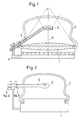
- the sound therapy device for the contactless smashing of concretes in Body of a patient is shown, the sound therapy device has a shock wave source 1 and an ultrasound location system in the form of an imaging scanner 3, which is carried by a bracket 5 which is articulated a fastening 4 is arranged.
- the scanner 3 feels the focus location F of the one emitted by the shock wave source 1 Therapy waves from, because usually at this focus the concrement to be broken is arranged.
- the coupling bellows 2 with at least one lateral lock 9 ( Figure 4) provided the z. B. is opened up by a flexible, stretchable membrane 10 Inserting the scanner 3 into the interior of the coupling bellows 2, such that the bracket 5 of the scanner 2 with an angle the axis of the therapy waves.
- the scanner can also be turned off completely the coupling bellows during the crushing of the concrement be removed to shade the area to be treated To minimize area in the body or in the event of X-ray localization.
- a suitable sound frequency is 7.5 MHz, while for higher depths of penetration, such as B. in gastroenterology or in the renal lithotripsy the 5 MHz or 3.5 MHz scanner are suitable.
- B. in gastroenterology or in the renal lithotripsy the 5 MHz or 3.5 MHz scanner are suitable.
- the scanner is laterally through one of the cutouts in the Coupling bellows inserted into its interior, the bracket either perpendicular to the axis of the therapy waves or can run at an angle to it; in the holder are also advantageously all the necessary cables included for the scanner.
- the scanner itself can either be arranged exactly in the axis of the therapy waves or obliquely in such a way that the focus location F of the therapy waves is contained in the ultrasound image window.
- the scanner can either be installed in the coupling system through complete introduction into the one contained in the coupling bellows Coupling liquid or the scanner can in a preferably thin-walled flexible hose 10 be introduced, which then with the insertion of the scanner even extends into the coupling fluid.
- the scanner with a suitable coupling gel or coupling oil be smeared.
- To different depths of penetration can also support multiple locks, i. H. bushings arranged with membranes or hoses in the coupling bellows become.
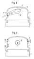
- the locks 9 in different positions on the coupling bellows 2 to distribute, so access from different Enable directions. It is particularly advantageous it when the openings are offset by 90 ° to each other are then a simple three-dimensional orientation is possible.
- Figure 2 are schematically with 6 and 7 two position sensors designated and by the double arrow different adjustment options the scanner 3 within the coupling bellows 2, the position sensors 6 and 7 serve to connect an external one Control of the scanner 3 through the holder 5 make.
- Figure 3 shows how an elastic membrane 10 in shape a thin, elongated tube around the scanner 3 extends around after this into the interior of the coupling bellows 2 has been introduced.
- Figure 5 finally shows two different arrangements of the scanner 3, this for minimal shadowing during therapy, as the solid lines show, is located near the introducer and during his Use, as shown in dashed lines, roughly the Axis of the therapy wave.
- a targeted adjustment in one Position outside the axis of the therapy waves is special in renal lithotripsy, a suitable one To seek out sound windows between the ribs.
- To the to change the scanner's lateral angle of view it can either through one of the staggered locks, or else the therapy source is around the axis of the Therapy waves pivoted at an angle of 90 to 110 °.
- the scanner can also move freely to a large extent in which case e.g. a magnetic or position sensor working with ultrasound (FIG. 2) or mechanical adjustment of the scanner holder is also required is for an overlay of the therapy focus mark in the ultrasound image, e.g. B. by means of a video overlay provides.
- the holder for the scanner is included advantageously designed so that the doctor at the Search for the destination as little as possible, but during the therapy can be locked in a simple way.
Landscapes
- Health & Medical Sciences (AREA)
- Life Sciences & Earth Sciences (AREA)
- Engineering & Computer Science (AREA)
- Physics & Mathematics (AREA)
- Surgery (AREA)
- Nuclear Medicine, Radiotherapy & Molecular Imaging (AREA)
- Animal Behavior & Ethology (AREA)
- Veterinary Medicine (AREA)
- Biomedical Technology (AREA)
- Heart & Thoracic Surgery (AREA)
- Medical Informatics (AREA)
- Molecular Biology (AREA)
- Remote Sensing (AREA)
- General Health & Medical Sciences (AREA)
- Public Health (AREA)
- Radar, Positioning & Navigation (AREA)
- Acoustics & Sound (AREA)
- Radiology & Medical Imaging (AREA)
- Vascular Medicine (AREA)
- Orthopedic Medicine & Surgery (AREA)
- Biophysics (AREA)
- Pathology (AREA)
- Computer Networks & Wireless Communication (AREA)
- General Physics & Mathematics (AREA)
- Surgical Instruments (AREA)
- Ultra Sonic Daignosis Equipment (AREA)
Claims (16)
- Appareil pour thérapie à ultrasons permettant de fragmenter, sans contact, des concrétions dans le corps d'un patient, comprenant une source d'ondes de choc, un système de localisation par ultrasons sous la forme d'un scanner fournissant des images qui balaye l'emplacement du foyer de l'onde thérapeutique émise par la source d'ondes de choc, et un coussin de couplage situé entre la source d'ondes de choc et la surface du corps du patient, caractérisé en ce que le coussin de couplage (2) ou une structure de la source d'ondes de choc est muni d'au moins un hublot (9) latéral qui est fermé par une membrane (10) flexible et extensible, afin de pouvoir introduire le scanner (3) à l'intérieur du coussin de couplage de sorte que le manche (5) du scanner (3) forme un angle avec l'axe (11) des ondes thérapeutiques.
- Appareil pour thérapie à ultrasons selon la revendication 1, caractérisé en ce que le scanner est situé, pendant sa mise en oeuvre, dans le coussin de couplage de sorte que l'image produite comprend l'emplacement du foyer thérapeutique F.
- Appareil pour thérapie à ultrasons selon l'une quelconque des revendications précédentes, caractérisé en ce que le hublot est fermé par une membrane en forme d'opercule à membrane extensible.
- Appareil pour thérapie à ultrasons selon l'une quelconque des revendications précédentes, caractérisé en ce que le scanner est relié, de manière fixe, à un support de sorte qu'il détermine sans équivoque, après une orientation sur le foyer thérapeutique, l'emplacement du foyer thérapeutique, à l'aide des repères affichés sur l'image d'ultrasons.
- Appareil pour thérapie à ultrasons selon l'une quelconque des revendications précédentes, caractérisé en ce que le scanner est disposé de manière mobile sur un support (4), afin de permettre une affectation sans équivoque du foyer thérapeutique (F) sur l'image d'ultrasons.
- Appareil pour thérapie à ultrasons selon l'une quelconque des revendications précédentes, caractérisé en ce que le scanner est situé de manière mobile dans le coussin de couplage de sorte que l'on peut, afin de réaliser la localisation et la thérapie, le placer dans des positions différentes dans lesquelles on peut le bloquer.
- Appareil pour thérapie à ultrasons selon l'une quelconque des revendications précédentes, caractérisé en ce que le scanner est équipé d'une technologie par capteurs permettant de déterminer sa position par rapport au foyer thérapeutique, de sorte que le scanner peut être déplacé librement perdre le contact avec le foyer.
- Appareil pour thérapie à ultrasons selon l'une quelconque des revendications précédentes, caractérisé en ce que l'on peut faire tourner et/ou pivoter la source thérapeutique autour de l'axe des ondes de choc.
- Appareil pour thérapie à ultrasons selon l'une quelconque des revendications précédentes, caractérisé en ce que le scanner présente plusieurs quartz ou matrices de quartz ou qu'il comprend plusieurs axes de mouvement mécaniques pour pouvoir enregistrer, simultanément ou successivement, des images sur plusieurs plans ou bien des images en trois dimensions.
- Appareil pour thérapie à ultrasons selon l'une quelconque des revendications précédentes, caractérisé en ce que le scanner est protégé, sur son côté arrière qui est opposé à l'emplacement du foyer (F), par une protection mécanique, contre l'action des ondes thérapeutiques.
- Appareil pour thérapie à ultrasons selon la revendication 10, caractérisé en ce que la protection du scanner est composée d'une combinaison d'une mousse à pores fermés et d'une plaque de métal ou de matière plastique.
- Appareil pour thérapie à ultrasons selon l'une quelconque des revendications précédentes, caractérisé en ce qu'il s'agit d'un scanner mécanique.
- Appareil pour thérapie à ultrasons selon l'une quelconque des revendications précédentes, caractérisé en ce que le scanner est un scanner à balayage linéaire ou un scanner à balayage courbe.
- Appareil pour thérapie à ultrasons selon l'une quelconque des revendications précédentes, caractérisé en ce que la fixation du scanner comprend au moins un positionneur (6, 7) que l'on peut commander de manière externe et qui permet, au moyen d'un traitement externe des images, la production d'une image d'ultrasons en trois dimensions.
- Appareil pour thérapie à ultrasons selon l'une quelconque des revendications précédentes, caractérisé en ce que le scanner présente un boítier essentiellement tubulaire, que le plan de balayage est situé essentiellement perpendiculairement à l'axe du boítier, et que l'on peut introduire, essentiellement dans le sens radial, le boítier tubulaire dans les ondes thérapeutiques.
- Appareil pour thérapie à ultrasons selon l'une quelconque des revendications précédentes, caractérisé en ce que le transducteur d'ultrasons produisant les images du scanner est disposé à l'extrémité du boítier tubulaire.
Applications Claiming Priority (2)
| Application Number | Priority Date | Filing Date | Title |
|---|---|---|---|
| DE19641935A DE19641935C1 (de) | 1996-10-11 | 1996-10-11 | Schalltherapiegerät |
| DE19641935 | 1996-10-11 |
Publications (3)
| Publication Number | Publication Date |
|---|---|
| EP0835643A2 EP0835643A2 (fr) | 1998-04-15 |
| EP0835643A3 EP0835643A3 (fr) | 1998-07-01 |
| EP0835643B1 true EP0835643B1 (fr) | 2003-08-27 |
Family
ID=7808454
Family Applications (1)
| Application Number | Title | Priority Date | Filing Date |
|---|---|---|---|
| EP97117456A Expired - Lifetime EP0835643B1 (fr) | 1996-10-11 | 1997-10-09 | Thérapie avec ultrasons |
Country Status (3)
| Country | Link |
|---|---|
| EP (1) | EP0835643B1 (fr) |
| DE (2) | DE19641935C1 (fr) |
| ES (1) | ES2205109T3 (fr) |
Families Citing this family (3)
| Publication number | Priority date | Publication date | Assignee | Title |
|---|---|---|---|---|
| DE10163019A1 (de) * | 2001-12-20 | 2003-07-10 | Dornier Medtech Holding Int Gmbh | Ultraschall-Scanner |
| JP6783319B2 (ja) | 2016-03-11 | 2020-11-11 | ソルボンヌ・ユニヴェルシテSorbonne Universite | 脊髄の治療および/または脊髄神経の治療のための埋め込み型超音波発生治療デバイス、該デバイスを備える装置、および方法 |
| CA3017916C (fr) | 2016-03-11 | 2023-11-07 | Sorbonne Universite | Dispositif de traitement externe generant des ultrasons pour le traitement de la moelle epiniere et des nerfs rachidiens, appareil comprenant un tel dispositif et methode mettant en oeuvre un tel dispositif |
Family Cites Families (9)
| Publication number | Priority date | Publication date | Assignee | Title |
|---|---|---|---|---|
| DE3503702C2 (de) * | 1985-02-04 | 1993-10-28 | Siemens Ag | Ankoppeleinrichtung für akustische Wellen |
| US4763652A (en) * | 1986-04-16 | 1988-08-16 | Northgate Research, Inc. | Aiming system for kidney stone disintegrator |
| DE3724562C1 (de) | 1987-07-24 | 1989-01-12 | Spectrospin Ag | Kryostat und Verfahren zu seiner Montage |
| US4928672A (en) * | 1987-07-31 | 1990-05-29 | Siemens Aktiengesellschaft | Shockwave source having a centrally disposed ultrasound locating system |
| EP0355175A1 (fr) * | 1988-08-17 | 1990-02-28 | Siemens Aktiengesellschaft | Dispositif pour fragmenter à distance des concrétions dans le corps d'un être vivant |
| US5078144A (en) * | 1988-08-19 | 1992-01-07 | Olympus Optical Co. Ltd. | System for applying ultrasonic waves and a treatment instrument to a body part |
| DE4007669C3 (de) * | 1990-03-10 | 1997-11-13 | Wolf Gmbh Richard | Vorrichtung zur Stoßwellenbehandlung |
| US5329928A (en) * | 1993-08-16 | 1994-07-19 | Bantum Tripter Joint Venture | Reflected shockwave shielding device |
| DE19543344C1 (de) * | 1995-11-22 | 1996-12-12 | Dornier Medizintechnik | Vorrichtung zur Stoßwellentherapie |
-
1996
- 1996-10-11 DE DE19641935A patent/DE19641935C1/de not_active Expired - Fee Related
-
1997
- 1997-10-09 DE DE59710651T patent/DE59710651D1/de not_active Expired - Lifetime
- 1997-10-09 ES ES97117456T patent/ES2205109T3/es not_active Expired - Lifetime
- 1997-10-09 EP EP97117456A patent/EP0835643B1/fr not_active Expired - Lifetime
Also Published As
| Publication number | Publication date |
|---|---|
| EP0835643A2 (fr) | 1998-04-15 |
| EP0835643A3 (fr) | 1998-07-01 |
| ES2205109T3 (es) | 2004-05-01 |
| DE19641935C1 (de) | 1997-09-11 |
| DE59710651D1 (de) | 2003-10-02 |
Similar Documents
| Publication | Publication Date | Title |
|---|---|---|
| DE3826709C2 (de) | Ultraschall-Therapiegerät | |
| DE4143540C2 (de) | Therapieeinrichtung zur Behandlung eines Patienten mit fokussierten akustischen Wellen | |
| DE69414121T2 (de) | Endoskopische sonde zur abbildung und therapie und ihr behandlungssystem | |
| DE4443947B4 (de) | Endoskop | |
| EP0397980B1 (fr) | Lithotriteur | |
| EP0301360B1 (fr) | Générateur d'ondes de choc avec système central de repèrage ultrasonique | |
| DE4241161C2 (de) | Akustische Therapieeinrichtung | |
| DE3743883C2 (de) | Medizinische Ultraschall-Behandlungsvorrichtung | |
| EP0133946B1 (fr) | Appareil pour la destruction à distance des concrétions | |
| EP1749488B1 (fr) | Appareil de thérapie par ondes de choc, avec imagerie | |
| DE3009482A1 (de) | Diagnosegeraet fuer ein endoskop | |
| DE4318237A1 (de) | Vorrichtung zur Behandlung von biologischem Gewebe und Körperkonkrementen | |
| DE3617032C2 (de) | Lithotripsiegerät mit extrakorporalem Stoßwellengenerator | |
| DE19548000C1 (de) | Vorrichtung zur Ortung von Konkrementen im Körper eines Patienten | |
| DE3328039C2 (de) | Einrichtung zum beruehrungslosen zertruemmern eines im koerper eines lebewesens befindlichen konkrements | |
| DE3817726A1 (de) | Vorrichtung zur raeumlichen ultraschall-ortung von konkrementen | |
| EP0265742A1 (fr) | Lithotripteur avec moyens de localisation | |
| DE19622919C1 (de) | Vorrichtung zur Ortung und Zertrümmerung von Konkrementen | |
| EP0328943B1 (fr) | Générateur d'ondes de choc pour la désintégration à distance des concrétions | |
| EP0400196A1 (fr) | Transducteur d'ondes de choc pour destruction de concrétions | |
| DE3932364A1 (de) | Vorrichtung zur raeumlichen ortung und zur zerstoerung von koerperinneren objekten | |
| EP0835643B1 (fr) | Thérapie avec ultrasons | |
| EP0312847A1 (fr) | Source d'ondes de choc avec système central de repérage ultrasonore | |
| DE4135177C2 (de) | Theraphieeinrichtung zur Behandlung eines Lebewesens mit fokussierten akustischen Wellen | |
| DE68929265T2 (de) | Gerät zum zielen und visulaisieren von konkrementen in echtzeit |
Legal Events
| Date | Code | Title | Description |
|---|---|---|---|
| PUAI | Public reference made under article 153(3) epc to a published international application that has entered the european phase |
Free format text: ORIGINAL CODE: 0009012 |
|
| AK | Designated contracting states |
Kind code of ref document: A2 Designated state(s): CH DE ES FR GB LI NL |
|
| PUAL | Search report despatched |
Free format text: ORIGINAL CODE: 0009013 |
|
| AK | Designated contracting states |
Kind code of ref document: A3 Designated state(s): AT BE CH DE DK ES FI FR GB GR IE IT LI LU MC NL PT SE |
|
| AKX | Designation fees paid |
Free format text: CH DE ES FR GB LI NL |
|
| RBV | Designated contracting states (corrected) |
Designated state(s): CH DE ES FR GB LI NL |
|
| 17P | Request for examination filed |
Effective date: 19990309 |
|
| RAP1 | Party data changed (applicant data changed or rights of an application transferred) |
Owner name: DORNIER MEDTECH HOLDING INTERNATIONAL GMBH |
|
| RAP1 | Party data changed (applicant data changed or rights of an application transferred) |
Owner name: DORNIER MEDIZINTECHNIK GMBH |
|
| GRAH | Despatch of communication of intention to grant a patent |
Free format text: ORIGINAL CODE: EPIDOS IGRA |
|
| RAP1 | Party data changed (applicant data changed or rights of an application transferred) |
Owner name: DORNIER MEDIZINTECHNIK GMBH |
|
| RAP1 | Party data changed (applicant data changed or rights of an application transferred) |
Owner name: DORNIER MEDTECH SYSTEMS GMBH |
|
| GRAS | Grant fee paid |
Free format text: ORIGINAL CODE: EPIDOSNIGR3 |
|
| GRAA | (expected) grant |
Free format text: ORIGINAL CODE: 0009210 |
|
| AK | Designated contracting states |
Designated state(s): CH DE ES FR GB LI NL |
|
| REG | Reference to a national code |
Ref country code: GB Ref legal event code: FG4D Free format text: NOT ENGLISH |
|
| REG | Reference to a national code |
Ref country code: CH Ref legal event code: EP |
|
| GBT | Gb: translation of ep patent filed (gb section 77(6)(a)/1977) | ||
| REF | Corresponds to: |
Ref document number: 59710651 Country of ref document: DE Date of ref document: 20031002 Kind code of ref document: P |
|
| REG | Reference to a national code |
Ref country code: CH Ref legal event code: NV Representative=s name: AVV. FRANCO PAGANI |
|
| REG | Reference to a national code |
Ref country code: ES Ref legal event code: FG2A Ref document number: 2205109 Country of ref document: ES Kind code of ref document: T3 |
|
| ET | Fr: translation filed | ||
| PLBE | No opposition filed within time limit |
Free format text: ORIGINAL CODE: 0009261 |
|
| STAA | Information on the status of an ep patent application or granted ep patent |
Free format text: STATUS: NO OPPOSITION FILED WITHIN TIME LIMIT |
|
| 26N | No opposition filed |
Effective date: 20040528 |
|
| PGFP | Annual fee paid to national office [announced via postgrant information from national office to epo] |
Ref country code: DE Payment date: 20101130 Year of fee payment: 14 |
|
| REG | Reference to a national code |
Ref country code: CH Ref legal event code: PFA Owner name: DORNIER MEDTECH SYSTEMS GMBH Free format text: DORNIER MEDTECH SYSTEMS GMBH#ARGELSRIEDER FELD 7#82234 WESSLING (DE) -TRANSFER TO- DORNIER MEDTECH SYSTEMS GMBH#ARGELSRIEDER FELD 7#82234 WESSLING (DE) |
|
| PGFP | Annual fee paid to national office [announced via postgrant information from national office to epo] |
Ref country code: GB Payment date: 20101025 Year of fee payment: 14 |
|
| PGFP | Annual fee paid to national office [announced via postgrant information from national office to epo] |
Ref country code: CH Payment date: 20111020 Year of fee payment: 15 Ref country code: NL Payment date: 20111024 Year of fee payment: 15 Ref country code: FR Payment date: 20111026 Year of fee payment: 15 Ref country code: ES Payment date: 20111011 Year of fee payment: 15 |
|
| REG | Reference to a national code |
Ref country code: NL Ref legal event code: V1 Effective date: 20130501 |
|
| REG | Reference to a national code |
Ref country code: CH Ref legal event code: PL |
|
| GBPC | Gb: european patent ceased through non-payment of renewal fee |
Effective date: 20121009 |
|
| REG | Reference to a national code |
Ref country code: FR Ref legal event code: ST Effective date: 20130628 |
|
| PG25 | Lapsed in a contracting state [announced via postgrant information from national office to epo] |
Ref country code: CH Free format text: LAPSE BECAUSE OF NON-PAYMENT OF DUE FEES Effective date: 20121031 Ref country code: LI Free format text: LAPSE BECAUSE OF NON-PAYMENT OF DUE FEES Effective date: 20121031 Ref country code: DE Free format text: LAPSE BECAUSE OF NON-PAYMENT OF DUE FEES Effective date: 20130501 Ref country code: GB Free format text: LAPSE BECAUSE OF NON-PAYMENT OF DUE FEES Effective date: 20121009 |
|
| REG | Reference to a national code |
Ref country code: DE Ref legal event code: R119 Ref document number: 59710651 Country of ref document: DE Effective date: 20130501 |
|
| PG25 | Lapsed in a contracting state [announced via postgrant information from national office to epo] |
Ref country code: NL Free format text: LAPSE BECAUSE OF NON-PAYMENT OF DUE FEES Effective date: 20130501 Ref country code: FR Free format text: LAPSE BECAUSE OF NON-PAYMENT OF DUE FEES Effective date: 20121031 |
|
| REG | Reference to a national code |
Ref country code: ES Ref legal event code: FD2A Effective date: 20140116 |
|
| PG25 | Lapsed in a contracting state [announced via postgrant information from national office to epo] |
Ref country code: ES Free format text: LAPSE BECAUSE OF NON-PAYMENT OF DUE FEES Effective date: 20121010 |