EP0857009A1 - Röntgeneinrichtung mit einer Primärblendenanordnung - Google Patents
Röntgeneinrichtung mit einer Primärblendenanordnung Download PDFInfo
- Publication number
- EP0857009A1 EP0857009A1 EP98200132A EP98200132A EP0857009A1 EP 0857009 A1 EP0857009 A1 EP 0857009A1 EP 98200132 A EP98200132 A EP 98200132A EP 98200132 A EP98200132 A EP 98200132A EP 0857009 A1 EP0857009 A1 EP 0857009A1
- Authority
- EP
- European Patent Office
- Prior art keywords
- ray
- value
- correction value
- measuring field
- determining
- Prior art date
- Legal status (The legal status is an assumption and is not a legal conclusion. Google has not performed a legal analysis and makes no representation as to the accuracy of the status listed.)
- Granted
Links
Images
Classifications
-
- A—HUMAN NECESSITIES
- A61—MEDICAL OR VETERINARY SCIENCE; HYGIENE
- A61B—DIAGNOSIS; SURGERY; IDENTIFICATION
- A61B6/00—Apparatus or devices for radiation diagnosis; Apparatus or devices for radiation diagnosis combined with radiation therapy equipment
- A61B6/42—Arrangements for detecting radiation specially adapted for radiation diagnosis
- A61B6/4208—Arrangements for detecting radiation specially adapted for radiation diagnosis characterised by using a particular type of detector
- A61B6/4225—Arrangements for detecting radiation specially adapted for radiation diagnosis characterised by using a particular type of detector using image intensifiers
-
- A—HUMAN NECESSITIES
- A61—MEDICAL OR VETERINARY SCIENCE; HYGIENE
- A61B—DIAGNOSIS; SURGERY; IDENTIFICATION
- A61B6/00—Apparatus or devices for radiation diagnosis; Apparatus or devices for radiation diagnosis combined with radiation therapy equipment
- A61B6/06—Diaphragms
-
- A—HUMAN NECESSITIES
- A61—MEDICAL OR VETERINARY SCIENCE; HYGIENE
- A61B—DIAGNOSIS; SURGERY; IDENTIFICATION
- A61B6/00—Apparatus or devices for radiation diagnosis; Apparatus or devices for radiation diagnosis combined with radiation therapy equipment
- A61B6/54—Control of apparatus or devices for radiation diagnosis
- A61B6/542—Control of apparatus or devices for radiation diagnosis involving control of exposure
-
- H—ELECTRICITY
- H04—ELECTRIC COMMUNICATION TECHNIQUE
- H04N—PICTORIAL COMMUNICATION, e.g. TELEVISION
- H04N23/00—Cameras or camera modules comprising electronic image sensors; Control thereof
- H04N23/30—Cameras or camera modules comprising electronic image sensors; Control thereof for generating image signals from X-rays
-
- H—ELECTRICITY
- H05—ELECTRIC TECHNIQUES NOT OTHERWISE PROVIDED FOR
- H05G—X-RAY TECHNIQUE
- H05G1/00—X-ray apparatus involving X-ray tubes; Circuits therefor
- H05G1/08—Electrical details
- H05G1/26—Measuring, controlling or protecting
- H05G1/30—Controlling
Definitions
- Such an X-ray device is known from DE-A 30 06 774.
- the control loop has an adder that assigns two stored values to one setpoint superimposed, the first storage value being the dependency of the conversion factor of an X-ray image intensifier used by the X-ray image converter Electrode voltage and the second storage value the dependence of the substrate (the residual brightness) of the X-ray image intensifier from which the image format determining opening of the primary aperture assembly contains. This will make one Correction of the measurement values falsified by the X-ray image intensifier for the automatic exposure control achieved.
- the area of diagnostic interest should be one have certain medium darkness.
- the measuring field of the detector is with this area of interest is covered, and in this measuring field during the image acquisition continuously by recording the X-ray dose rate the X-ray dose already accumulated is measured. Is a given X-ray dose reached that corresponds to an average degree of blackening, the automatic exposure control switches off the X-rays.
- the conventional automatic Exposure control then leads to an increase in the X-ray dose until the predetermined x-ray dose is reached.
- the result is often overexposed X-ray images in which important image information has been lost, and an increased dose burden on the patient.
- the invention is therefore based on the object of an x-ray device type mentioned at the beginning for generating x-ray images with improved Specify image quality.
- This task is based on an X-ray device mentioned at the beginning solved in that means for determining an influencing the control loop Correction value, the size of the exposed area of the measuring field is dependent, and are provided to influence the control loop such that the X-ray dose is almost the same even in the case of an unexposed measuring field remains unchanged.
- control circuit means for determining an actual dose value, a desired dose value and a control signal by comparing the actual dose value with the Has setpoint value for controlling the X-ray source can this control loop be influenced by means of the correction value such that at a partial shadowing of the measuring field due to a small Primary aperture opening of the dose setpoint based on a predetermined dose standard setpoint reduced and / or the actual dose value, which is from the detector in Measuring field is detected, is increased.
- the dose setpoints can be in one Storage and thus be predetermined or dependent on different Settings of the x-ray device such as the distance between the X-ray source and the X-ray image converter and the magnification factor of the X-ray image converter can be selected or determined.
- the calculated The control signal serves to supply the X-ray source or an X-ray generator control by influencing tube current, tube voltage and / or Exposure time.
- the means for determining the correction value a computing unit to determine the correction value by forming the ratio from the Area of the exposed area of the measuring field to the total area of the measuring field and that the control loop has a multiplier for determining the dose setpoint by multiplying a specified standard dose setpoint by Have correction value.
- the correction value does not exceed certain limit values, is in one Development of the X-ray device according to the invention provided that the A limiting unit for determining the correction value Limitation of the correction value to a predetermined minimum value and to one Have a maximum value of 1. This is to prevent the regulation in certain Borderline situations works incorrectly.
- the minimum value for example at 0.20 should lie, should apply if the primary aperture is very small and only a very small area, for example less than 20% of the measuring field exposed becomes.
- the maximum value of the correction value should be 1 and then apply if the primary aperture is so large that the entire measuring field is exposed.
- Another embodiment of the invention provides that the means for determination of the correction value, an encoder for detecting the size of the opening of the Have primary aperture arrangement. This means that this measured value can be flow into the control loop, for example in determining the dose setpoint.
- Another embodiment of the invention provides that the means for determination the correction value an encoder for recording the size of the distance between Have X-ray source and X-ray image converter.
- a preferred embodiment of the invention provides that the x-ray device is designed for operation with pulsed fluoroscopy and that the means for Determination of the correction value are designed such that for each X-ray pulse a correction value is determined.
- the X-ray device according to the invention is suitable for fluoroscopic operation since this contains a particularly simple and quick regulation.
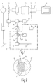
- FIG. 1 shows an x-ray tube 1 that is generated by an x-ray generator 2 is fed.
- an examination object 4 located in front of the radiation exit opening of the X-ray tube 1 a primary aperture 3, which limit the emerging X-rays and the Can determine the opening angle of the X-ray beam.
- an examination object 4 behind it, after which an X-ray image intensifier 5 follows, one with an optical device 6 and a television camera 7 Unity.
- a monitor 8 is connected to the television camera 7, on which X-rays are shown.
- a control circuit 10 is provided for the automatic regulation of the exposure, which generates a control signal S which is fed to the X-ray generator 2 and with which the tube current, the tube voltage and / or the on-time of the X-ray radiation is controlled.
- the control circuit 10 has a photo sensor 9, on which a part of the rays of the image, which is generated on the output screen of the X-ray image intensifier 5, is coupled out of the optical device 6, for example by means of a beam splitter or a partially transparent mirror.
- the photo sensor 9 converts the brightness values of the decoupled optical image into a photo signal.
- a defined area of the photosensor 9 works as a measuring field in which the dose for the exposure control is measured indirectly via the image brightness.
- the mean image brightness is measured in the entire measuring field by integration and an actual dose value D i is determined as the initial value.
- This actual dose value D i is compared in the comparison unit 11 with a target dose value D s , for example by subtracting these two values from one another.
- the target dose value D s contains the information about how large the exposure must be so that the x-ray image in the area of the measuring field reaches an average density value and the x-ray image thus has the highest possible image quality.
- the control signal S is generated which, in the event that the target dose value D s has not yet been reached, contains the information to further increase the x-ray dose and otherwise to switch off the x-ray radiation.
- the target dose value D s is calculated in the exemplary embodiment shown by multiplying a standard dose value D s0 by a correction value K in the multiplication unit 12.
- the standard dose value D s0 is supplied by a memory unit 13 in which standard dose values D s0 are stored for different image intensifier formats, dose levels selected by the user, different examination modes and different pulse rates (with pulsed fluoroscopy).
- the computing unit 16 has also stored the information on how large the area of the measuring field of the Is photosensor 9 and what position the measuring field with respect to the output image of the X-ray image intensifier 5 occupies.
- the arithmetic unit 16 determines from the values mentioned how large the part is is the area of the measuring field that is exposed and not through the diaphragms of the Primary diaphragm arrangement 3 is shadowed.
- the exposed (not shaded) area of the measuring field means that in the area of Input screen of the X-ray image intensifier 5, which the exposed area of the is assigned to the selected measuring field, X-ray radiation strikes and image brightness is produced.
- Under an unexposed (shadowed) area of the measuring field to understand accordingly that in the associated area of the entrance screen of the X-ray image intensifier 5 due to the very small opening of the Primary diaphragm arrangement 3 no X-rays and therefore none Image brightness is generated.
- Size of the opening of the primary aperture assembly 3 varies in size exposed area of the measuring field.
- the dashed border Area 31 forms the measuring field, which in the case shown is about 50% of the Total image area of the photo sensor is 30.
- 32 the areas are designated which due to a small opening of the primary aperture assembly 3 through the Primary screens are shadowed. So in the areas designated 32 arises no exposure. Only the part of the (designated by hatching) designated 33 The area of the measuring field 31 is exposed and that in this part by averaging The measured x-ray dose is said to have an effect on the exposure control to have.
- the correction value K is not calculated from the ratio of the shaded area 33 to the total area 31 of the measuring field calculated what for shown in Figure 2 would give a value for K of about 0.75. Since the Size of the unshaded area 33 of the measuring field, however, not directly can be measured, the values of the Size of the primary aperture and the distance between the X-ray source and X-ray image converter for quasi-indirect calculation of the unshaded Area 33 used.
- the correction value K is then increased by means of a limiting unit 14 limits a minimum value, if arithmetically a smaller value for K than that would result in a minimum value, or the value of K is set to the value one in the event that the entire measuring field 31 is exposed. Between the minimum value and values of K lying at one remain unchanged.
- the correction value K is then multiplied in the multiplication unit 12 by the standard dose set value D s0 , from which the set dose value D s results.
- the standard dose value D s0 is therefore reduced in the event of partial shadowing of the measuring field 31. This prevents the control signal S from incorrectly containing the information that the x-ray dose would have to be increased, since the average density value has not yet been reached, as is the case with conventional exposure control, but the x-radiation is already switched off when in the exposed area a medium blackening value of the measuring field is reached.
- the invention accordingly prevents the x-ray images from being overexposed and important image information being destroyed in the process.
- the invention has the advantage that the patient is not burdened by an increased dose due to a non-optimal regulation.
- the X-ray device according to the invention could also be designed such that not the standard dose setpoint, but the actual dose value accordingly is increased with the help of the correction value with partial shadowing of the measuring field.
- the size of the To vary the measuring field In the case of special x-ray devices, means are also provided, the size of the To vary the measuring field. In such X-ray devices are then also Realization of the invention to provide means for the computing unit (16) Transmit information about the size of the current measuring field, for example a Measuring field encoder.
- the donor 17 is omitted.
- the use of the invention is not on special X-ray devices or Operating modes limited. However, the invention is particularly advantageous in pulsed fluoroscopic operation can be used since the regulation according to the invention works very quickly and easily and therefore one for each fluoroscopy pulse Control signal S can be determined.
Landscapes
- Health & Medical Sciences (AREA)
- Life Sciences & Earth Sciences (AREA)
- Engineering & Computer Science (AREA)
- Medical Informatics (AREA)
- General Health & Medical Sciences (AREA)
- Biophysics (AREA)
- Heart & Thoracic Surgery (AREA)
- High Energy & Nuclear Physics (AREA)
- Veterinary Medicine (AREA)
- Nuclear Medicine, Radiotherapy & Molecular Imaging (AREA)
- Optics & Photonics (AREA)
- Pathology (AREA)
- Radiology & Medical Imaging (AREA)
- Biomedical Technology (AREA)
- Physics & Mathematics (AREA)
- Molecular Biology (AREA)
- Surgery (AREA)
- Animal Behavior & Ethology (AREA)
- Public Health (AREA)
- Multimedia (AREA)
- Signal Processing (AREA)
- Toxicology (AREA)
- Apparatus For Radiation Diagnosis (AREA)
- X-Ray Techniques (AREA)
- Transforming Light Signals Into Electric Signals (AREA)
Abstract
Description
- einer Röntgenstrahlenquelle zur Emission eines Röntgenstrahlenbündels,
- einem Röntgenbildwandler zur Detektion von Röntgenstrahlung und Erzeugung eines Röntgenbildes,
- einer zwischen der Röntgenstrahlenquelle und einem Untersuchungsobjekt angeordneten Primärblendenanordnung zur Begrenzung des Röntgenstrahlenbündels,
- einem Regelkreis zur automatischen Belichtungsregelung, welcher einen Detektor mit wenigstens einem die Röntgenstrahlendosisleistung erfassenden Meßfeld aufweist.
- Fig. 1
- ein Blockschaltbild einer erfindungsgemäßen Röntgeneinrichtung und
- Fig. 2
- eine Skizze der Detektorfläche zur Erläuterung der Berechnung des Korrekturwertes.
Claims (7)
- Röntgeneinrichtung mitdadurch gekennzeichnet, daß Mittel (12, 14, 15, 16, 17) zur Bestimmung eines den Regelkreis (10) beeinflussenden Korrekturwertes (K), der von der Größe des belichteten Bereiches (33) des Meßfeldes (31) abhängig ist, und zur Beeinflussung des Regelkreises (10) vorgesehen sind derart, daß auch bei bereichsweise unbelichtetem Meßfeld (31) die Röntgenstrahlendosis nahezu unverändert bleibt.einer Röntgenstrahlenquelle (1) zur Emission eines Röntgenstrahlenbündels,einem Röntgenbildwandler (5, 6, 7) zur Detektion von Röntgenstrahlung und Erzeugung eines Röntgenbildes,einer zwischen der Röntgenstrahlenquelle (1) und einem Untersuchungsobjekt (4) angeordneten Primärblendenanordnung (3) zur Begrenzung des Röntgenstrahlenbündels,einem Regelkreis (10) zur automatischen Belichtungsregelung, welcher einen Detektor (9) mit wenigstens einem die Röntgenstrahlendosisleistung erfassenden Meßfeld (31) aufweist,
- Röntgeneinrichtung nach Anspruch 1,
dadurch gekennzeichnet, daß der Regelkreis (10) Mittel (9, 11, 12, 13) zur Bestimmung eines Dosis-Istwertes (Di), eines Dosis-Sollwertes (Ds) und eines Steuersignals (S) durch Vergleich des Dosis-Istwertes (Di) mit dem Dosis-Sollwert (Ds) zur Steuerung der Röntgenstrahlenquelle (1) aufweist. - Röntgeneinrichtung nach Anspruch 1 oder 2,
dadurch gekennzeichnet, daß die Mittel zur Bestimmung des Korrekturwertes (K) eine Recheneinheit (16) zur Bestimmung des Korrekturwertes (K) durch Bildung des Verhältnisses aus der Fläche des belichteten Bereiches (33) des Meßfeldes (31) zur Gesamtfläche des Meßfeldes (31) und daß der Regelkreis (10) eine Multipliziereinheit (12) zur Bestimmung des Dosis-Sollwertes (Ds) durch Multiplikation eines vorgegebenen Dosis-Standardsollwertes (Ds0) mit dem Korrekturwert (K) aufweisen. - Röntgeneinrichtung nach einem der vorhergehenden Ansprüche,
dadurch gekennzeichnet, daß die Mittel zur Bestimmung des Korrekturwertes (K) eine Begrenzungseinheit (14) zur Begrenzung des Korrekturwertes (K) auf einen vorgegebenen Minimalwert und auf einen Maximalwert 1 aufweisen. - Röntgeneinrichtung nach einem der vorhergehenden Ansprüche,
dadurch gekennzeichnet, daß die Mittel zur Bestimmung des Korrekturwertes (K) einen Geber (15) zur Erfassung der Größe der Öffnung der Primärblendenanordnung (3) aufweisen. - Röntgeneinrichtung nach einem der vorhergehenden Ansprüche,
dadurch gekennzeichnet, daß die Mittel zur Bestimmung des Korrekturwertes (K) einen Geber (17) zur Erfassung der Größe des Abstandes zwischen Röntgenstrahlenquelle (1) und Röntgenbildwandler (5, 6, 7) aufweisen. - Röntgeneinrichtung nach einem der vorhergehenden Ansprüche,
dadurch gekennzeichnet, daß die Röntgeneinrichtung für den Betrieb mit gepulster Durchleuchtung ausgebildet ist und daß die Mittel zur Bestimmung des Korrekturwertes (K) derart ausgebildet sind, daß für jeden Durchleuchtungspuls ein Korrekturwert (K) ermittelt wird.
Applications Claiming Priority (2)
| Application Number | Priority Date | Filing Date | Title |
|---|---|---|---|
| DE19702739 | 1997-01-27 | ||
| DE19702739A DE19702739A1 (de) | 1997-01-27 | 1997-01-27 | Röntgeneinrichtung mit einer Primärblendenanordnung |
Publications (2)
| Publication Number | Publication Date |
|---|---|
| EP0857009A1 true EP0857009A1 (de) | 1998-08-05 |
| EP0857009B1 EP0857009B1 (de) | 2006-11-08 |
Family
ID=7818396
Family Applications (1)
| Application Number | Title | Priority Date | Filing Date |
|---|---|---|---|
| EP98200132A Expired - Lifetime EP0857009B1 (de) | 1997-01-27 | 1998-01-20 | Röntgeneinrichtung mit einer Primärblendenanordnung |
Country Status (4)
| Country | Link |
|---|---|
| US (1) | US6067343A (de) |
| EP (1) | EP0857009B1 (de) |
| JP (1) | JP4267091B2 (de) |
| DE (2) | DE19702739A1 (de) |
Families Citing this family (22)
| Publication number | Priority date | Publication date | Assignee | Title |
|---|---|---|---|---|
| US6222906B1 (en) * | 1998-01-29 | 2001-04-24 | Kabushiki Kaisha Toshiba | X-ray diagnostic apparatus using an X-ray flat panel detector and method for controlling the X-ray diagnostic apparatus |
| JP4776798B2 (ja) * | 2000-03-29 | 2011-09-21 | 東芝医用システムエンジニアリング株式会社 | X線診断装置 |
| WO2002078546A1 (en) * | 2001-03-29 | 2002-10-10 | Kabushiki Kaisha Toshiba | X-ray diagnosing apparatus |
| AU2002351087A1 (en) * | 2001-12-11 | 2003-06-23 | Koninklijke Philips Electronics N.V. | X-ray examination apparatus and method |
| US7372942B2 (en) * | 2004-03-29 | 2008-05-13 | Siemens Medical Solutions Usa, Inc. | Medical imaging system with dosimetry for estimating circuit board life |
| DE102004017180B4 (de) * | 2004-04-07 | 2007-08-02 | Siemens Ag | Röntgendiagnostikeinrichtung zur digitalen Radiographie |
| WO2006046206A1 (en) * | 2004-10-29 | 2006-05-04 | Koninklijke Philips Electronics N.V. | Portable x-ray detector unit |
| DE102005003226B4 (de) * | 2005-01-24 | 2014-02-06 | Siemens Aktiengesellschaft | Verfahren und Einrichtung zur Wiedergabe eines Röntgenbildes |
| DE102005003225A1 (de) * | 2005-01-24 | 2006-07-27 | Siemens Ag | Verfahren und Einrichtung zum Erzeugen eines Röntgenbildes |
| JP4713952B2 (ja) * | 2005-06-02 | 2011-06-29 | 株式会社東芝 | X線透視撮影用自動露出制御装置 |
| DE102006025411B4 (de) * | 2006-05-31 | 2013-07-04 | Siemens Aktiengesellschaft | Mobiler Röntgenempfänger für eine Röntgenvorrichtung |
| JP5344807B2 (ja) * | 2007-10-19 | 2013-11-20 | 株式会社東芝 | X線診断装置 |
| EP2408375B1 (de) | 2009-03-20 | 2017-12-06 | Orthoscan Incorporated | Bewegliche bildgebungsvorrichtung |
| JP5443100B2 (ja) * | 2009-08-25 | 2014-03-19 | 富士フイルム株式会社 | 放射線画像撮影装置、放射線画像撮影システム及び放射線画像撮影方法 |
| JP5333248B2 (ja) * | 2010-01-14 | 2013-11-06 | 株式会社島津製作所 | X線撮影装置 |
| JP5749898B2 (ja) * | 2010-05-14 | 2015-07-15 | 株式会社島津製作所 | X線透視撮影装置 |
| US9125611B2 (en) | 2010-12-13 | 2015-09-08 | Orthoscan, Inc. | Mobile fluoroscopic imaging system |
| JP6176821B2 (ja) * | 2012-02-01 | 2017-08-09 | 東芝メディカルシステムズ株式会社 | 医用画像診断装置 |
| DE102012216269A1 (de) * | 2012-09-13 | 2014-03-13 | Siemens Aktiengesellschaft | Röntgensystem und Verfahren zur Erzeugung von Bilddaten |
| JP2013223814A (ja) * | 2013-08-09 | 2013-10-31 | Toshiba Corp | X線診断装置 |
| JP5955896B2 (ja) * | 2014-07-07 | 2016-07-20 | 東芝メディカルシステムズ株式会社 | X線撮像装置 |
| CN105528764B (zh) | 2015-12-01 | 2019-01-15 | 沈阳东软医疗系统有限公司 | 一种图像亮度调节方法、装置及设备 |
Citations (7)
| Publication number | Priority date | Publication date | Assignee | Title |
|---|---|---|---|---|
| DE2411630A1 (de) * | 1974-03-12 | 1975-09-18 | Philips Patentverwaltung | Roentgen-belichtungsautomat mit automatischer dominantenwahl |
| DE3006774A1 (de) * | 1980-02-22 | 1981-09-03 | Siemens AG, 1000 Berlin und 8000 München | Roentgendiagnostikeinrichtung |
| EP0121151A1 (de) * | 1983-03-25 | 1984-10-10 | Siemens Aktiengesellschaft | Röntgendiagnostikgerät |
| US4573183A (en) * | 1984-06-29 | 1986-02-25 | General Electric Company | X-Ray image brightness control |
| DE3802351A1 (de) * | 1988-01-27 | 1989-08-10 | Siemens Ag | Roentgendiagnostikanlage |
| EP0480096A1 (de) * | 1990-10-12 | 1992-04-15 | Siemens Aktiengesellschaft | Röntgendiagnostikanlage mit einem Röntgenbildverstärker und einem Detektor für die Bildhelligkeit auf dessen Ausgangsschirm |
| DE4411729A1 (de) * | 1994-04-05 | 1995-10-12 | Siemens Ag | Zahnärztliche Röntgendiagnostikeinrichtung |
Family Cites Families (3)
| Publication number | Priority date | Publication date | Assignee | Title |
|---|---|---|---|---|
| NL9100019A (nl) * | 1991-01-09 | 1992-08-03 | Philips Nv | Roentgenonderzoekapparaat. |
| DE4106596C1 (de) * | 1991-03-01 | 1992-04-30 | Siemens Ag, 8000 Muenchen, De | |
| US5396531A (en) * | 1992-11-05 | 1995-03-07 | General Electric Company | Method of achieving reduced dose X-ray fluoroscopy by employing statistical estimation of poisson noise |
-
1997
- 1997-01-27 DE DE19702739A patent/DE19702739A1/de not_active Withdrawn
-
1998
- 1998-01-20 US US09/009,746 patent/US6067343A/en not_active Expired - Lifetime
- 1998-01-20 EP EP98200132A patent/EP0857009B1/de not_active Expired - Lifetime
- 1998-01-20 DE DE59813789T patent/DE59813789D1/de not_active Expired - Lifetime
- 1998-01-26 JP JP01262498A patent/JP4267091B2/ja not_active Expired - Fee Related
Patent Citations (7)
| Publication number | Priority date | Publication date | Assignee | Title |
|---|---|---|---|---|
| DE2411630A1 (de) * | 1974-03-12 | 1975-09-18 | Philips Patentverwaltung | Roentgen-belichtungsautomat mit automatischer dominantenwahl |
| DE3006774A1 (de) * | 1980-02-22 | 1981-09-03 | Siemens AG, 1000 Berlin und 8000 München | Roentgendiagnostikeinrichtung |
| EP0121151A1 (de) * | 1983-03-25 | 1984-10-10 | Siemens Aktiengesellschaft | Röntgendiagnostikgerät |
| US4573183A (en) * | 1984-06-29 | 1986-02-25 | General Electric Company | X-Ray image brightness control |
| DE3802351A1 (de) * | 1988-01-27 | 1989-08-10 | Siemens Ag | Roentgendiagnostikanlage |
| EP0480096A1 (de) * | 1990-10-12 | 1992-04-15 | Siemens Aktiengesellschaft | Röntgendiagnostikanlage mit einem Röntgenbildverstärker und einem Detektor für die Bildhelligkeit auf dessen Ausgangsschirm |
| DE4411729A1 (de) * | 1994-04-05 | 1995-10-12 | Siemens Ag | Zahnärztliche Röntgendiagnostikeinrichtung |
Also Published As
| Publication number | Publication date |
|---|---|
| DE59813789D1 (de) | 2006-12-21 |
| US6067343A (en) | 2000-05-23 |
| EP0857009B1 (de) | 2006-11-08 |
| JP4267091B2 (ja) | 2009-05-27 |
| JPH10228995A (ja) | 1998-08-25 |
| DE19702739A1 (de) | 1998-07-30 |
Similar Documents
| Publication | Publication Date | Title |
|---|---|---|
| EP0857009B1 (de) | Röntgeneinrichtung mit einer Primärblendenanordnung | |
| DE69104401T2 (de) | Automatische Helligkeitskompensation für bildgebende Röntgenanlage. | |
| DE10164170A1 (de) | Automatische Belichtungssteuerung und Belichtungsoptimierung digitaler Röntgenradiographie | |
| DE1929894B2 (de) | Einrichtung zum herstellen kinematographischer roentgenaufnahmen mit einer photoelektrisch gesteurten spannungs-steuerschaltung | |
| DE60225041T2 (de) | Roentgenbildverbesserung | |
| EP0234603B1 (de) | Röntgengenerator mit Dosisleistungsregelung | |
| EP1257155B1 (de) | Verfahren und Vorrichtung zur Belichtung von Röntgenaufnahmen | |
| US4980905A (en) | X-ray imaging apparatus dose calibration method | |
| DE69023462T2 (de) | Verfahren zur Regelung der Belichtungszeit eines Röntgengerätes, insbesondere für die Mammographie. | |
| DE3402888A1 (de) | Roentgendiagnostikanlage | |
| DE69727007T2 (de) | Verfahren und Vorrichtung zur Erkennung und Korrektur von fehlerhaften Belichtungen während der Bilderzeugung mittels Röntgenstrahlen | |
| DE69531395T2 (de) | Röntgenuntersuchungsapparat mit einer belichtungssteuerschaltung | |
| EP0255017B1 (de) | Röntgendiagnostikgerät für Röntgenaufnahmen | |
| DE69919260T2 (de) | Röntgeneinrichtung | |
| DE3215552C1 (de) | Röntgendiagnostikeinrichtung für angiographische Röntgenuntersuchungen | |
| DE3132057C2 (de) | ||
| DE3824135C2 (de) | Röntgenstrahl-Durchleuchtungsvorrichtung | |
| DE19746941A1 (de) | Normalisierung von Projektionsdaten bei einem Computer-Tomographie-System | |
| DE69913311T2 (de) | Röntgenuntersuchungsvorrichtung mit regelung der strahlendosis | |
| DE69429489T2 (de) | Apparat zur Erzeugung von Röntgenbildern | |
| US5509044A (en) | Medical diagnostics system having optimized signal acquisition for radiation exposure control | |
| DE69105101T2 (de) | Röntgenbildanlage. | |
| DE3650674T2 (de) | Automatische Helligkeitssteuerung für Röntgenbilder | |
| DE2610845C3 (de) | Schaltungsanordnung für einen Detektor für ionisierende Strahlung | |
| DE4235173C1 (de) | Röntgendiagnostikeinrichtung mit Mitteln zur Bildung eines Transparenzsignals |
Legal Events
| Date | Code | Title | Description |
|---|---|---|---|
| PUAI | Public reference made under article 153(3) epc to a published international application that has entered the european phase |
Free format text: ORIGINAL CODE: 0009012 |
|
| AK | Designated contracting states |
Kind code of ref document: A1 Designated state(s): DE FR GB |
|
| AX | Request for extension of the european patent |
Free format text: AL;LT;LV;MK;RO;SI |
|
| RAP3 | Party data changed (applicant data changed or rights of an application transferred) |
Owner name: KONINKLIJKE PHILIPS ELECTRONICS N.V. Owner name: PHILIPS PATENTVERWALTUNG GMBH |
|
| 17P | Request for examination filed |
Effective date: 19990205 |
|
| AKX | Designation fees paid |
Free format text: DE FR GB |
|
| RBV | Designated contracting states (corrected) |
Designated state(s): DE FR GB |
|
| RAP3 | Party data changed (applicant data changed or rights of an application transferred) |
Owner name: KONINKLIJKE PHILIPS ELECTRONICS N.V. Owner name: PHILIPS CORPORATE INTELLECTUAL PROPERTY GMBH |
|
| 17Q | First examination report despatched |
Effective date: 20020314 |
|
| RAP1 | Party data changed (applicant data changed or rights of an application transferred) |
Owner name: KONINKLIJKE PHILIPS ELECTRONICS N.V. Owner name: PHILIPS CORPORATE INTELLECTUAL PROPERTY GMBH |
|
| RAP1 | Party data changed (applicant data changed or rights of an application transferred) |
Owner name: KONINKLIJKE PHILIPS ELECTRONICS N.V. Owner name: PHILIPS INTELLECTUAL PROPERTY & STANDARDS GMBH |
|
| GRAP | Despatch of communication of intention to grant a patent |
Free format text: ORIGINAL CODE: EPIDOSNIGR1 |
|
| RIN1 | Information on inventor provided before grant (corrected) |
Inventor name: ALLMENDINGER, HORST,C/O PHILIPS PATENTVERWALTUNG Inventor name: BRENDLER, JOACHIM,C/O PHILIPS PATENTVERWALTUNG |
|
| GRAS | Grant fee paid |
Free format text: ORIGINAL CODE: EPIDOSNIGR3 |
|
| GRAA | (expected) grant |
Free format text: ORIGINAL CODE: 0009210 |
|
| AK | Designated contracting states |
Kind code of ref document: B1 Designated state(s): DE FR GB |
|
| REG | Reference to a national code |
Ref country code: GB Ref legal event code: FG4D Free format text: NOT ENGLISH |
|
| REF | Corresponds to: |
Ref document number: 59813789 Country of ref document: DE Date of ref document: 20061221 Kind code of ref document: P |
|
| GBT | Gb: translation of ep patent filed (gb section 77(6)(a)/1977) |
Effective date: 20070117 |
|
| REG | Reference to a national code |
Ref country code: GB Ref legal event code: 746 Effective date: 20070117 |
|
| EN | Fr: translation not filed | ||
| PLBE | No opposition filed within time limit |
Free format text: ORIGINAL CODE: 0009261 |
|
| STAA | Information on the status of an ep patent application or granted ep patent |
Free format text: STATUS: NO OPPOSITION FILED WITHIN TIME LIMIT |
|
| 26N | No opposition filed |
Effective date: 20070809 |
|
| PG25 | Lapsed in a contracting state [announced via postgrant information from national office to epo] |
Ref country code: FR Free format text: LAPSE BECAUSE OF FAILURE TO SUBMIT A TRANSLATION OF THE DESCRIPTION OR TO PAY THE FEE WITHIN THE PRESCRIBED TIME-LIMIT Effective date: 20070622 |
|
| PG25 | Lapsed in a contracting state [announced via postgrant information from national office to epo] |
Ref country code: FR Free format text: LAPSE BECAUSE OF FAILURE TO SUBMIT A TRANSLATION OF THE DESCRIPTION OR TO PAY THE FEE WITHIN THE PRESCRIBED TIME-LIMIT Effective date: 20061108 |
|
| PGFP | Annual fee paid to national office [announced via postgrant information from national office to epo] |
Ref country code: GB Payment date: 20130201 Year of fee payment: 16 |
|
| PGFP | Annual fee paid to national office [announced via postgrant information from national office to epo] |
Ref country code: DE Payment date: 20130328 Year of fee payment: 16 |
|
| REG | Reference to a national code |
Ref country code: DE Ref legal event code: R081 Ref document number: 59813789 Country of ref document: DE Owner name: PHILIPS DEUTSCHLAND GMBH, DE Free format text: FORMER OWNER: PHILIPS INTELLECTUAL PROPERTY & STANDARDS GMBH, 20099 HAMBURG, DE Effective date: 20140331 |
|
| REG | Reference to a national code |
Ref country code: DE Ref legal event code: R119 Ref document number: 59813789 Country of ref document: DE |
|
| GBPC | Gb: european patent ceased through non-payment of renewal fee |
Effective date: 20140120 |
|
| REG | Reference to a national code |
Ref country code: DE Ref legal event code: R119 Ref document number: 59813789 Country of ref document: DE Effective date: 20140801 |
|
| PG25 | Lapsed in a contracting state [announced via postgrant information from national office to epo] |
Ref country code: DE Free format text: LAPSE BECAUSE OF NON-PAYMENT OF DUE FEES Effective date: 20140801 |
|
| PG25 | Lapsed in a contracting state [announced via postgrant information from national office to epo] |
Ref country code: GB Free format text: LAPSE BECAUSE OF NON-PAYMENT OF DUE FEES Effective date: 20140120 |