EP0862896A2 - Dispositif pour la détection des caries, de la plaque ou de l'attaque bactérienne dentaire - Google Patents
Dispositif pour la détection des caries, de la plaque ou de l'attaque bactérienne dentaire Download PDFInfo
- Publication number
- EP0862896A2 EP0862896A2 EP98103839A EP98103839A EP0862896A2 EP 0862896 A2 EP0862896 A2 EP 0862896A2 EP 98103839 A EP98103839 A EP 98103839A EP 98103839 A EP98103839 A EP 98103839A EP 0862896 A2 EP0862896 A2 EP 0862896A2
- Authority
- EP
- European Patent Office
- Prior art keywords
- emission
- detection
- tooth
- fibers
- dental handpiece
- Prior art date
- Legal status (The legal status is an assumption and is not a legal conclusion. Google has not performed a legal analysis and makes no representation as to the accuracy of the status listed.)
- Granted
Links
- 208000002925 dental caries Diseases 0.000 title claims description 13
- 230000001580 bacterial effect Effects 0.000 title description 4
- 230000005855 radiation Effects 0.000 claims abstract description 42
- 230000005284 excitation Effects 0.000 claims abstract description 32
- 239000002184 metal Substances 0.000 claims abstract description 5
- 238000001514 detection method Methods 0.000 claims description 56
- 239000000835 fiber Substances 0.000 claims description 48
- 208000035143 Bacterial infection Diseases 0.000 claims description 4
- 208000022362 bacterial infectious disease Diseases 0.000 claims description 4
- 230000008878 coupling Effects 0.000 claims description 3
- 238000010168 coupling process Methods 0.000 claims description 3
- 238000005859 coupling reaction Methods 0.000 claims description 3
- 230000001678 irradiating effect Effects 0.000 claims 1
- 239000000523 sample Substances 0.000 description 54
- 239000013307 optical fiber Substances 0.000 description 41
- 238000011156 evaluation Methods 0.000 description 6
- 238000002189 fluorescence spectrum Methods 0.000 description 6
- 230000003595 spectral effect Effects 0.000 description 5
- 206010061217 Infestation Diseases 0.000 description 4
- 230000007423 decrease Effects 0.000 description 2
- 238000010586 diagram Methods 0.000 description 2
- 238000005259 measurement Methods 0.000 description 2
- 230000035945 sensitivity Effects 0.000 description 2
- 239000007921 spray Substances 0.000 description 2
- 230000005540 biological transmission Effects 0.000 description 1
- 239000000463 material Substances 0.000 description 1
- 239000011343 solid material Substances 0.000 description 1
- 230000006641 stabilisation Effects 0.000 description 1
- 238000011105 stabilization Methods 0.000 description 1
Images



Classifications
-
- A—HUMAN NECESSITIES
- A61—MEDICAL OR VETERINARY SCIENCE; HYGIENE
- A61B—DIAGNOSIS; SURGERY; IDENTIFICATION
- A61B5/00—Measuring for diagnostic purposes; Identification of persons
- A61B5/0059—Measuring for diagnostic purposes; Identification of persons using light, e.g. diagnosis by transillumination, diascopy, fluorescence
- A61B5/0082—Measuring for diagnostic purposes; Identification of persons using light, e.g. diagnosis by transillumination, diascopy, fluorescence adapted for particular medical purposes
- A61B5/0088—Measuring for diagnostic purposes; Identification of persons using light, e.g. diagnosis by transillumination, diascopy, fluorescence adapted for particular medical purposes for oral or dental tissue
Definitions
- the invention relates to a device for detecting caries, plaque or bacterial attack on teeth.
- Such a device is known for example from DE 30 31 249 C2.
- a tooth to be examined with an almost monochromatic light source irradiated wherein fluorescent radiation is excited on the irradiated tooth.
- the thus fluorescence radiation excited on the tooth is recorded and evaluated, whereby in the fluorescence spectrum clear differences between carious and healthy Tooth areas occur. So is in the red spectral range of the fluorescence spectrum Tooth (approx. 550 to 650 nm) the intensity significantly higher than that of a healthy tooth.
- healthy tooth area clearly from a carious tooth area without contact be distinguished.
- a similar device for detecting caries is in DE 42 00 741 A1 described, which in this document also proposes the fluorescence of Tooth by an excitation radiation with a wavelength in the range 360 to 580 nm stimulate and the fluorescence radiation thus obtained from the tooth for wavelengths Filter out 620 nm.
- These measures are the distance between the wavelength the excitation radiation and the received fluorescence radiation are sufficiently large, so that the excitation radiation does not affect the evaluation results by superimposing the Can falsify fluorescence radiation.
- DE-U1-93 17 984 also discloses a device for detecting caries known the preamble of claim 1, wherein the excitation radiation is not continuously, but pulsed during an excitation interval. The due to the excitation radiation on the tooth caused fluorescence radiation of the examined tooth is compared to the excitation interval time-delayed evaluation interval detected.
- the present invention is therefore based on the object of a device for Detection of caries, plaque or bacterial infection according to the generic term of the To design claim 1 such that a tooth to be examined is irradiated homogeneously and the excited fluorescence spectrum of the tooth can be detected precisely.
- a tooth to be examined is irradiated homogeneously and the excited fluorescence spectrum of the tooth can be detected precisely.
- the emission and detection device has a plurality of individual emission fibers and detection fibers, which alternate with each other can be arranged, the number of emission fibers and detection fibers can generally lie between 1 and x. In particular, only one emission fiber from several detection fibers be coaxially surrounded.
- the individual fibers of the Fiber optic bundle of the emission and detection device usually through a removable light probe is formed, are surrounded by a sheathing that can be made of metal or plastic, for example.
- the casing protects the individual fibers of the fiber optic bundle from damage and breakage, so that by this additional protection, the manageability of the emission and detection device, i.e. the light probe is improved.
- the light probe which serves as an emission and detection device, can be removed with coupled with a dental handpiece, with the help of which the light probe is closed examining tooth can be executed.
- the dental handpiece also points an internal light guide system, which with the light guide attached to the light guide fibers the light probe is coupled.
- the dental handpiece has one central emission light guide on by a variety of individual Detection optical fiber is coaxially surrounded.
- the embodiment is the only emission optical fiber of the dental handpiece surrounded by nine detection optical fibers coaxially, so that one Emission fiber optic fiber diameter of 0.5 mm and one Detection fiber diameter of 0.25 mm an almost closed Ring cross section with a diameter of 1 mm results.
- the sensitivity for the detection of the stimulated on the irradiated tooth Fluorescence radiation essentially depends on the design of the detector serving light probe. For example, with a rounded or cone-like light probe tip a significantly higher sensitivity can be achieved.
- a tooth to be examined is advantageously irradiated with a Excitation light, the wavelength of which is in the range 600-670 nm. In particular is the wavelength of the excitation light 655 nm.
- the emission of the excitation light and detection of the excited Fluorescence spectrum serving light probe removable from the dental handpiece is configured, depending on the respective area of application, i.e. depending on the tooth area to be examined, the appropriate light probe from a set selected by interchangeable light probes and onto the dental handpiece be plugged on.
- Fig. 1 shows an overall view of the device according to the invention of caries, plaque or bacterial infection.
- the device comprises a central unit 12 to which a Supply hose 3 a dental handpiece 1 with one on the dental Handpiece 1 pluggable light probe 2 is connected.
- the central unit 12 includes a laser light source that serves as an excitation light for examining a tooth. For Irradiation of a tooth to be examined with excitation light and to record the this indicates the fluorescence radiation coming from the tooth to be examined Light probe 2 with the central unit 12 connecting light guide system at least one Emission optical fiber, which the excitation light from the central unit 12 to Light probe 2 conducts, as well as a variety of detection fibers that Detect the fluorescence spectrum of the irradiated tooth and from the light probe 2 to the Return central unit 12.
- the mode of operation of the device according to the invention is shown in FIG. 2 block diagram shown clearly.
- the central unit 12 comprises a light source 4 for Generation of the excitation radiation 6 and an evaluation and detection device 5 for detecting, displaying and, if necessary, evaluating those excited on tooth 8 Fluorescence radiation 7.
- the excitation radiation 6 generated by the light source 4 becomes via a coupling lens system 11 from the supply hose 3, the dental handpiece 1 and the light probe 2 supplied existing light guide system.
- the light probe 2 irradiates an area 9 of the tooth 8 to be examined with the Excitation radiation, the wavelength of which is advantageously in the range 600 nm to 670 nm lies.
- the detection fibers of the light probe 2 detect the fluorescent radiation from the tooth 7, the evaluation and a beam splitter 10 and a spectral filter 18 Detection device 5 is supplied.
- the spectral filter 18 is preferred designed such that it only contains fluorescence radiation with a wavelength greater than 670 nm passes.
- the evaluation and detection device 5 evaluates the one supplied to it Fluorescence radiation 7 directly and excludes from the detected fluorescence radiation 7 immediately on the presence or absence of caries, plaque or bacterial infestation.
- the light source 1 is preferably a HeNe laser or a Laser diode.
- the excitation radiation in particular has a wavelength of approximately 655 nm because at this wavelength the best possible compromise between the available Output power that increases with increasing wavelength and the spectral Difference between the excitation radiation and the fluorescence radiation with increasing excitation wavelength decreases, can be achieved.
- the spectral filter 18 has a pass band from 670 nm, since the proposed Excitation wavelength range between 600 nm and 670 nm the fluorescence radiation of the Tooth with wavelengths greater than 670 nm the maximum distance between the Fluorescence intensities for carious areas and the fluorescence intensities for healthy ones Has tooth areas.
- the device according to the invention shown in FIG. 1 also has two display areas 13, 14, with the display 13 showing the instantaneous measured value of the fluorescent radiation and the display 14 shows the peak value occurring during the measurement Displays fluorescent radiation.
- the dentist can treat the treatment of a fluorescent radiation of the display 14 Patients easily by comparing the two displays 13 and 14 again Find the location with the greatest caries infestation.
- the device according to the invention has also three indicators 15a-c, which light up those of three available Show selected light probe 2 to different light probes.
- the device is preferably with a battery or an accumulator operated, a warning indicator 16 by lighting the decrease of the Battery or the accumulator supplied supply voltage under one Limit voltage value.
- the device shown in FIG. 1 also has a plurality of keys 17 for calibrating the device according to the invention before using it and to select certain predetermined operating modes.
- keys 17 the type of the light probe 2 which is plugged onto the dental handpiece 1 Central unit 12 are communicated.
- a Storage bracket 19 is provided, which is rotatable in stages or continuously and also can be attached to the side of the central unit 12.
- a ring switch 9 is attached on the dental handpiece 1 .
- This ring switch 9 corresponds to one in Circumferential direction of the dental handpiece 1 extending annular switch, so that the operator regardless of the position of the dental handpiece 1 in it Hand by pressing with only one finger the device according to the invention can turn off.
- the ring switch 9 by briefly pressing the the peak value indicated by the display 14 is deleted and a new one is pressed by a long press Reference value set, i.e. the light probe 2 can be adjusted.
- Fig. 3 shows a partial cross-sectional view of the supply hose 3 with the dental handpiece 1 and one which can be plugged onto the dental handpiece 1
- Both the Light probe 2 and the hose connection of the supply hose 3 are on the dental handpiece 1 attachable.
- the handpiece 1 has an internal channel 20 in which the light guide system connecting the hose 3 and the light probe 2 is stored.
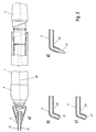
- the light probe 2 has a channel 21 arranged in a rigid shell 22 in which the emission and detection fibers run. According to the invention depending on the design of the tooth area to be examined from a variety of predefined light probe types can be selected.
- 3b shows the cross section of a Light probe tip for searching smooth tooth surfaces
- Fig. 3c the cross section a light probe tip for examining fissures, the tooth end of which Optical fiber probe is rounded or frustoconical and Fig. 3d is an optical fiber probe for examining approximal areas (interdental areas) of a tooth, the end of this fiber optic probe is pointed.
- FIG. 4 shows a cross-sectional view through the light guide probe shown in FIG. 3b optical fibers guided in it.
- Some of the individual optical fibers 24 serve to transmit the excitation light and others for the detection of those emitted by the tooth to be examined Fluorescence radiation.
- the arrangement of the excitation optical fibers compared to the Detection light guide fibers in the light guide probe 2 are initially not fixed. Around however, the most homogeneous possible irradiation of the tooth to be examined and one detection of the excited fluorescence radiation as uniform and precise as possible To achieve, it is advantageous to the excitation fibers and Arrange detection optical fibers alternately to each other.
- the individual optical fibers in the optical fiber channel 21 as shown in FIG. 5 be, with a plurality of detection optical fibers 24b concentric around one Emission optical fiber 24a are arranged. In this way, the Detection reliability and accuracy can be increased or stabilized.
- the optical fibers are arranged in a sheath 22, for example made of metal or Plastic or any other suitable material can be made. They are also Optical fibers are also surrounded by a sheath 23. Through this sheathing there is a risk of damage to that guided in the light guide channel 21 The fiber bundle is reduced and the breakage of the individual optical fibers is prevented
- the internal channel 20 shown in FIG dental handpiece 1 on a variety of individual optical fibers that either guide the excitation light to the fiber optic probe or that from the fiber optic probe 2 detected fluorescence radiation to the evaluation and determination device return.
- the fiber optic probe 2 on the dental Handpiece 1 guided in the light guide channel 21 of the light guide probe 2 Optical fibers with those guided in the channel 20 of the dental handpiece 1 Optical fibers connected.
- this can be done in the light guide channel 20 of the dental handpiece 1 guided light guide system the arrangement shown in Fig. 5 of individual optical fibers, i.e.
- a plurality of individual detection optical fibers 24b coaxially around the Emission optical fiber 24a are arranged around.
- the arrangement of the optical fibers in the optical fiber probe 2 not necessarily the arrangement of the optical fiber in must correspond to the dental handpiece. Rather, it is only necessary that after placing the optical fiber probe 2 on the dental handpiece 1 in the Optical fiber probe 2 guided emission optical fibers or detection optical fibers the corresponding emission or detection optical fibers of the dental Handpiece 1 can be connected by an appropriate coupling mechanism.
- the dental handpiece may be the one shown in FIG Have light guide arrangement, while the light guide probe 2 not only one, but has a plurality of emission light guide fibers which alternate with the corresponding ones Detection light guide fibers are arranged, these multiple emission light guide fibers with the central emission optical fiber 24a of the dental shown in FIG Handpiece are coupled. Therefore, it is also possible that the diameter of the Fiber optic bundle of the light guide probe larger than the corresponding Fiber optic bundle diameter of the dental handpiece is. In particular, can the diameter of the fiber optic bundle of the light guide probe must be greater than 1 mm.
- FIG. 6 shows a further sectional view of a device in the device according to the invention used light probe.
- the individual optical fibers 24a, 24b of surround three tubes 25a-25c evenly distributed in the circumferential direction of the light probe, which are made of solid material, such as metal, and thus for stabilization serve the light probe.
- the tubes 25a-25c can be of a flexible type, for example Shrink tubing 22 or similar be surrounded because a pipe arrangement already sufficient stability and adequate protection of the optical fibers is guaranteed.
- the tubes 25a-25c can also be within the fixed ones shown in FIG Sheath 22 of the light probe can be arranged.
- the tubes advantageously serve not only as a means of stability for the light probe, but also for the supply of Resources that are necessary for the operation of the device according to the invention or the Light probe are required or suitable, such as illuminating light or Spray air.
- the supply of spray or blown air via the pipes 25a-25c is helpful because it has been found that a more reliable measurement result can be achieved can, if the area of the tooth to be examined is blown clean or dry.
Landscapes
- Health & Medical Sciences (AREA)
- Life Sciences & Earth Sciences (AREA)
- Heart & Thoracic Surgery (AREA)
- Medical Informatics (AREA)
- Physics & Mathematics (AREA)
- Dentistry (AREA)
- Biophysics (AREA)
- Pathology (AREA)
- Engineering & Computer Science (AREA)
- Biomedical Technology (AREA)
- Audiology, Speech & Language Pathology (AREA)
- Oral & Maxillofacial Surgery (AREA)
- Molecular Biology (AREA)
- Surgery (AREA)
- Animal Behavior & Ethology (AREA)
- General Health & Medical Sciences (AREA)
- Public Health (AREA)
- Veterinary Medicine (AREA)
- Dental Tools And Instruments Or Auxiliary Dental Instruments (AREA)
- Medicines Containing Antibodies Or Antigens For Use As Internal Diagnostic Agents (AREA)
- Endoscopes (AREA)
Applications Claiming Priority (2)
| Application Number | Priority Date | Filing Date | Title |
|---|---|---|---|
| DE29704185U | 1997-03-07 | ||
| DE29704185U DE29704185U1 (de) | 1997-03-07 | 1997-03-07 | Vorrichtung zum Erkennen von Karies, Plaque oder bakteriellem Befall an Zähnen |
Publications (3)
| Publication Number | Publication Date |
|---|---|
| EP0862896A2 true EP0862896A2 (fr) | 1998-09-09 |
| EP0862896A3 EP0862896A3 (fr) | 2001-07-04 |
| EP0862896B1 EP0862896B1 (fr) | 2005-06-01 |
Family
ID=8037137
Family Applications (1)
| Application Number | Title | Priority Date | Filing Date |
|---|---|---|---|
| EP98103839A Expired - Lifetime EP0862896B1 (fr) | 1997-03-07 | 1998-03-04 | Dispositif pour la détection des caries, de la plaque ou de l'attaque bactérienne dentaire |
Country Status (5)
| Country | Link |
|---|---|
| US (1) | US6053731A (fr) |
| EP (1) | EP0862896B1 (fr) |
| JP (1) | JPH10309290A (fr) |
| AT (1) | ATE296572T1 (fr) |
| DE (2) | DE29704185U1 (fr) |
Cited By (3)
| Publication number | Priority date | Publication date | Assignee | Title |
|---|---|---|---|---|
| DE10129694C1 (de) * | 2001-06-22 | 2003-02-06 | Sirona Dental Systems Gmbh | Verfahren und eine Vorrichtung zum Erkennen von krankhaften Veränderungen wie Karies, Plaque, Konkrementen oder bakteriellem Befall an einer Gewebeoberfläche, insbesondere an Zähnen |
| DE102007047068A1 (de) | 2007-10-01 | 2009-04-02 | Ferton Holding S.A. | Vorrichtung zum Erkennen von bakteriellem Befall im Wurzelkanal von Zähnen |
| US9119548B2 (en) | 2011-04-21 | 2015-09-01 | Numed Sp Z O.O. | Device and method for secondary dental caries diagnosis |
Families Citing this family (105)
| Publication number | Priority date | Publication date | Assignee | Title |
|---|---|---|---|---|
| DE29705934U1 (de) * | 1997-04-03 | 1997-06-05 | Kaltenbach & Voigt Gmbh & Co, 88400 Biberach | Diagnose- und Behandlungsvorrichtung für Zähne |
| DE19714167B4 (de) * | 1997-04-07 | 2010-01-28 | Hahn, Rainer, Dr. | Gerät zum Zuführen von Behandlungsmedium in Hartgewebe und Verwendung eines solchen |
| DE19825021A1 (de) * | 1998-06-04 | 1999-12-09 | Kaltenbach & Voigt | Verfahren und Vorrichtung zum Erkennen von Karies, Plaque, Konkrementen oder bakteriellem Befall an Zähnen |
| DE19827417B4 (de) * | 1998-06-19 | 2004-10-28 | Hahn, Rainer, Dr.Med.Dent. | Material zur unterschiedlichen Modifizierung der optischen Eigenschaften unterschiedlicher Zellen |
| GB9816914D0 (en) * | 1998-08-05 | 1998-09-30 | Smithkline Beecham Gmbh | Novel device |
| US11026768B2 (en) | 1998-10-08 | 2021-06-08 | Align Technology, Inc. | Dental appliance reinforcement |
| DE19906047C2 (de) * | 1999-02-12 | 2001-10-18 | Fraunhofer Ges Forschung | Verfahren und Vorrichtung zur Detektion biotischer Kontaminationen auf einer Oberfläche |
| NL1012012C2 (nl) * | 1999-05-10 | 2000-11-23 | Inspektor Res Systems B V | Werkwijze en inrichting voor het bepalen van cariÙsactiviteit van een carieuze laesie in een tand. |
| CA2297476A1 (fr) | 2000-01-21 | 2001-07-21 | Neks Recherche & Developpement Inc. | Systeme pour la detection de tartre dentaire, i.e. tartre subgingival |
| DE10013210A1 (de) | 2000-03-17 | 2001-09-20 | Kaltenbach & Voigt | Vorrichtung zum Erkennen von Karies, Plaque, bakteriellen Befall, Konkrementen, Zahnstein und anderen fluoreszierenden Substanzen an Zähnen |
| CA2341105A1 (fr) * | 2001-03-21 | 2002-09-21 | Unknown | Systeme et methode de detection et d'elimination du tartre, p. ex. du tartre sous-gingival |
| US6769911B2 (en) | 2001-04-16 | 2004-08-03 | Advanced Research & Technology Institue | Luminescence assisted caries excavation |
| CA2385981A1 (fr) * | 2002-05-08 | 2003-11-08 | Neks Recherche & Developpement Inc. | Appareil et methode de detection des caries dentaires |
| US20040240716A1 (en) * | 2003-05-22 | 2004-12-02 | De Josselin De Jong Elbert | Analysis and display of fluorescence images |
| WO2005013843A2 (fr) * | 2003-08-08 | 2005-02-17 | The Regents Of The Univeristy Of California | Diaphanoscopie dans l'infrarouge proche pour imagerie de carie dentaire recente |
| US9492245B2 (en) | 2004-02-27 | 2016-11-15 | Align Technology, Inc. | Method and system for providing dynamic orthodontic assessment and treatment profiles |
| DE102004024164A1 (de) * | 2004-05-14 | 2005-12-08 | Kaltenbach & Voigt Gmbh & Co. Kg | Zahnärztliches System zum Untersuchen der optischen Eigenschaften von Zahngewebe mit optischer Untersuchungsvorrichtung und Abgleicheinrichtung |
| DE102004024165A1 (de) | 2004-05-14 | 2005-12-01 | Kaltenbach & Voigt Gmbh & Co. Kg | Zahnärztliche Vorrichtung zum Untersuchen der optischen Eigenschaften von Zahngewebe |
| US7270543B2 (en) * | 2004-06-29 | 2007-09-18 | Therametric Technologies, Inc. | Handpiece for caries detection |
| JP5175029B2 (ja) * | 2005-06-17 | 2013-04-03 | パナソニック株式会社 | レーザ装置及び電池残量検出方法 |
| CA2517252A1 (fr) * | 2005-08-29 | 2007-02-28 | Neks Technologies Inc. | Detection de la carie interproximale a l'aide d'une sonde optique examinant la surface occlusale de la dent |
| US8360771B2 (en) * | 2006-12-28 | 2013-01-29 | Therametric Technologies, Inc. | Handpiece for detection of dental demineralization |
| US7878805B2 (en) | 2007-05-25 | 2011-02-01 | Align Technology, Inc. | Tabbed dental appliance |
| EP2197350A2 (fr) * | 2007-09-11 | 2010-06-23 | Baxter International Inc. | Système de capteur pour thérapie par perfusion |
| US8738394B2 (en) | 2007-11-08 | 2014-05-27 | Eric E. Kuo | Clinical data file |
| US8108189B2 (en) | 2008-03-25 | 2012-01-31 | Align Technologies, Inc. | Reconstruction of non-visible part of tooth |
| US8092215B2 (en) | 2008-05-23 | 2012-01-10 | Align Technology, Inc. | Smile designer |
| US9492243B2 (en) | 2008-05-23 | 2016-11-15 | Align Technology, Inc. | Dental implant positioning |
| US8172569B2 (en) | 2008-06-12 | 2012-05-08 | Align Technology, Inc. | Dental appliance |
| JP2009006155A (ja) * | 2008-08-04 | 2009-01-15 | J Morita Tokyo Mfg Corp | 歯科光診断装置 |
| JP4803405B2 (ja) * | 2008-08-11 | 2011-10-26 | 株式会社モリタ東京製作所 | 歯科光診断用プローブ |
| US8152518B2 (en) | 2008-10-08 | 2012-04-10 | Align Technology, Inc. | Dental positioning appliance having metallic portion |
| US8292617B2 (en) | 2009-03-19 | 2012-10-23 | Align Technology, Inc. | Dental wire attachment |
| US8765031B2 (en) | 2009-08-13 | 2014-07-01 | Align Technology, Inc. | Method of forming a dental appliance |
| JP5391019B2 (ja) * | 2009-10-01 | 2014-01-15 | 株式会社長田中央研究所 | 口腔内スコープ |
| US8314377B2 (en) * | 2009-12-23 | 2012-11-20 | Mcneil-Ppc, Inc. | Device and method for detecting plaque in the oral cavity |
| US9211166B2 (en) | 2010-04-30 | 2015-12-15 | Align Technology, Inc. | Individualized orthodontic treatment index |
| US9241774B2 (en) | 2010-04-30 | 2016-01-26 | Align Technology, Inc. | Patterned dental positioning appliance |
| US8187002B2 (en) | 2010-06-29 | 2012-05-29 | Mcneil-Ppc, Inc. | Method for cleaning the oral cavity |
| US8186997B2 (en) | 2010-06-29 | 2012-05-29 | Mcneil-Ppc, Inc. | Method for cleaning the oral cavity |
| US8702422B2 (en) | 2010-06-29 | 2014-04-22 | Mcneil-Ppc, Inc. | Device and method for cleaning the oral cavity |
| JP5507376B2 (ja) * | 2010-07-28 | 2014-05-28 | 三洋電機株式会社 | 撮像装置 |
| RU2464549C1 (ru) * | 2011-05-12 | 2012-10-20 | Ираида Николаевна Сарычева | Оптоволоконное устройство для регистрации флуоресценции |
| DE102011101934B4 (de) * | 2011-05-18 | 2017-07-06 | Christian-Albrechts-Universität Zu Kiel | Großflächiger Biofilmsensor |
| US9403238B2 (en) | 2011-09-21 | 2016-08-02 | Align Technology, Inc. | Laser cutting |
| EP2578200B1 (fr) | 2011-10-04 | 2018-03-28 | VOCO GmbH | Compositions pour l'infiltration et/ou le scellement de la matière dentaire dure et procédé correspondant |
| WO2013056071A1 (fr) | 2011-10-14 | 2013-04-18 | Beam Technologies, Llc | Appareil d'hygiène bucco-dentaire et système à capteur oxymétrique |
| US9375300B2 (en) | 2012-02-02 | 2016-06-28 | Align Technology, Inc. | Identifying forces on a tooth |
| US9220580B2 (en) | 2012-03-01 | 2015-12-29 | Align Technology, Inc. | Determining a dental treatment difficulty |
| US9414897B2 (en) | 2012-05-22 | 2016-08-16 | Align Technology, Inc. | Adjustment of tooth position in a virtual dental model |
| JP6015501B2 (ja) * | 2012-06-01 | 2016-10-26 | ソニー株式会社 | 歯用装置及び医療用装置 |
| US10010250B2 (en) * | 2012-12-19 | 2018-07-03 | Koninklijke Philips N.V. | Dental apparatus and method of utilizing the same |
| US10772506B2 (en) | 2014-07-07 | 2020-09-15 | Align Technology, Inc. | Apparatus for dental confocal imaging |
| US9675430B2 (en) | 2014-08-15 | 2017-06-13 | Align Technology, Inc. | Confocal imaging apparatus with curved focal surface |
| US9610141B2 (en) | 2014-09-19 | 2017-04-04 | Align Technology, Inc. | Arch expanding appliance |
| US10449016B2 (en) | 2014-09-19 | 2019-10-22 | Align Technology, Inc. | Arch adjustment appliance |
| US9744001B2 (en) | 2014-11-13 | 2017-08-29 | Align Technology, Inc. | Dental appliance with cavity for an unerupted or erupting tooth |
| US10504386B2 (en) | 2015-01-27 | 2019-12-10 | Align Technology, Inc. | Training method and system for oral-cavity-imaging-and-modeling equipment |
| US10248883B2 (en) | 2015-08-20 | 2019-04-02 | Align Technology, Inc. | Photograph-based assessment of dental treatments and procedures |
| US11554000B2 (en) | 2015-11-12 | 2023-01-17 | Align Technology, Inc. | Dental attachment formation structure |
| US11931222B2 (en) | 2015-11-12 | 2024-03-19 | Align Technology, Inc. | Dental attachment formation structures |
| US11596502B2 (en) | 2015-12-09 | 2023-03-07 | Align Technology, Inc. | Dental attachment placement structure |
| US11103330B2 (en) | 2015-12-09 | 2021-08-31 | Align Technology, Inc. | Dental attachment placement structure |
| ES2886082T3 (es) | 2016-04-13 | 2021-12-16 | Inspektor Res Systems B V | Examen dental bifrecuencia |
| EP4670634A3 (fr) | 2016-06-17 | 2026-01-21 | Align Technology, Inc. | Système de détermination de l'observance d'un patient par un plan de traitement ou de rétention orthodontique |
| EP3471653B1 (fr) | 2016-06-17 | 2021-12-22 | Align Technology, Inc. | Dispositif de surveillance de performances d'un appareil orthodontique |
| US10507087B2 (en) | 2016-07-27 | 2019-12-17 | Align Technology, Inc. | Methods and apparatuses for forming a three-dimensional volumetric model of a subject's teeth |
| EP4252698B1 (fr) | 2016-07-27 | 2025-04-30 | Align Technology, Inc. | Scanner intra-buccal pouvant établir un diagnostic dentaire |
| CN109922754B (zh) | 2016-11-04 | 2021-10-01 | 阿莱恩技术有限公司 | 用于牙齿图像的方法和装置 |
| CA3043049A1 (fr) | 2016-12-02 | 2018-06-07 | Align Technology, Inc. | Procedes et appareils pour personnaliser des dispositifs d'expansion palatine rapides a l'aide de modeles numeriques |
| US11026831B2 (en) | 2016-12-02 | 2021-06-08 | Align Technology, Inc. | Dental appliance features for speech enhancement |
| EP3824843B1 (fr) | 2016-12-02 | 2024-12-25 | Align Technology, Inc. | Serie de dispositifs d'expansion palatine |
| WO2018102770A1 (fr) | 2016-12-02 | 2018-06-07 | Align Technology, Inc. | Commande de force, mécanisme d'arrêt, structure de régulation d'appareil de réglage d'arcade amovible |
| EP3551034A1 (fr) * | 2016-12-07 | 2019-10-16 | Progenity, Inc. | Méthodes, dispositifs et systèmes de détection pour le tractus gastro-intestinal |
| US10548700B2 (en) | 2016-12-16 | 2020-02-04 | Align Technology, Inc. | Dental appliance etch template |
| US10456043B2 (en) | 2017-01-12 | 2019-10-29 | Align Technology, Inc. | Compact confocal dental scanning apparatus |
| US10779718B2 (en) | 2017-02-13 | 2020-09-22 | Align Technology, Inc. | Cheek retractor and mobile device holder |
| WO2018183358A1 (fr) | 2017-03-27 | 2018-10-04 | Align Technology, Inc. | Appareils et procédés d'aide à des thérapies dentaires |
| US10613515B2 (en) | 2017-03-31 | 2020-04-07 | Align Technology, Inc. | Orthodontic appliances including at least partially un-erupted teeth and method of forming them |
| US11045283B2 (en) | 2017-06-09 | 2021-06-29 | Align Technology, Inc. | Palatal expander with skeletal anchorage devices |
| US11996181B2 (en) | 2017-06-16 | 2024-05-28 | Align Technology, Inc. | Automatic detection of tooth type and eruption status |
| WO2019005808A1 (fr) | 2017-06-26 | 2019-01-03 | Align Technology, Inc. | Indicateur de performance de biocapteur pour appareils intrabuccaux |
| US10885521B2 (en) | 2017-07-17 | 2021-01-05 | Align Technology, Inc. | Method and apparatuses for interactive ordering of dental aligners |
| CN111107806B (zh) | 2017-07-21 | 2022-04-19 | 阿莱恩技术有限公司 | 颚轮廓锚固 |
| WO2019023631A1 (fr) | 2017-07-27 | 2019-01-31 | Align Technology, Inc. | Système et procédés de traitement d'un aligneur orthodontique au moyen d'une tomographie par cohérence optique |
| US11633268B2 (en) | 2017-07-27 | 2023-04-25 | Align Technology, Inc. | Tooth shading, transparency and glazing |
| US12274597B2 (en) | 2017-08-11 | 2025-04-15 | Align Technology, Inc. | Dental attachment template tray systems |
| WO2019035979A1 (fr) | 2017-08-15 | 2019-02-21 | Align Technology, Inc. | Évaluation et calcul de couloir buccal |
| WO2019036677A1 (fr) | 2017-08-17 | 2019-02-21 | Align Technology, Inc. | Surveillance de conformité d'appareil dentaire |
| WO2019071019A1 (fr) | 2017-10-04 | 2019-04-11 | Align Technology, Inc. | Appareils intra-oraux pour l'échantillonnage de tissu mou |
| US10813720B2 (en) | 2017-10-05 | 2020-10-27 | Align Technology, Inc. | Interproximal reduction templates |
| EP3700458B1 (fr) | 2017-10-27 | 2023-06-07 | Align Technology, Inc. | Autres structures de réglage de morsure |
| CN111295153B (zh) | 2017-10-31 | 2023-06-16 | 阿莱恩技术有限公司 | 具有选择性牙合负荷和受控牙尖交错的牙科器具 |
| WO2019089989A2 (fr) | 2017-11-01 | 2019-05-09 | Align Technology, Inc. | Planification automatique de traitement |
| WO2019100022A1 (fr) | 2017-11-17 | 2019-05-23 | Align Technology, Inc. | Dispositifs de retenue orthodontiques |
| EP3716885B1 (fr) | 2017-11-30 | 2023-08-30 | Align Technology, Inc. | Appareils buccaux orthodontiques comprenant des capteurs |
| US11432908B2 (en) | 2017-12-15 | 2022-09-06 | Align Technology, Inc. | Closed loop adaptive orthodontic treatment methods and apparatuses |
| US10980613B2 (en) | 2017-12-29 | 2021-04-20 | Align Technology, Inc. | Augmented reality enhancements for dental practitioners |
| KR20200115580A (ko) | 2018-01-26 | 2020-10-07 | 얼라인 테크널러지, 인크. | 구강 내 진단 스캔 및 추적 |
| US11937991B2 (en) | 2018-03-27 | 2024-03-26 | Align Technology, Inc. | Dental attachment placement structure |
| WO2019200008A1 (fr) | 2018-04-11 | 2019-10-17 | Align Technology, Inc. | Appareils d'expansion palatine libérables |
| CN109009235B (zh) * | 2018-06-22 | 2023-10-13 | 四川大学 | 一种龈下厌氧菌群采集器及使用方法 |
| RU2712789C1 (ru) * | 2019-03-06 | 2020-01-31 | федеральное государственное бюджетное образовательное учреждение высшего образования "Воронежский государственный университет" (ФГБОУ ВО "ВГУ") | Оптоволоконный конфокальный сканирующий микроскоп |
| KR102700947B1 (ko) * | 2023-10-17 | 2024-08-30 | (주)스마투스코리아 | 광 기반의 구강 진단 장치 |
| DE102024201487A1 (de) * | 2024-02-19 | 2025-08-21 | Robert Bosch Gesellschaft mit beschränkter Haftung | Optische Einheit zur Fluoreszenzlichtbilddarstellung, insbesondere für eine Analyseeinrichtung zum Nachweis von Krankheitserregern und Verfahren zum Herstellen einer optischen Einheit |
Citations (3)
| Publication number | Priority date | Publication date | Assignee | Title |
|---|---|---|---|---|
| DE3031249C2 (de) | 1979-08-20 | 1986-11-06 | Philips Medical Systems Inc., Shelton, Conn. | Vorrichtung zum Entdecken von Karies und Anwendung dieser Vorrichtung |
| DE4200741A1 (de) | 1992-01-14 | 1993-07-15 | Kaltenbach & Voigt | Einrichtung zum erkennen von karies an zaehnen |
| DE9317984U1 (de) | 1993-11-24 | 1995-03-23 | Kaltenbach & Voigt Gmbh & Co, 88400 Biberach | Vorrichtung zum Erkennen von Karies |
Family Cites Families (18)
| Publication number | Priority date | Publication date | Assignee | Title |
|---|---|---|---|---|
| US31815A (en) * | 1861-03-26 | Improvement in the mode of loading fire-arms | ||
| USRE31815E (en) * | 1979-08-20 | 1985-01-29 | Philips Medical Systems, Inc. | Method and apparatus for detecting the presence of caries in teeth using visible luminescence |
| SE442817B (sv) * | 1981-04-01 | 1986-02-03 | Hans Ingmar Bjelkhagen | Anordning for att okulert kunna faststella en diskrepans i en tandytas luminiscensformaga |
| US4479499A (en) * | 1982-01-29 | 1984-10-30 | Alfano Robert R | Method and apparatus for detecting the presence of caries in teeth using visible light |
| EP0113152A3 (fr) * | 1983-01-03 | 1985-07-03 | North American Philips Corporation | Procédé et appareil pour examiner des anomalies superficielles d'objets |
| DE3345465A1 (de) * | 1983-12-15 | 1985-06-27 | Siemens AG, 1000 Berlin und 8000 München | Zahnaerztliches diagnosegeraet |
| JPS60209146A (ja) * | 1984-03-31 | 1985-10-21 | Olympus Optical Co Ltd | 螢光スペクトル分析装置 |
| JPH0228543A (ja) * | 1988-07-18 | 1990-01-30 | Koden Electron Co Ltd | 光学式分析計 |
| US5055048A (en) * | 1988-08-25 | 1991-10-08 | American Dental Laser, Inc. | Dental laser assembly |
| US5324200A (en) * | 1988-08-25 | 1994-06-28 | American Dental Technologies, Inc. | Method for enlarging and shaping a root canal |
| DE9007569U1 (de) * | 1990-03-23 | 1992-09-24 | Kaltenbach & Voigt Gmbh & Co, 88400 Biberach | Zahnärztliche Sonde mit eingebautem Bild- und Lichtleiter zur Bild-Übertragung |
| JP2862099B2 (ja) * | 1990-10-12 | 1999-02-24 | 旭光学工業株式会社 | 早期癌診断装置 |
| JP3124643B2 (ja) * | 1991-11-27 | 2001-01-15 | 株式会社モリタ製作所 | 歯科治療用レーザハンドピース |
| US5382163A (en) * | 1992-07-20 | 1995-01-17 | Putnam; David L. | Method and apparatus for detecting the presence of dental plaque or calculus |
| US5415543A (en) * | 1993-12-20 | 1995-05-16 | Rozmajzl, Jr.; William F. | Dental composite curing apparatus and method |
| DE4420401A1 (de) * | 1994-06-10 | 1995-12-21 | Tim Dr Med Liesenhoff | Rückstrahlspektroskopische Vorrichtung |
| DE4433123C1 (de) * | 1994-09-16 | 1996-03-28 | Fraunhofer Ges Forschung | Einrichtung zur Diagnostik von Körpergewebe oder -proben |
| US5759030A (en) * | 1996-01-02 | 1998-06-02 | Lj Laboratories, L.L.C. | Method for determing optical characteristics of teeth |
-
1997
- 1997-03-07 DE DE29704185U patent/DE29704185U1/de not_active Expired - Lifetime
-
1998
- 1998-03-04 EP EP98103839A patent/EP0862896B1/fr not_active Expired - Lifetime
- 1998-03-04 DE DE59812822T patent/DE59812822D1/de not_active Expired - Lifetime
- 1998-03-04 AT AT98103839T patent/ATE296572T1/de active
- 1998-03-06 US US09/036,475 patent/US6053731A/en not_active Expired - Lifetime
- 1998-03-09 JP JP10056859A patent/JPH10309290A/ja active Pending
Patent Citations (3)
| Publication number | Priority date | Publication date | Assignee | Title |
|---|---|---|---|---|
| DE3031249C2 (de) | 1979-08-20 | 1986-11-06 | Philips Medical Systems Inc., Shelton, Conn. | Vorrichtung zum Entdecken von Karies und Anwendung dieser Vorrichtung |
| DE4200741A1 (de) | 1992-01-14 | 1993-07-15 | Kaltenbach & Voigt | Einrichtung zum erkennen von karies an zaehnen |
| DE9317984U1 (de) | 1993-11-24 | 1995-03-23 | Kaltenbach & Voigt Gmbh & Co, 88400 Biberach | Vorrichtung zum Erkennen von Karies |
Cited By (4)
| Publication number | Priority date | Publication date | Assignee | Title |
|---|---|---|---|---|
| DE10129694C1 (de) * | 2001-06-22 | 2003-02-06 | Sirona Dental Systems Gmbh | Verfahren und eine Vorrichtung zum Erkennen von krankhaften Veränderungen wie Karies, Plaque, Konkrementen oder bakteriellem Befall an einer Gewebeoberfläche, insbesondere an Zähnen |
| DE102007047068A1 (de) | 2007-10-01 | 2009-04-02 | Ferton Holding S.A. | Vorrichtung zum Erkennen von bakteriellem Befall im Wurzelkanal von Zähnen |
| US8345247B2 (en) | 2007-10-01 | 2013-01-01 | Ferton Holdings, S.A. | Device for detecting signs of bacterial infection of the root channel of teeth |
| US9119548B2 (en) | 2011-04-21 | 2015-09-01 | Numed Sp Z O.O. | Device and method for secondary dental caries diagnosis |
Also Published As
| Publication number | Publication date |
|---|---|
| EP0862896B1 (fr) | 2005-06-01 |
| ATE296572T1 (de) | 2005-06-15 |
| US6053731A (en) | 2000-04-25 |
| EP0862896A3 (fr) | 2001-07-04 |
| DE59812822D1 (de) | 2005-07-07 |
| JPH10309290A (ja) | 1998-11-24 |
| DE29704185U1 (de) | 1997-04-30 |
Similar Documents
| Publication | Publication Date | Title |
|---|---|---|
| EP0862896B1 (fr) | Dispositif pour la détection des caries, de la plaque ou de l'attaque bactérienne dentaire | |
| DE10133451B4 (de) | Vorrichtung zum Erkennen von Karies, Plaque, Konkrementen oder bakteriellem Befall an Zähnen | |
| EP0862897B1 (fr) | Procédé pour mesurer la fluorescence et dispositif pour détecter des caries dentaires | |
| EP0962185B1 (fr) | Dispositif destiné à la détermination de la cariée, de la plaque, de concrétions ou d' affectations bactérielles des dents | |
| DE102004024494B4 (de) | Medizinische Kamera | |
| DE19541686B4 (de) | Vorrichtung zum Erkennen von Karies, Plaque oder bakteriellem Befall an Zähnen | |
| EP1372518B1 (fr) | Appareil de traitement laser dote d'un systeme d'eclairage | |
| DE102013006636B4 (de) | Dentalkamera zur Kariesdetektion | |
| DE102006055588B4 (de) | Vorrichtung und System zum Erfassen der Form eines Endoskops | |
| DE60316699T2 (de) | Dentalgerät zur untersuchung der zahnoberfläche | |
| EP0862899B1 (fr) | Dispositif de traitement dentaire | |
| DE29705934U1 (de) | Diagnose- und Behandlungsvorrichtung für Zähne | |
| DE4200741C2 (de) | Einrichtung zum Erkennen von Karies an Zähnen | |
| DE10013210A1 (de) | Vorrichtung zum Erkennen von Karies, Plaque, bakteriellen Befall, Konkrementen, Zahnstein und anderen fluoreszierenden Substanzen an Zähnen | |
| DE102007046228A1 (de) | Verfahren zur Erkennung von Veränderungen an zumindest einem Zahn | |
| DE102007063419A1 (de) | Zahnbehandlungs- oder- untersuchungsvorrichtung | |
| EP1595495B1 (fr) | Dispositif dentaire pour l'examen de charactéristiques optiques dentaires | |
| DE10249674A1 (de) | Operationsinstrument zum Schneiden, Abtragen oder Absaugen von Material in einem Operationsgebiet | |
| EP2520221A1 (fr) | Pièce manuelle pour un appareil de diagnostic médical et appareil de diagnostic médical | |
| DE20209441U1 (de) | Vorrichtung zum Erkennen von bakteriellem Befall an Zähnen | |
| WO2009046922A1 (fr) | Dispositif pour détecter une infection bactérienne dentaire | |
| EP2203108B1 (fr) | Dispositif pour détecter une infection bactérienne dans le canal radiculaire de dents | |
| EP1269909A1 (fr) | Procédé et dispositif de reconnaissance des changements pathologiques sur la surface des tissus, notamment des dents | |
| EP1595494A1 (fr) | Système dentaire pour l'examen de charactéristiques optiques du tissue dentaire avec dispositif d'examen optique et dispositif de calibration | |
| WO2002078558A1 (fr) | Dispositif et procede d'ablation au laser de matiere organique et inorganique |
Legal Events
| Date | Code | Title | Description |
|---|---|---|---|
| PUAI | Public reference made under article 153(3) epc to a published international application that has entered the european phase |
Free format text: ORIGINAL CODE: 0009012 |
|
| AK | Designated contracting states |
Kind code of ref document: A2 Designated state(s): AT CH DE FR IT LI |
|
| AX | Request for extension of the european patent |
Free format text: AL;LT;LV;MK;RO;SI |
|
| PUAL | Search report despatched |
Free format text: ORIGINAL CODE: 0009013 |
|
| AK | Designated contracting states |
Kind code of ref document: A3 Designated state(s): AT BE CH DE DK ES FI FR GB GR IE IT LI LU MC NL PT SE |
|
| AX | Request for extension of the european patent |
Free format text: AL;LT;LV;MK;RO;SI |
|
| 17P | Request for examination filed |
Effective date: 20011114 |
|
| AKX | Designation fees paid |
Free format text: AT CH DE FR IT LI |
|
| RAP1 | Party data changed (applicant data changed or rights of an application transferred) |
Owner name: KALTENBACH & VOIGT GMBH & CO. KG |
|
| GRAP | Despatch of communication of intention to grant a patent |
Free format text: ORIGINAL CODE: EPIDOSNIGR1 |
|
| GRAS | Grant fee paid |
Free format text: ORIGINAL CODE: EPIDOSNIGR3 |
|
| GRAA | (expected) grant |
Free format text: ORIGINAL CODE: 0009210 |
|
| AK | Designated contracting states |
Kind code of ref document: B1 Designated state(s): AT CH DE FR IT LI |
|
| REG | Reference to a national code |
Ref country code: CH Ref legal event code: NV Representative=s name: A. BRAUN, BRAUN, HERITIER, ESCHMANN AG PATENTANWAE Ref country code: CH Ref legal event code: EP |
|
| REF | Corresponds to: |
Ref document number: 59812822 Country of ref document: DE Date of ref document: 20050707 Kind code of ref document: P |
|
| ET | Fr: translation filed | ||
| PLBE | No opposition filed within time limit |
Free format text: ORIGINAL CODE: 0009261 |
|
| STAA | Information on the status of an ep patent application or granted ep patent |
Free format text: STATUS: NO OPPOSITION FILED WITHIN TIME LIMIT |
|
| 26N | No opposition filed |
Effective date: 20060302 |
|
| REG | Reference to a national code |
Ref country code: CH Ref legal event code: PFA Owner name: KALTENBACH & VOIGT GMBH & CO. KG Free format text: KALTENBACH & VOIGT GMBH & CO. KG#BISMARCKRING 39#88400 BIBERACH/RISS (DE) -TRANSFER TO- KALTENBACH & VOIGT GMBH & CO. KG#BISMARCKRING 39#88400 BIBERACH/RISS (DE) |
|
| PGFP | Annual fee paid to national office [announced via postgrant information from national office to epo] |
Ref country code: AT Payment date: 20110328 Year of fee payment: 14 Ref country code: FR Payment date: 20110401 Year of fee payment: 14 Ref country code: IT Payment date: 20110323 Year of fee payment: 14 |
|
| PGFP | Annual fee paid to national office [announced via postgrant information from national office to epo] |
Ref country code: CH Payment date: 20110405 Year of fee payment: 14 Ref country code: DE Payment date: 20110411 Year of fee payment: 14 |
|
| REG | Reference to a national code |
Ref country code: CH Ref legal event code: PL |
|
| REG | Reference to a national code |
Ref country code: AT Ref legal event code: MM01 Ref document number: 296572 Country of ref document: AT Kind code of ref document: T Effective date: 20120304 |
|
| REG | Reference to a national code |
Ref country code: FR Ref legal event code: ST Effective date: 20121130 |
|
| REG | Reference to a national code |
Ref country code: DE Ref legal event code: R119 Ref document number: 59812822 Country of ref document: DE Effective date: 20121002 |
|
| PG25 | Lapsed in a contracting state [announced via postgrant information from national office to epo] |
Ref country code: LI Free format text: LAPSE BECAUSE OF NON-PAYMENT OF DUE FEES Effective date: 20120331 Ref country code: FR Free format text: LAPSE BECAUSE OF NON-PAYMENT OF DUE FEES Effective date: 20120402 Ref country code: AT Free format text: LAPSE BECAUSE OF NON-PAYMENT OF DUE FEES Effective date: 20120304 Ref country code: CH Free format text: LAPSE BECAUSE OF NON-PAYMENT OF DUE FEES Effective date: 20120331 |
|
| PG25 | Lapsed in a contracting state [announced via postgrant information from national office to epo] |
Ref country code: IT Free format text: LAPSE BECAUSE OF NON-PAYMENT OF DUE FEES Effective date: 20120304 |
|
| PG25 | Lapsed in a contracting state [announced via postgrant information from national office to epo] |
Ref country code: DE Free format text: LAPSE BECAUSE OF NON-PAYMENT OF DUE FEES Effective date: 20121002 |