EP0871896B1 - Procede servant a determiner la concentration d'oxygene dans un echantillon - Google Patents
Procede servant a determiner la concentration d'oxygene dans un echantillon Download PDFInfo
- Publication number
- EP0871896B1 EP0871896B1 EP96929434A EP96929434A EP0871896B1 EP 0871896 B1 EP0871896 B1 EP 0871896B1 EP 96929434 A EP96929434 A EP 96929434A EP 96929434 A EP96929434 A EP 96929434A EP 0871896 B1 EP0871896 B1 EP 0871896B1
- Authority
- EP
- European Patent Office
- Prior art keywords
- sample
- bis
- radical
- image
- mmol
- Prior art date
- Legal status (The legal status is an assumption and is not a legal conclusion. Google has not performed a legal analysis and makes no representation as to the accuracy of the status listed.)
- Expired - Lifetime
Links
- 239000001301 oxygen Substances 0.000 title claims abstract description 95
- 229910052760 oxygen Inorganic materials 0.000 title claims abstract description 95
- QVGXLLKOCUKJST-UHFFFAOYSA-N atomic oxygen Chemical compound [O] QVGXLLKOCUKJST-UHFFFAOYSA-N 0.000 title claims abstract description 94
- 238000000034 method Methods 0.000 title claims abstract description 83
- 238000004435 EPR spectroscopy Methods 0.000 claims abstract description 9
- 150000003254 radicals Chemical class 0.000 claims description 91
- 125000002221 trityl group Chemical group [H]C1=C([H])C([H])=C([H])C([H])=C1C([*])(C1=C(C(=C(C(=C1[H])[H])[H])[H])[H])C1=C([H])C([H])=C([H])C([H])=C1[H] 0.000 claims description 23
- 230000007704 transition Effects 0.000 claims description 16
- 230000005855 radiation Effects 0.000 claims description 15
- 230000005291 magnetic effect Effects 0.000 claims description 10
- 125000004169 (C1-C6) alkyl group Chemical group 0.000 claims description 6
- 150000003839 salts Chemical class 0.000 claims description 5
- 125000003545 alkoxy group Chemical group 0.000 claims description 4
- 125000003178 carboxy group Chemical group [H]OC(*)=O 0.000 claims description 3
- 239000001257 hydrogen Substances 0.000 claims description 3
- 229910052739 hydrogen Inorganic materials 0.000 claims description 3
- 230000001678 irradiating effect Effects 0.000 claims description 3
- 210000003722 extracellular fluid Anatomy 0.000 claims description 2
- 125000004435 hydrogen atom Chemical group [H]* 0.000 claims 1
- 238000002595 magnetic resonance imaging Methods 0.000 abstract description 6
- XLYOFNOQVPJJNP-UHFFFAOYSA-N water Substances O XLYOFNOQVPJJNP-UHFFFAOYSA-N 0.000 description 61
- RTZKZFJDLAIYFH-UHFFFAOYSA-N Diethyl ether Chemical compound CCOCC RTZKZFJDLAIYFH-UHFFFAOYSA-N 0.000 description 58
- 239000000243 solution Substances 0.000 description 57
- OKKJLVBELUTLKV-UHFFFAOYSA-N Methanol Chemical compound OC OKKJLVBELUTLKV-UHFFFAOYSA-N 0.000 description 51
- -1 nitroxide radical Chemical class 0.000 description 49
- WEVYAHXRMPXWCK-UHFFFAOYSA-N Acetonitrile Chemical compound CC#N WEVYAHXRMPXWCK-UHFFFAOYSA-N 0.000 description 46
- UHOVQNZJYSORNB-UHFFFAOYSA-N Benzene Chemical compound C1=CC=CC=C1 UHOVQNZJYSORNB-UHFFFAOYSA-N 0.000 description 45
- HEDRZPFGACZZDS-MICDWDOJSA-N Trichloro(2H)methane Chemical compound [2H]C(Cl)(Cl)Cl HEDRZPFGACZZDS-MICDWDOJSA-N 0.000 description 42
- YMWUJEATGCHHMB-UHFFFAOYSA-N Dichloromethane Chemical compound ClCCl YMWUJEATGCHHMB-UHFFFAOYSA-N 0.000 description 39
- 239000000203 mixture Substances 0.000 description 32
- 239000000523 sample Substances 0.000 description 32
- 125000005605 benzo group Chemical group 0.000 description 27
- 125000002496 methyl group Chemical group [H]C([H])([H])* 0.000 description 26
- 239000000047 product Substances 0.000 description 26
- 238000005160 1H NMR spectroscopy Methods 0.000 description 25
- IMNFDUFMRHMDMM-UHFFFAOYSA-N N-Heptane Chemical compound CCCCCCC IMNFDUFMRHMDMM-UHFFFAOYSA-N 0.000 description 24
- 238000001704 evaporation Methods 0.000 description 22
- 230000008020 evaporation Effects 0.000 description 22
- HEMHJVSKTPXQMS-UHFFFAOYSA-M Sodium hydroxide Chemical compound [OH-].[Na+] HEMHJVSKTPXQMS-UHFFFAOYSA-M 0.000 description 21
- CSCPPACGZOOCGX-WFGJKAKNSA-N acetone d6 Chemical compound [2H]C([2H])([2H])C(=O)C([2H])([2H])[2H] CSCPPACGZOOCGX-WFGJKAKNSA-N 0.000 description 19
- 210000004369 blood Anatomy 0.000 description 19
- 239000008280 blood Substances 0.000 description 19
- 239000002904 solvent Substances 0.000 description 19
- XKRFYHLGVUSROY-UHFFFAOYSA-N Argon Chemical compound [Ar] XKRFYHLGVUSROY-UHFFFAOYSA-N 0.000 description 18
- VLKZOEOYAKHREP-UHFFFAOYSA-N n-Hexane Chemical compound CCCCCC VLKZOEOYAKHREP-UHFFFAOYSA-N 0.000 description 18
- 239000012074 organic phase Substances 0.000 description 18
- 239000011541 reaction mixture Substances 0.000 description 17
- LFQSCWFLJHTTHZ-UHFFFAOYSA-N Ethanol Chemical compound CCO LFQSCWFLJHTTHZ-UHFFFAOYSA-N 0.000 description 16
- 238000003756 stirring Methods 0.000 description 16
- 239000007832 Na2SO4 Substances 0.000 description 15
- PMZURENOXWZQFD-UHFFFAOYSA-L Sodium Sulfate Chemical compound [Na+].[Na+].[O-]S([O-])(=O)=O PMZURENOXWZQFD-UHFFFAOYSA-L 0.000 description 15
- 230000000694 effects Effects 0.000 description 15
- 238000002953 preparative HPLC Methods 0.000 description 15
- 229910052938 sodium sulfate Inorganic materials 0.000 description 15
- 238000003384 imaging method Methods 0.000 description 14
- 239000011159 matrix material Substances 0.000 description 14
- OKKJLVBELUTLKV-MZCSYVLQSA-N Deuterated methanol Chemical compound [2H]OC([2H])([2H])[2H] OKKJLVBELUTLKV-MZCSYVLQSA-N 0.000 description 12
- 238000002360 preparation method Methods 0.000 description 12
- 238000001644 13C nuclear magnetic resonance spectroscopy Methods 0.000 description 11
- FAPWRFPIFSIZLT-UHFFFAOYSA-M Sodium chloride Chemical compound [Na+].[Cl-] FAPWRFPIFSIZLT-UHFFFAOYSA-M 0.000 description 11
- 238000002347 injection Methods 0.000 description 11
- 239000007924 injection Substances 0.000 description 11
- VEXZGXHMUGYJMC-UHFFFAOYSA-N Hydrochloric acid Chemical compound Cl VEXZGXHMUGYJMC-UHFFFAOYSA-N 0.000 description 10
- CSNNHWWHGAXBCP-UHFFFAOYSA-L Magnesium sulfate Chemical compound [Mg+2].[O-][S+2]([O-])([O-])[O-] CSNNHWWHGAXBCP-UHFFFAOYSA-L 0.000 description 10
- 239000007983 Tris buffer Substances 0.000 description 10
- 150000001875 compounds Chemical class 0.000 description 10
- 230000006870 function Effects 0.000 description 10
- MZRVEZGGRBJDDB-UHFFFAOYSA-N n-Butyllithium Substances [Li]CCCC MZRVEZGGRBJDDB-UHFFFAOYSA-N 0.000 description 10
- 229920006395 saturated elastomer Polymers 0.000 description 10
- AJPJDKMHJJGVTQ-UHFFFAOYSA-M sodium dihydrogen phosphate Chemical compound [Na+].OP(O)([O-])=O AJPJDKMHJJGVTQ-UHFFFAOYSA-M 0.000 description 10
- 229910000162 sodium phosphate Inorganic materials 0.000 description 10
- LENZDBCJOHFCAS-UHFFFAOYSA-N tris Chemical compound OCC(N)(CO)CO LENZDBCJOHFCAS-UHFFFAOYSA-N 0.000 description 10
- XEKOWRVHYACXOJ-UHFFFAOYSA-N Ethyl acetate Chemical compound CCOC(C)=O XEKOWRVHYACXOJ-UHFFFAOYSA-N 0.000 description 9
- 229910052786 argon Inorganic materials 0.000 description 9
- 239000012298 atmosphere Substances 0.000 description 9
- 230000008878 coupling Effects 0.000 description 9
- 238000010168 coupling process Methods 0.000 description 9
- 238000005859 coupling reaction Methods 0.000 description 9
- 239000007789 gas Substances 0.000 description 9
- 210000001519 tissue Anatomy 0.000 description 9
- IJGRMHOSHXDMSA-UHFFFAOYSA-N Atomic nitrogen Chemical compound N#N IJGRMHOSHXDMSA-UHFFFAOYSA-N 0.000 description 8
- OFBQJSOFQDEBGM-UHFFFAOYSA-N Pentane Chemical compound CCCCC OFBQJSOFQDEBGM-UHFFFAOYSA-N 0.000 description 8
- 239000008346 aqueous phase Substances 0.000 description 8
- BWHMMNNQKKPAPP-UHFFFAOYSA-L potassium carbonate Chemical compound [K+].[K+].[O-]C([O-])=O BWHMMNNQKKPAPP-UHFFFAOYSA-L 0.000 description 8
- 239000011734 sodium Substances 0.000 description 8
- 241000700159 Rattus Species 0.000 description 7
- 239000007864 aqueous solution Substances 0.000 description 7
- 238000006243 chemical reaction Methods 0.000 description 7
- 238000001727 in vivo Methods 0.000 description 7
- 238000000746 purification Methods 0.000 description 7
- 238000005070 sampling Methods 0.000 description 7
- 229910052708 sodium Inorganic materials 0.000 description 7
- QTBSBXVTEAMEQO-UHFFFAOYSA-N Acetic acid Chemical compound CC(O)=O QTBSBXVTEAMEQO-UHFFFAOYSA-N 0.000 description 6
- XEEYBQQBJWHFJM-UHFFFAOYSA-N Iron Chemical compound [Fe] XEEYBQQBJWHFJM-UHFFFAOYSA-N 0.000 description 6
- KWYUFKZDYYNOTN-UHFFFAOYSA-M Potassium hydroxide Chemical compound [OH-].[K+] KWYUFKZDYYNOTN-UHFFFAOYSA-M 0.000 description 6
- UIIMBOGNXHQVGW-UHFFFAOYSA-M Sodium bicarbonate Chemical compound [Na+].OC([O-])=O UIIMBOGNXHQVGW-UHFFFAOYSA-M 0.000 description 6
- 239000012300 argon atmosphere Substances 0.000 description 6
- 125000004356 hydroxy functional group Chemical group O* 0.000 description 6
- RAXXELZNTBOGNW-UHFFFAOYSA-N imidazole Natural products C1=CNC=N1 RAXXELZNTBOGNW-UHFFFAOYSA-N 0.000 description 6
- 238000005259 measurement Methods 0.000 description 6
- 230000005298 paramagnetic effect Effects 0.000 description 6
- 239000012071 phase Substances 0.000 description 6
- 239000002243 precursor Substances 0.000 description 6
- 238000005809 transesterification reaction Methods 0.000 description 6
- 210000003462 vein Anatomy 0.000 description 6
- 238000005303 weighing Methods 0.000 description 6
- IAZDPXIOMUYVGZ-WFGJKAKNSA-N Dimethyl sulfoxide Chemical compound [2H]C([2H])([2H])S(=O)C([2H])([2H])[2H] IAZDPXIOMUYVGZ-WFGJKAKNSA-N 0.000 description 5
- AVXURJPOCDRRFD-UHFFFAOYSA-N Hydroxylamine Chemical class ON AVXURJPOCDRRFD-UHFFFAOYSA-N 0.000 description 5
- 229910021626 Tin(II) chloride Inorganic materials 0.000 description 5
- 239000002253 acid Substances 0.000 description 5
- 210000001124 body fluid Anatomy 0.000 description 5
- 239000010839 body fluid Substances 0.000 description 5
- 238000001816 cooling Methods 0.000 description 5
- 239000003480 eluent Substances 0.000 description 5
- 239000010410 layer Substances 0.000 description 5
- 229910052943 magnesium sulfate Inorganic materials 0.000 description 5
- 239000012044 organic layer Substances 0.000 description 5
- 238000002496 oximetry Methods 0.000 description 5
- 230000002085 persistent effect Effects 0.000 description 5
- 230000035945 sensitivity Effects 0.000 description 5
- 239000007787 solid Substances 0.000 description 5
- 239000000725 suspension Substances 0.000 description 5
- AXZWODMDQAVCJE-UHFFFAOYSA-L tin(II) chloride (anhydrous) Chemical compound [Cl-].[Cl-].[Sn+2] AXZWODMDQAVCJE-UHFFFAOYSA-L 0.000 description 5
- IVJFXSLMUSQZMC-UHFFFAOYSA-N 1,3-dithiole Chemical compound C1SC=CS1 IVJFXSLMUSQZMC-UHFFFAOYSA-N 0.000 description 4
- CSCPPACGZOOCGX-UHFFFAOYSA-N Acetone Chemical compound CC(C)=O CSCPPACGZOOCGX-UHFFFAOYSA-N 0.000 description 4
- KWYHDKDOAIKMQN-UHFFFAOYSA-N N,N,N',N'-tetramethylethylenediamine Chemical compound CN(C)CCN(C)C KWYHDKDOAIKMQN-UHFFFAOYSA-N 0.000 description 4
- 238000004458 analytical method Methods 0.000 description 4
- 230000001419 dependent effect Effects 0.000 description 4
- 150000002148 esters Chemical class 0.000 description 4
- 238000002474 experimental method Methods 0.000 description 4
- 238000004108 freeze drying Methods 0.000 description 4
- UBJFKNSINUCEAL-UHFFFAOYSA-N lithium;2-methylpropane Chemical compound [Li+].C[C-](C)C UBJFKNSINUCEAL-UHFFFAOYSA-N 0.000 description 4
- 229910052757 nitrogen Inorganic materials 0.000 description 4
- 230000010287 polarization Effects 0.000 description 4
- 229910000027 potassium carbonate Inorganic materials 0.000 description 4
- 230000009467 reduction Effects 0.000 description 4
- 239000011780 sodium chloride Substances 0.000 description 4
- ITMCEJHCFYSIIV-UHFFFAOYSA-N triflic acid Chemical compound OS(=O)(=O)C(F)(F)F ITMCEJHCFYSIIV-UHFFFAOYSA-N 0.000 description 4
- WSGDRFHJFJRSFY-UHFFFAOYSA-N 4-oxo-TEMPO Chemical compound CC1(C)CC(=O)CC(C)(C)N1[O] WSGDRFHJFJRSFY-UHFFFAOYSA-N 0.000 description 3
- WFDIJRYMOXRFFG-UHFFFAOYSA-N Acetic anhydride Chemical compound CC(=O)OC(C)=O WFDIJRYMOXRFFG-UHFFFAOYSA-N 0.000 description 3
- CAQWNKXTMBFBGI-UHFFFAOYSA-N C.[Na] Chemical class C.[Na] CAQWNKXTMBFBGI-UHFFFAOYSA-N 0.000 description 3
- VYPSYNLAJGMNEJ-UHFFFAOYSA-N Silicium dioxide Chemical compound O=[Si]=O VYPSYNLAJGMNEJ-UHFFFAOYSA-N 0.000 description 3
- YXFVVABEGXRONW-UHFFFAOYSA-N Toluene Chemical compound CC1=CC=CC=C1 YXFVVABEGXRONW-UHFFFAOYSA-N 0.000 description 3
- 239000000872 buffer Substances 0.000 description 3
- 238000004364 calculation method Methods 0.000 description 3
- 238000004440 column chromatography Methods 0.000 description 3
- 239000002872 contrast media Substances 0.000 description 3
- FFYPMLJYZAEMQB-UHFFFAOYSA-N diethyl pyrocarbonate Chemical compound CCOC(=O)OC(=O)OCC FFYPMLJYZAEMQB-UHFFFAOYSA-N 0.000 description 3
- 210000002216 heart Anatomy 0.000 description 3
- 229910052742 iron Inorganic materials 0.000 description 3
- 210000004185 liver Anatomy 0.000 description 3
- 239000000463 material Substances 0.000 description 3
- 150000004702 methyl esters Chemical group 0.000 description 3
- 229910000030 sodium bicarbonate Inorganic materials 0.000 description 3
- 229910000104 sodium hydride Inorganic materials 0.000 description 3
- 235000011150 stannous chloride Nutrition 0.000 description 3
- 239000000126 substance Substances 0.000 description 3
- CSRZQMIRAZTJOY-UHFFFAOYSA-N trimethylsilyl iodide Chemical compound C[Si](C)(C)I CSRZQMIRAZTJOY-UHFFFAOYSA-N 0.000 description 3
- PJGSXYOJTGTZAV-MGKWXGLJSA-N 1,1,1,4,4,4-hexadeuterio-3,3-bis(trideuteriomethyl)butan-2-one Chemical compound [2H]C([2H])([2H])C(=O)C(C([2H])([2H])[2H])(C([2H])([2H])[2H])C([2H])([2H])[2H] PJGSXYOJTGTZAV-MGKWXGLJSA-N 0.000 description 2
- IXPVSRAWHLKCJB-UHFFFAOYSA-N 2,2,6,6-tetramethyl-[1,3]dioxolo[4,5-f][1,3]benzodioxole-8-carboxylic acid Chemical compound OC(=O)C1=C2OC(C)(C)OC2=CC2=C1OC(C)(C)O2 IXPVSRAWHLKCJB-UHFFFAOYSA-N 0.000 description 2
- 101100482081 Agrobacterium vitis (strain S4 / ATCC BAA-846) iaaM gene Proteins 0.000 description 2
- 102100029647 Apoptosis-associated speck-like protein containing a CARD Human genes 0.000 description 2
- 101100170834 Arabidopsis thaliana ERDJ3A gene Proteins 0.000 description 2
- CURLTUGMZLYLDI-UHFFFAOYSA-N Carbon dioxide Chemical compound O=C=O CURLTUGMZLYLDI-UHFFFAOYSA-N 0.000 description 2
- HEDRZPFGACZZDS-UHFFFAOYSA-N Chloroform Chemical compound ClC(Cl)Cl HEDRZPFGACZZDS-UHFFFAOYSA-N 0.000 description 2
- YZCKVEUIGOORGS-OUBTZVSYSA-N Deuterium Chemical compound [2H] YZCKVEUIGOORGS-OUBTZVSYSA-N 0.000 description 2
- OIFBSDVPJOWBCH-UHFFFAOYSA-N Diethyl carbonate Chemical compound CCOC(=O)OCC OIFBSDVPJOWBCH-UHFFFAOYSA-N 0.000 description 2
- 101100110004 Homo sapiens PYCARD gene Proteins 0.000 description 2
- OFOBLEOULBTSOW-UHFFFAOYSA-N Malonic acid Chemical compound OC(=O)CC(O)=O OFOBLEOULBTSOW-UHFFFAOYSA-N 0.000 description 2
- 101100095608 Mus musculus Serinc3 gene Proteins 0.000 description 2
- 238000005481 NMR spectroscopy Methods 0.000 description 2
- 206010028980 Neoplasm Diseases 0.000 description 2
- 229910002640 NiOOH Inorganic materials 0.000 description 2
- JUJWROOIHBZHMG-UHFFFAOYSA-N Pyridine Chemical compound C1=CC=NC=C1 JUJWROOIHBZHMG-UHFFFAOYSA-N 0.000 description 2
- KEAYESYHFKHZAL-UHFFFAOYSA-N Sodium Chemical compound [Na] KEAYESYHFKHZAL-UHFFFAOYSA-N 0.000 description 2
- 229960000583 acetic acid Drugs 0.000 description 2
- WEVYAHXRMPXWCK-FIBGUPNXSA-N acetonitrile-d3 Chemical compound [2H]C([2H])([2H])C#N WEVYAHXRMPXWCK-FIBGUPNXSA-N 0.000 description 2
- PNEYBMLMFCGWSK-UHFFFAOYSA-N aluminium oxide Inorganic materials [O-2].[O-2].[O-2].[Al+3].[Al+3] PNEYBMLMFCGWSK-UHFFFAOYSA-N 0.000 description 2
- 230000015572 biosynthetic process Effects 0.000 description 2
- 210000004556 brain Anatomy 0.000 description 2
- 201000011510 cancer Diseases 0.000 description 2
- 239000003795 chemical substances by application Substances 0.000 description 2
- KIGALSBMRYYLFJ-UHFFFAOYSA-N chloro-(2,3-dimethylbutan-2-yl)-dimethylsilane Chemical compound CC(C)C(C)(C)[Si](C)(C)Cl KIGALSBMRYYLFJ-UHFFFAOYSA-N 0.000 description 2
- 238000004587 chromatography analysis Methods 0.000 description 2
- 229940039231 contrast media Drugs 0.000 description 2
- 239000013078 crystal Substances 0.000 description 2
- 229910052805 deuterium Inorganic materials 0.000 description 2
- IIJREXIVDSIOFR-UHFFFAOYSA-N dichloromethane;heptane Chemical compound ClCCl.CCCCCCC IIJREXIVDSIOFR-UHFFFAOYSA-N 0.000 description 2
- 238000006471 dimerization reaction Methods 0.000 description 2
- 238000009826 distribution Methods 0.000 description 2
- 125000001495 ethyl group Chemical group [H]C([H])([H])C([H])([H])* 0.000 description 2
- 230000007717 exclusion Effects 0.000 description 2
- 238000001914 filtration Methods 0.000 description 2
- 239000012530 fluid Substances 0.000 description 2
- 239000012362 glacial acetic acid Substances 0.000 description 2
- 238000009499 grossing Methods 0.000 description 2
- 150000002431 hydrogen Chemical group 0.000 description 2
- 230000007062 hydrolysis Effects 0.000 description 2
- 238000006460 hydrolysis reaction Methods 0.000 description 2
- 239000005457 ice water Substances 0.000 description 2
- 238000010348 incorporation Methods 0.000 description 2
- 238000001990 intravenous administration Methods 0.000 description 2
- DLEDOFVPSDKWEF-UHFFFAOYSA-N lithium butane Chemical compound [Li+].CCC[CH2-] DLEDOFVPSDKWEF-UHFFFAOYSA-N 0.000 description 2
- 210000004072 lung Anatomy 0.000 description 2
- CTCKAWJFFJAAQS-UHFFFAOYSA-N methyl 2,2,6,6-tetramethyl-[1,3]dioxolo[4,5-f][1,3]benzodioxole-8-carboxylate Chemical compound COC(=O)C1=C2OC(C)(C)OC2=CC2=C1OC(C)(C)O2 CTCKAWJFFJAAQS-UHFFFAOYSA-N 0.000 description 2
- 239000002480 mineral oil Substances 0.000 description 2
- 235000010446 mineral oil Nutrition 0.000 description 2
- 239000003921 oil Substances 0.000 description 2
- 150000002923 oximes Chemical class 0.000 description 2
- 238000006213 oxygenation reaction Methods 0.000 description 2
- 239000002907 paramagnetic material Substances 0.000 description 2
- 238000012805 post-processing Methods 0.000 description 2
- 229910052700 potassium Inorganic materials 0.000 description 2
- 239000011591 potassium Substances 0.000 description 2
- 239000002244 precipitate Substances 0.000 description 2
- 239000013074 reference sample Substances 0.000 description 2
- 238000010992 reflux Methods 0.000 description 2
- 230000009291 secondary effect Effects 0.000 description 2
- 239000000741 silica gel Substances 0.000 description 2
- 229910002027 silica gel Inorganic materials 0.000 description 2
- 239000012312 sodium hydride Substances 0.000 description 2
- 239000012064 sodium phosphate buffer Substances 0.000 description 2
- 159000000000 sodium salts Chemical class 0.000 description 2
- 230000000638 stimulation Effects 0.000 description 2
- 238000003786 synthesis reaction Methods 0.000 description 2
- 125000000999 tert-butyl group Chemical group [H]C([H])([H])C(*)(C([H])([H])[H])C([H])([H])[H] 0.000 description 2
- FPGGTKZVZWFYPV-UHFFFAOYSA-M tetrabutylammonium fluoride Chemical compound [F-].CCCC[N+](CCCC)(CCCC)CCCC FPGGTKZVZWFYPV-UHFFFAOYSA-M 0.000 description 2
- 101150116154 tms1 gene Proteins 0.000 description 2
- 238000005406 washing Methods 0.000 description 2
- FANCTJAFZSYTIS-IQUVVAJASA-N (1r,3s,5z)-5-[(2e)-2-[(1r,3as,7ar)-7a-methyl-1-[(2r)-4-(phenylsulfonimidoyl)butan-2-yl]-2,3,3a,5,6,7-hexahydro-1h-inden-4-ylidene]ethylidene]-4-methylidenecyclohexane-1,3-diol Chemical compound C([C@@H](C)[C@@H]1[C@]2(CCCC(/[C@@H]2CC1)=C\C=C\1C([C@@H](O)C[C@H](O)C/1)=C)C)CS(=N)(=O)C1=CC=CC=C1 FANCTJAFZSYTIS-IQUVVAJASA-N 0.000 description 1
- QILCUDCYZVIAQH-UHFFFAOYSA-N 1-$l^{1}-oxidanyl-2,2,5,5-tetramethylpyrrole-3-carboxylic acid Chemical compound CC1(C)C=C(C(O)=O)C(C)(C)N1[O] QILCUDCYZVIAQH-UHFFFAOYSA-N 0.000 description 1
- YOWKOPKYRBOWMO-DYCDLGHISA-N 1-deuterio-2,2,5,5-tetramethylpyrrole Chemical compound [2H]N1C(C)(C)C=CC1(C)C YOWKOPKYRBOWMO-DYCDLGHISA-N 0.000 description 1
- VBLVGORCSZWNOH-UHFFFAOYSA-N 2,2,6,6-tetramethyl-[1,3]dioxolo[4,5-f][1,3]benzodioxole Chemical compound C1=C2OC(C)(C)OC2=CC2=C1OC(C)(C)O2 VBLVGORCSZWNOH-UHFFFAOYSA-N 0.000 description 1
- OOZLPQIJKNLNAP-UHFFFAOYSA-N 2,2,6,6-tetramethyl-[1,3]dithiolo[4,5-f][1,3]benzodithiole Chemical compound C1=C2SC(C)(C)SC2=CC2=C1SC(C)(C)S2 OOZLPQIJKNLNAP-UHFFFAOYSA-N 0.000 description 1
- MKVHKCQFSRHELX-UHFFFAOYSA-N 2,2-dibromo-3-ethoxy-3-oxopropanoic acid Chemical compound CCOC(=O)C(Br)(Br)C(O)=O MKVHKCQFSRHELX-UHFFFAOYSA-N 0.000 description 1
- GCUOLJOTJRUDIZ-UHFFFAOYSA-N 2-(2-bromoethoxy)oxane Chemical compound BrCCOC1CCCCO1 GCUOLJOTJRUDIZ-UHFFFAOYSA-N 0.000 description 1
- WSGDRFHJFJRSFY-NMRSHPDNSA-N 3,3,5,5-tetradeuterio-1-$l^{1}-oxidanyl-2,2,6,6-tetrakis(trideuteriomethyl)piperidin-4-one Chemical group [2H]C([2H])([2H])C1(C([2H])([2H])[2H])N([O])C(C([2H])([2H])[2H])(C([2H])([2H])[2H])C([2H])([2H])C(=O)C1([2H])[2H] WSGDRFHJFJRSFY-NMRSHPDNSA-N 0.000 description 1
- WKBOTKDWSSQWDR-UHFFFAOYSA-N Bromine atom Chemical compound [Br] WKBOTKDWSSQWDR-UHFFFAOYSA-N 0.000 description 1
- 0 CC(C)(OCC(C)(SC1)S/C1=C(\*)/C1=*SC2(COC(C)(C)OC2)S1C)I Chemical compound CC(C)(OCC(C)(SC1)S/C1=C(\*)/C1=*SC2(COC(C)(C)OC2)S1C)I 0.000 description 1
- PFNRWZRDUSAYKV-UHFFFAOYSA-N CC(C)(Oc1c2)Oc1c(C)c1c2OC(C)(C)O1 Chemical compound CC(C)(Oc1c2)Oc1c(C)c1c2OC(C)(C)O1 PFNRWZRDUSAYKV-UHFFFAOYSA-N 0.000 description 1
- 108090000371 Esterases Proteins 0.000 description 1
- 206010061216 Infarction Diseases 0.000 description 1
- 238000012307 MRI technique Methods 0.000 description 1
- OFOBLEOULBTSOW-UHFFFAOYSA-L Malonate Chemical compound [O-]C(=O)CC([O-])=O OFOBLEOULBTSOW-UHFFFAOYSA-L 0.000 description 1
- 241001465754 Metazoa Species 0.000 description 1
- FXHOOIRPVKKKFG-UHFFFAOYSA-N N,N-Dimethylacetamide Chemical compound CN(C)C(C)=O FXHOOIRPVKKKFG-UHFFFAOYSA-N 0.000 description 1
- 229910004878 Na2S2O4 Inorganic materials 0.000 description 1
- 238000010475 Pinacol rearrangement reaction Methods 0.000 description 1
- 239000004809 Teflon Substances 0.000 description 1
- 229920006362 Teflon® Polymers 0.000 description 1
- JFBZPFYRPYOZCQ-UHFFFAOYSA-N [Li].[Al] Chemical compound [Li].[Al] JFBZPFYRPYOZCQ-UHFFFAOYSA-N 0.000 description 1
- 230000002159 abnormal effect Effects 0.000 description 1
- 230000002378 acidificating effect Effects 0.000 description 1
- 238000005904 alkaline hydrolysis reaction Methods 0.000 description 1
- 125000000217 alkyl group Chemical group 0.000 description 1
- 210000001367 artery Anatomy 0.000 description 1
- 230000008901 benefit Effects 0.000 description 1
- KVPDTCNNKWOGMZ-UHFFFAOYSA-N benzene-1,2,4,5-tetrathiol Chemical compound SC1=CC(S)=C(S)C=C1S KVPDTCNNKWOGMZ-UHFFFAOYSA-N 0.000 description 1
- SIPUZPBQZHNSDW-UHFFFAOYSA-N bis(2-methylpropyl)aluminum Chemical compound CC(C)C[Al]CC(C)C SIPUZPBQZHNSDW-UHFFFAOYSA-N 0.000 description 1
- 210000004204 blood vessel Anatomy 0.000 description 1
- 230000037396 body weight Effects 0.000 description 1
- 238000009835 boiling Methods 0.000 description 1
- 210000000481 breast Anatomy 0.000 description 1
- GDTBXPJZTBHREO-UHFFFAOYSA-N bromine Substances BrBr GDTBXPJZTBHREO-UHFFFAOYSA-N 0.000 description 1
- 229910052794 bromium Inorganic materials 0.000 description 1
- 244000309464 bull Species 0.000 description 1
- 229910052799 carbon Inorganic materials 0.000 description 1
- 239000001569 carbon dioxide Substances 0.000 description 1
- 229910002092 carbon dioxide Inorganic materials 0.000 description 1
- 230000021523 carboxylation Effects 0.000 description 1
- 238000006473 carboxylation reaction Methods 0.000 description 1
- 210000000748 cardiovascular system Anatomy 0.000 description 1
- 238000003759 clinical diagnosis Methods 0.000 description 1
- 238000001202 continuous-wave electron spin resonance spectrum Methods 0.000 description 1
- 239000012059 conventional drug carrier Substances 0.000 description 1
- 238000007796 conventional method Methods 0.000 description 1
- 238000012937 correction Methods 0.000 description 1
- 239000012043 crude product Substances 0.000 description 1
- 230000002950 deficient Effects 0.000 description 1
- 238000002059 diagnostic imaging Methods 0.000 description 1
- IJKVHSBPTUYDLN-UHFFFAOYSA-N dihydroxy(oxo)silane Chemical compound O[Si](O)=O IJKVHSBPTUYDLN-UHFFFAOYSA-N 0.000 description 1
- 238000010790 dilution Methods 0.000 description 1
- 239000012895 dilution Substances 0.000 description 1
- IEJIGPNLZYLLBP-UHFFFAOYSA-N dimethyl carbonate Chemical compound COC(=O)OC IEJIGPNLZYLLBP-UHFFFAOYSA-N 0.000 description 1
- 239000006185 dispersion Substances 0.000 description 1
- 239000003814 drug Substances 0.000 description 1
- 238000002252 electron spin resonance oximetry Methods 0.000 description 1
- 238000001362 electron spin resonance spectrum Methods 0.000 description 1
- 239000003995 emulsifying agent Substances 0.000 description 1
- 239000003623 enhancer Substances 0.000 description 1
- 238000011067 equilibration Methods 0.000 description 1
- 210000003743 erythrocyte Anatomy 0.000 description 1
- 125000004494 ethyl ester group Chemical group 0.000 description 1
- 230000005284 excitation Effects 0.000 description 1
- 230000001747 exhibiting effect Effects 0.000 description 1
- 238000000605 extraction Methods 0.000 description 1
- 238000009472 formulation Methods 0.000 description 1
- 239000005350 fused silica glass Substances 0.000 description 1
- 210000001035 gastrointestinal tract Anatomy 0.000 description 1
- 230000000762 glandular Effects 0.000 description 1
- 238000010438 heat treatment Methods 0.000 description 1
- 238000004128 high performance liquid chromatography Methods 0.000 description 1
- XLYOFNOQVPJJNP-UHFFFAOYSA-M hydroxide Chemical compound [OH-] XLYOFNOQVPJJNP-UHFFFAOYSA-M 0.000 description 1
- 150000002443 hydroxylamines Chemical class 0.000 description 1
- 238000011503 in vivo imaging Methods 0.000 description 1
- 238000012623 in vivo measurement Methods 0.000 description 1
- 238000011065 in-situ storage Methods 0.000 description 1
- 230000007574 infarction Effects 0.000 description 1
- 238000001802 infusion Methods 0.000 description 1
- 230000003993 interaction Effects 0.000 description 1
- 238000011835 investigation Methods 0.000 description 1
- INQOMBQAUSQDDS-UHFFFAOYSA-N iodomethane Chemical compound IC INQOMBQAUSQDDS-UHFFFAOYSA-N 0.000 description 1
- 238000012886 linear function Methods 0.000 description 1
- 210000003563 lymphoid tissue Anatomy 0.000 description 1
- 238000013507 mapping Methods 0.000 description 1
- 238000001819 mass spectrum Methods 0.000 description 1
- 230000002503 metabolic effect Effects 0.000 description 1
- 230000004060 metabolic process Effects 0.000 description 1
- GBMDVOWEEQVZKZ-UHFFFAOYSA-N methanol;hydrate Chemical compound O.OC GBMDVOWEEQVZKZ-UHFFFAOYSA-N 0.000 description 1
- XTXCFTMJPRXBBC-UHFFFAOYSA-N methyl 4,4-dimethyl-3-oxopentanoate Chemical compound COC(=O)CC(=O)C(C)(C)C XTXCFTMJPRXBBC-UHFFFAOYSA-N 0.000 description 1
- 230000000877 morphologic effect Effects 0.000 description 1
- 210000003205 muscle Anatomy 0.000 description 1
- 230000001338 necrotic effect Effects 0.000 description 1
- 230000007935 neutral effect Effects 0.000 description 1
- PXHVJJICTQNCMI-UHFFFAOYSA-N nickel Substances [Ni] PXHVJJICTQNCMI-UHFFFAOYSA-N 0.000 description 1
- 229910052759 nickel Inorganic materials 0.000 description 1
- 230000009022 nonlinear effect Effects 0.000 description 1
- 238000000655 nuclear magnetic resonance spectrum Methods 0.000 description 1
- 210000000056 organ Anatomy 0.000 description 1
- 239000003960 organic solvent Substances 0.000 description 1
- 150000002926 oxygen Chemical class 0.000 description 1
- 230000035515 penetration Effects 0.000 description 1
- 230000010412 perfusion Effects 0.000 description 1
- 239000000546 pharmaceutical excipient Substances 0.000 description 1
- 230000004962 physiological condition Effects 0.000 description 1
- UMJSCPRVCHMLSP-UHFFFAOYSA-N pyridine Natural products COC1=CC=CN=C1 UMJSCPRVCHMLSP-UHFFFAOYSA-N 0.000 description 1
- 238000011002 quantification Methods 0.000 description 1
- 238000001959 radiotherapy Methods 0.000 description 1
- 238000011160 research Methods 0.000 description 1
- 238000006798 ring closing metathesis reaction Methods 0.000 description 1
- 238000007363 ring formation reaction Methods 0.000 description 1
- 238000000926 separation method Methods 0.000 description 1
- 239000002002 slurry Substances 0.000 description 1
- JVBXVOWTABLYPX-UHFFFAOYSA-L sodium dithionite Chemical compound [Na+].[Na+].[O-]S(=O)S([O-])=O JVBXVOWTABLYPX-UHFFFAOYSA-L 0.000 description 1
- AKHNMLFCWUSKQB-UHFFFAOYSA-L sodium thiosulfate Chemical compound [Na+].[Na+].[O-]S([O-])(=O)=S AKHNMLFCWUSKQB-UHFFFAOYSA-L 0.000 description 1
- 239000007790 solid phase Substances 0.000 description 1
- 241000894007 species Species 0.000 description 1
- 238000001228 spectrum Methods 0.000 description 1
- 239000007858 starting material Substances 0.000 description 1
- 238000001356 surgical procedure Methods 0.000 description 1
- 230000008685 targeting Effects 0.000 description 1
- 230000001225 therapeutic effect Effects 0.000 description 1
- 230000001988 toxicity Effects 0.000 description 1
- 231100000419 toxicity Toxicity 0.000 description 1
- CVKIQYITHFNPGP-UHFFFAOYSA-N tris(8-carboxy-2,2,6,6-tetra(hydroxyethoxy)methylbenzo[1,2-d:4,5-d']bis(1,3)dithiole-4-yl)methyl sodium Chemical class [Na+].S1C(COCCO)(COCCO)SC2=C1C(C(O)=O)=C1SC(COCCO)(COCCO)SC1=C2[C-](C=1C=2SC(COCCO)(COCCO)SC=2C(C(O)=O)=C2SC(COCCO)(COCCO)SC2=1)C1=C2SC(COCCO)(COCCO)SC2=C(C(O)=O)C2=C1SC(COCCO)(COCCO)S2 CVKIQYITHFNPGP-UHFFFAOYSA-N 0.000 description 1
- 238000002604 ultrasonography Methods 0.000 description 1
- 210000004291 uterus Anatomy 0.000 description 1
- 210000005166 vasculature Anatomy 0.000 description 1
Images


Classifications
-
- A—HUMAN NECESSITIES
- A61—MEDICAL OR VETERINARY SCIENCE; HYGIENE
- A61K—PREPARATIONS FOR MEDICAL, DENTAL OR TOILETRY PURPOSES
- A61K49/00—Preparations for testing in vivo
- A61K49/06—Nuclear magnetic resonance [NMR] contrast preparations; Magnetic resonance imaging [MRI] contrast preparations
- A61K49/20—Nuclear magnetic resonance [NMR] contrast preparations; Magnetic resonance imaging [MRI] contrast preparations containing free radicals, e.g. trityl radical for overhauser
-
- G—PHYSICS
- G01—MEASURING; TESTING
- G01R—MEASURING ELECTRIC VARIABLES; MEASURING MAGNETIC VARIABLES
- G01R33/00—Arrangements or instruments for measuring magnetic variables
- G01R33/20—Arrangements or instruments for measuring magnetic variables involving magnetic resonance
- G01R33/62—Arrangements or instruments for measuring magnetic variables involving magnetic resonance using double resonance
-
- G—PHYSICS
- G01—MEASURING; TESTING
- G01R—MEASURING ELECTRIC VARIABLES; MEASURING MAGNETIC VARIABLES
- G01R33/00—Arrangements or instruments for measuring magnetic variables
- G01R33/20—Arrangements or instruments for measuring magnetic variables involving magnetic resonance
- G01R33/28—Details of apparatus provided for in groups G01R33/44 - G01R33/64
- G01R33/281—Means for the use of in vitro contrast agents
Definitions
- This invention relates to a method for determining the oxygen concentration of a sample, for example a human or animal body, more particularly to the use of electron spin resonance enhanced magnetic resonance imaging (OMRI) of a sample to determine its oxygen concentration, and especially to the use of OMRI for the generation of images indicative of dissolved oxygen concentration in a sample.
- OMRI electron spin resonance enhanced magnetic resonance imaging
- Non-invasive techniques have been slow to develop and generally are not suited to the study of tissues lying deep beneath the surface of a sample.
- spin-label oximetry in which changes in the esr linewidth of a free radical caused by the presence of oxygen are monitored.
- spin-label oximetry in which changes in the esr linewidth of a free radical caused by the presence of oxygen are monitored.
- Such techniques generally use solid phase immobilized paramagnetic species as the spin-label and thus are not suited for in vivo measurements.
- Electron spin resonance enhanced MRI referred to herein as OMRI(Overhauser MRI) but also referred to in earlier publications as ESREMRI or PEDRI, is a well-established form of MRI in which enhancement of the magnetic resonance signals from which the images are generated is achieved by virtue of the dynamic nuclear polarization (the Overhauser effect) that occurs on VHF stimulation of an esr transition in a paramagnetic material, generally a persistent free radical, in the subject under study.
- Magnetic resonance signal enhancement may be by a factor of a hundred or more thus allowing OMRI images to be generated rapidly and with relatively low primary magnetic fields.
- OMRI techniques have been described by several authors, notably Leunbach, Lurie, Ettinger, Grücker, Ehnholm and Sepponen, for example in EP-A-296833, EP-A-361551, WO-A-90/13047, J. Mag. Reson. 76 :366-370(1988), EP-A-302742, SMRM 9:619(1990), SMRM 6:24(1987), SMRM 7:1094(1988), SMRM 8:329(1989), US-A-4719425, SMRM 8:816(1989), Mag. Reson. Med.
- the imaging sequence involves initially irradiating a subject placed in a uniform magnetic field (the primary field B o ) with radiation, usually VHF radiation, of a frequency selected to excite a narrow linewidth esr transition in a paramagnetic enhancement agent which is in or has been administered to the subject.
- radiation usually VHF radiation
- Dynamic nuclear polarization results in an increase in the population difference between the excited and ground nuclear spin states of the imaging nuclei, i.e. those nuclei, generally protons, which are responsible for the magnetic resonance signals. Since MR signal intensity is proportional to this population difference, the subsequent stages of each imaging sequence, performed essentially as in conventional MRI techniques, result in larger amplitude MR signals being detected.
- Grücker et al MRM, 34 :219-225(1995)
- MRM Overhauser Factor
- This involved taking two images, one on-resonance and one off-resonance, and using a first order approximation to arrive at the oxygen concentration.
- Ehnholm (US-A-5289125) has proposed an OMRI technique in which signals from a paramagnetic material are detected under at least two different sets of operating parameters whereby to generate images of various physical, chemical or biological parameters. While oxygen tension was one of several such parameters, Ehnholm did not demonstrate the use of the technique to quantitate dissolved oxygen.
- the present invention relates to a non-invasive method for determining the oxygen concentration of a sample. It involves manipulation of the Overhauser effect in which polarisation is dynamically transferred to protons when an electron spin resonance transition of an administered persistent free radical is saturated. More specifically, the method is based on observing and manipulating the varying enhancement of a proton signal due to the changed saturation characteristics of a free radical in the presence of oxygen.
- the present invention provides a method of determining oxygen concentration in a sample, for example a human or non-human, preferably mammalian, subject, said method comprising the following steps: introducing into said sample an effective amount of a physiologically tolerable free radical (generally a persistent radical) having an esr transition with a linewidth (measured in water at 37°C) of up to 50 ⁇ T (500mG), preferably less than 15 ⁇ T (150mG); irradiating said sample with radiation (generally referred to herein as VHF radiation) of an amplitude (i.e.
- a physiologically tolerable free radical generally a persistent radical having an esr transition with a linewidth (measured in water at 37°C) of up to 50 ⁇ T (500mG), preferably less than 15 ⁇ T (150mG
- VHF radiation radiation of an amplitude
- the method of the invention comprises:
- a fourth and fifth OMRI image are additionally generated in the imaging sequence.
- a native image i.e. one obtained by conventional MRI
- the sample e.g. body
- structural e.g. anatomical
- oxygen imaging e.g. mapping
- malignant tumours for example in the brain, breast, lung, lymphoid tissues and superficial areas of the liver.
- oxygen imaging e.g. mapping
- success in treatment of malignant tumours by radiotherapy may be reflected in the level of oxygen in the tissue (typically an oxygen concentration of less than 0.01mM will indicate that the tissue is necrotic and thus that treatment is likely to be ineffective).
- the manipulation of the detected MR signals in the method of the invention will generally be to generate an image data set (i.e. a data set from which an image may be generated) indicative of radical concentration and one or more image data sets indicative of radical electron relaxation times (generally T 1e , T 2e or T 1e .T 2e ) and manipulation of these data sets and calibration with ex vivo calibration data to yield an image data set indicative of oxygen concentration.
- This oxygen concentration image data set can be transformed into an oxygen concentration image or can be subject to an upper or lower limit filter to identify regions of high or low oxygen concentration, which can again if desired be displayed as an image.
- the Overhauser enhancement of the proton MR signal is dependent on the relaxation times T 1e and T 2e of the esr transition of the radical used in the method of the invention. These relaxation times themselves are dependent on the concentrations of the radical and dissolved oxygen in the body fluid as well as on the temperature and chemical nature of the body fluid.
- the Overhauser enhancement can easily be used to determine the oxygen concentration for an isolated small volume sample of known radical concentration ex vivo, the determination of oxygen concentration in vivo is complicated since the Overhauser enhancement is also strongly dependent on the sample structure for a large non-isolated sample, such as a living body, due inter alia to non-uniform radiation penetration into the large sample.
- the method of the invention requires calibration data, obtained for a range of radical and oxygen concentrations in a fluid sample (e.g. blood) which corresponds to the body fluid in which oxygenation is to be determined and at a pre-set temperature (e.g. 37°C), further data manipulation is required in order to extract the in vivo oxygen concentrations from the OMRI signals detected for the sample.
- a fluid sample e.g. blood
- a pre-set temperature e.g. 37°C
- the calibration data are generated by determining Overhauser enhancement values for the radical in the selected body fluid, at the selected temperature and at a range of oxygen (and preferably also radical) concentrations.
- the intrinsic esr relaxation times for the radical can be determined, under the same conditions, using a conventional esr spectrometer equipped with a temperature controller, with oxygen concentration being determined using the method of Ravin et al J. Appl. Physiol. 18 :784-790(1964), a method known to produce accurate and reproducible results.
- radical concentrations up to 0.2, preferably up to 1.0, especially up to 1.5mM, and oxygen concentrations of up to 0.1, preferably up to 0.5mM should be investigated to generate the calibration data.
- equations (1), (2) or (3) can easily be used to determine the oxygen concentration for that pixel.
- the radical concentration can be determined by manipulation of the MR signals detected in the method of the invention whereby to generate a radical concentration image data set.
- T 1e , T 2e or T 1e .T 2e values for the pixel must be extracted from the OMRI signals detected in the imaging procedure.
- the OMRI imaging sequence used in the method of the invention may be any one of the conventional sequences. However an example of one such usable sequence is shown schematically in Figure 1.
- This sequence involves a VHF irradiation period (T VHF ) of approximately the same magnitude as T 1 for the water proton, and a single echo of time TE much less than T 2 .
- Pixel intensity (I) is then given by equation (4): I ⁇ (1-exp (-T VHF /T 1 ))
- VHF irradiation dynamic proton polarization ⁇ I z > occurs.
- the steady state is governed by the Overhauser equation (5) where S o I o is equal to 658 for an electron: proton dynamic nuclear polarization (I o here represents the equilibrium magnetisation),
- the pixel intensity of the final image is given by equation (7) I ⁇ (1-exp (-T VHF /T 1 )) (1-329rC rad T 1 SAT)I o (where I o is the intensity of the native image pixel).
- SAT ⁇ P ⁇ e 2 T 1e T 2e 1+ ⁇ P ⁇ e 2 T 1e T 2e +( ⁇ T 2e ) 2 (where ⁇ is a conversion factor; P is the VHF power; and ⁇ is the distance from resonance of the off-resonance VHF excitation frequency (where an on-resonance frequency is used, ⁇ is of course zero)).
- the conversion factor ⁇ is strongly spatially variant in in vivo large sample images, and thus knowledge of P, SAT, ⁇ e and ⁇ is not in itself sufficient to enable oxygen concentration to be determined.
- the esr transition will not be a single Lorentzian due to residual magnetic couplings within the radical molecule.
- the resonance lineshape will become a Voigt function and SAT will be the integral of all off-resonance values weighted by a Gaussian intensity function as in equation (9) (where ⁇ H G / pp and ⁇ H L / pp are the first derivative peak-to-linewidth of the Gaussian and Lorentzian functions and in field units ⁇ H is the off-resonance field).
- Equations (8) and (9) apply to single esr peaks homogeneously or inhomogeneously broadened respectively.
- the saturation degree will be reduced by a factor corresponding to the far-off-resonance fraction (1/3 for nitroxides due to nitrogen coupling and 0.8 for trityls due to multiple 13 C couplings).
- the data manipulation will in general be to fit SAT as determined on a pixel-by-pixel basis to one of equations (8) or (9) and thereby extract T 1e , T 2e or T 1e .T 2e , again on a pixel-by-pixel basis so permitting pixel oxygen concentration to be calculated from equations (1), (2) or (3) (or the appropriate equivalent equation for the radical used in the method).
- data manipulation is effected to calculate esr linewidth based on inhomogeneous broadening (equation (9)).
- this method requires three OMRI images to be generated. These however can be and preferably are supplemented with further images recorded off-resonance, and also preferably are supplemented by images recorded with different irradiation times and native images.
- Image E gives the native intensity I o for the pixels.
- the oxygen concentration image can be adjusted to express concentration even more accurately.
- a further preferred embodiment of the method of the invention takes advantage of the greater sensitivity to oxygen concentration of equation (3), i.e. of the product T 1e .T 2e .
- This method however requires ⁇ , which gives the VHF magnetic field at the pixel, to be determined.
- oxygen concentration and radical concentration images are calculated from three or more images as above, a 1/ T 1e image is calculated from these images and an ⁇ -image is calculated by multiplying the 1/ T 1e image by ⁇ T 1e as determined.
- the ⁇ -image is then smoothed using for example a polynomial function. It is preferred that reference samples be disposed about the sample under investigation as discussed above. If this is done then the smoothing of the ⁇ image can be achieved using a smoothing function with fixed values at the reference sample sites. This reduces statistical error in the images, is justified as the spatial variance of ⁇ is slow and, with fixed reference points, produces an accurate ⁇ image.
- the product of ⁇ H L pp and 1/ T 1e can be calculated and from this (which is dependant on 1/ T 1e .T 2e ) and the radical concentration image, a more precise oxygen image can be calculated.
- the smoothed ⁇ -image can still be calculated but in this event the ⁇ -values determined are preferably used in the calculation of the three (or five) variables from the detected OMRI images with a further smoothed ⁇ -image being calculated from the resulting 1/ T 1e image and with the procedure being repeated until successively generated ⁇ -images are essentially unchanged (i.e. the procedure converges to a best-fit).
- the method according to the invention leads to an agreement factor between actual and calculated oxygen concentration typically less than or equal to about 5% for a 3x3x10mm voxel, 100 second acquisition time, 0-0.1mM oxygen concentration and radical dosage of 0.1-0.2mmol/kg bodyweight, for samples being typically of the size of a human body.
- the further method described above yields an absolute quantification of the longitudinal relation time (or the product of the longitudinal and transverse relaxation time).
- the longitudinal relaxation time (and even more so the product of the longitudinal and transverse relaxation rates) is more sensitive to oxygen and so this method overall is the more sensitive technique.
- the method of the invention may equally involve the use of Lorentzian functions where these are an accurate model of the esr lineshape and such a method forms a further embodiment of the invention.
- Lorentzian functions typically an accurate model of the esr lineshape and such a method forms a further embodiment of the invention.
- large linewidth radicals typically nitroxide radicals
- the effects of inhomogeneity may be neglected and the lineshape will essentially be Lorentzian.
- the data manipulation step will essentially amount to fitting SAT (as determined on a pixel-by-pixel basis) to equation (8), extracting T 1e , T 2e and T 1e .T 2e on a pixel-by-pixel basis thereby permitting oxygen concentration to be determined from empirical relationships such as equations (1), (2) and (3).
- any conventional persistent free radical may be used provided it is stable under physiological conditions, has a sufficiently long half life (at least one minute, preferably at least one hour), has a long electronic relaxation time and good relaxivity. It will be apparent from the discussion of the method of the invention that the sensitivity of the oxygen measurement will be improved with radicals having narrow linewidth esr transitions, e.g. up to 50 ⁇ T (500mG), preferably less than 15 ⁇ T (150mG), especially less than 6 ⁇ T (60mG).
- narrow linewidth esr transitions e.g. up to 50 ⁇ T (500mG), preferably less than 15 ⁇ T (150mG), especially less than 6 ⁇ T (60mG).
- a radical with a T 2e related linewidth of 50 ⁇ T (for example nitroxide radicals of the type proposed by Lurie et al in J. Mag. Reson. 76 :366-370(1988)) would give only a 10% increase in linewidth for an increase in oxygen concentration of 0.1mM, whereas for a radical with a linewidth of 5 ⁇ T (50mG) there would be a 100% increase for an equivalent increase in oxygen concentration.
- the radical selected for use in the present method should distribute substantially into the extracellular fluid (i.e. should be an ECF agent) since the effects of paramagnetic iron (e.g. the iron within the red blood cells) may be avoided there.
- radicals for use in the present method should have a low self-broadening effect, preferably less than 10 ⁇ T (100mG), especially preferably between 0 and 5 ⁇ T per mM (0 and 50mG per mM) of the radical itself.
- trityls triarylmethyl radicals
- trityls for use in the method of the invention are those of formula: wherein:
- radical precursors which may undergo a radical generation step shortly before administration or even in situ to produce the free radical.
- Radical precursors and radical generation steps are well-known to those skilled in the art.
- trityls are those of the following formulae (herein referred to as perdeuterated trityl, non-deuterated hydroxy trityl, deuterated hydroxy trityl and symmetric trityl respectively): and also the compound: and deuterated analogs thereof.
- Perdeuterated trityl may be prepared by the method described for the preparation of its non-deuterated analogue in Examples 15 to 20 below but with the use of acetone-d 6 instead of acetone in the initial ketalisation step (described in Example 2 of WO-A-91/12024).
- Deuterated hydroxy trityl is prepared generally by successive steps of fused ring formation and deuterative reduction followed by analogous steps to those described for the preparation of the non-deuterated analogue in Examples 23 to 27 below. It may also be prepared in accordance with Examples 33-37.
- radical compounds for the method of the invention are the deuterated nitroxide radicals, especially perdeuterated 2,5-di-t-butyl-3,4-dimethoxycarbonyl-pyrryloxyls which have remarkably low linewidths.
- These compounds may be prepared from 2,5-di-t-butyl-3,4-dimethoxycarbonylpyrryloxyl by deuterating the methyl ester moiety by transesterification with methanol-d 4 and/or the t-butyl groups via a multistep sequence starting from acetone-d 6 .
- the radical compound should of course be a physiologically tolerable radical or one presented in a physiologically tolerable form (e.g. in solution, encapsulated or as a precursor).
- the radicals may be conveniently formulated into contrast media together with conventional pharmaceutical carriers or excipients.
- Contrast media used according to this invention may contain, besides the inert free radicals (or the non-radical precursor where radical formation is to be effected immediately before administration), formulation aids such as are conventional for therapeutic and diagnostic compositions in human or veterinary medicine.
- the media may for example include solubilizing agents, emulsifiers, viscosity enhancers, buffers, etc.
- the media may be in forms suitable for parenteral (e.g. intravenous) or enteral (e.g. oral) application, for example for application directly into body cavities having external voidance ducts (such as the gastrointestinal tract the bladder and the uterus), or for injection or infusion into the cardiovascular system, muscle or other tissue.
- parenteral e.g. intravenous
- enteral e.g. oral
- solutions, suspension and dispersions in physiological tolerable media will generally be preferred.
- the medium which preferably will be substantially isotonic, may conveniently be administered at a concentration sufficient to yield a 1 micromolar to 10mM preferably 0.05 to 1mM, especially 0.1 to 0.3mM concentration of the free radical in the imaging zone; however the precise concentration and dosage will of course depend upon a range of factors such as toxicity, the organ targeting ability of the contrast agent, and the administration route.
- the optimum concentration for the free radical represents a balance between various factors. In general, optimum concentrations would in most cases lie in the range 0.1 to 100mM, especially 0.2 to 10mM, more especially 0.5 to 5mM.
- compositions for intravenous administration would preferably contain the free radical in concentrations of 1 to 1000mM especially 5 to 500mM.
- concentration will particularly preferably be in the range 5 to 200mM, especially 10 to 150mM and for non-ionic materials 20 to 400mM, especially 30 to 300mM .
- the present invention provides the use of persistent free radicals, preferably radicals of low intrinsic esr linewidth, particularly preferably trityl radicals, in in vivo oximetry.
- the relaxivity and DNP enhancement data were measured in water, plasma and blood at 23°C and 37°C, The results are set out in Tables 1 and 2.
- a (infinite concentration and power) plasma 23°C 0.48 37°C 0.45 0.32 231 blood 23°C 0.53 37°C 0.44 0.44 192 Relaxivities and enhancements at infinite concentration and power for three radicals in water Perdeuterated trityl Deuterated hydroxy trityl Symmetric trityl Relaxivity 23°C 0.19 0.26 0.21 [mM -1 s -1 ] 37°C 0.14 0.20 0.15 A 267 278 266
- Electron spin relaxation rates were measured by analysis of CW ESR spectra and DNP data at 23°C and 37°C in water, isotonic saline, plasma and blood for the deuterated hydroxy trityl and in water and isotonic saline for the perdeuterated and symmetric trityl.
- Oximetric calibrations were made for the perdeuterated trityl and the non-deuterated hydroxy trityl in water and plasma at 37°C with ESR in the X-band at 9 GHz.
- the experiment was performed on a Varian X-band spectrometer with temperature controller.
- a thermocouple was placed near the microwave cavity for accurate determination of the temperature.
- the samples were placed in thin wall teflon capillaries for rapid equilibration with a flowing gas mixture.
- a Sensormedics Oxygen Analyzer OM-11 was used to determine the oxygen percentage in the flowing gas.
- the gas pressure for temperature control and oxygenation was maintained at 20psi.
- the linewidth and the saturation of the electron spin resonance was measured.
- Fremys salt was chosen as a B 1 calibration standard and a conversion factor of 36.6 ⁇ 0.2 mG/ ⁇ mW was obtained.
- the oxygen sensitivity of the deuterated hydroxy trityl was examined at 260 MHz in water and blood at 23°C and 37°C by ESR and DNP.
- the desired oxygen partial pressures were obtained in a simple shaking tonometer.
- the sample of 1-2 ml volume was shaken with a water saturated gas mixture flowing slowly above the sample for 5 min.
- the sample and gas was in a water bath maintaining the temperature.
- the gas mixtures were of high purity, chemically analysed. The results are shown in Figures 4-7 and in Table 4.
- the Lorentzian line broadenings are 51.1 and 36.9 ⁇ T/mM o 2 (511 and 369 mG/mMo 2 ) in water at 37°C and 23°C respectively and 32.9 ⁇ T/mM o 2 (329 mG/mM o 2 ) in blood at 23°C.
- Rats weighing 150g were injected in the tail vein with the radical in a dose of 1.0mmol/kg in an injection volume of 0.5ml and an injection time of 10s (10s before the first image was obtained). The results are shown in Figure 11.
- matrix 512 x 96 (Oversampling in read direction) Recon.
- Methyl-4,4-dimethyl-3-oxo-pentanoate (Aldrich, 99%) ( 5a ), trimethylsilyl iodide (TMSI, Jansen, 97%), acetone-d 6 (Glaser AG,>99.5% D) and methanol-d 4 (CIL, >99.8% D) were used as supplied.
- Diethyl ether (Anhydroscan ⁇ 0.01% H 2 O) was passed through neutral alumina prior to use.
- Sodium hydride Aldrich, 80% suspension in mineral oil
- nickel peroxide (Aldrich) were used as received. All other chemicals were of highest commercial quality available and used as supplied.
- the ESR-spectra were recorded by the Upgrade Version ESP 3220-200SH of a Bruker ER-200D SRC instrument at 22°.
- the radical concentration was in the range 0.1-0.2 mM and the modulation amplitude was 10 mG.
- the microwave power was well below saturation.
- NMR-spectra were recorded on a Varian XL-300 spectrometer.
- Mass spectra were recorded on a VG Quattor II instrument equipped with ESPC electrospray.
- GLC analyses were performed on a HP 5830 ser II instrument, equipped with a fused-silica column (30 m, 0.25 ⁇ m, HP-1701). TLC analyses and column chromotographic separations were performed on Silica Gel 60, using heptane/ether as the eluent.
- 3a-3d Preparation of 3a-3d.
- 3a was prepared from the methyl ester 5a as previously described for the ethyl ester in Bull. Soc. Chim. France, 72 , 4330 (1970) and 3b was prepared similarly via transesterification of 5a .
- the extreme unwillingness of the methyl ester moiety of 3a , 6a and 7a to transesterificate and to undergo conventional hydrolysis meant the transesterification step was performed before the dimerization step in the preparation of 3b .
- ESR-measurements Summarised in Table 5 are the ESR line widths for the nitroxides discussed above. All spectra were recorded at high dilution in carefully degassed benzene at 22°C. Perdeuteration results in a reduction of theline width by a factor of 2.2 for 1 and of 2.5 for 2b . For nitroxides 3a-d deuteration of the alkyl groups of the ester moiety causes a decrease by a factor of 1.9 ( 3c : 3d ) whereas deuteratin of the t-butyl groups alone has almost no influence ( 3a : 3c ).
- the fully deuterated nitroxide 3d was found to have the most narrow line width hitherto recorded for a nitroxide, 113 mG, and a nitrogen coupling constant of 4.4 G (in benzene).
- the spin density distributions, nitrogen coupling constants and intrinsic line widths for 3d are compared in Table 6 to other nitroxides. ESR Line Widths of Nondeuterated and Perdeuterated Nitroxides in Benzene at 23°C Compound No.
- 2,2,6,6-Tetra(methoxycarbonyl)-4,8-dibromobenzo[1,2-d:4,5-d']bis(1,3)dithiole (6.76 g, 10.0 mmol) was dissolved in dry THF, the solution was cooled to 0 °C and a solution of DIBAL in toluene (17.8 ml, 100 mmol) was added dropwise. The solution was heated to reflux temperature for 3 h and then allowed to cool to room temperature. Methanol (20 ml) was added dropwise followed by water (60 ml) and the pH was adjusted to 2 using aqueous 6 M HCl.
- Tris(benzo[1,2-d:4,5-d']bis(1,3)dithiole-4-yl-2,6-dispiro-(4,4-dimethyl-3,5-dioxane))methanol (0.205 g, 0.156 mmol) was dissolved in dry benzene (12 ml) containing N,N,N',N'-tetramethylethylene diamine (0.33 ml, 2.18 mmol) under an atmosphere of argon.
- Tris(8-ethoxycarbonylbenzo[1,2-d:4,5-d']bis(1,3)dithiole -4-yl-2,6-dispiro-(4,4-dimethyl-3,5-dioxane))methanol (55 mg, 0.0359 mmol) was dissolved in a mixture of glacial acetic acid (20 ml) and water (5 ml) and the solution was stirred at room temperature for 42 h. The solvents were removed by evaporation, traces of acid were removed by addition of benzene followed by evaporation. HPLC analysis indicated >98 purity of the product. Yield: 42.4 mg (91 %). MS (ESP - , m/e) : 1293 (M + , 68 %), 1291 ([M-2] - , 100 %).
- Tris(8-ethoxycarbonyl-2,2,6,6-tetrahydroxymethylbenzo [1,2-d:4,5-d']bis(1,3)dithiole-4-yl)methanol (3.4 mg, 0.0026 mmol) was dissolved in acetonitrile (2 ml) and the solution was cooled to 0 °C.
- Trifluoromethanesulfonic acid (0.017 ml) was added and after 15 min, a solution of SnCl 2 (0.4 mg) in acetonitrile (1 ml) was added. After another 15 min, an aqueous NaH 2 PO 4 buffer was added and the solvents were removed by evaporation.
- This compound is also useful in the method of the invention.
- 2,2,6,6-Tetramethylbenzo[1,2-d:4,5-d']-bis(1,3)dioxole (10.0 g, 45.0 mmol; prepared according to WO-91/12024) was dissolved in dry THF (200 mL) under an argon atmosphere. The solution was cooled to -20 °C and n-butyllithium (20.0 mL, 50.0 mmol) in hexane was added. After attaining ambient temperature, the reaction mixture was transferred onto solid carbon dioxide (150 g) and allowed to stand overnight. Water (200 mL) was added and pH was adjusted to 10 using 2M aqueous NaOH.
- 2,2,6,6-Tetramethylbenzo[1,2-d:4,5-d']bis(1,3)dithiole (2.86 g, 10 mmol; prepared according to WO-91/12024) was dissolved in anhydrous THF (75 mL) and cooled to -70 °C. n-Butyllithium (4.4 mL, 2.5 M in hexane) was added. The reaction mixture was allowed to reach ambient temperature. 4-Methoxycarbonyl-2,2,6,6-tetramethylbenzo-[1,2-d:4,5-d')-bis-(1,3)-dioxole (1.4 g, 5 mmol) was added as a solid.
- reaction mixture was subjected to ultrasound for 25 minutes and was then slowly added to a solution of diethyl carbonate (7.2 mL, 59.4 mmol) in dry benzene (16 mL). After stirring for 1.5 hours, aqueous NaH 2 PO 4 (50 mL) was added. The organic layer was separated, washed with water, dried (Na 2 SO 4 ) and evaporated. After purification by preparative HPLC 130.0 mg (21%)) of the pure product was obtained.
- a dry Soxhlet setup was provided with Benzo[1,2-d:4,5-d']bis(1,3)dithiole-2,2,6,6-tetracarboxylic acid tetraethyl ester (5.0g, 9.65mmol) in the upper compartment and a mixture of lithium aluminium deuteride (1.62g, 38.6mmol) and diethyl ether (300ml) in the lower, round-bottomed flask.
- the ether was heated to reflux temperature for 20h and the mixture was then allowed to cool.
- Methanol 150ml was added dropwise by water (50ml).
- the mixture was acidified with concentrated HCl (20ml) and the solvent was reduced to 50ml by evaporation in vacuum.
- the solution was cooled to -22 °C and t-BuLi (14.4 mL, 1.5M in pentane) was added. After stirring for 3 h at -22 °C, the reaction mixture was transferred into a solution of diethyl pyrocarbonate (12.8 mL, 87 mmol) in heptane (23 mL) and dry benzene (23 mL) which was kept at -22 °C. The reaction mixture was then allowed to attain ambient temperature. After stirring for an additional hour, a saturated aqueous solution of NaH 2 PO 4 (40 mL) was added.
- the pure acid (240 mg, 82 %) was isolated by preparative HPLC followed by lyophilization.
- the acid was converted into the corresponding sodium salt by the addition of water (50 mL) followed by adjustment of the pH to 7 with 1M aqueous NaOH and lyophilization.
- ESR 3.4 mM in H 2 O, 200 G
- Overhauser enhancement aqueous solution as above
- Stability measurements Half life in water without exclusion of air: 120 h.
- Tris(8-ethoxycarbonyl-2,2,6,6-tetrahydroxymethylbenzo [1,2-d:4,5-d']bis(1,3)dithiole-4-yl)methanol (169 mg, 0.13 mmol) was dissolved in N,N-dimethylacetamide (18 ml) together with sodium hydride (280 mg, 80% in mineral oil) and 2-(2-bromoethoxy)tetrahydropyrane (2.16g, 10.3 mmol). The reaction mixture was stirred for 3.5 hours at 55°C, allowed to cool and then poured onto an aqueous sodium phosphate buffer (100 ml, pH 7).
- Tris(8-ethoxycarbonyl-2,2,6,6-tetra(hydroxyethoxy)-methylbenzo[1,2-d:4,5-d']bis(1,3)dithiole-4-yl)methanol (40 mg, 0.022 mmol) was dissolved in a mixture of acetic anhydride (5 ml) and pyridine (10 ml) and the solution was stirred at room temperature for 2 hours. The organic solvents were removed by evaporation, the residue was dissolved in dichloromethane which was washed with water.
- the solution was cooled to -22 °C and t-BuLi (54 mL, 2.5 M in pentane) was added. After stirring for 1 h at -22 °C, the reaction mixture was transferred into a solution of diethyl pyrocarbonate (216 mL, 1.47 mol) in heptane (640 mL) which was kept at -40 °C. The reaction mixture was then allowed to attain room temperature. After stirring for an additional hour, a saturated aqueous solution of NaH 2 PO 4 (40 mL) was added. The mixture was stirred for one hour, the organic phase was separated, washed with water (2x100 mL) and acetonitrile (2*100 mL).
Landscapes
- Health & Medical Sciences (AREA)
- Physics & Mathematics (AREA)
- Animal Behavior & Ethology (AREA)
- General Health & Medical Sciences (AREA)
- Nuclear Medicine, Radiotherapy & Molecular Imaging (AREA)
- Radiology & Medical Imaging (AREA)
- Epidemiology (AREA)
- Life Sciences & Earth Sciences (AREA)
- Condensed Matter Physics & Semiconductors (AREA)
- General Physics & Mathematics (AREA)
- Public Health (AREA)
- Veterinary Medicine (AREA)
- Magnetic Resonance Imaging Apparatus (AREA)
- Other Investigation Or Analysis Of Materials By Electrical Means (AREA)
- Analysing Materials By The Use Of Radiation (AREA)
- Investigating And Analyzing Materials By Characteristic Methods (AREA)
- Medicines Containing Antibodies Or Antigens For Use As Internal Diagnostic Agents (AREA)
- Investigating Or Analyzing Non-Biological Materials By The Use Of Chemical Means (AREA)
Claims (12)
- Méthode de détermination de la concentration en oxygène dans un échantillon, ladite méthode comprenant les étapes suivantes : introduire dans ledit échantillon une quantité efficace d'un radical libre physiologiquement tolérable ayant une transition esr (résonance du spin électronique) avec une largeur de raie jusqu'à 50 µT (500 mG) ; irradier ledit échantillon avec une radiation d'une amplitude et d'une fréquence choisies pour stimuler une transition de résonance de spin électronique dudit radical ; détecter des signaux de résonance magnétique accrus par résonance de spin électronique dudit échantillon au moins dans des première, deuxième et troisième conditions, de sorte que dans lesdites première et deuxième conditions, ladite radiation a une première fréquence, dans ladite troisième condition, ladite radiation a une seconde fréquence différente de ladite première fréquence, dans lesdites première, deuxième et troisième conditions, ladite radiation a une première, une deuxième et une troisième amplitudes, lesdites première et deuxième amplitudes au moins étant différentes l'une de l'autre ; et traiter lesdits signaux détectés de façon à déterminer la concentration d'oxygène dans ledit échantillon.
- Méthode selon la revendication 1, dans laquelle l'étape de traitement desdits signaux détectés comprend le fait produire un ensemble de données d'image.
- Méthode selon l'une quelconque des revendications 1 ou 2, comprenant le fait de(a) produire une première image IRMO (imagerie par résonance magnétique à effet Overhauser dudit échantillon à puissance VHF PA, période d'irradiation TVHF1 et à la fréquence de résonance (ΔH=0),(b) produire une deuxième image IRMO dudit échantillon à une seconde puissance VHF PB, période d'irradiation TVHF1 et à la fréquence de résonance (ΔH=0),(c) produire une troisième image IRMO dudit échantillon à puissance VHF PC, période d'irradiation TVHF1 et hors résonance (ΔH≠0),(d) manipuler les images obtenues dans les étapes (a) à (c) et étalonner en utilisant des paramètres déterminés ex vivo pour fournir une image de l'oxygène dudit échantillon.
- Méthode selon la revendication 3, dans laquelle en outre une quatrième image est produite à une puissance VHF PA, une période d'irradiation TVHF2 et une cinquième image de RM est produite sans irradiation VHF.
- Méthode selon l'une quelconque des revendications précédentes, comprenant l'étape supplémentaire consistant à produire une image de RM native de l'échantillon.
- Méthode selon la revendication 1, dans laquelle l'étape de traitement desdits signaux détectés comprend l'ajustement du degré mesuré de saturation de la transition de résonance de spin électronique à une fonction Voigtienne.
- Méthode selon l'une quelconque des revendications précédentes, dans laquelle ledit radical libre tolérable physiologiquement est un radical qui se répartit dans le fluide extracellulaire.
- Méthode selon l'une quelconque des revendications précédentes, dans laquelle ledit radical libre tolérable physiologiquement a une transition de résonance de spin électronique avec une largeur de raie inférieure à 15 µT (150 mG).
- Méthode selon la revendication 8, dans laquelle ledit radical a une transition de résonance de spin électronique avec une largeur de raie inférieure à 6 µT (60 mG).
- Méthode selon l'une quelconque des revendications précédentes, dans laquelle ledit radical libre physiologiquement tolérable est un groupe trityle.
- Méthode selon la revendication 10, dans laquelle ledit groupe trityle a la formule(dans laquelle :
 ou des analogues deutérés ou les sels de ceux-ci.n est 0, 1, 2 ou 3 ;R1 est un groupe carboxyle ou un dérivé de celui-ci ;R2 est un hydrogène ou un groupe alkyle en C1 à C6 éventuellement hydroxylé ou alcoxylé, le groupe alcoxyle pouvant lui-même être hydroxylé)
 - Méthode selon la revendication 10, dans laquelle ledit groupe trityle a la formule :

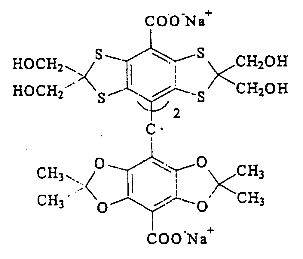
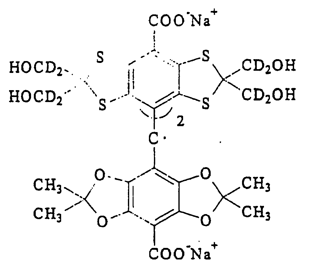 ou
ou

Applications Claiming Priority (7)
| Application Number | Priority Date | Filing Date | Title |
|---|---|---|---|
| GB9518442 | 1995-09-08 | ||
| GBGB9518442.0A GB9518442D0 (en) | 1995-09-08 | 1995-09-08 | Method |
| US08/546,146 US5765562A (en) | 1995-09-08 | 1995-10-20 | Method for determining oxygen concentration using magnetic resonance imaging |
| US546146 | 1995-10-24 | ||
| GBGB9612931.7A GB9612931D0 (en) | 1996-06-20 | 1996-06-20 | Method |
| GB9612931 | 1996-06-20 | ||
| PCT/GB1996/002198 WO1997009633A1 (fr) | 1995-09-08 | 1996-09-06 | Procede servant a determiner la concentration d'oxygene dans un echantillon |
Publications (2)
| Publication Number | Publication Date |
|---|---|
| EP0871896A1 EP0871896A1 (fr) | 1998-10-21 |
| EP0871896B1 true EP0871896B1 (fr) | 2002-01-30 |
Family
ID=27267898
Family Applications (1)
| Application Number | Title | Priority Date | Filing Date |
|---|---|---|---|
| EP96929434A Expired - Lifetime EP0871896B1 (fr) | 1995-09-08 | 1996-09-06 | Procede servant a determiner la concentration d'oxygene dans un echantillon |
Country Status (13)
| Country | Link |
|---|---|
| EP (1) | EP0871896B1 (fr) |
| JP (1) | JP2002502491A (fr) |
| CN (1) | CN1200179A (fr) |
| AT (1) | ATE212727T1 (fr) |
| AU (1) | AU717685B2 (fr) |
| CA (1) | CA2231405A1 (fr) |
| CZ (1) | CZ66998A3 (fr) |
| DE (1) | DE69618991T2 (fr) |
| ES (1) | ES2175123T3 (fr) |
| NO (1) | NO980965L (fr) |
| NZ (1) | NZ316583A (fr) |
| PL (1) | PL325428A1 (fr) |
| WO (1) | WO1997009633A1 (fr) |
Cited By (2)
| Publication number | Priority date | Publication date | Assignee | Title |
|---|---|---|---|---|
| US7662362B2 (en) | 2003-09-05 | 2010-02-16 | The Ohio State University Research Foundation | Nanoparticulate probe for in vivo monitoring of tissue oxygenation |
| US8066973B2 (en) | 2003-09-05 | 2011-11-29 | The Ohio State University Research Foundation | Nanoparticulate probe for in vivo monitoring of tissue oxygenation |
Families Citing this family (7)
| Publication number | Priority date | Publication date | Assignee | Title |
|---|---|---|---|---|
| DE10164293A1 (de) * | 2001-12-28 | 2003-07-10 | Wagner Alarm Sicherung | Verfahren und Vorrichtung zum Messen des Sauerstoffgehaltes |
| NZ552898A (en) | 2004-07-30 | 2011-02-25 | Ge Healthcare As | Method of producing a composition, composition and its use |
| JP5475995B2 (ja) | 2005-12-01 | 2014-04-16 | ジーイー・ヘルスケア・アクスイェ・セルスカプ | 動的核分極(dnp)法 |
| CN101970014A (zh) * | 2007-07-26 | 2011-02-09 | 通用电气医疗集团英国有限公司 | 包含超极化的13c-乳酸盐的成像介质及其用途 |
| EP2072061A1 (fr) | 2007-12-19 | 2009-06-24 | GE Healthcare Limited | Composition et procédé pour générer un profil métabolique par 13C-MR |
| US8697034B2 (en) | 2008-10-10 | 2014-04-15 | The Board Of Regents Of The University Of Texas System | Hyperpolarized 89-yttrium and methods relating thereto |
| US8968703B2 (en) | 2009-09-10 | 2015-03-03 | Ge Healthcare Limited | 13C-MR detection using hyperpolarised 13C-fructose |
Family Cites Families (2)
| Publication number | Priority date | Publication date | Assignee | Title |
|---|---|---|---|---|
| GB8909270D0 (en) * | 1989-04-24 | 1989-06-07 | Hafslund Nycomed Innovation | Method |
| FR2664705B1 (fr) * | 1990-07-10 | 1992-09-18 | Commissariat Energie Atomique | Procede de detection ou de dosage d'oxygene par spectrometrie rpe utilisant des phtalocyanines de lithium radicalaire et composition utilisable pour le dosage "in vivo". |
-
1996
- 1996-09-06 ES ES96929434T patent/ES2175123T3/es not_active Expired - Lifetime
- 1996-09-06 WO PCT/GB1996/002198 patent/WO1997009633A1/fr not_active Ceased
- 1996-09-06 CA CA002231405A patent/CA2231405A1/fr not_active Abandoned
- 1996-09-06 AU AU68847/96A patent/AU717685B2/en not_active Ceased
- 1996-09-06 NZ NZ316583A patent/NZ316583A/xx unknown
- 1996-09-06 EP EP96929434A patent/EP0871896B1/fr not_active Expired - Lifetime
- 1996-09-06 DE DE69618991T patent/DE69618991T2/de not_active Expired - Fee Related
- 1996-09-06 PL PL96325428A patent/PL325428A1/xx unknown
- 1996-09-06 JP JP51098697A patent/JP2002502491A/ja active Pending
- 1996-09-06 AT AT96929434T patent/ATE212727T1/de not_active IP Right Cessation
- 1996-09-06 CN CN96197752A patent/CN1200179A/zh active Pending
- 1996-09-06 CZ CZ98669A patent/CZ66998A3/cs unknown
-
1998
- 1998-03-05 NO NO980965A patent/NO980965L/no not_active Application Discontinuation
Cited By (4)
| Publication number | Priority date | Publication date | Assignee | Title |
|---|---|---|---|---|
| US7662362B2 (en) | 2003-09-05 | 2010-02-16 | The Ohio State University Research Foundation | Nanoparticulate probe for in vivo monitoring of tissue oxygenation |
| US8066973B2 (en) | 2003-09-05 | 2011-11-29 | The Ohio State University Research Foundation | Nanoparticulate probe for in vivo monitoring of tissue oxygenation |
| US8569482B2 (en) | 2003-09-05 | 2013-10-29 | The Ohio State University Research Foundation | Nanoparticulate probe for in vivo monitoring of tissue oxygenation |
| US8568694B2 (en) | 2003-09-05 | 2013-10-29 | The Ohio State University Research Foundation | Nanoparticulate probe for in vivo monitoring of tissue oxygenation |
Also Published As
| Publication number | Publication date |
|---|---|
| CA2231405A1 (fr) | 1997-03-13 |
| ATE212727T1 (de) | 2002-02-15 |
| DE69618991T2 (de) | 2002-10-31 |
| CZ66998A3 (cs) | 1998-07-15 |
| ES2175123T3 (es) | 2002-11-16 |
| AU6884796A (en) | 1997-03-27 |
| PL325428A1 (en) | 1998-07-20 |
| AU717685B2 (en) | 2000-03-30 |
| WO1997009633A1 (fr) | 1997-03-13 |
| EP0871896A1 (fr) | 1998-10-21 |
| NO980965D0 (no) | 1998-03-05 |
| CN1200179A (zh) | 1998-11-25 |
| NZ316583A (en) | 1999-08-30 |
| JP2002502491A (ja) | 2002-01-22 |
| DE69618991D1 (de) | 2002-03-14 |
| NO980965L (no) | 1998-04-28 |
Similar Documents
| Publication | Publication Date | Title |
|---|---|---|
| EP0832054B1 (fr) | Radicaux methyle heterocycliques libres utilises comme agents d'amelioration d'image | |
| KR101126952B1 (ko) | 건강한 조직과 종양 조직을 구별하기 위한 mr 영상화방법 | |
| CN101102799B (zh) | 使用超极化13c-丙酮酸盐的心脏成像方法 | |
| EP0485479B1 (fr) | Composes contenant du perfluoro-t-butyle destines a etre utilises en rmn et/ou irm au fluor-19 | |
| US6256527B1 (en) | Method for determining oxygen concentration | |
| US8697034B2 (en) | Hyperpolarized 89-yttrium and methods relating thereto | |
| WO1990001024A1 (fr) | Agents d'imagerie a resonance magnetique | |
| EP0871896B1 (fr) | Procede servant a determiner la concentration d'oxygene dans un echantillon | |
| JP5693224B2 (ja) | 方法及び該方法で使用するためのイメージング媒体 | |
| KR20070061805A (ko) | 라디칼 및 동적 핵 편극화 공정에서 상자성 물질로서의그의 용도 | |
| US5384108A (en) | Magnetic resonance imaging agents | |
| EP0966414B1 (fr) | Radicaux triarylmethyle libres utilises comme agents d'amelioration d'image | |
| KR101690821B1 (ko) | 과분극화된 13c-프럭토즈를 사용하는 13c-자기 공명 검출 방법 | |
| US6013810A (en) | Free radicals | |
| US5217706A (en) | Complexes and compositions for magnetic resonance imaging | |
| US5264204A (en) | Hydrophilic free radicals for magnetic resonance imaging | |
| KR19990044499A (ko) | 시료의 산소 농도 측정 방법 | |
| ITOH et al. | Development of a functional spin probe for in vivo ESR measurement | |
| AU650615C (en) | Novel magnetic resonance imaging agents | |
| WO2009150211A1 (fr) | Polariseur dnp et procédé de production d’un matériau sélectionné hyperpolarisé |
Legal Events
| Date | Code | Title | Description |
|---|---|---|---|
| PUAI | Public reference made under article 153(3) epc to a published international application that has entered the european phase |
Free format text: ORIGINAL CODE: 0009012 |
|
| 17P | Request for examination filed |
Effective date: 19980302 |
|
| AK | Designated contracting states |
Kind code of ref document: A1 Designated state(s): AT BE CH DE DK ES FI FR GB GR IE IT LI LU MC NL PT SE |
|
| AX | Request for extension of the european patent |
Free format text: AL PAYMENT 980302;LT PAYMENT 980302;LV PAYMENT 980302;SI PAYMENT 980302 |
|
| 17Q | First examination report despatched |
Effective date: 20001011 |
|
| GRAG | Despatch of communication of intention to grant |
Free format text: ORIGINAL CODE: EPIDOS AGRA |
|
| GRAG | Despatch of communication of intention to grant |
Free format text: ORIGINAL CODE: EPIDOS AGRA |
|
| GRAG | Despatch of communication of intention to grant |
Free format text: ORIGINAL CODE: EPIDOS AGRA |
|
| GRAH | Despatch of communication of intention to grant a patent |
Free format text: ORIGINAL CODE: EPIDOS IGRA |
|
| RAP1 | Party data changed (applicant data changed or rights of an application transferred) |
Owner name: NYCOMED IMAGING AS |
|
| RIN1 | Information on inventor provided before grant (corrected) |
Inventor name: LEUNBACH, IB Inventor name: ARDENKJAER-LARSEN, JAN, HENRIK |
|
| GRAH | Despatch of communication of intention to grant a patent |
Free format text: ORIGINAL CODE: EPIDOS IGRA |
|
| GRAA | (expected) grant |
Free format text: ORIGINAL CODE: 0009210 |
|
| REG | Reference to a national code |
Ref country code: GB Ref legal event code: IF02 |
|
| AK | Designated contracting states |
Kind code of ref document: B1 Designated state(s): AT BE CH DE DK ES FI FR GB GR IE IT LI LU MC NL PT SE |
|
| AX | Request for extension of the european patent |
Free format text: AL PAYMENT 19980302;LT PAYMENT 19980302;LV PAYMENT 19980302;SI PAYMENT 19980302 |
|
| LTIE | Lt: invalidation of european patent or patent extension | ||
| PG25 | Lapsed in a contracting state [announced via postgrant information from national office to epo] |
Ref country code: NL Free format text: LAPSE BECAUSE OF FAILURE TO SUBMIT A TRANSLATION OF THE DESCRIPTION OR TO PAY THE FEE WITHIN THE PRESCRIBED TIME-LIMIT Effective date: 20020130 Ref country code: LI Free format text: LAPSE BECAUSE OF FAILURE TO SUBMIT A TRANSLATION OF THE DESCRIPTION OR TO PAY THE FEE WITHIN THE PRESCRIBED TIME-LIMIT Effective date: 20020130 Ref country code: GR Free format text: LAPSE BECAUSE OF FAILURE TO SUBMIT A TRANSLATION OF THE DESCRIPTION OR TO PAY THE FEE WITHIN THE PRESCRIBED TIME-LIMIT Effective date: 20020130 Ref country code: FI Free format text: LAPSE BECAUSE OF FAILURE TO SUBMIT A TRANSLATION OF THE DESCRIPTION OR TO PAY THE FEE WITHIN THE PRESCRIBED TIME-LIMIT Effective date: 20020130 Ref country code: CH Free format text: LAPSE BECAUSE OF FAILURE TO SUBMIT A TRANSLATION OF THE DESCRIPTION OR TO PAY THE FEE WITHIN THE PRESCRIBED TIME-LIMIT Effective date: 20020130 Ref country code: BE Free format text: LAPSE BECAUSE OF FAILURE TO SUBMIT A TRANSLATION OF THE DESCRIPTION OR TO PAY THE FEE WITHIN THE PRESCRIBED TIME-LIMIT Effective date: 20020130 Ref country code: AT Free format text: LAPSE BECAUSE OF FAILURE TO SUBMIT A TRANSLATION OF THE DESCRIPTION OR TO PAY THE FEE WITHIN THE PRESCRIBED TIME-LIMIT Effective date: 20020130 |
|
| REF | Corresponds to: |
Ref document number: 212727 Country of ref document: AT Date of ref document: 20020215 Kind code of ref document: T |
|
| REG | Reference to a national code |
Ref country code: CH Ref legal event code: EP |
|
| REF | Corresponds to: |
Ref document number: 69618991 Country of ref document: DE Date of ref document: 20020314 |
|
| REG | Reference to a national code |
Ref country code: IE Ref legal event code: FG4D |
|
| PG25 | Lapsed in a contracting state [announced via postgrant information from national office to epo] |
Ref country code: SE Free format text: LAPSE BECAUSE OF FAILURE TO SUBMIT A TRANSLATION OF THE DESCRIPTION OR TO PAY THE FEE WITHIN THE PRESCRIBED TIME-LIMIT Effective date: 20020430 Ref country code: PT Free format text: LAPSE BECAUSE OF FAILURE TO SUBMIT A TRANSLATION OF THE DESCRIPTION OR TO PAY THE FEE WITHIN THE PRESCRIBED TIME-LIMIT Effective date: 20020430 Ref country code: DK Free format text: LAPSE BECAUSE OF FAILURE TO SUBMIT A TRANSLATION OF THE DESCRIPTION OR TO PAY THE FEE WITHIN THE PRESCRIBED TIME-LIMIT Effective date: 20020430 |
|
| NLV1 | Nl: lapsed or annulled due to failure to fulfill the requirements of art. 29p and 29m of the patents act | ||
| ET | Fr: translation filed | ||
| REG | Reference to a national code |
Ref country code: CH Ref legal event code: PL |
|
| PG25 | Lapsed in a contracting state [announced via postgrant information from national office to epo] |
Ref country code: LU Free format text: LAPSE BECAUSE OF NON-PAYMENT OF DUE FEES Effective date: 20020906 Ref country code: IE Free format text: LAPSE BECAUSE OF NON-PAYMENT OF DUE FEES Effective date: 20020906 |
|
| REG | Reference to a national code |
Ref country code: ES Ref legal event code: FG2A Ref document number: 2175123 Country of ref document: ES Kind code of ref document: T3 |
|
| PLBE | No opposition filed within time limit |
Free format text: ORIGINAL CODE: 0009261 |
|
| STAA | Information on the status of an ep patent application or granted ep patent |
Free format text: STATUS: NO OPPOSITION FILED WITHIN TIME LIMIT |
|
| 26N | No opposition filed | ||
| PG25 | Lapsed in a contracting state [announced via postgrant information from national office to epo] |
Ref country code: MC Free format text: LAPSE BECAUSE OF NON-PAYMENT OF DUE FEES Effective date: 20030401 |
|
| REG | Reference to a national code |
Ref country code: IE Ref legal event code: MM4A |
|
| REG | Reference to a national code |
Ref country code: HK Ref legal event code: WD Ref document number: 1011881 Country of ref document: HK |
|
| REG | Reference to a national code |
Ref country code: ES Ref legal event code: PC2A |
|
| PGFP | Annual fee paid to national office [announced via postgrant information from national office to epo] |
Ref country code: ES Payment date: 20070926 Year of fee payment: 12 |
|
| PGFP | Annual fee paid to national office [announced via postgrant information from national office to epo] |
Ref country code: GB Payment date: 20070926 Year of fee payment: 12 |
|
| PGFP | Annual fee paid to national office [announced via postgrant information from national office to epo] |
Ref country code: IT Payment date: 20070926 Year of fee payment: 12 Ref country code: DE Payment date: 20071031 Year of fee payment: 12 |
|
| PGFP | Annual fee paid to national office [announced via postgrant information from national office to epo] |
Ref country code: FR Payment date: 20070917 Year of fee payment: 12 |
|
| GBPC | Gb: european patent ceased through non-payment of renewal fee |
Effective date: 20080906 |
|
| REG | Reference to a national code |
Ref country code: FR Ref legal event code: ST Effective date: 20090529 |
|
| PG25 | Lapsed in a contracting state [announced via postgrant information from national office to epo] |
Ref country code: IT Free format text: LAPSE BECAUSE OF NON-PAYMENT OF DUE FEES Effective date: 20080906 Ref country code: DE Free format text: LAPSE BECAUSE OF NON-PAYMENT OF DUE FEES Effective date: 20090401 |
|
| PG25 | Lapsed in a contracting state [announced via postgrant information from national office to epo] |
Ref country code: FR Free format text: LAPSE BECAUSE OF NON-PAYMENT OF DUE FEES Effective date: 20080930 |
|
| REG | Reference to a national code |
Ref country code: ES Ref legal event code: FD2A Effective date: 20080908 |
|
| PG25 | Lapsed in a contracting state [announced via postgrant information from national office to epo] |
Ref country code: GB Free format text: LAPSE BECAUSE OF NON-PAYMENT OF DUE FEES Effective date: 20080906 |
|
| PG25 | Lapsed in a contracting state [announced via postgrant information from national office to epo] |
Ref country code: ES Free format text: LAPSE BECAUSE OF NON-PAYMENT OF DUE FEES Effective date: 20080908 |