EP0966414B1 - Radicaux triarylmethyle libres utilises comme agents d'amelioration d'image - Google Patents
Radicaux triarylmethyle libres utilises comme agents d'amelioration d'image Download PDFInfo
- Publication number
- EP0966414B1 EP0966414B1 EP98910833A EP98910833A EP0966414B1 EP 0966414 B1 EP0966414 B1 EP 0966414B1 EP 98910833 A EP98910833 A EP 98910833A EP 98910833 A EP98910833 A EP 98910833A EP 0966414 B1 EP0966414 B1 EP 0966414B1
- Authority
- EP
- European Patent Office
- Prior art keywords
- sample
- radical
- group
- radical compound
- image
- Prior art date
- Legal status (The legal status is an assumption and is not a legal conclusion. Google has not performed a legal analysis and makes no representation as to the accuracy of the status listed.)
- Expired - Lifetime
Links
- 230000002708 enhancing effect Effects 0.000 title description 2
- 125000005039 triarylmethyl group Chemical group 0.000 title description 2
- -1 radical compound Chemical class 0.000 claims description 54
- 239000001301 oxygen Substances 0.000 claims description 53
- 229910052760 oxygen Inorganic materials 0.000 claims description 53
- QVGXLLKOCUKJST-UHFFFAOYSA-N atomic oxygen Chemical compound [O] QVGXLLKOCUKJST-UHFFFAOYSA-N 0.000 claims description 52
- 238000000034 method Methods 0.000 claims description 47
- XLYOFNOQVPJJNP-UHFFFAOYSA-N water Substances O XLYOFNOQVPJJNP-UHFFFAOYSA-N 0.000 claims description 41
- 229910001868 water Inorganic materials 0.000 claims description 38
- 230000007704 transition Effects 0.000 claims description 18
- 230000005855 radiation Effects 0.000 claims description 17
- 239000002872 contrast media Substances 0.000 claims description 13
- 238000003384 imaging method Methods 0.000 claims description 13
- 239000002243 precursor Substances 0.000 claims description 13
- 125000000217 alkyl group Chemical group 0.000 claims description 11
- 125000004435 hydrogen atom Chemical group [H]* 0.000 claims description 11
- 230000005291 magnetic effect Effects 0.000 claims description 10
- NINIDFKCEFEMDL-UHFFFAOYSA-N Sulfur Chemical group [S] NINIDFKCEFEMDL-UHFFFAOYSA-N 0.000 claims description 9
- 238000002595 magnetic resonance imaging Methods 0.000 claims description 9
- 238000004435 EPR spectroscopy Methods 0.000 claims description 6
- 125000004429 atom Chemical group 0.000 claims description 3
- 239000001257 hydrogen Substances 0.000 claims description 3
- 229910052739 hydrogen Inorganic materials 0.000 claims description 3
- 230000001678 irradiating effect Effects 0.000 claims description 3
- 239000000546 pharmaceutical excipient Substances 0.000 claims description 3
- 150000003839 salts Chemical class 0.000 claims description 3
- 238000006467 substitution reaction Methods 0.000 claims description 3
- 125000004169 (C1-C6) alkyl group Chemical group 0.000 claims description 2
- 125000004423 acyloxy group Chemical group 0.000 claims description 2
- 125000003118 aryl group Chemical group 0.000 claims description 2
- 229910002091 carbon monoxide Inorganic materials 0.000 claims description 2
- 125000002915 carbonyl group Chemical group [*:2]C([*:1])=O 0.000 claims description 2
- 125000002887 hydroxy group Chemical group [H]O* 0.000 claims description 2
- 230000006698 induction Effects 0.000 claims description 2
- 238000011835 investigation Methods 0.000 claims description 2
- 238000004519 manufacturing process Methods 0.000 claims description 2
- 229910052717 sulfur Inorganic materials 0.000 claims description 2
- 210000005166 vasculature Anatomy 0.000 claims description 2
- UFHFLCQGNIYNRP-UHFFFAOYSA-N Hydrogen Chemical compound [H][H] UFHFLCQGNIYNRP-UHFFFAOYSA-N 0.000 claims 1
- 125000001183 hydrocarbyl group Chemical group 0.000 claims 1
- 238000002496 oximetry Methods 0.000 claims 1
- 125000000999 tert-butyl group Chemical group [H]C([H])([H])C(*)(C([H])([H])[H])C([H])([H])[H] 0.000 claims 1
- 150000003254 radicals Chemical class 0.000 description 70
- RTZKZFJDLAIYFH-UHFFFAOYSA-N Diethyl ether Chemical compound CCOCC RTZKZFJDLAIYFH-UHFFFAOYSA-N 0.000 description 53
- 239000000203 mixture Substances 0.000 description 48
- YMWUJEATGCHHMB-UHFFFAOYSA-N Dichloromethane Chemical compound ClCCl YMWUJEATGCHHMB-UHFFFAOYSA-N 0.000 description 41
- WEVYAHXRMPXWCK-UHFFFAOYSA-N Acetonitrile Chemical compound CC#N WEVYAHXRMPXWCK-UHFFFAOYSA-N 0.000 description 39
- 239000000243 solution Substances 0.000 description 29
- LFQSCWFLJHTTHZ-UHFFFAOYSA-N Ethanol Chemical compound CCO LFQSCWFLJHTTHZ-UHFFFAOYSA-N 0.000 description 27
- HEMHJVSKTPXQMS-UHFFFAOYSA-M Sodium hydroxide Chemical compound [OH-].[Na+] HEMHJVSKTPXQMS-UHFFFAOYSA-M 0.000 description 27
- HEDRZPFGACZZDS-MICDWDOJSA-N Trichloro(2H)methane Chemical compound [2H]C(Cl)(Cl)Cl HEDRZPFGACZZDS-MICDWDOJSA-N 0.000 description 24
- 239000007983 Tris buffer Substances 0.000 description 21
- 125000005605 benzo group Chemical group 0.000 description 21
- VEXZGXHMUGYJMC-UHFFFAOYSA-N Hydrochloric acid Chemical compound Cl VEXZGXHMUGYJMC-UHFFFAOYSA-N 0.000 description 20
- CSNNHWWHGAXBCP-UHFFFAOYSA-L Magnesium sulfate Chemical compound [Mg+2].[O-][S+2]([O-])([O-])[O-] CSNNHWWHGAXBCP-UHFFFAOYSA-L 0.000 description 16
- WYURNTSHIVDZCO-UHFFFAOYSA-N Tetrahydrofuran Chemical compound C1CCOC1 WYURNTSHIVDZCO-UHFFFAOYSA-N 0.000 description 16
- 239000012074 organic phase Substances 0.000 description 16
- 238000005160 1H NMR spectroscopy Methods 0.000 description 15
- XKRFYHLGVUSROY-UHFFFAOYSA-N Argon Chemical compound [Ar] XKRFYHLGVUSROY-UHFFFAOYSA-N 0.000 description 12
- VLKZOEOYAKHREP-UHFFFAOYSA-N n-Hexane Chemical compound CCCCCC VLKZOEOYAKHREP-UHFFFAOYSA-N 0.000 description 12
- LENZDBCJOHFCAS-UHFFFAOYSA-N tris Chemical compound OCC(N)(CO)CO LENZDBCJOHFCAS-UHFFFAOYSA-N 0.000 description 12
- 230000000694 effects Effects 0.000 description 11
- 239000000047 product Substances 0.000 description 11
- 239000011541 reaction mixture Substances 0.000 description 11
- 238000003756 stirring Methods 0.000 description 11
- OKKJLVBELUTLKV-UHFFFAOYSA-N Methanol Chemical class OC OKKJLVBELUTLKV-UHFFFAOYSA-N 0.000 description 10
- XEKOWRVHYACXOJ-UHFFFAOYSA-N Ethyl acetate Chemical compound CCOC(C)=O XEKOWRVHYACXOJ-UHFFFAOYSA-N 0.000 description 9
- ZMXDDKWLCZADIW-UHFFFAOYSA-N N,N-Dimethylformamide Chemical compound CN(C)C=O ZMXDDKWLCZADIW-UHFFFAOYSA-N 0.000 description 9
- 239000007787 solid Substances 0.000 description 9
- MZRVEZGGRBJDDB-UHFFFAOYSA-N N-Butyllithium Chemical compound [Li]CCCC MZRVEZGGRBJDDB-UHFFFAOYSA-N 0.000 description 8
- 229910052943 magnesium sulfate Inorganic materials 0.000 description 8
- 210000001519 tissue Anatomy 0.000 description 8
- 210000004369 blood Anatomy 0.000 description 7
- 239000008280 blood Substances 0.000 description 7
- 238000001816 cooling Methods 0.000 description 7
- 239000002002 slurry Substances 0.000 description 7
- XEEYBQQBJWHFJM-UHFFFAOYSA-N Iron Chemical compound [Fe] XEEYBQQBJWHFJM-UHFFFAOYSA-N 0.000 description 6
- VQTUBCCKSQIDNK-UHFFFAOYSA-N Isobutene Chemical compound CC(C)=C VQTUBCCKSQIDNK-UHFFFAOYSA-N 0.000 description 6
- IMNFDUFMRHMDMM-UHFFFAOYSA-N N-Heptane Chemical compound CCCCCCC IMNFDUFMRHMDMM-UHFFFAOYSA-N 0.000 description 6
- VYPSYNLAJGMNEJ-UHFFFAOYSA-N Silicium dioxide Chemical compound O=[Si]=O VYPSYNLAJGMNEJ-UHFFFAOYSA-N 0.000 description 6
- CDBYLPFSWZWCQE-UHFFFAOYSA-L Sodium Carbonate Chemical compound [Na+].[Na+].[O-]C([O-])=O CDBYLPFSWZWCQE-UHFFFAOYSA-L 0.000 description 6
- 239000008346 aqueous phase Substances 0.000 description 6
- 229910052786 argon Inorganic materials 0.000 description 6
- 229910052799 carbon Inorganic materials 0.000 description 6
- 150000001875 compounds Chemical class 0.000 description 6
- 239000012071 phase Substances 0.000 description 6
- 238000002953 preparative HPLC Methods 0.000 description 6
- ITMCEJHCFYSIIV-UHFFFAOYSA-N triflic acid Chemical compound OS(=O)(=O)C(F)(F)F ITMCEJHCFYSIIV-UHFFFAOYSA-N 0.000 description 6
- CURLTUGMZLYLDI-UHFFFAOYSA-N Carbon dioxide Chemical compound O=C=O CURLTUGMZLYLDI-UHFFFAOYSA-N 0.000 description 5
- KWYHDKDOAIKMQN-UHFFFAOYSA-N N,N,N',N'-tetramethylethylenediamine Chemical compound CN(C)CCN(C)C KWYHDKDOAIKMQN-UHFFFAOYSA-N 0.000 description 5
- 239000005864 Sulphur Substances 0.000 description 5
- 239000007864 aqueous solution Substances 0.000 description 5
- 239000012300 argon atmosphere Substances 0.000 description 5
- 229910002092 carbon dioxide Inorganic materials 0.000 description 5
- 230000005298 paramagnetic effect Effects 0.000 description 5
- AJPJDKMHJJGVTQ-UHFFFAOYSA-M sodium dihydrogen phosphate Chemical compound [Na+].OP(O)([O-])=O AJPJDKMHJJGVTQ-UHFFFAOYSA-M 0.000 description 5
- 229910000162 sodium phosphate Inorganic materials 0.000 description 5
- CAQWNKXTMBFBGI-UHFFFAOYSA-N C.[Na] Chemical class C.[Na] CAQWNKXTMBFBGI-UHFFFAOYSA-N 0.000 description 4
- 210000001124 body fluid Anatomy 0.000 description 4
- 239000010839 body fluid Substances 0.000 description 4
- 239000000706 filtrate Substances 0.000 description 4
- 238000001727 in vivo Methods 0.000 description 4
- 238000002347 injection Methods 0.000 description 4
- 239000007924 injection Substances 0.000 description 4
- 239000000463 material Substances 0.000 description 4
- BDAGIHXWWSANSR-UHFFFAOYSA-N methanoic acid Natural products OC=O BDAGIHXWWSANSR-UHFFFAOYSA-N 0.000 description 4
- 125000002496 methyl group Chemical group [H]C([H])([H])* 0.000 description 4
- BWHMMNNQKKPAPP-UHFFFAOYSA-L potassium carbonate Chemical compound [K+].[K+].[O-]C([O-])=O BWHMMNNQKKPAPP-UHFFFAOYSA-L 0.000 description 4
- 239000002244 precipitate Substances 0.000 description 4
- 238000010992 reflux Methods 0.000 description 4
- QTBSBXVTEAMEQO-UHFFFAOYSA-N Acetic acid Chemical compound CC(O)=O QTBSBXVTEAMEQO-UHFFFAOYSA-N 0.000 description 3
- IAZDPXIOMUYVGZ-UHFFFAOYSA-N Dimethylsulphoxide Chemical compound CS(C)=O IAZDPXIOMUYVGZ-UHFFFAOYSA-N 0.000 description 3
- AVXURJPOCDRRFD-UHFFFAOYSA-N Hydroxylamine Chemical class ON AVXURJPOCDRRFD-UHFFFAOYSA-N 0.000 description 3
- 239000007832 Na2SO4 Substances 0.000 description 3
- PMZURENOXWZQFD-UHFFFAOYSA-L Sodium Sulfate Chemical compound [Na+].[Na+].[O-]S([O-])(=O)=O PMZURENOXWZQFD-UHFFFAOYSA-L 0.000 description 3
- DWAQJAXMDSEUJJ-UHFFFAOYSA-M Sodium bisulfite Chemical compound [Na+].OS([O-])=O DWAQJAXMDSEUJJ-UHFFFAOYSA-M 0.000 description 3
- 229910021626 Tin(II) chloride Inorganic materials 0.000 description 3
- 239000002253 acid Substances 0.000 description 3
- 238000006243 chemical reaction Methods 0.000 description 3
- 239000003795 chemical substances by application Substances 0.000 description 3
- 230000008878 coupling Effects 0.000 description 3
- 238000010168 coupling process Methods 0.000 description 3
- 238000005859 coupling reaction Methods 0.000 description 3
- 230000001419 dependent effect Effects 0.000 description 3
- 238000001704 evaporation Methods 0.000 description 3
- 230000008020 evaporation Effects 0.000 description 3
- 238000009472 formulation Methods 0.000 description 3
- 229910052742 iron Inorganic materials 0.000 description 3
- WGOPGODQLGJZGL-UHFFFAOYSA-N lithium;butane Chemical compound [Li+].CC[CH-]C WGOPGODQLGJZGL-UHFFFAOYSA-N 0.000 description 3
- 229920002521 macromolecule Polymers 0.000 description 3
- 238000005259 measurement Methods 0.000 description 3
- 229910000029 sodium carbonate Inorganic materials 0.000 description 3
- 239000004289 sodium hydrogen sulphite Substances 0.000 description 3
- 235000010267 sodium hydrogen sulphite Nutrition 0.000 description 3
- 229910052938 sodium sulfate Inorganic materials 0.000 description 3
- 239000002904 solvent Substances 0.000 description 3
- 239000000725 suspension Substances 0.000 description 3
- YLQBMQCUIZJEEH-UHFFFAOYSA-N tetrahydrofuran Natural products C=1C=COC=1 YLQBMQCUIZJEEH-UHFFFAOYSA-N 0.000 description 3
- AXZWODMDQAVCJE-UHFFFAOYSA-L tin(II) chloride (anhydrous) Chemical compound [Cl-].[Cl-].[Sn+2] AXZWODMDQAVCJE-UHFFFAOYSA-L 0.000 description 3
- RILZRCJGXSFXNE-UHFFFAOYSA-N 2-[4-(trifluoromethoxy)phenyl]ethanol Chemical compound OCCC1=CC=C(OC(F)(F)F)C=C1 RILZRCJGXSFXNE-UHFFFAOYSA-N 0.000 description 2
- 125000000954 2-hydroxyethyl group Chemical group [H]C([*])([H])C([H])([H])O[H] 0.000 description 2
- OSWFIVFLDKOXQC-UHFFFAOYSA-N 4-(3-methoxyphenyl)aniline Chemical compound COC1=CC=CC(C=2C=CC(N)=CC=2)=C1 OSWFIVFLDKOXQC-UHFFFAOYSA-N 0.000 description 2
- ZCYVEMRRCGMTRW-UHFFFAOYSA-N 7553-56-2 Chemical compound [I] ZCYVEMRRCGMTRW-UHFFFAOYSA-N 0.000 description 2
- IJGRMHOSHXDMSA-UHFFFAOYSA-N Atomic nitrogen Chemical compound N#N IJGRMHOSHXDMSA-UHFFFAOYSA-N 0.000 description 2
- GAWIXWVDTYZWAW-UHFFFAOYSA-N C[CH]O Chemical group C[CH]O GAWIXWVDTYZWAW-UHFFFAOYSA-N 0.000 description 2
- OIFBSDVPJOWBCH-UHFFFAOYSA-N Diethyl carbonate Chemical compound CCOC(=O)OCC OIFBSDVPJOWBCH-UHFFFAOYSA-N 0.000 description 2
- IAZDPXIOMUYVGZ-WFGJKAKNSA-N Dimethyl sulfoxide Chemical compound [2H]C([2H])([2H])S(=O)C([2H])([2H])[2H] IAZDPXIOMUYVGZ-WFGJKAKNSA-N 0.000 description 2
- LRHPLDYGYMQRHN-UHFFFAOYSA-N N-Butanol Chemical compound CCCCO LRHPLDYGYMQRHN-UHFFFAOYSA-N 0.000 description 2
- 206010028980 Neoplasm Diseases 0.000 description 2
- 229910006146 SO3M1 Inorganic materials 0.000 description 2
- 229910052784 alkaline earth metal Inorganic materials 0.000 description 2
- 239000012455 biphasic mixture Substances 0.000 description 2
- 239000012267 brine Substances 0.000 description 2
- 201000011510 cancer Diseases 0.000 description 2
- 239000002775 capsule Substances 0.000 description 2
- 125000003178 carboxy group Chemical group [H]OC(*)=O 0.000 description 2
- 238000004587 chromatography analysis Methods 0.000 description 2
- 229940039231 contrast media Drugs 0.000 description 2
- 239000012043 crude product Substances 0.000 description 2
- 239000003480 eluent Substances 0.000 description 2
- 239000012065 filter cake Substances 0.000 description 2
- 235000019253 formic acid Nutrition 0.000 description 2
- 238000011503 in vivo imaging Methods 0.000 description 2
- 238000011065 in-situ storage Methods 0.000 description 2
- 238000001990 intravenous administration Methods 0.000 description 2
- 239000011630 iodine Substances 0.000 description 2
- 229910052740 iodine Inorganic materials 0.000 description 2
- 230000004060 metabolic process Effects 0.000 description 2
- 150000004702 methyl esters Chemical class 0.000 description 2
- 125000005188 oxoalkyl group Chemical group 0.000 description 2
- 125000004430 oxygen atom Chemical group O* 0.000 description 2
- 239000002907 paramagnetic material Substances 0.000 description 2
- 230000010287 polarization Effects 0.000 description 2
- 229920000642 polymer Polymers 0.000 description 2
- 229910000027 potassium carbonate Inorganic materials 0.000 description 2
- 238000011160 research Methods 0.000 description 2
- 230000035945 sensitivity Effects 0.000 description 2
- 239000000741 silica gel Substances 0.000 description 2
- 229910002027 silica gel Inorganic materials 0.000 description 2
- 239000000377 silicon dioxide Substances 0.000 description 2
- HPALAKNZSZLMCH-UHFFFAOYSA-M sodium;chloride;hydrate Chemical compound O.[Na+].[Cl-] HPALAKNZSZLMCH-UHFFFAOYSA-M 0.000 description 2
- 230000003381 solubilizing effect Effects 0.000 description 2
- 235000011150 stannous chloride Nutrition 0.000 description 2
- 239000000126 substance Substances 0.000 description 2
- 125000001424 substituent group Chemical group 0.000 description 2
- XLHUBROMZOAQMV-UHFFFAOYSA-N 1,4-benzosemiquinone Chemical compound [O]C1=CC=C(O)C=C1 XLHUBROMZOAQMV-UHFFFAOYSA-N 0.000 description 1
- PDQRQJVPEFGVRK-UHFFFAOYSA-N 2,1,3-benzothiadiazole Chemical compound C1=CC=CC2=NSN=C21 PDQRQJVPEFGVRK-UHFFFAOYSA-N 0.000 description 1
- HEWZVZIVELJPQZ-UHFFFAOYSA-N 2,2-dimethoxypropane Chemical compound COC(C)(C)OC HEWZVZIVELJPQZ-UHFFFAOYSA-N 0.000 description 1
- LSTRKXWIZZZYAS-UHFFFAOYSA-N 2-bromoacetyl bromide Chemical compound BrCC(Br)=O LSTRKXWIZZZYAS-UHFFFAOYSA-N 0.000 description 1
- SAMGBMSEBPZABZ-UHFFFAOYSA-N 3-(2,3-dihydroxypropylamino)propane-1,2-diol Chemical compound OCC(O)CNCC(O)CO SAMGBMSEBPZABZ-UHFFFAOYSA-N 0.000 description 1
- RNJOKCPFLQMDEC-UHFFFAOYSA-N 4(R),8-dimethyl-trans-2-nonenoyl-CoA Chemical compound COC(=O)CC(=O)CC(=O)OC RNJOKCPFLQMDEC-UHFFFAOYSA-N 0.000 description 1
- OIVUHPTVQVCONM-UHFFFAOYSA-N 6-bromo-4-methyl-1h-indazole Chemical compound CC1=CC(Br)=CC2=C1C=NN2 OIVUHPTVQVCONM-UHFFFAOYSA-N 0.000 description 1
- QGZKDVFQNNGYKY-UHFFFAOYSA-O Ammonium Chemical compound [NH4+] QGZKDVFQNNGYKY-UHFFFAOYSA-O 0.000 description 1
- 0 CC[C@@](CCO)(SC1C(C(C)C)C2SC(CCO)(CCI)SC22)SC1C2C(OC*=O)=* Chemical compound CC[C@@](CCO)(SC1C(C(C)C)C2SC(CCO)(CCI)SC22)SC1C2C(OC*=O)=* 0.000 description 1
- ZOCLUYMIUWBCRH-UHFFFAOYSA-N CCc(c1c2SC(CCO)(CCO)S1)c1SC(CCO)(CCO)Sc1c2SCC(N(CC(CO)O)CC(CO)O)=O Chemical compound CCc(c1c2SC(CCO)(CCO)S1)c1SC(CCO)(CCO)Sc1c2SCC(N(CC(CO)O)CC(CO)O)=O ZOCLUYMIUWBCRH-UHFFFAOYSA-N 0.000 description 1
- OKTJSMMVPCPJKN-UHFFFAOYSA-N Carbon Chemical compound [C] OKTJSMMVPCPJKN-UHFFFAOYSA-N 0.000 description 1
- XDTMQSROBMDMFD-UHFFFAOYSA-N Cyclohexane Chemical compound C1CCCCC1 XDTMQSROBMDMFD-UHFFFAOYSA-N 0.000 description 1
- 229920002307 Dextran Polymers 0.000 description 1
- DGAQECJNVWCQMB-PUAWFVPOSA-M Ilexoside XXIX Chemical compound C[C@@H]1CC[C@@]2(CC[C@@]3(C(=CC[C@H]4[C@]3(CC[C@@H]5[C@@]4(CC[C@@H](C5(C)C)OS(=O)(=O)[O-])C)C)[C@@H]2[C@]1(C)O)C)C(=O)O[C@H]6[C@@H]([C@H]([C@@H]([C@H](O6)CO)O)O)O.[Na+] DGAQECJNVWCQMB-PUAWFVPOSA-M 0.000 description 1
- 229910010084 LiAlH4 Inorganic materials 0.000 description 1
- 239000012448 Lithium borohydride Substances 0.000 description 1
- 238000012307 MRI technique Methods 0.000 description 1
- 241001465754 Metazoa Species 0.000 description 1
- DBTDEFJAFBUGPP-UHFFFAOYSA-N Methanethial Chemical compound S=C DBTDEFJAFBUGPP-UHFFFAOYSA-N 0.000 description 1
- 238000005481 NMR spectroscopy Methods 0.000 description 1
- 229920002873 Polyethylenimine Polymers 0.000 description 1
- 108010039918 Polylysine Proteins 0.000 description 1
- UIIMBOGNXHQVGW-UHFFFAOYSA-M Sodium bicarbonate Chemical class [Na+].OC([O-])=O UIIMBOGNXHQVGW-UHFFFAOYSA-M 0.000 description 1
- QAOWNCQODCNURD-UHFFFAOYSA-N Sulfuric acid Chemical compound OS(O)(=O)=O QAOWNCQODCNURD-UHFFFAOYSA-N 0.000 description 1
- 238000000862 absorption spectrum Methods 0.000 description 1
- WEVYAHXRMPXWCK-FIBGUPNXSA-N acetonitrile-d3 Chemical compound [2H]C([2H])([2H])C#N WEVYAHXRMPXWCK-FIBGUPNXSA-N 0.000 description 1
- 230000002378 acidificating effect Effects 0.000 description 1
- 239000003513 alkali Substances 0.000 description 1
- 125000003342 alkenyl group Chemical group 0.000 description 1
- 125000004183 alkoxy alkyl group Chemical group 0.000 description 1
- 125000004453 alkoxycarbonyl group Chemical group 0.000 description 1
- 125000002877 alkyl aryl group Chemical group 0.000 description 1
- VZTDIZULWFCMLS-UHFFFAOYSA-N ammonium formate Chemical compound [NH4+].[O-]C=O VZTDIZULWFCMLS-UHFFFAOYSA-N 0.000 description 1
- 150000008064 anhydrides Chemical class 0.000 description 1
- 210000001367 artery Anatomy 0.000 description 1
- 230000009286 beneficial effect Effects 0.000 description 1
- 210000003445 biliary tract Anatomy 0.000 description 1
- 230000015572 biosynthetic process Effects 0.000 description 1
- 210000004556 brain Anatomy 0.000 description 1
- 210000000481 breast Anatomy 0.000 description 1
- 239000000872 buffer Substances 0.000 description 1
- 238000004364 calculation method Methods 0.000 description 1
- 125000003917 carbamoyl group Chemical group [H]N([H])C(*)=O 0.000 description 1
- 150000001718 carbodiimides Chemical class 0.000 description 1
- 125000004432 carbon atom Chemical group C* 0.000 description 1
- 239000001569 carbon dioxide Substances 0.000 description 1
- 150000001723 carbon free-radicals Chemical class 0.000 description 1
- 210000000748 cardiovascular system Anatomy 0.000 description 1
- 150000001768 cations Chemical class 0.000 description 1
- 238000004891 communication Methods 0.000 description 1
- 230000021615 conjugation Effects 0.000 description 1
- 239000012059 conventional drug carrier Substances 0.000 description 1
- 238000007796 conventional method Methods 0.000 description 1
- ARUVKPQLZAKDPS-UHFFFAOYSA-L copper(II) sulfate Chemical compound [Cu+2].[O-][S+2]([O-])([O-])[O-] ARUVKPQLZAKDPS-UHFFFAOYSA-L 0.000 description 1
- 229910000366 copper(II) sulfate Inorganic materials 0.000 description 1
- 239000013058 crude material Substances 0.000 description 1
- 230000002950 deficient Effects 0.000 description 1
- 239000008367 deionised water Substances 0.000 description 1
- 229910021641 deionized water Inorganic materials 0.000 description 1
- 238000002059 diagnostic imaging Methods 0.000 description 1
- 239000012502 diagnostic product Substances 0.000 description 1
- 238000012631 diagnostic technique Methods 0.000 description 1
- ZSANYRMTSBBUCA-UHFFFAOYSA-N diethyl 3-oxopentanedioate Chemical compound CCOC(=O)CC(=O)CC(=O)OCC ZSANYRMTSBBUCA-UHFFFAOYSA-N 0.000 description 1
- 239000006185 dispersion Substances 0.000 description 1
- 239000003814 drug Substances 0.000 description 1
- 238000001983 electron spin resonance imaging Methods 0.000 description 1
- 238000001362 electron spin resonance spectrum Methods 0.000 description 1
- 239000003995 emulsifying agent Substances 0.000 description 1
- 239000003623 enhancer Substances 0.000 description 1
- 210000003743 erythrocyte Anatomy 0.000 description 1
- 150000002170 ethers Chemical class 0.000 description 1
- PQJJJMRNHATNKG-UHFFFAOYSA-N ethyl bromoacetate Chemical compound CCOC(=O)CBr PQJJJMRNHATNKG-UHFFFAOYSA-N 0.000 description 1
- 125000001495 ethyl group Chemical group [H]C([H])([H])C([H])([H])* 0.000 description 1
- 238000002474 experimental method Methods 0.000 description 1
- 210000003722 extracellular fluid Anatomy 0.000 description 1
- 238000001914 filtration Methods 0.000 description 1
- 239000012530 fluid Substances 0.000 description 1
- 239000006260 foam Substances 0.000 description 1
- 210000004051 gastric juice Anatomy 0.000 description 1
- 210000001035 gastrointestinal tract Anatomy 0.000 description 1
- 150000004676 glycans Chemical class 0.000 description 1
- 238000010438 heat treatment Methods 0.000 description 1
- 125000005842 heteroatom Chemical group 0.000 description 1
- 238000004128 high performance liquid chromatography Methods 0.000 description 1
- 150000002430 hydrocarbons Chemical group 0.000 description 1
- 125000002768 hydroxyalkyl group Chemical group 0.000 description 1
- CBOIHMRHGLHBPB-UHFFFAOYSA-N hydroxymethyl Chemical compound O[CH2] CBOIHMRHGLHBPB-UHFFFAOYSA-N 0.000 description 1
- 238000001802 infusion Methods 0.000 description 1
- 230000003993 interaction Effects 0.000 description 1
- 230000009021 linear effect Effects 0.000 description 1
- 150000002634 lipophilic molecules Chemical class 0.000 description 1
- 239000012280 lithium aluminium hydride Substances 0.000 description 1
- 210000004185 liver Anatomy 0.000 description 1
- 150000004668 long chain fatty acids Chemical group 0.000 description 1
- 210000004072 lung Anatomy 0.000 description 1
- 210000003563 lymphoid tissue Anatomy 0.000 description 1
- 238000005404 magnetometry Methods 0.000 description 1
- 238000013507 mapping Methods 0.000 description 1
- 229960003194 meglumine Drugs 0.000 description 1
- 239000002207 metabolite Substances 0.000 description 1
- VNWKTOKETHGBQD-UHFFFAOYSA-N methane Chemical class C VNWKTOKETHGBQD-UHFFFAOYSA-N 0.000 description 1
- OKKJLVBELUTLKV-VMNATFBRSA-N methanol-d1 Chemical compound [2H]OC OKKJLVBELUTLKV-VMNATFBRSA-N 0.000 description 1
- 125000002816 methylsulfanyl group Chemical group [H]C([H])([H])S[*] 0.000 description 1
- 238000012986 modification Methods 0.000 description 1
- 230000004048 modification Effects 0.000 description 1
- 230000001338 necrotic effect Effects 0.000 description 1
- 229910052757 nitrogen Inorganic materials 0.000 description 1
- 230000009022 nonlinear effect Effects 0.000 description 1
- 210000000056 organ Anatomy 0.000 description 1
- 230000003647 oxidation Effects 0.000 description 1
- 238000007254 oxidation reaction Methods 0.000 description 1
- 150000002926 oxygen Chemical class 0.000 description 1
- 238000006213 oxygenation reaction Methods 0.000 description 1
- 230000020477 pH reduction Effects 0.000 description 1
- 230000035515 penetration Effects 0.000 description 1
- 230000010412 perfusion Effects 0.000 description 1
- 230000002085 persistent effect Effects 0.000 description 1
- 239000000825 pharmaceutical preparation Substances 0.000 description 1
- 229940127557 pharmaceutical product Drugs 0.000 description 1
- 230000004962 physiological condition Effects 0.000 description 1
- 229920000656 polylysine Polymers 0.000 description 1
- 229920001184 polypeptide Polymers 0.000 description 1
- 229920001282 polysaccharide Polymers 0.000 description 1
- 239000005017 polysaccharide Substances 0.000 description 1
- IHSLHAZEJBXKMN-UHFFFAOYSA-L potassium nitrosodisulfonate Chemical compound [K+].[K+].[O-]S(=O)(=O)N([O])S([O-])(=O)=O IHSLHAZEJBXKMN-UHFFFAOYSA-L 0.000 description 1
- 239000000843 powder Substances 0.000 description 1
- 238000002360 preparation method Methods 0.000 description 1
- 230000008569 process Effects 0.000 description 1
- 108090000765 processed proteins & peptides Proteins 0.000 description 1
- 102000004196 processed proteins & peptides Human genes 0.000 description 1
- 102000004169 proteins and genes Human genes 0.000 description 1
- 108090000623 proteins and genes Proteins 0.000 description 1
- 238000000746 purification Methods 0.000 description 1
- 238000001959 radiotherapy Methods 0.000 description 1
- 230000009467 reduction Effects 0.000 description 1
- 210000005227 renal system Anatomy 0.000 description 1
- 230000009291 secondary effect Effects 0.000 description 1
- 229910052708 sodium Inorganic materials 0.000 description 1
- 239000011734 sodium Substances 0.000 description 1
- 125000003003 spiro group Chemical group 0.000 description 1
- 239000007858 starting material Substances 0.000 description 1
- 230000000638 stimulation Effects 0.000 description 1
- 239000012258 stirred mixture Substances 0.000 description 1
- 125000001174 sulfone group Chemical group 0.000 description 1
- 125000000472 sulfonyl group Chemical group *S(*)(=O)=O 0.000 description 1
- 239000001117 sulphuric acid Substances 0.000 description 1
- 235000011149 sulphuric acid Nutrition 0.000 description 1
- 238000001356 surgical procedure Methods 0.000 description 1
- UGNWTBMOAKPKBL-UHFFFAOYSA-N tetrachloro-1,4-benzoquinone Chemical compound ClC1=C(Cl)C(=O)C(Cl)=C(Cl)C1=O UGNWTBMOAKPKBL-UHFFFAOYSA-N 0.000 description 1
- 230000001225 therapeutic effect Effects 0.000 description 1
- 150000003568 thioethers Chemical group 0.000 description 1
- HPGGPRDJHPYFRM-UHFFFAOYSA-J tin(iv) chloride Chemical compound Cl[Sn](Cl)(Cl)Cl HPGGPRDJHPYFRM-UHFFFAOYSA-J 0.000 description 1
- 230000001988 toxicity Effects 0.000 description 1
- 231100000419 toxicity Toxicity 0.000 description 1
- 210000001635 urinary tract Anatomy 0.000 description 1
- 210000004291 uterus Anatomy 0.000 description 1
Classifications
-
- C—CHEMISTRY; METALLURGY
- C07—ORGANIC CHEMISTRY
- C07D—HETEROCYCLIC COMPOUNDS
- C07D519/00—Heterocyclic compounds containing more than one system of two or more relevant hetero rings condensed among themselves or condensed with a common carbocyclic ring system not provided for in groups C07D453/00 or C07D455/00
-
- A—HUMAN NECESSITIES
- A61—MEDICAL OR VETERINARY SCIENCE; HYGIENE
- A61K—PREPARATIONS FOR MEDICAL, DENTAL OR TOILETRY PURPOSES
- A61K49/00—Preparations for testing in vivo
- A61K49/06—Nuclear magnetic resonance [NMR] contrast preparations; Magnetic resonance imaging [MRI] contrast preparations
- A61K49/20—Nuclear magnetic resonance [NMR] contrast preparations; Magnetic resonance imaging [MRI] contrast preparations containing free radicals, e.g. trityl radical for overhauser
-
- C—CHEMISTRY; METALLURGY
- C07—ORGANIC CHEMISTRY
- C07B—GENERAL METHODS OF ORGANIC CHEMISTRY; APPARATUS THEREFOR
- C07B61/00—Other general methods
- C07B61/02—Generation of organic free radicals; Organic free radicals per se
-
- G—PHYSICS
- G01—MEASURING; TESTING
- G01R—MEASURING ELECTRIC VARIABLES; MEASURING MAGNETIC VARIABLES
- G01R33/00—Arrangements or instruments for measuring magnetic variables
- G01R33/20—Arrangements or instruments for measuring magnetic variables involving magnetic resonance
- G01R33/44—Arrangements or instruments for measuring magnetic variables involving magnetic resonance using nuclear magnetic resonance [NMR]
- G01R33/48—NMR imaging systems
- G01R33/54—Signal processing systems, e.g. using pulse sequences ; Generation or control of pulse sequences; Operator console
- G01R33/56—Image enhancement or correction, e.g. subtraction or averaging techniques, e.g. improvement of signal-to-noise ratio and resolution
- G01R33/5601—Image enhancement or correction, e.g. subtraction or averaging techniques, e.g. improvement of signal-to-noise ratio and resolution involving use of a contrast agent for contrast manipulation, e.g. a paramagnetic, super-paramagnetic, ferromagnetic or hyperpolarised contrast agent
-
- G—PHYSICS
- G01—MEASURING; TESTING
- G01R—MEASURING ELECTRIC VARIABLES; MEASURING MAGNETIC VARIABLES
- G01R33/00—Arrangements or instruments for measuring magnetic variables
- G01R33/20—Arrangements or instruments for measuring magnetic variables involving magnetic resonance
- G01R33/62—Arrangements or instruments for measuring magnetic variables involving magnetic resonance using double resonance
Definitions
- the present invention relates to novel triarylmethyl free radicals and their use as image enhancing agents in magnetic resonance imaging (MRI), in particular to their use in electron spin resonance enhanced magnetic resonance imaging (OMRI) of a sample (for example a human or animal body) for determining the oxygen concentration of said sample.
- MRI magnetic resonance imaging
- OMRI electron spin resonance enhanced magnetic resonance imaging
- MRI Magnetic resonance
- Electron spin resonance enhanced MRI referred to herein as OMRI (Overhauser MRI) but also referred to in earlier publications as ESREMRI or PEDRI, is a particular form of MRI in which enhancement of the magnetic resonance signals from which the images are generated is achieved by virtue of the dynamic nuclear polarization (the Overhauser effect) that occurs on VHF stimulation of an esr transition in a paramagnetic material, generally a persistent free radical, in the subject under study.
- Magnetic resonance signal enhancement may be by a factor of a hundred or more thus allowing OMRI images to be generated rapidly and with relatively low primary magnetic fields.
- OMRI techniques have been described by several authors, notably Leunbach, Lurie, Ettinger, Grücker, Ehnholm and Sepponen, for example in EP-A-296833, EP-A-361551, WO-A-90/13047, J. Mag. Reson. 76 :366-370(1988), EP-A-302742, SMRM 9:619(1990), SMRM 6:24(1987), SMRM 7:1094(1988), SMRM 8:329(1989), US-A-4719425, SMRM 8:816(1989), Mag. Reson. Med.
- the imaging sequence involves initially irradiating a subject placed in a uniform magnetic field (the primary field B o ) with radiation, usually VHF radiation, of a frequency selected to excite a narrow linewidth esr transition in a paramagnetic enhancement agent (hereinafter "an OMRI contrast agent") which is in or has been administered to the subject.
- a paramagnetic enhancement agent hereinafter "an OMRI contrast agent"
- Dynamic nuclear polarization results in an increase in the population difference between the excited and ground nuclear spin states of the imaging nuclei, i.e. those nuclei, generally protons, which are responsible for the magnetic resonance signals. Since MR signal intensity is proportional to this population difference, the subsequent stages of each imaging sequence, performed essentially as in conventional MRI techniques, result in larger amplitude MR signals being detected and more effective contrast.
- OMRI contrast agents including for example nitroxide stable free radicals, chloranil semiquinone radical and Fremy's salt (US-A-4984573) and deuterated stable free radicals, in particular deuterated nitroxide stable free radicals (WO-A-90/00904).
- Nycomed Imaging AS proposed various sulphur-based triarylmethyl radicals for use as OMRI contrast agents.
- Grücker et al MRM, 34 : 219-225(1995)
- MRM Overhauser effect attributable to a nitroxide radical
- Grücker observed that the correlation between actual and calculated oxygen concentration was poor and therefore that the method was inherently inaccurate. This was attributed to the large number of parameters involved in the calculation.
- Ehnholm (US-A-5289125) proposed an OMRI technique in which signals from a paramagnetic material were detected under at least two different sets of operating parameters whereby to generate images of various physical, chemical or biological parameters. While oxygen tension was one of several such parameters, Ehnholm did not demonstrate the use of the technique to quantitate dissolved oxygen.
- radical precursors which may conveniently undergo a radical generation step shortly before administration or even in situ to produce the desired free radical.
- Radical precursors and radical generation steps are well-known to those skilled in the art.
- Preferred radical compounds according to the invention include those of formula I wherein n is at least two and more preferably is three.
- the solubilising groups M may be any of t solubilising groups conventionally used in diagnostic and pharmaceutical products.
- Particularly preferred solubilizing groups M include optionally hydroxylated, optionally alkoxylated alkyl or oxo-alkyl groups and groups of formulae R 5 , COOR 5 , OCOR 5 , CHO, CN, CH 2 S(O)R 5 CONR 5 2 , NR 5 COR 5 , NR 5 2 SO 2 NR 5 2 OR 5 , PO 3 2- , SOR 5 , SO 2 R 5 , SO 3 M 1 COOM 1 (where R 5 represents a hydrogen atom or an optionally hydroxylated, optionally aminated, optionally alkoxylated, optionally carboxylated alkyl, oxo-alkyl, alkenyl or alkaryl group and M 1 is one equivalent of a physiologically tolerable cation, for example an alkali or alkaline earth metal cation, an am
- solubilizing groups M are groups of formula C(H) 3-n (CH 2 OH) n , R 9 , COR 9 , SR 9 , SOR 9 , SO 2 R 9 , CON (R 9 ) 2 , NR 9 2 , NHR 9 and CONHR 9 [where R 9 may represent a hydroxylated alkyl group such as a group (although any R 9 group attached to a sulphur, nitrogen or oxygen atom is preferably not hydroxylated at the ⁇ carbon)], and groups of formula SO 2 R 12 or SR 12 where R 12 is a group CH 2 COOR 13 , CH(COOR 13 ) 2 , CH 2 CONHR 9 , CH 2 CONR 9 2 , CR 5 (COOR 13 ) 2 , CH(CN)CO 2 R 13 , (CH 2 ) n SO 3 - M 1 , (CH 2 ) n COR 13 , CH(COR 9 )CH 2 COR 9 and CH(R 5 )COR 9
- X is preferably selected from oxygen or sulphur atoms or SO 2 groups.
- X groups are identical, especially preferably they are all sulphur atoms.
- R 1 may be selected from the group consisting of -H, -SCH 2 COO - Na + , -SO 2 R 2 , -SR 2 , -SCH 2 COOCH 2 CH 3 , -SO 2 C(R 2 ) 2 CH 2 CHOHCH 2 OH, -SO 2 NR 2 2 , -SO 2 CH 2 CON(R 2 ) 2 , -C-(CH 2 CH 2 OH) 3 , -SO 2 -C (H) (COOCH 2 CH 3 ) 2 , -CH 2 CON(CH 2 CH 2 OH) 2 , -COOH, -CO 2 Me, CO 2 Et, -CO 2 M 1 (where M 1 is as hereinbefore defined and R 2 is H or optionally hydroxylated alkyl eg. CH 2 CH 2 OH, CH 2 CHOHCH 2 OH, CH 3 , CH 2 CH 3 , CH 2 (CHOH) 4 CH 2 OH) or deuter
- a radical compound according to the invention for the manufacture of a contrast medium for use in OMRI and a method of magnetic resonance investigation of a sample, said method comprising introducing into said sample a radical compound according to the invention, exposing said sample to a first radiation of a frequency selected to excite electron spin transitions in said radical, exposing said sample to a second radiation of a frequency selected to excite nuclear spin transitions in selected nuclei (eg. protons) in said sample, detecting free induction decay signals from said sample, and, optionally, generating an image or dynamic flow data from said detected signals.
- nuclei eg. protons
- the invention provides a magnetic resonance imaging contrast medium composition
- a magnetic resonance imaging contrast medium composition comprising a radical compound according to the invention together with at least one pharmacologically acceptable carrier or excipient.
- the radical compound should preferably be a physiologically tolerable radical, or one presented in a physiologically tolerable, e.g. encapsulated, form.
- the preferred radicals are those which have relatively few transitions, e.g. less than 15, preferably less than 10, in their ESR spectra and radicals having narrow linewidth ESR transitions, e.g. up to 500 mG, preferably less than 150 mG, especially less than 60 mG and particularly less than 25mG, are especially preferred.
- the linewidths referred to are conveniently the intrinsic linewidths (full width at half maximum in the absorption spectrum) under ambient conditions).
- the hyperfine splitting constant is preferably very small.
- radicals having as few as possible non-zero spin nuclei, positioned as far away as possible from the paramagnetic centre are thus especially preferred.
- the radical compounds according to the invention may be coupled to further molecules for example to lipophilic moieties such as long chain fatty acids or to macromolecules, such as polymers, proteins, polysaccharides (e.g. dextrans), polypeptides and polyethyleneimines.
- the macromolecule may be a tissue-specific biomolecule such as an antibody or a backbone polymer such as polylysine capable of carrying a number of independent radical groups which may itself be attached to a further macromolecule. Coupling to lipophilic molecules or substitution of the radical with lipophilic groups is particularly useful since it may enhance the relaxivity of the radicals in certain systems such as blood.
- lipophilic and macromolecular derivatives of the radical compound of formula I and salts thereof form a further aspect of the present invention.
- the linkage of a compound according to the invention to the further molecule may be effected by any of the conventional methods such as the carbodiimide method, the mixed anhydride procedure of Krejcarek et al. (see Biochemical and Biophysical Research Communications 77 : 581 (1977)), the cyclic anhydride method of Hnatowich et al. (see Science 220 : 613 (1983) and elsewhere), the backbone conjugation techniques of Meares et al. (see Anal. Biochem.
- one further aspect of the present invention is the use of the radical compounds of the invention as conventional OMRI contrast agents, as ESR contrast agents or as ESR spin labels in ESR imaging or in magnetometry.
- the radical compounds according to the invention have been found to be useful in providing a non-invasive method based on OMRI for determining the oxygen concentration of a sample (hereinafter the OXI method) which involves manipulation of the Overhauser effect attributable to the radical compounds of the invention. More specifically, the method is based on observing and manipulating the varying enhancement of a proton signal due to the changed saturation characteristics of the radical in the presence of oxygen.
- the present invention provides a method of determining oxygen concentration in a sample, for example a human or non-human, preferably mammalian, subject, said method comprising the following steps:
- the OXI method comprises:
- a fourth and fifth OMRI image are additionally generated in the imaging sequence.
- a native image i.e. one obtained by conventional MRI
- the sample e.g. body
- structural e.g. anatomical
- OXI method uses of the OXI method to be readily apparent to the skilled person and include oxygen imaging (e.g. mapping) of, for example, the heart and arteries and of malignant tumours, for example in the brain, breast, lung, lymphoid tissues and superficial areas of the liver.
- oxygen imaging e.g. mapping
- success in treatment of malignant tumours by radiotherapy may be reflected in the level of oxygen in the tissue (typically an oxygen concentration of less than 0.01mM will indicate that the tissue is necrotic and thus that treatment is likely to be ineffective).
- OXI method will be useful in cardiology, surgery and intensive care where levels of oxygen and even perfusion can be non-invasively assessed in almost any tissue.
- the manipulation of the detected MR signals in the OXI method will generally be to generate an image data set (i.e. a data set from which an image may be generated) indicative of radical concentration and one or more image data sets indicative of radical electron relaxation times (generally T 1e , T 2e or T 1e .T 2e ) and manipulation of these data sets and calibration with ex vivo calibration data to yield an image data set indicative of oxygen concentration.
- This oxygen concentration image data set can be transformed into an oxygen concentration image or can be subject to an upper or lower limit filter to identify regions of high or low oxygen concentration, which can again if desired be displayed as an image.
- the Overhauser enhancement of the proton MR signal is dependent on the relaxation times T 1e and T 2e of the esr transition of the radical used in the OXI method. These relaxation times themselves are dependent on the concentrations of the radical and dissolved oxygen in the body fluid as well as on the temperature and chemical nature of the body fluid.
- the Overhauser enhancement can easily be used to determine the oxygen concentration for an isolated small volume sample of known radical concentration ex vivo , the determination of oxygen concentration in vivo is complicated since the Overhauser enhancement is also strongly dependent on the sample structure for a large non-isolated sample, such as a living body, due inter alia to non-uniform radiation penetration into the large sample.
- the OXI method requires calibration data, obtained for a range of radical and oxygen concentrations in a fluid sample (e.g. blood) which corresponds to the body fluid in which oxygenation is to be determined and at a pre-set temperature (e.g. 37°C), further data manipulation is required in order to extract the in vivo oxygen c oncentrations from the OMRI signals detected for the sample.
- a fluid sample e.g. blood
- a pre-set temperature e.g. 37°C
- the calibration data are generated ex vivo by determining Overhauser enhancement values for the radical in the selected body fluid, at the selected temperature and at a range of oxygen (and preferably also radical) concentrations.
- the intrinsic esr relaxation times for the radical can be determined, under the same conditions, using a conventional esr spectrometer equipped with a temperature controller, with the oxygen concentration being determined using a method known to produce accurate and reproducible results (see Ravin et al J. Appl. Physiol. 18 : 784-790(1964)).
- radical concentrations up to 0.2, preferably up to 1.0, especially up to 1.5mM, and oxygen concentrations of up to 0.1, preferably up to 0.5mM should be investigated to generate the calibration data.
- Calibration of a blood sample at 37° can be used to determine maximum Overhauser enhancement (ie. at infinite VHF power and infinite radical concentration and T 1 . Equations which relate T 1e and T 2e to the radical and oxygen concentration by a linear relationship can be derived experimentally for whatever radical is used in the method of the invention.
- equations (1), (2) or (3) can easily be used to determine the oxygen concentration for that pixel.
- the radical concentration can be determined by manipulation of the MR signals detected in the OXI method (as described in WO97/09633) whereby to generate a radical concentration image data set.
- the radical for use in the OXI method it is preferred for the radical to be stable under physiological conditions with a sufficiently long half life (at least one minute, preferably at least 30 minutes) and a long electronic relaxation time and good relaxivity. It will be apparent from the discussion of the OXI method that the sensitivity of the oxygen measurement will be improved with radicals having narrow linewidth esr transitions, e.g. up to 500mG, preferably less than 150mG, especially less than 60mG.
- the radical selected for use in the OXI method should distribute substantially into the extracellular fluid (i.e. should be an ECF agent) since the effects of paramagnetic iron (e.g. the iron within the red blood cells) may be avoided there.
- radical chosen for use in the OXI method should have a low self-broadening effect, preferably less than 100mG, especially preferably between 0 and 50mG per mM of the radical itself.
- the radical compounds according to the invention may be prepared from their non-radical precursor compounds by conventional radical generation methods.
- Suitable non-radical precursor compounds include the corresponding triaryl methanes, triaryl methyl halides and triaryl methanols, and derivatives, e.g. ethers, of the triaryl methanols.
- the invention provides a process for the preparation of the radical compounds according to the invention which comprises subjecting a radical precursor therefor to a radical generation step and optionally subsequently modifying the substitution on the aryl moieties, e.g. by oxidation or reduction.
- a radical precursor therefor to a radical generation step and optionally subsequently modifying the substitution on the aryl moieties, e.g. by oxidation or reduction.
- sulphide substituents e.g. -SCH 3 or -SCH 2 COOEt
- lipophilic substituents such as -SCH 2 COOEt
- hydrophilic substituents e.g. -SCH 2 CH 2 OH.
- the radical-precursor conveniently contains a group displaceable to produce a radical eg. an OH, Hal, H, COOH, -CO.O.O.CO.C- or -C.NN.C- group.
- a radical eg. an OH, Hal, H, COOH, -CO.O.O.CO.C- or -C.NN.C- group.
- non-radical precursors may themselves be prepared by methods conventional in the art and a number of suitable methods are described in WO-A-91/12024 and WO-A-96/39367.
- Radicals with long half lives in aqueous solution for example at least one hour, preferably ten days, more preferably fifty days and especially preferably at least one year are particularly desirable for use in in vivo imaging.
- the radical compounds according to the invention are physiologically tolerable or in a physiologically tolerable form (eg. in solution, encapsulated or as a precursor).
- the compounds are formulated into contrast media together with conventional pharmaceutical carriers or excipients.
- Contrast media manufactured or used according to this invention may contain, besides the radical (or the non-radical precursor where radical formation is to be effected immediately before administration or in situ ), formulation aids such as are conventional for therapeutic and diagnostic compositions in human or veterinary medicine.
- the media may for example include solubilizing agents, emulsifiers, viscosity enhancers, buffers, etc.
- the media may be in forms suitable for parenteral (e.g.
- intravenous or enteral (e.g. oral) application for example for application directly into body cavities having external voidance ducts (such as the gastrointestinal tract, the bladder and the uterus), or for injection or infusion into the cardiovascular system.
- enteral e.g. oral
- solutions, suspensions and dispersions in physiological tolerable media will generally be preferred.
- Radicals which are relatively unstable or insoluble in the sample environment may be encapsulated, e.g. in gastric juice resistant capsules containing a medium in which they are stable.
- the radical may be presented as an encapsulated freeze dried powder in a soluble capsule. Such formulations might conveniently be dissolved shortly before in vivo use.
- the medium which preferably will be substantially isotonic, may conveniently be administered at a concentration sufficient to yield a 1 micromolar to 10 mM concentration of the free radical in the imaging zone; however the precise concentration and dosage will of course depend upon a range of factors such as toxicity, the organ targetting ability of the contrast agent, and the administration route.
- the optimum concentration for the free radical represents a balance between various factors. In general, optimum concentrations would in most cases lie in the range 0.1 to 100mM, especially 0.2 to 10mM, more especially 0.5 to 5mM.
- Compositions for intravenous administration would preferably contain the free radical in concentrations of 10 to 1000mM especially 50 to 500 mM.
- the concentration will particularly preferably be in the range 50 to 200mM, especially 130 to 170mM and for non-ionic materials 200 to 400mM, especially 290 to 330mM.
- compositions may perhaps be used having concentrations of for example 10 to 100mM for ionic or 20 to 200mM for non-ionic materials.
- concentration may conveniently be 0.1 to 100mM, preferably 5 to 25mM, especially preferably 6 to 15mM.
- Tris (8-N,N-bis(2,2-dimethyl-1,3-dioxolane-4-methyl)aminocarbonylmethylthiol-2,2,6,6-tetra-(2-(1-t-butoxyethyl))benzo[1,2-d:4,5-d']bis(1,3)dithiole-4-yl)methanol
- Tris (8-carboxyl-2,2,6,6-tetra (2-(1-hydroxy-2.2-d 2 -ethyl))-benzo[1,2-d:4,5-d']bis(1,3)dithiole -4-yl)methyl sodium salt
- Example 20 To a sample of the solid radical triacid, obtained in Example 20, water was added and the solution was neutralized to pH 7 using 0.05 M sodium hydroxide and the solution was lyophilized. ESR (1.0 mM in H 2 O, 100 G): singlet, linewidth 70 mG.
Landscapes
- Organic Chemistry (AREA)
- Chemical & Material Sciences (AREA)
- Health & Medical Sciences (AREA)
- Physics & Mathematics (AREA)
- Nuclear Medicine, Radiotherapy & Molecular Imaging (AREA)
- Radiology & Medical Imaging (AREA)
- General Health & Medical Sciences (AREA)
- Veterinary Medicine (AREA)
- Public Health (AREA)
- Animal Behavior & Ethology (AREA)
- Life Sciences & Earth Sciences (AREA)
- Engineering & Computer Science (AREA)
- Signal Processing (AREA)
- Epidemiology (AREA)
- High Energy & Nuclear Physics (AREA)
- Condensed Matter Physics & Semiconductors (AREA)
- General Physics & Mathematics (AREA)
- Medicines Containing Antibodies Or Antigens For Use As Internal Diagnostic Agents (AREA)
Claims (12)
- Composé radicalaire de formule I(dans laquelle :
 ou un analogue deutéré, un précurseur ou un sel de celui-ci.chaque R1 qui peut être identique ou différent représente un atome d'hydrogène ou un groupe de formule -M ou -XM;chaque X qui peut être identique ou différent représente un atome d'oxygène ou de soufre ou un groupe CO ou S(O)m (où m est de 1 à 3);M représente un groupe solubilisant dans l'eau ;chaque R7 qui peut être identique ou différent représente un atome d'hydrogène, ou un groupe hydrocarboné, ou un groupe solubilisant dans l'eau M ou deux groupes R7 représentent ensemble avec l'atome auquel ils sont liés un groupe carbonyle ou un groupe cycloalkylidène, mono- ou dioxacycloalkylidène, mono- ou diazacycloalkylidène ou mono- ou dithiacyclo-alkylidène à 5 à 8 chaínons et R7 lorsqu'il est différent de l'hydrogène, est éventuellement substitué par un groupe hydroxyle, un groupe acyloxy ou alkyle éventuellement alcoxylé, éventuellement hydroxylé ou un groupe solubilisant dans l'eau M ;n représente 1,2 ou 3 ; etchaque groupe Y représente CH2CH2OR22, R22 étant H ou alkyle en C1 à C6 ou un analogue deutéré de celui-ci)
 - Composé radicalaire selon la revendication 1 dans lequel R22 est l'hydrogène ou t-Bu.
- Composé radicalaire selon l'une quelconque des revendications précédentes dans lequel n représente 3.
- Composé radicalaire selon la revendication 1 étant
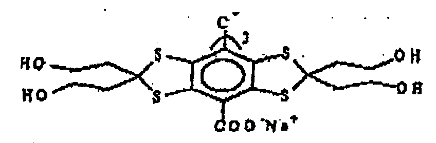
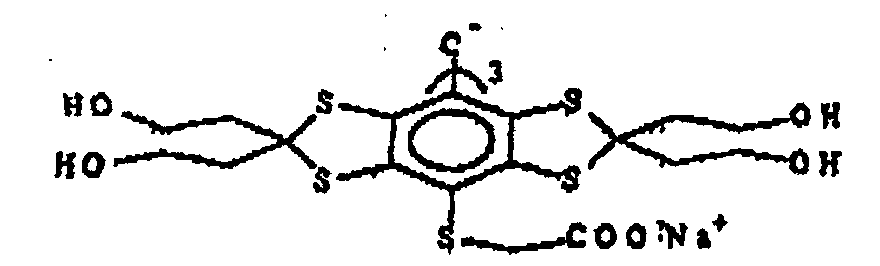
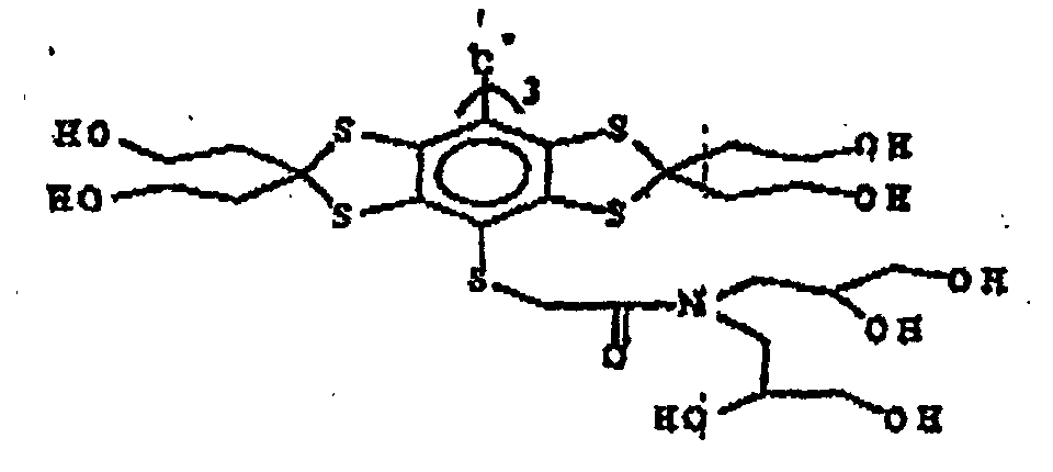 ou
ou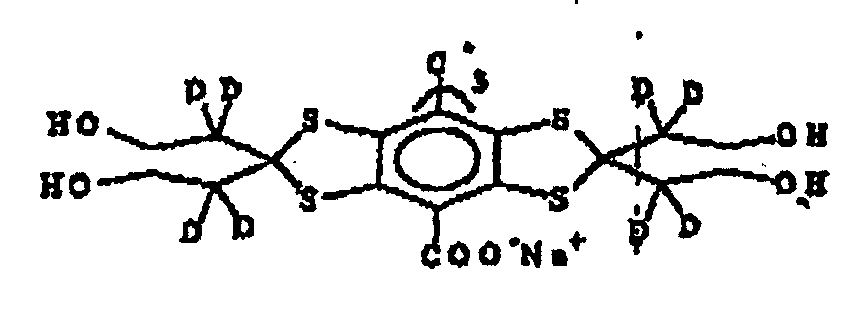

- Agent de contraste pour imagerie par résonance magnétique comprenant un composé radicalaire selon l'une quelconque des revendications 1 à 4 en même temps qu'au moins un support ou excipient pharmacologiquement acceptable.
- Procédé d'analyse d'un échantillon par résonance magnétique, ledit procédé comprenant l'introduction dans ledit échantillon d'un composé radicalaire selon l'une quelconque des revendications 1 à 4, l'exposition dudit échantillon à un premier rayonnement d'une fréquence choisie pour exciter les transitions de spin électronique dans ledit radical, l'exposition dudit échantillon à un second rayonnement à une fréquence choisie pour exciter les transitions de spin nucléaire dans des noyaux choisis dans ledit échantillon, la détection de signaux de décroissance d'induction libre, et, éventuellement, la génération d'une image ou de données d'écoulement dynamique à partir desdits signaux détectés.
- Procédé de détermination de la concentration en oxygène dans un échantillon, ledit procédé comprenant les étapes suivantes :introduire dans ledit échantillon une quantité efficace d'un composé radicalaire physiologiquement tolérable selon l'une quelconque des revendications 1 à 4;irradier ledit échantillon avec un rayonnement d'une amplitude et d'une fréquence choisies pour stimuler une transition de résonance de spin électronique dudit radical ; détecter les signaux de résonance magnétique renforcés par la résonance de spin électronique provenant dudit échantillon dans au moins les première, seconde et troisièmes conditions, ce par quoi dans lesdites première et seconde conditions ledit rayonnement est d'une première fréquence, dans lesdites troisièmes conditions ledit rayonnement est d'une seconde fréquence différente de ladite première fréquence, dans lesdites première, seconde et troisième conditions ledit rayonnement est d'une première, seconde et troisième amplitude, lesdites première et seconde amplitudes étant au moins différentes l'une de l'autre; etmanipuler lesdits signaux détectés pour déterminer ainsi la concentration d'oxygène dans ledit échantillon.
- Procédé tel que revendiqué à la revendication 7 comprenant le fait de:(a) introduire un composé radicalaire selon la revendication 1 dans un tissu corporel ou le système vasculaire;(b) générer une première image OMRI dudit échantillon à une puissance VHF PA, une période d'irradiation TVHF1 et en résonance;(c) générer une seconde image OMRI dudit échantillon à une seconde puissance VHF PB, un temps d'irradiation TVHF1 et en résonance;(d) générer une troisième image OMRI dudit échantillon à une puissance VHF PC, un temps d'irradiation TVHF1 et hors résonance ;(e) manipuler les images obtenues dans les étapes (b) à (d) et calibrer en utilisant des paramètres déterminés ex vivo pour obtenir une image d'oxygène dudit échantillon.
- Procédé selon la revendication 8 dans lequel une quatrième et cinquième images sont générées dans la séquence d'imagerie.
- Procédé de préparation d'un composé radicalaire selon l'une quelconque des revendications 1 à 4 comprenant la soumission d'un précurseur radicalaire correspondant à une étape de génération de radical et en modifiant éventuellement la substitution sur les fragments aryle.
- Utilisation d'un composé radicalaire selon l'une quelconque des revendications 1 à 4 en OMRI.
- Utilisation d'un composé radicalaire selon l'une quelconque des revendications 1 à 4 en oxymétrie.
Applications Claiming Priority (5)
| Application Number | Priority Date | Filing Date | Title |
|---|---|---|---|
| GB9704669 | 1997-03-06 | ||
| GBGB9704669.2A GB9704669D0 (en) | 1997-03-06 | 1997-03-06 | Free radicals |
| US882147 | 1997-06-25 | ||
| US08/882,147 US6013810A (en) | 1997-03-06 | 1997-06-25 | Free radicals |
| PCT/GB1998/000672 WO1998039277A1 (fr) | 1997-03-06 | 1998-03-06 | Radicaux triarylmethyle libres utilises comme agents d'amelioration d'image |
Publications (2)
| Publication Number | Publication Date |
|---|---|
| EP0966414A1 EP0966414A1 (fr) | 1999-12-29 |
| EP0966414B1 true EP0966414B1 (fr) | 2003-06-04 |
Family
ID=26311128
Family Applications (1)
| Application Number | Title | Priority Date | Filing Date |
|---|---|---|---|
| EP98910833A Expired - Lifetime EP0966414B1 (fr) | 1997-03-06 | 1998-03-06 | Radicaux triarylmethyle libres utilises comme agents d'amelioration d'image |
Country Status (5)
| Country | Link |
|---|---|
| EP (1) | EP0966414B1 (fr) |
| AU (1) | AU6506598A (fr) |
| DE (1) | DE69815305T2 (fr) |
| ES (1) | ES2201462T3 (fr) |
| WO (1) | WO1998039277A1 (fr) |
Families Citing this family (10)
| Publication number | Priority date | Publication date | Assignee | Title |
|---|---|---|---|---|
| NZ552898A (en) * | 2004-07-30 | 2011-02-25 | Ge Healthcare As | Method of producing a composition, composition and its use |
| ES2545377T3 (es) * | 2004-07-30 | 2015-09-10 | Ge Healthcare As | Radicales y su uso como agentes paramagnéticos en un proceso de polarización nuclear dinámica |
| NO338547B1 (no) * | 2004-07-30 | 2016-09-05 | Ge Healthcare As | Fremgangsmåte for fremstilling av en flytende sammensetning omfattende hyperpolarisert 13C-pyruvat, sammensetningen og anvendelse av den for fremstilling av hyperpolarisert 13C-pyruvat |
| JP5475995B2 (ja) * | 2005-12-01 | 2014-04-16 | ジーイー・ヘルスケア・アクスイェ・セルスカプ | 動的核分極(dnp)法 |
| EP2072061A1 (fr) | 2007-12-19 | 2009-06-24 | GE Healthcare Limited | Composition et procédé pour générer un profil métabolique par 13C-MR |
| US20110038802A1 (en) | 2008-05-02 | 2011-02-17 | Zhong-Min Hu | Method of determining alanine transaminase (alt) activity by 13c-mr detection using hyperpolarised 13c-pyruvate |
| US8697034B2 (en) | 2008-10-10 | 2014-04-15 | The Board Of Regents Of The University Of Texas System | Hyperpolarized 89-yttrium and methods relating thereto |
| US8968703B2 (en) * | 2009-09-10 | 2015-03-03 | Ge Healthcare Limited | 13C-MR detection using hyperpolarised 13C-fructose |
| FR2967158A1 (fr) * | 2010-11-08 | 2012-05-11 | Phosphoenix Sarl | Nouveaux radicaux triarylmethyle: leur preparation et application |
| DE102018126261A1 (de) | 2018-10-22 | 2020-04-23 | Universität Bielefeld | Tritylverbindungen vom Finland-Typ |
Family Cites Families (1)
| Publication number | Priority date | Publication date | Assignee | Title |
|---|---|---|---|---|
| US5728370A (en) * | 1993-04-02 | 1998-03-17 | Nycomed Imaging As | Free Radicals |
-
1998
- 1998-03-06 ES ES98910833T patent/ES2201462T3/es not_active Expired - Lifetime
- 1998-03-06 DE DE69815305T patent/DE69815305T2/de not_active Expired - Lifetime
- 1998-03-06 AU AU65065/98A patent/AU6506598A/en not_active Abandoned
- 1998-03-06 WO PCT/GB1998/000672 patent/WO1998039277A1/fr not_active Ceased
- 1998-03-06 EP EP98910833A patent/EP0966414B1/fr not_active Expired - Lifetime
Also Published As
| Publication number | Publication date |
|---|---|
| EP0966414A1 (fr) | 1999-12-29 |
| WO1998039277A1 (fr) | 1998-09-11 |
| ES2201462T3 (es) | 2004-03-16 |
| DE69815305D1 (de) | 2003-07-10 |
| AU6506598A (en) | 1998-09-22 |
| DE69815305T2 (de) | 2004-04-29 |
Similar Documents
| Publication | Publication Date | Title |
|---|---|---|
| EP0832054B1 (fr) | Radicaux methyle heterocycliques libres utilises comme agents d'amelioration d'image | |
| US5141740A (en) | Complexes and compositions for magnetic resonance imaging and usage methods | |
| US5242681A (en) | Lipophilic contrast agents for diagnostic image analysis | |
| EP0966414B1 (fr) | Radicaux triarylmethyle libres utilises comme agents d'amelioration d'image | |
| US5154914A (en) | Methods of diagnostic image analysis using lipophilic contrast agents | |
| US6256527B1 (en) | Method for determining oxygen concentration | |
| US5384108A (en) | Magnetic resonance imaging agents | |
| US5530140A (en) | Free radicals | |
| EP0871896B1 (fr) | Procede servant a determiner la concentration d'oxygene dans un echantillon | |
| US6013810A (en) | Free radicals | |
| US5217706A (en) | Complexes and compositions for magnetic resonance imaging | |
| US5264204A (en) | Hydrophilic free radicals for magnetic resonance imaging | |
| US6063360A (en) | Free radicals comprising benzodithiole derivatives | |
| CA2271735C (fr) | Agents d'imagerie du pool sanguin par resonance magnetique | |
| CA2096543A1 (fr) | Agents chelateurs derives de l'alkoxyamide pour la rmn | |
| KR19990044499A (ko) | 시료의 산소 농도 측정 방법 |
Legal Events
| Date | Code | Title | Description |
|---|---|---|---|
| PUAI | Public reference made under article 153(3) epc to a published international application that has entered the european phase |
Free format text: ORIGINAL CODE: 0009012 |
|
| 17P | Request for examination filed |
Effective date: 19990903 |
|
| AK | Designated contracting states |
Kind code of ref document: A1 Designated state(s): DE ES FR GB IT |
|
| RAP1 | Party data changed (applicant data changed or rights of an application transferred) |
Owner name: NYCOMED IMAGING AS |
|
| 17Q | First examination report despatched |
Effective date: 20020703 |
|
| GRAH | Despatch of communication of intention to grant a patent |
Free format text: ORIGINAL CODE: EPIDOS IGRA |
|
| GRAH | Despatch of communication of intention to grant a patent |
Free format text: ORIGINAL CODE: EPIDOS IGRA |
|
| GRAA | (expected) grant |
Free format text: ORIGINAL CODE: 0009210 |
|
| RAP1 | Party data changed (applicant data changed or rights of an application transferred) |
Owner name: AMERSHAM HEALTH AS |
|
| AK | Designated contracting states |
Designated state(s): DE ES FR GB IT |
|
| REG | Reference to a national code |
Ref country code: GB Ref legal event code: FG4D |
|
| REF | Corresponds to: |
Ref document number: 69815305 Country of ref document: DE Date of ref document: 20030710 Kind code of ref document: P |
|
| REG | Reference to a national code |
Ref country code: ES Ref legal event code: FG2A Ref document number: 2201462 Country of ref document: ES Kind code of ref document: T3 |
|
| ET | Fr: translation filed | ||
| PLBE | No opposition filed within time limit |
Free format text: ORIGINAL CODE: 0009261 |
|
| STAA | Information on the status of an ep patent application or granted ep patent |
Free format text: STATUS: NO OPPOSITION FILED WITHIN TIME LIMIT |
|
| 26N | No opposition filed |
Effective date: 20040305 |
|
| REG | Reference to a national code |
Ref country code: DE Ref legal event code: R082 Ref document number: 69815305 Country of ref document: DE Representative=s name: J D REYNOLDS & CO., GB |
|
| REG | Reference to a national code |
Ref country code: FR Ref legal event code: PLFP Year of fee payment: 19 |
|
| REG | Reference to a national code |
Ref country code: FR Ref legal event code: PLFP Year of fee payment: 20 |
|
| PGFP | Annual fee paid to national office [announced via postgrant information from national office to epo] |
Ref country code: FR Payment date: 20170327 Year of fee payment: 20 |
|
| PGFP | Annual fee paid to national office [announced via postgrant information from national office to epo] |
Ref country code: GB Payment date: 20170327 Year of fee payment: 20 |
|
| PGFP | Annual fee paid to national office [announced via postgrant information from national office to epo] |
Ref country code: IT Payment date: 20170324 Year of fee payment: 20 |
|
| PGFP | Annual fee paid to national office [announced via postgrant information from national office to epo] |
Ref country code: DE Payment date: 20170329 Year of fee payment: 20 |
|
| PGFP | Annual fee paid to national office [announced via postgrant information from national office to epo] |
Ref country code: ES Payment date: 20170328 Year of fee payment: 20 |
|
| REG | Reference to a national code |
Ref country code: DE Ref legal event code: R071 Ref document number: 69815305 Country of ref document: DE |
|
| REG | Reference to a national code |
Ref country code: GB Ref legal event code: PE20 Expiry date: 20180305 |
|
| PG25 | Lapsed in a contracting state [announced via postgrant information from national office to epo] |
Ref country code: GB Free format text: LAPSE BECAUSE OF EXPIRATION OF PROTECTION Effective date: 20180305 |
|
| REG | Reference to a national code |
Ref country code: ES Ref legal event code: FD2A Effective date: 20200723 |
|
| PG25 | Lapsed in a contracting state [announced via postgrant information from national office to epo] |
Ref country code: ES Free format text: LAPSE BECAUSE OF EXPIRATION OF PROTECTION Effective date: 20180307 |