EP0885018B1 - Procede permettant d'inactiver des contaminants dans un liquide biologique et appareil correspondant - Google Patents
Procede permettant d'inactiver des contaminants dans un liquide biologique et appareil correspondant Download PDFInfo
- Publication number
- EP0885018B1 EP0885018B1 EP97911794A EP97911794A EP0885018B1 EP 0885018 B1 EP0885018 B1 EP 0885018B1 EP 97911794 A EP97911794 A EP 97911794A EP 97911794 A EP97911794 A EP 97911794A EP 0885018 B1 EP0885018 B1 EP 0885018B1
- Authority
- EP
- European Patent Office
- Prior art keywords
- light
- container
- light source
- biological fluid
- containers
- Prior art date
- Legal status (The legal status is an assumption and is not a legal conclusion. Google has not performed a legal analysis and makes no representation as to the accuracy of the status listed.)
- Expired - Lifetime
Links
Images




Classifications
-
- A—HUMAN NECESSITIES
- A61—MEDICAL OR VETERINARY SCIENCE; HYGIENE
- A61M—DEVICES FOR INTRODUCING MEDIA INTO, OR ONTO, THE BODY; DEVICES FOR TRANSDUCING BODY MEDIA OR FOR TAKING MEDIA FROM THE BODY; DEVICES FOR PRODUCING OR ENDING SLEEP OR STUPOR
- A61M1/00—Suction or pumping devices for medical purposes; Devices for carrying-off, for treatment of, or for carrying-over, body-liquids; Drainage systems
- A61M1/36—Other treatment of blood in a by-pass of the natural circulatory system, e.g. temperature adaptation, irradiation ; Extra-corporeal blood circuits
- A61M1/3681—Other treatment of blood in a by-pass of the natural circulatory system, e.g. temperature adaptation, irradiation ; Extra-corporeal blood circuits by irradiation
-
- A—HUMAN NECESSITIES
- A61—MEDICAL OR VETERINARY SCIENCE; HYGIENE
- A61M—DEVICES FOR INTRODUCING MEDIA INTO, OR ONTO, THE BODY; DEVICES FOR TRANSDUCING BODY MEDIA OR FOR TAKING MEDIA FROM THE BODY; DEVICES FOR PRODUCING OR ENDING SLEEP OR STUPOR
- A61M1/00—Suction or pumping devices for medical purposes; Devices for carrying-off, for treatment of, or for carrying-over, body-liquids; Drainage systems
- A61M1/36—Other treatment of blood in a by-pass of the natural circulatory system, e.g. temperature adaptation, irradiation ; Extra-corporeal blood circuits
- A61M1/3681—Other treatment of blood in a by-pass of the natural circulatory system, e.g. temperature adaptation, irradiation ; Extra-corporeal blood circuits by irradiation
- A61M1/3683—Other treatment of blood in a by-pass of the natural circulatory system, e.g. temperature adaptation, irradiation ; Extra-corporeal blood circuits by irradiation using photoactive agents
-
- A—HUMAN NECESSITIES
- A61—MEDICAL OR VETERINARY SCIENCE; HYGIENE
- A61L—METHODS OR APPARATUS FOR STERILISING MATERIALS OR OBJECTS IN GENERAL; DISINFECTION, STERILISATION OR DEODORISATION OF AIR; CHEMICAL ASPECTS OF BANDAGES, DRESSINGS, ABSORBENT PADS OR SURGICAL ARTICLES; MATERIALS FOR BANDAGES, DRESSINGS, ABSORBENT PADS OR SURGICAL ARTICLES
- A61L2/00—Disinfection or sterilisation of materials or objects, in general; Accessories therefor
- A61L2/02—Disinfection or sterilisation of materials or objects, in general; Accessories therefor using physical processes
-
- A—HUMAN NECESSITIES
- A61—MEDICAL OR VETERINARY SCIENCE; HYGIENE
- A61L—METHODS OR APPARATUS FOR STERILISING MATERIALS OR OBJECTS IN GENERAL; DISINFECTION, STERILISATION OR DEODORISATION OF AIR; CHEMICAL ASPECTS OF BANDAGES, DRESSINGS, ABSORBENT PADS OR SURGICAL ARTICLES; MATERIALS FOR BANDAGES, DRESSINGS, ABSORBENT PADS OR SURGICAL ARTICLES
- A61L2103/00—Materials or objects being the target of disinfection or sterilisation
- A61L2103/05—Living organisms or biological materials
-
- A—HUMAN NECESSITIES
- A61—MEDICAL OR VETERINARY SCIENCE; HYGIENE
- A61L—METHODS OR APPARATUS FOR STERILISING MATERIALS OR OBJECTS IN GENERAL; DISINFECTION, STERILISATION OR DEODORISATION OF AIR; CHEMICAL ASPECTS OF BANDAGES, DRESSINGS, ABSORBENT PADS OR SURGICAL ARTICLES; MATERIALS FOR BANDAGES, DRESSINGS, ABSORBENT PADS OR SURGICAL ARTICLES
- A61L2103/00—Materials or objects being the target of disinfection or sterilisation
- A61L2103/05—Living organisms or biological materials
- A61L2103/09—Blood or products thereof
Definitions
- the present invention relates to methods and apparatus for treating biological fluids such as blood and blood components. More particularly, the present invention relates to methods and apparatus for inactivating contaminants, such as viruses, in such biological fluids.
- Plasma is a non-cellular component of blood and is the liquid medium in which the cellular components are suspended. Plasma also includes various other components such as proteins (e.g. fibrinogens, albumin and globulins), electrolytes and metabolites.
- proteins e.g. fibrinogens, albumin and globulins
- viruses such as hepatitis or HIV virus
- the viruses residing in blood may be "intracellular,” i.e. contained within one of the cellular components of blood, such as white blood cells, or they may be "extracellular” i.e. freely existing in the plasma.
- the hepatitis virus is primarily an extracellular virus
- the cytomegalovirus the virus responsible for herpes
- the HIV virus the virus responsible for AIDS
- the presence of the virus in the bloodstream poses the risk of infection and disease not only to the host, but also, if the blood or a blood component is collected and transfused, to a recipient.
- the medical community has attempted to reduce the risk of transfusing blood that is tainted with active virus by developing methods and apparatus to remove virus from the blood stream or otherwise inactivate the virus.
- one such attempt involves the filtration of blood and/or blood components to remove intracellular viruses entrained, for example, in white blood cells, Rawal et al., "Reduction of human immunodeficiency virus-infected cells from donor blood by leukocyte filtration," Transfusion, pp. 460-462 (1989).
- filtration of blood and/or blood components has been somewhat effective in removing the intracellular viruses, it has been generally ineffective in the removal of extracellular viruses because such viruses are typically too small to be captured by currently commercially available filters.
- a more recent approach to viral inactivation is the treatment of blood or blood components with a photochemical agent and light .
- the photochemical agent When activated by light of an appropriate wavelength, the photochemical agent either kills the virus directly or indirectly inhibits the ability of the virus to replicate and, thus, in either case "inactivates” the virus.
- the term "inactivate” (and forms thereof) mean the actual destruction, eradication of a contaminant such as a virus, or a direct or indirect effect on the contaminant that inhibits its ability to replicate or otherwise to adversely affect a living recipient.
- photochemical agents have been used or disclosed for use in inactivating viruses in blood. They include, for example, psoralens (which have been used in the inactivation of viruses in collected platelets) as described in U.S. Patent No. 5,459,030.
- Other photochemical agents that have been disclosed for the inactivation of viruses in blood include the family of light activated drugs derived from benzoporphyrin, as described in U.S. Patent No. 5,536,238, which is assigned to the assignee of the present application.
- Still other photochemical agents considered for use in the inactivation of viruses in biological fluids are compounds from the family of phenothiazine dyes, which include, but are not limited to toluidine blue O, azure A, azure B, azure C, thionine methylene blue, and methylene green.
- the light applied to the photochemical agent must be of a wavelength that can be absorbed by the photochemical agent.
- U.S. Patent no. 5,527,704 also assigned to the assignee of the present application, in the case of methylene blue, it is known that methylene blue absorbs light having wavelengths of between about 550 and 700 nm.
- a methylene blue molecule that has been activated by light becomes a catalyst for secondary and tertiary reactions that inactivate virus. More specifically, activation of the photochemical agent such as methylene blue is believed to result in the production of singlet oxygen which enhances the secondary and tertiary reactions.
- activation of the photochemical agent such as methylene blue is believed to result in the production of singlet oxygen which enhances the secondary and tertiary reactions.
- photochemical agents when activated by light
- therapeutic proteins include any biologically active protein which has qualities which make it useful in the treatment of medical disorders.
- proteins include human-blood plasma proteins such as factor VIII, Von Willebrand factor, factor IX, factor X, factor XI, Hageman factor, the activated forms of such factors, prothrombin, anti-thrombin III, fibronectin, plasminogen, immune serum globulin, modified immune globulin, albumin, 1-antitrypsin, and prekallikrein.
- factor VIII Von Willebrand factor
- factor IX factor X
- factor XI factor XI
- Hageman factor the activated forms of such factors
- prothrombin anti-thrombin III
- fibronectin plasminogen
- immune serum globulin plasminogen
- modified immune globulin modified immune globulin
- albumin 1-antitrypsin
- prekallikrein prekallikrein
- U.S. Patent No. 5,300,019 assigned to the assignee of the present application, an apparatus for treating a fluid containing a biological contaminant with a photochemical agent is described.
- a contaminant such as a virus
- a photochemical agent is pumped from a source container through a treatment chamber to a collection container. While in the treatment chamber, the blood is exposed to a light source that activates the photochemical agent as the blood is processed within the treatment chamber. To ensure that the blood is sufficiently and uniformly exposed to the light source, the blood (with contaminants and photochemical agent) is continuously mixed within the treatment chamber. After treatment, the blood is collected in the collection container.
- the photochemical agent described in that patent is benzoporphyrin.
- a single container of blood or a blood component is placed between two facing arrays of light emitting diodes .
- the container includes a blood component (plasma), a viral contaminant and a quantity of methylene blue.
- the container is irradiated by the light emitting diodes which produce light having a wavelength of approximately 620-670 nm to activate methylene blue.
- the container of blood or blood component is subjected to light for a period of approximately five (5) minutes.
- one of the prime areas of concern is to better assure inactivation of a substantial portion of the Contaminant with the goal being 100% inactivation. More specifically, it is desirable that exposure of the biological fluid to a light source provide maximum viral inactivation with minimal damage to the therapeutic proteins. Also, it is desirable that exposure of the biological fluid to a light source be of short duration to increase efficiency of treatment and reduce the cost. It is further desirable that the exposure of the biological fluid to the light source be substantially uniform in its application and preferably without the need to continuously mix the blood and/or blood components. Because biological fluids such as blood or blood components are often collected or stored in plastic containers, it is further desirable to be able to treat more than one unit or container at a time to improve efficiency, but without adverse effect on the viral inactivation.
- the present invention generally is embodied in method and apparatus for inactivating contaminants in a biological fluid.
- the apparatus includes at least one wall defining a chamber and a quantity of biological fluid including one or more contaminants within the chamber.
- the biological fluid also includes a photochemical agent.
- the photochemical agent is operable to cause the inactivation of at least some of the contaminants upon exposure to a light source.
- the apparatus includes a high intensity light source providing an intensity of at least 30 mW/cm 2 . At least a portion of the wall of the chamber permits the light emitted by the light source to contact the fluid.
- the biological fluid may comprise a component of blood, such as blood plasma, and the photochemical agent is a phenothiazine dye such as methylene blue.
- the light source may comprise a sodium light, and a control system may operably be connected to the light source and one or more sensors for providing that the light contacting the biological fluid is between about 1 and 100 Joules/cm 2 .
- the chamber may be a flexible plastic container, one or more of which are carried by a carriage relatively rotatable around the light source.
- the light source may be elongated to provide light along the length of source, in contrast, for example, to a point source light.
- the apparatus for treating a biological fluid with light may apparatus include a housing defining an interior chamber and a light source disposed within the chamber.
- the housing includes a reflective interior surface and is adapted to hold a quantity of biological fluid in the chamber between the light source and the reflective interior surface of the housing. Light passes through the fluid directly from the source and upon reflection from the interior surface.
- the method for substantially inactivating contaminants in the biological fluid that includes a photochemical agent and other compounds includes contacting the fluid with light having an intensity operative to activate the photochemical agent and substantially at least about 30 mW/cm 2 to inactivate the contaminants.
- the biological fluid includes other compounds which, preferably, are not adversely affected by the light source.
- the amount of light contacting the biological fluid and the time of contact are monitored and, if desired, controlled. This invention finds particular application when the biological fluid is blood plasma and the photochemical agent is methylene blue.
- a method for activating methylene blue includes the steps of providing a quantity of methylene blue and contacting the methylene blue with light having an intensity of at least 30 mW/cm 2 .
- the methylene blue may be disposed within a biological fluid and may be contacted by the light for a period between approximately 0.3 and 30 minutes.
- light box 10 includes a generally cylindrical housing_12 having a top portion or cover 14, a midportion 16, and a bottom portion 18.
- the midportion may include a control panel 20 for operator control of light box operation.
- a door 22 in the midportion allows access into the interior of the light box 10.
- Door 22 may be either hinged or, as shown in Fig. 1 slidable.
- Light box 10 may also include retractable spill tray (not shown) for collecting spilled liquid from within the light box 10.
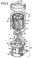
- Figure 2 is an exploded view of the light box 10 and shows its basic component parts.
- the illustrated light box 10 includes a base assembly 23, which supports a centrally located light source 24 and shutter assembly 26.
- a light transparent tube 28 rests on the base assembly and supports, at its upper end, a carriage assembly 30 for rotation about the transparent tube and light source.
- the carriage assembly is a generally cylindrical hexagonal framework for holding at least one substantially clear plastic container 32 containing biological fluid to be treated.
- the outer housing 12 and particularly the top and mid portions enclose the carriage and light source.
- the base assembly has a lowermost base plate 34 and a raised platform 36 supported above the base plate by rigid vertical support members 38.
- the space between the base plate and platform permits retraction of the shutter assembly 26, provides room for mounting cooling fans 39a and 39b for cooling different regions of the light box, and contains the mounting socket for the light source 24.
- the light source 24 is generally centrally mounted on the base assembly 23 and more specifically on platform 36.
- the light source includes preferably an elongated tube to provide uniform illumination of the interior of the light box 10.
- Light source 24 may be any lamp or bulb, preferably capable of emitting a high intensity light.
- light source 24 may be any lamp or bulb that can provide light with a wavelength and intensity effective (a) to inactivate contaminants in the biological fluid to be treated, or more precisely (b) to activate the photochemical agent (if any) used in treating the biological fluid.
- the photochemical agent is methylene blue
- the light source should be capable of providing more than 25% of its light in the visible spectrum in a wavelength range of approximately 550-700 nm to provide better efficiency and lower heat generation.
- light source 24 should provide a high intensity light that is capable of providing maximum activation of the photochemical agent without significantly harming other desirable components in the biological fluid.
- high intensity as used herein means an intensity of at least 30 mW/cm 2 as measured at the biological fluid or container thereof.
- the intensity should be at least 30 mW/cm 2 and, preferably between 85-130 mW/cm 2 measured at the biological fluid (or container thereof) and in a wavelength range from 550-700 nm.
- other photochemical agents may be operable at different intensities and wavelengths.
- Examples of light sources that can be used to activate photochemical agents include high pressure sodium lamps, halogen lamps, sulphur lamps, metal halide lamps or xenon lamps.
- One such lamp is the high pressure sodium vapor lamp that includes a ceramic are tube, such as alumina (Al 2 O 3 ), in a clear or coated outer bulb with a medium or mogul screwbase for mounting into socket (in base assembly 23).
- alumina Al 2 O 3
- Such a sodium lamp is sold by Phillips Lighting under the tradename Ceramalux, Model No. C1000S52.
- light box 10 includes retractable shutter assembly 26 that encloses the light source 24 during, for example, loading and start-up procedures and retracts after loading is complete and the light is fully energized.
- the shutter assembly 26 comprises a cylindrical tube 41 that, when raised, shields the light source.
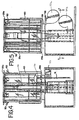
- the tube has an upper radial flange 26a which mounts to the shutter arms 51. Movement of the shutter is effected by shutter drive motor 53 as shown in Fig. 4, in a rack and pinion gear.
- the top of shutter 26 includes a top flange 26a that, when shutter 26 is retracted covers most of the portion of platform 36 within tube 28.
- the upper surface of 26a is reflective to further distribute light to the interior of the light box.
- the flange is preferably coated with a highly reflective material, such as white 91 sold by Alcoa Brite Products.
- light box includes cylindrical tube 28, attached to the top of platform 36 of base assembly 23.
- tube 28 separates the interior of light box 10 into a light source region 40 and a fluid treatment region 42 between the tube 28 and the housing 12.
- tube 28 encloses light source 24 and retractable shutter 26.
- Tube 28 is made of any material substantially transparent to the light emitted by the light source and should be made of a material that is heat tolerant. Suitable materials with good light transmission and thermal properties include many of the acrylic polymers, although other materials may also be used.
- the interior (or exterior) surface of tube 28 may be made of or coated with a material that is translucent to the desired wavelengths but reflective as to undesired wavelengths.
- the surface of tube 28 may include a material that removes ultraviolet (UV) or other light that is unnecessary for activation of the photochemical agent.
- the surface of tube 28 may include a material that removes infrared light which would otherwise create undesired heat (that will heat the biological fluid).
- a material includes a film consisting of a PET substrate onto which metallic layers are sputtered. Such a film is marketed by Southwall Technologies under the name Altair ALT-M-20.
- the top of tube 28 includes a motor and carriage mounting plate 44, as shown in Fig. 2.
- the mounting plate 44 is largely open to allow heat generated within the light source region 40 to exit out the top of tube 28 (and through a vent in the housing).
- drive motor is suspended from the mounting plate 44.
- the shaft of the drive motor extends through plate 44 and is attached to an upper frame piece of the carriage 30 to rotate the carriage about its central axis 48.
- Motor 46 is connected to a power supply via connection wires (not shown).
- motor 46 may be located elsewhere.
- motor 46 may be located, for example, between base plate 34 and platform 36 and the carriage may be mounted on a large bearing ring on the platform 36 for rotation about the light source.
- the motor in this embodiment, could drive the carriage via gears or drive belts as one skilled in the art would understand.
- motor 46 should be of the type that allows for precise control of the motor functions and, more specifically, allows for incremental rotation of carriage assembly 30.
- motors are step motors. Step motors are well known to those of ordinary skill in the art and available from manufacturers such as Applied Motion Products of Watsonville, CA.
- carriage 30 For holding bags of biological fluid, such as blood plasma, the carriage, generally at 30, is located within housing 12.
- carriage 30 includes a top bracket 25a and a bottom bracket 25b and vertical framing 25c therebetween, to form a generally cylindrical framework for supporting bags of biological fluid to be treated.
- brackets 25a and 25b are hexagonal, providing a hexagonal cylindrical framework, with each side of the hexagon defining a bag receiving area on the framework.
- carriage assembly 30 including its top bracket 25a, bottom bracket 25b and vertical framing 25c may form other regular polygonal structures to accommodate a different number of containers.
- the cylindrical framework may be triangular, octagonal or other shape).
- the top bracket 25a includes hooks 50 for suspending plastic containers or bags of biological fluid such as blood plaema or other blood components.
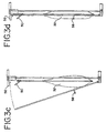
- a hook 50 is located at each of the hexagonal sides of the carriage, allowing flexible plastic containers 32 (shown in Fig. 3-3(a-d) of a biological fluid such as blood plasma to be hung within the bag receiving area.
- a pair of fixed vertical bars 52 expend between top and bottom brackets 25a and 25b in each bag receiving area to provide inner rests, against which the container 32 of biological fluid may be pressed to distribute the fluid more evenly within the container for treatment.
- each bag receiving area of carriage 30 includes a bag holder 54 for compressing the bag to more evenly distribute fluid for treatment.
- the bagholder 54 includes a wire frame, hinged to the bottom bracket 25b for opening and closing.
- the top of each bag receiving area (above hook 50) includes a latch 56 for holding the holder 54 in a closed position.
- Container 32 is made of any translucent material that is suitable for storing a biological fluid, such as blood or blood components and a photochemical agent.
- the container 32 is made of a plastic such as a polymeric material or a polymeric blend.
- Containers useful in the present invention are described in U.S. Patent No. 5,514,106, U.S. patent Application Serial No. 08/121,820, also incorporated by reference and/or U.S. Patent Application entitled "Systems and Methods for Removing Viral Agents from Blood' in the name of Robert Herman, John Chapman, Sun Chong-Sun, Jean M. Mathias, Veronique Mayadoun, Serge Degheidere and Daniel Bischof, filed on October 28, 1996
- carriage 30 can accommodate up to six containers (one container within each compartment) of biological fluid.
- carriage 30 can have as many or as few bag receiving areas as possible and/or necessary.
- carriage 30 can be removed from platform 36 and replaced with a carriage capable of holding, for example, three larger containers or more (e.g. 8) smaller containers.
- carriage 30 also includes wedged or V-shaped reflectors 60 along vertical members 25c and spaced between each of the bag receiving areas for reflecting light from light source 24 (and that has been transmitted through containers 32).
- surfaces 62 of reflectors 60 are angled toward the interior of light box 10 and, more specifically, with the vertex of the wedge aligned toward light source 24.
- the surface 62 of reflector 60 may be a composite of two or more angles or may be a generally smooth concave surface. Reflectors 60 and, more particularly, surfaces 62 may be coated, covered or made of any highly reflective material that, does not significantly diminish the intensity of the light and/or light energy reflected back onto containers.
- Everbrite 95 sold by Alcoa Brite products.
- Everbrite 95 includes a highly reflective layer of silver sputtered on a layer of polyethylene terephthalate (PET) film.
- PET polyethylene terephthalate
- the treated PET film is then bonded to an aluminum or steel substrate.
- carriage assembly 30 is enclosed by housing 12.
- the interior surface 64 of housing 12 (shown in Fig. 2) is also coated, covered or made of a highly reflective surface similar or identical to the material used for reflectors 60 as described above. Having the interior surface made of a highly reflective surface allows light from light source 24 (and transmitted through container 32) to be reflected back onto the container.
- the highly reflective nature of the inner surfaces reflects light back at the containers with minimum reduction in its intensity. This helps to provide for substantially uniform exposure of the fluid, as well as uniform illumination from container to a container.
- light box 10 includes fans 39a and 39b.
- Fan 39b blows cool air through the tube 28 and the into light source region to cool light source 24.
- Fan 39a blows cool air through fluid treatment region 42 to prevent undue heating of the containers 32, as they are rotated by carriage 30.
- a programmable computer-based control system may be used to control the operation of the light box 10.
- the system tests, monitors and controls various aspects of the light box operation such as the start-up, container loading, container treatment and container unloading stages of the light box operation.
- the various stages may be initiated by the operator through control panel 20 or automatically by the control system.
- the control system tests the operation of the light source to determine whether it is functioning properly.
- control system activates light source 24, raises shutter 26 and, after a warm up period, measures the energy generated by the light source. (Alternatively, the energy generated by the light source may be measured with shutter 26 lowered).
- light source 24 is turned off and shutter 26 is lowered. (Alternatively, light source may remain on and be covered by shutter 26 until the time of the container treatment phase.) Based on the light energy readings, an estimated treatment time may be calculated by the control system. If the control system determines that the light box components are functioning properly, the operator may instruct the control system (or the control system may do so automatically) to proceed to the "Container Loading" phase of the system operation.
- the control system determines the carriage type and the number of bags to be processed.
- carriage 30 may be automatically advanced to allow for balanced loading of the carriage. For example, if carriage is designed to hold 6 containers, the carriage will advance in such a way as to evenly distribute the containers 32 around the carriage 30. Thus, if for example, only 4 containers are to be treated on a carriage capable of holding six containers, the carriage will allow for placement of the first container in a first position and will then automatically advance to a position directly opposite the first position. The carriage will then allow for loading of the third container and advance to a position directly opposite the third container for loading the fourth container.
- Balancing the carriage as described above makes it more likely that during the illumination cycle each of the containers will receive a substantially uniform quantity of light and none of the containers will be substantially blocked from the light by adjacent containers. It also reduces undue wear on the motor and bearings due to unbalanced loads.
- the system may also check and record the particulars of the container and, if the biological fluid is blood or a blood component, check that the blood or blood component is appropriate for treatment (e.g. that the component is blood plasma).
- the containers may include bar codes or other identifiers that identify the product codes and lot numbers of the container and other specifics about the blood donation (e.g. type of component). If the control system recognizes an invalid code, lot number or other defect, it marks the container and/or alerts the operator.
- the operator may instruct the system to proceed to the next phase, the Container Treatment phase.
- the Container treatment phase the instrument again goes through a sequence of tests to ensure that its component parts, such as the light source, are functioning properly. If the light source has been turned off (after the start-up phase), the light source is activated and allowed to reach its required intensity before shutter 26 is lowered. This prevents containers 32 from being exposed to a light that has not reached its desired intensity and that may be spectrally unstable or undesirable.
- Light box 10 includes, as part of its control system, sensors 66 and 68 for monitoring the amount of light being emitted by the light source 24 and the amount of light transmitted through the biological fluid in containers 32.
- sensor 66 may be located on or near tube 28 and measures and monitors the amount of light contacting the biological fluid directly from the light source 24.
- a second sensor 68 may be placed on or near the interior surface of the housing 12 to measure and monitor the amount of light transmitted through the biological fluid for reflection back onto containers 32.
- the control system may utilize one sensor as the primary sensor and a second sensor as a back up or check.
- control system may monitor the treatment of the biological fluid by measuring the amount of time the biological fluid is exposed to the light from the light source and calculating the amount of energy received by the biological fluid. Also, the control system may monitor the treatment of the biological fluid in each container on a container per container basis, or may monitor the entire treatment process and arrive at an average treatment profile for the containers .
- the control system may be preprogrammed to determine if the amount of light emitted directly by the light source and transmitted through containers 32 is sufficient to effectively treat the biological fluid. Thus, if the light box is being used to inactivate virus in blood or a blood component with a photochemical agent, the control system may be preprogrammed to determine if the amount of light is within the range required to activate the photochemical agent or, in other words, to inactivate the contaminants.
- the control system may be preprogrammed to determine whether the light energy contacting the container (from both sides) is within the intended exposure range. If sensors 66 and 68 determine that one or more of the containers has not been sufficiently illuminated, the treatment time may be extended to ensure that the deficient container receives additional treatment, but without overexposing the remaining containers that may be within the preferred intensity range. If providing further treatment to the deficient container would result in overexposure of the remaining containers, no further treatment will be provided and the control system will mark the deficient container or otherwise alert the operator that the deficient container has not been sufficiently treated.
- Light box 10 described above may be used for treating any fluids with light, but is particularly useful in the treatment of blood or other biological fluids with light and, more specifically, for the viral inactivation of blood and blood components.
- Light box 10 allows for multiple containers or units of biological fluid to be treated at the same time. This results in lower cost per unit treated. The ability to treat several containers or units of biological fluid provides a savings to the treatment center, as fewer instruments for treating a given amount of biological fluid will be required. Light box 10 is fairly compact (approximately 32 inches high x 30 inches wide by 18 inches) thus, requiring less space and making it more convenient to place in different locations.
- the operation of the light box 10 will be described in the context of treating blood with light for the purpose of inactivating viruses in the blood, it is to be understood that the invention is not limited to the specific example described below, nor is it limited to viral inactivation of blood or blood components.
- containers 32 of biological fluid containing a photochemical agent and contaminants are placed on carriage 30 of the light box 10.
- contaminants refers to any harmful biological material, and particularly includes bacteria, parasites and viruses.
- containers 32 may be hung from hooks 50 of carriage 30 and secured by bag holder 54.
- retractable shutter 26 be in the closed position to shield the operator and the containers 32 from the light source before it has reached its desired intensity. Shielding the containers from the light source as it is being activated and as the containers are placed on the carriage is another way to ensure uniform treatment of the containers by preventing some of the containers from receiving some light before the others have been placed on the carriage.
- the shutter also shields the containers from a light source that might be spectrally unstable.
- a spectrally unstable light source may cause erroneous readings in the useful (i.e. energy in the absorption band of the photochemical agent) energy received at the container.
- Carriage 30 is rotated around its central axis 48. for a predetermined period of time.
- the typical time of exposure of the biological fluid may be anywhere between 0.3 and 30 minutes or, more preferably 1 to 5 minutes.
- the time of exposure will depend on the amount of light energy contacting the containers, and the fluid therein.
- the fluid to be treated is blood plasma and the photochemical agent is methylene blue combined in the ratios below
- Rotation of carriage 30 also provides for uniform treatment of the biological fluid. Rotation of carriage 30 ensures that each container will be exposed to every part of the fluid treatment region 42 for similar duration. Rotation also ensures that the each container is uniformly cooled by, for example, fan 39a.
- a high intensity light with a biological fluid such as blood or blood plasma containing a selected amount of methylene blue enhances the virucidal effect of the methylene blue.
- a biological fluid such as blood or blood plasma containing a selected amount of methylene blue
- use of the high intensity light to contact the biological fluid increases the number of photons contacting the methylene blue molecule per unit of time and, if those photons are of energies corresponding to the absorption wavelengths (in free space) of methylene blue, increases the production of the singlet oxygen which causes the secondary reactions responsible for inactivating the viruses in, for example, blood plasma.
- the preferred ratio of methylene blue to blood plasma is between about 1:20 to 1:35, but may be 1:200 to 1:350.
- approximately 1-10 ml of methylene blue may be used when the volume of plasma is generally between 200 and 350 ml.
- the concentration of methylene blue can be approximately between 1 to 10 ⁇ M and, preferably, approximately 1 ⁇ M.
- methylene blue is activated by light having wavelengths of between approximately 550 and 700nm with a peak at 663 nm. Absorbance of light in this range is understood to provide for activation of methylene blue without any significant negative effect on plasma or plasma proteins (which absorb light primarily between 300 to 560 nm).
- the light intensity should be greater than 30 mW/cm 2 at the biological fluid or container thereof, and preferably between 85-130 mW/cm 2 measured at the biological fluid (or container thereof) and in the wavelength range from 550-700 nm.
- units of fresh frozen plasma spiked with pseudorabies virus were processed through a leukoreduction filter and illuminated with a high pressure sodium light in a prototype light box similar in substantial respects to light box 10.
- PRV pseudorabies virus
- a single pack of plasma including 310 ml and plasma spiked with PRV (1:10 spike) and 10 ml of methylene blue was irradiated with a high pressure sodium light having an intensity of approximately 119 mW/cm 2 when measured in the absorption range of 550-700 (which corresponds to 137 mW/cm 2 when measured across the band of approximately 350-800 nm) for up to eight minutes.
- Levels of virus were measured at .5, 1.0, 2.0, 4.0 and 8.0 minute intervals using the conventional plaque assay.
- levels of virus in 5 ml aliquots were also measured at the above time intervals.
- the apparatus and method of the present invention also provides more effective viral kill as compared to other apparatus or methods that can provide the same or similar amount of energy (often expressed in Joules/cm 2 or J/cm 2 ).
- Energy is the product of light intensity and time.
- increasing the intensity results in a greater biological effect (such as viral kill and/or recovery of therapeutic protein) than does an increase in the time of exposure. Stated differently, a greater biological effect is obtained at an energy level where the intensity has been increased than at substantially the same energy level where the time of exposure has been increased.
- units of blood plasma (approximately 300-310 ml per unit) was spiked with pseudorabies virus to achieve an approximate final virus load of 6 x 10 5 PFU/ml.
- the plasma units were filtered to remove intracellular contaminants and collected into treatment containers containing 10 ml of methylene blue in each container.
- the plasma with methylene blue was illuminated with a high pressure sodium light providing an intensity of approximately 120 mW/cm 2 as measured at the container in the absorption range of 550-700 nm (which corresponds to 140 mW/cm 2 at the biological fluid or container thereof when measured across the band of approximately 350-800 nm).
- the amount of remaining virus was measured at different energy levels. The results are shown in Table 2.
- Another batch of plasma units (approximately 300-315 ml per unit) were also spiked with pseudorabies virus to achieve an approximate final virus load of 1 x 10 6 PFU/ml. These units of plasma were filtered to remove intracellular contaminants and collected into similar treatment containers containing 10 ml of methylene blue in each bag to achieve 1 ⁇ M concentration. The plasma units with methylene blue were illuminated with a white fluorescent light providing an intensity of approximately 24 mw/cm 2 at the at the container when measured across the band of 350-500 nm). Using the conventional plaque assay, the amount of remaining virus was measured at different energy levels. The results are shown in Table 2 .
- the intensities and doses were measured in the range from 350-800 nm. If these intensities and doses were measured over the absorption spectrum of methylene blue (550-700), the intensity for sodium light would be 121 mW/cm 2 and the doses would be, from top to bottom of the left hand column, 0, 1.7, 7, 14 and 28. Similarly, for white fluorescent light the intensity would be 12 mW/cm 2 and the doses would be, from top to bottom of column 3, 0, 0.5, 2.5, 7.5 and 15.
- the log reduction of virus was (LRV) was approximately 4.8 as compared to greater than 6.2 logs at an energy level of approximately 14 J/cm 2 as measured at the container in the absorption range of 550-700 nm (which corresponds to 16 J/cm 2 when measured across the band of approximately 350-800 nm) with the high pressure sodium light.
- the apparatus and method of the present invention may also allow for less damage to the therapeutic proteins.
- samples of approximately 235 ml with approximately 10 ml of methylene blue (to obtain a concentration of approximately 1.1 ⁇ M) were prepared.
- One sample was treated with white fluorescent light providing an intensity at the container of 8.8 mW/cm 2 when measured in the absorption range of 550nm-700nm, and the other sample was treated with a high pressure sodium light providing an intensity at the container of 121 mW/cm 2 when measured in the wavelength range of 550nm-700nm.
- Table 3 shows that after approximately 2 minutes of exposure to the high pressure sodium light, no recoverable virus was detected and the recovery of fibrinogen was approximately 88%.
- the contacted with white fluorescent light after 30 minutes of exposure did not provide complete viral kill and resulted in approximately 81% fibrinogen recovery.
Landscapes
- Health & Medical Sciences (AREA)
- Vascular Medicine (AREA)
- Heart & Thoracic Surgery (AREA)
- Life Sciences & Earth Sciences (AREA)
- Animal Behavior & Ethology (AREA)
- General Health & Medical Sciences (AREA)
- Public Health (AREA)
- Veterinary Medicine (AREA)
- Engineering & Computer Science (AREA)
- Cardiology (AREA)
- Anesthesiology (AREA)
- Biomedical Technology (AREA)
- Hematology (AREA)
- Epidemiology (AREA)
- Apparatus For Disinfection Or Sterilisation (AREA)
- External Artificial Organs (AREA)
- Physical Water Treatments (AREA)
- Hydroponics (AREA)
- Medicines Containing Material From Animals Or Micro-Organisms (AREA)
- Agricultural Chemicals And Associated Chemicals (AREA)
Claims (43)
- Appareil (10) pour inactiver substantiellement des contaminants dans un fluide biologique, l'appareil comprenant :un boítier (12),une région de source de lumière (40) à l'intérieur du boítier,une source de lumière (24) disposée à l'intérieur de la région de source de lumière, qui est prévue pour fournir une lumière ayant une intensité d'au moins 30 mW/cm2,une région de traitement de fluide (42) située à l'intérieur du boítier et comportant un ensemble porteur (30) pour tenir une pluralité de récipients (32) afin de contenir le fluide biologique et un agent photochimique pouvant provoquer l'inactivation d'au moins certains contaminants par exposition à la source de lumière, etune paroi séparant la région de source de lumière de la région de traitement de fluide, la paroi étant en une matière qui est substantiellement transparente à la lumière émise par la source de lumière.
- Appareil selon la revendication 1, dans lequel l'ensemble porteur (30) est prévu pour rotation autour de la source de lumière (24).
- Appareil selon la revendication 1 ou 2, dans lequel l'ensemble porteur (30) comprend un support supérieur (25a), un support inférieur (25b) et des éléments verticaux (25c) placés entre ces supports de manière à constituer une ossature pour supporter les récipients (32).
- Appareil selon la revendication 3, dans lequel le support supérieur (25a) comprend en outre des crochets (50) pour suspendre les récipients (32).
- Appareil selon la revendication 3 ou 4, dans lequel l'ensemble porteur (30) forme une structure polygonale régulière, chaque côté de la structure polygonale définissant une région de réception de récipient sur l'ossature, afin de procurer une pluralité de régions de réception de récipient.
- Appareil selon la revendication 5, dans lequel chaque région de réception de récipient comprend un porte-récipient (54) pour comprimer un récipient (32) de façon à répartir uniformément le fluide biologique.
- Appareil selon la revendication 6, dans lequel le porte-récipient (54) comprend deux barres verticales (52) et un cadre en fil métallique, et dans lequel les barres verticales s'étendent entre le support supérieur (25a) et le support inférieur (25b) de façon à constituer un appui intérieur dans chaque région de réception de récipient, et le cadre en fil métallique est relié de façon pivotante au support inférieur.
- Appareil selon la revendication 7, dans lequel le porte-récipient (54) comprend en outre une barre (58) prévue pour presser le fluide biologique contre les barres verticales (52).
- Appareil selon une quelconque des revendications 5 à 8,
dans lequel l'ensemble porteur (30) comprend en outre des réflecteurs (60) espacés entre les régions de réception de récipient. - Appareil selon une quelconque des revendications précédentes, dans lequel la source de lumière (24) fournit une lumière ayant une intensité comprise entre 85 et 130 mW/cm2.
- Appareil selon une quelconque des revendications précédentes, dans lequel la source de lumière (24) comprend une lampe au sodium à haute pression.
- Appareil selon une quelconque des revendications précédentes, comprenant en outre un système de commande prévu pour commander le fonctionnement de l'appareil.
- Appareil selon la revendication 12, dans lequel le système de commande comprend des capteurs (66, 68) pour mesurer la quantité de lumière émise par la source de lumière (24) et la quantité de lumière transmise à travers le fluide biologique contenu dans les récipients (32).
- Appareil selon la revendication 13, dans lequel le système de commande est prévu pour régler sélectivement la quantité d'énergie de lumière reçue par les récipients (32).
- Appareil selon une quelconque des revendications 12 à 14, dans lequel le système de commande est en outre prévu pour tester, surveiller et/ou régler le chargement des récipients (32).
- Appareil selon la revendication 15, dans lequel le système de commande est prévu pour effectuer un chargement équilibré des récipients (32) sur l'ensemble porteur (30).
- Appareil selon une quelconque des revendications précédentes, dans lequel la paroi est un tube (28) qui entoure la source de lumière (24).
- Appareil selon la revendication 17, dans lequel le tube (28) est en une matière comprenant un polymère acrylique.
- Appareil selon la revendication 17 ou 18, dans lequel une surface intérieure du tube (28) est constituée ou revêtue d'une matière qui est translucide à la lumière de longueurs d'onde désirées mais qui réfléchit la lumière de longueurs d'onde non désirées.
- Appareil selon la revendication 19, dans lequel la surface intérieure du tube (28) est constituée ou revêtue d'une matière qui élimine la lumière ultraviolette ou autre qui est inutile pour l'activation de l'agent photochimique.
- Appareil selon la revendication 19 ou 20, dans lequel la surface intérieure du tube (28) est constituée ou revêtue d'une matière qui élimine la lumière infrarouge.
- Appareil selon une quelconque des revendications précédentes, comprenant en outre un dispositif obturateur rétractable (26) prévu pour entourer la source de lumière (24).
- Appareil selon une quelconque des revendications précédentes, comprenant en outre un ventilateur de refroidissement (39a, 39b).
- Procédé pour inactiver substantiellement des contaminants dans un fluide biologique, le procédé comprenant les étapes de :préparation d'un appareil (10) selon la revendication 1,mise en place d'au moins un récipient (32), contenant un mélange de fluide biologique incluant des contaminants et un agent photochimique, dans la région de traitement de fluide (42), etmise en contact du mélange du fluide biologique incluant les contaminants et de l'agent photochimique avec une lumière ayant une intensité d'au moins 30 mW/cm2 pour activer l'agent photochimique afin d'inactiver substantiellement les contaminants.
- Procédé selon la revendication 24, dans lequel le fluide biologique comprend des composés qui ne sont pas affectés de façon négative par le contact avec la lumière.
- Procédé selon la revendication 24 ou 25, dans lequel le mélange est mis en contact avec la lumière pendant une durée comprise entre 0,3 et 30 minutes.
- Procédé selon la revendication 26, dans lequel le mélange est mis en contact avec la lumière pendant une durée comprise entre 1 et 5 minutes.
- Procédé selon une quelconque des revendications 24 à 27, comprenant en outre l'étape de mesure de la quantité de lumière qui vient en contact avec le mélange.
- Procédé selon une quelconque des revendications 24 à 28, comprenant en outre l'étape de mesure de la durée pendant laquelle la lumière vient en contact avec le mélange.
- Procédé selon une quelconque des revendications 24 à 29, dans lequel le mélange est obtenu par combinaison du fluide biologique incluant des contaminants avec l'agent photochimique.
- Procédé selon une quelconque des revendications 25 à 30, comprenant en outre le refroidissement du dit au moins un récipient (32).
- Procédé selon une quelconque des revendications 24 à 31, dans lequel le fluide biologique est le sang ou un composant du sang.
- Procédé selon la revendication 32, dans lequel le fluide biologique est du plasma.
- Procédé selon la revendica(tion 32 ou 33, dans lequel l'agent photochimique est une teinture de type phénothiazine.
- Procédé selon une quelconque des revendications 32 à 34, dans lequel l'agcntphotochimique est le bleu de méthylène.
- Procédé selon la revendication 35, dans lequel le volume de plasma est compris entre 200 et 350 ml et le volume de bleu de méthylène est compris entre 1 et 10 ml.
- Procédé selon la revendication 35 ou 36, dans lequel le rapport du bleu de méthylène au plasma est compris entre 1:1 et 1:35.
- Procédé selon une quelconque des revendications 35 à 37, dans lequel la concentration du bleu de méthylène est comprise entre 1 et 10 µM.
- Procédé selon une quelconque des revendications 35 à 38, dans lequel le mélange est mis en contact avec une lumière ayant une intensité comprise entre 85 et 130 mW/cm2.
- Procédé selon une quelconque des revendications 24 à 39, dans lequel l'agent photochimique est le bleu de méthylène et le mélange est mis en contact avec une lumière ayant une longueur d'onde comprise entre 550 et 700 nm.
- Procédé selon une quelconque des revendications 24 à 40, dans lequel on fait tourner le dit au moins un récipient (32) autour de la source de lumière (24).
- Procédé selon une quelconque des revendications 24 à 39, comprenant en outre l'étape de suspension du dit au moins un récipient (32) sur l'ensemble porteur (30).
- Procédé selon une quelconque des revendications 24 à 42, dans lequel le dit au moins un récipient (32) est comprimé de façon à répartir uniformément le mélange.
Applications Claiming Priority (3)
| Application Number | Priority Date | Filing Date | Title |
|---|---|---|---|
| US08/752,606 US5922278A (en) | 1996-11-19 | 1996-11-19 | Method and apparatus for inactivating contaminants in biological fluid |
| US752606 | 1996-11-19 | ||
| PCT/US1997/018854 WO1998022150A1 (fr) | 1996-11-19 | 1997-10-23 | Procede permettant d'inactiver des contaminants dans un liquide biologique et appareil correspondant |
Publications (4)
| Publication Number | Publication Date |
|---|---|
| EP0885018A1 EP0885018A1 (fr) | 1998-12-23 |
| EP0885018A4 EP0885018A4 (fr) | 2001-02-07 |
| EP0885018B1 true EP0885018B1 (fr) | 2004-01-02 |
| EP0885018B2 EP0885018B2 (fr) | 2007-09-26 |
Family
ID=25027014
Family Applications (1)
| Application Number | Title | Priority Date | Filing Date |
|---|---|---|---|
| EP97911794A Expired - Lifetime EP0885018B2 (fr) | 1996-11-19 | 1997-10-23 | Procede permettant d'inactiver des contaminants dans un liquide biologique et appareil correspondant |
Country Status (12)
| Country | Link |
|---|---|
| US (1) | US5922278A (fr) |
| EP (1) | EP0885018B2 (fr) |
| JP (1) | JP2000506420A (fr) |
| KR (1) | KR100545595B1 (fr) |
| CN (1) | CN1136011C (fr) |
| AU (1) | AU729090B2 (fr) |
| CA (1) | CA2242966C (fr) |
| DE (1) | DE69727049T3 (fr) |
| NO (1) | NO313986B1 (fr) |
| RU (1) | RU2219951C2 (fr) |
| TW (1) | TW347336B (fr) |
| WO (1) | WO1998022150A1 (fr) |
Families Citing this family (48)
| Publication number | Priority date | Publication date | Assignee | Title |
|---|---|---|---|---|
| US6190609B1 (en) * | 1996-11-19 | 2001-02-20 | Baxter International Inc. | Methods and apparatus for inactivating contaminants in biological fluid |
| JP3774053B2 (ja) * | 1997-12-15 | 2006-05-10 | 藤森工業株式会社 | 袋体内容物の殺菌装置及び殺菌方法 |
| US6258577B1 (en) | 1998-07-21 | 2001-07-10 | Gambro, Inc. | Method and apparatus for inactivation of biological contaminants using endogenous alloxazine or isoalloxazine photosensitizers |
| US7498156B2 (en) | 1998-07-21 | 2009-03-03 | Caridianbct Biotechnologies, Llc | Use of visible light at wavelengths of 500 to 550 nm to reduce the number of pathogens in blood and blood components |
| US20030215784A1 (en) * | 1998-07-21 | 2003-11-20 | Dumont Larry Joe | Method and apparatus for inactivation of biological contaminants using photosensitizers |
| US6277337B1 (en) | 1998-07-21 | 2001-08-21 | Gambro, Inc. | Method and apparatus for inactivation of biological contaminants using photosensitizers |
| US7049110B2 (en) | 1998-07-21 | 2006-05-23 | Gambro, Inc. | Inactivation of West Nile virus and malaria using photosensitizers |
| US6387982B1 (en) * | 1998-11-23 | 2002-05-14 | Dentsply Detrey G.M.B.H. | Self etching adhesive primer composition and polymerizable surfactants |
| JP2002533169A (ja) * | 1998-12-29 | 2002-10-08 | オキュロジックス コーポレイション | レオロジー的処置方法および関連するアフェレーシスシステム |
| US7445756B2 (en) * | 1999-06-03 | 2008-11-04 | Fenwal, Inc. | Fluid processing sets and organizers for the same |
| US7068361B2 (en) | 1999-06-03 | 2006-06-27 | Baxter International | Apparatus, systems and methods for processing and treating a biological fluid with light |
| US6565802B1 (en) | 1999-06-03 | 2003-05-20 | Baxter International Inc. | Apparatus, systems and methods for processing and treating a biological fluid with light |
| US7025877B1 (en) * | 1999-06-03 | 2006-04-11 | Baxter International Inc. | Processing set for processing and treating a biological fluid |
| US6219584B1 (en) * | 1999-07-09 | 2001-04-17 | Therakos, Inc. | Method and system for determining an effective amount of light energy to delivery to fluids having targets for the light energy |
| US7220747B2 (en) | 1999-07-20 | 2007-05-22 | Gambro, Inc. | Method for preventing damage to or rejuvenating a cellular blood component using mitochondrial enhancer |
| US7094378B1 (en) | 2000-06-15 | 2006-08-22 | Gambro, Inc. | Method and apparatus for inactivation of biological contaminants using photosensitizers |
| US6268120B1 (en) | 1999-10-19 | 2001-07-31 | Gambro, Inc. | Isoalloxazine derivatives to neutralize biological contaminants |
| TW590780B (en) | 2000-06-02 | 2004-06-11 | Gambro Inc | Additive solutions containing riboflavin |
| US7985588B2 (en) | 2000-06-02 | 2011-07-26 | Caridianbct Biotechnologies, Llc | Induction of and maintenance of nucleic acid damage in pathogens using riboflavin and light |
| US7648699B2 (en) | 2000-06-02 | 2010-01-19 | Caridianbct Biotechnologies, Llc | Preventing transfusion related complications in a recipient of a blood transfusion |
| US6843961B2 (en) | 2000-06-15 | 2005-01-18 | Gambro, Inc. | Reduction of contaminants in blood and blood products using photosensitizers and peak wavelengths of light |
| US9044523B2 (en) | 2000-06-15 | 2015-06-02 | Terumo Bct, Inc. | Reduction of contaminants in blood and blood products using photosensitizers and peak wavelengths of light |
| US7381976B2 (en) | 2001-03-13 | 2008-06-03 | Triton Thalassic Technologies, Inc. | Monochromatic fluid treatment systems |
| US20030030011A1 (en) * | 2001-05-17 | 2003-02-13 | Purepulse Technologies, Inc. | Light treatment control in a fluid treatment system using light for the treatment of fluid products |
| US20030060747A1 (en) * | 2001-05-17 | 2003-03-27 | Fries William M. | Fluid flow path for a fluid treatment system using light for the decontamination of fluid products |
| US8986944B2 (en) * | 2001-10-11 | 2015-03-24 | Aviva Biosciences Corporation | Methods and compositions for separating rare cells from fluid samples |
| US8980568B2 (en) * | 2001-10-11 | 2015-03-17 | Aviva Biosciences Corporation | Methods and compositions for detecting non-hematopoietic cells from a blood sample |
| EP1501555B1 (fr) * | 2002-04-26 | 2007-02-21 | Gambro, Inc., | Appareil d'irradiation et de melange de fluides dans des recipients |
| WO2003105926A1 (fr) * | 2002-06-14 | 2003-12-24 | Karp Nelson M | Traitement du sang par rayonnement lumineux |
| US7547391B2 (en) | 2004-11-22 | 2009-06-16 | Energex Systems, Inc. | Blood irradiation system, associated devices and methods for irradiating blood |
| US20090274576A1 (en) * | 2006-01-18 | 2009-11-05 | Barry Ressler | System and method for container sterilization using UV light source |
| CA2657621A1 (fr) * | 2006-07-14 | 2008-01-17 | Aviva Biosciences Corporation | Procedes et compositions servant a detecter des cellules rares dans un echantillon biologique |
| WO2009026073A1 (fr) * | 2007-08-22 | 2009-02-26 | Caridianbct Biotechnologies, Llc | Prévention d'une lésion aiguë des poumons liée à une transfusion en utilisant de la riboflavine et de la lumière |
| US8835104B2 (en) * | 2007-12-20 | 2014-09-16 | Fenwal, Inc. | Medium and methods for the storage of platelets |
| WO2009149020A1 (fr) * | 2008-06-04 | 2009-12-10 | Triton Thalassic Technologies, Inc. | Procédés, systèmes et appareil destinés à une stérilisation par lumière ultraviolette monochromatique |
| US8785434B2 (en) | 2010-04-30 | 2014-07-22 | Prosetta Antiviral Inc. | Antiviral compounds |
| US8828986B2 (en) | 2011-04-20 | 2014-09-09 | Prosetta Antiviral Inc. | Antiviral compounds |
| WO2012139017A1 (fr) | 2011-04-07 | 2012-10-11 | Fenwal, Inc. | Procédés et systèmes automatisés pour produire des concentrés de plaquettes avec des volumes de plasma résiduel réduits et des milieux de conservation pour de tels concentrés de plaquettes |
| US20130172536A1 (en) * | 2011-08-16 | 2013-07-04 | Shenzhen Weiguang Biological Products Co.,Ltd. | Intravenous Cytomegalovirus Human Immune Globulin and Manufacturing Method Thereof |
| WO2014021917A1 (fr) * | 2012-07-30 | 2014-02-06 | Fenwal, Inc. | Détection optique de lipides |
| KR101563499B1 (ko) * | 2013-11-04 | 2015-10-27 | 삼성메디슨 주식회사 | 광음향 프로브 |
| EP3632474A1 (fr) | 2014-05-23 | 2020-04-08 | Mkrtchyan, Ovik Leonardovich | Dispositif d'inactivation de virus d'arn et d'adn sur des instruments médicaux |
| CN108079331A (zh) * | 2017-12-20 | 2018-05-29 | 南京喜悦科技股份有限公司 | 一种手术器械辐照灭菌装置 |
| GB2603874B (en) * | 2018-08-29 | 2023-02-22 | Cleanbox Tech Inc | Pivoting light carousel for use with cleaning and/or disinfecting cradle for virtual reality headsets |
| US12433987B2 (en) * | 2019-02-25 | 2025-10-07 | Otsuka Electronics Co., Ltd. | Photodynamic therapy device |
| EP3725335A3 (fr) * | 2019-04-15 | 2020-11-18 | Grifols Worldwide Operations Limited | Récipient de stérilisation de sacs souples |
| DE102020205036B4 (de) * | 2020-04-21 | 2022-08-11 | Fraunhofer-Gesellschaft zur Förderung der angewandten Forschung e.V. | Vorrichtung und Verfahren zur Erzeugung eines Flüssigkeitsfilms eines flüssigen Mediums in einem Folienbeutel, sowie Anordnung zur kontrollierten Exposition eines flüssigen Mediums in einem Folienbeutel mit physikalischer Strahlung |
| CN120285244B (zh) * | 2025-04-23 | 2026-06-30 | 苏州良辰工程技术有限公司 | 一种液体生物制品全自动病毒灭活系统 |
Family Cites Families (37)
| Publication number | Priority date | Publication date | Assignee | Title |
|---|---|---|---|---|
| US4608255A (en) * | 1985-01-31 | 1986-08-26 | American National Red Cross | Biocompatible method for in situ production of functional platelets and product produced thereby lacking immunogenicity |
| JPS61275228A (ja) * | 1985-03-14 | 1986-12-05 | バクスタ−、トラベノ−ル、ラボラトリ−ズ、インコ−ポレイテツド | 治療用タンパク組成物中のビ−ルスの光動的不活性化 |
| US4866282A (en) * | 1986-08-26 | 1989-09-12 | Baxter International Inc. | Irradiation of blood products |
| US4726949A (en) * | 1986-08-26 | 1988-02-23 | Baxter Travenol Laboratories, Inc. | Irradiation of blood products |
| US4952812A (en) * | 1986-08-26 | 1990-08-28 | Baxter International Inc. | Irradiation of blood products |
| US4878891A (en) * | 1987-06-25 | 1989-11-07 | Baylor Research Foundation | Method for eradicating infectious biological contaminants in body tissues |
| US5571666A (en) * | 1988-10-28 | 1996-11-05 | Oklahoma Medical Research Foundation | Thiazine dyes used to inactivate HIV in biological fluids |
| US4921473A (en) * | 1989-02-02 | 1990-05-01 | Therakos, Inc. | Multicomponent fluid separation and irradiation system |
| SU1593216A1 (ru) * | 1989-04-25 | 1995-10-20 | Всесоюзный научно-исследовательский и технологический институт биологической промышленности Госагропрома СССР | Установка для инактивации микроорганизмов |
| DE3930510A1 (de) * | 1989-09-13 | 1991-03-21 | Blutspendedienst Dt Rote Kreuz | Verfahren zur inaktivierung von viren in blut und blutprodukten |
| US5184020A (en) * | 1989-10-26 | 1993-02-02 | Hearst David P | Device and method for photoactivation |
| US5503721A (en) * | 1991-07-18 | 1996-04-02 | Hri Research, Inc. | Method for photoactivation |
| US5087636A (en) * | 1990-02-20 | 1992-02-11 | University Of British Columbia | Method to destroy malignant cells in mononuclear cell populations |
| US5545516A (en) * | 1990-05-01 | 1996-08-13 | The American National Red Cross | Inactivation of extracellular enveloped viruses in blood and blood components by phenthiazin-5-ium dyes plus light |
| US5658722A (en) * | 1990-05-15 | 1997-08-19 | New York Blood Center, Inc. | Process for the sterilization of biological compositions using UVA1 irradiation |
| SU1763379A1 (ru) * | 1990-06-11 | 1992-09-23 | Центральный Научно-Исследовательский И Проектно-Технологический Институт Механизации И Электрификации Животноводства | Установка дл ультрафиолетового облучени жидкостей |
| CA2074830C (fr) * | 1990-12-20 | 1999-03-02 | Daniel F. Bischof | Systemes et methodes permettant d'eliminer simultanement des contaminants libres ou entraines dans un liquide tel que le sang au moyen d'une therapie photodynamique et de techniques de separation cellulaire |
| EP0516836B1 (fr) * | 1990-12-20 | 1996-03-13 | Baxter International Inc. | Dispositif et procede pour supprimer des contaminants dans des fluides |
| JP3051996B2 (ja) * | 1990-12-20 | 2000-06-12 | バクスター、インターナショナル、インコーポレイテッド | 液体中の汚染物を根絶するためのシステム |
| ZA919934B (en) * | 1990-12-20 | 1992-09-30 | Baxter Int | Systems and methods for eradicating contaminants using photoactive materials in fluids like blood using discrete sources of radiation |
| US5269946A (en) * | 1991-05-22 | 1993-12-14 | Baxter Healthcare Corporation | Systems and methods for removing undesired matter from blood cells |
| US5459030A (en) * | 1992-03-02 | 1995-10-17 | Steritech, Inc. | Synthetic media compositions for inactivating bacteria and viruses in blood preparations with 8-methoxypsoralen |
| US5288605A (en) † | 1992-03-02 | 1994-02-22 | Steritech, Inc. | Methods for inactivating bacteria in blood preparations with 8-methoxypsoralen |
| US5709991A (en) * | 1992-03-02 | 1998-01-20 | Cerus Corporation | Proralen inactivation of microorganisms and psoralen removal |
| WO1993021842A1 (fr) † | 1992-04-30 | 1993-11-11 | Quadra Logic Technologies, Inc. | Diodes electroluminescentes tres puissantes destinees a la therapie photodynamique |
| AU681088B2 (en) † | 1992-09-21 | 1997-08-21 | Quadra Logic Technologies Inc. | Transcutaneous in vivo activation of photosensitive agents in blood |
| US5637452A (en) * | 1992-10-02 | 1997-06-10 | Analytical Control Systems, Inc. | Kit for an initial screen for abnormal platelet condition comprising propyl gallate or tannin and a metal ion |
| RU2068272C1 (ru) * | 1992-12-24 | 1996-10-27 | Панин Михаил Георгиевич | Устройство для селективного разрушения клеток вируса |
| US5627426A (en) * | 1993-03-22 | 1997-05-06 | General Electric Company | Lamp with IR reflecting film and light-scattering coating |
| US5593823A (en) * | 1993-06-28 | 1997-01-14 | Cerus Corporation | Method for inactivating pathogens in blood using photoactivation of 4'-primary amino-substituted psoralens |
| CA2143684C (fr) * | 1993-07-12 | 2002-09-17 | Ludwig Wolf Jr. | Appareil et methode d'inactivation de contaminants viraux dans les liquides organiques |
| US5427695A (en) * | 1993-07-26 | 1995-06-27 | Baxter International Inc. | Systems and methods for on line collecting and resuspending cellular-rich blood products like platelet concentrate |
| US5691132A (en) * | 1994-11-14 | 1997-11-25 | Cerus Corporation | Method for inactivating pathogens in red cell compositions using quinacrine mustard |
| US5527704A (en) * | 1994-12-06 | 1996-06-18 | Baxter International Inc. | Apparatus and method for inactivating viral contaminants in body fluids |
| US5557098A (en) * | 1994-12-20 | 1996-09-17 | Baxter International Inc. | System to identify bags disinfected by irradiation which punches holes in a polarized portion of the bag to indicate processing thereof |
| US5637451A (en) * | 1995-03-29 | 1997-06-10 | New York Blood Center, Inc. | Photodynamic treatment of red blood cells with phthalocyanines and red light at higher light fluence rates is protective of red blood cells |
| US5606169A (en) * | 1995-09-25 | 1997-02-25 | Westvaco Corporation | Ultraviolet light sterilization retrofit for paperboard packaging filling machines |
-
1996
- 1996-11-19 US US08/752,606 patent/US5922278A/en not_active Expired - Lifetime
-
1997
- 1997-10-23 DE DE69727049T patent/DE69727049T3/de not_active Expired - Lifetime
- 1997-10-23 EP EP97911794A patent/EP0885018B2/fr not_active Expired - Lifetime
- 1997-10-23 CN CNB97193018XA patent/CN1136011C/zh not_active Expired - Lifetime
- 1997-10-23 KR KR1019980705519A patent/KR100545595B1/ko not_active Expired - Lifetime
- 1997-10-23 RU RU98115539/13A patent/RU2219951C2/ru active
- 1997-10-23 AU AU49086/97A patent/AU729090B2/en not_active Expired
- 1997-10-23 WO PCT/US1997/018854 patent/WO1998022150A1/fr not_active Ceased
- 1997-10-23 CA CA002242966A patent/CA2242966C/fr not_active Expired - Lifetime
- 1997-10-23 JP JP10523651A patent/JP2000506420A/ja active Pending
- 1997-11-06 TW TW086116526A patent/TW347336B/zh active
-
1998
- 1998-07-17 NO NO19983329A patent/NO313986B1/no not_active IP Right Cessation
Also Published As
| Publication number | Publication date |
|---|---|
| CA2242966A1 (fr) | 1998-05-28 |
| CA2242966C (fr) | 2006-02-14 |
| KR19990077370A (ko) | 1999-10-25 |
| NO313986B1 (no) | 2003-01-13 |
| DE69727049T3 (de) | 2008-05-08 |
| EP0885018A1 (fr) | 1998-12-23 |
| EP0885018A4 (fr) | 2001-02-07 |
| CN1213317A (zh) | 1999-04-07 |
| WO1998022150A1 (fr) | 1998-05-28 |
| NO983329L (no) | 1998-09-17 |
| KR100545595B1 (ko) | 2006-03-23 |
| DE69727049T2 (de) | 2004-10-28 |
| JP2000506420A (ja) | 2000-05-30 |
| EP0885018B2 (fr) | 2007-09-26 |
| AU729090B2 (en) | 2001-01-25 |
| AU4908697A (en) | 1998-06-10 |
| CN1136011C (zh) | 2004-01-28 |
| US5922278A (en) | 1999-07-13 |
| TW347336B (en) | 1998-12-11 |
| DE69727049D1 (de) | 2004-02-05 |
| RU2219951C2 (ru) | 2003-12-27 |
| NO983329D0 (no) | 1998-07-17 |
Similar Documents
| Publication | Publication Date | Title |
|---|---|---|
| EP0885018B2 (fr) | Procede permettant d'inactiver des contaminants dans un liquide biologique et appareil correspondant | |
| EP1079868B1 (fr) | Dispositif d'inactivation de contaminants dans un fluide biologique | |
| EP0425593B1 (fr) | Irradiation de produits sanguins | |
| EP1181061B1 (fr) | Appareil, systemes et procedes utilisant de la lumiere pour traiter un fluide biologique | |
| JP4541614B2 (ja) | 生物学的流体を加工および処理するための加工セットおよび方法 | |
| KR101093423B1 (ko) | 유체 프로세싱 세트 및 유체 프로세싱 세트용 오거나이저 | |
| JPH085795B2 (ja) | 血液製品の照射 | |
| JP2000508428A (ja) | 光活性化材料における光反応を定量化するシステムおよび方法 | |
| IL129958A (en) | Blood product irradiation device incorporating agitation | |
| WO2023130850A1 (fr) | Procédé de traitement d'inactivation de pathogènes du plasma fondé sur un procédé photochimique de riboflavine | |
| EP1400253B1 (fr) | Appareil d'irradiation de produit sanguin incorporant un dispositif d'agitation | |
| WO1999016308A1 (fr) | Dispositif et technique d'irradiation du sang avec la lumiere solaire et de la lumiere a spectre continu |
Legal Events
| Date | Code | Title | Description |
|---|---|---|---|
| PUAI | Public reference made under article 153(3) epc to a published international application that has entered the european phase |
Free format text: ORIGINAL CODE: 0009012 |
|
| 17P | Request for examination filed |
Effective date: 19980703 |
|
| AK | Designated contracting states |
Kind code of ref document: A1 Designated state(s): BE CH DE DK FR GB IT LI NL SE |
|
| A4 | Supplementary search report drawn up and despatched |
Effective date: 20001229 |
|
| AK | Designated contracting states |
Kind code of ref document: A4 Designated state(s): BE CH DE DK FR GB IT LI NL SE |
|
| 17Q | First examination report despatched |
Effective date: 20020816 |
|
| GRAH | Despatch of communication of intention to grant a patent |
Free format text: ORIGINAL CODE: EPIDOS IGRA |
|
| GRAS | Grant fee paid |
Free format text: ORIGINAL CODE: EPIDOSNIGR3 |
|
| GRAA | (expected) grant |
Free format text: ORIGINAL CODE: 0009210 |
|
| AK | Designated contracting states |
Kind code of ref document: B1 Designated state(s): BE CH DE DK FR GB IT LI NL SE |
|
| PG25 | Lapsed in a contracting state [announced via postgrant information from national office to epo] |
Ref country code: NL Free format text: LAPSE BECAUSE OF FAILURE TO SUBMIT A TRANSLATION OF THE DESCRIPTION OR TO PAY THE FEE WITHIN THE PRESCRIBED TIME-LIMIT Effective date: 20040102 Ref country code: LI Free format text: LAPSE BECAUSE OF FAILURE TO SUBMIT A TRANSLATION OF THE DESCRIPTION OR TO PAY THE FEE WITHIN THE PRESCRIBED TIME-LIMIT Effective date: 20040102 Ref country code: IT Free format text: LAPSE BECAUSE OF FAILURE TO SUBMIT A TRANSLATION OF THE DESCRIPTION OR TO PAY THE FEE WITHIN THE PRESCRIBED TIME-LIMIT;WARNING: LAPSES OF ITALIAN PATENTS WITH EFFECTIVE DATE BEFORE 2007 MAY HAVE OCCURRED AT ANY TIME BEFORE 2007. THE CORRECT EFFECTIVE DATE MAY BE DIFFERENT FROM THE ONE RECORDED. Effective date: 20040102 Ref country code: CH Free format text: LAPSE BECAUSE OF FAILURE TO SUBMIT A TRANSLATION OF THE DESCRIPTION OR TO PAY THE FEE WITHIN THE PRESCRIBED TIME-LIMIT Effective date: 20040102 Ref country code: BE Free format text: LAPSE BECAUSE OF FAILURE TO SUBMIT A TRANSLATION OF THE DESCRIPTION OR TO PAY THE FEE WITHIN THE PRESCRIBED TIME-LIMIT Effective date: 20040102 |
|
| REG | Reference to a national code |
Ref country code: GB Ref legal event code: FG4D |
|
| REG | Reference to a national code |
Ref country code: CH Ref legal event code: EP |
|
| REF | Corresponds to: |
Ref document number: 69727049 Country of ref document: DE Date of ref document: 20040205 Kind code of ref document: P |
|
| PG25 | Lapsed in a contracting state [announced via postgrant information from national office to epo] |
Ref country code: SE Free format text: LAPSE BECAUSE OF FAILURE TO SUBMIT A TRANSLATION OF THE DESCRIPTION OR TO PAY THE FEE WITHIN THE PRESCRIBED TIME-LIMIT Effective date: 20040402 Ref country code: DK Free format text: LAPSE BECAUSE OF FAILURE TO SUBMIT A TRANSLATION OF THE DESCRIPTION OR TO PAY THE FEE WITHIN THE PRESCRIBED TIME-LIMIT Effective date: 20040402 |
|
| NLV1 | Nl: lapsed or annulled due to failure to fulfill the requirements of art. 29p and 29m of the patents act | ||
| REG | Reference to a national code |
Ref country code: CH Ref legal event code: PL |
|
| ET | Fr: translation filed | ||
| PLBQ | Unpublished change to opponent data |
Free format text: ORIGINAL CODE: EPIDOS OPPO |
|
| PLBI | Opposition filed |
Free format text: ORIGINAL CODE: 0009260 |
|
| PLAX | Notice of opposition and request to file observation + time limit sent |
Free format text: ORIGINAL CODE: EPIDOSNOBS2 |
|
| 26 | Opposition filed |
Opponent name: MACO PHARMA Effective date: 20041004 |
|
| PLAX | Notice of opposition and request to file observation + time limit sent |
Free format text: ORIGINAL CODE: EPIDOSNOBS2 |
|
| PLAX | Notice of opposition and request to file observation + time limit sent |
Free format text: ORIGINAL CODE: EPIDOSNOBS2 |
|
| PLAF | Information modified related to communication of a notice of opposition and request to file observations + time limit |
Free format text: ORIGINAL CODE: EPIDOSCOBS2 |
|
| PLBB | Reply of patent proprietor to notice(s) of opposition received |
Free format text: ORIGINAL CODE: EPIDOSNOBS3 |
|
| PUAH | Patent maintained in amended form |
Free format text: ORIGINAL CODE: 0009272 |
|
| STAA | Information on the status of an ep patent application or granted ep patent |
Free format text: STATUS: PATENT MAINTAINED AS AMENDED |
|
| 27A | Patent maintained in amended form |
Effective date: 20070926 |
|
| AK | Designated contracting states |
Kind code of ref document: B2 Designated state(s): BE CH DE DK FR GB IT LI NL SE |
|
| REG | Reference to a national code |
Ref country code: GB Ref legal event code: 732E |
|
| ET3 | Fr: translation filed ** decision concerning opposition | ||
| REG | Reference to a national code |
Ref country code: FR Ref legal event code: TP |
|
| REG | Reference to a national code |
Ref country code: FR Ref legal event code: PLFP Year of fee payment: 19 |
|
| REG | Reference to a national code |
Ref country code: FR Ref legal event code: PLFP Year of fee payment: 20 |
|
| PGFP | Annual fee paid to national office [announced via postgrant information from national office to epo] |
Ref country code: GB Payment date: 20161027 Year of fee payment: 20 Ref country code: FR Payment date: 20161025 Year of fee payment: 20 Ref country code: DE Payment date: 20161027 Year of fee payment: 20 |
|
| REG | Reference to a national code |
Ref country code: DE Ref legal event code: R071 Ref document number: 69727049 Country of ref document: DE |
|
| REG | Reference to a national code |
Ref country code: GB Ref legal event code: PE20 Expiry date: 20171022 |
|
| PG25 | Lapsed in a contracting state [announced via postgrant information from national office to epo] |
Ref country code: GB Free format text: LAPSE BECAUSE OF EXPIRATION OF PROTECTION Effective date: 20171022 |