EP0895094A1 - Verfahren zur bidimensionalen Ultraschall-Bilderzeugung mit Vorrichtung zur Doppler-Leistungs-Prüfung - Google Patents
Verfahren zur bidimensionalen Ultraschall-Bilderzeugung mit Vorrichtung zur Doppler-Leistungs-Prüfung Download PDFInfo
- Publication number
- EP0895094A1 EP0895094A1 EP98113343A EP98113343A EP0895094A1 EP 0895094 A1 EP0895094 A1 EP 0895094A1 EP 98113343 A EP98113343 A EP 98113343A EP 98113343 A EP98113343 A EP 98113343A EP 0895094 A1 EP0895094 A1 EP 0895094A1
- Authority
- EP
- European Patent Office
- Prior art keywords
- ultrasound
- power doppler
- mode
- frequency
- imaging
- Prior art date
- Legal status (The legal status is an assumption and is not a legal conclusion. Google has not performed a legal analysis and makes no representation as to the accuracy of the status listed.)
- Withdrawn
Links
- 238000002604 ultrasonography Methods 0.000 title claims abstract description 24
- 238000003384 imaging method Methods 0.000 title claims description 35
- 238000000034 method Methods 0.000 claims abstract description 41
- 238000002592 echocardiography Methods 0.000 claims abstract description 23
- 239000002872 contrast media Substances 0.000 claims abstract description 22
- 238000012545 processing Methods 0.000 claims abstract description 22
- 229940039231 contrast media Drugs 0.000 claims abstract description 13
- 239000000126 substance Substances 0.000 claims abstract description 3
- 230000005540 biological transmission Effects 0.000 claims description 17
- 238000001228 spectrum Methods 0.000 claims description 10
- 230000008569 process Effects 0.000 claims description 7
- 230000010412 perfusion Effects 0.000 claims description 5
- 238000012935 Averaging Methods 0.000 claims description 4
- 238000006073 displacement reaction Methods 0.000 claims description 4
- 230000003595 spectral effect Effects 0.000 claims description 4
- 230000002123 temporal effect Effects 0.000 claims description 4
- 230000033001 locomotion Effects 0.000 claims description 3
- 230000015654 memory Effects 0.000 claims description 2
- 238000012285 ultrasound imaging Methods 0.000 claims description 2
- 230000000007 visual effect Effects 0.000 claims description 2
- 230000006835 compression Effects 0.000 claims 1
- 238000007906 compression Methods 0.000 claims 1
- 238000011161 development Methods 0.000 abstract description 7
- 239000000523 sample Substances 0.000 abstract description 6
- 238000005206 flow analysis Methods 0.000 abstract description 2
- 230000006978 adaptation Effects 0.000 abstract 1
- 230000008901 benefit Effects 0.000 description 7
- 230000004048 modification Effects 0.000 description 7
- 238000012986 modification Methods 0.000 description 6
- 238000004458 analytical method Methods 0.000 description 5
- 210000004204 blood vessel Anatomy 0.000 description 5
- 230000000694 effects Effects 0.000 description 5
- 238000001914 filtration Methods 0.000 description 4
- 230000004044 response Effects 0.000 description 4
- 230000035945 sensitivity Effects 0.000 description 4
- 238000005516 engineering process Methods 0.000 description 3
- 239000002961 echo contrast media Substances 0.000 description 2
- 230000006870 function Effects 0.000 description 2
- 238000002347 injection Methods 0.000 description 2
- 239000007924 injection Substances 0.000 description 2
- 239000000203 mixture Substances 0.000 description 2
- 210000000056 organ Anatomy 0.000 description 2
- 230000003936 working memory Effects 0.000 description 2
- 208000031481 Pathologic Constriction Diseases 0.000 description 1
- 230000009471 action Effects 0.000 description 1
- 230000003213 activating effect Effects 0.000 description 1
- 230000004075 alteration Effects 0.000 description 1
- 230000006399 behavior Effects 0.000 description 1
- 230000017531 blood circulation Effects 0.000 description 1
- 238000004364 calculation method Methods 0.000 description 1
- 239000000969 carrier Substances 0.000 description 1
- 230000001427 coherent effect Effects 0.000 description 1
- 238000004040 coloring Methods 0.000 description 1
- 238000010276 construction Methods 0.000 description 1
- 238000013461 design Methods 0.000 description 1
- 230000001627 detrimental effect Effects 0.000 description 1
- 238000002405 diagnostic procedure Methods 0.000 description 1
- 238000010586 diagram Methods 0.000 description 1
- 230000005284 excitation Effects 0.000 description 1
- 210000004185 liver Anatomy 0.000 description 1
- 238000005259 measurement Methods 0.000 description 1
- 230000007246 mechanism Effects 0.000 description 1
- 230000004089 microcirculation Effects 0.000 description 1
- 210000000496 pancreas Anatomy 0.000 description 1
- 238000003672 processing method Methods 0.000 description 1
- 230000001902 propagating effect Effects 0.000 description 1
- 230000009467 reduction Effects 0.000 description 1
- 230000003068 static effect Effects 0.000 description 1
- 208000037804 stenosis Diseases 0.000 description 1
- 230000036262 stenosis Effects 0.000 description 1
- 238000003325 tomography Methods 0.000 description 1
- 238000013519 translation Methods 0.000 description 1
- 230000002792 vascular Effects 0.000 description 1
Images

Classifications
-
- G—PHYSICS
- G01—MEASURING; TESTING
- G01S—RADIO DIRECTION-FINDING; RADIO NAVIGATION; DETERMINING DISTANCE OR VELOCITY BY USE OF RADIO WAVES; LOCATING OR PRESENCE-DETECTING BY USE OF THE REFLECTION OR RERADIATION OF RADIO WAVES; ANALOGOUS ARRANGEMENTS USING OTHER WAVES
- G01S7/00—Details of systems according to groups G01S13/00, G01S15/00, G01S17/00
- G01S7/52—Details of systems according to groups G01S13/00, G01S15/00, G01S17/00 of systems according to group G01S15/00
- G01S7/52017—Details of systems according to groups G01S13/00, G01S15/00, G01S17/00 of systems according to group G01S15/00 particularly adapted to short-range imaging
- G01S7/52023—Details of receivers
- G01S7/52036—Details of receivers using analysis of echo signal for target characterisation
- G01S7/52038—Details of receivers using analysis of echo signal for target characterisation involving non-linear properties of the propagation medium or of the reflective target
-
- G—PHYSICS
- G01—MEASURING; TESTING
- G01S—RADIO DIRECTION-FINDING; RADIO NAVIGATION; DETERMINING DISTANCE OR VELOCITY BY USE OF RADIO WAVES; LOCATING OR PRESENCE-DETECTING BY USE OF THE REFLECTION OR RERADIATION OF RADIO WAVES; ANALOGOUS ARRANGEMENTS USING OTHER WAVES
- G01S15/00—Systems using the reflection or reradiation of acoustic waves, e.g. sonar systems
- G01S15/88—Sonar systems specially adapted for specific applications
- G01S15/89—Sonar systems specially adapted for specific applications for mapping or imaging
- G01S15/8906—Short-range imaging systems; Acoustic microscope systems using pulse-echo techniques
- G01S15/8979—Combined Doppler and pulse-echo imaging systems
Definitions
- the invention relates to a method for ultrasound bidimensional image reconstruction and acquisition (B-Mode) through insonation, and reception thereby, of the echoes backscattered by the tissues under examination.
- Insonation i.e. irradiation of tissues with ultrasonic waves
- a scanning process intended to cover the whole section of tissues to be examined.
- the intensity of the received echoes provides an indication on the type tissues passed through by the ultrasound beam.
- the received echoes are used to drive the display devices.
- the intensity of the echoes received for each scan line is displayed on the screen on the basis of a selected color scale.
- the different scan lines, properly arranged and interpolated on the display screen reproduce the tissue map relating to the examined section.
- B-mode scanning may be obtained in ultrasound equipment either by analog or by digital circuitry.
- analog apparati were introduced first on the market and digital equipment were introduced only later. While the former have the advantage of a higher simplicity, digital equipment are surely to be preferred for their being more flexible per se, for requiring less critical setup procedures, and for providing the possibility of more complex processing operations.
- the proposed method being based on digital processing, has all the advantages involved by the technology in use, and is particularly interesting in upgrading analog equipment having Power Doppler [15] flow analysis facilities, the latter characteristic being currently available among the basic facilities of almost all low-price class equipment.
- the circuitry in use is substantially the same, variations thereto mainly depending on the control software, which may be modified in a less troublesome way, both in cost and in developing time.
- a Power Doppler chain provides dynamic information, in the form of an image of the flows being in the section. This is obtained by exploiting the frequency variations of the echoes received from the areas in which the flow is present, with respect to the characteristic frequency of the transmitted ultrasound pulses.
- the physical phenomenon whereon these frequency variations are based is known in literature as Doppler effect, which also names the examination technique.
- This diagnostic method alternative with respect to B-Mode, by considerably increasing the associated sensitivity, has expanded the field of application of ultrasounds.
- contrast media for ultrasound applications on the market is changing this scenario [1-8].
- These substances when injected in the tissues to be examined, artificially increase their echogenicity, and extend diagnostic possibilities to applications, like the examination of deep vessels having a low-speed flow, in which the equipment sensitivity would be insufficient.
- these techniques are based on the attempt to exploit the fact that contrast media, wherever they are permeated, not only increase the energy level of the backscattered echoes, but also cause the backscattering spectra to be shifted, with respect to the typical spectra of the transmission signals.
- This "coloring" behavior of the backscattering spectrum is due to the fact that contrast media are in fact nonlinear reflectors. Therefore, all that cannot be distinguished by simply observing the energy of the backscattered signals, can be distinguished by observing the differences between backscattering spectra, which, in the regions penetrated by the contrast medium, show specific characteristics.
- the digital implementation also has a higher potential as regards processing possibilities, which are well above simply tuning the response to a frequency other than the transmission one; algorithms may be implemented, involving several measures on the receiving spectrum, with subsequent compositions thereof, in order to enhance the contrast between perfused areas and the others, not penetrated by the contrast medium [10].
- the invention is proposed as a practical and cheap method of implementing a digital B-Mode imaging. It may be applied both on digital equipment and on analog equipment having Power Doppler operating facilities. It is apparent that its main interest is for the upgrading of analog equipment, which are allowed to operate in the numeric mode, with all evident advantages implied thereby. Particularly, it is fit for being used as an efficient method for the development of B-Mode Harmonic Imaging.
- the invention achieves the above stated objects, by at least partially using, for the so-called B-Mode Harmonic Imaging, the processing chain for Power Doppler ultrasound imaging, wherein Wall-Filters for suppressing the artifacts, produced by vessel wall motion and by the unavoidable relative probe-patient displacements, are by-passed.
- the measurements made by the Power Doppler chain provide a measure of the intensity of echoes coming from the different tissues irradiated by the ultrasonic probe; these measures, properly shown on the display screen on the basis of a selected color scale, produce an image similar to that obtained by a traditional B-Mode chain.
- the method according to the invention provides the following steps:
- the demodulating operation is to be carried out because the received echoes are signals, having the carrier frequency of the irradiation signals, whose amplitude is modulated by the different backscattering level of the tissues contacted by the propagating transmission signal.
- the receiving signal also has, besides an amplitude modulation, a frequency modulation (Doppler shift).
- This operation in case of B-Mode imaging, has the function to separate, from the received signals, the modulating signal, which contains information about the type of tissues being in the insonated areas.
- the demodulation frequency may be equal to the one used for transmission, in which case a normal B-Mode imaging is performed, or a frequency, harmonic or sub-harmonic, other than the transmission frequency, when a B-Mode Harmonic Imaging analysis is to be performed.
- the quadrature demodulator is incoherent, i.e. not linked to the carrier phase, and is the equivalent of the envelope detector used in analog B-Mode chains.
- the received signals are multiplied by sin/cos driving signals at the desired demodulation frequency. What is physically obtained is the translation of the spectrum of the received signals towards low frequencies, to an extent corresponding to the demodulation frequency. In this manner, the demodulation frequency is translated to the origin (0 Hz) and the signal resulting from the two chains is the envelope of the received radio-frequency signal.
- a further low-pass filtering operation executed separately on the output of the two sin/cos channels, selects the portion of the spectrum to be examined.
- the residual apparent amplitude modulation caused by the unavoidable phase variations of the transmission carrier, may be avoided.
- Demodulation may be considered as a sort of passband filter, centered on the demodulation frequency.
- Harmonic Imaging examinations may be performed by simply tuning the demodulator to a harmonic or a sub-harmonic of the transmission carrier.
- the proposed B-Mode imaging technique does not have such a problem, since it makes it possible to select different receiving frequency bands by simply driving the demodulator with sin/cos tones having different frequencies: by providing the system with an oscillator being able to provide tones within a reasonable sufficient frequency range, a system may be obtained which can virtually adapt to any contrast medium.
- the output signals of the demodulator are digitized and processed as if they were normal Power Doppler signals, except that high-pass Wall-filters, normally used to suppress the low-frequency contribute of surrounding tissues and the artifacts, determined by vessel wall and probe-patient movements have been by-passed.
- high-pass Wall-filters normally used to suppress the low-frequency contribute of surrounding tissues and the artifacts, determined by vessel wall and probe-patient movements have been by-passed.
- each irradiation consists of a number of pulses. Even though this is detrimental to axial resolution, it is acceptable for this particular application, the object being to detect the flow presence and not to obtain high resolution imaging.
- the temporal averaging process between consecutive estimates along a line of sight is to be reduced.
- the number of shots should be preferably limited to the minimum needed to perform an estimate of the backscattered echoes: for example, if the typical cross-correlation algorithm is used, two shots are sufficient.
- the oscillator for driving the quadrature demodulator is the only element to be replaced in Power chain, for implementing the invention illustrated herein. All circuitry normally in use, both in terms of passband, and of resolution of analog-to-digital converters, are generally sufficient for imaging applications.
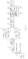
- a probe 1 consisting of an array of ultrasonic transducers, is used to insonate the tissues to be examined, and to detect the consequent backscattered echoes.
- the transmit and receive circuits 2, 3 are used to generate and time the driving pulses for the different transducer elements, in such a way as to properly focus the ultrasonic beam along the different lines of sight and on predetermined focuses.
- the transducer elements of the probe are driven so as to emit ultrasonic waves in the patient body, by activating proper groups of elements, and by delaying excitation, so as to ensure the desired beam focusing.
- the unit After each transmission, the unit is switched to receiving.
- the backscattered echoes after being properly amplified 2, are realigned according to the transmission focusing, and combined to produce a sequence of coherent echoes 3.
- the signals are quadrature-demodulated 4.
- This operation consists in base-band translating the band corresponding to the n ⁇ f0 harmonic (n being an integral or fractional number) of the f0 transmission carrier frequency.
- the operations of the demodulator consist in multiplying the received signals by two sin/cos signals at n ⁇ f0 frequency 5, 6 and in rejecting the signals outside the band of interest, by a low-pass filtering operation 7,8.
- the phase (I) and quadrature (Q) signals are discretized 9, digitized by the analog-to-digital converter 10, and stored in the working memory 12.
- the results of the processing operations are converted into video signals by the Scan Converter 13, using a suitable grey scale for the display 14.
- the demodulation frequency is selected so as to be coincident with a typical frequency, whereat the echoes of the tissues/vessels perfused by the contrast medium produce considerably higher echoes than in the regions which the contrast medium has not penetrated, it is possible to create a map of the section under examination in which only the areas penetrated by the echoenhancing agent are highlighted.
- Fig. 2 shows the pictures of two B-Mode images, relating to the same section, as they could be obtained through the proposed processing chain operating in normal mode (fig. 2a), i.e. by driving the demodulator with tones having a frequency f0, or in Harmonic Imaging mode (fig. 2b), i.e. by driving the quadrature demodulator with tones having a frequency n ⁇ f0, n being not equal to 1.
- the invention also relates to an apparatus for implementing the method according to the above disclosure, comprising a Power Doppler chain for echographic imaging, wherein there are provided means for by-passing the so-called wall-filters, and means for tuning the quadrature demodulator to the desired spectrum component of the n ⁇ f0 received signal.
Landscapes
- Physics & Mathematics (AREA)
- Engineering & Computer Science (AREA)
- Radar, Positioning & Navigation (AREA)
- Remote Sensing (AREA)
- Computer Networks & Wireless Communication (AREA)
- General Physics & Mathematics (AREA)
- Acoustics & Sound (AREA)
- Nonlinear Science (AREA)
- Ultra Sonic Daignosis Equipment (AREA)
Applications Claiming Priority (2)
| Application Number | Priority Date | Filing Date | Title |
|---|---|---|---|
| IT97SV000039A IT1298014B1 (it) | 1997-07-31 | 1997-07-31 | Metodo per la generazione d'immagini ecografiche bidimensionali tramite macchine aventi capacita' di analisi power poppler. |
| ITSV970039 | 1997-07-31 |
Publications (1)
| Publication Number | Publication Date |
|---|---|
| EP0895094A1 true EP0895094A1 (de) | 1999-02-03 |
Family
ID=11408235
Family Applications (1)
| Application Number | Title | Priority Date | Filing Date |
|---|---|---|---|
| EP98113343A Withdrawn EP0895094A1 (de) | 1997-07-31 | 1998-07-17 | Verfahren zur bidimensionalen Ultraschall-Bilderzeugung mit Vorrichtung zur Doppler-Leistungs-Prüfung |
Country Status (2)
| Country | Link |
|---|---|
| EP (1) | EP0895094A1 (de) |
| IT (1) | IT1298014B1 (de) |
Cited By (2)
| Publication number | Priority date | Publication date | Assignee | Title |
|---|---|---|---|---|
| WO2001050962A1 (en) * | 2000-01-14 | 2001-07-19 | Western Sydney Area Health Service | A cardiac valve movement measuring device |
| FR2841342A1 (fr) * | 2002-06-21 | 2003-12-26 | Thales Ultrasonics Sas | Structure d'entree pour echographie a ultrasons |
Citations (3)
| Publication number | Priority date | Publication date | Assignee | Title |
|---|---|---|---|---|
| EP0582462A2 (de) * | 1992-08-03 | 1994-02-09 | Olympus Optical Co., Ltd | Ultraschalldiagnosegerät |
| US5348015A (en) * | 1992-09-17 | 1994-09-20 | Applied Physiology And Medicine | Method and apparatus for ultrasonically detecting, counting and/or characterizing emboli |
| US5628322A (en) * | 1995-05-15 | 1997-05-13 | Kabushiki Kaisha Toshiba | Method of ultrasound imaging and diagnostic ultrasound system |
-
1997
- 1997-07-31 IT IT97SV000039A patent/IT1298014B1/it active IP Right Grant
-
1998
- 1998-07-17 EP EP98113343A patent/EP0895094A1/de not_active Withdrawn
Patent Citations (3)
| Publication number | Priority date | Publication date | Assignee | Title |
|---|---|---|---|---|
| EP0582462A2 (de) * | 1992-08-03 | 1994-02-09 | Olympus Optical Co., Ltd | Ultraschalldiagnosegerät |
| US5348015A (en) * | 1992-09-17 | 1994-09-20 | Applied Physiology And Medicine | Method and apparatus for ultrasonically detecting, counting and/or characterizing emboli |
| US5628322A (en) * | 1995-05-15 | 1997-05-13 | Kabushiki Kaisha Toshiba | Method of ultrasound imaging and diagnostic ultrasound system |
Non-Patent Citations (1)
| Title |
|---|
| PI HSIEN CHANG ET AL: "SECOND HARMONIC IMAGING AND HARMONIC DOPPLER MEASUREMENTS WITH ALBUNEX", IEEE JOURNAL OF SOLID-STATE CIRCUITS, vol. 42, no. 6, 1 November 1995 (1995-11-01), pages 1020 - 1026, XP000543760 * |
Cited By (3)
| Publication number | Priority date | Publication date | Assignee | Title |
|---|---|---|---|---|
| WO2001050962A1 (en) * | 2000-01-14 | 2001-07-19 | Western Sydney Area Health Service | A cardiac valve movement measuring device |
| FR2841342A1 (fr) * | 2002-06-21 | 2003-12-26 | Thales Ultrasonics Sas | Structure d'entree pour echographie a ultrasons |
| WO2004001444A1 (fr) * | 2002-06-21 | 2003-12-31 | Thales Ultrasonics Sas | Structure d’entrée pour echographie a ultrasons |
Also Published As
| Publication number | Publication date |
|---|---|
| ITSV970039A1 (it) | 1999-01-31 |
| ITSV970039A0 (it) | 1997-07-31 |
| IT1298014B1 (it) | 1999-12-20 |
Similar Documents
| Publication | Publication Date | Title |
|---|---|---|
| US8454517B2 (en) | Ultrasonic diagnostic apparatus and ultrasonic diagnostic method | |
| US5628322A (en) | Method of ultrasound imaging and diagnostic ultrasound system | |
| US6419632B1 (en) | High resolution flow imaging for ultrasound diagnosis | |
| US8435182B1 (en) | Methods and apparatus for ultrasound imaging | |
| US6814703B2 (en) | Apparatus and method for ultrasonic diagnostic imaging using a contrast medium | |
| KR100742466B1 (ko) | 이미징 방법 및 장치와 그를 이용한 혈액 움직임 이미징 시스템 | |
| US6676606B2 (en) | Ultrasonic diagnostic micro-vascular imaging | |
| JP3580627B2 (ja) | 超音波診断装置 | |
| CN100466986C (zh) | 使用谐波造影剂的灌注和血流超声成像 | |
| JP4408988B2 (ja) | 超音波診断装置 | |
| CN101128154B (zh) | 检测肝脏损伤的超声诊断成像系统和方法 | |
| US6620103B1 (en) | Ultrasonic diagnostic imaging system for low flow rate contrast agents | |
| US7985183B2 (en) | Ultrasonic diagnostic equipment and ultrasonic image generation method | |
| US5394874A (en) | Angiography using ultrasound | |
| Hohmann et al. | Liver metastases in cancer: detection with contrast-enhanced ultrasonography: J. Hohmann et al. | |
| JP2001212144A (ja) | 超音波診断装置及び超音波画像化方法 | |
| JP4226882B2 (ja) | 超音波診断装置 | |
| US20040087858A1 (en) | Method and apparatus for improving contrast-to-tissue ratio in ultrasound contrast imaging with subharmonic imaging | |
| JP3355140B2 (ja) | 超音波撮像装置 | |
| JP2007090075A (ja) | コントラスト剤を超音波によって特徴付ける方法およびコントラスト剤を超音波によって特徴付けるためにプログラムされたプロセッサによって実行可能な命令を表すデータが記憶されたコンピュータ読み出し可能記憶媒体 | |
| JP4574790B2 (ja) | 超音波診断装置及び超音波診断方法 | |
| Chen et al. | Color and power Doppler imaging of the kidneys | |
| US8366624B1 (en) | Methods and apparatus for ultrasound imaging | |
| JP3746119B2 (ja) | 超音波診断装置 | |
| JPH10314170A (ja) | 超音波診断装置 |
Legal Events
| Date | Code | Title | Description |
|---|---|---|---|
| PUAI | Public reference made under article 153(3) epc to a published international application that has entered the european phase |
Free format text: ORIGINAL CODE: 0009012 |
|
| AK | Designated contracting states |
Kind code of ref document: A1 Designated state(s): AT BE CH CY DE DK ES FI FR GB GR IE IT LI LU MC NL PT SE |
|
| AX | Request for extension of the european patent |
Free format text: AL;LT;LV;MK;RO;SI |
|
| AKX | Designation fees paid | ||
| STAA | Information on the status of an ep patent application or granted ep patent |
Free format text: STATUS: THE APPLICATION IS DEEMED TO BE WITHDRAWN |
|
| 18D | Application deemed to be withdrawn |
Effective date: 19990804 |
|
| REG | Reference to a national code |
Ref country code: DE Ref legal event code: 8566 |
|
| RAP1 | Party data changed (applicant data changed or rights of an application transferred) |
Owner name: ESAOTE S.P.A. |