EP0906762A1 - Diagnostikum für angiopathische erkrankungen - Google Patents
Diagnostikum für angiopathische erkrankungen Download PDFInfo
- Publication number
- EP0906762A1 EP0906762A1 EP97904567A EP97904567A EP0906762A1 EP 0906762 A1 EP0906762 A1 EP 0906762A1 EP 97904567 A EP97904567 A EP 97904567A EP 97904567 A EP97904567 A EP 97904567A EP 0906762 A1 EP0906762 A1 EP 0906762A1
- Authority
- EP
- European Patent Office
- Prior art keywords
- smooth muscle
- muscle myosin
- angiopathic
- antibody
- solution
- Prior art date
- Legal status (The legal status is an assumption and is not a legal conclusion. Google has not performed a legal analysis and makes no representation as to the accuracy of the status listed.)
- Withdrawn
Links
Images


Classifications
-
- A—HUMAN NECESSITIES
- A61—MEDICAL OR VETERINARY SCIENCE; HYGIENE
- A61K—PREPARATIONS FOR MEDICAL, DENTAL OR TOILETRY PURPOSES
- A61K51/00—Preparations containing radioactive substances for use in therapy or testing in vivo
- A61K51/02—Preparations containing radioactive substances for use in therapy or testing in vivo characterised by the carrier, i.e. characterised by the agent or material covalently linked or complexing the radioactive nucleus
- A61K51/04—Organic compounds
- A61K51/08—Peptides, e.g. proteins, carriers being peptides, polyamino acids, proteins
- A61K51/10—Antibodies or immunoglobulins; Fragments thereof, the carrier being an antibody, an immunoglobulin or a fragment thereof, e.g. a camelised human single domain antibody or the Fc fragment of an antibody
- A61K51/1018—Antibodies or immunoglobulins; Fragments thereof, the carrier being an antibody, an immunoglobulin or a fragment thereof, e.g. a camelised human single domain antibody or the Fc fragment of an antibody against material from animals or humans
-
- A—HUMAN NECESSITIES
- A61—MEDICAL OR VETERINARY SCIENCE; HYGIENE
- A61K—PREPARATIONS FOR MEDICAL, DENTAL OR TOILETRY PURPOSES
- A61K51/00—Preparations containing radioactive substances for use in therapy or testing in vivo
- A61K51/02—Preparations containing radioactive substances for use in therapy or testing in vivo characterised by the carrier, i.e. characterised by the agent or material covalently linked or complexing the radioactive nucleus
- A61K51/04—Organic compounds
- A61K51/08—Peptides, e.g. proteins, carriers being peptides, polyamino acids, proteins
- A61K51/10—Antibodies or immunoglobulins; Fragments thereof, the carrier being an antibody, an immunoglobulin or a fragment thereof, e.g. a camelised human single domain antibody or the Fc fragment of an antibody
Definitions
- the present invention relates to a diagnostic agent useful for diagnostic imaging for angiopathic diseases such as dissecting aortic aneurysm (also called aortic dissection) and angiitis, and to a kit for diagnosis.
- angiopathic diseases such as dissecting aortic aneurysm (also called aortic dissection) and angiitis
- the onset mechanism of dissecting aortic aneurysm includes partial breakage of intima of an aorta, flow of blood from a formed hiatus to media to cause dissection of a vascular wall, and necrosis of vascular smooth muscle cells. Grave cases bring strong pectralgia and a high fatality rate. As onset factors for these diseases, there are indicated hypertension, as well as degeneration and fragility-increase of media caused by arterial sclerosis and cystic medial necrosis.
- angiitis is caused by a factor such as collagen disease, and a lesion thereof often extends blood vessels of the whole body accompanied by necrosis of vascular smooth muscle cells and induces a grave fatal complication such as DIC (disseminated intravascular coagulation), thrombosis, or embolism.
- DIC dissminated intravascular coagulation
- thrombosis or embolism.
- angiopathic diseases accompanied by necrosis of vascular smooth muscle cells such as dissecting aortic aneurysm or angiitis, are grave diseases and therefore a speedy and accurate diagnosis is needed for effective medical treatment and prevention of complications.
- diagnosis methods there are utilized echography, CT (X-ray computer tomography), DSA (digital subtraction angiography), MRI (magnetic resonance imaging), and aortography used as a method for diagnosing dissecting aortic aneurysm similar to that for diagnosing other heart deseases.
- DSA and aortagraphy are dangerous test methods in an acute stage of the diseases due to considerable invasiveness.
- diagnostic imaging including echography, CT, and MRI although less dangerous even in an acute stage, generally yields a morphological diagnosis, which is not able to distinguish between an old inveterate lesion and a new lesion in an acute stage.
- the above diagnostic imaging is not able to specifically diagnose a pathological region in an acute stage at which vascular smooth muscle cells are being damaged.
- a method for diagnosing angiitis through conventional diagnostic imaging is more difficult than that for diagnosing dissecting aortic aneurysm, since angiitis itself is a disease accompanied by no characteristic morphological changes.
- an object of the present invention is to develop a tracer preparation to enable accurate diagnostic imaging for a region affected by these diseases by binding specifically to vascular smooth muscle cells damaged at an acute stage of dissecting aortic aneurysm or angiitis.
- the present inventors have conducted earnest studies to achieve the above object, and have found that smooth muscle myosin serving as a primary protein in blood vessels is useful for diagnostic imaging for angiopathic diseases such as dissecting aortic aneurysm and angiitis, particularly in an acute stage.
- the present invention was accomplished based on this finding.
- the present invention provides a diagnostic agent for angiopathic diseases containing a monoclonal antibody against human smooth muscle myosin or active fragments of the antibody labeled with a radioactive isotope.
- the present invention also provides a kit for diagnosing angiopathic diseases comprising a coupling compound formed of a bifunctional chelating agent and either a monoclonal antibody against human smooth muscle myosin or active fragments of the antibody, and a solution of radioactive isotopes.
- the present invention provides a method of diagnostic imaging for angiopathic diseases by use of the above diagnostic agent or kit.
- the diagnostic agent of the present invention is used to perform proper and accurate diagnostic imaging for a region affected by angiopathic diseases by binding a monoclonal antibody against human smooth muscle myosin or active fragments of the antibody labeled with a radioactive isotope, specifically to the region affected by these diseases. Therefore, the diagnostic agent must contain at least a monoclonal antibody against human smooth muscle myosin or active fragments of the antibody labeled with a radioactive isotope.
- the monoclonal antibody used in the present invention so long as it specifically binds to human smooth muscle myosin, particularly to a heavy chain of human smooth muscle myosin, and there is preferably used a monoclonal antibody having low cross-reactivity to other types of myosin.
- Such a monoclonal antibody may be prepared by employing a known method shown in the below-described Examples (See, e.g., " Men-eki Saikagaku Kankyuhou (Zoku-Seikagaku Jikken Kouza 5)," Edited by The Biochemical Society of Japan, pages 1-88 (1986); Biochemistry, 27, 3807-3811 (1988); Eur. J. Biochem., 179, 79-85 (1989); J. Mol. Biol., 198, 143-157 (1987); J. Biol. Chem., 264, 9734-9737 (1989); J. Biol. Chem., 264, 18272-18275 (1989); J. Biol. Chem., 266, 3768-3773 (1991); and Circulation, 88, 1804-1810 (1993)).
- a monoclonal antibody itself or active fragments of the monoclonal antibody may be used. No particular limitation is imposed on the active fragments; there may be used any of a variety of fragments such as F(ab') 2 , Fab', or Fab, so long as it has characteristics of the monoclonal antibody of the present invention. By use of such active fragments, the half-life may be reduced and in vivo clearance may increase.
- Such active fragments may be prepared by employing a known method such as papain-, pepsin-, or tripsin-treatment to a purified monoclonal antibody (See, “ Men-eki Saikagaku Kenkyuhou (Zoku-Seikagaku Jikken Kouza 5)," Edited by The Biochemical Society of Japan, page 89 (1986)).
- the thus-prepared monoclonal antibody or active fragments may be used as a diagnositic agent of the present invention by labeling with radioactive isotopes.
- examples of the radioactive isotopes include iodine-125, iodine-123, iodine-131, indium-111, indium-113m, technetium-99m, gallium-67, lead-203, ruthenium-97, mercury-197, thallium-201, and bismuth-212.
- the labeling method may be selected from among a variety of known methods in accordance with the employed nuclear species, and examples include a direct labeling of the antibody or active fragments with radioactive isotopes, such as a chloramine T method or a lactoperoxidase method, and a method involving bonding a bifunctional chelating agent to the antibody or active fragments to form a covalent bond and labeling the formed coupling compound with the above nuclear species.
- bifunctional chelating agents used in the present invention include 1-amino-6,17-dihydroxy-7,10,28,21-tetraoxo-27-(N-acetylhydroxyimino)-6,11,17,22-tetrazaheptaeicosane (despherioxamine), 8-hydroxyquinoline, ethylenediaminetetraacetic acid, diethylenetriaminepentaacetic acid (DTPA), and diaminocyclohexyltetraacetic acid, and these chelating agents and the antibody or active fragments are bonded by a customarily employed method such as a carbodiimide method, an acid anhydride method, or a glutaraldehyde method.
- a customarily employed method such as a carbodiimide method, an acid anhydride method, or a glutaraldehyde method.
- the kit for the diagnosis of the present invention is used to prepare a monoclonal antibody against human smooth muscle myosin or active fragments of the antibody labeled with a radioactive isotope; in order to perform accurate diagnostic imaging for a region affected by angiopathic diseases by binding the labeled monoclonal antibody or active fragments of the antibody specifically to the region affected by these diseases. Therefore, the kit for the diagnosis must be fabricated so as to enable the preparation of at least a monoclonal antibody against human smooth muscle myosin or active fragments of the antibody labeled with a radioactive isotope.
- kits containing a coupling compound comprising a bifunctional chelating agent and either a monoclonal antibody against human smooth muscle myosin or active fragments of the antibody, as well as a solution of radioactive isotopes.
- the diagnostic agent and kit of the present invention may contain, in addition to the above reagents, a chromatographic column to purify nuclear species; a carrier such as a sodium chloride solution or a glucose solution to prepare into a form of administration; other stabilizers; etc.
- the diagnostic agent of the present invention is administered to the human body through intravenous injection. Therefore, the diagnostic agent of the present invention is used in a form suitable for intravenous injection by use of the above-mentioned carrier, etc.
- the diagnostic agent of the present invention is usually administered in an amount of 100 ⁇ Ci-30 mCi, preferably 500 ⁇ Ci-3 mCi, depending on the radioactive isotope used for labeling.
- the diagnostic imaging by use of the diagnostic agent of the present invention may be performed as follows. One to 48 hours after administration of the agent of the present invention, a region such as a region of the patient's heart where angiopathic disorders seem to be generated is scanned by autoradiography, or by use of a scintillation scanner or a scintillation camera, to thereby detect radioactivity attributed to the agent of the present invention and to depict the image thereof.
- Human uterus smooth muscle myosin, human aorta smooth muscle myosin, human small intestine smooth muscle myosin, human platelet myosin, and human skeletal muscle myosin were provided by Dr. Matsumura, Saga Medical College.
- Human cardiac muscle myosin was purified according to the method of Yazaki (Circ. Res., 36:208, 1975). For each type of myosin, purity was assessed by SDS-PAGE, and then the concentration of protein was determined according to the method of Lawry (J. Biol. Chem., 193:265-275, 1951) using bovine serum albumin as a standard.
- mice aged 6 to 8 weeks were immunized intraperitoneally with 25 to 50 ⁇ g of human uterus smooth muscle myosin emulsified with Freund's complete adjuvant, 4 to 7 times at 2 to 4 weeks interval, after which human uterus smooth muscle myosin (10 ⁇ g) was administered by intravenous injection.
- the spleen of each mouse was removed three days after the final immunization, and the spleen cells and mouse myeloma cells P3 ⁇ 63Ag8U.1(P3U1) (ATCC CRL-1597) were mixed at a ratio of 10 : 1.
- An RPMI 1640 solution (1 ml) containing 50% polyethylene glycol was gradually added to pellets obtained by centrifugal separation of the mixture to thereby conduct cell fusion.
- an RPMI 1640 culture medium was added thereto to adjust the volume to 10 ml and the mixture was centrifuged to thereby obtain pellets, which were suspended in an RPMI 1640 culture medium containing 10% fetal calf serum at the concentration of P3U1 3 ⁇ 10 4 cells/0.1 ml. The suspension was dispensed to a 96-well microplate in an amount of 0.1 ml/well.
- HAT culture medium (0.1 ml) was added to each well, and half of the medium was replaced with fresh HAT medium every 3-4 days.
- the culture supernatant was sampled 7-10 days after fusion, dispensed in an amount of 50 ⁇ l/well into a 96-well polyvinyl chloride (PVC) plate which had been precoated with human uterus smooth muscle myosin and blocked with 3% gelatin, and allowed to react at room temperature for one hour.
- PVC polyvinyl chloride
- a solution containing biotinylated horse anti-mouse IgG Vector Co.
- PBSA bovine serum albumin
- hybridoma 1H6, 4E12, 9A12, 9D7, 10G2
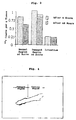
- the number of specific antibody-positive wells, growth wells, and the total number of wells are shown in Table 1.
- hybridoma 1H6 and hybridoma 4E12 were deposited with the National Institute of Bioscience and Human-Technology, Agency of Industrial Science and Technology (1-3, Higashi 1 chome, Tsukuba-shi, Ibaraki-ken, 305, JAPAN) as SMHMW 1H6 and SMHMW 4E12 on October 13, 1994 under Budapest Treaty, and were allotted accession numbers FERM BP-4829 and FERM BP-4830 dated October 13, 1994. Wells which indicated positive to specific antibody Wells which indicated growth Total No. of wells 11 653 940
- the ascites was mixed with an equal volume of 1.5 M glycine-hydrochloric acid buffer (pH 8.9) containing 3 M sodium chloride, and was passed through a Protein A Sepharose CL-4B (Pharmacia Co.) equilibrated with the same buffer as above. After the column was washed with a sufficient amount of the same glycine-hydrochloric acid buffer, the antibody was eluted with 0.1 M citrate buffer (pH 6.0). The eluate was dialyzed against PBS, and purity was confirmed by SDS-polyacrylamide gel electrophoresis (SDS-PAGE) to thereby obtain a purified monoclonal antibody.
- SDS-PAGE SDS-polyacrylamide gel electrophoresis
- Human uterus smooth muscle myosin (1 mg/ml) was mixed with an equal volume of a reducing solution and the mixture was heated at 100°C for five minutes.
- SDS-PAGE was performed with a mini-gel electrophoresis apparatus (Marysal Co.) using a 10% separated gel and a 5% concentrated gel at 10 mV for approximately three hours.
- Blotting was performed with a blotting apparatus for mini-gel (Marysal Co.) at 37 V for about 18 hours to transcribe protein to a nitrocellulose membrane, which was subsequently cut along electrophoresis lines into strips. Some of them were treated with Amido Black to stain protein and the remainder were blocked with 3% gelatin and then reacted with a culture supernatant of each hybridoma at room temperature for one hour.
- reaction mixture was washed twice for 10 minutes each time with 20 mM Tris-500 mM NaCl (pH 7.5) buffer containing 0.05% Tween 20(T-TBS) and reacted with 500 fold-diluted biotinylated horse anti-mouse IgG (Vector Co.) at room temperature for one hour.
- the reaction mixture was then washed twice with T-TBS for 10 minutes and reacted with 2000 fold-diluted peroxidase-avidin D (Vector Co.) at room temperature for 15 minutes.
- reaction mixture was then washed twice with T-TBS for 10 minutes, treated with a color development solution (containing 30 mg of HRP color development reagent (Bio-Rad Co.), 10 ml of methanol, 50 ml of TBS and 30 ⁇ l of a 30% hydrogen peroxide), and then washed with distilled water.
- a color development solution containing 30 mg of HRP color development reagent (Bio-Rad Co.), 10 ml of methanol, 50 ml of TBS and 30 ⁇ l of a 30% hydrogen peroxide
- Each of the above monoclonal antibodies was dialyzed against a 0.1 M sodium hydrogencarbonate solution, and the dialyzed solution was concentrated to 2 mg/ml by use of a Centriflow (Amicon Co.).
- Biotin (Long-arm) NHS reagent (Vector Co.) was dissolved in dimethylformamide to a concentration of 10 mg/ml, of which 20 ⁇ l was mixed with the above antibody solution (1 ml). The mixture was allowed to react at room temperature for two hours. After addition of ethanolamine (5 ⁇ l) to terminate reaction, the mixture was dialyzed twice against PBS, to thereby obtain a biotinylated antibody.
- the biotinylated antibody was diluted to 1 ⁇ g/ml with PBS containing 1% BSA, to thereby obtain a solution of the biotinylated antibody.
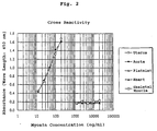
- An anti-smooth muscle myosin monoclonal antibody (4E12) was diluted with PBS to 10 ⁇ g/ml and dispensed into a 96-well plate (H type: Sumitomo Bakelite Co., Ltd.) in an amount of 50 ⁇ l/well, and the microplate was allowed to stand at 4°C overnight. After the antibody was washed three times with PBS containing 0.05% Tween 20, 0.5% skim milk was dispensed thereto in an amount of 300 ⁇ l/well and the plate was allowed to stand at room temperature for one hour. The skim milk solution was removed to thereby obtain an immobilized antibody reagent.
- the substrate solution was added in an amount of 100 ⁇ l/well and the mixture was allowed to stand at room temperature for 10 minutes to cause coloring.
- the enzyme reaction stopping solution was dispensed in an amount of 100 ⁇ l/well to terminate reaction, and the absorbance at 450 nm was measured with a microplate photometer. The standard curve obtained is shown in Fig. 1.
- a 0.5% solution of sodium pyrosulfite (10 ⁇ l), a 1% solution of potassium iodide (5 ⁇ l), and a 1% BSA solution (5 ⁇ l) were successively added and mixed.
- the reaction mixture was passed through a PD10 column (Pharmacia Co.) equilibrated with a 1% solution of BSA, then eluted with a 1% solution of BSA, and fractionated by 1 ml. The fractions were measured with an auto-gamma-counter and the counts of them showed two peaks. Fractions attributed to the first peak were collected to obtain a 125 I anti-smooth muscle myosin monoclonal antibody.
- Example 5 Diagnostic imaging by use of a 125 I anti-smooth muscle myosin monoclonal antibody
- Hybridoma 8B8 which produces a monoclonal antibody specifically reacting to human small intestine smooth muscle myosin was established in a manner similar to that described in 1) of Example 2 by use of human small intestine smooth muscle myosin instead of human uterus smooth muscle myosin.
- This hybridoma was deposited with the National Institute of Bioscience and Human-Technology, Agency of Industrial Science and Technology (1-3, Higashi 1 chome, Tsukuba-shi, Ibaraki-ken, 305, JAPAN) as IMH8B8 on March 5, 1996 under Budapest Treaty, and was allotted an accession number FERM BP-5444 dated March 5, 1996.
- the monoclonal antibody produced from this hybridoma was purified by the method 2) of Example 2, and properties of the monoclonal antibody were examined by the methods described in Example 3. The results are as follows.
- the monoclonal antibody (8B8) was labeled by the method of Example 4, and diagnostic imaging for damaged parts of aorta was performed in a model experiment using rats in accordance with Example 5. As a result, accumulation of 125 I was confirmed in the damaged parts and the image had a contrast higher than that obtained by use of a monoclonal antibody (9D7).
- the concentration of the anti-smooth muscle myosin monoclonal antibody (IMH8B8) was adjusted with PBS to 2 mg/ml, and EDTA was added thereto so that the concentration thereof became 2 mM.
- the concentration of mercury papain (Sigma Co.) was adjusted to 1 mg/ml with PBS containing cystein (5 mM) and EDTA (2 mM), and the mixture was preincubated at 37° for 30 minutes.
- a papain solution (1 wt.% based on the amount of the antibody) was added to the antibody solution, and the mixture was incubated at 37° for 15 minutes. Iodoacetamide was added thereto so that the final concentration thereof became 5 mM to terminate the enzyme reaction.
- the enzyme-treated antibody solution was mixed with an equal volume of 3 M NaCl and 1.5 M giycine buffer (pH 8.9), and the mixture was passed through a Protein A Sepharose column (Pharmacia Co.) equilibrated with the above buffer to adsorb Fc contained in the antibody solution.
- the eluate was concentrated by ultrafiltration and then dialyzed against PBS, and the purity of Fab was confirmed by SDS-PAGE.
- Example 8 (Diagnostic imaging with a 125 I anti-smooth muscle myosin monoclonal Fab antibody)
- IMH8B8-Fab formed in Example 7 was labeled with 125 I by use of the method of Example 4.
- Male rats weighing 300 g were anesthetized with pentobarbital by way of cannulation through the femoral artery.
- a spring coil guide wire of 0.014 inch was inserted to the abdominal aorta, which was exposed and compressed with forceps to damage aorta media under operation of the guide wire.
- a 125 I-labeled IMH8B8-Fab antibody (1.23 MBq) was intravenously injected from a femoral artery and the wound was sutured.
- the present inventors have found that accurate and specific diagnostic imaging for a region affected by the above-mentioned diseases can be performed by binding and concentrating a monoclonal antibody against human smooth muscle myosin or active fragments of the antibody labeled with a radioactive isotope, specifically to the region affected by angiopathic diseases.
- the diagnostic agent and kit of the present invention are useful for diagnostic imaging of angiopathic diseases such as dissecting aortic aneurysm or angiitis, particularly for diagnostic imaging of these diseases in an acute stage.
- Use of the diagnostic agent and kit has enabled to specify the region affected by these diseases.
Landscapes
- Health & Medical Sciences (AREA)
- Life Sciences & Earth Sciences (AREA)
- Proteomics, Peptides & Aminoacids (AREA)
- Pharmacology & Pharmacy (AREA)
- Physics & Mathematics (AREA)
- Chemical & Material Sciences (AREA)
- Medicinal Chemistry (AREA)
- Optics & Photonics (AREA)
- Immunology (AREA)
- Epidemiology (AREA)
- Animal Behavior & Ethology (AREA)
- General Health & Medical Sciences (AREA)
- Public Health (AREA)
- Veterinary Medicine (AREA)
- Medicines Containing Antibodies Or Antigens For Use As Internal Diagnostic Agents (AREA)
- Preparation Of Compounds By Using Micro-Organisms (AREA)
Applications Claiming Priority (5)
| Application Number | Priority Date | Filing Date | Title |
|---|---|---|---|
| JP55512/96 | 1996-02-19 | ||
| JP5551296 | 1996-02-19 | ||
| JP9003496 | 1996-03-18 | ||
| JP90034/96 | 1996-03-18 | ||
| PCT/JP1997/000435 WO1997029784A1 (en) | 1996-02-19 | 1997-02-19 | Diagnostic agent for angiopathic diseases |
Publications (1)
| Publication Number | Publication Date |
|---|---|
| EP0906762A1 true EP0906762A1 (de) | 1999-04-07 |
Family
ID=26396399
Family Applications (1)
| Application Number | Title | Priority Date | Filing Date |
|---|---|---|---|
| EP97904567A Withdrawn EP0906762A1 (de) | 1996-02-19 | 1997-02-19 | Diagnostikum für angiopathische erkrankungen |
Country Status (5)
| Country | Link |
|---|---|
| US (1) | US6270744B1 (de) |
| EP (1) | EP0906762A1 (de) |
| JP (1) | JP4035167B2 (de) |
| CA (1) | CA2246666A1 (de) |
| WO (1) | WO1997029784A1 (de) |
Families Citing this family (1)
| Publication number | Priority date | Publication date | Assignee | Title |
|---|---|---|---|---|
| CN116350809A (zh) * | 2023-03-28 | 2023-06-30 | 江苏省苏北人民医院 | 一种wvp多肽放射性药物及其制备方法和应用 |
Family Cites Families (12)
| Publication number | Priority date | Publication date | Assignee | Title |
|---|---|---|---|---|
| JPS6028998A (ja) * | 1983-07-06 | 1985-02-14 | Yamasa Shoyu Co Ltd | 心筋ミオシン重鎖に対する単一クロ−ン抗体 |
| JPS60201260A (ja) * | 1984-03-27 | 1985-10-11 | Yamasa Shoyu Co Ltd | 心疾患診断薬 |
| JPH03504499A (ja) * | 1988-05-27 | 1991-10-03 | セントカー・インコーポレーテツド | 抗体試薬のための配合物 |
| US5046499A (en) | 1988-06-13 | 1991-09-10 | Centocor, Inc. | Method for myocardial infarct risk assessment |
| US5626830A (en) * | 1989-03-23 | 1997-05-06 | Biotech Cardio-Vision, Societe En Commandite Enregistree | Anti-myosin mouse monoclonal antibody and method of use for diagnosis of myocardial infarction |
| US5747652A (en) * | 1989-03-28 | 1998-05-05 | Yamasa Shoyu Kabushiki Kaisha | Antibody to smooth muscle myosin heavy chains |
| DE69019968T2 (de) * | 1989-03-28 | 1995-10-12 | Yamasa Shoyu Kk | Antikörper gegen die schwere kette eines glatt-muskel-myosins. |
| JPH02219596A (ja) * | 1989-12-09 | 1990-09-03 | Yamasa Shoyu Co Ltd | 心筋ミオシン重鎖に対する単一クローン抗体 |
| JPH05176790A (ja) | 1990-02-21 | 1993-07-20 | Mercian Corp | 脳ミオシンに対するモノクローナル抗体 |
| JP3152706B2 (ja) * | 1991-11-28 | 2001-04-03 | 株式会社東芝 | 心筋特異性を有するmri造影剤 |
| DE59308123D1 (de) * | 1993-11-03 | 1998-03-12 | Cooper Ind Inc | Wischblatt |
| JP3499875B2 (ja) | 1994-10-25 | 2004-02-23 | ヤマサ醤油株式会社 | 解離性大動脈瘤を検出するための抗体試薬及びその用途 |
-
1997
- 1997-02-19 EP EP97904567A patent/EP0906762A1/de not_active Withdrawn
- 1997-02-19 JP JP52920297A patent/JP4035167B2/ja not_active Expired - Lifetime
- 1997-02-19 US US09/125,430 patent/US6270744B1/en not_active Expired - Fee Related
- 1997-02-19 WO PCT/JP1997/000435 patent/WO1997029784A1/ja not_active Ceased
- 1997-02-19 CA CA002246666A patent/CA2246666A1/en not_active Abandoned
Non-Patent Citations (1)
| Title |
|---|
| See references of WO9729784A1 * |
Also Published As
| Publication number | Publication date |
|---|---|
| JP4035167B2 (ja) | 2008-01-16 |
| US6270744B1 (en) | 2001-08-07 |
| WO1997029784A1 (en) | 1997-08-21 |
| CA2246666A1 (en) | 1997-08-21 |
Similar Documents
| Publication | Publication Date | Title |
|---|---|---|
| CA1338706C (en) | Monoclonal anti-human breast cancer antibodies suitable for imaging breast cancer | |
| US5648471A (en) | One vial method for labeling antibodies with Technetium-99m | |
| AU639458B2 (en) | Novel monoclonal antibody to novel antigen associated with human tumors | |
| US6375925B1 (en) | Methods and reagents for non-invasive imaging of atherosclerotic plaque | |
| US4927916A (en) | Method of producing fibrin-specific monoclonal antibodies lacking fibrinogen-cross-reactivity using fibrin-specific peptides | |
| US4943427A (en) | Diagnostic agent for heart disease and use thereof | |
| Andrew et al. | Comparative imaging and biodistribution studies with an anti-CEA monoclonal antibody and its F (ab) 2 and Fab fragments in mice with colon carcinoma xenografts | |
| US5411884A (en) | Monoclonal antibody L53 which recognizes a human tumor-associated antigen | |
| US6270744B1 (en) | Diagnostic agent for angiopathic diseases | |
| WO1998021581A1 (en) | Methods and reagents for non-invasive imaging of atherosclerotic plaque | |
| US5626830A (en) | Anti-myosin mouse monoclonal antibody and method of use for diagnosis of myocardial infarction | |
| DE3885975T2 (de) | Monoklonale Antikörper gegen menschliche alpha-Atrialnatriüretic-Polypeptide und entsprechende Hybridome, deren Herstellung und Verwendung. | |
| EP0575906A2 (de) | Sandwich-Immunoassay für beta-N-Acetylglukosaminidase und monoklonaler Antikörper dafür | |
| US5250297A (en) | Tumor-associated antigen, antibodies, compositions and uses therefor | |
| Cummins et al. | Uptake of radioiodinated cardiac specific troponin-I antibodies in myocardial infarction | |
| JPWO1997029784A1 (ja) | 血管障害性疾患用診断薬 | |
| EP0898970A1 (de) | Diagnostikum für erkrankungen des verdauungstrakts | |
| EP0424107A1 (de) | Ein neuartiges tumorzugehöriges Antigen | |
| Jones et al. | Comparison of pharmacokinetics of radiolabeled monoclonal antibody UJ13A in patients and animal models | |
| JP3981147B2 (ja) | 消化器疾患用抗体試薬 | |
| Buraggi | Tumor radioimmunodetection by anti-melanoma and anti-CEA monoclonal antibodies: pilot and prospective studies | |
| KIM et al. | Applications of Six Monoclonal Antibodies in In Vitro and In Vivo Assays to Evaluate the Tissue Specificity ofy-Glutamyltransferase | |
| Lavie et al. | Labeling of sarcoma associated monoclonal antibody with 111In, 67Ga and 125I | |
| JPWO1997029785A1 (ja) | 消化器疾患用診断薬 | |
| Yachi et al. | Monoclonal Antibody AM34 Detects New Proteins Associated with Secondary Amyloidosis |
Legal Events
| Date | Code | Title | Description |
|---|---|---|---|
| PUAI | Public reference made under article 153(3) epc to a published international application that has entered the european phase |
Free format text: ORIGINAL CODE: 0009012 |
|
| 17P | Request for examination filed |
Effective date: 19980916 |
|
| AK | Designated contracting states |
Kind code of ref document: A1 Designated state(s): DE FR GB |
|
| STAA | Information on the status of an ep patent application or granted ep patent |
Free format text: STATUS: THE APPLICATION HAS BEEN WITHDRAWN |
|
| 18W | Application withdrawn |
Withdrawal date: 20010919 |