EP1004665A1 - Proteines du type recepteurs accepteurs - Google Patents
Proteines du type recepteurs accepteurs Download PDFInfo
- Publication number
- EP1004665A1 EP1004665A1 EP98937802A EP98937802A EP1004665A1 EP 1004665 A1 EP1004665 A1 EP 1004665A1 EP 98937802 A EP98937802 A EP 98937802A EP 98937802 A EP98937802 A EP 98937802A EP 1004665 A1 EP1004665 A1 EP 1004665A1
- Authority
- EP
- European Patent Office
- Prior art keywords
- cells
- protein
- human
- csr
- gene
- Prior art date
- Legal status (The legal status is an assumption and is not a legal conclusion. Google has not performed a legal analysis and makes no representation as to the accuracy of the status listed.)
- Withdrawn
Links
- 108090000623 proteins and genes Proteins 0.000 title claims abstract description 328
- 102000004169 proteins and genes Human genes 0.000 title claims abstract description 177
- 239000002516 radical scavenger Substances 0.000 title description 2
- 241000282414 Homo sapiens Species 0.000 claims abstract description 177
- 108020004414 DNA Proteins 0.000 claims abstract description 139
- 210000004027 cell Anatomy 0.000 claims description 261
- 239000012634 fragment Substances 0.000 claims description 89
- 239000013604 expression vector Substances 0.000 claims description 49
- 125000003729 nucleotide group Chemical group 0.000 claims description 44
- 239000002773 nucleotide Substances 0.000 claims description 35
- 210000004408 hybridoma Anatomy 0.000 claims description 22
- 241000124008 Mammalia Species 0.000 claims description 13
- 206010035226 Plasma cell myeloma Diseases 0.000 claims description 11
- 201000000050 myeloid neoplasm Diseases 0.000 claims description 11
- 239000008194 pharmaceutical composition Substances 0.000 claims description 9
- 210000003719 b-lymphocyte Anatomy 0.000 claims description 7
- 239000003937 drug carrier Substances 0.000 claims description 5
- 125000003275 alpha amino acid group Chemical group 0.000 claims 12
- 230000014509 gene expression Effects 0.000 abstract description 109
- 108020004635 Complementary DNA Proteins 0.000 abstract description 90
- 239000002299 complementary DNA Substances 0.000 abstract description 80
- 101000693970 Homo sapiens Scavenger receptor class A member 3 Proteins 0.000 abstract description 51
- 230000003834 intracellular effect Effects 0.000 abstract description 39
- 102100027192 Scavenger receptor class A member 3 Human genes 0.000 abstract description 35
- 101150084419 CSR2 gene Proteins 0.000 abstract description 34
- 239000000523 sample Substances 0.000 abstract description 34
- 102000014452 scavenger receptors Human genes 0.000 abstract description 28
- 230000027455 binding Effects 0.000 abstract description 19
- 230000030833 cell death Effects 0.000 abstract description 16
- 108010078070 scavenger receptors Proteins 0.000 abstract description 15
- 108091005485 macrophage scavenger receptors Proteins 0.000 abstract description 13
- 102000005962 receptors Human genes 0.000 abstract description 8
- 108020003175 receptors Proteins 0.000 abstract description 8
- 230000001875 tumorinhibitory effect Effects 0.000 abstract description 6
- 210000004556 brain Anatomy 0.000 abstract description 5
- 230000001605 fetal effect Effects 0.000 abstract description 5
- 230000002401 inhibitory effect Effects 0.000 abstract description 3
- 230000009257 reactivity Effects 0.000 abstract description 2
- MWUXSHHQAYIFBG-UHFFFAOYSA-N Nitric oxide Chemical compound O=[N] MWUXSHHQAYIFBG-UHFFFAOYSA-N 0.000 description 174
- 235000018102 proteins Nutrition 0.000 description 165
- MHAJPDPJQMAIIY-UHFFFAOYSA-N Hydrogen peroxide Chemical compound OO MHAJPDPJQMAIIY-UHFFFAOYSA-N 0.000 description 107
- 238000000034 method Methods 0.000 description 100
- 238000010804 cDNA synthesis Methods 0.000 description 79
- 150000001413 amino acids Chemical group 0.000 description 71
- 230000035882 stress Effects 0.000 description 58
- 108020004999 messenger RNA Proteins 0.000 description 54
- 241000699666 Mus <mouse, genus> Species 0.000 description 53
- 239000013598 vector Substances 0.000 description 50
- 102100025064 Cellular tumor antigen p53 Human genes 0.000 description 47
- 239000002609 medium Substances 0.000 description 41
- 108091032973 (ribonucleotides)n+m Proteins 0.000 description 31
- 235000001014 amino acid Nutrition 0.000 description 31
- 229940024606 amino acid Drugs 0.000 description 31
- 239000013612 plasmid Substances 0.000 description 28
- 108090000765 processed proteins & peptides Proteins 0.000 description 26
- 238000003757 reverse transcription PCR Methods 0.000 description 26
- 108060003951 Immunoglobulin Proteins 0.000 description 25
- 102000018358 immunoglobulin Human genes 0.000 description 25
- 229920001184 polypeptide Polymers 0.000 description 25
- 102000004196 processed proteins & peptides Human genes 0.000 description 25
- 241001465754 Metazoa Species 0.000 description 22
- 238000000636 Northern blotting Methods 0.000 description 22
- 240000004808 Saccharomyces cerevisiae Species 0.000 description 20
- 235000014680 Saccharomyces cerevisiae Nutrition 0.000 description 20
- 230000006698 induction Effects 0.000 description 20
- 230000036542 oxidative stress Effects 0.000 description 19
- 238000004458 analytical method Methods 0.000 description 17
- AOJJSUZBOXZQNB-TZSSRYMLSA-N Doxorubicin Chemical compound O([C@H]1C[C@@](O)(CC=2C(O)=C3C(=O)C=4C=CC=C(C=4C(=O)C3=C(O)C=21)OC)C(=O)CO)[C@H]1C[C@H](N)[C@H](O)[C@H](C)O1 AOJJSUZBOXZQNB-TZSSRYMLSA-N 0.000 description 16
- 239000003814 drug Substances 0.000 description 16
- 238000002474 experimental method Methods 0.000 description 16
- 210000002950 fibroblast Anatomy 0.000 description 16
- 241000699670 Mus sp. Species 0.000 description 15
- 238000012258 culturing Methods 0.000 description 15
- ZHNUHDYFZUAESO-UHFFFAOYSA-N Formamide Chemical compound NC=O ZHNUHDYFZUAESO-UHFFFAOYSA-N 0.000 description 14
- 241000700159 Rattus Species 0.000 description 14
- 230000033228 biological regulation Effects 0.000 description 14
- 239000012528 membrane Substances 0.000 description 14
- 230000008685 targeting Effects 0.000 description 14
- 108700024394 Exon Proteins 0.000 description 13
- 108010007622 LDL Lipoproteins Proteins 0.000 description 13
- 102000007330 LDL Lipoproteins Human genes 0.000 description 13
- 102000047147 human SCARA3 Human genes 0.000 description 13
- 238000009396 hybridization Methods 0.000 description 13
- 230000002441 reversible effect Effects 0.000 description 13
- 238000012360 testing method Methods 0.000 description 13
- 241000588724 Escherichia coli Species 0.000 description 12
- 108020005187 Oligonucleotide Probes Proteins 0.000 description 12
- PXIPVTKHYLBLMZ-UHFFFAOYSA-N Sodium azide Chemical compound [Na+].[N-]=[N+]=[N-] PXIPVTKHYLBLMZ-UHFFFAOYSA-N 0.000 description 12
- 238000002105 Southern blotting Methods 0.000 description 12
- 239000002751 oligonucleotide probe Substances 0.000 description 12
- 239000008363 phosphate buffer Substances 0.000 description 12
- 102000007469 Actins Human genes 0.000 description 11
- 108010085238 Actins Proteins 0.000 description 11
- 238000003556 assay Methods 0.000 description 11
- 230000001086 cytosolic effect Effects 0.000 description 11
- 239000003446 ligand Substances 0.000 description 11
- 238000012216 screening Methods 0.000 description 11
- 230000035939 shock Effects 0.000 description 11
- 238000010561 standard procedure Methods 0.000 description 11
- TWRXJAOTZQYOKJ-UHFFFAOYSA-L Magnesium chloride Chemical compound [Mg+2].[Cl-].[Cl-] TWRXJAOTZQYOKJ-UHFFFAOYSA-L 0.000 description 10
- 108091028043 Nucleic acid sequence Proteins 0.000 description 10
- 108091034117 Oligonucleotide Proteins 0.000 description 10
- 241000283973 Oryctolagus cuniculus Species 0.000 description 10
- 239000000872 buffer Substances 0.000 description 10
- 230000000694 effects Effects 0.000 description 10
- 230000006870 function Effects 0.000 description 10
- 239000000499 gel Substances 0.000 description 10
- 239000000203 mixture Substances 0.000 description 10
- 108091008146 restriction endonucleases Proteins 0.000 description 10
- 239000000243 solution Substances 0.000 description 10
- 108091003079 Bovine Serum Albumin Proteins 0.000 description 9
- 241000699800 Cricetinae Species 0.000 description 9
- 229930193140 Neomycin Natural products 0.000 description 9
- 210000004899 c-terminal region Anatomy 0.000 description 9
- 239000003153 chemical reaction reagent Substances 0.000 description 9
- 210000000805 cytoplasm Anatomy 0.000 description 9
- 239000012091 fetal bovine serum Substances 0.000 description 9
- BRZYSWJRSDMWLG-CAXSIQPQSA-N geneticin Chemical compound O1C[C@@](O)(C)[C@H](NC)[C@@H](O)[C@H]1O[C@@H]1[C@@H](O)[C@H](O[C@@H]2[C@@H]([C@@H](O)[C@H](O)[C@@H](C(C)O)O2)N)[C@@H](N)C[C@H]1N BRZYSWJRSDMWLG-CAXSIQPQSA-N 0.000 description 9
- 230000003053 immunization Effects 0.000 description 9
- 238000002347 injection Methods 0.000 description 9
- 239000007924 injection Substances 0.000 description 9
- 239000003550 marker Substances 0.000 description 9
- 229960004927 neomycin Drugs 0.000 description 9
- 210000001519 tissue Anatomy 0.000 description 9
- 238000013518 transcription Methods 0.000 description 9
- 230000035897 transcription Effects 0.000 description 9
- 230000009261 transgenic effect Effects 0.000 description 9
- 206010009944 Colon cancer Diseases 0.000 description 8
- 229940009456 adriamycin Drugs 0.000 description 8
- 235000012000 cholesterol Nutrition 0.000 description 8
- 238000009826 distribution Methods 0.000 description 8
- 238000011813 knockout mouse model Methods 0.000 description 8
- 229940126619 mouse monoclonal antibody Drugs 0.000 description 8
- XLYOFNOQVPJJNP-UHFFFAOYSA-N water Substances O XLYOFNOQVPJJNP-UHFFFAOYSA-N 0.000 description 8
- 241000894006 Bacteria Species 0.000 description 7
- 108010021625 Immunoglobulin Fragments Proteins 0.000 description 7
- 102000008394 Immunoglobulin Fragments Human genes 0.000 description 7
- HYHSBSXUHZOYLX-WDSKDSINSA-N S-nitrosoglutathione Chemical compound OC(=O)[C@@H](N)CCC(=O)N[C@@H](CSN=O)C(=O)NCC(O)=O HYHSBSXUHZOYLX-WDSKDSINSA-N 0.000 description 7
- FAPWRFPIFSIZLT-UHFFFAOYSA-M Sodium chloride Chemical compound [Na+].[Cl-] FAPWRFPIFSIZLT-UHFFFAOYSA-M 0.000 description 7
- 210000004102 animal cell Anatomy 0.000 description 7
- 101150014721 cdr gene Proteins 0.000 description 7
- 150000001841 cholesterols Chemical class 0.000 description 7
- 239000012228 culture supernatant Substances 0.000 description 7
- 229940079593 drug Drugs 0.000 description 7
- 210000004940 nucleus Anatomy 0.000 description 7
- 108700025694 p53 Genes Proteins 0.000 description 7
- 230000002093 peripheral effect Effects 0.000 description 7
- 230000005855 radiation Effects 0.000 description 7
- 210000002966 serum Anatomy 0.000 description 7
- 239000002676 xenobiotic agent Substances 0.000 description 7
- IEPRKVQEAMIZSS-UHFFFAOYSA-N Di-Et ester-Fumaric acid Natural products CCOC(=O)C=CC(=O)OCC IEPRKVQEAMIZSS-UHFFFAOYSA-N 0.000 description 6
- IAZDPXIOMUYVGZ-UHFFFAOYSA-N Dimethylsulphoxide Chemical compound CS(C)=O IAZDPXIOMUYVGZ-UHFFFAOYSA-N 0.000 description 6
- PEDCQBHIVMGVHV-UHFFFAOYSA-N Glycerine Chemical compound OCC(O)CO PEDCQBHIVMGVHV-UHFFFAOYSA-N 0.000 description 6
- 101710154606 Hemagglutinin Proteins 0.000 description 6
- 101000773184 Homo sapiens Twist-related protein 1 Proteins 0.000 description 6
- 102000006496 Immunoglobulin Heavy Chains Human genes 0.000 description 6
- 108010019476 Immunoglobulin Heavy Chains Proteins 0.000 description 6
- PWKSKIMOESPYIA-BYPYZUCNSA-N L-N-acetyl-Cysteine Chemical compound CC(=O)N[C@@H](CS)C(O)=O PWKSKIMOESPYIA-BYPYZUCNSA-N 0.000 description 6
- 102000000853 LDL receptors Human genes 0.000 description 6
- 108010001831 LDL receptors Proteins 0.000 description 6
- 206010058467 Lung neoplasm malignant Diseases 0.000 description 6
- OKKJLVBELUTLKV-UHFFFAOYSA-N Methanol Chemical compound OC OKKJLVBELUTLKV-UHFFFAOYSA-N 0.000 description 6
- 101710093908 Outer capsid protein VP4 Proteins 0.000 description 6
- 101710135467 Outer capsid protein sigma-1 Proteins 0.000 description 6
- 101710176177 Protein A56 Proteins 0.000 description 6
- 125000000539 amino acid group Chemical group 0.000 description 6
- 239000003963 antioxidant agent Substances 0.000 description 6
- 210000002459 blastocyst Anatomy 0.000 description 6
- 238000010367 cloning Methods 0.000 description 6
- 231100000135 cytotoxicity Toxicity 0.000 description 6
- 230000003013 cytotoxicity Effects 0.000 description 6
- 230000004927 fusion Effects 0.000 description 6
- 238000010353 genetic engineering Methods 0.000 description 6
- 239000001963 growth medium Substances 0.000 description 6
- 239000000185 hemagglutinin Substances 0.000 description 6
- 235000003642 hunger Nutrition 0.000 description 6
- 238000012760 immunocytochemical staining Methods 0.000 description 6
- 238000000338 in vitro Methods 0.000 description 6
- 201000005202 lung cancer Diseases 0.000 description 6
- 208000020816 lung neoplasm Diseases 0.000 description 6
- 238000004519 manufacturing process Methods 0.000 description 6
- 235000015097 nutrients Nutrition 0.000 description 6
- 239000000047 product Substances 0.000 description 6
- 239000003642 reactive oxygen metabolite Substances 0.000 description 6
- 238000010186 staining Methods 0.000 description 6
- 230000037351 starvation Effects 0.000 description 6
- 238000003756 stirring Methods 0.000 description 6
- 238000011830 transgenic mouse model Methods 0.000 description 6
- FWBHETKCLVMNFS-UHFFFAOYSA-N 4',6-Diamino-2-phenylindol Chemical compound C1=CC(C(=N)N)=CC=C1C1=CC2=CC=C(C(N)=N)C=C2N1 FWBHETKCLVMNFS-UHFFFAOYSA-N 0.000 description 5
- 201000001320 Atherosclerosis Diseases 0.000 description 5
- 241000700199 Cavia porcellus Species 0.000 description 5
- 206010008342 Cervix carcinoma Diseases 0.000 description 5
- PHEDXBVPIONUQT-UHFFFAOYSA-N Cocarcinogen A1 Natural products CCCCCCCCCCCCCC(=O)OC1C(C)C2(O)C3C=C(C)C(=O)C3(O)CC(CO)=CC2C2C1(OC(C)=O)C2(C)C PHEDXBVPIONUQT-UHFFFAOYSA-N 0.000 description 5
- 208000001333 Colorectal Neoplasms Diseases 0.000 description 5
- 108010047041 Complementarity Determining Regions Proteins 0.000 description 5
- 241000238631 Hexapoda Species 0.000 description 5
- 101000756632 Homo sapiens Actin, cytoplasmic 1 Proteins 0.000 description 5
- ROHFNLRQFUQHCH-YFKPBYRVSA-N L-leucine Chemical compound CC(C)C[C@H](N)C(O)=O ROHFNLRQFUQHCH-YFKPBYRVSA-N 0.000 description 5
- ROHFNLRQFUQHCH-UHFFFAOYSA-N Leucine Natural products CC(C)CC(N)C(O)=O ROHFNLRQFUQHCH-UHFFFAOYSA-N 0.000 description 5
- 108091007491 NSP3 Papain-like protease domains Proteins 0.000 description 5
- 108020005038 Terminator Codon Proteins 0.000 description 5
- 102100030398 Twist-related protein 1 Human genes 0.000 description 5
- 208000006105 Uterine Cervical Neoplasms Diseases 0.000 description 5
- 238000005273 aeration Methods 0.000 description 5
- 210000000628 antibody-producing cell Anatomy 0.000 description 5
- 230000003078 antioxidant effect Effects 0.000 description 5
- 230000001413 cellular effect Effects 0.000 description 5
- 201000010881 cervical cancer Diseases 0.000 description 5
- 238000006243 chemical reaction Methods 0.000 description 5
- 210000000349 chromosome Anatomy 0.000 description 5
- 208000037265 diseases, disorders, signs and symptoms Diseases 0.000 description 5
- 238000001962 electrophoresis Methods 0.000 description 5
- 239000000284 extract Substances 0.000 description 5
- 229940025294 hemin Drugs 0.000 description 5
- BTIJJDXEELBZFS-QDUVMHSLSA-K hemin Chemical compound CC1=C(CCC(O)=O)C(C=C2C(CCC(O)=O)=C(C)\C(N2[Fe](Cl)N23)=C\4)=N\C1=C/C2=C(C)C(C=C)=C3\C=C/1C(C)=C(C=C)C/4=N\1 BTIJJDXEELBZFS-QDUVMHSLSA-K 0.000 description 5
- 238000002649 immunization Methods 0.000 description 5
- 229910001629 magnesium chloride Inorganic materials 0.000 description 5
- 230000031864 metaphase Effects 0.000 description 5
- -1 olive oil Chemical compound 0.000 description 5
- PHEDXBVPIONUQT-RGYGYFBISA-N phorbol 13-acetate 12-myristate Chemical compound C([C@]1(O)C(=O)C(C)=C[C@H]1[C@@]1(O)[C@H](C)[C@H]2OC(=O)CCCCCCCCCCCCC)C(CO)=C[C@H]1[C@H]1[C@]2(OC(C)=O)C1(C)C PHEDXBVPIONUQT-RGYGYFBISA-N 0.000 description 5
- 230000026731 phosphorylation Effects 0.000 description 5
- 238000006366 phosphorylation reaction Methods 0.000 description 5
- 238000002360 preparation method Methods 0.000 description 5
- 102200082402 rs751610198 Human genes 0.000 description 5
- PTLRDCMBXHILCL-UHFFFAOYSA-M sodium arsenite Chemical compound [Na+].[O-][As]=O PTLRDCMBXHILCL-UHFFFAOYSA-M 0.000 description 5
- 239000000126 substance Substances 0.000 description 5
- 210000004881 tumor cell Anatomy 0.000 description 5
- 208000035143 Bacterial infection Diseases 0.000 description 4
- 108020004705 Codon Proteins 0.000 description 4
- 102000008186 Collagen Human genes 0.000 description 4
- 108010035532 Collagen Proteins 0.000 description 4
- 238000000116 DAPI staining Methods 0.000 description 4
- 230000004544 DNA amplification Effects 0.000 description 4
- KCXVZYZYPLLWCC-UHFFFAOYSA-N EDTA Chemical compound OC(=O)CN(CC(O)=O)CCN(CC(O)=O)CC(O)=O KCXVZYZYPLLWCC-UHFFFAOYSA-N 0.000 description 4
- 102000004190 Enzymes Human genes 0.000 description 4
- 108090000790 Enzymes Proteins 0.000 description 4
- 208000000461 Esophageal Neoplasms Diseases 0.000 description 4
- LFQSCWFLJHTTHZ-UHFFFAOYSA-N Ethanol Chemical compound CCO LFQSCWFLJHTTHZ-UHFFFAOYSA-N 0.000 description 4
- DHMQDGOQFOQNFH-UHFFFAOYSA-N Glycine Chemical compound NCC(O)=O DHMQDGOQFOQNFH-UHFFFAOYSA-N 0.000 description 4
- 101150009006 HIS3 gene Proteins 0.000 description 4
- 241000282412 Homo Species 0.000 description 4
- 108700005091 Immunoglobulin Genes Proteins 0.000 description 4
- 229910009891 LiAc Inorganic materials 0.000 description 4
- ISWSIDIOOBJBQZ-UHFFFAOYSA-N Phenol Chemical compound OC1=CC=CC=C1 ISWSIDIOOBJBQZ-UHFFFAOYSA-N 0.000 description 4
- 239000004365 Protease Substances 0.000 description 4
- 102100024952 Protein CBFA2T1 Human genes 0.000 description 4
- 102100027584 Protein c-Fos Human genes 0.000 description 4
- 108091034057 RNA (poly(A)) Proteins 0.000 description 4
- 101100394989 Rhodopseudomonas palustris (strain ATCC BAA-98 / CGA009) hisI gene Proteins 0.000 description 4
- 108091081024 Start codon Proteins 0.000 description 4
- JLCPHMBAVCMARE-UHFFFAOYSA-N [3-[[3-[[3-[[3-[[3-[[3-[[3-[[3-[[3-[[3-[[3-[[5-(2-amino-6-oxo-1H-purin-9-yl)-3-[[3-[[3-[[3-[[3-[[3-[[5-(2-amino-6-oxo-1H-purin-9-yl)-3-[[5-(2-amino-6-oxo-1H-purin-9-yl)-3-hydroxyoxolan-2-yl]methoxy-hydroxyphosphoryl]oxyoxolan-2-yl]methoxy-hydroxyphosphoryl]oxy-5-(5-methyl-2,4-dioxopyrimidin-1-yl)oxolan-2-yl]methoxy-hydroxyphosphoryl]oxy-5-(6-aminopurin-9-yl)oxolan-2-yl]methoxy-hydroxyphosphoryl]oxy-5-(6-aminopurin-9-yl)oxolan-2-yl]methoxy-hydroxyphosphoryl]oxy-5-(6-aminopurin-9-yl)oxolan-2-yl]methoxy-hydroxyphosphoryl]oxy-5-(6-aminopurin-9-yl)oxolan-2-yl]methoxy-hydroxyphosphoryl]oxyoxolan-2-yl]methoxy-hydroxyphosphoryl]oxy-5-(5-methyl-2,4-dioxopyrimidin-1-yl)oxolan-2-yl]methoxy-hydroxyphosphoryl]oxy-5-(4-amino-2-oxopyrimidin-1-yl)oxolan-2-yl]methoxy-hydroxyphosphoryl]oxy-5-(5-methyl-2,4-dioxopyrimidin-1-yl)oxolan-2-yl]methoxy-hydroxyphosphoryl]oxy-5-(5-methyl-2,4-dioxopyrimidin-1-yl)oxolan-2-yl]methoxy-hydroxyphosphoryl]oxy-5-(6-aminopurin-9-yl)oxolan-2-yl]methoxy-hydroxyphosphoryl]oxy-5-(6-aminopurin-9-yl)oxolan-2-yl]methoxy-hydroxyphosphoryl]oxy-5-(4-amino-2-oxopyrimidin-1-yl)oxolan-2-yl]methoxy-hydroxyphosphoryl]oxy-5-(4-amino-2-oxopyrimidin-1-yl)oxolan-2-yl]methoxy-hydroxyphosphoryl]oxy-5-(4-amino-2-oxopyrimidin-1-yl)oxolan-2-yl]methoxy-hydroxyphosphoryl]oxy-5-(6-aminopurin-9-yl)oxolan-2-yl]methoxy-hydroxyphosphoryl]oxy-5-(4-amino-2-oxopyrimidin-1-yl)oxolan-2-yl]methyl [5-(6-aminopurin-9-yl)-2-(hydroxymethyl)oxolan-3-yl] hydrogen phosphate Polymers Cc1cn(C2CC(OP(O)(=O)OCC3OC(CC3OP(O)(=O)OCC3OC(CC3O)n3cnc4c3nc(N)[nH]c4=O)n3cnc4c3nc(N)[nH]c4=O)C(COP(O)(=O)OC3CC(OC3COP(O)(=O)OC3CC(OC3COP(O)(=O)OC3CC(OC3COP(O)(=O)OC3CC(OC3COP(O)(=O)OC3CC(OC3COP(O)(=O)OC3CC(OC3COP(O)(=O)OC3CC(OC3COP(O)(=O)OC3CC(OC3COP(O)(=O)OC3CC(OC3COP(O)(=O)OC3CC(OC3COP(O)(=O)OC3CC(OC3COP(O)(=O)OC3CC(OC3COP(O)(=O)OC3CC(OC3COP(O)(=O)OC3CC(OC3COP(O)(=O)OC3CC(OC3COP(O)(=O)OC3CC(OC3COP(O)(=O)OC3CC(OC3CO)n3cnc4c(N)ncnc34)n3ccc(N)nc3=O)n3cnc4c(N)ncnc34)n3ccc(N)nc3=O)n3ccc(N)nc3=O)n3ccc(N)nc3=O)n3cnc4c(N)ncnc34)n3cnc4c(N)ncnc34)n3cc(C)c(=O)[nH]c3=O)n3cc(C)c(=O)[nH]c3=O)n3ccc(N)nc3=O)n3cc(C)c(=O)[nH]c3=O)n3cnc4c3nc(N)[nH]c4=O)n3cnc4c(N)ncnc34)n3cnc4c(N)ncnc34)n3cnc4c(N)ncnc34)n3cnc4c(N)ncnc34)O2)c(=O)[nH]c1=O JLCPHMBAVCMARE-UHFFFAOYSA-N 0.000 description 4
- 238000001042 affinity chromatography Methods 0.000 description 4
- 230000003321 amplification Effects 0.000 description 4
- 230000000692 anti-sense effect Effects 0.000 description 4
- 239000000427 antigen Substances 0.000 description 4
- 108091007433 antigens Proteins 0.000 description 4
- 102000036639 antigens Human genes 0.000 description 4
- 208000022362 bacterial infectious disease Diseases 0.000 description 4
- 238000001193 catalytic steam reforming Methods 0.000 description 4
- 238000005119 centrifugation Methods 0.000 description 4
- 229920001436 collagen Polymers 0.000 description 4
- 230000000295 complement effect Effects 0.000 description 4
- 238000010276 construction Methods 0.000 description 4
- 201000009101 diabetic angiopathy Diseases 0.000 description 4
- 201000002249 diabetic peripheral angiopathy Diseases 0.000 description 4
- 208000035475 disorder Diseases 0.000 description 4
- 238000004520 electroporation Methods 0.000 description 4
- 229940088598 enzyme Drugs 0.000 description 4
- 201000004101 esophageal cancer Diseases 0.000 description 4
- 238000001914 filtration Methods 0.000 description 4
- 108020001507 fusion proteins Proteins 0.000 description 4
- 102000037865 fusion proteins Human genes 0.000 description 4
- 230000013595 glycosylation Effects 0.000 description 4
- 238000006206 glycosylation reaction Methods 0.000 description 4
- 230000006801 homologous recombination Effects 0.000 description 4
- 238000002744 homologous recombination Methods 0.000 description 4
- 210000004185 liver Anatomy 0.000 description 4
- 230000004807 localization Effects 0.000 description 4
- 210000002540 macrophage Anatomy 0.000 description 4
- 230000001404 mediated effect Effects 0.000 description 4
- 230000004660 morphological change Effects 0.000 description 4
- 238000003199 nucleic acid amplification method Methods 0.000 description 4
- 238000004806 packaging method and process Methods 0.000 description 4
- 230000036961 partial effect Effects 0.000 description 4
- 239000013600 plasmid vector Substances 0.000 description 4
- 229920002401 polyacrylamide Polymers 0.000 description 4
- 230000008488 polyadenylation Effects 0.000 description 4
- 210000000952 spleen Anatomy 0.000 description 4
- 239000013589 supplement Substances 0.000 description 4
- 230000009466 transformation Effects 0.000 description 4
- 102000040650 (ribonucleotides)n+m Human genes 0.000 description 3
- QKNYBSVHEMOAJP-UHFFFAOYSA-N 2-amino-2-(hydroxymethyl)propane-1,3-diol;hydron;chloride Chemical compound Cl.OCC(N)(CO)CO QKNYBSVHEMOAJP-UHFFFAOYSA-N 0.000 description 3
- 206010003445 Ascites Diseases 0.000 description 3
- 241000193830 Bacillus <bacterium> Species 0.000 description 3
- 102100026189 Beta-galactosidase Human genes 0.000 description 3
- 241000283690 Bos taurus Species 0.000 description 3
- OYPRJOBELJOOCE-UHFFFAOYSA-N Calcium Chemical compound [Ca] OYPRJOBELJOOCE-UHFFFAOYSA-N 0.000 description 3
- UXVMQQNJUSDDNG-UHFFFAOYSA-L Calcium chloride Chemical compound [Cl-].[Cl-].[Ca+2] UXVMQQNJUSDDNG-UHFFFAOYSA-L 0.000 description 3
- 241000283707 Capra Species 0.000 description 3
- OKTJSMMVPCPJKN-UHFFFAOYSA-N Carbon Chemical compound [C] OKTJSMMVPCPJKN-UHFFFAOYSA-N 0.000 description 3
- 108091035707 Consensus sequence Proteins 0.000 description 3
- 102000012410 DNA Ligases Human genes 0.000 description 3
- 108010061982 DNA Ligases Proteins 0.000 description 3
- 230000004568 DNA-binding Effects 0.000 description 3
- WSFSSNUMVMOOMR-UHFFFAOYSA-N Formaldehyde Chemical compound O=C WSFSSNUMVMOOMR-UHFFFAOYSA-N 0.000 description 3
- 108700007698 Genetic Terminator Regions Proteins 0.000 description 3
- 102100034343 Integrase Human genes 0.000 description 3
- 241000699660 Mus musculus Species 0.000 description 3
- 108700026244 Open Reading Frames Proteins 0.000 description 3
- 108090000526 Papain Proteins 0.000 description 3
- DNIAPMSPPWPWGF-UHFFFAOYSA-N Propylene glycol Chemical compound CC(O)CO DNIAPMSPPWPWGF-UHFFFAOYSA-N 0.000 description 3
- 102000003923 Protein Kinase C Human genes 0.000 description 3
- 108010071563 Proto-Oncogene Proteins c-fos Proteins 0.000 description 3
- 108010092799 RNA-directed DNA polymerase Proteins 0.000 description 3
- 108020004511 Recombinant DNA Proteins 0.000 description 3
- HEMHJVSKTPXQMS-UHFFFAOYSA-M Sodium hydroxide Chemical compound [OH-].[Na+] HEMHJVSKTPXQMS-UHFFFAOYSA-M 0.000 description 3
- 229930006000 Sucrose Natural products 0.000 description 3
- CZMRCDWAGMRECN-UGDNZRGBSA-N Sucrose Chemical compound O[C@H]1[C@H](O)[C@@H](CO)O[C@@]1(CO)O[C@@H]1[C@H](O)[C@@H](O)[C@H](O)[C@@H](CO)O1 CZMRCDWAGMRECN-UGDNZRGBSA-N 0.000 description 3
- 108020004440 Thymidine kinase Proteins 0.000 description 3
- 238000000246 agarose gel electrophoresis Methods 0.000 description 3
- BFNBIHQBYMNNAN-UHFFFAOYSA-N ammonium sulfate Chemical class N.N.OS(O)(=O)=O BFNBIHQBYMNNAN-UHFFFAOYSA-N 0.000 description 3
- 229960000723 ampicillin Drugs 0.000 description 3
- AVKUERGKIZMTKX-NJBDSQKTSA-N ampicillin Chemical compound C1([C@@H](N)C(=O)N[C@H]2[C@H]3SC([C@@H](N3C2=O)C(O)=O)(C)C)=CC=CC=C1 AVKUERGKIZMTKX-NJBDSQKTSA-N 0.000 description 3
- 238000013459 approach Methods 0.000 description 3
- 108010005774 beta-Galactosidase Proteins 0.000 description 3
- 229940098773 bovine serum albumin Drugs 0.000 description 3
- 239000011575 calcium Substances 0.000 description 3
- 229910052791 calcium Inorganic materials 0.000 description 3
- 239000001110 calcium chloride Substances 0.000 description 3
- 229910001628 calcium chloride Inorganic materials 0.000 description 3
- 229910052799 carbon Inorganic materials 0.000 description 3
- 239000006285 cell suspension Substances 0.000 description 3
- 239000003795 chemical substances by application Substances 0.000 description 3
- 208000029742 colonic neoplasm Diseases 0.000 description 3
- 201000010989 colorectal carcinoma Diseases 0.000 description 3
- 239000013601 cosmid vector Substances 0.000 description 3
- RGWHQCVHVJXOKC-SHYZEUOFSA-J dCTP(4-) Chemical compound O=C1N=C(N)C=CN1[C@@H]1O[C@H](COP([O-])(=O)OP([O-])(=O)OP([O-])([O-])=O)[C@@H](O)C1 RGWHQCVHVJXOKC-SHYZEUOFSA-J 0.000 description 3
- 239000012153 distilled water Substances 0.000 description 3
- 238000005516 engineering process Methods 0.000 description 3
- 239000003623 enhancer Substances 0.000 description 3
- 239000012530 fluid Substances 0.000 description 3
- 150000003278 haem Chemical class 0.000 description 3
- HNDVDQJCIGZPNO-UHFFFAOYSA-N histidine Natural products OC(=O)C(N)CC1=CN=CN1 HNDVDQJCIGZPNO-UHFFFAOYSA-N 0.000 description 3
- 210000005260 human cell Anatomy 0.000 description 3
- 210000003917 human chromosome Anatomy 0.000 description 3
- 238000003780 insertion Methods 0.000 description 3
- 230000037431 insertion Effects 0.000 description 3
- 238000002955 isolation Methods 0.000 description 3
- 125000001909 leucine group Chemical group [H]N(*)C(C(*)=O)C([H])([H])C(C([H])([H])[H])C([H])([H])[H] 0.000 description 3
- 230000007246 mechanism Effects 0.000 description 3
- 210000002500 microbody Anatomy 0.000 description 3
- 238000010369 molecular cloning Methods 0.000 description 3
- 229940055729 papain Drugs 0.000 description 3
- 235000019834 papain Nutrition 0.000 description 3
- 239000008188 pellet Substances 0.000 description 3
- 210000002824 peroxisome Anatomy 0.000 description 3
- 229920001223 polyethylene glycol Polymers 0.000 description 3
- 230000001105 regulatory effect Effects 0.000 description 3
- 239000011780 sodium chloride Substances 0.000 description 3
- 238000002415 sodium dodecyl sulfate polyacrylamide gel electrophoresis Methods 0.000 description 3
- 239000005720 sucrose Substances 0.000 description 3
- 239000006228 supernatant Substances 0.000 description 3
- 238000003786 synthesis reaction Methods 0.000 description 3
- 230000002103 transcriptional effect Effects 0.000 description 3
- 238000001890 transfection Methods 0.000 description 3
- 230000001131 transforming effect Effects 0.000 description 3
- 102000035160 transmembrane proteins Human genes 0.000 description 3
- 108091005703 transmembrane proteins Proteins 0.000 description 3
- 239000013638 trimer Substances 0.000 description 3
- 241001430294 unidentified retrovirus Species 0.000 description 3
- 238000011144 upstream manufacturing Methods 0.000 description 3
- 210000005253 yeast cell Anatomy 0.000 description 3
- ILBKBRBXCXDCFB-UHFFFAOYSA-N 4-(1h-indol-2-yl)benzene-1,3-diamine Chemical compound NC1=CC(N)=CC=C1C1=CC2=CC=CC=C2N1 ILBKBRBXCXDCFB-UHFFFAOYSA-N 0.000 description 2
- 108010005094 Advanced Glycation End Products Proteins 0.000 description 2
- IJGRMHOSHXDMSA-UHFFFAOYSA-N Atomic nitrogen Chemical compound N#N IJGRMHOSHXDMSA-UHFFFAOYSA-N 0.000 description 2
- 101100329226 Bacillus anthracis cpfC1 gene Proteins 0.000 description 2
- 244000063299 Bacillus subtilis Species 0.000 description 2
- 235000014469 Bacillus subtilis Nutrition 0.000 description 2
- 101150111062 C gene Proteins 0.000 description 2
- 102000002110 C2 domains Human genes 0.000 description 2
- 108050009459 C2 domains Proteins 0.000 description 2
- 238000011740 C57BL/6 mouse Methods 0.000 description 2
- 102000052052 Casein Kinase II Human genes 0.000 description 2
- 108010010919 Casein Kinase II Proteins 0.000 description 2
- 102100035882 Catalase Human genes 0.000 description 2
- 108010053835 Catalase Proteins 0.000 description 2
- 241000700198 Cavia Species 0.000 description 2
- 108091026890 Coding region Proteins 0.000 description 2
- 229920002307 Dextran Polymers 0.000 description 2
- 239000006144 Dulbecco’s modified Eagle's medium Substances 0.000 description 2
- 241000588722 Escherichia Species 0.000 description 2
- 241000233866 Fungi Species 0.000 description 2
- 102100028501 Galanin peptides Human genes 0.000 description 2
- 108700028146 Genetic Enhancer Elements Proteins 0.000 description 2
- 239000004471 Glycine Substances 0.000 description 2
- HVLSXIKZNLPZJJ-TXZCQADKSA-N HA peptide Chemical compound C([C@@H](C(=O)N[C@@H](CC(O)=O)C(=O)N[C@@H](C(C)C)C(=O)N1[C@@H](CCC1)C(=O)N[C@@H](CC(O)=O)C(=O)N[C@@H](CC=1C=CC(O)=CC=1)C(=O)N[C@@H](C)C(O)=O)NC(=O)[C@H]1N(CCC1)C(=O)[C@@H](N)CC=1C=CC(O)=CC=1)C1=CC=C(O)C=C1 HVLSXIKZNLPZJJ-TXZCQADKSA-N 0.000 description 2
- 102100028006 Heme oxygenase 1 Human genes 0.000 description 2
- 101150008942 J gene Proteins 0.000 description 2
- 125000003338 L-glutaminyl group Chemical group O=C([*])[C@](N([H])[H])([H])C([H])([H])C([H])([H])C(=O)N([H])[H] 0.000 description 2
- AGPKZVBTJJNPAG-WHFBIAKZSA-N L-isoleucine Chemical compound CC[C@H](C)[C@H](N)C(O)=O AGPKZVBTJJNPAG-WHFBIAKZSA-N 0.000 description 2
- FFEARJCKVFRZRR-BYPYZUCNSA-N L-methionine Chemical compound CSCC[C@H](N)C(O)=O FFEARJCKVFRZRR-BYPYZUCNSA-N 0.000 description 2
- 125000002707 L-tryptophyl group Chemical group [H]C1=C([H])C([H])=C2C(C([C@](N([H])[H])(C(=O)[*])[H])([H])[H])=C([H])N([H])C2=C1[H] 0.000 description 2
- CSNNHWWHGAXBCP-UHFFFAOYSA-L Magnesium sulfate Chemical compound [Mg+2].[O-][S+2]([O-])([O-])[O-] CSNNHWWHGAXBCP-UHFFFAOYSA-L 0.000 description 2
- WSMYVTOQOOLQHP-UHFFFAOYSA-N Malondialdehyde Chemical compound O=CCC=O WSMYVTOQOOLQHP-UHFFFAOYSA-N 0.000 description 2
- 108010052285 Membrane Proteins Proteins 0.000 description 2
- 239000004677 Nylon Substances 0.000 description 2
- 206010030155 Oesophageal carcinoma Diseases 0.000 description 2
- 102000016610 Oxidized LDL Receptors Human genes 0.000 description 2
- 108010028191 Oxidized LDL Receptors Proteins 0.000 description 2
- 241001494479 Pecora Species 0.000 description 2
- 102000057297 Pepsin A Human genes 0.000 description 2
- 108090000284 Pepsin A Proteins 0.000 description 2
- 108010002747 Pfu DNA polymerase Proteins 0.000 description 2
- 108090000315 Protein Kinase C Proteins 0.000 description 2
- 239000012980 RPMI-1640 medium Substances 0.000 description 2
- MUPFEKGTMRGPLJ-RMMQSMQOSA-N Raffinose Natural products O(C[C@H]1[C@@H](O)[C@H](O)[C@@H](O)[C@@H](O[C@@]2(CO)[C@H](O)[C@@H](O)[C@@H](CO)O2)O1)[C@@H]1[C@H](O)[C@@H](O)[C@@H](O)[C@@H](CO)O1 MUPFEKGTMRGPLJ-RMMQSMQOSA-N 0.000 description 2
- 102000001332 SRC Human genes 0.000 description 2
- 108060006706 SRC Proteins 0.000 description 2
- 108010003723 Single-Domain Antibodies Proteins 0.000 description 2
- 241000282898 Sus scrofa Species 0.000 description 2
- 108010006785 Taq Polymerase Proteins 0.000 description 2
- 239000004098 Tetracycline Substances 0.000 description 2
- 108010022394 Threonine synthase Proteins 0.000 description 2
- IQFYYKKMVGJFEH-XLPZGREQSA-N Thymidine Chemical compound O=C1NC(=O)C(C)=CN1[C@@H]1O[C@H](CO)[C@@H](O)C1 IQFYYKKMVGJFEH-XLPZGREQSA-N 0.000 description 2
- 239000007983 Tris buffer Substances 0.000 description 2
- GLNADSQYFUSGOU-GPTZEZBUSA-J Trypan blue Chemical compound [Na+].[Na+].[Na+].[Na+].C1=C(S([O-])(=O)=O)C=C2C=C(S([O-])(=O)=O)C(/N=N/C3=CC=C(C=C3C)C=3C=C(C(=CC=3)\N=N\C=3C(=CC4=CC(=CC(N)=C4C=3O)S([O-])(=O)=O)S([O-])(=O)=O)C)=C(O)C2=C1N GLNADSQYFUSGOU-GPTZEZBUSA-J 0.000 description 2
- MUPFEKGTMRGPLJ-UHFFFAOYSA-N UNPD196149 Natural products OC1C(O)C(CO)OC1(CO)OC1C(O)C(O)C(O)C(COC2C(C(O)C(O)C(CO)O2)O)O1 MUPFEKGTMRGPLJ-UHFFFAOYSA-N 0.000 description 2
- 108010062497 VLDL Lipoproteins Proteins 0.000 description 2
- 235000010724 Wisteria floribunda Nutrition 0.000 description 2
- 238000002835 absorbance Methods 0.000 description 2
- 238000009825 accumulation Methods 0.000 description 2
- 229960004308 acetylcysteine Drugs 0.000 description 2
- 239000004480 active ingredient Substances 0.000 description 2
- 208000009956 adenocarcinoma Diseases 0.000 description 2
- 239000002671 adjuvant Substances 0.000 description 2
- 239000011543 agarose gel Substances 0.000 description 2
- 229910052921 ammonium sulfate Inorganic materials 0.000 description 2
- 235000011130 ammonium sulphate Nutrition 0.000 description 2
- 238000012197 amplification kit Methods 0.000 description 2
- 239000003242 anti bacterial agent Substances 0.000 description 2
- 229940088710 antibiotic agent Drugs 0.000 description 2
- 238000000376 autoradiography Methods 0.000 description 2
- 230000008901 benefit Effects 0.000 description 2
- 210000004369 blood Anatomy 0.000 description 2
- 239000008280 blood Substances 0.000 description 2
- 230000037396 body weight Effects 0.000 description 2
- AIYUHDOJVYHVIT-UHFFFAOYSA-M caesium chloride Chemical compound [Cl-].[Cs+] AIYUHDOJVYHVIT-UHFFFAOYSA-M 0.000 description 2
- 238000004113 cell culture Methods 0.000 description 2
- 210000000170 cell membrane Anatomy 0.000 description 2
- 239000000287 crude extract Substances 0.000 description 2
- 101150029681 csr-1 gene Proteins 0.000 description 2
- 235000018417 cysteine Nutrition 0.000 description 2
- XUJNEKJLAYXESH-UHFFFAOYSA-N cysteine Natural products SCC(N)C(O)=O XUJNEKJLAYXESH-UHFFFAOYSA-N 0.000 description 2
- 231100000433 cytotoxic Toxicity 0.000 description 2
- 230000001472 cytotoxic effect Effects 0.000 description 2
- 230000003247 decreasing effect Effects 0.000 description 2
- 230000001419 dependent effect Effects 0.000 description 2
- 238000011161 development Methods 0.000 description 2
- 230000018109 developmental process Effects 0.000 description 2
- 230000029087 digestion Effects 0.000 description 2
- 102000004419 dihydrofolate reductase Human genes 0.000 description 2
- 239000003085 diluting agent Substances 0.000 description 2
- 238000010494 dissociation reaction Methods 0.000 description 2
- 230000005593 dissociations Effects 0.000 description 2
- 210000001671 embryonic stem cell Anatomy 0.000 description 2
- 239000000839 emulsion Substances 0.000 description 2
- 230000012202 endocytosis Effects 0.000 description 2
- 239000000706 filtrate Substances 0.000 description 2
- 108010081400 fluorescein isothiocyante avidin Proteins 0.000 description 2
- MHMNJMPURVTYEJ-UHFFFAOYSA-N fluorescein-5-isothiocyanate Chemical compound O1C(=O)C2=CC(N=C=S)=CC=C2C21C1=CC=C(O)C=C1OC1=CC(O)=CC=C21 MHMNJMPURVTYEJ-UHFFFAOYSA-N 0.000 description 2
- 238000013467 fragmentation Methods 0.000 description 2
- 238000006062 fragmentation reaction Methods 0.000 description 2
- 238000003209 gene knockout Methods 0.000 description 2
- 238000001415 gene therapy Methods 0.000 description 2
- 230000002068 genetic effect Effects 0.000 description 2
- 125000003630 glycyl group Chemical group [H]N([H])C([H])([H])C(*)=O 0.000 description 2
- 230000012010 growth Effects 0.000 description 2
- FUZZWVXGSFPDMH-UHFFFAOYSA-N hexanoic acid Chemical compound CCCCCC(O)=O FUZZWVXGSFPDMH-UHFFFAOYSA-N 0.000 description 2
- 125000000487 histidyl group Chemical group [H]N([H])C(C(=O)O*)C([H])([H])C1=C([H])N([H])C([H])=N1 0.000 description 2
- 230000002209 hydrophobic effect Effects 0.000 description 2
- 238000003384 imaging method Methods 0.000 description 2
- 238000003018 immunoassay Methods 0.000 description 2
- 230000002163 immunogen Effects 0.000 description 2
- 238000011532 immunohistochemical staining Methods 0.000 description 2
- 238000001727 in vivo Methods 0.000 description 2
- 230000005764 inhibitory process Effects 0.000 description 2
- 238000010253 intravenous injection Methods 0.000 description 2
- 238000004255 ion exchange chromatography Methods 0.000 description 2
- 229960000310 isoleucine Drugs 0.000 description 2
- AGPKZVBTJJNPAG-UHFFFAOYSA-N isoleucine Natural products CCC(C)C(N)C(O)=O AGPKZVBTJJNPAG-UHFFFAOYSA-N 0.000 description 2
- 229960000318 kanamycin Drugs 0.000 description 2
- 229930027917 kanamycin Natural products 0.000 description 2
- SBUJHOSQTJFQJX-NOAMYHISSA-N kanamycin Chemical compound O[C@@H]1[C@@H](O)[C@H](O)[C@@H](CN)O[C@@H]1O[C@H]1[C@H](O)[C@@H](O[C@@H]2[C@@H]([C@@H](N)[C@H](O)[C@@H](CO)O2)O)[C@H](N)C[C@@H]1N SBUJHOSQTJFQJX-NOAMYHISSA-N 0.000 description 2
- 229930182823 kanamycin A Natural products 0.000 description 2
- 210000000265 leukocyte Anatomy 0.000 description 2
- 238000011068 loading method Methods 0.000 description 2
- 210000004072 lung Anatomy 0.000 description 2
- 229940118019 malondialdehyde Drugs 0.000 description 2
- 210000004962 mammalian cell Anatomy 0.000 description 2
- 238000013507 mapping Methods 0.000 description 2
- 230000013011 mating Effects 0.000 description 2
- 229930182817 methionine Natural products 0.000 description 2
- 108020004707 nucleic acids Proteins 0.000 description 2
- 102000039446 nucleic acids Human genes 0.000 description 2
- 150000007523 nucleic acids Chemical class 0.000 description 2
- 229920001778 nylon Polymers 0.000 description 2
- WWZKQHOCKIZLMA-UHFFFAOYSA-N octanoic acid Chemical compound CCCCCCCC(O)=O WWZKQHOCKIZLMA-UHFFFAOYSA-N 0.000 description 2
- 125000001477 organic nitrogen group Chemical group 0.000 description 2
- 108010071584 oxidized low density lipoprotein Proteins 0.000 description 2
- 230000037361 pathway Effects 0.000 description 2
- 229940111202 pepsin Drugs 0.000 description 2
- 239000004033 plastic Substances 0.000 description 2
- 229920003023 plastic Polymers 0.000 description 2
- 231100000614 poison Toxicity 0.000 description 2
- 230000007096 poisonous effect Effects 0.000 description 2
- 238000001556 precipitation Methods 0.000 description 2
- 238000002203 pretreatment Methods 0.000 description 2
- 230000037452 priming Effects 0.000 description 2
- 210000001938 protoplast Anatomy 0.000 description 2
- 238000000746 purification Methods 0.000 description 2
- MUPFEKGTMRGPLJ-ZQSKZDJDSA-N raffinose Chemical compound O[C@H]1[C@H](O)[C@@H](CO)O[C@@]1(CO)O[C@@H]1[C@H](O)[C@@H](O)[C@H](O)[C@@H](CO[C@@H]2[C@@H]([C@@H](O)[C@@H](O)[C@@H](CO)O2)O)O1 MUPFEKGTMRGPLJ-ZQSKZDJDSA-N 0.000 description 2
- 230000006798 recombination Effects 0.000 description 2
- 238000005215 recombination Methods 0.000 description 2
- FGDZQCVHDSGLHJ-UHFFFAOYSA-M rubidium chloride Chemical compound [Cl-].[Rb+] FGDZQCVHDSGLHJ-UHFFFAOYSA-M 0.000 description 2
- 230000028327 secretion Effects 0.000 description 2
- 210000000813 small intestine Anatomy 0.000 description 2
- 239000002904 solvent Substances 0.000 description 2
- 239000000725 suspension Substances 0.000 description 2
- 229960002180 tetracycline Drugs 0.000 description 2
- 229930101283 tetracycline Natural products 0.000 description 2
- 235000019364 tetracycline Nutrition 0.000 description 2
- 150000003522 tetracyclines Chemical class 0.000 description 2
- 231100000331 toxic Toxicity 0.000 description 2
- 230000002588 toxic effect Effects 0.000 description 2
- LENZDBCJOHFCAS-UHFFFAOYSA-N tris Chemical compound OCC(N)(CO)CO LENZDBCJOHFCAS-UHFFFAOYSA-N 0.000 description 2
- 241001515965 unidentified phage Species 0.000 description 2
- 210000003556 vascular endothelial cell Anatomy 0.000 description 2
- 238000001262 western blot Methods 0.000 description 2
- DGVVWUTYPXICAM-UHFFFAOYSA-N β‐Mercaptoethanol Chemical compound OCCS DGVVWUTYPXICAM-UHFFFAOYSA-N 0.000 description 2
- QGVLYPPODPLXMB-UBTYZVCOSA-N (1aR,1bS,4aR,7aS,7bS,8R,9R,9aS)-4a,7b,9,9a-tetrahydroxy-3-(hydroxymethyl)-1,1,6,8-tetramethyl-1,1a,1b,4,4a,7a,7b,8,9,9a-decahydro-5H-cyclopropa[3,4]benzo[1,2-e]azulen-5-one Chemical compound C1=C(CO)C[C@]2(O)C(=O)C(C)=C[C@H]2[C@@]2(O)[C@H](C)[C@@H](O)[C@@]3(O)C(C)(C)[C@H]3[C@@H]21 QGVLYPPODPLXMB-UBTYZVCOSA-N 0.000 description 1
- KDELTXNPUXUBMU-UHFFFAOYSA-N 2-[2-[bis(carboxymethyl)amino]ethyl-(carboxymethyl)amino]acetic acid boric acid Chemical compound OB(O)O.OB(O)O.OB(O)O.OC(=O)CN(CC(O)=O)CCN(CC(O)=O)CC(O)=O KDELTXNPUXUBMU-UHFFFAOYSA-N 0.000 description 1
- JKMHFZQWWAIEOD-UHFFFAOYSA-N 2-[4-(2-hydroxyethyl)piperazin-1-yl]ethanesulfonic acid Chemical compound OCC[NH+]1CCN(CCS([O-])(=O)=O)CC1 JKMHFZQWWAIEOD-UHFFFAOYSA-N 0.000 description 1
- BFSVOASYOCHEOV-UHFFFAOYSA-N 2-diethylaminoethanol Chemical compound CCN(CC)CCO BFSVOASYOCHEOV-UHFFFAOYSA-N 0.000 description 1
- 101710169336 5'-deoxyadenosine deaminase Proteins 0.000 description 1
- WOVKYSAHUYNSMH-RRKCRQDMSA-N 5-bromodeoxyuridine Chemical compound C1[C@H](O)[C@@H](CO)O[C@H]1N1C(=O)NC(=O)C(Br)=C1 WOVKYSAHUYNSMH-RRKCRQDMSA-N 0.000 description 1
- 241000186361 Actinobacteria <class> Species 0.000 description 1
- 229920001817 Agar Polymers 0.000 description 1
- 102000006822 Agouti Signaling Protein Human genes 0.000 description 1
- 108010072151 Agouti Signaling Protein Proteins 0.000 description 1
- 239000004475 Arginine Substances 0.000 description 1
- DCXYFEDJOCDNAF-UHFFFAOYSA-N Asparagine Natural products OC(=O)C(N)CC(N)=O DCXYFEDJOCDNAF-UHFFFAOYSA-N 0.000 description 1
- 241000972773 Aulopiformes Species 0.000 description 1
- WOVKYSAHUYNSMH-UHFFFAOYSA-N BROMODEOXYURIDINE Natural products C1C(O)C(CO)OC1N1C(=O)NC(=O)C(Br)=C1 WOVKYSAHUYNSMH-UHFFFAOYSA-N 0.000 description 1
- DWRXFEITVBNRMK-UHFFFAOYSA-N Beta-D-1-Arabinofuranosylthymine Natural products O=C1NC(=O)C(C)=CN1C1C(O)C(O)C(CO)O1 DWRXFEITVBNRMK-UHFFFAOYSA-N 0.000 description 1
- 206010006187 Breast cancer Diseases 0.000 description 1
- 101100348617 Candida albicans (strain SC5314 / ATCC MYA-2876) NIK1 gene Proteins 0.000 description 1
- 239000005635 Caprylic acid (CAS 124-07-2) Substances 0.000 description 1
- 201000009030 Carcinoma Diseases 0.000 description 1
- 102000011632 Caseins Human genes 0.000 description 1
- 108010076119 Caseins Proteins 0.000 description 1
- 102000016289 Cell Adhesion Molecules Human genes 0.000 description 1
- 108010067225 Cell Adhesion Molecules Proteins 0.000 description 1
- 241000282693 Cercopithecidae Species 0.000 description 1
- 102100035360 Cerebellar degeneration-related antigen 1 Human genes 0.000 description 1
- 241000725101 Clea Species 0.000 description 1
- 102100038385 Coiled-coil domain-containing protein R3HCC1L Human genes 0.000 description 1
- 241000484025 Cuniculus Species 0.000 description 1
- 102000000634 Cytochrome c oxidase subunit IV Human genes 0.000 description 1
- 108090000365 Cytochrome-c oxidases Proteins 0.000 description 1
- 102000004127 Cytokines Human genes 0.000 description 1
- 108090000695 Cytokines Proteins 0.000 description 1
- 102000053602 DNA Human genes 0.000 description 1
- 230000005778 DNA damage Effects 0.000 description 1
- 231100000277 DNA damage Toxicity 0.000 description 1
- 239000003298 DNA probe Substances 0.000 description 1
- 108010014303 DNA-directed DNA polymerase Proteins 0.000 description 1
- 102000016928 DNA-directed DNA polymerase Human genes 0.000 description 1
- IEPRKVQEAMIZSS-WAYWQWQTSA-N Diethyl maleate Chemical compound CCOC(=O)\C=C/C(=O)OCC IEPRKVQEAMIZSS-WAYWQWQTSA-N 0.000 description 1
- BWGNESOTFCXPMA-UHFFFAOYSA-N Dihydrogen disulfide Chemical compound SS BWGNESOTFCXPMA-UHFFFAOYSA-N 0.000 description 1
- 108091000126 Dihydroorotase Proteins 0.000 description 1
- 206010059866 Drug resistance Diseases 0.000 description 1
- 238000002965 ELISA Methods 0.000 description 1
- 108010067770 Endopeptidase K Proteins 0.000 description 1
- 241000283073 Equus caballus Species 0.000 description 1
- 241001302160 Escherichia coli str. K-12 substr. DH10B Species 0.000 description 1
- 241000701959 Escherichia virus Lambda Species 0.000 description 1
- 241001524679 Escherichia virus M13 Species 0.000 description 1
- 102000010834 Extracellular Matrix Proteins Human genes 0.000 description 1
- 108010037362 Extracellular Matrix Proteins Proteins 0.000 description 1
- 208000034454 F12-related hereditary angioedema with normal C1Inh Diseases 0.000 description 1
- 241000282326 Felis catus Species 0.000 description 1
- 102000008946 Fibrinogen Human genes 0.000 description 1
- 108010049003 Fibrinogen Proteins 0.000 description 1
- 102000016359 Fibronectins Human genes 0.000 description 1
- 108010067306 Fibronectins Proteins 0.000 description 1
- 102000018691 Focal Adhesion Kinase 1 Human genes 0.000 description 1
- 108010091824 Focal Adhesion Kinase 1 Proteins 0.000 description 1
- 102000003688 G-Protein-Coupled Receptors Human genes 0.000 description 1
- 108090000045 G-Protein-Coupled Receptors Proteins 0.000 description 1
- 101150094690 GAL1 gene Proteins 0.000 description 1
- 108700039691 Genetic Promoter Regions Proteins 0.000 description 1
- WQZGKKKJIJFFOK-GASJEMHNSA-N Glucose Natural products OC[C@H]1OC(O)[C@H](O)[C@@H](O)[C@@H]1O WQZGKKKJIJFFOK-GASJEMHNSA-N 0.000 description 1
- 108020000311 Glutamate Synthase Proteins 0.000 description 1
- WHUUTDBJXJRKMK-UHFFFAOYSA-N Glutamic acid Natural products OC(=O)C(N)CCC(O)=O WHUUTDBJXJRKMK-UHFFFAOYSA-N 0.000 description 1
- 108010024636 Glutathione Proteins 0.000 description 1
- 102100031181 Glyceraldehyde-3-phosphate dehydrogenase Human genes 0.000 description 1
- 244000068988 Glycine max Species 0.000 description 1
- 235000010469 Glycine max Nutrition 0.000 description 1
- 239000007995 HEPES buffer Substances 0.000 description 1
- 108010018924 Heme Oxygenase-1 Proteins 0.000 description 1
- 102100040615 Homeobox protein MSX-2 Human genes 0.000 description 1
- 101000743767 Homo sapiens Coiled-coil domain-containing protein R3HCC1L Proteins 0.000 description 1
- 101100121078 Homo sapiens GAL gene Proteins 0.000 description 1
- 101000967222 Homo sapiens Homeobox protein MSX-2 Proteins 0.000 description 1
- 206010020772 Hypertension Diseases 0.000 description 1
- 206010061216 Infarction Diseases 0.000 description 1
- 102000003746 Insulin Receptor Human genes 0.000 description 1
- 108010001127 Insulin Receptor Proteins 0.000 description 1
- 108010025815 Kanamycin Kinase Proteins 0.000 description 1
- QNAYBMKLOCPYGJ-REOHCLBHSA-N L-alanine Chemical compound C[C@H](N)C(O)=O QNAYBMKLOCPYGJ-REOHCLBHSA-N 0.000 description 1
- 125000003412 L-alanyl group Chemical group [H]N([H])[C@@](C([H])([H])[H])(C(=O)[*])[H] 0.000 description 1
- 125000000570 L-alpha-aspartyl group Chemical group [H]OC(=O)C([H])([H])[C@]([H])(N([H])[H])C(*)=O 0.000 description 1
- 125000002059 L-arginyl group Chemical group O=C([*])[C@](N([H])[H])([H])C([H])([H])C([H])([H])C([H])([H])N([H])C(=N[H])N([H])[H] 0.000 description 1
- DCXYFEDJOCDNAF-REOHCLBHSA-N L-asparagine Chemical compound OC(=O)[C@@H](N)CC(N)=O DCXYFEDJOCDNAF-REOHCLBHSA-N 0.000 description 1
- 125000000010 L-asparaginyl group Chemical group O=C([*])[C@](N([H])[H])([H])C([H])([H])C(=O)N([H])[H] 0.000 description 1
- CKLJMWTZIZZHCS-REOHCLBHSA-N L-aspartic acid Chemical compound OC(=O)[C@@H](N)CC(O)=O CKLJMWTZIZZHCS-REOHCLBHSA-N 0.000 description 1
- 125000000415 L-cysteinyl group Chemical group O=C([*])[C@@](N([H])[H])([H])C([H])([H])S[H] 0.000 description 1
- WHUUTDBJXJRKMK-VKHMYHEASA-N L-glutamic acid Chemical compound OC(=O)[C@@H](N)CCC(O)=O WHUUTDBJXJRKMK-VKHMYHEASA-N 0.000 description 1
- 125000002061 L-isoleucyl group Chemical group [H]N([H])[C@]([H])(C(=O)[*])[C@](C([H])([H])[H])([H])C(C([H])([H])[H])([H])[H] 0.000 description 1
- 125000003440 L-leucyl group Chemical group O=C([*])[C@](N([H])[H])([H])C([H])([H])C(C([H])([H])[H])([H])C([H])([H])[H] 0.000 description 1
- 125000001176 L-lysyl group Chemical group [H]N([H])[C@]([H])(C(=O)[*])C([H])([H])C([H])([H])C([H])([H])C(N([H])[H])([H])[H] 0.000 description 1
- COLNVLDHVKWLRT-QMMMGPOBSA-N L-phenylalanine Chemical compound OC(=O)[C@@H](N)CC1=CC=CC=C1 COLNVLDHVKWLRT-QMMMGPOBSA-N 0.000 description 1
- 125000002435 L-phenylalanyl group Chemical group O=C([*])[C@](N([H])[H])([H])C([H])([H])C1=C([H])C([H])=C([H])C([H])=C1[H] 0.000 description 1
- 125000000174 L-prolyl group Chemical group [H]N1C([H])([H])C([H])([H])C([H])([H])[C@@]1([H])C(*)=O 0.000 description 1
- 125000002842 L-seryl group Chemical group O=C([*])[C@](N([H])[H])([H])C([H])([H])O[H] 0.000 description 1
- 125000000769 L-threonyl group Chemical group [H]N([H])[C@]([H])(C(=O)[*])[C@](O[H])(C([H])([H])[H])[H] 0.000 description 1
- QIVBCDIJIAJPQS-VIFPVBQESA-N L-tryptophane Chemical compound C1=CC=C2C(C[C@H](N)C(O)=O)=CNC2=C1 QIVBCDIJIAJPQS-VIFPVBQESA-N 0.000 description 1
- OUYCCCASQSFEME-QMMMGPOBSA-N L-tyrosine Chemical compound OC(=O)[C@@H](N)CC1=CC=C(O)C=C1 OUYCCCASQSFEME-QMMMGPOBSA-N 0.000 description 1
- 125000003798 L-tyrosyl group Chemical group [H]N([H])[C@]([H])(C(=O)[*])C([H])([H])C1=C([H])C([H])=C(O[H])C([H])=C1[H] 0.000 description 1
- KZSNJWFQEVHDMF-BYPYZUCNSA-N L-valine Chemical compound CC(C)[C@H](N)C(O)=O KZSNJWFQEVHDMF-BYPYZUCNSA-N 0.000 description 1
- 125000003580 L-valyl group Chemical group [H]N([H])[C@]([H])(C(=O)[*])C(C([H])([H])[H])(C([H])([H])[H])[H] 0.000 description 1
- 102000007547 Laminin Human genes 0.000 description 1
- 108010085895 Laminin Proteins 0.000 description 1
- 108091026898 Leader sequence (mRNA) Proteins 0.000 description 1
- 102000004882 Lipase Human genes 0.000 description 1
- 108090001060 Lipase Proteins 0.000 description 1
- 239000000232 Lipid Bilayer Substances 0.000 description 1
- 108010013563 Lipoprotein Lipase Proteins 0.000 description 1
- 102100022119 Lipoprotein lipase Human genes 0.000 description 1
- 102000004895 Lipoproteins Human genes 0.000 description 1
- 108090001030 Lipoproteins Proteins 0.000 description 1
- WHXSMMKQMYFTQS-UHFFFAOYSA-N Lithium Chemical compound [Li] WHXSMMKQMYFTQS-UHFFFAOYSA-N 0.000 description 1
- 239000006137 Luria-Bertani broth Substances 0.000 description 1
- KDXKERNSBIXSRK-UHFFFAOYSA-N Lysine Natural products NCCCCC(N)C(O)=O KDXKERNSBIXSRK-UHFFFAOYSA-N 0.000 description 1
- 239000004472 Lysine Substances 0.000 description 1
- 101150075078 MEP10 gene Proteins 0.000 description 1
- 102000012750 Membrane Glycoproteins Human genes 0.000 description 1
- 108010090054 Membrane Glycoproteins Proteins 0.000 description 1
- 102000018697 Membrane Proteins Human genes 0.000 description 1
- 101100366137 Mesembryanthemum crystallinum SODCC.1 gene Proteins 0.000 description 1
- NTIZESTWPVYFNL-UHFFFAOYSA-N Methyl isobutyl ketone Chemical compound CC(C)CC(C)=O NTIZESTWPVYFNL-UHFFFAOYSA-N 0.000 description 1
- 102000016943 Muramidase Human genes 0.000 description 1
- 108010014251 Muramidase Proteins 0.000 description 1
- 101100020679 Mus musculus Lcn5 gene Proteins 0.000 description 1
- 102000005604 Myosin Heavy Chains Human genes 0.000 description 1
- 108010084498 Myosin Heavy Chains Proteins 0.000 description 1
- 108010062010 N-Acetylmuramoyl-L-alanine Amidase Proteins 0.000 description 1
- 230000004988 N-glycosylation Effects 0.000 description 1
- 102000004722 NADPH Oxidases Human genes 0.000 description 1
- 108010002998 NADPH Oxidases Proteins 0.000 description 1
- 206010028980 Neoplasm Diseases 0.000 description 1
- 108700020796 Oncogene Proteins 0.000 description 1
- 108700005126 Ornithine decarboxylases Proteins 0.000 description 1
- 238000012408 PCR amplification Methods 0.000 description 1
- 229910019142 PO4 Inorganic materials 0.000 description 1
- 101100096142 Panax ginseng SODCC gene Proteins 0.000 description 1
- 229930040373 Paraformaldehyde Natural products 0.000 description 1
- 108091005804 Peptidases Proteins 0.000 description 1
- 239000001888 Peptone Substances 0.000 description 1
- 108010080698 Peptones Proteins 0.000 description 1
- 241000009328 Perro Species 0.000 description 1
- 241000235648 Pichia Species 0.000 description 1
- 239000002202 Polyethylene glycol Substances 0.000 description 1
- 239000004353 Polyethylene glycol 8000 Substances 0.000 description 1
- ONIBWKKTOPOVIA-UHFFFAOYSA-N Proline Natural products OC(=O)C1CCCN1 ONIBWKKTOPOVIA-UHFFFAOYSA-N 0.000 description 1
- 229940124158 Protease/peptidase inhibitor Drugs 0.000 description 1
- 108010076504 Protein Sorting Signals Proteins 0.000 description 1
- KDCGOANMDULRCW-UHFFFAOYSA-N Purine Natural products N1=CNC2=NC=NC2=C1 KDCGOANMDULRCW-UHFFFAOYSA-N 0.000 description 1
- 238000002123 RNA extraction Methods 0.000 description 1
- 108700008625 Reporter Genes Proteins 0.000 description 1
- 102100037486 Reverse transcriptase/ribonuclease H Human genes 0.000 description 1
- 102000006382 Ribonucleases Human genes 0.000 description 1
- 108010083644 Ribonucleases Proteins 0.000 description 1
- 108010001742 S-Nitrosoglutathione Proteins 0.000 description 1
- 241000235070 Saccharomyces Species 0.000 description 1
- 101100007329 Saccharomyces cerevisiae (strain ATCC 204508 / S288c) COS1 gene Proteins 0.000 description 1
- 101100007331 Saccharomyces cerevisiae (strain ATCC 204508 / S288c) COS3 gene Proteins 0.000 description 1
- 101100221606 Saccharomyces cerevisiae (strain ATCC 204508 / S288c) COS7 gene Proteins 0.000 description 1
- 229920002684 Sepharose Polymers 0.000 description 1
- 206010040070 Septic Shock Diseases 0.000 description 1
- MTCFGRXMJLQNBG-UHFFFAOYSA-N Serine Natural products OCC(N)C(O)=O MTCFGRXMJLQNBG-UHFFFAOYSA-N 0.000 description 1
- 102000005686 Serum Globulins Human genes 0.000 description 1
- 108010045362 Serum Globulins Proteins 0.000 description 1
- 244000061456 Solanum tuberosum Species 0.000 description 1
- 235000002595 Solanum tuberosum Nutrition 0.000 description 1
- 229920002472 Starch Polymers 0.000 description 1
- 208000005718 Stomach Neoplasms Diseases 0.000 description 1
- 208000037065 Subacute sclerosing leukoencephalitis Diseases 0.000 description 1
- 206010042297 Subacute sclerosing panencephalitis Diseases 0.000 description 1
- 241000282887 Suidae Species 0.000 description 1
- 102000015774 TYK2 Kinase Human genes 0.000 description 1
- 108010010057 TYK2 Kinase Proteins 0.000 description 1
- 101710136739 Teichoic acid poly(glycerol phosphate) polymerase Proteins 0.000 description 1
- 108091036066 Three prime untranslated region Proteins 0.000 description 1
- AYFVYJQAPQTCCC-UHFFFAOYSA-N Threonine Natural products CC(O)C(N)C(O)=O AYFVYJQAPQTCCC-UHFFFAOYSA-N 0.000 description 1
- 239000004473 Threonine Substances 0.000 description 1
- 229920004890 Triton X-100 Polymers 0.000 description 1
- QIVBCDIJIAJPQS-UHFFFAOYSA-N Tryptophan Natural products C1=CC=C2C(CC(N)C(O)=O)=CNC2=C1 QIVBCDIJIAJPQS-UHFFFAOYSA-N 0.000 description 1
- 239000006035 Tryptophane Substances 0.000 description 1
- 108091023045 Untranslated Region Proteins 0.000 description 1
- 241000700618 Vaccinia virus Species 0.000 description 1
- KZSNJWFQEVHDMF-UHFFFAOYSA-N Valine Natural products CC(C)C(N)C(O)=O KZSNJWFQEVHDMF-UHFFFAOYSA-N 0.000 description 1
- 101100068489 Vicia faba AGPC gene Proteins 0.000 description 1
- 241000700605 Viruses Species 0.000 description 1
- 240000008042 Zea mays Species 0.000 description 1
- 235000005824 Zea mays ssp. parviglumis Nutrition 0.000 description 1
- 235000002017 Zea mays subsp mays Nutrition 0.000 description 1
- 108010022164 acetyl-LDL Proteins 0.000 description 1
- 230000009471 action Effects 0.000 description 1
- 239000000654 additive Substances 0.000 description 1
- 239000008272 agar Substances 0.000 description 1
- 235000004279 alanine Nutrition 0.000 description 1
- 230000004075 alteration Effects 0.000 description 1
- 150000003863 ammonium salts Chemical class 0.000 description 1
- 238000012870 ammonium sulfate precipitation Methods 0.000 description 1
- 238000000137 annealing Methods 0.000 description 1
- 230000000259 anti-tumor effect Effects 0.000 description 1
- ODKSFYDXXFIFQN-UHFFFAOYSA-N arginine Natural products OC(=O)C(N)CCCNC(N)=N ODKSFYDXXFIFQN-UHFFFAOYSA-N 0.000 description 1
- 125000003118 aryl group Chemical group 0.000 description 1
- 235000009582 asparagine Nutrition 0.000 description 1
- 229960001230 asparagine Drugs 0.000 description 1
- 235000003704 aspartic acid Nutrition 0.000 description 1
- 230000036523 atherogenesis Effects 0.000 description 1
- 230000003143 atherosclerotic effect Effects 0.000 description 1
- QVGXLLKOCUKJST-UHFFFAOYSA-N atomic oxygen Chemical compound [O] QVGXLLKOCUKJST-UHFFFAOYSA-N 0.000 description 1
- 239000003899 bactericide agent Substances 0.000 description 1
- IQFYYKKMVGJFEH-UHFFFAOYSA-N beta-L-thymidine Natural products O=C1NC(=O)C(C)=CN1C1OC(CO)C(O)C1 IQFYYKKMVGJFEH-UHFFFAOYSA-N 0.000 description 1
- OQFSQFPPLPISGP-UHFFFAOYSA-N beta-carboxyaspartic acid Natural products OC(=O)C(N)C(C(O)=O)C(O)=O OQFSQFPPLPISGP-UHFFFAOYSA-N 0.000 description 1
- 230000004071 biological effect Effects 0.000 description 1
- 210000004204 blood vessel Anatomy 0.000 description 1
- 210000001185 bone marrow Anatomy 0.000 description 1
- 201000008274 breast adenocarcinoma Diseases 0.000 description 1
- 229950004398 broxuridine Drugs 0.000 description 1
- 235000014121 butter Nutrition 0.000 description 1
- 230000006891 calcium independent cell adhesion Effects 0.000 description 1
- 239000001506 calcium phosphate Substances 0.000 description 1
- 229910000389 calcium phosphate Inorganic materials 0.000 description 1
- 235000011010 calcium phosphates Nutrition 0.000 description 1
- 239000002775 capsule Substances 0.000 description 1
- 239000000969 carrier Substances 0.000 description 1
- 239000005018 casein Substances 0.000 description 1
- BECPQYXYKAMYBN-UHFFFAOYSA-N casein, tech. Chemical compound NCCCCC(C(O)=O)N=C(O)C(CC(O)=O)N=C(O)C(CCC(O)=N)N=C(O)C(CC(C)C)N=C(O)C(CCC(O)=O)N=C(O)C(CC(O)=O)N=C(O)C(CCC(O)=O)N=C(O)C(C(C)O)N=C(O)C(CCC(O)=N)N=C(O)C(CCC(O)=N)N=C(O)C(CCC(O)=N)N=C(O)C(CCC(O)=O)N=C(O)C(CCC(O)=O)N=C(O)C(COP(O)(O)=O)N=C(O)C(CCC(O)=N)N=C(O)C(N)CC1=CC=CC=C1 BECPQYXYKAMYBN-UHFFFAOYSA-N 0.000 description 1
- 235000021240 caseins Nutrition 0.000 description 1
- 230000005779 cell damage Effects 0.000 description 1
- 230000007910 cell fusion Effects 0.000 description 1
- 230000003833 cell viability Effects 0.000 description 1
- 238000013043 cell viability test Methods 0.000 description 1
- 210000002421 cell wall Anatomy 0.000 description 1
- 230000007960 cellular response to stress Effects 0.000 description 1
- 239000001913 cellulose Substances 0.000 description 1
- 229920002678 cellulose Polymers 0.000 description 1
- 208000019065 cervical carcinoma Diseases 0.000 description 1
- 239000007795 chemical reaction product Substances 0.000 description 1
- 239000013611 chromosomal DNA Substances 0.000 description 1
- 238000003200 chromosome mapping Methods 0.000 description 1
- 230000024321 chromosome segregation Effects 0.000 description 1
- 210000001072 colon Anatomy 0.000 description 1
- 239000003086 colorant Substances 0.000 description 1
- 150000001875 compounds Chemical class 0.000 description 1
- 235000005822 corn Nutrition 0.000 description 1
- 208000015842 craniosynostosis 2 Diseases 0.000 description 1
- 230000009089 cytolysis Effects 0.000 description 1
- SUYVUBYJARFZHO-RRKCRQDMSA-N dATP Chemical compound C1=NC=2C(N)=NC=NC=2N1[C@H]1C[C@H](O)[C@@H](COP(O)(=O)OP(O)(=O)OP(O)(O)=O)O1 SUYVUBYJARFZHO-RRKCRQDMSA-N 0.000 description 1
- SUYVUBYJARFZHO-UHFFFAOYSA-N dATP Natural products C1=NC=2C(N)=NC=NC=2N1C1CC(O)C(COP(O)(=O)OP(O)(=O)OP(O)(O)=O)O1 SUYVUBYJARFZHO-UHFFFAOYSA-N 0.000 description 1
- HAAZLUGHYHWQIW-KVQBGUIXSA-N dGTP Chemical compound C1=NC=2C(=O)NC(N)=NC=2N1[C@H]1C[C@H](O)[C@@H](COP(O)(=O)OP(O)(=O)OP(O)(O)=O)O1 HAAZLUGHYHWQIW-KVQBGUIXSA-N 0.000 description 1
- NHVNXKFIZYSCEB-XLPZGREQSA-N dTTP Chemical compound O=C1NC(=O)C(C)=CN1[C@@H]1O[C@H](COP(O)(=O)OP(O)(=O)OP(O)(O)=O)[C@@H](O)C1 NHVNXKFIZYSCEB-XLPZGREQSA-N 0.000 description 1
- 230000006378 damage Effects 0.000 description 1
- 238000000354 decomposition reaction Methods 0.000 description 1
- 230000007423 decrease Effects 0.000 description 1
- 230000002950 deficient Effects 0.000 description 1
- 230000006735 deficit Effects 0.000 description 1
- 238000012217 deletion Methods 0.000 description 1
- 230000037430 deletion Effects 0.000 description 1
- 238000000432 density-gradient centrifugation Methods 0.000 description 1
- 239000003599 detergent Substances 0.000 description 1
- 238000000502 dialysis Methods 0.000 description 1
- 239000005546 dideoxynucleotide Substances 0.000 description 1
- 201000010099 disease Diseases 0.000 description 1
- XEYBHCRIKKKOSS-UHFFFAOYSA-N disodium;azanylidyneoxidanium;iron(2+);pentacyanide Chemical compound [Na+].[Na+].[Fe+2].N#[C-].N#[C-].N#[C-].N#[C-].N#[C-].[O+]#N XEYBHCRIKKKOSS-UHFFFAOYSA-N 0.000 description 1
- VHJLVAABSRFDPM-QWWZWVQMSA-N dithiothreitol Chemical compound SC[C@@H](O)[C@H](O)CS VHJLVAABSRFDPM-QWWZWVQMSA-N 0.000 description 1
- 230000003828 downregulation Effects 0.000 description 1
- 239000003995 emulsifying agent Substances 0.000 description 1
- 210000001163 endosome Anatomy 0.000 description 1
- 210000003238 esophagus Anatomy 0.000 description 1
- ZMMJGEGLRURXTF-UHFFFAOYSA-N ethidium bromide Chemical compound [Br-].C12=CC(N)=CC=C2C2=CC=C(N)C=C2[N+](CC)=C1C1=CC=CC=C1 ZMMJGEGLRURXTF-UHFFFAOYSA-N 0.000 description 1
- 229960005542 ethidium bromide Drugs 0.000 description 1
- 210000003527 eukaryotic cell Anatomy 0.000 description 1
- 239000013613 expression plasmid Substances 0.000 description 1
- 210000002744 extracellular matrix Anatomy 0.000 description 1
- 238000000605 extraction Methods 0.000 description 1
- 229940012952 fibrinogen Drugs 0.000 description 1
- 239000000796 flavoring agent Substances 0.000 description 1
- 235000019634 flavors Nutrition 0.000 description 1
- 238000001943 fluorescence-activated cell sorting Methods 0.000 description 1
- 239000006260 foam Substances 0.000 description 1
- 210000000497 foam cell Anatomy 0.000 description 1
- 230000037406 food intake Effects 0.000 description 1
- 235000012631 food intake Nutrition 0.000 description 1
- 235000003599 food sweetener Nutrition 0.000 description 1
- 108700025906 fos Genes Proteins 0.000 description 1
- 239000012737 fresh medium Substances 0.000 description 1
- 229930182830 galactose Natural products 0.000 description 1
- IRSCQMHQWWYFCW-UHFFFAOYSA-N ganciclovir Chemical compound O=C1NC(N)=NC2=C1N=CN2COC(CO)CO IRSCQMHQWWYFCW-UHFFFAOYSA-N 0.000 description 1
- 229960002963 ganciclovir Drugs 0.000 description 1
- 206010017758 gastric cancer Diseases 0.000 description 1
- 238000002523 gelfiltration Methods 0.000 description 1
- 239000008103 glucose Substances 0.000 description 1
- 235000013922 glutamic acid Nutrition 0.000 description 1
- 239000004220 glutamic acid Substances 0.000 description 1
- ZDXPYRJPNDTMRX-UHFFFAOYSA-N glutamine Natural products OC(=O)C(N)CCC(N)=O ZDXPYRJPNDTMRX-UHFFFAOYSA-N 0.000 description 1
- RWSXRVCMGQZWBV-WDSKDSINSA-N glutathione Chemical compound OC(=O)[C@@H](N)CCC(=O)N[C@@H](CS)C(=O)NCC(O)=O RWSXRVCMGQZWBV-WDSKDSINSA-N 0.000 description 1
- 230000036252 glycation Effects 0.000 description 1
- 108020004445 glyceraldehyde-3-phosphate dehydrogenase Proteins 0.000 description 1
- PCHJSUWPFVWCPO-UHFFFAOYSA-N gold Chemical compound [Au] PCHJSUWPFVWCPO-UHFFFAOYSA-N 0.000 description 1
- 239000010931 gold Substances 0.000 description 1
- 229910052737 gold Inorganic materials 0.000 description 1
- 239000008187 granular material Substances 0.000 description 1
- 230000037308 hair color Effects 0.000 description 1
- 210000002216 heart Anatomy 0.000 description 1
- 238000010438 heat treatment Methods 0.000 description 1
- 230000002440 hepatic effect Effects 0.000 description 1
- 210000003494 hepatocyte Anatomy 0.000 description 1
- 208000016861 hereditary angioedema type 3 Diseases 0.000 description 1
- 239000000833 heterodimer Substances 0.000 description 1
- 230000006266 hibernation Effects 0.000 description 1
- 239000000710 homodimer Substances 0.000 description 1
- 239000005556 hormone Substances 0.000 description 1
- 229940088597 hormone Drugs 0.000 description 1
- XMBWDFGMSWQBCA-UHFFFAOYSA-N hydrogen iodide Chemical group I XMBWDFGMSWQBCA-UHFFFAOYSA-N 0.000 description 1
- 238000012872 hydroxylapatite chromatography Methods 0.000 description 1
- 230000001900 immune effect Effects 0.000 description 1
- 210000000987 immune system Anatomy 0.000 description 1
- 238000007901 in situ hybridization Methods 0.000 description 1
- 230000000415 inactivating effect Effects 0.000 description 1
- 230000002779 inactivation Effects 0.000 description 1
- 238000011534 incubation Methods 0.000 description 1
- 230000007574 infarction Effects 0.000 description 1
- 229910017053 inorganic salt Inorganic materials 0.000 description 1
- 230000003993 interaction Effects 0.000 description 1
- 230000002452 interceptive effect Effects 0.000 description 1
- 230000008810 intracellular oxidative stress Effects 0.000 description 1
- 238000010255 intramuscular injection Methods 0.000 description 1
- 239000007927 intramuscular injection Substances 0.000 description 1
- 239000007928 intraperitoneal injection Substances 0.000 description 1
- 238000001155 isoelectric focusing Methods 0.000 description 1
- 210000003734 kidney Anatomy 0.000 description 1
- 230000003902 lesion Effects 0.000 description 1
- 150000002632 lipids Chemical class 0.000 description 1
- 239000007788 liquid Substances 0.000 description 1
- 229910052744 lithium Inorganic materials 0.000 description 1
- XIXADJRWDQXREU-UHFFFAOYSA-M lithium acetate Chemical compound [Li+].CC([O-])=O XIXADJRWDQXREU-UHFFFAOYSA-M 0.000 description 1
- 150000004668 long chain fatty acids Chemical class 0.000 description 1
- 210000001165 lymph node Anatomy 0.000 description 1
- 125000003588 lysine group Chemical group [H]N([H])C([H])([H])C([H])([H])C([H])([H])C([H])([H])C([H])(N([H])[H])C(*)=O 0.000 description 1
- 239000004325 lysozyme Substances 0.000 description 1
- 229960000274 lysozyme Drugs 0.000 description 1
- 235000010335 lysozyme Nutrition 0.000 description 1
- 229910052943 magnesium sulfate Inorganic materials 0.000 description 1
- 235000019341 magnesium sulphate Nutrition 0.000 description 1
- FYYHWMGAXLPEAU-OUBTZVSYSA-N magnesium-25 atom Chemical compound [25Mg] FYYHWMGAXLPEAU-OUBTZVSYSA-N 0.000 description 1
- 238000012423 maintenance Methods 0.000 description 1
- 210000001161 mammalian embryo Anatomy 0.000 description 1
- 208000010658 metastatic prostate carcinoma Diseases 0.000 description 1
- 238000000520 microinjection Methods 0.000 description 1
- 235000013336 milk Nutrition 0.000 description 1
- 239000008267 milk Substances 0.000 description 1
- 210000004080 milk Anatomy 0.000 description 1
- 239000006151 minimal media Substances 0.000 description 1
- 210000003470 mitochondria Anatomy 0.000 description 1
- 230000011278 mitosis Effects 0.000 description 1
- 238000002156 mixing Methods 0.000 description 1
- 239000003068 molecular probe Substances 0.000 description 1
- 239000000178 monomer Substances 0.000 description 1
- 229910000403 monosodium phosphate Inorganic materials 0.000 description 1
- 235000019799 monosodium phosphate Nutrition 0.000 description 1
- 230000035772 mutation Effects 0.000 description 1
- CJWXCNXHAIFFMH-AVZHFPDBSA-N n-[(2s,3r,4s,5s,6r)-2-[(2r,3r,4s,5r)-2-acetamido-4,5,6-trihydroxy-1-oxohexan-3-yl]oxy-3,5-dihydroxy-6-methyloxan-4-yl]acetamide Chemical compound C[C@H]1O[C@@H](O[C@@H]([C@@H](O)[C@H](O)CO)[C@@H](NC(C)=O)C=O)[C@H](O)[C@@H](NC(C)=O)[C@@H]1O CJWXCNXHAIFFMH-AVZHFPDBSA-N 0.000 description 1
- 239000013642 negative control Substances 0.000 description 1
- 208000015122 neurodegenerative disease Diseases 0.000 description 1
- 150000002823 nitrates Chemical class 0.000 description 1
- 229910052757 nitrogen Inorganic materials 0.000 description 1
- 231100000252 nontoxic Toxicity 0.000 description 1
- 230000003000 nontoxic effect Effects 0.000 description 1
- 229960002446 octanoic acid Drugs 0.000 description 1
- 235000008390 olive oil Nutrition 0.000 description 1
- 239000004006 olive oil Substances 0.000 description 1
- 210000001672 ovary Anatomy 0.000 description 1
- 230000002018 overexpression Effects 0.000 description 1
- 238000007500 overflow downdraw method Methods 0.000 description 1
- 230000003647 oxidation Effects 0.000 description 1
- 238000007254 oxidation reaction Methods 0.000 description 1
- 230000001590 oxidative effect Effects 0.000 description 1
- 239000001301 oxygen Substances 0.000 description 1
- 229910052760 oxygen Inorganic materials 0.000 description 1
- 210000002741 palatine tonsil Anatomy 0.000 description 1
- 210000000496 pancreas Anatomy 0.000 description 1
- 229920002866 paraformaldehyde Polymers 0.000 description 1
- 238000007911 parenteral administration Methods 0.000 description 1
- 230000001575 pathological effect Effects 0.000 description 1
- 101150019841 penP gene Proteins 0.000 description 1
- 230000000149 penetrating effect Effects 0.000 description 1
- 239000000137 peptide hydrolase inhibitor Substances 0.000 description 1
- 235000019319 peptone Nutrition 0.000 description 1
- 210000001322 periplasm Anatomy 0.000 description 1
- 150000002978 peroxides Chemical class 0.000 description 1
- COLNVLDHVKWLRT-UHFFFAOYSA-N phenylalanine Natural products OC(=O)C(N)CC1=CC=CC=C1 COLNVLDHVKWLRT-UHFFFAOYSA-N 0.000 description 1
- QGVLYPPODPLXMB-QXYKVGAMSA-N phorbol Natural products C[C@@H]1[C@@H](O)[C@]2(O)[C@H]([C@H]3C=C(CO)C[C@@]4(O)[C@H](C=C(C)C4=O)[C@@]13O)C2(C)C QGVLYPPODPLXMB-QXYKVGAMSA-N 0.000 description 1
- NBIIXXVUZAFLBC-UHFFFAOYSA-K phosphate Chemical compound [O-]P([O-])([O-])=O NBIIXXVUZAFLBC-UHFFFAOYSA-K 0.000 description 1
- 239000010452 phosphate Substances 0.000 description 1
- 239000002504 physiological saline solution Substances 0.000 description 1
- INAAIJLSXJJHOZ-UHFFFAOYSA-N pibenzimol Chemical compound C1CN(C)CCN1C1=CC=C(N=C(N2)C=3C=C4NC(=NC4=CC=3)C=3C=CC(O)=CC=3)C2=C1 INAAIJLSXJJHOZ-UHFFFAOYSA-N 0.000 description 1
- 239000006187 pill Substances 0.000 description 1
- 210000002826 placenta Anatomy 0.000 description 1
- 229940057838 polyethylene glycol 4000 Drugs 0.000 description 1
- 235000019446 polyethylene glycol 8000 Nutrition 0.000 description 1
- 229940085678 polyethylene glycol 8000 Drugs 0.000 description 1
- 239000013641 positive control Substances 0.000 description 1
- 230000004481 post-translational protein modification Effects 0.000 description 1
- 239000000843 powder Substances 0.000 description 1
- 238000011533 pre-incubation Methods 0.000 description 1
- 239000002244 precipitate Substances 0.000 description 1
- 239000003755 preservative agent Substances 0.000 description 1
- 230000002335 preservative effect Effects 0.000 description 1
- 210000002307 prostate Anatomy 0.000 description 1
- 235000019419 proteases Nutrition 0.000 description 1
- 230000004853 protein function Effects 0.000 description 1
- 108010005709 protein kinase C kinase Proteins 0.000 description 1
- 235000021251 pulses Nutrition 0.000 description 1
- 125000000561 purinyl group Chemical group N1=C(N=C2N=CNC2=C1)* 0.000 description 1
- 150000003254 radicals Chemical class 0.000 description 1
- 101150079601 recA gene Proteins 0.000 description 1
- 230000002829 reductive effect Effects 0.000 description 1
- 230000003362 replicative effect Effects 0.000 description 1
- 230000029058 respiratory gaseous exchange Effects 0.000 description 1
- 238000004007 reversed phase HPLC Methods 0.000 description 1
- 238000012552 review Methods 0.000 description 1
- PYWVYCXTNDRMGF-UHFFFAOYSA-N rhodamine B Chemical compound [Cl-].C=12C=CC(=[N+](CC)CC)C=C2OC2=CC(N(CC)CC)=CC=C2C=1C1=CC=CC=C1C(O)=O PYWVYCXTNDRMGF-UHFFFAOYSA-N 0.000 description 1
- 229940102127 rubidium chloride Drugs 0.000 description 1
- 235000019515 salmon Nutrition 0.000 description 1
- 238000005185 salting out Methods 0.000 description 1
- 150000003839 salts Chemical class 0.000 description 1
- 238000010187 selection method Methods 0.000 description 1
- 230000036303 septic shock Effects 0.000 description 1
- 210000002027 skeletal muscle Anatomy 0.000 description 1
- 101150017120 sod gene Proteins 0.000 description 1
- AJPJDKMHJJGVTQ-UHFFFAOYSA-M sodium dihydrogen phosphate Chemical compound [Na+].OP(O)([O-])=O AJPJDKMHJJGVTQ-UHFFFAOYSA-M 0.000 description 1
- 229940083618 sodium nitroprusside Drugs 0.000 description 1
- 239000007787 solid Substances 0.000 description 1
- 239000008247 solid mixture Substances 0.000 description 1
- 238000000527 sonication Methods 0.000 description 1
- 125000006850 spacer group Chemical group 0.000 description 1
- 239000003381 stabilizer Substances 0.000 description 1
- 239000008107 starch Substances 0.000 description 1
- 235000019698 starch Nutrition 0.000 description 1
- 201000011549 stomach cancer Diseases 0.000 description 1
- 238000003860 storage Methods 0.000 description 1
- 238000010254 subcutaneous injection Methods 0.000 description 1
- 239000007929 subcutaneous injection Substances 0.000 description 1
- 239000000829 suppository Substances 0.000 description 1
- 239000003765 sweetening agent Substances 0.000 description 1
- 208000024891 symptom Diseases 0.000 description 1
- 238000010189 synthetic method Methods 0.000 description 1
- 239000006188 syrup Substances 0.000 description 1
- 235000020357 syrup Nutrition 0.000 description 1
- 239000003826 tablet Substances 0.000 description 1
- 210000001550 testis Anatomy 0.000 description 1
- 238000010257 thawing Methods 0.000 description 1
- 229940126585 therapeutic drug Drugs 0.000 description 1
- 230000001225 therapeutic effect Effects 0.000 description 1
- 229940104230 thymidine Drugs 0.000 description 1
- 210000001541 thymus gland Anatomy 0.000 description 1
- 238000012546 transfer Methods 0.000 description 1
- 238000013519 translation Methods 0.000 description 1
- QORWJWZARLRLPR-UHFFFAOYSA-H tricalcium bis(phosphate) Chemical compound [Ca+2].[Ca+2].[Ca+2].[O-]P([O-])([O-])=O.[O-]P([O-])([O-])=O QORWJWZARLRLPR-UHFFFAOYSA-H 0.000 description 1
- 229960004799 tryptophan Drugs 0.000 description 1
- OUYCCCASQSFEME-UHFFFAOYSA-N tyrosine Natural products OC(=O)C(N)CC1=CC=C(O)C=C1 OUYCCCASQSFEME-UHFFFAOYSA-N 0.000 description 1
- 238000000108 ultra-filtration Methods 0.000 description 1
- 238000005199 ultracentrifugation Methods 0.000 description 1
- 241000701366 unidentified nuclear polyhedrosis viruses Species 0.000 description 1
- 210000004291 uterus Anatomy 0.000 description 1
- 239000004474 valine Substances 0.000 description 1
- 235000015112 vegetable and seed oil Nutrition 0.000 description 1
- 239000008158 vegetable oil Substances 0.000 description 1
- 230000035899 viability Effects 0.000 description 1
- 239000011782 vitamin Substances 0.000 description 1
- 229940088594 vitamin Drugs 0.000 description 1
- 235000013343 vitamin Nutrition 0.000 description 1
- 229930003231 vitamin Natural products 0.000 description 1
Images
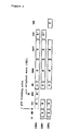
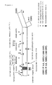
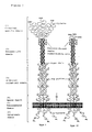

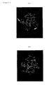
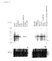
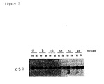


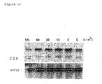
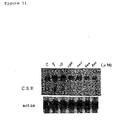
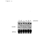
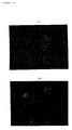
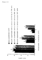
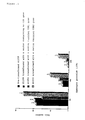
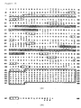
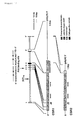


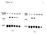


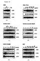

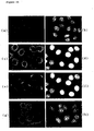
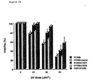

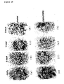

Classifications
-
- C—CHEMISTRY; METALLURGY
- C07—ORGANIC CHEMISTRY
- C07K—PEPTIDES
- C07K14/00—Peptides having more than 20 amino acids; Gastrins; Somatostatins; Melanotropins; Derivatives thereof
- C07K14/435—Peptides having more than 20 amino acids; Gastrins; Somatostatins; Melanotropins; Derivatives thereof from animals; from humans
- C07K14/705—Receptors; Cell surface antigens; Cell surface determinants
-
- A—HUMAN NECESSITIES
- A61—MEDICAL OR VETERINARY SCIENCE; HYGIENE
- A61K—PREPARATIONS FOR MEDICAL, DENTAL OR TOILETRY PURPOSES
- A61K39/00—Medicinal preparations containing antigens or antibodies
- A61K2039/505—Medicinal preparations containing antigens or antibodies comprising antibodies
Definitions
- the present invention relates to novel scavenger receptor-like proteins and fragments thereof, DNAs encoding said proteins and fragments thereof, expression vectors containing said DNAs, transformants transformed with said expression vectors, antibodies capable of reacting with said proteins or fragments thereof, and cells producing said antibodies.
- Cholesterols are present in every tissue and plasma of the body in the free, long-chain fatty acid, or esterified form. The former two forms play critical roles in cell membrane construction, and the esterified one is a physiologically inactive storage form. Cholesterols in the body originates from dietary cholesterol absorbed through small intestine following food intake, or are biosynthesized in various tissues, especially in liver. Free cholesterols synthesized in and secreted from the liver is incorporated into very low density lipoproteins (VLDL), and metabolized via intermediary density lipoproteins (IDL) to low density lipoproteins by the action of lipoprotein lipase in the blood and hepatic triglyceride lipase (HTGL). LDL thus formed is delivered to peripheral tissues by the blood stream, taken up into peripheral cells such as vascular endothelial cells mediated by peripheral LDL receptors to supply necessary amount of cholesterols for constructing peripheral cells.
- VLDL very low density lipoproteins
- IDL intermediar
- the LDL receptor is down-regulated with the increase of intracellular cholesterols due to the LDL uptake so as to prevent its excessive accumulation.
- peripheral macrophages usually express almost no normal LDL receptors but the scavenger receptors to recognize modified LDLs such as oxidized LDL and take up modified LDLs via this receptors (Nature, 343 , 531-535, 1990; ibid., 343 , 570-572, 1990; Proc. Natl. Acad. Sci. USA, 87 , 9133-9137, 1990; ibid., 87 , 8810-8814, 1990: Curr. Opin. Lipodol., 2 , 295-300, 1991, and J. Clin. Invest., 90 , 1450-1457, 1991).
- this scavenger receptors unlike the above LDL receptors, do not undergo downregulation due to an increase of intracellular cholesterols, they take up modified LDLs (cholesterols) in excess to metamorphose macrophages into foam cells. Accumulation of these foam macrophages in the subendothelium and inner wall of blood vessels is considered as one of the principal causes of atherosclerosis. In fact, a recent report confirmed that atherosclerotic lesion was reduced in size in knockout mice in which the macrophage scavenger receptors were inactivated by genetic engineering, compared with control mice, proving profound involvement of the scavenger receptors in the development of atherogenesis.
- LDLs cholesterols
- the macrophage scavenger receptors There are two types of the macrophage scavenger receptors, type I and type II. Both types consist of six domains, differing only in the C-terminal structure. The receptors are unique membrane proteins with the structure of inside-out type trimeric membrane glycoprotein. Amino acid sequences of both type I and type II are highly homologous among animal species including humans, mice, cattle, and rabbits (type I: 64-81%, type II: 60-81%). The domains, from the C-terminus, comprise 1) C-terminal specific domain, 2) collagen-like domain, 3) ⁇ -helical coiled-coil domain, 4) spacer domain, 5) transmembrane domain, and 6) cytoplasmic domain.
- C-terminal specific domain of type I is cysteine-rich, and highly homologous to complement factor 1, CD5, CD6, etc.
- the "collagen-like domain” has the glycine (Gly)-X-Y repeat structure peculiar to collagen. Its C-terminal side is abundant in positive-charged amino acid residues that suitably bind to negative-charged ligands. It has been confirmed that a mutant in which C-terminal 22 amino acid residues of the collagen-like domain are deleted no longer binds to the ligand. Furthermore, the experiments using various deletion mutants clarified that the lysine residue (K 337) is critical for the ligand binding.
- the " ⁇ helical coiled-coil domain” serves to form an active receptor in terms of the protein primary structure by associating three scavenger receptor polypeptides. This domain also plays a role in binding to a ligand such as modified LDLs to enter a cell and dissociating the ligand by altering the higher order structure of the receptor depending on the pH decrease inside endosomes.
- This domain has an ⁇ -helical coiled-coil structure in which right-handed heptad repeats turn twice per seven amino acid residues, and said three polypeptides form a trimer inwardly directing hydrophobic amino acids such as leucine and isoleucine, which are present at every seven amino acid residues, and outwardly directing hydrophilic amino acids and glycosylation sites.
- the "cytoplasmic domain” has a tight-turn structure characteristic of endocytosis signals similar to NPXY sequence present in the LDL receptors and insulin receptors and YXRF sequence in the transferin receptors, and it has been demonstrated that endocytosis is suppressed by deleting these sequences.
- the sturcture of the collagen-like domain, the ligand-binding site of the scavenger receptors is known to be suitable for removing xenobiotics and wastes such as modified LDLs that are bound and precipitate on the extracellular matrix including collagen.
- An objective of this invention is to identify a novel scavenger receptor having a structure analogous to that characteristic to the known macrophage scavenger receptors and protectively acting against various intracellularly generated stresses, and provide pharmaceuticals and methods for preventing and treating morbidities and disorders caused by intracellular stresses, xenobiotics or denatured proteins generated in the body.
- the present inventors analyzed various human genes transcriptionally controlled by the tumor suppressing protein p53, which belongs to a different technical field from the known LDL receptors, oxidized LDL receptors and macrophage scavenger receptors.
- CSR genes cellular sensor-related or cellular stress response genes
- novel genes and proteins of this invention are considered to be of new scavenger receptors having the following characteristics to protectively act against various stresses generated in the cell.
- genes encoding CSRs, CSR proteins, or fragments thereof are extremely useful for developing pharmaceuticals targeting the genes or proteins that prevent and treat morbidities and disorders (for example, atherosclerosis, diabetic angiopathy, bacterial infection, etc.) caused by intracellular stresses, xenobiotics or denatured proteins produced in the body.
- morbidities and disorders for example, atherosclerosis, diabetic angiopathy, bacterial infection, etc.
- the DNAs of the present invention themselves are useful as antisense pharmaceuticals that regulate the functions of CSR at the gene level, and in gene therapy.
- the fragments (e.g. extracellular regions, various domains) of the proteins are useful, for example, as soluble protein pharmaceuticals.
- the antibodies or a portion of them reactive with the CSR proteins of the present invention are extremely useful as antibody pharmaceuticals that regulate the functions of CSR.
- DNAs genes
- proteins proteins, and antibodies of the present invention are useful as reagents for searching proteins (ligands) interacting with the proteins of the present invention, thereby elucidating the function of said ligands, and developing therapeutic drugs targeting said ligands.
- model animals can be produced by disrupting (inactivating) said genes. Function of genes and proteins of the present invention can be elucidated by analyzing the physical, biological, pathologic, and genetic characteristics of these model animals.
- model animals that have only the human-derived gene of the present invention can be produced.
- drugs compounds, antibodies, and so on
- the present invention provides the DNAs, proteins, expression vectors, transformants, antibodies, pharmaceutical compositions, and cells, mentioned below.
- proteins or fragments thereof of this invention meant proteins and fragments thereof derived from mammals such as humans, cattle, sheep, pigs, goats, rabbits, rats, hamsters, guinea pigs and mice, preferably from humans, rabbits, rats or mice, most preferably from humans.
- Particularly preferable examples are (1) a protein having the amino acid sequence of SEQ ID NO: 10 or an amino acid sequence substantially the same as said amino acid sequence, or its fragment, (2) a protein having the amino acid sequence of SEQ ID NO: 12 or an amino acid sequence substantially the same as said amino acid sequence, or its fragment, and (3) a protein having the amino acid sequence of SEQ ID NO: 14 or an amino acid sequence substantially the same as said amino acid sequence, or its fragment.
- “having substantially the same amino acid sequence” means to include a protein having an amino acid sequence where multiple amino acids, preferably 1 to 10 amino acids, particularly preferably 1 to 5 amino acids, in the amino acid sequence shown in SEQ ID NO: 10, 12, or 14, are substituted, deleted, and/or modified, and a protein having an amino acid sequence where multiple amino acids, preferably 1 to 10 amino acids, particularly preferably 1 to 5 amino acids, are added to said amino acid sequence, as far as the protein has substantially the same biological properties as the protein having said amino acid sequence.
- fragments of proteins mean any fragments of amino acid sequences in CSR proteins of this invention, including, for example, each domain such as extracelluar region, transmembrane region or cytoplasmic region, or sites of each region for binding to (interacting with) other proteins and low molecular weight ligands.
- fusion proteins comprising the extracellular region and other molecules (for example, the constant region (Fc) of human immunoglobulin heavy chain) or portion thereof are also in the scope of protein fragments of this invention.
- a transmembrane protein such as CSR proteins of the present invention, various G protein-coupled receptors, or cell surface molecules connects with the membrane through the hydrophobic peptide region penetrating the lipid bilayer of the membrane once or several times and has structure composed of three main regions, that is, extracellular region, transmembrane region, and cytoplasmic region.
- Such a transmembrane protein exists as a monomer, homodimer, heterodimer, or oligomer with another chain(s) having the same or different amino acid sequence.
- extracellular region means the partial structure (partial sequence) existing outside of the membrane that holds the transmembrane protein as mentioned above among the whole structure of said membrane protein. In other words, it corresponds to the region excluding the region incorporated into the membrane (transmembrane region) and the region existing in the cytoplasm following the transmembrane region (cytoplasmic region). If desired, one to five amino acids derived from the amino acids constituting the transmembrane and/or cytoplasmic region can be added to the N- terminus and/or C- terminus of the extracellular region in the present invention.
- the constant region or its portion of human immunoglobulin heavy chain used herein means the constant region or the Fc region of human-derived immunoglobulin heavy chain (H chain) as described, or a portion of them.
- the immunoglobulin can be any immunoglobulin belonging to any class and any subclass. Specifically, examples of the immunoglobulin are IgG (IgG1, IgG2, IgG3, and IgG4), IgM, IgA (IgA1 and IgA2), IgD, and IgE. Preferably, the immunoglobulin is IgG (IgG1, IgG2, IgG3, or IgG4), or IgM. Examples of particularly preferable immunoglobulin of the present invention are those belonging to human-derived IgG (IgG1, IgG2, IgG3, or IgG4).
- Immunoglobulin has a Y-shaped structural unit in which four chains composed of two homologous light chains (L chains) and two homologous heavy chains (H chains) are connected through disulfide bonds (S-S bonds).
- the light chain is composed of the light chain variable region (VL) and the light chain constant region (CL).
- the heavy chain is composed of the heavy chain variable region (VH) and the heavy chain constant region (CH).
- the heavy chain constant region is composed of some domains having the amino acid sequences inherent in each class (IgG, IgM, IgA, IgD, and IgE) and each subclass (IgG1, IgG2, IgG3, and IgG4, IgA1, and IgA2).
- the heavy chain of IgG (IgG1, IgG2, IgG3, and IgG4) is composed of VH, CH1 domain, hinge region, CH2 domain, and CH3 domain in this order from N terminus.
- the heavy chain of IgG1 is composed of VH, C ⁇ 1 1 domain, hinge region, C ⁇ 1 2 domain, and C ⁇ 1 3 domain in this order from N terminus.
- the heavy chain of IgG2 is composed of VH, C ⁇ 2 1 domain, hinge region, C ⁇ 2 2 domain, and C ⁇ 2 3 domain in this order from N terminus.
- the heavy chain of IgG3 is composed of VH, C ⁇ 3 1 domain, hinge region, C ⁇ 3 2 domain, and C ⁇ 3 3 domain in this order from N terminus.
- the heavy chain of IgG4 is composed of VH, C ⁇ 4 1 domain, hinge region, C ⁇ 4 2 domain, and C ⁇ 4 3 domain in this order from N terminus.
- the heavy chain of IgA is composed of VH, C ⁇ 1 domain, hinge region, C ⁇ 2 domain, and C ⁇ 3 domain in this order from N terminus.
- the heavy chain of IgA1 is composed of VH, C ⁇ 1 1 domain, hinge region, C ⁇ 1 2 domain, and C ⁇ 1 3 domain in this order from N terminus.
- the heavy chain of IgA2 is composed of VH, C ⁇ 2 1 domain, hinge region, C ⁇ 2 2 domain, and C ⁇ 2 3 domain in this order from N terminus.
- the heavy chain of IgD is composed of VH, C ⁇ 1 domain, hinge region, C ⁇ 2 domain, and C ⁇ 3 domain in this order from N terminus.
- the heavy chain of IgM is composed of VH, C ⁇ 1 domain, C ⁇ 2 domain, C ⁇ 3 domain, and C ⁇ 4 domain in this order from N terminus and have no hinge region as seen in IgG, IgA, and IgD.
- the heavy chain of IgE is composed of VH, C ⁇ 1 domain, C ⁇ 2 domain, C ⁇ 3 domain, and C ⁇ 4 domain in this order from N terminus and have no hinge region as seen in IgG, IgA, and IgD.
- IgG is treated with papain, it is cleaved at the slightly N terminal side beyond the disulfide bonds existing in the hinge region where the disulfide bonds connect the two heavy chains to generate two homologous Fab, in which a heavy chain fragment composed of VH and CH1 is connected with one light chain through a disulfide bond, and one Fc, in which two homologous heavy chain fragments composed of the hinge region, CH2 domain, and CH3 domain are connected through disulfide bonds (See “Immunology Illustrated", original 2nd ed., Nankodo, pp.65-75 (1992); and "Focus of Newest Medical Science 'Recognition Mechanism of Immune System'", Nankodo, pp.4-7 (1991); and so on).
- a portion of a constant region of immunoglobulin heavy chain of the present invention means a portion of a constant region of an immunoglobulin heavy chain having the structural characteristics as mentioned above, and preferably, is the constant region without C1 domain, or the Fc region.
- examples thereof are the region composed of hinge region, C2 domain, and C3 domain from each of IgG, IgA, and IgD, and are the region composed of C2 domain, C3 domain, and C4 domain from each of IgM and IgE.
- a particularly preferable example thereof is the Fc region of human-derived IgG1.
- a preferable example of the fusion protein included in the protein fragments of the present invention is that composed of the above-described extracellular region of the protein of the present invention and a constant region or a portion of a constant region of human immunoglobulin heavy chain.
- it is a fusion polypeptide composed of an extracellular region of a protein of the present invention and a portion of a constant region of human IgG heavy chain, and particularly preferably, it is a fusion polypeptide composed of an extracellular region of a protein of the present invention and the region (Fc) composed of a hinge region, CH2 domain, and CH3 domain of human IgG heavy chain.
- IgG1 is preferable among IgG.
- a protein derived from human, mouse, or rat is preferable as the protein of the present invention.
- Alphabetical triplet or single letter codes used to represent amino acids in the present specification or figures mean amino acids as follows.
- the protein, protein fragment, and fusion protein of the present invention can be produced not only by recombinant DNA technology as mentioned below but also by a method well known in the art such as a chemical synthetic method and a cell culture method, or a modified method thereof.
- the above fusion protein of the present invention has the advantage that the fusion polypeptide can be purified extremely easily by using affinity column chromatography using the property of protein A, which binds specifically to the immunoglobulin fragment because the fusion polypeptide of the present invention has a portion of a constant region (for example Fc) of an immunoglobulin such as IgG as mentioned above as a fusion partner. Moreover, since various antibodies against the Fc of various immunoglobulin are available, an immunoassay for the fusion polypeptides can be easily performed with antibodies against the Fc.
- Fc constant region
- the DNA of the present invention encodes the above-mentioned protein of the present invention or its fragment, and includes any nucleotide sequence that can encode the protein of the present invention.
- the DNA of the present invention includes either a genomic DNA or cDNA.
- the DNA includes any DNA composed of any codons encoding the same amino acids.
- a preferable examle of the DNA of the present invention is a DNA encoding a human-derived protein.
- Tm 82.3°C + 0.41 x (G+C) % - 500/n - 0.61 x (formamide) % (n means the number of the nucleotide of probe).
- Temperature Tm -25°C.
- Tm varies as (1) and (2) mentioned below.
- the temperature conditions for the combination of completely complementary strands can be set as follows.
- the temperature conditions for the combination of incompletely complementary strands can be set as follows.
- the DNA of the present invention can be a DNA obtained by any method.
- the DNA includes complementary DNA (cDNA) prepared from mRNA, DNA prepared from genomic DNA, DNA prepared by chemical synthesis, DNA obtained by PCR amplification with RNA or DNA as a template, and DNA constructed by appropriately combining these methods.
- cDNA complementary DNA
- the DNA encoding the protein of the present invention can be obtained by the usual method such as a method to clone cDNA from mRNA encoding the protein of the present invention, a method to isolate genomic DNA and then splice them, chemical synthesis and so on.
- the present invention also relates to a recombinant vector comprising the DNA encoding the protein of the present invention.
- the recombinant vector of the present invention is not limited as long as it can be replicated and maintained or can autonomously replicate in various prokaryotic and/or eukaryotic hosts.
- the vector of the present invention includes plasmid vectors and phage vectors.
- the recombinant vector can easily be prepared by ligating the DNA encoding the protein of the present invention with a vector for recombination available in the art (plasmid DNA and bacteriophage DNA) by the usual method.
- plasmid DNA and bacteriophage DNA plasmid DNA and bacteriophage DNA
- specific examples of the vectors for recombination used ar e E.coli -derived plasmids such as pBR322, pBR325, pUC12, pUC13, and pUC19, yeast-derived plasmids such as pSH19 and pSH15, and Bacillus subtilis -derived plasmids such as pUB110, pTP5, and pC194.
- phages are a bacteriophage such as ⁇ phage, and an animal or insect virus (pVL1393, Invitrogen) such as a retrovirus, vaccinia virus, and nuclear polyhedrosis virus.
- An expression vector is useful for expressing the DNA encoding the protein of the present invention and for producing the polypeptide of the present invention.
- the expression vector is not limited as long as it expresses the gene encoding the polypeptide of the present invention in various prokaryotic and/or eukaryotic host cells and produces this protein. Examples thereof are pMAL C2, pEF-BOS (Nucleic Acids Res. Vol.18, p.5322 (1990)), pME18S (Experimental Medicine: SUPPLEMENT, " Handbook of Genetic Engineering” (1992)), and so on.
- an expression vector is generally comprised of, at least, a promoter/operator region, an initiation codon, the DNA encoding the protein of the present invention, termination codon, terminator region, and replicon.
- an expression vector is preferably comprised of, at least, a promoter, an initiation codon, the DNA encoding the protein of the present invention, and a termination codon. It may also comprise the DNA encoding a signal peptide, enhancer sequence, 5'- and 3'-untranslated region of the gene encoding the protein of the present invention, splicing junctions, polyadenylation site, selectable marker region, and replicon.
- the expression vector may also contain, if required, a gene for gene amplification (marker) that is usually used.
- a promoter/operator region to express the polypeptide of the present invention in bacteria comprises a promoter, an operator, and a Shine-Dalgarno (SD) sequence (for example, AAGG).
- SD Shine-Dalgarno
- the host when the host is Escherichia , it preferably comprises Trp promoter, lac promoter, recA promoter, ⁇ PL promoter, lpp promoter, tac promoter, or the like.
- Examples of a promoter to express the polypeptide of the present invention in yeast are PH05 promoter, PGK promoter, GAP promoter, ADH promoter, and so on.
- examples thereof are SL01 promoter, SP02 promoter, penP promoter and so on.
- the host is a eukaryotic cell such as a mammalian cell
- examples thereof are SV40-derived promoter, retrovirus promoter, heat shock promoter, and so on, and preferably SV-40 and retrovirus-derived one.
- the promoter is not limited to the above examples.
- an enhancer is effective for expression.
- a preferable initiation codon is, for example, a methionine codon (ATG).
- termination codon for example, TAG, TGA, TAA, and so on
- TAG TAAA
- TAA TAA
- terminators are used as a terminator region.
- a replicon means a DNA capable of replicating the whole DNA sequence in host cells, and includes a wild-type plasmid, an artificially modified plasmid (DNA fragment prepared from a wild-type plasmid), a synthetic plasmid, and so on.
- a preferable plasmids are pBR322 or its artificial derivatives (DNA fragment obtained by treating pBR322 with appropriate restriction enzymes) for E. coli , yeast 2 ⁇ plasmid or yeast chromosomal DNA for yeast, and pRSVneo ATCC 37198, pSV2dhfr ATCC 37145, pdBPV-MMTneo ATCC 37224, pSV2neo ATCC 37149, etc. for mammalian cells.
- An enhancer sequence, polyadenylation site, and splicing junction that are usually used in the art, such as those derived from SV40 can be also used.
- a selectable marker usually used can be used according to the usual method.
- examples thereof are resistance genes for antibiotics, such as tetracycline, neomycin, ampicillin, or kanamycin, and thymidine kinase gene.
- Examples of a gene for gene amplification are dihydrofolate reductase (DHFR) gene, thymidine kinase gene, neomycin resistance gene, glutamate synthase gene, adenosine deaminase gene, ornithine decarboxylase gene, hygromycin-B-phophotransferase gene, aspartate transcarbamylase gene, etc.
- DHFR dihydrofolate reductase
- thymidine kinase gene thymidine kinase gene
- neomycin resistance gene glutamate synthase gene
- glutamate synthase gene glutamate synthase gene
- adenosine deaminase gene adenosine deaminase gene
- ornithine decarboxylase gene hygromycin-B-phophotransferase gene
- aspartate transcarbamylase gene etc
- the expression vector of the present invention can be prepared by continuously and circularly linking at least the above-mentioned promoter, initiation codon, DNA (gene) encoding the polypeptide of the present invention, termination codon, and terminator region, to an appropriate replicon.
- appropriate DNA fragments for example, linkers, restriction sites generated with other restriction enzyme
- Transformants of the present invention can be prepared by introducing the expression vector mentioned above into host cells.
- Host cells used in the present invention are not limited as long as they are compatible with an expression vector mentioned above and can be transformed. Examples thereof are various cells such as wild-type cells or artificially established recombinant cells usually used in technical field of the present invention (for example, bacteria ( Escherichia and Bacillus ), yeast ( Saccharomyces , Pichia , etc.), animal cells, or insect cells.
- bacteria Escherichia and Bacillus
- yeast Saccharomyces , Pichia , etc.
- animal cells for example, insect cells.
- E. coli or animal cells are preferably used. Specific examples are E. coli (DH5 ⁇ , TB1, HB101, etc.), mouse-derived cells (COP, L, C127, Sp2/0, NS-1, NIH 3T3, etc.), rat-derived cells, hamster-derived cells (BHK, CHO, etc.), monkey-derived cells (COS1, COS3, COS7, CV1, Velo, etc.), and human-derived cells (Hela, diploid fibroblast-derived cells, myeloma, Namalwa, etc.).
- E. coli DH5 ⁇ , TB1, HB101, etc.
- mouse-derived cells COP, L, C127, Sp2/0, NS-1, NIH 3T3, etc.
- rat-derived cells hamster-derived cells
- BHK, CHO, etc. monkey-derived cells
- COS1, COS3, COS7, CV1, Velo, etc. monkey-derived cells
- human-derived cells Hela, diploid fibro
- An expression vector can be introduced (transformed (transduced)) into host cells by known method.
- Transformation can be performed, for example, according to the method of Cohen et al. (Proc. Natl. Acad. Sci. USA, Vol.69, p.2110 (1972)), protoplast method (Mol. Gen. Genet., Vol.168, p.111 (1979)), or competent method (J. Mol. Biol., Vol.56, p.209 (1971)) when the hosts are bacteria ( E. coli , Bacillus subtilis , etc.), the method of Hinnen et al. (Proc. Natl. Acad. Sci. USA, Vol.75, p.1927 (1978)), or lithium method (J.
- the protein of the present invention can be produced by cultivating transformants (in the following this term includes transductants) comprising an expression vector prepared as mentioned above in nutrient media.
- the nutrient media preferably comprise carbon source, inorganic nitrogen source, or organic nitrogen source necessary for the growth of host cells (transformants).
- the carbon source are glucose, dextran, soluble starch, and sucrose
- examples of the inorganic or organic nitrogen source are ammonium salts, nitrates, amino acids, corn steep liquor, peptone, casein, meet extract, soy bean cake, and potato extract.
- they may comprise other nutrients (for example, an inorganic salt (for example, calcium chloride, sodium dihydrogenphosphate, and magnesium chloride), vitamins, antibiotics (for example, tetracycline, neomycin, ampicillin, kanamycin, etc.).
- Cultivation is performed by a method known in the art. Cultivation conditions such as temperature, pH of the media, and cultivation time are selected appropriately so that the protein of the present invention is overproduced.
- liquid media comprising the nutrient source mentioned above are appropriate.
- the media with pH 5 to 8 are preferably used.
- examples of preferable media are LB media, and M9 media (Miller et al. Exp. Mol. Genet., Cold Spring Harbor Laboratory, p.431 (1972)). Using these media, cultivation can be performed usually at 14 to 43 °C for about 3 to 24 hours with aeration and stirring, if necessary.
- cultivation can be performed usually at 30 to 40 °C for about 16 to 96 hours with aeration and stirring, if necessary.
- examples of media are Burkholder minimal media (Bostian, Proc. Natl. Acad. Sci. USA, Vol.77, p.4505 (1980)).
- the pH of the media is preferably 5 to 8. Cultivation can be performed usually at 20 to 35°C for about 14to 144 hours with aeration and stirring, if necessary.
- examples of media are MEM media containing about 5 to 20% fetal bovine serum (Science, Vol.122, p.501 (1952)), DMEM media (Virology, Vol.8, p.396 (1959)), RPMI1640 media (J. Am. Med. Assoc., Vol.199, p.519 (1967)), and 199 media (Proc. Soc. Exp. Biol. Med., Vol.73, p.1 (1950)).
- the pH of the media is preferably about 6 to 8. Cultivation can be performed usually at about 30 to 40°C for about 15 to 72 hours with aeration and stirring, if necessary.
- an example of media is Grace's media containing fetal bovine serum (Proc. Natl. Acad. Sci. USA, Vol.82, p.8404 (1985)).
- the pH thereof is preferably about 5to 8.
- Cultivation can be performed usually at about 20 to 40°C for 15 to 100 hours with aeration and stirring, if necessary.
- the protein of the present invention can be produced by cultivating transformants as mentioned above, in particular animal cells to overexpress the protein of the present invention on the surface of the cells.
- the protein of the present invention can be produced as a soluble protein fragment such as an extracellular region protein fragment by preparing the transformants as mentioned above using the DNA encoding the extracellular region and by cultivating the transformants to allow them to secrete the soluble polypeptide into the culture supernatant.
- a culture filtrate (supernatant) is obtained by the method such as filtration or centrifugation of the obtained culture, and the protein of the present invention is purified and isolated from the culture filtrate by the usual method commonly used in order to purify and isolate a natural or synthetic protein.
- isolation and purification method examples include a method utilizing solubility, such as salting out and solvent precipitation method, a method utilizing the difference in molecular weight, such as dialysis, ultrafiltration, gel filtration, and sodium dodecyl sulfate-polyacrylamide gel electrophoresis, a method utilizing charges, such as ion exchange chromatography and hydroxylapatite chromatography, a method utilizing specific affinity, such as affinity chromatography, a method utilizing the difference in hydrophobicity, such as reverse phase high performance liquid chromatography, and a method utilizing the difference in isoelectric point, such as isoelectric focusing.
- solubility such as salting out and solvent precipitation method
- a method utilizing the difference in molecular weight such as dialysis, ultrafiltration, gel filtration, and sodium dodecyl sulfate-polyacrylamide gel electrophoresis
- charges such as ion exchange chromatography and hydroxylapatite chromatography
- the fungus bodies or cells are harvested by the usual method such as filtration or centrifugation and suspended in appropriate buffer.
- the membrane fraction comprising the protein of the present invention is obtained by the method such as centrifugation or filtration.
- the membrane fraction is solubilized with a detergent such as Triton-X100 to obtain the crude extract.
- the polypeptide or the polypeptide fragment is isolated and purified from the crude extract by the usual method as illustrated above.
- a non-human transgenic animal in which the above human-derived DNA has been integrated into the endogenous locus of the animal.
- This non-human transgenic animal express and expresses and secretes the protein of the present invention encoded by the above DNA into its body.
- the present invention include such non-human transgenic animals.
- the non-human tranegenic animal can be prepared according to the method as usually used for producing a transgenic animal (for example, see “Newest Manual of Animal Cell Experiment", LIC press, Chapter 7, pp.361-408, (1990)).
- a transgenic mouse can be produced as follows. Embryonic stem cells (ES cells) obtained from normal mouse blastocysts are transformed with an expression vector in which the gene encoding human-derived polypeptide of the present invention and a marker gene (e.g., neomycin resistance gene) have been operably inserted. ES cells in which the gene encoding the human-derived polypeptide of the present invention has been integrated into the endogenous gene are screened in terms of the expression of the marker gene by the usual method. Then, the ES cells screened are microinjected into a fertilized egg obtained from another normal mouse (blastocyst) (Proc. Natl. Acad. Sci. USA, Vol.77, No.12, pp.7380-7384 (1980); U.S. Pat. No. 4,873,191).
- a marker gene e.g., neomycin resistance gene
- the blastocyst is transplanted into the uterus of another normal mouse as the foster mother. Then, founder mice (progeny mice) are born from the foster mother mouse. By mating the founder mice with normal mice, heterogeneic transgenic mice are obtained. By mating the heterogeneic transgenic mice with each other, homogeneic transgenic mice are obtained according to Mendel's laws.
- a so-called “knockout mouse” can be generated based on the information of the nucleotide sequence of the DNA encoding a mouse-derived protein that is included in the present invention.
- the "knockout mouse” of the present invention is a mouse wherein the endogenous gene encoding the mouse-derived protein of the present invention has been knocked out (inactivated). It can be prepared, for example, by positive-negative selection method in which homologous recombination is applied (U.S. Pat. No. 5,464,764; No. 5,487,992; No. 5,627,059; Proc. Natl. Acad. Sci. USA, Vol.86, pp.8932-8935 (1989);Nature,Vol.342,pp.435-438 (1989); etc.). This knockout mouse is also one embodiment of the present invention.
- the "antibody” of the present invention can be a polyclonal antibody (antiserum) or a monoclonal antibody, and preferably a monoclonal antibody.
- it is an antibody reactive to (against, which binds to) the above-mentioned protein or its fragment of the present invention.
- the antibody of the present invention can be natural antibodies obtained by immunizing mammals such as mice, rats, hamsters, guinea pigs, and rabbits with the protein of the present invention (natural, recombinant, synthetic, or cell-derived ones), its fragment, or transformants overexpressing the designed protein on the surface thereof prepared using recombinant DNA technology as described above on the cell surface.
- the antibody of the present invention also includes chimeric antibodies and humanized antibodies (CDR-grafted antibodies) that can be produced by recombinant DNA technology, and human antibodies that can be produced using human antibody-producing transgenic animals.
- the monoclonal antibody includes those having any one isotype of IgG, IgM, IgA, IgD, or IgE. IgG or IgM is preferable.
- the polyclonal antibody (antisera) or monoclonal antibody of the present invention can be produced by the known methods. Namely, a mammal, preferably, a mouse, rat, hamster, guinea pig, rabbit, cat, dog, pig, goat, horse, or cattle, or more preferably, a mouse, rat, hamster, guinea pig, or rabbit is immunized, for example, with an antigen mentioned above with Freund's adjuvant, if necessary.
- the polyclonal antibody can be obtained from the antiserum obtained from the animal so immunized.
- the monoclonal antibodies are produced as follows.
- Hybridomas are prepared from the antibody-producing cells obtaind from the animal so immunized and myeloma cells that are not capable of producing autoantibodies.
- the hybridomas are cloned, and clones producing the monoclonal antibodies showing the specific affinity to the antigen used for immunizing the mammal are screened.
- the monoclonal antibody can be produced as follows. Immunizations are performed by injecting or implanting once or several times the protein of the present invention, its fragment, cells expressing the protein and so on as mentioned above as an immunogen, if necessary, with Freund's adjuvant, subcutaneously, intramuscularly, intravenously, through the footpad, or intraperitoneally into a mouse, rat, hamster, guinea pig, or rabbit, preferably a mouse, rat, or hamster (including a transgenic animal generated so as to produce antibodies derived from another animal such as the transgenic mouse producing human antibody). Usually, immunizations are performed once to four times every one to fourteen days after the first immunization. Antibody-producing cells are obtained from the mammal so immunized in about one to five days after the last immunization.
- Hybridomas that secrete a monoclonal antibody can be prepared by the method of Köhler and Milstein (Nature, Vol.256, pp.495-497 (1975)) and by its modified method. Namely, hybridomas are prepared by fusing antibody-producing cells contained in a spleen, lymph node, bone marrow, or tonsil obtained from the mammal immunized as mentioned above, preferably a spleen, with myelomas without autoantibody-producing ability, which are derived from, preferably, a mammal such as a mouse, rat, guinea pig, hamster, rabbit, or human, or more preferably, a mouse, rat, or human.
- mouse-derived myeloma P3/X63-AG8.653 (653, ATCC No. CRL1580), P3/NSI/1-Ag4-1 (NS-1), P3/X63-Ag8.U1 (P3U1), SP2/0-Ag14 (Sp2/0, Sp2), PAI, F0, or BW5147, rat-derived myeloma 210RCY3-Ag.2.3., or human-derived myeloma U-266AR1, GM1500-6TG-A1-2, UC729-6, CEM-AGR, D1R11, or CEM-T15 can be used as a myeloma used for the cell fusion.
- Hybridoma clones producing monoclonal antibodies can be screened by cultivating hybridomas, for example, in microtiter plates and by measuring the reactivity of the culture supernatant in the well in which hybridoma growth is observed, to the immunogen used for the immunization mentioned above, for example, by enzyme immunoassay such as RIA and ELISA.
- the monoclonal antibodies can be produced from hybridomas by cultivating the hybridomas in vitro or in vivo such as in the ascites fluid of a mouse, rat, guinea pig, hamster, or rabbit, preferably a mouse or rat, more preferably mouse and isolating the antibodies from the resulting the culture supernatant or ascites fluid of a mammal.
- Cultivating hybridomas in vitro can be performed depending on the property of cells to be cultured, on the object of a test study, and on the various conditions of a cultivating method, by using known nutrient media or any nutrient media derived from known basal media for growing, maintaining, and storing the hybridomas to produce monoclonal antibodies in culture supernatant.
- basal media examples include low, calcium concentration media such as Ham'F12 medium, MCDB153 medium, or low calcium concentration MEM medium, and high calcium concentration media such as MCDB104 medium, MEM medium, D-MEM medium, RPMI1640 medium, ASF104 medium, or RD medium.
- the basal media can contain, for example, sera, hormones, cytokines, and/or various inorganic or organic substances depending on the objective.
- Monoclonal antibodies can be isolated and purified from the culture supernatant or ascites fluid mentioned above by saturated ammonium sulfate precipitation, euglobulin precipitation method, caproic acid method, caprylic acid method, ion exchange chromatography (DEAE or DE52), affinity chromatography using anti-immunoglobulin column or protein A column.
- monoclonal antibodies can be obtaied in a large quantity by cloning a gene encoding a monoclonal antibody from a hybridoma, generating transgenec animals such as a rabbit, goat, sheep, or pig in which the gene encoding the monoclonal antibody is integrated in its endogenous gene using transgenic animal generating technique, and recovering the monoclonal antibody derived from the antibody gene from milk of the transgenic animal (Nikkei Science, No.4, pp.78-84 (1997)).
- the "chimeric antibody” of the present invention is a monoclonal antibody prepared by genetic engineering, and specifically means a chimeric antibody such as mouse/human chimeric monoclonal antibody whose variable regions or the other regions are derived from mouse immunoglobulin and whose constant regions are derived from human immunoglobulin.
- the constant region derived from human immunoglobulin has the amino acid sequence inherent in each isotype such as IgG, IgM, IgA, IgD, and IgE.
- the constant region of the recombinant chimeric monoclonal antibody of the present invention can be that of human immunoglobulin belonging to any isotype. Preferably, it is the constant region of human IgG.
- the chimeric monoclonal antibody of the present invention can be produced, for example, as follows. Needless to say, the production method is not limited thereto.
- a mouse/human chimeric monoclonal antibody can be prepared, referring to Experimental Medicine: SUPPLEMENT, Vol.1.6, No.10 (1988); and examined published Japanese patent application (JP-B) No. Hei 3-73280. Namely, it can be prepared by operably inserting CH gene (C gene encoding the constant region of H chain) obtained from the DNA encoding human immunoglobulin downstream of active VH genes (rearranged VDJ gene encoding the variable region of H chain) obtained from the DNA encoding a mouse monoclonal antibody isolated from the hybridoma producing the mouse monoclonal antibody, and CL gene (C gene encoding the constant region of L chain) obtained from the DNA encoding human immunoglobulin downstream of active VL genes (rearranged VJ gene encoding the variable region of L chain) obtained from the DNA encoding the mouse monoclonal antibody isolated from the hybridoma, into the same or different vectors so as for them to be expressed, following by transforming host cells with the expression vector, and then by cultivating the transform
- DNAs are first extracted from mouse monoclonal antibody-producing hybridomas by the usual method, digested with appropriate restriction enzymes (for example, EcoRI and HindIII), electrophoresed (using, for example, 0.7% agarose gel), and analyzed by Southern blotting. After an electrophoresed gel is stained, for example, with ethidium bromide and photographed, the gel is given with marker positions, washed twice with water, and soaked in 0.25 M HCl for 15 minutes. Then, the gel is soaked in 0.4 N NaOH solution for 10 minutes with gently stirring. The DNAs are transferred to a filter for 4 hours by the usual method. The filter is recovered and washed twice with 2xSSC.
- appropriate restriction enzymes for example, EcoRI and HindIII
- electrophoresed using, for example, 0.7% agarose gel
- Southern blotting After an electrophoresed gel is stained, for example, with ethidium bromide and photographed, the gel is given with marker positions, washed twice with water, and soaked
- the filter After the filter is sufficiently dried, it is baked at 75°C for 3 hours. After baking, the filter is treated with 0.1 x SSC/0.1% SDS at 65°C for 30 minutes. Then, it is soaked in 3 x SSC/0.1% SDS. The filter obtained is treated with prehybridization solution in a plastic bag at 65°C for 3 to 4 hours.
- 32 P-labeled probe DNA and hybridization solution are added to the bag and reacted at 65°C about 12 hours.
- the filter is washed under appropriate salt concentration, reaction temperature, and time (for example, 2 x SSC-0.1% SDS, room temperature, 10 minutes).
- the filter is put into a plastic bag with a little 2 x SSC, and subjected to autoradiography after the bag is sealed.
- Rearranged VDJ gene and VJ gene encoding H chain and L chain of a mouse monoclonal antibody are identified by Southern blotting mentioned above.
- the region comprising the identified DNA fragment is fractioned by sucrose density gradient centrifugation and inserted into a phage vector (for example, Charon 4A, Charon 28, ⁇ EMBL3, ⁇ EMBL4, etc.).
- E. coli for example, LE392, NM539, etc. is transformed with the phage vector to generate a genomic library.
- the genomic library is screened by plaque hybridization such as Benton-Davis method (Science, Vol.196, pp.180-182 (1977)) using appropriate probes (H chain J gene, L chain ( ⁇ ) J gene, etc.) to obtain positive clones comprising rearranged VDJ gene or VJ gene.
- plaque hybridization such as Benton-Davis method (Science, Vol.196, pp.180-182 (1977)) using appropriate probes (H chain J gene, L chain ( ⁇ ) J gene, etc.) to obtain positive clones comprising rearranged VDJ gene or VJ gene.
- human CH gene and human CL gene used for chimerization are isolated.
- C ⁇ 1 gene as a CH gene and C ⁇ gene as a CL gene, are isolated.
- These genes can be isolated from human genomic library with mouse C ⁇ 1 gene and mouse C ⁇ gene, corresponding to human C ⁇ 1 gene and human C ⁇ gene, respectively, as probes, taking advantage of high homology between the nucleotide sequences of mouse immunoglobulin gene and that of human immunoglobulin gene.
- DNA fragments comprising human C ⁇ gene and an enhancer region are isolated from human ⁇ Charon 4A HaeIII-AluI genomic library (Cell, Vol.15, pp.1157-1174 (1978)), for example, with a 3 kb HindIII-BamHI fragment of clone Ig146 (Proc. Natl. Acad. Sci. USA, Vol.75, pp.4709-4713 (1978)) and a 6.8 kb EcoRI fragment of clone MEP10 (Proc. Natl. Acad. Sci. USA, Vol.78, pp.474-478 (1981)) as probes.
- mouse VH gene is inserted downstream mouse VH gene
- human CL gene is inserted downstream mouse VL gene into an expression vector such as pSV2gpt or pSV2neo with appropriate restriction enzymes and DNA ligase by the usual method.
- chimeric genes of mouse VH gene/human CH gene and mouse VL gene/human CL gene can be respectively inserted in the same expression vector or in different expression vectors.
- Chimeric gene-inserted expression vector(s) thus prepared are introduced into myelomas that do not produce antibodies, for example, P3X63 ⁇ Ag8 ⁇ 653 cells or SP210 cells by protoplast fusion method, DEAE-dextran method, calcium phosphate method, or electroporation method.
- the transformants are screened by cultivating in media containing a drug corresponding to the drug resistance gene inserted into the expression vector and, then, cells producing desired chimeric monoclonal antibodies are obtained.
- Desired chimeric monoclonal antibodies are obtained from the culture supernatant of antibody-producing cells thus screened.
- the "humanized antibody (CDR-grafted antibody)" of the present invention is a monoclonal antibody prepared by genetic engineering and specifically means a humanized monoclonal antibody wherein a portion or the whole of the complementarity determining regions of the hypervariable region are derived from the complementarity determining regions of the hypervariable region from a mouse monoclonal antibody, the framework regions of the variable region are derived from the framework regions of the variable region from human immunoglobulin, and the constant region is derived from human a constant region from immunoglobulin.
- the complementarity determining regions of the hypervariable region exists in the hypervariable region in the variable region of an antibody and means three regions which directly and complementary binds to an antigen (complementarity-determining residues, CDR1, CDR2, and CDR3).
- the framework regions of the variable region means four comparatively conserved regions lying upstream, downstream or between the three complementarity determining regions (framework region, FR1, FR2, FR3, and FR4).
- a humanized monoclonal antibody means that in which the whole region except a portion or the whole of the complementarity determining regions of the hypervariable region of a nonhuman mammal-derived monoclonal antibody have been replaced with their corresponding regions derived from human immunoglobulin.
- the constant region derived from human immunoglobulin has the amino acid sequence inherent in each isotype such as IgG (IgG1, IgG2, IgG3, IgG4), IgM, IgA, IgD, and IgE.
- the constant region of a humanized monoclonal antibody in the present invention can be that from human immunoglobulin belonging to any isotype. Preferably, it is the constant region of human IgG.
- the framework regions of the constant region derived from human immunoglobulin are not particularly limited.
- the humanized monoclonal antibody of the present invention can be produced, for example, as follows. Needless to say, the production method is not limited thereto.
- a recombinant humanized monoclonal antibody derived from mouse monoclonal antibody can be prepared by genetic engineering, referring to unexamined Japanese patent publication (JP-WA) No. Hei 4-506458 and unexamined Japanese patent publication (JP-A) No. Sho 62-296890.
- JP-WA Japanese patent publication
- JP-A unexamined Japanese patent publication
- JP-A unexamined Japanese patent publication
- Sho 62-296890 accession asarcomasarcomasarcomase.
- mouse H chain CDR gene(s) and the human H chain gene(s) so isolated are operably inserted into an appropriate vector so that they can be expressed.
- the mouse L chain CDR gene(s) and the human L chain gene(s) are operably inserted into another appropriate vector so that they can be expressed.
- the mouse H chain CDR gene(s)/human H chain gene(s) and mouse L chain CDR gene(s)/human L chain gene(s) can be operably inserted into the same expression vector so that they can be expressed.
- Host cells are transformed with the expression vector thus prepared to obtain transformants producing humanized monoclonal antibody. By cultivating the transformants, desired humanized monoclonal antibody is obtained from culture supernatant.
- the "human monoclonal antibody” of the present invention is immunoglobulin in which the entire regions comprising the variable and constant region of H chain, and the variable and constant region of L chain constituting immunoglobulin are derived from the gene encoding human immunoglobulin.
- the human antibody can be produced in the same way as the production method of polyclonal or monoclonal antibodies mentioned above by immunizing, with an antigen, a transgenic animal which for example, at least human immunoglobulin gene(s) have been integrated into the locus of a non-human mammal such as a mouse by the usual method.
- a transgenic mouse producing human antibodies is prepared by the methods described in Nature Genetics, Vol.15, pp.146-156 (1997); Nature Genetics, Vol.7, pp.13-21 (1994); JP-WA Nos. Hei 4-504365, International patent publication No. WO94/25585; Nikkei Science, No.6, pp.40-50 (1995); Nature, Vol.368, pp.856-859 (1994); and JP-WA No. Hei 6-500233.
- portion of an antibody used in the present invention means a partial region of the antibody, preferably monoclonal antibody of the present invention as mentioned above, and specifically, means F(ab') 2 , Fab', Fab, Fv (variable fragment of antibody), sFv, dsFv (disulfide stabilized Fv), or dAb (single domain antibody) (Exp. Opin. Ther. Patents, Vol.6, No.5, pp.441-456 (1996)).
- F(ab') 2 " and “Fab'” can be produced by treating immunoglobulin (monoclonal antibody) with a protease such as pepsin and papain, and means an antibody fragment generated by digesting immunoglobulin near the disulfide bonds existing between the hinge regions in each of the two H chains.
- a protease such as pepsin and papain
- papain cleaves IgG upstream of the disulfide bonds existing between the hinge regions in each of the two H chains to generate two homologous antibody fragments in which an L chain composed of VL (L chain variable region) and CL (L chain constant region), and an H chain fragment composed of VH (H chain variable region) and CH ⁇ 1 ( ⁇ 1 region in the constant region of H chain) are connected at their C terminal regions through a disulfide bond.
- Fab' Each of such two homologous antibody fragments is called Fab'.
- Pepsin also cleaves IgG downstream of the disulfide bonds existing between the hinge regions in each of the two H chains to generate an antibody fragment slightly larger than the fragment in which the two above-mentioned Fab' are connected at the hinge region. This antibody fragment is called F(ab') 2 .
- cells producing a monoclonal antibody reactive with proteins or fragments thereof mean any cells producing the above-described monoclonal antibody of this invention.
- non-human mammal-derived B cells producing a monoclonal antibody reactive with proteins of this invention or fragments thereof which is obtained by immunizing a non-human mammal with proteins or fragments thereof of this invention or cells producing the proteins, etc. as described above
- hybridomas fused cells
- monoclonal antibody producing transformants obtained by transforming other cells than the B cells and hybridomas with genes (either the heavy chain-encoding gene or the light chain encoding gene, or both) encoding the monoclonal antibody isolated from the monoclonal antibody producing B cells or hybridomas.
- the monoclonal antibody producing transformants of (3) mean recombinant cells producing a recombinant product of the monoclonal antibody produced by B cells of (1) or hybridomas of (2). These recombinant monoclonal antibody producing cells can be produced in the same manner as for the above-described chimeric monoclonal antibody and humanized antibody.
- the "pharmaceutical composition” of the present invention comprises any one of the protein, its fragment, antibody, or its portion of the present invention as defined above; and a pharmaceutically acceptable carrier.
- the "pharmaceutically acceptable carrier” includes a excipieut, a diluent, an expander, a decomposition agent, a stabilizer, a preservative, a buffer, an emulsifier, an aromatic, a colorant, a sweetener, a viscosity increasing agent, a flavor, a solubility increasing agent, or other additives.
- a pharmaceutical composition can be fomulated into tablets, pills, powders, granules, injections, solutions, capsules, troches, elixirs, suspensions, emulsions, or syrups.
- the pharmaceutical composition can be administered orally or parenterally.
- Other forms for parenteral administration include a solution for external application, suppository for rectal administration, and pessary, prescribed by the usual method, which comprises one or more active ingredient.
- the dosage can vary depending on the age, sex, weight, and symptom of a patient, effect of treatment, administration route, period of treatment, or the kind of active ingredient (protein or antibody mentioned above) contained in the pharmaceutical composition.
- the pharmaceutical composition can be administered to an adult in a dose of 10 ⁇ g to 1000 mg (or 10 ⁇ g to 500 mg) per one administration.
- the dosage less than that mentioned above may be sufficient in some cases, and the dosage more than that mentioned above may be necessary in other cases.
- the injection can be produced by dissolving or suspending the antibody in a non-toxic, pharmaceutically acceptable carrier such as physiological saline or commercially available distilled water for injection with adjusting a concentration to 0.1 ⁇ g antibody/ml carrier to 10 mg antibody/ml carrier.
- a non-toxic, pharmaceutically acceptable carrier such as physiological saline or commercially available distilled water for injection with adjusting a concentration to 0.1 ⁇ g antibody/ml carrier to 10 mg antibody/ml carrier.
- the injection thus produced can be administered to a human patient in need of treatment in a dose of 1 ⁇ g to 100 mg/kg body weight, preferably 50 ⁇ g to 50 mg/kg body weight once or more times a day.
- administration route are medically appropriate administration routes such as intravenous injection, subcutaneous injection, intradermal injection, intramuscular injection, or intraperitoneal injection, preferably intravenous injection.
- the injection can also be prepared into a non-aqueous diluent (for example, propylene glycol, polyethylene glycol, vegetable oil such as olive oil, and alcohol such as ethanol), suspension, or emulsion.
- a non-aqueous diluent for example, propylene glycol, polyethylene glycol, vegetable oil such as olive oil, and alcohol such as ethanol
- suspension or emulsion.
- the injection can be sterilized by filtration with a bacteria-non-penetrated filter, by mixing bacteriocide, or by irradiation.
- the injection can be produced in the form that is prepared upon use. Namely, it is freeze-dried to be a sterile solid composition, and can be dissolved in sterile distilled water for injection or another solvent before use.
- the pharmaceutical composition of the present invention is extremely useful for preventing or treating morbidities and disorders (for example, atherosclerosis, diabetic angiopathy, bacterial infection, etc.) caused by intracellular stresses, xenobiotics or denatured proteins generated in the body.
- morbidities and disorders for example, atherosclerosis, diabetic angiopathy, bacterial infection, etc.
- Human genomic DNA fragment binding to p53 was obtained by using a yeast expression system in which human genomic DNA cloned upstream of a selective reporter gene is expressed concurrently with tumor inhibitory protein p53 (Tokino, et al., Human Molecular Genetics, Vol. 3, No. 9, 1537-1542, 1994).
- GAL1 promoter in wild-type tumor inhibitory gene p53 expression vector pRS314-SN (Nigro, J. M., et al., Mol. Cell. Biol., Vol. 12, 1357-1365, 1992) was replaced with PGK promoter to construct plasmid pRS314-PGK-p53.
- the Xho I- Bam HI fragment containing p53 cDNA cleaved from pRS314-SN was subcloned at the Sal I and Bam HI sites of plasmid pPGK (Poon, D., et al., Mol. Cell Biol., Vol. 11, 4809-4821, 1991) to construct plasmid pPGK-SN.
- the Kpn I- Sac I fragment of pPGK-SN containing PGK promoter, p53 cDNA, and PGK terminator was inserted into plasmid pRS314 (Sikorski, R. S., et al., Genetics, Vol. 122, 19-27, 1989) to construct plasmid pRS314-PGK-p53.
- Yeast YPH681 containing the above-mentioned p53 expression vector pRS314-PGK-p53 was spread on 100 ml of a SD-Trp medium and incubated overnight (1 x 10 7 cells/ml). The medium was washed by sterilized water and resuspended with 10 ml of sterilized water. The cells were washed with 10 ml of LiAc/TE (0.1 M LiAc at pH 7.5, 10 mM Tris hydrochloric acid at pH 7.5, and 1 mM EDTA) and resuspended with LiAc/TE (1 x 10 9 cells/ml).
- LiAc/TE 0.1 M LiAc at pH 7.5, 10 mM Tris hydrochloric acid at pH 7.5, and 1 mM EDTA
- Yeast cell suspension solution (0.2 ml) was mixed with 2 ⁇ g of DNA to be used for transformation (1 ⁇ g/ ⁇ l of 1 x TE), 100 ⁇ g of single stranded salmon sperm DNA (10 ⁇ g/ ⁇ l), and 0.6 ml of 40% sterilized PEG solution (40% polyethylene glycol 4000/LiAc/TE).
- the obtained cell mixture was incubated at 30°C for 30 min while vigorously being shaken, heated at 42°C for 15 min and centrifuged to obtain the pellet.
- the cellular pellet was resuspended with 1 X TE (0.2 ml), spread on a SD-trp-ura plate, and incubated at 30°C for three days.
- the reporter plasmid library (1 mg) was transformed and spread onto 500 plates. Each plate contains about 2 x 10 4 clones.
- the yeast transformants were collected from the plates and pooled for each 10 plates apiece, resuspended in a mixture of 65% glycerol (v/v), 0.1M magnesium sulfate and 25 mM Tris hydrochloric acid (pH 8), and stored at -80°C.
- Yeast cell diluted (0.1 ml) to reach 10 at the absorbance of 600 nm was spread onto the selective SD-trp-ura-his plate and cultured at 30°C for 5 days. A small amount of the transformants was spread on a non-selective SD-trp-ura plate to determine the number of living yeast cells. Each HIS+ positive cell was cultured in the SD-ura medium and the extracted DNA was introduced into Ura-deficient E. coli strain KC8. The HIS3 activity in the yeast of the collected reporter plasmids was examined in the presence or absence of the p53 expression vector.
- sequences of the p53 dependent clones were analyzed using two primers adjacent to the cloning sites; (1) a primer derived from GAL promoter (SEQ ID NO. 1) (Johnston, M., Mol. Cell. Biol., Vol. 4, 1440-1448, 1984) and (2) that derived from the plasmid pBR322 (SEQ ID NO. 2).
- ⁇ -Galactosidase assay was conducted as follows. Each of the reporter plasmid and expression vector was introduced into yeast EGY048 strain (Gyuris, J., et al., Cell, Vol. 75, 791-803, 1993) to obtain the yeast transformants and cultured in the synthetic medium containing raffinose as a carbon source at 30°C overnight. The culture medium was diluted with the medium containing 1% raffinose and 1% galactose to reach 0.1 at the absorbance of 600 nm, and cultured for 19 hours to carry out ⁇ -galactosidase assay (Kern, S. E., et al., Science, Vol. 256, 827-830, 1992).
- Human genomic DNA clones (1.8 x 10 7 cells) were inserted into the HIS3 reporter vector to prepare a plasmid library. Yeast was transformed with this first generation clones to obtain 1 x 10 7 transformants, and separated into 50 pools each containing about 2 x 10 5 individual cells. About 5 x 10 5 cells from each pool were spread onto the medium lacking histidine to select clones containing DNA reacting to tumor inhibitory protein p53. In each pool from 20 to 93 (about 50 clones on an average) histidine auxotrophic clones were detected. About 20 clones were randomly selected from each pool to be used for preparing the above-mentioned reporter DNA (refer to 1-1 and 1-2 for details).
- Each plasmid thus obtained was used to transform yeast strains containing a p53 expression vector or a control vacant vector without p53 gene.
- 273 clones (29%) were p53 dependent.
- the 273 p53 reactive clones were analyzed by the digestion with restriction enzymes and the nucleotide sequences.
- the 273 clones contained 57 different types of Mbo I digested fragments. Nucleotide sequences of inserts in each clone were determined using the sequences near (just before and just after) the cloning site as primers (refer to 1-3 for details).
- 46 clones contained 2 copies of 10bp fragment similar to the reported consensus p53 binding sequence (El-Deiry, W.S., et al., Nature Genetics, Vol. 1, 45-49, 1992).
- a cosmid library used in this invention was prepared following "Laboratory Manual Human Genome Mapping" (M. Hori and Y. Nakamura, eds., Maruzen, pp. 41-48). The method is briefly described below.
- the Bam HI site of cosmid vector pWE15 (Wahl, GM., Proc. Natl. Acad. Sci. USA., Vol. 84, 2160-2164, 1987) was filled in using the Klenow enzyme and converted to the Xho I site using an oligonucleotide linker (5' CCTCGAGG 3') to construct the modified cosmid vector pWEX15.
- the vector was digested with Xho I and partially filled in with the Klenow enzyme, dCTP, and dTTP.
- Human chromosome genomic DNA was digested with Sau 3AI and fractionated by sucrose concentration gradient centrifugation to obtain genomic DNA fragments. The DNA fragments were filled in with the Klenow enzyme, dATP and dGTP.
- the vector DNA (1 ⁇ g) and the genomic DNA (1 ⁇ g) were ligated in a mixture of 10 x ligation buffer (0.5 M Tris hydrochloric acid buffer at pH 7.5), 100 mM magnesium chloride, 100 mM dithiothreitol, 500 ⁇ g/ml bovine serum albumin), 20 mM ATP, 50% polyethylene glycol 8000, 1M sodium chloride, T4 DNA ligase (Boehringer), and distilled water, and in vitro -packaged using GIGAPACK II GOLD (Stratagene).
- 10 x ligation buffer 0.5 M Tris hydrochloric acid buffer at pH 7.5
- 100 mM magnesium chloride 100 mM dithiothreitol
- 500 ⁇ g/ml bovine serum albumin bovine serum albumin
- 20 mM ATP 50% polyethylene glycol 8000, 1M sodium chloride
- T4 DNA ligase Boehringer
- distilled water 50% polyethylene glycol 8000,
- the human genomic DNA insert (SEQ ID NO: 3) in a clone of p53-reactive human genomic DNA fragment obtained and sequenced in Example 1 (clone No. 24, p53-binding consensus-like sequence: CAACTTGTCT, refer to Human Molecular Genetics, Vol. 3, No. 9, 1537-1542, 1994) was radio-labeled to serve as a probe for hybridization.
- the above-mentioned human DNA cosmid library was screened using the probe by the standard method to obtain 25 positive clones.
- each clone was digested with Bam HI and Bgl II and ligated with exon trapping vector pSPL3 (GIBCO-BRL).
- exon trapping vector pSPL3 GEBCO-BRL
- 5' RACE-PCR Rapid Amplification Ends-PCR
- 3' RACE-PCR Proc. Natl. Aca. Sci. USA, Vol. 85, 8998-9002, 1998 and "Gene Amplification PCR Method, Basis and New Approaches" Kyoritsu, 1992
- RT-PCR Reverse transcription PCR
- mRNA derived from human fetal brain was converted into cDNA by random priming with d (N) 10 (Boehringer Mannheim) in the presence of M-MuLV reverse transcriptase (200 units/ ⁇ l, GIBCO-BRL) to prepare a human long chain cDNA library.
- the long chain cDNA library was screened by the standard method using a human cDNA fragment cleaved from the above-mentioned positive clones as a probe to obtain four positive clones. Multiple long chain cDNAs encoding human CSR protein were cleaved from the positive clones and sequenced.
- the cDNA encoding CSR protein was then amplified by RT-PCR method using EC1 (SEQ ID NO: 5) and EC2a (SEQ ID NO: 6) and EC2b (SEQ ID NO: 7) as primers.
- RT-PCR was conducted 30 to 40 cycles in cDNA mixture solution (25 ⁇ l) using Thermocycler (Perkin Elmer Cetus).
- the cDNA mixture contains 40 ng of cDNA, 1 x PCR buffer (6.7 mM Tris, pH 8.8, 16.6 mM ammonium sulfate, 6.7 ⁇ M EDTA, and 10 mM ⁇ -mercaptoethanol), 25 pmol sense primer, 25 pmol antisense primer, 0.5 U of Taq DNA polymerase (TAKARA), 250 ⁇ M deoxynucleotides, 10% DMSO, and 7.6 mM magnesium chloride.
- TAKARA Taq DNA polymerase
- Each PCR cycle was comprised of the reactions at 94°C for 30 sec, at 55°C for 30 sec, and at 72°C for 1 min.
- the final PCR step was conducted at 72°C for 5 min.
- the resulting PCR product was applied to the 3% agarose gel electrophoresis, transferred to N+ nylon membrane (PALL), and hybridized with the internal oligonucleotide probe F12 (SEQ ID NO: 8).
- cDNA was amplified by 5' RACE-PCR and 3' RACE-PCR with cDNA Amplification Kit (Clonetech) using the cDNA obtained above as a template to identify 5' and 3' ends in each long chain cDNA sequence.
- Isolation of two full-length cDNA encoding human CSR proteins encoded by each cDNA were named CRS 1 protein and CSR 2 protein, hereafter abbreviated as CSR1 and CSR2 protein, hereafter abbreviated as CSR1 and CSR2
- CSR3 protein hereafter simply referred to as CSR3
- SEQ ID NO: 9 and NO: 10 show cDNA sequence of CRS1 and its deduced amino acid sequence, respectively, SEQ ID NO: 11 and NO: 12 those of CRS2, and SEQ ID NO. 13 and NO. 14 those of CSR3.
- the sizes of the gene comprising CSR1 cDNA and its open reading frame (ORF) were about 4.0 kb and 1.8 kb, respectably, to encode 606 amino acids.
- the sizes of the gene comprising CSR2 cDNA and its ORF were about 1.9 kb and 1.4 kb, respectably, to encode 466 amino acids.
- Fig. 1 schematically shows structural differences in these three splicing mutants.
- human CSR genomic DNA is about 90 kb, composed of 6 or more coding exons, and the partial sequence (SEQ ID NO: 20) existing in the second intron sequence (between exon A and exon B in Fig. 1) was considered to fit with the reported p53 binding sequence (nucleotide sequence composed of 2 copies of symmetry 10 mers [5' RRRC (A/T) (T/A) GYYY 3'' (R is purine, and Y is prymidine); El-Deiry, W.S., et al., Nature Genetics, Vol. 1, 45-49, 1992).
- CSR1 was composed of 6 exons, A, B, C, D, E, and E'', CSR 2 of 6 exons, A, B, C, D, E, and F, and CSR 3 of at least 4 exons, C, D, E, and E'' (CSR3 may comprise B and B'' as exons because coding sequences corresponding to B and B'' at 5' end of CSR3 cDNA were found).
- Fig. 17 shows that CSR1 genomic DNA is composed of 6 exons, exons 1, 2, 3, 4, 5, and 6'.
- Genomic DNA of CSR2 was composed of 6 exons, exons 1, 2, 3, 4, 5, and 6'', in which exon 6' was replaced by exon 6'' by splicing.
- exon E in Fig. 1 shows 61% amino acid homology to collagen.
- exon D shows homology in amino acids to yeast chromosome segregation protein-smc2P, and human myosin heavy chain, and exon C to hibernation related protein-25 (HP25).
- CSR1 and CSR2 presumably have the structure as schematically shown in Fig. 2.
- CSR1 is composed of three major different domains, (1) transmembrane domain with leucine zipper motif with four repeating leucine units, (2) ⁇ -helical coiled-coil domain, and (3) collagen-like domain with 49 repeating Gly-X-Y sequence units. Three molecules comprising these characteristic structures form a trimer, forming ⁇ helix at coiled-coil domain, and triple helix at collagen-like domain.
- CSR2 protein lacks collagen-like domain of CSR1 protein and comprises therefor C terminus composed of 10 amino acids.
- the collagen-like domain is known to bind to a wide range of molecules, such as lipids, plasma components (e.g. fibronectin, laminin, collagen, fibrinogen, etc.), and many negatively charged molecules. Analysis using computer software DNASIS revealed that the collagen-like domain of CSR protein has positive net charge at physiological pH.
- CSR protein comprised many phosphorylation sites and glycosylation sites, heme binding sites and microbody (peroxisome) C-terminal targeting signal (Figs. 4 and 16). These results imply that CSR proteins undergo post-translational modification.
- Non-translational region at 3' end of CSR1 and CSR2 genes comprised sequences composed of 6 base-sequences, AGTAAA and AATAAA, respectively, near the 3' end poly (A) site. Previous studies suggest that these sequences relates to addition of polyadenylation signals.
- FISH fluorescence in situ hybridization
- cosmid clone 2 containing exon sequence of CSR gene was used as a probe.
- human metaphase chromosomes were prepared using thymidine synchronization method and bromodeoxyuridine release method.
- CSR gene was detected in all tissue except for peripheral leukocytes, and at a relatively low level in spleen and liver.
- Poly (A) + RNA blotting membrane derived from human heart, brain, placenta, lung, liver, skeletal muscle, kidney, pancreas, spleen, thymus, prostate, testis, ovary, small intestine, colon, and peripheral leukocyte was hybridized with the above-mentioned CSR1 and CSR2 labeled with [ ⁇ - 32 p]dCTP.
- the hybridized membrane was washed with 2 x SSC/0.05% SDS for 15 min at room temperature twice, and with 0.1 x SSC/0.1% SDS for 10 min at 50°C, and subjected to autoradiography using BAS1000 Imaging Plate (Fuji) for 12 to 24 hours.
- the expression level of human ⁇ actin RNA was used as a control.
- Wild-type p53 gene or mutant p53-273 gene expression vector was transiently transfected into human colorectal cancer line SW480 expressing mutant p53 (with mutation at 273rd and 309th codons) (Rodrigues, N.R. et al., Proc. Natl. Acad. Sci. USA, Vol. 87, 7555-7559, 1990) (transformants with wild-type gene and those with mutant gene are referred as W and M, respectively). After the transfection, RNA was continuously collected from transformants M and N to examine the expression parttern of CSR gene by RT-PCR. The results are shown in Fig. 7.
- RT-PCR was conducted as follows.
- Extracted whole mRNA was converted into cDNA by random priming using d(N) 10 (Boehringer Mannheim)in the presence of M-MuLV reverse transcriptase (200 units/ ⁇ l, GIBCO-BRL) to prepare a cDNA library.
- EC12 SEQ ID NO: 15
- EC2 SEQ ID NO: 16
- RT-PCR was conducted 30 to 40 cycles in a cDNA mixture (25 ⁇ l) using Thermocycler (Perkin Elmer Cetus).
- the cDNA mixture contains 40 ng of cDNA, 1 x PCR buffer (6.7 mM Tris, pH 8.8, 16.6 mM ammonium sulfate, 6.7 ⁇ M EDTA, and 10 mM ⁇ -mercaptoethanol), 25 pmol of sense primer, 25 pmol of antisense primer, 0.5 U of Tag DNA polymerase (TAKARA), 250 ⁇ M deoxynucleotides, 10% DMSO, and 7.6 mM magnesium chloride.
- TAKARA Tag DNA polymerase
- Each PCR cycle was comprised of the reactions at 94°C for 30 sec, at 55°C for 30 sec, and at 72°C for 1 min. The final step was performed at 72°C for 1 min.
- the PCR product was subjected to 3% agarose gel electrophoresis, transferred to N + nylon membrane (PALL), and Southern-hybridized using the internal oligonucleotide probe F12 (SEQ ID NO: 8).
- PALL N + nylon membrane
- the amplification of GAPDH transcript using HGS (SEQ ID NO: 17) and HGA (SEQ ID NO: 18) as primers was used as a control.
- the internal probe SEQ ID NO: 19 was used in Southern hybridization.
- Example 8 In order to confirm the result of Example 8, whether p53 protein binds to CSR gene or not was examined by electorophoretic mobility shift assay (EMSA).
- ESA electorophoretic mobility shift assay
- Human lung cancer cell line H1299 which inherenly does not express p53 protein and weakly expresses CSR mRNA, was transformed with wild-type p53 gene or mutant p53-273 gene expression plasmids (each 15 ⁇ g) (wild-type gene transformant and mutant gene transformant are referred to as W and M, respectively).
- Nuclear extract was prepared from each transformant W and M 24 hours after the transformation following the known method (Mol. Cell. Biol., Vol. 12, p2866-2871, 1992).
- the transformants (1 X 10 6 or more cells each) were suspended in 5 volumes of buffer A (20 mM of HEPES, pH 7.5, 20% glycerol, 10 mM NaCl, 10 mM MgCl 2 , 0.2 mM EDTA, 1 mM DTT, 0.1% NP40, and protease inhibitor), incubated for 10 min on ice, and centrifuged for 10 min at 4°C.
- the resulting nuclear pellet was suspended in 2 volumes of buffer B (same as buffer A except for containing 500 mM NaCl), incubated for 30 min on ice, and centrifuged for 15 min at 14,000 rpm to collect supernatant. This supernatant was adjusted to 1 to 5 ⁇ l/ml for the EMSA test.
- Each EMSA test run was conducted using nuclear extract (8 ⁇ l), buffer A (9 ⁇ l), poly (dI-dC) (1 ⁇ g, Boehringer Mannheim), monoclonal antibody PAb421 (1 ⁇ l, Oncogene Science), and/or competitor DNA (100 times or more of the amount of a non-labeled probe) on the double-stranded oligonucleotide (about 5 ng) with the labeled ends. After incubation for 30 min at room temperature, the product was electrophoresed in the gel (4% polyacrylamide/0.5 x Tris-borate-EDTA) at 200 V for 3 hours and transferred to a film at -80°C.
- p53 DNA binding consensus sequence (p53-CON, SEQ ID NO: 21) was used.
- the probe against putative p53 binding sequence existing in CSR gene (p53-CSR, SEQ ID NO: 22) was constructed with annealing oligonucleotide (Fig. 18).
- CSR expression in many tumor cells other than those used in Example 8 was analyzed by Southern hybridization using RT-PCR.
- the amplification of human ⁇ actin transcript was used as a control.
- oligonucleotides of SEQ ID NO: 23 and SEQ ID NO: 24 were used as forward and reverse primers, respectively.
- the internal oligonucleotide of SEQ ID NO: 25 was used as a probe for Southern hybridization.
- Normal human fibroblast cell line NHDF4042 (Clonetics), human neuroglioblastoma cell line U87MG, human breast adenocarcinoma cell line MCF7, ceacum adenocarcinoma SNU-C4, and human colorectal carcinoma line HCT116.
- Human cervical cancer-derived epithelium-like cell line Hela Human lung cancel cell line H1299, human esophagus cell lines (TE-3, TE10), human neuroglioblastoma cell line T98G, human carcinoma cell line SK-BR-3, and human colorectal carcinoma cell lines (SW480, HCT-15, and DLD-1), human metastatic prostate cancer cell line LNCap.FGC, human ceacum adenocarcinoma cell lines (H498, SNU-C2A), human stomach cancer cell lines (SNU-1, SNU-5, and SNU-16), and human lung cancer cell lines (ACC-LC174, ACC-LC176).
- Fig. 19 indicates that status of p53 gene (expression of p53 protein) does not necessarily correlate with CSR gene expression.
- the presence or absence of p53 protein may partially be involve in induction of CSR gene expression and CSR gene expression may be regulated by other unknown factors.
- Each of the above-mentioned human normal fibroblast cell line NHDF4042 (3 x 10 4 cells), human colorectal carcinoma cell line SW480 (3 x 10 5 cells), human neuroglioblastoma cell line T98G (3 x 10 5 cells), human esophagus cancer cell line TE13 (3 x 10 5 cells), and human cervical cancer derived epithelium-like cell line Hela (3 x 10 5 cells) were spread onto the complete medium containing 10% fetal bovine serum (FBS, GIBCO-BRL) in a culture plate with a diameter of 100 mm and cultured under 5% CO 2 at 37°C. After one to two days, the culture medium was replaced by the complete medium containing 0.1% FBS and cultured for 4 days.
- RNA was extracted from each plate to examine the expression pattern of CSR mRNA by Northern blotting assay following the standard method. Northern blotting assay was conducted as follows.
- the hybridized membrane was washed with 2 x SSC/0.05% SDS twice (at room temperature for 15 min for each wash), and with 0.1 x SSC/0.1 % SDS once (at 65°C for 10 min).
- the membrane was exposed to Imaging Plate BAS1000 (Fuji, Japan) for 24 to 48 hours.
- Imaging Plate BAS1000 Fluji, Japan
- 0.24 to 9.5 kb of RNAs 0.24 to 9.5 kb of RNAs (GIBCO-BRL) were used (Fig. 9).
- Ultraviolet irradiation was performed as follows. The cells were recovered from the culture medium and washed with phosphate butter twice prior to ultraviolet irradiation. The cells were irradiated with ultraviolet light 50 cm apart from a 40W-ramp for 5 to 120 seconds. The amount of ultraviolet irradiation was measured by Black Ray UV meter.
- CSR mRNA The expression of CSR mRNA was induced by ultraviolet irradiation. In this experiment, the highest expression was observed at the irradiation of 15 J/m 2 .
- the above-mentioned human normal fibroblast cell line NHDF4042 (50% to 70% sub-confluent by culturing in a culture medium for 24 to 48 hours) was washed with phosphate buffer twice, spread onto the complete medium supplemented with 1% fetal bovine serum, and cultured in the presence of 5 to 800 ⁇ M 30% hydrogen peroxide solution (Wako Pure Chemicals Industries) for 1 hour. The medium was then replaced with the fresh one to further continue culturing for 24 hours. RNA was extracted from the cells, and expression patterns of CSR mRNA were assayed by Northern blotting in the same manner as above. The expression levels of mRNA were analyzed using the expression level of actin RNA as a control.
- the above-mentioned human normal fibroblast cell line NHDF4042 (80 % confluent, 5 ml of complete medium) was cultured at 43°C for 40 min, and further cultured at 37°C.
- RNA was extracted from the cells with the passage of time, and expression patterns of CSR mRNA were assayed by Northern blotting in the same manner as above.
- the expression levels of mRNA were analyzed using the expression level of actin RNA as a control.
- CSR mRNA The expression of CSR mRNA was detected from 24 to 48 hours after the heat treatment.
- Expression induction by heat shock observed in this experiment occurred later than the expression induction by the stresses caused by ultraviolet irradiation and hydrogen peroxide. This implies that induction by ultraviolet irradiation and hydrogen peroxide was more directly acted via a shorter pathway compared with the induction by heat shock.
- Example 9 The results obtained in Example 9 regarding stress caused by ultraviolet irradiation and hydrogen peroxide was reexamined as follows.
- the above-mentioned human normal fibroblast cell line NHDF4042 (3 x 10 4 cells) were spread onto the complete medium (a 10-cm culture plate) and cultured for 24 to 48 hours.
- the medium was removed from the sub-confluent cultured system (50 to 70%), and the cells were washed with phosphate buffer twice. These cells were exposed to 5 to 120 J/m 2 ultraviolet radiation, and the fresh culture medium was added thereto.
- the ultraviolet light was radiated 50 cm apart from a 40W-ramp for 5 to 120 seconds. The amount of ultraviolet radiation was measured with a radiometer (Model 254, ATTO).
- Example 9 Similar to the result of Example 9 (9-2), the expression of CSR mRNA was confirmed to be induced by ultraviolet irradiation. The highest expression was observed also in this experiment at 15 J/m 2 ultraviolet radiation.
- the above-mentioned human normal fibroblast cell line NHDF4042 (3 x 10 4 cells) were spread onto the complete medium (a 10-cm culture plate) and cultured for 24 to 48 hours.
- the medium was removed from the sub-confluent cultured system (50 to 70%), and the cells were washed with phosphate buffer twice.
- the expression levels of mRNA were analyzed using the expression level of actin RNA as a control.
- the results are shown in Fig. 20(b). Similar to the result of Example 9 (9-3), the expression of CSR mRNA was confirmed to be induced by intracellular stresses caused by hydrogen peroxide. The highest expression was also observed in this experiment when the cells were exposed to 10 ⁇ M of hydrogen peroxide.
- the above-mentioned human normal fibroblast cell line NHDF4042 (3 x 10 4 cells) were spread onto the complete medium (a 10-cm culture plate) and cultured for 24 to 48 hours.
- Adriamycin (ADR) (0.02 to 1 ⁇ g/ml) was added to the sub-confluent cell culture system (50% to 70%), and cultured for 24 hours.
- the cells were then washed with phosphate buffer twice and the fresh complete medium was added for further culturing.
- the expression levels of mRNA were analyzed using the expression level of actin RNA as a control. The results are shown in Fig. 21.
- Adriamycin induced the expression of p53 mRNA, but did not influence the expression of CSR mRNA at all. This result indicates that the expression of CSR gene was induced not by the poisonous reagent against the gene such as Adriamycin, but mainly by intracellular stresses such as serum starvation, ultraviolet irradiation, hydrogen peroxide, or heat shock.
- the above-mentioned human normal fibroblast cell line NHDF4042 is cultured in the 10% FBS-containing complete medium supplemented with 40 mM acethylcysteine (NAC), which is thio compound capable of eliminating oxidative stresses. Trypan blue staining by the standard method can be used to confirm that acetylcysteine is not toxic to the cells. After stresses by ultraviolet radiation or hydrogen peroxide are imparted to the cells, RNA is extracted to assay by Northern blotting in the same manner as described above. The expression levels of mRNA are analyzed using the expression level of actin RNA as a control.
- NAC acethylcysteine
- the above-mentioned human normal fibroblast cell line NHDF4042 was spread onto the complete medium and cultured for 24 to 48 hours.
- the sub-confluent cells (50% to 70%) were cultured for 1 hour in the presence of 3 mM acethylcysteine (NAC), which is a thio compound capable of eliminating oxidative stresses. Trypan blue staining by the standard method can be used to confirm that acetylcysteine is not toxic to the cells.
- NAC acethylcysteine
- RNA was extracted to be subjected to Northern blotting in the same manner as in Examples 9 and 9'.
- the mRNA expression level was analyzed using the expression level of actin RNA as a control. The results are shown in Fig. 22.
- the pre-treatment with the antioxidant inhibited expression induction of CSR mRNA by intracellular stresses due to ultraviolet irradiation and hydrogen peroxide.
- CSR gene was assayed by Southern hybridization by RT-PCR in the same manner as in Example 8. Amplification of human ⁇ actin transcript was used as a control.
- oligonucleotides of SEQ ID NO: 23 and SEQ ID NO: 24 were used as forward and reverse primers, respectively.
- the internal oligonucleotide of SEQ ID NO: 25 was used as a probe for Southern hybridization.
- oligonucleotides of SEQ ID NO: 26 and SEQ ID NO: 27 were used as forward and reverse primers, respectively.
- the internal oligonucleotide of SEQ ID NO: 28 was used as a probe for Southern hybridization.
- the above-mentioned human normal fibroblast cell line NHDF4042 were treated as follows to give stresses before mRNA was isolated therefrom. mRNA was isolated within 2 hours after the the treatment and subjected to RT-PCR.
- CSR gene was significantly induced by 0.1% DEM, 600 ⁇ g/ml PAM, and 0.01% sodium azide. In addition, significant induction of CSR gene expression was observed for 50 ⁇ M sodium arsenite, for 0.5 mM GSNO (only four-hour culturing), and 50 to 100 ⁇ M SNP.
- Nitrogen monoxide is known to function as an important regulation factor in center, immune and circulation systems, while it acts as a cause of such as septic shock, hypertension, infarct, neurodegenerative disorder, etc. (Annu. Rev. Biochem. Vol. 63, p. 175-195, 1994). Therefore, the fact that CSR gene expression was induced by SNP and GSNO suggests that CSR gene is an oxidative stress-reactive gene and is involved in development of the diseases described above.
- each gene was analyzed by Southern hybridization with RT-PCR in the same manner as in Example 8.
- Forward and reverse primers used for RT-PCR and internal oligonucleotide probes used for Southern hybridization are as follows.
- CSR proteins Distribution of CSR proteins in cells was determined by the following immunocytochemical staining.
- CSR cDNA obtained in the same manner as described above was amplified by the standard PCR method using a mixture of 0.25 U of Tag polymerase (TAKARA) and 0.5 U of pfuDNA polymerase (Stratagene), and cloned in a mammalian expression vector pCDNA3.1 (Invitrogen). The insertion of cDNA was confirmed by analyzing nucleotide sequences. The 5' and 3' ends of CSR cDNA were tagged with the nucleotide sequence of hemagglutinin (HA) epitope (YPYDVPDYA; Wilson, I.A., et al., Cell, Vol. 37, 767-778, 1984).
- HA hemagglutinin
- Human lung cancer cell line H1299 and human cervical cancer cell line Hela were transformed with the cDNA.
- the cells were cultured for 10 days in the presence of Geneticin (G418, GIBCO-BRL).
- the cells constantly expressing CSR were selected by RT-PCR, Western blotting and SDS polyacrylamide electrophoresis (SDS-PAGE).
- SDS-PAGE SDS polyacrylamide electrophoresis
- the selected cells were sowed onto a 2-well slide chamber (Falcon) 24 hours before immunohistochemical staining. After culturing, the cells were gently washed with phosphate buffer and fixed with 4% paraformaldehyde.
- the cells were washed with phosphate buffer, cultured for 30 min at room temperature in the presence of 2% bovine serum albumin, then cultured with anti-HA epitope polyclonal antibody (MBL) at 37°C for 1 hour.
- the cells were washed with phosphate buffer three times, cultured with FITC-labeled secondary antibody at 37°C for 40 min, and observed under a fluorescence microscope (Fig. 13 (a)).
- the nuclei were stained with 4',6'-diamino-2-phenylindole (DAPI, Sigma) and observed under a fluorescence microscope ( Figure 13 (b)).
- Fig. 13 (a) shows the wider stained region (around the nuclei) than the region of nuclei stained with DAPI at the same position (Fig. 13 (b)), indicating that CSR protein exists in the cytoplasm regardless of HA-tagged sites (C- and N-termini).
- the reference staining is conducted using anti golgi-58K protein antibody (Sigma) for further examination in the same manner is described above.
- CSR protein damaged by intracellular stresses cytotoxicity
- the cells constantly expressing CSR on the slide chamber are incubated with 20 ⁇ M hydrogen peroxide as a cytotoxic reagent for 1 hour. After the medium is replaced by a fresh one, the cells are further cultured for 24 hour. The cells are then immunohistochemically stained with anti-HA epitope polyclonal antibody in the same manner as described above.
- CRS protein is distributed in the cytoplasm like when intracellular stresses (cytotoxicity) are not given and expressed more strongly than when the stresses are not given, secretion of CSR protein in cytoplasm depends on the stresses.
- Example 11 was further reexamined in the same manner as in Example 11.
- cDNAs of CSR1 and CSR2 obtained above were amplified by PCR using a mixture of 0.25 U of Taq polymerase (TAKARA) and 0.5 U of pfuDNA polymerase (Stratagene) by the standard method, and cloned in a mammalian expression vector pCDNA3.1 (Invitrogen). The insertion of cDNA was confirmed by analyzing nucleotide sequences. The 5' and 3' ends of CSR cDNA were tagged with the nucleotide sequence of hemagglutinin (HA) epitope (YPYDVPDYA; Wilson, I.A., et al., Cell, Vol. 37, 767-778, 1984).
- HA hemagglutinin
- Human cervical cancer cell line Hela was transformed with the cDNA.
- the cells were cultured for 10 days in the presence of Geneticin (G418, GIBCO-BRL).
- the cells constantly expressing CSR were selected by RT-PCR, Western blotting, and SDS polyacrylamide electrophoresis (SDS-PAGE).
- the selected cells were spread onto a 2-well slide chamber (Falcon) 24 hours before immunohistochemical staining. After culturing, the cells were gently washed with phosphate buffer and fixed with cold methanol. The cells were washed with phosphate buffer, cultured for 30 min at room temperature in the presence of 2% bovine serum albumin, then cultured with anti-HA epitope polyclonal antibody (MBL) at 37°C for 1 hour.
- MBL anti-HA epitope polyclonal antibody
- the cells were washed with phosphate buffer three times, cultured with FITC-labeled secondary antibody at 37°C for 40 min and observed under a fluorescence microscope.
- the nuclei were stained with 4',6'-diamino-2-phenylindole (DAPI, Boehringer Mannheim) and observed under a fluorescence microscope. The results are shown in Fig. 25 (a) to (d).
- the cells constantly expressing CSR on the slide chamber were incubated with 20 ⁇ M of hydrogen peroxide as a cytotoxic reagent for 1 hour. After the medium was replaced by a fresh one, the cells were further cultured for 24 hours. The cells were then immunohistochemically stained with anti-HA epitope polyclonal antibody in the same manner as described above. The results are shown in Fig. 25 (g) and (h).
- CRS protein was distributed in the cytoplasm like when intracellular stress (cytotoxicity) was not given, and expressed more strongly than when the stress was not given. This result suggests that secretion of CSR protein in cytoplasm depends on the stresses.
- CSR1 and CSR 2 were each cloned in a mammalian expression vector pCDNA3.1 (Invitrogen), and human lung cancer cell line H1299 was transformed with the vector.
- the cells were cultured with Geneticin (GIBCO-BRL) for 10 days to select the cells constantly expressing CSR.
- the overexpression of CSR was detected by RT-PCR.
- the cells overexpresssing CSR 500 cells were spread onto a culture plate wit a diameter of 60 mm, cultured for 18 hours, and exposed to 5 to 60 J/m 2 ultraviolet radiation or 50 to 200 ⁇ M hydrogen peroxide, and cultured for 1 hour.
- the medium was replaced by a fresh one, and the cells were further cultured for 10 to 20 days.
- the obtained colonies were fixed with cold methanol and stained with Giemsa's solution (Merck) to count the number of living cells.
- Example 12 The result obtained for the ultraviolet irradiation in Example 12 was further examined in the same manner as in Example 12.
- the cells co-transfected with the above-mentioned CSR1 gene expression vector and CSR2 gene expression vector (CSR1/CSR2) were also examined (Fig. 26).
- Transformants with the CSR1 gene expression vector (H1299-CSR1), those with the CSR2 gene expression vector (H1299-CSR2), and those with both vectors (CSR1/CSR2) were all resistant to the cell death caused by intracellular stresses due to ultraviolet irradiation, compared with the control H1299 parental cells (H1299) and with the cells transfected with the vacant vector without CSR gene (H1299-vector).
- CSR/CSR2 showed the highest resistance, followed by H1299-CSR1 and H1299-CSR2 in this order. These results indicate that CSR protein has a inhibiting activity against cell death caused by various intracellular stresses (cytotoxicity).
- Example 12' Four types of cells (H1299-vector, H1299-CSR1, H1299-CSR2, and CSR1/CSR2, each 2 x 10 5 cells) used in Example 12' were cultured in the presence of hydrogen peroxide (20 ⁇ M) for 1 hour. After the medium was replaced with a fresh complete medium, the cells were further cultured for 24 hours. The cells were then cultured in the presence of 5 ⁇ M 2',7,-dichlorofluorescein diacetate (DCFH-DA, Molecular Probes) at 37°C for 30 min, and suspended in phosphate buffer. The amount of reactive oxygen species produced by cells in each cell group was measured with FACS Calibur (Becton Dickinson).
- DCFH-DA 2',7,-dichlorofluorescein diacetate
- Fig. 28 (a) to (h) The results are shown in Fig. 28 (a) to (h).
- the morphological change (fragmentation) was observed in the control H1299-vector group from immediately after loading of oxidative stresses due to hydrogen peroxide, indicating that the cells were led to cell death with the passage of time.
- the CSR1/CSR 2 group the morphological change was temporarily observed immediately after the loading of oxidative stresses due to hydrogen peroxide, but the cell morphology was gradually recovered.
- Knockout mice in which endogenous CSR gene corresponding to human CSR gene was inactivated were generated as follows.
- a targeting vector for generation of a knockout mouse in which the endogenous gene encoding mouse CSR protein was inactivated (knocked out) by homologous recombination (Nikkei-Science, No.5, pp. 52-62, 1994) was constructed as follows.
- the cDNA encoding human CSR protein which was cloned in the previous Example, was labeled with 32 P by the standard method to obtain a probe used in hybridization.
- the probe was used to screen a cosmid library in which mouse genomic DNA was introduced (prepared in accordance with the method described in "Laboratory Manual" (M. Hori and Y. Nakamura, eds., Maruzen), and a mouse CSR genomic DNA clone containing exons (mouse exons corresponding to a part of exons A, B, C, D, and E of human CSR gene shown in Fig. 1) that encode mouse CSR protein was isolated.
- the plasmid pBluescript II SK(-) was digested with KpnI and ligated with blunted HindIII-XhoI-digested thymidine kinase gene (TK, as a negative selection marker).
- the resulting pBluescript II SK(-) was digested with XbaI and SalI and ligated with a blunted HpaI-Acc65I-digested DNA fragment of the mouse CSR genomic DNA.
- neomycin resistance gene (neo, as a positive selection marker) was digested with XbaI and EcoRV, blunted, and inserted between the NciI site of exon B and the Aor51HI site of the exon D in the mouse CSR genomic DNA.
- the resulting plasmid was digested with NotI and linealized to use as a targeting vector.
- Fig. 29 The structure of the thus-prepared targeting vector is schematically shown in Fig. 29.
- ES cells Mouse embryonic stem cells (ES cells, 1 x 10 8 cells) (Nature (1993) 362, 255-258; Nature (1987) 326, 292-295), which were cultured in DMEM containing 15% fetal bovine serum, were trypsinized to obtain single isolated cells, washed three times in phosphate buffer, and then prepared as a cell suspension of 1 x 10 7 cells/ml.
- the targeting vector was added to the (25 ⁇ g/1 ml cell suspension), and electroporation was performed with a single pulse of 350 V/cm (25 ⁇ F).
- the ES cells were seeded into 10 cm dishes (1 x 10 7 cells/dish), cultured 1 day in maintenance medium, and then the medium was replaced with selection medium (containing G418 (250 ⁇ g/ml) and 2 ⁇ M gancyclovir). The culture was continued with replacing the medium every two days.
- selection medium containing G418 (250 ⁇ g/ml) and 2 ⁇ M gancyclovir. The culture was continued with replacing the medium every two days.
- selection medium containing G418 (250 ⁇ g/ml) and 2 ⁇ M gancyclovir
- neomycin resistant ES clone is examined by genomic Southern blotting whether its endogenous gene encoding mouse CSR protein is inactivated (knocked out) through homologous recombination.
- Genomic DNA is extracted from each neomycin resistant ES clone, and genomic Southern blotting is performed on EcoRI digested genomic DNA fragments according to the standard method using the two probes; one comprises 5'-end sequence of mouse CSR genomic DNA and another comprises 3'-end sequence thereof. DNA is purified using an automated DNA purification robot (Kubota).
- the ES cell clones are used for generation of knockout mice as described below.
- the ES clones obtained above, having inactivation in the endogenous gene encoding mouse CSR protein as a result of homologous recombination, are microinjected into blastocysts obtained by crossing C57BL6 mice (Japan Charles River) (15 cells/embryo). Immediately after microinjection, the blastocysts are transferred to uterines of ICR mice (Clea Japan) (10 blastocysts/one side of the uterine), which have undergone pseudopregnancy treatment two days and half before. As a result, desired knockout chimera mice are obtained from each ES clone.
- the chimera are crossed with normal C57BL6 mice to obtain agouti mice whose color is attributed to a gene determining hair color, originating from ES cells.
- novel genes and proteins having characteristics described below are considered as novel scavenger receptors protectively acting against various stresses generated in cells and are thus extremely useful for developing pharmaceuticals targeting the genes or proteins of the scavenger receptors (cellular sensor-related protein, CSR) of the present invention that prevent and treat morbidities and disorders (for example, atherosclerosis, diabetic angiopathy, bacterial infection, etc.) caused by intracellular stresses, xenobiotics or denatured proteins produced in the body.
- CSR cellular sensor-related protein
- the genes can be used as antisense pharmaceuticals and used in gene therapy.
- the proteins or soluble fragments (extracellular regions, various domains) thereof can be produced to serve as soluble protein pharmaceuticals.
- the antibodies or a portion thereof reactive with the proteins or fragments thereof are extremely useful as antibody pharmaceuticals that regulate the functions of CSR.
Landscapes
- Health & Medical Sciences (AREA)
- Chemical & Material Sciences (AREA)
- Life Sciences & Earth Sciences (AREA)
- Organic Chemistry (AREA)
- Biochemistry (AREA)
- Genetics & Genomics (AREA)
- Gastroenterology & Hepatology (AREA)
- Toxicology (AREA)
- Immunology (AREA)
- Biophysics (AREA)
- General Health & Medical Sciences (AREA)
- Zoology (AREA)
- Medicinal Chemistry (AREA)
- Molecular Biology (AREA)
- Proteomics, Peptides & Aminoacids (AREA)
- Cell Biology (AREA)
- Preparation Of Compounds By Using Micro-Organisms (AREA)
- Peptides Or Proteins (AREA)
- Micro-Organisms Or Cultivation Processes Thereof (AREA)
Applications Claiming Priority (5)
| Application Number | Priority Date | Filing Date | Title |
|---|---|---|---|
| JP23339697 | 1997-08-13 | ||
| JP23339697 | 1997-08-13 | ||
| JP10230121A JPH11123094A (ja) | 1997-08-13 | 1998-07-30 | スカベンジャー受容体様タンパク |
| JP23012198 | 1998-07-30 | ||
| PCT/JP1998/003602 WO1999009159A1 (fr) | 1997-08-13 | 1998-08-12 | Proteines du type recepteurs accepteurs |
Publications (2)
| Publication Number | Publication Date |
|---|---|
| EP1004665A1 true EP1004665A1 (fr) | 2000-05-31 |
| EP1004665A4 EP1004665A4 (fr) | 2002-11-27 |
Family
ID=26529154
Family Applications (1)
| Application Number | Title | Priority Date | Filing Date |
|---|---|---|---|
| EP98937802A Withdrawn EP1004665A4 (fr) | 1997-08-13 | 1998-08-12 | Proteines du type recepteurs accepteurs |
Country Status (4)
| Country | Link |
|---|---|
| EP (1) | EP1004665A4 (fr) |
| JP (1) | JPH11123094A (fr) |
| CA (1) | CA2300476A1 (fr) |
| WO (1) | WO1999009159A1 (fr) |
Families Citing this family (2)
| Publication number | Priority date | Publication date | Assignee | Title |
|---|---|---|---|---|
| US8690851B2 (en) | 2008-08-29 | 2014-04-08 | Kimberly-Clark Worldwide, Inc. | Disposable absorbent article having tailored leg edge |
| CN114990158B (zh) * | 2022-05-19 | 2023-05-26 | 北京大学 | Scara3基因敲除仓鼠模型的构建方法 |
Family Cites Families (1)
| Publication number | Priority date | Publication date | Assignee | Title |
|---|---|---|---|---|
| US5691147A (en) * | 1994-06-02 | 1997-11-25 | Mitotix, Inc. | CDK4 binding assay |
-
1998
- 1998-07-30 JP JP10230121A patent/JPH11123094A/ja active Pending
- 1998-08-12 EP EP98937802A patent/EP1004665A4/fr not_active Withdrawn
- 1998-08-12 CA CA002300476A patent/CA2300476A1/fr not_active Abandoned
- 1998-08-12 WO PCT/JP1998/003602 patent/WO1999009159A1/fr not_active Ceased
Non-Patent Citations (9)
| Title |
|---|
| ACTON S L ET AL: "EXPRESSION CLONING OF SR-BI, A CD36-RELATED CLASS B SCAVENGER RECEPTOR" JOURNAL OF BIOLOGICAL CHEMISTRY, AMERICAN SOCIETY OF BIOLOGICAL CHEMISTS, BALTIMORE, MD, US, vol. 269, no. 33, 19 August 1994 (1994-08-19), pages 21003-21009, XP000644467 ISSN: 0021-9258 * |
| ENDEMANN G ET AL: "CD36 IS A RECEPTOR FOR OXIDIZED LOW DENSITY LIPOPROTEIN" JOURNAL OF BIOLOGICAL CHEMISTRY, AMERICAN SOCIETY OF BIOLOGICAL CHEMISTS, BALTIMORE, MD, US, vol. 268, no. 16, 5 June 1993 (1993-06-05), pages 11811-11816, XP001058978 ISSN: 0021-9258 * |
| FUKASAWA M ET AL: "CHINESE HAMSTER OVARY CELLS EXPRESSING A NOVEL TYPE OF ACETYLATED LOW DENSITY LIPOPROTEIN RECEPTOR. ISOLATION AND CHARACTERIZATION" JOURNAL OF BIOLOGICAL CHEMISTRY, AMERICAN SOCIETY OF BIOLOGICAL CHEMISTS, BALTIMORE, MD, US, vol. 270, no. 4, 27 January 1995 (1995-01-27), pages 1921-1927, XP000644466 ISSN: 0021-9258 * |
| KODAMA T ET AL: "TYPE I MACROPHAGE SCAVENGER RECEPTOR CONTAINS ALPHA-HELICAL AND COLLAGEN-LIKE COILED COILS" NATURE, MACMILLAN JOURNALS LTD. LONDON, GB, vol. 343, 8 February 1990 (1990-02-08), pages 531-535, XP002937332 ISSN: 0028-0836 * |
| MIETUS-SNYDER MICHELE ET AL: "Regulation of scavenger receptor expression in smooth muscle cells by protein kinase C: A role for oxidative stress." ARTERIOSCLEROSIS THROMBOSIS AND VASCULAR BIOLOGY, vol. 17, no. 5, 1997, pages 969-978, XP002211700 ISSN: 1079-5642 * |
| ROHRER L ET AL: "COILED-COIL FIBROUS DOMAINS MEDIATE LIGAND BINDING BY MACROPHAGE SCAVENGER RECEPTOR TYPE II" NATURE, MACMILLAN JOURNALS LTD. LONDON, GB, vol. 343, 8 February 1990 (1990-02-08), pages 570-572, XP000775516 ISSN: 0028-0836 * |
| SAWAMURA T ET AL: "AN ENDOTHELIAL RECEPTOR FOR OXIDIZED LOW-DENSITY LIPOPROTEIN" NATURE, MACMILLAN JOURNALS LTD. LONDON, GB, vol. 386, 6 March 1997 (1997-03-06), pages 73-77, XP002039551 ISSN: 0028-0836 * |
| See also references of WO9909159A1 * |
| STANTON LAWRENCE W ET AL: "A macrophage Fc receptor for IgG is also a receptor for oxidized low density lipoprotein." JOURNAL OF BIOLOGICAL CHEMISTRY, vol. 267, no. 31, 1992, pages 22446-22451, XP002211699 ISSN: 0021-9258 * |
Also Published As
| Publication number | Publication date |
|---|---|
| CA2300476A1 (fr) | 1999-02-25 |
| JPH11123094A (ja) | 1999-05-11 |
| WO1999009159A1 (fr) | 1999-02-25 |
| EP1004665A4 (fr) | 2002-11-27 |
Similar Documents
| Publication | Publication Date | Title |
|---|---|---|
| EP1111048B1 (fr) | Transporteur d'acides amines neutres et gene correspondant | |
| US6562618B1 (en) | Monoclonal antibody against connective tissue growth factor and medicinal uses thereof | |
| JP3786909B2 (ja) | ポリペプチド及び同じものをコードしている核酸 | |
| US7112655B1 (en) | JTT-1 protein and methods of inhibiting lymphocyte activation | |
| US7196175B2 (en) | Antibodies to JTT-1 protein and cells secreting such antibodies | |
| US20020168672A1 (en) | Nucleic acid encoding schwannomin-binding-proteins and products related thereto | |
| JPH10513359A (ja) | サイクリン依存性キナーゼCDK4およびCDK6のインヒビター、InK4c−p18およびInK4d−p19、ならびにそれらの使用 | |
| EP1004665A1 (fr) | Proteines du type recepteurs accepteurs | |
| US6710171B1 (en) | Physiologically active protein originating in mammals | |
| JP5205440B2 (ja) | アミノ酸輸送蛋白及びその遺伝子 | |
| CA2462670A1 (fr) | Canal cations catsper2 specifique du sperme et utilisations correspondantes | |
| US20050019868A1 (en) | Neutral amino acid transporter and gene thereof | |
| US7030235B1 (en) | Compositions to detect lesions associated with hearing loss in the cochlear gene, COCH5B2 | |
| JP2000037190A (ja) | 哺乳動物由来組織特異的生理活性タンパク | |
| HK1073317A (en) | Cell surface molecule mediating cell adhesion and signal transmission | |
| EP0968289A1 (fr) | Substances icam-6 et procedes correspondants |
Legal Events
| Date | Code | Title | Description |
|---|---|---|---|
| PUAI | Public reference made under article 153(3) epc to a published international application that has entered the european phase |
Free format text: ORIGINAL CODE: 0009012 |
|
| 17P | Request for examination filed |
Effective date: 20000313 |
|
| AK | Designated contracting states |
Kind code of ref document: A1 Designated state(s): AT BE CH CY DE DK ES FI FR GB GR IE IT LI LU MC NL PT SE |
|
| RIN1 | Information on inventor provided before grant (corrected) |
Inventor name: TOKINO, TAKASHI Inventor name: NAKAMURA, YUSUKE |
|
| RAP1 | Party data changed (applicant data changed or rights of an application transferred) |
Owner name: CENTER FOR ADVANCED SCIENCE AND TECHNOLOGY INCUBAT |
|
| A4 | Supplementary search report drawn up and despatched |
Effective date: 20021011 |
|
| AK | Designated contracting states |
Kind code of ref document: A4 Designated state(s): AT BE CH CY DE DK ES FI FR GB GR IE IT LI LU MC NL PT SE |
|
| RAP1 | Party data changed (applicant data changed or rights of an application transferred) |
Owner name: TOKINO, TAKASHI Owner name: NAKAMURA, YUSUKE |
|
| RIN1 | Information on inventor provided before grant (corrected) |
Inventor name: TOKINO, TAKASHI Inventor name: NAKAMURA, YUSUKE |
|
| STAA | Information on the status of an ep patent application or granted ep patent |
Free format text: STATUS: THE APPLICATION IS DEEMED TO BE WITHDRAWN |
|
| 18D | Application deemed to be withdrawn |
Effective date: 20021219 |