EP1029046B1 - Durch wnt-1 induzierbare gene - Google Patents
Durch wnt-1 induzierbare gene Download PDFInfo
- Publication number
- EP1029046B1 EP1029046B1 EP98954038A EP98954038A EP1029046B1 EP 1029046 B1 EP1029046 B1 EP 1029046B1 EP 98954038 A EP98954038 A EP 98954038A EP 98954038 A EP98954038 A EP 98954038A EP 1029046 B1 EP1029046 B1 EP 1029046B1
- Authority
- EP
- European Patent Office
- Prior art keywords
- clone
- polypeptide
- sequence
- dna
- seq
- Prior art date
- Legal status (The legal status is an assumption and is not a legal conclusion. Google has not performed a legal analysis and makes no representation as to the accuracy of the status listed.)
- Expired - Lifetime
Links
Images









Classifications
-
- G—PHYSICS
- G01—MEASURING; TESTING
- G01N—INVESTIGATING OR ANALYSING MATERIALS BY DETERMINING THEIR CHEMICAL OR PHYSICAL PROPERTIES
- G01N33/00—Investigating or analysing materials by specific methods not covered by groups G01N1/00 - G01N31/00
- G01N33/48—Biological material, e.g. blood, urine; Haemocytometers
- G01N33/50—Chemical analysis of biological material, e.g. blood, urine; Testing involving biospecific ligand binding methods; Immunological testing
- G01N33/53—Immunoassay; Biospecific binding assay; Materials therefor
- G01N33/575—Immunoassay; Biospecific binding assay; Materials therefor for cancer
- G01N33/5758—Immunoassay; Biospecific binding assay; Materials therefor for cancer involving compounds serving as markers for tumours, cancers or neoplasias, e.g. cellular determinants, receptors, heat shock/stress proteins, A-protein, oligosaccharides or metabolites
-
- C—CHEMISTRY; METALLURGY
- C07—ORGANIC CHEMISTRY
- C07K—PEPTIDES
- C07K14/00—Peptides having more than 20 amino acids; Gastrins; Somatostatins; Melanotropins; Derivatives thereof
- C07K14/435—Peptides having more than 20 amino acids; Gastrins; Somatostatins; Melanotropins; Derivatives thereof from animals; from humans
- C07K14/46—Peptides having more than 20 amino acids; Gastrins; Somatostatins; Melanotropins; Derivatives thereof from animals; from humans from vertebrates
- C07K14/47—Peptides having more than 20 amino acids; Gastrins; Somatostatins; Melanotropins; Derivatives thereof from animals; from humans from vertebrates from mammals
-
- G—PHYSICS
- G01—MEASURING; TESTING
- G01N—INVESTIGATING OR ANALYSING MATERIALS BY DETERMINING THEIR CHEMICAL OR PHYSICAL PROPERTIES
- G01N33/00—Investigating or analysing materials by specific methods not covered by groups G01N1/00 - G01N31/00
- G01N33/48—Biological material, e.g. blood, urine; Haemocytometers
- G01N33/50—Chemical analysis of biological material, e.g. blood, urine; Testing involving biospecific ligand binding methods; Immunological testing
- G01N33/68—Chemical analysis of biological material, e.g. blood, urine; Testing involving biospecific ligand binding methods; Immunological testing involving proteins, peptides or amino acids
-
- C—CHEMISTRY; METALLURGY
- C07—ORGANIC CHEMISTRY
- C07K—PEPTIDES
- C07K2317/00—Immunoglobulins specific features
- C07K2317/20—Immunoglobulins specific features characterized by taxonomic origin
- C07K2317/24—Immunoglobulins specific features characterized by taxonomic origin containing regions, domains or residues from different species, e.g. chimeric, humanized or veneered
-
- C—CHEMISTRY; METALLURGY
- C07—ORGANIC CHEMISTRY
- C07K—PEPTIDES
- C07K2319/00—Fusion polypeptide
- C07K2319/01—Fusion polypeptide containing a localisation/targetting motif
- C07K2319/02—Fusion polypeptide containing a localisation/targetting motif containing a signal sequence
Definitions
- the present invention relates generally to the identification and isolation of novel DNA and to the recombinant production of novel polypeptides useful in the management of malignancies.
- Wnts are encoded by a large gene family whose members have been found in round worms, insects, cartilaginous fish, and vertebrates. Holland et al., Dev. Suppl., 125-133 (1994 ). Wnts are thought to function in a variety of developmental and physiological processes since many diverse species have multiple conserved Wnt genes. McMahon, Trends Genet., 8: 236-242 (1992 ); Nusse and Varmus, Cell, 69: 1073-1087 (1992 ). Wnt genes encode secreted glycoproteins that are thought to function as paracrine or autocrine signals active in several primitive cell types. McMahon, supra ; Nusse and Varmus, supra.
- the Wnt growth factor family includes more than ten genes identified in the mouse ( Wnt- 1, -2, -3A, -3B, -4, -5A, -5B, -6, -7A, -7B, -8A, -8B, -10B, -11, -12, and 13) (see, e.g., Gavin et al., Genes Dev., 4: 2319-2332 (1990 ); Lee et al., Proc. Natl. Acad. Sci. USA, 92: 2268-2272 (1995 ); Christiansen et al., Mech.
- the Wnt-1 proto-oncogene ( int-1 ) was originally identified from mammary tumors induced by mouse mammary tumor virus (MMTV) due to an insertion of viral DNA sequence. Nusse and Varmus, Cell, 31: 99-109 (1982 ). In adult mice, the expression level of Wnt- 1 mRNA is detected only in the testis during later stages of sperm development. Wnt-1 protein is about 42 KDa and contains an amino- terminal hydrophobic region, which may function as a signal sequence for secretion. Nusse and Varmus, supra, 1992. The expression of Wnt- 2/irp is detected in mouse fetal and adult tissues and its distribution does not overlap with the expression pattern for Wnt-1.
- Wnt -3 is associated with mouse mammary tumorigenesis.
- the expression of Wnt-3 in mouse embryos is detected in the neural tubes and in the limb buds.
- Wnt -5a transcripts are detected in the developing fore- and hind limbs at 9.5 through 14.5 days and highest levels are concentrated in apical ectoderm at the distal tip of limbs. Nusse and Varmus, supra (1992).
- Wnt-x a Wnt growth factor, termed Wnt-x, was described ( WO95/17416 ) along with the detection of Wnt -x expression in bone tissues and in bone-derived cells. Also described was the role of Wnt-x in the maintenance of mature osteoblasts and the use of the Wnt-x growth factor as a therapeutic agent or in the development of other therapeutic agents to treat bone-related diseases.
- Wnts may play a role in local cell signaling. Biochemical studies have shown that much of the secreted Wnt protein can be found associated with the cell surface or extracellular matrix rather than freely diffusible in the medium. Papkoff and Schryver, Mol. Cell. Biol., 10: 2723-2730 (1990 ); Bradley and Brown, EMBO J., 9: 1569-1575 (1990 ).
- wingless encodes a Wnt-related gene ( Rijsewik et al., Cell, 50: 649-657 (1987 )) and wg mutations alter the pattern of embryonic ectoderm, neurogenesis, and imaginal disc outgrowth.
- wingless encodes a Wnt-related gene ( Rijsewik et al., Cell, 50: 649-657 (1987 )) and wg mutations alter the pattern of embryonic ectoderm, neurogenesis, and imaginal disc outgrowth.
- Morata and Lawerence Dev. Biol., 56: 227-240 (1977 ); Baker. Dev. Biol., 125: 96-108 (1988 ); klingensmith and Nusse, Dev. Biol., 166: 396-414 (1994 ).
- lin -44 encodes a Wnt homolog which is required for asymmetric cell divisions.
- Knock-out mutations in mice have shown Wnts to be essential for brain development ( McMahon and Bradley, Cell, 62: 1073-1085 (1990 ); Thomas and Cappechi, Nature, 346: 847-850 (1990 )), and the outgrowth of embryonic primordia for kidney ( Stark et al., Nature, 372: 679-683 (1994 )), tail bud ( Takada et al., Genes Dev., 8: 174-189 (1994 )), and limb bud.
- Wnt -5a and Wnt -5b are expressed in the posterior and lateral mesoderm and the extraembryonic mesoderm of the day 7-8 murine embryo. Gavin et al., supra. These embryonic domains contribute to the AGM region and yolk sac tissues from which multipotent hematopoietic precursors and HSCs are derived. Dzierzak and Medvinsky, Trends Genet., 11: 359-366 (1995 ); Zon et al., in Gluckman and Coulombel, ed., Colloque, INSERM, 235:17-22 (1995 ), presented at the Joint International.
- the Wnt/Wg signal transduction pathway plays an important role in the biological development of the organism and has been implicated in several human cancers. This pathway also includes the tumor suppressor gene, APC. Mutations in the APC gene are associated with the development of sporadic and inherited forms of human colorectal cancer.
- the Wnt/Wg signal leads to the accumulation of beta-catenin/Armadillo in the cell, resulting in the formation of a bipartite transcription complex consisting of beta-catenin and a member of the lymphoid enhancer binding factor/T cell factor (LEF/TCF)HMG box transcription factor family.
- LEF/TCF lymphoid enhancer binding factor/T cell factor
- This complex translocates to the nucleus where it can activate expression of genes downstream of the Wnt/Wg signal, such as the engrailed and Ultrabithorax genes in Drosophila.
- genes downstream of the Wnt/Wg signal such as the engrailed and Ultrabithorax genes in Drosophila.
- Rho and Rac subfamilies of Ras proteins have been implicated in transformation by oncogenic ras.
- activation of the pathways governed by three members of the Rho family of GTP-binding proteins, CDC42, Rac, and Rho has been found to be necessary for Ras transformation.
- Activating Ras mutations occur in about 30% of all human tumors, indicating that elements of the CDC42, Rac, and Rho signaling pathways are drug targets for cancer therapy.
- These three members play a central role in the organization of the actin cytoskeleton and regulate transcription.
- the Rho proteins interact directly with protein kinases, which are likely to serve as downstream effector targets of the activated GTPase.
- Rho proteins The roles of the different Rho proteins in Ras transformation appear to be distinct: CDC42 specifically controls anchorage-independent growth, whereas Rac controls Rac-induced mitogenicity.
- the small G proteins Rac1, Rac2, and Rac3 are highly related GTPases. Didsbury et al., J. Biol. Chem., 264: 16378-16382 (1989 ); Moll et al., Oncogene, 6: 863-866 (1991 ); Shirsat et al., Oncogene, 5: 769-772 (1990 ); Haataja et al., J. Biol. Chem., 272: 20384-20388 (1997 ).
- RAC 3 is located at chromosome 17q23-25, a region frequently deleted in breast cancer. Cropp et al., Proc. Natl. Acad. Sci. USA, 87: 7737-7741 (1990 ); Cornelis et al, Oncogene, 8: 781-785 (1993 ). Recent data have Provided evidence that constitutive activity of the Rho-family GTPases is associated with cytoskeletal rearrangement and disorganized growth, motility, and invasiveness of cells, all hallmarks of neoplasia.
- clone 65 and clone 320 novel DNA clones that encode novel polypeptides that are Wnt induced, designated as clone 65 and clone 320, respectively.
- the clone 65 molecules have homology to the Rac and Rho subfamily noted above.
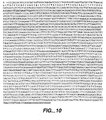
- this invention provides isolated nucleic acid comprising DNA having at least 800 nucleotides and at least a 70% sequence identity to (a) the sequence of nucleic acids 229 - 1002 of SEQ ID NO:1 or (b) the complement of the DNA molecule of (a).
- this nucleic acid has at least one clone 65 biological activity.
- the invention provides isolated nucleic acid comprising DNA having at least 700 nucleotides and at least a 95% sequence identity determined over the full length of the sequences being compared to the sequence of nucleic acids 229-1002 of SEQ ID NO:1, or (b) the complement of the DNA molecule of (a).
- this nucleic acid comprises DNA encoding a human clone 65 polypeptide having amino acid residues 1 to 258 of Figures 5A and 5B (SEQ ID NO:3), or the complement thereof.
- the invention provides isolated nucleic acid comprising DNA having at least 800 nucleotides and at least a 70% sequence identity to the sequence of nucleic acids 1-783 of SEQ ID NO:4, or (b) the complement of the DNA molecule of (a).
- this nucleic acid comprises DNA having at least about a 85% sequence identity to the sequence of nucleic acids 1-783 of SEQ ID NO:4 or (b) the complement of the DNA molecule of (a).
- this nucleic acid comprises DNA encoding a mouse clone 65 polypeptide having amino acid residues 1 to 261 of Figures 1A and 1B (SEQ ID NO:6), or the complement thereof.
- the invention provides an isolated nucleic acid comprising DNA having at least 800 nucleotides and at least a 70% sequence identity to the human clone 65 polypeptide cDNA in ATCC Deposit No. 209536 (pRK5E.h.WIG-3.65.4A), or (b) the complement of the DNA molecule of (a).
- this nucleic acid comprises DNA having at least about a 95% sequence identity to the human clone 65 polypeptide cDNA in ATCC Deposit No.209536 (pRK5E.h.WIG-3.65.4A), or (b) the complement of the DNA molecule of (a).
- a still further aspect of the invention involves a process for producing a clone 65 polypeptide comprising culturing a host cell comprising clone 65-encoding nucleic acid under conditions suitable for expression of the clone 65 polypeptide and recovering the clone 65 polypeptide from the cell culture.
- clone 65 polypeptide encoded by the clone 65-encoding nucleic acid.
- this polypeptide is human clone 65 or mouse clone 65.
- the invention provides an isolated nucleic acid having at least about 800 nucleotides which hybridises under stringent conditions with (a) the sequence of nucleic acids 229-1002 of SEQ ID NO:1 or (b) the complement of the DNA molecule of (a), and has at least about a 70% sequence identity to (a) or (b).
- polypeptide encoded by a nucleic acid having at least 800 nucleotides which hybridises under stringent conditions with the sequence of nucleic acids 229-1002 of SEQ ID NO:1 and has at least 70% sequence identity thereto.
- the complements of the DNA molecules herein remain stably bound to the primary sequence under at least moderate, and optionally, under high stringency conditions.
- victors comprising the above nucleic acids, host cells comprising the vector, preferably wherein the cell is a Chinese hamster ovary (CHO) cell, an E. coli cell, a baculovirus-infected cell, or a yeast cell.
- CHO Chinese hamster ovary
- a chimeric molecule comprising one of the above polypeptides or an inactivated variant thereof, fused to a heterologous amino acid sequence, wherein the heterologous amino acid sequence may be, for example, an epitope tag sequence or an Fc region of an immunoglobulin.
- an antibody which specifically binds to one of the above polypeptides, wherein the antibody can be a monoclonal antibody.
- compositions comprising one of the above polypeptides and a carrier therefor, and a composition comprising an antagonist to one of the polypeptides and a carrier therefor, wherein the antagonist is an antibody which specifically binds to a clone 65 polypeptide.
- these compositions may also comprise a chemotherapeutic agent or growth-inhibitory agent.
- a method for treating a clone 65-related disorder in a mammal comprising administering to the mammal an effective amount of any of the above compositions.
- the disorder is a malignant disorder or arteriosclerosis. More preferably, the malignant disorder is breast cancer, ovarian cancer, colon cancer, or melanoma.
- kits comprising one of the above clone 65 polypeptides or clone 65 antagonists, such as anti-clone 65 antibodies, and instructions for using the antibody to detect a cancer induced by Wnt.
- Also contemplated is a method for inducing cell death comprising exposing a cell which is induced by Wnt to an effective amount of one of the above clone 65 polypeptides or clone 65 antagonists, such as anti-clone 65 antibodies.
- a cell is a cancer cell. More preferably, the cell is in a mammal, more preferably a human.
- an effective amount of another chemotherapeutic antibody is also exposed to the cell, such as an anti-ErbB2 antibody.
- the method comprises exposing the cell to a chemotherapeutic agent, a growth-inhibitory agent, or radiation.
- the cell is exposed to the growth-inhibitory agent prior to exposure to the anti-clone 65 antibody.
- the application also describes an article of manufacture, comprising:
- clone 65 polypeptide encompass native- sequence protein derived from clone 65 and variants thereof (which are further defined herein).
- the clone 65 polypeptide may be isolated from a variety of sources, such as from human tissue types or from another source, or prepared by recombinant or synthetic methods, or by any combination of these and similar techniques.
- a “native-sequence clone 65 polypeptide” comprises a polypeptide having the same amino acid sequence as a clone 65 polypeptide derived from nature. Such native-sequence clone 65 polypeptides can be isolated from nature or can be produced by recombinant or synthetic means.
- the term "native-sequence clone 65 polypeptide” specifically encompasses naturally-occurring truncated or other forms of a clone 65 polypeptide disclosed herein, naturally-occurring variant forms ( e.g., alternatively-spliced forms or splice variants), and naturally-occurring allelic variants of a clone 65 polypeptide.
- the native-sequence clone 65 polypeptide is a full-length, native-sequence human clone 65 polypeptide comprising amino acids 1 to 258 of Figures 5A and 5B (SEQ ID NO:3), with or without the N-terminal methionine.
- the native-sequence clone 65 polypeptide is a full-length native-sequence mouse clone 65 polypeptide comprising amino acids 1 to 261 of Figures 1A and 1B (SEQ ID NO:6), with or without the N-terminal methionine.
- the native-sequence clone 65 polypeptide is one which is encoded by a nucleotide sequence comprising one of the mouse clone 65 splice or other native-sequence variants, including SEQ ID NOS: 11, 12, 13, 14, 15, 16, 17, 18, or 19, with or without an N-terminal methionine.
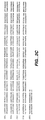
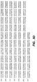
- clone 65 variant means an active clone 65 polypeptide as defined below having at least about 80%; preferably at least about 85%, more preferably at least about 90%, most preferably at least about 95% amino acid sequence identity with human clone 65 having the deduced amino acid sequence shown in Figs. 5A and 5B (SEQ ID NO:3) and/or with mouse clone 65 having the deduced amino acid sequence shown in Figs. 1A and 1B (SEQ ID NO:6).
- variants include, for instance, clone 65 polypeptides wherein one or more amino acid residues are added to, or deleted from, the N- or C-terminus of the full-length sequences of Figures 5A-B and 1A-B (SEQ ID NOS:3 and 6, respectively), including variants from other species, but excludes a native-sequence clone 65 polypeptide.
- Percent (%) amino acid sequence identity with respect to the clone 65 sequences identified herein is defined as the percentage of amino acid residues in a candidate sequence that are identical with the amino acid residues in a clone 65 polypeptide sequence, after aligning the sequences and introducing gaps, if necessary, to achieve the maximum percent sequence identity, and not considering any conservative substitutions as part of the sequence identity. Alignment for purposes of determining percent amino acid sequence identity can be achieved in various ways that are within the skill in the art, for instance, using publicly available computer software such as BLAST, ALIGN, or Megalign (DNASTAR) software. Those skilled in the art can determine appropriate parameters for measuring alignment, including any algorithms needed to achieve maximal alignment over the full length of the sequences being compared.
- Percent (%) nucleic acid sequence identity with respect to the coding region of the clone 65 sequences identified herein is defined as the percentage of nucleotides in a candidate sequence that are identical with the nucleotides in the coding region of the clone 65 sequence of interest, after aligning the sequences and introducing gaps, if necessary, to achieve the maximum percent sequence identity. Alignment for purposes of determining percent nucleic acid sequence identity can be achieved in various ways that are within the skill in the art, for instance, using publicly available computer software such as BLAST, ALIGN, or Megalign (DNASTART TM ) software. Those skilled in the art can determine appropriate parameters for measuring alignment, including any algorithms needed to achieve maximal alignment over the full length of the sequences being compared.
- “Stringent conditions” are those that (1) employ low ionic strength and high temperature for washing, for example, 0.015 M sodium chloride/0.0015 M sodium citrate/0.1% sodium dodecyl sulfate at 50°C; (2) employ during hybridization a denaturing agent, such as formamide, for example, 50% (vol/vol) formamide with 0.1% bovine serum albumin/0.1% Ficoll/0.1% polyvinylpyrrolidone/50 mM sodium phosphate buffer at pH.6.5 with 750 mM sodium chloride, 75 mM sodium citrate at 42°C; (3) employ 50% formamide, 5 x SSC (0.75 M NaCl, 0.075 M sodium citrate), 50 mM sodium phosphate (pH 6.8), 0.1% sodium pyrophosphate, 5 x Denhardt's solution, sonicated salmon sperm DNA (50 ⁇ g/ml); 0.1% SDS, and 10% dextran sulfate at 42°C, with washes at 42°C
- Modely stringent conditions are described in Sambrook et al., Molecular Cloning: A Laboratory Manual (New York: Cold Spring Harbor Laboratory Press, 1989 ), and include the use of a washing solution and hybridization conditions (e.g., temperature, ionic strength, and percent SDS) less stringent than described above.
- a washing solution and hybridization conditions e.g., temperature, ionic strength, and percent SDS
- moderately stringent conditions is a condition such as overnight incubation at 37°C in a solution comprising: 20% formamide, 5 x SSC (150 mM NaCl, 15 mM trisodium citrate), 50 mM sodium phosphate (pH 7.6), 5 x Denhardt's solution, 10% dextran sulfate, and 20 mg/mL denatured sheared salmon sperm DNA, followed by washing the filters in 1 x SSC at about 37-50°C.
- the skilled artisan will recognize how to adjust the temperature, ionic strength, etc ., as necessary to accommodate factors such as probe length and the like.
- isolated when used to describe the various polypeptides disclosed herein, means polypeptide that has been identified and separated and/or recovered from a component of its natural environment. Contaminant components of its natural environment are materials that would typically interfere with diagnostic or therapeutic uses for the polypeptide, and may include enzymes, hormones, and other proteinaceous or non-proteinaceous solutes.
- the polypeptide will be purified (1) to a degree sufficient to obtain at least 15 residues of N-terminal or internal amino acid sequence by use of a spinning cup sequenator, or (2) to homogeneity by SDS-PAGE under non-reducing or reducing conditions using Coomassie blue or, preferably, silver stain.
- Isolated polypeptide includes polypeptide in situ within recombinant cells, since at least one component of the natural environment of clone 65 polypeptide will not be present. Ordinarily, however, isolated polypeptide will be prepared by at least one purification step.
- An "isolated" nucleic acid encoding a clone 65 polypeptide is a nucleic acid molecule that is identified and separated from at least one contaminant nucleic acid molecule with which it is ordinarily associated in the natural source of the respective nucleic acid.
- An isolated clone 65-encoding nucleic acid molecule is other than in the form or setting in which it is found in nature.
- An isolated clone 65-encoding nucleic acid molecule therefore is distinguished from the clone 65-encoding nucleic acid molecule as it exists in natural cells.
- an isolated clone 65-encoding nucleic acid molecule includes a nucleic acid molecule contained in cells that ordinarily express clone 65-encoding DNA, where, for example, the nucleic acid molecule is in a chromosomal location different from that of natural cells.
- control sequences refers to DNA sequences necessary for the expression of an operably-linked coding sequence in a particular host organism.
- the control sequences that are suitable for prokaryotes include a promoter, optionally an operator sequence, and a ribosome binding site.
- Eukaryotic cells are known to utilize promoters, polyadenylation signals, and enhancers.
- Nucleic acid is "operably linked" when it is placed into a functional relationship with another nucleic acid sequence.
- DNA for a presequence or secretory leader is operably linked to DNA for a polypeptide if it is expressed as a preprotein that participates in the secretion of the polypeptide;
- a promoter or enhancer is operably linked to a coding sequence if it affects the transcription of the sequence; or
- a ribosome binding site is operably linked to a coding sequence if it is positioned so as to facilitate translation.
- "operably linked” means that the DNA sequences being linked are contiguous, and, in the case of a secretory leader, contiguous and in reading phase. However, enhancers do not have to be contiguous. Linking is accomplished by ligation at convenient restriction sites. If such sites do not exist, the synthetic oligonucleotide adaptors or linkers are used in accordance with conventional practice.
- antibody is used in the broadest sense and specifically covers single anti-clone 65 monoclonal antibodies (including agonist, antagonist, and neutralizing antibodies), and anti-clone 65 antibodies, and antibody compositions with polyepitopic specificity.
- monoclonal antibody refers to an antibody obtained from a population of substantially homogeneous antibodies, i.e ., the individual antibodies comprising the population are identical except for possible naturally- occurring mutations that may be present in minor amounts.
- Activity or “activity” or “biological activity”, for purposes herein, describes the activity of form(s) of a clone 65 polypeptide, including its variants, or its antagonists, which retain the biologic and/or immunologic activities of a native or naturally-occurring (native-sequence) clone 65 polypeptide or its antagonist.
- Preferred "activities" for a clone 65 polypeptide or its antagonist include the ability to inhibit proliferation of tumor cells or to stimulate proliferation of normal cells and to treat arteriosclerosis, including atherosclerosis, as well as to induce wound repair and hematopoiesis, prevent desmoplasia, prevent fibrotic lesions associated with skin disorders such as scleroderma, keloid, eosinophilic fasciitis, nodular fasciitis, and Dupuytren's contracture, to treat bone-related diseases such as osteoporosis, to regulate anabolism including promotion of growth, to treat immune disorders, to treat Wilms' tumor and kidney-related disorders, to treat testis-related disorders, to treat lung-related disorders, and to treat cardiac disorders.
- an "antagonist" of a clone 65 polypeptide is a molecule that inhibits an activity of a clone 65 polypeptide.
- Preferred antagonists are those which interfere with or block an undesirable biological activity of a clone 65 polypeptide, such as where a clone 65 polypeptide might act to stimulate cancer cells and the antagonist would serve to inhibit the growth of those cells.
- Such molecules include antibodies and small molecules that have such inhibitory capability, as well as polypeptide variants of, and receptors for, a clone 65 polypeptide (if available) or portions thereof that bind to clone 65 polypeptide.
- the receptor can be expression cloned from the family; then a soluble form of the receptor is made by identifying the extracellular domain and excising the transmembrane domain therefrom.
- the soluble form of the receptor can then be used as an antagonist, or the receptor can be used to screen for small molecules that would antagonize clone 65 polypeptide activity.
- Antagonist activity can be determined by several means, including standard assays for induction of cell death such as that described herein, e.g. , 3 H-thymidine proliferation assays, or other mitogenic assays, such as an assay measuring the capability of the candidate antagonist of inducing EGF-potentiated anchorage independent growth of target cell lines ( Volckaert et al., Gene, 15:215-223 (1981 )) and/or growth inhibition of neoplastic cell lines. Roberts et al., Proc, Natl. Acad. Sci. USA, 82:119-123 (1985 )..
- Anchorage-independent growth refers to the ability of clone 65 polypeptide-treated or clone 320 polypeptide-treated, or TGF- ⁇ -treated and EGF-treated non-neoplastic target cells to form colonies in soft agar, a characteristic ascribed to transformation of the cells.
- the candidate is incubated together with an equimolar amount of a clone 65 polypeptide otherwise detectable in the EGF-potentiated anchorage-independent target cell growth assay, and the culture observed for failure to induce anchorage-independent growth.
- Treatment refers to both therapeutic treatment and prophylactic or preventative measures. Those in need of treatment include those already with the disorder or condition as well as those in which the disorder or condition is to be prevented.
- mammal for purposes of treatment refers to any animal classified as a mammal, including humans, domestic, and farm animals, and zoo, sports, or pet animals, such as dogs, horses, cats, sheep, pigs, cows, etc. Preferably, the mammal is human.
- a "clone 65-related disorder” is any condition that would benefit from treatment with the clone 65 polypeptides or clone 65 antagonists herein. This includes chronic and acute disorders, as well as those pathological conditions which predispose the mammal to the disorder in question.
- Non-limiting examples of disorders to be treated herein include benign and malignant tumors; leukemias and lymphoid malignancies; neuronal, glial, astrocytal, hypothalamic and other glandular, macrophagal, epithelial, stromal, and blastocoelic disorders; hematopoiesis-related disorders; tissue-growth disorders; skin disorders; desmoplasia, fibrotic lesions; kidney disorders; bone-related disorders; trauma such as burns, incisions, and other wounds; catabolic states; testicular-related disorders; and inflammatory, angiogenic, and immunologic disorders, including arteriosclerosis.
- a "Wnt-related disorder” is one caused at least by the upregulation of the Wnt gene pathway, including Wnt-1 and Wnt-4, but preferably Wnt-1, and may include cancer.
- cancer refers to or describe the physiological condition in mammals that is typically characterized by unregulated cell growth.
- examples of cancer include but are not limited to, carcinoma including adenocarcinoma, lymphoma, blastoma, melanoma, sarcoma, and leukemia.
- cancers include squamous cell cancer, small-cell lung cancer, non-small cell lung cancer, gastrointestinal cancer, Hodgkin's and non-Hodgkin's lymphoma, pancreatic cancer, glioblastoma, cervical cancer, ovarian cancer, liver cancer such as hepatic carcinoma and hepatoma, bladder cancer, breast cancer, colon cancer, colorectal cancer, endometrial carcinoma, salivary gland carcinoma, kidney cancer such as renal cell carcinoma and Wilms' tumors, basal cell carcinoma, melanoma, prostate cancer, vulval cancer, thyroid cancer, testicular cancer, esophageal cancer, and various types of head and neck cancer.
- the preferred cancers for treatment herein are breast, colon, lung, and melanoma.
- cytotoxic agent refers to a substance that inhibits or prevents the function of cells and/or causes destruction of cells.
- the term is intended to include radioactive isotopes (e.g., 131 I, 125 I, 90 Y, and 186 Re), chemotherapeutic agents, and toxins such as enzymatically active toxins of bacterial, fungal, plant, or animal origin, or fragments thereof.
- chemotherapeutic agent is a chemical compound useful in the treatment of cancer.
- chemotherapeutic agents include Adriamycin, Doxorubicin, 5-Fluorouracil, Cytosine arabinoside ("Ara-C"), Cyclophosphamide, Thiotepa, Busulfan, Cytoxin, Taxol, Toxotere, Methotrexate, Cisplatin, Melphalan, Vinblastine, Bleomycin, Etoposide, Ifosfamide, Mitomycin C, Mitoxantrone, Vincreistine, Vinorelbine, Carboplatin, Teniposide, Daunomycin, Caminomycin, Aminopterin, Dactinomycin, Mitomycins, Esperamicins (see U.S. Pat. No. 4,675,187 ), Melphalan, and other related nitrogen mustards.
- hormonal agents that act to regulate or inhibit hormone action on tumors, such as tamoxifen and
- a “growth-inhibitory agent” when used herein refers to a compound or composition which inhibits growth of a cell, such as an Wnt-overexpressing cancer cell, either in vitro or in vivo.
- the growth-inhibitory agent is one which significantly reduces the percentage of malignant cells in S phase.
- growth- inhibitory agents include agents that block cell cycle progression (at a place other than S phase), such as agents that induce G1 arrest and M-phase arrest.
- Classical M-phase blockers include the vincas (vincristine and vinblastine), taxol, and topo II inhibitors such as doxorubicin, daunorubicin, etoposide, and bleomycin.
- DNA alkylating agents such as tamoxifen, prednisone, dacarbazine, mechlorethamine, cisplatin, methotrexate, 5-fluorouracil, and ara-C.
- DNA alkylating agents such as tamoxifen, prednisone, dacarbazine, mechlorethamine, cisplatin, methotrexate, 5-fluorouracil, and ara-C.
- DNA alkylating agents such as tamoxifen, prednisone, dacarbazine, mechlorethamine, cisplatin, methotrexate, 5-fluorouracil, and ara-C.
- Northern analysis or “Northern blot” is a method used to identify RNA sequences that hybridize to a known probe such as an oligonucleotide, DNA fragment, cDNA or fragment thereof, or RNA fragment.
- the probe is labeled with a radioisotope such as 32 P, or by biotinylation, or with an enzyme.
- the RNA to be analyzed is usually electrophoretically separated on an agarose or polyacrylamide gel, transferred to nitrocellulose, nylon, or other suitable membrane, and hybridized with the probe, using standard techniques well known in the art such as those described in sections 7.39-7.52 of Sambrook et al., supra.
- PCR polymerase chain reaction
- RNA and/or DNA are amplified as described in U.S. Pat. No. 4.683,195 issued 28 July 1987 .
- sequence information from the ends of the region of interest or beyond needs to be available, such that oligonucleotide primers can be designed; these primers will be identical or similar in sequence to opposite strands of the template to be amplified.
- the 5' terminal nucleotides of the two primers may coincide with the ends of the amplified material.
- PCR can be used to amplify specific RNA sequences, specific DNA sequences from total genomic DNA.
- PCR is considered to be one, but not the only, example of a nucleic acid polymerase reaction method for amplifying a nucleic acid test sample comprising the use of a known nucleic acid as a primer and a nucleic acid polymerase to amplify or generate a specific piece of nucleic acid.
- the present invention provides newly identified and isolated nucleotide sequences encoding a polypeptide referred to in the present application as a clone 65 polypeptide.
- cDNAs have been identified and isolated encoding novel murine and human clone 65 polypeptides as disclosed in further detail in the Examples below.
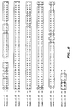
- mouse and human clone 65 show significant homology to members of the Rho family of small GTPases (mouse and human clone 65 are 58-59% and 59% homologous, respectively, to human G25k gtp-binding protein, placental and brain isoforms, and 59% homologous to canine CDC42 GTP-binding protein). Accordingly, it is presently believed that the clone 65 polypeptides disclosed in the present application possess activity relating to the treatment of various cancers with which Ras is associated, as well as to arteriosclerosis, such as atherosclerosis.
- variants of these sequences can be prepared.
- Clone 65 variants can be prepared by introducing appropriate nucleotide changes into the clone 65-encoding DNA, or by synthesis of the desired variant clone 65 polypeptides.
- amino acid changes may alter post-translational processes of the clone 65 polypeptides, such as changing the number or position of glycosylation sites or altering the membrane-anchoring characteristics, if the native clone 65 polypeptide is membrane bound.
- Variations in the native full-length clone 65 sequences, or in various domains of the clone 65 polypeptides described herein, can be made, for example, using any of the techniques and guidelines for conservative and non-conservative mutations set forth, for instance, in U.S. Patent No. 5,364,934 .
- Variations may be a substitution, deletion, or insertion of one or more codons encoding the clone 65 polypeptide that results in a change in the amino acid sequence as compared with the native-sequence clone 65 polypeptide.
- the variation is by substitution of at least one amino acid with any other amino acid in any portion of the clone 65 polypeptide.
- Guidance in determining which amino acid residue may be inserted, substituted, or deleted without adversely affecting the desired activity may be found by comparing the sequence of the clone 65 polypeptide with that of homologous known Rac or Rho protein molecules, and minimizing the number of amino acid sequence changes made in regions of high homology.
- Amino acid substitutions can be the result of replacing one amino acid with another amino acid having similar structural and/or chemical properties, such as the replacement of a leucine with a serine, i.e., conservative amino acid replacements.
- Insertions or deletions may optionally be in the range of 1 to about 5 amino acids. The variation allowed may be determined by systematically making insertions, deletions, or substitutions of amino acids in the sequence and testing the resulting variants for activity in in vitro assays for gene upregulation or downregulation and in transgenic or knock-out animals.
- variations can be made on the cloned DNA to produce variant DNA using methods known in the art such as oligonucleotide-mediated (site-directed) mutagenesis ( Carter et al., Nucl. Acids Res., 13:4331 (1986 ); Zoller et al., Nucl. Acids Res., 10:6497 (1987 )), cassette mutagenesis ( Wells et al., Gene, 34:315 (1985 )), alanine scanning, PCR mutagenesis, restriction selection mutagenesis ( Wells et al., Philos. Trans. R. Soc. London SerA, 317:415 (1986 )), or other known techniques.
- oligonucleotide-mediated (site-directed) mutagenesis Carter et al., Nucl. Acids Res., 13:4331 (1986 ); Zoller et al., Nucl. Acids Res., 10:6497 (1987 )
- Scanning amino acid analysis can also be employed to identify one or more amino acids along a contiguous sequence.
- preferred scanning amino acids are relatively small, neutral amino acids.
- Such amino acids include alanine, glycine, serine, and cysteine.
- Alanine is typically a preferred scanning amino acid among this group because it eliminates the side-chain beyond the beta-carbon and is less likely to alter the main-chain conformation of the variant. Alanine is also typically preferred because it is the most common amino acid. Further, it is frequently found in both buried and exposed positions.
- T.E. Creighton Proteins: Structure and Molecular Properties (W.H. Freeman & Co., San Francisco, 1983 ); Chothia, J. Mol. Biol., 150:1 (1976 ). If alanine substitution does not yield adequate amounts of variant, an isoteric amino acid can be used.
- variants of the full-length clone 65 polypeptides include variants from which the N-terminal signal peptide, if any, and/or the initiating methionine has been deleted.
- Covalent modifications of the clone 65 polypeptides are included within the scope of this invention.
- One type of covalent modification includes reacting targeted amino acid residues of a clone 65 polypeptide with an organic derivatizing agent that is capable of reacting with selected side chains or the N- or C- terminal residues.
- Derivatization with bifunctional agents is useful, for instance, for crosslinking a clone 65 polypeptide to a water-insoluble support matrix or surface for use in the method for purifying anti-clone 65 antibodies, and vice-versa.
- crosslinking agents include, e.g ., 1,1-bis(diazoacetyl)-2-phenylethane, glutaraldehyde, N-hydroxysuccinimide esters, for example, esters with 4-azido-salicylic acid, homobifunctional imidoesters, including disuccinimidyl esters such as 3,3'-dithiobis(succinimidyl-propionate), bifunctional maleimides such as bis-N-maleimido-1,8-octane, and agents such as methyl-3-((p-azidophenyl)dithio)propioimidate.
- 1,1-bis(diazoacetyl)-2-phenylethane glutaraldehyde
- N-hydroxysuccinimide esters for example, esters with 4-azido-salicylic acid
- homobifunctional imidoesters including disuccinimidyl esters such as 3,3
- Another type of covalent modification of the clone 65 polypeptide included within the scope of this invention comprises altering the native glycosylation pattern of the polypeptide.
- "Altering the native glycosylation pattern" is intended for purposes herein to mean deleting one or more carbohydrate moieties found in the native sequence (either by deleting the underlying glycosylation site or by removing the glycosylation moieties by chemical and/or enzymatic means) and/or adding one or more glycosylation sites that are not present in the native sequence.
- the phrase includes qualitative changes if the glycosylation of the native proteins, involving a change in the nature and proportion of the various sugar residues present.
- Addition of glycosylation sites to the clone 65 polypeptide herein may be accomplished by altering the amino acid sequence.
- the alteration may be made, for example, by the addition of, or substitution by, one or more serine or threonine residues to the native sequence (for O-linked glycosylation sites).
- the amino acid sequence may optionally be altered through changes at the DNA level, particularly by mutating the DNA encoding the clone 65 or 320 polypeptide at preselected bases such that codons are generated that will translate into the desired amino acids.
- the DNA mutation(s) may be made using methods described above.
- Another means of increasing the number of carbohydrate moieties on the clone 65 polypeptide is by chemical or enzymatic coupling of glycosides to the polypeptide. Such methods are described in the art, e.g ., in WO 87/05330 published 11 September 1987 , and in Aplin and Wriston, CRC Crit, Rev. Biochem., pp. 259-306 (1981 ).
- Removal of carbohydrate moieties present on the clone 65 polypeptide may be accomplished chemically or enzymatically or by mutational substitution of codons encoding amino acid residues that serve as targets for glycosylation.
- Chemical deglycosylation techniques are known in the art and described, for instance, by Hakimuddin, et al., Arch. Biochem. Biophys., 259:52 (1987 ) and by Edge et al., Anal. Biochem., 118:131 (1981 ).
- Enzymatic cleavage of carbohydrate moieties on polypeptides can be achieved by the use of a variety of endo- and exo-glycosidases as described by Thotakura et al., Meth. Enzymol., 138:350 (1987 ).
- Another type of covalent modification comprises linking the clone 65 polypeptide to one of a variety of nonproteinaceous polymers, e.g., polyethylene glycol, polypropylene glycol, or polyoxyalkylenes, in the manner set forth, e.g., in U.S. Patent Nos. 4,640,835 ; 4,496,689 ; 4,301,144 ; 4,670,417 ; 4,791,192 or 4,179,337 .
- nonproteinaceous polymers e.g., polyethylene glycol, polypropylene glycol, or polyoxyalkylenes
- the clone 65 polypeptide of the present invention may also be modified in a way to form a chimeric molecule comprising a clone 65 polypeptide, or a fragment thereof, fused to a heterologous polypeptide or amino acid sequence.
- a chimeric molecule comprises a fusion of the clone 65 polypeptide with a tag polypeptide which provides an epitope to which an anti-tag antibody can selectively bind.
- the epitope tag is generally placed at the amino- or carboxyl-terminus of a native or variant clone 65 molecule. The presence of such epitope-tagged forms can be detected using an antibody against the tag polypeptide.
- the epitope tag enables the clone 65 polypeptide to be readily purified by affinity purification using an anti-tag antibody or another type of affinity matrix that binds to the epitope tag.
- the chimeric molecule may comprise a fusion of the clone 65 polypeptide, or fragments thereof, with an immunoglobulin or a particular region of an immunoglobulin.
- such a fusion could be to the Fc region of an 1g, such as an IgG molecule.
- tag polypeptides and their respective antibodies are well known in the art. Examples include poly-histidine (poly-his) or poly-histidine-glycine (poly-his-gly) tags; the flu HA tag polypeptide and its antibody 12CA5 ( Field et al., Mol. Cell. Biol., 8:2159-2165 (1988 )); the c-myc tag and the 8F9, 3C7, 6E10, G4, B7, and 9E10 antibodies thereto ( Evan el al., Molecular and Cellular Biology, 5:3610-3616 (1985 )); and the Herpes Simplex virus glycoprotein D (gD) tag and its antibody.
- poly-histidine poly-his
- poly-histidine-glycine poly-his-gly
- tag polypeptides include the Flag-peptide ( Hopp et al., Biotechnology, 6:1204-1210 (1988 )); the KT3 epitope peptide ( Martin et al, Science, 255:192-194 (1992 )); an ⁇ -tubulin epitope peptide (Skinner et al.,J Biol. Chem., 266:15163-15166 (1991 )); and the T7 gene 10 protein peptide tag. Lutz-Freyermuth et al., Proc. Natl. Acad, Sci. USA, 87:6393-6397 (1990 ).
- the description below relates primarily to production of clone 65 polypeptides by culturing cells transformed or transfected with a vector containing at least DNA encoding the mature or full-length sequences of human or mouse clone 65 (SEQ ID NOS:3 or 6, respectively).
- clone 65 polypeptide sequence may be produced by direct peptide synthesis using solid-phase techniques. See, e.g ., Stewart et al., Solid-Phase Peptide Synthesis (W.H. Freeman Co.: San Francisco, CA, 1969 ); Merrifield, J. Am. Chem. Soc., 85:2149-2154 (1963 ). In vitro protein synthesis may be performed using manual techniques or by automation.
- Automated synthesis may be accomplished, for instance, using an Applied Biosystems peptide synthesizer (Foster City, CA) in accordance with manufacturer's instructions.
- Various portions of clone 65 polypeptides may be chemically synthesized separately and combined using chemical or enzymatic methods to produce a full-length clone 65 polypeptide.
- DNA encoding a clone 65 polypeptide may be obtained from a cDNA library prepared from tissue believed to possess the mRNA for clone 65 polypeptide and to express it at a detectable level. Accordingly, DNA encoding human clone 65 polypeptide can be conveniently obtained from a cDNA library prepared from human tissue, such as a human fetal liver library or as otherwise described in the Examples. The genes encoding clone 65 polypeptides may also be obtained from a genomic library or by oligonucleotide synthesis.
- a still alternative method of cloning clone 65 polypeptide is suppressive subtractive hybridization, which is a method for generating differentially regulated or tissue-specific cDNA probes and libraries. This is described, for example, in Diatchenko et al., Proc. Natl. Acad. Sci USA, 93: 6025-6030 (1996 ). The procedure is based primarily on a technique called suppression PCR and combines normalization and subtraction in a single procedure. The normalization step equalizes the abundance of cDNAs within the target population and the subtraction step excludes the common sequences between the target and driver populations.
- Probes such as antibodies to a clone 65 polypeptide or oligonucleotides of at least about 20-80 bases
- Screening the cDNA or genomic library with the selected probe may be conducted using standard procedures, such as described in Sambrook et al., supra.
- An alternative means to isolate the gene encoding clone 65 polypeptide is to use PCR methodology. Sambrook et al., supra; Dieffenbach et al., PCR Primer: A Laboratory Manual (New York: Cold Spring Harbor Laboratory Press, 1995 ).
- the oligonucleotide sequences selected as probes should be of sufficient length and sufficiently unambiguous that false positives are minimized.
- the oligonucleotide is preferably labeled such that it can be detected upon hybridization to DNA in the library being screened. Methods of labeling are well known in the art, and include the use of radiolabels like 32 P-labeled ATP, biotinylation, or enzyme labeling. Hybridization conditions, including moderate stringency and high stringency, are provided in Sambrook et al., supra .
- Sequences identified in such library screening methods can be compared and aligned to other known sequences deposited and available in public databases such as GenBank or other private sequence databases. Sequence identity (at either the amino acid or nucleotide level) within defined regions of the molecule or across the full-length sequence can be determined through sequence alignment using computer software-programs such as ALIGN, DNAstar, and INHERIT which employ various algorithms to measure homology.
- Nucleic acid having polypeptide-coding sequence may be obtained by screening selected cDNA or genomic libraries using the deduced amino acid sequences disclosed herein for the first time, and, if necessary, using conventional primer extension procedures as described in Sambrook et al., supra , to detect precursors and processing intermediates of mRNA that may not have been reverse-transcribed into cDNA.
- Host cells are transfected or transformed with expression or cloning vectors described herein for clone 65 polypeptide production and cultured in conventional nutrient media modified as appropriate for inducing promoters, selecting transformants, or amplifying the genes encoding the desired sequences.
- the culture conditions such as media, temperature, pH, and the like, can be selected by the skilled artisan without undue experimentation. In general, principles, protocols, and practical techniques for maximizing the productivity of cell cultures can be found in Mammalian Cell Biotechnology: a Practical Approach, M. Butler, ed. (IRL Press, 1991 ) and Sambrook et al., supra.
- transfection is known to the ordinarily skilled artisan, for example, CaPO 4 and electroporation.
- transformation is performed using standard techniques appropriate to such cells.
- the calcium treatment employing calcium chloride, as described in Sambrook et al., supra, or electroporation is generally used for prokaryotes or other cells that contain substantial cell-wall barriers.
- Infection with Agrobacterium tumefaciens is used for transformation of certain plant cells, as described by Shaw et al., Gene, 23:315 (1983 ) and WO 89/05859 published 29 June 1989 .
- DNA into cells such as by nuclear microinjection, electroporation, bacterial protoplast fusion with intact cells, or polycations, e.g ., polybrene or polyomithine, may also be used.
- polycations e.g ., polybrene or polyomithine
- transforming mammalian cells see Keown et al., Methods in Enzymology, 185:527-537 (1990 ) and Mansour et al., Nature, 336:348-352 (1988 ).
- Suitable host cells for cloning or expressing the DNA in the vectors herein include prokaryote, yeast, or higher eukaryote cells.
- Suitable prokaryotes include but are not limited to eubacteria, such as Gram-negative or Gram-positive organisms, for example, Enterobacteriaceae such as Escherichia, e.g., E. coli, Enterobacter, Erwinia, Klebsiella, Proteus, Salmonella, e.g., Salmonella typhimurium, Serratia, e.g., Serratia marcescans, and Shigella, as well as Bacilli such as B. subtilis and B. licheniformis ( e.g., B.
- E. coli K12 strain MM294 ATCC 31,446
- E . coli X1776 ATCC 31,537
- E . coli strain W3110 ATCC 27,325
- K5 772 ATCC 53,635
- strain W3.110 may be modified to effect a genetic mutation in the genes encoding proteins endogenous to the host, with examples of such hosts including E . coli W3110 strain 1A2, which has the complete genotype tonA; E. coli W3110 strain 9E4, which has the complete genotype tonA ptr3; E. coli W3110 strain 27C7 (ATCC 55,244), which has the complete genotype tonA ptr3 phoA E15 (argF-lac)169 degP ompT kan r ; E .
- E. coli W3110 strain 37D6 which has the complete genotype tonA ptr3 phoA E15 (argF-lac)169 degP ompT rbs7 ilvG kan r ; E. coli W3110 strain 40B4, which is strain 37D6 with a non-kanamycin resistant degP deletion mutation; and an E . coli strain having mutant periplasmic protease disclosed in U.S. Patent No. 4,946,783 issued 7 August 1990 .
- in vitro methods of cloning e.g. , PCR or other nucleic acid polymerase reactions, are suitable.
- eukaryotic microbes such as filamentous fungi or yeast are suitable cloning or expression hosts for vectors containing nucleic acid encoding clone 65 or 320 polypeptide.
- Saccharomyces cerevisiae is a commonly used lower eukaryotic host microorganism.
- Schizosaccharomyces pombe Beach and Nurse, Nature, 290: 140 (1981 ); EP 139,383 published 2 May 1985 ); Kluyveromyces hosts ( U.S. Patent No.
- K lactis MW98-8C, CBS683, CBS4574; Louvencourt et al., J. Bacteriol., 737 (1983 )
- K. fragilis ATCC 12,424)
- K. bulgaricus ATCC 16,045)
- K. wickeramii ATCC 24,178
- K. waltii ATCC 56,500
- K. drosophilarum ATCC 36,906; Van den Berg et al., BiolTechnology, 8: 135 (1990 )
- K. thermotolerans K.
- Schwanniomyces such as Schwanniomyces occidentalis ( EP 394,538 published 31 October 1990 ); and filamentous fungi such as, e.g., Neurospora, Penicillium, Tolypocladium ( WO 91/00357 published 10 January 1991 ), and Aspergillus hosts such as A. nidulans ( Ballance et al., Biochem. Biophys. Res. Commun., 112: 284-289 (1983 ); Tilburn et al., Gene, 26: 205-221 (1983 ); Yelton et al., Proc. Natl. Acad. Sci.
- Methylotropic yeasts are suitable herein and include, but are not limited to, yeast capable of growth on methanol selected from the genera consisting of Hansenula, Candida, Kloeckera, Pichia, Saccharomyces, Torulopsis, and Rhodotorula. A list of specific species that are exemplary of this class of yeasts may be found in C. Anthony, The Biochemistry of Methylotrophs, 269 (1982 ).
- Suitable host cells for the expression of glycosylated clone 65 polypeptide are derived from multicellular organisms.
- invertebrate cells include insect cells such as Drosophila S2 and Spodoptera Sf9, as well as plant cells.
- useful mammalian host cell lines include Chinese hamster ovary (CHO) and COS cells. More specific examples include monkey kidney CV1 line transformed by SV40 (COS-7, ATCC CRL 1651); human embryonic kidney line (293 or 293 cells subcloned for growth in suspension culture ( Graham et al., J. Gen Virol., 36:59 (1977 )); Chinese hamster ovary cells/-DHFR (CHO, Urlaub and Chasin, Proc. Natl. Acad.
- mice sertoli cells TM4, Mather, Biol. Reprod., 23:243-251 (1980 )
- human lung cells W138, ATCC CCL 75
- human liver cells Hep G2, HB 8065
- mouse mammary tumor MMT 060562, ATCC CCL51. The selection of the appropriate host cell is deemed to be within the skill in the art.
- the nucleic acid (e . g., cDNA or genomic DNA) encoding the desired clone 65 polypeptide may be inserted into a replicable vector for cloning (amplification of the DNA) or for expression.
- a replicable vector for cloning (amplification of the DNA) or for expression.
- the vector may, for example, be in the form of a plasmid, cosmid, viral particle, or phage.
- the appropriate nucleic acid sequence may be inserted into the vector by a variety of procedures. In general, DNA is inserted into an appropriate restriction endonuclease site(s) using techniques known in the art.
- Vector components generally include, but are not limited to; one or more of a signal sequence, an origin of replication, one or more marker genes, an enhancer element, a promoter, and a transcription termination sequence. Construction of suitable vectors containing one or more of these components employs standard ligation techniques which are known to the skilled artisan.
- the desired clone 65 polypeptide may be produced recombinantly not only directly; but also as a fusion polypeptide with a heterologous polypeptide, which may be a signal sequence, if the clone 65 polypeptide is conducive to being secreted, or other polypeptide having a specific cleavage site at the N-terminus of the mature or full-length protein or polypeptide.
- a heterologous polypeptide which may be a signal sequence, if the clone 65 polypeptide is conducive to being secreted, or other polypeptide having a specific cleavage site at the N-terminus of the mature or full-length protein or polypeptide.
- the signal sequence may be a component of the vector, or it may be a part of the DNA encoding the clone 65 polypeptide that is inserted into the vector.
- the signal sequence may be a prokaryotic signal sequence such as, for example, the alkaline phosphatase, penicillinase, lpp, or heat-stable enterotoxin II leaders.
- the signal sequence may be, e.g ., the yeast invertase leader, alpha factor leader (including Saccharomyces and Kluyveromyces ⁇ -factor leaders, the latter described in U.S. Patent No. 5,010,182 ), or acid phosphatase leader, the C. albicans glucoamylase leader ( EP 362,179 published 4 April 1990 ), or the signal described in WO 90/13646 published 15 November 1990 .
- mammalian signal sequences may be used to direct secretion of the protein, such as signal sequences from secreted polypeptides of the same or related species, as well as viral secretory leaders, and including signals from clone 65 polypeptide.
- Both expression and cloning vectors contain a nucleic acid sequence that enables the vector to replicate in one or more selected host cells. Such sequences are well known for a variety of bacteria, yeast, and viruses.
- the origin of replication from the plasmid pBR322 is suitable for most Gram-negative bacteria, the 2 ⁇ plasmid origin is suitable for yeast, and various viral origins (SV40, polyoma, adenovirus, VSV, or BPV) are useful for cloning vectors in mammalian cells.
- Selection genes will typically contain a selection gene, also termed a selectable marker.
- Typical selection genes encode proteins that (a) confer resistance to antibiotics or other toxins, e.g. , ampicillin, neomycin, methotrexate, or tetracycline, (b) complement auxotrophic deficiencies, or (c) supply critical nutrients not available from complex media, e.g. , the gene encoding D-alanine racemase for Bacilli.
- suitable selectable markers for mammalian cells are those that enable the identification of cells competent to take up the nucleic acid encoding clone 65 polypeptide, such as DHFR or thymidine kinase.
- An appropriate host cell when wild-type DHFR is employed is the CHO cell line deficient in DHFR activity, prepared and propagated as described by Urlaub et al., Proc, Natl. Acad. Sci. USA, 77:4216 (1980 ).
- a suitable selection gene for use in yeast is the trp 1 gene present in the yeast plasmid YRp7.
- the trp 1 gene provides a selection marker for a mutant strain of yeast lacking the ability to grow in tryptophan, for example, ATCC No. 44076 or PEP4-1. Jones, Genetics, 85:12(1977 ).
- Expression and cloning vectors usually contain a promoter operably linked to the nucleic acid sequence encoding clone 65 polypeptide to direct mRNA synthesis. Promoters recognized by a variety of potential host cells are well know. Promotes suitable for use with prokaryotic hosts include the ⁇ -lactamase and lactose promoter systems ( Chang et al., Nature, 275:615 (1978 ); Goeddel et al., Nature, 281:544 (1979 )), alkaline phosphatase, a tryptophan (trp)promoter system ( Goeddel, Nucleic Acids Res., 8:4057 (1980 ); EP 36,776 ), and hybrid promoters such as the tac promoter.
- ⁇ -lactamase and lactose promoter systems Chang et al., Nature, 275:615 (1978 ); Goeddel et al., Nature, 281:544 (1979 )
- Promoters for use in bacterial systems also will contain a Shine-Dalgamo (S.D.) sequence operably linked to the DNA encoding the clone 65 polypeptide.
- S.D. Shine-Dalgamo
- Suitable promoting sequences for use with yeast hosts include the promoters for 3-phosphoglycerate kinase ( Hitzeman et al., J. Biol. Chem., 255:2073 (1980 )) or other glycolytic enzymes ( Hess et al., J. Adv.
- enolase such as enolase, glyceraldehyde-3-phosphate dehydrogenase, hexokinase, pyruvate decarboxylase, phosphofructokinase, glucose-6-phosphate isomerase, 3-phosphoglycerate mutase, pyruvate kinase, triosephosphate isomerase, phosphoglucose is
- yeast promoters which are inducible promoters having the additional advantage of transcription controlled by growth conditions, are the promoter regions for alcohol dehydrogenase 2, isocytochrome C, acid phosphatase, degradative enzymes associated with nitrogen metabolism, metallothionein, glyceraldehyde-3-phosphate dehydrogenase, and enzymes responsible for maltose and galactose utilization. Suitable vectors and promoters for use in yeast expression are further described in EP 73,657 .
- Clone 65 transcription from vectors in mammalian host cells is controlled, for example, by promoters obtained from the genomes of viruses such as polyoma virus, fowlpox virus ( UK 2,211,504 published 5 July 1989 ), adenovirus (such as Adenovirus 2), bovine papilloma virus, avian sarcoma virus, cytomegalovirus, a retrovirus, hepatitis-B virus, and Simian Virus 40 (SV40); from heterologous mammalian promoters, e.g. , the actin promoter or an immunoglobulin promoter; and from heat-shock promoters, provided such promoters are compatible with the host cell systems.
- viruses such as polyoma virus, fowlpox virus ( UK 2,211,504 published 5 July 1989 ), adenovirus (such as Adenovirus 2), bovine papilloma virus, avian sarcoma virus, cytomegalovirus,
- Enhancers are cis-acting elements of DNA, usually about from 10 to 300 bp, that act on a promoter to increase its transcription.
- Many enhancer sequences are now known from mammalian genes (globin, elastase, albumin, ⁇ -fetoprotein, and insulin). Typically, however, one will use an enhancer from a eukaryotic cell virus.
- Examples include the SV40 enhancer on the late side of the replication origin (bp 100-270), the cytomegalovirus early promoter enhancer, the polyoma enhancer on the late side of the replication origin, and adenovirus enhancers.
- the enhancer may be spliced into the vector at a position 5' or 3' to the sequence coding for a clone 65 polypeptide, but is preferably located at a site 5' from the promoter.
- Expression vectors used in eukaryotic host cells will also contain sequences necessary for the termination of transcription and for stabilizing the mRNA. Such sequences are commonly available from the 5' and, occasionally 3', untranslated regions of eukaryotic or viral DNAs or cDNAs. These regions contain nucleotide segments transcribed as polyadenylated fragments in the untranslated portion of the mRNA encoding clone 65 polypeptide.
- Gene amplification and/or expression may be measured in a sample directly, for example, by conventional Southern blotting, Northern blotting to quantitate the transcription of mRNA ( Thomas, Proc, Natl, Acad, Sci, USA, 77:5201-5205 (1980 )), dot blotting (DNA analysis), or in situ hybridization, using an appropriately labeled probe, based on the sequences provided herein.
- antibodies may be employed that can recognize specific duplexes, including DNA duplexes, RNA duplexes, and DNA-RNA hybrid duplexes or DNA-protein duplexes. The antibodies in turn may be labeled and the assay may be carried out where the duplex is bound to a surface, so that upon the formation of duplex on the surface, the presence of antibody bound to the duplex can be detected.
- Gene expression may be measured by immunological methods, such as immunohistochemical staining of cells or tissue sections and assay of cell culture or body fluids, to quantitate directly the expression of gene product.
- Antibodies useful for immunohistochemical staining and/or assay of sample fluids may be either monoclonal or polyclonal, and may be prepared in any mammal. Conveniently, the antibodies may be prepared against a native-sequence clone 65 polypeptide or against a synthetic peptide based on the DNA sequences provided herein or against exogenous sequence fused to DNA encoding clone 65 polypeptide and encoding a specific antibody epitope.
- clone 65 polypeptide may be recovered from culture medium or from host cell lysates. If membrane-bound, it can be released from the membrane using a suitable detergent solution (e.g. , Triton-X 100) or by enzymatic cleavage. Cells employed in expression of clone 65 polypeptides can be disrupted by various physical or chemical means, such as freeze-thaw cycling, sonication, mechanical disruption, or cell lysing agents.
- clone 65 polypeptide may be desired to purify clone 65 polypeptide from recombinant cell proteins or polypeptides.
- the following procedures are exemplary of suitable purification procedures: by fractionation on an ion-exchange column; ethanol precipitation; reverse phase HPLC; chromatography on silica or on a cation-exchange resin such as DEAE; chromatofocusing; SDS-PAGE; ammonium sulfate precipitation; gel filtration using, for example, SEPHADEX TM G-75; protein A SEPHAROSE TM columns to remove contaminants such as IgG; and metal chelating columns to bind epitope-tagged forms of the clone 65 polypeptide.
- either a poly-HIS tag or the Fc portion of human IgG is added to the C-terminal coding region of the cDNA for clone 65 before expression.
- the conditioned media from the transfected cells are harvested by centrifugation to remove the cells and filtered.
- the protein may be purified using a Ni-NTA column. After loading, the column may be washed with additional equilibration buffer and the protein eluted with equilibration buffer containing 0.25 M imidazole. The highly purified protein may then be desalted into a storage buffer if desired.
- Immunoadhesin (Fc-containing) constructs of the clone 65 polypeptides may be purified from the conditioned media by pumping them onto a 5-ml Protein A column that had been equilibrated in a phosphate buffer. After loading, the column may be washed extensively with equilibration buffer before elution with citric acid. The eluted protein may be immediately neutralized by collecting 1-ml fractions into tubes containing TRIS buffer. The highly purified protein may be subsequently desalted into storage buffer as described above for the poly-HIS tagged proteins. The homogeneity of the protein may be assessed by SDS polyacrylamide gels and by N-terminal amino acid sequencing by Edman degradation.
- the purification step(s) selected will depend, for example, on the nature of the production process used and the particular clone 65 polypeptide produced.
- Nucleotide sequences (or their complement) encoding clone 65 polypeptides have various applications in the art of molecular biology, including uses as hybridization probes, in chromosome and gene mapping, and in the generation of anti-sense RNA and DNA. Nucleic acid encoding clone 65 polypeptide will also be useful for the preparation of clone 65 polypeptide by the recombinant techniques described herein.
- the full-length nucleotide sequences for mouse or human clone 65 may be used as hybridization probes for a cDNA library to isolate or detect the full-length gene encoding the clone 65 polypeptide of interest or to isolate or detect still other genes (for instance, those encoding naturally occurring variants of clone 65 polypeptide, other clone 65 polypeptide family members, or clone 65 polypeptides from other species) which have a desired sequence identity to the clone 65 polypeptide sequences disclosed in Figures 1 and 5 (SEQ ID NOS:6 and 3, respectively).
- the hybridization probes may be derived from ESTs, cloned sequences, or genomic sequences including promoters, enhancer elements, and introns of DNA encoding native-sequence clone 65 polypeptide.
- a screening method will comprise isolating the coding region of the clone 65 gene using the known DNA sequence to synthesize a selected probe of about 40 bases.
- Hybridization probes may be labeled by a variety of labels, including radionucleotides such as 32 P or 35 S, or enzymatic labels such as alkaline phosphatase coupled to the probe via avidin/biotin coupling systems. Labeled probes having a sequence complementary to that of any of the genes encoding clone 65 polypeptide of the present invention can be used to screen libraries of human cDNA, genomic DNA, or mRNA to determine to which members of such libraries the probe hybridizes. Hybridization techniques are described in further detail in the Examples below.
- the probes may also be employed in PCR techniques to generate a pool of sequences for identification of closely-related clone 65 sequences.
- Nucleotide sequences encoding a clone 65 polypeptide can also be used to construct hybridization probes for mapping the gene that encodes the particular clone 65 polypeptide and for the genetic analysis of individuals with genetic disorders.
- the nucleotide sequences provided herein may be mapped to a chromosome and specific regions of a chromosome using known techniques, such as in situ hybridization, linkage analysis against known chromosomal markers, and hybridization screening with libraries.
- Nucleic acid encoding a clone 65 polypeptide may be used as a diagnostic to determine the extent and rate of the expression of the DNA encoding a clone 65 polypeptide in the cells of a patient.
- a sample of a patient's cells is treated, via in situ hybridization, or by other suitable means, and analyzed to determine whether the sample contains mRNA molecules capable of hybridizing with the nucleic acid molecule.
- Nucleic acids that encode clone 65 polypeptide or any of their modified forms can also be used to generate either transgenic animals or "knock-out" animals which, in turn, are useful in the development and screening of therapeutically useful reagents.
- a transgenic animal e.g. , a mouse or rat
- a transgenic animal is an animal having cells that contain a transgene, which transgene was introduced into the animal or an ancestor of the animal at a prenatal, e.g. , an embryonic stage.
- a transgene is a DNA that is integrated into the genome of a cell from which a transgenic animal develops.
- cDNA encoding a clone 65 polypeptide can be used to clone genomic DNA encoding the clone 65 polypeptide in accordance with established techniques and the genomic sequences used to generate transgenic animals that contain cells which express DNA encoding the clone 65 polypeptide.
- transgenic animals particularly animals such as mice or rats
- transgenic animals typically include a copy of a transgene encoding the clone 65 polypeptide introduced into the germ line of the animal at an embryonic stage can be used to examine the effect of increased expression of DNA encoding the clone 65 polypeptide.
- Such animals can be used as tester animals for reagents thought to confer protection from, for example, pathological conditions associated with its overexpression.
- an animal is treated with the reagent and a reduced incidence of the pathological condition, compared to untreated animals bearing the transgene, would indicate a potential therapeutic intervention for the pathological condition.
- non-human homologues of clone 65 polypeptides can be used to construct a clone 65 polypeptide "knock-out" animal which has a defective or altered gene encoding a clone 65 polypeptide as a result of homologous recombination between the endogenous gene encoding the clone 65 polypeptide and altered genomic DNA encoding the clone 65 polypeptide introduced into an embryonic cell of the animal.
- cDNA encoding the clone 65 polypeptide can be used to clone genomic DNA encoding the clone 65 polypeptide in accordance with established techniques.
- a portion of the genomic DNA encoding the clone 65 polypeptide can be deleted or replaced with another gene, such as a gene encoding a selectable marker which can be used to monitor integration.
- another gene such as a gene encoding a selectable marker which can be used to monitor integration.
- several kilobases of unaltered flanking DNA are included in the vector. See e.g. , Thomas and Capecchi, Cell, 51:503 (1987 ) for a description of homologous recombination vectors.
- the vector is introduced into an embryonic stem cell line ( e.g. , by electroporation) and cells in which the introduced DNA has homologously recombined with the endogenous DNA are selected. See e.g.
- the selected cells are then injected into a blastocyst of an animal (e.g. , a mouse or rat) to form aggregation chimeras.
- an animal e.g. , a mouse or rat
- a chimeric embryo can then be implanted into a suitable pseudopregnant female foster animal and the embryo brought to term to create a "knock-out" animal.
- Progeny harboring the homologously recombined DNA in their germ cells can be identified by standard techniques and used to breed animals in which all cells of the animal contain the homologously recombined DNA. Knock-out animals can be characterized, for instance, by their ability to defend against certain pathological conditions and by their development of pathological conditions due to absence of the clone 65 polypeptide.
- assays in which the Rac and Rho family members are usually used are preferably performed with the clone 65 polypeptide.
- an assay to determine whether TGF- ⁇ induces the clone 65 polypeptide, indicating a role in cancer may be performed as known in the art, as well as assays involving induction of cell death and 3 H-thymidine proliferation assays. Mitogenic and tissue growth assays are also performed with the clone 65 polypeptide as set forth above. The results are applied accordingly.
- the clone 65 polypeptides of the present invention may also be used to induce the formation of anti-clone 65 polypeptide antibodies, which are identified by routine screening as detailed below.
- the clone 65 polypeptide can be used in accordance with immunoassay technology.
- immunoassays are provided by Wide at pages 199-206 of Radioimmune Assay Method, Kirkham and Huner, ed., E & S. Livingstone, Edinburgh, 1970 .
- clone 65 polypeptides can be detectably labeled and incubated with a test sample containing the molecules of interest (such as biological fluids, e.g. , serum, sputum, urine, etc .), and the amount of clone 65 molecule bound to the sample ascertained.
- a test sample containing the molecules of interest (such as biological fluids, e.g. , serum, sputum, urine, etc .), and the amount of clone 65 molecule bound to the sample ascertained.
- Immobilization of reagents is required for certain assay methods. Immobilization entails separating the clone 65 polypeptide from any analyte that remains free in solution. This conventionally is accomplished by either insolubilizing the clone 65 polypeptide before the assay procedure, as by adsorption to a water-insoluble matrix or surface ( Bennich et al.., U.S. 3,720,760 ), by covalent coupling (for example, using glutaraldehyde cross-linking), or by insolubilizing the molecule afterward, e.g. , by immunoprecipitation.
- clone 65 polypeptides are useful for screening for compounds that bind to them as defined above.
- these compounds are small molecules such as organic or peptide molecules that exhibit one or more of the desired activities. Screening assays of this kind are conventional in the art, and any such screening procedure may be employed, whereby the test sample is contacted with the clone 65 polypeptide herein and the extent of binding and biological activity of the bound molecule are determined.
- Clone 65 polypeptides are additionally useful in affinity purification of a molecule that binds to clone 65 polypeptide and in purifying antibodies thereto.
- the clone 65 polypeptide is typically coupled to an immobilized resin such as Affi-Gel 10 TM (Bio-Rad, Richmond, CA) or other such resins (support matrices) by means well known in the art.
- the resin is equilibrated in a buffer (such as one containing 150 mM NaCl, 20 mM HEPES, pH 7.4 supplemented to contain 20% glycerol and 0.5% NP-40) and the preparation to be purified is placed in contact with the resin, whereby the molecules are selectively adsorbed to the clone 65 polypeptide on the resin.
- a buffer such as one containing 150 mM NaCl, 20 mM HEPES, pH 7.4 supplemented to contain 20% glycerol and 0.5% NP-40
- the resin is then sequentially washed with suitable buffers to remove non-adsorbed material, including unwanted contaminants, from the mixture to be purified, using, e.g. , 150 mM NaCl, 20 mM HEPES, pH 7.4, containing 0.5% NP-40; 150 mM NaCl, 20 mM HEPES, pH 7.4 containing 0.5 M NaCl and 0.1% NP-40; 150 mM NaCl, 20 mM HEPES, pH 7.4 containing 0.1% deoxycholate; 150 mM NaCl, 20 mM-HEPES, pH 7.4 containing 0.1% NP-40; and a solution of 0.1% NP-40, 20% glycerol and 50 mM glycine, pH 3.
- suitable buffers to remove non-adsorbed material, including unwanted contaminants, from the mixture to be purified, using, e.g. , 150 mM NaCl, 20 mM HEPES, pH 7.4, containing 0.5% NP-40
- the resin is then treated so as to elute the binding molecule using a buffer that will break the bond between the binding molecule and clone 65 polypeptide (using, e.g. , 50 mM glycine, pH 3, 0.1% NP-40, 20% glycerol, and 100 mM NaCl).
- a buffer that will break the bond between the binding molecule and clone 65 polypeptide using, e.g. , 50 mM glycine, pH 3, 0.1% NP-40, 20% glycerol, and 100 mM NaCl).
- the clone 65 polypeptides of the present invention may be used to treat various conditions, including those characterized by overexpression and/or activation of at least the Wnt pathway. Further, they are useful in diagnosing cancer, for example, as a marker for increased susceptibility to cancer or for having cancer. Exemplary conditions or disorders to be treated with the clone 65 polypeptides include benign or malignant tumors ( e.g.
- the clone 65 polypeptides of the invention are administered to a mammal, preferably a human, in accord with known methods, such as intravenous administration as a bolus or by continuous infusion over a period of time, by intramuscular, intraperitoneal, intracerebrospinal, subcutaneous, intra-articular, intrasynovial, intrathecal, oral, topical, or inhalation routes. Intravenous or subcutaneous administration of the polypeptide is preferred.
- Therapeutic formulations of the clone 65 polypeptide are prepared for storage by mixing the polypeptide having the desired degree of purity with optional pharmaceutically acceptable carriers, excipients, or stabilizers ( Remington's Pharmaceutical Sciences 16th edition, Osol, A. Ed. (1980 )), in the form of lyophilized formulations or aqueous solutions.
- Acceptable carriers, excipients, or stabilizers are nontoxic to recipients at the dosages and concentrations employed, and include buffers such as phosphate, citrate, and other organic acids; antioxidants including ascorbic acid and methionine; preservatives (such as octadecyldimethylbenzyl ammonium chloride; hexamethonium chloride; benzalkonium chloride, benzethonium chloride; phenol, butyl or benzyl alcohol; alkyl parabens such as methyl or propyl paraben; catechol; resorcinol; cyclohexanol; 3-pentanol; and m-cresol); low molecular weight (less than about 10 residues) polypeptides; proteins, such as serum albumin, gelatin, or immunoglobulins; hydrophilic polymers such as polyvinylpyrrolidone; amino acids such as glycine, glutamine, asparagine, histidine,
- chemotherapeutic agents may be administered to the patient with cancer. Preparation and dosing schedules for such chemotherapeutic agents may be used according to manufacturers' instructions or as determined empirically by the skilled practitioner. Preparation and dosing schedules for such chemotherapy are also described in Chemotherapy Service, Ed., M.C. Perry (Williams & Wilkins: Baltimore, MD, 1992 ). The chemotherapeutic agent may precede or follow administration of the polypeptide or may be given simultaneously therewith.
- the polypeptide may be combined with an anti-oestrogen compound such as tamoxifen or an anti-progesterone such as onapristone (see, EP 616812 ) in dosages known for such molecules.
- clone 65 polypeptide or anti-clone 65 polypeptide antibodies
- antibodies against other tumor-associated antigens such as antibodies which bind to HER-2, EGFR, ErbB2, ErbB3, ErbB4, or vascular endothelial factor (VEGF).
- VEGF vascular endothelial factor
- two or more different anti-cancer antibodies, such as anti-ErbB2 antibodies may be co-administered to the patient with the clone 65 polypeptide (or anti-clone 65 polypeptide antibodies).
- the clone 65 polypeptide is co-administered with a growth-inhibitory agent to the cancer patient.
- the growth-inhibitory agent may be administered first, followed by the clone 65 polypeptide.
- simultaneous administration or administration of the clone 65 polypeptide first is also contemplated.
- Suitable dosages for the growth-inhibitory agent are those presently used and may be lowered due to the combined action (synergy) of the growth-inhibitory agent and polypeptide.
- the antibodies, cytotoxic agents, cytokines, or growth-inhibitory agents are suitably present in combination in amounts that are effective for the purpose intended.
- the active ingredients may also be entrapped in microcapsules prepared, for example, by coacervation techniques or by interfacial polymerization, for example, hydroxymethylcellulose or gelatin microcapsules and poly-(methylmethacylate) microcapsules, respectively, in colloidal drug delivery systems (for example, liposomes, albumin microspheres, microemulsions, nano-particles, and nanocapsules) or in macroemulsions.
- colloidal drug delivery systems for example, liposomes, albumin microspheres, microemulsions, nano-particles, and nanocapsules
- the formulations to be used for in vivo administration must be sterile. This is readily accomplished by filtration through sterile filtration membranes.
- Sustained-release preparations may be prepared. Suitable examples of sustained-release preparations include semipermeable matrices of solid hydrophobic polymers containing the polypeptide, which matrices are in the form of shaped articles, e.g. , films, or microcapsules. Examples of sustained-release matrices include polyesters, hydrogels (for example, poly(2-hydroxyethyl-methacrylate), or poly(vinylalcohol)), polylactides ( U.S. Pat. No.
- copolymers of L-glutamic acid and y ethyl-L-glutamate non-degradable ethylene-vinyl acetate
- degradable lactic acid-glycolic acid copolymers such as the LUPRON DEPOT TM (injectable microspheres composed of lactic acid-glycolic acid copolymer and leuprolide acetate)
- poly-D-(-)-3-hydroxybutyric acid While polymers such as ethylene-vinyl acetate and lactic acid-glycolic acid enable release of molecules for over 100 days, certain hydrogels release proteins for shorter time periods.
- encapsulated polypeptides When encapsulated polypeptides remain in the body for a long time, they may denature or aggregate as a result of exposure to moisture at 37°C, resulting in a loss of biological activity and possible changes in immunogenicity. Rational strategies can be devised for stabilization depending on the mechanism involved. For example, if the aggregation mechanism is discovered to be intermolecular S-S bond formation through thio-disulfide interchange, stabilization may be achieved by modifying sulfhydryl residues, lyophilizing from acidic solutions, controlling moisture content, using appropriate additives, and developing specific polymer matrix composition.
- the appropriate dosage of clone 65 polypeptide will depend on the type of disorder to be treated, as defined above, the severity and course of the disorder, whether the polypeptide is administered for preventive or therapeutic purposes, previous therapy, the patient's clinical history and response to the polypeptide; the route of administration, the condition of the patient, and the discretion of the attending physician.
- the polypeptide is suitably administered to the patient at one time or over a series of treatments.
- clone 65 polypeptide is an initial candidate dosage for administration to the patient, whether, for example, by one or more separate administrations, or by continuous infusion.
- a typical daily dosage might range from about 1 ⁇ g/kg to 100 mg/kg or more, depending on the factors mentioned above.
- the treatment is sustained until a desired suppression of symptoms of the disorder occurs.
- other dosage regimens may be useful. The progress of this therapy is easily monitored by conventional techniques and assays.
- the article of manufacture comprises a container and a label.
- Suitable containers include, for example, bottles, vials, syringes, and test tubes.
- the containers may be formed from a variety of materials such as glass or plastic.
- the container holds a composition which is effective for treating the condition and may have a sterile access port (for example, the container may be an intravenous solution bag or a vial having a stopper pierceable by a hypodermic injection needle).
- the active agent in the composition is the clone 65 polypeptide.
- the label on, or associated with, the container indicates that the composition is used for treating the condition or disorder of choice.
- the article of manufacture may further comprise a second container comprising a pharmaceutically acceptable buffer, such as phosphate-buffered saline, Ringer's solution, and dextrose solution. It may further include other materials desirable from a commercial and user standpoint, including other buffers, diluents, filters, needles, syringes, and package inserts with instructions for use.
- a pharmaceutically acceptable buffer such as phosphate-buffered saline, Ringer's solution, and dextrose solution.
- It may further include other materials desirable from a commercial and user standpoint, including other buffers, diluents, filters, needles, syringes, and package inserts with instructions for use.
- the present invention further provides anti-clone 65 polypeptide antibodies.
- Exemplary antibodies include polyclonal, monoclonal, humanized, bispecific, and heteroconjugate antibodies.
- the anti-clone 65 polypeptide antibodies of the present invention may comprise polyclonal antibodies. Methods of preparing polyclonal antibodies are known to the skilled artisan. Polyclonal antibodies can be raised in a mammal, for example, by one or more injections of an immunizing agent and, if desired, an adjuvant. Typically, the immunizing agent and/or adjuvant will be injected in the mammal by multiple subcutaneous or intraperitoneal injections.
- the immunizing agent may include the clone 65 polypeptide or a fusion protein thereof. It may be useful to conjugate the immunizing agent to a protein known to be immunogenic in the mammal being immunized.
- immunogenic proteins include but are not limited to keyhole limpet hemocyanin, serum albumin, bovine thyroglobulin, and soybean trypsin inhibitor.
- adjuvants which may be employed include Freund's complete adjuvant and MPL-TDM adjuvant (monophosphoryl Lipid A, synthetic trehalose dicorynomycolate).
- the immunization protocol may be selected by one skilled in the art without undue experimentation.
- the anti-clone 65 polypeptide antibodies may, alternatively, be monoclonal antibodies.
- Monoclonal antibodies may be prepared using hybridoma methods, such as those described by Kohler and Milstein, Nature, 256:495 (1975 ).
- a hybridoma method a mouse, hamster, or other appropriate host animal is typically immunized with an immunizing agent to elicit lymphocytes that produce or are capable of producing antibodies that will specifically bind to the immunizing agent.
- the lymphocytes may be immunized in vitro.
- the immunizing agent will typically include the clone 65 polypeptide or a fusion protein thereof.
- PBLs peripheral blood lymphocytes
- the lymphocytes are then fused with an immortalized cell line using a suitable fusing agent, such as PEG, to form a hybridoma cell.
- a suitable fusing agent such as PEG
- rat or mouse myeloma cell lines are employed.
- the hybridoma cells may be cultured in a suitable culture medium that preferably contains one or more substances that inhibit the growth or survival of the unfused, immortalized cells.
- a suitable culture medium that preferably contains one or more substances that inhibit the growth or survival of the unfused, immortalized cells.
- the culture medium for the hybridomas typically will include hypoxanthine, aminopterin, and thymidine (“HAT medium”), which substances prevent the growth of HGPRT-deficient cells.
- Preferred immortalized cell lines are those that fuse efficiently, support stable high-level expression of antibody by the selected antibody-producing cells, and are sensitive to a medium such as HAT medium. More preferred immortalized cell lines are murine myeloma lines, which can be obtained, for instance, from the Salk Institute Cell Distribution Center, San Diego, California, and the American Type Culture Collection, Manassas, Virginia. Human myeloma and mouse-human heteromyeloma cell lines also have been described for the production of human monoclonal antibodies. Kozbor, J. Immunol., 133:3001 (1984 ); Brön et al., Monoclonal Antibody Production Techniques and Applications (Marcel Dekker, Inc.: New York, 1987) pp. 51-63 .
- the culture medium in which the hybridoma cells are cultured can then be assayed for the presence of monoclonal antibodies directed against a clone 65 polypeptide.
- the binding specificity of monoclonal antibodies produced by the hybridoma cells is determined by immunoprecipitation or by an in vitro binding assay, such as radioimmunoassay (RIA) or enzyme-linked immunoabsorbent assay (ELISA). Such techniques and assays are known in the art.
- the binding affinity of the monoclonal antibody can, for example, be determined by the Scatchard analysis of Munson and Pollard, Anal. Biochem., 107:220 (1980 ).
- the clones may be subcloned by limiting dilution procedures and grown by standard methods. Goding, supra. Suitable culture media for this purpose include, for example, Dulbecco's Modified Eagle's Medium and RPMI-1640 medium. Alternatively, the hybridoma cells may be grown in vivo as ascites in a mammal.
- the monoclonal antibodies secreted by the subclones may be isolated or purified from the culture medium or ascites fluid by conventional immunoglobulin purification procedures such as, for example, protein A-Sepharose, hydroxylapatite chromatography, gel electrophoresis, dialysis, or affinity chromatography.
- the monoclonal antibodies may also be made by recombinant DNA methods, such as those described in U.S. Patent No. 4,816,567 .
- DNA encoding the monoclonal antibodies of the invention can be readily isolated and sequenced using conventional procedures (e.g. , by using oligonucleotide probes that are capable of binding specifically to genes encoding the heavy and light chains of murine antibodies).
- the hybridoma cells of the invention serve as a preferred source of such DNA.
- the DNA may be placed into expression vectors, which are then transfected into host cells such as simian COS cells, CHO cells, or myeloma cells that do not otherwise produce immunoglobulin protein, to obtain the synthesis of monoclonal antibodies in the recombinant host cells.
- host cells such as simian COS cells, CHO cells, or myeloma cells that do not otherwise produce immunoglobulin protein, to obtain the synthesis of monoclonal antibodies in the recombinant host cells.
- the DNA also may be modified, for example, by substituting the coding sequence for human heavy and light chain constant domains in place of the homologous murine sequences ( U.S. Patent No. 4,816,567 ; Morrison et al., Proc. Natl. Acad. Sci.
- non-immunoglobulin polypeptide can be substituted for the constant domains of an antibody of the invention, or can be substituted for the variable domains of one antigen-combining site of an antibody of the invention to create a chimeric bivalent antibody.
- the antibodies may be monovalent antibodies.
- Methods for preparing monovalent antibodies are well known in the art. For example, one method involves recombinant expression of immunoglobulin light chain and modified heavy chain.
- the heavy chain is truncated generally at any point in the Fc region so as to prevent heavy-chain crosslinking.
- the relevant cysteine residues are substituted with another amino acid residue or are deleted so as to prevent crosslinking.
- In vitro methods are also suitable for preparing monovalent antibodies. Digestion of antibodies to produce fragments thereof, particularly Fab fragments, can be accomplished using routine techniques known in the art.
- the anti-clone 65 polypeptide antibodies of the invention may further comprise humanized antibodies or human antibodies.
- Humanized forms of non-human (e.g. , murine) antibodies are chimeric immunoglobulins, immunoglobulin chains, or fragments thereof (such as Fv, Fab, Fab', F(ab') 2 , or other antigen-binding subsequences of antibodies) that contain minimal sequence derived from non-human immunoglobulin.
- Humanized antibodies include human immunoglobulins (recipient antibody) in which residues from a complementary-determining region (CDR) of the recipient are replaced by residues from a CDR of a non-human species (donor antibody) such as mouse, rat, or rabbit having the desired specificity, affinity, and capacity.
- CDR complementary-determining region
- Fv framework residues of the human immunoglobulin are replaced by corresponding non-human residues.
- Humanized antibodies may also comprise residues which are found neither in the recipient antibody nor in the imported CDR or framework sequences.
- the humanized antibody will comprise substantially all of at least one, and typically two, variable domains, in which all or substantially all of the CDR regions correspond to those of a non-human immunoglobulin, and all or substantially all of the FR regions are those of a human immunoglobulin consensus sequence.
- the humanized antibody preferably also will comprise at least a portion of an immunoglobulin constant region (Fc), typically that of a human immunoglobulin.
- Fc immunoglobulin constant region
- a humanized antibody has one or more amino acid residues introduced into it from a source which is non-human. These non-human amino acid residues are often referred to as "import" residues, which are typically taken from an "import” variable domain. Humanization can be essentially performed following the method of Winter and co-workers ( Jones et al., Nature, 321:522-525 (1986 ); Riechmann et al., Nature, 332:323-327 (1988 ); Verhoeyen et al., Science, 239:1534-1536 (1988 )), by substituting rodent .CDRs or CDR sequences for the corresponding sequences of a human antibody.
- humanized antibodies are chimeric antibodies ( U.S. Patent No. 4,816,567 ), wherein substantially less than an intact human variable domain has been substituted by the corresponding sequence from a non-human species.
- humanized antibodies are typically human antibodies in which some CDR residues and possibly some FR residues are substituted by residues from analogous sites in rodent antibodies.
- Human antibodies can also be produced using various techniques known in the art, including phage-display libraries. Hoogenboom and Winter, J. Mol. Biol., 227:381 (1991 ); Marks et al., J. Mol, Biol., 222:581 (1991 ). The techniques of Cole et al. and Boerner et al. are also available for the preparation of human monoclonal antibodies. Cole et al., Monoclonal Antibodies and Cancer Therapy, Alan R. Liss, p. 77 (1985 ); Boemer et al., J. Immunol., 147(1):86-95 (1991 ).
- Bispecific antibodies are monoclonal, preferably human or humanized, antibodies that have binding specificities for at least two different antigens.
- one of the binding specificities is for a clone 65 polypeptide; the other one is for any other antigen, and preferably for a cell-surface protein or receptor or receptor subunit.
- bispecific antibodies are known in the art. Traditionally, the recombinant production of bispecific antibodies is based on the co-expression of two immunoglobulin heavy-chain/light-chain pairs, where the two heavy chains have different specificities. Milstein and Cuello, Nature, 305:537-539 (1983 ). Because of the random assortment of immunoglobulin heavy and light chains, these hybridomas (quadromas) produce a potential mixture of ten different antibody molecules, of which only one has the correct bispecific structure. The purification of the correct molecule is usually accomplished by affinity chromatography steps. Similar procedures are disclosed in WO 93/08829, published 13 May 1993 , and in Traunecker et al., EMBO J., 10:3655-3659 (1991 ).
- Antibody variable domains with the desired binding specificities can be fused to immunoglobulin constant-domain sequences.
- the fusion preferably is with an immunoglobulin heavy-chain constant domain, comprising at least part of the hinge, CH2, and CH3 regions. It is preferred to have the first heavy-chain constant region (CH1) containing the site necessary for light-chain binding present in at least one of the fusions.
- DNAs encoding the immunoglobulin heavy-chain fusions and, if desired, the immunoglobulin light chain are inserted into separate expression vectors, and are co-transfected into a suitable host organism.
- Heteroconjugate antibodies are also within the scope of the present invention.
- Heteroconjugate antibodies are composed of two covalently joined antibodies. Such antibodies have, for example, been proposed to target immune system cells to unwanted cells ( U.S. Patent No. 4,676,980 ), and for treatment of HIV infection.
- the antibodies may be prepared in vitro using known methods in synthetic protein chemistry, including those involving cross- linking agents.
- immunotoxins may be constructed using a disulfide-exchange reaction or by forming a thioether bond. Examples of suitable reagents for this purpose include iminothiolate and methyl-4-mercaptoburyrimidate and those disclosed, for example, in U.S. Patent No. 4,676,980 .
- the antibodies of the invention may be used as affinity purification agents.
- the antibodies are immobilized on a solid phase such a SEPHADEX TM resin or filter paper, using methods well known in the art.
- the immobilized antibody is contacted with a sample containing the clone 65 polypeptide (or fragment thereof) to be purified, and thereafter the support is washed with a suitable solvent that will remove substantially all the material in the sample except the clone 65 polypeptide, which is bound to the immobilized antibody. Finally, the support is washed with another suitable solvent, such as glycine buffer, pH 5.0, that will release the clone 65 polypeptide from the antibody.
- a suitable solvent such as glycine buffer, pH 5.0
- Anti-clone 65 polypeptide antibodies may also be useful in diagnostic assays for clone 65 polypeptide, e.g. , detecting its expression in specific cells, tissues, or serum. Thus, the antibodies may be used in the diagnosis of human malignancies (see, for example, U.S. Pat. No. 5,183,884 ).
- the antibody typically will be labeled with a detectable moiety.
- a detectable moiety Numerous labels are available which can be preferably grouped into the following categories:
- enzyme-substrate combinations examples include:
- the label is indirectly conjugated with the antibody.
- the antibody can be conjugated with biotin and any of the three broad categories of labels mentioned above can be conjugated with avidin, or vice versa .
- Biotin binds selectively to avidin, and thus, the label can be conjugated with the antibody in this indirect manner.
- the antibody is conjugated with a small hapten (e.g ., digoxin) and one of the different types of labels mentioned above is conjugated with an anti-hapten antibody (e.g ., anti-digoxin antibody).
- a small hapten e.g ., digoxin
- an anti-hapten antibody e.g ., anti-digoxin antibody
- the anti-clone 65 polypeptide antibody need not be labeled, and the presence thereof can be detected using a labeled antibody that binds to the anti-clone 65 polypeptide antibody.
- the antibodies of the present invention may be employed in any known assay method, such as competitive binding assays, direct and indirect sandwich assays, and immunoprecipitation assays. Zola, Monoclonal Antibodies: A Manual of Techniques (New York: CRC Press, Inc., 1987), pp.147-158 .
- ком ⁇ онентs rely on the ability of a labeled standard to compete with the test sample analyte for binding with a limited amount of antibody.
- the amount of clone 65 polypeptide in the test sample is inversely proportional to the amount of standard that becomes bound to the antibodies.
- the antibodies preferably are insolubilized before or after the competition, so that the standard and analyte that are bound to the antibodies may conveniently be separated from the standard and analyte which remain unbound.
- Sandwich assays involve the use of two antibodies, each capable of binding to a different immunogenic portion, or epitope, of the protein to be detected.
- the test sample analyte is bound by a first antibody which is immobilized on a solid support, and thereafter a second antibody binds to the analyte, thus forming an insoluble three-part complex.
- the second antibody may itself be labeled with a detectable moiety (direct sandwich assays) or may be measured using an anti-immunoglobulin antibody that is labeled with a detectable moiety (indirect sandwich assay).
- sandwich assay is an ELISA assay, in which case the detectable moiety is an enzyme.
- the tumor sample may be fresh or frozen or may be embedded in paraffin and fixed with a preservative such as formalin, for example.
- the antibodies may also be used for in vivo diagnostic assays.
- the antibody is labeled with a radionuclide (such as 111 In, 99 Tc, 14 C, 131 I, 125 I, 3 H, 32 P or 35 S) so that the tumor can be localized using immunoscintiography.
- a radionuclide such as 111 In, 99 Tc, 14 C, 131 I, 125 I, 3 H, 32 P or 35 S
- anti-clone 65 polypeptide antibodies may be useful as antagonists to clone 65 polypeptide functions where clone 65 polypeptide is upregulated in cancer cells or stimulates their prolilferation or is upregulated in atherosclerotic tissue.
- the anti-clone 65 polypeptide antibodies may by themselves or with a chemotherapeutic agent or other cancer treatment or drug such as anti-HER-2 antibodies be effective in treating certain forms of cancer such as breast cancer, colon cancer, lung cancer, and melanoma.
- Further uses for the antibodies include inhibiting the binding of a clone 65 polypeptide to its receptor, if applicable, or to a protein that binds to the clone 65 polypeptide, if applicable.
- the antibodies can be used in the formulations, schedules, routes, and doses indicated above under uses for the clone 65 polypeptides.
- anti-clone 65 polypeptide antibodies may be administered into the lymph as well as the blood stream.
- the anti-clone 65 polypeptide antibody of the present invention can be provided in a kit format, i.e ., a packaged combination of reagents in predetermined amounts with instructions for performing the diagnostic assay.
- the kit will include substrates and cofactors required by the enzyme (e.g. , a substrate precursor which provides the detectable chromophore or fluorophore).
- substrates and cofactors required by the enzyme e.g. , a substrate precursor which provides the detectable chromophore or fluorophore.
- other additives may be included such as stabilizers, buffers ( e.g. , a block buffer or lysis buffer), and the like.
- the relative amounts of the various reagents may be varied to provide for concentrations in solution of the reagents which substantially maximize the sensitivity of the assay.
- the reagents may be provided as dry powders, usually lyophilized, including excipients which on dissolution will provide a reagent solution having
- clone 65 and 320 polypeptides have been identified at the mRNA level in a high throughput PCR-select cDNA substraction experiment carried out using a mouse mammary cell line (C57MG), which has been transformed by a Wnt-1 retroviral vector and compared with the parental cell line.
- C57MG mouse mammary cell line
- Mouse clone 65 was isolated independently by Wnt-1 differential screening using suppression subtractive hybridization (SSH), as described by Diatchenko et al., Proc. Natl. Acad. Sci. USA, 93: 6025-6030 (1996 ). SSH was carried out using the PCR-SELECT® cDNA Subtraction Kit (Clontech Laboratories, Inc.) according to the manufacturer's protocol. Driver double-stranded (ds) cDNA was synthesized from 2 micrograms of polyA+ RNA isolated from a mouse mammary cell line (C57MG), obtainable from a mouse breast cancer myoepithelial cell line.
- SSH suppression subtractive hybridization
- Tester ds cDNA was synthesized from 2 micrograms of polyA+ RNA isolated from a transformed version of C57MG, called C57MG/wnt-1.
- the C57MG/wnt-1 mouse mammary derivative cell line was prepared by first transforming the parent line with a Wnt-1 retroviral vector, pBabe Puro (5.1 kb).
- This vector has a 5' LTR, packaging elements, a multiple cloning site, the puromycin-resistance gene driven off the SV40 promoter, a 3' LTR, and the bacterial elements for replication and ampicillin selection.
- the vector was modified slightly for Wnt-1 cloning by removing the Hin dIII site after the SV40 promoter and adding a Hin dIII site to the multiple cloning site.
- Wnt-1 is cloned from Eco RI- Hin dIII in the multiple cloning site.
- Figure 7 shows a map of the vector.
- the transformed derivative cells were grown up in a conventional fashion, and the final cell population was selected in DMEM + 10% FCS with 2.5 ⁇ g/ml puromycin to stabilize the expression vector.
- PCR was performed using the Clontech kit, including the cDNA synthesis primer (SEQ ID NO:20), adaptors 1 and 2 (SEQ ID NOS:21 and 22, respectively) and complementary sequences for the adaptors (SEQ ID NOS:23 and 24, respectively), PCR primer 1 (SEQ ID NO:25), PCR primer 2 (SEQ ID NO:26), nested PCR primer 1 (SEQ ID NO:27), nested PCR primer 2 (SEQ ID NO:28), control primer G3PDH5' primer (SEQ ID NO:29), and control primer G3PDH3' primer (SEQ ID NO:30), shown in Figure 8 .
- Products generated from the secondary PCR reaction were inserted into the cloning site region of pGEM-T vector (Promega), shown in Figure 9 (SEQ ID NOS:31 and 32 for 5' and 3' sequences, respectively). Plasmid DNAs were prepared using the Wizard Miniprep Kit TM (Promega). DNA sequencing of the subcloned PCR fragments was performed manually by the chain termination reaction (Sequenase 2.0 TM Kit, Pharmacia). Nucleic acid homology searches were performed using the BLAST program noted above.
- a total of 1384 clones were sequenced out of greater than 5000 found.
- a total of 1996 DNA templates were prepared.
- a program was used to trim the vector off, and a different program used to cluster the clones into two or more identical clones or with an overlap of 50 bases the same. Then a BLAST was performed of a representative clone from the cluster. Primers were designed for RT-PCR to see if the clones were differentially expressed.
- the initial clone isolated designated clone 65, had 212 bp and the sequence:
- RT-PCR primers were designed for confirming differential expression, pulling out additional clones, screening for full-length mouse clone, and screening for the human clone.
- the RT-PCR primers were designed as follows:
- RNA samples were grown to subconfluence before extracting the RNA.
- Total RNA was extracted using Stat-60 TM (Tel-Test TM B) per manufacturer's instructions.
- First-strand cDNA was prepared from 0.1 ⁇ g - 3 ⁇ g of total RNA with the Superscript TM RT kit (Gibco, BRL). PCR amplification of 5 ⁇ l of first-strand cDNA was performed in a 50- ⁇ l PCR reaction. The above primers were used to amplify first-strand cDNA.
- primers corresponding to nucleotide positions 707-729 (sense; 5'-GTGGCCCATGCTCTGGCAGAGGG (SEQ ID NO:35)) or 836-859 (sense; 5'-GACTGGAGCAAGGTCGTCCTCGCC (SEQ ID NO:36)) and 1048-1071 (anti-sense; 5'-GCACCACCCACAAGGAAGCCATCC (SEQ ID NO:37)) of human triosephosphate isomerase (huTPI) ( Maquat et al., J. Biol. Chem., 260: 3748-3753 (1985 ); Brown et al., Mol. Cell.
- huTPI human triosephosphate isomerase
- primers corresponding to nucleotide positions 433-456 (sense: 5'-GACGAAAGGGAAGCCGGCATCACC (SEQ ID NO: 38)) or 457-480 bp (sense; 5'-GAGAAGGTCGTGTTCGAGCAAACC (SEQ ID NO: 39)) and 577-600 bp (antisense; 5'-CTTCTCGTGTACTTCCTGTGCCTG (SEQ ID NO:40)) or 694-717 bp (antisense; 5'-CACGTCAGCTGGCGTTGCCAGCTC (SEQ ID NO:41)) were used for amplification.
- Clones encoding the full-length mouse clone 65 polypeptide were isolated by screening RNA library 211: C57MG/Wnt-1 by colony hybridization with the above probe. The inserts of certain of these clones were subcloned into pBluescript TM IISK+ and their DNA sequence determined by dideoxy DNA sequencing on both strands.
- the recently described technique of SSH combines a high subtraction efficiency with an equalized representation of differentially expressed sequences. This method is based on specific PCR reactions that permit exponential amplification of cDNAs which differ in abundance, whereas amplification of sequences of identical abundance in two populations is suppressed.
- the SSH technique was used herein to isolate genes expressed in a mouse mammary myoepithelial cell transformed with Wnt- 1 whose expression is reduced or absent in the parental myoepithelial cell.
- the polyA+RNA extracted from both types of cells was used to synthesize tester and driver cDNAs.
- the degree of subtraction efficiency was monitored by Southern blot analysis of unsubtracted and subtracted PCR products using a ⁇ -actin probe. No ⁇ -actin mRNA was apparent in the subtracted PCR products, confirming the efficiency of the subtraction.
- mouse clone 65 encodes a novel intracellular protein that is strongly induced in the Wnt-1/C57mg cell line, but is absent, or at very low levels, in the parent/C57mg cells.
- This clone, 65.11.3 encodes a protein about 48-60% identical in sequence to members of the Rho family of small GTPases.
- mouse clone 65 The nucleotide sequence and putative amino acid sequence of mouse clone 65 are shown in Figures 1A and 1B (SEQ ID NOS:4 and 6, respectively). The alignment of the human and mouse clone 65 amino acid sequences is shown in Figure 6 (SEQ ID NOS:3 and 6, respectively).
- the mouse clone was placed in pRK5E, described above, and deposited with the ATCC. Upon transformation into JM 109 cells, the plasmid renders the cells Amp resistant. Upon digestion with Hin dIII and Not I , the cells provide a mouse insert size of 786 base pairs from the Met codon to the stop codon.
- the PCR/RT primers may fall in the 3' UTR of an alternatively spliced clone because the final clone 65.11.3 (mouse clone 65) does not have these sequences.
- a number of other clones were obtained by screening a C57mg/Wnt-1 cDNA library using the specific probes described below.
- Clones 65.11 and 65.9 Two subsequent clones, designated as 65.11 and 65.9 and having 2224 and 2004 bp respectively, were identified using a probe derived from the original 212 bp clone 65. Their sequences (SEQ ID NOS:12 and 13, respectively) are shown in Figures 10 and 11 , respectively.
- Clones 65.11 and 65.9 have different 5' ends, virtually identical 3' ends, and CA repeats.
- the 5' end of clone 65.11 has a CDC-42-like region.
- the 5' end of clone 65.9 contains a region with homology to part of a 314-bp EST, AA462407 (isolated from a mouse mammary gland).
- Three other clones (65.1, 65.6, and 65.13) were also obtained from the primary screen (using a portion of the original clone 65 as a probe, one of which (clone 65.1) has a 5' end similar to that of clone 65.9.
- the sequences of these clones (SEQ ID NOS:17, 18, and 19, respectively) are shown in Figures 15 , 16 , and 17 , respectively.
- the subsequent clones which may be splice variants, contain pieces of the 3' end of the initial clone, and/or contain an unusual 5' end, and/or contain a CDC-42-like end.
- EXAMPLE 2 Isolation of a cDNA clone Encoding Mouse clone 320
- the cDNA for mouse clone 320 was isolated independently by Wnt-1 differential screening using the procedure described in Example I. The initial clone isolated was designated clone 320 and had 165 bp. There were two clones in this cluster. The clone was at least partially sequenced as described above and RT-PCR primers were designed as follows:
- RNA library 211 C57MG/Wnt-1 by colony hybridization with a probe designated 320.50.mer.1 of nucleotide bases 1997-2046 of Figures 4A-4B :
- the cDNA for mouse clone 320 encodes a novel protein that is strongly induced in the Wnt-1/C57mg cell line, but is absent in the parent/C57mg cells and may be useful in the regulation of cancer cells.
- the nucleotide sequence of a consensus sequence made up of all three clones is shown in Figs. 4A and 4B .
- the nucleotide sequence of another clone of mouse clone 320 is shown in Figs. 5A and 5B (SEQ ID NO:8) and of yet another clone is shown in Figure 6 (SEQ ID NO:9).
- the consensus sequence is a mouse clone 320 sequence of 2822 bp having no obvious or apparent open reading frame, and is probably a partial clone.
- RNA from tumors arising in mice in a colony established from two Wnt-1 male transgenic mice was subjected to RT-PCR using the above primers, clone 320 was strongly induced.
- a small section of only about 200 bp of the consensus sequence matches a region in the 3'UTR of human Wnt-5A.
- the mouse clone was placed in pRK5E. described above, and deposited with the ATCC. Upon transformation into JM 109 cells, the plasmid renders the cells ampicillin resistant. Upon digestion with Bam HI and Hind III , this provides a mouse insert size of about 3 kilobase pairs.
- a human fetal liver cDNA library (Clontech), treated with the SuperScript TM kit using the pRK5E vector as described above, was screened with a probe (65.50.mer.2 (SEQ ID NO:42) noted above in Example 1) at low stringency (20% formamide, 1 X SSC. 55°C wash).
- the second clone 65.4 starts at nucleotide position 51 and ends at 824 of SEQ ID NO: 1.
- the third clone 65.6 (SEQ ID NO:48) starts at nucleotide position 480 and ends at 1319 of SEQ ID NO:1) in Figs. 5A-5B .
- This consensus sequence of Figs. 5A-5B (SEQ ID NO:1) is 93% homologous to the mouse clone 65 nucleotide sequence of Fig. 1 (SEQ ID NO:4). See Fig. 6 .
- human clone 65 like mouse clone 65, is found to be a member of the Rho, Rac, and CDC42 family. Because of the homology to the Rho and Rac family, these proteins are believed to be involved in the upregulation of cancer genes.
- the clone was placed in a pRK5E plasmid as described above and deposited with the ATCC. Upon transformation into JM 109 cells, the cells become ampicillin resistant. Digestion with Xba I and Not I yields an insert size of 777 basepairs from the ATG to the stop codon.
- In situ hybridization is a powerful and versatile technique for the detection and localization of nucleic acid sequences within cell or tissue preparations. It may be useful, for example, to identify sites of gene expression, analyze the tissue distribution of transcription, identify and localize viral infection, follow changes in specific mRNA synthesis, and aid in chromosome mapping.
- In situ hybridization was performed following an optimized version of the protocol by Lu and Gillett, Cell Vision 1: 169-176 (1994 ), using PCR-generated 33 P-labeled riboprobes. Briefly, formalin-fixed, paraffin-embedded human tissues were sectioned, deparaffinized, deproteinated in proteinase K (20 g/ml) for 15 minutes at 37°C, and further processed for in situ hybridization as described by Lu and Gillett, supra. A ( 33 -P)UTP-labeled antisense riboprobe was generated from a PCR product and hybridized at 55°C overnight. The slides were dipped in Kodak NTB2 nuclear track emulsion and exposed for 4 weeks.
- the tubes were incubated at 37°C for one hour. A total of 1.0 ⁇ l RQ1 DNase was added, followed by incubation at 37°C for 15 minutes. A total of 90 ⁇ l TE (10 mM Tris pH 7.6/I mM EDTA pH 8.0) was added and the mixture was pipetted onto DE81 paper. The remaining solution was loaded in a MICROCON-50 TM ultrafiltration unit, and spun using program 10 (6 minutes). The filtration unit was inverted over a second tube and spun using program 2 (3 minutes). After the final recovery spin, a total of 100 ⁇ l TE was added. Then 1 ⁇ l of the final product was pipetted on DE81 paper and counted in 6 ml of BIOFLUOR II TM .
- the probe was run on a TBE/urea gel. A total of 1-3 ⁇ l of the probe or 5 ⁇ l of RNA Mrk III was added to 3 ⁇ l of loading buffer. After heating on a 95°C heat block for three minutes, the gel was immediately placed on ice. The wells of gel were flushed, and the sample was loaded and run at 180-250 volts for 45 minutes. The gel was wrapped in plastic wrap (SARAN TM brand) and exposed to XAR film with an intensifying screen in a -70°C freezer one hour to overnight.
- SARAN TM brand plastic wrap
- the slides were removed from the freezer, placed on aluminum trays, and thawed at room temperature for 5 minutes.
- the trays were placed in a 55°C incubator for five minutes to reduce condensation.
- the slides were fixed for 10 minutes in 4% paraformaldehyde on ice in the fume hood, and washed in 0.5 x SSC for 5 minutes, at room temperature (25 ml 20 x SSC + 975 ml s.c. H 2 O).
- the slides were deparaffinized, placed in s.c. H 2 O, and rinsed twice in 2 x SSC at room temperature, for 5 minutes each time.
- the sections were deproteinated in 20 ⁇ g/ml proteinase K (500 ⁇ l of 10 mg/ml in 250 ml RNAse-free RNAse buffer; 37°C, 15 minutes) for human embryo tissue, or 8 x proteinase K (100 ⁇ l in 250 ml RNASE buffer, 37°C, 30 minutes) for formalin tissues. Subsequent rinsing in 0.5 x SSC and dehydration were performed as described above.
- the slides were laid out in a plastic box lined with Box buffer (4 x SSC, 50% formamide)
- Box buffer (4 x SSC, 50% formamide)
- the filter paper was saturated.
- the tissue was covered with 50 ⁇ l of hybridization buffer (3.75 g dextran sulfate + 6 ml s.c. H 2 O) vortexed, and heated in the microwave for 2 minutes with the cap loosened. After cooling on ice, 18.75 ml formamide, 3.75 ml 20 x SSC, and 9 ml s.c. H 2 O were added, and the tissue was vortexed well and incubated at 42°C for 1-4 hours.
- hybridization buffer (3.75 g dextran sulfate + 6 ml s.c. H 2 O)
- the following method describes use of a nucleotide sequence encoding a clone 65 or 320 polypeptide as a hybridization probe.
- DNA comprising the coding sequence of full-length human clone 65 (as shown in Figures 5A and 5B , SEQ ID NO:3), or of mouse clone 65 (as shown in Figures 1A and 1B , SEQ ID NO:6), or of full-length mouse clone 320 (the partial sequences shown in Figures 2 , 3 , and 4 ; SEQ ID NOS:7, 8, and 9, respectively) is employed as a probe to screen for homologous DNAs (such as those encoding naturally- occurring variants of these particular clone 65 and 320 polypeptides in human tissue cDNA libraries or human tissue genomic libraries.
- homologous DNAs such as those encoding naturally- occurring variants of these particular clone 65 and 320 polypeptides in human tissue cDNA libraries or human tissue genomic libraries.
- Hybridization and washing of filters containing either library DNAs is performed under the following high-stringency conditions.
- Hybridization of radiolabeled clone 65-polypeptide- or clone 320-polypeptide-derived probe to the filters is performed in a solution of 50% formamide, 5x SSC, 0.1% SDS, 0.1% sodium pyrophosphate, 50 mM sodium phosphate, pH 6.8, 2x Denhardt's solution, and 10% dextran sulfate at 42°C for 20 hours. Washing of the filters is performed in an aqueous solution of 0.1 x SSC and 0.1% SDS at 42°C.
- DNAs having a desired sequence identity with the DNA encoding a full-length, native-sequence clone 65 or 320 polypeptide can then be identified using standard techniques known in the art.
- This example illustrates preparation of an unglycosylated form of clone 65 or 320 polypeptide by recombinant expression in E . coli.
- the DNA sequence encoding clone 65 or 320 polypeptide is initially amplified using selected PCR primers.
- the primers should contain restriction enzyme sites which correspond to the restriction enzyme sites on the selected expression vector.
- a variety of expression vectors may be employed.
- An example of a suitable vector is pBR322 (derived from E . coli; see Bolivar et al., Gene, 2:95 (1977 )) which contains genes for ampicillin and tetracycline resistance.
- the vector is digested with restriction enzyme and dephosphorylated.
- the PCR-amplified sequences are then ligated into the vector.
- the vector will preferably include sequences which encode an antibiotic-resistance gene, a trp promoter, a polyhis leader (including the first six STII codons, polyhis sequence, and enterokinase cleavage site), the clone 65-coding or clone 320-coding region, lambda transcriptional terminator, and an argU gene.
- the ligation mixture is then used to transform a selected E . coli strain using the methods described in Sambrook et al., supra. Transformants are identified by their ability to grow on LB plates, and antibiotic-resistant colonies are then selected. Plasmid DNA can be isolated and confirmed by restriction analysis and DNA sequencing.
- Selected clones can be grown overnight in liquid culture medium such as LB broth supplemented with antibiotics.
- the overnight culture may subsequently be used to inoculate a larger- scale culture.
- the cells are then grown to a desired optical density, during which the expression promoter is turned on.
- the cells can be harvested by centrifugation.
- the cell pellet obtained by the centrifugation can be solubilized using various agents known in the art, and the clone 65 or 320 polypeptide can then be purified using a metal-chelating column under conditions that allow tight binding of the protein.
- EXAMPLE 7 Expression of Clone 65 or 320 Polypeptide in Mammalian Cells
- This example illustrates preparation of a potentially glycosylated form of clone 65 or 320 polypeptide by recombinant expression in mammalian cells.
- the vector, pRK5E may be employed as the expression vector.
- the appropriate DNA encoding clone 65 or 320 polypeptide is ligated into pRK5E with selected restriction enzymes to allow insertion of the DNA for clone 65 or 320 polypeptide using ligation methods as described in Sambrook et al ., supra.
- the resulting vectors are conveniently referred to generically as pRK5E clone65 or pRK5E.clone320, respectively, in the general description below.
- the selected host cells may be 293 cells.
- Human 293 cells (ATCC CCL 1573) are grown to confluence in tissue culture plates in medium such as DMEM supplemented with fetal calf serum and optionally, nutrient components and/or antibiotics.
- About 10 ⁇ g pRK5E.clone65 or pRK5E.clone320 DNA is mixed with about 1 ⁇ g DNA encoding the VA RNA gene ( Thimmappaya et al., Cell, 31:543 (1982 )) and dissolved in 500 ⁇ l of 1 mM Tris-HCl, 0.1 mM EDTA, 0.227 M CaCl 2 .
- the culture medium is removed and replaced with culture medium (alone) or culture medium containing 200 ⁇ Ci/ml 35 S-cysteine and 200 pCi/ml 35 S-methionine.
- culture medium alone
- culture medium containing 200 ⁇ Ci/ml 35 S-cysteine and 200 pCi/ml 35 S-methionine After a 12-hour incubation, the conditioned medium is collected, concentrated on a spin filter, and loaded onto a 15% SDS gel. The processed gel may be dried and exposed to film for a selected period of time to reveal the presence of the clone 65 or 320 polypeptide.
- the cultures containing transfected cells may undergo further incubation (in serum-free medium) and the medium is tested in selected bioassays.
- the clone 65 or 320 polypeptide may be introduced into 293 cells transiently using the dextran sulfate method described by Somparyrac et al., Proc. Natl. Acad. Sci., 12:7575 (1981 ). 293 cells are grown to maximal density in a spinner flask and 700 ⁇ g pRK5E.clone65 or pPK5E.clone320 DNA is added. The cells are first concentrated from the spinner flask by centrifugation and washed with PBS. The DNA-dextran precipitate is incubated on the cell pellet for four hours.
- the cells are treated with 20% glycerol for 90 seconds, washed with tissue culture medium, and re-introduced into the spinner flask containing tissue culture medium, 5 ⁇ g/ml bovine insulin, and 0.1 ⁇ g/ml bovine transferrin. After about four days, the conditioned media are centrifuged and filtered to remove cells and debris. The sample containing expressed clone 65 or 320 polypeptide can then be concentrated and purified by any selected method, such as dialysis and/or column chromatography.
- the clone 65 or 320 polypeptide can be expressed in CHO cells.
- the pRK5E.clone65 or pRK5E.clone320 can be transfected into CHO cells using known reagents such as CaPO 4 or DEAE-dextran.
- the cell cultures can be incubated, and the medium replaced with culture medium (alone) or medium containing a radiolabel such as 35 S-methionine.
- the culture medium may be replaced with serum-free medium.
- the cultures are incubated for about 6 days, and then the conditioned medium is harvested.
- the medium containing the expressed clone 65 or 320 polypeptide can then be concentrated and purified by any selected method.
- Epitope-tagged clone 65 or 320 polypeptide may also be expressed in host CHO cells.
- the clone 65 or 320 polypeptide may be subcloned out of the pRK5 vector. Suva et al., Science, 237: 893-896 (1987 ); EP 307.247 published 3/15/89 .
- the subclone insert can undergo PCR to fuse in-frame with a selected epitope tag such as a poly-his tag into a baculovirus expression vector.
- the poly-his-tagged clone 65 or 320 polypeptide insert can then be subcloned into a SV40-driven vector containing a selection marker such as DHFR for selection of stable clones.
- the CHO cells can be transfected (as described above) with the SV40-driven vector. Labeling may be performed, as described above, to verify expression.
- the culture medium containing the expressed poly-His tagged clone 65 or 320 polypeptide can then be concentrated and purified by any selected method, such as by Ni 2+ -chelate affinity chromatography.
- EXAMPLE 8 Expression of Clone 65 or 320 Polypeptide in Yeast
- the following method describes recombinant expression of a clone 65 or 320 polypeptide in yeast.
- yeast expression vectors are constructed for intracellular production or secretion of a clone 65 or 320 polypeptide from the ADH2/GAPDH promoter.
- DNA encoding a clone 65 or 320 polypeptide and the promoter is inserted into suitable restriction enzyme sites in the selected plasmid to direct intracellular expression.
- DNA encoding a clone 65 or 320 polypeptide can be cloned into the selected plasmid, together with DNA encoding the ADH2/GAPDH promoter, a native clone 65 or clone 320 signal peptide or other mammalian signal peptide or yeast alpha-factor or invertase secretory signal/leader sequence, and linker sequences (if needed) for expression.
- yeast cells such as yeast strain AB110
- yeast cells can then be transformed with the expression plasmids described above and cultured in selected fermentation media.
- the transformed yeast supernatants can be analyzed by precipitation with 10% trichloroacetic acid and separation by SDS-PAGE, followed by staining of the gels with Coomassie Blue stain.
- Recombinant clone 65 or 320 polypeptide can subsequently be isolated and purified by removing the yeast cells from the fermentation medium by centrifugation and then concentrating the medium using selected cartridge filters.
- the concentrate containing the clone 65 or 320 polypeptide may further be purified using selected column chromatography resins.
- EXAMPLE 9 Expression of Clone 65 or 320 Polypeptide in Baculovirus-infected Insect Cells
- the following method describes recombinant expression of a clone 65 or 320 polypeptide in baculovirus-infected insect cells.
- sequence coding for clone 65 or 320 polypeptide is fused upstream of an epitope tag contained within a baculovirus expression vector.
- epitope tags include poly-his tags and immunoglobulin tags (like Fc regions of IgG).
- a variety of plasmids may be employed, including plasmids derived from commercially available plasmids such as pVL1393 (Novagen). Briefly, the sequence encoding clone 65 or 320 polypeptide or the desired portion of the coding sequence (such as the sequence encoding the mature protein if the protein is extracellular) is amplified by PCR with primers complementary to the 5' and 3' regions. The 5' primer may incorporate flanking (selected) restriction enzyme sites. The product is then digested with those selected restriction enzymes and subcloned into the expression vector.
- Recombinant baculovirus is generated by co-transfecting the above plasmid and BaculoGold TM virus DNA (Pharmingen) into Spodoptera frugiperda (“Sf9") cells (ATCC CRL 1711) using lipofectin (commercially available from GIBCO-BRL). After 4 - 5 days of incubation at 28°C. the released viruses are harvested and used for further amplifications. Viral infection and protein expression are performed as described by O'Reilley et al.. Baculovirus Expression Vectors: A Laboratory Manual (Oxford: Oxford University Press, 1994 ).
- Expressed poly-his-tagged clone 65 or 320 polypeptide can then be purified, for example, by Ni 2+ -chelate affinity chromatography as follows. Extracts are prepared from recombinant virus-infected Sf9 cells as described by Rupert et al., Nature, 362:175-179 (1993 ). Briefly. Sf9 cells are washed, resuspended in sonication buffer (25 mL HEPES, pH 7.9; 12.5 mM MgCl 2 ; 0.1 mM EDTA: 10% glycerol: 0.1% NP-40; 0.4 M KCI), and sonicated twice for 20 seconds on ice.
- sonication buffer 25 mL HEPES, pH 7.9; 12.5 mM MgCl 2 ; 0.1 mM EDTA: 10% glycerol: 0.1% NP-40; 0.4 M KCI
- the sonicates are cleared by centrifugation and the supernatant is diluted 50-fold in loading buffer (50 mM phosphate, 300 mM NaCl, 10% glycerol, pH 7.8), and filtered through a 0.45 ⁇ m filter.
- loading buffer 50 mM phosphate, 300 mM NaCl, 10% glycerol, pH 7.8
- a Ni 2+ -NTA agarose column (commercially available from Qiagen) is prepared with a bed volume of 5 mL, washed with 25 mL of water, and equilibrated with 25 mL of loading buffer.
- the filtered cell extract is loaded onto the column at 0.5 mL per minute.
- the column is washed to baseline A 280 with loading buffer, at which point fraction collection is started.
- the column is washed with a secondary wash buffer (50 mM phosphate; 300 mM NaCl, 10% glycerol, pH 6.0), which elutes non-specifically bound protein.
- a secondary wash buffer 50 mM phosphate; 300 mM NaCl, 10% glycerol, pH 6.0
- the column is developed with a 0 to 500 mM imidazole gradient in the secondary wash buffer.
- One-mL fractions are collected and analyzed by SDS-PAGE and silver staining or Western blot with Ni 2+ -NTA-conjugated to alkaline phosphatase (Qiagen). Fractions containing the eluted His 10 -tagged clone 65 or 320 polypeptide are pooled and dialyzed against loading buffer.
- purification of the IgG-tagged (or Fc-tagged) clone 65 or 320 polypeptide can be performed using known chromatography techniques, including, for instance, Protein A or protein G column chromatography.
- Polyclonal antisera are generated in female New Zealand White rabbits against murine and human clone 65 polypeptide and against murine clone 320 polypeptide.
- the antigens used are proteins fused with histidine or with the Fc portion of IgG. Each protein is homogenized with Freund's complete adjuvant for the primary injection and with Freund's incomplete adjuvant for all subsequent boosts.
- 3.3 ⁇ g per kg body weight is injected directly into the popliteal lymph nodes as described in Bennett et al., J. Biol. Chem., 266: 23060-23067 (1991 ) and " Production of Antibodies by Inoculation into Lymph Nodes" by Morton Sigel et al.
- Immunogens that may be employed include purified clone 65 or 320 polypeptide, fusion proteins containing clone 65 or 320 polypeptide, and cells expressing recombinant clone 65 or 320 polypeptide on the cell surface. Selection of the immunogen can be made by the skilled artisan without undue experimentation.
- mice such as Balb/c are immunized with the clone 65 or 320 immunogen emulsified in complete Freund's adjuvant and injected subcutaneously or intraperitoneally in an amount from 1 to 100 micrograms.
- the immunogen is emulsified in MPL-TDM adjuvant (Ribi Immunochemical Research. Hamilton. MT) and injected into the animal's hind foot pads.
- MPL-TDM adjuvant Ribi Immunochemical Research. Hamilton. MT
- the immunized mice are then boosted 10 to 12 days later with additional immunogen emulsified in the selected adjuvant. Thereafter, for several weeks, the mice may also be boosted with additional immunization injections. Serum samples may be periodically obtained from the mice by retro-orbital bleeding for testing in ELISA assays to detect antibodies to clone 65 or 320 polypeptide.
- the animals "positive" for antibodies can be injected with a final intravenous injection of a clone 65 or 320 polypeptide.
- a clone 65 or 320 polypeptide Three to four days later, the mice are sacrificed and the spleen cells are harvested. The spleen cells are then fused (using 35% PEG) to a selected murine myeloma cell line such as P3X63AgU.1, available from ATCC, No. CRL 1597.
- the fusions generate hybridoma cells which can then be plated in 96-well tissue culture plates containing HAT (hypoxanthine, aminopterin, and thymidine) medium to inhibit proliferation of non-fused cells, myeloma hybrids, and spleen cell hybrids.
- HAT hyperxanthine, aminopterin, and thymidine
- hybridoma cells will be screened in an ELISA for reactivity against a clone 65 or 320 polypeptide. Determination of "positive" hybridoma cells secreting the desired monoclonal antibodies against a clone 65 or 320 polypeptide is within the skill in the art.
- the positive hybridoma cells can be injected intraperitoneally into syngeneic Balb/c mice to produce ascites containing the anti-clone 65 or 320 polypeptide monoclonal antibodies.
- the hybridoma cells can be grown in tissue culture flasks or roller bottles. Purification of the monoclonal antibodies produced in the ascites can be accomplished using ammonium sulfate precipitation, followed by gel-exclusion chromatography. Alternatively, affinity chromatography based upon binding of antibody to protein A or protein G can be employed.
- the established human breast tumor cells BT474 and MDA-MB-23 (which are available from ATCC) are grown in minimum essential medium (Gibco, Grand Island, NY) supplemented with 10% heat-inactivated fetal bovine serum (FBS) (Hyclone, Logan, UT), sodium pyruvate, L-glutamine (2mM), non-essential amino acids, and 2x vitamin solution and maintained at 37°C in 5% CO 2 .
- FBS heat-inactivated fetal bovine serum
- mM sodium pyruvate
- 2mM L-glutamine
- Anti-clone 65 or anti-clone 320 monoclonal antibodies that may be prepared as described above are harvested with PBS containing 25mM EDTA and used to immunize BALB/c mice.
- the mice are given injections i.p. of 10 7 cells in 0.5 ml PBS on weeks 0, 2, 5 and 7.
- the mice with antisera that immunoprecipitated 32 P-labeled Wnt-1 are given i.p. injections of a wheat-germ agglutinin-Sepharose (WGA) purified Wnt membrane extract on weeks 9 and 13. This is followed by an i.v.
- WGA wheat-germ agglutinin-Sepharose
- anti-ErbB2 IgG 1 ⁇ murine monoclonal antibodies 4D5 (ATCC CRL 10463 deposited May 24, 1990) and 7C2, specific for the extracellular domain of ErbB2, may be used with the above antibodies. They are produced as described in Fendly et aL, Cancer Research, 50:1550-1558 (1990 ) and WO89/06692 .
- Cells are simultaneously examined for viability and cell cycle status by flow cytometry on a FACSTAR PLUS TM (Becton Dickinson Immunocytometry Systems USA, San Jose. CA).
- Breast tumor cells are harvested by washing the monolayer with PBS, incubating cells in 0.05% trypsin and 0.53 mM EDTA (Gibco), and resuspending them in culture medium.
- the cells are washed twice with PBS containing 1% FBS and the pellet is incubated for 30 minutes on ice with 50 ⁇ l of 400 ⁇ M 7-aminoactinomycin D (7AAD) (Molecular Probes. Eugene. OR), a vital dye which stains all permeable cells.
- 7AAD 7-aminoactinomycin D
- Cells are then fixed with 1.0 ml of 0.5% paraformaldehyde in PBS and simultaneously permeabilized and stained for 16 hours at 4°C with 220 ⁇ l of 10 ⁇ g/ml HOECHST 33342 TM dye (also a DNA binding dye) containing 5% TWEEN 20 TM .
- the data from I x 10 4 cells are collected and stored using LYSYS II TM software and analyzed using PAINT-A-GATE TM software (Becton Dickinson). Darzynkiewica et al., Cytometry, 13:795-808 (1992 ); Picker et al., J. Immunol., 150:1105-1121 (1993 ).
- the viability and percentage of cells in each stage of the cell cycle are determined on gated single cells using 7AAD and Hoechst staining, respectively. (Cell doublets are excluded by pulse analysis of width vs. area of the Hoechst signal.) Cell numbers are determined using a hemocytometer.
- Radioiodinated anti-clone 65 and anti-clone 320 antibodies are prepared by the lodogen TM method. Fracker et al., Biochem. Biophys. Res. Comm., 80:849-857 (1978 ). Binding assays are performed using appropriate receptor-expressing cells cultured in 96-well tissue culture plates (Falcon, Becton Dickinson Labware. Lincoln Park, N.J.). The cells are trypsinized and seeded in wells of 96-well plates at a density of 10 4 cells/well and allowed to adhere overnight.
- the monolayers are washed with cold culture medium supplemented with 0.1% sodium azide and then incubated in triplicate with 100 ⁇ l of serial dilutions of 125 I-anti-clone 65 or clone 320 antibodies in cold culture medium containing 0.1% sodium azide for 4 hours on ice.
- Non-specific binding is estimated by the preincubation of each sample with a 100-fold molar excess of nonradioactive antibodies in a total volume of 100 ⁇ l. Unbound radioactivity is removed by two washes with cold medium containing 0.1% sodium azide.
- the cell-associated radioactivity is detected in a gamma counter after solubilization of the cells with 150 ⁇ l of 0.1 M NaOH/well.
- the clone 65 polypeptide and clone 320 polypeptide binding constants ( K d ) and anti-clone 65 and anti-clone 320 antibody binding affinities are determined by Scatchard analysis.
Landscapes
- Health & Medical Sciences (AREA)
- Life Sciences & Earth Sciences (AREA)
- Chemical & Material Sciences (AREA)
- Engineering & Computer Science (AREA)
- Immunology (AREA)
- Molecular Biology (AREA)
- Biomedical Technology (AREA)
- Hematology (AREA)
- Urology & Nephrology (AREA)
- Medicinal Chemistry (AREA)
- General Health & Medical Sciences (AREA)
- Biochemistry (AREA)
- Microbiology (AREA)
- Food Science & Technology (AREA)
- Physics & Mathematics (AREA)
- Analytical Chemistry (AREA)
- Cell Biology (AREA)
- Biotechnology (AREA)
- General Physics & Mathematics (AREA)
- Pathology (AREA)
- Organic Chemistry (AREA)
- Proteomics, Peptides & Aminoacids (AREA)
- Biophysics (AREA)
- Gastroenterology & Hepatology (AREA)
- Genetics & Genomics (AREA)
- Zoology (AREA)
- Toxicology (AREA)
- Preparation Of Compounds By Using Micro-Organisms (AREA)
- Peptides Or Proteins (AREA)
- Micro-Organisms Or Cultivation Processes Thereof (AREA)
- General Preparation And Processing Of Foods (AREA)
- Macromonomer-Based Addition Polymer (AREA)
- Medicines That Contain Protein Lipid Enzymes And Other Medicines (AREA)
Claims (18)
- Isolierte Nucleinsäure, umfassend DNA mit:i) zumindest 800 Nucleotiden und zumindest 70 % Sequenzidentität mit der Sequenz der Nucleinsäuren 229 bis 1002 der Seq.-ID Nr. 1;ii) zumindest 700 Nucleotiden und zumindest 95 % Sequenzidentität, bestimmt über die gesamte Länge der verglichenen Sequenzen, mit der Sequenz der Nucleinsäuren 229 bis 1002 der Seq.-ID Nr. 1;iii) zumindest 800 Nucleotiden und zumindest 70 % Sequenzidentität mit der Sequenz der Nucleinsäuren 1 bis 783 der Seq.-ID Nr. 4; oderiv) zumindest 800 Nucleotiden und zumindest 70 % Sequenzidentität mit der Polypeptid-cDNA des menschlichen Klons 65 mit der ATCC-Hinterlegungsnr. 209536;worin eine Expression der Nucleinsäure durch Wnt-1 induziert werden kann.
- Nucleinsäure nach Anspruch 1 Teil (ii), umfassend DNA, die für ein Polypeptid des menschlichen Klons 65 mit den Aminosäureresten 1 bis 258 der Seq.-ID Nr. 3 kodiert.
- Nucleinsäure nach Anspruch 1 Teil (iii), umfassend DNA mit zumindest 85 % Sequenzidentität mit (a) der Sequenz der Nucleinsäuren 1 bis 783 der Seq.-ID Nr. 4.
- Nucleinsäure nach Anspruch 1 Teil (iii), umfassend DNA, die für ein Polypeptid des Mäuseklons 65 mit den Aminosäureresten 1 bis 261 der Seq.-ID Nr. 6 kodiert.
- Nucleinsäure nach Anspruch 1 Teil (i), die unter stringenten Bedingungen an das Komplement der Nucleinsäuren 229 bis 1002 der Seq.-ID Nr. 1 hybridisiert.
- Isolierte Nucleinsäure, umfassend DNA, die das Komplement der DNA-Sequenz nach einem der Ansprüche 1 bis 5 ist.
- Vektor, umfassend die Nucleinsäure nach einem der vorangegangenen Ansprüche.
- Gezüchtete Wirtszelle, umfassend den Vektor nach Anspruch 7.
- Verfahren zur Herstellung eines Polypeptids des Klons 65, umfassend die Züchtung der Wirtszelle nach Anspruch 8 unter Bedingungen, die zur Expression des Polypeptids des Klons 65 geeignet sind, und die Gewinnung des Polypeptids des Klons 65 aus der Zellkultur.
- Isoliertes Polypeptid des Klons 65, für das die Nucleinsäure nach einem der Ansprüche 1 bis 5 kodiert.
- Polypeptid nach Anspruch 10, das ein menschlicher Klon 65 oder ein Mäuseklon 65 ist.
- Chimäres Molekül, umfassend ein Polypeptid des Klons 65 nach Anspruch 10 oder 11, das an eine heterologe Aminosäuresequenz fusioniert ist.
- Chimäres Molekül nach Anspruch 12, worin die heterologe Aminosäuresequenz eine Epitop-Markierungssequenz oder eine Fc-Region eines Immunoglobulins ist.
- Antikörper, der spezifisch an ein Polypeptid des Klons 65 nach Anspruch 10 oder 11 bindet.
- Antikörper nach Anspruch 14, worin der Antikörper ein monoklonaler Antikörper ist.
- Zusammensetzung, umfassend das Polypeptid nach Anspruch 10 oder 11 und einen Träger dafür.
- Zusammensetzung, umfassend einen Antagonisten des Polypeptids nach Anspruch 10 oder 11 und einen Träger dafür, worin der Antagonist ein Antikörper ist, der spezifisch an ein Polypeptid des Klons 65 bindet.
- Verfahren zur Identifikation einer Verbindung, die an ein Polypeptid nach Anspruch 10 oder 11 bindet, wobei das Verfahren das Kontaktieren des Polypeptids mit einer Testverbindung und die Bestimmung des Bindungsausmaßes umfasst.
Priority Applications (1)
| Application Number | Priority Date | Filing Date | Title |
|---|---|---|---|
| EP08013645A EP2033970A3 (de) | 1997-10-29 | 1998-10-29 | WNT-1-induzierbare Gene |
Applications Claiming Priority (5)
| Application Number | Priority Date | Filing Date | Title |
|---|---|---|---|
| US6370497P | 1997-10-29 | 1997-10-29 | |
| US63704P | 1997-10-29 | ||
| US7361298P | 1998-02-04 | 1998-02-04 | |
| US73612P | 1998-02-04 | ||
| PCT/US1998/022992 WO1999021999A2 (en) | 1997-10-29 | 1998-10-29 | Wnt-1 inducible genes |
Related Child Applications (1)
| Application Number | Title | Priority Date | Filing Date |
|---|---|---|---|
| EP08013645A Division EP2033970A3 (de) | 1997-10-29 | 1998-10-29 | WNT-1-induzierbare Gene |
Publications (2)
| Publication Number | Publication Date |
|---|---|
| EP1029046A2 EP1029046A2 (de) | 2000-08-23 |
| EP1029046B1 true EP1029046B1 (de) | 2008-09-24 |
Family
ID=26743703
Family Applications (2)
| Application Number | Title | Priority Date | Filing Date |
|---|---|---|---|
| EP98954038A Expired - Lifetime EP1029046B1 (de) | 1997-10-29 | 1998-10-29 | Durch wnt-1 induzierbare gene |
| EP08013645A Withdrawn EP2033970A3 (de) | 1997-10-29 | 1998-10-29 | WNT-1-induzierbare Gene |
Family Applications After (1)
| Application Number | Title | Priority Date | Filing Date |
|---|---|---|---|
| EP08013645A Withdrawn EP2033970A3 (de) | 1997-10-29 | 1998-10-29 | WNT-1-induzierbare Gene |
Country Status (11)
| Country | Link |
|---|---|
| US (2) | US7396533B2 (de) |
| EP (2) | EP1029046B1 (de) |
| JP (1) | JP2001520885A (de) |
| AT (1) | ATE409225T1 (de) |
| AU (1) | AU1126099A (de) |
| CA (1) | CA2306450C (de) |
| DE (1) | DE69840051D1 (de) |
| DK (1) | DK1029046T3 (de) |
| ES (1) | ES2316173T3 (de) |
| IL (2) | IL135607A0 (de) |
| WO (1) | WO1999021999A2 (de) |
Family Cites Families (63)
| Publication number | Priority date | Publication date | Assignee | Title |
|---|---|---|---|---|
| US3720760A (en) | 1968-09-06 | 1973-03-13 | Pharmacia Ab | Method for determining the presence of reagin-immunoglobulins(reagin-ig)directed against certain allergens,in aqueous samples |
| US3773919A (en) | 1969-10-23 | 1973-11-20 | Du Pont | Polylactide-drug mixtures |
| US4179337A (en) | 1973-07-20 | 1979-12-18 | Davis Frank F | Non-immunogenic polypeptides |
| FR2413974A1 (fr) | 1978-01-06 | 1979-08-03 | David Bernard | Sechoir pour feuilles imprimees par serigraphie |
| US4275149A (en) | 1978-11-24 | 1981-06-23 | Syva Company | Macromolecular environment control in specific receptor assays |
| US4318980A (en) | 1978-04-10 | 1982-03-09 | Miles Laboratories, Inc. | Heterogenous specific binding assay employing a cycling reactant as label |
| JPS6023084B2 (ja) | 1979-07-11 | 1985-06-05 | 味の素株式会社 | 代用血液 |
| US4399216A (en) | 1980-02-25 | 1983-08-16 | The Trustees Of Columbia University | Processes for inserting DNA into eucaryotic cells and for producing proteinaceous materials |
| ZA811368B (en) | 1980-03-24 | 1982-04-28 | Genentech Inc | Bacterial polypedtide expression employing tryptophan promoter-operator |
| US4376110A (en) | 1980-08-04 | 1983-03-08 | Hybritech, Incorporated | Immunometric assays using monoclonal antibodies |
| NZ201705A (en) | 1981-08-31 | 1986-03-14 | Genentech Inc | Recombinant dna method for production of hepatitis b surface antigen in yeast |
| US4640835A (en) | 1981-10-30 | 1987-02-03 | Nippon Chemiphar Company, Ltd. | Plasminogen activator derivatives |
| US4943529A (en) | 1982-05-19 | 1990-07-24 | Gist-Brocades Nv | Kluyveromyces as a host strain |
| US4870009A (en) | 1982-11-22 | 1989-09-26 | The Salk Institute For Biological Studies | Method of obtaining gene product through the generation of transgenic animals |
| AU2353384A (en) | 1983-01-19 | 1984-07-26 | Genentech Inc. | Amplification in eukaryotic host cells |
| US4713339A (en) | 1983-01-19 | 1987-12-15 | Genentech, Inc. | Polycistronic expression vector construction |
| US4816567A (en) | 1983-04-08 | 1989-03-28 | Genentech, Inc. | Recombinant immunoglobin preparations |
| US4675187A (en) | 1983-05-16 | 1987-06-23 | Bristol-Myers Company | BBM-1675, a new antibiotic complex |
| DD266710A3 (de) | 1983-06-06 | 1989-04-12 | Ve Forschungszentrum Biotechnologie | Verfahren zur biotechnischen Herstellung van alkalischer Phosphatase |
| AU3145184A (en) | 1983-08-16 | 1985-02-21 | Zymogenetics Inc. | High expression of foreign genes in schizosaccharomyces pombe |
| US4496689A (en) | 1983-12-27 | 1985-01-29 | Miles Laboratories, Inc. | Covalently attached complex of alpha-1-proteinase inhibitor with a water soluble polymer |
| US4736866B1 (en) | 1984-06-22 | 1988-04-12 | Transgenic non-human mammals | |
| US4879231A (en) | 1984-10-30 | 1989-11-07 | Phillips Petroleum Company | Transformation of yeasts of the genus pichia |
| US4683195A (en) | 1986-01-30 | 1987-07-28 | Cetus Corporation | Process for amplifying, detecting, and/or-cloning nucleic acid sequences |
| US4737456A (en) | 1985-05-09 | 1988-04-12 | Syntex (U.S.A.) Inc. | Reducing interference in ligand-receptor binding assays |
| DE3675588D1 (de) | 1985-06-19 | 1990-12-20 | Ajinomoto Kk | Haemoglobin, das an ein poly(alkenylenoxid) gebunden ist. |
| US4676980A (en) | 1985-09-23 | 1987-06-30 | The United States Of America As Represented By The Secretary Of The Department Of Health And Human Services | Target specific cross-linked heteroantibodies |
| WO1987005330A1 (en) | 1986-03-07 | 1987-09-11 | Michel Louis Eugene Bergh | Method for enhancing glycoprotein stability |
| GB8610600D0 (en) | 1986-04-30 | 1986-06-04 | Novo Industri As | Transformation of trichoderma |
| US4791192A (en) | 1986-06-26 | 1988-12-13 | Takeda Chemical Industries, Ltd. | Chemically modified protein with polyethyleneglycol |
| US4946783A (en) | 1987-01-30 | 1990-08-07 | President And Fellows Of Harvard College | Periplasmic protease mutants of Escherichia coli |
| US5010182A (en) | 1987-07-28 | 1991-04-23 | Chiron Corporation | DNA constructs containing a Kluyveromyces alpha factor leader sequence for directing secretion of heterologous polypeptides |
| IL87737A (en) | 1987-09-11 | 1993-08-18 | Genentech Inc | Method for culturing polypeptide factor dependent vertebrate recombinant cells |
| GB8724885D0 (en) | 1987-10-23 | 1987-11-25 | Binns M M | Fowlpox virus promotors |
| KR0154872B1 (ko) | 1987-12-21 | 1998-10-15 | 로버트 에이. 아미테이지 | 발아하는 식물종자의 아크로박테리움 매개된 형질전환 |
| WO1989006692A1 (en) | 1988-01-12 | 1989-07-27 | Genentech, Inc. | Method of treating tumor cells by inhibiting growth factor receptor function |
| AU4005289A (en) | 1988-08-25 | 1990-03-01 | Smithkline Beecham Corporation | Recombinant saccharomyces |
| WO1990005183A1 (en) * | 1988-10-31 | 1990-05-17 | Immunex Corporation | Interleukin-4 receptors |
| ATE144281T1 (de) | 1989-04-28 | 1996-11-15 | Rhein Biotech Proz & Prod Gmbh | Hefezellen der gattung-schwanniomyces |
| FR2646437B1 (fr) | 1989-04-28 | 1991-08-30 | Transgene Sa | Nouvelles sequences d'adn, leur application en tant que sequence codant pour un peptide signal pour la secretion de proteines matures par des levures recombinantes, cassettes d'expression, levures transformees et procede de preparation de proteines correspondant |
| EP0402226A1 (de) | 1989-06-06 | 1990-12-12 | Institut National De La Recherche Agronomique | Transformationsvektoren für Hefe Yarrowia |
| EP0479909B1 (de) | 1989-06-29 | 1996-10-30 | Medarex, Inc. | Bispezifische reagenzien für die aids-therapie |
| FR2649120B1 (fr) | 1989-06-30 | 1994-01-28 | Cayla | Nouvelle souche et ses mutants de champignons filamenteux, procede de production de proteines recombinantes a l'aide de ladite souche et souches et proteines obtenues selon ce procede |
| US5183884A (en) | 1989-12-01 | 1993-02-02 | United States Of America | Dna segment encoding a gene for a receptor related to the epidermal growth factor receptor |
| US5206161A (en) | 1991-02-01 | 1993-04-27 | Genentech, Inc. | Human plasma carboxypeptidase B |
| WO1992020373A1 (en) | 1991-05-14 | 1992-11-26 | Repligen Corporation | Heteroconjugate antibodies for treatment of hiv infection |
| ATE255131T1 (de) | 1991-06-14 | 2003-12-15 | Genentech Inc | Humanisierter heregulin antikörper |
| US5408040A (en) | 1991-08-30 | 1995-04-18 | University Of South Florida | Connective tissue growth factor(CTGF) |
| WO1993008829A1 (en) | 1991-11-04 | 1993-05-13 | The Regents Of The University Of California | Compositions that mediate killing of hiv-infected cells |
| JP3254636B2 (ja) * | 1992-08-18 | 2002-02-12 | 株式会社リコー | ファクシミリの送信制御方法 |
| ATE186219T1 (de) | 1993-03-24 | 1999-11-15 | Berlex Biosciences | Kombination von antihormonale und bindende moleküle zur krebsbehandlung |
| US5536637A (en) | 1993-04-07 | 1996-07-16 | Genetics Institute, Inc. | Method of screening for cDNA encoding novel secreted mammalian proteins in yeast |
| EP0736034A4 (de) | 1993-12-22 | 1999-02-03 | Merck & Co Inc | Den wnt-x wachstumsfaktor kodierende dns |
| US5585087A (en) * | 1994-06-08 | 1996-12-17 | President And Fellows Of Harvard College | Assay for identifying extracellular signaling proteins |
| WO1999014327A2 (en) | 1997-09-17 | 1999-03-25 | Genentech, Inc. | Genes amplified in tumours, antibodies against the proteins coded thereby, and their use in diagnosis and treatment of cancer |
| US5516617A (en) * | 1995-07-14 | 1996-05-14 | Xerox Corporation | Photoreceptor material reclaim method |
| US6025539A (en) | 1996-04-09 | 2000-02-15 | Mayo Foundation For Medical Education And Research | IL-5 transgenic mouse |
| CA2270908A1 (en) | 1996-11-08 | 1998-05-22 | Human Genome Sciences, Inc. | Connective tissue growth factor-3 |
| US5840569A (en) | 1996-12-12 | 1998-11-24 | Incyte Pharmaceuticals, Inc. | Human GTP-binding proteins |
| US6100060A (en) | 1997-05-23 | 2000-08-08 | Smithkline Beecham P.L.C. | Compounds |
| EP0996724A1 (de) | 1997-06-19 | 2000-05-03 | Incyte Pharmaceuticals, Inc. | Wachstumsfaktorähnliches portein-kodierendes polynukleotid |
| AU5322900A (en) * | 1999-06-04 | 2000-12-28 | Yale University | Modulation of protein levels using the scf complex |
| US7682607B2 (en) | 2001-05-01 | 2010-03-23 | The Regents Of The University Of California | Wnt and frizzled receptors as targets for immunotherapy in head and neck squamous cell carcinomas |
-
1998
- 1998-10-29 DE DE69840051T patent/DE69840051D1/de not_active Expired - Lifetime
- 1998-10-29 WO PCT/US1998/022992 patent/WO1999021999A2/en not_active Ceased
- 1998-10-29 DK DK98954038T patent/DK1029046T3/da active
- 1998-10-29 CA CA2306450A patent/CA2306450C/en not_active Expired - Fee Related
- 1998-10-29 AT AT98954038T patent/ATE409225T1/de active
- 1998-10-29 EP EP98954038A patent/EP1029046B1/de not_active Expired - Lifetime
- 1998-10-29 JP JP2000518091A patent/JP2001520885A/ja active Pending
- 1998-10-29 IL IL13560798A patent/IL135607A0/xx unknown
- 1998-10-29 AU AU11260/99A patent/AU1126099A/en not_active Abandoned
- 1998-10-29 EP EP08013645A patent/EP2033970A3/de not_active Withdrawn
- 1998-10-29 ES ES98954038T patent/ES2316173T3/es not_active Expired - Lifetime
-
2000
- 2000-04-11 IL IL135607A patent/IL135607A/en not_active IP Right Cessation
-
2003
- 2003-07-28 US US10/628,770 patent/US7396533B2/en not_active Expired - Fee Related
-
2008
- 2008-06-03 US US12/132,350 patent/US7695962B2/en not_active Expired - Fee Related
Non-Patent Citations (2)
| Title |
|---|
| MUNEMITSU S. ET AL: "MOLECULAR CLONING AND EXPRESSION OF A G25K CDNA, THE HUMAN HOMOLOG OF THE YEAST CELL CYCLE GENE CDCA2", MOLECULAR AND CELLULAR BIOLOGY, vol. 10, no. 11, November 1990 (1990-11-01), pages 5977 - 5982, XP002061524 * |
| TAO W. ET AL: "Wrch-1, a novel member of the Rho gene family that is regulated by Wnt-1", GENES & DEVELOPMENT, vol. 15, 2001, pages 1796 - 1807, XP002412478, DOI: doi:10.1101/gad.894301 * |
Also Published As
| Publication number | Publication date |
|---|---|
| ES2316173T3 (es) | 2009-04-01 |
| WO1999021999A2 (en) | 1999-05-06 |
| EP1029046A2 (de) | 2000-08-23 |
| JP2001520885A (ja) | 2001-11-06 |
| US20080311621A1 (en) | 2008-12-18 |
| DE69840051D1 (de) | 2008-11-06 |
| US20040132052A1 (en) | 2004-07-08 |
| EP2033970A3 (de) | 2009-06-17 |
| ATE409225T1 (de) | 2008-10-15 |
| US7695962B2 (en) | 2010-04-13 |
| AU1126099A (en) | 1999-05-17 |
| CA2306450A1 (en) | 1999-05-06 |
| CA2306450C (en) | 2012-07-10 |
| WO1999021999A3 (en) | 1999-08-05 |
| IL135607A0 (en) | 2001-05-20 |
| IL135607A (en) | 2010-05-31 |
| DK1029046T3 (da) | 2009-02-02 |
| US7396533B2 (en) | 2008-07-08 |
| EP2033970A2 (de) | 2009-03-11 |
Similar Documents
| Publication | Publication Date | Title |
|---|---|---|
| US7115713B2 (en) | Compounds, compositions and methods for the treatment of diseases characterized by A-33 related antigens | |
| US7101850B2 (en) | WISP polypeptides and nucleic acids encoding same | |
| US6326482B1 (en) | SH2 domain-containing peptides | |
| US20100047772A1 (en) | Compounds, compositions and methods for the treatment of diseases characterized by a-33 related antigens | |
| EP1032667B1 (de) | Plättchen-spezifische antigene und deren pharmazeutische verwendung | |
| US20090182126A1 (en) | Guanylate-binding protein | |
| US6891022B1 (en) | NSP molecules | |
| US20030191283A1 (en) | Nsp molecules | |
| US7695962B2 (en) | Polypeptides and nucleic acids encoding same | |
| AU2003200585B2 (en) | Polypeptides and nucleic acids encoding the same | |
| EP1241186B2 (de) | Menschliches gas-6 (growth arrest-specific gene 6) Protein und dafür kodierende Nukleinsäuren | |
| AU741133B2 (en) | SH2 domain-containing peptides | |
| US7198917B2 (en) | Compounds, compositions and methods for the treatment of diseases characterized by A-33 related antigens | |
| EP1205550B1 (de) | Polypeptide und dafür kodierende Nukleinsäure | |
| EP1205489B1 (de) | Polypeptid und dafür kodierendes Nukleinsäure |
Legal Events
| Date | Code | Title | Description |
|---|---|---|---|
| PUAI | Public reference made under article 153(3) epc to a published international application that has entered the european phase |
Free format text: ORIGINAL CODE: 0009012 |
|
| 17P | Request for examination filed |
Effective date: 20000503 |
|
| AK | Designated contracting states |
Kind code of ref document: A2 Designated state(s): AT BE CH CY DE DK ES FI FR GB GR IE IT LI LU MC NL PT SE |
|
| 17Q | First examination report despatched |
Effective date: 20040329 |
|
| 17Q | First examination report despatched |
Effective date: 20040329 |
|
| GRAP | Despatch of communication of intention to grant a patent |
Free format text: ORIGINAL CODE: EPIDOSNIGR1 |
|
| GRAS | Grant fee paid |
Free format text: ORIGINAL CODE: EPIDOSNIGR3 |
|
| GRAA | (expected) grant |
Free format text: ORIGINAL CODE: 0009210 |
|
| AK | Designated contracting states |
Kind code of ref document: B1 Designated state(s): AT BE CH CY DE DK ES FI FR GB GR IE IT LI LU MC NL PT SE |
|
| REG | Reference to a national code |
Ref country code: GB Ref legal event code: FG4D |
|
| REG | Reference to a national code |
Ref country code: CH Ref legal event code: EP |
|
| REG | Reference to a national code |
Ref country code: IE Ref legal event code: FG4D |
|
| REF | Corresponds to: |
Ref document number: 69840051 Country of ref document: DE Date of ref document: 20081106 Kind code of ref document: P |
|
| REG | Reference to a national code |
Ref country code: SE Ref legal event code: TRGR |
|
| REG | Reference to a national code |
Ref country code: DK Ref legal event code: T3 |
|
| PG25 | Lapsed in a contracting state [announced via postgrant information from national office to epo] |
Ref country code: FI Free format text: LAPSE BECAUSE OF FAILURE TO SUBMIT A TRANSLATION OF THE DESCRIPTION OR TO PAY THE FEE WITHIN THE PRESCRIBED TIME-LIMIT Effective date: 20080924 |
|
| REG | Reference to a national code |
Ref country code: ES Ref legal event code: FG2A Ref document number: 2316173 Country of ref document: ES Kind code of ref document: T3 |
|
| PG25 | Lapsed in a contracting state [announced via postgrant information from national office to epo] |
Ref country code: PT Free format text: LAPSE BECAUSE OF FAILURE TO SUBMIT A TRANSLATION OF THE DESCRIPTION OR TO PAY THE FEE WITHIN THE PRESCRIBED TIME-LIMIT Effective date: 20090224 Ref country code: MC Free format text: LAPSE BECAUSE OF NON-PAYMENT OF DUE FEES Effective date: 20081031 |
|
| REG | Reference to a national code |
Ref country code: IE Ref legal event code: MM4A |
|
| PLBE | No opposition filed within time limit |
Free format text: ORIGINAL CODE: 0009261 |
|
| STAA | Information on the status of an ep patent application or granted ep patent |
Free format text: STATUS: NO OPPOSITION FILED WITHIN TIME LIMIT |
|
| 26N | No opposition filed |
Effective date: 20090625 |
|
| PG25 | Lapsed in a contracting state [announced via postgrant information from national office to epo] |
Ref country code: IE Free format text: LAPSE BECAUSE OF NON-PAYMENT OF DUE FEES Effective date: 20081029 |
|
| PG25 | Lapsed in a contracting state [announced via postgrant information from national office to epo] |
Ref country code: LU Free format text: LAPSE BECAUSE OF NON-PAYMENT OF DUE FEES Effective date: 20081029 |
|
| PG25 | Lapsed in a contracting state [announced via postgrant information from national office to epo] |
Ref country code: CY Free format text: LAPSE BECAUSE OF FAILURE TO SUBMIT A TRANSLATION OF THE DESCRIPTION OR TO PAY THE FEE WITHIN THE PRESCRIBED TIME-LIMIT Effective date: 20080924 |
|
| PG25 | Lapsed in a contracting state [announced via postgrant information from national office to epo] |
Ref country code: GR Free format text: LAPSE BECAUSE OF FAILURE TO SUBMIT A TRANSLATION OF THE DESCRIPTION OR TO PAY THE FEE WITHIN THE PRESCRIBED TIME-LIMIT Effective date: 20081225 |
|
| REG | Reference to a national code |
Ref country code: FR Ref legal event code: PLFP Year of fee payment: 18 |
|
| PGFP | Annual fee paid to national office [announced via postgrant information from national office to epo] |
Ref country code: GB Payment date: 20150924 Year of fee payment: 18 |
|
| PGFP | Annual fee paid to national office [announced via postgrant information from national office to epo] |
Ref country code: FR Payment date: 20150924 Year of fee payment: 18 |
|
| PGFP | Annual fee paid to national office [announced via postgrant information from national office to epo] |
Ref country code: DK Payment date: 20150925 Year of fee payment: 18 |
|
| PGFP | Annual fee paid to national office [announced via postgrant information from national office to epo] |
Ref country code: IT Payment date: 20151016 Year of fee payment: 18 Ref country code: DE Payment date: 20151030 Year of fee payment: 18 Ref country code: CH Payment date: 20151026 Year of fee payment: 18 |
|
| PGFP | Annual fee paid to national office [announced via postgrant information from national office to epo] |
Ref country code: NL Payment date: 20151007 Year of fee payment: 18 Ref country code: SE Payment date: 20151007 Year of fee payment: 18 Ref country code: ES Payment date: 20151019 Year of fee payment: 18 Ref country code: BE Payment date: 20151009 Year of fee payment: 18 Ref country code: AT Payment date: 20150925 Year of fee payment: 18 |
|
| PG25 | Lapsed in a contracting state [announced via postgrant information from national office to epo] |
Ref country code: BE Free format text: LAPSE BECAUSE OF NON-PAYMENT OF DUE FEES Effective date: 20161031 |
|
| REG | Reference to a national code |
Ref country code: DE Ref legal event code: R119 Ref document number: 69840051 Country of ref document: DE |
|
| REG | Reference to a national code |
Ref country code: DK Ref legal event code: EBP Effective date: 20161031 |
|
| REG | Reference to a national code |
Ref country code: CH Ref legal event code: PL |
|
| REG | Reference to a national code |
Ref country code: NL Ref legal event code: MM Effective date: 20161101 |
|
| REG | Reference to a national code |
Ref country code: AT Ref legal event code: MM01 Ref document number: 409225 Country of ref document: AT Kind code of ref document: T Effective date: 20161029 |
|
| GBPC | Gb: european patent ceased through non-payment of renewal fee |
Effective date: 20161029 |
|
| REG | Reference to a national code |
Ref country code: FR Ref legal event code: ST Effective date: 20170630 |
|
| PG25 | Lapsed in a contracting state [announced via postgrant information from national office to epo] |
Ref country code: CH Free format text: LAPSE BECAUSE OF NON-PAYMENT OF DUE FEES Effective date: 20161031 Ref country code: DE Free format text: LAPSE BECAUSE OF NON-PAYMENT OF DUE FEES Effective date: 20170503 Ref country code: GB Free format text: LAPSE BECAUSE OF NON-PAYMENT OF DUE FEES Effective date: 20161029 Ref country code: FR Free format text: LAPSE BECAUSE OF NON-PAYMENT OF DUE FEES Effective date: 20161102 Ref country code: LI Free format text: LAPSE BECAUSE OF NON-PAYMENT OF DUE FEES Effective date: 20161031 |
|
| PG25 | Lapsed in a contracting state [announced via postgrant information from national office to epo] |
Ref country code: SE Free format text: LAPSE BECAUSE OF NON-PAYMENT OF DUE FEES Effective date: 20161030 Ref country code: AT Free format text: LAPSE BECAUSE OF NON-PAYMENT OF DUE FEES Effective date: 20161029 Ref country code: NL Free format text: LAPSE BECAUSE OF NON-PAYMENT OF DUE FEES Effective date: 20161101 |
|
| PG25 | Lapsed in a contracting state [announced via postgrant information from national office to epo] |
Ref country code: IT Free format text: LAPSE BECAUSE OF NON-PAYMENT OF DUE FEES Effective date: 20161029 |
|
| PG25 | Lapsed in a contracting state [announced via postgrant information from national office to epo] |
Ref country code: DK Free format text: LAPSE BECAUSE OF NON-PAYMENT OF DUE FEES Effective date: 20161031 |
|
| REG | Reference to a national code |
Ref country code: BE Ref legal event code: MM Effective date: 20161031 |
|
| PG25 | Lapsed in a contracting state [announced via postgrant information from national office to epo] |
Ref country code: ES Free format text: LAPSE BECAUSE OF FAILURE TO SUBMIT A TRANSLATION OF THE DESCRIPTION OR TO PAY THE FEE WITHIN THE PRESCRIBED TIME-LIMIT Effective date: 20080924 |
|
| REG | Reference to a national code |
Ref country code: ES Ref legal event code: FD2A Effective date: 20180627 |
|
| PG25 | Lapsed in a contracting state [announced via postgrant information from national office to epo] |
Ref country code: ES Free format text: LAPSE BECAUSE OF FAILURE TO SUBMIT A TRANSLATION OF THE DESCRIPTION OR TO PAY THE FEE WITHIN THE PRESCRIBED TIME-LIMIT Effective date: 20161030 |