EP1041149A2 - Vaccin et antitoxines pour le traitement et la prévention de maladies causées par C. difficile - Google Patents
Vaccin et antitoxines pour le traitement et la prévention de maladies causées par C. difficile Download PDFInfo
- Publication number
- EP1041149A2 EP1041149A2 EP00105371A EP00105371A EP1041149A2 EP 1041149 A2 EP1041149 A2 EP 1041149A2 EP 00105371 A EP00105371 A EP 00105371A EP 00105371 A EP00105371 A EP 00105371A EP 1041149 A2 EP1041149 A2 EP 1041149A2
- Authority
- EP
- European Patent Office
- Prior art keywords
- toxin
- protein
- difficile
- fusion protein
- recombinant
- Prior art date
- Legal status (The legal status is an assumption and is not a legal conclusion. Google has not performed a legal analysis and makes no representation as to the accuracy of the status listed.)
- Granted
Links
Images


















Classifications
-
- C—CHEMISTRY; METALLURGY
- C07—ORGANIC CHEMISTRY
- C07K—PEPTIDES
- C07K14/00—Peptides having more than 20 amino acids; Gastrins; Somatostatins; Melanotropins; Derivatives thereof
- C07K14/195—Peptides having more than 20 amino acids; Gastrins; Somatostatins; Melanotropins; Derivatives thereof from bacteria
- C07K14/33—Peptides having more than 20 amino acids; Gastrins; Somatostatins; Melanotropins; Derivatives thereof from bacteria from Clostridium (G)
-
- A—HUMAN NECESSITIES
- A61—MEDICAL OR VETERINARY SCIENCE; HYGIENE
- A61P—SPECIFIC THERAPEUTIC ACTIVITY OF CHEMICAL COMPOUNDS OR MEDICINAL PREPARATIONS
- A61P31/00—Antiinfectives, i.e. antibiotics, antiseptics, chemotherapeutics
- A61P31/04—Antibacterial agents
-
- C—CHEMISTRY; METALLURGY
- C07—ORGANIC CHEMISTRY
- C07K—PEPTIDES
- C07K14/00—Peptides having more than 20 amino acids; Gastrins; Somatostatins; Melanotropins; Derivatives thereof
- C07K14/195—Peptides having more than 20 amino acids; Gastrins; Somatostatins; Melanotropins; Derivatives thereof from bacteria
- C07K14/24—Peptides having more than 20 amino acids; Gastrins; Somatostatins; Melanotropins; Derivatives thereof from bacteria from Enterobacteriaceae (F), e.g. Citrobacter, Serratia, Proteus, Providencia, Morganella, Yersinia
- C07K14/245—Escherichia (G)
-
- C—CHEMISTRY; METALLURGY
- C07—ORGANIC CHEMISTRY
- C07K—PEPTIDES
- C07K16/00—Immunoglobulins [IG], e.g. monoclonal or polyclonal antibodies
- C07K16/02—Immunoglobulins [IG], e.g. monoclonal or polyclonal antibodies from eggs
-
- C—CHEMISTRY; METALLURGY
- C07—ORGANIC CHEMISTRY
- C07K—PEPTIDES
- C07K16/00—Immunoglobulins [IG], e.g. monoclonal or polyclonal antibodies
- C07K16/06—Immunoglobulins [IG], e.g. monoclonal or polyclonal antibodies from serum
- C07K16/065—Purification, fragmentation
-
- C—CHEMISTRY; METALLURGY
- C07—ORGANIC CHEMISTRY
- C07K—PEPTIDES
- C07K16/00—Immunoglobulins [IG], e.g. monoclonal or polyclonal antibodies
- C07K16/12—Immunoglobulins [IG], e.g. monoclonal or polyclonal antibodies against material from bacteria
- C07K16/1267—Gram-positive bacteria
- C07K16/1282—Clostridium (G)
-
- C—CHEMISTRY; METALLURGY
- C07—ORGANIC CHEMISTRY
- C07K—PEPTIDES
- C07K16/00—Immunoglobulins [IG], e.g. monoclonal or polyclonal antibodies
- C07K16/18—Immunoglobulins [IG], e.g. monoclonal or polyclonal antibodies against material from animals or humans
-
- C—CHEMISTRY; METALLURGY
- C07—ORGANIC CHEMISTRY
- C07K—PEPTIDES
- C07K17/00—Carrier-bound or immobilised peptides; Preparation thereof
- C07K17/02—Peptides being immobilised on, or in, an organic carrier
- C07K17/08—Peptides being immobilised on, or in, an organic carrier the carrier being a synthetic polymer
-
- A—HUMAN NECESSITIES
- A61—MEDICAL OR VETERINARY SCIENCE; HYGIENE
- A61K—PREPARATIONS FOR MEDICAL, DENTAL OR TOILETRY PURPOSES
- A61K39/00—Medicinal preparations containing antigens or antibodies
- A61K2039/505—Medicinal preparations containing antigens or antibodies comprising antibodies
-
- A—HUMAN NECESSITIES
- A61—MEDICAL OR VETERINARY SCIENCE; HYGIENE
- A61K—PREPARATIONS FOR MEDICAL, DENTAL OR TOILETRY PURPOSES
- A61K38/00—Medicinal preparations containing peptides
-
- A—HUMAN NECESSITIES
- A61—MEDICAL OR VETERINARY SCIENCE; HYGIENE
- A61K—PREPARATIONS FOR MEDICAL, DENTAL OR TOILETRY PURPOSES
- A61K39/00—Medicinal preparations containing antigens or antibodies
-
- C—CHEMISTRY; METALLURGY
- C07—ORGANIC CHEMISTRY
- C07K—PEPTIDES
- C07K2317/00—Immunoglobulins specific features
- C07K2317/10—Immunoglobulins specific features characterized by their source of isolation or production
- C07K2317/11—Immunoglobulins specific features characterized by their source of isolation or production isolated from eggs
-
- C—CHEMISTRY; METALLURGY
- C07—ORGANIC CHEMISTRY
- C07K—PEPTIDES
- C07K2317/00—Immunoglobulins specific features
- C07K2317/20—Immunoglobulins specific features characterized by taxonomic origin
- C07K2317/23—Immunoglobulins specific features characterized by taxonomic origin from birds
-
- C—CHEMISTRY; METALLURGY
- C07—ORGANIC CHEMISTRY
- C07K—PEPTIDES
- C07K2317/00—Immunoglobulins specific features
- C07K2317/30—Immunoglobulins specific features characterized by aspects of specificity or valency
- C07K2317/34—Identification of a linear epitope shorter than 20 amino acid residues or of a conformational epitope defined by amino acid residues
-
- C—CHEMISTRY; METALLURGY
- C07—ORGANIC CHEMISTRY
- C07K—PEPTIDES
- C07K2319/00—Fusion polypeptide
-
- Y—GENERAL TAGGING OF NEW TECHNOLOGICAL DEVELOPMENTS; GENERAL TAGGING OF CROSS-SECTIONAL TECHNOLOGIES SPANNING OVER SEVERAL SECTIONS OF THE IPC; TECHNICAL SUBJECTS COVERED BY FORMER USPC CROSS-REFERENCE ART COLLECTIONS [XRACs] AND DIGESTS
- Y02—TECHNOLOGIES OR APPLICATIONS FOR MITIGATION OR ADAPTATION AGAINST CLIMATE CHANGE
- Y02A—TECHNOLOGIES FOR ADAPTATION TO CLIMATE CHANGE
- Y02A50/00—TECHNOLOGIES FOR ADAPTATION TO CLIMATE CHANGE in human health protection, e.g. against extreme weather
- Y02A50/30—Against vector-borne diseases, e.g. mosquito-borne, fly-borne, tick-borne or waterborne diseases whose impact is exacerbated by climate change
Definitions
- the present invention relates to clostridial antitoxin and vaccine therapy for humans and other animals.
- Antitoxins which neutralize the pathologic effects of clostridial toxins are provided.
- Vaccines which prevent the morbidity and mortality associated with clostridial diseases are provided.
- Clostridium The genus Clostridium is comprised of gram-positive, anaerobic, spore-forming bacilli.
- the natural habitat of these organisms is the environment and the intestinal tracts of humans and other animals.
- clostridia are ubiquitous: they are commonly found in soil, dust, sewage, marine sediments, decaying vegetation, and mud. [ See e.g ., P.H.A. Sneath et al ., " Clostridium ,” Bergey's Manual ® of Systematic Bacteriolog y, Vol. 2, pp.
- Clostridium botulinum produces toxins of significance to human and animal health. [C.L. Hatheway, Clin. Microbiol. Rev. 3:66-98 (1990).]
- the effects of these toxins range from diarrheal diseases that can cause destruction of the colon, to paralytic effects that can cause death.
- Particularly at risk for developing clostridial diseases are neonates and humans and animals in poor health ( e.g ., those suffering from diseases associated with old age or immunodeficiency diseases).
- C. botulinum spores are carried by dust and are found on vegetables taken from the soil, on fresh fruits, and on agricultural products such as honey. Under conditions favorable to the organism, the spores germinate to vegetative cells which produces toxin. [S. Arnon, Ann. Rev. Med. 31:541 (1980).]
- infant botulism strikes infants who are three weeks to eleven months old (greater than 90% of the cases are infants less than six months). [S. Arnon, J. Infect. Dis. 154:201 (1986).] It is believed that infants are susceptible, due, in large part, to the absence of the full adult complement of intestinal microflora.
- the benign microflora present in the adult intestine provide an acidic environment that is not favorable to colonization by C. botulinum .
- Infants begin life with a sterile intestine which is gradually colonized by microflora. Because of the limited microflora present in early infancy, the intestinal environment is not as acidic, allowing for C. botulinum spore germination, growth, and toxin production. In this regard, some adults who have undergone antibiotic therapy which alters intestinal microflora become more susceptible to botulism.
- Clostridium botulinum each produce antigenically distinct toxin designated by the letters A-G.
- Serotype A toxin has been implicated in 26% of the cases of food botulism; types B, E and F have also been implicated in a smaller percentage of the food botulism cases [H. Sugiyama, Microbiol. Rev. 44:419 (1980)].
- Wound botulism has been reportedly caused by only types A or B toxins [H. Sugiyama, supra ]. Nearly all cases of infant botulism have been caused by bacteria producing either type A or type B toxin.
- Type C toxin affects waterfowl, cattle, horses and mink.
- Type D toxin affects cattle, and type E toxin affects both humans and birds.
- a trivalent antitoxin derived from horse plasma is commercially available from Connaught Industries Ltd, as a therapy for toxin types A, B, and E.
- the antitoxin has several disadvantages. First, extremely large dosages must be injected intravenously and/or intramuscularly. Second, the antitoxin has serious side effects such as acute anaphylaxis which can lead to death, and serum sickness. Finally, the efficacy of the antitoxin is uncertain and the treatment is costly. [C.O. Tacket et al ., Am. J. Med. 76:794 (1984).]
- a C. botulinum vaccine comprising chemically inactivated (i.e ., formaldehyde-treated) type A, B,C, D and E toxin is commercially available for human usage.
- this vaccine preparation has several disadvantages. First, the efficacy of this vaccine is variable (in particular, only 78% of recipients produce protective levels of anti-type B antibodies following administration of the primary series).
- the enterotoxicity of C. difficile is primarily due to the action of two toxins, designated A and B, each of approximately 300,000 in molecular weight. Both are potent cytotoxins, with toxin A possessing direct enterocyxotoxic activity. [Lyerly et al ., Infect. Immun. 60:4633 (1992).] Unlike toxin A of C. perfringens , an organism rarely associated with antimicrobial-associated diarrhea, the toxin of C. difficile is not a spore coat constituent and is not produced during sporulation. (Swartz, at p. 644.) C.
- C. difficile disease Treatment of C. difficile disease is problematic, given the high resistance of the organism. Oral metronidazole, bacitracin and vancomycin have been reported to be effective, (Finegold et al ., p. 89.) However there are problems associated with treatment utilizing these compounds. Vancomycin is vent expensive, some patients are unable to take oral medication, and the relapse rate is high (20-25%), although it may not occur for several weeks. Id .
- C. difficile disease would be prevented or treated by neutralizing the effects of these toxins in the gastrointestinal tract.
- an effective therapy against C. difficile toxin that is free of dangerous side effects, is available in large supply at a reasonable price, and can be safely delivered so that prophylactic application to patients at risk of developing pseudomembranous enterocolitis can be effectively treated.
- neutralizing is used in reference to antitoxins, particularly antitoxins comprising antibodies, which have the ability to prevent the pathological actions of the toxin against which the antitoxin is directed.
- fusion protein refers to a chimeric protein containing the protein of interest (e.g ., C. difficile toxin A or B and fragments thereof) joined to an exogenous protein fragment (the fusion partner which consists of a non-toxin protein).
- the fusion partner may enhance solubility of the C. difficile protein as expressed in a host cell, may provide an affinity tag to allow purification of the recombinant fusion protein from the host cell or culture supernatant, or both.
- the fusion protein may be removed from the protein of interest (i.e ., toxin protein or fragments thereof) prior to immunization by a variety of enzymatic or chemical means known to the art.
- non-toxin protein or “non-toxin protein sequence” refers to that portion of a fusion protein which comprises a protein or protein sequence which is not derived from a bacterial toxin protein.
- maltose binding protein refers to the maltose binding protein of E. coli .
- a portion of the maltose binding protein may be added to a protein of interest to generate a fusion protein; a portion of the maltose binding protein may merely enhance the solubility of the resulting fusion protein when expressed in a bacterial host.
- a portion of the maltose binding protein may allow affinity purification of the fusion protein on an amylose resin.
- recombinant DNA molecule refers to a DNA molecule which is comprised of segments of DNA joined together by means of molecular biological techniques.
- recombinant protein or “recombinant polypeptide” as used herein refers to a protein molecule which is expressed from a recombinant DNA molecule.
- native protein refers to a protein which is isolated from a natural source as opposed to the production of a protein by recombinant means.
- portion when in reference to a protein (as in “a portion of a given protein”) refers to fragments of that protein.
- the fragments may range in size from four amino acid residues to the entire amino acid sequence minus one amino acid.
- soluble when in reference to a protein produced by recombinant DNA technology in a host cell is a protein which exists in solution in the cytoplasm of the host cell; if the protein contains a signal sequence the soluble protein is exported to the periplasmic space in bacteria hosts and is secreted into the culture medium in eucaryotic cells capable of secretion or by bacterial host possessing the appropriate genes ( i.e ., the kil gene).
- an insoluble protein is one which exists in denatured form inside cytoplasmic granules (called an inclusion bodies) in the host cell.
- a soluble protein is a protein which is not found in an inclusion body inside the host cell or is found both in the cytoplasm and in inclusion bodies and in this case the protein may be present at high or low levels in the cytoplasm.
- the two proteins are placed into a solution selected from the group consisting of PBS-NaCl (PBS containing 0.5 M NaCl), PBS-NaCl containing 0.2% Tween 20, PBS, PBS containing 0.2% Tween 20, PBS-C (PBS containing 2 mM CaCl 2 ), PBS-C containing either 0.1 or 0.5 % Tween 20, PBS-C containing either 0.1 or 0.5% NP-40, PBS-C containing either 0.1 or 0.5% Triton X-100, PBS-C containing 0.1% sodium deoxycholate.
- PBS-NaCl PBS containing 0.5 M NaCl
- PBS-C PBS containing 2 mM CaCl 2
- PBS-C containing either 0.1 or 0.5 % Tween 20 PBS-C containing either 0.1 or 0.5%
- Triton X-100 PBS-C containing 0.1% sodium de
- therapeutic amount refers to that amount of antitoxin required to neutralize the pathologic effects of one or more clostridial toxins in a subject.
- therapeutic vaccine when used in reference to a vaccine comprising one or more recombinant clostridial toxin fusion proteins means that the vaccine contains an immunologically-effective amount of the fusion proteins (i.e., the immunogens).
- immunogenically-effective amount refers to that amount of an immunogen required to invoke the production of protective levels of antibodies in a host (i.e ., a subject) upon vaccination.
- the term "endotoxin-free" when used in reference to a composition to be administered parenterally (with the exception of intrathecal administration) to a host means that the dose to be delivered contains less than 5 EU/kg body weight [FDA Guidelines for Parenteral Drugs (December 1987)]. Assuming a weight of 70 kg for an adult human, the dose must contain less than 350 EU to meet FDA Guidelines for parenteral administration. Endotoxin levels are measured herein using the Limulus Amebocyte Lysate (LAL) test (Limulus Amebocyte Lysate PyrochromeTM, Associates of Cape Cod, Inc. Woods Hole, MA).
- LAL Limulus Amebocyte Lysate
- the LAL test is accepted by the U.S. FDA as a means of detecting bacterial endotoxins (21 C.F.R. ⁇ 660.100 -105). Studies have shown that the LAL test is equivalent or superior to the USP rabbit pyrogen test for the detection of endotoxin and thus the LAL test can be used as a surrogate for pyrogenicity studies in animals [F.C. Perason, Pyrogens: endotoxins , LAL testing and depyrogenation , Marcel Dekker, New York (1985), pp.150-155].
- clostridial vaccine refers to a vaccine which is capable of provoking an immune response in a host ( i.e ., a subject) animal directed against a single type of clostridial toxin.
- a host i.e ., a subject
- the type A vaccine is said to be monovalent.
- a "multivalent" vaccine provokes an immune response in a host animal directed against several ( i.e ., more than one) clostridial toxins.
- the terms “aggregate” and “aggregation” refer to the production of clumps, groupings, or masses of materials. It is not intended that the terms be limited to a particular type of clumping. Rather, it is intended that the term be used in its broadest sense to encompass any situation where multiple items are brought together into close contact. Thus, the term encompasses agglutination of any type (including, but not limited to latex agglutination, hemagglutination, or any other method in which an immunological reaction is used to produce agglutination). The terms also apply to non-immunological methods, and also encompass non-specific relationships between multiple components; all that is required is that the individual components be clumped together.
- compositions comprising antitoxins or vaccines refers to the recipient animal to whom said antitoxins or vaccines are administered.
- the subject may be any animal, including mammals and more particularly, humans, in which it is desirable to administer said compositions.
- the subject may have been previously exposed to one or more C. difficile toxins prior to administration of said compositions (this constitutes therapeutic administration to the subject).
- the subject may not have been previously exposed to C. difficile toxins prior to administration of said compositions (this constitutes prophylactic administration to the subject).
- Environmental samples include environmental material such as surface matter, soil, water and industrial samples, as well as samples obtained from food and dairy processing instruments, apparatus, equipment, utensils, disposable and non-disposable items. These examples are not to be construed as limiting the sample types applicable to the present invention.
- culture is used in reference to the in vivo or in vitro growth of organisms, including, but not limited to bacteria. It is intended that the term encompass any form of microbial culture. It is intended that the term encompass the propagation of micoorganisms or other living cells in media and in an environment that is conducive to their growth. Such cultures may be grown in any format, including but not limited to agar plates, broths, and semi-solid media, and may be grown in any environment suitable for the organisms cultured ( i.e ., aerobic, anaerobic, microacrophilic, etc.).
- the term "supernatant” is used in reference to any liquid or fluid solution.
- This liquid or fluid may or may not contain soluble particles such as proteins (e.g ., antibodies or toxin molecules).
- the term encompasses any liquid lying above precipitated insoluble material, as well as liquids such as liquid culture media collected from a microbial or cell culture. It also encompasses the liquid portion of a sample which has been centrifuged to separate insoluble particles which are incapable of remaining in solution during centrifugation, from particles which are capable of remaining in solution during centrifugation. However, it is not intended that the term be limited to the situation in which centrifugation is utilized.
- protective level when used in reference to the level of antibodies induced upon immunization of the host with an immunogen which comprises a bacterial toxin, means a level of circulating antibodies sufficient to protect the host from challenge with a lethal dose of the toxin.
- protein and “polypeptide” refer to compounds comprising amino acids joined via peptide bonds and are used interchangeably.
- Standard techniques are known to the art for the encapsulation of solid compositions. These techniques include microencapsulation of a solid composition wherein an enteric coating is applied to the solid composition.
- the coated material may be delivered orally to a subject by suspending the microencapsulated particles in pharmaceutical suspension solutions known to the art.
- the enteric coating may be applied using a one step coating process in which the enteric film is directly applied to the solid antitoxin; the coated antitoxin is said to be overcoated with the enteric film.
- a two step coating process may be employed wherein the solid antitoxin is first used to overcoat a non-pariel (i.e ., a sugar particle of about 40-60 mesh size) and then the antitoxin-coated non-pariel is overcoated with the enteric film.
- Desirable enteric coatings for the delivery of antitoxins include polymethacrylates such as Eudragit® L30D (Röhm Tech, Inc.)
- Solid antitoxin may formulated for oral delivery by insertion of the desired qunatity of antitoxin into a capsule; the capsule would preferable have the characteristic of being resistant to dissolution in the stomach and being capable of dissolving in the intestines.
- Numerous suitable capsule formulations are available to the an; in addition standard techniques are available for the filling of capsules including the use of inert filler materials to provide sufficient bulk of the filling of a capsule with a therapeutic composition in a solid form.
- the solid antitoxin may be delivered orally in tablet or pill form.
- the solid antitoxin may be combined with inert materials to provide sufficient bulk for the pressing of the tablet or pill. Once formed, the tablet or pill may then be coated with an enteric film to prevent dissolution in the stomach and to enhance dissolution in the intestines.
- oral administration refers to the delivery of a composition, such as a composition comprising antitoxin, via the mouth.
- parenteral administration refers to the delivery of a composition, such as a composition comprising an antitoxin or vaccine, by a route other than through the gastrointestinal tract (e.g ., oral delivery) or the lungs.
- parenteral administration may be via intravenous, subcutaneous, intramuscular or intramedullary (i.e ., intrathecal) injection.
- symptoms and “symptoms of intoxication” when used in reference to a subject exposed to or at risk of exposure to C. difficile toxins refers to presence of any of the following phenomenon: diarrhea, enterocolitis, pseudomembranous colitis, hemorrhage, ulceration and/or inflammation of the intestinal mucosa, diarrheatis ( i.e ., inflammation of the cecum).
- ceases to exhibit symptoms refers to the situation in which a subject has stopped exhibiting the signs and/or symptoms associated with C. difficile disease and/or infection.
- substantially elimination of the symptoms of intoxication with C. difficile disease means that in subject exposed to and suffering from the symptoms of intoxication, the symptoms are abated, attenuated or eliminated. For example, if an intoxicated subject presents with severe diarrhea (i.e ., voluminous, watery diarrhea), a return to an at least loosely formed stool would constitute a substantial elimination of this symptom.
- severe diarrhea i.e ., voluminous, watery diarrhea
- the present invention relates to the production of polypeptides derived from toxins.
- the invention contemplates a fusion protein comprising a poly-histidine tract and a portion of a toxin.
- the invention is not intended to be limited by the type or nature of the toxin.
- portions of toxins produced by members of the genus Clostridium are expressed as fusion proteins comprising a poly-histidine tract.
- the toxin polypeptides comprise Clostridium botulinum neurotoxin.
- the invention contemplates the use of polypeptides derived from C. botulinum toxin as immunogens for the production of vaccines and antitoxins.
- the C. botulinum vaccines and antitoxins find use in humans and other animals.
- the present invention contemplates a fusion protein comprising a non-toxin protein sequence and a portion of the Clostridium botulinum type A toxin.
- the C. botulinum type A toxin sequences comprise a portion of the sequence of SEQ ID NO:28.
- botulinum type A toxin sequences comprise a portion of the sequence of SEQ ID NO:23. It is not intended that the present invention be limited by the nature of the fusion protein.
- the fusion protein may comprise the Clostridium botulinum type A toxin sequence as set forth in SEQ ID NO:23 along with a poly-histidine tract.
- the invention also contemplates a host cell containing a recombinant expression vector, wherein the vector encodes a fusion protein comprising a non-toxin protein sequence and a portion of the Clostridium botulinum type A toxin sequence of SEQ ID NO:28.
- the host cell is capable of expressing the encoded Clostridium botulinum type A toxin protein as a soluble protein at a level greater than or equal to 0.25% to 10% of the total cellular protein and preferably at a level greater than or equal to 0.75% of the total cellular protein.
- the fusion protein may comprise the Clostridium botulinum type A toxin sequence as set forth in SEQ ID NO:23, along with a poly-histidine tract.
- the present invention contemplates a method of generating neutralizing antitoxin directed against Clostridium botulinum type A toxin comprising (in any order), a purified soluble fusion protein comprising a non-toxin protein sequence and a portion of the Clostridium botulinum type A toxin sequence of SEQ ID NO:28, as well as host is immunized with the purified fusion protein so as to generate antibodies capable of neutralizing native Clostridium botulinum type A toxin.
- the fusion protein may comprise a portion of the Clostridium botulinum type A toxin sequence as set forth in SEQ ID NO:23, and a poly-histidine tract.
- the method may further comprise the additional step of collecting antibodies from the host. It is also contemplated that the collected antibodies be purified.
- the present invention contemplates the antibody, as a composition of matter, raised according to the above-described methods.
- this solution comprises a soluble extract derived from a cell pellet comprising host cells containing a recombinant fusion protein.
- the soluble extract is generated from the cell pellet by suspension of the cell pellet in a binding buffer and disrupting the suspension to cause the disruption of the membranes of the host cell to generate a mixture comprising soluble proteins and insoluble cellular debris.
- the method of purifying a recombinant fusion protein derived from a Clostridium botulinum type A toxin, wherein the recombinant fusion protein comprises a poly-histidine tract further includes the additional step of removing the insoluble cellular debris from the disrupted cell suspension to generate a clarified soluble lysate.
- the method of purifying the recombinant fusion protein employs the addition of a non-ionic detergent to the clarified soluble lysate.
- a preferred non-ionic detergent is Nonidet P-40.
- the present invention relates to clostridial antitoxin therapy for humans and other animals. Antitoxins which neutralize the pathologic effects or clostridial toxins are generated by immunization of avian hosts with recombinant toxin fragments.
- the present invention contemplates a fusion protein comprising a non-toxin protein sequence and a portion of the Clostridium difficile toxin B sequence of SEQ ID NO:10. It is not intended that the present invention be limited by the nature of the fusion protein.
- the fusion protein may comprise the portion of the Clostridium difficile toxin B sequence as set forth in SEQ ID NO:20 and the maltose binding protein (or portion thereof).
- the fusion protein may comprise the Clostridium difficile toxin B sequence as set forth in SEQ ID NO:21 along with a poly-histidine tract.
- the present invention contemplates a method of generating a neutralizing antitoxin directed against Clostridium difficile toxin B comprising: a) providing in any order: i) a purified fusion protein comprising a non-toxin protein sequence and a portion of the Clostridium difficile toxin B sequence of SEQ ID NO:10, and ii) an avian host; and b) immunizing said host with said purified fusion protein so as to generate an antitoxin capable of neutralizing native Clostridium difficile toxin B.
- the fusion protein may comprise the portion of the Clostridium difficile toxin B sequence as set forth in SEQ ID NO:20 and the maltose binding protein (or portion thereof).
- the fusion protein may comprise the Clostridium difficile toxin B sequence as set forth in SEQ ID NO:21 along with a poly-histidine tract.
- the method may further comprise a step c) collecting said antitoxin from said host and, even further, a step d) purifying said antitoxin.
- the present invention contemplates the antibody, as a composition of matter, raised according to the above-described methods.
- the present invention further contemplates a method of treatment comprising: a) providing: i) a subject, and ii) at least one neutralizing antitoxin directed against a fusion protein comprising a non-toxin protein sequence and a portion of Clostridium difficile toxin B sequence of SEQ ID NO:10; and b) administering said antitoxin to said subject.
- the present invention contemplates administering by oral administration.
- the present invention further contemplates that the subject treated may or may not have been exposed to Clostridium difficile and its toxins. That is to say, in one instance, exposure to at Clostridium difficile toxin B may have occurred prior to administration of the antitoxin. In another instance, the subject has not been exposed to at Clostridium difficile toxin B prior to administration of the antitoxin.
- the present invention contemplates a method of generating a neutralizing antitoxin directed against Clostridium difficile toxin A comprising: a) providing in any order: i) a purified fusion protein comprising a non-toxin protein sequence (for example, the maltose binding protein sequence or portion thereof) and a portion of the Clostridium difficile toxin A sequence (for example, the toxin A sequence as set forth in SEQ ID NO:7), and ii) an avian host; and b) immunizing said host with said purified fusion protein so as to generate an antitoxin capable of neutralizing said Clostridium difficile toxin A.
- the method may further comprise step c) collecting said antitoxin from said host and step d) purifying said antitoxin.
- the present invention also contemplates uses for the toxin fragments in vaccines and diagnostic assays.
- the fragments may be used separately as purified, soluble antigens or, alternatively, in mixtures or "cocktails.”
- compositions comprising an avian neutralizing antitoxin directed against a portion of C. difficile toxin A and a portion of C. difficile toxin B.
- the antitoxins find use in humans and other animals exposed to or at risk of exposure to C. difficile .
- the component of the avian neutralizing antitoxin directed against a portion C. difficile toxin A is directed against a first fusion protein comprising a portion of C. difficile toxin A and a second fusion protein comprising a portion of C. difficile toxin B.
- both first and second fusion proteins further comprise at least one non-toxin protein sequence.
- the antitoxin is directed against a portion of C.
- the antitoxin is directed against a portion of C. difficile toxin A, wherein the portion of SEQ ID NO:6 comprises a sequence selected from the group comprising SEQ ID NOS:7, 8 and 29.
- the first and second fusion proteins comprise at least one non-toxin protein sequence. It is not intended that the present invention be limited by the nature of the non-toxin protein sequence.
- the non-toxin protein sequence comprises a poly-histidine tract.
- the non-toxin protein sequence comprises the maltose kinding protein.
- the non-toxin protein sequence comprises a thioredoxin protein.
- the invention also contemplates a method of treatment comprising: a) providing: i) a subject ii) a first avian neutralizing antitoxin directed against a portion of Clostridium difficile toxin A sequence SEQ ID NO:6, and iii) a second avian neutralizing antitoxin directed against a portion of Clostridium difficile toxin B sequence SEQ ID NO:10; b) mixing the first and second antitoxins to create a therapeutic mixture; and c) administering the therapeutic mixture to a subject for a treatment period.
- the invention further contemplates a method of treatment which further comprises the step of, prior to step c), processing the therapeutic mixture to improve its enteric stability.
- this treating comprises encapsulating the antitoxins of the therapeutic mixture.
- the encapsulating step comprises overcoating the antitoxins in the therapeutic mixture with an enteric film.
- the invention further provides a fusion protein comprising at least one non-toxin protein sequence and a portion of the Clostridium difficile toxin A sequence consisting of SEQ ID NO:29.
- the non-toxin protein sequence comprises a thioredoxin protein.
- the non-toxin protein sequence further comprises a poly-histidine tract.
- the avian antibodies react with Toxin B of Clostridium difficile .
- the avian antibodies react with the B-3 interval of Toxin B.
- the avian antibodies react with Toxin A and Toxin B.
- the solid support used in the method comprises polystyrene particles.
- the mixing of step results in the formation of visible aggregates.
- the sample is human feces.
- compositions comprising an avian antitoxin directed against a clostridial toxin protein.
- these compositions are in a solid dosage form.
- solid dosage form means as dosage forms including tablets, pills, capsules (including, e.g ., a gel-cap as that term is commonly used in the pharmaceutical industry) and all extended-release variations thereof (e.g. , controlled-release, sustained-release, timed-release, prolonged-action and the like).
- solid dosage form can also include suspensions ( i.e ., solid particles comprising avian antitoxin suspended within a liquid vehicle; the solid particles may further comprise an enteric coating) which may be delivered orally.
- the solid dosage form of the avian antitoxin further comprises an enteric coating.
- the enteric coating dissolves at a pH about 7.0.
- the terms "at a pH about 7.0” and “about pH 7.0” are used interchangeably and refer to a pH range of 6.5 to 7.5.
- Particularly preferred enteric coating are those which remain essentially intact during transient of the enterically-coated tablet (pill, capsule, etc.) through the stomach (pH of about 0- 2.0) and small intestine (pH of about 5.0 - 6.5) but which dissolve when they reach the large intestine (pH of about 7.0).
- suitable enteric coatings are methacrylic acid copolymers [ e.g.
- essentially intact when used in reference to an enterically-coated tablet (pill, capsule, etc.) means less than 10% of the protein (i.e ., avian antitoxin) present in the coated tablet is released at pH below about 7.0.
- the present invention also provides a method of generating a solid dosage form of an avian antitoxin directed against a clostridial toxin protein, comprising: a) providing a composition comprising an avian antitoxin directed against a clostridial toxin protein in a dry form; and b) shaping said dry avian antitoxin into a tablet.
- the process of shaping dry antitoxin into a tablet encompasses any procedure capable of rendering the solid antitoxin preparation into a tablet form, including compression or molding of the antitoxin.
- the art is well aware of methods for the formation of tablets from dry material.
- the shaping of the dry antitoxin into a tablet is accomplished by compression of the dry antitoxin using a tablet press.
- the method of generating a solid dosage form comprising avian antitoxin further comprises the step of applying an enteric coating to tablet.
- the method of generating a solid dosage form comprising avian antitoxin utilizes a composition comprising dry antitoxin which contains polyethylene glycol.
- the present invention contemplates vaccinating humans and other animals polypeptides derived from C. botulinum neurotoxin which are substantially endotoxin-free. These botulinal peptides are also useful for the production of antitoxin. Anti-botulinal toxin antitoxin is useful for the treatment of patients effected by or at risk of symptoms due to the action of C. botulinum toxins. The organisms, toxins and individual steps of the present invention are described separately below.
- a preferred embodiment of the method of the present invention is directed toward obtaining antibodies against Clostridium species, their toxins, enzymes or other metabolic by-products, cell wall components, or synthetic or recombinant versions of any of these compounds. It is contemplated that these antibodies will be produced by immunization of humans or other animals. It is not intended that the present invention be limited to any particular toxin or any species of organism.
- toxins from all Clostridium species are contemplated as immunogens. Examples of these toxins include the neuraminidase toxin of C. butyricum , C. sordellii toxins HT and LT, toxins A, B, C, D, E, F, and G of C. botulinum and the numerous C.
- toxins A and B of C. difficile are contemplated as immunogens.
- Table 2 lists various Clostridium species, their toxins and some antigens associated with disease.
- Clostridial Toxins Organism Toxins and Disease-Associated Antigens C. botulinum A, B, C 1 , C 2 , D, E, F, G C. butyricum Neuraminidase C. difficile A, B, Enterotoxin (not A nor B), Motility Altering Factor, Low Molecular Weight Toxin, Others C. perfringens ⁇ , ⁇ , ⁇ , , ⁇ , ⁇ , ⁇ , ⁇ , ⁇ C.
- a preferred embodiment of the method of the present invention for obtaining antibodies involves immunization.
- antibodies could be obtained from non-mammals without immunization.
- the present invention may use non-mammals with preexisting antibodies to toxins as well as non-mammals that have antibodies to whole organisms by virtue of reactions with the administered antigen.
- An example of the latter involves immunization with synthetic peptides or recombinant proteins sharing epitopes with whole organism components.
- the method of the present invention contemplates immunizing non-mammals with bacterial toxin(s). It is not intended that the present invention be limited to any particular toxin.
- toxin from all clostridial bacteria sources are contemplated as immunogens. Examples of these toxins are C. butyricum neuraminidase toxin, toxins A, B, C, D, E, F, and G from C. botulinum , C. perfringens toxins ⁇ , ⁇ , ⁇ , and , and C. sordellii toxins HT and LT.
- C. difficile toxins A and B are contemplated as immunogens.
- a particularly preferred embodiment involves the use of bacterial toxin protein or fragments of toxin proteins produced by molecular biological means (i.e ., recombinant toxin proteins).
- the immunogen comprises interval 6 of C. difficile toxin A produced by recombinant DNA technology.
- the immunogen comprises interval 3 of C. difficile toxin B produced by recombinant DNA technology.
- the recombinant C. difficile toxin proteins may be used as immunogens separately or in combination to produce antibodies specific for either C. difficile toxin A, C. difficile toxin B or both C. difficile toxins A and B. Specifically, the recombinant C.
- C. difficile toxin A and/or B in a sample requires the availability of both pure preparations of toxin A and B and the availability of mono-specific antibodies.
- the use of recombinant C. difficile toxin proteins thus allows for the production of a polyclonal antibody preparation that can be used for accurate detection of individual C. difficile toxins as well as C. difficile organisms.
- the preferred non-mammal is from the class Aves . All birds are contemplated ( e.g ., duck, ostrich, emu, turkey, etc.).
- a preferred bird is a chicken.
- chicken antibody does not fix mammalian complement. [ See H.N. Benson et al ., J. Immunol. 87:616 (1961).] Thus, chicken antibody will normally not cause a complement-dependent reaction. [A.A. Benedict and K. Yamaga. " Immunoglobulins and Antibody Production in Avian Species .” in Comparative Immunology (J.J. Marchaloni, ed.), pp. 335-375, Blackwell, Oxford (1966).] Thus, the preferred antitoxins of the present invention will not exhibit complement-related side effects observed with antitoxins known presently.
- the antibody When birds are used, it is contemplated that the antibody will be obtained from either the bird serum or the egg.
- a preferred embodiment involves collection of the antibody from the egg. Laying hens transport immunoglobulin to the egg yolk ("IgY") in concentrations equal to or exceeding that found in serum. [ See R. Patterson et al ., J. Immunol. 89:272 (1962); and S.B. Carroll and B.D. Stollar, J. Biol. Chem. 258:24 (1983).]
- the large volume of egg yolk produced vastly exceeds the volume of serum that can be safely obtained from the bird over any given time period.
- the antibody from eggs is purer and more homogeneous; there is far less non-immunoglobulin protein (as compared to serum) and only one class of immunoglobulin is transported to the yolk.
- the present invention contemplates all modes of immunization, including subcutaneous, intramuscular, intraperitoneal, and intravenous or intravascular injection, as well as per os administration of immunogen.
- the present invention further contemplates immunization with or without adjuvant.
- adjuvant is defined as a substance known to increase the immune response to other antigens when administered with other antigens.
- adjuvant it is not intended that the present invention be limited to any particular type of adjuvant -- or that the same adjuvant, once used, be used all the time. While the present invention contemplates all types of adjuvant, whether used separately or in combinations, the preferred use of adjuvant is the use of Complete Freund's Adjuvant followed sometime later with Incomplete Freund's Adjuvant. Another preferred use of adjuvant is the use of Gerbu Adjuvant.
- the invention also contemplates the use of RIBI fowl adjuvant and Quil A adjuvant.
- the present invention contemplates a wide variety of immunization schedules.
- a chicken is administered toxin(s) on day zero and subsequently receives toxin(s) in intervals thereafter. It is not intended that the present invention be limited by the particular intervals or doses. Similarly, it is not intended that the present invention be limited to any particular schedule for collecting antibody. The preferred collection time is sometime after day 100.
- the eggs may be stored prior to processing for antibody. It is preferred that eggs be stored at 4°C for less than one year.
- the present invention contemplates purifying to increase the effectiveness of both non-mammalian antitoxins and mammalian antitoxins. Specifically, the present invention contemplates increasing the percent of toxin-reactive immunoglobulin.
- the preferred purification approach for avian antibody is polyethylene glycol (PEG) separation.
- compositions comprising antitoxins by enterically-coating a solid form of the antitoxin to improve the survival of the antitoxin in the gastrointestinal tract (i.e. , enteric stability) as discussed further below in section IV(C).
- the present invention contemplates antitoxin therapy for humans and other animals intoxicated by bacterial toxins.
- a preferred method of treatment is by oral administration of antitoxin.
- Another preferred method of treatment is by parenteral administration of antitoxin.
- the present invention contemplates using therapeutic compositions of antitoxins.
- the antitoxin compositions may comprise antitoxin in a solid or liquid form.
- these therapeutic preparations can be administered to mammals for veterinary use, such as with domestic animals, and clinical use in humans in a manner similar to other therapeutic agents.
- the dosage required for therapeutic efficacy will vary according to the type of use and mode of administration, as well as the particularized requirements of individual hosts.
- the antitoxins may be employed for oral, intravenous, intraperitoneal, intramuscular or intrathecal administration.
- Formulations for such administrations may comprise an effective amount of antitoxin in sterile water or physiological saline.
- formulations may contain such normally employed additives such as binders, fillers, carriers, preservatives, stabilizing agents, emulsifiers, buffers and excipients as, for example, pharmaceutical grades of mannitol, lactose, starch, magnesium stearate, sodium saccharin, cellulose, magnesium carbonate, and the like.
- binders such as binders, fillers, carriers, preservatives, stabilizing agents, emulsifiers, buffers and excipients as, for example, pharmaceutical grades of mannitol, lactose, starch, magnesium stearate, sodium saccharin, cellulose, magnesium carbonate, and the like.
- binders such as binders, fillers, carriers, preservatives, stabilizing agents, emulsifiers, buffers and excipients as, for example, pharmaceutical grades of mannitol, lactose, starch, magnesium stearate, sodium saccharin, cellulose, magnesium carbonate, and the like.
- the antitoxins of the present invention are often mixed with diluents or excipients which are physiological tolerable and compatible.
- Suitable diluents and excipients are, for example, water, saline, nutritional formulations (e.g ., Ensure®, Enfamil®, etc.) dextrose, glycerol, or the like, and combinations thereof.
- the compositions may contain minor amounts of auxiliary substances such as wetting or emulsifying agents, stabilizing or pH buffering agents.
- the present invention contemplates significantly reducing side effects so that this balance is more easily achieved.
- Treatment according to the present invention contemplates reducing side effects by using PEG-purified antitoxin from birds.
- the treatment of the present invention contemplates the use of PEG-purified antitoxin from birds.
- the use of yolk-derived, PEG-purified antibody as antitoxin allows for the administration of: 1) non(mammalian)-complement-fixing, avian antibody; 2) a less heterogeneous mixture of non-immunoglobulin proteins; and 3) less total protein to deliver the equivalent weight of active antibody present in currently available antitoxins.
- the non-mammalian source of the antitoxin makes it useful for treating patients who are sensitive to horse or other mammalian sera.
- antitoxin when antitoxin is to be administered parentally, the administration of antitoxin to an affected individual would involve an initial injection of an approximately 10 ml dose of immune globulin (with less than approximately 1 gram of total protein). In one preferred embodiment, it is contemplated that at least 50% of the initial injection comprises immune globulin. It is also contemplated that more purified immune globulin be used for treatment, wherein approximately 90% of the initial injection comprises immune globulin. When more purified immune globulin (e.g. , purified IgY) is used, it is contemplated that the total protein will be less than approximately 100 milligrams. It is also contemplated that additional doses be given, depending upon the signs and symptoms associated with C. difficile associated disease progression.
- antitoxin when antitoxin is to be administered orally, the administration of antitoxin to an affected individual would involve a treatment course (i.e ., initial and subsequent doses) comprising the administration of a therapeutic composition comprising about 50 gm of antitoxin and more preferably about 4-5 gm of antitoxin.
- a treatment course i.e ., initial and subsequent doses
- a therapeutic composition comprising about 50 gm of antitoxin and more preferably about 4-5 gm of antitoxin.
- the present invention contemplates a method for antitoxin treatment of bacterial intoxication in which delivery of antitoxin is parenteral or oral.
- antitoxin is delivered in a solid form (e.g. , tablets, capsules).
- antitoxin is delivered in an aqueous solution.
- the solution has sufficient ionic strength to solubilize antibody protein, yet is made palatable for oral administration.
- the delivery solution may also be buffered (e.g ., carbonate buffer pH 9.5) which can neutralize stomach acids and stabilize the antibodies when the antibodies are administered orally.
- the delivery solution is an aqueous solution.
- the delivery solution is a nutritional formula.
- the delivery solution is infant formula.
- Yet another embodiment contemplates the delivery of lyophilized antibody encapsulated or microencapsulated inside acid-resistant compounds.
- enteric coatings which are resistant to gastric fluid and whose release ( i.e ., dissolution of the coating to release the pharmaceutical compound) is pH dependent are commercially available [for example, the polymethacrylates Eudragit® L and Eudragit® S (Röhm GmbH)].
- Eudragit® S is soluble in intestinal fluid from pH 7.0; this coating can be used to microencapsulate lyophilized antitoxin antibodies and the particles are suspended in a solution having a pH above or below pH 7.0 for oral administration. The microparticles remain intact and undissolved until they reach the intestines where the intestinal pH causes them to dissolve thereby releasing the antitoxin.
- the invention is directed to the improvement of the enteric stability of the therapeutic antitoxin
- Enteric stability is defined as the stability of the antitoxin during passage through the gastrointestinal tract; the enteric stability is improved by increasing the amount of the orally administered antitoxin which is delivered to the desired site ( i.e ., the intestines) in a functional or active form].
- Antibodies, and avian antibodies in particular are known to be significantly denatured when exposed to acidic solutions (e.g ., gastric fluid). Denaturation of the antibody results in the loss of functionality ( i.e ., loss of the ability to bind to the specific target).
- enteric coatings are shown herein to prevent release of microencapsulated antitoxin (e.g. , enterically-coated antitoxin) into simulated gastric solution while permitting release of the antitoxin in simulated intestinal solution.
- the enteric survival of the therapeutic antitoxins may also be improved through the use of excipients (more or less inert substances added to a therapeutic compound as a diluent or to give form or consistency when the compound is provided in tablet form). Excipients, such as carbonate buffers of about pH 9.5 or nutritional formulations (e.g ..).
- Ensure®, Enfamil®, etc. may indirectly reduce the denaturation of the antitoxin in the stomach by raising the stomach pH or by providing additional protein to compete for degradation by gastric enzymes.
- enteric coatings on the antitoxin composition directly prevents the denaturation or digestion of the antitoxin in the stomach by preventing the release of the antitoxin from the enterically-coated particle until the particle reaches the intestinal fluid which has a basic pH.
- enteric coatings is a particularly preferred means of improving the acid stability of the therapeutic antitoxins of the invention.
- antitoxin is administered orally in either a delivery solution or in tablet form, in therapeutic dosage, to a subject intoxicated by the bacterial toxin which served as immunogen for the antitoxin.
- antitoxin is administered orally, in a delivery solution, in therapeutic dosage, to a subject, to prevent intoxication of the subject by the bacterial toxin which served as immunogen for the production of antitoxin.
- antitoxin is administered orally in solid form such as tablets or as microencapsulated particles.
- the lyophilized antibody is coated with Eudragit® L30D (Röhm Tech, Inc.).
- the subject is an child.
- antibody raised against whole bacterial organism is administered orally to a subject, in a delivery solution, in therapeutic dosage.
- the invention contemplates the generation of mono- and multivalent vaccines for the protection of an animal (particularly humans) against several clostridial species.
- vaccines which stimulate the production of a humoral immune response to C. difficile , C. tetani and C. botulinum in humans.
- the antigens comprising the vaccine preparation may be native or recombinantly produced toxin proteins from the clostridial species listed above. When toxin proteins are used as immunogens they are generally modified to reduce the toxicity. This modification may be by chemical or genetic ( i.e ., recombinant DNA technology) means.
- genetic detoxification i.e ., the expression of nontoxic fragments in a host cell
- the expression of nontoxic fragments in a host cell precludes the presence of intact, active toxin in the final preparation.
- the preferred toxin modification is formaldehyde treatment.
- the invention contemplates that recombinant C. difficile toxin proteins be used as antigens in mono- and multivalent vaccine preparations.
- Soluble, substantially endotoxin-free recombinant C. difficile toxin A and or toxin B proteins may be used alone or in conjunction with either recombinant or native toxins or toxoids from C. botulinum , C. difficile and C. tetani as antigens for the preparation of these mono- and multivalent vaccines.
- a vaccine comprising C. difficile and botulinum toxin proteins (native or recombinant or a mixture thereof) be used to stimulate an immune response against C. botulinum , C. tetani and C. difficile .
- Vaccines which confer immunity against one or more of the toxin types A and B would be useful as a means of protecting animals, including humans, from the deleterious effects of C. difficile toxins.
- a subject may be immunized with compositions comprising one or more C. difficile toxin proteins to generate neutralizing antibodies in the subject.
- a subject may be immunized with a first immunogen comprising C. difficile toxin A proteins followed by a separate immunization with a second immunogen comprising C. difficile B toxin proteins to produce neutralizing antibodies directed against C. difficile toxins A and B.
- the subject may be immunized with a single immunogen comprising C. difficile toxin A and B proteins.
- Recombinant C. difficile toxin proteins have be produced in a host cell such as E. coli in either a soluble or insoluble form. Insoluble recombinant proteins are found in inclusion bodies. Inclusion body protein must be solubilized prior to purification and/or administration to a host. The harsh treatment of inclusion body protein needed to accomplish this solubilization may reduce the immunogenicity of the purified protein. Ideally, recombinant proteins to be used as vaccines are expressed as soluble proteins at high levels ( i.e ., greater than or equal to about 0.75% of total cellular protein) in E. coli or other host cells.
- recombinant clostridial toxin proteins produced in gram-negative bacteria e.g ., E. coli
- they are purified to remove endotoxin prior to administration to a host animal.
- an immunogenically-effective amount of purified substantially endotoxin-free recombinant clostridial toxin protein is administered in any of a number of physiologically acceptable carriers known to the art.
- the purified substantially endotoxin-free recombinant clostridial toxin protein may be used alone or in conjunction with known adjutants, including potassium alum, aluminum phosphate, aluminum hydroxide, Gerbu adjuvant (GMDP; C.C. Biotech Corp.), RIBI adjuvant (MPL; RIBI Immunochemical Research. Inc.), QS21 (Cambridge Biotech).
- known adjutants including potassium alum, aluminum phosphate, aluminum hydroxide, Gerbu adjuvant (GMDP; C.C. Biotech Corp.), RIBI adjuvant (MPL; RIBI Immunochemical Research. Inc.), QS21 (Cambridge Biotech).
- the alum and aluminum-based adjutants are particularly preferred when vaccines are to be administered to humans.
- the route of immunization may be nasal, oral, intramuscular, intraperitoneal or subcutaneous.
- C difficile toxin fusion proteins containing a histidine tract is not limited to the use of a particular expression vector and host strain.
- Several commercially available expression vectors and host strains can be used to express the C. difficile protein sequences as a fusion protein containing a histidine tract (For example, the pQE series (pQE-8, 12, 16, 17, 18, 30, 31, 32, 40, 41, 42, 50, 51, 52, 60 and 70) of expression vectors (Qiagen) which are used with the host strains M15[pREP4] (Qiagen) and SG13009[pREP4] (Qiagen) can be used to express fusion proteins containing six histidine residues at the amino-terminus of the fusion protein).
- sample in the present specification and claims is used in its broadest sense. On the one hand it is meant to include a specimen or culture. On the other hand, it is meant to include both biological and environmental samples.
- Biological samples may be animal, including human, fluid, solid (e.g ., stool) or tissue; liquid and solid food products and ingredients such as dairy items, vegetables, meat and meat by-products, and waste.
- Environmental samples include environmental material such as surface matter, soil, water and industrial samples, as well as samples obtained from food and dairy processing instruments, apparatus, equipment, disposable and non-disposable items. These examples are not to be construed as limiting the sample types applicable to the present invention.
- toxin-associated diseases are medical emergencies which mandate immediate treatment. Because existing methodologies do not provide rapid, quantitative tests for the presence of C. difficile toxins or organisms, treatment of subjects suspected of having C. difficile associated disease is begun prior to a determination of the amount or nature of the toxin or organism present. If a rapid and quantitative test for C. difficile toxins or organisms were available, the dosage of therapeutic compounds could be adjusted to provide maximum benefit to the intoxicated subject.
- the specific anti- C. difficile toxin A and B antibodies of the invention and the purified recombinant C. difficile toxin A and B proteins enable rapid and quantitative tests for C. difficile toxins or organisms.
- the invention contemplates detecting bacterial toxin by a competitive immunoassay method that utilizes recombinant toxin A and toxin B proteins, antibodies raised against recombinant bacterial toxin proteins.
- a fixed amount of the recombinant toxin proteins are immobilized to a solid support (e.g ., a microtiter plate) followed by the addition of a biological sample suspected of containing a bacterial toxin.
- the biological sample is first mixed with affinity-purified or PEG fractionated antibodies directed against the recombinant toxin protein.
- a reporter reagent is then added which is capable of detecting the presence of antibody bound to the immobilized toxin protein.
- the reporter substance may comprise an antibody with binding specificity for the antitoxin attached to a molecule which is used to identify the presence of the reporter substance. If toxin is present in the sample, this toxin will compete with the immobilized recombinant toxin protein for binding to the anti-recombinant antibody thereby reducing the signal obtained following the addition of the reporter reagent. A control is employed where the antibody is not mixed with the sample. This gives the highest (or reference) signal.
- bacterial toxin be detected by pouring liquids (e.g ., soups and other fluid foods and feeds including nutritional supplements for humans and other animals) over immobilized antibody which is directed against the bacterial toxin. It is contemplated that the immobilized antibody will be present in or on such supports as cartridges, columns, beads, or any other solid support medium. In one embodiment, following the exposure of the liquid to the immobilized antibody, unbound toxin is substantially removed by washing. The exposure of the liquid is then exposed to a reporter substance which detects the presence of bound toxin.
- liquids e.g ., soups and other fluid foods and feeds including nutritional supplements for humans and other animals
- immobilized antibody will be present in or on such supports as cartridges, columns, beads, or any other solid support medium.
- unbound toxin is substantially removed by washing. The exposure of the liquid is then exposed to a reporter substance which detects the presence of bound toxin.
- the reporter substance is an enzyme, fluorescent dye, or radioactive compound attached to an antibody which is directed against the toxin (i.e ., in a "sandwich" immunoassay). It is also contemplated that the detection system will be developed as necessary (e.g ., the addition of enzyme substrate in enzyme systems; observation using fluorescent light for fluorescent dye systems; and quantitation of radioactivity for radioactive systems).
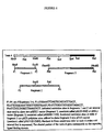
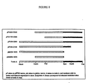
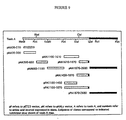
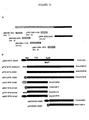
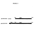
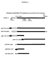
- the recombinant protein pMB1850-2360 contains amino acids 1852 through 2362 of the C. difficile toxin B protein.
- the specification gives detailed construction details for all recombinant proteins such that one skilled in the art will know precisely which amino acids are present in a given recombinant protein.
- Antibodies to certain pathogenic organisms have been shown to be effective in treating diseases caused by those organisms. It has not been shown whether antibodies can be raised, against Clostridium difficile , which would be effective in treating infection by this organism. Accordingly, C. difficile was tested as immunogen for production of hen antibodies.
- the example involved (a) preparation of the bacterial immunogen, (b) immunization, (c) purification of anti-bacterial chicken antibodies, and (d) detection of anti-bacterial antibodies in the purified IgY preparations.
- C. difficile strains 43594 (serogroup A) and 43596 (serogroup C) were originally obtained from the ATCC. These two strains were selected because they represent two of the most commonly-occurring serogroups isolated from patients with antibiotic-associated pseudomembranous colitis. [Delmee et al ., J. Clin. Microbiol., 28(10):2210 (1990).] Additionally, both of these strains have been previously characterized with respect to their virulence in the Syrian hamster model for C. difficile infection. [Delmee et al ., J. Med Microbiol., 33:85 (1990).]
- the bacterial strains were separately cultured on brain heart infusion agar for 48 hours at 37°C in a Gas Pack 100 Jar (BBL, Cockeysville, MD) equipped with a Gas Pack Plus anaerobic envelope (BBL). Forty-eight hour cultures were used because they produce better growth and the organisms have been found to be more cross-reactive with respect to their surface antigen presentation. The greater the degree of cross-reactivity of our IgY preparations, the better the probability of a broad range of activity against different strains/serogroups. [Toma et al ., J. Clin. Microbiol., 26(3):426 (1988).]
- the resulting organisms were removed from the agar surface using a sterile dacron-tip swab, and were suspended in a solution containing 0.4% formaldehyde in PBS, pH 7.2.
- This concentration of formaldehyde has been reported as producing good results for the purpose of preparing whole-organism immunogen suspensions for the generation of polyclonal anti- C. difficile antisera in rabbits.
- two separate bacterial suspensions were prepared, one for each strain. The two suspensions were then incubated at 4°C for 1 hour.
- the time point for the first series of immunizations was designated as "day zero.” All subsequent immunizations were performed as described above except that the bacterial immunogen suspensions were emulsified using IFA (GIBCO) instead of CFA, and for the later time point immunization, the stored frozen suspensions were used instead of freshly-prepared suspensions.
- the immunization schedule used is listed in Table 4. Immunization Schedule DAY OF IMMUNIZATION FORMALIN TREATMENT IMMUNOGEN PREPARATION USED 0 1%, 1 hr. freshly-prepared 14 1%, overnight freshly-prepared 21 1%, overnight freshly-prepared 35 1%, 48 hrs. freshly-prepared 49 1%, 72 hrs. freshly-prepared 70 1%, 72 hrs. stored frozen 85 1%, 72 hrs. stored frozen 105 1%, 72 hrs. stored frozen
- Table 5 shows the results of the whole-organism ELISA. All four IgY preparations demonstrated significant levels of activity, to a dilution of 1:62,500 or greater against both of the immunizing organism strains. Therefore, antibodies raised against one strain were highly cross-reactive with the other strain, and vice versa.
- the immunizing concentration of organisms did not have a significant effect on organism-specific IgY production, as both concentrations produced approximately equivalent responses. Therefore, the lower immunizing concentration of approximately 1.5 ⁇ 10 8 organisms/hen is the preferred immunizing concentration of the two tested.
- the preimmune IgY preparation appeared to possess relatively low levels of C. difficile -reactive activity to a dilution of 1:500, probably due to prior exposure of the animals to environmental clostridia.
- IgY Total yolk immunoglobulin
- Example 1(c). Antigenicity of the botulinal toxin was assessed on Western blots.
- the 150 kD detoxified type A neurotoxin and unmodified, toxic, 300 kD botulinal type A complex (toxin used for intragastric route administration for animal gut neutralization experiments; see Example 6) were separated on a SDS-polyacrylamide reducing gel.
- the Western blot technique was performed according to the method of Towbin. [H. Towbin et al ., Proc. Natl. Acad.
- the blot was cut into strips and each strip was incubated with the appropriate primary antibody.
- the avian anti- C. botulinum antibodies [described in (c)] and pre-immune chicken antibody (as control) were diluted 1:125 in PBS containing 1 mg/ml BSA for 2 hours at room temperature.
- the blots were washed with two changes each of large volumes of PBS, BBS-Tween and PBS, successively (10 min/wash).
- Goat anti-chicken IgG alkaline phosphatase conjugated secondary antibody (Fisher Biotech) was diluted 1:500 in PBS containing 1 mg/ml BSA and incubated with the blot for 2 hours at room temperature.
- the blots were washed with two changes each of large volumes of PBS and BBS-Tween, followed by one change of PBS and 0.1 M Tris-HCl, pH 9.5. Blots were developed in freshly prepared alkaline phosphatase substrate buffer (100 ⁇ g/ml nitroblue retrazolium (Sigma), 50 ⁇ g/ml 5-bromo-4-chloro-3-indolyl phosphate (Sigma), 5 mM MgCl 2 in 50 mM Na 2 CO 3 , pH 9.5).
- alkaline phosphatase substrate buffer 100 ⁇ g/ml nitroblue retrazolium (Sigma), 50 ⁇ g/ml 5-bromo-4-chloro-3-indolyl phosphate (Sigma), 5 mM MgCl 2 in 50 mM Na 2 CO 3 , pH 9.5).
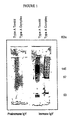
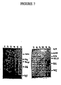
- the Western blots are shown in Figure 1.
- the anti- C. botulinum IgY reacted to the toxoid to give a broad immunoreactive band at about 145-150 kD on the reducing gel.
- This toxoid is refractive to disulfide cleavage by reducing agents due to formalin crosslinking.
- the immune IgY reacted with the active toxin complex, a 97 kD C. botulinum type A heavy chain and a 53 kD light chain.
- the preimmune IgY was unreactive to the C. botulinum complex or toxoid in the Western blot.
- the IgY antibody titer to C. botulinum type A toxoid of eggs harvested between day 409 and 423 was evaluated by ELISA, prepared as follows. Ninety-six-well Falcon Pro-bind plates were coated overnight at 4°C with 100 ⁇ l/well toxoid [B.R. Singh & B.R. Das Gupta, Toxicon 27:403 (1989)] at 2.5 ⁇ g/ml in PBS, pH 7.5 containing 0.005% thimerosal. The following day the wells were blocked with PBS containing 1% BSA for 1 hour at 37°C.
- the IgY from immune or preimmune eggs was diluted in PBS containing 1% BSA and 0.05% Tween 20 and the plates were incubated for I hour at 37°C. The plates were washed three times with PBS containing 0.05% Tween 20 and three times with PBS alone. Alkaline phosphatase-conjugated goat-anti-chicken IgG (Fisher Biotech) was diluted 1:750 in PBS containing 1% BSA and 0.05% Tween 20, added to the plates, and incubated 1 hour at 37°C. The plates were washed as before, and p-nitrophenyl phosphate (Sigma) at 1 mg/ml in 0.05 M Na 2 CO 3 , pH 9.5, 10 mM MgCl 2 was added.
- p-nitrophenyl phosphate Sigma
- ELISA enzyme-linked immunoassay
- IgY which was raised against Crotalus durissus terrificus venom.
- Three eggs were collected from hens immunized with the C. durissus terrificus venom and IgY was extracted from the yolks using the modified Polson procedure described by Thalley and Carroll [Bio/Technology, 8:934-938 (1990)] as described in Example 1(c).
- the antibody solution needs to be filter-sterilized (as an alternative to heat sterilization which would destroy the antibodies).
- the preparation of IgY resuspended in water was too cloudy to pass through either a 0.2 or 0.45 ⁇ m membrane filter, so 10 ml of the PBS resuspended fraction was dialyzed overnight at room temperature against 250 ml of water. The following morning the dialysis chamber was emptied and refilled with 250 ml of fresh H 2 O for a second dialysis. Thereafter, the yields of soluble antibody were determined at OD 280 and are compared in Table 7.
- the species specific secondary antibody (goat anti-chicken immunoglobulin alkaline-phosphatase conjugate (Sigma) was diluted 1:750 in PBS containing 1 mg/ml BSA and added to each well of the microtiter plate. After 2 hours of incubation at room temperature, the unbound secondary antibody was removed by washing the plate as before, and freshly prepared alkaline phosphatase substrate (Sigma) at 1 mg/ml in 50 mM Na 2 CO 3 , 10 mM MgCl 2 , pH 9.5 was added to each well. The color development was measured on a Dynatech MR 700 microplate reader using a 412 nm filter. The results are shown in Table 8.
- the ELISA plate was blocked with 5 mg/ml BSA, and the fecal samples were extracted as before, except that instead of heating the extracts at 37°C, the samples were kept on ice to limit proteolysis. The samples were assayed undiluted initially, and in 5X serial dilutions thereafter. Otherwise the assay was carried out as before. Specific Antibody Survives Passage Through The Gastrointestinal Tract DILUTION PREIMMUNE IgY CONTROL EXTRACT EXP.
- the experiment confirmed the previous results, with the antibody activity markedly higher.
- the control fecal extract showed no anti- C.d.t . activity, even undiluted, while the fecal extract from the anti- C.d.t . IgY/Enfamil®-fed mouse showed considerable anti- C.d.t . activity.
- This experiment (and the previous experiment) clearly demonstrate that active IgY antibody survives passage through the mouse digestive tract, a finding with favorable implications for the success of IgY antibodies administered orally as a therapeutic or prophylactic.
- botulinum toxin type A complex obtained from Eric Johnson (University Of Wisconsin, Madison) was 250 ⁇ g/ml in 50 mM sodium citrate, pH 5.5, specific toxicity 3 ⁇ 10 7 mouse LD 50 /mg with parenteral administration. Approximately 40-50 ng/gm body weight was usually fatal within 48 hours in mice maintained on conventional food and water. When mice were given a diet ad libitum of only Enfamil® the concentration needed to produce lethality was approximately 2.5 times higher (125 ng/gm body weight). Botulinal toxin concentrations of approximately 200 ng/gm body weight were fatal in mice fed Enfamil® containing preimmune IgY (resuspended in Enfamil® at the original yolk volume).
- mice The oral LD 100 of C. botulinum toxin was also determined in mice that received known amounts of a mixture of preimmune IgY-Ensure® delivered orally through feeding needles. Using a 22 gauge feeding needle, mice were given 250 ⁇ l each of a preimmune IgY-Ensure® mixture (preimmune IgY dissolved in 1/4 original yolk volume) 1 hour before and 1/2 hour and 5 hours after administering botulinal toxin. Toxin concentrations given orally ranged from approximately 12 to 312 ng/gm body weight (0.3 to 7.5 ⁇ g per mouse). Botulinal toxin complex concentration of approximately 40 ng/gm body weight (1 ⁇ g per mouse) was lethal in all mice in less than 36 hours.
- mice treated with the preimmune IgY-Ensure® mixture died within 46 hours post-toxin administration. The average time of death in the mice was 32 hours post toxin administration. Treatments of preimmune IgY-Ensure® mixture did not continue beyond 24 hours due to extensive paralysis of the mouth in mice of this group. In contrast, all ten mice treated with the immune anti-botulinal toxin IgY-Ensure® mixture survived past 96 hours. Only 4 mice in this group exhibited symptoms of botulism toxicity (two mice about 2 days after and two mice 4 days after toxin administration). These mice eventually died 5 and 6 days later. Six of the mice in this immune group displayed no adverse effects to the toxin and remained alive and healthy long term. Thus, the avian anti-botulinal toxin antibody demonstrated very good protection from the lethal effects of the toxin in the experimental mice.
- Toxin A is a potent cytotoxin secreted by pathogenic strains of C. difficile , that plays a direct role in damaging gastrointestinal tissues. In more severe cases of C. difiicile intoxication, pseudomembranous colitis can develop which may be fatal. This would be prevented by neutralizing the effects of this toxin in the gastrointestinal tract.
- antibodies were produced against a portion of the toxin. The example involved: (a) conjugation of a synthetic peptide of toxin A to bovine serum albumin; (b) immunization of hens with the peptide-BSA conjugate; and (c) detection of antitoxin peptide antibodies by ELISA.
- the synthetic peptide CQTIDGKKYYFN-NH 2 was prepared commercially (Multiple Peptide Systems, San Diego, CA) and validated to be >80% pure by high-pressure liquid chromatography.
- the eleven amino acids following the cysteine residue represent a consensus sequence of a repeated amino acid sequence found in Toxin A. [Wren et al ., Infect. Immun., 59:3151-3155 (1991).]
- the cysteine was added to facilitate conjugation to carrier protein.
- BSA (Sigma) was dissolved in 0.01 M NaPO 4 , pH 7.0 to a final concentration of 20 mg/ml and n-maleimidobenzoyl-N-hydroxysuccinimide ester (MBS; Pierce) was dissolved in N,N-dimethyl formamide to a concentration of 5 mg/ml.
- MBS solution 0.51 ml, was added to 3.25 ml of the BSA solution and incubated for 30 minutes at room temperature with stirring every 5 minutes.
- the MBS-activated BSA was then purified by chromatography on a Bio-Gel P-10 column (Bio-Rad; 40 ml bed volume) equilibrated with 50 mM NaPO 4 , pH 7.0 buffer. Peak fractions were pooled (6.0 ml).
- Lyophilized toxin A peptide (20 mg) was added to the activated BSA mixture, stirred until the peptide dissolved and incubated 3 hours at room temperature. Within 20 minutes, the reaction mixture became cloudy and precipitates formed. After 3 hours, the reaction mixture was centrifuged at 10,000 ⁇ g for 10 min and the supernatant analyzed for protein content. No significant protein could be detected at 280 nm. The conjugate precipitate was washed three times with PBS and stored at 4°C. A second conjugation was performed with 15 mg of activated BSA and 5 mg of peptide and the conjugates pooled and suspended at a peptide concentration of 10 mg/ml in 10 mM NaPO 4 , pH 7.2.
- hens were each initially immunized on day zero by injection into two subcutaneous and two intramuscular sites with 1 mg of peptide conjugate that was emulsified in CFA (GIBCO). The hens were boosted on day 14 and day 21 with 1 mg of peptide conjugate emulsified in IFA (GIBCO).
- IgY was purified from two eggs obtained before immunization (pre-immune) and two eggs obtained 31 and 32 days after the initial immunization using PEG fractionation as described in Example 1.
- Wells of a 96-well microtiter plate (Falcon Pro-Bind Assay Plate) were coated overnight at 4°C with 100 ⁇ g/ml solution of the toxin A synthetic peptide in PBS, pH 7.2 prepared by dissolving 1 mg of the peptide in 1.0 ml of H 2 O and dilution of PBS.
- the preimmune and immune IgY preparations were diluted in a five-fold series in a buffer containing 1% PEG 8000 and 0.1% Tween-20 (v/v) in PBS, pH 7.2.
- the immune antibodies contain titers against this repeated epitope of toxin A.
- avian antibodies were immunized using native C. difficile toxins A and B.
- the resulting egg yolk antibodies were then extracted and assessed for their ability to neutralize toxins A and B in vitro .
- the Example involved (a) preparation of the toxin immunogens, (b) immunization, (c) purification of the antitoxins, and (d) assay of toxin neutralization activity.
- Free formaldehyde was then removed from the resulting toxoid solutions by dialysis against PBS at 4°C. In previously published reports, this dialysis step was not performed. Therefore, free formaldehyde must have been present in their toxoid preparations.
- the toxoid solutions were concentrated, using a Centriprep concentrator unit (Amicon), to a final toxoid concentration of 4.0 mg/ml. The two resulting preparations were designated as toxoid A and toxoid B.
- C. difficile native toxins were prepared by concentrating stock solutions of toxin A and toxin B (Tech Lab, Inc), using Centriprep concentrator units (Amicon), to a final concentration of 4.0 mg/ml.
- the first two immunizations were performed using the toxoid A and toxoid B immunogens described above. A total of 3 different immunization combinations were employed.
- 0.2 ml of toxoid A was emulsified in an equal volume of Titer Max adjuvant (CytRx). Titer Max was used in order to conserve the amount of immunogen used, and to simplify the immunization procedure. This immunization group was designated "CTA.”
- 0.1 ml of toxoid B was emulsified in an equal volume of Titer Max adjuvant.
- CTB This group was designated "CTB.”
- CTA toxoid A
- CTB toxoid B
- CTAB Titer Max adjuvant
- White Leghorn hens obtained from a local breeder, were immunized as follows: Group CTA . Four hens were immunized, with each hen receiving 200 ⁇ g of toxoid A, via two intramuscular (I.M.) injections of 50 ⁇ l of CTA emulsion in the breast area, Group CTB . One hen was immunized with 200 ⁇ g of toxoid B, via two I.M. injections of 50 ⁇ l of CTB emulsion in the breast area. Group CTAB . Four hens were immunized, with each hen receiving a mixture containing 200 ⁇ g of toxoid A and 200 ⁇ g of toxoid B, via two I.M. injections of 100 ⁇ l of CTAB emulsion in the breast area. The second immunization was performed 5 weeks later, on day 35, exactly as described for the first immunization above.
- I.M. intramuscular
- a single hen from group CTAB was immunized for a third time, this time using a mixture of the native toxin A and native toxin B described in section (a) above (these toxins were not formaldehyde-treated, and were used in their active form). This was done in order to increase the amount (titer) and affinity of specific antitoxin antibody produced by the hen over that achieved by immunizing with toxoids only.
- 0.1 ml of a toxin mixture was prepared which contained 200 ⁇ g of native toxin A and 200 ⁇ g of native toxin B.
- This toxin mixture was then emulsified in 0.1 ml of Titer Max adjuvant.
- a single CTAB hen was then immunized with the resulting immunogen emulsion, via two I.M. injections of 100 ⁇ l each, into the breast area. This hen was marked with a wing band, and observed for adverse effects for a period of approximately 1 week, after which time the hen appeared to be in good health.
- the hen was then immunized with 200 ⁇ g of native toxin B, as described for the toxoid B immunizations above.
- Group CTAB A 0.15 ml volume of the 4 mg/ml native toxin A solution was first mixed with a 0.15 ml volume the 4 mg/ml native toxin B solution. The resulting toxin mixture was then emulsified in 0.3 ml of Titer Max adjuvant.
- the 3 remaining hens (the hen with the wing band was not immunized this time) were then immunized with 200 ⁇ g of native toxin A and 200 ⁇ g of native toxin B as described for the toxoid A+ toxoid B immunizations (CTAB) above.
- CTAB toxoid A+ toxoid B immunizations
- the toxin neutralization activity of the IgY solutions prepared in section (c) above was determined using an assay system that was modified from published methods. [Ehrich et al ., Infect. Immun. 28:1041-1043 (1992); and McGee et al . Microb. Path. 12:333-341 (1992).] As additional controls, affinity-purified goat anti- C. difficile toxin A (Tech Lab) and affinity-purified goat anti- C. difficile toxin B (Tech Lab) were also assayed for toxin neutralization activity.
- the IgY solutions and goat antibodies were serially diluted using F 12 medium (GIBCO) which was supplemented with 2% FCS (GIBCO)(this solution will be referred to as "medium” for the remainder of this Example).
- F 12 medium F 12 medium
- FCS 2% FCS
- the resulting antibody solutions were then mixed with a standardized concentration of either native C. difficile toxin A (Tech Lab), or native C. difficile toxin B (Tech Lab), at the concentrations indicated below.
- toxin + antibody mixtures were added to the wells of 96-well microtiter plates (Falcon Microtest III) which contained 2.5 ⁇ 10 4 Chinese Hamster Ovary (CHO) cells per well (the CHO cells were plated on the previous day to allow them to adhere to the plate wells).
- the final concentration of toxin, or dilution of antibody indicated below refers to the final test concentration of each reagent present in the respective microtiter plate wells.
- Toxin reference wells were prepared which contained CHO cells and toxin A or toxin B at the same concentration used for the toxin plus antibody mixtures (these wells contained no antibody).
- this Example utilized spectrophotometric methods to quantitate the C. difficile toxin bioassay.
- toxin A neutralizing activity of the CTA, CTAB, and pre-immune IgY preparations, as well as the affinity-purified goat antitoxin A control. dilutions of these antibodies were reacted against a 0.1 ⁇ g/ml concentration of native toxin A (this is the approx. 50% cytotoxic dose of toxin A in this assay system). The results are shown in Figure 3.
- the toxin B neutralizing activity of the CTAB and pre-immune IgY preparations, and also the affinity-purified goat antitoxin B control was determined by reacting dilutions of these antibodies against a concentration of native toxin B of 0.1 ng/ml (approximately the 50% cytotoxic dose of toxin B in the assay system). The results are shown in Figure 4.
- toxin B neutralizing activity of CTB, CTAB, and pre-immune IgY preparations was determined by reacting dilutions of these antibodies against a native toxin B concentration of 0.1 ⁇ g/ml (approximately 100% cytotoxic dose of toxin B in this assay system). The results are shown in Figure 5.
- C. difficile strain 43596 was chosen because it is representative of one of the most frequently-occurring serogroups isolated from patients with antibiotic-associated pseudomembranous colitis. [Delmee et al ., J. Clin. Microbiol., 28:2210-2214 (1990).] In addition, as this was the same C. difficile strain used in all of the previous Examples above, it was again used in order to provide experimental continuity.
- the carboxy terminal end consisting of the repeating units contains the hemagglutination activity or binding domain of C. difficile toxin A.
- hemagglutination assays were performed. Two toxin A recombinant proteins, one containing the binding domain as either soluble affinity purified protein (pMA1870-2680) or SDS solubilized inclusion body protein (pPA1870-2680) and soluble protein from one region outside that domain (pMA1100-1610) were tested using a described procedure. [H.C. Krivan et. al ., Infect.
- the volumes of the eluate increased to greater than 10 mls during dialysis in each case, due to the high viscosity of the Actisep elution media.
- Aliquots of each sample were 20x concentrated using Centricon 30 microconcentrators (Amicon) and stored at 4°C.
- the specificity of each region specific antibody pool was tested, relative to the dialyzed CTA IgY preparation, by Western blot analysis, exactly as described above, except that 4 ml samples of blocking buffer containing 100 ⁇ l region specific antibody (unconcentrated) were used instead of the depleted CTA IgY preparations.
- Each affinity purified antibody preparation was specific to the defined interval, except that samples purified against intervals 1-5 also reacted with interval 6.
- the pMAL protein was affinity purified as a soluble product [Example 12(d)] and the pET protein was isolated as insoluble inclusion bodies [Example 12(d)] and solubilized to an immunologically active protein using a proprietary method described in a pending patent application (U.S. Patent Application Serial No. 08/129,027).
- This Example involves (a) immunization, (b) antitoxin collection, (c) determination of antitoxin antibody titer, (d) anti-recombinant toxin A neutralization of toxin A hemagglutination activity in vitro , and (e) assay of in vitro toxin A neutralizing activity.
- the antibody titer of a recombinant toxin A polypeptide was determined by ELISA. Eggs from both hens were collected on day 32, the yolks pooled and the antibody was isolated using PEG as described. The immune recombinant IgY antibody titer was determined for the soluble recombinant protein containing the maltose binding protein fusion generated in p-Mal (pMA1870-2680).
- ELISA titers (reciprocal of the highest dilution of IgY generating a signal) in the preimmune IgY to the MBP or the recombinant was ⁇ 1:30 while the immune IgY titers to MBP and the toxin A recombinant were 1:18750 and > 1:93750 respectively.
- the anti-recombinant antibody titers generated in the hens against the recombinant polypeptide is much higher, compared to antibodies to that region raised using native toxin A.
- the recombinant antibody titer to region 1870-2680 in the CTA antibody preparation is at least five-fold lower compared to the recombinant generated antibodies (1:18750 versus >1:93750).
- it appears a better immune response can be generated against a specific recombinant using that recombinant as the immunogen compared to the native toxin A.
- Toxin A has hemagglutinating activity besides cytotoxic and enterotoxin properties. Specifically, toxin A agglutinates rabbit erythrocytes by binding to a trisaccharide (gal 1-3B1-4GlcNAc) on the cell surface.
- a trisaccharide gal 1-3B1-4GlcNAc
- Polyethylene glycol-fractionated immune or preimmune IgY were pre-absorbed with citrated rabbit erythrocytes prior to performing the hemagglutination assay because we have found that IgY alone can agglutinate red blood cells.
- Citrated rabbit red blood cells (RRBC's)(Cocalico) were washed twice by centrifugation at 450 x g with isotonic buffer (0.1 M Tris-HCl, 0.05 M NaCl, pH 7.2).
- RRBC-reactive antibodies in the IgY were removed by preparing a 10% RRBC suspension (made by adding packed cells to immune or preimmune IgY) and incubating the mixture for 1 hour at 37°C. The RRBCs were then removed by centrifugation.
- toxin A Neutralization of the hemagglutination activity of toxin A by antibody was tested in round-bottomed 96-well microtiter plates. Twenty-five ⁇ l of toxin A (36 ⁇ g /ml) (Tech Lab) in isotonic buffer was mixed with an equal volume of different dilutions of immune or preimmune IgY in isotonic buffer, and incubated for 15 minutes at room temperature. Then, 50 ⁇ l of a 1% RRBC suspension in isotonic buffer was added and the mixture was incubated for 3 hours at 4°C. Positive control wells containing the final concentration of 9 ⁇ g/ml of toxin A after dilution without IgY were also included.
- Hemagglutination activity was assessed visually, with a diffuse matrix of RRBC's coating the bottom of the well representing a positive hemagglutination reaction and a tight button of RRBC's at the bottom of the well representing a negative reaction.
- the anti-recombinant immune IgY neutralized toxin A hemagglutination activity, giving a neutralization titer of 1:8.
- preimmune IgY was unable to neutralize the hemagglutination ability of toxin A.
- the anti-recombinant toxin A IgY demonstrated only partial neutralization of the cytotoxic activity of toxin A, while the pre-immune IgY did not demonstrate any significant neutralizing activity.
- avian antibodies raised against recombinant toxin A binding domain to neutralize the enterotoxin activity of C. difficile toxin A was evaluated in vivo using Golden Syrian hamsters.
- Eggs were collected from hens which had been immunized with the recombinant C. difficile toxin A fragment pMA1870-2680 (described in Example 13, above).
- a second group of eggs purchased at a local supermarket was used as a pre-immune (negative) control.
- Egg yolk immunoglobulin (IgY) was extracted by PEG from the two groups of eggs as described in Example 8(c), and the final IgY pellets were solubilized in one-fourth the original yolk volume using 0.1M carbonate buffer (mixture of NaHCO 3 and Na 2 CO 3 ), pH 9.5.
- the basic carbonate buffer was used in order to protect the toxin A from the acidic pH of the stomach environment.
- the first group consisted of a single representative animal taken from each of the 4 groups of surviving hamsters at the conclusion of the study described in section (b) above. These animals represented the 24 hr. timepoint of the study.
- the second group consisted of two animals which were not part of the study described above, but were separately treated with the same toxin A + pre-immune IgY mixtures as described for the animals in section (b) above. Both of these hamsters developed diarrhea, and were sacrificed 8 hrs. after the time of administration of the toxin A + pre-immune IgY mixtures. At the time of sacrifice, both animals were presenting symptoms of diarrhea. These animals represented the acute phase of the study.
- the final group consisted of a single untreated hamster from the same shipment of animals as those used for the two previous groups. This animal served as the normal control.
- the tissues from the two 24 hr. animals which received the toxin A + preimmune IgY mixtures demonstrated significant pathology.
- the mucosal layer was observed to be less organized than in the normal control tissue.
- the cytoplasm of the epithelial cells had a vacuolated appearance, and gaps were present between the epithelium and the underlying cell layers.
- the lamina basement was largely absent. Intestinal villi and crypts were significantly diminished, and appeared to have been overgrown by a planar layer of epithelial cells and fibroblasts. Therefore, although these animals overtly appeared to recover from the acute symptoms of toxin A intoxication, lasting pathologic alterations to the cecal mucosa had occurred.
- the tissues obtained from the two acute animals which received mixtures of toxin A and pre-immune IgY demonstrated the most significant pathology. At the gross pathological level, both animals were observed to have severely distended ceca which were filled with watery, diarrhea-like material. At the microscopic level, the animal that was given the mixture containing 10 ⁇ g of toxin A and pre-immune IgY was found to have a mucosal layer which had a ragged, damaged appearance, and a disorganized, compacted quality. The crypts were largely absent, and numerous breaks in the epithelium had occurred. There was also an influx of erythrocytes into spaces between the epithelial layer and the underlying tissue.
- Example 14 The ability of avian antibodies directed against the recombinant C. difficile toxin A fragment 1870-2680 (as expressed by pMA1870-2680; see Example 13) to neutralize the enterotoxic activity of toxin A was demonstrated in Example 14.
- This example involved: (a) the preparation of IgYs against recombinant toxin A polypeptides; (b) in vivo protection of hamsters against toxin A by treatment with anti-recombinant toxin A IgYs and (c) quantification of specific antibody concentration in CTA and Interval 6 IgY PEG preparations.
- the nucleotide sequence of the coding region of the entire toxin A protein is listed in SEQ ID NO:5.
- the amino acid sequence of the entire toxin A protein is listed in SEQ ID NO:6.
- the amino acid sequence consisting of amino acid residues 1870 through 2680 of toxin A is listed in SEQ ID NO:7.
- the amino acid sequence consisting of amino acid residues 1870 through 1960 of toxin A is listed in SEQ ID NO:8.
- the amino acid sequence of residues 1873 through 2684 of toxin A is listed in SEQ ID NO:29.
- pP refers to the pET23 vector (New England BioLabs);
- pM refers to the pMALTM-c vector (New England BioLabs);
- A refers to toxin A; the numbers refer to the amino acid interval expressed in the clone.
- pMA30-300 indicates that the recombinant clone encodes amino acids 30-300 of toxin A and the vector used was pMALTM-c).
- the recombinant proteins were generated as described in Example 11.
- the IgYs were extracted and solubilized in 0.1M carbonate buffer pH 9.5 for oral administration as described in Example 14(a).
- the IgY reactivities against each individual recombinant interval was evaluated by ELISA as described in Example 13(c).
- Example 8 mixed with toxin A (CTA) served as a positive control.
- the mixture was incubated for 1 hour at 37°C then orally administered to lightly etherized hamsters using an 18G feeding needle.
- the animals were then observed for the onset of diarrhea and death for a period of approximately 24 hours.
- the results are shown in Table 18.
- Study Outcome After 24 Hours Treatment group Healthy Diarrhea Dead Preimmune 0 0 7 CTA 5 0 0 Interval 6 6 1 0 Interval 4 0 1 6 Interval 1235 0 0 7
- IgY PEG preparations To determine the purity of IgY PEG preparations, an aliquot of a pMA1870-2680 (Interval 6) IgY PEG preparation was chromatographed using HPLC and a KW-803 sizing column (Shodex). The resulting profile of absorbance at 280 nm is shown in Figure 16. The single large peak corresponds to the predicted MW of IgY. Integration of the area under the single large peak showed that greater than 95% of the protein eluted from the column was present in this single peak. This result demonstrated that the majority (>95%) of the material absorbing at 280 nm in the PEG preparation corresponds to IgY. Therefore, absorbance at 280 nm can be used to determine the total antibody concentration in PEG preparations.
- Interval 6-specific antibodies expressed as percent of total antibody
- PEG preparations defined quantities of these antibody preparations were affinity purified on a pPA1870-2680(H) (shown schematically in Figure 15B) affinity column and the specific antibodies were quantified.
- pP refers to the pET23 vector (New England BioLabs);
- pM refers to the pMALTM-c vector (New England BioLabs);
- pG refers to the pGEX vector (Pharmacia);
- pB refers to the PinPointTM Xa vector (Promega);
- A refers to toxin A; the numbers refer to the amino acid interval expressed in the clone.
- the solid black ovals represent the MBP; the hatched ovals represent glutathione S-transferase; the hatched circles represent the biotin tag; and HHH represents the poly-histidine tag.
- An affinity column containing recombinant toxin A repeat protein was made as follows. Four ml of PBS-washed Actigel resin (Sterogene) was coupled with 5-10 mg of pPA1870-2680 inclusion body protein [prepared as described in Example (17) and dialyzed into PBS] in a 15 ml tube (Falcon) containing 1/10 final volume Ald-coupling solution (1 M sodium cyanoborohydride). Aliquots of the supernatant from the coupling reactions, before and after coupling, were assessed by Coomassie staining of 7.5% SDS-PAGE gels. Based upon protein band intensities, greater than 6 mg of recombinant protein was coupled to the resin.
- each dose of the Interval 6 antibody prep contained approximately 400 ⁇ g of specific antibody.
- each hamster was predisposed to C. difficile infection by the oral administration of 3.0 mg of Clindamycin-HCl (Sigma) in 1 ml of water.
- the hamsters were orally challenged with 1 ml of C. difficile inoculum strain ATCC 43596 in sterile saline containing approximately 100 organisms. The animals were then observed for the onset of diarrhea and subsequent death during the treatment period. The results are shown in Table 19. Lethality After 12 Days Of Treatment Treatment Group Number Animals Alive Number Animals Dead Preimmune 0 7 CTAB 6 1 Interval 6 7 0
- Figure 17 shows schematically the location of the Lyerly, et al , intra-Interval 6 recombinant protein (cloned into the pUC vector) in comparison with the complete Interval 6 construct (pMA1870-2680) used herein to generate neutralizing antibodies directed against toxin A.
- the solid black oval represents the MBP which is fused to the toxin A Interval 6 in pMA1870-2680.
- the hamsters were first predisposed to C. difficile infection with a 3 mg dose of Clindamycin-HCl (Sigma) administered orally in 1 ml of water. Approximately 24 hrs later, the animals were orally challenged with 1 ml of C. difficile strain ATCC 43596 in sterile saline containing approximately 200 organisms. One day after infection, the presence of toxin A and B was determined in the feces of the hamsters using a commercial immunoassay kit (Cytoclone A+B EPA, Cambridge Biotech) to verify establishment of infection. Four members of each group were randomly selected and tested. Feces from an uninfected hamster was tested as a negative control. All infected animals tested positive for the presence of toxin according to the manufacturer's procedure. The initiation of treatment then started approximately 24 hr post-infection.
- the animals were dosed daily at roughly 4 hr intervals with 1 ml antibody preparation diluted in Ensure® (Ross Labs).
- the amount of specific antibodies given per dose was estimated to be about 400 ⁇ g of anti-Interval 6 IgY (for animals in the Interval 6 group) and 100 ⁇ g and 70 ⁇ g of anti-toxin A (Interval 6-specific) and anti-toxin B (Interval 3-specific; see Example 19), respectively, for the CTAB preparation.
- the animals were treated for 9 days and then observed for an additional 4 days for the presence of diarrhea and death. The results indicating the number of survivors and the number of dead 4 days post-infection are shown in Table 20.
- In vivo Therapeutic Treatment With Interval 6 Antibodies Treatment Group Number Animals Alive Number Animals Dead Preimmune 4 3 CTAB 8 0
- Antibodies directed against both interval 6 and CTAB successfully prevented death from C. difficile when therapeutically administered 24 hr after infection. This result is significant since many investigators begin therapeutic treatment of hamsters with existing drugs (e.g ., vancomycin, phenelfamycins, tiacumicins, etc.) 8 hr post-infection [Swanson, et al . (1991) Antimicrobial Agents and Chemotherapy 35:1108 and (1989) J. Antibiotics 42:94].
- drugs e.g ., vancomycin, phenelfamycins, tiacumicins, etc.
- the Interval 6 immunogen utilized in Examples 15 and 16 was the pMA1870-2680 protein, in which the toxin A repeats are expressed as a soluble fusion protein with the MBP (described in Example 11). Interestingly, expression of this region (from the Spe I site to the end of the repeats, see Figure 15B) in three other expression constructs, as either native (pPA1870-2680), poly-His tagged [pPA1870-2680 (H)] or biotin-tagged (pBA1870-2680) proteins resulted in completely insoluble protein upon induction of the bacterial host (see Figure 15B).
- the host strain BL21 (Novagen) was used for expression of pBA1870-2680 and host strain BL21(DE3) (Novagen) was used for expression of pPA1870-2680 and pPA1870-2680(H). These insoluble proteins accumulated to high levels in inclusion bodies. Expression of recombinant plasmids in E. coli host cells grown in 2X YT medium was performed as described [Williams, et al. (1994), supra ].
- solubility of expressed proteins from the pPG1870-2190 and pPA1870-2190 expression constructs was determined after induction of recombinant protein expression under conditions reported to enhance protein solubility [These conditions comprise growth of the host at reduced temperature (30°C) and the utilization of high (1 mM IPTG) or low (0.1 mM IPTG) concentrations of inducer [Williams et al . (1994), supra ]. All expressed recombinant toxin A protein was insoluble under these conditions. Thus, expression of these fragments of the toxin A repeats in pET and pGEX expression vectors results in the production of insoluble recombinant protein even when the host cells are grown at reduced temperature and using lower concentrations of the inducer.
- Toxin A repeat regions to which neutralizing antibodies bind were identified by utilizing recombinant toxin A repeat region proteins expressed as soluble or insoluble proteins to deplete protective antibodies from a polyclonal pool of antibodies against native C. difficile toxin A.
- An in vivo assay was developed to evaluate proteins for the ability to bind neutralizing antibodies.
- CTA antibody generated in Example 8
- C. difficile toxin A was added at a concentration lethal to hamsters and the mixture was administered to hamsters via IP injection. If the recombinant protein contains neutralizing epitopes, the CTA antibodies would lose their ability to bind toxin A resulting in diarrhea and/or death of the hamsters.
- insoluble toxin A repeat protein was solubilized and specific antibodies were raised in chickens.
- Insoluble pPA1870-2680 protein was solubilized using the method described in Williams et al . (1994), supra . Briefly, induced cultures (500 ml) were pelleted by centrifugation at 3,000 X g for 10 min at 4°C.
- the cell pellets were resuspended thoroughly in 10 ml of inclusion body sonication buffer (25 mM HEPES pH 7.7, 100 mM KCl, 12.5 mM MgCl 2 , 20% glycerol, 0.1% (v/v) Nonidet P-40, 1 mM DTT).
- inclusion body sonication buffer 25 mM HEPES pH 7.7, 100 mM KCl, 12.5 mM MgCl 2 , 20% glycerol, 0.1% (v/v) Nonidet P-40, 1 mM DTT).
- inclusion body sonication buffer 25 mM HEPES pH 7.7, 100 mM KCl, 12.5 mM MgCl 2 , 20% glycerol, 0.1% (v/v) Nonidet P-40, 1 mM DTT.
- the suspension was transferred to a 30 ml non-glass centrifuge tube. Five hundred ⁇ l of 10 mg/ml lyso
- the sonicated suspension was transferred to a 35 ml Oakridge tube and centrifuged at 6,000 X g for 10 min at 4°C to pellet the inclusion bodies.
- the pellet was washed 2 times by pipetting or vortexing in fresh, ice-cold RIPA buffer [0.1% SDS, 1% Triton X-100, 1% sodium deoxycholate in TBS (25 mM Tris-Cl pH 7.5, 150 mM NaCl)].
- the inclusion bodies were recentrifuged after each wash.
- the inclusion bodies were dried and transferred using a small metal spatula to a 15 ml tube (Falcon).
- One ml of 10% SDS was added and the pellet was solubilized by gently pipetting the solution up and down using a 1 ml micropipettor. The solubilization was facilitated by heating the sample to 95°C when necessary.
- the samples were diluted with 9 volumes of PBS.
- the protein solutions were dialyzed overnight against a 100-fold volume of PBS containing 0.05% SDS at room temperature.
- the dialysis buffer was then changed to PBS containing 0.01% SDS and the samples were dialyzed for several hours to overnight at room temperature.
- the samples were stored at 4°C until used. Prior to further use, the samples were warmed to room temperature to allow any precipitated SDS to go back into solution.
- the inclusion body preparation was used to immunize hens.
- the protein was dialyzed into PBS and emulsified with approximately equal volumes of CFA for the initial immunization or IFA for subsequent booster immunizations.
- Booster immunizations of 1.0 mg were given of days 14 and day 28. Eggs were collected on day 32 and the antibody isolated using PEG as described in Example 14(a).
- Antibodies raised against native toxin A were protective while preimmune antibodies had little effect. As found using the in vitro CHO assay [described in Example 8(d)] where antibodies raised against the soluble Interval 6 could partially neutralize the effects of toxin A, here they were able to completely neutralize toxin A in vivo . In contrast, the antibodies raised against the insoluble Interval 6 was unable to neutralize the effects of toxin A in vivo as shown above (Table 22) and in vitro as shown in the CHO assay [described in Example 8(d)].
- the toxin B gene has been cloned and sequenced; the amino acid sequence deduced from the cloned nucleotide sequence predicts a MW of 269.7 kD for toxin B [Barroso et al ., Nucl. Acids Res. 18:4004 (1990)].
- the nucleotide sequence of the coding region of the entire toxin B gene is listed in SEQ ID NO:9.
- the amino acid sequence of the entire toxin B protein is listed in SEQ ID NO:10.
- the amino acid sequence consisting of amino acid residues 1850 through 2360 of toxin B is listed in SEQ ID NO:11.
- the amino acid sequence consisting of amino acid residues 1750 through 2360 of toxin B is listed in SEQ ID NO:12.
- the amino acid sequence consisting of amino acid residues 1754 through 2362 of toxin B is listed in SEQ ID NO:30.
- the isolated recombinant protein would be soluble in order to preserve native antigenicity, since solubilized inclusion body proteins often do not fold into native conformations. Indeed as shown in Example 17, neutralizing antibodies against recombinant toxin A were only obtained when soluble recombinant toxin A polypeptides were used as the immunogen. To allow ease of purification, the recombinant protein should be expressed to levels greater than 1 mg/liter of E. coli culture.
- the toxin B gene was cloned using PCR amplification from C. difficile genomic DNA. Initially, the gene was cloned in two overlapping fragments, using primer pairs P5/P6 and P7/P8. The location of these primers along the toxin B gene is shown schematically in Figure 18. The sequence of each of these primers is:
- All MBP fusion proteins were constructed and expressed in the pMALTM-c or pMALTM-p2 vectors (New England Biolabs) in which the protein of interest is expressed as a C-terminal fusion with MBP. All pET plasmids were expressed in either the BL21(DE3) or BL21(DE3)LysS expression hosts, while pMal plasmids were expressed in the BL21 host.
- Extracts containing soluble pMAL fusion protein were prepared and chromatographed in PBS buffer over an amylose resin (New England Biolabs) column, and eluted with PBS containing 10 mM maltose as described [Williams et al . (1994), supra ].
- Protein preparations from induced cultures of each of these constructs were analyzed, by SDS-PAGE, to estimate protein stability (Coomassie Blue staining) and immunoreactivity against anti-toxin B specific antiserum (Western analysis). Higher levels of intact (i.e ., nondegraded), full length fusion proteins were observed with the smaller constructs as compared with the larger recombinants, and a series of expression constructs spanning the entire toxin B gene were constructed ( Figures 18, 19 and 20 and Table 23).
- the fusion of the blunt ended toxin B 3' end to the filled Hind III site should create an in-frame fusion with the C-terminal poly-histidine sequence in this vector.
- the pPB1750-2360 clone selected had lost, as predicted, the Hind III site at this clone junction; this eliminated the possibility that an additional adenosine residue was added to the 3' end of the PCR product by a terminal transferase activity of the Pfu polymerase, since fusion of this adenosine residue to the filled Hind III site would regenerate the restriction site (and was observed in several clones).
- a periplasmically secreted version of pMB1750-2360 was constructed by replacement of the promoter and MBP coding region of this construct with that from a related vector (pMALTM-p2; New England Biolabs) in which a signal sequence is present at the N-terminus of the MBP, such that fusion protein is exported. This was accomplished by substituting a Bgl II-EcoRV promoter fragment from pMAL-p2 into pMB1750-2360.
- the yields of secreted, affinity purified protein (recovered from osmotic shock extracts as described by Riggs in Current Protocols in Molecular Biology , Vol. 2, Ausubel, et al ., Eds. (1989), Current Protocols, pp. 16.6.1 - 16.6.14] from this vector (pMBp1750-2360) were 6.5 mg/liter culture, of which 50% was estimated to be full-length fusion protein.
- Constructs to precisely express the toxin B repeats in either pMalc (pMB1850-2360) or pET16b (pPB1850-2360) were constructed as follows.
- the DNA interval including the toxin B repeats was PCR amplified as described above utilizing PCR primers P14 (SEQ ID NO:19) and P8 (SEQ ID NO:14).
- Primer P14 adds a Eco RI site immediately flanking the start of the toxin B repeats.
- the amplified fragment was cloned into the pT7 Blue T-vector (Novagen) and recombinant clones in which single copies of the PCR fragment were inserted in either orientation were selected (pT71850-2360) and confirmed by restriction mapping.
- the insert was excised from two appropriately oriented independently isolated pT71850-2360 plasmids, with Eco RI (5' end of repeats) and Pst I (in the flanking polylinker of the vector), and cloned into Eco RI/ Pst I cleaved pMalc vector.
- An expression construct that directs expression of the 10-470 an interval of toxin B was constructed in the pMalc vector as follows.
- a Nhe I (a site 5' to the insert in the pET23 vector)- Afl II (filled) fragment of the toxin B gene from pPB10-1530 was cloned into Xba I (compatible with Nhe I)/ Hind III (filled) pMalc vector.
- the integrity of the construct (pMB10-470) was verified by restriction mapping and DNA sequencing of the 5' clone junction using a MBP specific DNA primer (New England Biolabs). However, all expressed protein was degraded to the MBP monomer MW.
- a second construct spanning this interval (aa 10-470) was constructed by cloning the PCR amplification product from a reaction containing the P9 (SEQ ID NO:15) and P10 (SEQ ID NO:16) primers ( Figure 18) into the pETHisa vector. This was accomplished by cloning the PCR product as an Eco RI-blunt fragment into Eco RI- Hinc II restricted vector DNA; recombinant clones were verified by restriction mapping. Although this construct (pPB10-520) allowed expression and purification (utilizing the N-terminal polyhistidine affinity tag) of intact fusion protein, yields were estimated at less than 500 ⁇ g per liter culture.
- the aa510-1110 interval was expressed as described below. This entire interval was expressed as a pMal fusion by cloning the Nhe I- Hind III fragment of pUCB10-1530 into Xba I- Hind III cleaved pMalc vector. The integrity of the construct (pMB510-1110) was verified by restriction mapping and DNA sequencing of the 5' clone junction using a MBP specific DNA primer. The yield of affinity purified protein was 25 mg/liter culture, of which 5% was estimated to be full-length fusion protein (1 mg/liter).
- a smaller derivative of pMB1100-1750 was constructed by restriction of pMB1100-1750 with Afl II and Sal I (in the pMalc polylinker 3' to the insert), filling in the overhanging ends, and religating the plasmid.
- the resultant clone (verified by restriction digestion and DNA sequencing) has deleted the aa1530-1750 interval, was designated pMB1100-1530.
- pMB1100-1530 expressed recombinant protein at a yield of greater than 40 mg/liter, of which 5% was estimated to be full-length fusion protein.
- the amplified fragment was cleaved with Eco RI and Spe I, and cloned into Eco RI/ Xba I cleaved pMalc vector.
- the resultant clone (pMB1530-1750) was verified by restriction map analysis, and recombinant protein was expressed and purified.
- the yield was greater than 20 mg protein per liter culture and it was estimated that 25% was full-length fusion protein; this was a significantly higher yield than the original pMB1570-1750 clone.
- the insert of pMB1530-1750 (in a Eco RI- Sal I fragment) was transferred to the pETHisa vector ( Eco RI/ Xho I cleaved, Xho I and Sal I ends are compatible). No detectable fusion protein was purified on Ni-Chelate columns from soluble lysates of cells induced to express fusion protein from this construct.
- recombinant toxin B polypeptide fragments can generate neutralizing antibodies
- typically animals would first be immunized with recombinant proteins and anti-recombinant antibodies are generated. These anti-recombinant protein antibodies are then tested for neutralizing ability in vivo or in vitro .
- the generation of high-titer antibodies against that protein may take several months.
- depletion studies were performed.
- recombinant toxin B polypeptide were pre-screened by testing whether they have the ability to bind to protective antibodies from a CTB antibody preparation and hence deplete those antibodies of their neutralizing capacity. Those recombinant polypeptides found to bind CTB, were then utilized to generate neutralizing antibodies.
- This Example involved: a) identification of recombinant sub-regions within toxin B to which neutralizing antibodies bind; b) identification of toxin B sub-region specific antibodies that neutralize toxin B in vivo ; and c) generation and evaluation of antibodies reactive to recombinant toxin B polypeptides.
- Sub-regions within toxin B to which neutralizing antibodies bind were identified by utilizing recombinant toxin B proteins to deplete protective antibodies from a polyclonal pool of antibodies against native C. difficile toxin B.
- An in vivo assay was developed to evaluate protein preparations for the ability to bind neutralizing antibodies.
- Recombinant proteins were first pre-mixed with antibodies directed against native toxin B (CTB antibody; see Example 8) and allowed to react for one hour at 37°C. Subsequently, C. difficile toxin B (CTB; Tech Lab) was added at a concentration lethal to hamsters and incubated for another hour at 37°C. After incubation this mixture was injected intraperitoneally (IP) into hamsters.
- C. difficile toxin B C. difficile toxin B
- IP intraperitoneally
- the CTB antibodies will lose its ability to protect the hamsters against death from CTB. If partial or complete protection occurs with the CTB antibody-recombinant mixture, that recombinant contains only weak or non-neutralizing epitopes of toxin B. This assay was performed as follows.
- Antibodies against CTB were generated in egg laying Leghorn hens as described in Example 8.
- the lethal dosage (LD 100 ) of C. difficile toxin B when delivered I.P. into 40g female Golden Syrian hamsters (Charles River) was determined to be 2.5 to 5 ⁇ g.
- Antibodies generated against CTB and purified by PEG precipitation could completely protect the hamsters at the I.P. dosage determined above.
- the minimal amount of CTB antibody needed to afford good protection against 5 ⁇ g of CTB when injected I.P. into hamsters was also determined (1X PEG prep).
- Intervals 1 through 5 (see Figure 19).
- Approximately equivalent final concentrations of each recombinant polypeptide were tested.
- the following recombinant polypeptides were used: 1) a mixture of intervals 1 and 2 (INT-1, 2); 2) a mixture of Intervals 4 and 5 (INT-4, 5) and 3) Interval 3 (INT-3).
- Recombinant proteins (each at about 1 mg total protein) were first preincubated with a final CTB antibody concentration of 1X [ i.e ., pellet dissolved in original yolk volume as described in Example 1(c)] in a final volume of 5 mls for 1 hour at 37°C.
- Interval 3 specific proteins were subsequently tested for the ability to remove neutralizing antibodies within the CTB antibody pool as described above.
- the Interval 3 specific proteins used in these studies are summarized in Figure 23.
- pP refers to the pET23 vector
- pM refers to the pMALc vector
- B refers to toxin B
- the numbers refer to the amino acid interval expressed in the clone.
- the solid black ovals represent the MBP
- HHH represents the poly-histidine tag:
- region specific antibodies within the CTB antibody preparation were affinity purified, and tested for in vivo neutralization.
- Affinity columns containing recombinant toxin B repeat proteins were made as described below.
- a separate affinity column was prepared using each of the following recombinant toxin B repeat proteins: pPB1750-2360, pPB1850-2360, pMB1750-1970 and pMB1970-2360.
- the resins were poured into 10 ml columns (BioRad), washed extensively with PBS, pre-eluted with 4 M guanidine-HCl (in 10 mM Tris-HCl, pH 8.0) and reequilibrated in PBS. The columns were stored at 4°C.
- the eluted antibody was immediately dialyzed against a 100-fold excess of PBS at 4°C for 2 hrs. The samples were then dialyzed extensively against at least 2 changes of PBS, and affinity purified antibody was collected and stored at 4°C. The antibody preparations were quantified by UV absorbance. The elution volumes were in the range of 4-8 ml. All affinity purified stocks contained similar total antibody concentrations, ranging from 0.25-0.35% of the total protein applied to the columns.
- the CTB PEG prep was sequentially depleted 2X on the pMB1750-1970 column; only a small elution peak was observed after the second chromatography, indicating that most reactive antibodies were removed. This interval depleted CTB preparation was then chromatographed on the pPB1850-2360 column; no antibody bound to the column.
- the reactivity of the CTB and CTB (pMB1750-1970 depleted) preps to pPB1750-2360, pPB1850-2360, pMB1750-1970 and pMB1970-2360 proteins was then determined by ELISA using the protocol described in Example 13(c).
- 96-well microtiter plates (Falcon, Pro-Bind Assay Plates) were coated with recombinant protein by adding 100 ⁇ l volumes of protein at 1-2 ⁇ g/ml in PBS containing 0.005% thimerosal to each well and incubating overnight at 4°C. The next morning, the coating suspensions were decanted and the wells were washed three times using PBS. In order to block non-specific binding sites, 100 ⁇ l of 1.0% BSA (Sigma) in PBS (blocking solution) was then added to each well, and the plates were incubated for 1 hr. at 37°C.
- the blocking solution was decanted and duplicate samples of 150 ⁇ l of diluted antibody was added to the first well of a dilution series.
- the initial testing serum dilution was (1/200 for CTB prep, (the concentration of depleted CTB was standardized by OD 280 ) in blocking solution containing 0.5% Tween 20, followed by 5-fold serial dilutions into this solution. This was accomplished by serially transferring 30 ⁇ l aliquots to 120 ⁇ l buffer, mixing, and repeating the dilution into a fresh well. After the final dilution, 30 ⁇ l was removed from the well such that all wells contained 120 ⁇ l final volume. A total of 5 such dilutions were performed (4 wells total).
- the conjugate solutions were decanted and the plates were washed 6 times in PBST, then once in 50 mM Na 2 CO 3 , 10 mM MgCl 2 , pH 9.5.
- the plates were developed by the addition of 100 ⁇ l of a solution containing 1 mg/ml para-nitro phenyl phosphate (Sigma) dissolved in 50 mM Na 2 CO 3 , 10 mM MgCl 2 , pH 9.5 to each well.
- the plates were then incubated at room temperature in the dark for 5-45 min.
- the absorbency of each well was measured at 410 nm using a Dynatech MR 700 plate reader.
- Antibodies against recombinant proteins were generated in egg laying Leghorn hens as described in Example 13. Antibodies were raised [using Freund's adjuvant (Gibco) unless otherwise indicated] against the following recombinant proteins: 1) a mixture of Interval 1+2 proteins (see Figure 18); 2) a mixture of interval 4 and 5 proteins (see Figure 18); 3) pMB1970-2360 protein; 4) pPB1750-2360 protein; 5) pMB1750-2360; 6) pMB1750-2360 [Titermax adjuvant (Vaxcell)]; 7) pMB1750-2360 [Gerbu adjuvant (Biotech)]; 8) pMBp1750-2360 protein; 9) pPB1850-2360; and 10) pMB1850-2360.
- Freund's adjuvant Gibco
- the CTB and pMB1970-2360 preps reacted strongly with the pPB1750-2360, pPB1850-2360, and pMB1970-2360 proteins while the pPB1750-2360 and pMB1970-2360 (Gerbu) preparations reacted strongly with all four proteins.
- the Western blot reactivity of the pPB1750-2360 and pMB1970-2360 (Gerbu) preparations were equivalent to that of the CTB preparation, while reactivity of the pMB1970-2360 preparation was ⁇ 10% that of the CTB prep.
- antibodies raised using Freund's adjuvant to pPB1850-2360 which contains a fragment that is only 100 amino acids smaller than recombinant pPB1750-2360, are unable to neutralize toxin B in vivo (Table 27); note also that the same vector is used for both pPB1850-2360 and pPB1750-2360.
- Example 19 it was demonstrated that toxin B neutralizing antibodies could be generated using specific recombinant toxin B proteins (pPB1750-2360) or specific adjuvants.
- Antibodies raised against pMB1750-2360 were capable of neutralizing the enterotoxin effect of toxin B when the recombinant protein was used to immunize hens in conjunction with the Gerbu adjuvant, but not when Freunds adjuvant was used.
- toxin B-specific antibodies present in the neutralizing and non-neutralizing PEG preparations were isolated by affinity chromatography and tested for qualitative or quantitative differences. The example involved a) purification of anti-toxin B specific antibodies from pMB1750-2360 and pPB1750-2360 PEG preparations and b) in vivo neutralization of toxin B using the affinity purified antibody.
- An affinity column containing the recombinant toxin B repeat protein, pPB1750-2360 was made as follows. Four ml of PBS-washed Actigel resin (Sterogene) was coupled with 5 mg of pPB1750-2360 affinity purified protein (dialyzed into PBS; estimated to be greater than 95% full length fusion protein) in a 15 ml tube (Falcon) containing 1/10 final volume Ald-coupling solution (1M sodium cyanoborohydride). Aliquots of the supernatant from the coupling reactions, before and after coupling, were assessed by Coomassie staining of 7.5% SDS-PAGE gels.
- Bound antibody was eluted from the column in 4M guanidine-HCl (in 10 mM Tris-HCL, pH 8.0, 0.005% thimerosal) and the entire elution peak collected in a 15 ml tube (Falcon). The column was re-equilibrated, and the column eluate re-chromatographed as described above. The antibody preparations were quantified by UV absorbance (the elution buffer was used to zero the spectrophotometer). Approximately 10 fold higher concentrations of total purified antibody was obtained upon elution of the first chromatography pass relative to the second pass. The low yield from the second chromatography pass indicated that most of the specific antibodies were removed by the first round of chromatography.
- the percentage of toxin B repeat-specific antibodies present in each preparation was determined using the quantifications of antibody yields from the first column pass (amount of specific antibody recovered after first pass/total protein loaded).
- the yield of repeat-specific affinity purified antibody (expressed as the percent of total protein in the preparation) in: 1) the pMB1750-2360 PEG prep was approximately 0.5%, 2) the pMB1750-2360 (Gerbu) prep was approximately 2.3%, and 3) the pPB1750-2360 prep was approximately 0.4%.
- Purification of a CTB IgY polyclonal antibody preparation on the same column demonstrated that the concentration of toxin B repeat specific antibodies in the CTB preparation was 0.35%.
- affinity purified anti-toxin B repeat (aa 1870-2360 of toxin B) antibodies from the different preparations was administered to hamsters using the in vivo hamster model as described below.
- the in vivo hamster model was utilized to assess the neutralizing ability of the affinity purified antibodies raised against recombinant toxin B proteins purified in (a) above.
- a 4X IgY PEG preparation from a second independent immunization utilizing the pPB1750-2360 antigen with Freunds adjuvant was tested for in vivo neutralization. The results are shown in Table 31.
- the ability of the recombinant toxin proteins and antibodies raised against these recombinant proteins was examined.
- Two immunoassay formats were tested to quantitatively detect C. difficile toxin A and toxin B from a biological specimen.
- the first format involved a competitive assay in which a fixed amount of recombinant toxin A or B was immobilized on a solid support (e.g ., microtiter plate wells) followed by the addition of a toxin-containing biological specimen mixed with affinity-purified or PEG fractionated antibodies against recombinant toxin A or B.
- toxin If toxin is present in a specimen, this toxin will compete with the immobilized recombinant toxin protein for binding to the anti-recombinant antibody thereby reducing the signal obtained following the addition of a reporter reagent.
- the reporter reagent detects the presence of antibody bound to the immobilized toxin protein.
- a sandwich immunoassay was developed using affinity-purified antibodies to recombinant toxin A and B.
- the affinity-purified antibodies to recombinant toxin A and B were used to coat microtiter wells instead of the recombinant polypeptides (as was done in the competitive assay format).
- Biological samples containing toxin A or B were then added to the wells followed by the addition of a reporter reagent to detect the presence of bound toxin in the well.
- Recombinant toxin A or B was attached to a solid support by coating 96 well microtiter plates with the toxin protein at a concentration of 1 ⁇ g/ml in PBS. The plates were incubated overnight at 2-8°C. The following morning, the coating solutions were removed and the remaining protein binding sites on the wells were blocked by filling each well with a PBS solution containing 0.5% BSA and 0.05% Tween-20. Native C. difficile toxin A or B (Tech Lab) was diluted to 4 ⁇ g/ml in stool extracts from healthy Syrian hamsters (Sasco).
- the stool extracts were made by placing fecal pellets in a 15 ml centrifuge tube; PBS was added at 2 ml/pellet and the tube was vortexed to create a uniform suspension. The tube was then centrifuged at 2000 rpm for 5 min at room temperature. The supernatant was removed; this comprises the stool extract. Fifty ⁇ l of the hamster stool extract was pipetted into each well of the microtiter plates to serve as the diluent for serial dilutions of the 4 ⁇ g/ml toxin samples.
- Unbound toxin and antibody were removed by washing the plates 3 to 5 times with PBS containing 0.05% Tween-20. Following the wash step, 100 ⁇ l of rabbit anti-chicken IgG antibody conjugated to alkaline phosphatase (Sigma) was added to each well and the plates were incubated for 2 hours at room temperature. The plates were then washed as before to remove unbound secondary antibody. Freshly prepared alkaline phosphatase substrate (1 mg/ml p-nitrophenyl phosphate (Sigma) in 50 mM Na 2 CO 3 , pH 9.5; 10 mM MgCl 2 ) was added to each well. Once sufficient color developed, the plates were read on a Dynatech MR700 microtiter plate reader using a 410 nm filter.
- Tables 32 and 33 The results are summarized in Tables 32 and 33.
- the wells were coated with recombinant toxin A protein (pMA1870-2680).
- the amount of native toxin A added (present as an addition to solubilized hamster stool) to a given well is indicated (0 to 200 ng).
- pMA1870-2680 was affinity purified on the an affinity column containing pPA1870-2680 (described in Example 20).
- the recombinant toxin A protein and affinity-purified antitoxin can be used for the basis of a competitive immunoassay for the detection of toxin A in biological samples.
- Affinity-purified antibodies against recombinant toxin A or toxin B were immobilized to 96 well microtiter plates as follows. The wells were passively coated overnight at 4°C with affinity purified antibodies raised against either pMA1870-2680 (toxin A) or pMB1750-2360(Gerbu) (toxin B). The antibodies were affinity purified as described in Example 20. The antibodies were used at a concentration of 1 ⁇ g/ml and 100 ⁇ l was added to each microtiter well. The wells were then blocked with 200 ⁇ l of 0.5% BSA in PBS for 2 hours at room temperature and the blocking solution was then decanted.
- the plates were incubated for 2 hours at room temperature and then were washed three times with PBS.
- One hundred ⁇ l of either goat anti-native toxin A or goat anti-native toxin B (Tech Lab) diluted 1:1000 in PBS containing 1% BSA and 0.05% Tween 20 was added to each well .
- the plates were incubated for another 2 hours at room temperature.
- the plates were then washed as before and 100 ⁇ l of alkaline phosphatase-conjugated rabbit anti-goat IgG (Cappel, Durham, N.C.) was added at a dilution of 1:1000.
- the plates were incubated for another 2 hours at room temperature.
- Tables 34 and 35 show that antibodies raised against recombinant toxin A and toxin B fragments can be used to detect the presence of C. difficile toxin in stool samples. These antibodies form the basis for a sensitive sandwich immunoassay which is capable of detecting as little as 6.25 ng of either toxin A or B in a 50 ⁇ l stool sample. As shown above in Tables 34 and 35, the background for this sandwich immunoassay is extremely low; therefore, the sensitivity of this assay is much lower than 6.25 ng toxin/well. It is likely that toxin levels of 0.5 to 1.0 pg/well could be detected by this assay.
- the C. botulinum type A neurotoxin gene has been cloned and sequenced [Thompson, et al ., Eur. J. Biochem. 189:73 (1990)].
- the nucleotide sequence of the toxin gene is available from the EMBL/GenBank sequence data banks under the accession number X52066; the nucleotide sequence of the coding region is listed in SEQ ID NO:27.
- the amino acid sequence of the C. botulinum type A neurotoxin is listed in SEQ ID NO:28.
- the type A neurotoxin gene is synthesized as a single polypeptide chain which is processed to form a dimer composed of a light and a heavy chain linked via disulfide bonds. The 50 kD carboxy-terminal portion of the heavy chain is referred to as the C fragment or the H C domain.
- fusion proteins comprising a synthetic C fragment gene derived from the C. botulinum type A toxin and either a portion of the C. difficile toxin protein or the MBP were constructed.
- This example involved a) the construction of plasmids encoding C fragment fusion proteins and b) expression of C. botulinum C fragment fusion proteins in E. coli .
- Figure 25 provides a schematic representation of the botulinal fusion proteins along with the donor constructs containing the C. difficile toxin A sequences or C. botulinum C fragment sequences which were used to generate the botulinal fusion proteins.
- the solid boxes represent C. difficile toxin A gene sequences
- the open boxes represent C. botulinum C fragment sequences
- the solid black ovals represent the E. coli MBP.
- FIG 25 a restriction map of the pMA1870-2680 and pPA1100-2680 constructs (described in Example 11) which contain sequences derived from the C. difficile toxin A repeat domain are shown; these constructs were used as the source of C. difficile toxin A gene sequences for the construction of plasmids encoding fusions between the C. botulinum C fragment gene and the C. difficile toxin A gene.
- the pMA1870-2680 expression construct expresses high levels of soluble, intact fusion protein (20 mg/liter culture) which can be affinity purified on an amylose column (purification described in Example 11d).
- the pMA1870-2680, pPA1100-2680 and pAlterBot constructs were used as progenitor plasmids to make expression constructs in which fragments of the C. difficile toxin A repeat domain were expressed as genetic fusions with the C. botulinum C fragment gene using the pMAL-c expression vector (New England BioLabs).
- the pMAL-c expression vector generates fusion proteins which contain the MBP at the amino-terminal end of the protein.
- a construct. pMBot, in which the C. botulinum C fragment gene was expressed as a fusion with only the MBP was constructed ( Figure 25). Fusion protein expression was induced from E. coli strains harboring the above plasmids, and induced protein was affinity purified on an amylose resin column.
- the botulinal gene sequences were removed from pAlterBot and were inserted into the pBluescript plasmid (Stratagene) to generate pBlueBot ( Figure 25).
- pBlueBot was constructed as follows. Bacteria containing the pAlterBot plasmid were grown in medium containing tetracycline and plasmid DNA was isolated using the QIAprep-spin Plasmid Kit (Qiagen).
- pAlterBot DNA was digested with Nco I and the resulting 3' recessed sticky end was made blunt using the Klenow fragment of DNA polymerase I (here after the Klenow fragment). The pAlterBot DNA was then digested with Hind III to release the botulinal gene sequences (the Bot insert) as a blunt (filled Nco I site)- Hind III fragment.
- pBluescript vector DNA was prepared by digesting 200 ng of pBluescript DNA with Sma I and Hind III The digestion products from both plasmids were resolved on an agarose gel. The appropriate fragments were removed from the gel, mixed and purified utilizing the Prep-a-Gene kit (BioRad).
- the pMABot clone contains a 2.4 kb insert derived from the C. difficile toxin A gene fused to the Bot insert (i.e , the C. botulinum C fragment sequences derived from pAlterBot).
- pMABot ( Figure 25) was constructed by mixing gel-purified DNA from Not I/ Hind III digested pBlueBot (the 1.2 kb Bot fragment), Spe I/ Not I digested pPA1100-2680 (the 2.4 kb C. difficile toxin A repeat fragment) and Xba I/ Hind III digested pMAL-c vector.
- the pMCABot construct contains a 1.0 kb insert derived from the C. difficile toxin A gene fused to the Bot insert (i.e , the C. botulinum C fragment sequences derived from pAlterBot).
- pMCABot was constructed by digesting the pMABot clone with Eco RI to remove the 5' end of the C. difficile toxin A repeat (see Figure 25, the pMAL-c vector contains a Eco RI site 5' to the C. difficile insert in the pMABot clone). The restriction sites were filled and religated together after gel purification.
- the resultant clone (pMCABot. Figure 25) generated an in-frame fusion between the MBP and the remaining 3' portion of the C. difficile toxin A repeat domain fused to the Bot gene.
- the plasmids were digested with the second enzyme (either Spe I or Hind III). All three fragments were gel purified, mixed and Prep-a-Gene purified prior to ligation. Following ligation and transformation, putative recombinants were analyzed by restriction analysis: the Eco RI site was found to be regenerated at the fusion junction, as was predicted for a fusion between the filled Eco RI and Nco I sites.
- the protein samples were prepared for electrophoresis by mixing 5 ⁇ l of eluted protein with 5 ⁇ l of 2X SDS-PAGE sample buffer (0.125 mM Tris-HCl, pH 6.8, 2 mM EDTA, 6% SDS, 20% glycerol, 0.025% bromophenol blue; ⁇ -mercaptoethanol is added to 5% before use).
- the samples were heated to 95°C for 5 min, then cooled and loaded on a 7.5% agarose SDS-PAGE gel. Broad range molecular weight protein markers were also loaded to allow estimation of the MW of identified fusion proteins. After electrophoresis, protein was detected generally by staining the gel with Coomassie blue.
- botulinum C Fragment / MBP Fusion Proteins Construct Yield (mg/liter of Culture) Percentage Of Total Soluble Protein pMABot 24 5.0 pMCABot 34 5.0 pMNABot 40 5.5 pMBot 22 5.0 pMA1870-2680 40 4.8
- the rabbit anti-chicken antibody was diluted to 1 ⁇ g/ml in blocking buffer (10 ml final volume per blot) and the blots were incubated at room temperature for 1 hour in the presence of the secondary antibody. The blots were then washed successively with PBST, BBS-Tween and 50 mM Na 2 CO 3 , pH 9.5. The blots were then developed in freshly-prepared alkaline phosphatase substrate buffer (100 ⁇ g/ml nitro blue tetrazolium. 50 ⁇ g/ml 5-bromo-chloro-indolylphosphate, 5 mM MgCl 2 in 50 mM Na 2 CO 3 , pH 9.5). Development was stopped by flooding the blots with distilled water and the blots were air dried.
- This Western blot analysis detected anti- C. botulinum toxin reactive proteins in the pMABot, pMCABot, pMNABot and pMBot protein samples (corresponding to the predicted full length proteins identified above by Coomassie staining in Figure 26), but not in the pMA1100-2680 or pMALc protein samples.
- a second set of 5 groups containing 3 CF female rats/group were immunized nasally or orally with one of the following combinations (4) 250 ⁇ g pMNABot protein per rat (nasal and oral) or 5) 250 ⁇ g pMAL-c protein per rat (nasal and oral).
- blocking solution [0.5% BSA in PBS] was then added to each well and the plates were incubated for 1 hr at 37°C.
- the blocking solution was decanted and duplicate samples of 150 ⁇ l of diluted rat serum added to the first well of a dilution series.
- the initial testing serum dilution was 1:30 in blocking solution containing 0.5% Tween 20 followed by 5-fold dilutions into this solution. This was accomplished by serially transferring 30 ⁇ l aliquots to 120 ⁇ l blocking solution containing 0.5% Tween 20, mixing, and repeating the dilution into a fresh well.
- the LD 50 of a solution of purified C. botulinum type A toxin complex obtained from Dr. Eric Johnson (University of Wisconsin Madison), was determined using the intraperitoneal (IP) method of Schantz and Kautter [J. Assoc. Off. Anal. Chem. 61:96 (1978)] using 18-22 grain female ICR mice and was found to be 3500 LD 50 /ml.
- the determination of the LD 50 was performed as follows.
- a Type A toxin standard was prepared by dissolving purified type A toxin complex in 25 mM sodium phosphate buffer, pH 6.8 to yield a stock toxin solution of 3.15 x 10 7 LD 50 /mg.
- the toxin titer was assumed to be between 2560 LD 50 /ml and 5120 LD 50 /ml (or about 3840 LD 50 /ml). This value was rounded to 3500 LD 50 /ml for the sake of calculation.
- Example 23 demonstrated that neutralizing antibodies are generated by immunization with the pMBot protein expressed in E. coli . These results showed that the pMBot fusion protein is a good vaccine candidate. However, immunogens suitable for use as vaccines should be pyrogen-free in addition to having the capability of inducing neutralizing antibodies. Expression clones and conditions that facilitate the production of C. botulinum C fragment protein for utilization as a vaccine were developed.
- pP refers to the pET23 vector.
- pHIS refers to the pETHisa vector.
- pBlue refers to the pBluescript vector.
- pM refers to the pMAL-c vector and pG refers to the pGEX3T vector (described in Example 11).
- the solid black lines represent C. botulinum C fragment gene sequences; the solid black ovals represent the MBP; the hatched ovals represent GST; "HHHHH" represents the poly-histidine tag.
- the name for a restriction enzyme appears inside parenthesis, this indicates that the restriction site was destroyed during construction. An asterisk appearing with the name for a restriction enzyme indicates that this restriction size was recreated at a cloning junction.
- the pPBot plasmid (shown schematically in Figure 27) was constructed as follows.
- the C fragment sequences present in pAlterBot (Example 22) were removed by digestion of pAlterBot with Nco I and Hind III.
- the Nco I/ Hind III C fragment insert was ligated to pETHisa vector (described in Example 18b) which was digested with Nco I and Hind III.
- This ligation creates an expression construct in which the Nco I-encoded methionine of the botulinal C fragment is the initiator codon and directs expression of the native botulinal C fragment.
- the ligation products were used to transform competent BL21(DE3)pLysS cells (Novagen). Recombinant clones were identified by restriction mapping.
- the pHisBot plasmid (shown schematically in Figure 27) was constructed as follows.
- the Nco I/ Hind III botulinal C fragment insert from pAlterbot was ligated into the pETHisa vector which was digested with Nhe I and Hind III.
- the Nco I (on the C fragment insert) and Nhe I (on the pETHisa vector) sites were filled in using the Klenow fragment prior to ligation; these sites were then blunt end ligated (the Nde I site was regenerated at the clone junction as predicted).
- the ligation products were used to transform competent BL21(DE3)pLysS cells and recombinant clones were identified by restriction mapping.
- Soluble lysates of the pGBot large scale (resuspended in PBS) or pHisBot large scale [resuspended in Novagen 1X binding buffer (5 mM imidazole, 0.5 M NaCl, 20 mM Tris-HCl, pH 7.9)] cultures were prepared and used to affinity purify soluble affinity-tagged protein as follows.
- the pGBot lysate was affinity purified on a glutathione-agarose resin (Pharmacia) exactly as described in Smith and Corcoran [Current Protocols in Molecular Biology, Supplement 28 (1994), pp. 16.7.1-16.7.7].
- the pHisBot protein was purified on the His-Bind resin (Novagen) utilizing the His-bind buffer kit (Novagen) exactly as described by manufacturer.
- Ni-NTA resin poly-histidine binding affinity resin
- a soluble lysate (in Novagen 1X binding buffer) from an induced 1 liter 2X YT culture was prepared as described above. Briefly, the culture of pHisBot [B121(DE3)pLysS host] was grown at 37°C to an OD 600 of 0.7 in 1 liter of 2X YT medium containing 100 ⁇ g/ml ampicillin, 34 ⁇ g/ml chloramphenicol and 0.2% glucose. Protein expression was induced by the addition of IPTG to 1 mM. Three hours after the addition of the IPTG, the cells were cooled for 15 min in a ice water bath and then centrifuged 10 min at 5000 rpm in a JA10 rotor (Beckman) at 4°C.
- the pellets were resuspended in a total volume of 40 mls Novagen 1X binding buffer (5 mM imidazole, 0.5 M NaCl, 20 mM Tris-HCl, pH 7.9), transferred to two 35 ml Oakridge tubes and frozen at -70°C for at least 1 hr.
- the tubes were thawed and the cells were lysed by sonication (4 X 20 second bursts using a Branson Sonifier 450 with a power setting of 6-7) on ice.
- the suspension was clarified by centrifugation for 20 min at 9,000 rpm (10,000 x g) in a JA-17 rotor (Beckman).
- the soluble lysate was brought to 0.1% NP40 and then was batch absorbed to 7 ml of a 1:1 slurry of Ni-NTA resin:binding buffer by stirring for 1 hr at 4°C.
- the slurry was poured into a column having an internal diameter of 1 or 2.5 cm (BioRad). The column was then washed sequentially with 15 mls of Novagen 1X binding buffer containing 0.1% NP40.
- pHisBot protein resulted in a yield of 7 mg of affinity purified protein from a 1 liter starting culture of BL21(DE3)pLysS cells harboring the pHisBot plasmid.
- the yield of purified pHisBot protein represented approximately 0.4% of the total soluble protein in the induced culture.
- Analysis of the purified pHisBot protein by SDS-PAGE revealed that at least 90-95% of the protein was present as a single band (Figure 28) of the predicted MW (50 kD). This 50 kD protein band was immunoreactive with anti- C. botulinum type A toxin antibodies.
- the extinction coefficient of the protein preparation was determined to be 1.4 (using the Pierce BCA assay) or 1.45 (using the Lowry assay) OD 280 per 1 mg/ml solution.
- pH neutralized eluted pHisBot protein were resolved on a KB 803 HPLC column (Shodex). Although His-tagged proteins are retained by this sizing column (perhaps due to the inherent metal binding ability of the proteins), the relative mobility of the pHisBot protein was consistent with that expected for a non-aggregated protein in solution. Most of the induced pHisBot protein was determined to be soluble under the growth and solubilization conditions utilized above (i.e ., greater than 90% of the pHisBot protein was found to be soluble as judged by comparison of the levels of pHisBot protein seen in total and soluble protein samples prepared from BL21(DE3)pLysS cells containing the pHisBot plasmid).
- LAL assay Associates of Cape Cod
- the assay was performed using the endpoint chromogenic method (without diazo-coupling) according to the manufacturer's instructions. This method can detect concentrations of endotoxin greater than or equal to 0.03 EU/ml (EU refers to endotoxin units).
- EU refers to endotoxin units.
- the LAL assay was run using 0.5 ml of a solution comprising 0.5 mg pHisBot protein in 50 mM NaHPO 4 , pH 7.0, 0.3 M NaCl, 10 % glycerol; 30-60 EU were detected in the 0.5 ml sample.
- the affinity purified pHisBot preparation contains 60-120 EU/mg of protein.
- FDA Guidelines for the administration of parenteral drugs require that a composition to be administered a human contain less than 5 EU/kg body weight (The average human body weight is 70 kg; therefore up to 349 EU units can be delivered in a parental dose.). Because very small amount of protein are administered in a vaccine preparation (generally in the range of 10-500 ⁇ g of protein), administration of affinity purified pHisBot containing 60-120 EU/mg protein would result in delivery of only a small percentage of the permissible endotoxin load.
- a lysate of pHisBot culture was prepared in PBS (pH 8.0) and applied to a 3 ml Ni-NTA column equilibrated in PBS (pH 8.0) using a flow rate of 0.2 ml/min (3-4 column volumes/hr) using an Econo chromatography system (BioRad).
- the column was washed with PBS (pH 8.0) until the absorbance (OD 280 ) of the elute was at baseline levels.
- the flow rate was then increased to 2 ml/min and the column was equilibrated in PBS (pH 7.0).
- a pH gradient (pH 7.0 to 4.0 in PBS) was applied in order to elute the bound pHisBot protein from the column.
- the protein produced by the pUC1960-2680 construct was unstable (i.e ., prone to proteolytic degradation) and furthermore, it lacks an affinity tag.
- the instability of the pUC1960-2680 protein may be due to the presence of the ⁇ -peptide of the lacZ gene at the C terminus of the fusion protein; the presence of these sequences on a fusion protein is known to results in the production of an unstable protein.
- affinity purified fusion protein representing the pUC1960-2680 interval could be isolated, the following two constructs were made.
- the pPA1960-2680 construct contains the 1960-2680 interval of C. difficile toxin A in the pET23c vector (Novagen).
- affinity purified pMA1870-2680 protein In order to assess whether affinity purified pMA1870-2680 protein could be used as a vaccine candidate, the endotoxin content in two independently affinity purified preparations of pMA1870-2680 protein was determined. Pyrogen content in the samples was assayed utilizing the Limulus assay (LAL kit; Associates of Cape Cod) as described in Example 24d. Both samples of affinity purified pMA1870-2680 were found to contain high levels of endotoxin (>50,000 EU/mg purified recombinant protein). As seen in Examples 24a and b, high endotoxin load was determined to be a general feature of affinity purified MBP fusion proteins, or MBP alone. The above results indicate that, using current purification protocols, affinity purified MBP- C. difficile toxin A fusion proteins are not suitable for use as vaccine antigens.
- the cells were collected by centrifugation for 10 min at 5,000 x g at room temperature. The cell pellet was resuspended in 10 ml ice-cold PBS. The cells were then disrupted by sonication as described in Example 24d. Triton X-100 was added to a final concentration of 1% and the sample was well mixed. Insoluble debris was removed by centrifugation of the sample for 5 min at 10,000 x g at 4°C. The supernatant was carefully removed and added to 1 ml of 50% slurry of glutathione-agarose beads (Pharmacia).
- the mixture was allowed incubate for 1 hr at 4°C to allow the GST-tagged fusion protein to bind to the resin.
- the glutathione-agarose beads were then washed by adding 50 ml of ice-cold PBS, mixing and centrifuging for 10 sec at 500 x g at room temperature. The wash step was repeated twice (for a total of 3 washes).
- the resin was resuspended in 1 ml of ice-cold PBS and transferred to a 1.5 ml microcentrifuge tube. The resin was pelleted by centrifugation for 10 sec at 500 x g at room temperature.
- Example 11 it was shown that, when produced by growth at 37°C, induced pPA1870-2680 protein is almost entirely insoluble. To determine if expression at a lower temperature could enhance solubility, a culture of pPA1870-2680(N/C) was grown at 30°C and the level of soluble affinity purifiable protein determined. A soluble lysate (in Novagen 1X binding buffer) from an induced 1 liter 2X YT culture was prepared as described below.
- the pellets were resuspended in a total volume of 40 mls Novagen 1X binding buffer (5 mM imidazole, 0.5 M NaCl, 20 mM Tris-HCl, pH 7.9), transferred to two 35 ml Oakridge tubes and frozen at -70°C for at least 1 hr.
- the tubes were thawed and the cells were lysed by sonication (4 x 20 second bursts) on ice using a Branson Sonifier 450 with a power setting of 6-7.
- the suspension was clarified by centrifugation for 20 min at 9,000 rpm (10,000 x g) in a JA-17 rotor.
- the soluble lysate (after addition of NP40 to 0.1%) was batch absorbed to 7 ml of a 1:1 slurry of NiNTA resin (Qiagen): binding buffer [50 mM NaHPO 4 , 0.5 M NaCl, 60 mM imidazole (pH 8.0)] by stirring the mixture for 3 hr at 4°C.
- the slurry was poured into a column having an internal diameter of 1 cm (BioRad), and washed with the following solutions in succession: 15 mls binding buffer containing 0.1%NP40, 15 ml binding buffer, 15 ml wash buffer (40 mM imidazole, 0.5 M NaCl, 20 mM Tris-HCl, pH 7.9).
- the bound protein was eluted in 200 mM imidazole. 0.5 M NaCl, 20 mM Tris-HCl, pH 7.9.
- the thioredoxin (Trx) expression system (Invitrogen) has been developed to facilitate soluble expression of normally insoluble or difficult to express proteins. Genes are cloned into the pTrxFus vector and expressed as fusions with the E. coli thioredoxin protein; this linkage often confers the solubility properties of thioredoxin to the fusion protein [La Vallie, et al . (1993) Bio/Technology 11:187].
- the pTrxFus vector has several undesirable properties for an expression vector. All plasmids must be grown in specific strains and growth media since fusion protein expression in this system is inducible by tryptophan. As well, the promoter is not stringently controlled, such that low level expression of fusion protein occurs at reduced temperatures ( i.e ., 30°C). Finally, the expression vector does not contain an affinity tag to facilitate high level affinity purification of soluble fusion protein.
- the Trx gene is expressed from the pET16b promoter and contains the pET16b N-terminal leader and his-tag sequence upstream of Trx, and the pET23b polylinker (from the Bam HI site) downstream of the Trx gene for construction of C-terminal genetic fusions.
- Three expression constructs which facilitate expression of a Trx-toxin A 1870-2680 interval fusion, as N, C or N/C terminal his-tags were constructed as follows.
- the pPTrxA1870-2680(N/C) construct was constructed by ligation of the Nde I- Bam HI (filled) Trx gene (isolated from the pTrxFus vector) and a Spe I (filled)- Xho I fragment containing the C. difficile toxin A 1870-2680 gene [isolated from pPA1100-2680 construct (Example 11)] into the Nde I- Xho I cleaved pETHisb vector (the filled Bam HI and Spe I sites blunt end ligate together and create an in-frame Trx- C. difficile toxin A fusion).
- Trx- C. difficile toxin A fusion was excised as an Nde I- Hind III fragment and inserted into Nde I- Hind III cleaved pET23a vector (Novagen) to create pPTrxA1870-2680(C).
- Hind III site of pPTrxA1870-2680(N/C) was cleaved, filled-in by treatment with the Klenow enzyme and religated to create pPTrxA1870-2680(N).
- TrxA1870-2680 fusions i.e ., pPTrxA1870-2680(C), pPTrxA1870-2680(N) and pPTrxA1870-2680(N/C)] were grown and soluble affinity purified protein was isolated as described in section c) above. In all cases, affinity purified Trx fusion protein yields were similar in terms of solubility, mg/liter culture yields, and purity to the parallel pPA1870-2680 N, C, or N/C constructs described in section d) above.
- Preparations of affinity purified pPA1870-2680(N/C) (Example 15) and pPB1750-2360 (Example 15b) protein were generated to determine the endotoxin levels in the purified samples. All buffers were filter sterilized and gloves were worn through the preparation of the buffers to reduce buffer-mediated endotoxin contamination of the purified recombinant protein samples. Large scale purifications of pPA1870-2680(N/C) and pPB1750-2360 proteins were performed as follows.
- the cell pellets were frozen at -70°C overnight and then thawed and resuspended in a total volume of 40 mls binding buffer (5 mM imidazole, 0.5 M NaCl, 50 mM NaPO 4 , pH 8.0) and transferred to two 35 ml Oakridge tubes.
- the cells lysed by sonication (8 x 20 second bursts) on ice using as described in Example 29c.
- the suspension was clarified by centrifugation for 30 min at 9,000 rpm (10,000 x g) in a JA- 17 rotor (Beckman). The supernatant was removed (this constitutes the soluble lysate) and NP40 was added to a final concentration of 1%.
- the soluble lysate (after addition of NP40 to 0.1%) was batch absorbed to 8 ml of a 1:1 slurry of NiNTA resin (Qiagen): binding buffer by stirring for 3 hr at 4°C.
- the slurry was centrifuged for 1 min at 500 x g in 50 ml tube (Falcon), resuspended in 5 mls binding buffer containing 0.1% NP40 and poured into a 2.5 cm diameter column (BioRad).
- the resin was then washed with 20 mls binding buffer containing 0.1% NP40.
- the column was attached to a UV monitor (ISCO) and was washed with binding buffer until the baseline was established.
- ISCO UV monitor
- wash buffer [20 mM (pPB1750-2360) or 40 mM (pPA1870-2680) imidazole, 0.5 M NaCl, 50 mM NaPO 4 , pH 8.0] until baseline was reestablished.
- Imidazole was removed by washing the column with 50 mM NaPO 4 , 0.3 M NaCl, 10% glycerol, pH 7.0, and the bound proteins were eluted in 50 mM NaPO 4 , 0.3 M NaCl, 10% glycerol, pH 3.5-4.0. Proteins samples from various stages in the purification process were resolved by electrophoresis on an SDS-PAGE gel;the resulting gel is shown in Figure 34.
- Figure 34 shows a Coomassie blue-stained gel showing the steps of the purification.
- Protein samples were prepared for electrophoresis by mixing 1 ⁇ l total (T) or soluble (S) or soluble protein after binding to NiNTA resin and centrifugation (A) protein with 4 ⁇ l PBS and 5 ⁇ l 2X SDS-PAGE sample buffer, or 5 ⁇ l eluted (E) protein and 5 ⁇ l 2X sample buffer. The samples were heated to 95°C for 5 min, then cooled and loaded onto a 7.5% SDS-PAGE gel. Broad range molecular weight protein markers (M; BioRad) were also loaded, to allow the estimation of the MW of identified fusion proteins.
- M molecular weight protein markers
- lanes 1-4 contain protein from the purification of the pPA1870-2680 protein and lanes 5-8 contain protein from the purification of the pPB1750-2360 protein.
- the purification resulted in a yield of approximately 30 mg/liter of affinity purified protein from 1 liter starting cultures (2-2.5 % of the total soluble extract) for both proteins, of which at least 90-95% of the protein migrated as a single band of the predicted MW (90 kD) for the recombinant C. difficile toxin A protein. In both cases, most ( i.e , greater than 90 %) of the induced protein was soluble, and bound the resin quantitatively under the purification conditions utilized.
- the endotoxin levels of the purified recombinant proteins was determined using the LAL kit (Example 24d) and was found to be less than 1.0 EU/mg purified protein for pPA1870-2680(N/C), and greater than 250 EU/mg purified protein for pPB1750-2360.
- the difference in endotoxin levels between these two purified recombinant proteins may reflect the lower stringency wash utilized during the purification of the pPB1750-2360 protein.
- the affinity purified pPB1750-2360 protein contained higher levels of endotoxin than did the purified pPA1870-2680(N/C) protein.
- the pPB1750-2360 protein contains a poly-histidine tract at the carboxy-terminus while pPA1870-2680(N/C) contains a poly-histidine tract at both the amino- and carboxy-termini.
- the presence of a poly-histidine tract at both ends of the fusion protein permitted higher stringency wash conditions to be employed during the affinity purification of pPA1870-2680(N/C) as compared to pPB1750-2360 (40 mM imidazole versus 20 mM imidazole, respectively).
- pPB1750-2360(N/C) was constructed as follows.
- pPB1750-2360 (Example 15b) was digested with Nde I and Xho I and the 1.5 kb Nde I/ Xho I fragment was isolated and inserted into pETHisb vector (Example 18) digested with Nde I and Xho I. Routine procedures were employed for this construction as described in the preceding Examples.
- wash buffer contained 40 mM imidazole, 0.5 M NaCl, 50 mM NaPO 4 , pH 8.0.
- imidazole was removed by washing the column with 50 mM NaPO 4 , 0.3 M NaCl, 10% glycerol, pH 7.0.
- the column was then washed with 50 mM NaPO 4 , 0.3 M NaCl, 10% glycerol, pH 3.0 in an attempt to elute the bound protein. No pPB1750-2360(N/C) was eluted under these conditions.
- the large scale purification was then repeated as described above with the exception that following the wash step using the wash buffer containing 40 mM imidazole, 0.5 M NaCl, 50 mM NaPO 4 , pH 8.0, the bound protein was eluted using a solution containing 200 mM imidazole, 0.5 M NaCl, 50 mM NaPO 4 , pH 8.0.
- the imidazole was removed from the eluted protein by dialysis against PBS.
- the endotoxin levels of the purified pPB1750-2360(N/C) protein was determined using the LAL kit (Example 24d). Three separate determinations were conducted and the endotoxin level was found to be 80, 300 or 450 EU/mg of purified recombinant protein. While not limited to any particular mechanism, it is believed that the inconsistent LAL assay results seen with pPB1750-2360(N/C) and the high reading obtained with pPB1750-2360 (see section f) are due to non-specific agglutination of the LAL components by carbohydrate binding moieties present on the C. difficile toxin B sequences present on these proteins.
- the affinity purified pPB1750-2360(N/C) preparation represents a substantially endotoxin-free preparation of recombinant protein (Administration of 10 to 500 ⁇ g of purified pPB1750-2360(N/C) would result in the introduction of only 4.5 to 225 EU; in a 70 kg human this amount of endotoxin is 1.3 to 64.5% of the maximum permissible dose).
- Example 29 methods for the production of soluble, substantially endotoxin-free samples of pPA1870-2680(N), (C) or (N/C) were provided which produced proteins that met the initial criteria set for antigen production, that is the proteins were 1) easily purified 2) well characterized and of a high purity and 3) substantially endotoxin-free.
- the ability to produce similarly pure antigen from the pPA1870-2190 or pPA1960-2680 expression constructs was examined. This example involved a) large scale purification of soluble pPA1870-2190 and pPA1960-2680 proteins and b) construction of the pPTrxA1870-2190 vector and large scale purification of soluble pPTrxA1870-2190 protein.
Landscapes
- Chemical & Material Sciences (AREA)
- Organic Chemistry (AREA)
- Health & Medical Sciences (AREA)
- Medicinal Chemistry (AREA)
- Life Sciences & Earth Sciences (AREA)
- General Health & Medical Sciences (AREA)
- Molecular Biology (AREA)
- Genetics & Genomics (AREA)
- Proteomics, Peptides & Aminoacids (AREA)
- Biophysics (AREA)
- Biochemistry (AREA)
- Immunology (AREA)
- Gastroenterology & Hepatology (AREA)
- Nuclear Medicine, Radiotherapy & Molecular Imaging (AREA)
- Oncology (AREA)
- Public Health (AREA)
- Animal Behavior & Ethology (AREA)
- Pharmacology & Pharmacy (AREA)
- General Chemical & Material Sciences (AREA)
- Chemical Kinetics & Catalysis (AREA)
- Veterinary Medicine (AREA)
- Communicable Diseases (AREA)
- Medicines Containing Antibodies Or Antigens For Use As Internal Diagnostic Agents (AREA)
- Peptides Or Proteins (AREA)
- Medicines That Contain Protein Lipid Enzymes And Other Medicines (AREA)
- Medicinal Preparation (AREA)
- Preparation Of Compounds By Using Micro-Organisms (AREA)
- Micro-Organisms Or Cultivation Processes Thereof (AREA)
Applications Claiming Priority (9)
| Application Number | Priority Date | Filing Date | Title |
|---|---|---|---|
| US422711 | 1989-10-16 | ||
| US32915494A | 1994-10-24 | 1994-10-24 | |
| US329154 | 1994-10-24 | ||
| US08/405,496 US5919665A (en) | 1989-10-31 | 1995-03-16 | Vaccine for clostridium botulinum neurotoxin |
| US405496 | 1995-03-16 | ||
| US42271195A | 1995-04-14 | 1995-04-14 | |
| US08/480,604 US5736139A (en) | 1989-10-31 | 1995-06-07 | Treatment of Clostridium difficile induced disease |
| US480604 | 1995-06-07 | ||
| EP95937626A EP0796326A4 (fr) | 1994-10-24 | 1995-10-23 | Vaccin et anti-toxine pour le traitement et la prevention de la maladie c. difficile |
Related Parent Applications (1)
| Application Number | Title | Priority Date | Filing Date |
|---|---|---|---|
| EP95937626A Division EP0796326A4 (fr) | 1994-10-24 | 1995-10-23 | Vaccin et anti-toxine pour le traitement et la prevention de la maladie c. difficile |
Publications (4)
| Publication Number | Publication Date |
|---|---|
| EP1041149A2 true EP1041149A2 (fr) | 2000-10-04 |
| EP1041149A3 EP1041149A3 (fr) | 2001-05-02 |
| EP1041149B1 EP1041149B1 (fr) | 2007-07-04 |
| EP1041149B8 EP1041149B8 (fr) | 2007-10-03 |
Family
ID=27502393
Family Applications (2)
| Application Number | Title | Priority Date | Filing Date |
|---|---|---|---|
| EP00105371A Expired - Lifetime EP1041149B8 (fr) | 1994-10-24 | 1995-10-23 | Vaccin et antitoxines pour le traitement et la prévention de maladies causées par C. difficile |
| EP95937626A Withdrawn EP0796326A4 (fr) | 1994-10-24 | 1995-10-23 | Vaccin et anti-toxine pour le traitement et la prevention de la maladie c. difficile |
Family Applications After (1)
| Application Number | Title | Priority Date | Filing Date |
|---|---|---|---|
| EP95937626A Withdrawn EP0796326A4 (fr) | 1994-10-24 | 1995-10-23 | Vaccin et anti-toxine pour le traitement et la prevention de la maladie c. difficile |
Country Status (17)
| Country | Link |
|---|---|
| US (1) | US6290960B1 (fr) |
| EP (2) | EP1041149B8 (fr) |
| JP (2) | JP2002514886A (fr) |
| CN (1) | CN1176658A (fr) |
| AT (1) | ATE366312T1 (fr) |
| AU (1) | AU709586B2 (fr) |
| BR (1) | BR9509903A (fr) |
| CA (1) | CA2203504A1 (fr) |
| CZ (1) | CZ296806B6 (fr) |
| DE (1) | DE69535530D1 (fr) |
| FI (1) | FI971732A7 (fr) |
| HU (1) | HUT78048A (fr) |
| MX (1) | MX9702955A (fr) |
| NO (1) | NO323305B1 (fr) |
| NZ (2) | NZ295998A (fr) |
| PL (1) | PL320214A1 (fr) |
| WO (1) | WO1996012802A1 (fr) |
Cited By (2)
| Publication number | Priority date | Publication date | Assignee | Title |
|---|---|---|---|---|
| CN100564527C (zh) * | 2006-10-23 | 2009-12-02 | 中国人民解放军军事医学科学院生物工程研究所 | A型肉毒毒素受体结合区Hc基因及其编码蛋白与应用 |
| US11433146B2 (en) | 2015-06-12 | 2022-09-06 | Vaxart, Inc. | Formulations for small intestinal delivery of RSV and norovirus antigens |
Families Citing this family (63)
| Publication number | Priority date | Publication date | Assignee | Title |
|---|---|---|---|---|
| US7214787B1 (en) * | 1993-09-21 | 2007-05-08 | United States Of America As Represented By The Secretary Of The Army | Recombinant vaccine against botulinum neurotoxin |
| GB9508204D0 (en) | 1995-04-21 | 1995-06-07 | Speywood Lab Ltd | A novel agent able to modify peripheral afferent function |
| GB9617671D0 (en) * | 1996-08-23 | 1996-10-02 | Microbiological Res Authority | Recombinant toxin fragments |
| US8012491B2 (en) | 1996-08-23 | 2011-09-06 | Syntaxin, Ltd. | Recombinant toxin fragments |
| US7192596B2 (en) | 1996-08-23 | 2007-03-20 | The Health Protection Agency Ipsen Limited | Recombinant toxin fragments |
| WO1998008540A1 (fr) * | 1996-08-28 | 1998-03-05 | Ophidian Pharmaceuticals, Inc. | VACCIN POLYVALENT CONTRE LA NEUROTOXINE DU $i(CLOSTRIDIUM BOTILINUM) |
| JP2001500730A (ja) * | 1996-09-10 | 2001-01-23 | ヘンリー エム ジャクソン ファンデーション フォージ アドバンスメント オブ ミリタリー メディシン | ヒスチジン―タグ付き志賀毒素およびトキソイド、該毒素およびトキソイドとの融合タンパク質ならびにそれらの精製及び調製方法 |
| WO1998023763A1 (fr) * | 1996-11-29 | 1998-06-04 | The General Hospital Corporation | Antigenes heterologues exprimes dans des souches de cellules vivantes v. cholerae |
| US7179611B2 (en) * | 1997-02-10 | 2007-02-20 | Bd Lee Laboratories | Mono-specific polyclonal antibodies and methods for detecting Clostridium difficile Toxin A |
| EP1000155A1 (fr) * | 1997-06-20 | 2000-05-17 | QUEEN MARY & WESTFIELD COLLEGE | Fragments immunogenes de toxine a de clostridium difficile |
| DE19739685A1 (de) | 1997-09-10 | 1999-03-11 | Eichel Streiber Christoph Von | Monoklonale Antikörper zur Therapie und Prophylaxe von Erkrankungen durch Clostridium difficile |
| GB9721189D0 (en) | 1997-10-08 | 1997-12-03 | Speywood Lab The Limited | Analgesic conjugates |
| EP2305303A3 (fr) * | 1997-10-20 | 2013-11-13 | Sanofi Pasteur Biologics Co. | Immunisation contre la maladie de Clostridium difficile |
| US6051239A (en) * | 1997-10-20 | 2000-04-18 | Thomas Jefferson University | Compositions and methods for systemic delivery of oral vaccines and therapeutic agents |
| US6969520B2 (en) * | 1997-10-20 | 2005-11-29 | Acambis Inc. | Active immunization against clostridium difficile disease |
| US6780617B2 (en) | 2000-12-29 | 2004-08-24 | Chen & Chen, Llc | Sample processing device and method |
| US6500431B1 (en) * | 1998-07-13 | 2002-12-31 | University Of Southern California | Inhibitors of angiogenesis and tumor growth |
| GB9818548D0 (en) | 1998-08-25 | 1998-10-21 | Microbiological Res Authority | Treatment of mucas hypersecretion |
| US20040071736A1 (en) | 1998-08-25 | 2004-04-15 | Health Protection Agency | Methods and compounds for the treatment of mucus hypersecretion |
| GB9907429D0 (en) | 1999-03-31 | 1999-05-26 | Microbiological Res Authority | Modulation of C-fibre activity |
| US6733760B1 (en) | 1999-04-09 | 2004-05-11 | Techlab, Inc. | Recombinant toxin A/toxin B vaccine against Clostridium difficile |
| JP2002542169A (ja) * | 1999-04-09 | 2002-12-10 | テクラブ, インコーポレイテッド | Clostridiumdifficileに対する、組換え毒素A/毒素Bワクチン |
| EP1057895A1 (fr) * | 1999-06-04 | 2000-12-06 | Lohmann Animal Health GmbH & Co. KG | Protéine de fusion comprenant le fragment B de la toxine de Shiga, préparation (vaccinale) la comprenant, et procédé pour leur préparation |
| FR2796385A1 (fr) * | 1999-07-16 | 2001-01-19 | Inst Nat Sante Rech Med | Methode de production d'anticorps dans le jaune d'oeuf et utilisation des anticorps ainsi obtenus |
| GB9921592D0 (en) | 1999-09-13 | 1999-11-17 | Microbiological Res Authority | Preparation of highly pure toxin fragments |
| US20080038274A1 (en) | 1999-09-23 | 2008-02-14 | Foster Keith A | Inhibition of secretion from non-neuronal cells |
| UA79735C2 (uk) * | 2000-08-10 | 2007-07-25 | Глаксосмітклайн Байолоджікалз С.А. | Очищення антигенів вірусу гепатиту b (hbv) для використання у вакцинах |
| CA2460192C (fr) | 2001-09-11 | 2011-04-19 | Iquum, Inc. | Tubes echantillons |
| US20040086532A1 (en) * | 2002-11-05 | 2004-05-06 | Allergan, Inc., | Botulinum toxin formulations for oral administration |
| CA2515075C (fr) | 2003-02-05 | 2012-10-02 | Iquum, Inc. | Traitement d'echantillons |
| US9701737B2 (en) * | 2003-02-19 | 2017-07-11 | Camas, Incorporated | Immunogen adherence and method of making and using same |
| DK2270045T3 (en) | 2004-02-06 | 2015-04-07 | Univ Massachusetts | ANTIBODIES AGAINST CLOSTRIDIUM DIFFICILE TOXINES AND APPLICATIONS THEREOF |
| US20080057575A1 (en) * | 2004-08-04 | 2008-03-06 | Allergan, Inc. | Optimizing Expression of Active Botulinum Toxin Type A |
| PL1824991T3 (pl) * | 2004-11-24 | 2016-08-31 | Techlab Inc | Urządzenie oraz sposób do wykrywania analitów |
| GB0426397D0 (en) * | 2004-12-01 | 2005-01-05 | Health Prot Agency | Fusion proteins |
| WO2007146139A2 (fr) * | 2006-06-08 | 2007-12-21 | Cornell Research Foundation, Inc. | Molécules d'adn optimisées au niveau des codons codant pour les domaines de liaison avec le récepteur des toxines a et b |
| JP5305427B2 (ja) * | 2007-01-11 | 2013-10-02 | 公立大学法人大阪府立大学 | インフルエンザウイルスに対する抗体の産生方法 |
| JP2008169142A (ja) * | 2007-01-11 | 2008-07-24 | Osaka Prefecture Univ | 食中毒菌に対する抗体の産生方法 |
| CA2764931A1 (fr) * | 2009-05-20 | 2010-11-25 | University Of Maryland, Baltimore | Piline de type iv de clostridium difficile formee par genie genetique |
| US9802988B2 (en) | 2009-05-20 | 2017-10-31 | University Of Maryland, Baltimore | Engineered type IV pilin of Clostridium difficile |
| WO2011068953A2 (fr) * | 2009-12-02 | 2011-06-09 | Tufts University | Holotoxines recombinantes atoxiques de clostridium difficile au titre d'immunogènes |
| GB0921288D0 (en) * | 2009-12-04 | 2010-01-20 | Health Prot Agency | Therapies for preventing or suppressing clostridium difficile infection |
| PL2753352T5 (pl) * | 2010-09-03 | 2022-10-17 | Valneva Austria Gmbh | Izolowany polipeptyd białek toksyny a i toksyny b z c. difficile i jego zastosowania |
| GB201016742D0 (en) * | 2010-10-05 | 2010-11-17 | Health Prot Agency | Clostridium difficile antigens |
| CN102533867A (zh) * | 2010-12-10 | 2012-07-04 | 西北民族大学 | 一种艰难梭菌a毒素的类毒素的制备方法 |
| CN102181457B (zh) * | 2011-03-21 | 2012-07-04 | 王世霞 | 密码子优化的艰难梭菌外毒素b氨基端基因序列及其核酸疫苗 |
| TWI815599B (zh) | 2011-04-22 | 2023-09-11 | 美商惠氏有限責任公司 | 與難養芽胞梭菌(Clostridium difficile)之突變毒素有關之組成物及彼之方法 |
| HUE037126T2 (hu) | 2011-05-27 | 2018-08-28 | Glaxosmithkline Biologicals Sa | Immunogén készítmény |
| US20150071958A1 (en) * | 2011-11-30 | 2015-03-12 | Board Of Trustees Of Michigan State University | Immunological composition for clostridium difficile |
| WO2013084071A2 (fr) | 2011-12-08 | 2013-06-13 | Novartis Ag | Vaccin à base de toxines de clostridium difficile |
| BR122016023101B1 (pt) * | 2012-10-21 | 2022-03-22 | Pfizer Inc | Polipeptídeo, composição imunogênica que o compreende, bem como célula recombinante derivada de clostridium difficile |
| WO2014086787A1 (fr) | 2012-12-05 | 2014-06-12 | Glaxosmithkline Biologicals S.A. | Composition immunogène |
| CN103865938B (zh) * | 2012-12-16 | 2016-10-05 | 山东国际生物科技园发展有限公司 | 艰难梭菌外毒素a羧基端序列密码子优化基因片段、表达载体构建方法及其表达蛋白的应用 |
| EP2969012A4 (fr) | 2013-03-12 | 2016-09-14 | Univ Yale | Compositions et méthodes pour identifier des microbes liés à des anticorps sécrétoires |
| BE1022949B1 (fr) | 2014-06-25 | 2016-10-21 | Glaxosmithkline Biologicals S.A. | Composition immunogene |
| WO2016033439A2 (fr) * | 2014-08-28 | 2016-03-03 | Yale University | Compositions et méthodes pour le traitement d'une maladie ou d'un trouble inflammatoire |
| WO2017066134A1 (fr) * | 2015-10-16 | 2017-04-20 | Merck Sharp & Dohme Corp. | Procédés pour préparer des préparations pour thérapies ciblant le tractus gastro-intestinal |
| JP6612807B2 (ja) * | 2017-03-29 | 2019-11-27 | バルネバ オーストリア ジーエムビーエイチ | C.ディフィシルの毒素aおよび毒素bタンパク質の単離ポリペプチドならびにその使用 |
| JP6964623B2 (ja) * | 2017-03-29 | 2021-11-10 | バルネバ オーストリア ジーエムビーエイチ | C.ディフィシルの毒素aおよび毒素bタンパク質の単離ポリペプチドならびにその使用 |
| CN108822211B (zh) * | 2018-06-05 | 2019-12-24 | 中国人民解放军军事科学院军事医学研究院 | 一种制备a、b、e、f型四价肉毒抗毒素的方法 |
| CN113329766A (zh) * | 2018-11-16 | 2021-08-31 | 马特里瓦克斯公司 | 艰难梭菌多组分疫苗 |
| WO2021255690A2 (fr) | 2020-06-19 | 2021-12-23 | Pfizer Inc. | Compositions immunogènes contre clostridioides (clostridium) difficile et méthodes associées |
| EP4658302A1 (fr) | 2023-02-02 | 2025-12-10 | GlaxoSmithKline Biologicals S.A. | Composition immunogène |
Family Cites Families (13)
| Publication number | Priority date | Publication date | Assignee | Title |
|---|---|---|---|---|
| US4550019A (en) * | 1978-03-22 | 1985-10-29 | South Africa Inventions Development Corporation | Manufacture and use of fowl egg antibodies |
| ES2052496T3 (es) | 1985-11-25 | 1994-07-16 | Ghen Corp | Sustancia que contiene anticuerpo especifico obtenida de huevos y metodo para su produccion y uso. |
| US5831022A (en) * | 1986-02-18 | 1998-11-03 | Hoffmann-La Roche Inc. | Purification of recombinant human IL-1α |
| CA1340522C (fr) * | 1987-03-10 | 1999-05-04 | Heinz Dobeli | Proteins hybrides renfermant des histidines voisines pour une purification amelioree |
| US5196193A (en) * | 1989-10-31 | 1993-03-23 | Ophidian Pharmaceuticals, Inc. | Antivenoms and methods for making antivenoms |
| US5736139A (en) * | 1989-10-31 | 1998-04-07 | Ochidian Pharmaceuticals, Inc. | Treatment of Clostridium difficile induced disease |
| US5601823A (en) * | 1989-10-31 | 1997-02-11 | Ophidian Pharmaceuticals, Inc. | Avian antitoxins to clostridium difficle toxin A |
| US5268295A (en) * | 1991-05-31 | 1993-12-07 | W. Alton Jones Cell Science Center, Inc. | Mammalian adipocyte protein p154, nucleic acids coding therefor and uses thereof |
| GB9120306D0 (en) * | 1991-09-24 | 1991-11-06 | Graham Herbert K | Method and compositions for the treatment of cerebral palsy |
| EP0659214A1 (fr) * | 1992-09-01 | 1995-06-28 | Immulogic Pharmaceutical Corporation | Proteines et peptides allergenes tires du pollen du cedre du japon |
| JPH06192118A (ja) * | 1992-09-28 | 1994-07-12 | Wisconsin Alumni Res Found | ボツリヌス毒素を含む異常に過敏な筋運動障害の治療用薬剤組成物およびその製造方法 |
| JPH06192296A (ja) * | 1992-10-28 | 1994-07-12 | Chiba Pref Gov | 治療用医薬品としての結晶a型ボツリヌス毒素の製造法。 |
| US5480972A (en) * | 1992-10-30 | 1996-01-02 | The University Of Melbourne | Allergenic proteins from Johnson grass pollen |
-
1995
- 1995-10-23 NZ NZ295998A patent/NZ295998A/en unknown
- 1995-10-23 NZ NZ337543A patent/NZ337543A/en unknown
- 1995-10-23 WO PCT/US1995/013737 patent/WO1996012802A1/fr not_active Ceased
- 1995-10-23 FI FI971732A patent/FI971732A7/fi unknown
- 1995-10-23 HU HU9901238A patent/HUT78048A/hu active IP Right Revival
- 1995-10-23 AU AU39683/95A patent/AU709586B2/en not_active Ceased
- 1995-10-23 PL PL95320214A patent/PL320214A1/xx unknown
- 1995-10-23 AT AT00105371T patent/ATE366312T1/de not_active IP Right Cessation
- 1995-10-23 EP EP00105371A patent/EP1041149B8/fr not_active Expired - Lifetime
- 1995-10-23 JP JP51412796A patent/JP2002514886A/ja active Pending
- 1995-10-23 CZ CZ0125097A patent/CZ296806B6/cs not_active IP Right Cessation
- 1995-10-23 EP EP95937626A patent/EP0796326A4/fr not_active Withdrawn
- 1995-10-23 CN CN95196424A patent/CN1176658A/zh active Pending
- 1995-10-23 CA CA002203504A patent/CA2203504A1/fr not_active Abandoned
- 1995-10-23 BR BR9509903A patent/BR9509903A/pt not_active Application Discontinuation
- 1995-10-23 DE DE69535530T patent/DE69535530D1/de not_active Expired - Lifetime
-
1997
- 1997-04-23 MX MX9702955A patent/MX9702955A/es unknown
- 1997-04-23 NO NO19971868A patent/NO323305B1/no unknown
- 1997-08-20 US US08/915,136 patent/US6290960B1/en not_active Expired - Fee Related
-
2002
- 2002-08-20 JP JP2002238940A patent/JP3914484B2/ja not_active Expired - Fee Related
Cited By (2)
| Publication number | Priority date | Publication date | Assignee | Title |
|---|---|---|---|---|
| CN100564527C (zh) * | 2006-10-23 | 2009-12-02 | 中国人民解放军军事医学科学院生物工程研究所 | A型肉毒毒素受体结合区Hc基因及其编码蛋白与应用 |
| US11433146B2 (en) | 2015-06-12 | 2022-09-06 | Vaxart, Inc. | Formulations for small intestinal delivery of RSV and norovirus antigens |
Also Published As
| Publication number | Publication date |
|---|---|
| WO1996012802A9 (fr) | 1999-09-16 |
| JP2002514886A (ja) | 2002-05-21 |
| BR9509903A (pt) | 1997-11-25 |
| NZ337543A (en) | 2002-06-28 |
| FI971732L (fi) | 1997-06-23 |
| JP2003137897A (ja) | 2003-05-14 |
| NO971868L (no) | 1997-06-24 |
| AU709586B2 (en) | 1999-09-02 |
| CA2203504A1 (fr) | 1996-05-02 |
| FI971732A7 (fi) | 1997-06-23 |
| DE69535530D1 (de) | 2007-08-16 |
| EP1041149B1 (fr) | 2007-07-04 |
| WO1996012802A1 (fr) | 1996-05-02 |
| CZ296806B6 (cs) | 2006-06-14 |
| PL320214A1 (en) | 1997-09-15 |
| US6290960B1 (en) | 2001-09-18 |
| JP3914484B2 (ja) | 2007-05-16 |
| FI971732A0 (fi) | 1997-04-23 |
| MX9702955A (es) | 1998-07-31 |
| EP0796326A1 (fr) | 1997-09-24 |
| EP1041149A3 (fr) | 2001-05-02 |
| AU3968395A (en) | 1996-05-15 |
| EP1041149B8 (fr) | 2007-10-03 |
| CN1176658A (zh) | 1998-03-18 |
| HUT78048A (hu) | 1999-07-28 |
| NZ295998A (en) | 1999-10-28 |
| ATE366312T1 (de) | 2007-07-15 |
| EP0796326A4 (fr) | 2000-01-19 |
| CZ125097A3 (cs) | 1998-03-18 |
| NO323305B1 (no) | 2007-03-05 |
| NO971868D0 (no) | 1997-04-23 |
Similar Documents
| Publication | Publication Date | Title |
|---|---|---|
| EP1041149B1 (fr) | Vaccin et antitoxines pour le traitement et la prévention de maladies causées par C. difficile | |
| US5736139A (en) | Treatment of Clostridium difficile induced disease | |
| US6967088B1 (en) | Soluble recombinant botulinum toxin proteins | |
| US5919665A (en) | Vaccine for clostridium botulinum neurotoxin | |
| US5814477A (en) | Recombinant clostridial toxin protein | |
| EP1105153A1 (fr) | Vaccin polyvalent contre la neurotoxine du (clostridium botilinum) | |
| AU758820B2 (en) | Soluble recombinant botulinum toxin proteins | |
| AU2002317523B2 (en) | Vaccine for Clostridium Botulinum Neurotoxin | |
| KR100497700B1 (ko) | 용해성재조합보툴리누스독소단백질 | |
| CA2416318A1 (fr) | Proteines recombinantes solubles de la toxine botulinique |
Legal Events
| Date | Code | Title | Description |
|---|---|---|---|
| PUAI | Public reference made under article 153(3) epc to a published international application that has entered the european phase |
Free format text: ORIGINAL CODE: 0009012 |
|
| AC | Divisional application: reference to earlier application |
Ref document number: 796326 Country of ref document: EP |
|
| AK | Designated contracting states |
Kind code of ref document: A2 Designated state(s): AT BE CH DE DK ES FR GB GR IE IT LI LU MC NL PT SE |
|
| AX | Request for extension of the european patent |
Free format text: LT PAYMENT 20000317;LV PAYMENT 20000317;SI PAYMENT 20000317 |
|
| PUAL | Search report despatched |
Free format text: ORIGINAL CODE: 0009013 |
|
| AK | Designated contracting states |
Kind code of ref document: A3 Designated state(s): AT BE CH DE DK ES FR GB GR IE IT LI LU MC NL PT SE |
|
| AX | Request for extension of the european patent |
Free format text: LT PAYMENT 20000317;LV PAYMENT 20000317;SI PAYMENT 20000317 |
|
| 17P | Request for examination filed |
Effective date: 20011017 |
|
| AKX | Designation fees paid |
Free format text: AT BE CH DE DK ES FR GB GR IE IT LI LU MC NL PT SE |
|
| AXX | Extension fees paid |
Free format text: LT PAYMENT 20000317;LV PAYMENT 20000317;SI PAYMENT 20000317 |
|
| 17Q | First examination report despatched |
Effective date: 20041227 |
|
| RIC1 | Information provided on ipc code assigned before grant |
Ipc: C07K 16/12 20060101ALI20060426BHEP Ipc: A61K 38/16 20060101ALI20060426BHEP Ipc: C12N 1/21 20060101ALI20060426BHEP Ipc: A61K 39/08 20060101ALN20060426BHEP Ipc: C07K 14/33 20060101ALI20060426BHEP Ipc: C07K 1/22 20060101ALI20060426BHEP Ipc: C12N 15/31 20060101AFI20060426BHEP |
|
| GRAP | Despatch of communication of intention to grant a patent |
Free format text: ORIGINAL CODE: EPIDOSNIGR1 |
|
| GRAS | Grant fee paid |
Free format text: ORIGINAL CODE: EPIDOSNIGR3 |
|
| GRAA | (expected) grant |
Free format text: ORIGINAL CODE: 0009210 |
|
| AC | Divisional application: reference to earlier application |
Ref document number: 0796326 Country of ref document: EP Kind code of ref document: P |
|
| AK | Designated contracting states |
Kind code of ref document: B1 Designated state(s): AT BE CH DE DK ES FR GB GR IE IT LI LU MC NL PT SE |
|
| AX | Request for extension of the european patent |
Extension state: LT LV SI |
|
| REG | Reference to a national code |
Ref country code: GB Ref legal event code: FG4D |
|
| REG | Reference to a national code |
Ref country code: CH Ref legal event code: EP |
|
| RAP2 | Party data changed (patent owner data changed or rights of a patent transferred) |
Owner name: ALLERGAN, INC. |
|
| REG | Reference to a national code |
Ref country code: IE Ref legal event code: FG4D |
|
| REF | Corresponds to: |
Ref document number: 69535530 Country of ref document: DE Date of ref document: 20070816 Kind code of ref document: P |
|
| NLT2 | Nl: modifications (of names), taken from the european patent patent bulletin |
Owner name: ALLERGAN, INC. Effective date: 20070808 |
|
| LTIE | Lt: invalidation of european patent or patent extension |
Effective date: 20070704 |
|
| NLV1 | Nl: lapsed or annulled due to failure to fulfill the requirements of art. 29p and 29m of the patents act | ||
| REG | Reference to a national code |
Ref country code: CH Ref legal event code: PL |
|
| PG25 | Lapsed in a contracting state [announced via postgrant information from national office to epo] |
Ref country code: PT Free format text: LAPSE BECAUSE OF FAILURE TO SUBMIT A TRANSLATION OF THE DESCRIPTION OR TO PAY THE FEE WITHIN THE PRESCRIBED TIME-LIMIT Effective date: 20071204 Ref country code: NL Free format text: LAPSE BECAUSE OF FAILURE TO SUBMIT A TRANSLATION OF THE DESCRIPTION OR TO PAY THE FEE WITHIN THE PRESCRIBED TIME-LIMIT Effective date: 20070704 Ref country code: ES Free format text: LAPSE BECAUSE OF FAILURE TO SUBMIT A TRANSLATION OF THE DESCRIPTION OR TO PAY THE FEE WITHIN THE PRESCRIBED TIME-LIMIT Effective date: 20071015 |
|
| PGFP | Annual fee paid to national office [announced via postgrant information from national office to epo] |
Ref country code: DE Payment date: 20071130 Year of fee payment: 13 |
|
| EN | Fr: translation not filed | ||
| PG25 | Lapsed in a contracting state [announced via postgrant information from national office to epo] |
Ref country code: CH Free format text: LAPSE BECAUSE OF FAILURE TO SUBMIT A TRANSLATION OF THE DESCRIPTION OR TO PAY THE FEE WITHIN THE PRESCRIBED TIME-LIMIT Effective date: 20070704 Ref country code: LI Free format text: LAPSE BECAUSE OF FAILURE TO SUBMIT A TRANSLATION OF THE DESCRIPTION OR TO PAY THE FEE WITHIN THE PRESCRIBED TIME-LIMIT Effective date: 20070704 Ref country code: AT Free format text: LAPSE BECAUSE OF FAILURE TO SUBMIT A TRANSLATION OF THE DESCRIPTION OR TO PAY THE FEE WITHIN THE PRESCRIBED TIME-LIMIT Effective date: 20070704 |
|
| PG25 | Lapsed in a contracting state [announced via postgrant information from national office to epo] |
Ref country code: BE Free format text: LAPSE BECAUSE OF FAILURE TO SUBMIT A TRANSLATION OF THE DESCRIPTION OR TO PAY THE FEE WITHIN THE PRESCRIBED TIME-LIMIT Effective date: 20070704 |
|
| PGFP | Annual fee paid to national office [announced via postgrant information from national office to epo] |
Ref country code: BE Payment date: 20071107 Year of fee payment: 13 |
|
| PG25 | Lapsed in a contracting state [announced via postgrant information from national office to epo] |
Ref country code: DK Free format text: LAPSE BECAUSE OF FAILURE TO SUBMIT A TRANSLATION OF THE DESCRIPTION OR TO PAY THE FEE WITHIN THE PRESCRIBED TIME-LIMIT Effective date: 20070704 Ref country code: GR Free format text: LAPSE BECAUSE OF FAILURE TO SUBMIT A TRANSLATION OF THE DESCRIPTION OR TO PAY THE FEE WITHIN THE PRESCRIBED TIME-LIMIT Effective date: 20071005 |
|
| PGFP | Annual fee paid to national office [announced via postgrant information from national office to epo] |
Ref country code: FR Payment date: 20071017 Year of fee payment: 13 |
|
| PLBE | No opposition filed within time limit |
Free format text: ORIGINAL CODE: 0009261 |
|
| STAA | Information on the status of an ep patent application or granted ep patent |
Free format text: STATUS: NO OPPOSITION FILED WITHIN TIME LIMIT |
|
| PG25 | Lapsed in a contracting state [announced via postgrant information from national office to epo] |
Ref country code: MC Free format text: LAPSE BECAUSE OF NON-PAYMENT OF DUE FEES Effective date: 20071031 |
|
| 26N | No opposition filed |
Effective date: 20080407 |
|
| PG25 | Lapsed in a contracting state [announced via postgrant information from national office to epo] |
Ref country code: SE Free format text: LAPSE BECAUSE OF FAILURE TO SUBMIT A TRANSLATION OF THE DESCRIPTION OR TO PAY THE FEE WITHIN THE PRESCRIBED TIME-LIMIT Effective date: 20071004 |
|
| PG25 | Lapsed in a contracting state [announced via postgrant information from national office to epo] |
Ref country code: FR Free format text: LAPSE BECAUSE OF FAILURE TO SUBMIT A TRANSLATION OF THE DESCRIPTION OR TO PAY THE FEE WITHIN THE PRESCRIBED TIME-LIMIT Effective date: 20080229 Ref country code: DE Free format text: LAPSE BECAUSE OF FAILURE TO SUBMIT A TRANSLATION OF THE DESCRIPTION OR TO PAY THE FEE WITHIN THE PRESCRIBED TIME-LIMIT Effective date: 20071005 |
|
| PG25 | Lapsed in a contracting state [announced via postgrant information from national office to epo] |
Ref country code: LU Free format text: LAPSE BECAUSE OF NON-PAYMENT OF DUE FEES Effective date: 20071023 |
|
| PGFP | Annual fee paid to national office [announced via postgrant information from national office to epo] |
Ref country code: IE Payment date: 20071025 Year of fee payment: 13 |
|
| PG25 | Lapsed in a contracting state [announced via postgrant information from national office to epo] |
Ref country code: IE Free format text: LAPSE BECAUSE OF NON-PAYMENT OF DUE FEES Effective date: 20081023 |
|
| PG25 | Lapsed in a contracting state [announced via postgrant information from national office to epo] |
Ref country code: IT Free format text: LAPSE BECAUSE OF NON-PAYMENT OF DUE FEES Effective date: 20071031 |
|
| PGFP | Annual fee paid to national office [announced via postgrant information from national office to epo] |
Ref country code: GB Payment date: 20121025 Year of fee payment: 18 |
|
| GBPC | Gb: european patent ceased through non-payment of renewal fee |
Effective date: 20131023 |
|
| PG25 | Lapsed in a contracting state [announced via postgrant information from national office to epo] |
Ref country code: GB Free format text: LAPSE BECAUSE OF NON-PAYMENT OF DUE FEES Effective date: 20131023 |