EP1058738B1 - Procede d'incineration de dechets et incinerateur pour la mise en oeuvre dudit procede - Google Patents
Procede d'incineration de dechets et incinerateur pour la mise en oeuvre dudit procede Download PDFInfo
- Publication number
- EP1058738B1 EP1058738B1 EP99906092A EP99906092A EP1058738B1 EP 1058738 B1 EP1058738 B1 EP 1058738B1 EP 99906092 A EP99906092 A EP 99906092A EP 99906092 A EP99906092 A EP 99906092A EP 1058738 B1 EP1058738 B1 EP 1058738B1
- Authority
- EP
- European Patent Office
- Prior art keywords
- cells
- biological compound
- fluorescent substance
- labeled
- fluorescence polarization
- Prior art date
- Legal status (The legal status is an assumption and is not a legal conclusion. Google has not performed a legal analysis and makes no representation as to the accuracy of the status listed.)
- Expired - Lifetime
Links
Images


Classifications
-
- C—CHEMISTRY; METALLURGY
- C12—BIOCHEMISTRY; BEER; SPIRITS; WINE; VINEGAR; MICROBIOLOGY; ENZYMOLOGY; MUTATION OR GENETIC ENGINEERING
- C12N—MICROORGANISMS OR ENZYMES; COMPOSITIONS THEREOF; PROPAGATING, PRESERVING, OR MAINTAINING MICROORGANISMS; MUTATION OR GENETIC ENGINEERING; CULTURE MEDIA
- C12N15/00—Mutation or genetic engineering; DNA or RNA concerning genetic engineering, vectors, e.g. plasmids, or their isolation, preparation or purification; Use of hosts therefor
- C12N15/09—Recombinant DNA-technology
- C12N15/10—Processes for the isolation, preparation or purification of DNA or RNA
- C12N15/1034—Isolating an individual clone by screening libraries
- C12N15/1086—Preparation or screening of expression libraries, e.g. reporter assays
-
- C—CHEMISTRY; METALLURGY
- C12—BIOCHEMISTRY; BEER; SPIRITS; WINE; VINEGAR; MICROBIOLOGY; ENZYMOLOGY; MUTATION OR GENETIC ENGINEERING
- C12Q—MEASURING OR TESTING PROCESSES INVOLVING ENZYMES, NUCLEIC ACIDS OR MICROORGANISMS; COMPOSITIONS OR TEST PAPERS THEREFOR; PROCESSES OF PREPARING SUCH COMPOSITIONS; CONDITION-RESPONSIVE CONTROL IN MICROBIOLOGICAL OR ENZYMOLOGICAL PROCESSES
- C12Q1/00—Measuring or testing processes involving enzymes, nucleic acids or microorganisms; Compositions therefor; Processes of preparing such compositions
- C12Q1/34—Measuring or testing processes involving enzymes, nucleic acids or microorganisms; Compositions therefor; Processes of preparing such compositions involving hydrolase
-
- C—CHEMISTRY; METALLURGY
- C12—BIOCHEMISTRY; BEER; SPIRITS; WINE; VINEGAR; MICROBIOLOGY; ENZYMOLOGY; MUTATION OR GENETIC ENGINEERING
- C12Q—MEASURING OR TESTING PROCESSES INVOLVING ENZYMES, NUCLEIC ACIDS OR MICROORGANISMS; COMPOSITIONS OR TEST PAPERS THEREFOR; PROCESSES OF PREPARING SUCH COMPOSITIONS; CONDITION-RESPONSIVE CONTROL IN MICROBIOLOGICAL OR ENZYMOLOGICAL PROCESSES
- C12Q1/00—Measuring or testing processes involving enzymes, nucleic acids or microorganisms; Compositions therefor; Processes of preparing such compositions
- C12Q1/34—Measuring or testing processes involving enzymes, nucleic acids or microorganisms; Compositions therefor; Processes of preparing such compositions involving hydrolase
- C12Q1/40—Measuring or testing processes involving enzymes, nucleic acids or microorganisms; Compositions therefor; Processes of preparing such compositions involving hydrolase involving amylase
-
- C—CHEMISTRY; METALLURGY
- C12—BIOCHEMISTRY; BEER; SPIRITS; WINE; VINEGAR; MICROBIOLOGY; ENZYMOLOGY; MUTATION OR GENETIC ENGINEERING
- C12Q—MEASURING OR TESTING PROCESSES INVOLVING ENZYMES, NUCLEIC ACIDS OR MICROORGANISMS; COMPOSITIONS OR TEST PAPERS THEREFOR; PROCESSES OF PREPARING SUCH COMPOSITIONS; CONDITION-RESPONSIVE CONTROL IN MICROBIOLOGICAL OR ENZYMOLOGICAL PROCESSES
- C12Q1/00—Measuring or testing processes involving enzymes, nucleic acids or microorganisms; Compositions therefor; Processes of preparing such compositions
- C12Q1/34—Measuring or testing processes involving enzymes, nucleic acids or microorganisms; Compositions therefor; Processes of preparing such compositions involving hydrolase
- C12Q1/44—Measuring or testing processes involving enzymes, nucleic acids or microorganisms; Compositions therefor; Processes of preparing such compositions involving hydrolase involving esterase
-
- C—CHEMISTRY; METALLURGY
- C12—BIOCHEMISTRY; BEER; SPIRITS; WINE; VINEGAR; MICROBIOLOGY; ENZYMOLOGY; MUTATION OR GENETIC ENGINEERING
- C12Q—MEASURING OR TESTING PROCESSES INVOLVING ENZYMES, NUCLEIC ACIDS OR MICROORGANISMS; COMPOSITIONS OR TEST PAPERS THEREFOR; PROCESSES OF PREPARING SUCH COMPOSITIONS; CONDITION-RESPONSIVE CONTROL IN MICROBIOLOGICAL OR ENZYMOLOGICAL PROCESSES
- C12Q1/00—Measuring or testing processes involving enzymes, nucleic acids or microorganisms; Compositions therefor; Processes of preparing such compositions
- C12Q1/48—Measuring or testing processes involving enzymes, nucleic acids or microorganisms; Compositions therefor; Processes of preparing such compositions involving transferase
-
- G—PHYSICS
- G01—MEASURING; TESTING
- G01N—INVESTIGATING OR ANALYSING MATERIALS BY DETERMINING THEIR CHEMICAL OR PHYSICAL PROPERTIES
- G01N21/00—Investigating or analysing materials by the use of optical means, i.e. using sub-millimetre waves, infrared, visible or ultraviolet light
- G01N21/62—Systems in which the material investigated is excited whereby it emits light or causes a change in wavelength of the incident light
- G01N21/63—Systems in which the material investigated is excited whereby it emits light or causes a change in wavelength of the incident light optically excited
- G01N21/64—Fluorescence; Phosphorescence
- G01N21/6445—Measuring fluorescence polarisation
-
- G—PHYSICS
- G01—MEASURING; TESTING
- G01N—INVESTIGATING OR ANALYSING MATERIALS BY DETERMINING THEIR CHEMICAL OR PHYSICAL PROPERTIES
- G01N2333/00—Assays involving biological materials from specific organisms or of a specific nature
- G01N2333/90—Enzymes; Proenzymes
- G01N2333/91—Transferases (2.)
- G01N2333/91045—Acyltransferases (2.3)
- G01N2333/91074—Aminoacyltransferases (general) (2.3.2)
- G01N2333/9108—Aminoacyltransferases (general) (2.3.2) with definite EC number (2.3.2.-)
-
- G—PHYSICS
- G01—MEASURING; TESTING
- G01N—INVESTIGATING OR ANALYSING MATERIALS BY DETERMINING THEIR CHEMICAL OR PHYSICAL PROPERTIES
- G01N2333/00—Assays involving biological materials from specific organisms or of a specific nature
- G01N2333/90—Enzymes; Proenzymes
- G01N2333/914—Hydrolases (3)
- G01N2333/916—Hydrolases (3) acting on ester bonds (3.1), e.g. phosphatases (3.1.3), phospholipases C or phospholipases D (3.1.4)
- G01N2333/918—Carboxylic ester hydrolases (3.1.1)
-
- G—PHYSICS
- G01—MEASURING; TESTING
- G01N—INVESTIGATING OR ANALYSING MATERIALS BY DETERMINING THEIR CHEMICAL OR PHYSICAL PROPERTIES
- G01N2333/00—Assays involving biological materials from specific organisms or of a specific nature
- G01N2333/90—Enzymes; Proenzymes
- G01N2333/914—Hydrolases (3)
- G01N2333/924—Hydrolases (3) acting on glycosyl compounds (3.2)
Definitions
- the invention relates to a method for screening for a nucleotide sequence in a gene expression system, e.g. a gene library or an in vitro expression system encoding for a biological compound.
- the method exploits the features of fluorescence polarization (FP) for detecting the biological compound generated in the expression system upon reaction with a fluorescent substance.
- Fluorescence assays are very sensitive, and thus suitable for detecting minute amounts of a biological compound.
- Fluorescence polarization assay is particularly useful as this technique enables sensitive detection as well as robustness against signal scattering or quenching.
- fluorescence polarization enables homogenous screening assays, i.e. it is not necessary to separate the products from the reaction between the biological compound and the fluorescent substance and unreacted fluorescent substance.
- EP 194472 B1 describes a method for measuring hydrolase activity by measuring the change in fluorescence polarization upon degradation of the substrate by the hydrolase.
- Schade, S.Z. et al describes an assay wherein activity of surface associated proteases is detected on spirochetes bacteria, thereby reflecting the pathological potential of the bacteria (Schade, S.Z., Jolley M., Sarauer B.J., Simonson L.G., Spirochete protease detection by fluorescence polarization, Journal of Dental Research, 73, pp. 248-248, 1994.
- WO 93/11249 describes screening procedures wherein interesting nucleotide sequences encoding for enzymes are transformed into host cells and are identified by measuring clearing zones in substrate agars or gels.
- WO 98/58085 (published on 23.12.98 after the priority date of this application) describes a method for screening a prokaryotic expression library for bioactivities (i.e. enzymes) by generating the expression library, inserting, e.g. by staining, a fluorescent substrate in the cells. Enzymes produced by the cells may react with the substrate and generate a fluorescence signal, which on a single cell basis is used to separate positive cells in FACS (Fluorescence Activated Cell Sorter) machine. In order for this technique to work, it is required that the substrate may be constrained within the cellular entity, which in turn means that the substrate must possess properties enabling insertion into a cell.
- bioactivities i.e. enzymes
- fluorescence polarization technology which provides a fast, sensitive and accurate mean for screening nucleotide sources for nucleotide sequences encoding a biological compound suitable for industrial production and application such as an enzyme or a medical drug.
- Measurements of fluorescence polarization of a fluorescent molecule in stead of changes in the emission intensity of the fluorescent molecule provides a large degree of freedom in choosing the type fluorescent molecule.
- Also using fluorescence polarization provides finding of considerably more nucleotide sequences encoding relevant biological compounds than is obtainable from methods described in prior art such visual detection of clearing zones around, colonies of microorganisms plated out on an agar plate.
- a method for screening for a nucleotide sequence encoding for a biological compound comprising:
- the invention also in turn provides: A method for producing a biological compound comprising,
- New fluorescent substances which are particularly useful when applied in the screening method of the invention are :
- fluorescence polarization is in the present context abbreviated FP.
- a fluorescent substance if excited by a beam of plane polarized light, it will in turn emit plane polarized light.
- the degree of polarization or the angle of the plane of the emitted light is inversely related to how much the fluorescent molecule has moved, e.g. rotated, between the time of excitation and emission.
- the time it takes for a molecule to rotate through an angle of approximately 68.5 degrees has been defined as the rotational relaxation time of the molecule.
- the rotational relaxation time is relatively small (about 1 nanosecond) for small molecules, and large (about 100 nanoseconds) for large molecules.
- the rotational relaxation time of a molecule is primarily dependent on its volume, so that the observed polarization of its fluorescence in solution gives a direct indication of its size.
- nucleotide source is to be understood as any DNA, RNA or cDNA material or material comprising DNA, RNA or cDNA.
- the term expression system is to be understood as a system enabling transcription of a nucleotide sequence and translation into the synthesis of the corresponding biological compound.
- the expression system may be a cell or an in vitro system.
- gene library is to be understood as fragments of DNA or cDNA derived from a nucleotide source.
- the term "host cell” is to be understood as a cell, which may host and may express an inserted DNA or cDNA fragment from a gene library.
- transformant or "transformed host cell” is to be understood as a host cell in which a DNA or a cDNA fragment from a gene library has been inserted.
- clone is to be understood as a copy of a cell or a transformed host cell.
- the nucleotide source is a cell, e.g. a prokaryotic cell, an archaeal cell or an eucaryotic cell.
- the cell may further have been modified by genetic engineering.
- a preferred bacterial cell is of the genus Bacillus, e.g. B. licheniformis, while a preferred eucaryotic cell is a mammal cell, e.g. a human cell, a plant cell, e.g. Arabidopsis thaliana or a fungus, e.g. Meribipilus gigantus.
- the nucleotide source is a mixed population of cells.
- the DNA or RNA of the cells may further be extracted, as described vide infra, directly from any biotic or abiotic sample, e.g. a soil sample, a water sample, or a rumen sample.
- a biotic or abiotic sample e.g. a soil sample, a water sample, or a rumen sample.
- preferred nucleotide sources are cells of extremeophile prokaryotics, such as thermophiles.
- the nucleotide source may also be cells which have been subjected to classical mutagenesis, e.g. by UV irradiation of the cells or treatment of cells with chemical mutagens as described by Gerhardt et al. (1994).
- nucleotide source may be a population of cells genetically modified by in vivo gene shuffling as described in WO 97/07205.
- the nucleotide source is in vitro made preparations of sequences of DNA, RNA, cDNA or artificial genes obtainable by e.g. gene shuffling (e.g. described by Stemmer, (1994) or WO 95/17413), random mutagenesis (e.g. described by Eisenstadt E. et al., (1994) or constructed by use of PCR techniques (e.g. described by Poulsen et al. (1991).
- gene shuffling e.g. described by Stemmer, (1994) or WO 95/17413
- random mutagenesis e.g. described by Eisenstadt E. et al., (1994
- constructed by use of PCR techniques e.g. described by Poulsen et al. (1991).
- the expression system is a system enabling transcription of a nucleotide sequence and translation into the synthesis of the corresponding biological compound.
- the expression system may be cellular or an in vitro system. A description of in vitro coupled transcription and translation may be found in Ohuchi et al. (1998) or Ellman et al. (1991) enabling expression of a nucleotide sequence, e.g. a gene library derived from a nucleotide source.
- the cell may be the nucleotide source itself, e.g. a wild type cell or it may be a cell from a population of transformed host cells or clones thereof comprising a gene library prepared from a nucleotide source according to methods known to the art (e.g. described vide infra).
- the host cell The host cell
- the host cell according to the definition may be any cell able of hosting and expressing a nucleotide fragment from a gene library.
- a preferred host cell does not in itself contain or express nucleotide sequences encoding for biological compounds (i.e. untransformed host cells are unable of significantly expressing the biological compound), which will interfere with the FP screening method.
- This cell characteristic may either be a natural feature of the cell or it may be obtained by deletion of such sequences as described e.g. in Christiansen et al. (1997) or Stoss et al. (1997).
- the host cell is a bacterial cell or an eucaryotic cell.
- the bacterial cell is preferably a ElectroMAX DH10B (GibcoBRL/Life technologies, UK)cell or of the genus E. coli, e.g. SJ2 E. coli of Diderichsen et al. (1990).
- Other preferred host cells may be strains of Bacillus, such as Bacillus subtilis or Bacillus sp.
- a preferred eucaryotic cell is preferably a yeast, e.g. S. cerevisae.
- Preparation of a gene library can be achieved by use of known methods.
- Procedures for preparing a gene library from an in vitro made synthetic nucleotide source can be found in (e.g. described by Stemmer, (1994) or WO 95/17413).
- Procedures for transformation of a host cell by insertion of a plasmid comprising a DNA or cDNA fragment from a gene library is well known to the art, e.g. Sambrook et al, 1989, Ausubel et al., 1995 and Harwood and Cutting, 1990.
- the plasmid to be inserted into a host cell also contains a nucleotide sequence (denoted as an antibiotic marker), which may enable resistance of a transformant to an antibacterial or antifungal agent e.g. an antibiotic. Resistance to chloramphenicol, tetracycline, kanamycin, ampicillin, erythromycin or zeocin is preferred.
- the pSJ1678 plasmid DNA of WO 94/19454,3969 and Diderichsen et al. (1990), which enables resistance to chloramphenicol, may be used for transforming a SJ2 E. coli host cell.
- the plasmid pZErO-2 Invitrogen, CA, USA may be used).
- the invention provides a method for screening for a nucleotide sequence encoding for a biological compound comprising:
- the screening procedure preferably comprises:
- the separation of gene fragments of the library may preferably be achieved by diluting the library to a degree which enables sampling of aliquots containing a gene fragment, preferably an average of one gene fragment per sample and then transferring the samples to separate containers, e.g. microtiter wells.
- the amplification of the separated gene fragments may be achieved by conventional PCR techniques as well as the in vitro coupled transcription/translation of the amplified gene fragments (see Ohuchi et al. (1998) page 4340 or Eliman et al. (1991) which is hereby incorporated by reference. Further preferred conditions for performing steps e), f) and g) is described below under cellular expression systems.
- the screening procedure preferably comprises:
- Pre-propagation and dilution of the cells may in one embodiment of the invention be designed to obtain a concentration of cells per volume suitable for sampling aliquots containing an optimal number of cells to be separated.
- a suitable average number cells per aliquot may be 0,5-10, preferably 0,5-5, e.g. 0,5-1.
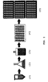
- Pre-propagation (picture 1 in figure 1) of the cells in a preferably aqueous medium preferably provides a 2-5 times increase in the number of cells clones.
- Suitable incubation temperatures may be within the range of 10-60°C, preferable 30-50°C, e.g. 37 °C, while pH may be kept between 4-10, preferably 6-8.
- the incubation period should be adjusted, preferably 15-60 minutes, e.g. 40 minutes, so as to meet the requirements of the desired transformant and clone concentration.
- pre-propagation may also be performed to secure expression of an antibiotic marker which may be comprised in the inserted plasmid of transformed host cells enabling resistance to an antibiotic in the medium.
- Pre-propagation of the host culture may accordingly be achieved by incubation at conditions favorable for expression of the chosen type of antibiotic marker as well as securing viability of the transformant.
- Dilution (picture 2 of figure 1) may be performed by addition of a medium, to ensure that all cells and clones thereof resides in the diluted solution.
- the cells may be separated by transferring aliquots of the planned specific volume to separate containers, e.g. wells in commercial microtiter plates (picture 3 of figure 1).
- step b) may comprise the steps:
- the separated cells are propagated to increase the number of clones in each container, preferably to a range between 10 7 -10 8 clones/ml. If microtiter plates are used these plates may be denoted "master plates". For screening a bacterial gene library 50-100 master plates with each 96 wells may typically be employed.
- master plates For screening a bacterial gene library 50-100 master plates with each 96 wells may typically be employed.
- One advantage of having a master plate is the possibility of keeping viable samples of the cells to be screened, so that even if the subsequent screening conditions results in death of the screened cells, it is possible to track back a screening result to a viable sample of a screened cell.
- suitable incubation temperatures may be in the range of 10-60°C, preferable 30-50°C, e.g. 37°C, while the pH may be kept between 4-10, preferably 6-8.
- the incubation medium should meet the nutritional requirements of the cells and clones to ensure sufficient growth.
- a medium may suitable be chosen enabling killing or suppressing non-transformed host cells.
- Incubation times should be adjusted so as to ensure growth yielding a sufficient number of cells/clones suitable for sampling aliquots containing a suitable number of clones for screening leaving a number of viable clones in the master plate.
- a preferred propagation period may be 40-90 hours, e.g. 48 hours, depending on the type of microbial host cell and the propagation conditions.
- the pre-propagation, dilution and/or propagation in a preferred embodiment of the invention, are performed in a medium capable of selectively killing or suppressing growth of non-transformed host cells.
- This may preferably be achieved by adding e.g. an antibiotic to the culture medium towards which the transformants or clones thereof are resistant, in an amount effective on non-transformed host cells.
- a microtiter assay plate (picture 4 in figure 1) suitable for measuring FP e.g. black microtiter plates from Bibby Sterilin Ltd., UK or Dynex Technologies, VA, USA.
- the cells/clones are contacted with one or more fluorescent substances having FP properties, e.g. by adding a substance mixture to each well.
- the fluorescent substance(s) should be suitable for reaction with the type of biological compound(s) of interest (e.g. fluorescently labeled pectin which is suitable for reaction with pectinase enzymes released from a cell).
- the contact between the biological compound and the fluorescent substance(s) usually occur in the extracellular medium, a condition which may be required in order to obtain an adequate FP response.
- the biological compound In case the biological compound is confined within the interior of the cell, the cell may lysed or its integrity otherwise disrupted in order to release the biological compound to the medium.
- the mixture of cells, medium and fluorescent substance(s) are incubated so that a biological compound may react with the fluorescent substance(s).
- the incubation may be performed at conditions, which favor the reaction between the biological compound and the fluorescent substance(s).
- the reaction between the fluorescent substance(s) and the biological compound is optimized with respect to pH and temperature.
- the incubation may also be performed at extreme conditions (such as very low temperatures (e.g. below 30°C or below 20°C) or high temperatures (e.g. above 60°C or above 70°C), low pH (e.g. below 5 or below 4) or high pH (e.g. above 9 or above 10), low or high ionic strength, presence of hostile chemicals such as detergents) causing death of the cells, depending on which biological compound is to be detected.
- the biological compound to be found is a thermostabile compound the incubation may be performed at high temperatures, conditions providing that only biological compounds, which remain active at high temperatures will react with the fluorescent substance.
- Positive hits are preferably identified as cells/clones or in vitro expression systems generating a FP change, which is more than 3.5 standard deviations of the based on the variance in background FP of the chosen fluorescent substance at the chosen conditions.
- the standard derivation within each 96 wells plate is typically 2 to 3% when a soluble fluorescently labelled substrate (e.g. soluble starch) is being applied.
- a soluble fluorescently labelled substrate e.g. soluble starch
- some substrates which do not dissolve that well e.g. fluorescently labelled amylose and PASC
- DNA in a cellular nucleotide source or a gene library derived from a nucleotide source and expressed either in an in vitro expression system or by transformation into a host cell expression system may thus be screened for nucleotide sequences encoding for biological compounds which reacts with a chosen range of fluorescent substance(s).
- the reaction between the fluorescent substance and a biological compound is measured in real time by placing the assay plate in a FP equipment and measuring changes in FP during incubation.
- a reaction between the fluorescent substance and a biological compound is detected by measuring changes in FP after a predefined time of incubation compared to a background measurement of the fluorescent substance alone (endpoint measurement).
- the found biological compound originates from clones of a single cell by employing conventional screening techniques or by repeating the method of the invention. This may be achieved by restreaking the clones onto a suitable agar plate and assaying the thereby obtained single colonies for the biological compound in question.
- the biological compound in the context of the invention may be any compound subject to industrial applicability which may be expressed in an expression system.
- the biological compound may be proteins, polypeptides or peptides
- a preferred biological compound is an enzyme, which in accordance with Recommendations (1992) of the Nomenclature Committee of the International Union of Biochemistry and Molecular Biology, Academic Press, Inc., 1992 may be classified as e.g. an oxidoreductase (EC 1.-.-.-), a transferase(EC 2.-.-.), a hydrolase (EC 3.-.-.-), a lyase (EC 4.-.-.-), an isomerase (EC 5.-.-.-), a ligase (EC 6.-.-.-).
- an oxidoreductase EC 1.-.-.-
- a transferase EC 2.-.-.
- a hydrolase EC 3.-.-.-
- a lyase EC 4.-.-.-
- an isomerase EC 5.-.-.-
- a ligase EC 6.-.-.-.-).
- Said hydrolase may be an endo-, exo-, carboxy- or amino peptidase, an endo- or exo glucanase, an endo- or exo amylase, a lipase or an esterase.
- the biological compound may further have a medical indication on a human or animal body, e.g. a hormone or other proteins, polypeptides or peptides, which acts on human or animal receptors.
- a medical indication on a human or animal body e.g. a hormone or other proteins, polypeptides or peptides, which acts on human or animal receptors.
- a preferred biological compound is an extra cellular compound such as an extra cellular enzyme, i.e. a compound which is secreted or otherwise exported from a cell to its surrounding medium.
- the biological compound being an extra cellular compound is a necessity because the compound may not be active when confined within a cell.
- some enzymes such as certain proteases are synthesized as inactive precursors, e.g. having attached a signal peptide or propeptide rendering the enzyme inactive as long as it is inside the cell. Upon export from the cell the signal peptide may be cleaved off rendering the enzyme active, when entering the surrounding extra cellular medium (Simonen and Palva (1993)).
- Some pullulanases and lipases do also show lack of activity or reduced activity in the cytoplasm and are activated upon secretion (Gotz et al. (1998) and Pugsley et al. (1990).
- the substance in the context of the invention may be any organic or biological compound which in itself posses fluorescence properties or which is labeled with a fluorescent marker.
- the skilled person may choose from a broad range of possible fluorescent substances, because the method is based on measurement of FP on samples of constant fluorescence intensity, i.e. changes in the polarization of emitted light of a constant intensity. This has several advantages as compared to methods based on measurement of changes in intensity of a fluorescent substance:
- the fluorescent substance is an organic molecule labeled with a fluorescent marker (fluorophore).
- the organic molecule may be a poly-, oligo- or monomer and may further be labeled with more than one fluorescent markers. Markers on the organic molecule may be attached at 0,01-10% of the available bonding sites of the organic molecule (degree of labeling). A preferred range is 0,5-5%, e.g. 2 %.
- the fluorescent marker may be attached to the organic molecule by covalent, ionic or hydrogen bonding.
- the organic molecules may preferably be proteins, peptides, glycoproteins, lipoprotein, mono-, poly- or oligosaccharides, triglycerides or nucleotides. Further the organic molecule may act as a substrate or an inhibitor, a human or animal receptor, a mono- or polyclonal antibody for the biological compound encoded for by the nucleotide sequence for which the cell or the gene library is to be screened for. Preferably the organic molecule is soluble or dispersible in an aqueous medium.
- Preferred proteins may be casein, derivatives of casein, gelatins, albumins, soy protein, polyclonal antibodies, monoclonal antibodies, human receptors or an animal receptors, e.g. tissue factor as described in national patent DK/97/0879 or WO 99/03498.
- a preferred peptide is cadaverine.
- Preferred polysaccharides may be glucans such as amyloglucans (e.g. amylose and/or amylopectin), dextrins (e.g. limit dextrin), pectins/polygalacturonic acids, celluloses (e.g. phosphoric acid swollen cellulose as described in Schülein (1997)) or derivatives thereof or hemi-celluloses. Further preferred hemicellulose may be xyloglucans, xylans, arabinoglucans or arabinans. Also mannans (such as galactomanan) and galacturonan (such as rhamnogalacturonan) are useful.
- amyloglucans e.g. amylose and/or amylopectin
- dextrins e.g. limit dextrin
- pectins/polygalacturonic acids e.g. phosphoric acid swollen cellulose as described in Schülein (1997))
- phenolic molecules such as phenolic polysaccharides which by oxidation by an oxidoreductase may be polymerized.
- the organic molecule may be chemically or enzymatically synthesized. Alternatively it may be obtainable from an animal, human, plant or microbial source and may further subsequently be chemically or enzymatically modified and/or purified.
- the marker may suitably have an excitation maximum in the range 350 - 700 nm, e.g. 400 - 600 nm, while the emission maximum suitable may be in the range 450-800 nm, e.g. 500-700 nm.
- the marker may preferably be less liable to be quenched by conjugating or attaching it to the organic molecule so that it maintains a distance of 20-50 ⁇ ngstrom from the organic molecule and other fluorescent markers.
- the bond between the marker and the organic molecule preferably is a rigid bond limiting rotational movement of the marker itself around the bond axis, independently of the movement of the organic molecule, so that the movement of the marker is primarily or essentially governed by the rotational movement of the organic molecule (around the rotation axis of the organic molecule).
- the marker may further have a fluorescence relaxation time in the range of 1-7 nanoseconds, preferably 3-5 nanoseconds.
- Preferred markers are derivatives of BODIPY, fluorescein including derivatives thereof, rhodamine including derivatives thereof, dansyls including derivatives thereof (e.g. dansyl cadaverine), Texas red (all available from Molecular Probes, OR, USA) as well as cyanine dyes (available from Amersham, Buckinghamshire, UK)
- the fluorescent substance (organic molecule and fluorescent marker(s) in combination) should have a polarization value in the range of 50-500 mP (milli polarization units), preferably 100-300 mP, e.g. 200 mP.
- a fluorophore labeled cellulose For example phosphoric acid swollen cellulose (PASC) or carboxy methyl cellulose (CMC) may be labeled with a fluorophore such as 6-DTAF (6-Fluorescein dichlorotriazine).
- PASC phosphoric acid swollen cellulose
- CMC carboxy methyl cellulose
- 6-DTAF 6-Fluorescein dichlorotriazine
- a process for producing a fluorescent cellulose may comprise: i) suspension or dissolution of the cellulose or derivative thereof in an aqueous alkaline reaction medium; ii) incubation/reaction of the suspended or dissolved cellulose or derivative thereof with the fluorophore; iii); separation of the labeled cellulose or derivative thereof from the alkaline reaction medium and wash of the labeled cellulose and iv) neutralization of the labeled cellulose or derivative thereof.
- Another preferred fluorescent substance is a fluorophore labeled galacturonan.
- rhamnogalacturonan may be labeled with a fluorophore such as 6-DTAF (6-Fluorescein dichlorotriazine).
- a process for producing a fluorescent rhamnogalacturonan may comprise: i) Suspension and/or dissolution of the rhamnogalacturonan in an alkaline aqueous medium ii) incubation/reaction of the suspended or dissolved rhamnogalacturonan with the fluorophore; iii) neutralization of the alkaline reaction medium, iv) separation of the labeled rhamnogalacturonan from the neutralized reaction medium and wash of the labeled rhamnogalacturonan.
- another preferred fluorescent substance is a fluorophore labeled mannan.
- galactomannan may be labeled with a fluorophore such as 6-DTAF (6-Fluorescein dichlorotriazine).
- a process for producing a fluorescent galactomannan may comprise: i) Suspension and/or dissolution of the galactomannan in an alkaline aqueous medium ii). incubation/reaction of the suspended or dissolved galactomannan with the fluorophore; iii) separation of the labeled galactomannan from the alkaline reaction medium and wash of the labeled galactomannan.
- xylan may be labeled with a fluorophore such as lissamine-rhodamine- sulphonyl chloride.
- a process for producing a fluorescent xylan may comprise: i) Suspension and/or dissolution of the xylan in an alkaline aqueous medium ii) amidation of the xylan; iii) incubation/reaction of the amidated xylan with the fluorophore; iv) separation and wash/purification of the labeled xylan.
- xyloglucan may be labeled with a fluorophore such as fluorescein-5-thiosemicarbazide.
- a process for producing a fluorescent xyloglucan may comprise: i) suspension and/or dissolution of the xyloglucan in an aqueous medium comprising an oxidant ii) separation/purification of a partially oxidized xyloglucan fraction of interest iii) incubation/reaction of the oxidized xyloglucan with the fluorophore; iv) separation and wash/purification of the labeled xyloglucan.
- arabinan may be labeled with a fluorophore such as fluorescein-5-thiosemicarbazide.
- a process for producing a fluorescent arabinan may comprise: i) suspension and/or dissolution of the arabinan in an aqueous medium comprising an oxidant ii) separation/purification of a partially oxidized arabinan fraction of interest iii) incubation/reaction of the oxidized arabinan with the fluorophore; iv) separation and wash/purification of the labeled arabinan.
- another preferred fluorescent substance is a fluorophore labeled pectin/polygalacturonic acid.
- pectin may be labeled on carboxylic acid groups with a fluorophore such as fluorescein-5-thiosemicarbazide using a mediator such as 1-ethyl-3-(dimethylaminopropyl)carbodiimide hydrochloride.
- a process for producing a fluorescent pectin may comprise: i) suspension and/or dissolution of the pectin in an aqueous medium ii) incubation/reaction of the pectin with the fluorophore and the mediator; iv) separation and wash/purification of the labeled pectin.
- the FP methods described vide supra may further be used for quality or fermentation control in industrial production of biological compounds.
- the bacterial strain from which DNA was extracted for library construction was propagated in liquid LB medium. Cells were harvested, and genomic DNA isolated by the method described by Pitcher et al., 1989.
- genomic DNA was partially digested with restriction enzyme Sau3A, and size-fractionated by electrophoresis on a 0.7 % agarose gel. Fragments between 2 and 7 kb in size were isolated by electrophoresis onto DEAE-cellulose paper (Dretzen, G., et al., 1981).
- Isolated DNA fragments were ligated to BamHI digested pSJ1678 (see WO 94/19454,3969) plasmid DNA, and the ligation mixture was used to transform the strain E. coli SJ2 (Diderichsen et al., (1990)).
- E. coli SJ2 cells were prepared for and transformed by electroporation using a Gene PulserTM electroporator from BIO-RAD as described by the supplier. All molecular biology work was done as described by Sambrook et al, 1989, Ausubel et al., 1995 and Harwood and Cutting, 1990.
- libraries generated in pZErO-2 were created as follows: Chromosomal DNA was partially digested with restriction enzyme Sau3A and subsequently size fractionated by gel electrophoresis. Fragments of 2-8 kb were purified and ligated into pZErO-2 restricted with BamHI. The ligation mix was transformed into ElectroMAX DH10B (GibcoBRL/Life technologies, UK) cells by electroporation as described by the vendor. The transformed cells were grown in LB medium without antibiotics for 60 minutes, glycerol was added to 15% and frozen in aliquots. The number of transformants per aliquot was determined by plating on LB agar plates containing 10 ⁇ g/ml kanamycin.
- a Titertek Microdrop station equipped with an S20 stacker (Life Technologies, MD, USA) was used for automated filling of the transformed library into the wells.
- S20 stacker Life Technologies, MD, USA
- a Platemate automated pipettor (Matrix Technologies, MA, USA) station was used.
- E. coli transformants and clones thereof were propagated in a LB broth medium (5g/l NaCl, 5g/l yeast extract (Difco, MI, USA), 10 g/l Tryptone (Difco, MI, USA)).
- the E. coli transformants and clones thereof were propagated in Nunclon MicroWell 96F plates (NUNC, Denmark) at 37°C and shaked at 200 rpm for three days prior to the screening procedures.
- Prior to FP measurements transformants or clones thereof and fluorescent substance are mixed by mixing 50 ⁇ L transformants or clones thereof with 150 ⁇ L solution comprising the fluorescent substance.
- an excitation filter was inserted allowing 485 nm light to pass with a band pass of 22 nm. Further an emission filter was inserted allowing 520 nm light to pass with a band pass of 30 nm.
- an excitation filter was inserted allowing 546 nm light to pass with a band pass of 12 nm. Further an emission filter was inserted allowing 590 nm light to pass with a band pass of 35 nm.
- the grating factor was calculated according to the instrument instruction manual.
- Instruments for high through put measurements of fluorescence polarization may by a flow cuvette based instrument e.g. modified LS50B (Perkin-Elmer Corporation, CT, USA), microtiter plate based readers e.g. Polarstar (BMG, Germany) or a FACS (flow activated cell sorter) equipped for fluorescence polarization as described by Arndt D.J., et al. (1976) and Lindmo, T and H.B. Steen, 1977.
- LS50B Perkin-Elmer Corporation, CT, USA
- microtiter plate based readers e.g. Polarstar (BMG, Germany) or a FACS (flow activated cell sorter) equipped for fluorescence polarization as described by Arndt D.J., et al. (1976) and Lindmo, T and H.B. Steen, 1977.
- Cadaverine labeled with dansyl, tetramethyl rhodamine, BODIPY®-TR and Texas Red were obtained from Molecular Probes, OR, USA.
- Dephosphorylated ⁇ -casein from bovine milk and fluorescein labeled potato starch were obtained from Sigma, MO, USA.
- Citrus pectin with a low degree of esterification (3%) was obtained from Copenhagen Pectin, Lille Skensved, Denmark.
- Apple pectin with a degree of esterification of 77,4% was from Herbstreith KG, Germany.
- Birch xylan used in the xylanase experiment was obtained from Sigma, MO, USA.
- Arabinan was from sugar beet and xyloglucan, (amyloid) from Tamaring seed were both obtained from Megazyme (Sydney, Australia).
- Amylose from potato (type III) and amylopectin from corn were both obtained from Sigma, MO, USA.
- Galactomannan from Locus bean gum was obtained from Sigma, MO, USA.
- Rhamnogalacturonan was obtained from Copenhagen Pectin, Lille Skensved, Denmark.
- Phosphoric acid swollen cellulose was prepared as described in Schülein (1997). Limit dextrin was obtained by hydrolysing corn-starch by an amylase from Aspergillus oryzae as described Tada et al. (1989).
- Polygalacturonases I, II, III of WO 9414952 were used.
- a pectin lyase obtainable from Aspergillus aculeatus as described in WO 94/21786 was used.
- a pectate lyase obtainable from Bacillus licheniformis as described in national patent application number DK/97/1344 was used.
- An arabinanase obtainable from Aspergillus aculeatus as described in WO 94/20611 was used.
- a xyloglucanase (endoglucanase II) obtainable from A. aculeatus as described in WO 94/14953 was used.
- amylase Novamyl is available from Novo Nordisk A/S and the ⁇ -amylase from C. thermosulfurogenes was obtained as described in Shen et al. (1988).
- a rhamnogalacturonase obtainable from A. aculeatus as described in WO 94/20612 was used.
- a Cel45 cellulase obtainable from Humicola insolens as described in WO 96/29397 was used.
- BP-X medium was prepared as follows: 100 g potato flour, 50 g barley flakes and 800 mg BAN amylase (available from Novo Nordisk A/S, Denmark) was mixed in 800 ml water. The solution was heated to approximately 70°C and incubated for 30 minutes. During cooling 10 g sodium caseinat (MD-Food, Denmark) was added. Upon cooling to ambient temperature 20 g soy meal, 9 g di-sodiumhydrogenphosphate (MERCK), 0.1 g pluronic PE 6100 (BASF) was added along with additional 1000 ml water.
- MERCK di-sodiumhydrogenphosphate
- BASF pluronic PE 6100
- Oligo-xyloglucan was end-labeled with sulphorhodamine as described by Fry et al.1997.
- Xyloglucan (0.50 g) was dissolved in water (60 ml) at 60°C by stirring for 7 h. The solution was allowed to cool to room temperature and a solution containing sodium metaperiodate (15 mg) (Merck, Germany) in water (10 ml) was added. The mixture was left overnight in the dark and then dialyzed (MW cut-off 12.000-14.000) against demineralized water with frequent exchange of the acceptor solution. 25 mg fluorescein-5-thiosemicarbazide supplied by Molecular Probes, OR, USA) was dissolved in 1 ml DMF (Sigma, MO, USA) and the now orange solution was added to the dialyzed polymer ( ⁇ 60 ml solution).
- the solution was stirred in the dark for 18 h and the polymer then precipitated by addition of 96 % ethanol (20 ml).
- the wet polymer was collected by filtration on Miracloth (Calbiochem, Germany) and washed with a mixture of water and ethanol (1:1) until a colorless filtrate was obtained.
- the labeled yellow xyloglucan was finally washed with absolute ethanol and air dried.
- the estimated degree of labeling (calculated from absorbance at 492 nm) was 0.6%.
- the labeling strategy comprised labeling of the carboxylic acid groups of pectin/polygalacturonic acids by coupling a hydrazine or amine activated probe (e.g. fluorescein-5-thiosemicarbazide) to the carboxylic acids using 1-ethyl-3-(dimethylaminopropyl)carbodiimide hydrochloride (FLUKA, Switzerland) as a mediator.
- a hydrazine or amine activated probe e.g. fluorescein-5-thiosemicarbazide
- FLUKA 1-ethyl-3-(dimethylaminopropyl)carbodiimide hydrochloride
- the polygalacturonic acid with a degree of esterification of 3%, 35% or 77% (1 g) was dissolved in 25 ml of demineralized water (additional heating and water was needed to dissolve pectin with degrees of esterification of 35% and 77%) and subsequently 20 mg of fluorescein-5-thiosemicarbazide (Molecular Probes, OR, USA) was applied while stirring.
- the pH was adjusted to 5.9 with 1 M NaOH and (1 g) 1-ethyl-3-(dimethylaminopropyl)carbodiimide hydrochloride was added gradually over 1 h while stirring. The solution was then incubated overnight in darkness.
- the moist xylan was re-dissolved in pyridine/acetic acid/H 2 O (1:1:18 by vol.) to a final volume of 125 ml. 375 ml methanol was added to the mix and incubated for 1 hour. The precipitate of xylan was collected on Miracloth as before. The xylan was rinsed with 1 liter of methanol/pyridine/acetic acid/H 2 O (60:1:1:18 by vol.) followed by 1 liter of ethanol and finally with 0.5 1 acetone. The amidated polysaccharide was collected and dried. 1.40 g of amidated xylan was dissolved in 35 ml hot 200 mM sodium pyrophosphate solution.
- the precipitated xylan was re-dissolved in 50 ml pyridine/acetic acid/H 2 O (1:1:18 by vol.) and reprecipitated by addition of 150 ml methanol. The washing step was repeated until the supernatant did not contain any dye. Precipitated xylan was washed twice in 200 ml acetone and dried.
- Amylose 250 mg was refluxed for 10 min. at 100 °C in water (30 ml) and pH adjusted to pH 11 with NaOH. The turbid solution was cooled to room temperature and treated with 11 mg 6-DTAF (6-Fluorescein dichlorotriazine). The labeled polymer was precipitated in a mixture of EtOH/MeOH/AcOH (15:2:1, 20 ml) and collected on Miracloth and washed with EtOH (96 %, 30 ml) and acetone (140 ml). The polymer was finally dissolved in water and freeze-dried.
- Amylopectin 250 mg was refluxed for 10 min. at 100 °C in water (30 mL) and pH was adjusted to 11 with NaOH. The turbid solution was cooled to room temperature and treated with 11 mg 6-DTAF (6-Fluorescein dichlorotriazine). The labeled polymer was precipitated in a mixture of EtOH/MeOH/AcOH (15:2:1, 15 ml) and washed with EtOH (96 %, 30 ml). The precipitate was collected on Miracloth and washed with EtOH (96 %, 70 ml). The polymer was finally dissolved in water and freeze-dried.
- Corn-starch was hydrolyzed by an amylase from Aspergillus oryzae (Tada et al. (1989) at pH 4.5, 50°C for 120 h.
- the limit dextrins were purified on Bio-gel P-2 column. Fractions containing DP 15-33 were pooled and freeze-dried. 508 mg dextrins were re-dissolved in 20 ml water and 75 mg 5-aminofluorescein in 1 ml DMF was added and stirred for 2 h in the dark. The resulting imine-bond was reduced using 76.2 mg NaCNBH 3 .
- the labeled dextrins was purified on a SEC-column.
- 6-DTAF 6-Fluorescein dichlorotriazine
- Example 12 amylase activity assayed by fluorescence polarization
- a method for detection of amylase activity was performed by incubating an amylase (Termamyl®) with a fluorescein labeled potato starch according to example 7 and measuring the changes in fluorescence polarization. A decrease in fluorescence polarization of the fluorescent starch upon hydrolysis by the amylase was observed as shown in Table 1. Results + Termamyl® - Termamyl® FP value 126 mP 166 mP
- the hydrolysis reaction was conducted by incubating Termamyl® with 33 ⁇ g fluorescein labeled potato starch per well for 180 minutes at ambient temperature in a 200 ⁇ L volume of pH 6.8 BR-buffer (23 mM H 3 PO 4 , 23 mM CH 3 COOH, 23 mM H 3 BO 3 ). After the 180 min reaction time the decrease in fluorescence polarization was approximately 20%.
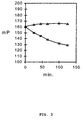
- Figure 3 shows the FP changes over time, thus demonstrating the rate of which the starch is hydrolyzed by the amylase.
- FP measurements of the fluorescent starch with and without addition of Termamyl® are indicated by squares and triangles respectively.
- Example 13 xylanase activity assayed by fluorescence polarization
- a method for detecting xylanase activity was performed by incubating a BioFeed® Wheat xylanase with a rhodamine labeled xylan and measuring the changes in fluorescence polarization. A decrease in fluorescence polarization of the fluorescent substrate upon hydrolysis by the xylanase was observed.
- Column B of figure 4 shows a decrease in fluorescence polarization as the labeled xylan is hydrolyzed by the xylanase compared to column A where no xylanase is present.
- the hydrolysis reaction was conducted by incubating the xylanase with 0,001 % (w/v) rhodamine labeled birch xylan for 30 minutes at 40°C in a pH 6.0 sodium phosphate buffer (100 mM phosphate).
- Example 14 transglutaminase activity assayed by fluorescence polarization
- a method for detecting transglutaminase activity was performed by incubating a transglutaminase from Phytophthora cactorum with a fluorescently labeled cadaverine as donor and caseine (dephosphorylated) as acceptor and measuring changes in fluorescence polarization.
- Various fluorescent markers was been tested: fluorescein, dansyl, rhodamine and BODIPY-TR.
- the transfer reactions were conducted by incubating the transglutaminase with 16 ⁇ M labeled cadaverine and 20.8 ⁇ M caseine for 3 hours at 42°C in a pH 7.6 Tris buffer (10 mM Tris, 20 mM CaCl 2 100 mM NaCl and 4.5 mM dithiothreitol).
- the FP measurements were performed at room temperature.
- dansyl labeled cadaverine excitation and emission wavelengths were 385 nm and 520 nm respectively.
- fluorescein labeled cadaverine excitation and emission wavelengths were 485 nm and 525 nm respectively.
- rhodamine labeled cadaverine excitation and emission wavelengths were 545 nm and 578 nm respectively.
- BODIPY®-TR labeled cadaverine excitation and emission wavelengths were 588 nm and 615 nm respectively. Measurements were obtained by the use of a Perkin Elmer LS50B equipped for fast polarization. Integration time was 1 second.
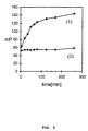
- Transglutaminase activity could also be monitored as an increase in fluorescence polarization over time.
- Figure 5 shows the transfer of rhodamine labeled cadaverine (16 ⁇ M) to casein (20,8 ⁇ M) by a transglutaminase from Phytophthora cactorum over 0-300 minutes (curve 1) compared to the reaction with no transglutaminase present (curve 2).
- the reactions were conducted at room temperature in a pH 7.6 Tris buffer (10 mM Tris, 20 mM CaCl 2 100 mM NaCl and 4.5 mM dithiothreitol).
- Example 15 xyloglucan endotransglycosylase activity assayed by fluorescence polarization
- a method for detecting xyloglucan endotransglycosylase (XET) activity was performed by incubating a xyloglucan endotransglycosylase with a sulphorhodamine labeled oligo-xyloglucan as donor and xyloglucan as acceptor and measuring changes in fluorescence polarization.
- the results of Table 4 show an increase in fluorescence polarization as the labeled oligo-xyloglucan is transferred to the much bigger xyloglucan molecule by the XET enzyme compared to no XET is present.
- the transfer reaction was conducted by incubating the XET with 2.25 ⁇ M sulphohodamin labeled oligo-xyloglucan and 2 g/L xyloglucan over night at ambient temperature in a pH 5,5 succinate buffer (133 mM Na-succinate, 3.8 mM CaCl 2 , 1.9 mM L-cystein).
- a positive response was obtainable in 2-3 hours by lowering the concentration of oligo-xyloglucan to 0.45 ⁇ M (not shown).
- PME P ectin M ethyl E sterase
- Table 5 shows the increase in fluorescence polarization as the labeled pectin cross-links after demethylation by the PME compared to no PME is present.
- the demethylation reaction was conducted by incubating a purified PME with 70 ⁇ g labeled pectin for 180 minutes at ambient temperature in 200 ⁇ l pH 5.0 citrate buffer (100 mM Na-citrate and 2 mM CaCl 2 ).
- Example 17 arabinanase activity assayed by fluorescence polarization
- a method for detecting arabinanase activity was performed by incubating an A. aculeatus arabinanase with a fluorescein labeled (0.7%) non-debranched arabinan from sugar beet and measuring the changes in fluorescence polarization. A decrease in fluorescence polarization of the labeled arabinan due to hydrolysis or depolymeration by the arabinanase was observed. Results + 5 ⁇ L arabinanase + 0.5 ⁇ L arabinanase + 0.0 ⁇ L arabinanase FP value 128 133 141
- Table 6 shows the decrease in fluorescence polarization as the labeled arabinan is hydrolyzed by respectively 0,5 ⁇ L and 5 ⁇ L added arabinanase compared to no arabinanase is added.
- the hydrolysis reaction was conducted by incubating the arabinanase with 170 ⁇ g/ml labeled arabinan for 16 hours at 50°C in a pH 5.0 citrate buffer (100 mM Na-citrate and 7 mM NaCl). The reaction volume was 200 ⁇ l. FP measurements were performed at ambient temperature.
- Example 18 pectinase activity assayed by fluorescence polarization
- a method for detecting pectinase (polygalacturonase I, II and III and a pectin lyase from A. aculeatus and a pectate lyase from B. licheniformis ) activities was performed by incubating various pectinases with two fluorescein labeled pectins with different degrees of methylation or esterification (% DE) and measuring the changes in fluorescence polarization. Decreases in fluorescence polarization of the labeled pectins due to hydrolysis by the pectinases were observed.
- Table 7 shows the results of the reactions between different combinations of pectin and pectinase compared to control samples with no pectinase present.
- the hydrolysis reactions for polygalacturonases were conducted by incubating the polygalacturonases with respectively 70 ⁇ g/ml 77% DE or 35 ⁇ g/ml 3% DE labeled pectin for 75 minutes at ambient temperature in a pH 5.0 citrate buffer (100 mM Na-citrate and 2 mM CaCl 2 ).
- the hydrolysis reactions for pectin lyase were conducted by incubating the pectin lyase with respectively 70 ⁇ g/ml 77% DE or 35 ⁇ g/ml 3% DE labeled pectin for 180 minutes at ambient temperature in a pH 5.0 citrate buffer (100 mM Na-citrate and 2 mM CaCl 2 ).
- the hydrolysis reactions for pectate lyase were conducted by incubating the pectate lyase with respectively 70 ⁇ g/ml 77% DE or 35 ⁇ g/ml 3% DE labeled pectin for 75 minutes at ambient temperature in a pH 10.0 glycine buffer (50 mM glycine, 100 mM NaCl and 2 mM CaCl 2 ).
- a pectinase expressed by a cloned E. coli SJ2 transformant could also be detected by fluorescence polarization.
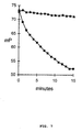
- a 50 ⁇ l aliquot of clones was taken from a master plate propagated overnight at 37 °C with shaking. The aliquot was mixed in a microtiter well with 150 ⁇ l glycine buffer pH 10.0 (4 mM CaCl 2 and 100 mM glycine) containing 6.8 ⁇ g fluorescein labeled 3% DE pectin.
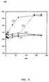
- the fluorescence polarization changes caused by the depolymeration of the pectin was measured over 15 minutes at ambient temperature. The results are shown in figure 7, where FP measurements of a sample containing a transformed E.
- E. coli SJ2 expressing an uncharacterized pectinase (squares) and a non-transformed E. coli SJ2 (triangles) are shown. It can further be derived from figure 7 that the inherent background of E. coli SJ2 was very low and did not interfere with the assay.
- Example 19 mannanase activity assayed by fluorescence polarization.
- a method for detecting mannanase activity was performed by incubating an A. aculeatus mannanase with fluorescein labeled (0.8%) galactomannan from Locus bean gum and measure the changes in fluorescence polarization. A decrease in fluorescence polarisation of the labeled galactomannan due to depolymeration by the mannanase activity was observed. Results + 1 ⁇ l mannanase + 0 ⁇ l mannanase FP value 133 156
- Example 20 rhamnogalacturonase activity assayed by fluorescence polarization
- a method for detecting rhamnogalacturonase activity was performed by incubating an A. aculeatus rhamnogalacturonase with fluorescein labeled (0.1%) rhamnogalacturonan and measure the changes in fluorescence polarization. A decrease in fluorescence polarisation of the labeled rhamnogalacturonan due to depolymeration by the rhamnogalacturonase activity was observed. Results + 1 ⁇ l rhamnogalacturonase + 0 ⁇ l rhamnogalacturonase FP value 80 90
- the results of Table 9 show the decrease in fluorescence polarization as labeled rhamnogalacturonan is depolymerized by addition of 1 ⁇ l rhamnogalacturonase compared to no rhamnogalacturonase added.
- the depolymerization was conducted by incubating the rhamnogalacturonase with 50 ⁇ g/ml labeled rhamnogalacturonan for 2 hours at 40°C in a pH 5.0 citrate buffer (100 mM Na-citrate and 1 mM CaCl 2 ).
- the reaction volume was 250 ⁇ l and the fluorescence polarization measurements were conducted at ambient temperature.
- a method for detecting amylase activity by the use of fluorescently labeled amylose was performed by incubating an C. thermosulfurogenes ⁇ -amylase or the Novamyl amylase with fluorescein labeled (0.7%) amylose and measure the change of the fluorescence polarization. A decrease in fluorescence polarization of the labeled amylose due to depolymeration by the amylase activity was observed. Results + 1 ⁇ l ⁇ -amylase + 0 ⁇ l ⁇ -amylase + 1 ⁇ l Novamyl + 0 ⁇ l Novamyl FP value 188 225 134 225
- the results of table 10 show the decrease in fluorescence polarization as labeled amylose is depolymerized by addition of 1 ⁇ l ⁇ -amylase or 1 ⁇ l Novamyl compared to no amylase added.
- the depolymerization was conducted by incubating the amylases with approximately 10 ⁇ g/ml labeled amylose for 2 hours at 42°C in a pH 6 buffer (50 mM MES (2-[N-Morpholino]ethanesulfonic acid) and 1 mM CaCl 2 ).
- the reaction volume was 250 ⁇ l and the fluorescence polarization measurements were conducted at 42°C.
- a method for detecting amylase activity by the use of fluorescently labeled amylopectin was performed by incubating an C. thermosulfurogenes ⁇ -amylase or the Novamyl amylase with fluorescein labeled (0.4%) amylopectin and measure the change of the fluorescence polarization. A decrease in fluorescence polarization of the labeled amylopectin due to depolymeration by the amylase activity was observed. Results + 1 ⁇ l ⁇ -amylase + 0 ⁇ l ⁇ -amylase + 1 ⁇ l Novamyl + 0 ⁇ l Novamyl FP value 139 184 113 184
- the results of Table 11 show the decrease in fluorescence polarization as labeled amylose is depolymerized by addition of 1 ⁇ l ⁇ -amylase or 1 ⁇ l Novamyl compared to no amylase added.
- the depolymerization was conducted by incubating the amylases with approximately 10 ⁇ g/ml labeled amylose for 2 hours at 42°C in a pH 6 MES buffer (50 mM MES (2-[N-Morpholino]ethanesulfonic acid) 1 mM CaCl 2 ).
- the reaction volume was 250 ⁇ l and the fluorescence polarization measurements were conducted at 42°C.
- a method for detecting amylase activity by the use of fluorescently labeled limit dextrin was performed by incubating the Termamyl amylase with fluorescein labeled limit dextrin and measure the change of the fluorescence polarization. A decrease in fluorescence polarization of the labeled limit dextrin due to depolymeration by the amylase activity was observed. Results + 1 ⁇ l Termamyl + 0 ⁇ l Termamyl - FP value 168 204
- the results of Table 12 show the decrease in fluorescence polarization as labeled limit dextrin is depolymerized by addition of 10 ⁇ l Termamyl compared to no amylase added.
- the depolymerization was conducted by incubating the amylase with 1 mg/ml labeled limit dextrin for 24 hours at 50 °C in a pH 6.5 MES buffer (50 mM MES (2-[N-Morpholino]ethanesulfonic acid), 1 mM CaCl 2 ).
- the fluorescence polarization measurements were conducted at ambient temperature.
- Example 24 cellulase activity assayed by fluorescence polarization
- a method for measuring cellulase activity was performed by incubating 20 ⁇ l of Humicola insolens Cel45 cellulase in various dilutions with 200 ⁇ l substrate in a microtiter plate for 1h at 40°C, 400 rpm shaking, and measuring the resulting change in fluorescence polarization.
- the substrate was 2 mg/ml phosphoric acid swollen cellulose (PASC) which had been covalently labeled with fluorescein.
- the fluorescence polarization values decrease steadily with increasing enzyme concentration, as shown in table 13 (double determination).
- the instrument was calibrated using fluorescein as known standard (35 mP).
- Cellullase conc. (% of stock) mP mP 0,0% 216,67 214,58 1,6% 189,31 192,40 3,1% 175,74 181,16 6,3% 160,67 159,82 12,5% 146,20 154,92 25,0% 132,09 138,31 50,0% 127,23 124,54 100,0% 123,00 125,04
- Example 25 high through-put screening of genomic libraries derived from a strain of Bacillus (identical to B. licheniformis, CA63, DSM 9552) and a strain of Bacillus (identical to B. clausii, NCIMB 10309, DSM 8716) formerly named B. lentus for nucleotide sequences encoding for pectinase
- FIG. 1 A schematic representation of the screening procedure is shown in figure 1.
- the content of a vial with SJ2 E. coli host cells transformed with a library was inoculated into 10 ml of LB medium and incubated for 40 minutes at 37°C. Subsequently, the 10 ml of cells were diluted in cold LB medium with chloramphenicol (10 ⁇ g/ml) to a final concentration of cells corresponding to 0.5-1 cell/300 ⁇ l medium.
- Table 14 shows the nucleotide sources for libraries screened for pectinases, the used substrates, and the number of positive hits obtained.
- Example 26 The number of hits as a function of the number of different transformed host cells per microtiter plate well
- Figure 2 shows the same information in a graphical format.
- the diamonds in figure 2 indicate hits per 1000 screened transformants.
- the content of a vial with DH10B E. coli host cells transformed with a library cloned in the pZErO-2 vector was inoculated into 2.9 litre of LB medium supplemented with kanamycin (10 ⁇ g/ml).
- the LB medium with the inoculated library was kept on ice while aliquots of 300 ⁇ l/well were transferred into NUNC 96 wells microtiter plates using a Micro droplet instrument giving a total of 100 plates. In average 1.5 clones were distributed into each well making up a total of approximately 14400 clones.
- the plates were placed in a tightly closed box in order to avoid evaporation of medium from the wells and placed on a shaker for approximately 48 hours with shaking (approximately 200 rpm) at 37°C.
- the plates were read by a fluorescence polarization reader (Polarstar, BMG, Germany) at ambient temperature and positive hits were scored as lowering of the fluorescence polarization value. Positive hits were defined as polarization values lower than the average polarization value of each microtiter plate minus 3.5 times the standard derivation of the 96 measurements derived from each microtiter plate. The average standard variation was approximately 2.2 mP units. A total of 10 confirmed positive hits (amylase expressing clones) were identified which corresponds to 1 hit in 1440 clones screened. DNA sequencing of the clones demonstrated that at least 2 different amylase encoding genes had been cloned.
- the content of a vial with DH10B E. coli host cells transformed with a library cloned in the pZErO-2 vector was inoculated into 2.9 litre of LB medium supplemented with kanamycin (10 ⁇ g/ml).
- the LB medium with the inoculated library was kept on ice while aliquots of 300 ⁇ l/well were transferred into NUNC 96 wells microtiter plates using a Micro droplet instrument giving a total of 100 plates. In average 0.4 clones were distributed into each well making up a total of approximately 3840 clones.
- the plates were placed in a tightly closed box in order to avoid evaporation of medium from the wells and placed on a shaker for approximately 48 hours with shaking (approximately 200 rpm) at 37°C.
- microtiter plates were read by a fluorescence polarization reader (Polarstar, BMG, Germany) at ambient temperature and positive hits were scored as lowering of the fluorescence polarization value. Positive hits were defined as polarization values lower than the average polarization value of each microtiter plate minus 3.5 times the standard derivation of the 96 measurements derived from each microtiter plate. The average standard variation was 5.6 mP units. A total of 3 confirmed positive hits (amylase expressing clones) were found which corresponds to 1 hit in 1280 clones screened.
- DSM 12682 is a B. subtilis 168 with deleted apr, npr, amy E and celA genes and which had been transformed with a pUB110 vector containing the Ce15a gene from B. agaradhaerens (GenBank AF067428) in fusion with the amy L signal peptide of B. licheniformis and the amy L promoter.
- DSM 12682 was incubated overnight in TY media at 37°C at 250 rpm.
- Example 30 screening for enzymatic activities in wild type isolates by fluorescence polarization
- the Bacillus (DSM 12663) isolate had a lower fluorescence polarization value than the Bacillus (DSM 12664) isolate and the growth medium without cells. It was later made it likely that Bacillus (DSM 12663) did encode a pectinase as a chromosomal library from a phylogenetic related species (most likely the same species) was constructed and screened for the presence of nucleotide sequences encoding pectinase activity. A nucleotide sequence encoding a pectinase was isolated. This shows that enzymatic activities assayed in wild type isolates by fluorescence polarization may later be cloned.
- Gerhardt P. et al. Methods for general and molecular biology; American Society for Microbiology; 1994; Eds: Gerhardt P., Murray R.G.E., Wood W.A., Krieg N.R.
- thermostabel beta-amylase from Clostridium thermosulphurogenes; Biochem. J.; 1988; vol. 254; pp. 835-840.
Landscapes
- Chemical & Material Sciences (AREA)
- Life Sciences & Earth Sciences (AREA)
- Organic Chemistry (AREA)
- Health & Medical Sciences (AREA)
- Engineering & Computer Science (AREA)
- Zoology (AREA)
- Wood Science & Technology (AREA)
- Proteomics, Peptides & Aminoacids (AREA)
- Genetics & Genomics (AREA)
- Bioinformatics & Cheminformatics (AREA)
- Biotechnology (AREA)
- Biochemistry (AREA)
- Physics & Mathematics (AREA)
- General Engineering & Computer Science (AREA)
- General Health & Medical Sciences (AREA)
- Microbiology (AREA)
- Molecular Biology (AREA)
- Immunology (AREA)
- Biophysics (AREA)
- Analytical Chemistry (AREA)
- Biomedical Technology (AREA)
- Pathology (AREA)
- General Physics & Mathematics (AREA)
- Bioinformatics & Computational Biology (AREA)
- Crystallography & Structural Chemistry (AREA)
- Nuclear Medicine, Radiotherapy & Molecular Imaging (AREA)
- Plant Pathology (AREA)
- Measuring Or Testing Involving Enzymes Or Micro-Organisms (AREA)
- Investigating, Analyzing Materials By Fluorescence Or Luminescence (AREA)
- Micro-Organisms Or Cultivation Processes Thereof (AREA)
- Investigating Materials By The Use Of Optical Means Adapted For Particular Applications (AREA)
- Investigating Or Analysing Biological Materials (AREA)
- Investigating Or Analysing Materials By The Use Of Chemical Reactions (AREA)
- Preparation Of Compounds By Using Micro-Organisms (AREA)
Claims (52)
- Procédé de criblage d'une séquence nucléotidique codant pour un composé biologique, qui comprend :a) l'expression de la séquence nucléotidique dans un système d'expression, de façon à produire le composé biologique,b) la mise en contact du composé biologique avec une substance fluorescente capable de réagir avec le composé biologique de façon à laisser la réaction avoir lieu,c) la mesure de la polarisation de fluorescence de la substance fluorescente, etd) la sélection de systèmes d'expression pour lesquels a eu lieu un changement de la polarisation de fluorescence.
- Procédé selon la revendication 1, dans lequel le système d'expression est un système de traduction/transcription couplé in vitro, et la séquence nucléotidique est une génothèque qui dérive d'une source de nucléotides.
- Procédé selon la revendication 1, dans lequel le système d'expression est un système d'expression cellulaire.
- Procédé selon la revendication 3, dans lequel les cellules sont des cellules de type sauvage.
- Procédé selon la revendication 3, dans lequel les cellules sont une culture de cellules hôtes dans laquelle les transformants comprennent une génothèque qui dérive d'une source de nucléotides.
- Procédé selon la revendication 2 ou 5, dans lequel la source de nucléotides est une cellule choisie dans l'ensemble comprenant les cellules bactériennes, les cellules d'archées et les cellules eucaryotes.
- Procédé selon la revendication 6, dans lequel la cellule bactérienne est de l'espèce Bacillus.
- Procédé selon la revendication 6, dans lequel la cellule eucaryote est choisie dans l'ensemble comprenant les cellules fongiques, les cellules humaines et les cellules végétales.
- Procédé selon la revendication 5, dans lequel la source de nucléotides est constituée de cellules modifiées par réarrangement génétique in vivo.
- Procédé selon la revendication 5, dans lequel la source de nucléotides est constituée d'une préparation préparée in vitro de séquences nucléotidiques choisies dans l'ensemble comprenant l'ADN, l'ARN, l'ADNc et les gènes artificiels.
- Procédé selon la revendication 10, dans lequel les séquences nucléotidiques préparées in vitro sont préparées par des techniques choisies dans l'ensemble comprenant le réarrangement génétique, la mutagenèse aléatoire et l'amplification PCR.
- Procédé selon la revendication 5, dans lequel la cellule hôte est choisie dans l'ensemble comprenant les bactéries, les archées et les champignons.
- Procédé selon la revendication 12, dans lequel une cellule hôte non-transformées est incapable d'exprimer d'une manière significative le composé biologique.
- Procédé selon la revendication 13, dans lequel des séquences nucléotidiques permettant à la cellule hôte non-transformée d'exprimer d'une manière significative le composé biologique sont supprimées par délétion.
- Procédé selon la revendication 12, dans lequel la cellule hôte est une bactérie de l'espèce E. coli.
- Procédé selon la revendication 12, dans lequel la cellule hôte est une bactérie de l'espèce Bacillus.
- Procédé selon la revendication 12, dans lequel le champignon est un S. cerevisiae.
- Procédé selon les revendications 12 à 17, dans lequel la cellule hôte est transformée par un plasmide.
- Procédé selon la revendication 18, dans lequel le plasmide comprend une séquence nucléotidique qui confère à la cellule hôte transformée une résistance à un antibiotique.
- Procédé selon la revendication 19, dans lequel la cellule hôte transformée est résistante à un antibiotique choisi dans l'ensemble comprenant le chloramphénicol, la tétracycline, la kanamycine, l'ampicilline, l'érythromycine et la zéocine.
- Procédé selon l'une quelconque des revendications précédentes, dans lequel le composé biologique est choisi dans l'ensemble comprenant les protéines, les polypeptides et les peptides.
- Procédé selon la revendication 21, dans lequel le composé biologique est une enzyme.
- Procédé selon la revendication 22, dans lequel l'enzyme est choisie dans l'ensemble comprenant les oxydoréductases (EC 1.-.-.-), les transférases (EC 2.-.-.-), les hydrolases (EC 3.-.-.-), les lyases (EC 4.-.-.-), les isomérases (EC 5.-.-.-) et les ligases (EC 6.-.-.-).
- Procédé selon la revendication 23, dans lequel l'hydrolase est choisie dans l'ensemble comprenant les endopeptidases, les exopeptidases, les carboxypeptidases, les aminopeptidases, les endoglucanases, les exoglucanases, les lipases et les estérases.
- Procédé selon la revendication 21, dans lequel le composé biologique est une hormone.
- Procédé selon l'une quelconque des revendications précédentes, dans lequel la substance fluorescente est une molécule organique marquée par un marqueur fluorescent.
- Procédé selon la revendication 26, dans lequel la molécule organique est un polymère, un oligomère ou un monomère.
- Procédé selon la revendication 26, dans lequel la molécule organique est marquée par le marqueur fluorescent sur 0,01-10 % des sites de liaison disponibles de la molécule organique.
- Procédé selon la revendication 26, dans lequel le marqueur fluorescent est fixé à la molécule organique par une liaison covalente, ionique ou hydrogène.
- Procédé selon la revendication 27, dans lequel la molécule organique est choisie dans l'ensemble comprenant les protéines, les peptides, les glycoprotéines, les lipoprotéines, les polysaccharides, les oligosaccharides, les triglycérides et les nucléotides.
- Procédé selon la revendication 30, dans lequel la protéine est choisie dans l'ensemble comprenant la caséine, les dérivés de la caséine, la gélatine, l'albumine, la protéine du soja, les anticorps polyclonaux, les anticorps monoclonaux, les protéines des récepteurs humains et les protéines des récepteurs animaux.
- Procédé selon la revendication 30, dans lequel le peptide est la cadavérine.
- Procédé selon la revendication 30, dans lequel le polysaccharide est choisi dans l'ensemble comprenant l'aminoglucane, les pectines/acide polygalacturonique, la cellulose et l'hémicellulose.
- Procédé selon la revendication 33, dans lequel le polysaccharide est choisi dans l'ensemble comprenant le xyloglucane, le xylane, l'arabinoglucane et l'arabinane, le PASC, le galacturonane, le mannane, l'amylose, l'amylopectine, une dextrine limite.
- Procédé selon la revendication 26, dans lequel la molécule organique peut être obtenue à partir d'une source animale, humaine, végétale ou microbienne.
- Procédé selon la revendication 35, dans lequel la molécule organique est chimiquement ou enzymatiquement modifiée ou purifiée.
- Procédé selon la revendication 26, dans lequel la molécule organique est chimiquement ou enzymatiquement synthétisée.
- Procédé selon la revendication 26, dans lequel le marqueur fluorescent est stériquement situé à une distance de 20-50 Ångstroms de la molécule organique et d'autres marqueurs fluorescents.
- Procédé selon la revendication 26, dans lequel le marqueur fluorescent a un temps de relaxation de fluorescence compris entre 1-7 nanosecondes.
- Procédé selon la revendication 26, dans lequel la substance fluorescente a une valeur de polarisation comprise entre 50-500 mP.
- Procédé selon la revendication 26, dans lequel le marqueur fluorescent est choisi dans l'ensemble comprenant les dérivés du BODIPY, la fluorescéine, les rhodamines, les dansyles, les colorants de cyanine et le rouge Texas.
- Procédé selon la revendication 2, qui comprend les étapes suivantes :a) préparation d'une génothèque,b) séparation des fragments géniques de la génothèque dans des récipients distincts,c) amplification des fragments géniques séparés,d) mise en oeuvre d'une transcription/traducton couplée in vitro des fragments géniques amplifiés de façon à exprimer un composé biologique,e) mise en contact, avec une substance fluorescente, du composé biologique se trouvant dans chaque récipient distinct, ou de sous-échantillons de ce composé,f) incubation du composé biologique avec la substance fluorescente,g) détection des changements de la polarisation de fluorescence de la substance fluorescente après réaction avec le composé biologique.
- Procédé selon la revendication 3, comprenant les étapes suivantes :a) pré-propagation et dilution des cellules comprenant la séquence nucléotidique,b) séparation des cellules dans des récipients distincts,c) propagation des cellules séparées pour augmenter le nombre de clones de chaque cellule dans chaque récipient distinct,d) mise en contact des cellules se trouvant dans chaque récipient distinct avec une substance fluorescente,e) incubation des cellules avec la substance fluorescente,f) détection des changements de polarisation de fluorescence de la substance fluorescente après réaction avec un composé biologique qui s'est échappé des cellules.
- Procédé selon la revendication 43, dans lequel l'étape a) multiplie par un facteur de 1-5 le nombre total de clones de la culture.
- Procédé selon la revendication 43, dans lequel l'étape b). est mise en oeuvre par dilution de la culture et transfert, dans des récipients distincts, d'aliquotes de 0,5-10 transformants/clone, plus particulièrement de 0,5-5 transformants/clone et tout spécialement de 0,5-1 transformant/clone par aliquote.
- Procédé selon la revendication 43, dans lequel l'étape b) comprend les étapes suivantes :a) insertion d'une substance fluorescente dans les cellules, caractérisée par un changement de l'intensité d'émission après réaction avec un composé biologique se trouvant à l'intérieur des cellules,b) criblage, sélection et séparation des cellules pour lesquelles un changement de l'intensité d'émission a eu lieu, au moyen d'un appareil d'analyse en cytométrie de flux (FACS).
- Procédé selon la revendication 43, dans lequel l'étape c) élève le nombre de clones se trouvant dans chaque récipient à une valeur comprise entre 107-108 clones/ml.
- Procédé selon la revendication 43, dans lequel les cellules sont mises en contact avec la substance fluorescente par mélange des cellules avec une solution comprenant la substance fluorescente.
- Procédé selon la revendication 43, dans lequel les cellules sont une culture de cellules hôtes dans laquelle les transformants comprennent une génothèque qui dérive d'une source de nucléotides, et l'étape a) ou c) est mise en oeuvre dans un milieu capable de détruire sélectivement les cellules hôtes non-transformées ou d'en supprimer sélectivement la croissance.
- Procédé selon la revendication 49, dans lequel la destruction ou la suppression de la croissance est obtenue par utilisation d'un milieu contenant un antibiotique en une quantité ayant un effet sur les cellules hôtes non-clonées.
- Procédé selon la revendication 43, dans lequel l'étape d) comprend la mise en contact des cellules avec au moins deux substances fluorescentes.
- Procédé de production industrielle d'un nouveau composé biologique, qui comprenda) l'identification du composé biologique et de la séquence nucléotidique codant pour le composé biologique selon l'une quelconque des revendications précédentes,b) la culture d'un microorganisme comprenant la séquence nucléotidique identifiée,c) la récupération du composé biologique et éventuellement la formulation du composé biologique.
Priority Applications (1)
| Application Number | Priority Date | Filing Date | Title |
|---|---|---|---|
| EP01118580A EP1156122A3 (fr) | 1998-03-06 | 1999-03-05 | Méthode d'essai à polarisation de fluorescence |
Applications Claiming Priority (3)
| Application Number | Priority Date | Filing Date | Title |
|---|---|---|---|
| DK30898 | 1998-03-06 | ||
| DK30898 | 1998-03-06 | ||
| PCT/DK1999/000112 WO1999045143A2 (fr) | 1998-03-06 | 1999-03-05 | Procede d'incineration de dechets et incinerateur pour la mise en oeuvre dudit procede |
Related Child Applications (1)
| Application Number | Title | Priority Date | Filing Date |
|---|---|---|---|
| EP01118580A Division EP1156122A3 (fr) | 1998-03-06 | 1999-03-05 | Méthode d'essai à polarisation de fluorescence |
Publications (2)
| Publication Number | Publication Date |
|---|---|
| EP1058738A2 EP1058738A2 (fr) | 2000-12-13 |
| EP1058738B1 true EP1058738B1 (fr) | 2001-11-14 |
Family
ID=8092089
Family Applications (2)
| Application Number | Title | Priority Date | Filing Date |
|---|---|---|---|
| EP01118580A Withdrawn EP1156122A3 (fr) | 1998-03-06 | 1999-03-05 | Méthode d'essai à polarisation de fluorescence |
| EP99906092A Expired - Lifetime EP1058738B1 (fr) | 1998-03-06 | 1999-03-05 | Procede d'incineration de dechets et incinerateur pour la mise en oeuvre dudit procede |
Family Applications Before (1)
| Application Number | Title | Priority Date | Filing Date |
|---|---|---|---|
| EP01118580A Withdrawn EP1156122A3 (fr) | 1998-03-06 | 1999-03-05 | Méthode d'essai à polarisation de fluorescence |
Country Status (11)
| Country | Link |
|---|---|
| EP (2) | EP1156122A3 (fr) |
| JP (1) | JP2002505114A (fr) |
| CN (2) | CN1560609A (fr) |
| AT (1) | ATE208828T1 (fr) |
| AU (1) | AU2612299A (fr) |
| CA (1) | CA2322488A1 (fr) |
| DE (1) | DE69900465T2 (fr) |
| DK (1) | DK1058738T3 (fr) |
| ES (1) | ES2168848T3 (fr) |
| PT (1) | PT1058738E (fr) |
| WO (1) | WO1999045143A2 (fr) |
Families Citing this family (19)
| Publication number | Priority date | Publication date | Assignee | Title |
|---|---|---|---|---|
| CA2328719A1 (fr) * | 1998-04-15 | 1999-10-21 | Hiroyoshi Hidaka | Procede de detection in vivo d'un gene proteique cible a l'aide de medicament |
| DE60016951T2 (de) * | 1999-10-01 | 2005-12-15 | Novozymes A/S | Zellulosefilme zur durchfuehrung von screening-verfahren |
| US6426189B1 (en) | 1999-10-01 | 2002-07-30 | Novozymes A/S | Cellulose films for screening |
| AU7646100A (en) * | 1999-11-05 | 2001-05-14 | Novozymes A/S | A high throughput screening (hts) method |
| US6723537B2 (en) | 2001-05-18 | 2004-04-20 | Rigel Pharmaceuticals, Incorporated | Directed evolution of protein in mammalian cells |
| JP4607950B2 (ja) * | 2004-03-18 | 2011-01-05 | トランス テック ファーマ,インコーポレイテッド | 蛍光偏光アッセイ |
| FR2897164B1 (fr) | 2006-02-09 | 2008-03-14 | Commissariat Energie Atomique | Realisation de cavites pouvant etre remplies par un materiau fluidique dans un compose microtechnologique optique |
| FR2929715B1 (fr) | 2008-04-04 | 2010-09-10 | Commissariat Energie Atomique | Procede de realisation de cavites microniques ou submicroniques |
| FR2933205B1 (fr) | 2008-06-27 | 2010-08-13 | Commissariat Energie Atomique | Procede ameliore de realisation d'enceintes remplies de liquide et fermees par une membrane |
| FR2938834B1 (fr) * | 2008-11-27 | 2011-03-04 | Commissariat Energie Atomique | Procede de realisation d'une matrice de retention et comprenant un liquide fonctionnel |
| FR2963112B1 (fr) | 2010-07-21 | 2013-02-15 | Commissariat Energie Atomique | Microstructure a parois a propriete optique determinee et procede de realisation de microstructures |
| WO2014067933A1 (fr) | 2012-10-31 | 2014-05-08 | C-Lecta Gmbh | Préparation de support bioactif pour une sécurité améliorée dans les produits de soin et les aliments |
| KR101494881B1 (ko) | 2013-05-06 | 2015-02-24 | 한국과학기술연구원 | 설퍼 옥시도리덕타아제 결합 형광 화합물을 이용한 바실러스 서브틸리스 검출 방법 |
| CN105866316B (zh) * | 2016-06-24 | 2018-03-30 | 曲阜师范大学 | 一种同时检测食品中氨基酸和生物胺的分析方法 |
| CN110954517A (zh) * | 2019-12-11 | 2020-04-03 | 西北大学 | 还原性糖的一步荧光衍生方法及其应用 |
| CN113514445B (zh) * | 2021-08-05 | 2023-12-26 | 荆州市第一人民医院(荆州市肿瘤医院、长江大学附属第一医院) | 一种基于荧光偏振的去泛素化酶活性检测方法 |
| CN114166807B (zh) * | 2021-11-22 | 2023-12-12 | 中国农业大学 | 用于检测多粘菌素的荧光偏振免疫分析方法 |
| WO2025168975A1 (fr) * | 2024-02-10 | 2025-08-14 | Biuro Techniczno-Prawne Adam Turczyński | Procédé de fabrication d'une sonde pour déterminer l'activité d'enzymes pectinolytiques et procédé de détermination de l'activité d'enzymes pectinolytiques utilisant ladite sonde |
| WO2025168976A1 (fr) * | 2024-02-10 | 2025-08-14 | Biuro Techniczno-Prawne Adam Turczyński | Procédé de détection d'agents pathogènes pectinolytiques de cultures et de plantes ornementales, milieu microbien destiné à être utilisé dans ce procédé et kit de test mettant en oeuvre ce procédé |
Family Cites Families (4)
| Publication number | Priority date | Publication date | Assignee | Title |
|---|---|---|---|---|
| ATE78517T1 (de) * | 1985-02-25 | 1992-08-15 | Abbott Lab | Polarisierter fluoreszenztest von makromolekularen hydrolasen. |
| MX9206979A (es) * | 1991-12-04 | 1993-07-01 | Novo Nordisk As | Metodo de clonacion de proteinas en levaduras |
| JPH0723800A (ja) * | 1993-07-08 | 1995-01-27 | Tanabe Seiyaku Co Ltd | 核酸の検出方法 |
| GB9519393D0 (en) * | 1995-09-22 | 1995-11-22 | Univ Edinburgh | Novel compounds and assay method |
-
1999
- 1999-03-05 AT AT99906092T patent/ATE208828T1/de active
- 1999-03-05 WO PCT/DK1999/000112 patent/WO1999045143A2/fr not_active Ceased
- 1999-03-05 PT PT99906092T patent/PT1058738E/pt unknown
- 1999-03-05 ES ES99906092T patent/ES2168848T3/es not_active Expired - Lifetime
- 1999-03-05 EP EP01118580A patent/EP1156122A3/fr not_active Withdrawn
- 1999-03-05 CN CNA2004100586739A patent/CN1560609A/zh active Pending
- 1999-03-05 CA CA002322488A patent/CA2322488A1/fr not_active Abandoned
- 1999-03-05 AU AU26122/99A patent/AU2612299A/en not_active Abandoned
- 1999-03-05 DE DE69900465T patent/DE69900465T2/de not_active Expired - Lifetime
- 1999-03-05 CN CNB998051519A patent/CN1167810C/zh not_active Expired - Fee Related
- 1999-03-05 EP EP99906092A patent/EP1058738B1/fr not_active Expired - Lifetime
- 1999-03-05 DK DK99906092T patent/DK1058738T3/da active
- 1999-03-05 JP JP2000534674A patent/JP2002505114A/ja active Pending
Also Published As
| Publication number | Publication date |
|---|---|
| CN1297487A (zh) | 2001-05-30 |
| EP1156122A3 (fr) | 2005-02-09 |
| CA2322488A1 (fr) | 1999-09-10 |
| CN1560609A (zh) | 2005-01-05 |
| CN1167810C (zh) | 2004-09-22 |
| DE69900465T2 (de) | 2002-09-05 |
| DE69900465D1 (de) | 2001-12-20 |
| JP2002505114A (ja) | 2002-02-19 |
| WO1999045143A2 (fr) | 1999-09-10 |
| PT1058738E (pt) | 2002-05-31 |
| AU2612299A (en) | 1999-09-20 |
| ATE208828T1 (de) | 2001-11-15 |
| EP1156122A2 (fr) | 2001-11-21 |
| EP1058738A2 (fr) | 2000-12-13 |
| ES2168848T3 (es) | 2002-06-16 |
| DK1058738T3 (da) | 2002-03-11 |
| WO1999045143A3 (fr) | 1999-10-21 |
Similar Documents
| Publication | Publication Date | Title |
|---|---|---|
| EP1058738B1 (fr) | Procede d'incineration de dechets et incinerateur pour la mise en oeuvre dudit procede | |
| DK2183363T3 (en) | fungal xylanase | |
| Zhang et al. | Detection of catechol using an electrochemical biosensor based on engineered Escherichia coli cells that surface-display laccase | |
| JP5680302B2 (ja) | キネティクス及び機能発現が高められた突然変異形ヒドロラーゼ・タンパク質 | |
| Kranen et al. | Autodisplay for the co-expression of lipase and foldase on the surface of E. coli: washing with designer bugs | |
| US5719031A (en) | Dye labeled polymers as reagents for measuring polymer degradation | |
| Kallmyer et al. | Substrate-wrapped, single-walled carbon nanotube probes for hydrolytic enzyme characterization | |
| Nguyen et al. | Probing of exopolysaccharides with green fluorescence protein-labeled carbohydrate-binding module in Escherichia coli biofilms and flocs induced by bcsB overexpression | |
| Hori et al. | Chemical tools with fluorescence switches for verifying epigenetic modifications | |
| Becker et al. | Ultrahigh‐throughput screening to Identify E. coli cells expressing functionally active enzymes on their surface | |
| Karpati et al. | Interaction of Azospirillum lipoferum with wheat germ agglutinin stimulates nitrogen fixation | |
| US6426189B1 (en) | Cellulose films for screening | |
| Hui et al. | A panel of visual bacterial biosensors for the rapid detection of genotoxic and oxidative damage: A proof of concept study | |
| Koné et al. | Digital screening methodology for the directed evolution of transglycosidases | |
| WO2001032829A2 (fr) | Procedes permettant de cribler des micro-organismes ou des banques de genes pour des produits secretes par une cellule | |
| EP1222306B1 (fr) | Films de cellulose pour criblage | |
| JP4346844B2 (ja) | (1→3)−β−D−グルカン結合ドメインタンパク質、該物質を使用した測定法及び測定キット | |
| JP2007513878A (ja) | 宿主細胞のユビキチン化の調節を通じたレトロウイルス複製の阻害 | |
| Tikhonov et al. | Endochitinase activity determination using N-fluorescein-labeled chitin | |
| US20030039990A1 (en) | Non-destructive cell-based assay | |
| Carbó et al. | A novel mechanism for bacterial sporulation based on programmed peptidoglycan degradation | |
| US20260049366A1 (en) | Enzyme-linked biosensors | |
| Son et al. | Lytic transglycosylase repertoire diversity enables daughter cell separation and antibiotic resistance in Escherichia coli under acidic stress | |
| WO2001032858A1 (fr) | Procede de criblage a haut debit (hts) | |
| Su et al. | Engineered yeast with PNGase F on cell surface for releasing of N-glycans from glycoproteins |
Legal Events
| Date | Code | Title | Description |
|---|---|---|---|
| PUAI | Public reference made under article 153(3) epc to a published international application that has entered the european phase |
Free format text: ORIGINAL CODE: 0009012 |
|
| 17P | Request for examination filed |
Effective date: 20001006 |
|
| AK | Designated contracting states |
Kind code of ref document: A2 Designated state(s): AT BE CH DE DK ES FI FR GB GR IE IT LI NL PT SE |
|
| RAP1 | Party data changed (applicant data changed or rights of an application transferred) |
Owner name: NOVOZYMES A/S |
|
| GRAG | Despatch of communication of intention to grant |
Free format text: ORIGINAL CODE: EPIDOS AGRA |
|
| 17Q | First examination report despatched |
Effective date: 20010607 |
|
| GRAG | Despatch of communication of intention to grant |
Free format text: ORIGINAL CODE: EPIDOS AGRA |
|
| GRAH | Despatch of communication of intention to grant a patent |
Free format text: ORIGINAL CODE: EPIDOS IGRA |
|
| GRAH | Despatch of communication of intention to grant a patent |
Free format text: ORIGINAL CODE: EPIDOS IGRA |
|
| GRAA | (expected) grant |
Free format text: ORIGINAL CODE: 0009210 |
|
| RIN1 | Information on inventor provided before grant (corrected) |
Inventor name: MOELLER, SOEREN Inventor name: ERNST, STEFFEN Inventor name: HUSUM, TOMMY, LYKKE Inventor name: JOERGENSEN, CHRISTEL, THEA Inventor name: VALBJOERN, JESPER Inventor name: JOERGENSEN, KRISTIAN, SKOVGAARD Inventor name: KONGSBAK, LARS |
|
| AK | Designated contracting states |
Kind code of ref document: B1 Designated state(s): AT BE CH DE DK ES FI FR GB GR IE IT LI NL PT SE |
|
| REF | Corresponds to: |
Ref document number: 208828 Country of ref document: AT Date of ref document: 20011115 Kind code of ref document: T |
|
| REG | Reference to a national code |
Ref country code: CH Ref legal event code: EP |
|
| REG | Reference to a national code |
Ref country code: IE Ref legal event code: FG4D |
|
| REF | Corresponds to: |
Ref document number: 69900465 Country of ref document: DE Date of ref document: 20011220 |
|
| REG | Reference to a national code |
Ref country code: GB Ref legal event code: IF02 |
|
| REG | Reference to a national code |
Ref country code: CH Ref legal event code: NV Representative=s name: R. A. EGLI & CO. PATENTANWAELTE |
|
| REG | Reference to a national code |
Ref country code: DK Ref legal event code: T3 |
|
| REG | Reference to a national code |
Ref country code: PT Ref legal event code: SC4A Free format text: AVAILABILITY OF NATIONAL TRANSLATION Effective date: 20020213 |
|
| REG | Reference to a national code |
Ref country code: ES Ref legal event code: FG2A Ref document number: 2168848 Country of ref document: ES Kind code of ref document: T3 |
|
| REG | Reference to a national code |
Ref country code: GR Ref legal event code: EP Ref document number: 20020400597 Country of ref document: GR |
|
| PLBE | No opposition filed within time limit |
Free format text: ORIGINAL CODE: 0009261 |
|
| STAA | Information on the status of an ep patent application or granted ep patent |
Free format text: STATUS: NO OPPOSITION FILED WITHIN TIME LIMIT |
|
| 26N | No opposition filed | ||
| PGFP | Annual fee paid to national office [announced via postgrant information from national office to epo] |
Ref country code: IE Payment date: 20110310 Year of fee payment: 13 |
|
| PGFP | Annual fee paid to national office [announced via postgrant information from national office to epo] |
Ref country code: PT Payment date: 20110221 Year of fee payment: 13 Ref country code: AT Payment date: 20110228 Year of fee payment: 13 Ref country code: CH Payment date: 20110314 Year of fee payment: 13 Ref country code: IT Payment date: 20110315 Year of fee payment: 13 |
|
| PGFP | Annual fee paid to national office [announced via postgrant information from national office to epo] |
Ref country code: GR Payment date: 20110318 Year of fee payment: 13 |
|
| PGFP | Annual fee paid to national office [announced via postgrant information from national office to epo] |
Ref country code: ES Payment date: 20110414 Year of fee payment: 13 Ref country code: BE Payment date: 20110311 Year of fee payment: 13 |
|
| PGFP | Annual fee paid to national office [announced via postgrant information from national office to epo] |
Ref country code: FR Payment date: 20120319 Year of fee payment: 14 |
|
| PGFP | Annual fee paid to national office [announced via postgrant information from national office to epo] |
Ref country code: GB Payment date: 20120301 Year of fee payment: 14 Ref country code: DK Payment date: 20120327 Year of fee payment: 14 Ref country code: FI Payment date: 20120312 Year of fee payment: 14 Ref country code: SE Payment date: 20120313 Year of fee payment: 14 |
|
| PGFP | Annual fee paid to national office [announced via postgrant information from national office to epo] |
Ref country code: DE Payment date: 20120404 Year of fee payment: 14 Ref country code: NL Payment date: 20120321 Year of fee payment: 14 |
|
| REG | Reference to a national code |
Ref country code: PT Ref legal event code: MM4A Free format text: LAPSE DUE TO NON-PAYMENT OF FEES Effective date: 20120905 |
|
| BERE | Be: lapsed |
Owner name: *NOVOZYMES A/S Effective date: 20120331 |
|
| REG | Reference to a national code |
Ref country code: CH Ref legal event code: PL |
|
| PG25 | Lapsed in a contracting state [announced via postgrant information from national office to epo] |
Ref country code: PT Free format text: LAPSE BECAUSE OF NON-PAYMENT OF DUE FEES Effective date: 20120905 |
|
| REG | Reference to a national code |
Ref country code: GR Ref legal event code: ML Ref document number: 20020400597 Country of ref document: GR Effective date: 20121008 |
|
| REG | Reference to a national code |
Ref country code: AT Ref legal event code: MM01 Ref document number: 208828 Country of ref document: AT Kind code of ref document: T Effective date: 20120305 |
|
| REG | Reference to a national code |
Ref country code: IE Ref legal event code: MM4A |
|
| PG25 | Lapsed in a contracting state [announced via postgrant information from national office to epo] |
Ref country code: LI Free format text: LAPSE BECAUSE OF NON-PAYMENT OF DUE FEES Effective date: 20120331 Ref country code: AT Free format text: LAPSE BECAUSE OF NON-PAYMENT OF DUE FEES Effective date: 20120305 Ref country code: BE Free format text: LAPSE BECAUSE OF NON-PAYMENT OF DUE FEES Effective date: 20120331 Ref country code: CH Free format text: LAPSE BECAUSE OF NON-PAYMENT OF DUE FEES Effective date: 20120331 Ref country code: IE Free format text: LAPSE BECAUSE OF NON-PAYMENT OF DUE FEES Effective date: 20120305 |
|
| PG25 | Lapsed in a contracting state [announced via postgrant information from national office to epo] |
Ref country code: GR Free format text: LAPSE BECAUSE OF NON-PAYMENT OF DUE FEES Effective date: 20121008 Ref country code: IT Free format text: LAPSE BECAUSE OF NON-PAYMENT OF DUE FEES Effective date: 20120305 |
|
| REG | Reference to a national code |
Ref country code: ES Ref legal event code: FD2A Effective date: 20130710 |
|
| PG25 | Lapsed in a contracting state [announced via postgrant information from national office to epo] |
Ref country code: ES Free format text: LAPSE BECAUSE OF NON-PAYMENT OF DUE FEES Effective date: 20120306 |
|
| REG | Reference to a national code |
Ref country code: NL Ref legal event code: V1 Effective date: 20131001 |
|
| REG | Reference to a national code |
Ref country code: DK Ref legal event code: EBP Effective date: 20130331 Ref country code: DK Ref legal event code: EBP |
|
| REG | Reference to a national code |
Ref country code: SE Ref legal event code: EUG |
|
| PG25 | Lapsed in a contracting state [announced via postgrant information from national office to epo] |
Ref country code: FI Free format text: LAPSE BECAUSE OF NON-PAYMENT OF DUE FEES Effective date: 20130305 Ref country code: SE Free format text: LAPSE BECAUSE OF NON-PAYMENT OF DUE FEES Effective date: 20130306 |
|
| GBPC | Gb: european patent ceased through non-payment of renewal fee |
Effective date: 20130305 |
|
| REG | Reference to a national code |
Ref country code: FR Ref legal event code: ST Effective date: 20131129 |
|
| REG | Reference to a national code |
Ref country code: DE Ref legal event code: R119 Ref document number: 69900465 Country of ref document: DE Effective date: 20131001 |
|
| PG25 | Lapsed in a contracting state [announced via postgrant information from national office to epo] |
Ref country code: DE Free format text: LAPSE BECAUSE OF NON-PAYMENT OF DUE FEES Effective date: 20131001 Ref country code: FR Free format text: LAPSE BECAUSE OF NON-PAYMENT OF DUE FEES Effective date: 20130402 Ref country code: GB Free format text: LAPSE BECAUSE OF NON-PAYMENT OF DUE FEES Effective date: 20130305 |
|
| PG25 | Lapsed in a contracting state [announced via postgrant information from national office to epo] |
Ref country code: NL Free format text: LAPSE BECAUSE OF NON-PAYMENT OF DUE FEES Effective date: 20131001 |
|
| PG25 | Lapsed in a contracting state [announced via postgrant information from national office to epo] |
Ref country code: DK Free format text: LAPSE BECAUSE OF NON-PAYMENT OF DUE FEES Effective date: 20130331 |