EP1069821B1 - Procede de lutte contre la proliferation et la differenciation de cellules souches et parentes - Google Patents
Procede de lutte contre la proliferation et la differenciation de cellules souches et parentes Download PDFInfo
- Publication number
- EP1069821B1 EP1069821B1 EP99906799A EP99906799A EP1069821B1 EP 1069821 B1 EP1069821 B1 EP 1069821B1 EP 99906799 A EP99906799 A EP 99906799A EP 99906799 A EP99906799 A EP 99906799A EP 1069821 B1 EP1069821 B1 EP 1069821B1
- Authority
- EP
- European Patent Office
- Prior art keywords
- cells
- stem
- hematopoietic stem
- tepa
- hematopoietic
- Prior art date
- Legal status (The legal status is an assumption and is not a legal conclusion. Google has not performed a legal analysis and makes no representation as to the accuracy of the status listed.)
- Expired - Lifetime
Links
- 210000000130 stem cell Anatomy 0.000 title claims abstract description 135
- 238000000034 method Methods 0.000 title claims description 72
- 230000004069 differentiation Effects 0.000 title claims description 58
- 230000035755 proliferation Effects 0.000 title claims description 25
- 210000004027 cell Anatomy 0.000 claims abstract description 261
- 239000002738 chelating agent Substances 0.000 claims abstract description 71
- FAGUFWYHJQFNRV-UHFFFAOYSA-N tetraethylenepentamine Chemical compound NCCNCCNCCNCCN FAGUFWYHJQFNRV-UHFFFAOYSA-N 0.000 claims description 130
- 210000003958 hematopoietic stem cell Anatomy 0.000 claims description 84
- 102000004127 Cytokines Human genes 0.000 claims description 80
- 108090000695 Cytokines Proteins 0.000 claims description 80
- 229910052723 transition metal Inorganic materials 0.000 claims description 63
- 150000003624 transition metals Chemical class 0.000 claims description 62
- 230000007774 longterm Effects 0.000 claims description 36
- 210000004700 fetal blood Anatomy 0.000 claims description 30
- FAKRSMQSSFJEIM-RQJHMYQMSA-N captopril Chemical compound SC[C@@H](C)C(=O)N1CCC[C@H]1C(O)=O FAKRSMQSSFJEIM-RQJHMYQMSA-N 0.000 claims description 28
- 229960000830 captopril Drugs 0.000 claims description 28
- 230000004663 cell proliferation Effects 0.000 claims description 24
- 108700014844 flt3 ligand Proteins 0.000 claims description 22
- 102000004889 Interleukin-6 Human genes 0.000 claims description 20
- 108090001005 Interleukin-6 Proteins 0.000 claims description 20
- 229940100601 interleukin-6 Drugs 0.000 claims description 20
- 230000001225 therapeutic effect Effects 0.000 claims description 19
- 102000004269 Granulocyte Colony-Stimulating Factor Human genes 0.000 claims description 18
- 108010017080 Granulocyte Colony-Stimulating Factor Proteins 0.000 claims description 18
- 230000003021 clonogenic effect Effects 0.000 claims description 18
- 102000000646 Interleukin-3 Human genes 0.000 claims description 17
- 108010002386 Interleukin-3 Proteins 0.000 claims description 17
- 229940076264 interleukin-3 Drugs 0.000 claims description 17
- LSHROXHEILXKHM-UHFFFAOYSA-N n'-[2-[2-[2-(2-aminoethylamino)ethylamino]ethylamino]ethyl]ethane-1,2-diamine Chemical compound NCCNCCNCCNCCNCCN LSHROXHEILXKHM-UHFFFAOYSA-N 0.000 claims description 16
- VILCJCGEZXAXTO-UHFFFAOYSA-N 2,2,2-tetramine Chemical compound NCCNCCNCCN VILCJCGEZXAXTO-UHFFFAOYSA-N 0.000 claims description 15
- PIICEJLVQHRZGT-UHFFFAOYSA-N Ethylenediamine Chemical compound NCCN PIICEJLVQHRZGT-UHFFFAOYSA-N 0.000 claims description 15
- 102000036693 Thrombopoietin Human genes 0.000 claims description 12
- 108010041111 Thrombopoietin Proteins 0.000 claims description 12
- 230000002401 inhibitory effect Effects 0.000 claims description 10
- 102000003951 Erythropoietin Human genes 0.000 claims description 9
- 108090000394 Erythropoietin Proteins 0.000 claims description 9
- 229940105423 erythropoietin Drugs 0.000 claims description 9
- 238000012423 maintenance Methods 0.000 claims description 9
- 239000000203 mixture Substances 0.000 claims description 9
- OXCMYAYHXIHQOA-UHFFFAOYSA-N potassium;[2-butyl-5-chloro-3-[[4-[2-(1,2,4-triaza-3-azanidacyclopenta-1,4-dien-5-yl)phenyl]phenyl]methyl]imidazol-4-yl]methanol Chemical compound [K+].CCCCC1=NC(Cl)=C(CO)N1CC1=CC=C(C=2C(=CC=CC=2)C2=N[N-]N=N2)C=C1 OXCMYAYHXIHQOA-UHFFFAOYSA-N 0.000 claims description 9
- 210000001671 embryonic stem cell Anatomy 0.000 claims description 8
- 210000001161 mammalian embryo Anatomy 0.000 claims description 8
- 230000002463 transducing effect Effects 0.000 claims description 8
- 230000002062 proliferating effect Effects 0.000 claims description 7
- 230000002035 prolonged effect Effects 0.000 claims description 6
- 210000003714 granulocyte Anatomy 0.000 claims description 5
- 235000015097 nutrients Nutrition 0.000 claims description 5
- 102000007651 Macrophage Colony-Stimulating Factor Human genes 0.000 claims description 4
- 108010046938 Macrophage Colony-Stimulating Factor Proteins 0.000 claims description 4
- 239000003814 drug Substances 0.000 claims description 4
- 238000002360 preparation method Methods 0.000 claims description 4
- 238000004321 preservation Methods 0.000 claims description 4
- 238000003860 storage Methods 0.000 claims description 3
- 239000013598 vector Substances 0.000 claims description 3
- 238000003306 harvesting Methods 0.000 claims description 2
- 238000011134 hematopoietic stem cell transplantation Methods 0.000 claims description 2
- 210000004976 peripheral blood cell Anatomy 0.000 claims description 2
- 102100031573 Hematopoietic progenitor cell antigen CD34 Human genes 0.000 claims 1
- 101000777663 Homo sapiens Hematopoietic progenitor cell antigen CD34 Proteins 0.000 claims 1
- 210000002798 bone marrow cell Anatomy 0.000 claims 1
- 239000010949 copper Substances 0.000 abstract description 48
- RYGMFSIKBFXOCR-UHFFFAOYSA-N Copper Chemical group [Cu] RYGMFSIKBFXOCR-UHFFFAOYSA-N 0.000 abstract description 45
- 229910052802 copper Inorganic materials 0.000 abstract description 45
- 239000001963 growth medium Substances 0.000 abstract description 3
- 230000000694 effects Effects 0.000 description 51
- 239000002609 medium Substances 0.000 description 31
- 210000001185 bone marrow Anatomy 0.000 description 20
- 239000012737 fresh medium Substances 0.000 description 19
- 210000005259 peripheral blood Anatomy 0.000 description 17
- 239000011886 peripheral blood Substances 0.000 description 17
- 230000003442 weekly effect Effects 0.000 description 17
- 238000009630 liquid culture Methods 0.000 description 16
- 239000007787 solid Substances 0.000 description 16
- 238000002054 transplantation Methods 0.000 description 16
- 239000011701 zinc Substances 0.000 description 14
- 238000011282 treatment Methods 0.000 description 13
- HCHKCACWOHOZIP-UHFFFAOYSA-N Zinc Chemical compound [Zn] HCHKCACWOHOZIP-UHFFFAOYSA-N 0.000 description 12
- 230000000977 initiatory effect Effects 0.000 description 12
- 229910052725 zinc Inorganic materials 0.000 description 12
- BNQSTAOJRULKNX-UHFFFAOYSA-N N-(6-acetamidohexyl)acetamide Chemical compound CC(=O)NCCCCCCNC(C)=O BNQSTAOJRULKNX-UHFFFAOYSA-N 0.000 description 10
- 230000024245 cell differentiation Effects 0.000 description 10
- 238000004113 cell culture Methods 0.000 description 9
- 230000000925 erythroid effect Effects 0.000 description 9
- 238000004519 manufacturing process Methods 0.000 description 9
- 229920000768 polyamine Polymers 0.000 description 9
- LJRDOKAZOAKLDU-UDXJMMFXSA-N (2s,3s,4r,5r,6r)-5-amino-2-(aminomethyl)-6-[(2r,3s,4r,5s)-5-[(1r,2r,3s,5r,6s)-3,5-diamino-2-[(2s,3r,4r,5s,6r)-3-amino-4,5-dihydroxy-6-(hydroxymethyl)oxan-2-yl]oxy-6-hydroxycyclohexyl]oxy-4-hydroxy-2-(hydroxymethyl)oxolan-3-yl]oxyoxane-3,4-diol;sulfuric ac Chemical compound OS(O)(=O)=O.N[C@@H]1[C@@H](O)[C@H](O)[C@H](CN)O[C@@H]1O[C@H]1[C@@H](O)[C@H](O[C@H]2[C@@H]([C@@H](N)C[C@@H](N)[C@@H]2O)O[C@@H]2[C@@H]([C@@H](O)[C@H](O)[C@@H](CO)O2)N)O[C@@H]1CO LJRDOKAZOAKLDU-UDXJMMFXSA-N 0.000 description 8
- 108010054147 Hemoglobins Proteins 0.000 description 8
- 102000001554 Hemoglobins Human genes 0.000 description 8
- SHGAZHPCJJPHSC-YCNIQYBTSA-N all-trans-retinoic acid Chemical compound OC(=O)\C=C(/C)\C=C\C=C(/C)\C=C\C1=C(C)CCCC1(C)C SHGAZHPCJJPHSC-YCNIQYBTSA-N 0.000 description 8
- 238000010367 cloning Methods 0.000 description 8
- 208000037265 diseases, disorders, signs and symptoms Diseases 0.000 description 8
- 229960001639 penicillamine Drugs 0.000 description 8
- 229930002330 retinoic acid Natural products 0.000 description 8
- 229960001727 tretinoin Drugs 0.000 description 8
- 206010010957 Copper deficiency Diseases 0.000 description 7
- -1 aqueous Substances 0.000 description 7
- 230000008859 change Effects 0.000 description 7
- 210000004524 haematopoietic cell Anatomy 0.000 description 7
- 230000011132 hemopoiesis Effects 0.000 description 7
- 230000035800 maturation Effects 0.000 description 7
- 230000032823 cell division Effects 0.000 description 6
- 238000002512 chemotherapy Methods 0.000 description 6
- 201000010099 disease Diseases 0.000 description 6
- 230000012010 growth Effects 0.000 description 6
- 239000007788 liquid Substances 0.000 description 6
- 230000008569 process Effects 0.000 description 6
- XEEYBQQBJWHFJM-UHFFFAOYSA-N Iron Chemical compound [Fe] XEEYBQQBJWHFJM-UHFFFAOYSA-N 0.000 description 5
- 230000010261 cell growth Effects 0.000 description 5
- 230000003111 delayed effect Effects 0.000 description 5
- 210000002901 mesenchymal stem cell Anatomy 0.000 description 5
- 230000036961 partial effect Effects 0.000 description 5
- 210000002536 stromal cell Anatomy 0.000 description 5
- 210000001519 tissue Anatomy 0.000 description 5
- 108091003079 Bovine Serum Albumin Proteins 0.000 description 4
- 108010044495 Fetal Hemoglobin Proteins 0.000 description 4
- PTFCDOFLOPIGGS-UHFFFAOYSA-N Zinc dication Chemical compound [Zn+2] PTFCDOFLOPIGGS-UHFFFAOYSA-N 0.000 description 4
- 238000013459 approach Methods 0.000 description 4
- HFACYLZERDEVSX-UHFFFAOYSA-N benzidine Chemical compound C1=CC(N)=CC=C1C1=CC=C(N)C=C1 HFACYLZERDEVSX-UHFFFAOYSA-N 0.000 description 4
- 210000000988 bone and bone Anatomy 0.000 description 4
- 239000000872 buffer Substances 0.000 description 4
- 230000002950 deficient Effects 0.000 description 4
- 238000011161 development Methods 0.000 description 4
- 230000018109 developmental process Effects 0.000 description 4
- 238000010790 dilution Methods 0.000 description 4
- 239000012895 dilution Substances 0.000 description 4
- 238000000684 flow cytometry Methods 0.000 description 4
- 239000003102 growth factor Substances 0.000 description 4
- 230000002607 hemopoietic effect Effects 0.000 description 4
- 238000000338 in vitro Methods 0.000 description 4
- 238000001727 in vivo Methods 0.000 description 4
- 230000007246 mechanism Effects 0.000 description 4
- 230000004060 metabolic process Effects 0.000 description 4
- 210000005087 mononuclear cell Anatomy 0.000 description 4
- 239000002243 precursor Substances 0.000 description 4
- 108090000623 proteins and genes Proteins 0.000 description 4
- 208000024891 symptom Diseases 0.000 description 4
- 241001465754 Metazoa Species 0.000 description 3
- 206010028980 Neoplasm Diseases 0.000 description 3
- 230000001464 adherent effect Effects 0.000 description 3
- 238000010171 animal model Methods 0.000 description 3
- 230000008901 benefit Effects 0.000 description 3
- 210000004369 blood Anatomy 0.000 description 3
- 239000008280 blood Substances 0.000 description 3
- 210000000601 blood cell Anatomy 0.000 description 3
- 210000000845 cartilage Anatomy 0.000 description 3
- 230000001413 cellular effect Effects 0.000 description 3
- 239000013522 chelant Substances 0.000 description 3
- 230000004087 circulation Effects 0.000 description 3
- 230000001332 colony forming effect Effects 0.000 description 3
- 230000000295 complement effect Effects 0.000 description 3
- 230000003247 decreasing effect Effects 0.000 description 3
- 230000007547 defect Effects 0.000 description 3
- 239000003085 diluting agent Substances 0.000 description 3
- 210000002308 embryonic cell Anatomy 0.000 description 3
- 238000002474 experimental method Methods 0.000 description 3
- 239000000499 gel Substances 0.000 description 3
- 238000001415 gene therapy Methods 0.000 description 3
- 230000009395 genetic defect Effects 0.000 description 3
- 238000009169 immunotherapy Methods 0.000 description 3
- 238000011534 incubation Methods 0.000 description 3
- 239000000411 inducer Substances 0.000 description 3
- 238000001990 intravenous administration Methods 0.000 description 3
- 150000002500 ions Chemical class 0.000 description 3
- 208000032839 leukemia Diseases 0.000 description 3
- 230000004148 metal metabolism Effects 0.000 description 3
- 210000000440 neutrophil Anatomy 0.000 description 3
- 239000002953 phosphate buffered saline Substances 0.000 description 3
- 239000000843 powder Substances 0.000 description 3
- 238000003793 prenatal diagnosis Methods 0.000 description 3
- 238000000926 separation method Methods 0.000 description 3
- 210000002966 serum Anatomy 0.000 description 3
- 239000006228 supernatant Substances 0.000 description 3
- 239000013589 supplement Substances 0.000 description 3
- 239000002562 thickening agent Substances 0.000 description 3
- 230000026683 transduction Effects 0.000 description 3
- 238000010361 transduction Methods 0.000 description 3
- 229960001124 trientine Drugs 0.000 description 3
- XKRFYHLGVUSROY-UHFFFAOYSA-N Argon Chemical compound [Ar] XKRFYHLGVUSROY-UHFFFAOYSA-N 0.000 description 2
- 208000031637 Erythroblastic Acute Leukemia Diseases 0.000 description 2
- 208000036566 Erythroleukaemia Diseases 0.000 description 2
- 208000032843 Hemorrhage Diseases 0.000 description 2
- 241001529936 Murinae Species 0.000 description 2
- 108010083674 Myelin Proteins Proteins 0.000 description 2
- 102000006386 Myelin Proteins Human genes 0.000 description 2
- 210000001744 T-lymphocyte Anatomy 0.000 description 2
- 108700019146 Transgenes Proteins 0.000 description 2
- 230000004913 activation Effects 0.000 description 2
- 208000021841 acute erythroid leukemia Diseases 0.000 description 2
- 230000003321 amplification Effects 0.000 description 2
- 238000004458 analytical method Methods 0.000 description 2
- 108091007433 antigens Proteins 0.000 description 2
- 102000036639 antigens Human genes 0.000 description 2
- 230000015572 biosynthetic process Effects 0.000 description 2
- 210000003995 blood forming stem cell Anatomy 0.000 description 2
- 229940098773 bovine serum albumin Drugs 0.000 description 2
- 230000011712 cell development Effects 0.000 description 2
- 230000009920 chelation Effects 0.000 description 2
- 239000013043 chemical agent Substances 0.000 description 2
- 239000003795 chemical substances by application Substances 0.000 description 2
- HVYWMOMLDIMFJA-DPAQBDIFSA-N cholesterol Chemical compound C1C=C2C[C@@H](O)CC[C@]2(C)[C@@H]2[C@@H]1[C@@H]1CC[C@H]([C@H](C)CCCC(C)C)[C@@]1(C)CC2 HVYWMOMLDIMFJA-DPAQBDIFSA-N 0.000 description 2
- 150000001875 compounds Chemical class 0.000 description 2
- 230000001276 controlling effect Effects 0.000 description 2
- 229910000365 copper sulfate Inorganic materials 0.000 description 2
- 229960000355 copper sulfate Drugs 0.000 description 2
- ARUVKPQLZAKDPS-UHFFFAOYSA-L copper(II) sulfate Chemical compound [Cu+2].[O-][S+2]([O-])([O-])[O-] ARUVKPQLZAKDPS-UHFFFAOYSA-L 0.000 description 2
- 210000004748 cultured cell Anatomy 0.000 description 2
- 230000001351 cycling effect Effects 0.000 description 2
- 230000001419 dependent effect Effects 0.000 description 2
- 208000035475 disorder Diseases 0.000 description 2
- 229940079593 drug Drugs 0.000 description 2
- 210000000267 erythroid cell Anatomy 0.000 description 2
- 230000010437 erythropoiesis Effects 0.000 description 2
- 238000011124 ex vivo culture Methods 0.000 description 2
- 239000012894 fetal calf serum Substances 0.000 description 2
- MHMNJMPURVTYEJ-UHFFFAOYSA-N fluorescein-5-isothiocyanate Chemical compound O1C(=O)C2=CC(N=C=S)=CC=C2C21C1=CC=C(O)C=C1OC1=CC(O)=CC=C21 MHMNJMPURVTYEJ-UHFFFAOYSA-N 0.000 description 2
- 238000009472 formulation Methods 0.000 description 2
- 230000006870 function Effects 0.000 description 2
- 208000034737 hemoglobinopathy Diseases 0.000 description 2
- 230000001771 impaired effect Effects 0.000 description 2
- 208000018337 inherited hemoglobinopathy Diseases 0.000 description 2
- 230000005764 inhibitory process Effects 0.000 description 2
- 230000003834 intracellular effect Effects 0.000 description 2
- 229910052742 iron Inorganic materials 0.000 description 2
- 230000000670 limiting effect Effects 0.000 description 2
- 230000036210 malignancy Effects 0.000 description 2
- 230000003211 malignant effect Effects 0.000 description 2
- 239000011159 matrix material Substances 0.000 description 2
- 210000005012 myelin Anatomy 0.000 description 2
- 208000004235 neutropenia Diseases 0.000 description 2
- 230000003448 neutrophilic effect Effects 0.000 description 2
- 238000003199 nucleic acid amplification method Methods 0.000 description 2
- 210000004248 oligodendroglia Anatomy 0.000 description 2
- 210000003819 peripheral blood mononuclear cell Anatomy 0.000 description 2
- 239000008194 pharmaceutical composition Substances 0.000 description 2
- 238000007747 plating Methods 0.000 description 2
- 238000011533 pre-incubation Methods 0.000 description 2
- 230000000644 propagated effect Effects 0.000 description 2
- 238000011084 recovery Methods 0.000 description 2
- 230000002829 reductive effect Effects 0.000 description 2
- 230000001105 regulatory effect Effects 0.000 description 2
- 230000008439 repair process Effects 0.000 description 2
- 238000004904 shortening Methods 0.000 description 2
- 230000036560 skin regeneration Effects 0.000 description 2
- 239000000243 solution Substances 0.000 description 2
- 230000001502 supplementing effect Effects 0.000 description 2
- 230000002459 sustained effect Effects 0.000 description 2
- 238000012360 testing method Methods 0.000 description 2
- 238000002560 therapeutic procedure Methods 0.000 description 2
- 230000017423 tissue regeneration Effects 0.000 description 2
- 210000004881 tumor cell Anatomy 0.000 description 2
- 230000003612 virological effect Effects 0.000 description 2
- 239000011534 wash buffer Substances 0.000 description 2
- UTZAFOQPCXRRFF-RKBILKOESA-N (beta-D-glucosyl)-O-mycofactocinone Chemical compound CC1(C(NC(=O)C1=O)CC2=CC=C(C=C2)O[C@H]3[C@@H]([C@H]([C@@H]([C@H](O3)CO)O)O)O)C UTZAFOQPCXRRFF-RKBILKOESA-N 0.000 description 1
- 208000036762 Acute promyelocytic leukaemia Diseases 0.000 description 1
- 241000283690 Bos taurus Species 0.000 description 1
- 210000001239 CD8-positive, alpha-beta cytotoxic T lymphocyte Anatomy 0.000 description 1
- 101100258233 Caenorhabditis elegans sun-1 gene Proteins 0.000 description 1
- 208000015943 Coeliac disease Diseases 0.000 description 1
- 102000008186 Collagen Human genes 0.000 description 1
- 108010035532 Collagen Proteins 0.000 description 1
- JPVYNHNXODAKFH-UHFFFAOYSA-N Cu2+ Chemical compound [Cu+2] JPVYNHNXODAKFH-UHFFFAOYSA-N 0.000 description 1
- 101100125027 Dictyostelium discoideum mhsp70 gene Proteins 0.000 description 1
- 102000004190 Enzymes Human genes 0.000 description 1
- 108090000790 Enzymes Proteins 0.000 description 1
- 108010049003 Fibrinogen Proteins 0.000 description 1
- 102000008946 Fibrinogen Human genes 0.000 description 1
- 229920002683 Glycosaminoglycan Polymers 0.000 description 1
- 208000009329 Graft vs Host Disease Diseases 0.000 description 1
- 101150031823 HSP70 gene Proteins 0.000 description 1
- 206010018910 Haemolysis Diseases 0.000 description 1
- 101710178376 Heat shock 70 kDa protein Proteins 0.000 description 1
- 208000002972 Hepatolenticular Degeneration Diseases 0.000 description 1
- 101150025117 IL3 gene Proteins 0.000 description 1
- 206010061598 Immunodeficiency Diseases 0.000 description 1
- 208000029462 Immunodeficiency disease Diseases 0.000 description 1
- 208000026350 Inborn Genetic disease Diseases 0.000 description 1
- 102000002704 Leucyl aminopeptidase Human genes 0.000 description 1
- 108010004098 Leucyl aminopeptidase Proteins 0.000 description 1
- 208000002720 Malnutrition Diseases 0.000 description 1
- 208000008948 Menkes Kinky Hair Syndrome Diseases 0.000 description 1
- 208000012583 Menkes disease Diseases 0.000 description 1
- 102000003792 Metallothionein Human genes 0.000 description 1
- 108090000157 Metallothionein Proteins 0.000 description 1
- 101100024583 Mus musculus Mtf1 gene Proteins 0.000 description 1
- 208000012902 Nervous system disease Diseases 0.000 description 1
- 208000025966 Neurological disease Diseases 0.000 description 1
- 208000001132 Osteoporosis Diseases 0.000 description 1
- 206010033661 Pancytopenia Diseases 0.000 description 1
- 101800001821 Precursor of protein E3/E2 Proteins 0.000 description 1
- 206010036618 Premenstrual syndrome Diseases 0.000 description 1
- 208000033826 Promyelocytic Acute Leukemia Diseases 0.000 description 1
- 102000003901 Ras GTPase-activating proteins Human genes 0.000 description 1
- 108090000231 Ras GTPase-activating proteins Proteins 0.000 description 1
- 208000007660 Residual Neoplasm Diseases 0.000 description 1
- BUGBHKTXTAQXES-UHFFFAOYSA-N Selenium Chemical compound [Se] BUGBHKTXTAQXES-UHFFFAOYSA-N 0.000 description 1
- 102100020814 Sequestosome-1 Human genes 0.000 description 1
- 208000018020 Sickle cell-beta-thalassemia disease syndrome Diseases 0.000 description 1
- FAPWRFPIFSIZLT-UHFFFAOYSA-M Sodium chloride Chemical compound [Na+].[Cl-] FAPWRFPIFSIZLT-UHFFFAOYSA-M 0.000 description 1
- 102100024550 Sphingomyelin phosphodiesterase 2 Human genes 0.000 description 1
- 102000019197 Superoxide Dismutase Human genes 0.000 description 1
- 108010012715 Superoxide dismutase Proteins 0.000 description 1
- 206010043391 Thalassaemia beta Diseases 0.000 description 1
- 208000037063 Thinness Diseases 0.000 description 1
- 108090000190 Thrombin Proteins 0.000 description 1
- 102000040945 Transcription factor Human genes 0.000 description 1
- 108091023040 Transcription factor Proteins 0.000 description 1
- 102000004338 Transferrin Human genes 0.000 description 1
- 108090000901 Transferrin Proteins 0.000 description 1
- 208000018839 Wilson disease Diseases 0.000 description 1
- 230000005856 abnormality Effects 0.000 description 1
- 230000001133 acceleration Effects 0.000 description 1
- 238000009825 accumulation Methods 0.000 description 1
- 230000001154 acute effect Effects 0.000 description 1
- 239000000654 additive Substances 0.000 description 1
- 230000032683 aging Effects 0.000 description 1
- 230000000735 allogeneic effect Effects 0.000 description 1
- 208000007502 anemia Diseases 0.000 description 1
- 230000003540 anti-differentiation Effects 0.000 description 1
- 239000000427 antigen Substances 0.000 description 1
- 210000000612 antigen-presenting cell Anatomy 0.000 description 1
- 238000003782 apoptosis assay Methods 0.000 description 1
- 230000006907 apoptotic process Effects 0.000 description 1
- 229910052786 argon Inorganic materials 0.000 description 1
- 239000012298 atmosphere Substances 0.000 description 1
- 210000000013 bile duct Anatomy 0.000 description 1
- 239000011230 binding agent Substances 0.000 description 1
- 230000000975 bioactive effect Effects 0.000 description 1
- 230000004071 biological effect Effects 0.000 description 1
- 208000034158 bleeding Diseases 0.000 description 1
- 230000000740 bleeding effect Effects 0.000 description 1
- 230000000903 blocking effect Effects 0.000 description 1
- 230000017531 blood circulation Effects 0.000 description 1
- 210000004204 blood vessel Anatomy 0.000 description 1
- 230000008468 bone growth Effects 0.000 description 1
- 230000037118 bone strength Effects 0.000 description 1
- 230000004641 brain development Effects 0.000 description 1
- 239000011575 calcium Substances 0.000 description 1
- 239000002775 capsule Substances 0.000 description 1
- 239000000969 carrier Substances 0.000 description 1
- 210000003321 cartilage cell Anatomy 0.000 description 1
- 230000011748 cell maturation Effects 0.000 description 1
- 230000003833 cell viability Effects 0.000 description 1
- 230000033077 cellular process Effects 0.000 description 1
- 235000012000 cholesterol Nutrition 0.000 description 1
- 230000002648 chondrogenic effect Effects 0.000 description 1
- 230000007012 clinical effect Effects 0.000 description 1
- 229920001436 collagen Polymers 0.000 description 1
- 239000012059 conventional drug carrier Substances 0.000 description 1
- 229910001431 copper ion Inorganic materials 0.000 description 1
- 238000009145 copper supplementation Methods 0.000 description 1
- 238000012937 correction Methods 0.000 description 1
- 235000020247 cow milk Nutrition 0.000 description 1
- 239000006071 cream Substances 0.000 description 1
- 238000012258 culturing Methods 0.000 description 1
- 230000001086 cytosolic effect Effects 0.000 description 1
- 230000007812 deficiency Effects 0.000 description 1
- 230000006735 deficit Effects 0.000 description 1
- 210000004443 dendritic cell Anatomy 0.000 description 1
- LOKCTEFSRHRXRJ-UHFFFAOYSA-I dipotassium trisodium dihydrogen phosphate hydrogen phosphate dichloride Chemical compound P(=O)(O)(O)[O-].[K+].P(=O)(O)([O-])[O-].[Na+].[Na+].[Cl-].[K+].[Cl-].[Na+] LOKCTEFSRHRXRJ-UHFFFAOYSA-I 0.000 description 1
- BVTBRVFYZUCAKH-UHFFFAOYSA-L disodium selenite Chemical compound [Na+].[Na+].[O-][Se]([O-])=O BVTBRVFYZUCAKH-UHFFFAOYSA-L 0.000 description 1
- 238000009826 distribution Methods 0.000 description 1
- 101150052825 dnaK gene Proteins 0.000 description 1
- 239000006196 drop Substances 0.000 description 1
- 230000005584 early death Effects 0.000 description 1
- 230000008030 elimination Effects 0.000 description 1
- 238000003379 elimination reaction Methods 0.000 description 1
- 239000003995 emulsifying agent Substances 0.000 description 1
- 238000005516 engineering process Methods 0.000 description 1
- 229940088598 enzyme Drugs 0.000 description 1
- 210000001339 epidermal cell Anatomy 0.000 description 1
- 210000003743 erythrocyte Anatomy 0.000 description 1
- 238000011156 evaluation Methods 0.000 description 1
- 230000005284 excitation Effects 0.000 description 1
- 230000001605 fetal effect Effects 0.000 description 1
- 229940012952 fibrinogen Drugs 0.000 description 1
- 239000012530 fluid Substances 0.000 description 1
- 238000007710 freezing Methods 0.000 description 1
- 230000008014 freezing Effects 0.000 description 1
- 208000016361 genetic disease Diseases 0.000 description 1
- 239000011521 glass Substances 0.000 description 1
- 230000004153 glucose metabolism Effects 0.000 description 1
- 208000024908 graft versus host disease Diseases 0.000 description 1
- 239000008187 granular material Substances 0.000 description 1
- 230000003394 haemopoietic effect Effects 0.000 description 1
- 230000002489 hematologic effect Effects 0.000 description 1
- 230000008588 hemolysis Effects 0.000 description 1
- 210000001357 hemopoietic progenitor cell Anatomy 0.000 description 1
- 230000002440 hepatic effect Effects 0.000 description 1
- 206010073071 hepatocellular carcinoma Diseases 0.000 description 1
- 231100000844 hepatocellular carcinoma Toxicity 0.000 description 1
- 210000003494 hepatocyte Anatomy 0.000 description 1
- 230000028993 immune response Effects 0.000 description 1
- 230000007813 immunodeficiency Effects 0.000 description 1
- 238000003125 immunofluorescent labeling Methods 0.000 description 1
- 230000001976 improved effect Effects 0.000 description 1
- 230000006698 induction Effects 0.000 description 1
- 230000001939 inductive effect Effects 0.000 description 1
- 208000015181 infectious disease Diseases 0.000 description 1
- 230000008595 infiltration Effects 0.000 description 1
- 238000001764 infiltration Methods 0.000 description 1
- 238000001802 infusion Methods 0.000 description 1
- 230000010354 integration Effects 0.000 description 1
- 230000016507 interphase Effects 0.000 description 1
- 230000000968 intestinal effect Effects 0.000 description 1
- 238000010255 intramuscular injection Methods 0.000 description 1
- 238000007912 intraperitoneal administration Methods 0.000 description 1
- 238000010253 intravenous injection Methods 0.000 description 1
- 210000002510 keratinocyte Anatomy 0.000 description 1
- 230000002045 lasting effect Effects 0.000 description 1
- 239000006210 lotion Substances 0.000 description 1
- 210000004072 lung Anatomy 0.000 description 1
- 230000002934 lysing effect Effects 0.000 description 1
- 239000011777 magnesium Substances 0.000 description 1
- 230000008774 maternal effect Effects 0.000 description 1
- 238000005259 measurement Methods 0.000 description 1
- 229920000609 methyl cellulose Polymers 0.000 description 1
- 239000001923 methylcellulose Substances 0.000 description 1
- 238000010369 molecular cloning Methods 0.000 description 1
- 210000000865 mononuclear phagocyte system Anatomy 0.000 description 1
- 210000003205 muscle Anatomy 0.000 description 1
- 230000009756 muscle regeneration Effects 0.000 description 1
- 210000001167 myeloblast Anatomy 0.000 description 1
- 230000002107 myocardial effect Effects 0.000 description 1
- 210000001178 neural stem cell Anatomy 0.000 description 1
- 108040005466 neutral sphingomyelin phosphodiesterase activity proteins Proteins 0.000 description 1
- 239000012457 nonaqueous media Substances 0.000 description 1
- 230000037311 normal skin Effects 0.000 description 1
- 210000003924 normoblast Anatomy 0.000 description 1
- 239000002674 ointment Substances 0.000 description 1
- 210000000056 organ Anatomy 0.000 description 1
- 101800002664 p62 Proteins 0.000 description 1
- 238000007911 parenteral administration Methods 0.000 description 1
- 230000007119 pathological manifestation Effects 0.000 description 1
- 239000000137 peptide hydrolase inhibitor Substances 0.000 description 1
- 230000010412 perfusion Effects 0.000 description 1
- 210000001778 pluripotent stem cell Anatomy 0.000 description 1
- 239000003755 preservative agent Substances 0.000 description 1
- 230000005522 programmed cell death Effects 0.000 description 1
- 230000000750 progressive effect Effects 0.000 description 1
- 210000004765 promyelocyte Anatomy 0.000 description 1
- 230000001902 propagating effect Effects 0.000 description 1
- 238000001959 radiotherapy Methods 0.000 description 1
- 230000008929 regeneration Effects 0.000 description 1
- 238000011069 regeneration method Methods 0.000 description 1
- 230000010076 replication Effects 0.000 description 1
- 238000002271 resection Methods 0.000 description 1
- 230000004043 responsiveness Effects 0.000 description 1
- 230000000717 retained effect Effects 0.000 description 1
- 230000001177 retroviral effect Effects 0.000 description 1
- 229920006395 saturated elastomer Polymers 0.000 description 1
- 238000012216 screening Methods 0.000 description 1
- 239000011669 selenium Substances 0.000 description 1
- 229910052711 selenium Inorganic materials 0.000 description 1
- 229940091258 selenium supplement Drugs 0.000 description 1
- 239000012679 serum free medium Substances 0.000 description 1
- 208000007056 sickle cell anemia Diseases 0.000 description 1
- 210000003491 skin Anatomy 0.000 description 1
- 239000011780 sodium chloride Substances 0.000 description 1
- 229960001471 sodium selenite Drugs 0.000 description 1
- 239000011781 sodium selenite Substances 0.000 description 1
- 235000015921 sodium selenite Nutrition 0.000 description 1
- 239000007921 spray Substances 0.000 description 1
- 239000008174 sterile solution Substances 0.000 description 1
- 230000004936 stimulating effect Effects 0.000 description 1
- 230000000638 stimulation Effects 0.000 description 1
- 230000035882 stress Effects 0.000 description 1
- 238000010254 subcutaneous injection Methods 0.000 description 1
- 239000000126 substance Substances 0.000 description 1
- 230000009469 supplementation Effects 0.000 description 1
- 230000003319 supportive effect Effects 0.000 description 1
- 239000000829 suppository Substances 0.000 description 1
- 239000004094 surface-active agent Substances 0.000 description 1
- 230000004083 survival effect Effects 0.000 description 1
- 239000000725 suspension Substances 0.000 description 1
- 208000011580 syndromic disease Diseases 0.000 description 1
- 230000009885 systemic effect Effects 0.000 description 1
- JBQYATWDVHIOAR-UHFFFAOYSA-N tellanylidenegermanium Chemical compound [Te]=[Ge] JBQYATWDVHIOAR-UHFFFAOYSA-N 0.000 description 1
- 229960004072 thrombin Drugs 0.000 description 1
- 206010043554 thrombocytopenia Diseases 0.000 description 1
- 238000011200 topical administration Methods 0.000 description 1
- 235000021476 total parenteral nutrition Nutrition 0.000 description 1
- 238000013518 transcription Methods 0.000 description 1
- 230000035897 transcription Effects 0.000 description 1
- 238000012546 transfer Methods 0.000 description 1
- 239000012581 transferrin Substances 0.000 description 1
- 230000001131 transforming effect Effects 0.000 description 1
- 230000007704 transition Effects 0.000 description 1
- 210000003171 tumor-infiltrating lymphocyte Anatomy 0.000 description 1
- 206010048828 underweight Diseases 0.000 description 1
- 230000035899 viability Effects 0.000 description 1
- XLYOFNOQVPJJNP-UHFFFAOYSA-N water Substances O XLYOFNOQVPJJNP-UHFFFAOYSA-N 0.000 description 1
- NWONKYPBYAMBJT-UHFFFAOYSA-L zinc sulfate Chemical compound [Zn+2].[O-]S([O-])(=O)=O NWONKYPBYAMBJT-UHFFFAOYSA-L 0.000 description 1
- 229910000368 zinc sulfate Inorganic materials 0.000 description 1
- 229960001763 zinc sulfate Drugs 0.000 description 1
Images







Classifications
-
- C—CHEMISTRY; METALLURGY
- C12—BIOCHEMISTRY; BEER; SPIRITS; WINE; VINEGAR; MICROBIOLOGY; ENZYMOLOGY; MUTATION OR GENETIC ENGINEERING
- C12N—MICROORGANISMS OR ENZYMES; COMPOSITIONS THEREOF; PROPAGATING, PRESERVING, OR MAINTAINING MICROORGANISMS; MUTATION OR GENETIC ENGINEERING; CULTURE MEDIA
- C12N5/00—Undifferentiated human, animal or plant cells, e.g. cell lines; Tissues; Cultivation or maintenance thereof; Culture media therefor
- C12N5/06—Animal cells or tissues; Human cells or tissues
- C12N5/0602—Vertebrate cells
- C12N5/0634—Cells from the blood or the immune system
- C12N5/0647—Haematopoietic stem cells; Uncommitted or multipotent progenitors
-
- A—HUMAN NECESSITIES
- A61—MEDICAL OR VETERINARY SCIENCE; HYGIENE
- A61P—SPECIFIC THERAPEUTIC ACTIVITY OF CHEMICAL COMPOUNDS OR MEDICINAL PREPARATIONS
- A61P43/00—Drugs for specific purposes, not provided for in groups A61P1/00-A61P41/00
-
- A—HUMAN NECESSITIES
- A61—MEDICAL OR VETERINARY SCIENCE; HYGIENE
- A61K—PREPARATIONS FOR MEDICAL, DENTAL OR TOILETRY PURPOSES
- A61K35/00—Medicinal preparations containing materials or reaction products thereof with undetermined constitution
- A61K35/12—Materials from mammals; Compositions comprising non-specified tissues or cells; Compositions comprising non-embryonic stem cells; Genetically modified cells
- A61K2035/124—Materials from mammals; Compositions comprising non-specified tissues or cells; Compositions comprising non-embryonic stem cells; Genetically modified cells the cells being hematopoietic, bone marrow derived or blood cells
-
- A—HUMAN NECESSITIES
- A61—MEDICAL OR VETERINARY SCIENCE; HYGIENE
- A61K—PREPARATIONS FOR MEDICAL, DENTAL OR TOILETRY PURPOSES
- A61K48/00—Medicinal preparations containing genetic material which is inserted into cells of the living body to treat genetic diseases; Gene therapy
-
- C—CHEMISTRY; METALLURGY
- C12—BIOCHEMISTRY; BEER; SPIRITS; WINE; VINEGAR; MICROBIOLOGY; ENZYMOLOGY; MUTATION OR GENETIC ENGINEERING
- C12N—MICROORGANISMS OR ENZYMES; COMPOSITIONS THEREOF; PROPAGATING, PRESERVING, OR MAINTAINING MICROORGANISMS; MUTATION OR GENETIC ENGINEERING; CULTURE MEDIA
- C12N2500/00—Specific components of cell culture medium
- C12N2500/05—Inorganic components
- C12N2500/10—Metals; Metal chelators
- C12N2500/20—Transition metals
-
- C—CHEMISTRY; METALLURGY
- C12—BIOCHEMISTRY; BEER; SPIRITS; WINE; VINEGAR; MICROBIOLOGY; ENZYMOLOGY; MUTATION OR GENETIC ENGINEERING
- C12N—MICROORGANISMS OR ENZYMES; COMPOSITIONS THEREOF; PROPAGATING, PRESERVING, OR MAINTAINING MICROORGANISMS; MUTATION OR GENETIC ENGINEERING; CULTURE MEDIA
- C12N2500/00—Specific components of cell culture medium
- C12N2500/05—Inorganic components
- C12N2500/10—Metals; Metal chelators
- C12N2500/20—Transition metals
- C12N2500/22—Zinc; Zn chelators
-
- C—CHEMISTRY; METALLURGY
- C12—BIOCHEMISTRY; BEER; SPIRITS; WINE; VINEGAR; MICROBIOLOGY; ENZYMOLOGY; MUTATION OR GENETIC ENGINEERING
- C12N—MICROORGANISMS OR ENZYMES; COMPOSITIONS THEREOF; PROPAGATING, PRESERVING, OR MAINTAINING MICROORGANISMS; MUTATION OR GENETIC ENGINEERING; CULTURE MEDIA
- C12N2501/00—Active agents used in cell culture processes, e.g. differentation
- C12N2501/10—Growth factors
- C12N2501/125—Stem cell factor [SCF], c-kit ligand [KL]
-
- C—CHEMISTRY; METALLURGY
- C12—BIOCHEMISTRY; BEER; SPIRITS; WINE; VINEGAR; MICROBIOLOGY; ENZYMOLOGY; MUTATION OR GENETIC ENGINEERING
- C12N—MICROORGANISMS OR ENZYMES; COMPOSITIONS THEREOF; PROPAGATING, PRESERVING, OR MAINTAINING MICROORGANISMS; MUTATION OR GENETIC ENGINEERING; CULTURE MEDIA
- C12N2501/00—Active agents used in cell culture processes, e.g. differentation
- C12N2501/10—Growth factors
- C12N2501/145—Thrombopoietin [TPO]
-
- C—CHEMISTRY; METALLURGY
- C12—BIOCHEMISTRY; BEER; SPIRITS; WINE; VINEGAR; MICROBIOLOGY; ENZYMOLOGY; MUTATION OR GENETIC ENGINEERING
- C12N—MICROORGANISMS OR ENZYMES; COMPOSITIONS THEREOF; PROPAGATING, PRESERVING, OR MAINTAINING MICROORGANISMS; MUTATION OR GENETIC ENGINEERING; CULTURE MEDIA
- C12N2501/00—Active agents used in cell culture processes, e.g. differentation
- C12N2501/20—Cytokines; Chemokines
- C12N2501/23—Interleukins [IL]
-
- C—CHEMISTRY; METALLURGY
- C12—BIOCHEMISTRY; BEER; SPIRITS; WINE; VINEGAR; MICROBIOLOGY; ENZYMOLOGY; MUTATION OR GENETIC ENGINEERING
- C12N—MICROORGANISMS OR ENZYMES; COMPOSITIONS THEREOF; PROPAGATING, PRESERVING, OR MAINTAINING MICROORGANISMS; MUTATION OR GENETIC ENGINEERING; CULTURE MEDIA
- C12N2501/00—Active agents used in cell culture processes, e.g. differentation
- C12N2501/20—Cytokines; Chemokines
- C12N2501/26—Flt-3 ligand (CD135L, flk-2 ligand)
Definitions
- the present invention relates to the embodiments characterized in the claims. Thus, it relates to
- hematopoiesis Normal production of blood cells (hematopoiesis) involves the processes of proliferation and differentiation which are tightly coupled. In most hematopoietic cells following division the daughter cells undergo a series of progressive changes which eventually culminate in fully differentiated (mature), functional blood cells, which in most part are devoid of proliferative potential.
- the process of differentiation limits, and eventually halts cell division.
- stem cells Only in a small minority of the hematopoietic cells, known as stem cells, cell division may result in progeny which are similar or identical to their parental cells.
- This type of cell division known as self-renewal, is an inherent property of stem cells and helps to maintain a small pool of stem cells in their most undifferentiated state. Some stem cells lose their self-renewal capacity and following cell division differentiate into various types of lineage committed progenitors which finally give rise to mature cells.
- the stem cells are responsible for the maintaining of hemopoiesis throughout life despite a continuous loss of the more differentiated cells through apoptosis (programmed cell death) and/or active removal of aging mature cells by the reticuloendothelial system.
- Copper deficiency can result from hereditary defects, such as Menkes syndrome or Celiac disease, or from acquired conditions. The latter is typically associated with malnourishment. It may be caused by Copper non-supplemented total parenteral nutrition (e.g., following intestinal resection), by consumption of high levels of Zinc, which interferes with Copper utilization, in underweight and/or cow milk (poor source of Copper) fed newborns, which may result in severe cases in Shwanchman syndrome. Unbalanced treatment with Copper chelators in Copper overload cases such as in Wilson's disease may also lead to Copper deficiency.
- the clinical symptoms of Copper deficiency may include impairment of growth, brain development, bone strength and morphology, myocardial contractility, cholesterol and glucose metabolism, host defence (immune) mechanisms and more.
- Copper deficiency is often associated with hematological abnormalities, including anemia, neutropenia and thrombocytopenia. All these pathological manifestations are unresponsive to iron therapy, but are rapidly reversed following Copper supplementation (27-28).
- the effect of Copper in cell lines was also studied in vitro established cell lines (31-34).
- One such line (HL-60) was derived from a patient with acute promyelocytic leukemia. These cells, that have the characteristics of myeloblasts and promyelocytes, can grow indefinitely in culture.
- various agents such as retinoic acid (RA), to the culture medium, the cells undergo differentiation, which results in cells which demonstrate some, but not all, features of mature granulocytes.
- RA retinoic acid
- HL-60 cells can be made Copper deficient by treatment with Copper chelators, and that following such treatment their viability and growth rate were unaffected.
- transition metals are known to influence the production and activities of many enzymes and transcription factors associated with differentiation. Examples include the Cu/Zn containing superoxide dismutase (46); the metallothioneins and their transcription regulating factors (e.g., MTF-1) (47-49); the 70 kDa heat shock protein (hsp70) (50); the p62 protein which associates with the ras-GTPase activating protein during keratinocyte differentiation (51); a neutral sphingomyelinase which is activated during induced differentiation of HL-60 cells (52); and the bovine lens leucine aminopeptidase (53).
- MTF-1 the metallothioneins and their transcription regulating factors
- hsp70 70 kDa heat shock protein
- p62 protein which associates with the ras-GTPase activating protein during keratinocyte differentiation
- a neutral sphingomyelinase which is activated during induced differentiation of
- the early acting cytokines are selected from the group consisting of stem cell factor, FLT3 ligand, interleukin-6, thrombopoietin and interleukin-3.
- the method further comprises providing a late acting cytokine or late acting cytokines.
- the late acting cytokines are selected from the group consisting of granulocyte colony stimulating factor, granulocyte/macrophage colony stimulating factor and erythropoietin.
- the cells are derived from a source selected from the group consisting of bone marrow, peripheral blood and neonatal umbilical cord blood.
- the cells are enriched for hemopoietic CD 34 + cells.
- the cells may be selected from the group consisting of non-differentiated stem cells and committed progenitor cells.
- transducing is effected by a vector including the exogene.
- a therapeutical ex vivo cultured hematopoietic stem and progenitor cell population as further characterized in the claims.
- a method of preservation of undifferentiated hematopoietic stem and progenitor cells as further characterized in the claims.
- stem cells collection bags as further characterized in the claims.
- the present invention successfully addresses the shortcomings of the presently known configurations by providing a method of propagating cells, yet delaying their differentiation by transition metals deficiency.
- the present invention is of a method of controlling proliferation and differentiation of hematopoietic stem and progenitor cells as further characterized in the claims which can be used to provide a therapeutical ex vivo cultured cell preparation which includes a large population of cells, in which differentiation was inhibited while expansion propagated.
- the present invention can be used to provide hematopoietic stem cells, as well as progenitor cells, for hematopoietic cell transplantations, stem cells suitable for generic manipulations, which may be used for gene therapy, and new treatment means for diseases, such as, but not limited to, ⁇ -hemoglobinopathia.
- the present invention relates to a method of controlling proliferation and differentiation of hematopoietic stem and progenitor cells as further characterized in the claims. More particularly, the present invention relates to a method of imposing proliferation yet restricting differentiation of hematopoietic stem and progenitor cells by modifying the availability of transition metals, Copper in particular, as further characterized in the claims.
- Hemopoietic cell transplantation Transplantation of hemopoietic cells has become the treatment of choice for a variety or inherited or malignant diseases. While early transplantation procedures utilized the entire bone marrow (BM) population, recently, more defined populations, enriched for stem cells (CD 34 + cells) have been used (1).
- BM bone marrow
- CD 34 + cells stem cells
- PB peripheral blood
- CB neonatal umbilical cord blood
- An additional advantage of using PB for transplantation is its accessibility,
- the limiting factor for PB transplantation is the low number of circulating pluripotent stem/progenitor cells.
- PB-derived stem cells are “harvested” by repeated leukapheresis following their mobilization from the marrow into the circulation by treatment with chemotherapy and cytokines (3-4). Such treatment is obviously not suitable for normal donors.
- ex vivo expended stem cells for transplantation has the following advantages (2, 6-7).
- the cultures provide a significant depletion of T lymphocytes, which may be useful in the allogeneic transplant setting for reducing graft-versus-host disease.
- ex vivo expanded cells will include, in addition to stem cells, more differentiated progenitors in order to optimize short-term recovery and long term restoration of hemopoiesis.
- Expansion of intermediate and late progenitor cells especially those committed to the neutrophilic and megakayocytic lineages, concomitant with expansion of stem cells, should serve this purpose (8).
- Such cultures may be useful not only in restoring hematopoiesis in completely bone marrow ablated patients but also as supportive measure for shortening bone marrow recovery following conventional radio- or chemotherapies.
- Prenatal diagnosis of genetic defects in scarce cells Prenatal diagnosis involved the collection of embryonic cells from a pregnant woman and analysis thereof for genetic defects.
- a preferred, non-invasive, way of collecting embryonic cells involves separation of embryonic nucleated red blood cell precursors that infiltrated into the maternal blood circulation.
- the present invention therefore offers means to expand embryonic cells for prenatal diagnosis.
- Viral-based (e.g., retroviral) vectors require active cell division for integration of the transgene into the host genome. For these reasons gene transfer into fresh BM stem cells is very inefficient. The ability to expand a purified population of stem cells and to regulate their cell division ex vivo would permit increased probability of their transduction (9).
- Adoptive Immunotherapy Ex vivo -expanded, defined lymphoid subpopulations have been studied and used for adoptive immunotherapy of various malignancies, immunodeficiency, viral and genetic diseases (10-12).
- Ex vivo expansion of non-hemopoietic stem and progenitor cells For example, ex vivo expansion of neural stem cells or oligodendrocyte progenitors.
- Myelin disorders form an important group of human neurological diseases that are as yet incurable. Progress in animal models, particularly in transplanting cells of the oligodendrocyte lineage, has resulted in significant focal remyelination and physiological evidence of restoration of function (36). Future therapies could involve both transplantation and promotion of endogenous repair, and the two approaches could be combined with ex vivo manipulation of the donor tissue.
- U.S. Pat. No. 5,486,359 teaches isolated human mesenchymal stem cells which can differentiate into more than one tissue type (e.g. bone, cartilage, muscle or marrow stroma) and a method for isolating, purifying, and culturally expanding human mesenchymal stem cells.
- tissue type e.g. bone, cartilage, muscle or marrow stroma
- U.S. Pat. No. 5,736,396 teaches methods for in vitro or ex vivo lineage-directed induction of isolated, culture expanded human mesenchymal stem cells comprising the steps of contacting the mesenchymal stem cells with a bioactive factor effective to induce differentiation thereof into a lineage of choice. Further disclosed is a method which also includes introducing such culturally expanded lineage-induced mesenchymal stem cells into a host from which they have originated for purposes of mesenchymal tissue regeneration or repair.
- U.S. Pat. No. 4,642,120 teaches compositions for repairing defects of cartilage and bones. These are provided in gel form either as such, or embedded in natural or artificial bones.
- the gel comprises certain types of cells. These may be committed embryonal chondocytes or any kind of mesenchyme originated cells which potentially can be converted to cartilage cells, generally by the influence of chondrogenic inducing factors, in combination with fibrinogen, antiprotease and thrombin.
- U.S. Pat. No. 5,654,186 teaches that blood-borne mesenchymal cells proliferate in culture, and in vivo, as demonstrated in animal models, are capable of migrating into wound sites from the blood to form skin.
- U.S. Pat. No. 5,716,411 teaches to a method of skin regeneration of a wound or burn in an animal or human.
- This method comprises the steps of initially covering the wound with a collagen glycosaminoglycan matrix, allowing infiltration of the grafted GC matrix by mesenchymal cells and blood vessels from healthy underlying tissue and applying a cultured epithelial autograft sheet grown from epidermal cells taken from the animal or human at a wound-free site on the animal's or human's body surface.
- the resulting graft has excellent take rates and has the appearance, growth, maturation and differentiation of normal skin.
- U.S. Pat. No. 5,716,616 teaches methods of treating patients who are suffering from a disease, disorder or condition characterized by a bone cartilage or lung defects.
- the methods comprising the step of intravenous administration of stromal cells isolated from normal syngeneic individuals or intravenous administration of stromal cells isolated from the patient subsequent to correction of the genetic defect in the isolated cells.
- Methods of introducing genes into a recipient individual are also disclosed.
- the methods comprise the steps of obtaining a bone marrow sample from either the recipient individual or a matched syngeneic donor, isolating adherent cells from the sample, transfecting the adherent cells that were isolated from the recipient or a matched syngeneic donor with a gene and administering the transfected adherent cells to the recipient individual intravenously.
- Compositions that comprise isolated stromal cells that include exogenous genes operably linked to regulatory sequences are disclosed.
- non-hemopoietic stem and progenitor cells are used as an external source of cells for replenishing missing or damaged cells of an organ.
- Such use requires cell expansion prior to differentiation in order to first obtain the required cell mass. It is in this step where the method of the present invention can become highly effective and useful while implementing any of the methods disclosed in the above U.S. patents.
- transition metal chelators such as TEPA
- TEPA transition metal chelators
- Leukemia Unlike normal hematopoiesis, in leukemia, the processes of proliferation and differentiation are uncoupled; the malignant cells are unable to differentiate and consequently maintain continuous proliferation ability.
- Stimulation of fetal hemoglobin production Increased fetal hemoglobin has been shown to ameliorate the clinical symptoms in patients with ⁇ -hemoglobinopathies such as sickle cell anemia and ⁇ -thalassemia (38).
- Fetal hemoglobin which normally comprises about 1 % of the total hemoglobin, becomes elevated in accelerated erythropoiesis (e.g., following acute hemolysis or hemorrhage or administration of erythropoietin) (35).
- transition metal chelators such as TEPA to patients with ⁇ -hemoglobinopathies might first increase and synchronize their early erythroid progenitor pool (by blocking differentiation).
- a method of expanding a population of cells ex-vivo while at the same time inhibiting differentiation of the cells.
- the method includes the step of providing the cells with conditions for cell proliferation and, at the same time, reducing a capacity of the cells in utilizing transition metals, such as Copper.
- Reducing the capacity of the cells in utilizing transition metals may be effected, for example, either by depletion thereof (e.g., via suitable chelators) or by interference in their metabolism (e.g., via addition of Zinc ions).
- inhibiting refers to slowing, decreasing, delaying, preventing or abolishing.
- differentiation refers to change from relatively generalized to specialized kinds during development. Cell differentiation of various cell lineages is a well documented process and requires no further description herein.
- Transition metal chelator and/or Zinc ions may be by a pharmaceutical composition including same, which may further include thickeners, carriers, buffers, diluents, surface active agents, preservatives, and the like, all as well known in the art.
- the pharmaceutical composition may be administered in either one or more of ways depending on whether local or systemic treatment is of choice, and on the area to be treated. Administration may be done topically (including ophtalmically, vaginally, rectally, intranasally), orally, by inhalation, or parenterally, for example by intravenous drip or intraperitoneal, subcutaneous, intramuscular or intravenous injection.
- Formulations for topical administration may include but are not limited to lotions, ointments, gels, creams, suppositories, drops, liquids, sprays and powders.
- Conventional pharmaceutical carriers, aqueous, powder or oily bases, thickeners and the like may be necessary or desirable.
- compositions for oral administration include powders or granules, suspensions or solutions in water or non-aqueous media, sachets, capsules or tablets. Thickeners, diluents, flavorings, dispersing aids, emulsifiers or binders may be desirable.
- Formulations for parenteral administration may include but are not limited to sterile solutions which may also contain buffers, diluents and other suitable additives.
- Dosing is dependent on severity and responsiveness of the condition to be treated, but will normally be one or more doses per day, with course of treatment lasting from several days to several months or until a cure is effected or a diminution of disease state is achieved. Persons ordinarily skilled in the art can easily determine optimum dosages, dosing methodologies and repetition rates. Slow release administration regime may be advantageous in some applications.
- the cells to be expanded are present ex vivo.
- ex vivo refers to cells removed from a living organism and are propagated outside the organism (e.g., in a test tube). As used herein, the term “ ex vivo ", however, does not refer to cells known to propagate only in vitro, such as various cell lines (e.g., HL-60, HeLa, etc.).
- Providing the ex vivo grown cells with the conditions for cell proliferation include providing the cells with nutrients and preferably with one or more cytokines. Again, reducing the capacity of the cells in utilizing transition metals, such as Copper is effected by a suitable transition metal chelator and/or Zinc ions.
- Final concentrations of the chelator and/or Zinc ions may be, depending on the specific application, in the micromolar or milimolar ranges. For example, within about 0.1 ⁇ M to about 100 mM, preferably within about 4 ⁇ M to about 50 mM, more preferably within about 5 ⁇ M to about 40 mM.
- the chelator may be a polyamine chelating agent, such as, but not limited to ethylendiamine, triethylenetetramine, tetraethylenepentamine, pentaethylenehexamine, captopril or penicilamine, preferably tetraethylpentamine.
- polyamine chelating agent such as, but not limited to ethylendiamine, triethylenetetramine, tetraethylenepentamine, pentaethylenehexamine, captopril or penicilamine, preferably tetraethylpentamine.
- the cytokines are early acting cytokines, such as, but not limited to, stem cell factor, FLT3 ligand, interleukin-6, thrombopoietin and interleukin-3, and/or late acting cytokines, such as, but not limited to, granulocyte colony stimulating factor, granulocyte/macrophage colony stimulating factor and erythropoietin.
- early acting cytokines such as, but not limited to, stem cell factor, FLT3 ligand, interleukin-6, thrombopoietin and interleukin-3
- late acting cytokines such as, but not limited to, granulocyte colony stimulating factor, granulocyte/macrophage colony stimulating factor and erythropoietin.
- the cells are hematopoietic cells.
- hematopoietic cells may be obtained for ex vivo expansion according to the method of the present invention from bone marrow, peripheral blood, or neonatal umbilical cord blood.
- the hematopoietic cells are enriched for hemopoietic CD 34 + cells (i.e., stem cells). Enriching the fraction of stem cells may be effected by cell sorting, as well known in the art.
- the cells expanded according to the present invention may be either non-differentiated stem cells or committed progenitor cells.
- Stem cells are known for many cell lineages. These cells are characterized by being the most undifferentiated cells of the lineage.
- Progenitor cells are more differentiated, as they are already committed to a differentiation path within the cell lineage. Further according to the present invention there is provided a method of transducing (transfecting, transforming) expanded, undifferentiated therapeutic hematopoietic stem and progenitor cells or further characterized in the claims.
- Transduction methods are well known in the art and require no further description herein. Examples of transduction protocols are found in many laboratory manuals including Sambrook, J., Fritsch, E.F., Maniatis, T. (1989) Molecular Cloning. A Laboratory Manual. Cold Spring Further according to the present invention there is provided a therapeutical ex vivo cultured hematopoietic stem and progenitor cell population as further characterized in the claims.
- CD 34 cells selection Peripheral blood "buffy coat” cells derived from a whole blood unit, peripheral blood cells obtained following leukapheresis, or cord blood cells where layered on Ficoll-Hypaque (density 1.077 g/ml) and centrifuged at 1,000 x g for 20 min. at room temperature. The interphase layer of mononuclear cells were collected, washed three times with Ca/Mg free phosphate buffered saline containing 1 % bovine serum albumin (BSA). The cells were incubated for 30 min.
- Ficoll-Hypaque density 1.077 g/ml
- BSA bovine serum albumin
- CD 34 + enriched fractions or unseparated mononuclear cells were seeded at about 1-3x10 4 cells/ml in either alpha minimal essential medium containing 10 % preselected fetal calf serum (FCS) (both from GIBCO, Grand Island, NY), or serum-free medium (Progenitor-34 medium, Life Technologies, Grand Island, NY).
- FCS fetal calf serum
- FCS-34 medium Life Technologies, Grand Island, NY
- the media were supplemented with a mixture of growth factors and transition metal chelators.
- the cultures were incubated at 37 °C in an atmosphere of 5 % CO 2 in air with extra humidity. Half of the medium was changed weekly with fresh medium containing all the supplements.
- Cloning potential evaluations The cloning potential of cells developed in the liquid culture was assayed, at different intervals, in semi-solid medium. The cells were washed and seeded in 35 mm dishes in methylcellulose containing alpha medium supplemented with recombinant growth factors (SCF, G-CSF, GM-CSF and EPO). Following 2 weeks incubation, the cultures were scored with an inverted microscope. Colonies were classified as blast, mixed, erythroid, myeloid, and megakaryocytic, according to their cellular composition.
- SCF methylcellulose containing alpha medium supplemented with recombinant growth factors
- Morpholigical assessment In order to characterize the resulting culture populations, aliquots of cells were deposited on a glass slide (cytocentrifuge, Shandon, Runcorn, UK), fixed and stained in May-Grunwald Giemsa. Other aliquots were stained by benzidine for intracellular hemoglobin.
- Immunofluorescence staining At different intervals, cells from the liquid cultures were assayed for CD 34 antigen. Aliquots were harvested, washed and incubated on ice with FITC-labeled anti CD 45 monoclonal antibody and either PE-labeled anti CD 34 (HPCA-2) monoclonal antibody or PE-labeled control mouse Ig. After incubation, red cells were lysed with lysing solution, while the remaining cells were washed and analyzed by flow cytometer.
- Flow cytometry Cells were analyzed and sorted using FACStar plus flow cytometer (Becton-Dickinson, Immunofluorometry systems, Mountain View, CA). Cells were passed at a rate of 1,000 cells/second through a 70 mm nozzle, using saline as the sheath fluid. A 488 nm argon laser beam at 250 mW served as the light source for excitation. Green (FITC-derived) fluorescence was measured using a 530 ⁇ 30 nm band-pass filter and red (PE-derived) fluorescence - using a 575 ⁇ 26 nm band filter. The PMTs was set at the appropriate voltage. Logarithmic amplification was applied for measurements of fluorescence and linear amplification - for forward light scatter. At least 10 4 cells were analyzed.
- FACStar plus flow cytometer Becton-Dickinson, Immunofluorometry systems, Mountain View, CA.
- CD 34 + cells were cultured with the following supplements:
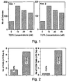
- TEPA effects on proliferation and clonability of short term CD 34 + cultures Addition of TEPA to CD 34 + cells cultured with low doses of early-acting cytokines resulted in a significant increase in total cell number, in the number of CD 34 + cells (measured by flow cytometry utilizing fluorescence labeled specific antibodies, Figure 2 ) and in cell clonability (measured by plating culture aliquots in semi-solid medium and scoring colonies that develop two weeks later, Figure 1 ), compared to cultures supplemented only with cytokines.
- the colonies which developed in semi-solid medium in the presence of TEPA were of myeloid, erythroid and mixed phenotype.
- TEPA TEPA significantly increased the clonability and the percentage of CD 34 + cells in these cultures.
- total cell number it was increased by TEPA in cultures supplemented with early cytokines (Table 1; Figure 2 ), whereas in cultures supplemented with both early and late cytokines, TEPA caused a marginal inhibition ( Figure 3 ).
- Cord blood-derived CD 34 cells were plated in liquid culture in the presence of: FLT 3 ligand - 50ng/ml, SCF - 50ng/ml, IL-6 - 50ng/ml, with or without IL-3 - 20ng/ml, with or without TEPA - 10 ⁇ M.
- the percentage of CD 34 cells and the total cell number were determined Aliquots equivalent to 1x10 3 initiating cells were assayed on days 0 and 7 for colony forming cells (CFU) by cloning in semi-solid medium.
- CFU expansion represents the ratio of CFU present on day 7 to CFU present on day 0.
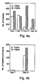
- TEPA on proliferation and clonability of long-term CD 34 + cultures Long-term cultures were maintained for 3-5 weeks by weekly demi-depopulation (one half of the culture volume was removed and replaced by fresh medium and cytokines). Addition of TEPA resulted in a higher clonability in long-term cultures supplemented with either early cytokines ( Figure 4 ) or both early and late cytokines ( Figure 3 ), as compared to cultures supplemented only with cytokines.
- TEPA TEPA on the maturation of hematopoietic cells
- MEL cells are erythroblast like cells. Following treatment with several chemicals (differentiation inducers) the cells undergo erythroid differentiation and accumulate hemoglobin. MEL cells were cultured in the presence of the differentiation inducer hexamethylene bisacetamide (HMBA) and the chelators TEPA or Captopril. At day 3 of the culture, the total number of cells and the percentage of hemoglobin-containing cells were determined (Table 2). The results indicated that both TEPA and captopril inhibited the HMBA-induced differentiation of MEL cells.
- HMBA hexamethylene bisacetamide
- Human erythroid cell cultures Normal human erythroid cells were grown according to the two-phase liquid culture procedure, essentially as described in references 23-26. In the first phase, peripheral blood mononuclear cells were incubated in the presence of early growth factors for 5-7 days. In the second phase, these factors were replaced by the erythroid specific proliferation/differentiation factor, erythropoietin.
- the cultures were supplemented with TEPA at the initiation of the second phase.
- the total cell number and the percentage of hemoglobin-containing cells were determined after 14 days.
- the results ( Figure 5 ) showed that in the presence of TEPA there was a sharp decrease in hemoglobin-containing cells, while the total number of cells decreased only slightly.
- CD 34 + initiated cultures Long term liquid cultures initiated with CD 34 + cells were maintain with different cocktails of cytokines. Half of the cultures were continuously supplemented with TEPA. In order to test the status of cell differentiation, cytospin preparation were stained with May-Grunwald Giemsa ( Figures 6a-d ). The results showed that cultures which were maintained for 4-5 weeks without TEPA contained only fully differentiated cells, while with TEPA the cultures contained, in addition to fully differentiated cells, a subset of 10 % - 40 % of undifferentiated blast-like cells.
- TEPA induces a delay in CD 34 + cell differentiation which results in prolonged proliferation and accumulation of early progenitor cells in long-term ex vivo cultures.
- TEPA's mechanism of activity In order to determine whether TEPA affects CD 34 + cells via depletion of transition metals, such as Copper, two approaches were taken.
- TEPA tetra-ethylpentamine
- CAP captopril
- PEN penicilamine
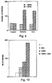
- Zinc which is known to interfere with transition metal metabolism, e.g., with Copper metabolism, expand the clonability of the cultures by itself. This effect was even more pronounced in the presence of both Zinc and TEPA ( Figure 10 ).
- TEPA and other transition metal chelators sustains long-term cultures by inhibiting/delaying cellular differentiation through chelation of transition metals, Copper in particular.
- Example No. 3 further substantiate the results described hereinabove; teaches optimal culture conditions for long-term cultures, teaches additional chelating agents that affect hemopoietic cell differentiation and sheds more light on the mechanism of activity of TEPA and other chelators on their target cells.
- CD 34 + cells derived from human neonatal cord blood were purified by immunomagnetic method and then cultured in liquid medium supplemented with cytokines either with or without transition metal chelators. At weekly intervals, the cultures were demi-depopulated by removing half of the culture content (supernatant and cells) and replacing it with fresh medium, cytokines and the chelators. At the indicated weeks the cellular content of the cultures were quantitated for total cells (by a manual microscopic/hemocytometric method), for CD 34 + cells (by immuno-flow cytometry) and for clonogenic cells (by cloning the cells in cytokine-supplemented semi-solid medium).
- the cultures were initiated with 1x10 4 cells, 50-80 % of which were CD 34 + and 25-50 % of which were CFUc.
- the results presented in Figures 11 to 24 were calculated per 1x10 4 initiating cells (the numbers were multiplied by the dilution factors).
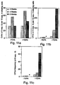
- FIG 11 shows the effect of TEPA on long-term CD 34 cultures.
- Cultures initiated with CD 34 cells in liquid medium supplemented with early-acting cytokines (in the absence of stromal cells) could be maintained by TEPA for a long time (>6 weeks).
- Figures 12-14 show the effect of TEPA on cell proliferation, CFUc and CFUc frequency in the presence of different combination of early cytokines.
- the combination of the early-acting cytokines TPO, SCF, FLT 3 ligand IL-6 and TEPA was found to be the optimal combination for the maintenance and long term expansion of cells with clonogenic potential.
- Figure 15 shows the effect of G-CSF and GM-CSF on CFUc frequency of control and TEPA-supplemented CD 34 cultures. Supplementing the cultures with the late-acting cytokines G-CSF and GM-CSF, which stimulate cell differentiation, resulted in rapid loss of clonogenic cells. This differentiation stimulatory effect is blocked by TEPA.
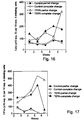
- FIGs 16-17 show the effect of partial or complete medium + TEPA change on long-term cell proliferation ( Figure 16 ) and CFUc production ( Figure 17 ). The results obtained indicate that for maintaining maximal expansion, TEPA should be completely replaced, at least, at weekly intervals.
- FIG 19 shows the effect of delayed addition of TEPA on CFUc frequency. It is evident that early exposure of CD 34 cells to TEPA was crucial for long-term maintenance and expansion of CFUc, suggesting that TEPA affects differentiation of progenitors at various stages of differentiation.
- Figure 20 shows the effect of short-term preincubation with a single cytokine on long-term CFUc production.
- the results indicate that LTC-CFC are more preserved in TEPA-treated cultures when supplemented for the first 24 hours with a single cytokine rather than the full complement of cytokines, suggesting that under the former conditions cells are blocked more efficiently.
- Figures 21 a-b show the effect of polyamine chelating agents on CD 34 cell cultures.
- Polyamine chelating agents sustained cell proliferation and expanded CFUc during long term cultures.
- the long-chain polyamines, TEPA and PEHA were found to be more effective,than the short-chain polyamines.
- Figures 22a-b show the effect of transition metal chelating agents on CD 34 cell cultures.
- CAP captopril
- Figure 23a-b show the effect of Zinc on CD 34 cell cultures.
- Zinc which is known to interfere with transition metal metabolism, Copper in particular, mimicked the effect of the chelating agents in long term cultures, but to a smaller extent than the chelators themselves.
- ex vivo expansion of hematopoietic progenitor cells is limited by the progression of these cells into non-dividing differentiated cells.
- This differentiation process can be delayed by cultivating the progenitor cells on stroma cell layer. Since the stroma supports continuous cell proliferation and long-term generation of CFUc, it is believed that the stroma inflict an anti differentiation effect on the progenitor cells.
- the system combines the use of early-acting cytokines, such as stem cell factor (SCF), FLT3 ligand, interleukin-6 (IL-6), thrombopoietin (TPO) with or without interleukin-3, and transition metal chelating agents ( Figures 12-14 ).
- SCF stem cell factor
- IL-6 interleukin-6
- TPO thrombopoietin
- the early cytokines support the survival and proliferation of the progenitors with reduced stimulus for differentiation compared to late-acting cytokines, such as G-CSF and GM-CSF ( Figure 15 ).
- the chelators inhibit differentiation through chelation of transition metals, Copper in particular. Complete medium change at weekly intervals, as compared to partial change, improved LTC-CFC maintenance, suggesting that the TEPA-transition metal complex, e.g., TEPA-Copper complex, may not be stable ( Figures 16-17 ).
- chelating agents have been found to support continuous cell proliferation and long-term generation of CFUc and to delay cell differentiation.
- polyamines such as, but not limited to, TEPA, EDA, PEHA and TETA ( Figures 21a-b ) or chelators such as, but not limited to, penicilamine (PEN) and captopril (CAP) ( Figures 22a-b ).
- PEN penicilamine
- CAP captopril
- Zinc which interfere with transition metals (Copper in particular) metabolism also supported LTC-CFC ( Figures 23a-b ).
- stem cells such as, but not limited to, cord blood derived stem cells, peripheral blood derived stem cells and bone marrow-derived stem cells.
- the method is effected by handling the stem cell while being harvested, isolated and/or stored, in a presence of a transition metal chelator, e.g., TEPA.
- a transition metal chelator e.g., TEPA.
- Cord blood-derived cells were collected and stored (unseparated) for 24 hours, at 4 °C, either in the presence or absence of 10 ⁇ M TEPA.
- CD 34 + cells were then separated using either 10 ⁇ M TEPA-PBS buffer or TEPA free PBS buffer, respectively. Then, cells were grown in long-term cultures in the presence of 10 ⁇ M TEPA.
- stem cell collection bags and separation and washing buffers supplemented with an effective amount or concentration of transition metal chelator, which inhibits differentiation.
Landscapes
- Health & Medical Sciences (AREA)
- Engineering & Computer Science (AREA)
- Life Sciences & Earth Sciences (AREA)
- Bioinformatics & Cheminformatics (AREA)
- Chemical & Material Sciences (AREA)
- Biomedical Technology (AREA)
- Organic Chemistry (AREA)
- Genetics & Genomics (AREA)
- Hematology (AREA)
- Biotechnology (AREA)
- Wood Science & Technology (AREA)
- Zoology (AREA)
- General Health & Medical Sciences (AREA)
- Nuclear Medicine, Radiotherapy & Molecular Imaging (AREA)
- Microbiology (AREA)
- Developmental Biology & Embryology (AREA)
- Public Health (AREA)
- Immunology (AREA)
- Animal Behavior & Ethology (AREA)
- Pharmacology & Pharmacy (AREA)
- Cell Biology (AREA)
- Medicinal Chemistry (AREA)
- Veterinary Medicine (AREA)
- General Chemical & Material Sciences (AREA)
- Chemical Kinetics & Catalysis (AREA)
- Biochemistry (AREA)
- General Engineering & Computer Science (AREA)
- Micro-Organisms Or Cultivation Processes Thereof (AREA)
- Medicines Containing Material From Animals Or Micro-Organisms (AREA)
- Agricultural Chemicals And Associated Chemicals (AREA)
Claims (26)
- Procédé d'expansion de cellules souches et progénitrices hématopoïétiques ex-vivo, inhibant en même temps la différenciation desdites cellules souches et progénitrices hématopoïétiques, le procédé comprenant la fourniture de conditions de prolifération cellulaire pour lesdites cellules souches et progénitrices hématopoïétiques ex vivo avec un chélateur de métal de transition choisi dans le groupe constitué par la tétraéthylène pentamine (TEPA), l'éthylène diamine (EDA), la pentaéthylène hexamine (PEHA), la triéthylène tétramine (TETA), la pénicilamine et le captopril, dans lequel lesdites conditions de prolifération cellulaire comprennent des nutriments et au moins une cytokine à action précoce ou des cytokines à action précoce, dans lequel lesdites cellules souches et progénitrices hématopoïétiques sont des cellules souches et progénitrices hématopoïétiques thérapeutiques, et dans lequel ledit chélateur et lesdites conditions de prolifération résultent en(i) une prolifération active prolongée ;(ii) une expansion à long terme de cellules clonogènes (CFUc) ; et(iii) le maintien des cellules indifférenciées dans leur état indifférencié ;
à condition que lesdites cellules hématopoïétiques ne soient pas des cellules souches embryonnaires humaines obtenues à partir d'un embryon humain. - Procédé selon la revendication 1, dans lequel ledit chélateur de métal de transition est TEPA.
- Procédé selon la revendication 1, dans lequel la concentration dudit chélateur de métal de transition est de 0,1 µM à 100 mM.
- Procédé selon la revendication 3, dans lequel la concentration dudit chélateur de métal de transition est de 4 µM à 50 mM.
- Procédé selon la revendication 4, dans lequel la concentration dudit chélateur de métal de transition est de 5 µM à 40 mM.
- Procédé selon la revendication 1, dans lequel lesdites conditions de prolifération cellulaire comprennent, en outre, la fourniture d'une cytokine à action tardive ou de cytokines à action tardive.
- Procédé selon la revendication 6, dans lequel ladite cytokine à action tardive est choisie dans le groupe constitué par le facteur de stimulation des colonies granulocytaires, le facteur de stimulation des colonies granulocytaires/macrophages et l'érythropoïétine.
- Procédé selon la revendication 6, dans lequel ladite cytokine à action tardive est le facteur de stimulation des colonies granulocytaires.
- Procédé selon la revendication 1, dans lequel lesdites cellules hématopoïétiques sont dérivées d'une source choisie dans le groupe constitué par les cellules de moelle osseuse, les cellules du sang périphérique et les cellules du sang de cordon ombilical néonatal.
- Procédé selon la revendication 1, dans lequel lesdites cellules souches et progénitrices hématopoïétiques sont dérivées de cellules du sang de cordon ombilical néonatal.
- Procédé selon la revendication 1, comprenant, en outre, l'étape consistant à sélectionner une population de cellules hématopoïétiques enrichie en cellules souches hématopoïétiques.
- Procédé selon la revendication 11, dans lequel lesdites cellules sont enrichies en CD34+.
- Procédé de transduction de cellules souches et progénitrices hématopoïétiques thérapeutiques indifférenciées, expansées avec un exogène, le procédé comprenant les étapes consistant à :(a) expanser et inhiber la différenciation desdites cellules souches et progénitrices hématopoïétiques par :(i) fourniture de conditions de prolifération cellulaire pour lesdites cellules souches et progénitrices hématopoïétiques, dans lequel lesdites conditions de prolifération cellulaire comprennent des nutriments et au moins une cytokine à action précoce ou des cytokines à action précoce et dans lequel lesdites cellules souches et progénitrices hématopoïétiques sont des cellules souches et progénitrices hématopoïétiques thérapeutiques ;(ii) mise en contact desdites cellules souches et progénitrices hématopoïétiques avec un chélateur de métal de transition choisi dans le groupe constitué par TEPA, EDA, PEHA, TETA, la pénicilamine et le captopril, dans lequel ledit chélateur et lesdites conditions de prolifération induisent une prolifération active prolongée ; une expansion à long terme de cellules clonogènes (CFUc) ; et le maintien des cellules indifférenciées à leur état indifférencié ; pour inhiber ainsi la différenciation et expanser lesdites cellules hématopoïétiques ex-vivo ; et(b) transduire lesdites cellules souches et progénitrices hématopoïétiques thérapeutiques expansées et indifférenciées avec l'exogène, à condition que lesdites cellules souches et progénitrices hématopoïétiques ne soient pas des cellules souches embryonnaires humaines obtenues à partir d'un embryon humain.
- Procédé selon la revendication 13, dans lequel ladite transduction est réalisée par un vecteur contenant l'exogène.
- Population de cellules souches et progénitrices hématopoïétiques thérapeutiques cultivée ex-vivo comprenant des cellules souches et progénitrices hématopoïétiques indifférenciées, expansées selon les procédés de l'une quelconque des revendications 1 à 14, ladite population ayant :(i) une plus grande proportion de cellules souches et progénitrices hématopoïétiques en cours de prolifération ;(ii) une plus grande proportion de cellules clonogènes (CFUc) ; et(iii) une plus grande proportion de cellules indifférenciées maintenues à leur état indifférencié
comparativement à une population similaire de cellules souches et progénitrices hématopoïétiques cultivée ex-vivo sans mise en contact avec ledit chélateur de métal de transition, à condition que lesdites cellules hématopoïétiques ne soient pas des cellules souches embryonnaires humaines obtenues à partir d'un embryon humain. - Procédé de conservation de cellules souches et progénitrices hématopoïétiques indifférenciées comprenant la mise en contact des cellules souches et progénitrices hématopoïétiques indifférenciées avec un chélateur de métal de transition choisi dans le groupe constitué par TEPA, EDA, PEHA, TETA, la pénicilamine et le captopril, dans lequel ladite mise en contact s'effectue au moins au cours d'une des étapes de récolte, isolation et stockage des cellules hématopoïétiques indifférenciées, à condition que lesdites cellules hématopoïétiques ne soient pas des cellules souches embryonnaires humaines obtenues à partir d'un embryon humain.
- Procédé selon la revendication 16, dans lequel ledit chélateur de métal de transition est TEPA.
- Sacs de collecte de cellules souches, additionnés d'une quantité de chélateur de métal de transition choisi dans le groupe constitué par TEPA, EDA, PEHA, TETA, la pénicilamine et le captopril, ladite quantité étant suffisante pour inhiber la différenciation d'une population de cellules hématopoïétiques indifférenciées.
- Sacs de collecte de cellules souches selon la revendication 18, dans lesquels ledit chélateur de métal de transition est TEPA.
- Sacs de collecte de cellules souches selon la revendication 18, dans lesquels la concentration dudit chélateur de métal de transition est de 0,1 µM à 100 mM.
- Sacs de collecte de cellules souches selon la revendication 20, dans lesquels la concentration dudit chélateur de métal de transition est de 4 µM à 50 mM.
- Sacs de collecte de cellules souches selon la revendication 20, dans lesquels la concentration dudit chélateur de métal de transition est de 5 µM à 40 mM.
- Utilisation d'une composition de cellules hématopoïétiques thérapeutiques comprenant une population de cellules souches et progénitrices hématopoïétiques indifférenciées, expansées selon les procédés de l'une quelconque des revendications 1 à 14, dans la préparation d'un médicament destiné à traiter un sujet ayant besoin d'une greffe de cellules souches hématopoïétiques, dans laquelle ladite population est caractérisée en ce qu'elle a :(i) une plus grande proportion de cellules souches et progénitrices hématopoïétiques en cours de prolifération ;(ii) une plus grande proportion de cellules clonogènes (CFUc) ; et(iii) une plus grande proportion de cellules indifférenciées maintenues à leur état indifférencié
comparativement à une population similaire de cellules souches et progénitrices hématopoïétiques cultivée ex-vivo sans mise en contact avec ledit chélateur de métal de transition. - Procédés selon les revendications 1 et 13, dans lesquels ladite au moins ou lesdites cytokines à action précoce sont choisies dans le groupe constitué par le facteur de cellule souche, le ligand FLT3, l'interleukine-6, la thrombopoïétine et l'interleukine-3.
- Procédés selon les revendications 1 et 13, dans lesquels ladite cytokine à action précoce est le ligand FLT3.
- Population de cellules souches et progénitrices hématopoïétiques thérapeutiques cultivée ex-vivo comprenant des cellules souches et progénitrices hématopoïétiques indifférenciées, expansées selon les procédés de l'une quelconque des revendications 1 à 14, dans laquelle ladite population de cellules est fournie dans un milieu comprenant ledit chélateur de métal de transition, à condition que lesdites cellules hématopoïétiques ne soient pas des cellules souches embryonnaires humaines obtenues à partir d'un embryon humain.
Priority Applications (1)
| Application Number | Priority Date | Filing Date | Title |
|---|---|---|---|
| EP03003275A EP1332673B1 (fr) | 1998-02-17 | 1999-02-08 | Procédé pour contrôler la prolifération et la différentiation de cellules souches et progenitrices |
Applications Claiming Priority (5)
| Application Number | Priority Date | Filing Date | Title |
|---|---|---|---|
| US2419598A | 1998-02-17 | 1998-02-17 | |
| US24195 | 1998-02-17 | ||
| US13036798A | 1998-08-07 | 1998-08-07 | |
| US130367 | 1998-08-07 | ||
| PCT/US1999/002664 WO1999040783A1 (fr) | 1998-02-17 | 1999-02-08 | Procede de lutte contre la proliferation et la differenciation de cellules souches et parentes |
Related Child Applications (2)
| Application Number | Title | Priority Date | Filing Date |
|---|---|---|---|
| EP03003275A Division EP1332673B1 (fr) | 1998-02-17 | 1999-02-08 | Procédé pour contrôler la prolifération et la différentiation de cellules souches et progenitrices |
| EP03003275.9 Division-Into | 2003-02-24 |
Publications (3)
| Publication Number | Publication Date |
|---|---|
| EP1069821A1 EP1069821A1 (fr) | 2001-01-24 |
| EP1069821A4 EP1069821A4 (fr) | 2001-11-21 |
| EP1069821B1 true EP1069821B1 (fr) | 2011-06-08 |
Family
ID=26698157
Family Applications (2)
| Application Number | Title | Priority Date | Filing Date |
|---|---|---|---|
| EP03003275A Expired - Lifetime EP1332673B1 (fr) | 1998-02-17 | 1999-02-08 | Procédé pour contrôler la prolifération et la différentiation de cellules souches et progenitrices |
| EP99906799A Expired - Lifetime EP1069821B1 (fr) | 1998-02-17 | 1999-02-08 | Procede de lutte contre la proliferation et la differenciation de cellules souches et parentes |
Family Applications Before (1)
| Application Number | Title | Priority Date | Filing Date |
|---|---|---|---|
| EP03003275A Expired - Lifetime EP1332673B1 (fr) | 1998-02-17 | 1999-02-08 | Procédé pour contrôler la prolifération et la différentiation de cellules souches et progenitrices |
Country Status (8)
| Country | Link |
|---|---|
| EP (2) | EP1332673B1 (fr) |
| JP (1) | JP3810273B2 (fr) |
| AT (1) | ATE511752T1 (fr) |
| AU (1) | AU759522C (fr) |
| CA (1) | CA2320073C (fr) |
| ES (1) | ES2391055T3 (fr) |
| IL (2) | IL137757A0 (fr) |
| WO (1) | WO1999040783A1 (fr) |
Families Citing this family (24)
| Publication number | Priority date | Publication date | Assignee | Title |
|---|---|---|---|---|
| US6962698B1 (en) | 1998-02-17 | 2005-11-08 | Gamida Cell Ltd. | Methods of controlling proliferation and differentiation of stem and progenitor cells |
| CA2344653A1 (fr) | 1998-09-29 | 2000-04-06 | Gamida Cell Ltd. | Procedes de regulation de la proliferation et de la differentiation de cellules souches et precurseurs |
| NL1017973C2 (nl) * | 2000-05-10 | 2002-11-08 | Tristem Trading Cyprus Ltd | Inrichting. |
| IL152904A0 (en) | 2002-01-24 | 2003-06-24 | Gamida Cell Ltd | Utilization of retinoid and vitamin d receptor antagonists for expansion of renewable stem cell populations |
| WO2003062404A1 (fr) | 2002-01-25 | 2003-07-31 | Gamida-Cell Ltd. | Methodes de developpement de cellules souches et de cellules precurseurs, populations de cellules multipliees obtenues a l'aide de ces methodes |
| AU2004217699B2 (en) * | 2003-03-07 | 2008-07-03 | Gamida-Cell Ltd. | Expansion of renewable stem cell populations using modulators of PI 3-kinase |
| IL161903A0 (en) * | 2003-07-17 | 2005-11-20 | Gamida Cell Ltd | Ex vivo progenitor and stem cell expansion for usein the treatment of disease of endodermally- deri ved organs |
| WO2005007799A2 (fr) * | 2003-07-17 | 2005-01-27 | Gamida-Cell Ltd. | Procedes de multiplication ex-vivo de cellules souches / progenitrices |
| JP2007535479A (ja) | 2003-08-13 | 2007-12-06 | チルドレンズ ホスピタル メディカル センター | GTPaseを調節するためのキメラペプチド |
| US7417026B2 (en) | 2003-08-13 | 2008-08-26 | Children's Hospital Medical Center | Mobilization of hematopoietic cells |
| EP1799812A4 (fr) | 2004-09-16 | 2009-09-09 | Gamida Cell Ltd | Méthodes de culture ex vivo de cellules souches et de précurseur par co-culture avec des cellules mésenchymales |
| US8846393B2 (en) | 2005-11-29 | 2014-09-30 | Gamida-Cell Ltd. | Methods of improving stem cell homing and engraftment |
| WO2007108689A2 (fr) * | 2006-03-21 | 2007-09-27 | Stichting Skeletal Tissue Engineering | Nouveau procede d'induction de la differenciation de cellules souches et progenitrices |
| JP5573160B2 (ja) | 2007-12-05 | 2014-08-20 | 日産化学工業株式会社 | ヘテロ環化合物による造血幹細胞の増幅方法 |
| US9115341B2 (en) | 2007-12-05 | 2015-08-25 | Nissan Chemical Industries, Ltd. | Method for expanding hematopoietic stem cells using heterocyclic compound |
| WO2009072624A1 (fr) | 2007-12-05 | 2009-06-11 | Nissan Chemical Industries, Ltd. | Procédé d'amplification d'une cellule souche hématopoïétique utilisant un composé hétérocyclique |
| CN101889007A (zh) | 2007-12-06 | 2010-11-17 | 日产化学工业株式会社 | 采用杂环化合物的造血干细胞的扩增方法 |
| BRPI1014635A8 (pt) | 2009-06-04 | 2016-10-11 | Nissan Chemical Ind Ltd | compostos heterocíclicos e agentes de expansão para células-tronco hematopoiéticas |
| EP2581442A4 (fr) | 2010-06-14 | 2013-11-06 | Nissan Chemical Ind Ltd | Procédé de production de cellules souches hématopoïétiques |
| WO2012074068A1 (fr) | 2010-12-01 | 2012-06-07 | 日産化学工業株式会社 | Procédé de production de cellules souches hématopoïétiques à l'aide d'un composé pyrazole |
| CN104204192A (zh) | 2012-02-13 | 2014-12-10 | 加米达细胞有限公司 | 间充质干细胞调理的基质及其产生和使用方法 |
| US9175266B2 (en) | 2012-07-23 | 2015-11-03 | Gamida Cell Ltd. | Enhancement of natural killer (NK) cell proliferation and activity |
| US9567569B2 (en) | 2012-07-23 | 2017-02-14 | Gamida Cell Ltd. | Methods of culturing and expanding mesenchymal stem cells |
| WO2021149127A1 (fr) * | 2020-01-21 | 2021-07-29 | オリンパス株式会社 | Dispositif de surveillance de la différenciation érythroïde et procédé de surveillance de la différenciation érythroïde |
Citations (3)
| Publication number | Priority date | Publication date | Assignee | Title |
|---|---|---|---|---|
| EP0331464A2 (fr) * | 1988-03-02 | 1989-09-06 | Tosoh Corporation | Méthode de pliage de la protéine similaire au précurseur de l'urokinase |
| WO2000018885A1 (fr) * | 1998-09-29 | 2000-04-06 | Gamida Cell Ltd. | Procedes de regulation de la proliferation et de la differentiation de cellules souches et precurseurs |
| US20020159981A1 (en) * | 1999-02-08 | 2002-10-31 | Gamida-Cell Ltd. | Methods of controlling proliferation and differentiation of stem and progenitor cells |
Family Cites Families (3)
| Publication number | Priority date | Publication date | Assignee | Title |
|---|---|---|---|---|
| US5631219A (en) * | 1994-03-08 | 1997-05-20 | Somatogen, Inc. | Method of stimulating hematopoiesis with hemoglobin |
| JP2000513329A (ja) * | 1996-03-01 | 2000-10-10 | イムクローン システムズ インコーポレイテッド | 幹細胞の分化を阻害させるためのデルタ様蛋白質の使用 |
| JP2000507812A (ja) * | 1996-03-12 | 2000-06-27 | ライフ テクノロジーズ,インコーポレイテッド | 造血細胞培養栄養補充成分 |
-
1999
- 1999-02-08 IL IL13775799A patent/IL137757A0/xx active IP Right Grant
- 1999-02-08 WO PCT/US1999/002664 patent/WO1999040783A1/fr not_active Ceased
- 1999-02-08 CA CA2320073A patent/CA2320073C/fr not_active Expired - Fee Related
- 1999-02-08 ES ES03003275T patent/ES2391055T3/es not_active Expired - Lifetime
- 1999-02-08 EP EP03003275A patent/EP1332673B1/fr not_active Expired - Lifetime
- 1999-02-08 JP JP2000531059A patent/JP3810273B2/ja not_active Expired - Fee Related
- 1999-02-08 AT AT99906799T patent/ATE511752T1/de not_active IP Right Cessation
- 1999-02-08 AU AU26624/99A patent/AU759522C/en not_active Ceased
- 1999-02-08 EP EP99906799A patent/EP1069821B1/fr not_active Expired - Lifetime
-
2000
- 2000-08-08 IL IL137757A patent/IL137757A/en not_active IP Right Cessation
Patent Citations (3)
| Publication number | Priority date | Publication date | Assignee | Title |
|---|---|---|---|---|
| EP0331464A2 (fr) * | 1988-03-02 | 1989-09-06 | Tosoh Corporation | Méthode de pliage de la protéine similaire au précurseur de l'urokinase |
| WO2000018885A1 (fr) * | 1998-09-29 | 2000-04-06 | Gamida Cell Ltd. | Procedes de regulation de la proliferation et de la differentiation de cellules souches et precurseurs |
| US20020159981A1 (en) * | 1999-02-08 | 2002-10-31 | Gamida-Cell Ltd. | Methods of controlling proliferation and differentiation of stem and progenitor cells |
Non-Patent Citations (5)
| Title |
|---|
| CHISI J ET AL: "Inhibitory action of the peptide AcSDKP on the proliferative state of hematopoietic stem cells in the presence of Captopril but not Lisinopril", STEM CELLS, vol. 15, 1997, pages 455 - 460 * |
| HAMMOND W P ET AL: "Suppression of in vitro granulocytopoiesis by captopril and penicillamine", EXP. HEMATOL., vol. 16, 1988, pages 674 - 680 * |
| PERCIVAL S: "Copper and immunity", AMERICAN JOURNAL OF CLINICAL NUTRITION, vol. 67(suppl.), 1998, pages 1064S - 1068S * |
| PIACIBELLO W ET AL: "Extensive amplification and self-renewal of human primitive hematopoietic stem cells from cord blood", BLOOD, vol. 89, no. 8, 15 April 1997 (1997-04-15), pages 2644 - 2653 * |
| SEKHAR M ET AL: "Retroviral transduction of CD34-enriched hematopoietic progenitor cells under serum-free conditions", HUMAN GENE THERAPY, vol. 7, 1 January 1996 (1996-01-01), pages 33 - 38 * |
Also Published As
| Publication number | Publication date |
|---|---|
| WO1999040783A1 (fr) | 1999-08-19 |
| JP3810273B2 (ja) | 2006-08-16 |
| ES2391055T3 (es) | 2012-11-21 |
| EP1069821A4 (fr) | 2001-11-21 |
| EP1332673A1 (fr) | 2003-08-06 |
| IL137757A0 (en) | 2001-10-31 |
| AU759522B2 (en) | 2003-04-17 |
| EP1332673B1 (fr) | 2012-05-30 |
| EP1069821A1 (fr) | 2001-01-24 |
| ATE511752T1 (de) | 2011-06-15 |
| AU759522C (en) | 2005-05-12 |
| HK1057976A1 (en) | 2004-04-30 |
| CA2320073A1 (fr) | 1999-08-19 |
| HK1034162A1 (en) | 2001-10-19 |
| CA2320073C (fr) | 2011-11-01 |
| IL137757A (en) | 2006-08-20 |
| JP2002502617A (ja) | 2002-01-29 |
| AU2662499A (en) | 1999-08-30 |
Similar Documents
| Publication | Publication Date | Title |
|---|---|---|
| EP1332673B1 (fr) | Procédé pour contrôler la prolifération et la différentiation de cellules souches et progenitrices | |
| AU770896B2 (en) | Methods of controlling proliferation and differentiation of stem and progenitor cells | |
| US6962698B1 (en) | Methods of controlling proliferation and differentiation of stem and progenitor cells | |
| US20080279828A1 (en) | Methods of expanding stem and progenitor cells and expanded cell populations obtained thereby | |
| US5955357A (en) | In-vitro-derived human neutrophil precursor cells | |
| AU2003200124B2 (en) | Method of Controlling Proliferation and Differentiation of Stem and Progenitor Cells | |
| US5888499A (en) | Method for increasing neutrophil populations using in vitro-derived human neutrophil precursor cells | |
| HK1034162B (en) | Method of controlling proliferation and differentiation of stem and progenitor cells | |
| HK1057976B (en) | Method of controlling proliferation and differentiation of stem and progenitor cells | |
| ES2370203T3 (es) | Método de control de la proliferación y diferenciación de células progenitoras y madre. | |
| ZA200102073B (en) | Methods of controlling proliferation and differentiation of stem and progenitor cells. |
Legal Events
| Date | Code | Title | Description |
|---|---|---|---|
| PUAI | Public reference made under article 153(3) epc to a published international application that has entered the european phase |
Free format text: ORIGINAL CODE: 0009012 |
|
| 17P | Request for examination filed |
Effective date: 20000821 |
|
| AK | Designated contracting states |
Kind code of ref document: A1 Designated state(s): AT BE CH DE ES FR GB GR IT LI NL PT SE |
|
| A4 | Supplementary search report drawn up and despatched |
Effective date: 20011005 |
|
| AK | Designated contracting states |
Kind code of ref document: A4 Designated state(s): AT BE CH DE ES FR GB GR IT LI NL PT SE |
|
| 17Q | First examination report despatched |
Effective date: 20030425 |
|
| 17Q | First examination report despatched |
Effective date: 20030425 |
|
| GRAP | Despatch of communication of intention to grant a patent |
Free format text: ORIGINAL CODE: EPIDOSNIGR1 |
|
| GRAS | Grant fee paid |
Free format text: ORIGINAL CODE: EPIDOSNIGR3 |
|
| GRAA | (expected) grant |
Free format text: ORIGINAL CODE: 0009210 |
|
| AK | Designated contracting states |
Kind code of ref document: B1 Designated state(s): AT BE CH DE ES FR GB GR IT LI NL PT SE |
|
| REG | Reference to a national code |
Ref country code: GB Ref legal event code: FG4D |
|
| REG | Reference to a national code |
Ref country code: CH Ref legal event code: EP |
|
| REG | Reference to a national code |
Ref country code: DE Ref legal event code: R096 Ref document number: 69943478 Country of ref document: DE Effective date: 20110721 |
|
| REG | Reference to a national code |
Ref country code: NL Ref legal event code: VDEP Effective date: 20110608 |
|
| REG | Reference to a national code |
Ref country code: HK Ref legal event code: GR Ref document number: 1034162 Country of ref document: HK |
|
| PG25 | Lapsed in a contracting state [announced via postgrant information from national office to epo] |
Ref country code: SE Free format text: LAPSE BECAUSE OF FAILURE TO SUBMIT A TRANSLATION OF THE DESCRIPTION OR TO PAY THE FEE WITHIN THE PRESCRIBED TIME-LIMIT Effective date: 20110608 |
|
| REG | Reference to a national code |
Ref country code: CH Ref legal event code: NV Representative=s name: VOSSIUS & PARTNER |
|
| PG25 | Lapsed in a contracting state [announced via postgrant information from national office to epo] |
Ref country code: AT Free format text: LAPSE BECAUSE OF FAILURE TO SUBMIT A TRANSLATION OF THE DESCRIPTION OR TO PAY THE FEE WITHIN THE PRESCRIBED TIME-LIMIT Effective date: 20110608 Ref country code: GR Free format text: LAPSE BECAUSE OF FAILURE TO SUBMIT A TRANSLATION OF THE DESCRIPTION OR TO PAY THE FEE WITHIN THE PRESCRIBED TIME-LIMIT Effective date: 20110909 |
|
| REG | Reference to a national code |
Ref country code: ES Ref legal event code: FG2A Ref document number: 2370203 Country of ref document: ES Kind code of ref document: T3 Effective date: 20111213 |
|
| PG25 | Lapsed in a contracting state [announced via postgrant information from national office to epo] |
Ref country code: NL Free format text: LAPSE BECAUSE OF FAILURE TO SUBMIT A TRANSLATION OF THE DESCRIPTION OR TO PAY THE FEE WITHIN THE PRESCRIBED TIME-LIMIT Effective date: 20110608 |
|
| PG25 | Lapsed in a contracting state [announced via postgrant information from national office to epo] |
Ref country code: PT Free format text: LAPSE BECAUSE OF FAILURE TO SUBMIT A TRANSLATION OF THE DESCRIPTION OR TO PAY THE FEE WITHIN THE PRESCRIBED TIME-LIMIT Effective date: 20111010 |
|
| PLBE | No opposition filed within time limit |
Free format text: ORIGINAL CODE: 0009261 |
|
| STAA | Information on the status of an ep patent application or granted ep patent |
Free format text: STATUS: NO OPPOSITION FILED WITHIN TIME LIMIT |
|
| 26N | No opposition filed |
Effective date: 20120309 |
|
| REG | Reference to a national code |
Ref country code: DE Ref legal event code: R097 Ref document number: 69943478 Country of ref document: DE Effective date: 20120309 |
|
| REG | Reference to a national code |
Ref country code: FR Ref legal event code: PLFP Year of fee payment: 17 |
|
| PGFP | Annual fee paid to national office [announced via postgrant information from national office to epo] |
Ref country code: ES Payment date: 20150225 Year of fee payment: 17 Ref country code: DE Payment date: 20150219 Year of fee payment: 17 Ref country code: IT Payment date: 20150219 Year of fee payment: 17 Ref country code: CH Payment date: 20150218 Year of fee payment: 17 |
|
| PGFP | Annual fee paid to national office [announced via postgrant information from national office to epo] |
Ref country code: FR Payment date: 20150219 Year of fee payment: 17 Ref country code: GB Payment date: 20150218 Year of fee payment: 17 |
|
| PGFP | Annual fee paid to national office [announced via postgrant information from national office to epo] |
Ref country code: BE Payment date: 20150218 Year of fee payment: 17 |
|
| PG25 | Lapsed in a contracting state [announced via postgrant information from national office to epo] |
Ref country code: BE Free format text: LAPSE BECAUSE OF NON-PAYMENT OF DUE FEES Effective date: 20160229 |
|
| REG | Reference to a national code |
Ref country code: CH Ref legal event code: PFA Owner name: HADASIT MEDICAL RESEARCH SERVICES AND DEVELOPM, IL Free format text: FORMER OWNER: HADASIT MEDICAL RESEARCH SERVICES AND DEVELOPMENT LTD., IL |
|
| REG | Reference to a national code |
Ref country code: DE Ref legal event code: R119 Ref document number: 69943478 Country of ref document: DE |
|
| REG | Reference to a national code |
Ref country code: CH Ref legal event code: PL |
|
| GBPC | Gb: european patent ceased through non-payment of renewal fee |
Effective date: 20160208 |
|
| PG25 | Lapsed in a contracting state [announced via postgrant information from national office to epo] |
Ref country code: LI Free format text: LAPSE BECAUSE OF NON-PAYMENT OF DUE FEES Effective date: 20160229 Ref country code: CH Free format text: LAPSE BECAUSE OF NON-PAYMENT OF DUE FEES Effective date: 20160229 |
|
| REG | Reference to a national code |
Ref country code: FR Ref legal event code: ST Effective date: 20161028 |
|
| PG25 | Lapsed in a contracting state [announced via postgrant information from national office to epo] |
Ref country code: IT Free format text: LAPSE BECAUSE OF NON-PAYMENT OF DUE FEES Effective date: 20160208 |
|
| PG25 | Lapsed in a contracting state [announced via postgrant information from national office to epo] |
Ref country code: DE Free format text: LAPSE BECAUSE OF NON-PAYMENT OF DUE FEES Effective date: 20160901 Ref country code: GB Free format text: LAPSE BECAUSE OF NON-PAYMENT OF DUE FEES Effective date: 20160208 Ref country code: FR Free format text: LAPSE BECAUSE OF NON-PAYMENT OF DUE FEES Effective date: 20160229 |
|
| PG25 | Lapsed in a contracting state [announced via postgrant information from national office to epo] |
Ref country code: ES Free format text: LAPSE BECAUSE OF NON-PAYMENT OF DUE FEES Effective date: 20160209 |