EP1077263A1 - Verfahren zur Herstellung von natürlich gefalteten und sekretierten Proteinen durch Co-Sekretion von Chaperonen - Google Patents
Verfahren zur Herstellung von natürlich gefalteten und sekretierten Proteinen durch Co-Sekretion von Chaperonen Download PDFInfo
- Publication number
- EP1077263A1 EP1077263A1 EP99114811A EP99114811A EP1077263A1 EP 1077263 A1 EP1077263 A1 EP 1077263A1 EP 99114811 A EP99114811 A EP 99114811A EP 99114811 A EP99114811 A EP 99114811A EP 1077263 A1 EP1077263 A1 EP 1077263A1
- Authority
- EP
- European Patent Office
- Prior art keywords
- polypeptide
- periplasm
- dnaj
- nucleic acid
- protein
- Prior art date
- Legal status (The legal status is an assumption and is not a legal conclusion. Google has not performed a legal analysis and makes no representation as to the accuracy of the status listed.)
- Withdrawn
Links
Images




Classifications
-
- C—CHEMISTRY; METALLURGY
- C12—BIOCHEMISTRY; BEER; SPIRITS; WINE; VINEGAR; MICROBIOLOGY; ENZYMOLOGY; MUTATION OR GENETIC ENGINEERING
- C12N—MICROORGANISMS OR ENZYMES; COMPOSITIONS THEREOF; PROPAGATING, PRESERVING, OR MAINTAINING MICROORGANISMS; MUTATION OR GENETIC ENGINEERING; CULTURE MEDIA
- C12N15/00—Mutation or genetic engineering; DNA or RNA concerning genetic engineering, vectors, e.g. plasmids, or their isolation, preparation or purification; Use of hosts therefor
-
- C—CHEMISTRY; METALLURGY
- C12—BIOCHEMISTRY; BEER; SPIRITS; WINE; VINEGAR; MICROBIOLOGY; ENZYMOLOGY; MUTATION OR GENETIC ENGINEERING
- C12N—MICROORGANISMS OR ENZYMES; COMPOSITIONS THEREOF; PROPAGATING, PRESERVING, OR MAINTAINING MICROORGANISMS; MUTATION OR GENETIC ENGINEERING; CULTURE MEDIA
- C12N9/00—Enzymes; Proenzymes; Compositions thereof; Processes for preparing, activating, inhibiting, separating or purifying enzymes
- C12N9/14—Hydrolases (3)
- C12N9/48—Hydrolases (3) acting on peptide bonds (3.4)
- C12N9/50—Proteinases, e.g. Endopeptidases (3.4.21-3.4.25)
- C12N9/64—Proteinases, e.g. Endopeptidases (3.4.21-3.4.25) derived from animal tissue
- C12N9/6421—Proteinases, e.g. Endopeptidases (3.4.21-3.4.25) derived from animal tissue from mammals
- C12N9/6424—Serine endopeptidases (3.4.21)
- C12N9/6456—Plasminogen activators
- C12N9/6459—Plasminogen activators t-plasminogen activator (3.4.21.68), i.e. tPA
-
- C—CHEMISTRY; METALLURGY
- C12—BIOCHEMISTRY; BEER; SPIRITS; WINE; VINEGAR; MICROBIOLOGY; ENZYMOLOGY; MUTATION OR GENETIC ENGINEERING
- C12N—MICROORGANISMS OR ENZYMES; COMPOSITIONS THEREOF; PROPAGATING, PRESERVING, OR MAINTAINING MICROORGANISMS; MUTATION OR GENETIC ENGINEERING; CULTURE MEDIA
- C12N1/00—Microorganisms; Compositions thereof; Processes of propagating, maintaining or preserving microorganisms or compositions thereof; Processes of preparing or isolating a composition containing a microorganism; Culture media therefor
- C12N1/20—Bacteria; Culture media therefor
-
- C—CHEMISTRY; METALLURGY
- C12—BIOCHEMISTRY; BEER; SPIRITS; WINE; VINEGAR; MICROBIOLOGY; ENZYMOLOGY; MUTATION OR GENETIC ENGINEERING
- C12P—FERMENTATION OR ENZYME-USING PROCESSES TO SYNTHESISE A DESIRED CHEMICAL COMPOUND OR COMPOSITION OR TO SEPARATE OPTICAL ISOMERS FROM A RACEMIC MIXTURE
- C12P21/00—Preparation of peptides or proteins
- C12P21/02—Preparation of peptides or proteins having a known sequence of two or more amino acids, e.g. glutathione
-
- C—CHEMISTRY; METALLURGY
- C12—BIOCHEMISTRY; BEER; SPIRITS; WINE; VINEGAR; MICROBIOLOGY; ENZYMOLOGY; MUTATION OR GENETIC ENGINEERING
- C12Y—ENZYMES
- C12Y304/00—Hydrolases acting on peptide bonds, i.e. peptidases (3.4)
- C12Y304/21—Serine endopeptidases (3.4.21)
- C12Y304/21069—Protein C activated (3.4.21.69)
-
- C—CHEMISTRY; METALLURGY
- C07—ORGANIC CHEMISTRY
- C07K—PEPTIDES
- C07K2319/00—Fusion polypeptide
- C07K2319/01—Fusion polypeptide containing a localisation/targetting motif
- C07K2319/02—Fusion polypeptide containing a localisation/targetting motif containing a signal sequence
Definitions
- the invention relates to a method for producing water-soluble, naturally folded and secreted polypeptides after expression in prokaryotic cells by co-secretion of molecular chaperones.
- Protein synthesis also called translation, is found in prokaryotic organisms the ribosomes in the cytoplasm.
- prokaryotic Host organisms it is often desirable that the recombinant obtained Gene product or protein from the cytoplasm through the inner bacterial Membrane secreted in the periplasmic space between the inner and outer membrane becomes. Proteins secreted by the periplasm can then e.g. through an osmotic Shock can be released into the nutrient medium.
- a disadvantage of this method is that the secreted polypeptides often do not form the native, biologically active conformation (Hockney, TIBTECH 12 (1994) 456-463).
- Protein disulfide isomerase (PDI; Glockshuber et al., EP-A 0 510 658) or Peptidyl prolyl cis / trans isomerases or Dsb proteins (Knappik et al., Bio / Technology 11 (1993) 77-83; Qiu et al., Appl. Environm. Microbiol. 64 (1998) 4891-4896 and Schmidt et al., Prot. Engin. 11 (1998) 601-607.
- PDI Protein disulfide isomerase
- EP-A 0 510 658 Peptidyl prolyl cis / trans isomerases or Dsb proteins
- the object of the invention is to provide a process for the production of water-soluble, of course folded eukaryotic polypeptides after expression in prokaryotes to provide, which is easy to carry out and in which a complex in vitro after-treatment, such as dissolution of inclusion bodies, reduction and naturation, is not required.
- nutrient medium used to cultivate the prokaryotic cells additionally reducing thiol reagents which contain SH groups are added, wherein the yield of recombinantly obtained protein is further increased.
- reducing thiol reagents which contain SH groups are added.
- thiol reagent is either a reducing (reduced) thiol reagent SH groups or a mixture of reducing thiol reagents with SH groups and understand oxidizing thiol reagents with disulfide groups.
- Preferred substances are reduced glutathione (GSH), cysteine, N-acetylcysteine, cysteamine, ⁇ -mercaptoethanol and similar compounds.
- the thiol reagents can be used both individually and can be used in mixtures.
- Thiol reagents such as, for example, are particularly suitable Glutathione (GSH), which have a single SH group per molecule.
- GSH Glutathione
- Thiol reagents like glutathione are used to improve the yield of native folded proteins the expression of recombinant DNA in prokaryotic cells (Glockshuber et al., EP-A 0 510 658).
- Chaperones according to the invention are understood to mean proteins that are other, non-native Protect proteins from aggregation in vivo and the formation of their native conformation promote.
- Molecular chaperones are used in the prior art to Stabilize proteins and thus protect them against aggregation and inactivation (Buchner et al., EP-A 0 556 726 A1).
- ATP-independence is preferred Chaperones of the HSP40 type (molecular weight approx. 40 kDa) or a small heat shock protein (sHSP) used.
- DnaJ is a 40 kDa heat shock protein found in the cytoplasm of E.
- Hsp70 Hsp70 chaperone
- GrpE DnaK
- the DnaK system makes certain proteins dependent on ATP Process for native conformation folded (Schröder et al., EMBO J. 12 (1993) 4137-4144; Langer et al., Nature 356 (1992) 683-689). For refolding denatured proteins this system also requires ATP.
- DnaJ protects in the absence of DnaK and ATP non-native proteins before aggregation and mediates a folding-competent state (Schröder et al., EMBO J. 12 (1993) 4137-4144). Co-secretion is also preferred of an N-terminal fragment of DnaJ, which comprises amino acids 1-108 and in Hereinafter referred to as "J domain" (Kelley, TIBS 23 (1998) 222-227). In this The area contains the J-domain and a G / F-rich domain, the interactions with DnaK (Wall et al., J. Biol. Chem. 270 (1995) 2139-2144). It was shown, that the co-expression of DnaJ in the cytosol to increase the yield of soluble Protein can lead (Yokoyama et al., Microbiol. Ferment. Technol. 62 (1998) 1205-1210).
- Hsp25 (e.g. from the mouse) is a representative of the small heat shock proteins (Gaestel et al., Eur. J. Biochem. 179 (1989) 209-213), a class of chaperones that is ubiquitous is. The molecular weight of these proteins is between 15 and 30 kDa. In case of heat shock the sHsps are highly enriched in the cell (up to 1% of the total cell protein - Arrigo & Landry (1994), In Morimoto (ed.): The Biology of Heat Shock Proteins and Molecular Chaperones, Cold Spring Harbor Press, 335-373).
- sHsps Like DnaJ proteins, sHsps have that Property to prevent the aggregation of non-native proteins and these in one maintain folding-competent state (Jakob et al., J. Biol. Chem. 268 (1993) 1517-1520; Ehrsperger et al., EMBO J. 16 (1997) 221-229).
- overexpression means an increase in Expression of the secreted chaperones such as e.g. DnaJ- and Hsp25 (preferably around at least 100%) compared to the wild type expression of the prokaryotic used in each case Host organism.
- overexpression can e.g. achieve by that the genes (for the protein, chaperone and / or signal peptide) are under control a strong prokaryotic, preferably inducible expression signal (e.g. one lac or T7 promoters or a derivative thereof).
- the secretion construct for the overexpression of the polypeptides (proteins) together with regulatory regions (promoter and terminator) on the recombinant DNA is preferably integrated into a vector which additionally encodes the arginine tRNA AGA / AGG , which is rare in prokaryotes, or is combined with a vector which does this tRNA encoded, co-expressed (Brinkmann et al., Gene 85 (1989) 109-114).
- This enables both the co-overexpression of the respective proteins in the bacterial periplasm and the transcription of the rare tRNA Arg AGA / AGG , which results in an increased synthesis of the desired protein in the bacterial host organism.
- the nucleic acids encoding the polypeptide and the chaperone can be located on one vector or on two separate vectors.
- a prokaryotic signal sequence in the sense of the invention is a nucleic acid fragment to understand which of prokaryotes, preferably Gram-negative bacteria, and the penetration of proteins bound to the signal peptide guaranteed by the inner bacterial membranes. As a result, the proteins in the Periplasm or localized in the cell supernatant.
- Such signal sequences usually have a length of 18-30 amino acids and are described for example in Murphy & Beckwith: Export of Proteins to the Cell Envelope in Escherichia coli and in Neidhardt et al. (Ed.): Escherichia coli and Salmonella, Second Edition, Vol. 1, ASM Press, Washington, 1996, pp. 967-978.
- the cleavage of bacterial signal sequences can e.g. following a sequence Ala-X-Ala (von Heijne et al., J. Mol. Biol. 184 (1985) 99-105).
- the structure of the bacterial signal peptidase is described in Paetzel et al., Nature 396 (1998) 186-190.
- Signal sequences are preferably used, which are characterized by im Periplasm of prokaryotic cells localized proteases from the desired protein be split off again. Alternatively, by adding such proteases to the Cell supernatant or to the isolated protein, the cleavage of the signal sequence takes place.
- the heterologous expression of a variety of eukaryotic proteins such as Proteases, interferons, protein hormones, antibodies or fragments thereof are improved.
- the method is particularly suitable for heterologous production of proteins which, in their native state, have at least two over one Contain disulfide bridge linked cysteines and when they N-terminally not fused have prokaryotic signal sequence in prokaryotic expression as insoluble inclusion bodies are formed.
- the method is particularly suitable for proteins that contain more than 5 disulfide bridges in their native state.
- One such protein is for example a recombinant plasminogen activator (hereinafter called rPA, Martin et al., Cardiovasc. Drug Rev. 11 (1993) 299-311, U.S. Patent No. 5,223,256).
- rPA has 9 disulfide bridges that are not formed in the reducing cytosol of E. coli become.
- the periplasmic localization of the protein and the chaperone is carried out "Operational linkage" with a signal peptide to penetrate inner bacterial Guaranteed membranes.
- Secreted protein can then fold in the periplasm.
- the oxidizing conditions of this compartment enable the formation of disulfide bridges.
- the yield of chaperones such as DnaJ, J domain or Hsp25 in the periplasm is successful of functional protein to increase more than 100 times.
- ScF V s are shortened antibodies which only come from the variable There are sections (F V ) of the heavy and light chain of an antibody which are artificially fused via a short (mostly Gly 4 Ser 3 ) linker (Hudson, Curr. Opin Biotechnol. 9 (1998) 395-402).
- ScF V s normally the same affinity for the antigen as the paternal F V strands, but can be overexpressed in E. coli.
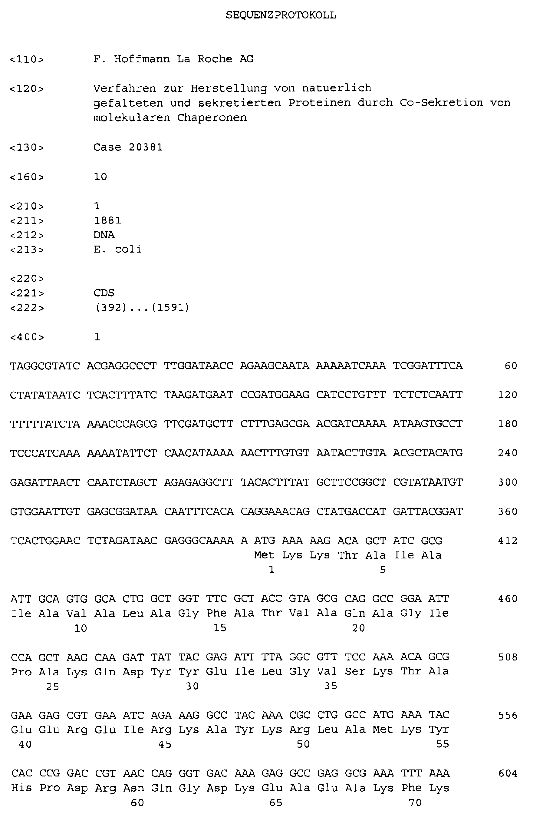
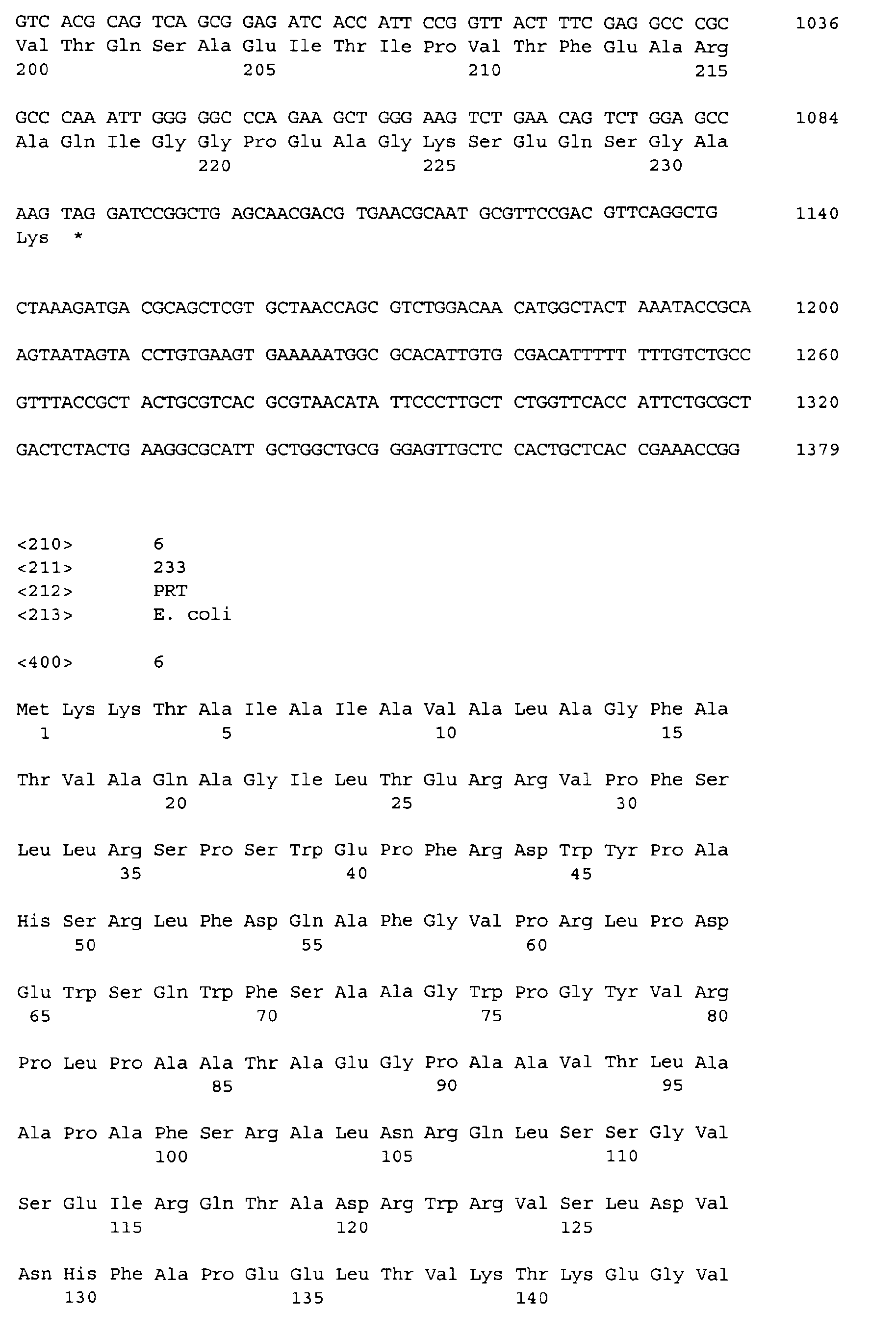
- SEQ ID NO: 1 and 2 show the sequence of the part of the expression plasmid pUBS520-pIN-dnaJ which codes for the fusion protein from OmpA signal sequence and DnaJ, together with the regulatory sequences (promoter, terminator) which consist of pIN III ompA3-dnaJ were amplified.
- SEQ ID NO: 3 and 4 show the sequence of the part of the expression plasmid pUBS520-pIN-J-Domain, which codes for the fusion protein from OmpA signal sequence and J-domain, together with the regulatory sequences (promoter, terminator), which result from pIN III ompA3-dnaJ were amplified.
- SEQ ID NO: 5 and 6 show the sequence of the part of the expression plasmid pUBS520-pIN-hsp25 which codes for the fusion protein from OmpA signal sequence and Hsp25, together with the regulatory sequences (promoter, terminator) which consist of pIN III ompA3-hsp25 were amplified.
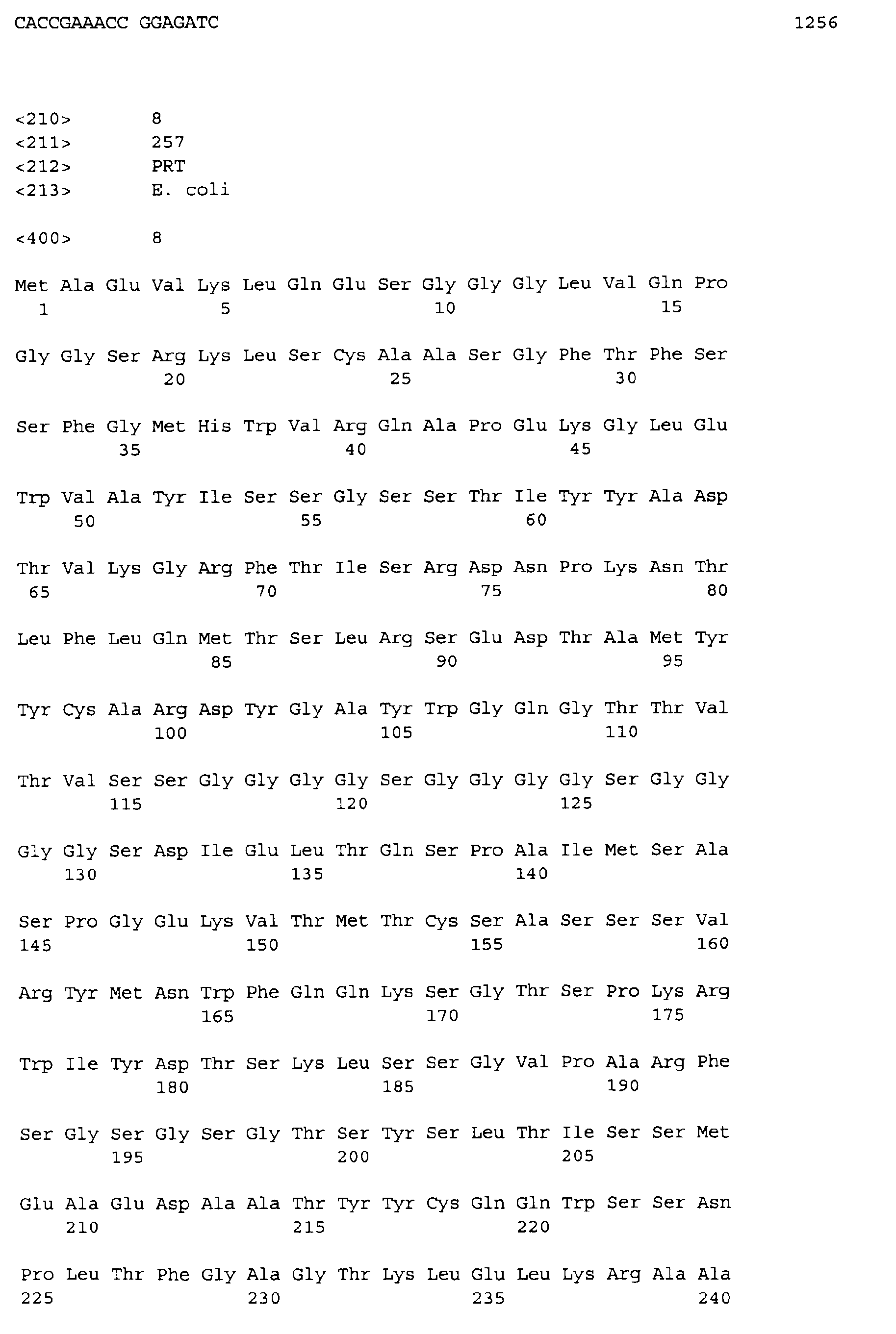
- SEQ ID NO: 7 and 8 show the sequence of the part of the expression plasmid pUBS520-ScFvOx which codes for the fusion protein from the PelB signal sequence and ScFvOxazolon, together with the regulatory sequences (promoter, terminator) which consist of pHEN-ScFv and pIN III ompA3 were amplified.
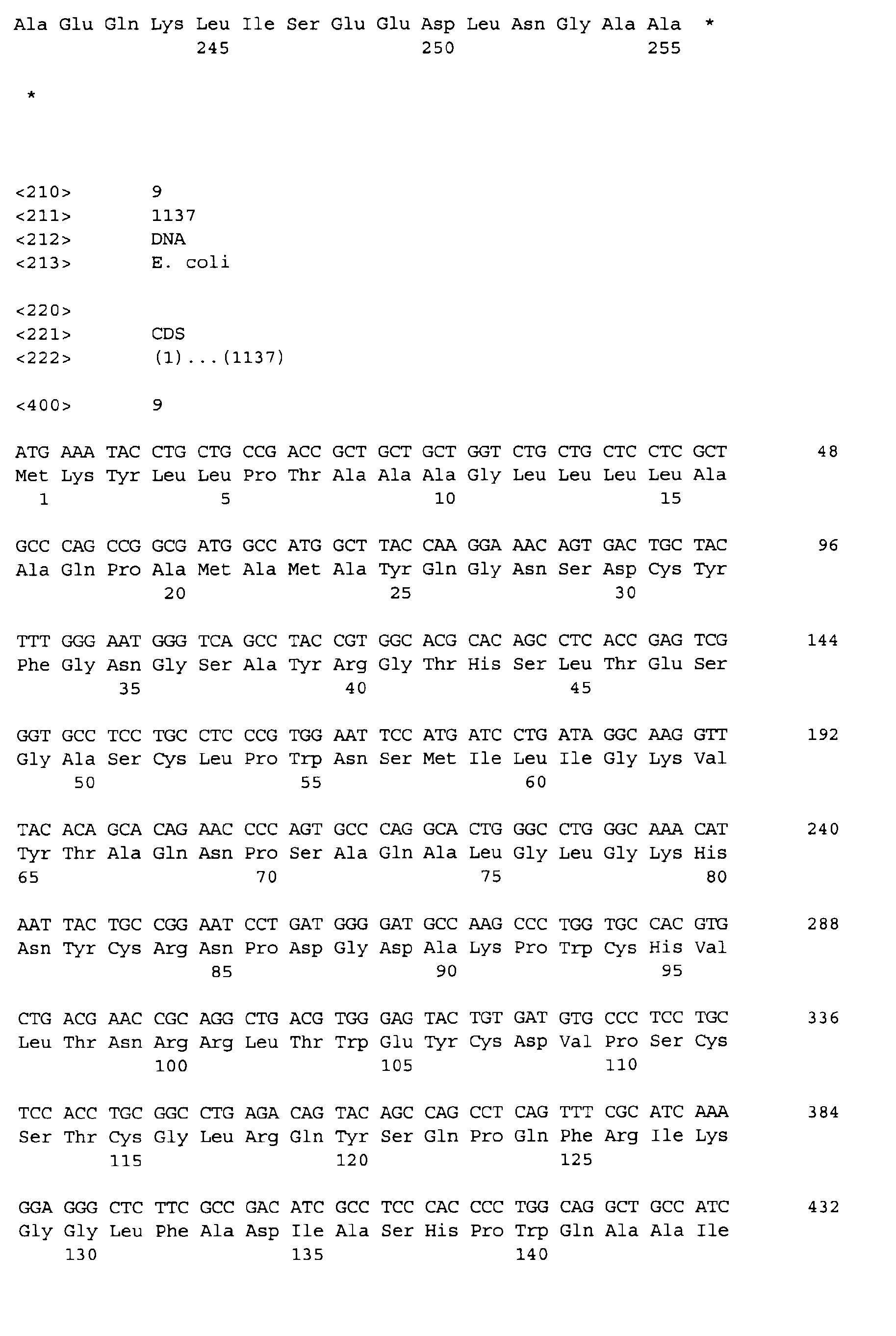
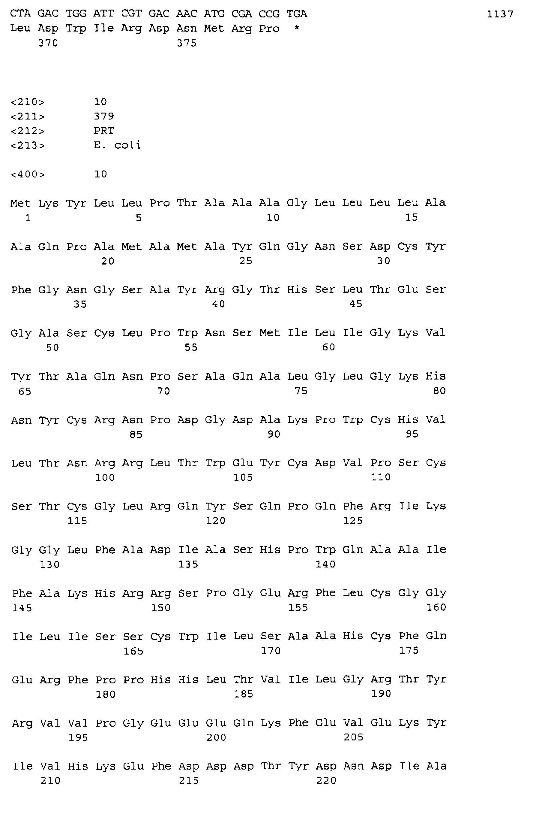
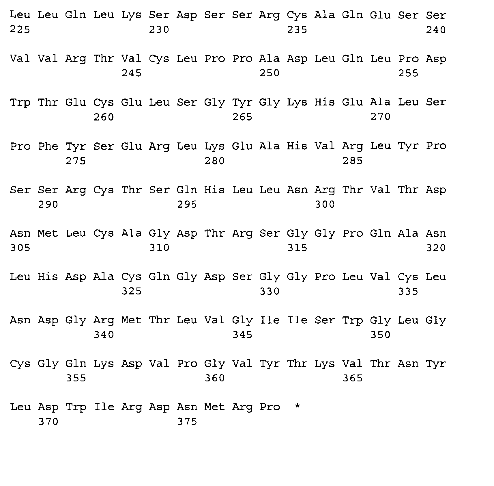
- SEQ ID NO: 9 and 10 show the sequence of the part of the expression plasmid pET20b (+) - rPA which codes for the fusion protein from the PelB signal sequence and rPA.
- FIG. 1 shows a Western blot of the limited proteolysis of periplasmic and cytosolic expressed DnaJ with 50 ⁇ g / ml trypsin for the detection of the cellular localization and the native folding.
- the molecular weight standards were plotted on the left and right.
- purified DnaJ (left) was subjected to the same procedure, but with 6.25 ⁇ g / ml trypsin.
- FIG. 2 shows a comparison of the expression of rPA in the periplasm of E. coli BL21 (DE3) with co-secretion of DnaJ and with the addition of GSH and various low molecular weight folding-improving substances to the medium.
- FIG. 3 shows a schematic representation of the expression plasmid pUBS520-pIN-dnaJ.
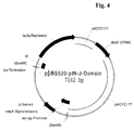
- FIG. 4 shows a schematic representation of the expression plasmid pUBS520-pIN-J-Domain.
- FIG. 5 shows a schematic representation of the expression plasmid pUBS520-pIN-hsp25.
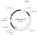
- FIG. 6 shows a schematic representation of the expression plasmid pUBS520-ScFvOx.
- FIG. 7 shows a schematic representation of the expression plasmid pET20b (+) - rPA.
- the DNA coding for these proteins has been genetically engineered to the Signal sequence of the outer membrane protein A (OmpA) from E. coli fused and the Fusion on a recombinant plasmid under the control of the lac-lpp promoter in E. coli expressed.
- OmpA outer membrane protein A
- the resulting plasmid was named pIN III ompA3-dnaJ.
- the sequence of the periplasmic expressed DnaJ differs from the wild type protein in that the polypeptide sequence starts with Gly-Ile-Pro instead of Met, es the N-terminus was thus extended by 2 amino acids. DnaJ is located thus under the control of the lac-lpp promoter, which is treated with IPTG (isopropyl- ⁇ -D-thiogalactoside) is induced.
- the region from the plasmid pIN III ompA3-dnaJ was amplified by means of PCR the lac-lpp operon, the signal sequence, the dnaJ gene and the terminator region of the Operons coded (SEQ ID NO: 1).
- the PCR product was with the restriction endonuclease BglII cut and linearized with the BamHI restriction endonuclease Vector pUBS520 cloned.
- the resulting plasmid became pUBS520-pIN-dnaJ designated (Fig. 3).
- the QuikChange mutagenesis system (Promega, Mannheim, DE) was used in the Plasmid pUBS 520-pIN-dnaJ inserted two stop codons after nucleotide 324 so that only the first 108 amino acids are expressed.
- the sequence of the mutagenized The range was determined by dideoxy sequencing (LiCor DNA Sequencer 4000, MWG Biotech, Ebersberg) and the expression of the truncated protein fragment by Western Blotting and detection with an anti-DnaJ antibody detected.
- the resulting Plasmid was designated pUBS 520-pIN-J domain (Fig. 4).
- the sequence of the periplasmic expressed Hsp25 differs from the wild-type protein in that the polypeptide sequence begins with Gly-Ile-Leu instead of Met, so the N-terminus was extended by 2 amino acids. Hsp25 is thus under the control of the lac-lpp promoter, which is induced with IPTG (isopropyl- ⁇ -D-thiogalactoside).
- the region from the plasmid pIN III ompA3-hsp25 was amplified by PCR, and the region for the lac-lpp operon, the signal sequence, the hsp25 gene and the terminator region of the Operons coded (SEQ ID NO: 5).
- the PCR product was with the restriction endonuclease BglII cut and linearized with the BamHI restriction endonuclease Vector pUBS520 cloned.
- the resulting plasmid became pUBS520-pIN-hsp25 referred to (Fig. 5).
- the region from the plasmid pHEN-ScFvOx was amplified by means of PCR, that for the lac promoter, encoding the signal sequence pelB and the scfvox gene.
- a second PCR the region from the plasmid pIN III ompA3 was amplified, that for the lpp terminator coded. The two fragments were fused in a subsequent PCR.
- the resulting PCR product (SEQ ID NO: 7) was with the restriction endonuclease BglII cut and linearized with the BamHI restriction endonuclease Vector pUBS520 cloned.
- the resulting plasmid was named pUBS520-ScFvOx (Fig. 6).
- the gene of a plasminogen activator (rPA) from the Plasmid vector pA27fd7 was amplified.
- the PCR product was digested with the restriction endonucleases NcoI and BamHI and cloned into the plasmid vector pET20b (+) (Novagen Inc., Madison, USA).
- the Plasmid codes for a fusion protein which is derived from the signal sequence of PelB (pectate lyase consists of Erwinia amylovora) and rPA and the secretion of rPA into the periplasm Dideoxy sequencing (LiCor DNA Sequencer 4000, MWG Biotech, Ebersberg, DE) checked.
- the construct was named pET20b (+) - rPA (Fig. 7).
- the T7 RNA polymerase in the Strain E. coli BL21 (DE3) is under the control of the lacUV5 promoter.
- the induction is done by adding IPTG.
- the periplasmic expressed rPA differs of the plasminogen activator described in Kohnert et al by exchanging the second amino acid (Ser) against Ala.
- E. coli BL21 (DE3), which was transformed with pET20b (+) - rPA and pUBS520-pIN-hsp25 (co-expression of Hsp25), an overnight culture of E. coli BL21 (DE3), which was transformed with pET20b (+) - rPA and pUBS520 -ScFvOx was transformed (co-expression of ScFvOx), an overnight culture of E. coli BL21 (DE3), which was transformed with pET20b (+) - rPA and pUBS520 or an overnight culture of E.
- coli BL21 (DE3), which with pET20b (+) and pUBS520 was transformed (control culture), was diluted 1:50 in 100 ml LB medium with ampicillin (100 ⁇ g / ml) and kanamycin (50 ⁇ g / ml, Fluka Chemica, Neu-Ulm, DE) and protected at 24 ° C and 170 rpm feeds.
- rPA The activity of rPA was essentially determined by the method by Verheijen et al. Thromb. Haemostasis 48 (1982) 266-269).
- the cultivation was carried out in the presence of 5 mM GSH.
- E. coli XLI-Blue cells which had been transformed with pIN III ompA3-dnaJ, were 1:50 from a stationary preculture in LB medium (11 LB medium contains 10 g of bacto-trypton (Difco Factories, Detroit, Michigan , USA), 5 g yeast extract (Difco Factories) and 5 g NaCl (Roth GmbH, Düsseldorf) diluted with 100 ⁇ g / ml ampiciline (Sigma, Deisenhofen), tightened at 37 ° C and 200 rpm and after 2.75 h (OD 600 ca.0.5) with 1 mM IPTG After 3 h of growth in the presence of the inductor, the cells were harvested by centrifugation (Eppendorf microcentrifuge,
- the spheroplasts obtained as a pellet were taken up in 30 ⁇ l 50 mM Tris / HCl, pH 8.0 with 100 mM NaCl. As a control, spheroplasts were taken up in the same buffer, but with an additional 0.1% Triton®-X-100 (Amresco, Solon, Ohio, USA).
- the secreted chaperone is protease sensitive after spheroplast preparation, it has been shown that it is localized on the periplasmic side of the inner membrane. In contrast, intracellular DnaJ is protected against proteases even after spheroplastization. If the spheroplasts were permeabilized by Triton-X-100, intracellular DnaJ is digested by trypsin. The cleavage pattern of the periplasmic expressed DnaJ is identical to that of purified, native DnaJ. This proved that the periplasmic expression product was native to this compartment.
Landscapes
- Life Sciences & Earth Sciences (AREA)
- Chemical & Material Sciences (AREA)
- Health & Medical Sciences (AREA)
- Engineering & Computer Science (AREA)
- Organic Chemistry (AREA)
- Wood Science & Technology (AREA)
- Zoology (AREA)
- Genetics & Genomics (AREA)
- Bioinformatics & Cheminformatics (AREA)
- General Engineering & Computer Science (AREA)
- Biotechnology (AREA)
- General Health & Medical Sciences (AREA)
- Biochemistry (AREA)
- Biomedical Technology (AREA)
- Microbiology (AREA)
- Molecular Biology (AREA)
- Medicinal Chemistry (AREA)
- Tropical Medicine & Parasitology (AREA)
- Virology (AREA)
- Proteomics, Peptides & Aminoacids (AREA)
- Chemical Kinetics & Catalysis (AREA)
- General Chemical & Material Sciences (AREA)
- Physics & Mathematics (AREA)
- Biophysics (AREA)
- Plant Pathology (AREA)
- Preparation Of Compounds By Using Micro-Organisms (AREA)
- Micro-Organisms Or Cultivation Processes Thereof (AREA)
- Peptides Or Proteins (AREA)
- Enzymes And Modification Thereof (AREA)
Abstract
Ein Verfahren zur Herstellung eines natürlich gefalteten eukaryontischen Polypeptids, enthaltend zwei oder mehrere über Disulfidbrücken verknüpfte Cysteine, durch a) Kultivierung prokaryontischer Zellen, wobei die genannten prokaryontischen Zellen einen Expressionsvektor enthalten, der für das genannte Polypeptid, das am N-Terminus eine prokaryontische Signalsequenz enthält, codiert, b) Sekretion des Polypeptids in das Periplasma oder das Medium, c) Abspaltung der Signalsequenz und Isolierung des Polypeptids aus dem Periplasma oder dem Medium, dadurch gekennzeichnet, daß in der genannten prokaryontischen Zelle zusätzlich eine für ein molekulares Chaperon codierende Nukleinsäure exprimiert und das Chaperon ins Periplasma sekretiert wird, ist zur rekombinanten Herstellung von Polypeptiden in Prokaryonten mit hoher Ausbeute geeignet. <IMAGE>
Description
Die Erfindung betrifft ein Verfahren zur Herstellung von wasserlöslichen, natürlich gefalteten
und sekretierten Polypeptiden nach Expression in prokaryontischen Zellen durch Co-Sekretion
von molekularen Chaperonen.
In prokaryontischen Organismen findet die Proteinsynthese, auch Translation genannt, an
den Ribosomen im Cytoplasma statt. Bei einer Expression rekombinanter DNA in prokaryontischen
Wirtsorganismen ist es oft wünschenswert, daß das dabei erhaltene rekombinante
Genprodukt bzw. Protein aus dem Cytoplasma durch die innere bakterielle
Membran in den periplasmatischen Raum zwischen innerer und äußerer Membran sekretiert
wird. Vom Periplasma können sekretierte Proteine dann z.B. durch einen osmotischen
Schock in das Nährmedium freigesetzt werden. Ein Nachteil dieses Verfahrens ist, daß die
sezernierten Polypeptide häufig nicht die native, biologisch aktive Konformation ausbilden
(Hockney, TIBTECH 12 (1994) 456 - 463).
In jüngster Zeit wurden molekulare Chaperone und Faltungskatalysatoren, wie Peptidyl-Prolyl-cis/trans-Isomerasen
oder Proteindisulfidisomerasen (Glockshuber et al., EP-A 0
510 658) eingesetzt, um die Ausbeute an nativem rekombinanten Protein bei der Faltung in
vivo zu erhöhen (Thomas et al., Appl. Biochem. Biotechnol. 66 (1997) 197-238). Dies
führte teilweise zu erheblichen Verbesserungen bei der Expression z.B. von Ribulosebisphosphat-Carboxylase
(RUBISCO; Goloubinoff et al., Nature 337 (1989) 44-47), humaner
Procollagenase (Lee & Olins, J. Biol. Chem. 267 (1992) 2849-2852) oder neuronaler
Stickstoffoxidsynthase aus Ratten (Roman et al., Proc. Natl. Acad. Sci. USA 92 (1995) 8428-8432).
In diesen Beispielen wurden GroEL/ES bzw. das DnaK-System aus E. coli im Cytosol
co-überexprimiert.
Auch bei der Sekretion rekombinanter Proteine ins Periplasma von E. coli wurde die Co-Expression
von Chaperone untersucht. Hier wurde jedoch nur eine cytosolische Überexpression
von Chaperone erprobt, um die Sekretion ins Periplasma zu optimieren (Perez-Perez
et al., Biochem. Biophys. Res. Commun. 210 (1995) 524-529; Sato et al., Biochem.
Biophys. Res. Commun. 202 (1994) 258-264; Berges et al., Appl. Environ. Microbiol. 62
(1996) 55-60). Bisherige Versuche zur Co-Sekretion in E. coli betrafen nur Faltungshelfer-Proteine,
wie z. B. Proteindisulfidisomerase (PDI; Glockshuber et al., EP-A 0 510 658) oder
Peptidyl-Prolyl-cis/trans-Isomerasen oder Dsb-Proteine (Knappik et al., Bio/Technology
11 (1993) 77-83; Qiu et al., Appl. Environm. Microbiol. 64 (1998) 4891-4896 und Schmidt
et al., Prot. Engin. 11 (1998) 601 - 607.
Aufgabe der Erfindung ist es, ein Verfahren zur Herstellung von wasserlöslichen, natürlich
gefalteten eukaryontischen Polypeptiden nach Expression in Prokaryonten zur Verfügung
zu stellen, welches auf einfache Weise durchführbar ist und bei dem eine aufwendige in
vitro-Nachbehandlung, wie Auflösung von inclusion bodies, Reduktion und Naturierung,
nicht erforderlich ist.
Die Aufgabe wird gelöst durch ein Verfahren zur Herstellung eines natürlich gefalteten
eukaryontischen Polypeptids, enthaltend zwei oder mehrere über Disulfidbrücken verknüpfte
Cysteine, durch
In einer bevorzugten Ausführungsform des erfindungsgemäßen Verfahrens werden dem
zur Kultivierung der prokaryontischen Zellen verwendeten Nährmedium (Fermentationsmedium)
zusätzlich reduzierende Thiolreagenzien, welche SH-Gruppen enthalten, zugesetzt,
wobei die Ausbeute an rekombinant gewonnenem Protein weiter erhöht wird. Vorzugsweise
werden 0,1-15 mmol/l Thiolreagenz zugesetzt. Erfindungsgemäß ist unter dem
Begriff "Thiolreagenz" entweder ein reduzierendes (reduziertes) Thiolreagenz mit
SH-Gruppen oder ein Gemisch von reduzierenden Thiolreagenzien mit SH-Gruppen und
oxidierenden Thiolreagenzien mit Disulfidgruppen zu verstehen. Bevorzugte Substanzen
sind reduziertes Glutathion (GSH), Cystein, N-Acetylcystein, Cysteamin, β-Mercaptoethanol
und ähnliche Verbindungen. Die Thiolreagenzien können sowohl einzeln als auch
in Gemischen verwendet werden. Besonders geeignet sind Thiolreagenzien wie beispielsweise
Glutathion (GSH), die eine einzige SH-Gruppe pro Molekül aufweisen. Thiolreagenzien
wie Glutathion, sind für die Verbesserung der Ausbeute nativ gefalteter Proteine bei
der Expression rekombinanter DNA in prokaryontischen Zellen bekannt (Glockshuber et
al., EP-A 0 510 658).
Unter Chaperonen gemäß der Erfindung sind Proteine zu verstehen, die andere, nicht-native
Proteine in vivo vor Aggregation schützen und die Ausbildung ihrer nativen Konformation
fördern. Molekulare Chaperone werden im Stand der Technik eingesetzt, um
Proteine zu stabilisieren und damit vor Aggregation und Inaktivierung zu schützen
(Buchner et al., EP-A 0 556 726 A1). Vorzugsweise werden erfindungsgemäß ATP-unabhängige
Chaperone des HSP40-Typs (Molmasse ca. 40 kDa) oder ein kleines Hitzeschockprotein
(sHSP) verwendet. DnaJ ist ein 40 kDa Hitzeschockprotein, das im Cytoplasma von
E. coli vorkommt und Teil des sogenannten Hsp70-Chaperonsystems ist (Bukau, B. &
Horwich, A., Cell 92 (1998) 351-366). Zu diesem System gehören außerdem DnaK (Hsp70)
und GrpE. Bestimmte Proteine werden durch das DnaK-System in einem ATP-abhängigen
Prozeß zur nativen Konformation gefaltet (Schröder et al., EMBO J. 12 (1993) 4137-4144;
Langer et al., Nature 356 (1992) 683 - 689). Zur Rückfaltung denaturierter Proteine
benötigt dieses System zusätzlich ATP. DnaJ schützt in Abwesenheit von DnaK und ATP
nicht-native Proteine vor Aggregation und vermittelt einen Faltungs-kompetenten Zustand
(Schröder et al., EMBO J. 12 (1993) 4137-4144). Weiterhin bevorzugt ist die Co-Sekretion
eines N-terminalen Fragmentes von DnaJ, das die Aminosäuren 1-108 umfaßt und im
Folgenden als "J-Domäne" (Kelley, TIBS 23 (1998) 222-227) bezeichnet wird. In diesem
Bereich befinden sich die J-Domäne und eine G/F-reiche Domäne, die Wechselwirkungen
mit DnaK ausüben (Wall et al., J. Biol. Chem. 270 (1995) 2139-2144). Es wurde gezeigt,
daß die Co-Expression von DnaJ im Cytosol zur Erhöhung der Ausbeute an löslichem
Protein führen kann (Yokoyama et al., Microbiol. Ferment. Technol. 62 (1998) 1205-1210).
Hsp25 (z.B. aus der Maus) ist ein Vertreter der kleinen Hitzeschockproteine (Gaestel et al.,
Eur. J. Biochem. 179 (1989) 209-213), einer Klasse von Chaperonen, die ubiquitär verbreitet
ist. Die Molmasse dieser Proteine liegt zwischen 15 und 30 kDa. Bei Hitzeschock werden
die sHsps in der Zelle stark angereichert (bis zu 1% des Gesamtzellproteins - Arrigo &
Landry (1994), In Morimoto (Hrsg.): The Biology of Heat Shock Proteins and Molecular
Chaperones, Cold Spring Harbour Press, 335-373). Wie DnaJ-Proteine besitzen sHsps die
Eigenschaft, die Aggregation von nichtnativen Proteinen zu verhindern und diese in einem
faltungskompetenten Zustand zu halten (Jakob et al., J. Biol. Chem. 268 (1993) 1517-1520;
Ehrsperger et al., EMBO J. 16 (1997) 221-229).
Der Begriff "Überexpression" gemäß vorliegender Erfindung bedeutet eine Steigerung der
Expression der sekretierten Chaperone wie z.B. DnaJ- und Hsp25 (vorzugsweise um
mindestens 100%) im Vergleich zur Expression im Wildtyp des jeweils verwendeten prokaryontischen
Wirtsorganismus. Eine solche Überexpression läßt sich z.B. dadurch erreichen,
daß sich die Gene (für das Protein, Chaperon und/oder Signalpeptid) unter Kontrolle
eines starken prokaryontischen, vorzugsweise induzierbaren Expressionssignals (z.B. eines
lac- oder T7-Promotors oder eines Derivates davon) befinden.
Das Sekretionskonstrukt für die Überexpression der Polypeptide (Proteine) samt regulatorischer
Regionen (Promotor und Terminator) auf der rekombinanten DNA ist vorzugsweise
in einen Vektor, welcher zusätzlich die in Prokaryonten seltene Arginin-tRNA AGA/AGG
codiert integriert oder wird mit einem Vektor, welcher diese tRNA codiert, co-exprimiert
(Brinkmann et al., Gene 85 (1989) 109-114). Dies ermöglicht sowohl die Co-Überexpression
der jeweiligen Proteine ins bakterielle Periplasma als auch die Transkription der seltenen
tRNAArg AGA/AGG, was eine erhöhte Synthese des gewünschten Proteins im bakteriellen
Wirtsorganismus zur Folge hat. Die für das Polypeptide und das Chaperon codierenden
Nukleinsäuren können auf einem Vektor oder auf zwei getrennten Vektoren lokalisiert
sein.
Unter einer prokaryontischen Signalsequenz im Sinne der Erfindung ist ein Nukleinsäurefragment
zu verstehen, welches aus Prokaryonten, vorzugsweise aus gramnegativen Bakterien,
abgeleitet ist und das Durchdringen von an das Signalpeptid gebundenen Proteinen
durch die inneren bakteriellen Membranen gewährleistet. Dadurch werden die Proteine im
Periplasma bzw. im Zellüberstand lokalisiert. Solche Signalsequenzen haben üblicherweise
eine Länge von 18 - 30 Aminosäuren und sind beispielsweise beschrieben in Murphy &
Beckwith: Export of Proteins to the Cell Envelope in Escherichia coli und in Neidhardt et
al. (Hrsg.): Escherichia coli and Salmonella, Second Edition, Vol. 1, ASM Press,
Washington, 1996, S. 967-978. Die Abspaltung von bakteriellen Signalsequenzen kann z.B.
nach einer Sequenz Ala-X-Ala stattfinden (von Heijne et al., J. Mol. Biol. 184 (1985) 99-105).
Die Struktur der bakteriellen Signalpeptidase ist beschrieben in Paetzel et al., Nature
396 (1998) 186-190. Vorzugweise werden Signalsequenzen verwendet, welche durch im
Periplasma von prokaryontischen Zellen lokalisierten Proteasen vom gewünschten Protein
wieder abgespalten werden. Alternativ kann durch Zugabe solcher Proteasen zum
Zellüberstand oder zum isolierten Protein die Abspaltung der Signalsequenz erfolgen.
Mit dem erfindungsgemäßen Verfahren kann die heterologe Expression einer Vielzahl von
eukaryontischen Proteinen wie z.B. Proteasen, Interferone, Proteinhormone, Antikörper
oder Fragmenten davon verbessert werden. Besonders geeignet ist das Verfahren für die
heterologe Herstellung von Proteinen, die im nativen Zustand mindestens zwei über eine
Disulfidbrücke verknüpfte Cysteine enthalten und dann, wenn sie N-terminal keine fusionierte
prokaryontische Signalsequenz besitzen, bei der prokaryontischen Expression als
unlösliche inclusion bodies entstehen. Besonders geeignet ist das Verfahren für Proteine,
die mehr als 5 Disulfidbrücken in nativem Zustand enthalten. Ein solches Protein ist
beispielsweise ein rekombinanter Plasminogenaktivator (im folgenden rPA genannt,
Martin et al., Cardiovasc. Drug Rev. 11 (1993) 299-311, US-Patent Nr. 5,223,256). rPA
besitzt 9 Disulfidbrücken, die im reduzierenden Cytosol von E. coli nicht ausgebildet
werden.
Dabei wird die periplasmatische Lokalisation des Proteins und des Chaperons durch
"operative Verknüpfung" mit einem Signalpeptid zum Durchdringen innerer bakterieller
Membranen gewährleistet.
Zur Gewinnung des sekretorischen rPA-Proteins in funktionaler Form in E. coli wurde das
Gen für dieses Protein aus dem Plasmid pA27fd7 (Kohnert et al., Protein Engineering 5
(1992) 93-100) mit gentechnologischen Mitteln an eine prokaryontische Signalsequenz
gramnegativer Bakterien, beispielsweise an die Signalsequenz von Pectatlyase B (PeIB) von
Erwinia amylovora, fusioniert. Die Genfüsion wurde durch Klonierung in den Vektor
pET20b(+) (Novagen Inc., Madison, USA) hergestellt. Damit unterliegt die Genexpression
der Kontrolle des T7-Promotors. Die im Fusionsprotein vorhandene Signalsequenz bewirkt
die Sekretion ins Periplasma. Während oder nach der Sekretion wird die Signalsequenz
durch eine in der inneren Membran lokalisierte Peptidase abgespalten. Sezerniertes Protein
kann dann im Periplasma falten. Die oxidierenden Bedingungen dieses Kompartiments
ermöglichen die Ausbildung von Disulfidbrücken. Durch gleichzeitige Co-Überexpression
von Chaperonen wie DnaJ, J-Domäne oder Hsp25 im Periplasma gelingt es, die Ausbeute
an funktionalen Protein um mehr als das 100-fache zu steigern.
Weitere Beispiele von erfindungsgemäß herstellbaren Polypeptiden sind Antikörper oder
Antikörperfragmente, beispielsweise ein Single-Chain Fv-Fragment (ScFv, z.B. gegen das
Schilddrüsen-stimulierende Hormon (thyroide stimulating hormone, TSH). ScFVs sind
verkürzte Antikörper, die nur aus den variablen Abschnitten (FV) der schweren und leichten
Kette eines Antikörpers bestehen, die über einen kurzen (meist Gly4Ser3) Linker künstlich
fusioniert sind (Hudson, Curr. Opin Biotechnol. 9 (1998) 395-402). ScFVs haben
normalerweise die gleiche Affinität zum Antigen wie die paternalen FV-Stränge, können
jedoch in E. coli überexprimiert werden. Da sie stabilisierende Intradomänen-Disulfidbrücken
besitzen, die essentiell für die Stabilität sind, führt eine Expression im Cytosol
meist zur Bildung von Inclusion Bodies (Übersichtsartikel: Shibui et al., Appl. Microbiol.
Biotechnol. 37 (1992) 352-357). ScFVs können durch Zufallsmutationen und anschließende
Phage-Display-Selektion auf Bindung gewünschter Antigene gezielt optimiert werden
(Übersichtsartikel: Allen et al., TIBS 20 (1995) 511-516).
Die folgenden Beispiele, Publikationen, das Sequenzprotokoll und die Abbildungen erläutern
die Erfindung, deren Schutzumfang sich aus den Patentansprüchen ergibt, weiter. Die
beschriebenen Verfahren sind als Beispiele zu verstehen, die auch noch nach Modifikationen
den Gegenstand der Erfindung beschreiben.
SEQ ID NO: 1 und 2 zeigen die Sequenz des Teils des Expressionsplasmides pUBS520-pIN-dnaJ,
der für das Fusionsprotein aus OmpA-Signalsequenz und DnaJ codiert, zusammen
mit den regulatorischen Sequenzen (Promotor, Terminator), die aus pIN III ompA3-dnaJ
amplifiziert wurden.
SEQ ID NO: 3 und 4 zeigen die Sequenz des Teils des Expressionsplasmides pUBS520-pIN-J-Domain,
der für das Fusionsprotein aus OmpA-Signalsequenz und J-Domäne codiert,
zusammen mit den regulatorischen Sequenzen (Promotor, Terminator), die aus pIN III
ompA3-dnaJ amplifiziert wurden.
SEQ ID NO: 5 und 6 zeigen die Sequenz des Teils des Expressionsplasmides pUBS520-pIN-hsp25,
der für das Fusionsprotein aus OmpA-Signalsequenz und Hsp25 codiert, zusammen
mit den regulatorischen Sequenzen (Promotor, Terminator), die aus pIN III ompA3-hsp25
amplifiziert wurden.
SEQ ID NO: 7 und 8 zeigen die Sequenz des Teils des Expressionsplasmides pUBS520-ScFvOx,
der für das Fusionsprotein aus PelB-Signalsequenz und ScFvOxazolon codiert,
zusammen mit den regulatorischen Sequenzen (Promotor, Terminator), die aus pHEN-ScFv
bzw. pIN III ompA3 amplifiziert wurden.
SEQ ID NO: 9 und 10 zeigen die Sequenz des Teils des Expressionsplasmides pET20b(+)-rPA,
der für das Fusionsprotein aus PelB-Signalsequenz und rPA codiert.
Fig. 1 zeigt einen Western Blot der limitierten Proteolyse von periplasmatisch und
cytosolisch exprimiertem DnaJ mit 50 µg/ml Trypsin zum Nachweis der zellulären Lokalisation
und der nativen Faltung. Die Molgewichtsstandards wurden links und rechts aufgetragen.
Zur Kontrolle wurde gereinigtes DnaJ (links) derselben Prozedur unterzogen,
jedoch mit 6,25 µg/ml Trypsin.
Fig. 2 zeigt einen Vergleich der Expression von rPA im Periplasma von E. coli BL21(DE3)
bei Co-Sekretion von DnaJ und bei Zusatz von GSH und verschiedenen niedermolekularen
faltungsverbessernden Stoffen zum Medium.
Fig. 3 zeigt eine schematische Darstellung des Expressionsplasmides pUBS520-pIN-dnaJ.
Fig. 4 zeigt eine schematische Darstellung des Expressionsplasmides pUBS520-pIN-J-Domain.
Fig. 5 zeigt eine schematische Darstellung des Expressionsplasmides pUBS520-pIN-hsp25.
Fig. 6 zeigt eine schematische Darstellung des Expressionsplasmides pUBS520-ScFvOx.
Fig. 7 zeigt eine schematische Darstellung des Expressionsplasmides pET20b(+)-rPA.
Zur periplasmatischen Überexpression von DnaJ, der J-Domäne sowie Hsp25 in E. coli
wurde die DNA, die für diese Proteine codiert, mit gentechnologischen Mitteln an die
Signalsequenz des Outer Membrane Proteins A (OmpA) von E. coli fusioniert und die
Fusion auf einem rekombinanten Plasmid unter Kontrolle des lac-lpp-Promotors in E. coli
exprimiert. Somit werden die Polypeptidkette von DnaJ und Hsp25 ins Periplasma des
prokaryontischen Wirtsorganismus transportiert und dort nativ gefaltet. Die Lokalisation
und native Faltung konnte dabei durch limitierte Proteolyse mit Trypsin und Western Blot
nachgewiesen werden.
Molekulargenetische Techniken beruhten auf Ausubel et al. (Hrsg.), J. Wiley & Sons, 1997,
Curr. Protocols of Molecular Biology. Oligonucleotide wurden von den Firmen MWG
Biotech, Ebersberg oder GIBCO Life Sciences, Eggenstein, DE bezogen.
Das Gen, das für DnaJ codiert, Gene Bank Accession No. M 12565, wurde über die Restriktionsschnittstellen
EcoRI und BamHI in das Expressionsplasmid pIN III ompA3 (Ghayreb
et al., EMBO J. 3 (1984) 2437-2442) kloniert. Die Sequenz des klonierten PCR-Fragments
wurde durch Didesoxy-Sequenzierung (LiCor DNA-Sequencer 4000, MWG Biotech,
Ebersberg) überprüft. Das resultierende Plasmid wurde pIN III ompA3-dnaJ bezeichnet.
Die Sequenz des periplasmatisch exprimierten DnaJ unterscheidet sich von dem Wildtyp-Protein
dahingehend, daß die Polypeptidsequenz mit Gly-Ile-Pro beginnt statt mit Met, es
fand somit eine Verlängerung des N-Terminus um 2 Aminosäuren statt. DnaJ befindet sich
damit unter Kontrolle des lac-lpp-Promotors, der mit IPTG (Isopropyl-β-D-Thiogalactosid)
induziert wird.
Mittels PCR wurde der Bereich aus dem Plasmid pIN III ompA3-dnaJ amplifiziert, der für
das lac-lpp Operon, die Signalsequenz, das dnaJ-Gen und die Terminator-Region des
Operons codiert (SEQ ID NO: 1). Das PCR-Produkt wurde mit der Restriktionsendonuclease
BglII geschnitten und in den mit der Restriktionsendonuclease BamHI linearisierten
Vektor pUBS520 kloniert. Das resultierende Plasmid wurde pUBS520-pIN-dnaJ
bezeichnet (Fig. 3).
Mittels des QuikChange-Mutagenese-Systems (Promega, Mannheim, DE) wurden im
Plasmid pUBS 520-pIN-dnaJ nach dem Nucleotid 324 zwei Stop-Codone eingefügt, so daß
nur noch die ersten 108 Aminosäuren exprimiert werden. Die Sequenz des mutagenisierten
Bereiches wurde durch Didesoxy-Sequenzierung (LiCor DNA-Sequencer 4000, MWG
Biotech, Ebersberg) und die Expression des verkürzten Proteinfragments durch Western
Blotting und Detektion mit einem Anti-DnaJ-Antikörper nachgewiesen. Das entstandene
Plasmid wurde mit pUBS 520-pIN-J-Domain (Fig. 4) bezeichnet.
Das Gen, das für Hsp25 codiert, Gene Bank Accession No.: L 07577, wurde über die
Restriktionsschnittstellen EcoRI und BamHI in das Expressionsplasmid pIN III ompA3
(Ghayreb et al., EMBO J. 3 (1984) 2437-2442) kloniert. Die Sequenz des klonierten PCR-Fragments
wurde durch Didesoxy-Sequenzierung (LiCor DNA-Sequencer 4000, MWG
Biotech, Ebersberg) überprüft. Das resultierende Plasmid wurde pIN III ompA3-hsp25
bezeichnet. Die Sequenz des periplasmatisch exprimierten Hsp25 unterscheidet sich von
dem Wildtyp-Protein dahingehend, daß die Polypeptidsequenz mit Gly-Ile-Leu beginnt
statt mit Met, es fand somit eine Verlängerung des N-Terminus um 2 Aminosäuren statt.
Hsp25 befindet sich damit unter Kontrolle des lac-lpp-Promotors, der mit IPTG (Isopropyl-β-D-Thiogalactosid)
induziert wird.
Mittels PCR wurde der Bereich aus dem Plasmid pIN III ompA3-hsp25 amplifiziert, der für
das lac-lpp Operon, die Signalsequenz, das hsp25-Gen und die Terminator-Region des
Operons codiert (SEQ ID NO: 5). Das PCR-Produkt wurde mit der Restriktionsendonuclease
BglII geschnitten und in den mit der Restriktionsendonuclease BamHI linearisierten
Vektor pUBS520 kloniert. Das resultierende Plasmid wurde pUBS520-pIN-hsp25
bezeichnet (Fig. 5).
Als negativ-Kontrolle wurde die Co-Expression eines Single-Chain-Fv-Fragmentes, das
gegen das Hapten Oxazolon gerichtet ist (ScFvOxazolon; Fiedler und Conrad,
Bio/Technology 13 (1995) 1090-1093 untersucht, das keine Chaperon-Eigenschaften
besitzt.
Mittels PCR wurde der Bereich aus dem Plasmid pHEN-ScFvOx amplifiziert, der für den
lac-Promotor, die Signalsequenz pelB und das scfvox-Gen codiert. In einer zweiten PCR
wurde der Bereich aus dem Plasmid pIN III ompA3 amplifiziert, der für den lpp-Terminator
codiert. In einer anschließenden PCR wurden die beiden Fragmente fusioniert. Das
so entstandene PCR-Produkt (SEQ ID NO: 7) wurde mit der Restriktionsendonuclease
BglII geschnitten und in den mit der Restriktionsendonuclease BamHI linearisierten
Vektor pUBS520 kloniert. Das resultierende Plasmid wurde pUBS520-ScFvOx bezeichnet
(Fig. 6).
Mit Hilfe der PCR-Methode wurde das Gen eines Plasminogenaktivators (rPA) aus dem
Plasmidvektor pA27fd7 (Kohnert et al., Protein Engineering 5 (1992) 93-100) amplifiziert.
Das PCR-Produkt wurde mit den Restriktionsendonucleasen NcoI und BamHI gespalten
und in den Plasmidvektor pET20b(+) (Novagen Inc., Madison, USA) kloniert. Das
Plasmid codiert für ein Fusionsprotein, welches aus der Signalsequenz von PelB (Pectatlyase
aus Erwinia amylovora) und rPA besteht und die Sekretion von rPA ins Periplasma
Didesoxy-Sequenzierung (LiCor DNA-Sequencer 4000, MWG Biotech, Ebersberg, DE)
überprüft. Das Konstrukt wurde als pET20b(+)-rPA bezeichnet (Fig. 7). In dem Plasmid
wird rPA unter Kontrolle des T7-Promotors exprimiert, wobei die T7-RNA-Polymerase im
Stamm E. coli BL21(DE3) der Kontrolle des lacUV5-Promotors unterliegt. Die Induktion
erfolgt durch Zugabe von IPTG. Das periplasmatisch exprimierte rPA unterscheidet sich
von dem bei Kohnert et al beschriebenen Plasminogenaktivator durch Austausch der
zweiten Aminosäure (Ser) gegen Ala.
Eine stationäre Übernachtkultur von E. coli BL21(DE3) (Studier & Moffat, J. Mol. Biol. 189
(1986) 113-130), die mit pET20b(+)-rPA und pUBS520-pIN-dnaJ transformiert wurde
(Co-Expression von DnaJ), eine Übernachtkultur von E. coli BL21(DE3), die mit
pET20b(+)-rPA und pUBS520-pIN-J-Domain transformiert wurde (Co-Expression der J-Domäne),
eine Übernachtkultur von E. coli BL21(DE3), die mit pET20b(+)-rPA und
pUBS520-pIN-hsp25 transformiert wurde (Co-Expression von Hsp25), eine Übernachtkultur
von E. coli BL21(DE3), die mit pET20b(+)-rPA und pUBS520-ScFvOx transformiert
wurde (Co-Expression von ScFvOx), eine Übernachtkultur von E. coli BL21(DE3), die mit
pET20b(+)-rPA und pUBS520 transformiert wurde bzw. eine Übernachtkultur von E. coli
BL21(DE3), die mit pET20b(+) und pUBS520 transformiert wurde (Kontrollkultur),
wurde im Verhältnis 1:50 in 100 ml LB-Medium mit Ampicillin (100 µg/ml) und
Kanamycin (50 µg/ml, Fluka Chemica, Neu-Ulm, DE) verdünnt und bei 24°C und 170 rpm
geschüttelt. Nach 3 h Wachstum wurden je 5ml der Kultur zu je 10 ml LB-Medium mit o.g.
Mengen an Ampicillin und Kanamycin und verschiedenen Konzentrationen von GSH (0-10
mM, Fluka, DE) gegeben und mit jeweils 1mM IPTG (Isopropyl-β-D-Thiogalactosid,
AppliChem, Darmstadt, DE) induziert. Die Zellen wurden weitere 21 h bei 24 °C und 170
rpm geschüttelt und nach Bestimmung der OD600 eine 1 ml-Probe genommen. Diese 1ml-Zellproben
wurden nach einer modifizierten Vorschrift nach Jacobi et al. (J. Biol. Chem.
272 (1997) 21692-21699) in 2 ml-Eppendorf-Reaktionsgefäßen fraktioniert. Im Detail
wurde das Zellpellet mit 500 µl Fraktionierungspuffer (150 mM NaCl (Roth GmbH), 50
mM Tris/HCl (Roth GmbH, 5mM EDTA (Biomol) und 1 mg/ml Polymyxin-B-Sulfat
(Sigma), pH 7,5) versetzt, 1 h bei 10 °C auf einem Eppendorf-Thermoschüttler bei 1400
rpm geschüttelt und dann 15 min bei 14 000 rpm in einer auf 10°C gekühlten Eppendorf-Mikrozentrifuge
zentrifugiert, so daß eine Fraktion mit den löslichen periplasmatischen
Proteinen (Überstand) und eine Restfraktion (Pellet) entstand.
Die Bestimmung von der Aktivität von rPA erfolgte im wesentlichen nach der Methode
von Verheijen et al. Thromb. Haemostasis 48 (1982) 266-269).
Alle ermittelten rPA-Konzentrationen in den Zellextrakten wurden auf Zellsuspensionen
von OD600=1 normiert, um den Fehler, der bei der Messung in verschiedenen Puffern auftritt,
zu korrigieren. Die Ergebnisse zeigt Tabelle 1.
| Effekt von L-Arginin im Fermentationsmedium auf die Ausbildung von nativem rPA im Periplasma | ||
| Co-sekretiertes Protein | rPA in ng/ml*OD600 | Stimulationsfaktor |
| - | 0,030 ± 0,001 | 29 |
| DnaJ | 0,197 ± 0,019 | 29 |
| J-Domäne | 0,339 ± 0,007 | 16 |
| Hsp25 | 0,053 ± 0,002 | 27 |
| ScFvOxazolon (Kontrolle) | 0,041 ± 0,003 | 13 |
Die Kultivierung erfolgte in Gegenwart von 5 mM GSH.
Um die periplasmatische Lokalisierung und korrekte Faltung von DnaJ, das mittels pIN III
ompA3-dnaJ ins Periplasma sekretiert wurde, zu überprüfen, wurden Sphäroplasten präpariert.
Dazu wurden E. coli XLI-Blue-Zellen, die mit pIN III ompA3-dnaJ transformiert
waren, 1:50 aus einer stationären Vorkultur in LB-Medium (11 LB-Medium enthält 10 g
Bacto-Trypton (Difco Factories, Detroit, Michigan, USA), 5 g Yeast-Extract (Difco
Factories) und 5 g NaCl (Roth GmbH, Karlsruhe) mit 100 µg/ml Ampicilin (Sigma,
Deisenhofen) verdünnt, bei 37°C und 200 rpm angezogen und nach 2,75 h (OD600 ca.0,5)
mit 1 mM IPTG induziert. Nach 3 h Wachstum in Anwesenheit des Induktors wurden die
Zellen mittels Zentrifugation (Eppendorf-Mikrozentrifuge, 5000 rpm, 5 min) geerntet. Als
Kontrolle wurde ein E. coli-Stamm, der ein Plasmid zur intrazellulären Überexpression von
DnaJ enthält, kultiviert und 3 h induziert. Aus den nach Zentrifugation erhaltenen
Zellpellets wurden Sphäroplasten folgendermaßen präpariert:
Das Äquivalent von 2 ml Bakterien, die einer OD600 von 1 entsprachen, wurde in einer
0,5 M Sucrose (ICN, Biomedicals, Eschwege)/0,2 M Tris/HCl/0,5 mM EDTA-(Biomol
Feinchemikalien, Hamburg) Lösung (pH 8), mit 2 µg/ml Lysozym (Sigma, Deisenhofen)
resuspendiert und 30 min auf Eis inkubiert. Danach wurden die Proben 5 min bei 15000
rpm in einer Eppendorf-Mikrozentrifuge pelletiert und mit 100 µl TE-Puffer (10 mM
Tris/HCl (Roth GmbH), 1 mM EDTA, pH 8) gewaschen. Die als Pellet anfallenden
Sphäroplasten wurden in 30 µl 50 mM Tris/HCl, pH 8,0 mit 100 mM NaCl aufgenommen.
Als Kontrolle wurden Sphäroplasten in demselben Puffer, jedoch mit zusätzlich 0,1 %
Triton®-X-100 (Amresco, Solon, Ohio, USA), aufgenommen. Für eine anschließende limitierte
Proteolyse mit Trypsin wurden 15 µl der jeweiligen Sphäroplastenpräparation (mit
bzw. ohne Triton®-X-100) mit 2 µl 1mg/ml Trypsin (Roche Diagnostics GmbH, DE) und
23 µl 50 mM Tris/HCl, pH 8,0 mit 100 mM NaCl gemischt und bei 20°C inkubiert. Nach 0,
5 und 30 Minuten wurden je 8 µl Proben genommen, mit 2 µl 4 mg/ml Soybean-Trypsin-Inhibitor
und 3 µl SDS-PAGE -Auftragspuffer (4% Glycerin (Sigma, Deisenhofen), 0,5 %
SDS (ICN), 2% Mercaptoethanol (Sigma), 0,0625 M Tris/HCl, pH 6,8 und Bromphenolblau
(Sigma)) versetzt und 5 min gekocht. In einem Kontrollversuch wurden 2 µg
gereinigtes DnaJ (2 µg/µl) mit 1 µl 100µg/ml Trypsin und 14 µl 50 mM Tris/HCl, pH 8,0
mit 100 mM NaCl gemischt, bei 20°C inkubiert und die Proteolyse zu den angegebenen
Zeitpunkten beendet. Die Proteolyseprodukte wurden durch SDS-PAGE nach Lämmli et
al., Nature 227 (1970) 680-685) aufgetrennt. Die aufgetrennten Proteine wurden auf
Nitrocellulosemembranen (BioRad Laboratories, München) übertragen (Khyse-Anderson,
J. Biochem. Biophys. Methods 10 (1984) 203-207; Towbin et al., Proc. Natl. Acad. Sci. USA
79 (1979) 267-271). Die Membranen wurden mit TBS-5% Milchpulver (Glücksklee, Nestlé
Frankfurt) über Nacht geblockt und im Folgenden 2h mit Anti-DnaJ-Antikörper in TBS-5%
Milchpulver für 2 h dekoriert. Nach 3 Waschschritten für jeweils 5 min in TBS wurde
mit einem weiteren Antikörper (Anti-Rabbit-IgG-Peroxidase, Amersham Life Sciences,
Braunschweig) in TBS-5% Milchpulver für 1,5 h inkubiert und wiederum 5x mit TBS-Puffer
gewaschen. Zur Detektion wurde das ECL-Western-Blotting-Detection-Kit der
Firma Amersham verwendet. Das Ergebnis ist in Fig. 1 gezeigt. Da das sekretierte Chaperon
nach Sphäroplastenpräparation Protease-sensitiv ist, wurde nachgewiesen, daß es auf der
periplasmatischen Seite der inneren Membran lokalisiert ist. Im Gegensatz dazu ist intrazelluläres
DnaJ auch nach der Sphäroplastierung proteasegeschützt. Wurden die
Sphäroplasten durch Triton-X-100 permeabilisiert, wird intrazelluläres DnaJ durch Trypsin
verdaut. Das Spaltmuster des periplasmatisch exprimierten DnaJ ist identisch zu dem von
gereinigtem, nativem DnaJ. Damit war nachgewiesen, daß das periplasmatische Expressionsprodukt
in diesem Kompartiment nativ vorliegt.





Claims (10)
- Verfahren zur Herstellung eines natürlich gefalteten eukaryontischen Polypeptids, enthaltend zwei oder mehrere über Disulfidbrücken verknüpfte Cysteine, durchdadurch gekennzeichnet, daß in der genannten prokaryontischen Zelle zusätzlich eine für ein molekulares Chaperon codierende Nukleinsäure exprimiert und das Chaperon ins Periplasma sekretiert wird.a) Kultivierung prokaryontischer Zellen, wobei die genannten prokaryontischen Zellen einen Expressionsvektor enthalten, der für das genannte Polypeptid, das am N-Terminus eine prokaryontische Signalsequenz enthält, codiert,b) Sekretion des Polypeptids in das Periplasma oder das Medium,c) Abspaltung der Signalsequenz und Isolierung des Polypeptids aus dem Periplasma oder dem Medium,
- Verfahren nach Anspruch 1, dadurch gekennzeichnet, daß als Chaperon DNAJ oder HSP25 verwendet wird.
- Verfahren nach den Ansprüchen 1 bis 2, dadurch gekennzeichnet, daß dem Nährmedium ein reduzierendes Thiolreagenz zugesetzt wird.
- Verfahren nach Anspruch 3, dadurch gekennzeichnet, daß als reduzierendes Thiolreagenz Glutathion (GSH) verwendet wird.
- Verfahren nach den Ansprüchen 1 bis 4, dadurch gekennzeichnet, daß die Signalsequenz aus gramnegativen Bakterien stammt.
- Verfahren nach einem der Ansprüche 1 bis 5, dadurch gekennzeichnet, daß die für das molekulare Chaperon codierende Nukleinsäure und die für das Polypeptid codierende Nukleinsäure auf zwei getrennten Vektoren lokalisiert sind.
- Verfahren nach einem der Ansprüche 1 bis 5, dadurch gekennzeichnet, daß die für das molekulare Chaperon codierende Nukleinsäure auf dem Expressionsvektor, welcher auch die für das Polypeptide codierende Nukleinsäure enthält, lokalisiert ist.
- Verfahren nach einem der Ansprüche 6 oder 7, dadurch gekennzeichnet, daß die für das molekulare Chaperon codierende rekombinante DNA in operativer Verknüpfung mit einem DNA-Fragment steht, das ein Signalpeptid zum Durchdringen der inneren bakteriellen Membran codiert.
- Verfahren nach einem der Ansprüche 6 bis 8, dadurch gekennzeichnet, daß sich die für das sekretierte molekulare Chaperon und/oder für das sekretierte Protein codierende DNA unter Kontrolle eines induzierbaren Expressionssignals befindet.
- Verfahren nach einem der Ansprüche 1 bis 9, dadurch gekennzeichnet, daß das Polypeptid ein Antikörper, Antikörperfragment, Interferon, Proteinhormon oder eine Protease ist.
Priority Applications (14)
| Application Number | Priority Date | Filing Date | Title |
|---|---|---|---|
| EP99114811A EP1077263A1 (de) | 1999-07-29 | 1999-07-29 | Verfahren zur Herstellung von natürlich gefalteten und sekretierten Proteinen durch Co-Sekretion von Chaperonen |
| US09/618,869 US6455279B1 (en) | 1999-07-29 | 2000-07-19 | Process for the production of naturally folded and secreted proteins by co-secretion of molecular chaperones |
| ES00115839T ES2247990T3 (es) | 1999-07-29 | 2000-07-24 | Procedimiento para la produccion de proteinas plegadas naturalmente y secretadas mediante co-secrecion de chaperones moleculares. |
| AT00115839T ATE299942T1 (de) | 1999-07-29 | 2000-07-24 | Verfahren zur herstellung von natürlich gefalteten proteinen durch co-sekretion von molekularen chaperonen |
| EP00115839A EP1077262B1 (de) | 1999-07-29 | 2000-07-24 | Verfahren zur Herstellung von natürlich gefalteten Proteinen durch Co-Sekretion von molekularen Chaperonen |
| DE60021318T DE60021318T2 (de) | 1999-07-29 | 2000-07-24 | Verfahren zur Herstellung von natürlich gefalteten Proteinen durch Co-Sekretion von molekularen Chaperonen |
| DK00115839T DK1077262T3 (da) | 1999-07-29 | 2000-07-24 | Fremgangsmåde til fremstilling af naturligt foldede og secernerede proteiner ved co-sekretion af molekylære chaperoner |
| JO2000125A JO2309B1 (en) | 1999-07-29 | 2000-07-25 | Method for the production of folded and naturally sorted proteins by the collective secretion of molecular spheres |
| CA2313248A CA2313248C (en) | 1999-07-29 | 2000-07-26 | Process for the production of naturally folded and secreted proteins by co-secretion of molecular chaperones |
| CNB001215000A CN1231595C (zh) | 1999-07-29 | 2000-07-26 | 通过分子伴侣的共分泌制备天然折叠的和分泌的蛋白质的方法 |
| IN586MA2000 IN190327B (de) | 1999-07-29 | 2000-07-26 | |
| MXPA00007339A MXPA00007339A (es) | 1999-07-29 | 2000-07-27 | PROCESO PARA LA PRODUCCION DE PROTEINAS PLEGADAS Y SEGREGADAS DE FORMA NATURAL POR LA CO-SECRECION DE ACOMPAnANTES MOLECULARES. |
| KR10-2000-0043241A KR100378325B1 (ko) | 1999-07-29 | 2000-07-27 | 분자 샤프론을 공동분비시켜 자연적으로 폴딩되고분비되는 단백질을 생산하는 방법 |
| JP2000231804A JP2001061487A (ja) | 1999-07-29 | 2000-07-31 | 分子シャペロンの共分泌による自然に折り畳まれた分泌タンパク質の製造法 |
Applications Claiming Priority (1)
| Application Number | Priority Date | Filing Date | Title |
|---|---|---|---|
| EP99114811A EP1077263A1 (de) | 1999-07-29 | 1999-07-29 | Verfahren zur Herstellung von natürlich gefalteten und sekretierten Proteinen durch Co-Sekretion von Chaperonen |
Publications (1)
| Publication Number | Publication Date |
|---|---|
| EP1077263A1 true EP1077263A1 (de) | 2001-02-21 |
Family
ID=8238680
Family Applications (2)
| Application Number | Title | Priority Date | Filing Date |
|---|---|---|---|
| EP99114811A Withdrawn EP1077263A1 (de) | 1999-07-29 | 1999-07-29 | Verfahren zur Herstellung von natürlich gefalteten und sekretierten Proteinen durch Co-Sekretion von Chaperonen |
| EP00115839A Expired - Lifetime EP1077262B1 (de) | 1999-07-29 | 2000-07-24 | Verfahren zur Herstellung von natürlich gefalteten Proteinen durch Co-Sekretion von molekularen Chaperonen |
Family Applications After (1)
| Application Number | Title | Priority Date | Filing Date |
|---|---|---|---|
| EP00115839A Expired - Lifetime EP1077262B1 (de) | 1999-07-29 | 2000-07-24 | Verfahren zur Herstellung von natürlich gefalteten Proteinen durch Co-Sekretion von molekularen Chaperonen |
Country Status (13)
| Country | Link |
|---|---|
| US (1) | US6455279B1 (de) |
| EP (2) | EP1077263A1 (de) |
| JP (1) | JP2001061487A (de) |
| KR (1) | KR100378325B1 (de) |
| CN (1) | CN1231595C (de) |
| AT (1) | ATE299942T1 (de) |
| CA (1) | CA2313248C (de) |
| DE (1) | DE60021318T2 (de) |
| DK (1) | DK1077262T3 (de) |
| ES (1) | ES2247990T3 (de) |
| IN (1) | IN190327B (de) |
| JO (1) | JO2309B1 (de) |
| MX (1) | MXPA00007339A (de) |
Cited By (4)
| Publication number | Priority date | Publication date | Assignee | Title |
|---|---|---|---|---|
| WO2002040650A3 (en) * | 2000-11-14 | 2003-12-31 | Boehringer Ingelheim Int | Methods for large scale production of recombinant dna-derived tpa or k2s molecules |
| WO2003025116A3 (de) * | 2001-09-17 | 2004-03-11 | Roche Diagnostics Gmbh | Verfahren zur erhöhung der löslichkeit, der expressionsrate und der aktivität von proteinen während der rekombinanten herstellung |
| US6955910B2 (en) | 2000-11-14 | 2005-10-18 | Boehringer Ingelheim International Gmbh | Method for large scale production of recombinant DNA-derived TPA or K2S molecules |
| US7087412B2 (en) | 2000-11-14 | 2006-08-08 | Boehringer Ingelheim International Gmbh | Methods for large scale protein production in prokaryotes |
Families Citing this family (35)
| Publication number | Priority date | Publication date | Assignee | Title |
|---|---|---|---|---|
| CA2414775A1 (en) * | 2000-05-03 | 2001-11-08 | Expressive Constructs, Inc. | A method and device for improving protein stability and solubility |
| US6979556B2 (en) * | 2000-12-14 | 2005-12-27 | Genentech, Inc. | Separate-cistron contructs for secretion of aglycosylated antibodies from prokaryotes |
| US7094884B2 (en) | 2001-06-22 | 2006-08-22 | Roche Diagnostics Corporation | Soluble complexes of amylod β peptide and peptidyl prolyl isomerase chaperone and methods of making and using them |
| US7094757B2 (en) | 2001-06-22 | 2006-08-22 | Roche Diagnostics Corporation | Complexes comprising a prion protein and a peptidyl prolyl isomerase chaperone, and method for producing and using them |
| US6962982B2 (en) | 2001-06-22 | 2005-11-08 | Roche Diagnostics Corporation | Soluble complexes of target proteins and peptidyl prolyl isomerase chaperones and methods of making and using them |
| ATE522603T1 (de) * | 2001-06-22 | 2011-09-15 | Hoffmann La Roche | Löslicher komplex, der retrovirale oberflächen- glykoproteine und fkpa oder slyd enthält |
| KR100441755B1 (ko) * | 2001-07-24 | 2004-08-02 | 재단법인서울대학교산학협력재단 | 분자 샤페론을 핵 내부로 이동시키는 방법 |
| EP1427744B1 (de) | 2001-08-27 | 2007-12-26 | Genentech, Inc. | System zur antikörperexpression und- synthese |
| MXPA05004677A (es) * | 2002-10-31 | 2005-11-17 | Genentech Inc | Metodos y composiciones para aumentar la produccion de anticuerpos. |
| WO2004065417A2 (en) * | 2003-01-23 | 2004-08-05 | Genentech, Inc. | Methods for producing humanized antibodies and improving yield of antibodies or antigen binding fragments in cell culture |
| EP1624886A2 (de) * | 2003-05-12 | 2006-02-15 | Expressive Constructs, Inc. | Verfahren zur erhöhung der zell- und gewebevariabilität |
| AU2004273791A1 (en) * | 2003-09-05 | 2005-03-31 | Genentech, Inc. | Antibodies with altered effector functions |
| US9057061B2 (en) * | 2003-12-23 | 2015-06-16 | Novozymes Biopharma Dk A/S | Gene expression technique |
| GB0329681D0 (en) * | 2003-12-23 | 2004-01-28 | Delta Biotechnology Ltd | Gene expression technique |
| WO2006028936A2 (en) * | 2004-09-02 | 2006-03-16 | Genentech, Inc. | Heteromultimeric molecules |
| CN101180308A (zh) * | 2005-05-23 | 2008-05-14 | 韩国科学技术院 | 利用sHSPs制备目的蛋白的方法 |
| GB0512707D0 (en) * | 2005-06-22 | 2005-07-27 | Delta Biotechnology Ltd | Gene expression technique |
| DK2102239T3 (da) | 2006-11-30 | 2012-05-29 | Res Dev Foundation | Forbedrede immunoglobulin-biblioteker |
| CN101294164B (zh) * | 2007-04-23 | 2013-01-16 | 南京大学 | 重组人中期因子(rh-Midkine)的表达及其单克隆抗体的制备和应用 |
| US8629245B2 (en) * | 2007-05-01 | 2014-01-14 | Research Development Foundation | Immunoglobulin Fc libraries |
| CN101307310B (zh) * | 2007-05-15 | 2012-08-08 | 康泰生医科技股份有限公司 | 具有提高产率及免疫原性的重组蛋白质表达系统 |
| CN102549016B (zh) * | 2009-06-30 | 2015-05-06 | 研究发展基金会 | 免疫球蛋白fc多肽 |
| CA2826467C (en) | 2011-02-07 | 2019-11-12 | Research Development Foundation | Engineered immunoglobulin fc polypeptides |
| WO2014054744A1 (ja) * | 2012-10-03 | 2014-04-10 | 協和発酵キリン株式会社 | 培養液にアミノ酸を添加することによるポリペプチドの還元防止方法 |
| EP2929045B1 (de) | 2012-12-05 | 2020-12-02 | SOLA Biosciences LLC | Proteinexpressionsverstärkende polypeptide |
| JP6724023B2 (ja) | 2015-02-09 | 2020-07-15 | リサーチ ディベロップメント ファウンデーション | 改善された補体活性化を示す操作された免疫グロブリンfcポリペプチド |
| CN105296651B (zh) * | 2015-11-26 | 2018-07-06 | 天津师范大学 | 采用荧光RT-PCR技术检测周氏啮小蜂Hsp90基因表达的方法 |
| CN105296654B (zh) * | 2015-11-26 | 2018-07-06 | 天津师范大学 | 采用荧光RT-PCR技术检测周氏啮小蜂Hsp60基因表达的方法 |
| CN105296653B (zh) * | 2015-11-26 | 2018-07-06 | 天津师范大学 | 采用荧光RT-PCR技术检测周氏啮小蜂Hsp83基因表达的方法 |
| CN105296652B (zh) * | 2015-11-26 | 2018-07-06 | 天津师范大学 | 采用荧光RT-PCR技术检测周氏啮小蜂Hsp40基因表达的方法 |
| CN110734480B (zh) * | 2019-11-07 | 2021-08-24 | 中国科学院遗传与发育生物学研究所 | 大肠杆菌分子伴侣GroEL/ES在协助合成植物Rubisco中的应用 |
| US10973908B1 (en) | 2020-05-14 | 2021-04-13 | David Gordon Bermudes | Expression of SARS-CoV-2 spike protein receptor binding domain in attenuated salmonella as a vaccine |
| US12537071B1 (en) | 2020-07-22 | 2026-01-27 | David Gordon Bermudes | Bacteria having boolean control pathways expressing therapeutic proteins including immunotherapeutic cytotoxins |
| CN112239760B (zh) * | 2020-09-18 | 2022-02-11 | 深圳科兴药业有限公司 | 高效表达重组hGH的重组工程菌及构建方法和应用 |
| CN112940955B (zh) * | 2021-04-18 | 2022-10-18 | 江南大学 | 一种高效合成乳铁蛋白的毕赤酵母及其构建方法与应用 |
Citations (8)
| Publication number | Priority date | Publication date | Assignee | Title |
|---|---|---|---|---|
| EP0219874A2 (de) * | 1985-10-23 | 1987-04-29 | Roche Diagnostics GmbH | Verfahren zur Aktivierung von gentechnologisch hergestellten, heterologen, Disulfidbrücken aufweisenden eukaryontischen Proteinen nach Expression in Prokaryonten |
| US4757013A (en) * | 1983-07-25 | 1988-07-12 | The Research Foundation Of State University Of New York | Cloning vehicles for polypeptide expression in microbial hosts |
| WO1989006283A1 (en) * | 1988-01-11 | 1989-07-13 | Ingene (International Genetic Engineering, Inc.) | Novel plasmid vector with pectate lyase signal sequence |
| EP0510658A2 (de) * | 1991-04-26 | 1992-10-28 | Roche Diagnostics GmbH | Verbesserung der Ausbeute bei der Sekretion von disulfidverbrückten Proteinen |
| WO1996014422A1 (en) * | 1994-11-03 | 1996-05-17 | Genentech, Inc. | Process for bacterial production of heterologous polypeptides with disulphide bonds |
| EP0774512A2 (de) * | 1995-09-14 | 1997-05-21 | Tadayuki Imanaka | Methode zur Herstellung von Protein mittels einem molekularen Chaperon |
| WO1998018946A1 (en) * | 1996-10-31 | 1998-05-07 | Genentech, Inc. | Process for bacterial production of polypeptides |
| EP0885967A2 (de) * | 1997-06-20 | 1998-12-23 | Hsp Research Institute, Inc. | Expressionsplasmide für Chaperon (Molekulärkappe) |
Family Cites Families (7)
| Publication number | Priority date | Publication date | Assignee | Title |
|---|---|---|---|---|
| US5453363A (en) | 1985-10-23 | 1995-09-26 | Boehringer Mannheim Gmbh | Process for the activation of t-PA or Ing after genetic expression in prokaryotes |
| DE3611817A1 (de) | 1986-04-08 | 1987-10-15 | Boehringer Mannheim Gmbh | Verfahren zur renaturierung von proteinen |
| DE3835350A1 (de) * | 1988-10-17 | 1990-04-19 | Boehringer Mannheim Gmbh | Aktivierung von gentechnologisch hergestellten, in prokaryonten exprimierten antikoerpern |
| US5824502A (en) * | 1996-03-28 | 1998-10-20 | Mitsui Chemicals, Inc. | Method for secretory production of protein |
| US6083715A (en) * | 1997-06-09 | 2000-07-04 | Board Of Regents, The University Of Texas System | Methods for producing heterologous disulfide bond-containing polypeptides in bacterial cells |
| EP1048732A1 (de) | 1999-04-26 | 2000-11-02 | F. Hoffmann-La Roche Ag | Verfahren zur Herstellung von natürlich gefalteten und sekretierten Proteinen |
| FI109361B (fi) * | 1999-05-04 | 2002-07-15 | Korpela Timo | Mikrobinen proteiinin ilmentämisjärjestelmä |
-
1999
- 1999-07-29 EP EP99114811A patent/EP1077263A1/de not_active Withdrawn
-
2000
- 2000-07-19 US US09/618,869 patent/US6455279B1/en not_active Expired - Lifetime
- 2000-07-24 EP EP00115839A patent/EP1077262B1/de not_active Expired - Lifetime
- 2000-07-24 AT AT00115839T patent/ATE299942T1/de active
- 2000-07-24 DE DE60021318T patent/DE60021318T2/de not_active Expired - Lifetime
- 2000-07-24 ES ES00115839T patent/ES2247990T3/es not_active Expired - Lifetime
- 2000-07-24 DK DK00115839T patent/DK1077262T3/da active
- 2000-07-25 JO JO2000125A patent/JO2309B1/en active
- 2000-07-26 CN CNB001215000A patent/CN1231595C/zh not_active Expired - Lifetime
- 2000-07-26 CA CA2313248A patent/CA2313248C/en not_active Expired - Lifetime
- 2000-07-26 IN IN586MA2000 patent/IN190327B/en unknown
- 2000-07-27 MX MXPA00007339A patent/MXPA00007339A/es active IP Right Grant
- 2000-07-27 KR KR10-2000-0043241A patent/KR100378325B1/ko not_active Expired - Lifetime
- 2000-07-31 JP JP2000231804A patent/JP2001061487A/ja active Pending
Patent Citations (8)
| Publication number | Priority date | Publication date | Assignee | Title |
|---|---|---|---|---|
| US4757013A (en) * | 1983-07-25 | 1988-07-12 | The Research Foundation Of State University Of New York | Cloning vehicles for polypeptide expression in microbial hosts |
| EP0219874A2 (de) * | 1985-10-23 | 1987-04-29 | Roche Diagnostics GmbH | Verfahren zur Aktivierung von gentechnologisch hergestellten, heterologen, Disulfidbrücken aufweisenden eukaryontischen Proteinen nach Expression in Prokaryonten |
| WO1989006283A1 (en) * | 1988-01-11 | 1989-07-13 | Ingene (International Genetic Engineering, Inc.) | Novel plasmid vector with pectate lyase signal sequence |
| EP0510658A2 (de) * | 1991-04-26 | 1992-10-28 | Roche Diagnostics GmbH | Verbesserung der Ausbeute bei der Sekretion von disulfidverbrückten Proteinen |
| WO1996014422A1 (en) * | 1994-11-03 | 1996-05-17 | Genentech, Inc. | Process for bacterial production of heterologous polypeptides with disulphide bonds |
| EP0774512A2 (de) * | 1995-09-14 | 1997-05-21 | Tadayuki Imanaka | Methode zur Herstellung von Protein mittels einem molekularen Chaperon |
| WO1998018946A1 (en) * | 1996-10-31 | 1998-05-07 | Genentech, Inc. | Process for bacterial production of polypeptides |
| EP0885967A2 (de) * | 1997-06-20 | 1998-12-23 | Hsp Research Institute, Inc. | Expressionsplasmide für Chaperon (Molekulärkappe) |
Non-Patent Citations (3)
| Title |
|---|
| K.-ICHI YOKOYAMA ET AL.: "Overproduction of DnaJ in Escherichia coli improves in vivo solubility of the recombinant Fish-derived transglutaminase", BIOSCIENCE BIOTECHNOLOGY BIOCHEMISTRY, vol. 62, no. 6, June 1998 (1998-06-01), TOKYO JP, pages 1205 - 1210, XP002114992 * |
| M. EHRNSPERGER ET AL.: "Binding of non-native protein to HSP-25 during heat shock creates a reservoir of folding intermediates for reactivation", EMBO J., vol. 16, no. 2, 15 January 1997 (1997-01-15), OXFORD UNIVERSITY PRESS,GB;, pages 221 - 229, XP002127324 * |
| M. EHRNSPERGER ET AL.: "The dynamics of HSP25 quaternary structure", J. BIOL. CHEM., vol. 274, no. 21, 21 May 1999 (1999-05-21), AM. SOC. BIOCHEM. MOL.BIOL.,INC.,BALTIMORE,US, pages 14867 - 14874, XP002127323 * |
Cited By (5)
| Publication number | Priority date | Publication date | Assignee | Title |
|---|---|---|---|---|
| WO2002040650A3 (en) * | 2000-11-14 | 2003-12-31 | Boehringer Ingelheim Int | Methods for large scale production of recombinant dna-derived tpa or k2s molecules |
| US6955910B2 (en) | 2000-11-14 | 2005-10-18 | Boehringer Ingelheim International Gmbh | Method for large scale production of recombinant DNA-derived TPA or K2S molecules |
| US7087412B2 (en) | 2000-11-14 | 2006-08-08 | Boehringer Ingelheim International Gmbh | Methods for large scale protein production in prokaryotes |
| US7462476B2 (en) | 2000-11-14 | 2008-12-09 | Boehringer Ingelheim International Gmbh | Methods for large scale production of recombinant DNA-derived tPA or K2S molecules |
| WO2003025116A3 (de) * | 2001-09-17 | 2004-03-11 | Roche Diagnostics Gmbh | Verfahren zur erhöhung der löslichkeit, der expressionsrate und der aktivität von proteinen während der rekombinanten herstellung |
Also Published As
| Publication number | Publication date |
|---|---|
| DK1077262T3 (da) | 2005-11-07 |
| KR100378325B1 (ko) | 2003-03-29 |
| DE60021318D1 (de) | 2005-08-25 |
| EP1077262B1 (de) | 2005-07-20 |
| ATE299942T1 (de) | 2005-08-15 |
| DE60021318T2 (de) | 2006-05-24 |
| US6455279B1 (en) | 2002-09-24 |
| JP2001061487A (ja) | 2001-03-13 |
| IN190327B (de) | 2003-07-19 |
| KR20010049895A (ko) | 2001-06-15 |
| CN1283702A (zh) | 2001-02-14 |
| EP1077262A1 (de) | 2001-02-21 |
| CA2313248C (en) | 2010-05-25 |
| CN1231595C (zh) | 2005-12-14 |
| CA2313248A1 (en) | 2001-01-29 |
| MXPA00007339A (es) | 2002-08-06 |
| JO2309B1 (en) | 2005-09-12 |
| ES2247990T3 (es) | 2006-03-16 |
Similar Documents
| Publication | Publication Date | Title |
|---|---|---|
| DE60021318T2 (de) | Verfahren zur Herstellung von natürlich gefalteten Proteinen durch Co-Sekretion von molekularen Chaperonen | |
| JP7148579B2 (ja) | ペプチド産生のための融合パートナー | |
| Khushoo et al. | Extracellular expression and single step purification of recombinant Escherichia coli L-asparaginase II | |
| EP0510658B1 (de) | Verbesserung der Ausbeute bei der Sekretion von disulfidverbrückten Proteinen | |
| DE60013689T3 (de) | Verfahren zur Herstellung natürlich gefalteter und sekretierter Proteine | |
| CN105473607B (zh) | 细菌透明质酸酶及其制备方法 | |
| US5389529A (en) | Modified lamβ signal sequence and processes for producing recombinant neurotrophins | |
| Ponniah et al. | The production of soluble and correctly folded recombinant bovine β-lactoglobulin variants A and B in Escherichia coli for NMR studies | |
| DE3687112T2 (de) | Verfahren zur ausscheidung von genprodukten in das wachstumsmedium von gram-negativen bakterien. | |
| CN120350044A (zh) | 一种大肠表达重组a型肉毒素的制备方法及其应用 | |
| EP3827085A1 (de) | Neue bakterielle lpp-mutante und deren verwendung zur sekretorischen herstellung von rekombinanten proteinen | |
| IE921888A1 (en) | Production and recovery of recombinant neurotrophins | |
| Guggenbichler et al. | Design of a covalently linked human interleukin-10 fusion protein and its secretory expression in Escherichia coli | |
| Austin | Novel approach to obtain biologically active recombinant heterodimeric proteins in Escherichia coli | |
| Zhao et al. | Expression of human cardiac-specific homeobox protein in Escherichia coli | |
| WO2004018685A2 (de) | Aktive fusionsproteine und verfahren zu ihrer herstellung | |
| EP4010500B1 (de) | Bakterienstamm zur freisetzung eines rekombinanten proteins in einem fermentationsverfahren | |
| US10457948B2 (en) | Biopharmaceutical production method | |
| WO2025077918A1 (zh) | 一种重组多肽以及制备神经毒素的方法 | |
| Montero | 2.3 Equal first author publication under review-Escherichia coli ‘TatExpress’ strains export over 5g/L human growth hormone to the periplasm by the Tat pathway. | |
| KIANI et al. | Secretory Production of Human Growth Hormone in Escherichia coli in presence of lactose as inducer using pelB signal sequence | |
| Malik | A novel strategy for the periplasmic production of heterologous proteins in E. coli | |
| DE102005050732A1 (de) | Periplasmatische Produktion therapeutisch relevanter Proteine durch Fusion an Escherichia coli Ecotin |
Legal Events
| Date | Code | Title | Description |
|---|---|---|---|
| PUAI | Public reference made under article 153(3) epc to a published international application that has entered the european phase |
Free format text: ORIGINAL CODE: 0009012 |
|
| AK | Designated contracting states |
Kind code of ref document: A1 Designated state(s): AT BE CH CY DE DK ES FI FR GB GR IE IT LI LU MC NL PT SE |
|
| AX | Request for extension of the european patent |
Free format text: AL;LT;LV;MK;RO;SI |
|
| STAA | Information on the status of an ep patent application or granted ep patent |
Free format text: STATUS: THE APPLICATION IS DEEMED TO BE WITHDRAWN |
|
| 18D | Application deemed to be withdrawn |
Effective date: 20001130 |