EP1125560A2 - Traitement de la presbytie et d'autres troubles oculaires - Google Patents
Traitement de la presbytie et d'autres troubles oculaires Download PDFInfo
- Publication number
- EP1125560A2 EP1125560A2 EP01201708A EP01201708A EP1125560A2 EP 1125560 A2 EP1125560 A2 EP 1125560A2 EP 01201708 A EP01201708 A EP 01201708A EP 01201708 A EP01201708 A EP 01201708A EP 1125560 A2 EP1125560 A2 EP 1125560A2
- Authority
- EP
- European Patent Office
- Prior art keywords
- sclera
- rim
- diameter
- band
- expansion band
- Prior art date
- Legal status (The legal status is an assumption and is not a legal conclusion. Google has not performed a legal analysis and makes no representation as to the accuracy of the status listed.)
- Granted
Links
Images



Classifications
-
- A—HUMAN NECESSITIES
- A61—MEDICAL OR VETERINARY SCIENCE; HYGIENE
- A61F—FILTERS IMPLANTABLE INTO BLOOD VESSELS; PROSTHESES; DEVICES PROVIDING PATENCY TO, OR PREVENTING COLLAPSING OF, TUBULAR STRUCTURES OF THE BODY, e.g. STENTS; ORTHOPAEDIC, NURSING OR CONTRACEPTIVE DEVICES; FOMENTATION; TREATMENT OR PROTECTION OF EYES OR EARS; BANDAGES, DRESSINGS OR ABSORBENT PADS; FIRST-AID KITS
- A61F2/00—Filters implantable into blood vessels; Prostheses, i.e. artificial substitutes or replacements for parts of the body; Appliances for connecting them with the body; Devices providing patency to, or preventing collapsing of, tubular structures of the body, e.g. stents
- A61F2/02—Prostheses implantable into the body
- A61F2/14—Eye parts, e.g. lenses or corneal implants; Artificial eyes
-
- A—HUMAN NECESSITIES
- A61—MEDICAL OR VETERINARY SCIENCE; HYGIENE
- A61F—FILTERS IMPLANTABLE INTO BLOOD VESSELS; PROSTHESES; DEVICES PROVIDING PATENCY TO, OR PREVENTING COLLAPSING OF, TUBULAR STRUCTURES OF THE BODY, e.g. STENTS; ORTHOPAEDIC, NURSING OR CONTRACEPTIVE DEVICES; FOMENTATION; TREATMENT OR PROTECTION OF EYES OR EARS; BANDAGES, DRESSINGS OR ABSORBENT PADS; FIRST-AID KITS
- A61F2/00—Filters implantable into blood vessels; Prostheses, i.e. artificial substitutes or replacements for parts of the body; Appliances for connecting them with the body; Devices providing patency to, or preventing collapsing of, tubular structures of the body, e.g. stents
- A61F2/02—Prostheses implantable into the body
- A61F2/14—Eye parts, e.g. lenses or corneal implants; Artificial eyes
- A61F2/147—Implants to be inserted in the stroma for refractive correction, e.g. ring-like implants
-
- A—HUMAN NECESSITIES
- A61—MEDICAL OR VETERINARY SCIENCE; HYGIENE
- A61F—FILTERS IMPLANTABLE INTO BLOOD VESSELS; PROSTHESES; DEVICES PROVIDING PATENCY TO, OR PREVENTING COLLAPSING OF, TUBULAR STRUCTURES OF THE BODY, e.g. STENTS; ORTHOPAEDIC, NURSING OR CONTRACEPTIVE DEVICES; FOMENTATION; TREATMENT OR PROTECTION OF EYES OR EARS; BANDAGES, DRESSINGS OR ABSORBENT PADS; FIRST-AID KITS
- A61F2/00—Filters implantable into blood vessels; Prostheses, i.e. artificial substitutes or replacements for parts of the body; Appliances for connecting them with the body; Devices providing patency to, or preventing collapsing of, tubular structures of the body, e.g. stents
- A61F2/02—Prostheses implantable into the body
- A61F2/14—Eye parts, e.g. lenses or corneal implants; Artificial eyes
- A61F2/15—Implant having one or more holes, e.g. for nutrient transport, for facilitating handling
-
- A—HUMAN NECESSITIES
- A61—MEDICAL OR VETERINARY SCIENCE; HYGIENE
- A61F—FILTERS IMPLANTABLE INTO BLOOD VESSELS; PROSTHESES; DEVICES PROVIDING PATENCY TO, OR PREVENTING COLLAPSING OF, TUBULAR STRUCTURES OF THE BODY, e.g. STENTS; ORTHOPAEDIC, NURSING OR CONTRACEPTIVE DEVICES; FOMENTATION; TREATMENT OR PROTECTION OF EYES OR EARS; BANDAGES, DRESSINGS OR ABSORBENT PADS; FIRST-AID KITS
- A61F9/00—Methods or devices for treatment of the eyes; Devices for putting in contact-lenses; Devices to correct squinting; Apparatus to guide the blind; Protective devices for the eyes, carried on the body or in the hand
- A61F9/007—Methods or devices for eye surgery
-
- A—HUMAN NECESSITIES
- A61—MEDICAL OR VETERINARY SCIENCE; HYGIENE
- A61F—FILTERS IMPLANTABLE INTO BLOOD VESSELS; PROSTHESES; DEVICES PROVIDING PATENCY TO, OR PREVENTING COLLAPSING OF, TUBULAR STRUCTURES OF THE BODY, e.g. STENTS; ORTHOPAEDIC, NURSING OR CONTRACEPTIVE DEVICES; FOMENTATION; TREATMENT OR PROTECTION OF EYES OR EARS; BANDAGES, DRESSINGS OR ABSORBENT PADS; FIRST-AID KITS
- A61F9/00—Methods or devices for treatment of the eyes; Devices for putting in contact-lenses; Devices to correct squinting; Apparatus to guide the blind; Protective devices for the eyes, carried on the body or in the hand
- A61F9/007—Methods or devices for eye surgery
- A61F9/00781—Apparatus for modifying intraocular pressure, e.g. for glaucoma treatment
-
- A—HUMAN NECESSITIES
- A61—MEDICAL OR VETERINARY SCIENCE; HYGIENE
- A61F—FILTERS IMPLANTABLE INTO BLOOD VESSELS; PROSTHESES; DEVICES PROVIDING PATENCY TO, OR PREVENTING COLLAPSING OF, TUBULAR STRUCTURES OF THE BODY, e.g. STENTS; ORTHOPAEDIC, NURSING OR CONTRACEPTIVE DEVICES; FOMENTATION; TREATMENT OR PROTECTION OF EYES OR EARS; BANDAGES, DRESSINGS OR ABSORBENT PADS; FIRST-AID KITS
- A61F2220/00—Fixations or connections for prostheses classified in groups A61F2/00 - A61F2/26 or A61F2/82 or A61F9/00 or A61F11/00 or subgroups thereof
- A61F2220/0008—Fixation appliances for connecting prostheses to the body
- A61F2220/0016—Fixation appliances for connecting prostheses to the body with sharp anchoring protrusions, e.g. barbs, pins, spikes
Definitions
- This invention relates to methods of treating presbyopia, hyperopia, primary open angle glaucoma and ocular hypertension and more particularly to methods of treating these diseases by increasing the effective working distance of the ciliary muscle.
- the invention also relates to increasing the amplitude of accommodation of the eye by increasing the effective working range of the ciliary muscle.
- the effective focal length of the eye In order for the human eye to have clear vision of objects at different distances, the effective focal length of the eye must be adjusted to keep the image of the object focused as sharply as possible on the retina. This change in effective focal length is known as accommodation and is accomplished in the eye by varying the shape of the crystalline lens.
- the curvature of the lens is such that distant objects are sharply imaged on the retina.
- near objects are not focused sharply on the retina because their images lie behind the retinal surface.
- the curvature of the crystalline lens In order to visualize a near object clearly, the curvature of the crystalline lens is increased, thereby increasing its refractive power and causing the image of the near object to fall on the retina.
- the change in shape of the crystalline lens is accomplished by the action of certain muscles and structures within the eyeball or globe of the eye.
- the lens is located in the forward part of the, immediately behind the pupil. It has the shape of a classical biconvex optical lens, i.e., it has a generally circular cross section having two convex refracting surfaces, and is located generally on the optical axis of the eye, i.e., a straight line drawn from the center of the cornea to the macula in the retina at the posterior portion of the globe.
- the curvature of the posterior surface of the lens i.e., the surface adjacent to the vitreous body is somewhat greater than that of the anterior surface.
- the lens is closely surrounded by a membranous capsule that serves as an intermediate structure in the support and actuation of the lens.
- the lens and its capsule are suspended on the optical axis behind the pupil by a circular assembly of very many radially directed collagenous fibers, the zonules, which are attached at their inner ends to the lens capsule and at their outer ends to the ciliary body, a muscular ring of tissue located just within the outer supporting structure of the eye, the sclera.
- the ciliary body is relaxed in the unaccommodated eye and therefore assumes its largest diameter.
- the relatively large diameter of the ciliary body in this condition causes a tension on the zonules which in turn pull radially outward on the lens capsule, causing the equatorial diameter of the lens to increase slightly and decreasing the anterior-posterior dimension of the lens at the optical axis.
- the tension on the lens capsule causes the lens to assume a flattened state wherein the curvature of the anterior surface, and to some extent of the posterior surface, is less than it would be in the absence of the tension. In this state the refractive power of the lens is relatively low and the eye is focused for clear vision of distant objects.
- the muscles of the ciliary body contract. According to the classical theory, this contraction causes the ciliary body to move forward and inward, thereby relaxing the outward pull of the zonules on the equator of the lens capsule.
- This reduced zonular tension allows the elastic capsule of the lens to contract causing an increase-in the antero-posterior diameter of the lens (i.e., the lens becomes more spherical) resulting in an increase in the optical power of the lens.
- the central anterior radius of curvature decreases more than the central posterior radius of curvature. This is the accommodated condition of the eye wherein the image of near objects falls sharply on the retina.
- Presbyopia is the universal decrease in the amplitude of accommodation that is typically observed in individuals over 40 years of age. In the person having normal vision, i.e., having emmetropic eyes, the ability to focus on near objects is gradually lost, and the individual comes to need glasses for tasks requiring near vision, such as reading.
- the amplitude of accommodation of the aging eye is decreased because of loss of elasticity of the lens capsule and/or sclerosis of the lens with age. Consequently, even though the radial tension on the zonules is relaxed by contraction of the muscles of the ciliary body, the lens does not assume a greater curvature. According to the conventional view, it is not possible by any treatment to restore the accommodative power to the presbyopic eye.
- the loss of elasticity of the lens and capsule is seen as irreversible, and the only solution to the problems presented by presbyopia is to use corrective lenses for close work, or bifocal lenses, if corrective lenses are also required for distant vision.
- Certain rings have been used in ocular surgery for various purposes. Rings of flexible and/or elastic material, prepared in situ by fastening the ends of strips of the material around the generally posterior portion of the globe have been used to compress the sclera in certain posterior regions. Supporting rings of metal, adapted to fit the contour of the sclera have been used as temporary supporting structures during surgery on the globe.
- none of these known devices have been used for surgical treatment of presbyopia, and none have been adapted to the special needs of prosthetic devices used in treating presbyopia.
- Figure 1 shows an isometric view of a scleral expansion band of this invention.
- Figure 2 shows an anterior elevational view of the band of Figure 1.
- Figure 3 shows a posterior elevational view of the band of Figure 1.
- Figure 4 shows a side view of the band of Figure 1.
- Figure 5 shows a lateral sectional view of the band of Figure 2 along the line 5-5.
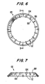
- Figure 6 shows a posterior elevation view of another embodiment of the scleral expansion band of the invention which uses pins for fixation.
- Figure 7 shows a lateral sectional view of the band of Figure 6 along the line 7-7.
- Figure 8 shows a posterior elevation view of another embodiment of the scleral expansion band of the invention which uses tangentially extending fixation pins.
- Figure 9 shows a lateral sectional view of the band of Figure 8 along the line 9-9.
- This invention is based on a different theory, developed by the inventor, regarding the cause of the loss of amplitude of accommodation that constitutes presbyopia.
- accommodation in the non-presbyopic eye is not due to relaxation of the lens and capsule when the zonular tension is relaxed as a result of the contraction of the ciliary body.
- the contraction of the ciliary body exerts a tension on the zonular fibers that in turn actually results in an increase in the equatorial diameter of the lens.
- the increased zonular tension causes a decrease in the peripheral volume of the lens that in turn results in a corresponding increase in the central volume of the lens.
- This view differs from all previous theories of the mechanism of accommodation and its loss in presbyopia, in particular from Tscherning's theory which required the vitreous and attributed presbyopia to an enlargement of the nucleus of the lens. From the inventor's point of view, the difference between the change in the central anterior radius of curvature and the change in the central posterior radius of curvature which occurs in accommodation is explained by the force distribution generated by the zonular attachment to the lens and is not dependent on the topographical thickness of the elastic capsule of the lens, the vitreous, or pressure changes which occur between the anterior and posterior chambers, explanations which have been proposed in the prior art.
- presbyopia results when the distance between the ciliary body and the equator of the lens and its capsule becomes less with age as a result of the normal growth of the lens.
- the lens being of ectodermal origin, continues to grow throughout life, albeit slowly.
- the rate of increase of the equatorial diameter is approximately 0.02 millimeters per year.
- the dimensions of the scleral shell of the eye which is of mesodermal origin, do not increase significantly after about age 13 in a normally emmetropic, hyperopic or myopic eye.
- the radial distance between the equator of the lens and capsule and the ciliary body decreases throughout life. It is well known in muscle physiology that as the length of the effective range of pull of a muscle, i.e., its effective working distance, is reduced, its effective force is reduced in a linear fashion. Since the distance between the ciliary body and the lens equator is decreasing throughout life, it is to be expected according to the theory of the invention that there would be a corresponding linear decrease in the amplitude of accommodation, as observed (Alpern, M., in The Eye , H. Davson, Ed., Academic Press, New York, 1969, pp. 237-238), which would lead eventually to presbyopia.
- presbyopia is treated by increasing the effective working distance of the ciliary muscle.
- a number of procedures are available to the surgeon which can accomplish this increase in effective working distance.
- a straightforward method of increasing the effective working distance of the ciliary muscle is to increase the distance between the equator of the crystalline lens and the inner diameter of the ciliary body in the presbyopic eye. This increased distance restores, at least to some extent, the distance through which the muscles of the ciliary body can contract, and thereby restores their ability to exert force on the lens and change its shape to accomplish accommodation. Any method that increases the radial distance between the lens and the ciliary body is effective in the method of this invention.
- the effective working distance of the ciliary muscle can also be increased by shortening the zonules that connect the ciliary muscle to the equator of the crystalline lens.
- procedures that shorten the body of the ciliary muscle itself or move its insertions in the scleral spur and the choroid can be employed to increase the effective working distance of the muscle.
- procedures that arrest the growth of the lens can stop the steady loss of amplitude of accommodation, and such procedures are considered as fully within the scope of this invention.
- a preferred method of increasing the diameter of the sclera in the region of the ciliary body is to fasten to the sclera in that region a relatively rigid band having a diameter slightly larger than the section of the globe of the eye in the region of the ciliary body. In this way the sclera in that region is stretched and expanded so that the diameter of the circle describing the intersection of the plane of the ciliary body with the sclera is slightly increased. The ciliary body, located immediately inside the globe and attached to the sclera in this expanded region is thereby also increased in diameter.
- the scleral expansion band of the invention adapted for fastening to the sclera of a human eyeball in the region of the ciliary body comprises an anterior rim and a posterior rim, the anterior rim being sized to lie adjacent to the anterior portion of a segment of the sclera of the human eyeball overlying the ciliary body of the eyeball and the posterior rim being sized to lie adjacent the posterior portion of the scleral segment, and rigid structural means extending between the rims and spacing said rims apart so that the anterior rim will lie outside the anterior portion of the scleral segment when the posterior rim lies outside the posterior portion of said scleral segment.
- the anterior rim, the posterior rim and/or the rigid structural means connecting the rims has a diameter greater than the exterior diameter of the scleral segment adjacent thereto.
- the scleral expansion band of the invention is adapted to be fitted to the sclera of the globe in the region of the ciliary body.
- the band will have, at least in part, a diameter slightly greater than that of the sclera at the location where it is to be attached.
- the scleral expansion band of the invention may be fastened to the sclera by any conventional surgical method.
- the band may be a smooth solid band and be sutured to the sclera with sutures passing around the body of the band.
- a preferred band of the invention is provided with suturing holes through which the sutures may be passed.
- the band may also be fastened with conventional surgical staples or clips as are well known in the ocular surgery art.
- the band may be provided with projections or cut-out portions that can be used as anchoring points for sutures.
- the band may also be provided with projecting pins or the like which are inserted into or under the sclera to position the band and exert the appropriate radially outward tension. These pins may extend in an anterior and/or a posterior direction from the band, and may project from one or both rims of the band or from the web portion connecting the rims.
- the pins may also project in a generally tangential direction from the rims or the inside of the web and be provided with sharpened tips or the like whereby they will the penetrate the sclera when the band is rotated and cause the sclera to be firmly fixed to the band and expanded.
- the band may also be adhesively fastened to the sclera with any physiologically acceptable surgical adhesive.
- a preferred surgical adhesive is a surgical grade cyanoacrylate adhesive.
- Mussel adhesive protein may also be used as a suitable adhesive.
- the band 102 has a low frustoconical shape and has a posterior rim 106 and an anterior rim 108, with a web 104 extending between the rims.
- the band may be provided with one or more holes 110 to assist in suturing the band to the sclera.
- the anterior rim 106 and the posterior rim 108 are both of generally circular shape.
- the taper in the diameter of the band is preferably selected in an individual case to fit the globe in the region of the ciliary body. Accordingly, a preferred band has a low frustoconical having a circular base about 20 millimeters in diameter.
- the radial thickness of the band will be determined by the rigidity required in the band and the strength of the material from which it is made. Typically the radial thickness of the band will be about 0.1 to 0.75 millimeters depending on the rigidity of the substance used to make the band.
- the diameter of the anterior rim 106 of the band will be determined by the size of the globe in the patient to be treated. Accordingly, different sizes of band are available wherein the lesser diameter ranges from about 14.5 millimeters to about 18.0 millimeters in 0.25 millimeter increments.
- the axial width of the band will typically be about 2 millimeters.
- FIG. 6 An alternate embodiment of the scleral expansion band of the invention adapted to be fixed to the sclera by pins is illustrated in Figures 6 and 7.
- posterior pins 112 are fixed to the band and extend therefrom in a posterior direction while anterior pins 114 extend in an anterior direction.
- FIG. 8 and 9 Another embodiment of the band of the invention using interior tangentially-directed pins 114 is illustrated in Figures 8 and 9.
- This embodiment can be easily installed by placing it in position on the globe and rotating it to cause the tangential pins to penetrate the sclera and fasten it firmly to the band.
- This embodiment can be easily removed at a later time by merely rotating it in the opposite direction to withdraw the pins from the sclera.
- the scleral expansion band of the invention is made of a material that is sufficiently rigid to exert a force on the sclera sufficient to produce the radial expansion required by the method of the invention and that is physiologically acceptable for long-term implantation or contact with the ocular tissues.
- Such materials are well-known in the surgical art and include suitable metals, ceramics, and synthetic resins.
- Suitable metals include titanium, gold, platinum, stainless steel, tantalum and various surgically acceptable alloys, and the like
- Suitable ceramics may include crystalline and vitreous materials such as porcelain, alumina, silica, silicon carbide, high-strength glasses and the like.
- Suitable synthetic resins include physiologically inert materials such as poly(methyl methacrylate), polyethylene, polypropylene, poly(tetrafluoroethylene), silicone resins and the like.
- the band may also be made of composite materials incorporating a synthetic resin or other matrix reinforced with fibers of high strength material such as glass fibers, boron fibers, carbon fibers, alumina fibers or the like.
- the band may be made of glass-fiber-reinforced epoxy resin, carbon fiber reinforced epoxy resin, carbon fiber reinforced carbon (carbon-carbon), or the like.
- a preferred material is surgical grade poly(methyl methacrylate).
- the scleral expansion band of the invention may be manufactured by any conventional technique appropriate to the material used, such as machining, injection molding, heat molding and the like.
- the scleral expansion band may also be made in a plurality of parts that can be assembled prior to use or may be installed separately to form a complete band.
- the band may be adjustable in circumference.
- the band may be formed from a strip of material, e.g., metal or synthetic resin, with overlapping ends so that the ends may slide past one another thereby adjusting the circumference of the band.
- the length of the overlap may be adjusted, for example, by means of a tangential screw mechanism to adjust the circumference of the band and thereby the amount by which the sclera is expanded.
- the surgeon locates the proper region of the sclera to be expanded by measuring a distance of 1.5 millimeters posterior of the limbus. At this point the diameter of the circle of sclera surrounding the ciliary body is carefully measured and a band is selected having a minor diameter about 0.5 millimeter greater than the measured diameter. A circular incision is made through the conjunctiva completely around the globe at the selected region and the conjunctiva and Tenon's capsule are reflected to expose the substance of the sclera.
- the diameter of the sclera as exposed by the reflection of the conjunctiva and Tenon's capsule is measured at a point 1.5 mm posterior to the limbus.
- the size of sclera can be measured before the conjunctiva is incised.
- the band is then placed on the surface of the globe and sutured thereto preferably with interrupted stitches as conventional in ophthalmological surgery.
- the band may be perforated with holes. For example, a total of sixteen holes may be placed equidistant around the band.
- the sclera in the region of the ciliary body by positioning a band within or just inside the sclera, the band having a diameter just greater than the natural diameter of the overlying tissue. In this way the interior band will expand the overlying tissue and produce the desired result of increasing the effective working distance of the ciliary muscle.
- the sclera itself is a tough strong capsule comprised largely of collagen and held in a rigid state of tension by the internal ocular pressure (IOP). If the sclera is weakened in the area adjacent to the ciliary body, the IOP will cause that portion of the sclera to bulge outward, thereby increasing the diameter of the ciliary body and increasing the distance of the ciliary body from the lens. Any method of weakening the structure of the sclera is suitable. For example enzymatic degradation of the collagen by collagenase may be employed.
- the collagenase may be carefully injected in to the appropriate region of the sclera or may be applied topically. Equivalent enzymes or other chemical treatments that weaken the collagen of the sclera can also be used. Certain antimitotic drugs such as mitomycin are known to cause a softening of the sclera (scleromalacia), and these drugs can be applied to the region of the sclera adjacent to the ciliary body, either topically or by injection, to effect the desired weakening of the sclera.
- antimitotic pharmaceutical agents e.g., methotrexate, azaribine, 6-mercaptopurine, 5-fluorouracil, daunorubicin, doxorubicin, cytarabine, the vinca alkaloids and the like, can also be applied to the sclera to weaken it and permit it to be expanded by the intraocular pressure.
- the sclera in the region of the ciliary body may be weakened by surgical means.
- the sclera may be thinned or weakened by the surgical removal of a portion of its collagenous substance, as, for example by ablating a portion of the thickness of the sclera. This thinning can be accomplished by paring or by abrading the surface or by ablating the surface with laser irradiation.
- the sclera can also be weakened by incisions carefully placed at appropriate angles in the region overlying the ciliary body.
- the diameter of the sclera overlying the ciliary body can also be increased by making a complete periglobular incision and grafting into the incision appropriate tissue and/or physiologically acceptable structural material to increase the dimensions of the sclera.
- an artificial scleral alloplant made of purified human collagen may be engrafted into such an incision.
- Other known biocompatible materials, e.g., poly(ethylene terephthalate), that are conventionally used in the construction of prosthetic devices may also be used for engrafting into such an incision.
- sclera in the region overlying the ciliary body can be weakened by irradiation with a laser beam to decompose partially the collagen fibers.
- Suitable lasers include those conventionally used in ocular surgery such as carbon dioxide lasers, helium-neon lasers, helium-cadmium lasers, argon ion lasers, krypton ion lasers, xenon lasers, nitrous oxide lasers, iodine lasers, holmium doped yttrium-aluminum garnet (YAG) lasers, excimer lasers, chemical lasers, harmonically oscillated lasers, dye lasers, nitrogen lasers, neodymium lasers, erbium lasers, ruby lasers, titanium-sapphire lasers, diode lasers and the like.
- YAG yttrium-aluminum garnet
- Any irradiative treatment with ionizing or non-ionizing radiation that weakens the sclera may be used.
- irradiation with electrons, protons, or x-rays and the like, or irradiation with ultrasonic waves or the like can be used.
- Thermal burning and/or scarring in the appropriate area may also be used to induce an enlargement of the sclera in the area adjacent to the ciliary body.
- Treatments designed to weaken the sclera in the region overlying the ciliary body can also be combined with application of the scleral expansion band of the invention.
- the sclera can be treated with collagenase, mitomycin, or other antimitotic agent, as described above and the scleral band subsequently applied and fastened to the sclera.
- the band itself, its components and/or the sutures may also be coated or impregnated with the sclera-weakening agents whereby they can come into contact with the sclera and exercise their effect when the band is applied. In some cases the band may be later removed to leave the patient with a suitably expanded sclera.
- the distance between the ciliary body and the equator of the lens can be increased by expanding the sclera and ciliary body, the distance can be maintained and the progress of presbyopia arrested by stopping the growth of the crystalline lens. While such a treatment cannot reverse the course of presbyopia, it can arrest the progress of the symptoms, and is appropriate for treatment of patients in whom the presbyopia has not progressed very far.
- the growth of the lens can be arrested by administration of pharmaceutical compounds which stop cell division and growth. For example, colchicine can be administered to prevent cell division, thereby stopping the growth of the lens. Conventional antimitotic drugs may also be used to arrest the growth of the lens.
- Such drugs are well known and include, for example, methotrexate, azaribine, 6-mercaptopurine, 5-fluorouracil, daunorubicin, doxorubicin, cytarabine, the vinca alkaloids and the like.
- Such drugs may be applied topically or by injection into the appropriate structure of the eye where they will come into contact with the lens and exercise their pharmacological activity. If the drugs are sufficiently free of side effects, they may also be administered systemically, either by mouth or parenterally.
- the growth of the lens may also be arrested by physical treatments directed at the newly forming cells in the epithelium of the lens, particularly in the equatorial region of the lens. Treatments that prevent further division of the cells are appropriate for preventing further lens growth.
- the epithelial cells may be heated by laser radiation or ultrasonic irradiation, or inactivated by laser irradiation with a laser capable of directly disrupting chemical bonds within the structures of the cells.
- laser irradiation with a laser capable of directly disrupting chemical bonds within the structures of the cells.
- Sharply focused laser beams or irradiation with microscopic diode lasers positioned close to the equator of the lens are suitable for applying this radiation.
- Irradiation with electrons, protons, x-rays or the like may also be used to stop the further division of the epithelial cells of the lens.
- Surgical methods that work more directly on the ciliary muscle may also be used to increase the effective working distance of the muscle.
- the body of the muscle itself may be shortened, for example by scarring induced by irradiation with suitable laser beams or beams of ionizing or non-ionizing radiation such as ultrasound or electron or proton beams or x-rays.
- the effective working range of the muscle may also be increased by moving its insertions to increase the distance between them.
- the ciliary muscle is inserted anteriorly into the scleral spur and posteriorly into the choroid. Treatment of either of these insertions to cause them to move apart from the complementary insertion will increase the effective working range of the ciliary muscle and improve the amplitude of accommodation according to the invention.
- Selective scarring of adjacent tissue planned to cause retraction of either insertion of the ciliary muscle is effective to accomplish this result.
- the scarring can be accomplished by thermal or radiative treatment of the tissue by the means and methods generally outlined above.
- the method of the invention which increase the amplitude of accommodation may also be of benefit in treatment of hyperopia in certain patients.
- Some youthful hyperopes can achieve relatively normal vision by compensating for their hyperopia through the natural accommodative ability of the eye. However, as this ability declines with age, they find that it becomes more difficult to attain normal vision by this process, and they begin to experience headaches and other symptoms, even at an age somewhat less than usual for the onset of presbyopia.
- increasing the amplitude of accommodation by the method of this invention would be useful in restoring the ability of these patients to compensate for their hyperopia
- the method of this invention also has utility in the treatment of primary open-angle glaucoma, which shows a correlation with age in certain individuals. It has been found that, in general, IOP exhibits a linear increase with increasing age. (Armaly, M.F., On the distribution of applanation pressure I. Statistical features and the effect of age, sex, and family history of glaucoma, Archives of Ophthalmology, Vol. 73, pp. 11-18 (1965).) Among the general population is found a group of individuals who develop abnormally high intraocular pressures as a result of primary open angle glaucoma, a disease which is one of the most prevalent causes of blindness in the world.
- the linear increase in IOP with age is a direct result of the decrease in distance between the lens equator and the ciliary muscle and the resulting linear decrease in the effective pull of the ciliary muscle. Since the ciliary muscle inserts into the trabecular meshwork, the decrease in pull will decrease the size of the trabeculum and/or the drainage pores and result in a linear increase of intraocular pressure with age.
- the patients who develop primary open angle glaucoma may have a congenital predilection to narrower pores and/or smaller trabecular meshwork, so that when the ability of the ciliary muscle to exert force declines, after the age of 40 or thereabouts, they tend to develop excessively elevated IOP.
- the method of the invention which increases the effective working distance of the ciliary muscle, and thereby increases the force that it can exert when it contracts, restores the level of force which the ciliary muscle exerts on the trabecular meshwork to a value characteristic of a more youthful eye. In this way it is expected that the tendency of an eye that is disposed to develop primary open angle glaucoma as it ages would be overcome and the onset of this disease would be prevented or at least postponed.
- the ability of the ciliary muscle to exert tension on the lens by means of the zonules can also be increased by shortening the zonules, a procedure which increases the effective working range of the ciliary muscle.
- This shortening can be produced by heating the zonules by ultrasonic irradiation or laser irradiation.
- the zonules may also be treated with electrons, protons, x-rays and the like which may cause changes in the structure of the zonules resulting in shortening.
- the shortening of the zonules may also be brought about by application of chemical compounds that act to cause a shrinkage of the collagenous zonules.
- This example illustrates the feasibility of increasing the diameter of the ciliary body by suturing an external band to the sclera.
- the diameter of the sclera was measured using a shadow graph at distances of 1.5 millimeters and 3.5 millimeters from the limbus. The diameters were found to be 16.2 mm and 20 mm respectively.
- Bands were made from medical grade poly(methyl methacrylate) having a low frustoconical shape and a radial thickness of 0.5 mm. The internal diameter of the larger (posterior) side was 20 mm, and the smaller (anterior) diameters varied from 14.5 mm to 18.0 mm in 0.25 mm steps. A band was selected having a major diameter of 20 mm and a minor diameter of 18.0 mm.
- the band was placed on the sclera of a human cadaver eye so that the 18.0 mm diameter end of the band was 1.5 mm from the limbus and the 20 mm diameter end was 3.5 mm from the limbus.
- the band was sutured through the holes with 16 interrupted 6-0 polyester (Mersilene, Ethicon, Inc.) stitches to the sclera. This resulted in a clearly observable stretching of the sclera in the region of the ciliary body.
- the pupil became dilated by about 1.5 mm. Since the iris inserts into the ciliary body, the pupillary dilation demonstrated that the induced scleral stretching was also moving the ciliary body outward, thereby increasing its diameter and its distance from the equator of the lens.
- This example illustrates the treatment of presbyopia in patients.
- Example 1 Using vernier calipers, a distance of 1.5 mm posterior to the limbus was measured from the limbus and the scleral diameter was measured at that location. A band of the type made in Example 1 was selected having a minor diameter of from 0.5 mm to 1.5 mm greater than the measured diameter of the sclera. The band was sutured to the sclera with 16 interrupted 6-0 polyester (Mersilene, Ethicon, Inc.) stitches. The patients were given antibiotic-steroid drops to use for five days. Postoperatively, the patients were followed daily for five days and then weekly. The results are presented in Table 1 below. Preoperative and Postoperative Amplitude of Accommodation Preoperative Postoperative Patient No.
Landscapes
- Health & Medical Sciences (AREA)
- Ophthalmology & Optometry (AREA)
- Animal Behavior & Ethology (AREA)
- General Health & Medical Sciences (AREA)
- Veterinary Medicine (AREA)
- Engineering & Computer Science (AREA)
- Biomedical Technology (AREA)
- Heart & Thoracic Surgery (AREA)
- Vascular Medicine (AREA)
- Life Sciences & Earth Sciences (AREA)
- Public Health (AREA)
- Oral & Maxillofacial Surgery (AREA)
- Cardiology (AREA)
- Transplantation (AREA)
- Nuclear Medicine, Radiotherapy & Molecular Imaging (AREA)
- Surgery (AREA)
- Prostheses (AREA)
- Medicines That Contain Protein Lipid Enzymes And Other Medicines (AREA)
- Medicines Containing Material From Animals Or Micro-Organisms (AREA)
- Pharmaceuticals Containing Other Organic And Inorganic Compounds (AREA)
- Surgical Instruments (AREA)
Priority Applications (1)
| Application Number | Priority Date | Filing Date | Title |
|---|---|---|---|
| EP05077198A EP1623684A1 (fr) | 1992-07-15 | 1993-07-13 | Traitement de la presbytie et d'autres troubles oculaires |
Applications Claiming Priority (3)
| Application Number | Priority Date | Filing Date | Title |
|---|---|---|---|
| US07/913,486 US5354331A (en) | 1992-07-15 | 1992-07-15 | Treatment of presbyopia and other eye disorders |
| US913486 | 1992-07-15 | ||
| EP93919820A EP0746271A1 (fr) | 1992-07-15 | 1993-07-13 | Traitement de la presbytie et d'autres troubles oculaires |
Related Parent Applications (1)
| Application Number | Title | Priority Date | Filing Date |
|---|---|---|---|
| EP93919820A Division EP0746271A1 (fr) | 1992-07-15 | 1993-07-13 | Traitement de la presbytie et d'autres troubles oculaires |
Related Child Applications (1)
| Application Number | Title | Priority Date | Filing Date |
|---|---|---|---|
| EP05077198A Division EP1623684A1 (fr) | 1992-07-15 | 1993-07-13 | Traitement de la presbytie et d'autres troubles oculaires |
Publications (3)
| Publication Number | Publication Date |
|---|---|
| EP1125560A2 true EP1125560A2 (fr) | 2001-08-22 |
| EP1125560A3 EP1125560A3 (fr) | 2001-10-04 |
| EP1125560B1 EP1125560B1 (fr) | 2005-11-09 |
Family
ID=25433318
Family Applications (3)
| Application Number | Title | Priority Date | Filing Date |
|---|---|---|---|
| EP05077198A Withdrawn EP1623684A1 (fr) | 1992-07-15 | 1993-07-13 | Traitement de la presbytie et d'autres troubles oculaires |
| EP01201708A Expired - Lifetime EP1125560B1 (fr) | 1992-07-15 | 1993-07-13 | Traitement de la presbytie et d'autres troubles oculaires |
| EP93919820A Withdrawn EP0746271A1 (fr) | 1992-07-15 | 1993-07-13 | Traitement de la presbytie et d'autres troubles oculaires |
Family Applications Before (1)
| Application Number | Title | Priority Date | Filing Date |
|---|---|---|---|
| EP05077198A Withdrawn EP1623684A1 (fr) | 1992-07-15 | 1993-07-13 | Traitement de la presbytie et d'autres troubles oculaires |
Family Applications After (1)
| Application Number | Title | Priority Date | Filing Date |
|---|---|---|---|
| EP93919820A Withdrawn EP0746271A1 (fr) | 1992-07-15 | 1993-07-13 | Traitement de la presbytie et d'autres troubles oculaires |
Country Status (11)
| Country | Link |
|---|---|
| US (5) | US5354331A (fr) |
| EP (3) | EP1623684A1 (fr) |
| AT (1) | ATE308947T1 (fr) |
| AU (1) | AU4992493A (fr) |
| CA (1) | CA2166537C (fr) |
| DE (1) | DE69333904T2 (fr) |
| ES (1) | ES2252146T3 (fr) |
| HK (1) | HK1039444B (fr) |
| IL (1) | IL106364A (fr) |
| TW (1) | TW244334B (fr) |
| WO (1) | WO1994002084A1 (fr) |
Cited By (4)
| Publication number | Priority date | Publication date | Assignee | Title |
|---|---|---|---|---|
| EP1351627B1 (fr) * | 2001-01-19 | 2004-08-18 | Georges Baikoff | Element correcteur de la presbytie |
| WO2006007475A3 (fr) * | 2004-07-01 | 2006-04-06 | Nicholas C Caro | Clip ophtalmique et methode chirurgicale associee |
| US7977310B2 (en) | 2003-01-14 | 2011-07-12 | Aventis Pharmaceuticals Inc. | Method for reducing cardiovascular morbidity and mortality in prediabetic patients and patients with type 2 diabetes |
| CN102917676A (zh) * | 2010-04-30 | 2013-02-06 | 塞罗斯医学有限责任公司 | 使用组合的方式治疗眼组织的方法和装置 |
Families Citing this family (292)
| Publication number | Priority date | Publication date | Assignee | Title |
|---|---|---|---|---|
| US20040015236A1 (en) * | 1991-11-18 | 2004-01-22 | Sarfarazi Faezeh M. | Sarfarazi elliptical accommodative intraocular lens for small incision surgery |
| US5465737A (en) * | 1992-07-15 | 1995-11-14 | Schachar; Ronald A. | Treatment of presbyopia and other eye disorders |
| US6197056B1 (en) | 1992-07-15 | 2001-03-06 | Ras Holding Corp. | Segmented scleral band for treatment of presbyopia and other eye disorders |
| US5354331A (en) * | 1992-07-15 | 1994-10-11 | Schachar Ronald A | Treatment of presbyopia and other eye disorders |
| US6966927B1 (en) | 1992-08-07 | 2005-11-22 | Addition Technology, Inc. | Hybrid intrastromal corneal ring |
| US5458596A (en) | 1994-05-06 | 1995-10-17 | Dorsal Orthopedic Corporation | Method and apparatus for controlled contraction of soft tissue |
| US6302877B1 (en) * | 1994-06-29 | 2001-10-16 | Luis Antonio Ruiz | Apparatus and method for performing presbyopia corrective surgery |
| US5472436A (en) * | 1994-07-26 | 1995-12-05 | Fremstad; Daria A. | Ocular appliance for delivering medication |
| US6461353B1 (en) | 1995-02-17 | 2002-10-08 | Oratec Interventions, Inc. | Orthopedic apparatus for controlled contraction of collagen tissue |
| US5697923A (en) * | 1995-03-29 | 1997-12-16 | Poler; Stanley | Corneal drape for use in performing a photorefractory keratectomy procedure |
| US5980504A (en) * | 1996-08-13 | 1999-11-09 | Oratec Interventions, Inc. | Method for manipulating tissue of an intervertebral disc |
| US6283960B1 (en) | 1995-10-24 | 2001-09-04 | Oratec Interventions, Inc. | Apparatus for delivery of energy to a surgical site |
| US6007570A (en) * | 1996-08-13 | 1999-12-28 | Oratec Interventions, Inc. | Apparatus with functional element for performing function upon intervertebral discs |
| US7166117B2 (en) * | 1996-02-07 | 2007-01-23 | Hellenkamp Johann F | Automatic surgical device and control assembly for cutting a cornea |
| US20070244496A1 (en) * | 1996-02-07 | 2007-10-18 | Hellenkamp Johann F | Automatic surgical device and control assembly for cutting a cornea |
| US5824073A (en) * | 1996-03-18 | 1998-10-20 | Peyman; Gholam A. | Macular indentor for use in the treatment of subretinal neovascular membranes |
| US5743274A (en) * | 1996-03-18 | 1998-04-28 | Peyman; Gholam A. | Macular bandage for use in the treatment of subretinal neovascular members |
| US7655002B2 (en) | 1996-03-21 | 2010-02-02 | Second Sight Laser Technologies, Inc. | Lenticular refractive surgery of presbyopia, other refractive errors, and cataract retardation |
| US6258086B1 (en) | 1996-10-23 | 2001-07-10 | Oratec Interventions, Inc. | Catheter for delivery of energy to a surgical site |
| US7069087B2 (en) | 2000-02-25 | 2006-06-27 | Oratec Interventions, Inc. | Apparatus and method for accessing and performing a function within an intervertebral disc |
| US6645203B2 (en) | 1997-02-12 | 2003-11-11 | Oratec Interventions, Inc. | Surgical instrument with off-axis electrode |
| US6733496B2 (en) | 2001-06-06 | 2004-05-11 | Oratec Interventions, Inc. | Intervertebral disc device employing flexible probe |
| US6126682A (en) | 1996-08-13 | 2000-10-03 | Oratec Interventions, Inc. | Method for treating annular fissures in intervertebral discs |
| US6726685B2 (en) | 2001-06-06 | 2004-04-27 | Oratec Interventions, Inc. | Intervertebral disc device employing looped probe |
| US6832997B2 (en) | 2001-06-06 | 2004-12-21 | Oratec Interventions, Inc. | Electromagnetic energy delivery intervertebral disc treatment devices |
| US6068628A (en) * | 1996-08-20 | 2000-05-30 | Oratec Interventions, Inc. | Apparatus for treating chondromalacia |
| US5772675A (en) * | 1996-10-31 | 1998-06-30 | Chiron Vision Corporation | Positioning assembly for retaining and positioning a cornea |
| US5956126A (en) * | 1996-11-19 | 1999-09-21 | Cody; Victor | Optical instrument and method for the treatment of amblyopia |
| JP2001511048A (ja) | 1997-02-12 | 2001-08-07 | オーレイテック インターヴェンションズ インコーポレイテッド | 組織の電気外科的な除去のための電極及びそれの製造方法 |
| EP1006908A2 (fr) * | 1997-02-12 | 2000-06-14 | Oratec Interventions, Inc. | Sonde concave pour chirurgie arthroscopique |
| US5954716A (en) * | 1997-02-19 | 1999-09-21 | Oratec Interventions, Inc | Method for modifying the length of a ligament |
| US5782894A (en) * | 1997-03-05 | 1998-07-21 | Israel; Ben | Device and method for improving ocular focusing at near vision points |
| US6135118A (en) * | 1997-05-12 | 2000-10-24 | Dailey; James P. | Treatment with magnetic fluids |
| US6004320A (en) * | 1997-09-19 | 1999-12-21 | Oratec Interventions, Inc. | Clip on electrocauterizing sheath for orthopedic shave devices |
| US6214001B1 (en) | 1997-09-19 | 2001-04-10 | Oratec Interventions, Inc. | Electrocauterizing tool for orthopedic shave devices |
| US6007533A (en) * | 1997-09-19 | 1999-12-28 | Oratec Interventions, Inc. | Electrocauterizing tip for orthopedic shave devices |
| AU3881301A (en) * | 1997-10-08 | 2001-07-26 | Ras Holding Corporation | Segmented scleral band for treatment of presbyopia and other eye disorders |
| US6991650B2 (en) | 1997-10-08 | 2006-01-31 | Refocus Ocular, Inc. | Scleral expansion device having duck bill |
| CA2274163C (fr) | 1997-10-08 | 2006-12-19 | Ras Holding Corporation | Bande sclerale segmentee pour traitement de la presbytie et d'autres troubles oculaires |
| US6007578A (en) * | 1997-10-08 | 1999-12-28 | Ras Holding Corp | Scleral prosthesis for treatment of presbyopia and other eye disorders |
| US6176857B1 (en) | 1997-10-22 | 2001-01-23 | Oratec Interventions, Inc. | Method and apparatus for applying thermal energy to tissue asymmetrically |
| US8016823B2 (en) | 2003-01-18 | 2011-09-13 | Tsunami Medtech, Llc | Medical instrument and method of use |
| US6053909A (en) * | 1998-03-27 | 2000-04-25 | Shadduck; John H. | Ionothermal delivery system and technique for medical procedures |
| US7892229B2 (en) | 2003-01-18 | 2011-02-22 | Tsunami Medtech, Llc | Medical instruments and techniques for treating pulmonary disorders |
| FR2784287B1 (fr) * | 1998-10-13 | 2000-12-08 | Georges Baikoff | Segment d'expansion sclerale |
| USRE40002E1 (en) | 1998-11-10 | 2008-01-15 | Surgilight, Inc. | Treatment of presbyopia and other eye disorders using a scanning laser system |
| US6745775B2 (en) | 1998-11-10 | 2004-06-08 | Surgilight, Inc. | Methods and apparatus for presbyopia treatment using a scanning laser system |
| FR2787991B1 (fr) * | 1998-12-31 | 2001-05-25 | Medicale De Prec S M P Sa Soc | Dispositif pour traiter la presbytie ou autre affection oculaire |
| US7449019B2 (en) | 1999-01-25 | 2008-11-11 | Smith & Nephew, Inc. | Intervertebral decompression |
| EP1173790A2 (fr) | 1999-03-01 | 2002-01-23 | Boston Innovative Optics, Inc. | Systeme et procede destines a l'augmentation de la profondeur focale de l'oeil humain |
| US6171337B1 (en) | 1999-03-31 | 2001-01-09 | Miles A. Galin | Positive power anterior chamber ocular implant |
| US7662179B2 (en) | 1999-04-09 | 2010-02-16 | Sarfarazi Faezeh M | Haptics for accommodative intraocular lens system |
| US8556967B2 (en) | 1999-04-09 | 2013-10-15 | Faezeh Mona Sarfarazi | Interior bag for a capsular bag and injector |
| US20060238702A1 (en) | 1999-04-30 | 2006-10-26 | Advanced Medical Optics, Inc. | Ophthalmic lens combinations |
| US20010029363A1 (en) * | 1999-05-03 | 2001-10-11 | Lin J. T. | Methods and apparatus for presbyopia correction using ultraviolet and infrared lasers |
| US6258082B1 (en) | 1999-05-03 | 2001-07-10 | J. T. Lin | Refractive surgery and presbyopia correction using infrared and ultraviolet lasers |
| TW587934B (en) * | 1999-06-07 | 2004-05-21 | Ronald A Schachar | Improved scleral prosthesis for the treatment of presbyopia and other eye disorders |
| FR2794965B1 (fr) | 1999-06-21 | 2001-07-20 | Ioltechnologie Production | Anneau de tension intraoculaire pour la correction de la presbytie |
| US6254594B1 (en) * | 1999-07-30 | 2001-07-03 | Quadrivium, Llc | Disposable light source for photothermal treatment of human tissue |
| US7008396B1 (en) * | 1999-09-03 | 2006-03-07 | Restorvision, Inc. | Ophthalmic device and method of manufacture and use |
| AU775373B2 (en) | 1999-10-01 | 2004-07-29 | Immunogen, Inc. | Compositions and methods for treating cancer using immunoconjugates and chemotherapeutic agents |
| RU2159098C1 (ru) * | 1999-12-29 | 2000-11-20 | Офтальмологическая клиническая больница им. Т.И. Ерошевского | Способ хирургического лечения пресбиопии |
| US7635388B1 (en) * | 2000-05-04 | 2009-12-22 | Tyler Thomas D | Device and method for incremental correction of sight disorders and occular diseases |
| US7135016B1 (en) * | 2000-05-08 | 2006-11-14 | Optotech Ltd. | Non-penetrating filtration surgery |
| US7886747B2 (en) * | 2000-05-08 | 2011-02-15 | I Optima Ltd. | Non-penetrating filtration surgery |
| US6491688B1 (en) * | 2000-06-21 | 2002-12-10 | J. T. Lin | Apparatus and methods for reversal of presbyopia using near infrared selective laser on zonnulas |
| US6511508B1 (en) * | 2000-08-04 | 2003-01-28 | Environmental Robots, Inc. | Surgical correction of human eye refractive errors by active composite artificial muscle implants |
| US7060094B2 (en) | 2000-08-07 | 2006-06-13 | Ophthalmotronics, Inc. | Accommodating zonular mini-bridge implants |
| US6923955B2 (en) * | 2000-08-16 | 2005-08-02 | Newlens, Llc | Presbyopia treatment by lens alteration |
| US8647612B2 (en) * | 2008-03-05 | 2014-02-11 | Encore Health, Llc | Dithiol compounds, derivatives, and treatment of presbyopia |
| US7914815B2 (en) * | 2000-08-16 | 2011-03-29 | Encore Health, Llc | Method for delivery of pharmaceuticals for treating or preventing presbyopia |
| US7935332B2 (en) * | 2000-08-16 | 2011-05-03 | Encore Health, Llc | Presbyopia treatment by lens alteration |
| US8697109B2 (en) | 2000-08-16 | 2014-04-15 | Encore Health, Llc | Caged mercaptan and seleno-mercaptan compounds and methods of using them |
| US8147816B2 (en) * | 2000-08-16 | 2012-04-03 | Encore Health, Llc | Presbyopia treatment by lens alteration |
| US20050112113A1 (en) * | 2000-08-16 | 2005-05-26 | Till Jonathan S. | Presbyopia treatment by lens alteration |
| US6517555B1 (en) * | 2000-09-05 | 2003-02-11 | Clear Sight, Inc. | Method for treating presbyopia |
| US6824540B1 (en) * | 2000-11-06 | 2004-11-30 | Surgilight, Inc. | Apparatus and methods for the treatment of presbyopia using fiber-coupled-lasers |
| US6679855B2 (en) | 2000-11-07 | 2004-01-20 | Gerald Horn | Method and apparatus for the correction of presbyopia using high intensity focused ultrasound |
| US7549987B2 (en) | 2000-12-09 | 2009-06-23 | Tsunami Medtech, Llc | Thermotherapy device |
| US9433457B2 (en) | 2000-12-09 | 2016-09-06 | Tsunami Medtech, Llc | Medical instruments and techniques for thermally-mediated therapies |
| US20030078657A1 (en) | 2001-01-25 | 2003-04-24 | Gholam-Reza Zadno-Azizi | Materials for use in accommodating intraocular lens system |
| US8062361B2 (en) * | 2001-01-25 | 2011-11-22 | Visiogen, Inc. | Accommodating intraocular lens system with aberration-enhanced performance |
| US6884261B2 (en) * | 2001-01-25 | 2005-04-26 | Visiogen, Inc. | Method of preparing an intraocular lens for implantation |
| US7226478B2 (en) * | 2001-01-25 | 2007-06-05 | Visiogen, Inc. | Optic configuration for intraocular lens system |
| US20030078658A1 (en) * | 2001-01-25 | 2003-04-24 | Gholam-Reza Zadno-Azizi | Single-piece accomodating intraocular lens system |
| US7780729B2 (en) | 2004-04-16 | 2010-08-24 | Visiogen, Inc. | Intraocular lens |
| US6695839B2 (en) | 2001-02-08 | 2004-02-24 | Oratec Interventions, Inc. | Method and apparatus for treatment of disrupted articular cartilage |
| IL141529A0 (en) * | 2001-02-20 | 2002-03-10 | Ben Nun Yehoshua | Intraocular lens with scleral fixation capability |
| US20020173777A1 (en) * | 2001-03-30 | 2002-11-21 | Sand Bruce J. | Treatment of collagen |
| US7044945B2 (en) * | 2001-03-30 | 2006-05-16 | Sand Bruce J | Prevention of regression in thermal ciliary muscle tendinoplasty |
| US20060241750A1 (en) * | 2001-05-22 | 2006-10-26 | Ras Holding Corp | Scleral expansion device having duck bill |
| US6638276B2 (en) | 2001-06-06 | 2003-10-28 | Oratec Interventions, Inc. | Intervertebral disc device employing prebent sheath |
| IL145015A0 (en) * | 2001-08-21 | 2002-06-30 | Nun Yehoshua Ben | Accommodating lens |
| GB0121168D0 (en) | 2001-08-31 | 2001-10-24 | Duckworth & Kent Ltd | Ophthalmic devices and procedures |
| AU2002321563A1 (en) * | 2001-08-31 | 2003-03-18 | Duckworth And Kent Limited | Ophthalmic devices and procedures |
| US7338506B2 (en) * | 2001-09-05 | 2008-03-04 | Caro Nicholas C | Scleral clip and procedures for using same |
| US7073507B2 (en) * | 2001-11-05 | 2006-07-11 | David Morris | Sterile ocular cover and method |
| WO2003039351A2 (fr) * | 2001-11-09 | 2003-05-15 | Surgilight, Inc. | Procedes et systemes de traitement de la presbytie par ablation au laser |
| US8444636B2 (en) | 2001-12-07 | 2013-05-21 | Tsunami Medtech, Llc | Medical instrument and method of use |
| US7763069B2 (en) | 2002-01-14 | 2010-07-27 | Abbott Medical Optics Inc. | Accommodating intraocular lens with outer support structure |
| FR2838955A1 (fr) * | 2002-04-30 | 2003-10-31 | Bernard Gambini | Pont d'expansion sclerale |
| US20040002756A1 (en) * | 2002-05-09 | 2004-01-01 | Georges Baikoff | Corrective element for presbyopia |
| FR2843015B1 (fr) * | 2002-08-01 | 2005-06-10 | Laredo Fernand | Implant pour le traitement de la presbytie |
| US20050205101A1 (en) * | 2002-10-17 | 2005-09-22 | Lin J T | Combined pharmocological and surgical method and system for the treatment of eye disorders |
| US20040078009A1 (en) * | 2002-10-17 | 2004-04-22 | Lin J. T. | Method and apparatus for the treatment of presbyopia and other eye disorders combining pharmocological and surgical means |
| US7037335B2 (en) | 2002-11-19 | 2006-05-02 | Eagle Vision, Inc. | Bulbous scleral implants for the treatment of eye disorders such as presbyopia and glaucoma |
| US7662180B2 (en) | 2002-12-05 | 2010-02-16 | Abbott Medical Optics Inc. | Accommodating intraocular lens and method of manufacture thereof |
| WO2006025806A1 (fr) * | 2003-01-07 | 2006-03-09 | Lin J T | Appareil pour le renversement de la presbytie utilisant des faisceaux lasers thermiques diriges sur les zonules |
| US20050197697A1 (en) * | 2003-01-13 | 2005-09-08 | Georges Baikoff | Corrective element for presbyopia |
| US7150747B1 (en) | 2003-01-22 | 2006-12-19 | Smith & Nephew, Inc. | Electrosurgical cutter |
| CN100486549C (zh) * | 2003-04-07 | 2009-05-13 | 泰克诺拉斯眼科系统有限公司 | 微型角膜刀的连杆驱动系统 |
| US7628810B2 (en) | 2003-05-28 | 2009-12-08 | Acufocus, Inc. | Mask configured to maintain nutrient transport without producing visible diffraction patterns |
| US20050046794A1 (en) | 2003-06-17 | 2005-03-03 | Silvestrini Thomas A. | Method and apparatus for aligning a mask with the visual axis of an eye |
| US20040260395A1 (en) * | 2003-06-19 | 2004-12-23 | Boxer Wachler Brian S. | Ophthalmological zonular stretch segment for treating presbyopia |
| RU2251978C2 (ru) * | 2003-06-26 | 2005-05-20 | ЗАО "Екатеринбургский центр МНТК "Микрохирургия глаза" | Способ создания объема иммерсионной жидкости при проведении ультразвуковой биомикроскопии переднего отрезка глаза |
| US7704278B2 (en) * | 2003-10-06 | 2010-04-27 | Crs & Associates | Method and apparatus for enhanced corneal accommodation |
| US20050106270A1 (en) * | 2003-10-06 | 2005-05-19 | Devore Dale P. | Chemical treatment of in vivo tissue to alter charge and net charge density characteristics |
| US20070055220A1 (en) * | 2003-11-14 | 2007-03-08 | Jui-Teng Lin | Methods and systems for treating presbyopia via laser ablation |
| DE10353264B4 (de) * | 2003-11-14 | 2022-07-07 | Carl Zeiss Meditec Ag | Adapter zum Koppeln einer Laserbearbeitungsvorrichtung mit einem Objekt |
| US20050131535A1 (en) | 2003-12-15 | 2005-06-16 | Randall Woods | Intraocular lens implant having posterior bendable optic |
| MXPA03011987A (es) | 2003-12-19 | 2005-06-23 | Osio Sancho Alberto | Metodo para el tratamiento de la presbicia induciendo cambios en el poder y fisiologia corneal. |
| CA2554165A1 (fr) * | 2004-01-22 | 2005-08-11 | Solx, Inc. | Methode de traitement du glaucome |
| IL161706A0 (en) | 2004-04-29 | 2004-09-27 | Nulens Ltd | Intraocular lens fixation device |
| US20050279369A1 (en) * | 2004-06-21 | 2005-12-22 | Lin J T | Method and apparatus for the treatment of presbyopia and glaucoma by ciliary body ablation |
| US20060004386A1 (en) * | 2004-07-01 | 2006-01-05 | Caro Nicholas C | Ophthalmic clip and associated surgical method |
| US7736389B1 (en) | 2004-08-23 | 2010-06-15 | Damiano Richard E | Reading enhancement device for preventing and treating presbyopia of the eye |
| JP4480766B2 (ja) * | 2004-10-13 | 2010-06-16 | ニューレンズ・リミテッド | 調節式眼内レンズ(aiol)及びそれを含んでいるaiolアッセンブリ |
| US7252662B2 (en) | 2004-11-02 | 2007-08-07 | Lenticular Research Group Llc | Apparatus and processes for preventing or delaying one or more symptoms of presbyopia |
| US8377123B2 (en) * | 2004-11-10 | 2013-02-19 | Visiogen, Inc. | Method of implanting an intraocular lens |
| WO2006055695A1 (fr) * | 2004-11-16 | 2006-05-26 | Barry Robert L | Traitement pulmonaire et appareil associe |
| US20060116759A1 (en) * | 2004-11-30 | 2006-06-01 | Thornton Spencer P | Method of treating presbyopia and other eye conditions |
| US20060113054A1 (en) * | 2004-12-01 | 2006-06-01 | Silvestrini Thomas A | Method of making an ocular implant |
| US7491350B2 (en) * | 2004-12-01 | 2009-02-17 | Acufocus, Inc. | Method of making an ocular implant |
| US7686804B2 (en) * | 2005-01-14 | 2010-03-30 | Covidien Ag | Vessel sealer and divider with rotating sealer and cutter |
| AU2006213997A1 (en) | 2005-02-19 | 2006-08-24 | Lenticular Research Group Llc | Apparatus and processes for preventing or delaying onset or progression of age-related cataract |
| US7871404B2 (en) * | 2005-03-15 | 2011-01-18 | Annmarie Hipsley | System and method for treating connective tissue |
| EP1890650A2 (fr) | 2005-03-30 | 2008-02-27 | Nulens Ltd | Ensembles lentille intraoculaire accommodative (aiol) et composants separes pour lesdits ensembles |
| US20060224146A1 (en) * | 2005-03-30 | 2006-10-05 | Lin J T | Method and system for non-invasive treatment of hyperopia, presbyopia and glaucoma |
| US7976577B2 (en) | 2005-04-14 | 2011-07-12 | Acufocus, Inc. | Corneal optic formed of degradation resistant polymer |
| US7878204B2 (en) * | 2005-04-22 | 2011-02-01 | Biolase Technology, Inc. | Methods for treating hyperopia and presbyopia via laser tunneling |
| WO2006116621A2 (fr) * | 2005-04-26 | 2006-11-02 | Biolase Technology, Inc. | Procedes de traitement de maladies de l'oeil |
| US7591849B2 (en) | 2005-07-01 | 2009-09-22 | Bausch & Lomb Incorpoted | Multi-component accommodative intraocular lens with compressible haptic |
| US20070032785A1 (en) | 2005-08-03 | 2007-02-08 | Jennifer Diederich | Tissue evacuation device |
| DE102005040338B4 (de) * | 2005-08-25 | 2019-08-29 | Carl Zeiss Meditec Ag | Kontaktglas für die Augenchirurgie |
| US9636213B2 (en) | 2005-09-30 | 2017-05-02 | Abbott Medical Optics Inc. | Deformable intraocular lenses and lens systems |
| US20070088352A1 (en) * | 2005-10-14 | 2007-04-19 | Rosen Robert S | Method and system for radio frequency ophthalmological presbyopia surgery |
| US9744279B2 (en) * | 2005-12-08 | 2017-08-29 | Heartware, Inc. | Implant connector |
| US8262646B2 (en) | 2006-01-20 | 2012-09-11 | Lensar, Inc. | System and method for providing the shaped structural weakening of the human lens with a laser |
| US10842675B2 (en) | 2006-01-20 | 2020-11-24 | Lensar, Inc. | System and method for treating the structure of the human lens with a laser |
| US9889043B2 (en) | 2006-01-20 | 2018-02-13 | Lensar, Inc. | System and apparatus for delivering a laser beam to the lens of an eye |
| US9545338B2 (en) | 2006-01-20 | 2017-01-17 | Lensar, Llc. | System and method for improving the accommodative amplitude and increasing the refractive power of the human lens with a laser |
| US20070235043A1 (en) * | 2006-04-10 | 2007-10-11 | Georges Baikoff | Presbyopia treatment by weakening the zonula |
| US8256431B2 (en) * | 2006-04-24 | 2012-09-04 | Biolase, Inc. | Methods for treating hyperopia and presbyopia via laser tunneling |
| US8544473B2 (en) * | 2006-04-26 | 2013-10-01 | Biolase, Inc. | Methods for treating eye conditions with low-level light therapy |
| US20070260310A1 (en) * | 2006-05-08 | 2007-11-08 | Richardson Gary A | Accommodative Intraocular Lens Having Defined Axial Compression Characteristics |
| US20070260309A1 (en) * | 2006-05-08 | 2007-11-08 | Richardson Gary A | Accommodating intraocular lens having a recessed anterior optic |
| MX2009000378A (es) | 2006-07-11 | 2009-08-12 | Refocus Group Inc | Protesis escleral para tratar presbiopia y otros trastornos del ojo y dispositivos y metodos relacionados. |
| ES2964952T3 (es) | 2006-07-11 | 2024-04-10 | Refocus Group Inc | Prótesis escleral para el tratamiento de la presbicia y otros trastornos oculares |
| US8911496B2 (en) | 2006-07-11 | 2014-12-16 | Refocus Group, Inc. | Scleral prosthesis for treating presbyopia and other eye disorders and related devices and methods |
| WO2008023379A2 (fr) * | 2006-08-25 | 2008-02-28 | Nulens Ltd | Trousse pour implantation d'un cristallin artificiel |
| US7993323B2 (en) | 2006-11-13 | 2011-08-09 | Uptake Medical Corp. | High pressure and high temperature vapor catheters and systems |
| WO2008079671A1 (fr) * | 2006-12-22 | 2008-07-03 | Bausch & Lomb Incorporated | Lentille intraoculaire d'accommodation à éléments multiples |
| CA2673388C (fr) | 2006-12-22 | 2015-11-24 | Amo Groningen B.V. | Lentille intraoculaire d'accommodation, systeme de lentille et structure pour lentille |
| US20080161914A1 (en) | 2006-12-29 | 2008-07-03 | Advanced Medical Optics, Inc. | Pre-stressed haptic for accommodating intraocular lens |
| US8123803B2 (en) * | 2007-01-24 | 2012-02-28 | Mohsen Shaninpoor | System and device for correcting hyperopia and presbyopia |
| USD702346S1 (en) | 2007-03-05 | 2014-04-08 | Nulens Ltd. | Haptic end plate for use in an intraocular assembly |
| ATE483427T1 (de) * | 2007-03-05 | 2010-10-15 | Nulens Ltd | Einheitlich akkommodierende intraokularlinsen (aiols) und diskrete basiselemente zur verwendung damit |
| PT2148628T (pt) * | 2007-05-07 | 2017-12-06 | Jaswant Rathore | Um método e um sistema para fotoablação a laser dentro do cristalino |
| WO2009009398A1 (fr) | 2007-07-06 | 2009-01-15 | Tsunami Medtech, Llc | Système médical et procédé d'utilisation |
| JP5323073B2 (ja) * | 2007-08-02 | 2013-10-23 | リフォーカス グループ、インコーポレイテッド | 老眼及び他の眼疾患を治療するためのクロスバーを有する強膜プロテーゼ |
| US8102027B2 (en) * | 2007-08-21 | 2012-01-24 | Broadcom Corporation | IC package sacrificial structures for crack propagation confinement |
| US8197470B2 (en) | 2007-08-23 | 2012-06-12 | Aegea Medical, Inc. | Uterine therapy device and method |
| US8322335B2 (en) | 2007-10-22 | 2012-12-04 | Uptake Medical Corp. | Determining patient-specific vapor treatment and delivery parameters |
| ES2456965T3 (es) * | 2007-10-22 | 2014-04-24 | Uptake Medical Corp. | Determinación de los parámetros del tratamiento de vapor y de administración específicos del paciente |
| US9924992B2 (en) | 2008-02-20 | 2018-03-27 | Tsunami Medtech, Llc | Medical system and method of use |
| WO2009111635A2 (fr) | 2008-03-05 | 2009-09-11 | Encore Health, Llc | Composés dithiols, dérivés et utilisations associées |
| US9044439B2 (en) * | 2008-03-05 | 2015-06-02 | Encore Health, Llc | Low dose lipoic and pharmaceutical compositions and methods |
| US8034108B2 (en) | 2008-03-28 | 2011-10-11 | Abbott Medical Optics Inc. | Intraocular lens having a haptic that includes a cap |
| AU2009231849A1 (en) * | 2008-03-31 | 2009-10-08 | Lenticular Research Group, Llc | Processes and apparatus for preventing, delaying or ameliorating one or more symptoms of presbyopia |
| US8721632B2 (en) | 2008-09-09 | 2014-05-13 | Tsunami Medtech, Llc | Methods for delivering energy into a target tissue of a body |
| US8579888B2 (en) | 2008-06-17 | 2013-11-12 | Tsunami Medtech, Llc | Medical probes for the treatment of blood vessels |
| EP2292282B1 (fr) | 2008-06-23 | 2017-11-15 | Thoratec Corporation | Pompe à sang |
| US8398709B2 (en) * | 2008-07-24 | 2013-03-19 | Nulens Ltd. | Accommodating intraocular lens (AIOL) capsules |
| US20100022996A1 (en) * | 2008-07-25 | 2010-01-28 | Frey Rudolph W | Method and system for creating a bubble shield for laser lens procedures |
| US8480659B2 (en) | 2008-07-25 | 2013-07-09 | Lensar, Inc. | Method and system for removal and replacement of lens material from the lens of an eye |
| US8500723B2 (en) | 2008-07-25 | 2013-08-06 | Lensar, Inc. | Liquid filled index matching device for ophthalmic laser procedures |
| US9561068B2 (en) | 2008-10-06 | 2017-02-07 | Virender K. Sharma | Method and apparatus for tissue ablation |
| US10064697B2 (en) | 2008-10-06 | 2018-09-04 | Santa Anna Tech Llc | Vapor based ablation system for treating various indications |
| US10695126B2 (en) | 2008-10-06 | 2020-06-30 | Santa Anna Tech Llc | Catheter with a double balloon structure to generate and apply a heated ablative zone to tissue |
| CN104739502B (zh) | 2008-10-06 | 2018-01-19 | 维兰德·K·沙马 | 用于组织消融的方法和装置 |
| US9561066B2 (en) | 2008-10-06 | 2017-02-07 | Virender K. Sharma | Method and apparatus for tissue ablation |
| WO2010046987A1 (fr) * | 2008-10-23 | 2010-04-29 | 志村 好美 | Procédé de compensation de la baisse d'extensibilité de la sclérotique, procédé de commande de laser femtoseconde s'utilisant pour ce procédé, et écarteur s'utilisant pour ce procédé |
| CN101721818A (zh) * | 2008-10-28 | 2010-06-09 | 鸿富锦精密工业(深圳)有限公司 | 仿真眼睛 |
| JP2012512668A (ja) * | 2008-11-19 | 2012-06-07 | リフォーカス グループ、インコーポレイテッド | 能力を改良するために1つ又は複数の強膜プロテーゼを備えた、人工眼内レンズ、変造された生来の水晶体、又は再充填された生来の水晶体嚢 |
| US20120232649A1 (en) * | 2008-11-20 | 2012-09-13 | Insight Innovations, Llc | Intraocular Lens Cell Migration Inhibition System |
| US9943402B2 (en) | 2008-11-20 | 2018-04-17 | Insight Innovations, Llc | Micropatterned intraocular implant |
| NZ592645A (en) | 2008-11-20 | 2013-01-25 | Insight Innovations Llc | Biocompatible biodegradable intraocular implant system |
| CN102239334B (zh) | 2008-12-08 | 2015-03-04 | 胸腔科技有限公司 | 离心式泵装置 |
| US11284931B2 (en) | 2009-02-03 | 2022-03-29 | Tsunami Medtech, Llc | Medical systems and methods for ablating and absorbing tissue |
| JP5378010B2 (ja) | 2009-03-05 | 2013-12-25 | ソラテック コーポレーション | 遠心式ポンプ装置 |
| CN102341600B (zh) | 2009-03-06 | 2014-12-10 | 胸腔科技有限公司 | 离心式泵装置 |
| EP3069612A3 (fr) | 2009-06-15 | 2016-10-19 | Encore Health, LLC | Composés dithiols, dérivés et leurs utilisations |
| WO2010147962A1 (fr) | 2009-06-15 | 2010-12-23 | Encore Health, Llc | Esters de choline |
| AU2010266022B2 (en) | 2009-06-26 | 2015-04-23 | Johnson & Johnson Surgical Vision, Inc. | Accommodating intraocular lenses |
| CA2769090A1 (fr) | 2009-07-24 | 2011-01-27 | Lensar, Inc. | Systeme et procede pour emettre des motifs de tir laser vers le cristallin |
| US8617146B2 (en) | 2009-07-24 | 2013-12-31 | Lensar, Inc. | Laser system and method for correction of induced astigmatism |
| US8758332B2 (en) | 2009-07-24 | 2014-06-24 | Lensar, Inc. | Laser system and method for performing and sealing corneal incisions in the eye |
| JP2013500063A (ja) * | 2009-07-24 | 2013-01-07 | レンサー, インク. | 眼科レーザ手術用の液体保持インタフェースデバイス |
| US8382745B2 (en) | 2009-07-24 | 2013-02-26 | Lensar, Inc. | Laser system and method for astigmatic corrections in association with cataract treatment |
| WO2011011788A1 (fr) | 2009-07-24 | 2011-01-27 | Lensar, Inc. | Système et procédé de mise en uvre de procédures chirurgicales assistées par laser sur le cristallin |
| EP3490122B1 (fr) | 2009-07-29 | 2021-01-27 | Thoratec Corporation | Dispositif de mise en rotation et dispositif de pompe centrifuge |
| AU2010279561B2 (en) | 2009-08-03 | 2014-11-27 | Johnson & Johnson Surgical Vision, Inc. | Intraocular lens for providing accomodative vision |
| US9492272B2 (en) | 2009-08-13 | 2016-11-15 | Acufocus, Inc. | Masked intraocular implants and lenses |
| US10004593B2 (en) | 2009-08-13 | 2018-06-26 | Acufocus, Inc. | Intraocular lens with elastic mask |
| WO2011020074A1 (fr) | 2009-08-13 | 2011-02-17 | Acufocus, Inc. | Incrustation de cornée ayant des structures de transport des nutriments |
| US8900223B2 (en) | 2009-11-06 | 2014-12-02 | Tsunami Medtech, Llc | Tissue ablation systems and methods of use |
| USD656526S1 (en) | 2009-11-10 | 2012-03-27 | Acufocus, Inc. | Ocular mask |
| US9161801B2 (en) | 2009-12-30 | 2015-10-20 | Tsunami Medtech, Llc | Medical system and method of use |
| EP2531090A4 (fr) * | 2010-02-01 | 2014-11-12 | Lensar Inc | Mesure des anneaux de placido de l'axe d'astigmatisme et marquage laser de cet axe |
| CN102843955A (zh) | 2010-02-01 | 2012-12-26 | 雷萨公司 | 眼科应用中吸环基于浦肯野图像的对准 |
| JP5443197B2 (ja) | 2010-02-16 | 2014-03-19 | ソラテック コーポレーション | 遠心式ポンプ装置 |
| WO2011118325A1 (fr) | 2010-03-26 | 2011-09-29 | テルモ株式会社 | Dispositif de pompe sanguine centrifuge |
| JP5681403B2 (ja) | 2010-07-12 | 2015-03-11 | ソーラテック コーポレイション | 遠心式ポンプ装置 |
| US9943353B2 (en) | 2013-03-15 | 2018-04-17 | Tsunami Medtech, Llc | Medical system and method of use |
| JP5577506B2 (ja) | 2010-09-14 | 2014-08-27 | ソーラテック コーポレイション | 遠心式ポンプ装置 |
| USD695408S1 (en) | 2010-10-15 | 2013-12-10 | Lensar, Inc. | Laser system for treatment of the eye |
| ES3064580T3 (en) | 2010-10-15 | 2026-04-27 | Lensar Inc | Scan-controlled illumination of structures within an eye |
| USD694890S1 (en) | 2010-10-15 | 2013-12-03 | Lensar, Inc. | Laser system for treatment of the eye |
| US9743974B2 (en) | 2010-11-09 | 2017-08-29 | Aegea Medical Inc. | Positioning method and apparatus for delivering vapor to the uterus |
| US10463541B2 (en) | 2011-03-25 | 2019-11-05 | Lensar, Inc. | System and method for correcting astigmatism using multiple paired arcuate laser generated corneal incisions |
| EP2693609B1 (fr) | 2011-03-28 | 2017-05-03 | Thoratec Corporation | Dispositif de rotation et d'entraînement, et dispositif de pompe centrifuge utilisant ce dispositif |
| CN104135960B (zh) | 2011-10-07 | 2017-06-06 | 埃杰亚医疗公司 | 一种子宫治疗装置 |
| US9393154B2 (en) | 2011-10-28 | 2016-07-19 | Raymond I Myers | Laser methods for creating an antioxidant sink in the crystalline lens for the maintenance of eye health and physiology and slowing presbyopia development |
| WO2013082545A1 (fr) | 2011-12-02 | 2013-06-06 | Acufocus, Inc. | Masque oculaire ayant une transmission spectrale sélective |
| US8579790B2 (en) | 2012-01-05 | 2013-11-12 | Thoratec Corporation | Apical ring for ventricular assist device |
| JP6083929B2 (ja) | 2012-01-18 | 2017-02-22 | ソーラテック コーポレイション | 遠心式ポンプ装置 |
| US9474594B2 (en) | 2012-02-22 | 2016-10-25 | Ira H. Schachar | Device and method for treatment of retinal detachment and other maladies of the eye |
| US10342700B2 (en) | 2012-02-22 | 2019-07-09 | Ira H. Schachar | Device and method for treatment of retinal detachment and other maladies of the eye |
| US9084674B2 (en) | 2012-05-02 | 2015-07-21 | Abbott Medical Optics Inc. | Intraocular lens with shape changing capability to provide enhanced accomodation and visual acuity |
| US9308082B2 (en) | 2012-08-07 | 2016-04-12 | RegenEye, L.L.C. | Ocular collar stent for treating narrowing of the irideocorneal angle |
| US9974645B2 (en) | 2012-08-07 | 2018-05-22 | RegenEye, L.L.C. | Method of reducing the occurrence of macular and neuroretinal degenerations by alleviating age related retinal stresses as a contributing factor in a mammalian eye |
| US10265161B2 (en) | 2012-08-07 | 2019-04-23 | Regeneye L. L. C. | Ocular collar stent for treating narrowing of the irideocorneal angle |
| DK2883104T3 (da) | 2012-08-10 | 2024-07-29 | Osio Corp D/B/A Yolia Health | Anvendelse af kontaktlinse ved behandling af en ofthalmologisk lidelse |
| US9421128B2 (en) * | 2012-08-14 | 2016-08-23 | Samir Sayegh | Positioning device for eye surgery and procedures |
| WO2014040000A1 (fr) | 2012-09-07 | 2014-03-13 | Robert Park | Systèmes de modification de la forme de l'œil |
| US20140074232A1 (en) * | 2012-09-07 | 2014-03-13 | Marcelo Francisco Pessoa SOARES | Implantable device for molding the curvature of the cornea |
| US9204930B2 (en) * | 2012-11-29 | 2015-12-08 | David C. Brown | Thermal induced necrosis of lens epithelial cells |
| EP2945556A4 (fr) | 2013-01-17 | 2016-08-31 | Virender K Sharma | Procédé et appareil d'ablation de tissu |
| US9371826B2 (en) | 2013-01-24 | 2016-06-21 | Thoratec Corporation | Impeller position compensation using field oriented control |
| US9556873B2 (en) | 2013-02-27 | 2017-01-31 | Tc1 Llc | Startup sequence for centrifugal pump with levitated impeller |
| US9204962B2 (en) | 2013-03-13 | 2015-12-08 | Acufocus, Inc. | In situ adjustable optical mask |
| US9427922B2 (en) | 2013-03-14 | 2016-08-30 | Acufocus, Inc. | Process for manufacturing an intraocular lens with an embedded mask |
| ES2980560T3 (es) | 2013-03-15 | 2024-10-02 | Ace Vision Group Inc | Sistemas para afectar las propiedades biomecánicas de tejido |
| CN108451713A (zh) * | 2013-03-15 | 2018-08-28 | 爱视珍科技有限责任公司 | 巩膜易位弹性调整方法和装置 |
| JP5528599B1 (ja) | 2013-03-29 | 2014-06-25 | 株式会社Frontier Vision | 瞳孔拡張器 |
| US9713663B2 (en) | 2013-04-30 | 2017-07-25 | Tc1 Llc | Cardiac pump with speed adapted for ventricle unloading |
| US10052420B2 (en) | 2013-04-30 | 2018-08-21 | Tc1 Llc | Heart beat identification and pump speed synchronization |
| CN108378984B (zh) * | 2013-06-25 | 2020-06-26 | Tec晶体有限责任公司 | 用于眼睛的光线疗法的装置 |
| US9782211B2 (en) | 2013-10-01 | 2017-10-10 | Uptake Medical Technology Inc. | Preferential volume reduction of diseased segments of a heterogeneous lobe |
| CA2941518A1 (fr) | 2014-03-03 | 2015-09-11 | Encore Vision, Inc. | Compositions d'ester de choline de l'acide lipoique et procedes d'utilisation correspondants |
| ES2942296T3 (es) | 2014-05-22 | 2023-05-31 | Aegea Medical Inc | Método de prueba de integridad y aparato para administrar vapor al útero |
| CN106794030B (zh) | 2014-05-22 | 2019-09-03 | 埃杰亚医疗公司 | 用于执行子宫内膜消融术的系统和方法 |
| US9623161B2 (en) | 2014-08-26 | 2017-04-18 | Tc1 Llc | Blood pump and method of suction detection |
| WO2016081493A1 (fr) | 2014-11-19 | 2016-05-26 | Acufocus, Inc. | Masque cassable pour traiter la presbytie |
| US10485604B2 (en) | 2014-12-02 | 2019-11-26 | Uptake Medical Technology Inc. | Vapor treatment of lung nodules and tumors |
| US10531906B2 (en) | 2015-02-02 | 2020-01-14 | Uptake Medical Technology Inc. | Medical vapor generator |
| EP4643926A3 (fr) | 2015-02-11 | 2025-12-24 | Tc1 Llc | Identification de battement cardiaque et synchronisation de vitesse de pompe |
| US10371152B2 (en) | 2015-02-12 | 2019-08-06 | Tc1 Llc | Alternating pump gaps |
| WO2016130944A1 (fr) | 2015-02-12 | 2016-08-18 | Thoratec Corporation | Système et procédé de commande de la position d'un rotor en lévitation |
| WO2016130989A1 (fr) | 2015-02-13 | 2016-08-18 | Thoratec Corporation | Mécanisme de suspension de rotor pour pompe cardiaque |
| EP3081198A1 (fr) * | 2015-04-14 | 2016-10-19 | Eyevensys | Dispositif d'électroporation pour l'oeil avec un support et avec une électrode aiguille |
| ES2972581T3 (es) | 2015-10-05 | 2024-06-13 | Acufocus Inc | Métodos de moldeo de lentes intraoculares |
| US10117983B2 (en) | 2015-11-16 | 2018-11-06 | Tc1 Llc | Pressure/flow characteristic modification of a centrifugal pump in a ventricular assist device |
| JP7055747B2 (ja) | 2015-11-24 | 2022-04-18 | アキュフォーカス・インコーポレーテッド | 焦点深度の拡張を伴うトーリック小開口眼内レンズ |
| US20170216628A1 (en) * | 2016-02-03 | 2017-08-03 | University Hospital Ostrava | Methods and devices for stereotactic radiosurgery |
| CN114983553A (zh) | 2016-02-19 | 2022-09-02 | 埃杰亚医疗公司 | 用于确定体腔的完整性的方法和设备 |
| US12364537B2 (en) | 2016-05-02 | 2025-07-22 | Santa Anna Tech Llc | Catheter with a double balloon structure to generate and apply a heated ablative zone to tissue |
| US11331140B2 (en) | 2016-05-19 | 2022-05-17 | Aqua Heart, Inc. | Heated vapor ablation systems and methods for treating cardiac conditions |
| IL245775A0 (en) | 2016-05-22 | 2016-08-31 | Joshua Ben Nun | Hybrid accommodative intraocular lens |
| US11129673B2 (en) | 2017-05-05 | 2021-09-28 | Uptake Medical Technology Inc. | Extra-airway vapor ablation for treating airway constriction in patients with asthma and COPD |
| US11344364B2 (en) | 2017-09-07 | 2022-05-31 | Uptake Medical Technology Inc. | Screening method for a target nerve to ablate for the treatment of inflammatory lung disease |
| US11350988B2 (en) | 2017-09-11 | 2022-06-07 | Uptake Medical Technology Inc. | Bronchoscopic multimodality lung tumor treatment |
| AU2018330604A1 (en) | 2017-09-11 | 2020-04-02 | Amo Groningen B.V. | Methods and apparatuses to increase intraocular lenses positional stability |
| USD845467S1 (en) | 2017-09-17 | 2019-04-09 | Uptake Medical Technology Inc. | Hand-piece for medical ablation catheter |
| US11419658B2 (en) | 2017-11-06 | 2022-08-23 | Uptake Medical Technology Inc. | Method for treating emphysema with condensable thermal vapor |
| US11490946B2 (en) | 2017-12-13 | 2022-11-08 | Uptake Medical Technology Inc. | Vapor ablation handpiece |
| WO2019217471A1 (fr) | 2018-05-09 | 2019-11-14 | Acufocus, Inc. | Implant intraoculaire avec optique amovible |
| WO2019232432A1 (fr) | 2018-06-01 | 2019-12-05 | Santa Anna Tech Llc | Procédés de traitement d'ablation à base de vapeur à plusieurs étapes et systèmes de génération et de distribution de vapeur |
| US20240398462A1 (en) | 2018-06-01 | 2024-12-05 | Aqua Medical, Inc. | Duodenal Ablation with Improved Depth and Consistency of Ablation |
| GB2578639A (en) | 2018-11-02 | 2020-05-20 | Rayner Intraocular Lenses Ltd | Hybrid accommodating intraocular lens assemblages including discrete lens unit with segmented lens haptics |
| US11653927B2 (en) | 2019-02-18 | 2023-05-23 | Uptake Medical Technology Inc. | Vapor ablation treatment of obstructive lung disease |
| US11571333B2 (en) | 2020-05-18 | 2023-02-07 | Refocus Group, Inc. | Apparatus and method for securing ocular tissue and providing surgical tool positioning points |
Family Cites Families (35)
| Publication number | Priority date | Publication date | Assignee | Title |
|---|---|---|---|---|
| US2952023A (en) * | 1957-03-19 | 1960-09-13 | Rosen Hyman | Corneal fabrication |
| US3064643A (en) * | 1960-12-13 | 1962-11-20 | James H Dixon | Scleral brace |
| US3454966A (en) * | 1965-02-11 | 1969-07-15 | Hyman Rosen | Prosthetic corneal fabrication with heating and cooling means to facilitate attachment to corneal tissue |
| US3996935A (en) * | 1969-02-14 | 1976-12-14 | Surgical Design Corporation | Surgical-type method for removing material |
| SU388746A1 (ru) * | 1971-08-02 | 1973-07-05 | Внутрироговичный имплантат | |
| US3884236A (en) * | 1971-10-28 | 1975-05-20 | Mikhail M Krasnov | Method of glaucoma treatment |
| US4174389A (en) * | 1977-05-13 | 1979-11-13 | Cope Louise A | Ophthalmic use of collagenase |
| US4452235A (en) * | 1982-01-04 | 1984-06-05 | Reynolds Alvin E | Method for corneal curvature adjustment |
| US4961744A (en) * | 1982-01-04 | 1990-10-09 | Keravision, Inc. | Holder for inserting corneal curvature adjustable ring |
| US4603697A (en) * | 1985-01-07 | 1986-08-05 | William Kamerling | System for preventing or treating open angle glaucoma and presbyopia |
| US4549529A (en) * | 1985-10-11 | 1985-10-29 | White Thomas C | Myopia alleviation prosthesis |
| SU1639647A1 (ru) * | 1986-11-24 | 1991-04-07 | Военно-Медицинская Краснознаменная Академия Им.С.М.Кирова | Способ сохранени формы глаза при хирургических операци х |
| US4846172A (en) * | 1987-05-26 | 1989-07-11 | Berlin Michael S | Laser-delivery eye-treatment method |
| KR940011206B1 (ko) * | 1987-06-15 | 1994-11-29 | 커러비젼 인코오퍼레이티드 | 각막곡률 조정링 삽입용 호울더 |
| US4839342A (en) * | 1987-09-03 | 1989-06-13 | University Of Georgia Research Foundation, Inc. | Method of increasing tear production by topical administration of cyclosporin |
| US4782820A (en) * | 1987-10-22 | 1988-11-08 | Woods Randall L | Iris retaining device |
| JPH02504347A (ja) * | 1988-03-25 | 1990-12-13 | ビテブスキ テフノロギチェスキ インスティテュト レグコイ プロミシュレンノスティ | 変形強膜を外科的に強化する外植片 |
| US4907586A (en) * | 1988-03-31 | 1990-03-13 | Intelligent Surgical Lasers | Method for reshaping the eye |
| US5022413A (en) * | 1988-04-21 | 1991-06-11 | Spina Jr Joseph | Intralenticular cataract surgical procedure |
| US4923699A (en) * | 1988-06-03 | 1990-05-08 | Kaufman Herbert E | Eye treatment suspension |
| SU1597188A1 (ru) * | 1988-07-11 | 1990-10-07 | Оренбургский Государственный Медицинский Институт | Устройство дл фиксации глазного блока при блокэксцизии опухоли в заэкваториальной части глаза |
| US4976719A (en) * | 1988-11-21 | 1990-12-11 | Siepser Steven B | Device used to change corneal curvature |
| US5152760A (en) * | 1989-03-17 | 1992-10-06 | The General Hospital Corporation | Non-invasive sclerostomy |
| US5098443A (en) * | 1989-03-23 | 1992-03-24 | University Of Miami | Method of implanting intraocular and intraorbital implantable devices for the controlled release of pharmacological agents |
| US4966452A (en) * | 1989-04-27 | 1990-10-30 | Ocular Instruments, Inc. | Contact lens for laser surgery |
| US5152759A (en) * | 1989-06-07 | 1992-10-06 | University Of Miami, School Of Medicine, Dept. Of Ophthalmology | Noncontact laser microsurgical apparatus |
| US5174304A (en) * | 1990-02-16 | 1992-12-29 | Latina Mark A | Electrocycloablation apparatus and method |
| US5025811A (en) * | 1990-02-16 | 1991-06-25 | Dobrogowski Michael J | Method for focal destruction of eye tissue by electroablation |
| AU1585992A (en) * | 1991-03-13 | 1992-10-21 | Georgetown University | Contact probe for laser cyclophotocoagulation |
| US5163419A (en) * | 1991-04-04 | 1992-11-17 | Goldman Kenneth N | Device for expanding the pupil of a human eye |
| US5312394A (en) * | 1991-04-29 | 1994-05-17 | Hugh Beckman | Apparatus and method for surgically performing a filtering operation on an eye for glaucoma |
| US5181922A (en) * | 1991-07-29 | 1993-01-26 | Escalon Ophthalmics, Inc. | Method for indenting the ocular coats of an eye |
| US5267553A (en) * | 1992-02-18 | 1993-12-07 | Graether John M | Pupil expander and method of using the same |
| US5439462A (en) * | 1992-02-25 | 1995-08-08 | Intelligent Surgical Lasers | Apparatus for removing cataractous material |
| US5354331A (en) * | 1992-07-15 | 1994-10-11 | Schachar Ronald A | Treatment of presbyopia and other eye disorders |
-
1992
- 1992-07-15 US US07/913,486 patent/US5354331A/en not_active Expired - Lifetime
- 1992-09-14 TW TW081107218A patent/TW244334B/zh not_active IP Right Cessation
-
1993
- 1993-07-13 AT AT01201708T patent/ATE308947T1/de not_active IP Right Cessation
- 1993-07-13 AU AU49924/93A patent/AU4992493A/en not_active Abandoned
- 1993-07-13 CA CA002166537A patent/CA2166537C/fr not_active Expired - Lifetime
- 1993-07-13 EP EP05077198A patent/EP1623684A1/fr not_active Withdrawn
- 1993-07-13 EP EP01201708A patent/EP1125560B1/fr not_active Expired - Lifetime
- 1993-07-13 ES ES01201708T patent/ES2252146T3/es not_active Expired - Lifetime
- 1993-07-13 WO PCT/US1993/006533 patent/WO1994002084A1/fr not_active Ceased
- 1993-07-13 DE DE69333904T patent/DE69333904T2/de not_active Expired - Lifetime
- 1993-07-13 EP EP93919820A patent/EP0746271A1/fr not_active Withdrawn
- 1993-07-15 IL IL106364A patent/IL106364A/en not_active IP Right Cessation
- 1993-10-22 US US08/139,756 patent/US5489299A/en not_active Expired - Lifetime
-
1995
- 1995-06-05 US US08/463,760 patent/US5529076A/en not_active Expired - Lifetime
- 1995-06-05 US US08/462,649 patent/US5722952A/en not_active Expired - Lifetime
- 1995-06-05 US US08/462,655 patent/US5503165A/en not_active Expired - Lifetime
-
2002
- 2002-02-08 HK HK02100990.8A patent/HK1039444B/en not_active IP Right Cessation
Cited By (4)
| Publication number | Priority date | Publication date | Assignee | Title |
|---|---|---|---|---|
| EP1351627B1 (fr) * | 2001-01-19 | 2004-08-18 | Georges Baikoff | Element correcteur de la presbytie |
| US7977310B2 (en) | 2003-01-14 | 2011-07-12 | Aventis Pharmaceuticals Inc. | Method for reducing cardiovascular morbidity and mortality in prediabetic patients and patients with type 2 diabetes |
| WO2006007475A3 (fr) * | 2004-07-01 | 2006-04-06 | Nicholas C Caro | Clip ophtalmique et methode chirurgicale associee |
| CN102917676A (zh) * | 2010-04-30 | 2013-02-06 | 塞罗斯医学有限责任公司 | 使用组合的方式治疗眼组织的方法和装置 |
Also Published As
| Publication number | Publication date |
|---|---|
| US5354331A (en) | 1994-10-11 |
| US5489299A (en) | 1996-02-06 |
| EP1125560A3 (fr) | 2001-10-04 |
| US5722952A (en) | 1998-03-03 |
| IL106364A (en) | 1998-06-15 |
| IL106364A0 (en) | 1993-11-15 |
| ES2252146T3 (es) | 2006-05-16 |
| DE69333904D1 (de) | 2005-12-15 |
| ATE308947T1 (de) | 2005-11-15 |
| US5529076A (en) | 1996-06-25 |
| TW244334B (fr) | 1995-04-01 |
| US5503165A (en) | 1996-04-02 |
| DE69333904T2 (de) | 2006-08-03 |
| EP1623684A1 (fr) | 2006-02-08 |
| HK1039444A1 (en) | 2002-04-26 |
| EP0746271A4 (fr) | 1995-11-23 |
| CA2166537A1 (fr) | 1994-02-03 |
| CA2166537C (fr) | 2003-04-15 |
| WO1994002084A1 (fr) | 1994-02-03 |
| EP1125560B1 (fr) | 2005-11-09 |
| EP0746271A1 (fr) | 1996-12-11 |
| AU4992493A (en) | 1994-02-14 |
| HK1039444B (en) | 2006-08-04 |
Similar Documents
| Publication | Publication Date | Title |
|---|---|---|
| EP1125560B1 (fr) | Traitement de la presbytie et d'autres troubles oculaires | |
| US8663206B2 (en) | Laser procedure for treatment of presbyopia and other eye disorders | |
| US5465737A (en) | Treatment of presbyopia and other eye disorders | |
| AU728981B2 (en) | Segmented scleral band for treatment of presbyopia and other eye disorders | |
| US6007578A (en) | Scleral prosthesis for treatment of presbyopia and other eye disorders | |
| US20030220630A1 (en) | Methods and systems for treating presbyopia via laser ablation | |
| US20060167422A1 (en) | Heat Shrink Scleral Band With Custom-Made Buckle For Retinal Detachment Surgery | |
| HK1087909A (en) | Treatment of presbyopia and other eye disorders | |
| US20070055220A1 (en) | Methods and systems for treating presbyopia via laser ablation | |
| AU2004201931B2 (en) | Segmented scleral band for treatment of presbyopia and other eye disorders | |
| HK1108347A (en) | Segmented scleral band for treatment of presbyopia and other eye disorders | |
| HK1030738B (en) | Segmented scleral band for treatment of presbyopia and other eye disorders |
Legal Events
| Date | Code | Title | Description |
|---|---|---|---|
| PUAI | Public reference made under article 153(3) epc to a published international application that has entered the european phase |
Free format text: ORIGINAL CODE: 0009012 |
|
| PUAL | Search report despatched |
Free format text: ORIGINAL CODE: 0009013 |
|
| 17P | Request for examination filed |
Effective date: 20010606 |
|
| AC | Divisional application: reference to earlier application |
Ref document number: 746271 Country of ref document: EP |
|
| AK | Designated contracting states |
Kind code of ref document: A2 Designated state(s): AT BE CH DE DK ES FR GB GR IE IT LI LU MC NL PT SE |
|
| AK | Designated contracting states |
Kind code of ref document: A3 Designated state(s): AT BE CH DE DK ES FR GB GR IE IT LI LU MC NL PT SE |
|
| RIC1 | Information provided on ipc code assigned before grant |
Free format text: 7A 61F 2/14 A, 7A 61F 2/16 B, 7A 61F 9/007 B |
|
| RIN1 | Information on inventor provided before grant (corrected) |
Inventor name: SCHACHAR, RONALD A. |
|
| 17Q | First examination report despatched |
Effective date: 20020121 |
|
| AKX | Designation fees paid |
Free format text: AT BE CH DE DK ES FR GB GR IE IT LI LU MC NL PT SE |
|
| GRAP | Despatch of communication of intention to grant a patent |
Free format text: ORIGINAL CODE: EPIDOSNIGR1 |
|
| GRAS | Grant fee paid |
Free format text: ORIGINAL CODE: EPIDOSNIGR3 |
|
| GRAA | (expected) grant |
Free format text: ORIGINAL CODE: 0009210 |
|
| AC | Divisional application: reference to earlier application |
Ref document number: 0746271 Country of ref document: EP Kind code of ref document: P |
|
| AK | Designated contracting states |
Kind code of ref document: B1 Designated state(s): AT BE CH DE DK ES FR GB GR IE IT LI LU MC NL PT SE |
|
| PG25 | Lapsed in a contracting state [announced via postgrant information from national office to epo] |
Ref country code: AT Free format text: LAPSE BECAUSE OF FAILURE TO SUBMIT A TRANSLATION OF THE DESCRIPTION OR TO PAY THE FEE WITHIN THE PRESCRIBED TIME-LIMIT Effective date: 20051109 |
|
| REG | Reference to a national code |
Ref country code: GB Ref legal event code: FG4D |
|
| REG | Reference to a national code |
Ref country code: CH Ref legal event code: EP |
|
| REG | Reference to a national code |
Ref country code: IE Ref legal event code: FG4D |
|
| REF | Corresponds to: |
Ref document number: 69333904 Country of ref document: DE Date of ref document: 20051215 Kind code of ref document: P |
|
| PG25 | Lapsed in a contracting state [announced via postgrant information from national office to epo] |
Ref country code: DK Free format text: LAPSE BECAUSE OF FAILURE TO SUBMIT A TRANSLATION OF THE DESCRIPTION OR TO PAY THE FEE WITHIN THE PRESCRIBED TIME-LIMIT Effective date: 20060209 Ref country code: GR Free format text: LAPSE BECAUSE OF FAILURE TO SUBMIT A TRANSLATION OF THE DESCRIPTION OR TO PAY THE FEE WITHIN THE PRESCRIBED TIME-LIMIT Effective date: 20060209 Ref country code: SE Free format text: LAPSE BECAUSE OF FAILURE TO SUBMIT A TRANSLATION OF THE DESCRIPTION OR TO PAY THE FEE WITHIN THE PRESCRIBED TIME-LIMIT Effective date: 20060209 |
|
| RAP2 | Party data changed (patent owner data changed or rights of a patent transferred) |
Owner name: REFOCUS OCULAR, INC. |
|
| NLT2 | Nl: modifications (of names), taken from the european patent patent bulletin |
Owner name: REFOCUS OCULAR, INC. Effective date: 20060215 |
|
| PG25 | Lapsed in a contracting state [announced via postgrant information from national office to epo] |
Ref country code: PT Free format text: LAPSE BECAUSE OF FAILURE TO SUBMIT A TRANSLATION OF THE DESCRIPTION OR TO PAY THE FEE WITHIN THE PRESCRIBED TIME-LIMIT Effective date: 20060410 |
|
| RAP2 | Party data changed (patent owner data changed or rights of a patent transferred) |
Owner name: REFOCUS OCULAR, INC. |
|
| REG | Reference to a national code |
Ref country code: CH Ref legal event code: PFA Owner name: REFOCUS OCULAR, INC. Free format text: REFOCUS OCULAR, INC.#5910 NORTH CENTRAL EXPRESSWAY, SUITE 1770#DALLAS, TX 75206 (US) -TRANSFER TO- REFOCUS OCULAR, INC.#10300 NORTH CENTRAL EXPRESSWAY, SUITE 104#DALLAS, TX 75231 (US) |
|
| REG | Reference to a national code |
Ref country code: ES Ref legal event code: FG2A Ref document number: 2252146 Country of ref document: ES Kind code of ref document: T3 |
|
| NLS | Nl: assignments of ep-patents |
Owner name: PRESBY CORP. Effective date: 20060503 |
|
| NLT1 | Nl: modifications of names registered in virtue of documents presented to the patent office pursuant to art. 16 a, paragraph 1 |
Owner name: REFOCUS OCULAR, INC. |
|
| NLT2 | Nl: modifications (of names), taken from the european patent patent bulletin |
Owner name: REFOCUS OCULAR, INC. Effective date: 20060426 |
|
| PG25 | Lapsed in a contracting state [announced via postgrant information from national office to epo] |
Ref country code: IE Free format text: LAPSE BECAUSE OF NON-PAYMENT OF DUE FEES Effective date: 20060713 |
|
| ET | Fr: translation filed | ||
| PG25 | Lapsed in a contracting state [announced via postgrant information from national office to epo] |
Ref country code: MC Free format text: LAPSE BECAUSE OF NON-PAYMENT OF DUE FEES Effective date: 20060731 |
|
| REG | Reference to a national code |
Ref country code: HK Ref legal event code: GR Ref document number: 1039444 Country of ref document: HK |
|
| PLBE | No opposition filed within time limit |
Free format text: ORIGINAL CODE: 0009261 |
|
| STAA | Information on the status of an ep patent application or granted ep patent |
Free format text: STATUS: NO OPPOSITION FILED WITHIN TIME LIMIT |
|
| 26N | No opposition filed |
Effective date: 20060810 |
|
| REG | Reference to a national code |
Ref country code: IE Ref legal event code: MM4A |
|
| BECN | Be: change of holder's name |
Owner name: *REFOCUS OCULAR INC. Effective date: 20051109 |
|
| PG25 | Lapsed in a contracting state [announced via postgrant information from national office to epo] |
Ref country code: LU Free format text: LAPSE BECAUSE OF NON-PAYMENT OF DUE FEES Effective date: 20060713 |
|
| REG | Reference to a national code |
Ref country code: CH Ref legal event code: PFA Owner name: REFOCUS OCULAR, INC. Free format text: REFOCUS OCULAR, INC.#10300 NORTH CENTRAL EXPRESSWAY, SUITE 104#DALLAS, TX 75231 (US) -TRANSFER TO- REFOCUS OCULAR, INC.#10300 NORTH CENTRAL EXPRESSWAY, SUITE 104#DALLAS, TX 75231 (US) |
|
| PGFP | Annual fee paid to national office [announced via postgrant information from national office to epo] |
Ref country code: CH Payment date: 20110715 Year of fee payment: 19 |
|
| PGFP | Annual fee paid to national office [announced via postgrant information from national office to epo] |
Ref country code: GB Payment date: 20120730 Year of fee payment: 20 |
|
| PGFP | Annual fee paid to national office [announced via postgrant information from national office to epo] |
Ref country code: BE Payment date: 20120810 Year of fee payment: 20 Ref country code: IT Payment date: 20120730 Year of fee payment: 20 Ref country code: FR Payment date: 20120816 Year of fee payment: 20 Ref country code: ES Payment date: 20120727 Year of fee payment: 20 Ref country code: DE Payment date: 20120824 Year of fee payment: 20 |
|
| PGFP | Annual fee paid to national office [announced via postgrant information from national office to epo] |
Ref country code: NL Payment date: 20120726 Year of fee payment: 20 |
|
| REG | Reference to a national code |
Ref country code: CH Ref legal event code: PL |
|
| REG | Reference to a national code |
Ref country code: DE Ref legal event code: R071 Ref document number: 69333904 Country of ref document: DE |
|
| REG | Reference to a national code |
Ref country code: NL Ref legal event code: V4 Effective date: 20130713 |
|
| BE20 | Be: patent expired |
Owner name: *REFOCUS OCULAR INC. Effective date: 20130713 |
|
| REG | Reference to a national code |
Ref country code: GB Ref legal event code: PE20 Expiry date: 20130712 |
|
| PG25 | Lapsed in a contracting state [announced via postgrant information from national office to epo] |
Ref country code: DE Free format text: LAPSE BECAUSE OF EXPIRATION OF PROTECTION Effective date: 20130716 |
|
| PG25 | Lapsed in a contracting state [announced via postgrant information from national office to epo] |
Ref country code: GB Free format text: LAPSE BECAUSE OF EXPIRATION OF PROTECTION Effective date: 20130712 |
|
| REG | Reference to a national code |
Ref country code: ES Ref legal event code: FD2A Effective date: 20140828 |
|
| PG25 | Lapsed in a contracting state [announced via postgrant information from national office to epo] |
Ref country code: ES Free format text: LAPSE BECAUSE OF EXPIRATION OF PROTECTION Effective date: 20130714 |