EP1147190B1 - Proteines de fusion her-2/neu - Google Patents
Proteines de fusion her-2/neu Download PDFInfo
- Publication number
- EP1147190B1 EP1147190B1 EP00905800A EP00905800A EP1147190B1 EP 1147190 B1 EP1147190 B1 EP 1147190B1 EP 00905800 A EP00905800 A EP 00905800A EP 00905800 A EP00905800 A EP 00905800A EP 1147190 B1 EP1147190 B1 EP 1147190B1
- Authority
- EP
- European Patent Office
- Prior art keywords
- sequence
- protein
- seq
- neu
- cells
- Prior art date
- Legal status (The legal status is an assumption and is not a legal conclusion. Google has not performed a legal analysis and makes no representation as to the accuracy of the status listed.)
- Expired - Lifetime
Links
Images













Classifications
-
- C—CHEMISTRY; METALLURGY
- C07—ORGANIC CHEMISTRY
- C07K—PEPTIDES
- C07K14/00—Peptides having more than 20 amino acids; Gastrins; Somatostatins; Melanotropins; Derivatives thereof
- C07K14/435—Peptides having more than 20 amino acids; Gastrins; Somatostatins; Melanotropins; Derivatives thereof from animals; from humans
- C07K14/705—Receptors; Cell surface antigens; Cell surface determinants
- C07K14/71—Receptors; Cell surface antigens; Cell surface determinants for growth factors; for growth regulators
-
- C—CHEMISTRY; METALLURGY
- C07—ORGANIC CHEMISTRY
- C07K—PEPTIDES
- C07K19/00—Hybrid peptides, i.e. peptides covalently bound to nucleic acids, or non-covalently bound protein-protein complexes
-
- A—HUMAN NECESSITIES
- A61—MEDICAL OR VETERINARY SCIENCE; HYGIENE
- A61P—SPECIFIC THERAPEUTIC ACTIVITY OF CHEMICAL COMPOUNDS OR MEDICINAL PREPARATIONS
- A61P35/00—Antineoplastic agents
-
- A—HUMAN NECESSITIES
- A61—MEDICAL OR VETERINARY SCIENCE; HYGIENE
- A61P—SPECIFIC THERAPEUTIC ACTIVITY OF CHEMICAL COMPOUNDS OR MEDICINAL PREPARATIONS
- A61P37/00—Drugs for immunological or allergic disorders
- A61P37/02—Immunomodulators
- A61P37/04—Immunostimulants
-
- C—CHEMISTRY; METALLURGY
- C12—BIOCHEMISTRY; BEER; SPIRITS; WINE; VINEGAR; MICROBIOLOGY; ENZYMOLOGY; MUTATION OR GENETIC ENGINEERING
- C12N—MICROORGANISMS OR ENZYMES; COMPOSITIONS THEREOF; PROPAGATING, PRESERVING, OR MAINTAINING MICROORGANISMS; MUTATION OR GENETIC ENGINEERING; CULTURE MEDIA
- C12N15/00—Mutation or genetic engineering; DNA or RNA concerning genetic engineering, vectors, e.g. plasmids, or their isolation, preparation or purification; Use of hosts therefor
- C12N15/09—Recombinant DNA-technology
- C12N15/11—DNA or RNA fragments; Modified forms thereof; Non-coding nucleic acids having a biological activity
- C12N15/62—DNA sequences coding for fusion proteins
-
- A—HUMAN NECESSITIES
- A61—MEDICAL OR VETERINARY SCIENCE; HYGIENE
- A61K—PREPARATIONS FOR MEDICAL, DENTAL OR TOILETRY PURPOSES
- A61K35/00—Medicinal preparations containing materials or reaction products thereof with undetermined constitution
-
- A—HUMAN NECESSITIES
- A61—MEDICAL OR VETERINARY SCIENCE; HYGIENE
- A61K—PREPARATIONS FOR MEDICAL, DENTAL OR TOILETRY PURPOSES
- A61K35/00—Medicinal preparations containing materials or reaction products thereof with undetermined constitution
- A61K35/12—Materials from mammals; Compositions comprising non-specified tissues or cells; Compositions comprising non-embryonic stem cells; Genetically modified cells
-
- A—HUMAN NECESSITIES
- A61—MEDICAL OR VETERINARY SCIENCE; HYGIENE
- A61K—PREPARATIONS FOR MEDICAL, DENTAL OR TOILETRY PURPOSES
- A61K38/00—Medicinal preparations containing peptides
-
- C—CHEMISTRY; METALLURGY
- C07—ORGANIC CHEMISTRY
- C07K—PEPTIDES
- C07K2319/00—Fusion polypeptide
- C07K2319/01—Fusion polypeptide containing a localisation/targetting motif
- C07K2319/02—Fusion polypeptide containing a localisation/targetting motif containing a signal sequence
-
- C—CHEMISTRY; METALLURGY
- C07—ORGANIC CHEMISTRY
- C07K—PEPTIDES
- C07K2319/00—Fusion polypeptide
- C07K2319/20—Fusion polypeptide containing a tag with affinity for a non-protein ligand
- C07K2319/21—Fusion polypeptide containing a tag with affinity for a non-protein ligand containing a His-tag
-
- C—CHEMISTRY; METALLURGY
- C07—ORGANIC CHEMISTRY
- C07K—PEPTIDES
- C07K2319/00—Fusion polypeptide
- C07K2319/30—Non-immunoglobulin-derived peptide or protein having an immunoglobulin constant or Fc region, or a fragment thereof, attached thereto
-
- C—CHEMISTRY; METALLURGY
- C07—ORGANIC CHEMISTRY
- C07K—PEPTIDES
- C07K2319/00—Fusion polypeptide
- C07K2319/32—Fusion polypeptide fusions with soluble part of a cell surface receptor, "decoy receptors"
-
- C—CHEMISTRY; METALLURGY
- C07—ORGANIC CHEMISTRY
- C07K—PEPTIDES
- C07K2319/00—Fusion polypeptide
- C07K2319/40—Fusion polypeptide containing a tag for immunodetection, or an epitope for immunisation
- C07K2319/43—Fusion polypeptide containing a tag for immunodetection, or an epitope for immunisation containing a FLAG-tag
-
- C—CHEMISTRY; METALLURGY
- C07—ORGANIC CHEMISTRY
- C07K—PEPTIDES
- C07K2319/00—Fusion polypeptide
- C07K2319/70—Fusion polypeptide containing domain for protein-protein interaction
- C07K2319/73—Fusion polypeptide containing domain for protein-protein interaction containing coiled-coiled motif (leucine zippers)
-
- C—CHEMISTRY; METALLURGY
- C07—ORGANIC CHEMISTRY
- C07K—PEPTIDES
- C07K2319/00—Fusion polypeptide
- C07K2319/70—Fusion polypeptide containing domain for protein-protein interaction
- C07K2319/74—Fusion polypeptide containing domain for protein-protein interaction containing a fusion for binding to a cell surface receptor
- C07K2319/75—Fusion polypeptide containing domain for protein-protein interaction containing a fusion for binding to a cell surface receptor containing a fusion for activation of a cell surface receptor, e.g. thrombopoeitin, NPY and other peptide hormones
Definitions
- the present invention is generally directed to HER-2/neu fusion proteins, nucleic acid molecules encoding HER-2/neu fusion proteins, viral vectors expressing HER-2/neu fusion proteins, and pharmaceutical compositions (e.g ., vaccines) comprising the HER-2/neu fusion proteins and/or nucleic acid molecules encoding the HER-2/neu fusion proteins.
- the present invention is also directed to methods of treating or preventing cancer by eliciting or enhancing an immune response to the HER-2/neu protein, including for uses in the treatment of malignancies associated with the HER-2/neu oncogene.
- cancer is the leading cause of death in women between the ages of 35 and 74.
- Breast cancer is the most common malignancy in women and the incidence for developing breast cancer is on the rise. It is estimated that one in nine women will be diagnosed with the disease. Standard approaches to cure breast cancer have centered around a combination of surgery, radiation and chemotherapy. These approaches have resulted in some dramatic successes in certain malignancies.
- breast cancer is most often incurable, when diagnosed beyond a certain stage.
- Alternative approaches to early diagnosis and therapy are necessary.
- a common characteristic of malignancies is uncontrolled cell growth. Cancer cells appear to undergo a process of transformation from the normal phenotype to a malignant phenotype capable of autonomous growth. Amplification and overexpression of somatic cell genes is considered to be a common primary event that results in the transformation of normal cells to malignant cells. The malignant phenotypic characteristics encoded by the oncogenic genes are passed on during cell division to the progeny of the transformed cells.
- oncogenes operative in malignant cells and responsible for, or associated with, transformation have been identified. These oncogenes have been classified into different groups based on the putative function or location of their gene products, such as the protein expressed by the oncogene.
- Oncogenes are believed to be essential for certain aspects of normal cellular physiology.
- the HER-2/neu oncogene appears to be a member of the tyrosine kinase family of receptor-like glycoproteins, and shares a high degree of identity with the epidermal growth factor receptor (EGFR).
- EGFR epidermal growth factor receptor
- HER-2/neu presumably plays a role in cell growth and/or differentiation.
- HER-2/neu appears to induce malignancies through quantitative mechanisms that result from increased or deregulated expression of an essentially normal gene product.
- the p185 glycoprotein is the protein product of the HER-2/neu oncogene.
- the HER-2/neu gene is amplified and p185 is overexpressed in a variety of cancers including breast, ovarian, colon, lung and prostate cancer.
- p185 is related to malignant transformation, and is found in 50-60% of ductal in situ carcinomas, in 20-40% of invasive breast cancers, and in a substantial fraction of adenocarcinomas arising in the ovaries, prostate, colon and lung.
- HER-2/neu expression is intimately associated not only with malignant phenotype, but also with the aggressiveness of the malignancy.
- HER-2/neu overexpression is correlated with a poor prognosis in both breast and ovarian cancers.
- p185 is a transmembrane protein with a predicted relative molecular mass of 185 kD that is about 1255 amino acids in length.
- p185 has an extracellular domain (ECD) of about 645 amino acids with at least a 40% identity to EGFR, a highly hydrophobic transmembrane domain, and a carboxy terminal intracellular domain (ICD) of about 580 amino acids with at least a 80% identity to EGFR.
- ECD extracellular domain
- ICD carboxy terminal intracellular domain
- WO90/14357 discloses the Her2 extracellular domain and vaccines containing it.
- WO96/30514 discloses further fragments of Her-2/neu which may be useful in treatment of malignancies.
- J Immunol 156, 3151-3158 discloses that Her-2/neu is a self protein and that rats immunized with the ICD or ECD of rat neu protein developed CD4+ T-cell immunity and Ab immunity to rat neu peptides and protein.
- the present invention provides HER-2/neu pal 85 fusion proteins, nucleic acid molecules that encode HER-2/neu fusion proteins, and viral vectors that comprise polynucleotide sequences encoding HER-2/neu fusion proteins, for uses that include the immunization of warm-blooded animals against malignancies with which the HER-2/neu oncogene is associated.
- Fusion proteins or nucleic acid molecules according to the invention may be present in compositions that include a pharmaceutically acceptable carrier or diluent, e.g., an oil-in water emulsion, and optionally include one or more additional active ingredients, such as an immunostimulatory substance, e.g., SBAS-2, 3D-MPL, QS21, or a combination of 3D-MPL and QS21.
- a pharmaceutically acceptable carrier or diluent e.g., an oil-in water emulsion
- additional active ingredients such as an immunostimulatory substance, e.g., SBAS-2, 3D-MPL, QS21, or a combination of 3D-MPL and QS21.
- the compositions of the invention are useful as, and can be in the form of vaccines.
- the fusion proteins, nucleic acid molecules, viral vectors, pharmaceutical compositions and/or vaccines may be administered on a one-time basis (e.g ., for an individual with an elevated risk of acquiring or reacquiring a malignancy or when a malignancy is suspected) or on a periodic basis ( e.g., for an individual with an elevated risk of acquiring or reacquiring a malignancy or when a malignancy is suspected).
- Compounds or compositions of the present invention are useful in treating one or more existing tumors, or preventing tumor occurrence or reoccurrence, in warm-blooded animals including humans.
- the present disclosure also provides methods for inhibiting or preventing the development of a cancer in a patient by eliciting or enhancing and immune response to the HER-2/neu protein, comprising administering to a patient a pharmaceutical composition or vaccine as recited above.
- the patient may be afflicted with, e.g ., breast, ovarian, colon, lung or prostate cancer, in which case the methods provide treatment for the disease, or a patient considered at risk for such a disease may be treated prophylactically.
- the administration of the pharmaceutical composition or vaccine comprises transfecting cells of a warm-blooded animal ex vivo with a,nucleic acid, molecule of the invention or infecting cells of a warm-blooded animal ex vivo with a viral vector comprising a nucleic acid molecule of the invention, and subsequently delivering the transfected or infected cells to the warm-blooded animal.
- the present disclosure further provides, within other aspects, methods for removing tumor cells from a biological sample, comprising contacting a biological sample with T cells that specifically react with a HER-2/neu fusion protein, wherein the step of contacting is performed under conditions and for a time sufficient to permit the removal of cells expressing the protein from the sample.
- methods for inhibiting the development of a cancer in a patient comprising administering to a patient a biological sample treated as described supra.
- methods for stimulating and/or expanding T cells specific for a HER-2/neu fusion protein comprising contacting T cells with one or more of: (i) a fusion protein as described above; (ii) a polynucleotide encoding such a fusion protein; and/or (iii) an antigen presenting cell that expresses such a fusion protein; under conditions and for a time sufficient to permit the stimulation and/or expansion of T cells.
- Isolated T cell populations comprising T cells prepared as described above are also provided.
- the present invention provides methods for inhibiting the development of a cancer in a patient, comprising administering to a patient an effective amount of a T cell population as described above.
- the present disclosure further provides methods for inhibiting the development of a cancer in a patient, comprising the steps of:
- the invention provides a method of making a HER-2/neu fusion protein, the method comprising the steps of (a) introducing into a cell an expression vector comprising a polynucleotide encoding a HER-2/neu fusion protein, (b) culturing the transfected cell; and (c) purifying the expressed protein.
- the cell is a CHO cell.
- the cell is cultured in suspension, under serum-free conditions.
- the expressed protein is purified by a two-step procedure, comprising an anion exchange chromatography on Q sepharose High Performance Columns, and a hydrophobic chromatography on Phenyl Sepharose 6 Fast Flow low substitution.
- the present invention is directed to compounds and compositions capable of modulating, preferably eliciting or enhancing, immunity to the protein product of HER-2/neu oncogene expression, including for malignancies in a warm-blooded animal where an amplified HER-2/neu gene with a malignancy does not require that the protein expression product of the gene be present on the tumor.
- overexpression of the gene may be involved with initiation and early stages of tumor formation, but the protein expression may subsequently be reduced or absent.
- the present invention may be used to elicit or enhance an effective immune response to convert a HER-2/neu positive tumor to HER-2/neu negative, in addition to preventing the establishment of HER-2/neu positive tumors and provoking the regression of existing HER-2/neu positive tumors.
- ECD refers to the extracellular domain
- ICD refers to the intracellular domain
- PD refers to the phosphorylation domain (i.e ., the domain that is phosphorylated) that is within the intracellular domain
- ⁇ PD refers to a fragment of the phosphorylation domain that is within the phosphorylation domain
- KD refers to the kinase domain that is within the intracellular domain.
- the product of expression of the HER-2/neu gene is referred to herein as the "HER-2/neu protein,” also known and referred to as "p185” or "c-erbB2.”
- HER-2/neu ECD-ICD fusion protein also referred to herein as “ECD-ICD” or “ECD-ICD fusion protein,” refers to a fusion protein (or fragments thereof) comprising the extracellular domain (or fragments thereof) and the intracellular domain (or fragments thereof) of the HER-2/neu protein.
- ECD-ICD fusion protein does not include a substantial portion of the HER-2/neu transmembrane domain, and preferably does not include any of the HER-2/neu transmembrane domain.
- the ECD-PD and ECD- ⁇ PD fusion proteins do not include a substantial portion of the HER-2/neu transmembrane domain, and preferably do not include any of the HER-2/neu transmembrane domain.
- HER-2/neu ECD-ICD fusion protein and "HER-2/neu ECD-PD fusion protein” and their related terms are also understood to refer to fragments thereof, homologs thereof and functional equivalents thereof (collectively referred to as "variants"), such as those in which one or more amino acids is inserted, deleted or replaced by other amino acid(s) or non-amino acid(s) which, in preferred embodiments of the invention and the disclosure, either (i) increase the elicitation or enhancement of an immune response as compared to the HER-2/neu protein, or (ii) do not substantially affect elicitation or enhancement of an immune response as compared to the HER-2/neu protein (e.g ., variant stimulates a response by helper T cells or cytotoxic T cells or stimulates the production of antibodies).
- variant stimulates a response by helper T cells or cytotoxic T cells or stimulates the production of antibodies.
- variants including exemplary fragments, homologs and functional equivalents of the HER-2/neu ECD-ICD fusion protein and HER-2/neu ECD-PD fusion protein are described in more detail herein.
- Variants can be "substantially identical” or “substantially similar” to a fusion protein comprising native polypeptide components, and retain the ability to stimulate an immune response.
- a "fusion protein” refers to a protein having at least two polypeptides covalently linked, in which one polypeptide comes from one protein sequence or domain and the other polypeptide comes from another protein sequence or domain.
- the polypeptides can be linked either directly or via a covalent linker, e.g ., an amino acid linker, such as a polyglycine linker, or another type of chemical linker, e.g ., a carbohydrate linker, a lipid linker, a fatty acid linker, a polyether linker, e.g ., PEG, etc. ( See, e.g., Hermanson, Bioconjugate techniques (1996 )).
- a covalent linker e.g ., an amino acid linker, such as a polyglycine linker, or another type of chemical linker, e.g ., a carbohydrate linker, a lipid linker, a fatty acid linker, a polyether linker,
- the polypeptides forming the fusion protein are typically linked C-terminus to N-terminus, although they can also be linked C-terminus to C-terminus, N-terminus to N-terminus, or N-terminus to C-terminus.
- the polypeptides of the fusion protein can be in any order.
- the term "fusion protein" also refers to conservatively modified variants, polymorphic variants, alleles, mutant, subsequences and interspecies homologues of the polypeptides that make up the fusion protein.
- Fusion proteins may be produced by covalently linking a chain of amino acids from one protein sequence to a chain of amino acids from another protein sequence, e.g ., by preparing a recombinant polynucleotide contiguously encoding the fusion protein. Fusion proteins can comprise 2, 3, 4 or more different chains of amino acids from the same or different species. The different chains of amino acids in a fusion protein may be directly spliced together or may be indirectly spliced together via a chemical linking group or an amino acid linking group.
- the fusion protein may optionally comprise other components, as described in more detail herein.
- protein is used herein interchangeably with “polypeptide” and “peptide.”
- Nucleic acid refers to deoxyribonucleotides or ribonucleotides and polymers thereof in either single- or double-stranded form.
- the term encompasses nucleic acids containing known nucleotide analogs or modified backbone residues or linkages, which are synthetic, naturally occurring, and non-naturally occurring, which have similar binding properties as the reference nucleic acid, and which are metabolized in a manner similar to the reference nucleotides. Examples of such analogs include, without limitation, phosphorothioates, phosphoramidates, methyl phosphonates, chiralmethyl phosphonates, 2-O-methyl ribonucleotides, peptide-nucleic acids (PNAs).
- PNAs peptide-nucleic acids
- nucleic acid sequence also implicitly encompasses conservatively modified variants thereof (e.g ., degenerate codon substitutions) and complementary sequences, as well as the sequence explicitly indicated.
- degenerate codon substitutions may be achieved by generating sequences in which the third position of one or more selected (or all) codons is substituted with mixed-base and/or deoxyinosine residues ( Batzer et al. (1991) Nucleic Acid Res. 19:5081 ; Ohtsuka et al. (1985) J. Biol. Chem. 260:2605-2608 ; Rossolini et al. (1994) Mol. Cell. Probes 8:91-98 ).
- nucleic acid is used interchangeably with gene, cDNA, mRNA, oligonucleotide, and polynucleotide.
- a polynucleotide sequence comprising a fusion protein of the invention hybridizes under stringent conditions to each of the nucleotide sequences encoding each individual polypeptide of the fusion protein.
- the polynucleotide sequences encoding the individual polypeptides of the fusion polypeptide therefore include conservatively modified variants, polymorphic variants, alleles, mutants, subsequences, and interspecies homologs.
- Percentage of sequence identity is determined by comparing two optimally aligned sequences over a comparison window, wherein the portion of the polynucleotide sequence in the comparison window may comprise additions or deletions ( i.e ., gaps) as compared to the reference sequence (which does not comprise additions or deletions) for optimal alignment of the two sequences. The percentage is calculated by determining the number of positions at which the identical nucleic acid base or amino acid residue occurs in both sequences to yield the number of matched positions, dividing the number of matched positions by the total number of positions in the window of comparison and multiplying the result by 100 to yield the percentage of sequence identity.
- substantially identical of polynucleotide sequences means that a polynucleotide comprises a sequence that has at least 25% sequence identity. Alternatively, percent identity can be any integer from 25% to 100%. More preferred embodiments include at least: 25%, 30%, 35%, 40%, 45%, 50%, 55%, 60%, 65%, 70%, 75%, 80%, 85%, 90%, 95%, or 99% or higher, compared to a reference sequence using the programs described herein; preferably BLAST using standard parameters, as described below.
- BLAST BLAST using standard parameters
- Substantial identity of amino acid sequences for these purposes normally means sequence identity of at least 40%.
- Preferred percent identity of polypeptides can be any integer from 40% to 100%. More preferred embodiments include at least 60%, 65%, 70%, 75%, 80%, 85%, 90%, 95%, or 99%.
- Polypeptides which are "substantially similar” share sequences as noted above except that residue positions which are not identical may differ by conservative amino acid changes. Conservative amino acid substitutions refer to the interchangeability of residues having similar side chains.
- a group of amino acids having aliphatic side chains is glycine, alanine, valine, leucine, and isoleucine; a group of amino acids having aliphatichydroxyl side chains is serine and threonine; a group of amino acids having amide-containing side chains is asparagine and glutamine; a group of amino acids having aromatic side chains is phenylalanine, tyrosine, and tryptophan; a group of amino acids having basic side chains is lysine, arginine, and histidine; and a group of amino acids having sulfur-containing side chains is cysteine and methionine.
- Preferred conservative amino acids substitution groups are: valine-leucine-isoleucine, phenylalanine-tyrosine, lysine-arginine, alanine-valine, aspartic acid-glutamic acid, and asparagine-glutamine.
- Optimal alignment of sequences for comparison may be conducted by the local identity algorithm of Smith and Waterman (1981) Add. APL. Math. 2:482 , by the identity alignment algorithm of Needleman and Wunsch (1970) J. Mol. Biol. 48:443 , by the search for similarity method of Pearson and Lipman (1988) Proc. Natl. Acad. Sci. (U.S.A.) 85: 2444 , by computerized implementations of these algorithms (GAP, BESTFIT, BLAST, FASTA, and TFASTA in the Wisconsin Genetics Software Package, Genetics Computer Group (GCG), 575 Science Dr., Madison, WI), or by inspection.
- BLAST and BLAST 2.0 are used, with the parameters described herein, to determine percent sequence identity for the nucleic acids and proteins of the invention.
- Software for performing BLAST analyses is publicly available through the National Center for Biotechnology Information (http://www.ncbi.nlm.nih.gov/).
- Cumulative scores are calculated using, for nucleotide sequences, the parameters M (reward score for a pair of matching residues; always > 0) and N (penalty score for mismatching residues; always ⁇ 0).
- M forward score for a pair of matching residues; always > 0
- N penalty score for mismatching residues; always ⁇ 0.
- a scoring matrix is used to calculate the cumulative score. Extension of the word hits in each direction are halted when: the cumulative alignment score falls off by the quantity X from its maximum achieved value; the cumulative score goes to zero or below, due to the accumulation of one or more negative-scoring residue alignments; or the end of either sequence is reached.
- the BLAST algorithm parameters W, T, and X determine the sensitivity and speed of the alignment.
- W wordlength
- E expectation
- B B-LOSUM62 scoring matrix
- nucleotide sequences are substantially identical is if two molecules hybridize to each other, or to a third nucleic acid, under moderately, and preferably highly, stringent conditions. Stringent conditions are sequence dependent and will be different in different circumstances. Longer sequences hybridize specifically at higher temperatures. An extensive guide to the hybridization of nucleic acids is found in Tijssen, Techniques in Biochemistry and Molecular Biology--Hybridization with Nucleic Probes, "Overview of principles of hybridization and the strategy of nucleic acid assays" (1993 ). Generally, stringent conditions are selected to be about 5-10°C lower than the thermal melting point (T m ) for the specific sequence at a defined ionic strength and pH.
- T m thermal melting point
- the T m is the temperature (under defined ionic strength and pH) at which 50% of the target sequence hybridizes to a perfectly matched probe.
- stringent conditions will be those in which the salt concentration is less than about 1.0 M sodium ion, typically about 0.01 to 1.0 M sodium ion concentration (or other salts) at pH 7.0 to 8.3 and the temperature is at least about 30°C for short probes ( e.g ., 10 to 50 nucleotides) and at least about 60°C for long probes ( e.g ., greater than 50 nucleotides).
- Stringent conditions may also be achieved with the addition of destabilizing agents such as formamide.
- a positive signal is at least two times background, preferably 10 times background hybridization.
- Exemplary stringent hybridization conditions can be as following: 50% formamide, 5x SSC, and 1% SDS, incubating at 42°C, or, 5x SSC, 1% SDS, incubating at 65°C, with wash in 0.2x SSC, and 0.1 % SDS at 65°C.
- suitable “moderately stringent conditions” include, for example, prewashing in a solution of 5 X SSC, 0.5% SDS, 1.0 mM EDTA (pH 8.0), hybridizing at 50°C-65°C, 5X SSC overnight, followed by washing twice at 65°C for 20 minutes with each of 2X, 0.5X and 0.2X SSC (containing 0.1% SDS).
- Such hybridizing DNA sequences are also within the scope of this invention.
- Proliferation of T cells includes the multiplication of T cells as well as the stimulation of T cells leading to multiplication, i.e ., the initiation of events leading to mitosis and mitosis itself. Methods for detecting proliferation of T cells are discussed below.
- HER-2/neu protein was selected as a target for anti-cancer vaccines based on observations of HER-2/neu as described above.
- One of the major obstacles to this approach is the difficulty of isolating a sufficient amount of HER-2/neu protein.
- One attempt to address this problem was to express the ECD and ICD separately in mammalian cells.
- the ECD expressed at a high level as a secreted protein, i.e., about 20 mg of the ECD protein were purified from one liter mouse cell culture.
- the level of expression of ICD was low, i.e ., only about 0.2 mg of the ICD protein were purified from one liter HEK-293 cells.
- the resulting ICD protein was very labile in cell lysate, creating numerous unexpected problems for purification of useful quantities.
- HER-2/neu is an oncogenic self protein, and immunological tolerance to self proteins can dampen the immune response.
- the level of immunological tolerance to different portions of any particular protein may depend upon whether the expressed portion of the protein resides within or without the cell membrane.
- the ECD resides on the cell surface and is shed.
- the ICD and portions thereof reside inside the cell and are not shed.
- the ECD readily comes in contact with the body's immune system, whereas the ICD and portions thereof are relatively sequestered from the body's immune system.
- the level of immunologic tolerance to ECD is greater than that to the ICD portion of the HER-2/neu protein.
- ICD protein and ICD peptides, and variants thereof, including PD proteins and PD peptides induce a relatively greater level of immune response than ECD protein and ECD peptides.
- the ICD and its variants are more immunogenic than the ECD and its variants, antibody to ECD and its variants is beneficial and possibly desirable.
- the ECD resides at the cell surface whereas the ICD and portions thereof are not secreted and are sequestered inside the cell. Thus, antibody responses to the ECD can have a greater therapeutic benefit, and accordingly are preferred according to the invention.
- the ECD by itself is not very immunogenic. Since the ICD (including the PD and ⁇ PD) is more immunogenic than the ECD, the ECD-ICD fusion protein and/or the ECD-PD fusion protein is more immunogenic than the ECD alone. The ECD-ICD fusion protein and/or the ECD-PD fusion protein is expected to be more effective for inducing antibody to the ECD than is the ECD alone.
- the ECD or its variants is combined, linked or fused (either directly or indirectly) with the PD or its variants.
- the ECD provides the structural conformation for inducing antibodies that react with HER-2/neu protein at the cell surface, while the PD increases the immunogenicity of the ECD.
- the combination is surprisingly more effective for inducing an immune response to the ECD than is the ECD alone.
- the ECD or portions of the ECD can be combined with the PD or its variants, including portions of the PD ( e.g ., the ⁇ PD).
- the ECD of the present invention is preferably a human, a rat or a mouse ECD.
- the human ECD is set forth in Fig. 9 and as SEQ ID NO:3.
- the rat ECD is set forth in Fig. 14 and as SEQ ID NO:8.
- the ICD of the present disclosure is preferably a human, a rat or a mouse ICD.
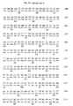
- the human ICD is set forth in Fig. 7 and SEQ ID NO:1 as inclusively spanning the region of Lys 676 to Val 1255.
- the rat ICD is set forth in Fig. 8 and SEQ ID NO:2 as inclusively spanning the region of Lys 677 to Val 1256.
- the PD of the present invention is preferably a human, a rat or a mouse PD.
- the human PD is set forth in Fig. 10 and as SEQ ID NO:4.
- the human PD may be the human ⁇ PD, which is set forth in Fig. 11 and as SEQ ID NO:5.
- the rat PD is shown in Fig. 8 and SEQ ID NO:2 as inclusively spanning the region of Gln 991 to Val 1256.
- the rat PD may be the rat ⁇ PD, which is shown in Fig. 8 and SEQ ID NO:2 as inclusively spanning the region of Gln 991 to Arg 1049.
- a human ECD can be fused with a human PD or ⁇ PD, or a rat PD or ⁇ PD.
- a rat ECD can be fused with a human PD or ⁇ PD, or a rat PD or ⁇ PD.
- the HER-2/neu PD is 268 amino acids in length, is intracellular, and can be phosphorylated by protein tyrosine kinases. This region shares no identity with the corresponding part of other tyrosine kinase receptors. Thus, the specificity and uniqueness of this domain makes it particularly preferred for use as a tumor vaccine. However, the expression of this domain alone in bacteria and mammalian cells is problematic. For example, the resultant PD protein is very labile and is not appropriate for large scale production. In one embodiment, this invention has solved such problems by fusing all or part of the intracellular domain or the phosphorylation domain to all or part of the HER-2/neu extracellular domain.
- the ECD-ICD fusion proteins and the ECD-PD fusion proteins of the invention and the disclosure are soluble, are secreted and are stable in culture media.
- This system can provide large quantities of intracellular domain or phosphorylation domain protein for cancer vaccine development, preferably breast cancer vaccine development, but will be useful for vaccines against any cancer characterized by HER-2/neu expression.
- the ECD-ICD and ECD-PD fusion proteins provide for an improved vaccine formulation.
- the PD was secreted by introducing a secretion signal sequence preceding the N-terminus of the PD to yield a soluble, secreted recombinant protein.
- the secretion process is preferred since the recombinant proteins accumulate in the culture media. Because the protein is not associated with the intracellular proteins, proteolysis is limited. The protein should be purified more easily and economically.
- pFLAGCMV-1 expression plasmid (Kodak) was used to determine which region of the HER-2/neu intracellular domain was able to be secreted.
- the proteins were expressed as fusion proteins with a preprotrypsin secretion signal and FLAG-Tag at their N-terminus.
- HEK-293 cells were transfected with such constructs, and cell and culture media were assayed for FLAG-Tag fusion proteins by western blot with FLAG-Tag M2 antibody as probe.
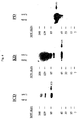
- the results in Fig. 4 demonstrate that neither full length ICD nor KD was secreted, but that the PD was soluble and was secreted and detected in the culture media. The results indicate that the full length structure ICD or KD protein did not result in secretion, that is in passage of the protein through the cell membrane.
- the ECD since the ECD has a secretion signal sequence and can be expressed well as a secreted protein, the ECD was used as a fusion partner for PD.
- the ECD-PD fusion protein was expressed in HEK-293 cells.
- the secretion of soluble ECD-PD fusion protein was determined by ELISA assay with HER-2/neu ECD-specific antibodies, followed by western blot with HER-2/neu PD-specific antibodies.
- the soluble ECD-PD expressed in HEK-293 and was secreted into the culture medium.
- the present invention is directed to a fusion protein based on particular portions (e.g ., HER-2/neu ECD-PD fusion protein) of the protein expression product of the HER-2/neu gene, which is capable of eliciting an antibody response and can be recognized by thymus-dependent lymphocytes ("T-cells"). Accordingly, the autochthonous immune T cell response can be used prophylactically or to treat malignancies in which HER-2/neu is or has been overexpressed.
- the present invention is directed to the use of nucleic acid molecules directing the expression of such ECD-ICD fusion proteins or ECD-PD fusion proteins, or their variants, alone or in a viral vector for immunization.
- CD4 + T cell populations are considered to function as helpers or inducers through the release of lymphokines when stimulated by a specific antigen; however, a subset of CD4 + cells can act as cytotoxic T lymphocytes (CTL).
- CD8 + T cells are considered to function by directly lysing antigenic targets; however, under a variety of circumstances they can secrete lymphokines to provide helper or DTH function.
- the phenotypic CD4 and CD8 markers are linked to the recognition of peptides bound to class I or class II MHC antigens.
- an ECD-ICD fusion protein or an ECD-PD fusion protein of the protein expression product of the HER-2/neu oncogene is recognized by T cells. Circulating HER-2/neu ECD-ICD fusion protein or HER-2/neu ECD-PD fusion protein is degraded to peptide fragments. Peptide fragments from the ECD-ICD fusion protein or ECD-PD fusion protein bind to major histocompatibility complex (MHC) antigens.
- MHC major histocompatibility complex
- the HER-2/neu ECD-ICD or the HER-2/neu ECD-PD fusion protein (including that expressed on a malignant cell) will be immunogenic to T cells.
- the extraordinar specificity of the T cell receptor enables individual T cells to discriminate between protein fragments which differ by a single amino acid residue.
- T cells expressing a T cell receptor with high affinity binding of the peptide-MHC complex will bind to the peptide-MHC complex and thereby become activated and induced to proliferate.
- small numbers of immune T cells will secrete lymphokines, proliferate and differentiate into effector and memory T cells.
- the primary immune response will occur in vivo but has been difficult to detect in vitro.
- a subsequent encounter with the same antigen by the memory T cells will lead to a faster and more intense immune response.
- the secondary response will occur either in vivo or in vitro.
- the in vitro response is easily gauged by measuring the degree of proliferation, the degree of cytokine production, or the generation of cytolytic activity of the T cell population re-exposed in the antigen.
- Substantial proliferation of the T cell population in response to a particular antigen is considered to be indicative of prior exposure to or priming with the antigen.
- the compounds of the present invention comprise HER-2/neu ECD-PD fusion proteins, or variants, or nucleic acid molecules that encode such ECD-PD fusion proteins.
- the HER-2/neu ECD is fused to the HER-2/neu ⁇ PD.
- the nucleic acid molecules are DNA molecules.
- the ECD and PD or ⁇ PD polypeptide components can be directly fused or fused via a linker, e.g ., a peptide linker.
- the ECD-PD fusion protein of the present invention comprises the HER-2/neu ECD fused directly to the HER-2/neu PD or to the HER-2/neu ⁇ PD.
- a preferred embodiment of the fusion proteins of the invention is the HER-2/neu PD fusion protein.
- the size of the ECD in the ECD-PD fusion protein is altered by sequentially removing anywhere from 1 to about 100 amino acids from the carboxyl terminus of the ECD, preferably about 100 amino acids.
- the size of the PD in the ECD-PD fusion protein can be altered by sequentially removing amino acids from the N-terminus of the PD.
- the preferred embodiment is PD.
- Other variant forms may be selected on the basis of their antigenicity and/or immunogenicity using appropriate screening methods as described in the literature and herein, for use according to the invention.
- Table 1 shows that removing 100 amino acids from the carboxyl terminus of the ECD does not have any impact on the expression level and stability of the ECD-PD fusion protein.
- Table 1 ECD truncated -PD Summary Clone I.D. area of deletion (bp/aa) # of bp # of aa relative expression A5 1655-1882/552-628 228 76 *** B4 1660-1866 / 554-622 207 69 ** B9 1595-1891 / 532-631 297 99 ** C2 1681-1902 / 561-634 222 74 * C7 1612-1902 / 538-634 291 97 ***** F10 1634-1951 / 545-651 318 106 **** ECD-PD WT - - - *
- Variants of the ECD-PD fusion protein also include various structural forms of the native ECD-PD fusion protein. Due to the presence of ionizable amino and carboxyl groups, for example, a ECD-PD fusion protein can be in the form of an acidic or basic salt, or may be in a neutral form. Individual amino acid residues may also be modified by oxidation or reduction.
- ECD-PD fusion proteins in which the primary amino acid structure native HER-2/neu ECD-PD protein, respectively, is modified by forming covalent or aggregative conjugates with other peptides or polypeptides, or chemical moieties such as glycosyl groups, lipids, phosphate, acetyl groups and the like.
- Covalent derivatives may be prepared, for example, by linking particular functional groups to amino acid side chains or at the N- or C-terminus.
- the present invention also includes HER-2/neu HER-2/neu ECD-PD fusion proteins with or without glycosylation.
- ECD-PD fusion proteins expressed in yeast or mammalian expression systems may be similar to, or slightly different in molecular weight and glycosylation pattern from, the native molecules, depending upon the expression system.
- Expression of DNA encoding polypeptides in bacteria such as E. coli typically provides non-glycosylated molecules.
- N-glycosylation sites of eukaryotic proteins are characterized by the amino acid triplet Asn-A 1 -Z, wherein A 1 is any amino acid except Pro, and Z is Ser or Thr.
- Variants of HER-2/neu ECD-PD fusion proteins having inactivated N-glycosylation sites can be produced by techniques known to one skilled in the art, such as oligonucleotide synthesis and ligation or site-specific mutagenesis techniques, and are within the scope of the invention.
- N-linked glycosylation sites can be added to a HER-2/neu ECD-PD fusion protein.
- the ECD-ICD fusion proteins of the present disclosure include any possible combination between human and non-human polypeptides.
- Non-human polypeptides comprise polypeptides from any mammal, such as, e.g., rat, mouse, guinea pig, horse, cow, pig, sheep, dog, etc.
- the ECD-ICD fusion proteins include:
- ECD-ICD fusion proteins of the present disclosure are included as embodiments of the present disclosure.
- such variants are substantially identical or substantially similar to the native HER-2/neu ECD-ICD protein and retain the ability to stimulate an immune response.
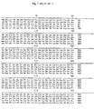
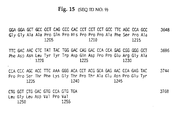
- Human DNA sequences that encode the ECD protein are shown, for example, in Fig. 15 (SEQ ID NO:9) as inclusively spanning nucleotide 1 to nucleotide 1959.
- Human DNA sequences that encode the ICD protein are shown, for example, in Fig. 15 (SEQ ID NO:9) as inclusively spanning nucleotide 2026 to nucleotide 3765.
- any sequence modification on the ability of a HER-2/neu ECD-ICD protein to produce an immune response may be readily determined, for example, by analyzing the ability of the mutated HER-2/neu ECD-ICD protein to induce a T cell response using, for example, the methods described herein, or by analyzing the ability of the mutated HER-2/neu ECD-ICD protein to produce antibodies.
- the ECD-PD fusion proteins of the present invention include any possible combination between human and non-human polypeptides.
- Non-human polypeptides comprise, e.g ., rat, mouse, guinea pig, horse, cow, pig, sheep, dog, etc.
- the ECD-PD fusion proteins include:
- ECD-PD fusion proteins of the present invention are included as embodiments of the present invention.
- such variants are substantially identical or substantially similar to the native HER-2/neu ECD-PD protein and retain the ability to stimulate an immune response.
- Human DNA sequences that encode the ECD protein are shown, for example, in Fig. 15 (SEQ ID NO:9) as inclusively spanning nucleotide 1 to nucleotide 1959.
- Human DNA sequences that encode the PD protein are shown, for example, in Fig. 15 (SEQ ID NO:9) as inclusively spanning nucleotide 2968 to nucleotide 3765.
- any sequence modification on the ability of a HER-2/neu ECD-PD protein to produce an immune response may be readily determined, for example, by analyzing the ability of the mutated HER-2/neu ECD-PD protein to induce a T cell response using, for example, the methods described herein, or by analyzing the ability of the mutated HER-2/neu ECD-PD protein to produce antibodies.
- the ECD-PD fusion proteins are ECD- ⁇ PD fusion proteins of the present invention, which will be understood to include variants, including any possible combination between human and non-human polypeptides.
- Non-human polypeptides comprise, e.g ., rat, mouse, guinea pig, horse, cow, pig, sheep, dog, etc.
- the ECD- ⁇ PD fusion proteins include:
- ECD- ⁇ PD fusion proteins of the present invention are included as embodiments of the present invention.
- such variants are substantially identical or substantially similar to the native HER-2/neu ECD- ⁇ PD protein and retain the ability to stimulate an immune response.
- Human DNA sequences that encode the ECD protein are shown, for example, in Fig. 15 (SEQ ID NO:9) as inclusively spanning nucleotide 1 to nucleotide 1959.
- Human DNA sequences that encode the ⁇ PD protein are shown, for example, in Fig. 15 (SEQ ID NO:9) as inclusively spanning nucleotide 2968 to nucleotide 3144.
- any sequence modification on the ability of a HER-2/neu ECD- ⁇ PD protein to produce an immune response may be readily determined, for example, by analyzing the ability of the mutated HER-2/neu ECD- ⁇ PD protein to induce a T cell response using, for example, the methods described herein, or by analyzing the ability of the mutated HER-2/neu ECD- ⁇ PD protein to produce antibodies.
- the HER-2/neu ECD-PD fusion proteins of the invention are ECD-PD fusion proteins.
- the fusion proteins of the invention may comprise a fusion partner, such as, e.g ., an immunological fusion partner or an expression enhancer.
- a fusion partner may, for example, assist in providing T helper epitopes (an immunological fusion partner), preferably T helper epitopes recognized by humans, or may assist in expressing the fusion protein (an expression enhancer) at higher yields than the recombinant fusion protein.
- Certain preferred fusion partners are both immunological and expression enhancing fusion partners.
- Other fusion partners may be selected so as to increase the solubility of the fusion protein or to enable the fusion protein to be targeted to desired intracellular compartments.
- Still further fusion partners include affinity tags, which facilitate purification of the fusion protein.
- fusion proteins that comprise a fusion polypeptide as described herein together with an unrelated immunogenic protein.
- the immunogenic protein is capable of eliciting a recall response.
- examples of such proteins include tetanus, tuberculosis and hepatitis proteins (see, e.g., Stoute et al. (1997) New Engl. J. Med. 336:86-91 ).
- an immunological fusion partner is derived from protein D, a surface protein of the gram-negative bacterium Haemophilus influenza B ( WO 91/18926 ).
- a protein D derivative comprises approximately the first third of the protein (e.g ., the first N-terminal 100-110 amino acids), and a protein D derivative may be lipidated.
- the first 109 residues of a Lipoprotein D fusion partner is included on the N-terminus to provide the fusion protein with additional exogenous T-cell epitopes and to increase the expression level in E. coli (thus functioning as an expression enhancer).
- the lipid tail ensures optimal presentation of the fusion protein to antigen presenting cells.
- Other fusion partners include the non-structural protein from influenzae virus, NS1 (hemaglutinin). Typically, the N-terminal 81 amino acids are used, although different fragments that include T-helper epitopes may be used.
- the immunological fusion partner is the protein known as LYTA, or a portion thereof (preferably a C-terminal portion).
- LYTA is derived from Streptococcus pneumoniae, which synthesizes an N-acetyl-L-alanine amidase known as amidase LYTA (encoded by the LytA gene; Gene 43:265-292, 1986 ).
- LYTA is an autolysin that specifically degrades certain bonds in the peptidoglycan backbone.
- the C-terminal domain of the LYTA protein is responsible for the affinity to the choline or to some choline analogues such as DEAE. This property has been exploited for the development of E.
- coli C-LYTA expressing plasmids useful for expression of fusion proteins. Purification of hybrid proteins containing the C-LYTA fragment at the amino terminus has been described ( see Biotechnology 10:795-798, 1992 ). Within a preferred embodiment, a repeat portion of LYTA may be incorporated into a fusion protein. A repeat portion is found in the C-terminal region starting at residue 178. A particularly preferred repeat portion incorporates residues 188-305.
- the fusion proteins of the present invention further comprise a fusion partner.
- Preferred fusion partners include, but are not limited to, e.g ., Ra12 or LeIF.
- the invention provides materials and methods for using a Ra12 or a LeIF sequence as a fusion partner to facilitate the stable and high yield expression of recombinant fusion polypeptides or break tolerance.
- Ra12 is a 14 kD C-terminal fragment of the M. tuberculosis MTB32A coding sequence, which is expressed at high levels on its own and remains soluble throughout the purification process.
- LeIF is a Leishmania antigen which is homologous to the eukaryotic ribosomal protein eIF and which is capable of stimulating a Th1 and/or CTL immune response ( See, e.g., U.S. Patent Nos. 5,876,966 , 5,876,735 , and 5,879,687 ).
- the present invention utilizes these properties of the Ra12 and LeIF polypeptides and provides recombinant nucleic acid molecules; expression vectors, host cells, and methods for stable and high yield expression of fusion polypeptides comprising, in addition to the HER-2/neu fusion polypeptides, a Ra12 or LeIF polypeptide.
- Recombinant nucleic acids which encode a fusion polypeptide comprising a Ra12 or LeIF polypeptide and a fusion protein of interest, can be readily constructed by conventional genetic engineering techniques. Recombinant nucleic acids are constructed so that, preferably, the fusion partner polynucleotide sequence is located 5' to a selected fusion protein sequence.
- fusion partner polynucleotide sequence 3' may also be appropriate to place an fusion partner polynucleotide sequence of the fusion protein of interest or to insert the polynucleotide sequence of the fusion protein into a site within an fusion partner polynucleotide sequence.
- any suitable polynucleotide that encodes a fusion partner or a portion or other variant thereof as described herein can be used in constructing recombinant fusion nucleic acids of the present invention.
- Nucleic acids encoding the fusion partner polypeptides of this invention can be prepared by any suitable method known in the art. Exemplary methods include cloning and restriction of appropriate sequences or direct chemical synthesis by methods such as the phosphotriester method of Narang et al. (1979) Meth. Enzymol. 68: 90-99 ; the phosphodiester method of Brown et al. (1979) Meth. Enzymol. 68: 109-151 ; the diethylphosphoramidite method of Beaucage et al. (1981) Tetra. Lett., 22: 1859-1862 ; and the solid support method of U.S. Patent No. 4,458,066 .
- Recombinant nucleic acids that encode a fusion polypeptide comprising an fusion partner polypeptide and a selected fusion protein can be prepared using any methods known in the art. As described above, recombinant nucleic acids are constructed so that, preferably, the fusion partner polynucleotide sequence is located 5' to the polynucleotide sequence encoding the fusion protein of interest. The fusion partner and fusion protein polynucleotide sequences can also be modified to facilitate their fusion and subsequent expression.
- the recombinant nucleic acids can further comprise other nucleotide sequences such as sequences that encode affinity tags to facilitate protein purification protocol.
- CD4 + T cells generally recognize antigens that have been external to the tumor cells.
- binding of peptides to class I MHC occurs only for proteins present in the cytosol and synthesized by the target itself, proteins in the external environment are excluded.
- An exception to this is the binding of exogenous peptides with a precise class I binding motif which are present outside the cell in high concentration.
- CD4 + and CD8 + T cells have broadly different functions and tend to recognize different antigens as a reflection of where the antigens normally reside.
- Another way to make amino acid substitutions to produce variants of the present invention is to identify and replace amino acids in T cell motifs with potential to bind to class II MHC molecules (for CD4 + T cell response) or class I MHC molecules (for CD8 + T cell response).
- Peptide segments (of a HER-2/neu ECD-PD fusion protein) with a motif with theoretical potential to bind to class II MHC molecules may be identified by computer analysis.
- a protein sequence analysis package, T Sites that incorporates several computer algorithms designed to distinguish potential sites for T cell recognition can be used ( Feller et al. (1991) Nature, 349:720-721 ). Two searching algorithms are used: (1) the AMPHI algorithm described by Margalit ( Feller et al.
- a peptide motif for binding in the groove of HLA-A2.1 has been defined by Edman degradation of peptides stripped from HLA-A2.1 molecules of a cultured cell line (Table 2, from Falk et al. (1991) Nature, 351:290-296 ).
- the method identified the typical or average HLA-A2.1 binding peptide as being 9 amino acids in length with dominant anchor residues occurring at positions 2 (L) and 9 (V). Commonly occurring strong binding residues have been identified at positions 2 (M), 4 (E,K), 6 (V), and 8 (K).
- the identified motif represents the average of many binding peptides.
- the derived peptide motif as currently defined is not particularly stringent. Some HLA-A2.1 binding peptides do not contain both dominant anchor residues and the amino acids flanking the dominant anchor residues play major roles in allowing or disallowing binding. Not every peptide with the current described binding motif will bind, and some peptides without the motif will bind. However, the current motif is valid enough to allow identification of some peptides capable of binding. Of note, all MHC molecules and respective motifs place 6 amino acids between the dominant anchor amino acids at residues 2 and 9.
- amino acid substitutions can be made conservatively or non-conservatively. The latter type of substitutions are intended to produce an improved ECD-PD fusion protein or polypeptide that is more potent and/or more broadly cross-reactive.
- An example of a more potent protein or peptide is one that binds with higher affinity to the same MHC molecule as the natural protein or polypeptide, without affecting recognition by T cells specific for natural protein or polypeptide.
- An example of a polypeptide with broader cross-reactivity is one that induces more broadly cross-reactive immune responses ( i.e ., binds to a greater range of MHC molecules) than natural polypeptide.
- one or more amino acids residing between peptide motifs and having a spacer function can be substituted conservatively or non-conservatively. It will be evident to one of ordinary skill in the art that polypeptides containing one or more amino acid substitutions can be tested for beneficial or adverse immunological interactions by a variety of assays, including those described herein for the ability to stimulate T cell recognition.
- Variants within the scope of this invention can also, or alternatively, contain other modifications, including the deletion or addition of amino acids, that have minimal influence on the desired immunological properties of the polypeptide, as described supra. It will be appreciated by one of ordinary skill in the art that truncated forms or non-native extended forms of a HER-2/neu ECD-PD fusion protein can be used, provided the desired immunological properties are at least roughly equivalent to that of full length, native HER-2/neu ECD-PD fusion protein. Cysteine residues may be deleted or replaced with other amino acids to prevent formation of incorrect intramolecular disulfide bridges upon renaturation. Other approaches to mutagenesis involve modification of adjacent dibasic amino acid residues to enhance expression in yeast systems in which KEX2 protease activity is present.
- the invention relates to isolated or purified polynucleotides that encode the HER-2/neu fusion proteins.
- any nucleotide sequence which encodes the amino acid sequence of the fusion protein can be used to generate recombinant molecules which direct the expression of the fusion protein.
- labeled DNA probes designed from any portion of the HER-2/neu nucleotide sequences or their complements may be used to screen a genomic or cDNA library, to identify the coding sequence of each individual component of the fusion protein.
- the Her-2/neu nucleotide sequences can be from any suitable mammal, e.g., rat, mouse, horse, cow, pig, sheep, dog, etc.
- the Her-2/neu sequence is from a human, a rat, or a mouse.
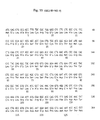
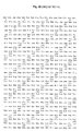
- the sequence of mouse Her-2/neu is shown in Figure 19 (SEQ ID NO:11).
- Mouse Her-2/neu can also be amplified from mouse brain RNA using the following primers: 5' primer: CCATGGAGCTGGCGGCCTGGTGCCGTTG (SEQ ID NO:12) and 3' primer: GGCCTTCTGGTTCATACTGGCACATCCAGGC (SEQ ID NO:13).
- the mouse Her-2/neu amino acid sequence is shown in Figure 20 (SEQ ID NO:14).
- a variant amino acid sequence for mouse Her-2/neu is described in Nagata et al., J. Immunol. 159:1336-1343 (1997 ).
- Such clones may be isolated by screening an appropriate expression library for clones that express full length HER-2/neu protein.
- the library preparation and screen may generally be performed using methods known to one of ordinary skill in the art, such as methods described in Sambrook et al., Molecular Cloning: A Laboratory Manual, Cold Spring Harbor Laboratories, Cold Spring Harbor, NY (1989 ), which is incorporated herein by reference. Briefly, a bacteriophage expression library may be plated and transferred to filters. The filters may then be incubated with a detection reagent.
- a "detection reagent" is any compound capable of binding to HER-2/neu protein, which may then be detected by any of a variety of means known to one of ordinary skill in the art.
- Typical detection reagents contain a "binding agent," such as Protein A, Protein G, IgG or a lectin, coupled to a reporter group.
- reporter groups include enzymes, substrates, cofactors, inhibitors, dyes, radionuclides, luminescent groups, fluorescent groups and biotin. More preferably, the reporter group is horseradish peroxidase, which may be detected by incubation with a substrate such as tetramethylbenzidine or 2,2'-azino-di-3-ethylbenz-thiazoline sulfonic acid.
- Plaques containing genomic or cDNA sequences that express HER-2/neu protein are isolated and purified by techniques known to one of ordinary skill in the art. Appropriate methods may be found, for example, in Sambrook et al., Molecular Cloning A Laboratory Manual, Cold Spring Harbor Laboratories, Cold Spring Harbor, NY (1989 ).
- Isolation of coding sequences may also be carried out by the polymerase chain reactions (PCR) using two degenerate oligonucleotide primer pools designed on the basis of the coding sequences disclosed herein.
- the desired nucleic acids can also be cloned using other well known amplification techniques. Examples of protocols sufficient to direct persons of skill through in vitro amplification methods, including PCR, ligase chain reaction (LCR), Q ⁇ -replicase amplification and other RNA polymerase mediated techniques are found in Sambrook, and Ausubel, as well as U.S. Patent No. 4,683,202 ; PCR Protocols A Guide to Methods and Applications (Innis et al. eds.
- a polynucleotide of the invention which encodes a fusion protein, fragments thereof, or functional equivalents thereof may be used to generate recombinant nucleic acid molecules that direct the expression of the fusion protein, fragments thereof, or functional equivalents thereof, in appropriate host cells.
- the fusion polypeptide products encoded by such polynucleotides may be altered by molecular manipulation of the coding sequence.
- DNA sequences which encode substantially the same or a functionally equivalent amino acid sequence, may be used in the practice of the invention for the expression of the fusion polypeptides.
- DNA sequences include those which are capable of hybridizing to the coding sequences or their complements disclosed herein under low, moderate or high stringency conditions described herein.
- Altered nucleotide sequences which may be used in accordance with the invention include deletions, additions or substitutions of different nucleotide residues resulting in a sequence that encodes the same or a functionally equivalent gene product.
- the gene product itself may contain deletions, additions or substitutions of amino acid residues, which result in a silent change thus producing a functionally equivalent antigenic epitope.
- conservative amino acid substitutions may be made on the basis of similarity in polarity, charge, solubility, hydrophobicity, hydrophilicity, and/or the amphipathic nature of the residues involved.
- negatively charged amino acids include aspartic acid and glutamic acid; positively charged amino acids include lysine, histidine and arginine; amino acids with uncharged polar head groups having similar hydrophilicity values include the following: glycine, asparagine, glutamine, serine, threonine and tyrosine; and amino acids with nonpolar head groups include alanine, valine, isoleucine, leucine, phenylalanine, proline, methionine and tryptophan.
- nucleotide sequences of the invention may be engineered in order to alter the fusion protein coding sequence for a variety of ends, including but not limited to, alterations which modify processing and expression of the gene product.
- mutations may be introduced using techniques which are well known in the art, e.g ., to insert or delete restriction sites, to alter glycosylation patterns, phosphorylation, to create and/or destroy translation, initiation, and/or termination sequences, or to create variations in coding regions, to facilitate further in vitro modification, etc.
- One of skill will recognize many ways of generating alterations in a given nucleic acid construct.
- Such well-known methods include, e.g ., site-directed mutagenesis, PCR amplification using degenerate oligonucleotides, exposure of cells containing the nucleic acid to chemical mutagenic agents or radiation, chemical synthesis of a desired oligonucleotide (e.g ., in conjunction with ligation and/or cloning to generate large nucleic acids) and other well-known techniques (see, e.g., Giliman et al. (1979) Gene 8:81-97 , Hutchinson, et al. (1978) J. Biol. Chem. 253:6551 ; Roberts et al. (1987) Nature 328: 731-734 ).
- the manipulations do not destroy immunogenicity of the fusion polypeptides.
- the coding sequence of a fusion protein could be synthesized in whole or in part, using chemical methods well known in the art (see, e.g., Caruthers et al. (1980) Nuc. Acids Res. Symp. Ser. 7:215-233 ; Crea et al. (1980) Nuc. Acids Res. 9(10):2331 ; Matteucci et al. (1980) Tetrahedron Letter 21:719 (1980 ); and Chow et al. (1981) Nuc. Acids Res. 9(12):2807-2817 ).
- the fusion polypeptide itself can be produced using chemical methods to synthesize an amino acid sequence in whole or in part.
- peptides can be synthesized by solid phase techniques, such as, e.g ., the Merrifield solid phase synthesis method, in which amino acids are sequentially added to a growing chain of amino acids (see, Merrifield (1963) J. Am. Chem. Soc., 85:2149-2146 ).
- Equipment for automated synthesis of polypeptides is commercially available from suppliers such as Perkin Elmer Biosystems, Inc. (Foster City, CA), and may generally be operated according to the manufacturer's instructions.
- the synthesized peptides can then be cleaved from the resin, and purified, e.g ., by preparative high performance liquid chromatography (see Creighton, Proteins Structures and Molecular Principles, 50-60 (1983 )).
- the composition of the synthetic fusion polypeptides may be confirmed by amino acid analysis or sequencing (e.g ., the Edman degradation procedure; see Creighton, Proteins, Structures and Molecular Principles, pp. 34-49 (1983 )).
- nonclassical amino acids or chemical amino acid analogs can be introduced as a substitution or addition into the sequence.
- Non-classical amino acids include, but are not limited to, the D-isomers of the common amino acids, ⁇ -amino isobutyric acid, 4-aminobutyric acid, Abu, 2-amino butyric acid, ⁇ -Abu, ⁇ -Ahx, 6-amino hexanoic acid, Aib, 2-amino isobutyric acid, 3-amino propionic acid, ornithine, norleucine, norvaline, hydroxyproline, sarcosine, citrulline, cysteic acid, t-butylglycine, t-butylalanine, phenylglycine, cyclohexylalanine, ⁇ -alanine, fluoro-amino acids, designer amino acids such as ⁇ -methyl amino acids, C ⁇ -methyl amino acids, N ⁇ -methyl amino acids, and amino acid analogs
- the polypeptides of the fusion protein are joined via a linking group.
- the linking group can be a chemical crosslinking agent, including, for example, succinimidyl-(N-maleimidomethyl)-cyclohexane-1-carboxylate (SMCC).
- SMCC succinimidyl-(N-maleimidomethyl)-cyclohexane-1-carboxylate
- the linking group can also be an additional amino acid sequence(s), including, for example, a polyglycine linking group.
- the coding sequences of each polypeptide in the fusion protein are directly joined at their amino- or carboxy-terminus via a peptide bond in any order.
- an amino acid linker sequence may be employed to separate the first and second polypeptide components by a distance sufficient to ensure that each polypeptide folds into its secondary and tertiary structures.
- Such an amino acid linker sequence is incorporated into the fusion protein using standard techniques well known in the art. Suitable peptide linker sequences may be chosen based on the following factors:
- Other chemical linkers include carbohydrate linkers, lipid linkers, fatty acid linkers, polyether linkers, e.g ., PEG, etc. ( See, e.g., Hermanson, Bioconjugate Techniques (1996 )).
- the fusion protein may be linked to one or more additional polypeptides.
- the fusion polypeptide may be linked to one or more copies of one of the two polypeptides of the fusion protein.
- the fusion protein may be linked to an additional heterologous polypeptide, such as, e.g ., Ra12 or LeIF, as described supra.
- the fusion polypeptide may also be fused to an affinity tag for ease of purification upon expression. For example, multiple histidine residues encoded by the tag allow the use of metal chelate affinity chromatography methods for the purification of fusion polypeptides.
- affinity tag molecules include, e.g ., Strep-tag, PinPoint, maltose binding protein, glutathione S-transferase, etc. ( see, e.g., Glick & Pasternak, Molecular Biotechnology Principles and Applications of Recombinant DNA (2nd ed. 1999 )).
- the fusion polypeptide is optionally linked to a lipid moiety, such as mycolic acid, lipoaribdomanin ("LAMs”), or trehalose derivatives.
- a lipid moiety such as mycolic acid, lipoaribdomanin ("LAMs"), or trehalose derivatives.
- such a fusion protein is produced by recombinant expression of a nucleic acid encoding the fusion protein.
- a fusion product can be made by ligating the appropriate nucleic acid sequences encoding the desired amino acid sequences to each other by methods known in the art, in the proper coding frame, and expressing the product by methods known in the art.
- such a product may be made by protein synthetic techniques, e.g ., by use of a peptide synthesizer. Coding sequences for other molecules such as a cytokine or an adjuvant can be added to the fusion polynucleotide as well.
- Variants of the fusion proteins of the invention that retain the ability to stimulate an immune response may generally be identified by modifying the sequence in one or more of the aspects described above and assaying the resulting fusion protein for the ability to stimulate an immune response, e.g ., a T cell response or an antibody response.
- assays may generally be performed by contacting T cells with the modified fusion protein and assaying the response.
- Naturally occurring variants of the individual polypeptide components of the fusion protein may also be isolated by, for example, screening an appropriate cDNA or genomic library with a DNA sequence encoding each individual polypeptide or a variant thereof.
- sequence modifications may be introduced using standard recombinant techniques or by automated synthesis of the modified fusion protein.
- mutations can be introduced at particular loci by synthesizing oligonucleotides containing a mutant sequence, flanked by restriction sites enabling ligation to fragments of the native sequence. Following ligation, the resulting reconstructed sequence encodes an analogue having the desired amino acid insertion, substitution, or deletion.
- oligonucleotide-directed site-specific mutagenesis procedures can be used to provide a gene in which particular codons are altered according to the substitution, deletion, or insertion required.
- Exemplary methods of making the alterations set forth above are described by Walder et al. (1986) Gene, 42:133 ; Bauer et al. (1985) Gene, 37:73 ; Craik (1985) BioTechniques, January:12-19 ; Smith et al., Genetic Engineering: Principles and Methods, Plenum Press (1981 ); and U.S. Patent Nos. 4,518,584 and 4,737,462 .
- Mutations in nucleotide sequences constructed for expression of such HER-2/neu fusion proteins must, of course, preserve the reading frame of the coding sequences and preferably will not create complementary regions that could hybridize to produce secondary mRNA structures, such as loops or hairpins, which would adversely affect translation of the mRNA.
- a mutation site may be predetermined, it is not necessary that the nature of the mutation per se be predetermined. For example, in order to select for optimum characteristics of mutants at a given site, random mutagenesis may be conducted at the target codon and the expressed HER-2/neu fusion protein mutants screened for the desired activity. Not all mutations in a nucleotide sequence which encodes a HER-2/neu fusion protein will be expressed in the final product.
- nucleotide substitutions may be made to enhance expression, primarily to avoid secondary structure loops in the transcribed mRNA (see, e.g., European Patent Application 75,444A ), or to provide codons that are more readily translated by the selected host, such as the well-known E. coli preference codons for E. coli expression.
- Her-2/neu fusion proteins, and variants thereof, of the present invention are preferably produced by recombinant DNA methods. Such methods include inserting a DNA sequence encoding a HER-2/neu fusion protein into a recombinant expression vector and expressing the DNA sequence in a recombinant microbial, mammalian, fungal or insect cell expression system under conditions promoting expression and, preferably, secretion of the fusion protein.
- DNA sequences encoding the Her-2/neu fusion proteins provided by this invention can be assembled from cDNA fragments and short oligonucleotide linkers, or from a series of oligonucleotides, to provide a synthetic gene which is capable of being inserted in a recombinant expression vector and expressed in a recombinant transcriptional unit.
- Recombinant expression vectors contain a DNA sequence encoding a HER-2/neu fusion protein operably linked to suitable transcriptional or translational regulatory elements derived from mammalian, fungal, microbial, viral or insect genes.
- suitable transcriptional or translational regulatory elements include a transcriptional promoter, an optional operator sequence to control transcription, a sequence encoding suitable mRNA ribosomal binding sites, and sequences which control the termination of transcription and translation.
- An origin of replication and a selectable marker to facilitate recognition of transformants may additionally be incorporated.
- DNA regions are operably linked when they are functionally related to each other.
- DNA for a signal peptide secretory leader
- DNA for a polypeptide if it is expressed as a precursor which participates in the secretion of the polypeptide
- a promoter is operably linked to a coding sequence if it controls the transcription of the sequence
- a ribosome binding site is operably linked to a coding sequence if it is positioned so as to permit translation.
- "operably linked” means contiguous and, in the case of secretory leaders, in reading frame.
- DNA sequences encoding HER-2/neu fusion proteins which are to be expressed in a microorganism will preferably contain no introns that could prematurely terminate transcription of DNA into mRNA.
- Expression vectors for bacterial use may comprise a selectable marker and bacterial origin of replication derived from commercially available plasmids comprising genetic elements of the well known cloning vector pBR322 (ATCC 37017).
- Such commercial vectors include, e.g ., pKK223-3 (Pharmacia Fine Chemicals, Uppsala, Sweden), pGEM1 (Promega Biotec, Madison, WI), pET28b (Novagen) and pPDM (a modified pET28b, Corixa).
- pBR322 "backbone" sections are combined with an appropriate promoter and the structural sequence to be expressed.
- E. coli is typically transformed using derivatives of pBR322, a plasmid derived from an E.
- pBR322 contains genes for ampicillin and tetracycline resistance and thus provides simple means for identifying transformed cells.
- Promoters commonly used in recombinant microbial expression vectors include the ⁇ -lactamase (penicillinase) and lactose promoter system ( Chang et al. (1978) Nature, 275:615 ; and Goeddel et al. (1979) Nature, 281:544 ), the tryptophan (trp) promoter system ( Goeddel et al. (1980) Nucl.
- Plasmid vectors available from the American Type Culture Collection which incorporate derivatives of the ⁇ P L promoter include plasmid pHUB2, resident in E. coli strain JMB9 (ATCC 37092) and pPLc28, resident in E. coli RR1 (ATCC 53082).
- Suitable promoter sequences in yeast vectors include the promoters for alcohol oxidase, metallothionein, 3-phosphoglycerate kinase ( Hitzeman et al. (1980) J. Biol. Chem., 255:2073 ) or other glycolytic enzymes ( Hess et al. (1968) J. Adv. Enzyme Reg., 7:149 ; and Holland et al.
- Preferred yeast vectors can be assembled using DNA sequences from pBR322 for selection and replication in E. coli (Amp r gene and origin of replication) and yeast DNA sequences including a glucose-repressible ADH2 promoter and ⁇ -factor secretion leader.
- the ADH2 promoter has been described by Russell et al. (1982) J Biol. Chem., 258:2674 and Beier et al. (1982) Nature, 300:724 .
- the yeast ⁇ -factor leader which directs secretion of heterologous proteins, can be inserted between the promoter and the structural gene to be expressed (see, e.g., Kurjan et al. (1982) Cell, 30:933 ; and Bitter et al. (1984) Proc. Natl. Acad. Sci. USA, 81:5330 .
- the leader sequence may be modified to contain, near its 3' end, one or more useful restriction sites to facilitate fusion of the leader sequence to foreign genes.
- the transcriptional and translational control sequences in expression vectors to be used in transforming vertebrate cells may be provided by viral sources.
- viral sources commonly used promoters and enhancers are derived, e.g ., from polyoma, adenovirus 2, simian virus (SV40), and human cytomegalovirus.
- DNA sequences derived from the SV40 viral genome for example, the SV40 origin, early and late promoter, enhancer, splice, and polyadenylation sites may be used to provide the other genetic elements required for expression of a heterologous DNA sequence.
- the early and late promoters are particularly useful because both are obtained easily from the virus as a fragment which also contains the SV40 viral origin of replication ( Fiers et al.
- SV40 fragments may also be used, provided the approximately 250 bp sequence extending from the Hind III site toward the Bgl II site located in the viral origin of replication is included.
- viral genomic promoter, control and/or signal sequences may be used, provided such control sequences are compatible with the host cell chosen.
- Exemplary vectors can be constructed as disclosed by Okayama et al. (1983) Mol. Cell. Biol., 3:280 .
- a useful system for stable high level expression of mammalian receptor cDNAs in C 127 murine mammary epithelial cells can be constructed substantially as described by Cosman et al. (1986) Mol. Immunol., 23:935 .
- a suitable eukaryotic vector for expression of the fusion proteins of the invention is pDC406 ( McMahan et al. (1991) EMBO J., 10:2821 which includes regulatory sequences derived from SV40, human immunodeficiency virus (HIV), and Epstein-Barr virus (EBV).
- Other vectors include pDC409 and pDC410, which are derived from pDC406.
- pDC410 was derived from pDC406 by substituting the EBV origin of replication with sequences encoding the SV40 large T antigen.
- pDC409 differs from pDC406 in that a Bgl II restriction site outside of the multiple cloning site has been deleted, making the Bgl II site within the multiple cloning site unique. Any other vector allowing expression of proteins under the direction of the CMV promoter, SV40 early promoter, SV40 later promoter, metallothionein promoter, murine mammary tumor virus promoter, Rous sarcoma virus promoter, polyhedrin promoter, or other promoters shown effective for expression in mammalian cells are also suitable.
- some expression systems have markers that provide gene amplification, such as neomycin, thymidine kinase, hygromycin B phosphotransferase, and dihydrofolate reductase.
- CV-1/EBNA A useful cell line that allows for episomal replication of expression vectors, such as pDC406 and pDC409, which contain the EBV origin of replication, is CV-1/EBNA (ATCC CRL 10478).
- the CV-1/EBNA cell line was derived by transfection of the CV-1 cell line with a gene encoding Epstein-Barr virus nuclear antigen-I (EBNA-1) and constitutively expresses EBNA-1 driven from human CMV immediate-early enhancer/promoter.
- EBNA-1 Epstein-Barr virus nuclear antigen-I
- Preferred vectors for expression in mammalian cultured cells include pFLAGCMV-1 (Kodak), pcDNA3.1/hyg (Invitrogen) and pEE14-GS (CellTech).
- Transformed host cells are cells which have been transformed or transfected with expression vectors constructed using recombinant DNA techniques and which contain sequences encoding HER-2/neu fusion proteins of the present invention. Transformed host cells may express the desired HER-2/neu fusion proteins, but host cells transformed for purposes of cloning or amplifying HER-2/neu DNA do not need to express the HER-2/neu fusion proteins. Expressed Her-2/neu fusion proteins will preferably be secreted into the culture medium or supernatant, depending on the DNA selected. One skilled in the art will appreciate that if Her-2/neu fusion proteins are secreted into the culture supernatant, then they are also soluble in the culture supernatant.
- any of the well known procedures for introducing foreign nucleotide sequences into host cells may be used to introduce the expression vector.
- These include the use of reagents such as Superfect (Qiagen), liposomes, calcium phosphate transfection, polybrene, protoplast fusion, electroporation, microinjection, plasmid vectors, viral vectors, biolistic particle acceleration (the gene gun), or any of the other well known methods for introducing cloned genomic DNA, cDNA, synthetic DNA or other foreign genetic material into a host cell (see, e.g., Sambrook et al., supra ) .
- Suitable host cells for expression of recombinant proteins include prokaryotes, yeast or higher eukaryotic cells under the control of appropriate promoters.
- Prokaryotes include gram negative or gram positive organisms, for example E. coli or Bacilli.
- Higher eukaryotic cells include established cell lines of insect or mammalian origin as described below. Cell-free translation systems could also be used to produce HER-2/neu fusion proteins using RNAs derived from DNA constructs.
- Appropriate cloning and expression vectors for use with bacterial, fungal, yeast, and mammalian cellular hosts are described, for example, by Pouwels et al., Cloning Vectors: A Laboratory Manual, Elsevier, New York (1985 ).
- Prokaryotic expression hosts may be used for expression of HER-2/neu fusion proteins that do not require extensive proteolytic and disulfide processing.
- Prokaryotic expression vectors generally comprise one or more phenotypic selectable markers, e.g ., a gene encoding proteins conferring antibiotic resistance or supplying an autotrophic requirement, and an origin of replication recognized by the host to ensure amplification within the host.
- Suitable prokaryotic hosts for transformation include E. coli (e.g., BL21 (DE3) CodonPlus E.coli), Bacillus subtilis, Salmonella typhimurium, and various species within the genera Pseudomonas, Streptomyces, and Staphylococcus, although other hosts may also be used.
- Recombinant HER-2/neu fusion proteins may also be expressed in yeast hosts such as P. pastoris.
- yeast hosts such as P. pastoris.
- Expression in Pichia is achieved by ligation of the gene to be expressed into a bacterial shuttle vector (e.g ., the pPICZ series from Invitrogen Co.), transformation of the yeast with this vector and chromosomal integration into the alcohol oxidase (AOX) locus of the yeast genome.
- AOX alcohol oxidase
- Suitable strai ns of Pichia for protein expression include, e.g ., the SMD1168 Pichia strain. Expression systems based on other methodologies, such as the ESP system (Stratagene) may also be used.
- Suitable yeast transformation protocols are known to one of skill in the art.
- Host strains transformed by vectors comprising the ADH2 promoter may be grown for expression in a rich medium consisting of 1% yeast extract, 2% peptone, and 1% glucose supplemented with 80 mg/ml adenine and 80 mg/ml uracil.
- ADH2 promoter occurs upon exhaustion of medium glucose. Crude yeast supernatants are harvested by filtration and held at 4°C prior to further purification.
- Various mammalian or insect (e.g., Spodoptera or Trichoplusia ) cell culture systems can also be used to express recombinant polypeptide. Baculovirus systems for production of heterologous polypeptides in insect cells are reviewed, for example, by Luckow et al. (1988) BioTechnology, 6:47 .
- suitable mammalian host cell lines include the COS-7 lines of monkey kidney cells, described by Gluzman (1981) Cell, 23:175 , and other cell lines capable of expressing an appropriate vector including, e.g ., CV-1/EBNA (ATCC CRL 10478), L cells, C127, 3T3, Chinese hamster ovary (CHO), COS, NS-1, HeLa, Human embryonic Kidney Fibroblasts (HEK 293) and BHK cell lines.
- CV-1/EBNA ATCC CRL 10478
- L cells C127, 3T3, Chinese hamster ovary (CHO)
- COS NS-1
- HeLa Human embryonic Kidney Fibroblasts
- HEK 293 Human embryonic Kidney Fibroblasts
- Mammalian expression vectors may comprise nontranscribed elements (e.g ., an origin of replication, a suitable promoter and/or an enhancer linked to the gene to be expressed, and other 5' or 3' flanking nontranscribed sequences) and 5' or 3' nontranslated sequences (e.g ., necessary ribosome binding sites, a polyadenylation site, splice donor and acceptor sites, and transcriptional termination sequences).
- a preferred mammalian expression system is the Chinese hamster ovary (CHO) cell line.
- Purified HER-2/neu fusion proteins may be prepared by culturing suitable host/vector systems to express the recombinant translation products of the DNAs of the present invention, which are then purified from culture media or cell extracts.
- suitable host/vector systems to express the recombinant translation products of the DNAs of the present invention, which are then purified from culture media or cell extracts.
- supernatants from systems which secrete recombinant polypeptide into culture media may be first concentrated using a commercially available protein concentration filter, such as, e.g ., an Amicon or Millipore Pellicon ultrafiltration unit. Following the concentration step, the concentrate may be applied to a suitable purification matrix.
- a suitable affinity matrix may comprise a counter structure protein (i.e., a protein to which a HER-2/neu fusion protein binds in a specific interaction based on structure) or lectin or antibody molecule bound to a suitable support.
- a counter structure protein i.e., a protein to which a HER-2/neu fusion protein binds in a specific interaction based on structure
- lectin or antibody molecule bound to a suitable support i.e., a protein to which a HER-2/neu fusion protein binds in a specific interaction based on structure
- an anion exchange resin can be used, for example, a matrix or substrate having pendant diethylaminoethyl (DEAE) groups.
- the matrices can be acrylamide, agarose, dextran, cellulose, polystyrene, sepharose or other types commonly used in protein purification.
- a cation exchange step can be used.
- Suitable cation exchangers include various insoluble matrices comprising sulfopropyl or carboxymethyl groups, preferably sulfopropyl groups.
- Gel filtration chromatography also provides a means of purifying HER-2/neu fusion proteins.
- the fusion proteins of the invention are preferably purified by anion exchange chromatography using, e.g ., monoQ columns or Q sepharose High Performance chromatography.
- Affinity chromatography is another preferred method of purifying HER-2/neu fusion proteins.
- monoclonal antibodies against the HER-2/neu fusion proteins may be useful in affinity chromatography purification, by using methods that are well-known in the art.
- RP-HPLC reverse-phase high performance liquid chromatography
- hydrophobic RP-HPLC media e.g ., silica gel having pendant methyl or other aliphatic groups
- Recombinant HER-2/neu fusion proteins produced in bacterial culture may be by initial extraction from cell pellets, followed by one or more concentration, salting-out, aqueous ion exchange or size exclusion chromatography steps. High performance liquid chromatography (HPLC) may be used for final purification steps.
- Microbial cells used in expression of recombinant HER-2/neu fusion proteins can be disrupted by any convenient method, including freeze-thaw cycling, sonication, mechanical disruption, or use of cell lysing agents.
- Fermentation of yeast which express HER-2/neu fusion proteins as a secreted protein greatly simplifies purification.
- the secreted recombinant proteins resulting from a large-scale fermentation can be purified by methods analogous to those disclosed by Urdal et al. (1984) J. Chromatog., 296:171 .
- This reference describes two sequential, reverse-phase HPLC steps for purification of recombinant human GM-CSF on a preparative HPLC column.
- Preparations of HER-2/neu fusion proteins synthesized in recombinant cultures may contain non-HER-2/neu cell components, including proteins, in amounts and of a character which depend upon the purification steps taken to recover the HER-2/neu fusion proteins from the culture. These components are ordinarily of yeast, prokaryotic or non-human eukaryotic origin. Such preparations are typically free of other proteins which may be normally associated with the HER-2/neu protein as it is found in nature in its species of origin.
- Automated synthesis provides an alternate method for preparing proteins and polypeptides of this invention.
- any of the commercially available solid-phase techniques may be used, such as, e.g ., the Merrifield solid phase synthesis method, in which amino acids are sequentially added to a growing amino acid chain. (See, Merrifield (1963) J. Am. Chem. Soc., 85:2149-2146 ).
- Equipment for automated synthesis of polypeptides is commercially available from suppliers such as Applied Biosystems, Inc. (Foster City, CA), and may generally be operated according to the manufacturer's instructions.
- polypeptides including fusion proteins and polynucleotides as described herein are isolated.
- An "isolated" polypeptide or polynucleotide is one that is removed from its original environment.
- a naturally-occurring protein is isolated if it is separated from some or all of the coexisting materials in the natural system.
- polypeptides are at least about 90% pure, more preferably at least about 95% pure and most preferably at least about 99% pure.
- a polynucleotide is considered to be isolated if, for example, it is cloned into a vector that is not a part of the natural environment.
- the present invention further provides agents, such as antibodies and antigen-binding fragments thereof, that specifically bind to a HER-2/neu fusion protein of the invention.
- agents such as antibodies and antigen-binding fragments thereof, that specifically bind to a HER-2/neu fusion protein of the invention.
- an antibody, or antigen-binding fragment thereof is said to "specifically bind" to a HER-2/neu fusion protein if it reacts at a detectable level, i.e., at least two fold over background signal (within, for example, an ELISA) with a HER-2/neu fusion protein, and does not react detectably with unrelated proteins under similar conditions.
- binding refers to a noncovalent association between two separate molecules such that a complex is formed.
- the ability to bind may be evaluated by, for example, determining a binding constant for the formation of the complex.
- the binding constant is the value obtained when the concentration of the complex is divided by the product of the component concentrations. In general, two compounds are said to "bind," in the context of the present invention, when the binding constant for complex formation exceeds about 10 3 l/mol.
- the binding constant maybe determined using methods well known in the art.
- Binding agents may be further capable of differentiating between patients with and without a cancer, such as breast, ovarian, colon, lung or prostate cancer, using the representative assays provided herein.
- a cancer such as breast, ovarian, colon, lung or prostate cancer
- antibodies or other binding agents that bind to a HER-2/neu fusion protein will generate a signal indicating the presence of a cancer in at least about 20% of patients with the disease, and will generate a negative signal indicating the absence of the disease in at least about 90% of individuals without the cancer.
- binding agent satisfies this requirement, biological samples (e.g ., blood, sera, plasma, urine and/or tumor biopsies) from patients with and without a cancer (as determined using standard clinical tests) may be assayed as described herein for the presence of polypeptides that bind to the binding agent. It will be apparent that a statistically significant number of samples with and without the disease should be assayed. Each binding agent should satisfy the above criteria; however, those of ordinary skill in the art will recognize that binding agents may be used in combination to improve sensitivity.
- a binding agent may be a ribosome, with or without a peptide component, an RNA molecule or a polypeptide.
- a binding agent is an antibody or an antigen-binding fragment thereof.
- Antibodies may be prepared by any of a variety of techniques known to those of ordinary skill in the art. See, e.g., Harlow and Lane, Antibodies: A Laboratory Manual, Cold Spring Harbor Laboratory, 1988 . In general, antibodies can be produced by cell culture techniques, including the generation of monoclonal antibodies as described herein, or via transfection of antibody genes into suitable bacterial or mammalian cell hosts, in order to allow for the production of recombinant antibodies.
- an immunogen comprising, e.g ., a fusion polypeptide or the sequence corresponding to the junction between the individual polypeptides of a fusion protein of interest (referred to as "junction region")
- junction region a fusion polypeptide or the sequence corresponding to the junction between the individual polypeptides of a fusion protein of interest
- mammals e.g ., mice, rats, rabbits, sheep or goats
- the fusion protein of interest or the junction region of the fusion protein of the invention may serve as the immunogen without modification.
- a superior immune response may be elicited if the sequence is joined to a carrier protein, such as bovine serum albumin or keyhole limpet hemocyanin.
- the immunogen is injected into the animal host, preferably according to a predetermined schedule incorporating one or more booster immunizations, and the animals are bled periodically.
- Polyclonal antibodies specific for the fusion polypeptide may then be purified from such antisera by, for example, affinity chromatography using the fusion polypeptide coupled to a suitable solid support.
- Polyclonal antibodies raised to a fusion protein of the invention can be selected to obtain only those polyclonal antibodies that are specifically immunoreactive with the fusion protein of interest and not with the individual polypeptide components of the fusion proteins. This selection may be achieved by subtracting out antibodies that cross-react with the individual polypeptide components of the fusion protein of interest.
- antibodies that recognize each or all of the individual polypeptide components of a fusion protein may be useful in the context of the present invention.
- Monoclonal antibodies specific for an immunogenic fusion polypeptide of interest may be prepared, for example, using the technique of Kohler and Milstein (1976) Eur. J Immunol. 6:511-519 , and improvements thereto. Briefly, these methods involve the preparation of immortal cell lines capable of producing antibodies having the desired specificity (i.e ., reactivity with the fusion polypeptide of interest). Such cell lines may be produced, e.g ., from spleen cells obtained from an animal immunized as described above. The spleen cells are then immortalized by, e.g ., fusion with a myeloma cell fusion partner, preferably one that is syngeneic with the immunized animal.
- fusion techniques may be employed.
- the spleen cells and myeloma cells may be combined with a nonionic detergent for a few minutes and then plated at low density on a selective medium that supports the growth of hybrid cells, but not myeloma cells.
- a preferred selection technique uses HAT (hypoxanthine, aminopterin, thymidine) selection. After a sufficient time, usually about 1 to 2 weeks, colonies of hybrids are observed. Single colonies are selected and their culture supernatants tested for binding activity against the fusion polypeptide. Hybridomas having high reactivity and specificity are preferred.
- Monoclonal antibodies may be isolated from the supernatants of growing hybridoma colonies.
- various techniques may be employed to enhance the yield, such as injection of the hybridoma cell line into the peritoneal cavity of a suitable vertebrate host, such as a mouse.
- Monoclonal antibodies may then be harvested from the ascites fluid or the blood.
- Contaminants may be removed from the antibodies by conventional techniques, such as chromatography, gel filtration, precipitation, and extraction.
- the fusion polypeptides of this invention may be used in the purification process in, for example, an affinity chromatography step.
- antigen-binding fragments of antibodies may be preferred.
- Such fragments include Fab fragments, which may be prepared using standard techniques. Briefly, immunoglobulins may be purified from rabbit serum by affinity chromatography on Protein A bead columns ( Harlow and Lane, Antibodies: A Laboratory Manual, Cold Spring Harbor Laboratory, 1988 ) and digested by papain to yield Fab and Fc fragments. The Fab and Fc fragments may be separated by affinity chromatography on protein A bead columns.
- Monoclonal antibodies of the present invention may be coupled to one or more therapeutic agents.
- Suitable agents in this regard include radionuclides, differentiation inducers, drugs, toxins, and derivatives thereof.
- Preferred radionuclides include 90 Y, 123 I, 125 I, 131 I, 186 Re, 188 Re, 211 At, and 212 Bi.
- Preferred drugs include methotrexate, and pyrimidine and purine analogs.
- Preferred differentiation inducers include phorbol esters and butyric acid.
- Preferred toxins include ricin, abrin, Diptheria toxin, cholera toxin, gelonin, Pseudomonas exotoxin, Shigella toxin, and pokeweed antiviral protein.
- a therapeutic agent may be coupled ( e.g ., covalently bonded) to a suitable monoclonal antibody either directly or indirectly ( e.g ., via a linker group).
- a direct reaction between an agent and an antibody is possible when each possesses a substituent capable of reacting with the other.
- a nucleophilic group such as an amino or sulfhydryl group
- on one may be capable of reacting with a carbonyl-containing group, such as an anhydride or an acid halide, or with an alkyl group containing a good leaving group ( e.g ., a halide) on the other.
- a linker group can function as a spacer to distance an antibody from an agent in order to avoid interference with binding capabilities.
- a linker group can also serve to increase the chemical reactivity of a substituent on an agent or an antibody, and thus increase the coupling efficiency. An increase in chemical reactivity may also facilitate the use of agents, or functional groups on agents, which otherwise would not be possible.
- a linker group which is cleavable during or upon internalization into a cell.
- a number of different cleavable linker groups have been described.
- the mechanisms for the intracellular release of an agent from these linker groups include cleavage by reduction of a disulfide bond (e.g ., U.S. Patent No. 4,489,710 ), by irradiation of a photolabile bond (e.g ., U.S. Patent No. 4,625,014 ), by hydrolysis of derivatized amino acid side chains (e.g., U.S. Patent No.
- immunoconjugates with more than one agent may be prepared in a variety of ways. For example, more than one agent may be coupled directly to an antibody molecule, or linkers that provide multiple sites for attachment can be used. Alternatively, a carrier can be used.
- a carrier may bear the agents in a variety of ways, including covalent bonding either directly or via a linker group.
- Suitable carriers include proteins such as, e.g ., albumins ( e.g ., U.S. Patent No. 4,507,234 ), peptides and polysaccharides such as, e.g ., aminodextran ( e.g ., U.S. Patent No. 4,699,784 ).
- a carrier may also bear an agent by noncovalent bonding or by encapsulation, such as within a liposome vesicle ( e.g ., U.S. Patent Nos. 4,429,008 and 4,873,088 ).
- Carriers specific for radionuclide agents include radiohalogenated small molecules and chelating compounds.
- U.S. Patent No. 4,735,792 discloses representative radiohalogenated small molecules and their synthesis.
- a radionuclide chelate may be formed from chelating compounds that include those containing nitrogen and sulfur atoms as the donor atoms for binding the metal, or metal oxide, radionuclide.
- U.S. Patent No. 4,673,562 discloses representative chelating compounds and their synthesis.
- a variety of routes of administration for the antibodies and immunoconjugates may be used. Typically, administration will be intravenous, intramuscular, subcutaneous or in the bed of a resected tumor. It will be evident that the precise dose of the antibody/immunoconjugate will vary depending upon the antibody used, the antigen density on the tumor, and the rate of clearance of the antibody.
- Examples of available suitable antibodies to the fusion proteins of the invention include, but are not limited to, the 8029K rabbit polyclonal antibody, the mouse monoclonal c-neu-3 antibody (Calbiochem), and the mouse monoclonal Herceptin antibody ( U.S. Patent 5,677,171 ).
- the monoclonal c-neu-3 antibody recognizes a sequential epitope in the PD domain which is deleted (1242-1255 aa) in the ECD- ⁇ PD construct.
- the Herceptin antibody binds to a conformational epitope in the ECD domain.
- Immunotherapeutic compositions may also, or alternatively, comprise T cells specific for a fusion protein of the present invention.
- T cells may generally be prepared in vitro or ex vivo, using standard procedures.
- T cells may be isolated from bone marrow, peripheral blood or a fraction of bone marrow or peripheral blood of a patient, using a commercially available cell separation system ( see also U.S. Patent Nos. 5,240,856 and 5,215,926 ; WO 89/06280 ; WO 91/16116 and WO 92/07243 ).
- T cells may be derived from related or unrelated humans, non-human mammals, cell lines or cultures.
- T cells may be stimulated with a HER-2/neu fusion polypeptide, a polynucleotide encoding a HER-2/neu fusion protein and/or an antigen presenting cell (APC) that expresses such a fusion polypeptide.
- APC antigen presenting cell
- Such stimulation is performed under conditions and for a time sufficient to permit the generation of T cells that are specific for the fusion polypeptide.
- a HER-2/neu fusion polypeptide or polynucleotide is present within a delivery vehicle, such as a microsphere, to facilitate the generation of specific T cells.
- T cells are considered to be specific for a HER-2/neu fusion polypeptide if the T cells kill target cells coated with the fusion polypeptide or expressing a polynucleotide encoding the fusion polypeptide.
- T cell specificity may be evaluated using any of a variety of standard techniques. For example, within a chromium release assay or proliferation assay, a stimulation index of more than two fold increase in lysis and/or proliferation, compared to negative controls, indicates T cell specificity. Such assays may be performed, for example, as described in Chen et al. (1994) Cancer Res. 54:1065-1070 . Alternatively, detection of the proliferation of T cells may be accomplished by a variety of known techniques.
- T cell proliferation can be detected by measuring an increased rate of DNA synthesis (e.g ., by pulse-labeling cultures of T cells with tritiated thymidine and measuring the amount of tritiated thymidine incorporated into DNA).
- a HER-2/neu fusion polypeptide 100 ng/ml - 100 ⁇ g/ml, preferably 200 ng/ml - 25 ⁇ g/ml
- 3 - 7 days should result in at least a two fold increase in proliferation of the T cells.
- T cells that have been activated in response to a HER-2/neu fusion polypeptide, polynucleotide or fusion polypeptide-expressing APC may be CD4 + and/or CD8 + .
- HER-2/neu fusion protein-specific T cells may be expanded using standard techniques.
- the T cells are derived from a patient, or from a related or unrelated donor, and are administered to the patient following stimulation and expansion.
- CD4 + or CD8 + T cells that proliferate in response to a HER-2/neu fusion polypeptide, polynucleotide or APC can be expanded in number either in vitro or in vivo. Proliferation of such T cells in vitro may be accomplished in a variety of ways.
- the T cells can be re-exposed to a HER-2/neu fusion polypeptide with or without the addition of T cell growth factors, such as interleukin-2, and/or stimulator cells that synthesize a HER-2/neu fusion polypeptide.
- T cell growth factors such as interleukin-2
- stimulator cells that synthesize a HER-2/neu fusion polypeptide.
- one or more T cells that proliferate in the presence of a HER-2/neu fusion protein can be expanded in number by cloning.
- Methods for cloning cells are well known in the art, and include limiting dilution. Following expansion, the cells may be administered back to the patient as described, for example, by Chang et al. (1996) Crit. Rev. Onco/. Hematol. 22:213 .
- the present invention is directed to compositions comprising HER-2/neu fusion proteins, or variants thereof.
- the fusion proteins are ECD-PD fusion proteins, or variants thereof, of the present invention, as described in detail herein.
- the protein may be the HER-2/neu PD protein, as described herein or the HER-2/neu ⁇ PD protein, as described herein.
- amino acid substitutions can generally be made in a variety of ways to provide other embodiments of variants within the present invention. In a preferred embodiment, conservative amino acid substitutions are made, as described supra.
- polypeptides, polynucleotides, T cells and/or binding agents described herein may be incorporated into pharmaceutical compositions or immunogenic compositions (i.e., vaccines).
- Pharmaceutical compositions comprise one or more such compounds and a physiologically acceptable carrier.
- Vaccines may comprise one or more such compounds and a non-specific immune response enhancer.
- a non-specific immune response enhancer may be any substance that enhances an immune response to an exogenous antigen. Examples of non-specific immune response enhancers include adjuvants, biodegradable microspheres (e.g ., polylactic galactide) and liposomes (into which the compound is incorporated; see, e.g., U.S. Patent No. 4,235,877 ).
- Vaccine preparation is generally described in, for example, Powell and Newman, eds., Vaccine Design (the subunit and adjuvant approach), Plenum Press (NY, 1995 ).
- Vaccines may be designed to generate antibody immunity and/or cellular immunity such as that arising from CTL or CD4+ T cells.
- compositions and vaccines within the scope of the present invention may also contain other compounds, which may be biologically active or inactive.
- one or more immunogenic portions of other tumor antigens may be present, either incorporated into a fusion polypeptide or as a separate compound, within the composition or vaccine.
- Polypeptides may, but need not, be conjugated to other macromolecules as described, for example, within US Patent Nos. 4,372,945 and 4,474,757 .
- Pharmaceutical compositions and vaccines may generally be used for prophylactic and therapeutic purposes.
- a pharmaceutical composition or vaccine may contain a polynucleotide encoding one or more of the Her-2/neu fusion proteins, e.g ., HER-2/neu ECD-PD, as described above, such that the fusion protein is generated in situ.
- a polynucleotide may comprise DNA, RNA, a modified nucleic acid or a DNA/RNA hybrid.
- a polynucleotide may be present within any of a variety of delivery systems known to those of ordinary skill in the art, including nucleic acid expression systems, bacteria and viral expression systems. Numerous gene delivery techniques are well known in the art, such as those described by Rolland (1998) Crit. Rev. Therap. Drug Carrier Systems 15:143-198 , and references cited therein.
- Appropriate nucleic acid expression systems contain the necessary DNA sequences for expression in the patient (such as a suitable promoter and terminating signal).
- Bacterial delivery systems involve the administration of a bacterium (such as Bacillus Calmette-Guerin) that expresses an immunogenic portion of the fusion protein on its cell surface or secretes such an epitope.
- the DNA may be introduced using a viral expression system (e.g ., vaccinia, pox virus, retrovirus, or adenovirus), which may involve the use of a non-pathogenic (defective), replication competent virus.
- vaccinia vaccinia, pox virus, retrovirus, or adenovirus
- Suitable systems are disclosed, for example, in Fisher-Hoch et al. (1989) Proc. Natl. Acad. Sci.
- a vaccine may comprise both a polynucleotide and a polypeptide component. Such vaccines may provide for an enhanced immune response.
- a vaccine may contain pharmaceutically acceptable salts of the polynucleotides and fusion polypeptides provided herein.
- Such salts may be prepared from pharmaceutically acceptable non-toxic bases, including organic bases (e.g ., salts of primary, secondary and tertiary amines and basic amino acids) and inorganic bases (e.g ., sodium, potassium, lithium, ammonium, calcium and magnesium salts).
- compositions of the present invention may be formulated for any appropriate manner of administration, including for example, topical, oral, nasal, intravenous, intracranial, intraperitoneal, subcutaneous or intramuscular administration.
- parenteral administration such as subcutaneous injection
- the carrier preferably comprises water, saline, alcohol, a fat, a wax or a buffer.
- any of the above carriers or a solid carrier such as mannitol, lactose, starch, magnesium stearate, sodium saccharine, talcum, cellulose, glucose, sucrose, and magnesium carbonate, may be employed.
- Biodegradable microspheres may also be employed as carriers for the pharmaceutical compositions of this invention.
- Suitable biodegradable microspheres are disclosed, for example, in U.S. Patent Nos. 4,897,268 ; 5,075,109 ; 5,928,647 ; 5,811,128 ; 5,820,883 ; 5,8-2/neu fusion protein may be encapsulated within the biodegradable microsphere or associated with the surface of the microsphere.
- an ECD-ICD fusion protein described herein is encapsulated within a biodegradable microsphere.
- an ECD-PD fusion protein described herein is encapsulated within a biodegradable microsphere.
- the microsphere can comprise, e.g., both an ECD-ICD fusion protein and an ECD-PD fusion protein.
- the microsphere be less than about 25 ⁇ m, preferably about 1 ⁇ m to about 10 ⁇ m. Encapsulation in liposomes is described, e.g ., in U.S. Patent No. 4,235,877 .
- compositions may also comprise buffers (e.g ., neutral buffered saline or phosphate buffered saline), carbohydrates (e.g ., glucose, mannose, sucrose or dextrans), mannitol, proteins, polypeptides or amino acids such as glycine, antioxidants, bacteriostats, chelating agents such as EDTA or glutathione, adjuvants (e.g ., aluminum hydroxide), solutes that render the formulation isotonic, hypotonic or weakly hypertonic with the blood of a recipient, suspending agents, thickening agents and/or preservatives.
- buffers e.g ., neutral buffered saline or phosphate buffered saline
- carbohydrates e.g ., glucose, mannose, sucrose or dextrans
- mannitol proteins
- proteins polypeptides or amino acids
- proteins e.glycine
- antioxidants e.g ., bacteriostat
- immune response enhancers or immunostimulatory suubstances may be employed in the vaccines of this invention.
- an adjuvant may be included.
- Most adjuvants contain a substance designed to protect the antigen from rapid catabolism, such as aluminum hydroxide or mineral oil, and a stimulator of immune responses, such as lipid A, Bortadella pertussis or Mycobacterium tuberculosis derived proteins.
- Suitable adjuvants are commercially available as, for example, Freund's Incomplete Adjuvant and Complete Adjuvant (Difco Laboratories, Detroit, MI); Merck Adjuvant 65 (Merck and Company, Inc., Rahway, NJ); AS-2 (SmithKline Beecham); aluminum salts such as aluminum hydroxide gel (alum) or aluminum phosphate; salts of calcium, iron or zinc; an insoluble suspension of acylated tyrosine; acylated sugars; cationically or anionically derivatized polysaccharides; polyphosphazenes; biodegradable microspheres; monophosphoryl lipid A and quil A. Cytokines, such as GM-CSF or interleukin-2, -7, or -12, may also be used as adjuvants.
- Cytokines such as GM-CSF or interleukin-2, -7, or -12, may also be used as adjuvants.
- the adjuvant composition is preferably designed to induce an immune response predominantly of the Th1 type.
- High levels of Th1-type cytokines e.g ., IFN- ⁇ , TNF- ⁇ , IL-2 and IL-12
- Th2-type cytokines e.g ., IL-4, IL-5, IL-6 and IL-10
- a patient will support an immune response that includes Th1- and Th2-type responses.
- Th1-type cytokines will increase to a greater extent than the level of Th2-type cytokines.
- the levels of these cytokines may be readily assessed using standard assays. For a review of the families of cytokines, see Mosmann and Coffman (1989) Ann. Rev. Immunol. 7:145-173 .
- Preferred adjuvants for use in eliciting a predominantly Th1-type response include, for example, a combination of monophosphoryl lipid A, preferably 3-de-O-acylated monophosphoryl lipid A (3D-MPL), together with an aluminum salt.
- MPL adjuvants are available from Corixa Corporation (Hamilton, MT; see US Patent Nos. 4,436,727 ; 4,877,611 ; 4,866,034 and 4,912,094 ).
- CpG-containing oligonucleotides in which the CpG dinucleotide is unmethylated also induce a predominantly Th1 response.
- oligonucleotides are well known and are described, for example, in WO 96/02555 and WP 99/33488 . Immunostimulatory DNA sequences are also described, for example, by Sato et al. (1996) Science 273:352 .
- Another preferred adjuvant is a saponin, preferably QS21 (Aquila, United States), which may be used alone or in combination with other adjuvants.
- QS21 Alpha, United States
- an enhanced system involves the combination of a monophosphoryl lipid A and saponin derivative, such as the combination of QS21 and 3D-MPL as described in WO 94/00153 , or a less reactogenic composition where the QS21 is quenched with cholesterol, as described in WO 96/33739 .
- compositions comprise an oil-in-water emulsion and tocopherol.
- a particularly potent adjuvant formulation involving QS21, 3D-MPL and tocopherol in an oil-in-water emulsion is described in WO 95/17210 .
- QS-21 and 3D-MPL are also described in EP 671 948 B1 .
- Advants include Montanide ISA 720 (Seppic, France), SAF (Chiron, California, United States), ISCOMS (CSL), MF-59 (Chiron), the SBAS series of adjuvants (e.g. , SBAS-2 or SBAS-4, available from SmithKline Beecham, Rixensart, Belgium), Detox (Corixa Corporation, Hamilton, MT), RC-529 (Corixa, USA) and Aminoalkyl glucosaminide 4-phosphates (AGPs).
- SBAS-2 or SBAS-4 available from SmithKline Beecham, Rixensart, Belgium
- Detox Corixa Corporation, Hamilton, MT
- RC-529 Corixa, USA
- Aminoalkyl glucosaminide 4-phosphates AGPs
- the adjuvant is SBAS-2 ( See, e.g., EP 735898B1 ).
- compositions described herein may be administered as part of a sustained release formulation (i.e ., a formulation such as a capsule or sponge that effects a slow release of compound following administration).
- a sustained release formulation i.e ., a formulation such as a capsule or sponge that effects a slow release of compound following administration.
- Such formulations may generally be prepared using well known technology (see, e.g., Coombes et al.(1996) Vaccine 14:1429-1438 ) and administered by, for example, oral, rectal or subcutaneous implantation, or by implantation at the desired target site.
- Sustained-release formulations may contain a polypeptide, polynucleotide or antibody dispersed in a carrier matrix and/or contained within a reservoir surrounded by a rate controlling membrane.
- Carriers for use within such formulations are biocompatible, and may also be biodegradable; preferably the formulation provides a relatively constant level of active component release.
- Such carriers include microparticles of poly(lactide-co-glycolide), as well as polyacrylate, latex, starch, cellulose and dextran.
- Other delayed-release carriers include supramolecular biovectors, which comprise a non-liquid hydrophilic core (e.g., a cross-linked polysaccharide or oligosaccharide) and, optionally, an external layer comprising an amphiphilic compound, such as a phospholipid ( see e.g., U.S. Patent No.
- the amount of active compound contained within a sustained release formulation depends upon the site of implantation, the rate and expected duration of release and the nature of the condition to be treated or prevented.
- APCs antigen presenting cells
- APCs may, but need not, be genetically modified to increase the capacity for presenting the antigen, to improve activation and/or maintenance of the T cell response, to have anti-tumor effects per se and/or to be immunologically compatible with the receiver ( i.e ., matched HLA haplotype).
- APCs may generally be isolated from any of a variety of biological fluids and organs, including tumor and peritumoral tissues, and may be autologous, allogeneic, syngeneic or xenogeneic cells.
- Dendritic cells are highly potent APCs ( Banchereau et al. (1998) Nature 392:245-251 ) and have been shown to be effective as a physiological adjuvant for eliciting prophylactic or therapeutic antitumor immunity (see Timmerman et al. (1999) Ann. Rev. Med. 50:507-529 ).
- dendritic cells may be identified based on their typical shape (stellate in situ, with marked cytoplasmic processes (dendrites) visible in vitro ) , their ability to take up process and present antigens with high efficiency and their ability to activate naive T cell responses.
- Dendritic cells may, of course, be engineered to express specific cell-surface receptors or ligands that are not commonly found on dendritic cells in vivo or ex vivo, and such modified dendritic cells are contemplated by the present invention.
- secreted vesicles antigen-loaded dendritic cells called exosomes
- exosomes antigen-loaded dendritic cells
- Dendritic cells and progenitors may be obtained from peripheral blood, bone marrow, tumor-infiltrating cells, peritumoral tissues-infiltrating cells, lymph nodes, spleen, skin, umbilical cord blood or any other suitable tissue or fluid.
- dendritic cells may be differentiated ex vivo by adding a combination of cytokines such as GM-CSF, IL-4, IL-13 and/or TNF ⁇ to cultures of monocytes harvested from peripheral blood.
- CD34 positive cells harvested from peripheral blood, umbilical cord blood or bone marrow may be differentiated into dendritic cells by adding to the culture medium combinations of GM-CSF, IL-3, TNF ⁇ , CD40 ligand, LPS, flt3 ligand and/or other compound(s) that induce maturation and proliferation of dendritic cells.
- Dendritic cells are conveniently categorized as “immature” and “mature” cells, which allows a simple way to discriminate between two well characterized phenotypes. However, this nomenclature should not be construed to exclude all possible intermediate stages of differentiation. Immature dendritic cells are characterized as APC with a high capacity for antigen uptake and processing, which correlates with the high expression of Fc ⁇ receptor and mannose receptor.
- the mature phenotype is typically characterized by a lower expression of these markers, but a high expression of cell surface molecules responsible for T cell activation such as class I and class II MHC, adhesion molecules (e.g ., CD54 and CD11) and costimulatory molecules (e.g ., CD40, CD80, CD86 and 4-1BB).
- cell surface molecules responsible for T cell activation such as class I and class II MHC, adhesion molecules (e.g ., CD54 and CD11) and costimulatory molecules (e.g ., CD40, CD80, CD86 and 4-1BB).
- APCs may generally be transfected with a polynucleotide encoding a fusion protein of the invention (or variant thereof) such that the fusion protein, or a variant thereof, is expressed on the cell surface. Such transfection may take place ex vivo, and a composition or vaccine comprising such transfected cells may then be used for therapeutic purposes, as described herein. Alternatively, a gene delivery vehicle that targets a dendritic or other antigen presenting cell may be administered to a patient, resulting in transfection that occurs in vivo.
- In vivo and ex vivo transfection of dendritic cells may generally be performed using any methods known in the art, such as those described in WO 97/24447 , or the gene gun approach described by Mahvi et al. (1997) Immunology and cell Biology 75:456-460 .
- Antigen loading of dendritic cells may be achieved by incubating dendritic cells or progenitor cells with the fusion protein of interest, DNA (naked or within a plasmid vector) or RNA; or with fusion protein-expressing recombinant bacterium or viruses (e.g ., vaccinia, fowlpox, adenovirus or lentivirus vectors).
- the fusion protein of interest may be covalently conjugated to an immunological partner that provides T cell help (e.g ., a carrier molecule).
- an immunological partner that provides T cell help e.g ., a carrier molecule.
- a dendritic cell may be pulsed with a non-conjugated immunological partner, separately or in the presence of the fusion protein.
- Vaccines and pharmaceutical compositions may be presented in unit-dose or multi-dose containers, such as sealed ampoules or vials. Such containers are preferably hermetically sealed to preserve sterility of the formulation until use.
- formulations may be stored as suspensions, solutions or emulsions in oily or aqueous vehicles.
- a vaccine or pharmaceutical composition may be stored in a freeze-dried condition requiring only the addition of a sterile liquid carrier immediately prior to use.
- HER-2/neu fusion protein or nucleic acid for a vaccine may be prepared synthetically or may be naturally derived.
- HER-2/neu fusion proteins are used to generate an immune response to the HER-2/neu protein, including that expressed on a malignancy in which a HER-2/neu oncogene is associated.
- malignancies include breast, ovarian, colon, lung and prostate cancers.
- An immune response to the HER-2/neu protein, once generated by HER-2/neu fusion proteins, can be long-lived and can be detected long after immunization, regardless of whether the protein is present or absent in the body at the time of testing.
- An immune response to the HER-2/neu protein generated by reaction to a HER-2/neu fusion protein can be detected by examining for the presence or absence, or enhancement, of specific activation of CD4 + or CD8 + T cells or by antibodies.
- T cells isolated from an immunized individual by routine techniques e.g ., by Ficoll/Hypaque density gradient centrifugation of peripheral blood lymphocytes
- a HER-2/neu fusion protein For example, T cells may be incubated in vitro for 2-9 days (typically 4 days) at 37°C with a HER-2/neu fusion protein (typically, 5 ⁇ g/ml of whole protein or graded numbers of cells synthesizing HER-2/neu protein). It may be desirable to incubate another aliquot of a T cell sample in the absence of HER-2/neu fusion protein to serve as a control.
- Specific activation of CD4 + or CD8 + T cells may be detected in a variety of ways.
- Methods for detecting specific T cell activation include detecting the proliferation of T cells, the production of cytokines (e.g ., lymphokines), or the generation of cytolytic activity (i.e ., generation of cytotoxic T cells specific for a HER-2/neu fusion protein).
- cytokines e.g ., lymphokines
- cytolytic activity i.e ., generation of cytotoxic T cells specific for a HER-2/neu fusion protein.
- CD4 + T cells a preferred method for detecting specific T cell activation is the detection of the proliferation of T cells.
- CD8 + T cells a preferred method for detecting specific T cell activation is the detection of the generation of cytolytic activity.
- T cell proliferation can be detected by measuring the rate of DNA synthesis.
- T cells which have been stimulated to proliferate exhibit an increased rate of DNA synthesis.
- a typical way to measure the rate of DNA synthesis is, for example, by pulse-labeling cultures of T cells with tritiated thymidine, a nucleoside precursor which is incorporated into newly synthesized DNA. The amount of tritiated thymidine incorporated can be determined using a liquid scintillation spectrophotometer.
- T cell proliferation examples include measuring increases in interleukin-2 (IL-2) production, Ca 2+ flux, or dye uptake, such as 3-(4,5-dimethylthiazol-2-yl)-2,5-diphenyltetrazolium.
- IL-2 interleukin-2
- dye uptake such as 3-(4,5-dimethylthiazol-2-yl)-2,5-diphenyltetrazolium.
- lymphokines e.g ., interferon-gamma
- the relative number of T cells that can respond to intact p185 HER-2/neu protein may be quantified.
- the present invention is also directed to HER-2/neu fusion proteins that, in addition to being immunogenic to T cells, appear to stimulate B-cells to produce antibodies capable of recognizing HER-2/neu fusion proteins. Detection of such antibodies provides another way to diagnose a malignancy in which a HER-2/neu oncogene is associated with the malignancy.
- Antibodies specific i.e ., which exhibit a binding affinity of about 10 7 liters/mole or better
- HER-2/neu fusion proteins may be found in a variety of body fluids including sera and ascites. Briefly, a body fluid sample is isolated from a warm-blooded animal, such as a human, for whom it is desired to determine whether antibodies specific for the fusion proteins are present.
- the body fluid is incubated with HER-2/neu fusion proteins under conditions and for a time sufficient to permit immunocomplexes to form between the Her-2/neu fusion proteins and antibodies specific for the fusion proteins.
- a body fluid and HER-2/neu fusion proteins may be incubated at 46°C for 24-48 hours.
- the reaction mixture is tested for the presence of immunocomplexes. Detection of one or more immunocomplexes formed between HER-2/neu fusion protein and antibodies specific for HER-2/neu fusion protein may be accomplished by a variety of known techniques, such as radioimmunoassays (RIA) and enzyme linked immunosorbent assays (ELISA).
- RIA radioimmunoassays
- ELISA enzyme linked immunosorbent assays
- Suitable immunoassays include the double monoclonal antibody sandwich immunoassay technique of David et al. (U.S. Patent No. 4,376,110 ); monoclonal-polyclonal antibody sandwich assays ( Wide et al., in Kirkham and Hunter, eds., Radioimmunoassay Methods, E. and S. Livingstone, Edinburgh (1970 )); the "western blot” method of Gordon et al. (U.S. Patent No. 4,452,901 ); immunoprecipitation of labeled ligand ( Brown et al. (1980) J. Biol.
- HER-2/neu fusion proteins i.e ., antigens
- fusion proteins find use in agglutination assays.
- unlabeled fusion proteins can be used in combination with labeled molecules that are reactive with immunocomplexes, or in combination with labeled antibodies (second antibodies) that are reactive with the antibody directed against HER-2/neu fusion protein, such as antibodies specific for immunoglobulin.
- the fusion protein can be directly labeled.
- the reporter group can include, e.g., radioisotopes, fluorophores, enzymes, luminescers, dye particles and the like.
- the fusion protein of interest is adsorbed to the surface of a microtiter well. Residual protein-binding sites on the surface are then blocked with an appropriate agent, such as bovine serum albumin (BSA), heat-inactivated normal goat serum (NGS), or BLOTTO (buffered solution of nonfat dry milk which also contains a preservative, salts, and an antifoaming agent).
- BSA bovine serum albumin
- NGS heat-inactivated normal goat serum
- BLOTTO bufferered solution of nonfat dry milk which also contains a preservative, salts, and an antifoaming agent.
- the well is then incubated with a sample suspected of containing specific antibody.
- the sample can be applied neat, or, more often, it can be diluted, usually in a buffered solution which contains a small amount (0.1 % -5.0% by weight) of protein, such as BSA, NGS, or BLOTTO.
- the well After incubating for a sufficient length of time to allow specific binding to occur, the well is washed to remove unbound protein and then incubated with an anti-species specific immunoglobulin antibody labeled with a reporter group.
- the reporter group can be chosen from a variety of enzymes, including, e.g., horseradish peroxidase, beta-galactosidase, alkaline phosphatase, and glucose oxidase.
- Sufficient time is allowed for specific binding to occur, then the well is again washed to remove unbound conjugate, and the substrate for the enzyme is added. Color is allowed to develop and the optical density of the contents of the well is determined visually or instrumentally.
- a reporter group is bound to the HER-2/neu fusion protein of interest.
- the step of detecting immunocomplexes involves removing substantially any unbound HER-2/neu fusion protein and then detecting the presence or absence of the reporter group.
- a reporter group is bound to a second antibody capable of binding to the antibodies specific for HER-2/neu fusion proteins.
- the step of detecting immunocomplexes involves (a) removing substantially any unbound antibody, (b) adding the second antibody, (c) removing substantially any unbound second antibody and then (d) detecting the presence or absence of the reporter group.
- the second antibody is an anti-human antibody.
- a reporter group is bound to a molecule capable of binding to the immunocomplexes.
- the step of detecting involves (a) adding the molecule, (b) removing substantially any unbound molecule, and then (c) detecting the presence or absence of the reporter group.
- An example of a molecule capable of binding to the immunocomplexes is protein A.
- Reporter groups suitable for use in any of the methods include, e.g ., radioisotopes, fluorophores, enzymes, luminescers, and dye particles.
- detection of immunocomplexes formed between HER-2/neu fusion proteins and antibodies in body fluid which are specific for HER-2/neu fusion proteins may be used to monitor the effectiveness of cancer therapy, which involves a HER-2/neu fusion protein, for a malignancy in which the HER-2/neu oncogene is associated.
- Samples of body fluid taken from an individual prior to and subsequent to initiation of therapy may be analyzed for the immunocomplexes by the methodologies described above. Briefly, the number of immunocomplexes detected in both samples are compared. A substantial change in the number of immunocomplexes in the second sample (post-therapy initiation) relative to the first sample (pre-therapy) reflects successful therapy.
- compositions described herein may be used for immunotherapy of cancer, such as breast, ovarian, colon, lung and prostate cancer.
- pharmaceutical compositions and vaccines are typically administered to a patient.
- a patient refers to any warm-blooded animal, preferably a human.
- a patient may or may not be afflicted with cancer.
- the above pharmaceutical compositions and vaccines may be used to prevent the development of a cancer or to treat a patient afflicted with a cancer.
- a cancer may be diagnosed using criteria generally accepted in the art, including the presence of a malignant tumor.
- compositions and vaccines may be administered either prior to or following surgical removal of primary tumors and/or treatment such as administration of radiotherapy or conventional chemotherapeutic drugs.
- Administration may be by any suitable method, including administration by intravenous, intraperitoneal, intramuscular, subcutaneous, intranasal, intradermal, anal, vaginal, topical, sublingual and oral routes.
- immunotherapy may be active immunotherapy, in which treatment relies on the in vivo stimulation of the endogenous host immune system to react against tumors with the administration of immune response-modifying agents (such as fusion polypeptides and polynucleotides as provided herein).
- immune response-modifying agents such as fusion polypeptides and polynucleotides as provided herein.
- immunotherapy may be passive immunotherapy, in which treatment involves the delivery of agents with established tumor-immune reactivity (such as effector cells or antibodies) that can directly or indirectly mediate antitumor effects and does not necessarily depend on an intact host immune system.
- agents with established tumor-immune reactivity such as effector cells or antibodies
- effector cells include T cells as discussed supra, T lymphocytes (such as CD8 + cytotoxic T lymphocytes and CD4 + T-helper tumor-infiltrating lymphocytes), killer cells (such as Natural Killer cells and lymphokine-activated killer cells), B cells and antigen-presenting cells (such as dendritic cells and macrophages) expressing a fusion protein provided herein.
- T cell receptors and antibody receptors specific for the fusion polypeptides recited herein may be cloned, expressed and transferred into other vectors or effector cells for adoptive immunotherapy.
- the fusion polypeptides provided herein may also be used to generate antibodies or anti-idiotypic antibodies (as described above and in U.S. Patent No. 4,918,164 ) for passive immunotherapy.
- Effector cells may generally be obtained in sufficient quantities for adoptive immunotherapy by growth in vitro, as described herein.
- Culture conditions for expanding single antigen-specific effector cells to several billion in number with retention of antigen recognition in vivo are well known in the art.
- Such in vitro culture conditions typically use intermittent stimulation with antigen, often in the presence of cytokines (such as IL-2) and non-dividing feeder cells.
- cytokines such as IL-2
- immunoreactive fusion polypeptides as provided herein may be used to rapidly expand antigen-specific T cell cultures in order to generate a sufficient number of cells for immunotherapy.
- antigen-presenting cells such as dendritic, macrophage or B cells
- antigen-presenting cells may be pulsed with immunoreactive fusion polypeptides or transfected with one or more polynucleotides using standard techniques well known in the art.
- antigen-presenting cells can be transfected with a polynucleotide having a promoter appropriate for increasing expression in a recombinant virus or other expression system.
- Cultured effector cells for use in therapy must be able to grow and distribute widely, and to survive long term in vivo. Studies have shown that cultured effector cells can be induced to grow in vivo and to survive long term in substantial numbers by repeated stimulation with antigen supplemented with IL-2 ( see, for example, Cheever et al. (1997) Immunological Reviews 157:177 ).
- a vector expressing a fusion polypeptide recited herein may be introduced into antigen presenting cells taken from a patient and clonally propagated ex vivo for transplant back into the same patient.
- Transfected cells may be reintroduced into the patient using any means known in the art, preferably in sterile form by intravenous, intracavitary, intraperitoneal or intratumor administration.
- the pharmaceutical compositions and vaccines may be administered by injection (e.g., intracutaneous, intramuscular, intravenous or subcutaneous), intranasally (e.g., by aspiration) or orally.
- injection e.g., intracutaneous, intramuscular, intravenous or subcutaneous
- intranasally e.g., by aspiration
- between 1 and 10 doses may be administered over a 52 week period.
- 6 doses are administered, at intervals of 1 month, and booster vaccinations may be given periodically thereafter.
- Alternate protocols may be appropriate for individual patients.
- a suitable dose is an amount of a compound that, when administered as described above, is capable of promoting an anti-tumor immune response, and is at least 10-50% above the basal ( i.e., untreated) level.
- Such response can be monitored by measuring the anti-tumor antibodies in a patient or by vaccine-dependent generation of cytolytic effector cells capable of killing the patient's tumor cells in vitro.
- Such vaccines should also be capable of causing an immune response that leads to an improved clinical outcome (e.g., more frequent remissions, complete or partial or longer disease-free survival) in vaccinated patients as compared to non-vaccinated patients.
- the amount of each fusion protein present in a dose ranges from about 1 ⁇ g to 5 mg, preferably 100 ⁇ g to 5 mg, and most preferably 5 ⁇ g to 250 ⁇ g per kg of host. Suitable dose sizes will vary with the size of the patient, but will typically range from about 0.1 ml to about 5 ml.
- an initial or primary immunization will be made with a Her-2/neu fusion protein having, e.g., at least one of an ECD and/or a ICD or PD, and a subsequent or booster immunization will be made with a Her-2/neu fusion protein having, e.g., at least one of a ECD and/or a ICD or PD.
- a Her-2/neu fusion protein having, e.g., at least one of a ECD and/or a ICD or PD Preferred ECD-ICD and/or ECD-PD fusion proteins for immunization include those described herein. It will be appreciated by one skilled in the art that the present invention contemplates the use of an intact HER-2/neu fusion protein as well as division of the Her-2/neu fusion protein into a plurality of peptides. Neither intact p185 HER-2/neu protein nor a peptide having the amino acid sequence of the entire HER-2/neu ECD domain (or a
- an appropriate dosage and treatment regimen provides the active compound(s) in an amount sufficient to provide therapeutic and/or prophylactic benefit.
- a response can be monitored by establishing an improved clinical outcome (e.g., more frequent remissions, complete or partial, or longer disease-free survival) in treated patients as compared to non-treated patients.
- Increases in preexisting immune responses to a HER-2/neu protein or fusion protein generally correlate with an improved clinical outcome.
- Such immune responses may generally be evaluated using standard proliferation, cytotoxicity or cytokine assays, which may be performed using samples obtained from a patient before and after treatment.
- a cancer may be detected in a patient based on the presence of HER-2/neu proteins and/or polynucleotides encoding such proteins in a biological sample (such as blood, sera, plasma, urine and/or tumor biopsies) obtained from the patient.
- a biological sample such as blood, sera, plasma, urine and/or tumor biopsies
- proteins may be used as markers to indicate the presence or absence of a cancer such as, e.g ., breast, ovarian, colon, lung, prostate cancer, etc.
- the binding agents provided herein generally permit detection of the level of HER-2/neu protein that binds to the agent in the biological sample.
- Polynucleotide primers and probes may be used to detect the level of mRNA encoding a HER-2/neu tumor protein, which is also indicative of the presence or absence of a cancer.
- a HER-2/neu tumor sequence should be present at a level that is at least three fold higher in tumor tissue than in normal tissue
- the presence or absence of a cancer in a patient may be determined by (a) contacting a biological sample obtained from a patient with a binding agent; (b) detecting in the sample a level of polypeptide that binds to the binding agent; and (c) comparing the level of polypeptide with a predetermined cut-off value.
- the assay involves the use of binding agent immobilized on a solid support to bind to and remove the polypeptide from the remainder of the sample.
- the bound polypeptide may then be detected using a detection reagent that contains a reporter group and specifically binds to the binding agent/polypeptide complex.
- detection reagents may comprise, for example, a binding agent that specifically binds to the polypeptide or an antibody or other agent that specifically binds to the binding agent, such as an anti-immunoglobulin, protein G, protein A or a lectin.
- a competitive assay may be utilized, in which a polypeptide is labeled with a reporter group and allowed to bind to the immobilized binding agent after incubation of the binding agent with the sample.
- the extent to which components of the sample inhibit the binding of the labeled polypeptide to the binding agent is indicative of the reactivity of the sample with the immobilized binding agent.
- Suitable polypeptides for use within such assays include full length HER-2/neu tumor proteins and portions thereof to which the binding agent binds, and HER-2/neu fusion proteins and portions thereof to which the binding agent binds, as described above.
- the solid support may be any material known to those of ordinary skill in the art to which the tumor protein may be attached.
- the solid support may be a test well in a microtiter plate or a nitrocellulose or other suitable membrane.
- the support may be a bead or disc, such as glass, fiberglass, latex or a plastic material such as polystyrene or polyvinylchloride.
- the support may also be a magnetic particle or a fiber optic sensor, such as those disclosed, for example, in U.S. Patent No. 5,359,681 .
- the binding agent may be immobilized on the solid support using a variety of techniques known to those of skill in the art, which are amply described in the patent and scientific literature.
- immobilization refers to both noncovalent association, such as adsorption, and covalent attachment (which may be a direct linkage between the agent and functional groups on the support or may be a linkage by way of a cross-linking agent). Immobilization by adsorption to a well in a microtiter plate or to a membrane is preferred. In such cases, adsorption may be achieved by contacting the binding agent, in a suitable buffer, with the solid support for a suitable amount of time. The contact time varies with temperature, but is typically between about 1 hour and about 1 day.
- contacting a well of a plastic microtiter plate (such as polystyrene or polyvinylchloride) with an amount of binding agent ranging from about 10 ng to about 10 ⁇ g, and preferably about 100 ng to about 1 ⁇ g, is sufficient to immobilize an adequate amount of binding agent.
- a plastic microtiter plate such as polystyrene or polyvinylchloride
- Covalent attachment of binding agent to a solid support may generally be achieved by first reacting the support with a bifunctional reagent that will react with both the support and a functional group, such as a hydroxyl or amino group, on the binding agent.
- a bifunctional reagent that will react with both the support and a functional group, such as a hydroxyl or amino group, on the binding agent.
- the binding agent may be covalently attached to supports having an appropriate polymer coating using benzoquinone or by condensation of an aldehyde group on the support with an amine and an active hydrogen on the binding partner (see, e.g., Pierce Immunotechnology Catalog and Handbook, 1991, at A12-A13 ).
- the assay is a two-antibody sandwich assay. This assay may be performed by first contacting an antibody that has been immobilized on a solid support, commonly the well of a microtiter plate, with the sample, such that polypeptides within the sample are allowed to bind to the immobilized antibody. Unbound sample is then removed from the immobilized polypeptide-antibody complexes and a detection reagent (preferably a second antibody capable of binding to a different site on the polypeptide) containing a reporter group is added. The amount of detection reagent that remains bound to the solid support is then determined using a method appropriate for the specific reporter group.
- a detection reagent preferably a second antibody capable of binding to a different site on the polypeptide
- the immobilized antibody is then incubated with the sample, and polypeptide is allowed to bind to the antibody.
- the sample may be diluted with a suitable diluent, such as phosphate-buffered saline (PBS) prior to incubation.
- PBS phosphate-buffered saline
- an appropriate contact time is a period of time that is sufficient to detect the presence of polypeptide within a sample obtained from an individual with breast, ovarian, colon, lung or prostate cancer.
- the contact time is sufficient to achieve a level of binding that is at least about 95% of that achieved at equilibrium between bound and unbound polypeptide.
- the time necessary to achieve equilibrium may be readily determined by assaying the level of binding that occurs over a period of time. At room temperature, an incubation time of about 30 minutes is generally sufficient.
- Unbound sample may then be removed by washing the solid support with an appropriate buffer, such as PBS containing 0.1% Tween 20TM.
- the second antibody which contains a reporter group, may then be added to the solid support.
- Preferred reporter groups include those groups recited above.
- the detection reagent is then incubated with the immobilized antibody-polypeptide complex for an amount of time sufficient to detect the bound polypeptide.
- An appropriate amount of time may generally be determined by assaying the level of binding that occurs over a period of time.
- Unbound detection reagent is then removed and bound detection reagent is detected using the reporter group.
- the method employed for detecting the reporter group depends upon the nature of the reporter group. For radioactive groups, scintillation counting or autoradiographic methods are generally appropriate. Spectroscopic methods may be used to detect dyes, luminescent groups and fluorescent groups. Biotin may be detected using avidin, coupled to a different reporter group (commonly a radioactive or fluorescent group or an enzyme). Enzyme reporter groups may generally be detected by the addition of substrate (generally for a specific period of time), followed by spectroscopic or other analysis of the reaction products.
- the signal detected from the reporter group that remains bound to the solid support is generally compared to a signal that corresponds to a predetermined cut-off value.
- the cut-off value for the detection of a cancer is the average mean signal obtained when the immobilized antibody is incubated with samples from patients without the cancer.
- a sample generating a signal that is three standard deviations above the predetermined cut-off value is considered positive for the cancer.
- the cut-off value is determined using a Receiver Operator Curve, according to the method of Sackett et al., Clinical Epidemiology: A Basic Science for Clinical Medicine, Little Brown and Co., 1985, p. 106-7 .
- the cut-off value may be determined from a plot of pairs of true positive rates (i.e ., sensitivity) and false positive rates (100%-specificity) that correspond to each possible cut-off value for the diagnostic test result.
- the cut-off value on the plot that is the closest to the upper left-hand corner is the most accurate cut-off value, and a sample generating a signal that is higher than the cut-off value determined by this method may be considered positive.
- the cut-off value may be shifted to the left along the plot, to minimize the false positive rate, or to the right, to minimize the false negative rate.
- a sample generating a signal that is higher than the cut-off value determined by this method is considered positive for a cancer.
- the assay is performed in a flow-through or strip test format, wherein the binding agent is immobilized on a membrane, such as nitrocellulose.
- a membrane such as nitrocellulose.
- polypeptides within the sample bind to the immobilized binding agent as the sample passes through the membrane.
- a second, labeled binding agent then binds to the binding agent-polypeptide complex as a solution containing the second binding agent flows through the membrane.
- the detection of bound second binding agent may then be performed as described above.
- the strip test format one end of the membrane to which binding agent is bound is immersed in a solution containing the sample. The sample migrates along the membrane through a region containing second binding agent and to the area of immobilized binding agent.
- Concentration of second binding agent at the area of immobilized antibody indicates the presence of a cancer.
- concentration of second binding agent at that site generates a pattern, such as a line, that can be read visually. The absence of such a pattern indicates a negative result.
- the amount of binding agent immobilized on the membrane is selected to generate a visually discernible pattern when the biological sample contains a level of polypeptide that would be sufficient to generate a positive signal in the two-antibody sandwich assay, in the format discussed above.
- Preferred binding agents for use in such assays are antibodies and antigen-binding fragments thereof.
- the amount of antibody immobilized on the membrane ranges from about 25 ng to about 1 ⁇ g, and more preferably from about 50 ng to about 500 ng. Such tests can typically be performed with a very small amount of biological sample.
- a cancer may also, or alternatively, be detected based on the presence of T cells that specifically react with a HER-2/neu fusion protein in a biological sample.
- a biological sample comprising CD4 + and/or CD8 + T cells isolated from a patient is incubated with a HER-2/neu fusion polypeptide, a polynucleotide encoding such a fusion polypeptide and/or an APC that expresses at least such fusion polypeptide, and the presence or absence of specific activation of the T cells is detected.
- Suitable biological samples include, but are not limited to, isolated T cells.
- T cells may be isolated from a patient by routine techniques (such as by Ficoll/Hypaque density gradient centrifugation of peripheral blood lymphocytes). T cells may be incubated in vitro for 2-9 days (typically 4 days) at 37°C with a HER-2/neu fusion polypeptide ( e.g ., 5 - 25 ⁇ g/ml). It may be desirable to incubate another aliquot of a T cell sample in the absence of HER-2/neu fusion polypeptide to serve as a control.
- HER-2/neu fusion polypeptide e.g ., 5 - 25 ⁇ g/ml
- activation is preferably detected by evaluating proliferation of the T cells.
- CD8 + T cells activation is preferably detected by evaluating cytolytic activity. A level of proliferation that is at least two fold greater and/or a level of cytolytic activity that is at least 20% greater than in disease-free patients indicates the presence of a cancer in the patient.
- a cancer may also, or alternatively, be detected based on the level of mRNA encoding a HER-2/neu protein in a biological sample.
- at least two oligonucleotide primers may be employed in a polymerase chain reaction (PCR) based assay to amplify a portion of a HER-2/neu cDNA derived from a biological sample, wherein at least one of the oligonucleotide primers is specific for ( i.e ., hybridizes to) a polynucleotide encoding the HER-2/neu protein.
- PCR polymerase chain reaction
- the amplified cDNA is then separated and detected using techniques well known in the art, such as gel electrophoresis.
- oligonucleotide probes that specifically hybridize to a polynucleotide encoding a HER-2/neu protein or fusion protein may be used in a hybridization assay to detect the presence of polynucleotide encoding the HER-2/neu protein in a biological sample.
- oligonucleotide primers and probes should comprise an oligonucleotide sequence that has at least about 60%, preferably at least about 75% and more preferably at least about 90%, identity to a portion of a polynucleotide encoding a HER-2/neu protein, or fusion protein, that is at least 10 nucleotides, and preferably at least 20 nucleotides, in length.
- oligonucleotide primers and/or probes hybridize to a polynucleotide encoding a HER-2/neu protein or fusion protein described herein under moderately stringent conditions, as defined above.
- Oligonucleotide primers and/or probes which may be usefully employed in the diagnostic methods described herein preferably are at least 10-40 nucleotides in length.
- the oligonucleotide primers comprise at least 10 contiguous nucleotides, more preferably at least 15 contiguous nucleotides, of a DNA molecule having a sequence recited SEQ ID NOS:6 or 7.
- Techniques for both PCR based assays and hybridization assays are well known in the art ( see, for example, Mullis et al. (1987) Cold Spring Harbor Symp. Quant. Biol., 51:263 ; Erlich ed., PCR Technology, Stockton Press, NY, 1989 ).
- RNA is extracted from a biological sample such as a biopsy tissue and is reverse transcribed to produce cDNA molecules.
- PCR amplification using at least one specific primer generates a cDNA molecule, which may be separated and visualized using, for example, gel electrophoresis.
- Amplification may be performed on biological samples taken from a test patient and from an individual who is not afflicted with a cancer.
- the amplification reaction may be performed on several dilutions of cDNA spanning two orders of magnitude. A two-fold or greater increase in expression in several dilutions of the test patient sample as compared to the same dilutions of the non-cancerous sample is typically considered positive.
- HER-2/neu proteins or fusion proteins and polynucleotides encoding such proteins or fusion proteins may be used as markers for monitoring the progression of cancer.
- assays as described above for the diagnosis of a cancer may be performed over time, and the change in the level of reactive polypeptide(s) evaluated. For example, the assays may be performed every 24-72 hours for a period of 6 months to 1 year, and thereafter performed as needed.
- a cancer is progressing in those patients in whom the level of polypeptide detected by the binding agent increases over time. In contrast, the cancer is not progressing when the level of reactive polypeptide either remains constant or decreases with time.
- Certain in vivo diagnostic assays may be performed directly on a tumor.
- One such assay involves contacting tumor cells with a binding agent.
- the bound binding agent may then be detected directly or indirectly via a reporter group.
- binding agents may also be used in histological applications.
- polynucleotide probes may be used within such applications.
- multiple HER-2/neu fusion protein markers may be assayed within a given sample. It will be apparent that binding agents specific for different proteins provided herein may be combined within a single assay. Further, multiple primers or probes may be used concurrently. The selection of tumor protein markers may be based on routine experiments to determine combinations that results in optimal sensitivity. In addition, or alternatively, assays for tumor proteins provided herein may be combined with assays for other known tumor antigens.
- kits for use within any of the above diagnostic methods.
- Such kits typically comprise two or more components necessary for performing a diagnostic assay.
- Components may be compounds, reagents, containers and/or equipment.
- one container within a kit may contain a monoclonal antibody or fragment thereof that specifically binds to a HER-2/neu fusion protein.
- Such antibodies or fragments may be provided attached to a support material, as described above.
- One or more additional containers may enclose elements, such as reagents or buffers, to be used in the assay.
- Such kits may also, or alternatively, contain a detection reagent as described above that contains a reporter group suitable for direct or indirect detection of antibody binding.
- kits may be designed to detect the level of mRNA encoding a HER-2/neu protein in a biological sample.
- kits generally comprise at least one oligonucleotide probe or primer, as described above, that hybridizes to a polynucleotide encoding a HER-2/neu protein or fusion protein.
- Such an oligonucleotide may be used, for example, within a PCR or hybridization assay. Additional components that may be present within such kits include a second oligonucleotide and/or a diagnostic reagent or container to facilitate the detection of a polynucleotide encoding a HER-2/neu protein.
- the DNA fragments of HER-2/neu that encode the intracellular domain (ICD), kinase domain (KD) and phosphorylation domain (PD) were obtained separately by polymerase chain reaction. Restriction digestion sites, Hind III and Xho I, were introduced at their 5' and 3' end respectively. This design allowed the cloning the DNA fragments into the pFLAGCMV-1 expression vector (Kodak) in frame with a preprotrypsin leader sequence and a FLAG Tag sequence at their N-terminus. The PCR products were gel purified and cloned into the Hind III and Sal I sites of pFLAGCMV-1. The resulting expression plasmids were designated as pFLAGCMV-1/ICD ( Fig. 1 ), pFLAGCMV-1/KD ( Fig. 2 ) and pFLAGCMV-1/PD ( Fig. 3 ).
- the DNA fragment encoding the HER-2/neu PD was amplified by polymerase chain reaction. After gel purification, it was cloned into the Aat II and Xho I sites of the pT7-HER-2/neu plasmid. This procedure generated a new cloning vector, pT7/ECD-PD which linked the ECD and PD together (including a Ser from the transmembrane domain).
- the pT7/ECD-PD plasmid was digested with Hind Ill and Xho I at 37°C for 1 hour.
- the 2.7 kb DNA fragment encoding the ECD-PD fusion protein was gel purified and subcloned into the Hind III and Xho I sites of pcDNA3.1/hyg (Invitrogen).
- the resulting expression vector was designated as pcDNA3.1/hyg/ECD-PD ( Fig. 5 ).
- the pFLAGCMV-1 expression plasmid (Kodak) was used to determine which region of the HER-2/neu intracellular domain could be secreted in culture media.
- the proteins were expressed as fusions with a preprotrypsin secretion signal and a FLAG-Tag at their N-terminus, as described in Example 1.
- Transfection and growth of ICD, KD and PD expressing cell lines was conducted as follows: human embryonic kidney fibroblasts (HEK-293 cells) were grown in Dulbecco's modified Eagle's medium containing 10% fetal calf serum. One day prior to transfection, 1.5 x 10 5 cells were seeded into each well of a six-well dish. Transfection was carried out using 1 ⁇ g plasmid DNA by lipofectamine (Gibco/BRL) in serum free media. Culture media and cells were harvested 72 hours later. For selection of stable transformants, the transfected cells were grown in media containing 200 ⁇ g/ml hygromycin.
- PVDF polyvinylidene difluoride
- the mouse monoclonal antibody, c-neu Ab-3 (“Ab-3”) (Oncogene Science), was used for detecting the HER-2/neu protein.
- Ab-3 recognizes the carboxyl terminal of the human HER-2/neu protein.
- the M2 monoclonal antibody (Kodak) was used as primary antibody in the analysis.
- the ECD-PD fusion protein with a preprotrypsin secretion signal and FLAG-Tag at its N-terminus was constructed, as described in Example 2.
- ECD-PD expressing cell lines were conducted as follows: HEK-293 cells and Chinese hamster ovary cells (CHO) were grown in Dulbecco's modified Eagle's medium containing 10% fetal calf serum. One day prior to transfection, 1.5 x 10 5 cells were seeded into each well of a six-well dish. Transfection was carried out using 1 ⁇ g of plasmid DNA by lipofectamine (Gibco/BRL) in serum free media. Culture media and cells were harvested 72 hour later. For selection of stable transformants, the transfected cells were grown in media containing 200 ⁇ g/ml hygromycin.
- the secretion of the soluble ECD-PD fusion protein was determined by an ELISA assay with HER-2/neu ECD specific antibodies as follows: the microplate was pre-coated with the HER-2/neu specific mouse antibody (Oncogene Science) to capture HER-2/neu protein in the samples. Test samples were incubated in the microplate overnight at room temperature, incubated for 1 hour with detector antibody, and incubated another hour with a horseradish peroxidase conjugated goat anti-rabbit antibody. After adding peroxidase substrate o-phenylenediamine, a colored product was formed. The colored product was quantitated by spectrophotometry. Absorbance at 490 nm reflected the amount of neu protein in the samples.
- the secretion of the soluble ECD-PD fusion protein was determined by Western blot analysis with HER-2/neu PD specific antibodies as follows: culture media and cell lysate from transfected HEK-293 cells were separated on 7.5% SDS polyacrylamide gel. For western blot analysis, the proteins were transferred electrophoretically to PVDF filters. The PVDF filters were incubated with 5% bovine serum albumin in TBST, incubated for 1 hour with the primary antibody, and incubated for another hour with peroxidase-conjugated goat anti-mouse antibody. Immunoblots were developed using the ECL system (Amersham Corp.).
- the mouse Ab-3 monoclonal antibody (Oncogene Science), was used for detecting the HER-2/neu protein in the present experiments. Ab-3 recognizes the carboxyl terminal of human HER-2/neu protein.
- M2 monoclonal antibody (Kodak) was used as primary antibody in the analysis. The results are presented in Fig. 6 .
- the human ECD- ⁇ PD fusion protein shown in Fig. 13 (SEQ ID NO:7), was prepared by polymerase chain reaction (PCR) with the following primers: PDM-251 5'-cctgaatcgcgaacccaagtgtgcaccggcac-3' (SEQ ID NO:15) Tm 69°C PDM-279 5'-ctggactcgagtcattagcggtgcctgtggtgg-3' (SEQ ID NO:16) Tm 69°C.
- the polymerase chain reaction conditions were: 10 ⁇ l 10x Pfu Buffer (Stratagene), 1 ⁇ l 10 mM dNTPs, 2 ⁇ l 10 ⁇ M each oligo, 83 ⁇ l sterile water, 1.5 ⁇ l Pfu DNA polymerase, and 50 ng template at 96°C for 2 minutes x 1 cycle; (96°C for 20 seconds, 69°C for 15 seconds, 72°C for 5 minutes) x 40 cycles; 72°C for 5 minutes x 1 cycle.
- the PCR product was digested with Nru I and Xho I, and cloned into a pPDM His vector (a modified pET28 vector which has a His tag-in-frame with a blunt Restriction enzyme cutter Eco 721), which was cut with Eco 721 and Xho I.
- the sequence was confirmed and the recombinant plasmid was then transformed into BL21 pLys S for E . coli expression.
- the plasmid construct was then digested with BamHI and XhoI and cloned into pcDNA3.1/hyg/ECD-PD which was cut with the same restriction enzymes.
- the human ECD-PD fusion protein was cloned into the pcDNA3.1/hyg vector as described in example 2 and was used as template for constructing the hECD-PD in frame with a C-terminal 6 x histidine tag.
- the hECD-PD was amplified by PCR using the following primers:
- the PCR product was then cut with the Nco I and Xho I restriction enzymes, purified, and ligated into the pET28b expression vector linearized with the same two restriction enzymes.
- the ligation product was transformed into NovaBlue cells and several colonies were selected for screening. Of those, the hECD-PD.C T his clones were confirmed by DNA sequence and used for subsequent protein expression.
- hECD-PD.C T his clone was transformed into BL21 (DE3) CodonPlus-RIU E . coli competent cells.
- a standard mini expression screen was carried out with clones from the transformation to determine the induction yield. The best results were obtained when the cells were grown in TB media at 37°C for 2 hours.
- the human ECD-PD fusion protein was cloned into the pPDM expression vector with a 5'Nde I and a 3' Xho I restriction sites.
- the ECD-PD insert was fused in frame with a N-terminal 6 x histidine tag.
- the pPDM vector containing the N T his-hECD-PD fusion protein was transformed into BL21 (DE3) CodonPlus-RIU E.coli competent cells.
- a standard mini expression screen was carried out with clones from the transformation to determine the induction yield. The best results were obtained when the cells were grown in TB media at 30°C for 2 hours.
- the E . coli derived unpurified N T his-hECD-PD fusion protein was recognized by the mouse c-neu-3 antibody and by a rabbit anti-ECD antibody. Following purification, the E . coli derived N T his-hECD-PD was recognized by Herceptin both in Western blots and in ELISA assays ( Fig. 17 ).
- the mouse ECD-PD fusion protein was cloned into the pcDNA3.1 vector following a protocol similar to that described in example 2 for cloning the human ECD-PD fusion protein. This construct was then used as template for constructing the mECD-PD in frame with a C-terminal 6 x histidine tag followed by a stop codon.
- the internal Nco I site in the mECD-PD/pcDNA3.1 construct (base pair 1932 of the ORF) was silently mutated by site-directed mutagenesis using the following primers:
- the mECD-PD fusion construct was amplified by PCR using the following primers:
- the PCR product was then cut with the Nco I and Xho I restriction enzymes, purified, and ligated into the pET28b expression vector linearized with the same two restriction enzymes.
- the ligation product was transformed into NovaBlue and yielded multiple colonies. Four colonies were selected for sequence analysis. Of those, a mECD-PD.C T his clone which had the correct sequence was transformed into BL21 (DE3) CodonPlus-RIU E . coli competent cells. A standard mini expression screen was carried out with clones from the transformation to determine the induction yield. The best results were obtained when the cells were grown in 2xYT media at 30°C for 3 hours.
- the mouse ECD-PD fusion protein was cloned into the pET28b expression vector as described in Example 8.
- the Ra12 sequence was amplified using the following PCR fragments that added Nco I sites on the 5' and 3' ends:
- the Ra12 PCR product was then digested with the Nco I restriction enzyme, and ligated into the Nco I digested and CIAP treated pET28b-mECD-PD vector.
- the ligation product was transformed into NovaBlue cells and yielded multiple colonies. Due to the non-directional specific ligation reaction, twice as many clones were picked for plasmid miniprep. These clones were screened by digestion with the Afi III restriction enzyme for correct orientation of the insert. The sequence of a few correctly oriented clones was analyzed.
- One clone with a correct sequence was transformed into BL21 (DE3) CodonPlus-RIU E . coli competent cells for expression.
- a standard mini expression screen was carried out with clones from the transformation to determine the induction yield. The best results were obtained when the cells were grown in LB media at 37°C for 3 hours.
- the mouse ECD-PD fusion protein was cloned into the pET28b expression vector as described in Example 8.
- the LeIF sequence was amplified using the following PCR fragments that added Nco I sites on the 5' and 3' ends:
- the LeIF PCR product was then digested by the Nco I restriction enzyme, and ligated into the Nco I digested and CIAP treated pET28b-mECD-PD vector.
- the ligation product was transformed into NovaBlue cells and yielded multiple colonies. Due to the non-directional specific ligation reaction, twice as many clones were picked for plasmid miniprep. These clones were screened by digestion with the Kpn I restriction enzyme for correct orientation of the insert. The sequence of a few correctly oriented clones was analyzed.
- One clone with a correct sequence was transformed into BL21 (DE3) CodonPlus-RIU E . coli competent cells for expression.
- a standard mini expression screen was carried out with clones from the transformation to determine the induction yield. The best results were obtained when the cells were grown in 2xYT media at 30°C for 3 hours.
- the ECD-PD recombinant protein used for expression in Pichia had the same design as for CHO expression with two modifications: (i) the native secretion signal sequence of the HER-2/neu gene had been replaced by the Saccharomyces cerevisiae alpha pre-pro signal sequence; and (ii) the C-terminal part of the recombinant protein was elongated by one glycine and six histidines.
- the ECD-PD fusion protein expression cassette was integrated into the SMD1168 Pichia strain using the Spheroplast method.
- Six multicopy integrant clones were selected among 250 clones by quantitative Dot Blot analysis. The selected clones were induced during 72 hours in Buffered Methanol-complex medium (BMMY-1% methanol) in shake-flasks conditions. The six candidate clones showed the same expression profile in the cell-free supernatants and in total cellular extracts.
- ECD-PD recombinant protein secretion of full-length ECD-PD recombinant protein was very weak and only detected on Western Blots using the c-neu-3 mouse antibody (Calbiochem). Secretion and accumulation (maximum after 72 hours) of a ⁇ 70kDa protein was visible on Silver stained SDS-PAGE and detected on Western blot under non-reducing conditions with Herceptin mouse antibody. This protein was not detected using the mouse c-neu-3 antibody or a mouse anti-histidine antibody (QIAGEN).
- the pcDNA3.1/hyg/ECD-PD and pcDNA3.1/hyg/ECD- ⁇ PD plasmids were digested by the Xba I restriction enzyme and the DNA fragments encompassing the ECD-PD and ECD- ⁇ PD fusion proteins were gel purified as Xba I fragments of 2783 and 2166 bp, respectively. Each fragment was transferred into the pEE14-GS vector (CellTech) linearized with Xba I (cloning site downstream of the CMV immediate early promoter). After ligation, transformation was performed into DH5 ⁇ competent E . coli cells. Out of sixteen colonies analyzed by restriction enzyme digestion, 2 positive colonies were found for ECD-PD, and 1 positive colony was found for ECD- ⁇ PD.
- the obtained plasmids were prepared at large scale and purified by double CsCl-EtBr gradient centrifugation.
- the plasmids were analyzed by restriction enzyme digestion and sequencing of the 5' and 3' junctions between insert and vector and no abnormalities were found.
- GS glutamine synthetase
- MSX methionine sulphoximine
- GMEM medium containing no glutamine and supplemented with additives (glutamate/asparagine/nucleosides) and 5% dialysed Fetal Bovine Serum (FBS).
- FBS dialysed Fetal Bovine Serum
- the MSX transfectant clones were transferred 3-5 weeks after transfection into 24-well plates and the culture supernatants were harvested. Expression of the ECD-PD or ECD- ⁇ PD fusion proteins was tested by Western blot analysis using Herceptin antibody under non reducing conditions. Expression of the ECD-PD fusion protein was detected in 18 out of 52 clones tested, while 13 out of 47 clones tested were positive for ECD- ⁇ PD expression. The selected clones expressing the fusion proteins were then readapted to suspension serum-free conditions. Based on the level of expression, growth and viability, 5 clones carrying the ECD-PD construct and 3 clones carrying the ECD- ⁇ PD construct were further evaluated and characterized. For the ECD-PD construct, clone 560 F3 showed the highest expression level.
- Expression was evaluated at 33°C in the presence or absence of sodium butyrate (2 mM) and of DMSO (2 %). Some of the clones were inducible by NaB or DMSO. Expression in CHO-K1 cells of ECD-PD and ECD- ⁇ PD was analyzed by Western blots and SDS-PAGE followed by either Silver or Coomassie staining. The Herceptin and the c-neu-3 mouse monoclonal antibodies, as well as the 8029K rabbit polyclonal antibody were used for Western blot analysis. Analysis of the culture supernatants from ECD-PD and ECD- ⁇ PD clones showed a band in Coomassie/Silver stained gels at 150 kDa and at 98 kDa, respectively.
- the expression level of the fusion proteins was also followed in terms of stability during the different cell passages.
- Five ECD-PD clones and two ECD- ⁇ PD clones were followed during passages and the stability of expression was evaluated by Western blot analysis. Out of the seven analyzed clones, four were stable after more than 32 passages, although one of them showed a high mortality and the other presented big cells.
- the ECD-PD and ECD- ⁇ PD fusion proteins were purified by anion exchange chromatography on Q sepharose High Performance columns. Before loading the supernatant onto the column, the pH was adjusted to 6.5 by adding 1N HCl. For the chromatography, 1 ml of Q Sepharose High Performance resin (Pharmacia) was used for a C10/10 column (Pharmacia).
- the column was first equilibrated with 10 column volumes of H 2 O at 4 ml/min, followed by 1 column volume of 0.5 M NaOH at 4 ml/min, and 10 column volumes of Buffer A (20 mM Bis-Tris propane pH 6.5 - 50 mM NaCl) at 4 ml/min. The sample was then loaded onto the column and allowed to pass through at a flow rate of 1 ml/min. The column was then washed with Buffer A at 1 ml/min until the O.D. at 280 nm reached 0.1, and an additional wash step of 20 column volumes was then performed. Before elution, the flow stream was reversed and an additional wash step of 3 column volumes was performed.
- the elution was carried out at 1 ml/min, first with Buffer B (20 mM Bis-Tris propane pH 6.5-250 mM NaCl), and then with Buffer C (20 mM Bis-Tris propane pH 6.5-1 mM NaCl).
- Buffer B (20 mM Bis-Tris propane pH 6.5-250 mM NaCl
- Buffer C (20 mM Bis-Tris propane pH 6.5-1 mM NaCl).
- the fusion proteins of interest were eluted with buffer B.
- the fusion proteins were further purified by hydrophobic chromatography on Phenyl Sepharose 6 Fast Flow low substitution.
- the eluate containing the ECD-PD and ECD- ⁇ PD fusion proteins (Buffer B eluate) was adjusted to obtain a concentration of 1M ammonium sulfate (AMS) by addition of solid AMS (140g/liter of solution).
- AMS ammonium sulfate
- the purified fusion proteins were analyzed by SDS-PAGE followed by silver staining using the Daiichi kit, and by Western blot, using the 8029K rabbit polyclonal antibody or the mouse Herceptin antibody.
- the analysis showed that the level of purity following the two purification steps was estimated at +/- 90% by densitometry (Biorad GS-700 Imaging Densitometer).
- the Western blot analysis showed that the monomers remained the major band all along the purification, that the level of oxydation was not increased, and that the detection of the epitope of interest was not modified by the conditions of purification, as shown by using the Herceptin antibody.
- the total amount of each fusion protein recovered was measured using a colorimetric protein assay (DOC TCA BCA). This assays estimated that 2 and 4 mg of ECD-PD and ECD- ⁇ PD fusion protein, respectively, were purified from 75 ml of culture, with a level of purity of +/- 90%.
Landscapes
- Health & Medical Sciences (AREA)
- Life Sciences & Earth Sciences (AREA)
- Chemical & Material Sciences (AREA)
- Organic Chemistry (AREA)
- Genetics & Genomics (AREA)
- General Health & Medical Sciences (AREA)
- Engineering & Computer Science (AREA)
- Medicinal Chemistry (AREA)
- Molecular Biology (AREA)
- Biochemistry (AREA)
- Immunology (AREA)
- Bioinformatics & Cheminformatics (AREA)
- Biophysics (AREA)
- Biomedical Technology (AREA)
- Zoology (AREA)
- Animal Behavior & Ethology (AREA)
- Wood Science & Technology (AREA)
- General Engineering & Computer Science (AREA)
- Veterinary Medicine (AREA)
- Biotechnology (AREA)
- Public Health (AREA)
- Pharmacology & Pharmacy (AREA)
- Nuclear Medicine, Radiotherapy & Molecular Imaging (AREA)
- General Chemical & Material Sciences (AREA)
- Proteomics, Peptides & Aminoacids (AREA)
- Chemical Kinetics & Catalysis (AREA)
- Gastroenterology & Hepatology (AREA)
- Toxicology (AREA)
- Plant Pathology (AREA)
- Microbiology (AREA)
- Cell Biology (AREA)
- Physics & Mathematics (AREA)
- Medicines Containing Antibodies Or Antigens For Use As Internal Diagnostic Agents (AREA)
- Medicines That Contain Protein Lipid Enzymes And Other Medicines (AREA)
- Peptides Or Proteins (AREA)
- Micro-Organisms Or Cultivation Processes Thereof (AREA)
- Medicines Containing Material From Animals Or Micro-Organisms (AREA)
- Preparation Of Compounds By Using Micro-Organisms (AREA)
- Pharmaceuticals Containing Other Organic And Inorganic Compounds (AREA)
Claims (35)
- Protéine isolée comprenant :(a) un domaine extracellulaire de Her-2/neu ou un fragment du domaine extracellulaire dans lequel 1 à environ 100 acides aminés provenant de l'extrémité carboxyle du domaine extracellulaire ont été éliminés ; fusionné à :(b) un domaine de phosphorylation de HER-2/neu ou un fragment du domaine de phosphorylation de HER-2/neu comprenant SEQ ID NO : 5 ou la séquence d'acides aminés y compris Gln 991 à Arg 1049 de SEQ ID NO : 2 où la protéine est capable de produire une réponse immunitaire chez un animal à sang chaud.
- Protéine isolée selon la revendication 1 comprenant :(a) un domaine extracellulaire de Her-2/neu ou un fragment du domaine extracellulaire dans lequel 1 à environ 100 acides aminés provenant de l'extrémité carboxyle du domaine extracellulaire ont été éliminés ; fusionnée à :(b) un domaine de phosphorylation de HER-2/neu ou un fragment du domaine de phosphorylation de HER-2/neu constitué de SEQ ID NO : 5 ou de à séquence d'acides aminés y compris Gln 991 à Arg 1049 de SEQ ID NO : 2.
- Protéine isolée choisie parmi :(a) une protéine qui a une séquence au moins identique à 90 % avec la séquence de SEQ ID NO : 6, ou bien où la protéine comprend une séquence au moins identique à 90 % avec la séquence de SEQ ID NO : 3 directement ou indirectement fusionnée à une séquence au moins identique à 90 % avec la séquence de SEQ ID NO : 4 ;(b) une protéine qui comprend une séquence au moins identique à 90 % avec la séquence de SEQ ID NO : 3 directement ou indirectement fusionnée avec une séquence d'acides aminés au moins identique à 90 % avec la séquence y compris Gln 991 à Val 1256 de SEQ ID NO : 2 ;(c) une protéine qui comprend une séquence au moins identique à 90 % avec la séquence de SEQ ID NO : 8 directement ou indirectement fusionnée à une séquence au moins identique à 90 % avec la séquence de SEQ ID NO : 4 ;(d) une protéine qui comprend une séquence au moins identique à 90 % avec la séquence de SEQ ID NO : 8 directement ou indirectement fusionnée à la séquence d'acides aminés y compris Gln 991 à Val 1256 de SEQ ID NO : 2 ;(e) une protéine qui a une séquence au moins identique à 90 % avec la séquence de SEQ ID NO : 7, ou bien où la protéine comprend une séquence au moins identique à 90 % avec la séquence de SEQ ID NO : 3 directement ou indirectement fusionnée à une séquence au moins identique à 90 % avec la séquence de SEQ ID NO : 5 ;(f) une protéine qui comprend une séquence au moins identique à 90 % avec la séquence de SEQ ID NO: 3 directement ou indirectement fusionnée à une séquence au moins identique à 90 % avec la séquence d'acides aminés y compris Gln 991 à Arg 1049 de SEQ ID NO: 2 ;(g) une protéine qui comprend une séquence au moins identique à 90 % avec la séquence de SEQ ID NO : 8 directement ou indirectement fusionnée à une séquence au moins identique à 90 % avec la séquence de SEQ ID NO : 5 ; et(h) une protéine qui comprend une séquence au moins identique à 90 % avec la séquence de SEQ ID NO : 8 directement ou indirectement fusionnée à une séquence au moins identique à 90 % avec la séquence d'acides aminés y compris Gln 991 à Arg 1049 de SEQ ID NO : 2 ;où la protéine est capable de produire une réponse immunitaire chez un animal à sang chaud.
- Protéine selon la revendication 3, où la protéine a une séquence au moins identique à 90 % avec la séquence de SEQ ID NO : 6, ou bien où la protéine comprend une séquence au moins identique à 90 % avec la séquence de SEQ ID NO : 3 directement ou indirectement fusionnée à une séquence au moins identique à 90 % avec la séquence de SEQ ID NO : 4.
- Protéine selon la revendication 3, où la protéine :comprend une séquence au moins identique à 90 % avec la séquence de SEQ ID NO : 3 directement ou indirectement fusionnée à une séquence d'acides aminés au moins identique à 90 % avec la séquence y compris Gln 991 à Val 1256 de SEQ ID NO : 2.
- Protéine selon la revendication 3, où la protéine comprend une séquence au moins identique à 90 % avec la séquence de SEQ ID NO : 8 directement ou indirectement fusionnée à une séquence au moins identique à 90 % avec la séquence de SEQ ID NO : 4.
- Protéine selon la revendication 3, où la protéine comprend une séquence au moins identique à 90 % avec la séquence de SEQ ID NO : 8 directement ou indirectement fusionnée à la séquence d'acides aminés y compris Gln 991 à Val 1256 de SEQ ID NO : 2.
- Protéine selon la revendication 3, où la protéine a une séquence au moins identique à 90 % avec la séquence de SEQ ID NO : 7, ou bien où la séquence comprend une séquence au moins identique à 90 % avec la séquence de SEQ ID NO : 3 directement ou indirectement fusionnée à une séquence au moins identique à 90 % avec la séquence de SEQ ID NO : 5.
- Protéine selon la revendication 3, où la protéine comprend une séquence au moins identique à 90 % avec la séquence de SEQ ID NO : 3 directement ou indirectement fusionnée à une séquence au moins identique à 90 % avec la séquence d'acides aminés y compris Gln 991 à Arg 1049 de SEQ ID NO : 2.
- Protéine selon la revendication 3, où la protéine comprend une séquence au moins identique à 90 % avec la séquence de SEQ ID NO : 8 directement ou indirectement fusionnée à une séquence au moins identique à 90 % avec la séquence de SEQ ID NO : 5.
- Protéine selon la revendication 3, où la protéine comprend une séquence au moins identique à 90 % avec la séquence de SEQ ID NO : 8 directement ou indirectement fusionnée à une séquence au moins identique à 90 % avec la séquence d'acides aminés y compris Gln 991 à Arg 1049 de SEQ ID NO : 2.
- Protéine selon l'une quelconque des revendications précédentes, dans laquelle le domaine extracellulaire de HER-2/neu est fusionné au domaine de phosphorylation de HER-2/neu ou à un fragment du domaine de phosphorylation par l'intermédiaire d'un lieur chimique.
- Protéine selon la revendication 12, dans laquelle le lieur chimique est un lieur d'acides aminés.
- Molécule d'acide nucléique codant pour la protéine selon l'une quelconque des revendications 1 à 11 ou 13.
- Vecteur viral comprenant une séquence polynucléotidique codant pour la protéine selon l'une quelconque des revendications 1 à 11 ou 13.
- Composition pharmaceutique comprenant la protéine selon l'une quelconque des revendications 1 à 13 ou la molécule d'acide nucléique selon la revendication 14 et un support ou un diluant pharmaceutiquement acceptable.
- Composition pharmaceutique selon la revendication 16, où la composition pharmaceutique est un vaccin.
- Composition pharmaceutique selon la revendication 16, comprenant en outre une substance immunostimulante ou un amplificateur de la réponse immunitaire.
- Composition pharmaceutique selon la revendication 18, dans laquelle l'amplificateur de la réponse immunitaire comprend : un adjuvant, des microsphères biodégradables ou des liposomes.
- Composition pharmaceutique selon la revendication 18, dans laquelle l'adjuvant comprend de l'AS-2 ; du monophosphoryl-lipide A ; du anonophosphoryl-lipide A 3-dés-O-acylé (3D-MPL) ; des oligonucléotides contenant des CpG ; de la saponine ; du QS21 ; une combinaison de QS21 et de 3D-MPL ; du QS21 stoppé par du cholestérol ; une combinaison d'une émulsion huile dans l'eau et de tocophérol ; une combinaison de QS21, de 3D-MPL et de tocophérol dans une émulsion huile dans l'eau ; des ISCOM ; du RC-529 ; ou des 4-phosphates d'aminoalkyl-glucosaminide (AGP).
- Composition pharmaceutique selon la revendication 18, dans laquelle la protéine est présentée dans une émulsion huile dans l'eau.
- Composition pharmaceutique selon la revendication 18, comprenant la protéine selon l'une quelconque des revendications 1 à 13, dans laquelle la substance immunostimulante est le SBAS2, le 3D-MPL, le QS21 ou une combinaison de 3D-MPL et de QS21.
- Composition pharmaceutique selon la revendication 16, dans laquelle la molécule d'acide nucléique est une molécule d'ADN.
- Utilisation de la protéine selon l'une quelconque des revendications 1 à 13, de l'acide nucléique selon la revendication 14 ou du vecteur viral selon la revendication 15, ou de cellules infectées ex vivo avec le vecteur viral selon la revendication 15, ou de cellules transfectées ex vivo avec l'acide nucléique selon la revendication 14, pour la fabrication d'un médicament destiné à provoquer ou à amplifier une réponse immunitaire envers la protéine Her-2/neu.
- Utilisation selon la revendication 24, où le médicament contenant la protéine ou l'acide nucléique doit être administré sous la forme d'un vaccin.
- Utilisation de la protéine selon l'une quelconque des revendications 1 à 13, de l'acide nucléique selon la revendication 14, ou d'une cellule présentant l'antigène qui exprime une protéine selon l'une quelconque des revendications 1 à 13 pour la fabrication d'un médicament destiné à inhiber le développement d'un cancer chez un patient.
- Utilisation selon la revendication 26, où la cellule présentant l'antigène est une cellule dendritique.
- Utilisation selon la revendication 26 ou 27, où le cancer est un cancer du sein, ovarien, du côlon, du poumon ou de la prostate.
- Procédé d'élimination de cellules tumorales à partir d'un échantillon biologique, le procédé comprenant l'étape consistant à mettre en contact un échantillon biologique avec des cellules T qui réagissent spécifiquement avec une protéine de fusion HER-2/neu, où la protéine de fusion comprend une séquence d'acides aminés qui est codée par une séquence polynucléotidique choisie dans le groupe constitué par :(i) le polynucléotide selon la revendication 14 ; et(ii) des complémentaires des polynucléotides précédents ;où l'étape de mise en contact est réalisée dans des conditions et pendant un temps suffisant pour permettre l'élimination des cellules exprimant l'antigène à partir de l'échantillon, et où l'échantillon biologique est du sang ou une fraction de celui-ci.
- Procédé de stimulation et/ou d'expansion in vitro ou ex vivo de cellules T spécifiques d'une protéine de fusion HER-2/neu, le procédé comprenant l'étape consistant à mettre en contact des cellules T avec la protéine selon les revendications 1 à 13 ; un polynucléotide codant pour une telle protéine de fusion ; ou une cellule présentant l'antigène qui exprime une telle protéine de fusion.
- Population de cellules T isolée, comprenant des cellules T préparées selon le procédé selon la revendication 30.
- Procédé de fabrication d'une protéine de fusion Her-2/neu selon l'une quelconque des revendications 1 à 13, le procédé comprenant les étapes consistant à :(a) introduire dans une cellule un vecteur d'expression comprenant un polynucléotide selon la revendication 14 ;(b) cultiver la cellule transfectée ; et(c) purifier la protéine exprimée.
- Procédé selon la revendication 32, dans lequel la cellule est l'une des cellules ou des lignées cellulaires suivantes : CV-1/EBNA, cellules L, C127, 3T3, ovaire de hamster de Chine (CHO), COS, NS-1, HeLa, fibroblastes de rein embryonnaire humain (HEK 293) et lignées cellulaires BHK.
- Procédé selon la revendication 33, dans lequel la cellule est cultivée en suspension, dans des conditions dépourvues de sérum.
- Procédé selon la revendication 32, dans lequel la protéine exprimée est purifiée par une procédure en deux étapes, la procédure comprenant :(a) une chromatographie avec échange d'anions sur des colonnes Q Sepharose High Performance ; et(b) une chromatographie hydrophobe sur des colonnes Phenyl Sepharose 6 Fast Flow à basse substitution.
Applications Claiming Priority (3)
| Application Number | Priority Date | Filing Date | Title |
|---|---|---|---|
| US11797699P | 1999-01-29 | 1999-01-29 | |
| US117976P | 1999-01-29 | ||
| PCT/US2000/002164 WO2000044899A1 (fr) | 1999-01-29 | 2000-01-28 | Proteines de fusion her-2/neu |
Publications (2)
| Publication Number | Publication Date |
|---|---|
| EP1147190A1 EP1147190A1 (fr) | 2001-10-24 |
| EP1147190B1 true EP1147190B1 (fr) | 2010-07-14 |
Family
ID=22375831
Family Applications (1)
| Application Number | Title | Priority Date | Filing Date |
|---|---|---|---|
| EP00905800A Expired - Lifetime EP1147190B1 (fr) | 1999-01-29 | 2000-01-28 | Proteines de fusion her-2/neu |
Country Status (19)
| Country | Link |
|---|---|
| EP (1) | EP1147190B1 (fr) |
| JP (1) | JP5164300B2 (fr) |
| KR (1) | KR100766653B1 (fr) |
| CN (1) | CN1201004C (fr) |
| AT (1) | ATE474048T1 (fr) |
| AU (1) | AU780109B2 (fr) |
| BR (1) | BR0007840A (fr) |
| CA (1) | CA2361009C (fr) |
| CZ (1) | CZ20012587A3 (fr) |
| DE (1) | DE60044665D1 (fr) |
| ES (1) | ES2348708T3 (fr) |
| HU (1) | HUP0105303A2 (fr) |
| IL (1) | IL144371A0 (fr) |
| MX (1) | MXPA01007721A (fr) |
| NO (1) | NO20013701L (fr) |
| NZ (1) | NZ513062A (fr) |
| TR (1) | TR200102191T2 (fr) |
| WO (1) | WO2000044899A1 (fr) |
| ZA (1) | ZA200106179B (fr) |
Families Citing this family (100)
| Publication number | Priority date | Publication date | Assignee | Title |
|---|---|---|---|---|
| US7198920B1 (en) | 1999-01-29 | 2007-04-03 | Corika Corporation | HER-2/neu fusion proteins |
| US7378096B2 (en) | 1999-09-30 | 2008-05-27 | Health Research, Inc. | Stress protein compositions and methods for prevention and treatment of cancer and infectious disease |
| WO2001023421A2 (fr) | 1999-09-30 | 2001-04-05 | Corixa Corporation | Compositions des proteines du stress et procedes de prevention et de traitement d'un cancer et d'une maladie infectieuse |
| AU2744201A (en) * | 1999-12-29 | 2001-07-09 | Corixa Corporation | Murine neu sequences and methods of use therefor |
| ATE507295T1 (de) * | 2000-03-30 | 2011-05-15 | Dendreon Corp | Zusammensetzungen und verfahren für eine auf dendritischen zellen basierende immuntherapie |
| US7229623B1 (en) | 2000-08-03 | 2007-06-12 | Corixa Corporation | Her-2/neu fusion proteins |
| PL365789A1 (pl) * | 2000-08-14 | 2005-01-10 | Corixa Corporation | Kompozycje oraz sposoby leczenia i diagnozowania nowotworów związanych z Her-2/neu |
| HU229642B1 (en) * | 2000-10-18 | 2014-03-28 | Glaxosmithkline Beecham Biolog S A | Vaccines against cancers |
| DK1889630T3 (da) | 2000-10-18 | 2012-03-05 | Glaxosmithkline Biolog Sa | Vacciner omfattende MAGE-antigen koblet til protein D-fragment |
| GB0109297D0 (en) | 2001-04-12 | 2001-05-30 | Glaxosmithkline Biolog Sa | Vaccine |
| CA2452382A1 (fr) | 2001-07-10 | 2003-01-23 | Corixa Corporation | Compositions et procedes destines a l'administration de proteines et d'adjuvants encapsules dans des microspheres |
| WO2004067716A2 (fr) * | 2003-01-24 | 2004-08-12 | Agensys, Inc. | Acides nucleiques et proteines correspondantes dites 254p1d6b utiles pour la detection et le traitement du cancer |
| EP1635766A4 (fr) * | 2003-06-06 | 2009-09-30 | Agennix Inc | Lactoferrine en tant qu'adjuvant de vaccins contre le cancer |
| EP2090586A1 (fr) * | 2003-06-25 | 2009-08-19 | BN ImmunoTherapeutics, Inc. | Purification de variants her-2 |
| EA008827B1 (ru) | 2003-06-25 | 2007-08-31 | Фармекса А/С | Очистка вариантов her-2 |
| CN100558894C (zh) * | 2003-07-21 | 2009-11-11 | P.安杰莱蒂分子生物学研究所 | 编码人表皮生长因子2/neu抗原的合成基因及其用途 |
| GB0321615D0 (en) | 2003-09-15 | 2003-10-15 | Glaxo Group Ltd | Improvements in vaccination |
| EP3434275A1 (fr) | 2003-11-06 | 2019-01-30 | Seattle Genetics, Inc. | Méthode de dépistage de cellules cancéreuses basé sur l'utilisation de conjugués d'auristatin avec anticorps |
| MXPA06014065A (es) | 2004-06-01 | 2007-01-31 | Genentech Inc | Conjugados de droga-anticuerpo y metodos. |
| US20100111856A1 (en) | 2004-09-23 | 2010-05-06 | Herman Gill | Zirconium-radiolabeled, cysteine engineered antibody conjugates |
| ES2579805T3 (es) | 2004-09-23 | 2016-08-16 | Genentech, Inc. | Anticuerpos y conjugados modificados por ingeniería genética con cisteína |
| ITMI20041965A1 (it) | 2004-10-15 | 2005-01-15 | Augusto Amici | "dna codificante forme tronche e chimeriche della proteina p185neu e suoi usi terapeutici" |
| GB0427131D0 (en) * | 2004-12-10 | 2005-01-12 | Glaxosmithkline Biolog Sa | Novel combination |
| TWI457133B (zh) | 2005-12-13 | 2014-10-21 | Glaxosmithkline Biolog Sa | 新穎組合物 |
| TW200817680A (en) | 2006-06-02 | 2008-04-16 | Glaxosmithkline Biolog Sa | Method |
| KR101238795B1 (ko) | 2007-05-24 | 2013-03-07 | 글락소스미스클라인 바이오로지칼즈 에스.에이. | 동결건조 항원 조성물 |
| JP2013504585A (ja) | 2009-09-09 | 2013-02-07 | セントローズ, エルエルシー | 細胞外標的化薬物複合体 |
| GB0917457D0 (en) | 2009-10-06 | 2009-11-18 | Glaxosmithkline Biolog Sa | Method |
| WO2011130598A1 (fr) | 2010-04-15 | 2011-10-20 | Spirogen Limited | Pyrrolobenzodiazépines et conjugués de celles-ci |
| SG185428A1 (en) | 2010-06-08 | 2012-12-28 | Genentech Inc | Cysteine engineered antibodies and conjugates |
| WO2012074757A1 (fr) | 2010-11-17 | 2012-06-07 | Genentech, Inc. | Conjugués d'anticorps alaninyl-maytansinol |
| US8679767B2 (en) | 2011-05-12 | 2014-03-25 | Genentech, Inc. | Multiple reaction monitoring LC-MS/MS method to detect therapeutic antibodies in animal samples using framework signature peptides |
| PT2750713E (pt) | 2011-10-14 | 2016-01-20 | Genentech Inc | Pirrolobenzodiazepinas e conjugados das mesmas |
| WO2013130093A1 (fr) | 2012-03-02 | 2013-09-06 | Genentech, Inc. | Biomarqueurs pour un traitement à base de composés chimiothérapeutiques anti-tubuline |
| US9931414B2 (en) | 2012-10-12 | 2018-04-03 | Medimmune Limited | Pyrrolobenzodiazepine-antibody conjugates |
| ES2649990T3 (es) | 2012-10-12 | 2018-01-16 | Medimmune Limited | Conjugados de anticuerpos anti-CD22-pirrolobenzodiazepinas |
| WO2014057120A1 (fr) | 2012-10-12 | 2014-04-17 | Adc Therapeutics Sàrl | Conjugués anticorps - pyrrolobenzodiazépine |
| ES2738310T3 (es) | 2012-10-12 | 2020-01-21 | Medimmune Ltd | Pirrolobenzodiazepinas y conjugados de las mismas |
| NZ707486A (en) | 2012-10-12 | 2018-09-28 | Adc Therapeutics Sa | Pyrrolobenzodiazepine - anti-psma antibody conjugates |
| WO2014057114A1 (fr) | 2012-10-12 | 2014-04-17 | Adc Therapeutics Sàrl | Conjugués pyrrolobenzodiazepine-anticorps anti-psma |
| EP2906296B1 (fr) | 2012-10-12 | 2018-03-21 | ADC Therapeutics SA | Conjugués anticorps - pyrrolobenzodiazépine |
| BR112015014669B1 (pt) | 2012-12-21 | 2023-09-26 | Medimmune Limited | Compostos pirrolobenzodiazepinas, conjugados compreendendo os mesmos e uso destes para tratar uma doença proliferativa |
| HK1216176A1 (zh) | 2012-12-21 | 2016-10-21 | Medlmmune Limited | 用於治疗增殖性和自身免疫疾病的非对称吡咯并苯并二氮杂卓二聚物 |
| AU2014244245C1 (en) | 2013-03-13 | 2018-04-19 | Genentech, Inc. | Pyrrolobenzodiazepines and conjugates thereof |
| CN105142674B (zh) | 2013-03-13 | 2018-11-13 | 麦迪穆有限责任公司 | 吡咯并苯并二氮杂卓和其结合物 |
| CA2905181C (fr) | 2013-03-13 | 2020-06-02 | Medimmune Limited | Pyrrolobenzodiazepines et ses conjugues servant a fournir une therapie ciblee |
| JP6993084B2 (ja) | 2013-08-12 | 2022-02-03 | ジェネンテック, インコーポレイテッド | 1-(クロロメチル)-2,3-ジヒドロ-1h-ベンゾ[e]インドール二量体抗体 - 薬物コンジュゲート化合物、並びに使用及び処置の方法 |
| EP3054986B1 (fr) | 2013-10-11 | 2019-03-20 | Medimmune Limited | Conjugués anticorps-pyrrolobenzodiazépine |
| GB201317982D0 (en) | 2013-10-11 | 2013-11-27 | Spirogen Sarl | Pyrrolobenzodiazepines and conjugates thereof |
| EP3054985B1 (fr) | 2013-10-11 | 2018-12-26 | Medimmune Limited | Conjugués anticorps-pyrrolobenzodiazépine |
| EP3054983B1 (fr) | 2013-10-11 | 2019-03-20 | Medimmune Limited | Conjugués anticorps-pyrrolobenzodiazépines |
| JP6425327B2 (ja) * | 2013-12-12 | 2018-11-21 | 国立大学法人東京農工大学 | タンパク質−磁性粒子複合体及びその製造方法 |
| EP3082876B1 (fr) | 2013-12-16 | 2018-01-17 | Genentech, Inc. | Composés conjugués anticorps-médicament dimérique à base de 1-(chlorométhyl)-2,3-dihydro-1 h-benzo [e]indole, et méthodes d'utilisation et de traitement |
| UA118113C2 (uk) | 2013-12-16 | 2018-11-26 | Дженентек, Інк. | Пептидоміметичні сполуки та їх кон'югати антитіла з лікарським засобом |
| CA2928952A1 (fr) | 2013-12-16 | 2015-06-25 | Genentech, Inc. | Composes peptidomimetiques et conjugues anticorps-medicament de ceux-ci |
| US10188746B2 (en) | 2014-09-10 | 2019-01-29 | Medimmune Limited | Pyrrolobenzodiazepines and conjugates thereof |
| TW201625688A (zh) | 2014-09-12 | 2016-07-16 | 建南德克公司 | 經半胱胺酸改造之抗體及接合物 |
| GB201416112D0 (en) | 2014-09-12 | 2014-10-29 | Medimmune Ltd | Pyrrolobenzodiazepines and conjugates thereof |
| US10149913B2 (en) | 2014-09-12 | 2018-12-11 | Genentech, Inc. | Anthracycline disulfide intermediates, antibody-drug conjugates and methods |
| JP2017533887A (ja) | 2014-09-17 | 2017-11-16 | ジェネンテック, インコーポレイテッド | ピロロベンゾジアゼピン類及びその抗体ジスルフィドコンジュゲート |
| EP3223854A1 (fr) | 2014-11-25 | 2017-10-04 | ADC Therapeutics SA | Conjugués anticorps-pyrrolobenzodiazépine |
| MX2017007169A (es) | 2014-12-03 | 2018-05-02 | Genentech Inc | Compuestos de amina cuaternaria y conjugados de anticuerpofármaco de los mismos. |
| GB201506411D0 (en) | 2015-04-15 | 2015-05-27 | Bergenbio As | Humanized anti-axl antibodies |
| GB201506402D0 (en) | 2015-04-15 | 2015-05-27 | Berkel Patricius H C Van And Howard Philip W | Site-specific antibody-drug conjugates |
| KR102111338B1 (ko) | 2015-10-02 | 2020-05-15 | 제넨테크, 인크. | 피롤로벤조디아제핀 항체 약물 콘쥬게이트 및 이의 사용 방법 |
| WO2017064675A1 (fr) | 2015-10-16 | 2017-04-20 | Genentech, Inc. | Conjugués médicamenteux à pont disulfure encombré |
| EP3365025B1 (fr) | 2015-10-20 | 2020-07-15 | Genentech, Inc. | Conjugués calichéamicine-anticorps-médicament et procédés d'utilisation |
| GB201601431D0 (en) | 2016-01-26 | 2016-03-09 | Medimmune Ltd | Pyrrolobenzodiazepines |
| GB201602356D0 (en) | 2016-02-10 | 2016-03-23 | Medimmune Ltd | Pyrrolobenzodiazepine Conjugates |
| GB201602359D0 (en) | 2016-02-10 | 2016-03-23 | Medimmune Ltd | Pyrrolobenzodiazepine Conjugates |
| US20170315132A1 (en) | 2016-03-25 | 2017-11-02 | Genentech, Inc. | Multiplexed total antibody and antibody-conjugated drug quantification assay |
| GB201607478D0 (en) | 2016-04-29 | 2016-06-15 | Medimmune Ltd | Pyrrolobenzodiazepine Conjugates |
| CN118436801A (zh) | 2016-05-20 | 2024-08-06 | 豪夫迈·罗氏有限公司 | Protac抗体缀合物及其使用方法 |
| JP7022080B2 (ja) | 2016-05-27 | 2022-02-17 | ジェネンテック, インコーポレイテッド | 部位特異的抗体-薬物複合体の特徴付けのための生化学分析的方法 |
| US10639378B2 (en) | 2016-06-06 | 2020-05-05 | Genentech, Inc. | Silvestrol antibody-drug conjugates and methods of use |
| EP3496763B1 (fr) | 2016-08-11 | 2026-04-01 | Genentech, Inc. | Prodrogues de pyrrolobenzodiazepines et leurs conjugués aux anticorps |
| EP3522933B1 (fr) | 2016-10-05 | 2021-12-15 | F. Hoffmann-La Roche AG | Procédés de préparation de conjugués anticorps-médicament |
| GB201617466D0 (en) | 2016-10-14 | 2016-11-30 | Medimmune Ltd | Pyrrolobenzodiazepine conjugates |
| US11160872B2 (en) | 2017-02-08 | 2021-11-02 | Adc Therapeutics Sa | Pyrrolobenzodiazepine-antibody conjugates |
| GB201702031D0 (en) | 2017-02-08 | 2017-03-22 | Medlmmune Ltd | Pyrrolobenzodiazepine-antibody conjugates |
| EP3612537B1 (fr) | 2017-04-18 | 2022-07-13 | Medimmune Limited | Conjugués de pyrrolobenzodiazépine |
| CN110536703B (zh) | 2017-04-20 | 2024-07-12 | Adc治疗有限公司 | 使用抗axl抗体-药物缀合物的组合疗法 |
| UA127900C2 (uk) | 2017-06-14 | 2024-02-07 | Ейдісі Терапьютікс Са | Схема дозування для введення adc до cd19 |
| RS62928B1 (sr) | 2017-08-18 | 2022-03-31 | Medimmune Ltd | Konjugati pirolobenzodiazepina |
| US10301319B2 (en) | 2017-09-20 | 2019-05-28 | Ph Pharma Co., Ltd. | Thailanstatin analogs |
| GB201803342D0 (en) | 2018-03-01 | 2018-04-18 | Medimmune Ltd | Methods |
| GB201806022D0 (en) | 2018-04-12 | 2018-05-30 | Medimmune Ltd | Pyrrolobenzodiazepines and conjugates thereof |
| GB201814281D0 (en) | 2018-09-03 | 2018-10-17 | Femtogenix Ltd | Cytotoxic agents |
| TW202037381A (zh) | 2018-10-24 | 2020-10-16 | 瑞士商赫孚孟拉羅股份公司 | 綴合化學降解誘導劑及使用方法 |
| EP3894427A1 (fr) | 2018-12-10 | 2021-10-20 | Genentech, Inc. | Peptides de photoréticulation pour conjugaison spécifique de site à des protéines contenant fc |
| GB201901197D0 (en) | 2019-01-29 | 2019-03-20 | Femtogenix Ltd | G-A Crosslinking cytotoxic agents |
| ES2967878T3 (es) | 2019-03-15 | 2024-05-06 | Medimmune Ltd | Dímeros de azetidobenzodiazepina y conjugados que los comprenden para uso en el tratamiento del cáncer |
| MX2023000622A (es) * | 2020-07-14 | 2023-02-22 | Genentech Inc | Ensayos para combinaciones de dosis fija. |
| GB2597532A (en) | 2020-07-28 | 2022-02-02 | Femtogenix Ltd | Cytotoxic compounds |
| CN117980327A (zh) | 2021-11-03 | 2024-05-03 | 杭州多禧生物科技有限公司 | 抗体的特异性偶联 |
| EP4637833A2 (fr) | 2022-12-23 | 2025-10-29 | Genentech, Inc. | Conjugués d'agent de dégradation de céréblon et leurs utilisations |
| KR102584276B1 (ko) * | 2023-03-30 | 2023-10-10 | 주식회사 애스톤사이언스 | Her2 백신, 및 면역관문 억제제의 조합 요법 |
| AU2024257248A1 (en) | 2023-04-17 | 2025-11-06 | Peak Bio, Inc. | Antibodies and antibody-drug conjugates and methods of use and synthetic processes and intermediates |
| WO2026006688A2 (fr) | 2024-06-28 | 2026-01-02 | Firefly Bio, Inc. | Conjugués d'anticorps de dégradation et leurs utilisations |
| WO2026006689A2 (fr) | 2024-06-28 | 2026-01-02 | Firefly Bio, Inc. | Conjugués anticorps-dégradeur de bcl-xl et leurs utilisations |
Family Cites Families (3)
| Publication number | Priority date | Publication date | Assignee | Title |
|---|---|---|---|---|
| DK0474727T3 (da) * | 1989-05-19 | 1998-01-12 | Genentech Inc | HER2 ekstracellulært domæne |
| US5801005A (en) * | 1993-03-17 | 1998-09-01 | University Of Washington | Immune reactivity to HER-2/neu protein for diagnosis of malignancies in which the HER-2/neu oncogene is associated |
| JPH0914357A (ja) * | 1995-06-27 | 1997-01-14 | Honda Motor Co Ltd | 伝動ベルト用ブロック |
-
2000
- 2000-01-28 AU AU27426/00A patent/AU780109B2/en not_active Ceased
- 2000-01-28 EP EP00905800A patent/EP1147190B1/fr not_active Expired - Lifetime
- 2000-01-28 DE DE60044665T patent/DE60044665D1/de not_active Expired - Lifetime
- 2000-01-28 MX MXPA01007721A patent/MXPA01007721A/es not_active Application Discontinuation
- 2000-01-28 CA CA2361009A patent/CA2361009C/fr not_active Expired - Fee Related
- 2000-01-28 CZ CZ20012587A patent/CZ20012587A3/cs unknown
- 2000-01-28 CN CNB008057524A patent/CN1201004C/zh not_active Expired - Fee Related
- 2000-01-28 NZ NZ513062A patent/NZ513062A/en unknown
- 2000-01-28 WO PCT/US2000/002164 patent/WO2000044899A1/fr not_active Ceased
- 2000-01-28 HU HU0105303A patent/HUP0105303A2/hu unknown
- 2000-01-28 JP JP2000596141A patent/JP5164300B2/ja not_active Expired - Fee Related
- 2000-01-28 AT AT00905800T patent/ATE474048T1/de not_active IP Right Cessation
- 2000-01-28 ES ES00905800T patent/ES2348708T3/es not_active Expired - Lifetime
- 2000-01-28 IL IL14437100A patent/IL144371A0/xx unknown
- 2000-01-28 KR KR1020017009563A patent/KR100766653B1/ko not_active Expired - Fee Related
- 2000-01-28 BR BR0007840-9A patent/BR0007840A/pt not_active IP Right Cessation
- 2000-01-28 TR TR2001/02191T patent/TR200102191T2/xx unknown
-
2001
- 2001-07-26 ZA ZA200106179A patent/ZA200106179B/en unknown
- 2001-07-27 NO NO20013701A patent/NO20013701L/no unknown
Also Published As
| Publication number | Publication date |
|---|---|
| TR200102191T2 (tr) | 2001-12-21 |
| ZA200106179B (en) | 2002-12-24 |
| HUP0105303A2 (en) | 2002-05-29 |
| EP1147190A1 (fr) | 2001-10-24 |
| MXPA01007721A (es) | 2003-07-14 |
| KR100766653B1 (ko) | 2007-10-15 |
| NO20013701L (no) | 2001-09-28 |
| CZ20012587A3 (cs) | 2002-05-15 |
| CA2361009C (fr) | 2012-10-23 |
| JP2002535004A (ja) | 2002-10-22 |
| KR20010101865A (ko) | 2001-11-15 |
| DE60044665D1 (de) | 2010-08-26 |
| HK1044798A1 (en) | 2002-11-01 |
| BR0007840A (pt) | 2002-01-22 |
| CN1345374A (zh) | 2002-04-17 |
| JP5164300B2 (ja) | 2013-03-21 |
| AU780109B2 (en) | 2005-03-03 |
| CN1201004C (zh) | 2005-05-11 |
| ES2348708T3 (es) | 2010-12-13 |
| ATE474048T1 (de) | 2010-07-15 |
| AU2742600A (en) | 2000-08-18 |
| CA2361009A1 (fr) | 2000-08-03 |
| WO2000044899A1 (fr) | 2000-08-03 |
| NO20013701D0 (no) | 2001-07-27 |
| NZ513062A (en) | 2003-10-31 |
| IL144371A0 (en) | 2002-05-23 |
Similar Documents
| Publication | Publication Date | Title |
|---|---|---|
| US7229623B1 (en) | Her-2/neu fusion proteins | |
| US8568733B2 (en) | HER-2/neu fusion proteins | |
| JP5164300B2 (ja) | HER−2/neu融合タンパク質 | |
| AU2001276973B2 (en) | Compositions and methods for the therapy and diagnosis of ovarian cancer | |
| US6962980B2 (en) | Compositions and methods for the therapy and diagnosis of ovarian cancer | |
| EP1183348B1 (fr) | Compositions pour le traitement et le diagnostic du cancer du sein et leurs procedes d'utilisation | |
| AU2001276973A1 (en) | Compositions and methods for the therapy and diagnosis of ovarian cancer | |
| WO2002013847A2 (fr) | Methodes diagnostiques et therapeutiques des tumeurs malignes d'origine hematologiques et virales | |
| EP1328287A1 (fr) | Compositions et methodes d'immunotherapie specifique a wt1 | |
| CA2383615A1 (fr) | Procedes de diagnostic et de therapie de malignites hematologiques ou associees aux virus | |
| EP1639109B1 (fr) | Vecteurs adn | |
| US6933363B1 (en) | Compositions and methods for therapy and diagnosis of lung cancer | |
| AU2005202415A1 (en) | HER-2/neu fusion proteins | |
| US20030059432A1 (en) | Lipophilin complexes for use in cancer diagnosis and therapy | |
| JP4942906B2 (ja) | 卵巣癌の治療および診断のための組成物および方法 | |
| US20030170246A1 (en) | Lipophilin complexes for use in cancer diagnosis and therapy | |
| KR20070017209A (ko) | HER-2/neu 융합단백질 | |
| WO2002053017A2 (fr) | Compositions et methodes destinees a la therapie, au diagnostic et la surveillance du cancer du sein |
Legal Events
| Date | Code | Title | Description |
|---|---|---|---|
| PUAI | Public reference made under article 153(3) epc to a published international application that has entered the european phase |
Free format text: ORIGINAL CODE: 0009012 |
|
| 17P | Request for examination filed |
Effective date: 20010809 |
|
| AK | Designated contracting states |
Kind code of ref document: A1 Designated state(s): AT BE CH CY DE DK ES FI FR GB GR IE IT LI LU MC NL PT SE |
|
| AX | Request for extension of the european patent |
Free format text: AL PAYMENT 20010809;LT PAYMENT 20010809;LV PAYMENT 20010809;MK PAYMENT 20010809;RO PAYMENT 20010809;SI PAYMENT 20010809 |
|
| 17Q | First examination report despatched |
Effective date: 20041028 |
|
| GRAP | Despatch of communication of intention to grant a patent |
Free format text: ORIGINAL CODE: EPIDOSNIGR1 |
|
| RIC1 | Information provided on ipc code assigned before grant |
Ipc: C12N 15/62 20060101ALI20090710BHEP Ipc: C07K 14/71 20060101ALI20090710BHEP Ipc: A61K 38/17 20060101ALI20090710BHEP Ipc: C12N 15/12 20060101AFI20090710BHEP Ipc: C12N 5/02 20060101ALI20090710BHEP |
|
| GRAS | Grant fee paid |
Free format text: ORIGINAL CODE: EPIDOSNIGR3 |
|
| GRAA | (expected) grant |
Free format text: ORIGINAL CODE: 0009210 |
|
| RAP1 | Party data changed (applicant data changed or rights of an application transferred) |
Owner name: CORIXA CORPORATION Owner name: GLAXOSMITHKLINE BIOLOGICALS SA |
|
| AK | Designated contracting states |
Kind code of ref document: B1 Designated state(s): AT BE CH CY DE DK ES FI FR GB GR IE IT LI LU MC NL PT SE |
|
| AX | Request for extension of the european patent |
Extension state: AL LT LV MK RO SI |
|
| REG | Reference to a national code |
Ref country code: GB Ref legal event code: FG4D |
|
| REG | Reference to a national code |
Ref country code: CH Ref legal event code: EP |
|
| REG | Reference to a national code |
Ref country code: IE Ref legal event code: FG4D |
|
| REF | Corresponds to: |
Ref document number: 60044665 Country of ref document: DE Date of ref document: 20100826 Kind code of ref document: P |
|
| REG | Reference to a national code |
Ref country code: NL Ref legal event code: VDEP Effective date: 20100714 |
|
| REG | Reference to a national code |
Ref country code: ES Ref legal event code: FG2A Effective date: 20101129 |
|
| LTIE | Lt: invalidation of european patent or patent extension |
Effective date: 20100714 |
|
| PG25 | Lapsed in a contracting state [announced via postgrant information from national office to epo] |
Ref country code: NL Free format text: LAPSE BECAUSE OF FAILURE TO SUBMIT A TRANSLATION OF THE DESCRIPTION OR TO PAY THE FEE WITHIN THE PRESCRIBED TIME-LIMIT Effective date: 20100714 Ref country code: FI Free format text: LAPSE BECAUSE OF FAILURE TO SUBMIT A TRANSLATION OF THE DESCRIPTION OR TO PAY THE FEE WITHIN THE PRESCRIBED TIME-LIMIT Effective date: 20100714 Ref country code: AT Free format text: LAPSE BECAUSE OF FAILURE TO SUBMIT A TRANSLATION OF THE DESCRIPTION OR TO PAY THE FEE WITHIN THE PRESCRIBED TIME-LIMIT Effective date: 20100714 |
|
| PG25 | Lapsed in a contracting state [announced via postgrant information from national office to epo] |
Ref country code: CY Free format text: LAPSE BECAUSE OF FAILURE TO SUBMIT A TRANSLATION OF THE DESCRIPTION OR TO PAY THE FEE WITHIN THE PRESCRIBED TIME-LIMIT Effective date: 20100714 Ref country code: PT Free format text: LAPSE BECAUSE OF FAILURE TO SUBMIT A TRANSLATION OF THE DESCRIPTION OR TO PAY THE FEE WITHIN THE PRESCRIBED TIME-LIMIT Effective date: 20101115 |
|
| PG25 | Lapsed in a contracting state [announced via postgrant information from national office to epo] |
Ref country code: SE Free format text: LAPSE BECAUSE OF FAILURE TO SUBMIT A TRANSLATION OF THE DESCRIPTION OR TO PAY THE FEE WITHIN THE PRESCRIBED TIME-LIMIT Effective date: 20100714 Ref country code: GR Free format text: LAPSE BECAUSE OF FAILURE TO SUBMIT A TRANSLATION OF THE DESCRIPTION OR TO PAY THE FEE WITHIN THE PRESCRIBED TIME-LIMIT Effective date: 20101015 |
|
| PG25 | Lapsed in a contracting state [announced via postgrant information from national office to epo] |
Ref country code: DK Free format text: LAPSE BECAUSE OF FAILURE TO SUBMIT A TRANSLATION OF THE DESCRIPTION OR TO PAY THE FEE WITHIN THE PRESCRIBED TIME-LIMIT Effective date: 20100714 |
|
| PLBE | No opposition filed within time limit |
Free format text: ORIGINAL CODE: 0009261 |
|
| STAA | Information on the status of an ep patent application or granted ep patent |
Free format text: STATUS: NO OPPOSITION FILED WITHIN TIME LIMIT |
|
| 26N | No opposition filed |
Effective date: 20110415 |
|
| REG | Reference to a national code |
Ref country code: DE Ref legal event code: R097 Ref document number: 60044665 Country of ref document: DE Effective date: 20110415 |
|
| PG25 | Lapsed in a contracting state [announced via postgrant information from national office to epo] |
Ref country code: MC Free format text: LAPSE BECAUSE OF NON-PAYMENT OF DUE FEES Effective date: 20110131 |
|
| REG | Reference to a national code |
Ref country code: CH Ref legal event code: PL |
|
| REG | Reference to a national code |
Ref country code: IE Ref legal event code: MM4A |
|
| PG25 | Lapsed in a contracting state [announced via postgrant information from national office to epo] |
Ref country code: LI Free format text: LAPSE BECAUSE OF NON-PAYMENT OF DUE FEES Effective date: 20110131 Ref country code: CH Free format text: LAPSE BECAUSE OF NON-PAYMENT OF DUE FEES Effective date: 20110131 |
|
| PG25 | Lapsed in a contracting state [announced via postgrant information from national office to epo] |
Ref country code: IE Free format text: LAPSE BECAUSE OF NON-PAYMENT OF DUE FEES Effective date: 20110128 |
|
| PG25 | Lapsed in a contracting state [announced via postgrant information from national office to epo] |
Ref country code: LU Free format text: LAPSE BECAUSE OF NON-PAYMENT OF DUE FEES Effective date: 20110128 |
|
| REG | Reference to a national code |
Ref country code: FR Ref legal event code: PLFP Year of fee payment: 17 |
|
| REG | Reference to a national code |
Ref country code: FR Ref legal event code: PLFP Year of fee payment: 18 |
|
| PGFP | Annual fee paid to national office [announced via postgrant information from national office to epo] |
Ref country code: GB Payment date: 20161228 Year of fee payment: 18 |
|
| PGFP | Annual fee paid to national office [announced via postgrant information from national office to epo] |
Ref country code: DE Payment date: 20170131 Year of fee payment: 18 Ref country code: FR Payment date: 20170103 Year of fee payment: 18 |
|
| PGFP | Annual fee paid to national office [announced via postgrant information from national office to epo] |
Ref country code: BE Payment date: 20170116 Year of fee payment: 18 |
|
| PGFP | Annual fee paid to national office [announced via postgrant information from national office to epo] |
Ref country code: ES Payment date: 20170119 Year of fee payment: 18 Ref country code: IT Payment date: 20170117 Year of fee payment: 18 |
|
| REG | Reference to a national code |
Ref country code: DE Ref legal event code: R119 Ref document number: 60044665 Country of ref document: DE |
|
| GBPC | Gb: european patent ceased through non-payment of renewal fee |
Effective date: 20180128 |
|
| PG25 | Lapsed in a contracting state [announced via postgrant information from national office to epo] |
Ref country code: FR Free format text: LAPSE BECAUSE OF NON-PAYMENT OF DUE FEES Effective date: 20180131 Ref country code: DE Free format text: LAPSE BECAUSE OF NON-PAYMENT OF DUE FEES Effective date: 20180801 |
|
| REG | Reference to a national code |
Ref country code: FR Ref legal event code: ST Effective date: 20180928 |
|
| REG | Reference to a national code |
Ref country code: BE Ref legal event code: MM Effective date: 20180131 |
|
| PG25 | Lapsed in a contracting state [announced via postgrant information from national office to epo] |
Ref country code: BE Free format text: LAPSE BECAUSE OF NON-PAYMENT OF DUE FEES Effective date: 20180131 Ref country code: GB Free format text: LAPSE BECAUSE OF NON-PAYMENT OF DUE FEES Effective date: 20180128 |
|
| PG25 | Lapsed in a contracting state [announced via postgrant information from national office to epo] |
Ref country code: IT Free format text: LAPSE BECAUSE OF NON-PAYMENT OF DUE FEES Effective date: 20180128 |
|
| REG | Reference to a national code |
Ref country code: ES Ref legal event code: FD2A Effective date: 20190731 |
|
| PG25 | Lapsed in a contracting state [announced via postgrant information from national office to epo] |
Ref country code: ES Free format text: LAPSE BECAUSE OF NON-PAYMENT OF DUE FEES Effective date: 20180129 |