EP1167390A1 - Kristall- und strukturkoordinaten eines proteinkomlexes und die verwendung der strukturkoordinaten - Google Patents
Kristall- und strukturkoordinaten eines proteinkomlexes und die verwendung der strukturkoordinaten Download PDFInfo
- Publication number
- EP1167390A1 EP1167390A1 EP00906616A EP00906616A EP1167390A1 EP 1167390 A1 EP1167390 A1 EP 1167390A1 EP 00906616 A EP00906616 A EP 00906616A EP 00906616 A EP00906616 A EP 00906616A EP 1167390 A1 EP1167390 A1 EP 1167390A1
- Authority
- EP
- European Patent Office
- Prior art keywords
- csf
- crh
- amino acid
- acid residues
- complex
- Prior art date
- Legal status (The legal status is an assumption and is not a legal conclusion. Google has not performed a legal analysis and makes no representation as to the accuracy of the status listed.)
- Withdrawn
Links
- 239000013078 crystal Substances 0.000 title claims abstract description 100
- 108090000623 proteins and genes Proteins 0.000 title claims abstract description 54
- 102000004169 proteins and genes Human genes 0.000 title claims abstract description 53
- 102000004269 Granulocyte Colony-Stimulating Factor Human genes 0.000 claims abstract description 286
- 108010017080 Granulocyte Colony-Stimulating Factor Proteins 0.000 claims abstract description 286
- 125000000539 amino acid group Chemical group 0.000 claims abstract description 129
- 238000000034 method Methods 0.000 claims abstract description 108
- 150000001875 compounds Chemical class 0.000 claims abstract description 83
- 102100039622 Granulocyte colony-stimulating factor receptor Human genes 0.000 claims abstract description 56
- 239000005557 antagonist Substances 0.000 claims abstract description 48
- 239000000556 agonist Substances 0.000 claims abstract description 45
- 230000027455 binding Effects 0.000 claims abstract description 40
- 230000004071 biological effect Effects 0.000 claims abstract description 20
- 108010054017 Granulocyte Colony-Stimulating Factor Receptors Proteins 0.000 claims abstract description 8
- 238000002050 diffraction method Methods 0.000 claims abstract description 7
- 238000007385 chemical modification Methods 0.000 claims abstract description 6
- 238000012217 deletion Methods 0.000 claims abstract description 4
- 230000037430 deletion Effects 0.000 claims abstract description 4
- 238000003780 insertion Methods 0.000 claims abstract description 4
- 230000037431 insertion Effects 0.000 claims abstract description 4
- 238000006467 substitution reaction Methods 0.000 claims abstract description 4
- 235000018102 proteins Nutrition 0.000 claims description 57
- 238000003860 storage Methods 0.000 claims description 26
- 230000000694 effects Effects 0.000 claims description 20
- 210000004027 cell Anatomy 0.000 claims description 16
- 239000002904 solvent Substances 0.000 claims description 7
- 239000004178 amaranth Substances 0.000 claims description 6
- 239000004175 ponceau 4R Substances 0.000 claims description 6
- FWMNVWWHGCHHJJ-SKKKGAJSSA-N 4-amino-1-[(2r)-6-amino-2-[[(2r)-2-[[(2r)-2-[[(2r)-2-amino-3-phenylpropanoyl]amino]-3-phenylpropanoyl]amino]-4-methylpentanoyl]amino]hexanoyl]piperidine-4-carboxylic acid Chemical compound C([C@H](C(=O)N[C@H](CC(C)C)C(=O)N[C@H](CCCCN)C(=O)N1CCC(N)(CC1)C(O)=O)NC(=O)[C@H](N)CC=1C=CC=CC=1)C1=CC=CC=C1 FWMNVWWHGCHHJJ-SKKKGAJSSA-N 0.000 claims description 5
- RKTYLMNFRDHKIL-UHFFFAOYSA-N copper;5,10,15,20-tetraphenylporphyrin-22,24-diide Chemical compound [Cu+2].C1=CC(C(=C2C=CC([N-]2)=C(C=2C=CC=CC=2)C=2C=CC(N=2)=C(C=2C=CC=CC=2)C2=CC=C3[N-]2)C=2C=CC=CC=2)=NC1=C3C1=CC=CC=C1 RKTYLMNFRDHKIL-UHFFFAOYSA-N 0.000 claims description 5
- 241000124008 Mammalia Species 0.000 claims description 4
- 241000894007 species Species 0.000 claims description 4
- 238000009510 drug design Methods 0.000 claims description 2
- 125000003275 alpha amino acid group Chemical group 0.000 claims 7
- 238000013461 design Methods 0.000 abstract description 23
- 230000002401 inhibitory effect Effects 0.000 abstract description 6
- 230000003993 interaction Effects 0.000 description 55
- 101710142125 Granulocyte colony-stimulating factor receptor Proteins 0.000 description 48
- 150000002611 lead compounds Chemical class 0.000 description 46
- 238000004364 calculation method Methods 0.000 description 29
- 239000000126 substance Substances 0.000 description 28
- 230000000007 visual effect Effects 0.000 description 21
- 239000002609 medium Substances 0.000 description 20
- 150000001413 amino acids Chemical group 0.000 description 18
- 229910052739 hydrogen Inorganic materials 0.000 description 14
- 239000001257 hydrogen Substances 0.000 description 14
- 230000015572 biosynthetic process Effects 0.000 description 13
- 230000002209 hydrophobic effect Effects 0.000 description 13
- 238000004590 computer program Methods 0.000 description 12
- 108020003175 receptors Proteins 0.000 description 12
- 102000005962 receptors Human genes 0.000 description 12
- 238000002360 preparation method Methods 0.000 description 11
- 238000002425 crystallisation Methods 0.000 description 10
- 230000008025 crystallization Effects 0.000 description 10
- 230000035772 mutation Effects 0.000 description 10
- XLYOFNOQVPJJNP-UHFFFAOYSA-N water Substances O XLYOFNOQVPJJNP-UHFFFAOYSA-N 0.000 description 10
- 238000002441 X-ray diffraction Methods 0.000 description 9
- 239000003814 drug Substances 0.000 description 9
- 238000011156 evaluation Methods 0.000 description 9
- 238000002424 x-ray crystallography Methods 0.000 description 9
- IJGRMHOSHXDMSA-UHFFFAOYSA-N Atomic nitrogen Chemical compound N#N IJGRMHOSHXDMSA-UHFFFAOYSA-N 0.000 description 8
- 229910001873 dinitrogen Inorganic materials 0.000 description 8
- 150000003384 small molecules Chemical class 0.000 description 8
- 239000011549 crystallization solution Substances 0.000 description 7
- 230000009881 electrostatic interaction Effects 0.000 description 7
- CKLJMWTZIZZHCS-REOHCLBHSA-N L-aspartic acid Chemical compound OC(=O)[C@@H](N)CC(O)=O CKLJMWTZIZZHCS-REOHCLBHSA-N 0.000 description 6
- 125000000524 functional group Chemical group 0.000 description 6
- 238000012900 molecular simulation Methods 0.000 description 6
- 230000000717 retained effect Effects 0.000 description 6
- 239000000243 solution Substances 0.000 description 6
- WHUUTDBJXJRKMK-UHFFFAOYSA-N Glutamic acid Natural products OC(=O)C(N)CCC(O)=O WHUUTDBJXJRKMK-UHFFFAOYSA-N 0.000 description 5
- KDXKERNSBIXSRK-YFKPBYRVSA-N L-lysine Chemical compound NCCCC[C@H](N)C(O)=O KDXKERNSBIXSRK-YFKPBYRVSA-N 0.000 description 5
- KDXKERNSBIXSRK-UHFFFAOYSA-N Lysine Natural products NCCCCC(N)C(O)=O KDXKERNSBIXSRK-UHFFFAOYSA-N 0.000 description 5
- 125000003295 alanine group Chemical group N[C@@H](C)C(=O)* 0.000 description 5
- 235000001014 amino acid Nutrition 0.000 description 5
- 229940024606 amino acid Drugs 0.000 description 5
- 238000004458 analytical method Methods 0.000 description 5
- 239000003446 ligand Substances 0.000 description 5
- QSHDDOUJBYECFT-UHFFFAOYSA-N mercury Chemical group [Hg] QSHDDOUJBYECFT-UHFFFAOYSA-N 0.000 description 5
- 229910052753 mercury Inorganic materials 0.000 description 5
- 239000004475 Arginine Substances 0.000 description 4
- 102000004190 Enzymes Human genes 0.000 description 4
- 108090000790 Enzymes Proteins 0.000 description 4
- ODKSFYDXXFIFQN-BYPYZUCNSA-P L-argininium(2+) Chemical compound NC(=[NH2+])NCCC[C@H]([NH3+])C(O)=O ODKSFYDXXFIFQN-BYPYZUCNSA-P 0.000 description 4
- HNDVDQJCIGZPNO-YFKPBYRVSA-N L-histidine Chemical compound OC(=O)[C@@H](N)CC1=CN=CN1 HNDVDQJCIGZPNO-YFKPBYRVSA-N 0.000 description 4
- COLNVLDHVKWLRT-QMMMGPOBSA-N L-phenylalanine Chemical compound OC(=O)[C@@H](N)CC1=CC=CC=C1 COLNVLDHVKWLRT-QMMMGPOBSA-N 0.000 description 4
- 239000004472 Lysine Substances 0.000 description 4
- 108091034117 Oligonucleotide Proteins 0.000 description 4
- RJFAYQIBOAGBLC-BYPYZUCNSA-N Selenium-L-methionine Chemical group C[Se]CC[C@H](N)C(O)=O RJFAYQIBOAGBLC-BYPYZUCNSA-N 0.000 description 4
- BFNBIHQBYMNNAN-UHFFFAOYSA-N ammonium sulfate Chemical compound N.N.OS(O)(=O)=O BFNBIHQBYMNNAN-UHFFFAOYSA-N 0.000 description 4
- 229910052921 ammonium sulfate Inorganic materials 0.000 description 4
- 235000011130 ammonium sulphate Nutrition 0.000 description 4
- ODKSFYDXXFIFQN-UHFFFAOYSA-N arginine Natural products OC(=O)C(N)CCCNC(N)=N ODKSFYDXXFIFQN-UHFFFAOYSA-N 0.000 description 4
- 235000009697 arginine Nutrition 0.000 description 4
- 235000003704 aspartic acid Nutrition 0.000 description 4
- OQFSQFPPLPISGP-UHFFFAOYSA-N beta-carboxyaspartic acid Natural products OC(=O)C(N)C(C(O)=O)C(O)=O OQFSQFPPLPISGP-UHFFFAOYSA-N 0.000 description 4
- 108010057085 cytokine receptors Proteins 0.000 description 4
- 102000003675 cytokine receptors Human genes 0.000 description 4
- 238000009792 diffusion process Methods 0.000 description 4
- 239000000539 dimer Substances 0.000 description 4
- 238000012617 force field calculation Methods 0.000 description 4
- 230000006870 function Effects 0.000 description 4
- 235000013922 glutamic acid Nutrition 0.000 description 4
- HNDVDQJCIGZPNO-UHFFFAOYSA-N histidine Natural products OC(=O)C(N)CC1=CN=CN1 HNDVDQJCIGZPNO-UHFFFAOYSA-N 0.000 description 4
- 235000014304 histidine Nutrition 0.000 description 4
- 235000018977 lysine Nutrition 0.000 description 4
- 125000001360 methionine group Chemical group N[C@@H](CCSC)C(=O)* 0.000 description 4
- 238000005457 optimization Methods 0.000 description 4
- 230000010355 oscillation Effects 0.000 description 4
- 108090000765 processed proteins & peptides Proteins 0.000 description 4
- 238000013519 translation Methods 0.000 description 4
- MTCFGRXMJLQNBG-REOHCLBHSA-N (2S)-2-Amino-3-hydroxypropansäure Chemical compound OC[C@H](N)C(O)=O MTCFGRXMJLQNBG-REOHCLBHSA-N 0.000 description 3
- 102000004127 Cytokines Human genes 0.000 description 3
- 108090000695 Cytokines Proteins 0.000 description 3
- 102100025698 Cytosolic carboxypeptidase 4 Human genes 0.000 description 3
- 241000588724 Escherichia coli Species 0.000 description 3
- PEDCQBHIVMGVHV-UHFFFAOYSA-N Glycerine Chemical compound OCC(O)CO PEDCQBHIVMGVHV-UHFFFAOYSA-N 0.000 description 3
- 101000932590 Homo sapiens Cytosolic carboxypeptidase 4 Proteins 0.000 description 3
- WHUUTDBJXJRKMK-VKHMYHEASA-N L-glutamic acid Chemical compound OC(=O)[C@@H](N)CCC(O)=O WHUUTDBJXJRKMK-VKHMYHEASA-N 0.000 description 3
- AGPKZVBTJJNPAG-WHFBIAKZSA-N L-isoleucine Chemical compound CC[C@H](C)[C@H](N)C(O)=O AGPKZVBTJJNPAG-WHFBIAKZSA-N 0.000 description 3
- ROHFNLRQFUQHCH-YFKPBYRVSA-N L-leucine Chemical compound CC(C)C[C@H](N)C(O)=O ROHFNLRQFUQHCH-YFKPBYRVSA-N 0.000 description 3
- OUYCCCASQSFEME-QMMMGPOBSA-N L-tyrosine Chemical compound OC(=O)[C@@H](N)CC1=CC=C(O)C=C1 OUYCCCASQSFEME-QMMMGPOBSA-N 0.000 description 3
- 101001033003 Mus musculus Granzyme F Proteins 0.000 description 3
- OVRNDRQMDRJTHS-FMDGEEDCSA-N N-acetyl-beta-D-glucosamine Chemical group CC(=O)N[C@H]1[C@H](O)O[C@H](CO)[C@@H](O)[C@@H]1O OVRNDRQMDRJTHS-FMDGEEDCSA-N 0.000 description 3
- XUIMIQQOPSSXEZ-UHFFFAOYSA-N Silicon Chemical compound [Si] XUIMIQQOPSSXEZ-UHFFFAOYSA-N 0.000 description 3
- 229930006000 Sucrose Natural products 0.000 description 3
- CZMRCDWAGMRECN-UGDNZRGBSA-N Sucrose Chemical compound O[C@H]1[C@H](O)[C@@H](CO)O[C@@]1(CO)O[C@@H]1[C@H](O)[C@@H](O)[C@H](O)[C@@H](CO)O1 CZMRCDWAGMRECN-UGDNZRGBSA-N 0.000 description 3
- KZSNJWFQEVHDMF-UHFFFAOYSA-N Valine Chemical compound CC(C)C(N)C(O)=O KZSNJWFQEVHDMF-UHFFFAOYSA-N 0.000 description 3
- 239000007853 buffer solution Substances 0.000 description 3
- 230000008859 change Effects 0.000 description 3
- 230000002349 favourable effect Effects 0.000 description 3
- 239000011521 glass Substances 0.000 description 3
- 210000003714 granulocyte Anatomy 0.000 description 3
- GPRLSGONYQIRFK-UHFFFAOYSA-N hydron Chemical compound [H+] GPRLSGONYQIRFK-UHFFFAOYSA-N 0.000 description 3
- 210000000265 leukocyte Anatomy 0.000 description 3
- 230000000865 phosphorylative effect Effects 0.000 description 3
- 102000004196 processed proteins & peptides Human genes 0.000 description 3
- 238000000746 purification Methods 0.000 description 3
- 150000003839 salts Chemical class 0.000 description 3
- 229910052710 silicon Inorganic materials 0.000 description 3
- 239000010703 silicon Substances 0.000 description 3
- 239000005720 sucrose Substances 0.000 description 3
- 238000003786 synthesis reaction Methods 0.000 description 3
- RYHBNJHYFVUHQT-UHFFFAOYSA-N 1,4-Dioxane Chemical compound C1COCCO1 RYHBNJHYFVUHQT-UHFFFAOYSA-N 0.000 description 2
- JKMHFZQWWAIEOD-UHFFFAOYSA-N 2-[4-(2-hydroxyethyl)piperazin-1-yl]ethanesulfonic acid Chemical compound OCC[NH+]1CCN(CCS([O-])(=O)=O)CC1 JKMHFZQWWAIEOD-UHFFFAOYSA-N 0.000 description 2
- OZDAOHVKBFBBMZ-UHFFFAOYSA-N 2-aminopentanedioic acid;hydrate Chemical compound O.OC(=O)C(N)CCC(O)=O OZDAOHVKBFBBMZ-UHFFFAOYSA-N 0.000 description 2
- 108020004414 DNA Proteins 0.000 description 2
- 108010051696 Growth Hormone Proteins 0.000 description 2
- 102000018997 Growth Hormone Human genes 0.000 description 2
- ONIBWKKTOPOVIA-BYPYZUCNSA-N L-Proline Chemical compound OC(=O)[C@@H]1CCCN1 ONIBWKKTOPOVIA-BYPYZUCNSA-N 0.000 description 2
- QNAYBMKLOCPYGJ-REOHCLBHSA-N L-alanine Chemical compound C[C@H](N)C(O)=O QNAYBMKLOCPYGJ-REOHCLBHSA-N 0.000 description 2
- DCXYFEDJOCDNAF-REOHCLBHSA-N L-asparagine Chemical compound OC(=O)[C@@H](N)CC(N)=O DCXYFEDJOCDNAF-REOHCLBHSA-N 0.000 description 2
- ZDXPYRJPNDTMRX-VKHMYHEASA-N L-glutamine Chemical compound OC(=O)[C@@H](N)CCC(N)=O ZDXPYRJPNDTMRX-VKHMYHEASA-N 0.000 description 2
- FFEARJCKVFRZRR-BYPYZUCNSA-N L-methionine Chemical compound CSCC[C@H](N)C(O)=O FFEARJCKVFRZRR-BYPYZUCNSA-N 0.000 description 2
- AYFVYJQAPQTCCC-GBXIJSLDSA-N L-threonine Chemical compound C[C@@H](O)[C@H](N)C(O)=O AYFVYJQAPQTCCC-GBXIJSLDSA-N 0.000 description 2
- QIVBCDIJIAJPQS-VIFPVBQESA-N L-tryptophane Chemical compound C1=CC=C2C(C[C@H](N)C(O)=O)=CNC2=C1 QIVBCDIJIAJPQS-VIFPVBQESA-N 0.000 description 2
- KZSNJWFQEVHDMF-BYPYZUCNSA-N L-valine Chemical compound CC(C)[C@H](N)C(O)=O KZSNJWFQEVHDMF-BYPYZUCNSA-N 0.000 description 2
- ROHFNLRQFUQHCH-UHFFFAOYSA-N Leucine Natural products CC(C)CC(N)C(O)=O ROHFNLRQFUQHCH-UHFFFAOYSA-N 0.000 description 2
- ONIBWKKTOPOVIA-UHFFFAOYSA-N Proline Natural products OC(=O)C1CCCN1 ONIBWKKTOPOVIA-UHFFFAOYSA-N 0.000 description 2
- 102000007056 Recombinant Fusion Proteins Human genes 0.000 description 2
- 108010008281 Recombinant Fusion Proteins Proteins 0.000 description 2
- MTCFGRXMJLQNBG-UHFFFAOYSA-N Serine Natural products OCC(N)C(O)=O MTCFGRXMJLQNBG-UHFFFAOYSA-N 0.000 description 2
- FAPWRFPIFSIZLT-UHFFFAOYSA-M Sodium chloride Chemical compound [Na+].[Cl-] FAPWRFPIFSIZLT-UHFFFAOYSA-M 0.000 description 2
- QIVBCDIJIAJPQS-UHFFFAOYSA-N Tryptophan Natural products C1=CC=C2C(CC(N)C(O)=O)=CNC2=C1 QIVBCDIJIAJPQS-UHFFFAOYSA-N 0.000 description 2
- 235000004279 alanine Nutrition 0.000 description 2
- 238000004440 column chromatography Methods 0.000 description 2
- 238000000502 dialysis Methods 0.000 description 2
- 238000006073 displacement reaction Methods 0.000 description 2
- 229940079593 drug Drugs 0.000 description 2
- 238000001641 gel filtration chromatography Methods 0.000 description 2
- 239000004220 glutamic acid Substances 0.000 description 2
- 239000000122 growth hormone Substances 0.000 description 2
- 230000003834 intracellular effect Effects 0.000 description 2
- 238000005342 ion exchange Methods 0.000 description 2
- AGPKZVBTJJNPAG-UHFFFAOYSA-N isoleucine Natural products CCC(C)C(N)C(O)=O AGPKZVBTJJNPAG-UHFFFAOYSA-N 0.000 description 2
- 229960000310 isoleucine Drugs 0.000 description 2
- 150000002605 large molecules Chemical class 0.000 description 2
- 150000002730 mercury Chemical class 0.000 description 2
- 229930182817 methionine Natural products 0.000 description 2
- 238000000329 molecular dynamics simulation Methods 0.000 description 2
- COLNVLDHVKWLRT-UHFFFAOYSA-N phenylalanine Natural products OC(=O)C(N)CC1=CC=CC=C1 COLNVLDHVKWLRT-UHFFFAOYSA-N 0.000 description 2
- 229920001184 polypeptide Polymers 0.000 description 2
- 239000012460 protein solution Substances 0.000 description 2
- 125000003748 selenium group Chemical group *[Se]* 0.000 description 2
- OUYCCCASQSFEME-UHFFFAOYSA-N tyrosine Natural products OC(=O)C(N)CC1=CC=C(O)C=C1 OUYCCCASQSFEME-UHFFFAOYSA-N 0.000 description 2
- 125000001493 tyrosinyl group Chemical group [H]OC1=C([H])C([H])=C(C([H])=C1[H])C([H])([H])C([H])(N([H])[H])C(*)=O 0.000 description 2
- 239000004474 valine Substances 0.000 description 2
- SXGZJKUKBWWHRA-UHFFFAOYSA-N 2-(N-morpholiniumyl)ethanesulfonate Chemical compound [O-]S(=O)(=O)CC[NH+]1CCOCC1 SXGZJKUKBWWHRA-UHFFFAOYSA-N 0.000 description 1
- DCXYFEDJOCDNAF-UHFFFAOYSA-N Asparagine Natural products OC(=O)C(N)CC(N)=O DCXYFEDJOCDNAF-UHFFFAOYSA-N 0.000 description 1
- 238000012935 Averaging Methods 0.000 description 1
- 108020004635 Complementary DNA Proteins 0.000 description 1
- 102000003951 Erythropoietin Human genes 0.000 description 1
- 108090000394 Erythropoietin Proteins 0.000 description 1
- 108010017213 Granulocyte-Macrophage Colony-Stimulating Factor Proteins 0.000 description 1
- 102100039620 Granulocyte-macrophage colony-stimulating factor Human genes 0.000 description 1
- 235000000177 Indigofera tinctoria Nutrition 0.000 description 1
- 102000014150 Interferons Human genes 0.000 description 1
- 108010050904 Interferons Proteins 0.000 description 1
- 102000015696 Interleukins Human genes 0.000 description 1
- 108010063738 Interleukins Proteins 0.000 description 1
- 206010028980 Neoplasm Diseases 0.000 description 1
- 108091028043 Nucleic acid sequence Proteins 0.000 description 1
- BUGBHKTXTAQXES-UHFFFAOYSA-N Selenium Chemical group [Se] BUGBHKTXTAQXES-UHFFFAOYSA-N 0.000 description 1
- RJFAYQIBOAGBLC-UHFFFAOYSA-N Selenomethionine Natural products C[Se]CCC(N)C(O)=O RJFAYQIBOAGBLC-UHFFFAOYSA-N 0.000 description 1
- 239000012505 Superdex™ Substances 0.000 description 1
- AYFVYJQAPQTCCC-UHFFFAOYSA-N Threonine Natural products CC(O)C(N)C(O)=O AYFVYJQAPQTCCC-UHFFFAOYSA-N 0.000 description 1
- 239000004473 Threonine Substances 0.000 description 1
- 230000002159 abnormal effect Effects 0.000 description 1
- 238000002835 absorbance Methods 0.000 description 1
- 230000004913 activation Effects 0.000 description 1
- 125000001931 aliphatic group Chemical group 0.000 description 1
- 230000004075 alteration Effects 0.000 description 1
- 125000003368 amide group Chemical group 0.000 description 1
- -1 amino, imino Chemical group 0.000 description 1
- 210000004102 animal cell Anatomy 0.000 description 1
- 238000013459 approach Methods 0.000 description 1
- 125000000637 arginyl group Chemical group N[C@@H](CCCNC(N)=N)C(=O)* 0.000 description 1
- 125000003118 aryl group Chemical group 0.000 description 1
- 235000009582 asparagine Nutrition 0.000 description 1
- 229960001230 asparagine Drugs 0.000 description 1
- 125000000613 asparagine group Chemical group N[C@@H](CC(N)=O)C(=O)* 0.000 description 1
- 229920001222 biopolymer Polymers 0.000 description 1
- 210000004369 blood Anatomy 0.000 description 1
- 239000008280 blood Substances 0.000 description 1
- 210000000601 blood cell Anatomy 0.000 description 1
- 201000011510 cancer Diseases 0.000 description 1
- 125000003178 carboxy group Chemical group [H]OC(*)=O 0.000 description 1
- 238000005119 centrifugation Methods 0.000 description 1
- 239000003153 chemical reaction reagent Substances 0.000 description 1
- 239000003795 chemical substances by application Substances 0.000 description 1
- 238000002512 chemotherapy Methods 0.000 description 1
- 238000001816 cooling Methods 0.000 description 1
- 238000002447 crystallographic data Methods 0.000 description 1
- 125000000151 cysteine group Chemical group N[C@@H](CS)C(=O)* 0.000 description 1
- 230000003247 decreasing effect Effects 0.000 description 1
- 230000004069 differentiation Effects 0.000 description 1
- 238000001962 electrophoresis Methods 0.000 description 1
- 229940105423 erythropoietin Drugs 0.000 description 1
- HXQVQGWHFRNKMS-UHFFFAOYSA-L ethylmercurithiosalicylate Chemical compound CC[Hg]SC1=CC=CC=C1C([O-])=O HXQVQGWHFRNKMS-UHFFFAOYSA-L 0.000 description 1
- 238000001704 evaporation Methods 0.000 description 1
- 230000008020 evaporation Effects 0.000 description 1
- 238000002474 experimental method Methods 0.000 description 1
- 239000013604 expression vector Substances 0.000 description 1
- 238000002523 gelfiltration Methods 0.000 description 1
- 238000012252 genetic analysis Methods 0.000 description 1
- 125000000291 glutamic acid group Chemical group N[C@@H](CCC(O)=O)C(=O)* 0.000 description 1
- ZDXPYRJPNDTMRX-UHFFFAOYSA-N glutamine Natural products OC(=O)C(N)CCC(N)=O ZDXPYRJPNDTMRX-UHFFFAOYSA-N 0.000 description 1
- 125000000404 glutamine group Chemical group N[C@@H](CCC(N)=O)C(=O)* 0.000 description 1
- 125000003630 glycyl group Chemical group [H]N([H])C([H])([H])C(*)=O 0.000 description 1
- 238000009499 grossing Methods 0.000 description 1
- 239000001963 growth medium Substances 0.000 description 1
- 229910052736 halogen Inorganic materials 0.000 description 1
- 150000002367 halogens Chemical class 0.000 description 1
- 230000036541 health Effects 0.000 description 1
- 125000000487 histidyl group Chemical group [H]N([H])C(C(=O)O*)C([H])([H])C1=C([H])N([H])C([H])=N1 0.000 description 1
- 239000000710 homodimer Substances 0.000 description 1
- 125000002887 hydroxy group Chemical group [H]O* 0.000 description 1
- 229940097275 indigo Drugs 0.000 description 1
- COHYTHOBJLSHDF-UHFFFAOYSA-N indigo powder Natural products N1C2=CC=CC=C2C(=O)C1=C1C(=O)C2=CC=CC=C2N1 COHYTHOBJLSHDF-UHFFFAOYSA-N 0.000 description 1
- 229940047124 interferons Drugs 0.000 description 1
- 229940047122 interleukins Drugs 0.000 description 1
- 238000010253 intravenous injection Methods 0.000 description 1
- 125000000741 isoleucyl group Chemical group [H]N([H])C(C(C([H])([H])[H])C([H])([H])C([H])([H])[H])C(=O)O* 0.000 description 1
- 125000001909 leucine group Chemical group [H]N(*)C(C(*)=O)C([H])([H])C(C([H])([H])[H])C([H])([H])[H] 0.000 description 1
- 125000003588 lysine group Chemical group [H]N([H])C([H])([H])C([H])([H])C([H])([H])C([H])([H])C([H])(N([H])[H])C(*)=O 0.000 description 1
- 238000004519 manufacturing process Methods 0.000 description 1
- 239000000463 material Substances 0.000 description 1
- 230000007246 mechanism Effects 0.000 description 1
- 239000012528 membrane Substances 0.000 description 1
- 230000004048 modification Effects 0.000 description 1
- 238000012986 modification Methods 0.000 description 1
- 125000000449 nitro group Chemical group [O-][N+](*)=O 0.000 description 1
- 230000003287 optical effect Effects 0.000 description 1
- 230000037361 pathway Effects 0.000 description 1
- 230000000704 physical effect Effects 0.000 description 1
- 230000004962 physiological condition Effects 0.000 description 1
- OXCMYAYHXIHQOA-UHFFFAOYSA-N potassium;[2-butyl-5-chloro-3-[[4-[2-(1,2,4-triaza-3-azanidacyclopenta-1,4-dien-5-yl)phenyl]phenyl]methyl]imidazol-4-yl]methanol Chemical compound [K+].CCCCC1=NC(Cl)=C(CO)N1CC1=CC=C(C=2C(=CC=CC=2)C2=N[N-]N=N2)C=C1 OXCMYAYHXIHQOA-UHFFFAOYSA-N 0.000 description 1
- 230000001376 precipitating effect Effects 0.000 description 1
- 230000008569 process Effects 0.000 description 1
- 230000035755 proliferation Effects 0.000 description 1
- 125000001500 prolyl group Chemical group [H]N1C([H])(C(=O)[*])C([H])([H])C([H])([H])C1([H])[H] 0.000 description 1
- 238000001959 radiotherapy Methods 0.000 description 1
- 230000001105 regulatory effect Effects 0.000 description 1
- 108091008146 restriction endonucleases Proteins 0.000 description 1
- 238000005185 salting out Methods 0.000 description 1
- 150000003342 selenium Chemical class 0.000 description 1
- 229910052711 selenium Chemical group 0.000 description 1
- 229960002718 selenomethionine Drugs 0.000 description 1
- 125000003607 serino group Chemical group [H]N([H])[C@]([H])(C(=O)[*])C(O[H])([H])[H] 0.000 description 1
- 230000019491 signal transduction Effects 0.000 description 1
- 239000004447 silicone coating Substances 0.000 description 1
- 239000011780 sodium chloride Substances 0.000 description 1
- 238000010254 subcutaneous injection Methods 0.000 description 1
- 239000007929 subcutaneous injection Substances 0.000 description 1
- 230000002194 synthesizing effect Effects 0.000 description 1
- 125000000341 threoninyl group Chemical group [H]OC([H])(C([H])([H])[H])C([H])(N([H])[H])C(*)=O 0.000 description 1
- 230000001988 toxicity Effects 0.000 description 1
- 231100000419 toxicity Toxicity 0.000 description 1
- 230000026683 transduction Effects 0.000 description 1
- 238000010361 transduction Methods 0.000 description 1
- 230000009466 transformation Effects 0.000 description 1
- 125000000430 tryptophan group Chemical group [H]N([H])C(C(=O)O*)C([H])([H])C1=C([H])N([H])C2=C([H])C([H])=C([H])C([H])=C12 0.000 description 1
- 238000000108 ultra-filtration Methods 0.000 description 1
- 125000002987 valine group Chemical group [H]N([H])C([H])(C(*)=O)C([H])(C([H])([H])[H])C([H])([H])[H] 0.000 description 1
Images

Classifications
-
- C—CHEMISTRY; METALLURGY
- C07—ORGANIC CHEMISTRY
- C07K—PEPTIDES
- C07K14/00—Peptides having more than 20 amino acids; Gastrins; Somatostatins; Melanotropins; Derivatives thereof
- C07K14/435—Peptides having more than 20 amino acids; Gastrins; Somatostatins; Melanotropins; Derivatives thereof from animals; from humans
- C07K14/52—Cytokines; Lymphokines; Interferons
- C07K14/53—Colony-stimulating factor [CSF]
- C07K14/535—Granulocyte CSF; Granulocyte-macrophage CSF
-
- A—HUMAN NECESSITIES
- A61—MEDICAL OR VETERINARY SCIENCE; HYGIENE
- A61P—SPECIFIC THERAPEUTIC ACTIVITY OF CHEMICAL COMPOUNDS OR MEDICINAL PREPARATIONS
- A61P43/00—Drugs for specific purposes, not provided for in groups A61P1/00-A61P41/00
-
- A—HUMAN NECESSITIES
- A61—MEDICAL OR VETERINARY SCIENCE; HYGIENE
- A61P—SPECIFIC THERAPEUTIC ACTIVITY OF CHEMICAL COMPOUNDS OR MEDICINAL PREPARATIONS
- A61P7/00—Drugs for disorders of the blood or the extracellular fluid
-
- C—CHEMISTRY; METALLURGY
- C07—ORGANIC CHEMISTRY
- C07K—PEPTIDES
- C07K14/00—Peptides having more than 20 amino acids; Gastrins; Somatostatins; Melanotropins; Derivatives thereof
- C07K14/435—Peptides having more than 20 amino acids; Gastrins; Somatostatins; Melanotropins; Derivatives thereof from animals; from humans
- C07K14/705—Receptors; Cell surface antigens; Cell surface determinants
- C07K14/715—Receptors; Cell surface antigens; Cell surface determinants for cytokines; for lymphokines; for interferons
-
- C—CHEMISTRY; METALLURGY
- C07—ORGANIC CHEMISTRY
- C07K—PEPTIDES
- C07K14/00—Peptides having more than 20 amino acids; Gastrins; Somatostatins; Melanotropins; Derivatives thereof
- C07K14/435—Peptides having more than 20 amino acids; Gastrins; Somatostatins; Melanotropins; Derivatives thereof from animals; from humans
- C07K14/705—Receptors; Cell surface antigens; Cell surface determinants
- C07K14/715—Receptors; Cell surface antigens; Cell surface determinants for cytokines; for lymphokines; for interferons
- C07K14/7153—Receptors; Cell surface antigens; Cell surface determinants for cytokines; for lymphokines; for interferons for colony-stimulating factors [CSF]
-
- C—CHEMISTRY; METALLURGY
- C07—ORGANIC CHEMISTRY
- C07K—PEPTIDES
- C07K2299/00—Coordinates from 3D structures of peptides, e.g. proteins or enzymes
Definitions
- the present invention relates to crystals of a complex between granulocyte colony-stimulating factor (hereinafter abbreviated as G-CSF) and the G-CSF binding region (hereinafter abbreviated as CRH-G-CSF-R) of the granulocyte colony-stimulating factor receptor (hereinafter abbreviated as G-CSF-R) and also relates to three-dimensional structure coordinates of the complex between G-CSF and CRH-G-CSF-R obtained by crystallography techniques using a X-ray diffraction method with said crystals of the complex.
- G-CSF granulocyte colony-stimulating factor
- CRH-G-CSF-R G-CSF binding region
- G-CSF-R granulocyte colony-stimulating factor receptor
- the present invention also relates to designing, selecting, and searching for G-CSF variants which bind to G-CSF-R and in which one or more amino acid residues in native G-CSF have been substituted, deleted, inserted, or chemically modified, by means of the three-dimensional structure coordinates of the complex between G-CSF and CRH-G-CSF-R.
- the present invention further relates to identifying, searching for, evaluating, or designing compounds which bind to G-CSF-R and which are agonists of G-CSF having biological activities equal or superior to those of G-CSF, by means of the three-dimensional structure coordinates of the complex between G-CSF and CRH-G-CSF-R.
- the present invention relates to identifying, searching for, evaluating, or designing compounds which bind to G-CSF and/or G-CSF-R and which are antagonists inhibiting normal binding of G-CSF to G-CSF-R and thereby reducing the effects of G-CSF, by means of the three-dimensional structure coordinates of the complex between G-CSF and CRH-G-CSF-R.
- Cytokine is the general term for proteinaceous factors which exhibit biological activities at a very small amount by binding to the specific receptors expressed on the cell surface.
- G-CSF is one of such cytokines and regulates the differentiation and proliferation of a blood cell group primarily classified as granulocytes existing in blood (Metcalf, D., Nature, 339 :27-30 (1989)).
- Genetic analyses of G-CSF revealed that it is a protein consisting of about 180 amino acid residues (Nagata, S. et al., Nature, 319 :415-318 (1989); Souza, L. M. et al. Science, 232: 61-65 (1986); and Japanese Patent Kohyo Publication No. S63-500636 (1988)).
- G-CSF has come into practical use as a medicine for patients having a decreased number of leukocytes such as granulocytes, for example, due to chemotherapy or radiotherapy of cancer to restore the number of leukocytes.
- G-CSF Since the medicine of GSF is a protein preparation, however, it is expensive. Therefore, G-CSF variant having sufficiently high biological activities to exert its efficacy at a less amount is being required. Furthermore, the G-CSF preparation in practical use cannot be orally administered because it is proteinaceous. In fact, the preparation is administered via intravenous or subcutaneous injection. Such a preparation cannot be administered by the patients themselves, and it has to be administered by a health professional such as a physician. In addition, such administration routes cause the patient's pain. Thus, an agonist having G-CSF biological activities which can be administered by a easier route of administration, for example, by oral administration, is being desired.
- an antagonist which inhibits the activities of G-CSF is also being desired as a medicine which can be administered, for example, when leukocytes such as granulocytes abnormally proliferate.
- G-CSF-R which has a function of transmitting the activities of G-CSF to cells, exists on the surfaces of G-CSF-responsive cells, and is a protein consisting of about 800 amino acid residues.
- G-CSF-R has an ability to bind with G-CSF at a particular region that exists extracellularly. This region has an amino acid sequence similar to those of other cytokine receptors, and is generally known as a cytokine-receptor homology (CRH) region.
- CCH cytokine-receptor homology
- the CRH region is a homology region consisting of about 200 amino acid residues which is found in extracellular regions of cytokine receptors such as those for interleukins 2 to 7, erythropoietin, growth hormone, GM-CSF, and interferons ⁇ , ⁇ , and ⁇ , and represents ligand binding sites of these receptors. These receptors are collectively called the cytokine receptor family (Bazan, J. F., Proc . Natl . Acad . Sci. USA , 87 :6934-6938 (1990)).
- the CRH regions are considered as the most critical domains for ligand binding and signal transduction of this family. Elucidation of binding between the CRH regions of receptors and their ligands are, therefore, essential to elucidate the interactions between the ligands and the whole receptors.
- the signals resulted from ligand binding of these receptors are transmitted to the intracellular moiety of the receptors to activate phosphorylating enzymes in the cells.
- the signals are further transduced through a pathway in which specific proteins in the cells are phosphorylated by those phosphorylating enzymes. It is described that biological activities of G-CSF also result from the activation of an intracellular phosphorylating enzyme stimulated by G-CSF binding to G-CSF-R.
- Three-dimensional structure coordinates of G-CSF are publicly known (Hill, C. P., et al ., Proc . Natl. Acad . Sci. USA , 90 :5167-5171 (1993); Protein Data Bank Entry Number: lhrg). Three-dimensional structure coordinates of a part of G-CSF-R have also been revealed (Yamasaki, K. et al., Nat . Struct. Biol., 4 :498-404 (1997); Protein Data Bank Entry Number: lgcf). Furthermore, for some cytokines other than G-CSF, a crystal structure of a complex between, for example, growth hormone and the extracellular portion of its receptor has also been solved (de Vos, A. M. et al., Science, 255 :306-312 (1992); Protein Data Bank Entry Number: 3hhr).
- the present inventors prepared crystals of the complex between G-CSF and CRH-G-CSF-R and revealed three-dimensional structure coordinates of the complex between G-CSF and CRH-G-CSF-R for the first time. Since CRH-G-CSF-R can be considered as an equivalent of G-CSF-R in respect to interactions with G-CSF, the three-dimensional structure coordinates can reveal for the first time details of chemical interactions between G-CSF and G-CSF-R in three-dimensional space.
- a detailed mechanism for transduction of stimulus by G-CSF can be revealed, and one can design a variant G-CSF having higher activities or inhibiting G-CSF activities by altering one or more amino acid residues in the G-CSF protein on the basis of the three-dimensional structure coordinates. Furthermore, according to the present invention, it becomes possible to identify, search for, evaluate, or design an agonist having G-CSF biological activities or an antagonist inhibiting G-CSF biological activities on the basis of the three-dimensional structure coordinates.
- the present invention relates to crystals of the protein complex between G-CSF and CRH-G-CSF-R.
- the present invention also relates to three-dimensional structure coordinates of the complex formed by G-CSF and CRH-G-CSF-R for use in identifying, searching for, evaluating, or designing a variant, agonist, or antagonist of G-CSF.
- the present invention further relates to a computer storage medium storing all or part of the above three-dimensional structure coordinates for use in identifying, searching for, evaluating, or designing a variant, agonist, or antagonist of G-CSF.
- the present invention further relates to use of all or part of the above three-dimensional structure coordinates or the above computer storage medium for identifying, searching for, evaluating, or designing a variant, agonist, or antagonist of G-CSF.
- the present invention further relates to a method of identifying, searching for, evaluating, or designing a G-CSF variant which has biological activities equal or superior to those of native G-CSF and in which one or more amino acid residues have been substituted, deleted, inserted, or chemically modified, the method being characterized in that it uses all or part of the above three-dimensional structure coordinates or the above computer storage medium.
- the present invention further relates to a method of identifying, searching for, evaluating, or designing a G-CSF variant which has activities as an antagonist of G-CSF and in which one or more amino acid residues have been substituted, deleted, inserted, or chemically modified, the method being characterized in that it uses all or part of the above three-dimensional structure coordinates or the above computer storage medium.
- the present invention further relates to a method of identifying, searching for, evaluating, or designing an agonist of G-CSF, the method being characterized in that it uses all or part of the above three-dimensional structure coordinates or the above computer storage medium.
- the present invention further relates to a method of identifying, searching for, evaluating, or designing an antagonist of G-CSF, the method being characterized in that it uses all or part of the above three-dimensional structure coordinates or the above computer storage medium.
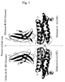
- Fig. 1 is a ribbon drawing of the crystal structure of G-CSF and CRH-G-CSF-R complex showing their backbones viewed from a direction approximately orthogonal to the pseudo-twofold axis of the molecules.
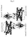
- Fig. 2 is a ribbon drawing of the crystal structure of G-CSF and CRH-G-CSF-R complex showing their backbones viewed from a direction approximately parallel to the non-crystallographic pseudo-twofold axis of the molecules.
- Fig. 3 one of the two molecules existing in an asymmetric unit (that is, one molecule of the complex between Molecules A and B, and one molecule of the complex between Molecules C and D) has been displaced onto the other in such a way that the G-CSF portions of respective molecules (Molecule A and Molecule C) are best superposed.
- This figure schematically represents the backbones of such structures.
- amino acids, peptides, and proteins are described using the following abbreviations adopted by IUPAC-IUB Commission on Biochemical Nomenclature (CBN). Unless otherwise specified, sequence of amino acid residues in a peptide or protein is indicated from its N-terminus to its C-terminus in the left-to-right direction, with the N-terminus being numbered 1.
- G-CSF and CRH-G-CSF-R used in the present invention are mammalian types, preferably a mouse or a human type, and particularly preferably a human type.
- CH-G-CSF-R refers to a region from tyrosine residue at position 97 to alanine residue at position 309 in the amino acid sequence of mouse-type G-CSF-R or to corresponding regions of other mammalian type G-CSF-Rs, the beginning and ending positions of CRH-G-CSF-R are not necessarily strict and do not have any great impact on the stereostructure of CRH-G-CSF-R as a whole.
- CH-G-CSF-R includes those regions in which the N-terminus and/or the C-terminus is shifted in either direction by several residues, or those regions in which several amino acid residues are added to the N-terminus and/or the C-terminus, provided that they retain the functions. Usually, such differences in the primary sequence will not have any great effect on the stereostructure of CRH-G-CSF-R as a whole, and the function is therefore expected to be retained. In the examples described below, the region from alanine residue at position 95 to alanine residue at position 309 in the above amino acid sequence was used for mouse-type CRH-G-CSF-R.
- the most popular technique for elucidating three-dimensional structure of a protein is X-ray crystallography.
- a crystallized protein is applied to monochromated X-ray to obtain X-ray diffraction patterns based on which three-dimensional structure of the protein is elucidated (Blundell, T. L. and Johnson, L. N., PROTEIN CRYSTALLOGRAPHY, pp. 1-565, 1976, Academic Press, New York).
- Crystallization exploits a protein's property of precipitating as crystals under certain conditions when a protein in solution becomes undissolved, for example, by addition of a precipitant to a protein solution or by reducing the volume of solvent through evaporation or the like.
- a highly purified protein is required for crystallization.
- physical and chemical factors such as protein concentration, salt concentration, hydrogen ion concentration (pH), type of precipitant added, temperature, and the like are involved in conditions under which crystallization occurs.
- crystallization techniques such as batch, dialysis, and vapor-diffusion procedures depending on the method of adding a precipitant or of regulating the volume of solvent (Blundell, T. L. and Johnson, L.
- the crystals of the present invention between human-type G-CSF and mouse-type CRH-G-CSF-R are prepared as follows.
- human-type G-CSF and mouse-type CRH-G-CSF-R are highly purified.
- Purified proteins are then combined with each other and allowed to form a complex.
- the complex is further purified to a high purity so that it becomes suitable for crystallization.
- Purification methods which may be used alone or in combination are those methods commonly used in the art for purifying proteins such as column chromatography (affinity, hydrophobic, ion exchange, gel filtration, and the like), salting out, centrifugation, and electrophoresis.
- the purification steps after the formation of the complex have to be conducted while retaining the complex. It is, therefore, preferred to use purification methods which allow the salt and hydrogen ion concentrations to be adjusted to values more similar to the physiological conditions, for example, gel filtration chromatography.
- crystals of the complex between G-CSF and CRH-G-CSF-R are prepared. This step may be conducted using a crystallization technique such as a batch, dialysis, or vapor-diffusion procedure. It is also necessary to determine physical and chemical factors such as protein concentration, salt concentration, hydrogen ion concentration (pH), type of precipitant added, temperature, and the like.
- crystallization of the complex between G-CSF and CRH-G-CSF-R it is essential to conduct the crystallization under conditions which retain the complex.
- a protein concentration of 0.5 to 2 mg/ml and a temperature of 20°C.
- larger crystals suitable for X-ray crystallography are obtained.
- the present invention is not limited to such conditions, and crystals of the complex between G-CSF and CRH-G-CSF which provide crystallographic constants substantially identical to those of the present invention are within the scope of the present invention.
- the crystal of the complex between human-type G-CSF shown in SEQ ID NO: 1 and mouse-type CRH-G-CSF-R shown in SEQ ID NO: 2 belongs to the tetragonal space group of I4 1 22, with unit cell parameters of 125 ⁇ 10 ⁇ in the a- and b-axis directions and 373 ⁇ 10 ⁇ in the c-axis direction.
- the crystal of the complex is further subjected to crystal structure analysis by X-ray diffraction to obtain the three-dimensional structure coordinates of the present invention (values indicating relative spatial positions of each atoms) of the complex between G-CSF and CRH-G-CSF-R.
- the structure coordinates obtained are shown in Table 1 according to a notation system for three-dimensional structure coordinates of a protein commonly used in the art.
- the first row is mathematical descriptions of the crystal and indicates parameters of the unit cell (in ⁇ for a-axis, b-axis, and c-axis directions in the order), the angles between the axes, and the crystal system.
- the second row through the last row but one describe three-dimensional coordinates for each atom.
- ATOM at the first column indicates that the row describes atom coordinates; the second column indicates the atom number; the third column indicates the atom type in the amino acid residue or the like; the fourth column indicates the amino acid residue or the like; the fifth column indicates the class of molecule (a particular class indicates a single polypeptide); the sixth column indicates the amino acid number or the like according to SEQ ID NOs: 1 and 2; the seventh, eighth, and ninth columns indicate coordinates of the atom (in ⁇ for a-axis, b-axis, and c-axis directions in the order); the tenth column indicates the occupancy of the atom (in the present invention 1.00 for all atoms); and the eleventh column indicates the temperature factor of the atom.
- a and C each indicate one molecule of G-CSF; B and D each indicate one molecule of CRH-G-CSF-R; E and F indicate sugar chains linked to B and D, respectively; and W indicates water molecule found in the crystal.
- abbreviations other than those for amino acid residues have the following meanings: NAG, N-acetylglucosamine residue; and WAT, water molecule.
- the structure coordinates show that one molecule of G-CSF (A) and one molecule of CRH-G-CSF-R (B) form a complex (A-B), and another molecule of G-CSF (C) and another molecule of CRH-G-CSF-R (D) form a complex (C-D). It is also found that the complex (A-B) and the complex (C-D) are associated to form an associate consisting of two molecules of the complex around a non-crystallographic pseudo-twofold axis. In the present specification, such an associate of the complexes may also sometimes be simply referred to as a "complex".
- a ribbon drawing (a representation method commonly used in the art for understanding a structure of a protein) showing the structure viewed from a direction orthogonal to the pseudo-twofold axis and a ribbon drawing showing the structure viewed from a direction parallel to the same axis are shown in Figs. 1 and 2, respectively.
- the crystal of the complex between G-CSF and CRH-G-CSF-R contains two molecules of the complex (A-B and C-D) in the minimal unit in which no crystallographic symmetry exists (i.e., an asymmetric unit). These two molecules of the complex are macroscopically related by a non-crystallographic pseudo-twofold axis.
- the G-CSF portion consists of 4 long ⁇ -helixes, and 1 short ⁇ -helix, and loop regions connecting the helixes.
- the CRH-G-CSF-R portion is broadly divided into two regions, each of which consists of about 7 ⁇ -sheets, and also contains loop regions connecting the sheets.
- BN domain one of these two regions that is closer to the N-terminus
- BC domain the other which is closer to the C-terminus
- G-CSF and the extracellular region of G-CSF-R form a complex consisting of stoichiometrically equivalent quantities of the components, and the complex may exist as a dimer or a tetramer (Horan, T. P. et al ., J. Biochem. (Tokyo), 121: 370-375 (1997); Horan, T. P. et al ., Biochemistry, 35: 4886-4896 (1996); Hiraoka, O. et al ., J. Biol. Chem., 270: 25928-25934 (1995); and Hiraoka, O. et al ., FEBS Lett., 356: 255-260 (1994)). It is considered that such facts are reflected in the associate demonstrated in the present invention which is formed by two molecules of G-CSF and two molecules of CRH-G-CSF-R.
- crystal structures comprising G-CSF variants and/or G-CSF-R variants which are substantially consistent with the crystal structure according to the present invention are within the scope of the present invention.
- substantially consistent it is meant that in the major part of the structure, in particular, in a part which forms a secondary structure such as ⁇ -helix or ⁇ -sheet or a part of which temperature factors are lower than other parts, the mean square deviation for the backbone or C ⁇ carbons is about 2 ⁇ or below.
- each sequence is not necessarily strictly defined for the present invention, and variants such as those in which another protein is attached to the N-terminus and/or the C-terminus or in which one or more amino acid residues are added to the N-terminus and/or the C-terminus are also included by the present invention, provided that they do not result in any substantial changes in molecular recognition between G-CSF and G-CSF-R.
- variants which have additional sugar chains linked thereto or from which a sugar chain moiety has been removed are also within the scope of the present invention so long as they do not result in any substantial changes in molecular recognition between G-CSF and G-CSF-R.
- molecular recognition it is meant by “substantially consistent” that the mean square deviation for the backbone or C ⁇ carbons of amino acid residues involved in the molecular recognition is about 2 ⁇ or below.
- the two molecules of the complex between G-CSF and CRH-G-CSF-R are subtly different from each other.
- one of the two molecules of the complex (Molecules A and B; and Molecules C and D) is mathematically subjected to translational and rotational operations in such a way that the respective portions of G-CSFs which form secondary structures, that is, ⁇ -helix regions of Molecules A and C, are best superposed, the orientation of Molecules B and D in the BN and BC domains are different from each other.
- Such difference in orientations of the BN and BC domains are schematically illustrated in Fig. 3.
- the relative positions of G-CSF and CRH-G-CSF-R are slightly different by about 10° for BN domain and by about 8° for BC domain.
- a substance crystallizes it proceeds toward the state of least free energy according to a law in natural science.
- the fact that the two molecules in an asymmetric unit have different structures in the crystal state indicates that the amount of decrease in energy due to formation of the crystal state is larger than the amount of increase in energy due to generation of the difference between the structures.
- the structure coordinates determined by the present invention it becomes possible for the first time to locate interacting amino acid residues in G-CSF and CRH-G-CSF-R.
- the residues having an interatomic distance of 4.2 ⁇ or less for van der Waals interacting atoms, of more than 3.4 ⁇ to 5.0 ⁇ for electrostatically interacting atoms, or of 3.4 ⁇ or less for hydrogen-bonding are shown in Tables 2, 3, 4, and 5, respectively. In these tables, hydrogen bonds via water molecules are also indicated.
- the residues having an interatomic distance of 4.2 ⁇ or less for van der Waals interacting atoms, of more than 3.4 ⁇ to 5.0 ⁇ for electrostatically interacting atoms, or of 3.4 ⁇ or less for hydrogen-bonding atoms are shown in each of Tables 2, 3, 4, and 5.
- hydrogen bonds via water molecules are also indicated.
- the first column describes amino acid residues and their number in Molecule A or C; the second column describes atoms in the amino acid residues; the third column describes amino acid residues and their number in Molecule B or D; the fourth column describes atoms in the amino acid residues; the fifth column describes interatomic distances between them in ⁇ ; and the sixth column describes interaction modes.
- VDW van der Waals interaction
- ESI electrostatic interaction
- HYB hydrogen bonding.
- WMH water molecule number involved
- the loop regions in each of the two domains recognize one molecule of G-CSF at one side.
- Amino acid residues characterizing this recognition are S13, L16, K17, E20, Q21, R23, K24, L109, D110, D113, T116, T117, Q120, E123, and E124 on the G-CSF side (Tables 2 and 4), and N20, S45, R46, R72, K73, L75, L76, L77, Y78, Q79, Y80, D102, M104, D105, Y143, M144, E145, R193, S195, and L196 on the CRH-G-CSF-R side (Tables 2 and 4), respectively.
- recognition between one molecule of G-CSF and one molecule of G-CSF-R is characterized by interactions among these and adjacent amino acid residues.
- the complex consisting of one molecule of G-CSF and one molecule of CRH-G-CSF-R further self-associates to form a homodimer of the complex, and this recognition is characterized by interactions among these and adjacent amino acid residues.
- the region surrounded by the dimer of the complex forms a space in which water molecules exist.
- This space is characterized by Y3 to L14, R46 to Y51, G92 to V106, E145 to E147, H166 to S169, S194 to G198, and amino acid residues adjacent thereto in CRH-G-CSF-R.
- three-dimensional structure coordinates of G-CSF and CRH-G-CSF-R derived from other species having homologous amino acid sequences can be derived by homology modelings (Haruki Nakamura and Kenta Nakai, "Biotechnology-no-tameno-Computer-nyumon" (An introduction to computers for biotechnology), pp. 186-204, Corona Publishing Co., 1995).
- homology modelings Harduki Nakamura and Kenta Nakai, "Biotechnology-no-tameno-Computer-nyumon" (An introduction to computers for biotechnology), pp. 186-204, Corona Publishing Co., 1995.
- the amino acid sequence of human-type G-CSF-R has a high homology to that of mouse-type G-CSF-R, three-dimensional structure coordinates of human-type G-CSF-R can easily be
- structure coordinates for human-type CRH-G-CSF-R were prepared by homology modeling.
- the side chain portions of amino acid residues which are not identical between mouse and human CRH-G-CSF-Rs were replaced by corresponding side chains of human amino acid residues.
- conformations of the side chains were selected so that the atoms do not stereochemically overlap one another and the energy became minimal.
- conformational calculations were conducted for all amino acid residues including their backbone portions to minimize the energy of the whole molecule.
- "ATOM" at the first column indicates that the row describes atom coordinates; the second column indicates the atom number; the third column indicates the atom type in the amino acid residue; the fourth column indicates the amino acid residue; the fifth column indicates the class of molecule (one particular class indicates a single polypeptide); the sixth column indicates the amino acid number according to SEQ ID NO: 3; the seventh, eighth, and ninth columns indicate coordinates of the atom (in ⁇ for a-axis, b-axis, and c-axis directions in the order); the tenth column indicates the occupancy of the atom (in the present invention 1.00 for all atoms); and the eleventh column indicates the temperature factor of the atom (although in the present invention the value is expedientially 0.00, this value has no special mean).
- B and D each indicate one molecule of CRH-G-CSF-R.
- This table is described according to the format of Protein Data Bank, a notation system commonly used in the art.
- amino acid residues characterizing the interactions can be located.
- the homology between amino acid sequences becomes less than 20%, the reliability of derived three-dimensional structure coordinates declines.
- both human and mouse belongs to Mammalia, it is expected that amino acid sequences of a mammal-type G-CSF and a mammal-type G-CSF-R are highly homologous to those of human and mouse-type, respectively. Accordingly, if an exact amino acid sequence is determined for such a protein derived from a mammal, three-dimensional structure coordinates can easily be derived using the structure coordinates of the present invention.
- the structure coordinates shown in Tables 1 and 6 are described by setting the origin of a unit cell in the crystal as the origin of the three-dimensional space.
- new structure coordinates obtained by mathematical operations such as translation, rotation, or symmetry transformation without altering relative positions of the atoms are also within the scope of the present invention.
- the above structure coordinates were determined for the complex between G-CSF and CRH-G-CSF-R.
- CRH-G-CSF-R can be considered as an equivalent of G-CSF-R in respect to binding with G-CSF, it is believed that the structure coordinates obtained above are substantially retained unchanged even in the complex between G-CSF and G-CSF-R. Therefore, the structure coordinates obtained above can be used to elucidate binding between G-CSF and G-CSF-R. Furthermore, they can also be used to identify, search for, evaluate, or design variants, agonists, or antagonists of G-CSF.
- Such a computer storage medium is not particularly restricted so long as it can load the structure coordinates obtained from the crystal of the complex between G-CSF and CRH-G-CSF-R into the program.
- it may be an electrical temporary storage medium known as a memory or a semipermanent storage medium such as a floppy disk, hard disk, optical disk, magneto-optical disk, or magnetic tape.
- a variant having activities as an agonist of G-CSF which possesses, for example, higher biological activiies, higher biological stability, or better physical properties such as higher thermodynamic stability when compared with the native G-CSF, or for obtaining a variant having activities as an antagonist, for example, which retains binding activity to G-CSF-R but inhibits inherent biological activities of G-CSF, by entering the structure coordinates obtained from the complex between G-CSF and CRH-G-CSF-R of the present invention into a computer on which a computer program expressing three-dimensional coordinates of molecules runs or into a storage medium of the computer and by conducting visual examinations and/or energy calculations.
- programs prepared so that it can provide a means for calculating structure energy of a molecule based on coordinates of the molecule and a means for calculating free energy taking into account solvent molecules such as water molecules may also be used.
- programs suitable for such purposes include, but not limited to, Insight II or QUANTA commercially available form Molecular Simulation, Inc.
- Such programs are usually installed for use on a computer called workstation supplied by Silicon Graphics, Inc., Sun Microsystems, Inc., or the like, although the present invention is not so limited.
- three-dimensional structure coordinates of the complex between G-CSF and CRH-G-CSF-R of the present invention are entered into a computer or a storage medium: of the computer, and the three-dimensional structure of the protein is displayed on the computer screen using a suitable program to conduct visual examinations.
- interacting amino acid residues and amino acid residue adjacent thereto in the complex between G-CSF and CRH-G-CSF-R are particularly displayed on the computer screen.
- One or more amino acid residues on the G-CSF side are then subjected to mutation such as substitution, deletion, or insertion, or to chemical modification in the computer, and changes in interactions produced by such alterations are observed on the computer screen.
- mutation such as substitution, deletion, or insertion, or to chemical modification in the computer
- changes in interactions produced by such alterations are observed on the computer screen.
- Stereo View which displays two kinds of pictures corresponding to the visual fields of right and left eyes at the same time may be used for easier understanding of three-dimensional space, but visual examinations can also be achieved without using such a three-dimensional representation.
- Local structure coordinates which change due to mutation such as substitution, deletion, or insertion or due to chemical modification can be obtained by determining spatial positions of the atoms so as to retain the validities of the chemical bonds.
- one. may let the computer display candidates for appropriate conformations and select therefrom or may also let the computer calculate to determine a structure lowering the energy state. From such candidates, mutations or chemical modifications of G-CSF which result in more preferable binding with CRH-G-CSF-R are found.
- amino acid residues shown in Tables 2, 3, 4, and 5 which interacts with CRH-G-CSF-R to form the complex that is, amino acid residues S13, L16, K17, E20, Q21, R23, K24, L109, D110, D113, T116, T117, Q120, E123, E124 and their adjacent regions and/or amino acid residues which form the associate, that is, amino acid residues G5, P6, A7, S8, S9, L10, P11, Q12, L125 and their adjacent regions so that they will more tightly bind with interacting amino acid residues in the corresponding regions on the CRH-G-CSF-R side.
- “their adjacent regions” refers to the regions which participate in interactions such as electrostatic interaction, hydrophobic interaction, van der Waals interaction, and hydrogen bonding with the amino acid residues in question, in particular the regions within about 5 ⁇ . The same applies throughout the present specification.
- Noncovalent interactions to be considered in this step include electrostatic interaction, hydrophobic interaction, van der Waals interaction, hydrogen bonding, and the like, and an ultimate design for a variant may be prepared comprehensively taking these interactions into account.
- mutations are introduced so that in the neighborhood of a side chain of an amino acid residue having negative charge such as glutamic or aspartic acid on the CRH-G-CSF-R side, a side chain of an amino acid residue having positive charge such as lysine, arginine, or histidine will be positioned at an adjacent amino acid residue on the G-CSF, or reversely so that in the neighborhood of a side chain of an amino acid residue having positive charge such as lysine, arginine, or histidine on the CRH-G-CSF-R side, a side chain of an amino acid residue having negative charge such as glutamic or aspartic acid will be positioned at an adjacent amino acid residue on the G-CSF.
- amino acid residues having highly hydrophobic side chains such as alanine, leucine, isoleucine, valine, proline, phenylalanine, tryptophan, and methionine mainly come together to interact
- a position at which a hydrophilic amino acid residue such as serine, threonine, tyrosine, asparagine, or glutamine or a changed amino acid residue such as aspartic acid, glutamic acid, lysine, arginine, or histidine exists is sought in G-CSF and the found amino acid residue is replaced by a hydrophobic amino acid residue in order to strengthen the hydrophobic interaction.
- mutations are firstly introduced into amino acid residues shown in Tables 2, 3, 4, and 5 which interacts with CRH-G-CSF-R to form the complex, that is, amino acid residues S13, L16, K17, E20, Q21, R23, K24, L109, D110, D113, T116, T117, Q120, E123, E124 and their adjacent regions and/or amino acid residues which form the associate, that is, amino acid residues G5, P6, A7, S8, S9, L10, P11, Q12, L125 and their adjacent regions.
- a variant is then selected so that by binding of the variant G-CSF to CRH-G-CSF-R, intrinsic relative positions in three-dimensional space are not retained between G-CSF and two molecules of CRH-G-CSF-R, or such that the variant G-CSF can not interact with CRH-G-CSF-R in the complex or associate forming regions described above and consequently has activity as an antagonist against native G-CSF.
- Noncovalent interactions to be considered in this step are the same as in the case of G-CSF variants having activity as an agonist and an ultimate design for a variant may be prepared visually and comprehensively taking into account electrostatic interaction, hydrophobic interaction, van der Waals interaction, hydrogen bonding, and other factors on a computer screen.
- the second method is to design the above variants by evaluation of binding with CRH-G-CSF-R using energy calculations on a computer.
- Energy calculations may be achieved by using a computer program which executes molecular force field calculations generally carried out in the art.
- Programs suitable for such purposes include, but not limited to, CVFF, AMBER force field optimized for proteins, which is contained in DISCOVER module of Insight II.
- first and the second design techniques are not strictly sorted out from each other, and both techniques may be used in combination. Specifically, with candidates expected by visual examinations to be more desirable, energy calculations are actually conducted using the second technique to evaluate their validities, and better variants are designed by repeating such steps.
- the structure coordinates of amino acid residues on the CRH-G-CSF-R side used in the first and second design techniques may be those of mouse-type CRH-G-CSF-R shown in Table 1 of the present specification or those of human-type CRH-G-CSF-R shown in Table 6. Furthermore, they may also be structure coordinates newly prepared, for example, by calculations with a computer based on such structure coordinates using sequences of G-CSF and G-CSF-R derived from other species having a high homology to the amino acid sequences of the above CRH-G-CSF-Rs, or they may even be structure coordinates excerpted in part therefrom.
- CRH-G-CSF-R structure coordinates of human-type CRH-G-CSF-R.
- structure coordinates of CRH-G-CSF-R used it is not necessary to use all coordinates of the receptor portion. Since the regions corresponding to the interacting portions of G-CSF and CRH-G-CSF-R shown in Tables 2, 3, 4, and 5 are important for designing a variant, it is also possible that the coordinates of amino acid residues involved in such interactions or, if necessary, of amino acid residues adjacent thereto are selected from Table 1 or 6 and used for designing.
- structure coordinates of G-CSF and CRH-G-CSF-R are usually used while being fixed in three-dimensional space, it is not necessarily required to fix the coordinates.
- the two molecules of the receptor-existing in a crystallographic asymmetric unit may be each subjected to translation or rotation in three-dimensional space as a block, and furthermore, amino acid residues in each block may also be displaced to such an extent that no chemical covalent bonds are cleaved to calculate energy of binding with a G-CSF variant.
- the structure coordinates changed due to translation, rotation, or displacement during such calculations in three-dimensional space are within the scope of the present invention.
- Variants designed according to the present invention may be prepared by many methods.
- a DNA encoding a variant designed on the basis of the present invention may be obtained as follows: at a position identified as an amino acid residue of which mutation will enhance biological activities on the basis of the present invention, an oligonucleotide portion encoding the corresponding amino acid residue is replaced by chemically synthesizing an oligonucleotide corresponding to the variant and substituting it for the native oligonucleotide using sequence-specific oligonucleotide cleaving enzymes (restriction enzymes).
- the variant DNA obtained may be then incorporated into an appropriate expression vector, introduced into an appropriate host cell, and expressed as a recombinant protein to obtain the above variant.
- agonists should bind to CRH-G-CSF while retaining spatial position of the two molecules of CRH-G-CSF-R in the associate.
- Indigo 2 a workstation supplied by Silicon Graphics, Inc., is suitable as a computer used for designing agonists.
- the computer is not limited to this one, and any computer may be used so long as it is tuned to run an appropriate program.
- the computer storage medium there is no particular limitation on the computer storage medium.
- Insight II a computer program commercially available from Molecular Simulation, Inc. may be used as a program for designing.
- a program Ludi or DOCK a module of Insight II specially prepared for such purposes, may be used alone or in combination to facilitate identification, searching, evaluation, or designing.
- designing of agonists according to the techniques described in Japanese Patent Kokai Publication Nos.
- H6-309385 (1994) and H7-133233 (1995) can also be achieved for the first time by using structure coordinates of the complex between G-CSF and CRH-G-CSF-R of the present invention.
- the present invention is, however, not limited to these programs and techniques.
- the first step is to find a compound which serves as a starting point for drug design, known for those skilled in the art as a lead compound.
- the next step is optimization of the lead compound wherein compounds having better properties as medicines, for example, having better activity, having better pharmacokinetics, or having less toxicities and side effects are sought starting from the lead compound.
- the step in which a lead compound is found using the structure coordinates of the complex between G-CSF and CRH-G-CSF-R provided by the present invention is achieved, for example, using a database in a computer into which structures of plural compounds have been entered, by a method in which interactions between three-dimensional structures of a compound in the database and CRH-G-CSF-R are sorted out in a visual manner one after another, or by a method in which amplitudes of binding energy are calculated one after another using a computer and compounds which stably bind to CRH-G-CSF-R are found from the database.
- the database of compound's structures contains determined three-dimensional structure coordinates entered therein, for low molecular weight compounds, it does not have to be a database of three-dimensional structure coordinates, because such low molecular weight compounds may change their conformations relatively freely, and also because three-dimensional structure coordinates for each conformation can be derived by calculations in a relatively short time. In the latter cases, information for chemical covalent bonds of low molecular weight compounds are entered into the database.
- two molecules of CRH-G-CSF-R in the associate are firstly displayed on a computer screen according to the structure coordinates of the present invention.
- a three-dimensional representation may be made on the computer screen using, for example, Crystal Eye as described above, visual examinations can also be achieved without using such a three-dimensional representation.
- compounds in the database are allowed to bind with two molecules of CRH-G-CSF-R taking chemical interactions into account, and are evaluated one after another whether or not it can strongly bind with CRH-G-CSF-R, and if it can do so, whether or not the relative positions taken by the two molecules of CRH-G-CSF-R interacting with the compound are similar to those of the two molecules of CRH-G-CSF-R in the associate.
- the compound binds with two molecules of CRH-G-CSF-R at a total of two or more sites (at least one site with each CRH-G-CSF-R) so that the relative positions taken by the two molecules of CRH-G-CSF-R in three-dimensional space are maximally conserved. Relative positions taken by the two molecules of CRH-G-CSF-R in three-dimensional space do not have to be strictly conserved, and are permitted to vary to a certain degree so long as activity of the compound as an agonist is retained.
- Chemical interactions to be considered include electrostatic interaction, hydrophobic interaction, hydrogen bonding, van der Waals interaction, and the like.
- the structure should be comprehensively examined whether it is favorable for interactions, for example, so that functional groups which tend to bear negative charge such as carboxyl group, nitro group, and halogens interact with amino acid residues in CRH-G-CSF-R having positive charge such as lysine, arginine, and histidine, so that functional groups which tend to bear positive charge such as amino, imino, and guanidyl groups interact with amino acid residues in CRH-G-CSF-R having negative charge such as glutamic acid and aspartic acid, so that hydrophobic functional groups such as aliphatic groups and aromatic groups interact with hydrophobic amino acid residues such as alanine, leucine, isoleucine, valine, proline, phenylalanine, tryptophan and methionine, so that functional groups involved in hydrogen bonding such as hydroxyl and amide groups can
- the energy of binding between a compound and the two molecules of CRH-G-CSF-R in the associate is determined by molecular force field calculations. Such calculations are applied to each compound in the database to find a compound which may serve as a lead compound capable of stable binding.
- a molecular force field used in the calculations for example, CVFF, AMBER force field optimized for proteins, which is contained in DISCOVER module of Insight II program may be used.
- some computer programs like Ludi in Insight II can automatically output candidates for lead compound when three-dimensional structure coordinates of interacting amino acid residues in a protein molecule are given, and such programs may also be applied to G-CSF or CRH-G-CSF-R.
- the next step in which optimization of the lead compound is conducted using the structure coordinates of the complex between G-CSF and CRH-G-CSF-R provided by the present invention, is used for the purpose of, where a lead compound which binds to CRH-G-CSF-R has already been found by the above method or separately found in an experimental manner, optimizing the lead compound to obtain a better compound, for example, a compound having higher biological activities as an agonist or a compound having a structure favorable for oral administration as a medicine.
- a lead compound may be selected, for example, from a series of compounds known in the art as a combinatorial library or from a culture medium of a microbe or the like.
- a model of the complex between the lead compound and CRH-G-CSF-R is firstly displayed on a computer screen by entering the three-dimensional structure coordinates of the lead compound and the structure coordinates of CRH-G-CSF-R provided by the present invention into a computer on which a computer program expressing three-dimensional coordinates of molecules runs or into a storage medium of the computer.
- a three-dimensional representation may be made on the computer screen using, for example, Crystal Eye as described above, visual examinations can also be achieved without using such a three-dimensional representation. It is a logical designing of a compound to modify the lead compound so as to yield a compound more favorably interacting with CRH-G-CSF-R or a compound having better pharmacokinetics while retaining the interactions.
- the energy of binding between a new compound designed from the lead compound and CRH-G-CSF-R is determined by molecular force field calculations to judge the validity of the design.
- a molecular force field used in the calculations for example, CVFF, AMBER force field optimized for proteins, which is contained in DISCOVER module of Insight II program may be used.
- the visual examinations and the method by energy evaluations may be used in combination as appropriate.
- the newly designed compound binds with two molecules of CRH-G-CSF-R at a total of two or more sites (at least one site with each CRH-G-CSF-R) so that the relative positions taken by the two molecules of CRH-G-CSF-R in three-dimensional space are maximally conserved.
- Relative positions taken by the two molecules of CRH-G-CSF-R in three-dimensional space do not have to be strictly conserved, and are permitted to vary to a certain degree so long as activity of the compound as an agonist is retained.
- the structure coordinates changed due to translation, rotation, or displacement during such calculations in three-dimensional space are within the scope of the present invention.
- the structure coordinates of amino acid residues on the CRH-G-CSF-R side used in the above techniques may be those of mouse-type CRH-G-CSF-R shown in Table 1 of the present specification, those of human-type CRH-G-CSF-R shown in Table 6, or even those prepared by calculations based on such structure coordinates.
- For designing a variant as a medicine for human it is more desirable to use structure coordinates of human-type CRH-G-CSF-R.
- a G-CSF agonist which retains relative positions taken by the two molecules of CRH-G-CSF-R in the associate can be obtained by selecting structure coordinates of the regions corresponding to those amino acid residues and by designing a compound so that one molecule of the compound simultaneously binds to the two regions of CRH-G-CSF-R to which one molecule of G-CSF binds.
- structure coordinates of the region spatially surrounded by the two molecules of CRH-GS-CSF-R are characterized by Y3 to L14, R46 to Y51, G92 to V106, E145 to E147, H166 to S169, S194 to G198. Accordingly, structure coordinates corresponding to these amino acid residues and, if necessary, amino acid residues adjacent thereto may be selected from Table 1 or 6 and used for designing.
- a G-CSF agonist which retains relative positions taken by the two molecules of CRH-G-CSF-R in the associate can be obtained by designing a compound so that the compound fits into the cavity surrounded by these amino acid residues and simultaneously binds to two or more regions of each CRH-G-CSF-R while minimizing the energy.
- Agonists designed by the above techniques may be obtained by commonly used techniques for chemical synthesis depending on the particular compound.
- the compounds may be natural or synthetic, and may be high molecular weight or low molecular weight compounds.
- an antagonist should bind to G-CSF and/or CRH-G-CSF so that it provides positions in three-dimensional space different from those of the two molecules of CRH-G-CSF-R in the associate formed by the natural G-CSF and CRH-G-CSF-R, or so that it inhibits formation of the complex or the associate between G-CSF and CRH-G-CSF.
- a computer used for designing antagonists is not particularly limited so long as it is tuned to run an appropriate program.
- the computer storage medium there is no particular limitation on the computer storage medium.
- Insight II a computer program commercially available from Molecular Simulation, Inc. may be used as a program for designing.
- a program Ludi or DOCK a module of Insight II specially prepared for such purposes, may be used alone or in combination to facilitate identification, searching, evaluation, or designing.
- designing of antagonists according to the techniques described in Japanese Patent Kokai Publication Nos.
- H6-309385 (1994) and H7-133233 (1995) can also be achieved for the first time by using structure coordinates of the complex between G-CSF and CRH-G-CSF-R of the present invention.
- the present invention is, however, not limited to these programs and techniques.
- the first step is to find a lead compound and the next step is a process for optimization of the lead compound.
- the step in which a lead compound for antagonists is found using the structure coordinates of the complex between G-CSF and CRH-G-CSF-R provided by the present invention is achieved, for example, using a database in a computer into which structures of plural compounds have been entered, by a method in which interactions between three-dimensional structures of a compound in the database and G-CSF and/or CRH-G-CSF-R are sorted out in a visual manner one after another, or by a method in which amplitudes of binding energy are calculated one after another using a computer and compounds which stably bind to G-CSF and/or CRH-G-CSF-R are found from the database.
- the database of compound's structures contains determined three-dimensional structure coordinates entered therein, for low molecular weight compounds, it does not have to be a database of three-dimensional structure coordinates, because such low molecular weight compounds may change their conformations relatively freely, and also because three-dimensional structure coordinates for each conformation can be derived by calculations in a relatively short time. In the latter cases, information for chemical covalent bonds of low molecular weight compounds are entered into the database.
- G-CSF and/or CRH-G-CSF-R molecule or molecules are firstly displayed on a computer screen according to the structure coordinates of the present invention.
- a three-dimensional representation may be made on the computer screen using, for example, Crystal Eye as described above, visual examinations can also be achieved without using such a three-dimensional representation.
- compounds in the database are allowed to bind with G-CSF and/or CRH-G-CSF-R molecule or molecules taking chemical interactions into account, and are evaluated one after another whether or not the compound can function to provide positions in three-dimensional space different from those taken by the two molecules of CRH-G-CSF-R in the associate formed by G-CSF and CRH-G-CSF-R or to inhibit formation of the complex or the associate between G-CSF and CRH-G-CSF-R.
- At least one of the sites at which an antagonist binds to G-CSF and/or CRH-G-CSF-R corresponds to the amino acid residue at which G-CSF binds to CRH-G-CSF-R or an amino acid residue adjacent thereto, and a compound of which binding to G-CSF and/or CRH-G-CSF-R sterically hinders the binding between G-CSF and CRH-G-CSF-R with the result that relative positions intrinsically taken by the two molecules of CRH-G-CSF-R in three-dimensional space are not retained or that formation of the complex and/or the associate between G-CSF and CRH-G-CSF-R is inhibited is selected.
- the compound does not need to simultaneously bind with two molecules of G-CSF, two molecules of CRH-G-CSF-R, or one molecule of G-CSF and one molecule of CRH-G-CSF-R, and it is preferred that a compound which binds to one molecule of G-CSF or CRH-G-CSF-R is selected.
- the energy of binding between a compound selected from the database and G-CSF and/or CRH-G-CSF-R is determined as in the case of agonists to find a compound which may serve as a lead compound for antagonists from the database.
- a molecular force field used in the calculations for example, CVFF, AMBER force field optimized for proteins, which is contained in DISCOVER module of Insight II program may be used.
- some computer programs like Ludi in Insight II can automatically output candidates for lead compound when three-dimensional structure coordinates of interacting amino acid residues in a protein molecule are given.
- the next step in which optimization of the lead compound is conducted using the structure coordinates of the complex between G-CSF and CRH-G-CSF-R provided by the present invention, is used for the purpose of, where a lead compound which binds to G-CSF and/or CRH-G-CSF-R has already been found by the above method or separately found in an experimental manner, optimizing the lead compound to obtain a better compound, for example, a compound having higher biological activities as an antagonist or a compound having a structure favorable for oral administration as a medicine.
- the method for experimentally finding a lead compound is the same as that for agonists, and a compound found in the above-described designing of agonists may also be used as a lead compound.
- a model of the complex between G-CSF and/or CRH-G-CSF-R and the lead compound is firstly displayed on a computer screen by entering the three-dimensional structure coordinates of the lead compound and the structure coordinates of G-CSF and/or CRH-G-CSF-R provided by the present invention into a computer on which a computer program expressing three-dimensional coordinates of molecules runs or into a storage medium of the computer.
- a three-dimensional representation may be made on the computer screen using, for example, Crystal Eye as described above, visual examinations can also be achieved without using such a three-dimensional representation.
- the energy of binding between a new compound designed from the lead compound and G-CSF and/or CRH-G-CSF-R. is determined by molecular force field calculations to judge the validity of the design.
- a molecular force field used in the calculations for example, CVFF, AMBER force field optimized for proteins, which is contained in DISCOVER module of Insight II program may be used.
- the visual examinations and the method by energy evaluations may be used in combination as appropriate.
- the structure coordinates of amino acid residues on the G-CSF and/or CRH-G-CSF-R side(s) used in the above techniques may be those of human-type G-CSF and mouse-type CRH-G-CSF-R shown in Table 1 of the present specification, those of human-type CRH-G-CSF-R shown in Table 6, or even those prepared by calculations based on such structure coordinates.
- designing of an antagonist involves designing of a compound which binds to G-CSF and/or CRH-G-CSF-R
- the amino acid residues corresponding to the interacting portions of G-CSF and CRH-G-CSF-R shown in Tables 2, 3, 4, and 5 as well as the regions of amino acid residues adjacent thereto are important among the structure coordinates of G-CSF or CRH-G-CSF-R, and it is also possible that the coordinates of such amino acid residues are selected from Table 1 or 6 and used for designing.
- an antagonist may be obtained by selecting the coordinates of the portions corresponding to such amino acid residues and by designing the compound so that it inhibits normal binding between G-CSF and CRH-G-CSF-R to prevent G-CSF from exerting its biological activities.
- Antagonists designed by the above techniques may be obtained by commonly used techniques for chemical synthesis depending on the particular compound.
- the three-dimensional structure coordinates of G-CSF and CRH-G-CSF-R according to the present invention which are shown in Table 1 or 6 may be used in X-ray crystallography, for example, of crystals containing the whole or part of G-CSF and G-CSF-R or crystals obtained from other proteins which comprise an amino acid sequence having a significant homology to G-CSF or G-CSF-R.
- X-ray crystallography for example, of crystals containing the whole or part of G-CSF and G-CSF-R or crystals obtained from other proteins which comprise an amino acid sequence having a significant homology to G-CSF or G-CSF-R.
- a molecular replacement method commonly used in the art as one of techniques for X-ray crystallography (see, e.g., Blundell, T. L. and Johnson, L. N., (1976), PROTEIN CRYSTALLOGRAPHY, pp.
- all or part of the three-dimensional structure coordinates of the complex between G-CSF and CRH-G-CSF-R of the present invention may be used to determine structure coordinates from a structure factor obtained from X-ray diffraction patterns of a crystal without using heavy atom isomorphous replacement methods and much rapidly even for a crystal of a protein having unknown structure coordinates as mentioned above.
- a computer tuned to run a program for use in the molecular replacement method is used.
- programs include Insight II (commercially available from MSI), X-PLOR (commercially available from Molecular Simulation, Inc.,), and AMORE (one of the programs in CCP4 (Collaborative Computational Project, Number 4., Acta Crystallogr. D 50, 670-673 (1994) ), but other programs may also be used.
- crystals to which a molecular replacement method should be applied using the three-dimensional coordinates of the complex between G-CSF and CRH-G-CSF-R of the present invention include, for example, crystals obtained from a complex between a compound binding with G-CSF-R (e.g., an agonist or antagonist) and G-CSF-R, a complex between a compound binding with G-CSF (e.g., an antagonist) and G-CSF, G-CSF-R containing amino acid residues other than those described in Table 6, a substance containing a protein having a significant homology to G-CSF at the amino acid level, a substance containing a protein having a significant homology to G-CSF-R at the amino acid level, a variant of G-CSF and a substance
- the term "significant homology" usually refers to a case wherein the identity of the amino acid sequences is 20% or above, and preferably 30% or above.
- the validity of application of the molecular replacement method may be determined by actually applying the molecular replacement method to the structure factor calculated from X-ray diffraction pattern of a crystal in question and thereby obtaining a significant solution.
- Mouse-type CRH-G-CSF-R was prepared by the method described in Japanese Patent Application Ser. No. H6-280655 (1994) (Japanese Patent Kokai Publication No. H8-140678 (1996)). In order to obtain a material suitable for crystallization, the CRH-G-CSF-R fraction was further purified by its electrical properties using ion-exchange column chromatography (Mono-S HR10/10, Pharmacia). Human-type G-CSF produced as a recombinant protein in E. coli was provided by Kirin Brewery Co., Ltd.
- protein concentrations of G-CSF and CRH-G-CSF-R obtained were each determined by measuring their ultraviolet absorbances, and the proteins were combined using stoichiometrically equivalent quantities calculated from these concentrations or using an excess of G-CSF.
- the sample was fractionated by gel filtration chromatography (HiLoad Superdex 200 HR26/60, Pharmacia) using a buffer solution consisting of 0.01M 2-(N-morpholino)ethanesulfonic acid (pH 6) containing 0.1M sodium chloride as the mobile phase to yield a highly purified complex between G-CSF and CRH-G-CSF-R.
- the crystal of the complex between G-CSF and CRH-G-CSF-R was prepared using a vapor diffusion technique describe below.
- the highly purified complex between G-CSF and CRH-G-CSF-R in Example 1 was adjusted to a protein concentration of 0.5 to 2 mg/ml using a device with an ultrafiltration membrane (Centricon-10, Grace Japan).
- Example 2 The crystal obtained in Example 2 was soaked in the crystallization solution containing 50% sucrose and then put under a nitrogen gas stream at 100 K to be flash frozen. Under a 100 K nitrogen gas stream, X-ray diffraction data were collected by the oscillation method. In addition, the crystal obtained in Example 2 was also soaked in the crystallization solution containing 1 mM ethylmercurithiosalicylate for 6 hours to obtain an isomorphous derivative crystal containing a mercury atom in the crystal. The crystal was soaked in the crystallization solution containing 50% sucrose and then put under a nitrogen gas stream at 100 K to be flash frozen. Under a 100 K nitrogen gas stream, X-ray diffraction data were collected by the oscillation method.
- G-CSF in which methionine residues have been substituted by selenomethionine residues was obtained by cultivating, in a medium containing selenomethionine, an E . coli strain prepared so as to express G-CSF.
- a crystal of the complex between G-CSF which contained selenomethionine residues substituting for methionine residues and CRH-G-CSF-R was prepared according to the method described in Example 2. The crystal was soaked in the crystallization solution containing 50% sucrose and then put under a nitrogen gas stream at 100 K to be flash frozen.
- the R factor recognized in the art as an indicator of accuracy of the structure coordinates was 19.5% when a structure factor obtained from a diffraction pattern having Bragg reflection angle of 6 to 2.8 ⁇ was used.
- An R-factor calculated from a structure factor independently excluded from the calculation for refinement at the refinement stage was 28.3%.
- the root mean square errors from the ideal states of bond distances and bond angles among atoms were 0.012 ⁇ and 2.0 degree, respectively.
- the structure coordinates of the complex between G-CSF and CRH-G-CSF-R obtained in Example 3 were further refined. Using the crystal structure factor and structure coordinates obtained in Example 3, calculations with REFMAC (in CCP4 programs) and X-PLOR and model modification with QUANTA were repeated to identify the following: structure coordinates of amino acid residues except for M1 to L4 and the side chain portions of Q71 and L131 in one molecule (Molecule A) of two molecules of G-CSF each consisting of 175 amino acid residues; structure coordinates of amino acid residues except for M1 to P6, and the side chain portions of H53, W59, Q68, L70, Q71 in Molecule C of G-CSF; structure coordinates of amino acid residues except for G120 to H126, K214 to A215 and the side chain portions of K63 and R64 in Molecule B of CRH-G-CSF-R; structure coordinates of amino acid residues except for A1, 1119 to H126, K214 to
- the R factor recognized in the art as an indicator of accuracy of the structure coordinates was 22.2% when a structure factor obtained from a diffraction pattern having Bragg reflection angle of 25 to 2.8 ⁇ was used.
- An R-factor calculated from a structure factor independently excluded from the calculation for refinement at the refinement stage was 29.8%.
- the root mean square errors from the ideal states of bond distances and bond angles among atoms were 0.014 ⁇ and 1.9 degree, respectively.
- the crystal of the complex between G-CSF and CRH-G-CSF-R obtained in Example 2 was soaked in 0.1 M N-2-hydroxyethylpiperazine-N'-2-ethanesulfonic acid buffer solution containing 20% glycerol and 2.4 M ammonium sulfate for one day and then put under a nitrogen gas stream at 100 K to be flash frozen. Under cooling with a 100 K nitrogen gas stream, X-ray diffraction data were collected by the oscillation method. Each diffraction pattern was converted into a structure factor using DENZO/SCALEPACK.
- Three-dimensional structure coordinates of the complex between G-CSF and CRH-G-CSF-R having a new structure were thereby obtained. It was also shown that the space group was P4 3 2 1 2.
- the obtained structure coordinates indicated that the complex between G-CSF and CRH-G-CSF-R contains four molecules of the complex in the asymmetric unit. It was also shown that when compared with the structure obtained from the I4 1 22 crystal, the structure obtained was different in the angle between G-CSF molecule and CRH-G-CSF-R molecule and in the angle between domains of CRH-G-CSF-R by several degrees, indicating that these molecular recognitions have variability within several degrees.
Landscapes
- Health & Medical Sciences (AREA)
- Chemical & Material Sciences (AREA)
- Life Sciences & Earth Sciences (AREA)
- Organic Chemistry (AREA)
- Medicinal Chemistry (AREA)
- General Health & Medical Sciences (AREA)
- Proteomics, Peptides & Aminoacids (AREA)
- Gastroenterology & Hepatology (AREA)
- Biochemistry (AREA)
- Biophysics (AREA)
- Zoology (AREA)
- Genetics & Genomics (AREA)
- Toxicology (AREA)
- Molecular Biology (AREA)
- Immunology (AREA)
- Bioinformatics & Cheminformatics (AREA)
- Animal Behavior & Ethology (AREA)
- Cell Biology (AREA)
- Chemical Kinetics & Catalysis (AREA)
- General Chemical & Material Sciences (AREA)
- Nuclear Medicine, Radiotherapy & Molecular Imaging (AREA)
- Pharmacology & Pharmacy (AREA)
- Engineering & Computer Science (AREA)
- Public Health (AREA)
- Veterinary Medicine (AREA)
- Diabetes (AREA)
- Hematology (AREA)
- Peptides Or Proteins (AREA)
- Information Retrieval, Db Structures And Fs Structures Therefor (AREA)
- Medicines That Contain Protein Lipid Enzymes And Other Medicines (AREA)
Applications Claiming Priority (5)
| Application Number | Priority Date | Filing Date | Title |
|---|---|---|---|
| JP5690599 | 1999-03-04 | ||
| JP5690599 | 1999-03-04 | ||
| JP21869199 | 1999-08-02 | ||
| JP11218691A JP2000319298A (ja) | 1999-03-04 | 1999-08-02 | 蛋白質複合体の結晶、構造座標、及び構造座標の使用 |
| PCT/JP2000/001217 WO2000052057A1 (fr) | 1999-03-04 | 2000-03-02 | Cristaux et coordonnees structurales d'un complexe proteique et utilisation desdites coordonnees structurales |
Publications (1)
| Publication Number | Publication Date |
|---|---|
| EP1167390A1 true EP1167390A1 (de) | 2002-01-02 |
Family
ID=26397905
Family Applications (1)
| Application Number | Title | Priority Date | Filing Date |
|---|---|---|---|
| EP00906616A Withdrawn EP1167390A1 (de) | 1999-03-04 | 2000-03-02 | Kristall- und strukturkoordinaten eines proteinkomlexes und die verwendung der strukturkoordinaten |
Country Status (4)
| Country | Link |
|---|---|
| EP (1) | EP1167390A1 (de) |
| JP (1) | JP2000319298A (de) |
| CA (1) | CA2363961A1 (de) |
| WO (1) | WO2000052057A1 (de) |
Cited By (10)
| Publication number | Priority date | Publication date | Assignee | Title |
|---|---|---|---|---|
| US6646110B2 (en) | 2000-01-10 | 2003-11-11 | Maxygen Holdings Ltd. | G-CSF polypeptides and conjugates |
| US6831158B2 (en) | 2000-01-10 | 2004-12-14 | Maxygen Holdings Ltd. | G-CSF conjugates |
| US7381805B2 (en) | 2005-06-01 | 2008-06-03 | Maxygen Holdings, Ltd. | Compositions comprising mixtures of positional PEG isomers of PEGylated G-CSF |
| US7550566B2 (en) | 2000-01-10 | 2009-06-23 | Maxygen Holdings Ltd. | G-CSF conjugates |
| WO2012171057A1 (en) * | 2011-06-13 | 2012-12-20 | Csl Limited | Antibodies against g-csfr and uses thereof |
| EP2482212A4 (de) * | 2009-09-25 | 2014-02-26 | Sh Nat Eng Res Ct Nanotech Co | Verfahren zur erfassung von hochaffinen proteinen mit computergestütztem entwurf |
| US12264202B2 (en) | 2017-11-29 | 2025-04-01 | Csl Limited | Methods of reducing or hindering development of ischemia-reperfusion injury due to kidney transplantation with an antibody that inhibits granulocyte-colony stimulating factor (G-CSF) signaling |
| US12281160B2 (en) | 2018-03-23 | 2025-04-22 | Csl Limited | Method of treating asthma |
| US12357689B2 (en) | 2023-07-05 | 2025-07-15 | CSL Innovation Pty Ltd | Methods of treating or preventing a cardiovascular system complication of sickle cell disease with an anti-G-CSFR antibody |
| US12583929B2 (en) | 2020-06-04 | 2026-03-24 | CSL Innovation Pty Ltd | Method of treating acute respiratory distress syndrome |
Families Citing this family (3)
| Publication number | Priority date | Publication date | Assignee | Title |
|---|---|---|---|---|
| WO2002044954A1 (en) * | 2000-12-01 | 2002-06-06 | Mitsubishi Chemical Corporation | Method of constructing three-dimensional structure of protein |
| WO2002101629A1 (en) * | 2001-06-07 | 2002-12-19 | Wyeth | Solution structure of il-13 and uses thereof |
| CA2449290A1 (en) * | 2001-06-07 | 2002-12-12 | F. Hoffmann-La Roche Ag | Mutants of igf binding proteins and methods of production of antagonists thereof |
Family Cites Families (8)
| Publication number | Priority date | Publication date | Assignee | Title |
|---|---|---|---|---|
| EP0344796B1 (de) * | 1988-06-03 | 1994-09-21 | Chugai Seiyaku Kabushiki Kaisha | Menschlicher kristallinischer Granulocyt-Koloniestimulierungsfaktor und dessen Herstellung |
| JPH02104597A (ja) * | 1988-10-14 | 1990-04-17 | Kyowa Hakko Kogyo Co Ltd | ヒト顆粒球コロニー刺激因子活性を有するペプチドの結晶およびその製造法 |
| JP3256307B2 (ja) * | 1993-01-07 | 2002-02-12 | 昭子 板井 | 生理活性を有するリガンドの分子構造を構築する方法 |
| US5581476A (en) * | 1993-01-28 | 1996-12-03 | Amgen Inc. | Computer-based methods and articles of manufacture for preparing G-CSF analogs |
| JP3785195B2 (ja) * | 1993-11-11 | 2006-06-14 | 昭子 板井 | 生理活性を有するリガンドの分子構造を構築する方法 |
| AUPM375094A0 (en) * | 1994-02-08 | 1994-03-03 | Ludwig Institute For Cancer Research | Cytokine receptor interactive molecules and methods for assaying cytokine function |
| JP3081761B2 (ja) * | 1994-11-15 | 2000-08-28 | 株式会社蛋白工学研究所 | 顆粒球コロニー刺激因子受容体のcrh領域を含むリガンド結合領域の蛋白質をコードしているdna |
| AUPN024994A0 (en) * | 1994-12-23 | 1995-01-27 | Ludwig Institute For Cancer Research | Assay and peptides for use therein |
-
1999
- 1999-08-02 JP JP11218691A patent/JP2000319298A/ja active Pending
-
2000
- 2000-03-02 WO PCT/JP2000/001217 patent/WO2000052057A1/ja not_active Ceased
- 2000-03-02 CA CA002363961A patent/CA2363961A1/en not_active Abandoned
- 2000-03-02 EP EP00906616A patent/EP1167390A1/de not_active Withdrawn
Non-Patent Citations (1)
| Title |
|---|
| See references of WO0052057A1 * |
Cited By (16)
| Publication number | Priority date | Publication date | Assignee | Title |
|---|---|---|---|---|
| US6646110B2 (en) | 2000-01-10 | 2003-11-11 | Maxygen Holdings Ltd. | G-CSF polypeptides and conjugates |
| US6831158B2 (en) | 2000-01-10 | 2004-12-14 | Maxygen Holdings Ltd. | G-CSF conjugates |
| US7550566B2 (en) | 2000-01-10 | 2009-06-23 | Maxygen Holdings Ltd. | G-CSF conjugates |
| US7550565B2 (en) | 2000-01-10 | 2009-06-23 | Maxygen Holdings Ltd. | G-CSF conjugates |
| US7696153B2 (en) | 2000-01-10 | 2010-04-13 | Maxygen, Inc. | G-CSF conjugates |
| US7381805B2 (en) | 2005-06-01 | 2008-06-03 | Maxygen Holdings, Ltd. | Compositions comprising mixtures of positional PEG isomers of PEGylated G-CSF |
| US7655766B2 (en) | 2005-06-01 | 2010-02-02 | Carsten Germansen | Compositions comprising positional isomers of PEGylated G-CSF |
| EP2482212A4 (de) * | 2009-09-25 | 2014-02-26 | Sh Nat Eng Res Ct Nanotech Co | Verfahren zur erfassung von hochaffinen proteinen mit computergestütztem entwurf |
| WO2012171057A1 (en) * | 2011-06-13 | 2012-12-20 | Csl Limited | Antibodies against g-csfr and uses thereof |
| AU2012269720B2 (en) * | 2011-06-13 | 2015-01-22 | Csl Limited | Antibodies against G-CSFR and uses thereof |
| US9193793B2 (en) | 2011-06-13 | 2015-11-24 | Csl Limited | Antibodies against G-CSFR and uses thereof |
| US12071484B2 (en) | 2011-06-13 | 2024-08-27 | Csl Limited | Nucleic acids encoding antibodies against human granulocyte-colony stimulating factor receptor (G-CSFR) and method of expressing encoded protein |
| US12264202B2 (en) | 2017-11-29 | 2025-04-01 | Csl Limited | Methods of reducing or hindering development of ischemia-reperfusion injury due to kidney transplantation with an antibody that inhibits granulocyte-colony stimulating factor (G-CSF) signaling |
| US12281160B2 (en) | 2018-03-23 | 2025-04-22 | Csl Limited | Method of treating asthma |
| US12583929B2 (en) | 2020-06-04 | 2026-03-24 | CSL Innovation Pty Ltd | Method of treating acute respiratory distress syndrome |
| US12357689B2 (en) | 2023-07-05 | 2025-07-15 | CSL Innovation Pty Ltd | Methods of treating or preventing a cardiovascular system complication of sickle cell disease with an anti-G-CSFR antibody |
Also Published As
| Publication number | Publication date |
|---|---|
| JP2000319298A (ja) | 2000-11-21 |
| CA2363961A1 (en) | 2000-09-08 |
| WO2000052057A1 (fr) | 2000-09-08 |
Similar Documents
| Publication | Publication Date | Title |
|---|---|---|
| EP1167390A1 (de) | Kristall- und strukturkoordinaten eines proteinkomlexes und die verwendung der strukturkoordinaten | |
| CA2316860A1 (en) | Three-dimensional structures and models of fc receptors and uses thereof | |
| Morera et al. | Refined X-ray structure of Dictyostelium discoideum nucleoside diphosphate kinase at 1.8 Å resolution | |
| Kaushansky et al. | A quantitative study of the recruitment potential of all intracellulartyrosine residues on EGFR, FGFR1 and IGF1R | |
| CA2490023A1 (en) | Crystal of glucokinase protein, and method for drug design using the crystal | |
| Wisniewska et al. | The 1.1 Å resolution crystal structure of the p130cas SH3 domain and ramifications for ligand selectivity | |
| Mulhern et al. | The solution structure of the cytokine-binding domain of the common β-chain of the receptors for granulocyte-macrophage colony-stimulating factor, interleukin-3 and interleukin-5 | |
| EP1573032B1 (de) | Kristallstruktur von menschlichem il-18 | |
| JP2003513016A (ja) | レセプタータンパク質チロシンキナーゼのドメイン、およびそのリガンドの結晶構造 | |
| Dando et al. | A reassessment of the MAdCAM-1 structure and its role in integrin recognition | |
| US20090263784A1 (en) | Three-dimensional structure of prostaglandin d synthase and utilization thereof | |
| CA2226963A1 (en) | Crystalline zap family proteins | |
| JP2003513005A (ja) | α1β1インテグリンI−ドメインの結晶およびそれらの使用 | |
| Sousa et al. | Structure of Haemophilus influenzae HslV protein at 1.9 Å resolution, revealing a cation-binding site near the catalytic site | |
| WO2000037500A1 (en) | Three dimensional structure of a sterile alpha motif domain | |
| Ladner et al. | The three-dimensional structures of two isoforms of nucleoside diphosphate kinase from bovine retina | |
| US20060177871A1 (en) | Murine IL-18 crystal structure | |
| Lenz et al. | Three‐dimensional structure of the ternary complex between ribonuclease T1, guanosine 3′, 5′‐bisphosphate and inorganic phosphate at 0.19 nm resolution | |
| EP4367226B1 (de) | Kristallstrukturen von alk- und ltk-rezeptortyrosinkinasen und deren liganden | |
| KR101821345B1 (ko) | 유비퀴틴 특이적 프로테아제 활성을 갖는 단백질 usp47, 이의 3차원구조 및 이를 이용한 유비퀴틴 특이적 프로테아제의 저해제 개발 방법 | |
| US20060234293A1 (en) | Polypeptide methods and means | |
| JP2004077229A (ja) | トランスリンの3次元構造座標、及び該3次元構造座標の使用 | |
| US20040248800A1 (en) | Crystals and structures of epidermal growth factor receptor kinase domain | |
| US20070026512A1 (en) | Atomic structure of the catalytic domain for use in designing and identifying inhibitors of zap-70 kinase | |
| JP2005029471A (ja) | ペプチドの立体構造モチーフおよび3次元構造座標ならびにそれらの使用 |
Legal Events
| Date | Code | Title | Description |
|---|---|---|---|
| PUAI | Public reference made under article 153(3) epc to a published international application that has entered the european phase |
Free format text: ORIGINAL CODE: 0009012 |
|
| 17P | Request for examination filed |
Effective date: 20011002 |
|
| AK | Designated contracting states |
Kind code of ref document: A1 Designated state(s): AT BE CH CY DE DK ES FI FR GB GR IE IT LI LU MC NL PT SE |
|
| STAA | Information on the status of an ep patent application or granted ep patent |
Free format text: STATUS: THE APPLICATION HAS BEEN WITHDRAWN |
|
| 18W | Application withdrawn |
Withdrawal date: 20020814 |
|
| RBV | Designated contracting states (corrected) |
Designated state(s): DE FR GB |