EP1180977B1 - Appareil d'initiation d'une serie de reactions en cascade dans la coagulation sanguine, et procede d'utilisation y relatif - Google Patents
Appareil d'initiation d'une serie de reactions en cascade dans la coagulation sanguine, et procede d'utilisation y relatif Download PDFInfo
- Publication number
- EP1180977B1 EP1180977B1 EP00938020A EP00938020A EP1180977B1 EP 1180977 B1 EP1180977 B1 EP 1180977B1 EP 00938020 A EP00938020 A EP 00938020A EP 00938020 A EP00938020 A EP 00938020A EP 1180977 B1 EP1180977 B1 EP 1180977B1
- Authority
- EP
- European Patent Office
- Prior art keywords
- blood
- agent
- blood fluid
- coagulating
- containment chamber
- Prior art date
- Legal status (The legal status is an assumption and is not a legal conclusion. Google has not performed a legal analysis and makes no representation as to the accuracy of the status listed.)
- Expired - Lifetime
Links
Images
Classifications
-
- A—HUMAN NECESSITIES
- A61—MEDICAL OR VETERINARY SCIENCE; HYGIENE
- A61B—DIAGNOSIS; SURGERY; IDENTIFICATION
- A61B17/00—Surgical instruments, devices or methods
- A61B17/00491—Surgical glue applicators
-
- A—HUMAN NECESSITIES
- A61—MEDICAL OR VETERINARY SCIENCE; HYGIENE
- A61B—DIAGNOSIS; SURGERY; IDENTIFICATION
- A61B17/00—Surgical instruments, devices or methods
- A61B17/0057—Implements for plugging an opening in the wall of a hollow or tubular organ, e.g. for sealing a vessel puncture or closing a cardiac septal defect
- A61B2017/00637—Implements for plugging an opening in the wall of a hollow or tubular organ, e.g. for sealing a vessel puncture or closing a cardiac septal defect for sealing trocar wounds through abdominal wall
-
- A—HUMAN NECESSITIES
- A61—MEDICAL OR VETERINARY SCIENCE; HYGIENE
- A61B—DIAGNOSIS; SURGERY; IDENTIFICATION
- A61B17/00—Surgical instruments, devices or methods
- A61B17/0057—Implements for plugging an opening in the wall of a hollow or tubular organ, e.g. for sealing a vessel puncture or closing a cardiac septal defect
- A61B2017/00646—Type of implements
- A61B2017/0065—Type of implements the implement being an adhesive
Definitions
- the present invention concerns the use of blood fluid according to claim 1 as well as an apparatus according to claim 12.
- the present invention relates to a novel apparatus and method for surgical wound closure applications. Numerous medical applications exist where sealing of biological tissue is desired.
- U.S. Pat. No. 5,510,102 to Conchrum identifies a variety of applications including traumas of the liver, spleen, pancreas, lung, bone, etc.; cardiovascular and vascular applications, such as microvascular anastomoses, vascular grafts, intraoperative bleeding and aortic repair; and the like.
- the described invention provides a means to remove the heparin from anticoagulated whole blood.
- a sample of blood intended for either diagnostic analysis or therapeutic use is drawn into a blood collection apparatus.
- the blood is exposed to a material that has an immobilized substrate that removes a negatively charged anti-coagulating agent, preferably heparin, from the sample.
- Blood drawn into and across the immobilized biomaterial will be de-heparinized, and therefore will be capable of clot formation.
- Percutaneous vascular access is typically required in the context of minimally invasive surgical procedures. These techniques are often used to reduce trauma to the patient. Reduced trauma typically translates to improved patient comfort, minimized procedural complications, and lower costs.
- the vessel accessed is typically the femoral or radial artery. Access involves placement of an introducer's distal tip beneath the patient's skin and through the arterial wall. To the extent possible, percutaneous access preserves the integrity of the tissue covering the artery. As a result, when the introducer is to be removed, the arterial access site is not exposed and the arterial wound is preferably closed without cutting down though the overlaying tissue to expose the site.
- the prior art devices used in these methods may be broadly classified into two groups, those that mechanically achieve hemostasis and those that rely on clotted blood or components of blood to form all or a part of the hemostatic agent.
- sutures whether placed through an open surgical exposure of the artery or by means of a device designed to place sutures percutaneously are associated with mechanical methods, as these methods rely on mechanically closing the opening in the artery.
- the second group, those using clotted blood include the predominant method of treatment, manual compression of the wound site.
- an attendant applies compression to the wound site until a clot of sufficient strength has formed to stop bleeding.
- the attendant may apply any of various mechanical devices that will hold compression, such as a sand bag or a pneumatic clamp, until hemostasis is achieved.
- Open vascular access closure by means of suturing requires the skill of a specialist, a vascular surgeon, a specialist not required for the procedure itself and, therefore, an additional expense.
- Suturing by means of a specially designed percutaneous device offers the advantage of a very rapid cessation of bleeding through the arteriotomy, but can lead to significant issues if the device malfunctions and becomes entangled in place.
- secondary bleeding that from the capillary bed of the adjacent soft tissue, can be left untreated. This secondary bleeding can continue for extended periods, causing weeping at the wound site and potentially leading to infections.
- the devices which have been designed to be used in association with methods which improve on devices using clotted blood generally work in a more rapid manner, but not without additional risks.
- the biologic materials employed, collagen and thrombin, are very potent clot inducing substances, so much so that if they are inadvertently deployed into the artery rather than outside, adjacent to the artery, a limb or life threatening clot may form inside of the artery. This condition can be difficult to treat and, in the best of circumstances, require dramatic, extensive intervention.
- the biologic materials used are derived from animal sources, typically bovine, and, therefore, bring along other inherent risks associated with such sources.
- BSE bovine spongiform encepholapathy
- the prior art devices are often technically difficult to use and their use can inhibit reaccess at the same site in situations where an additional procedure needs to be performed on the patient, such as in an emergency follow-up procedure. Transmission of infectious disease can also occur when the material used is manufactured from pooled human blood as reported in Opth. Surg., 3:640 (1992 ). Autologous preparations like fibrin glue, as described in U.S. Pat No. 5,674,394 to Whitmore, are well known, but significant preparation, with the associated labor and material costs, is required and typically an additional thrombin material is still required at the wound site.
- WO 99/01195 discloses an apparatus for removing a biologically active agent from body fluid that includes an inlet for receiving body fluid, a substrate that includes the reaction product of a substantially water insoluble polymer and a polyalkylene imine, the polyalkylene imine being in a form capable of binding a biologically active agent in the body fluid, and an outlet through which the body fluid flows flowing contact with the substrate.
- the prior art devices and methods have failed to adequately address the needs for a rapid and efficacious method of closing vascular and other wounds. Accordingly, it will be appreciated that there is a need for an efficient way of closing wounds.
- the present inventions provide advantages over the prior devices and the prior methods used to close wounds, and also offer other advantages over the prior art and solve other problems associated therewith.
- the present disclosure relates to improved methods of creating hemostasis or control of bleeding at the site of wounds, particularly wounds created in arteries during procedures employing percutaneous access.
- The preferably include the steps of acquiring an aliquot of a patient's blood, i.e., autologous blood, removing a negatively charged anti-coagulating agent, preferably heparin, from the blood, and preferably initiating the blood's natural clotting cascades and transporting the thus treated blood to the site of the wound where the clotting cascade will be completed producing a clot at the wound site that will create a condition of hemostasis.
- a patient's blood i.e., autologous blood
- removing a negatively charged anti-coagulating agent preferably heparin
- the invention may provide a clotting cascade initiation apparatus including a substantially enclosed sterile containment chamber within which an aliquot of blood fluid, either autologous or from donor sources, can be received and retained.
- the sterile containment chamber further may include a heparin binding agent which will bind heparin and remove it from the blood fluid.
- the containment chamber will also include a procoagulating agent wherein a clotting cascade can be initiated when the blood fluid is accepted into the sterile containment chamber. Once the clotting cascade is initiated, the blood fluid is returned to the wound within the patient such that the clotting cascade can be completed and a clot can form within the wound.
- the disclosure further relates to a method of using the clotting cascade initiation apparatus of the present invention to close a wound preferably including the step of drawing an aliquot of the patient's blood fluid.
- the disclosure further provides a method of using the clotting cascade initiation apparatus, preferably including providing a kit, preferably including providing a preferred sterile containment chamber and a vascular sheath for drawing an aliquot of blood fluid, preferably autologous blood fluid from the patient, and said blood fluid to a wound site within a patient once a substantial percentage of any heparin within the blood fluid drawn into the sterile containment chamber has been removed from the blood fluid within the sterile containment chamber, and once the clotting cascade has been initiated within such blood fluid.
- the disclosure further provides a method to initiate the natural intrinsic clotting cascade mechanism of human blood, wherein the intrinsic clotting cascade mechanism can be initiated when the blood is accepted into the sterile containment chamber, this blood with the intrinsic clotting cascade mechanism being useful when delivered to the wound site from the apparatus, where the natural extrinsic clotting mechanism is activated.
- This blood with both the natural intrinsic and extrinsic clotting mechanisms being activated, then completes the clotting cascade process once delivered to the wound site, thus forming a clot of sufficient strength to stop blood flow from the wound site.
- Alternate embodiments of the disclosure provide methods to inactivate or remove the anti-clotting drug heparin by binding the heparin and removing it from the blood.
- the disclosure provides the methods to activate the natural intrinsic clotting cascade mechanism by means of activating blood factor XII by exposure to sufficient, specific biomaterial surface contact; and blood factor VIII by way of introducing sufficient mechanical shear.
- the disclosure further provides the method to activate and benefit from the natural extrinsic clotting cascade mechanism by means of blood factor VII activation from wound tissue contact.
- the described invention provides a means to remove the heparin from anticoagulated whole blood.
- the blood is exposed to a material that has an immobilized substrate that removes a negatively charged anti-coagulating agent, preferably heparin, from the sample. Blood drawn into and across the immobilized biomaterial will be de-heparinized, and therefore will be capable of clot formation.
- the use of naturally clotted blood as a hemostatic agent is the way nature intended wounds to be sealed and is the means proven most safe and reliable in standard hospital practices.
- the blood is returned to a condition where natural clotting activities can occur and be manipulated.
- blood factors VIII and XII can be activated, thus producing a blood sample that will, in a predictable time frame, advance towards a completed clot. This process is further enhanced when the blood sample is returned or placed in the body in the soft tissue adjacent to the arteriotomy.
- the activated blood is inadvertently deployed to one degree or another into the accessed blood vessel, rather than just adjacent to it, the blood deposited inside the vessel will not progress to form a clot, but merely mixes with the blood within the vessel and dissipates in the blood.
- the treated blood within the wound will complete the clotting process and fibrin clot will form to close the wound and stop the flow of blood through the wound.
- Blood fluid means whole blood or a fluid containing natural components of whole blood, whether derived from whole blood, whole blood extracts, or products of ex vivo cell cultures, the blood fluid containing sufficient blood components to enable a portion of the blood fluid to clot subsequent to the initiation of a clotting cascade;
- Clotting cascade(s) means a sequential series of chemical, proteolytic/enzymatic reactions naturally occurring which, if not inhibited or interrupted, result in clot formation;
- Clot means a solidified mass of blood fluid having most of the available fibrin polymer cross-linked;
- Biocompatible means an agent is regarded by the regulating government body to be acceptable for use in the human body;
- Procoagulant means any process, activity, material or substance that serves to initiate, continue or accelerate a clotting cascade;
- Anti-coagulating agent means a component capable of preventing blood fluid clot formation.
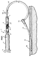
- a preferred embodiment of the present clotting cascade initiation device 1 is shown in fluid communication with a hemostasis sheath 34 having a hemostasis valve 33 interconnected with the clotting cascade initiation device 1 via a side-arm 29.
- the clotting cascade initiation device 1 shown in Figure 1 includes a syringe 3 having a plunger 2 which slides tightly against the inner walls of the syringe 3, enabling one to draw fluids into the syringe when the plunger 2 is drawn back.
- the syringe is preferably interconnected with a cartridge 9 containing a sterile containment chamber 20 the cartridge 9 is interconnected with the side arm 29 by a stop cock 7 adding a luer fitting for engagement with the cartridge 9.
- blood (not shown) may be withdrawn from the blood vessel 36 shown in cross-section within a portion of a patient's body 41 when the plunger 2 is partially withdrawn from the syringe 3 to create a vacuum in the syringe 3 which draws blood (not shown) from the blood vessel 36 through the hemostasis sheath 34, the side arm 29, the stop cock 7 and the sterile containment chamber 20 within the cartridge 9 before entering the syringe 3.
- the blood passes through the sterile containment chamber 20 it passes over the surfaces of a porous matrix of glass beads 14 within the sterile containment chamber 20.
- the glass beads are preferably coated with a positively charged polymer which binds negatively charged molecular species such as heparin which is often found in blood of patients being treated in surgical procedures and the like.
- the positively charged polymeric coating will preferably bind heparin found in the blood and remove it from the blood.
- the shear created by the passage of the blood fluid across the surfaces of the glass beads will initiate certain elements of the clotting cascade and begin the natural process of clotting the blood by so initiating the clotting cascade.
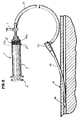
- an alternate clotting cascade initiation device 1' is shown in communication with a hemostasis sheath 34 interconnected with the clotting cascade initiation device 1' via the side arm 29 and the stop cock 7.
- the glass beads 14' are located within a sterile containment chamber 20' within the syringe.
- a pair of porous filters 43a and 43b, respectively allow blood to pass through while retaining any solids larger than the filter pore size, including the glass beads 14' which are preferably coated with a positively charged coating polymer which will bind heparin and substantially remove the heparin from any blood passing through the porous matrix created by the glass beads 14'.
- the clotting cascade in blood passing through the porous matrix created by the glass beads 14' will be initiated as the blood passes over the surfaces of the coated glass beads 14'.
- uncoated glass beads may be used to initiate the clotting cascade, however, if the blood contains a significant amount of heparin, the heparin will generally prevent the blood from clotting so long as it is present in the blood and is not either removed or inactivated.
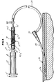
- a further alternate embodiment of the present invention shown in Figure 3 includes a clotting cascade initiation device 1" in which a preferred cartridge 9" includes a porous matrix made up of coated glass beads 14" and a porous matrix of biomaterials 15" within the sterile containment chamber 20".
- the porous matrix of coated glass beads 14" and the porous matrix of biomaterials 15" are retained within the sterile containment chamber 20" by a pair of porous membranes 43a" and 43b" approximate respective ends of the cartridge 9".
- the porous membranes 43a” and 43b" preferably have sufficiently small pores to prevent the coated glass beads 14" or fibers within the porous biomaterials 15" from passing through the porous membranes 43a” and 43b".
- the clotting cascade initiation device contains an agent or agents within a sterile containment chamber which either binds a negatively charged anti-coagulating agent or drug such as heparin, hirudin or the like so as to substantially reduce the anti-coagulating agent concentration in blood passing through the sterile containment chamber or contains an agent which degrades, neutralizes, cleaves, digests, or by other means inactivates the anti-coagulating agents so as to effectively reduce the active anti-coagulating agent concentration within the blood passing through the sterile containment chamber and effectively reduce the activity of the agent in preventing the blood from clotting.
- a negatively charged anti-coagulating agent or drug such as heparin, hirudin or the like
- the term "negatively charged” refers to something that has a negative charge when located in an aqueous solution or and aqueous system such as blood or a blood fluid.
- Heparin and hirudin are negatively charged anti-coagulating agents.
- the negatively charged anti-coagulating agent generally encountered in human blood fluids is heparin.
- the sterile containment chamber or a further sterile containment chamber removed from the first containment chamber containing the heparin binding or heparin neutralizing agent will contain a biomaterial which enhances the initiation of the clotting cascade so that the blood will clot in a shorter period of time than it might in the absence of the biomaterial once the effective concentration of heparin in the blood is substantially reduce by the heparin binding or heparin neutralizing agent or agents.
- a preferred method of closing a wound in a patient using a device in accord with the present invention comprises the steps of: treating a blood fluid with an agent which substantially reduces the anticoagulation effect in the blood fluid prior to treatment, the agent interacting with heparin in the blood fluid in a manner selected from the group consisting of binding to the anti-coagulating agent so as to remove the anti-coagulating agent from the blood fluid, degrading, immobilizing or neutralizing molecules so as to make them ineffective as an anti-coagulating agent and binding to the heparin so as to make the heparin molecules substantially ineffective as an anti-coagulating drug which can either inhibit or impede either the initiation or the continuance of the clotting cascade; and transporting the treated blood fluid to the wound in the patient such that the blood fluid comes into contact with the patient approximate the wound and the clotting cascade comes generally to a conclusion and a clot forms approximate the wound such that the clot is formed and the clo
- the step of treating the blood fluid includes treating the blood fluid with a procoagulating agent to initiate a clotting cascade during a first time period such that at least a portion of the blood fluid will form a clot of blood during a second time period subsequent to the first time period, the blood fluid containing sufficient blood components to enable a portion of the blood fluid to clot during the second time period subsequent to the initiation of the clotting cascade during the first time period; the blood fluid being selected from the group whole blood, natural components of whole blood, whether derived from whole blood, blood extracts, or products of ex-vivo cell cultures, and procoagulating agents which assist in or enhance the clotting or coagulation of the blood fluids, the step of treating the blood fluid initiating the clotting cascade.
- autologous blood is drawn from the patient and exposed to a porous matrix within which is contained such an agent which substantially reduces the anticoagulation effect of heparin present in the blood fluid prior to such treatment.
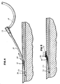
- this autologous blood is subsequently returned to the wounds site in the manner shown in Figure 4 where the sheath is being removed from the blood vessel 36 and the wound proximate the arteriotomy as blood fluids so treated are returned to the wound site following treatment of the blood fluid with an agent which substantially reduces the anticoagulation effect of the heparin present in the blood fluid prior to such treatment and preferably provides a blood fluid in which the clotting cascade has been initiated such that the blood fluid will clot after being delivered to the wound site.
- a fibrin clot 49 is shown as it is beginning to knit together within the wound site 50.
- a piece of sterile gauze 55, sterile cotton or the like is preferably placed upon the exterior of the tissue proximate the wound site 50 so that downward pressure (denoted by the three arrows proximate the wound site) may be applied to the wound site 50 for about 3 to 5 minutes to impede any potential flow of blood fluid through the wound site and enhance the ability of the fibrin clot 49 to continue to knit together as the clotting cascade continues to proceed to a conclusion where the clotting cascade is completed and the fibrin clot 49' is fully formed as shown in Figure 5 .
- the present invention provides apparatii for use in achieving hemostasis and wound closure in wounds in tissues.
- the invention preferably neutralizes or substantially eliminates the effect of anti-coagulating agents, such as heparin and initiates the blood clotting cascades in a discrete dose of blood fluid so that it can be transported to a wound in a patient such that the treated blood fluid dose can come into contact with the patient proximate the wound such that the clotting cascades can be completed proximate the wound, thereby forming a clot in the wound which prevents fluid from passing through the wound.
- the invention provides the opportunity to manipulate the blood fluid in respects needed to achieve the desired clot formation in the wound. This typically includes the elements of neutralizing or substantially eliminating the effects of anti-coagulating drugs such as heparin when found in the blood fluid which is treated and also those natural elements of the clotting cascade and of basic clot formation.
- the steps of treating a blood fluid with an agent which substantially reduces the anticoagulation effect present in the blood fluid includes binding any heparin present in a discrete dose of blood fluid and thus removing the negatively charged anti-coagulating either by simple ion exchange, by substantially irreversible ion binding or by administering an agent that chemically inactivates, incapacitates, degrades, neutralizes or destroys the heparin molecule.
- an agent that chemically inactivates, incapacitates, degrades, neutralizes or destroys the heparin molecule are also included in preferred treatment.
- the elements in the intrinsic and extrinsic clotting cascade mechanism for human blood fluid including activation of blood factors VII, VIII and XII which in turn lead, eventually to thrombin formation which lead to the conversion of the naturally occurring blood product fibrinogen into a solid phase fibrin clot.
- the present method and apparatus provides the opportunity to manipulate blood fluid used to form a wound sealing clot in many ways not possible before.
- the mechanism and method to contain the blood fluid can be achieved in any relationship with respect to the patient's body.
- the mechanism to receive and contain the blood fluid can be in fluid communication with a patient's body and blood supply by means of any device commonly used for patient vascular access, such as catheters, introducer sheaths, needles, angiocaths or other commonly used devices.
- the mechanism in this situation, can be completely outside of the body, adjacent to the body, partially in the body as with an introducer sheath or it can be contained completely within the body such as with an expandable balloon.
- the mechanism to receive and contain the blood fluid can also be separable from any direct fluid communication with the body.
- the mechanism can be a stand alone device or system to which blood fluid, previously drawn or gathered, can be brought and within which the steps of heparin neutralization and clotting cascade initiation can be achieved.
- the thus treated blood fluid can then be delivered to the wound site by any of various means including a needle, introducer sheath, catheter, etc.
- An apparatus to be used for sterile enclosed containment of the blood fluid may exist in many forms. A representative example of such enclosures would include syringes, cartridges, vials, test tubes, jars, bags, balloons, pouches, trays, dishes, bowls, tubing, catheters, cannulae in general, and the like.
- a common feature to such containers would include the ability to keep the container substantially enclosed thereby preventing a level of contamination or loss of sterility unacceptable to the user.
- one or more containers may be used separately or in combination.
- a syringe connected to a catheter may be regarded as two containers or a single container with a transfer means, depending on the context of the discussion it is used in. Both context are acceptable and not intended to be limiting.
- a single container may have one or more chambers internal to it.
- a single container with multiple chambers may be referred to as a single container or multiple containers. Again, both referrals are acceptable and should be interpreted in the context of the discussion and not be interpreted as to be limiting.
- the containers have at least one fluid communication element associated with it which connects the inside to the outside of the container.
- a fluid communication element may have a normally open hole such as a luer fitting, a normally closed hole such as a septum, or a selective opening such as a molecular sieve or semipermeable membrane, and the like.
- kits of components bundled together for ease of use.
- this invention can take many forms and is typically dependent on clinical application, user preferences, and the like.
- the following is merely an illustrative example of what a kit may consist of and should not be interpreted as a limitation of the present invention.
- this illustration will be in the context of a typical kit for use with minimally invasive angiographic procedures and the like.
- the kit may contain a wound closure apparatus or clotting cascade initiation apparatus of the present invention singularly or in combination with one or more of the following: an introducer, guidewire, dilator, obturator, collapsible catheter sheath, Seldinger needle, balloon catheter, infusion catheter, stent, scalpel, suture line, needle, pouch, tray, tray lid, instructions for use, adhesive identification label, sterility indicator, and the like.
- Heparin neutralization can most effectively be achieved by either binding and thereby removing substantially all of the heparin from an aliquot of blood by exposing the blood to a pharmacological agent that serves to bind so as to remove, chemically degrade or otherwise inactivate the heparin.
- Hirudin which is also an anti-coagulating agent, is similar to heparin in that they are both negatively charged molecules when contained within a aqueous system such as blood or a blood fluid. As such, both will generally bind to positively charged polymers such as DEAE Cellulose and the like.
- Polyethyleneimine (PEI) is an example of a preferred agent useful in binding and thus removing negatively charged anti-coagulating agents such as heparin from a discrete aliquot of blood.
- Polyethyleneimine is a branched chain polymer having a ratio of 1:2: of primary:secondary:tertiary amines with a branching site every 3 to 3.5 nitrogen atoms and a general backbone of (CH 2 CH 2 NH) x .
- the nitrogen atoms are protonated so as to achieve a positive cationic charge.
- Heparin is highly sulfated resulting in a negative charge and is therefore readily attracted to, substantially irreversibly bound by and effectively removed from blood fluids when exposed to a biomaterial substrate or other surface coated with immobilized polyethyleneimine.
- diethylaminoethyl cellulose a positively charged ion exchange polymer or resin is another material that can be utilized to attract, bind and remove heparin from a sample of blood, although its use has provided less effective results in the hands of the present inventors.
- Another reliable method to attract, bind and remove heparin is by use of a surface treatment, as opposed to a coating, one such surface treatment being aminoacetylization.
- Aminoacetylization as described by Shiomi, provides a dense concentration of positively charged functional groups on the surface of a chosen substrate. These cationic surface charges work in the same manner as the positively charged nitrogen atoms present with polyethyleneimine.
- Heparinase is an example of a material that will chemically break heparin down, thus rendering it ineffective. Heparinase is a naturally occurring blood component. Protamine sulfate is another material that will chemically inactivate heparin. In addition, platelet factor IV (PF4) is another substance that is capable of reversing heparin in a protamine like fashion. In addition, heparin reversal can be achieved by immobilization of protamine onto a suitable substrate, thus providing the same reversal capability as achieved using positively charged substances. Yet another approach is to override the heparin by exposing the blood fluid by exposing it to surface immobilized thrombin within the sterile containment chamber.
- PF4 platelet factor IV
- a cross-linked fibrin clot is a gelatinous-like substance with a very high water content.
- An additional element of this invention is a means to partially reduce the free fluid component of the clot so as to produce a firmer and possibly stronger clot substance. Any of various materials are capable of absorbing and binding water when blood is passed over them.
- procoagulant refers to any process, activity, material or substance that serves to initiate, continue or accelerate a clotting cascade.
- Human blood naturally has a redundant system for achieving clot formation as a natural, efficient way of protecting against hemorrhaging. The two arms of this redundant system are the extrinsic and the intrinsic clotting cascade.
- Each cascade includes a complex, sequential series of steps or stages during which a blood factor necessary for clot formation is activated, the activated blood factor, furthering the cascade, will initiate steps causing the formation of controlled amounts of the powerful, natural clot inducing substance thrombin.
- the thrombin will, in turn, work to convert the naturally circulating, fluid phase plasma protein fibrinogen into a cross-linked, solid phase mass known as a fibrin clot. It will be appreciated that a fibrin clot is an effective, natural agent to achieve hemostasis, or the cessation of bleeding, from a wound.
- Blood factor XII can be initiated and converted into activated blood factor XII, known as XIIa, by means of contact with the surface of biocompatible materials as are used widely in the practice of medicine. All materials, to varying degree, will activate blood factor XII into XIIa, depending upon a complex series of variables including material type, surface area, and exposure time.
- a material beneficially used to activate blood factor XII is cotton, such as is used in bandages and in the packing used by a dentist. Cotton is also effective in binding water which will aid in eventually producing a firmer and stronger clot.
- any of various polymers conventionally used for medical devices polyethylene, polycarbonate, polypropylene, ethylenevinylalchohol (EVAL), cellulose acetate, urethane and silicone being examples, materials such as glass, metal or ceramics also used in medical devices or, as is the case in a preferred embodiment of this invention, any of the above materials used as a substrate for a second biomaterial which is coated upon the substrate and that also has the ability to neutralize the anti-coagulating drug heparin, can be beneficially used to activate blood factor XII.
- Blood factor VIII a constituent within another of the intrinsic sub-pathways, is known to be converted to activated blood factor VIII (or VIIIa) by means of mechanical agitation or shear. Blood factor VIIIa, as with XIIa, initiates a series of enzymatic reactions that leads to the production of controlled amounts of thrombin.
- Blood exposed to mechanical agitation such as shear
- Blood factor VIIIa Blood factor VIIIa in proportion to the amount or degree of agitation induced.
- manufacturers and designers of medical devices that require the transport and manipulation of large amounts of blood fluid such as oxygenators, dialysis machines, etc. will go to great lengths to minimize the amount of agitation and shear generated during the handling of the blood so as to minimize the amount of blood factor VIIIa generated.
- the controlled activation of blood factor VIII is desired, such as when a discrete dose of blood thus treated will be used to create hemostasis in a wound
- the amount of agitation or shear induced when transporting a discrete dose of blood fluid can be manipulated by control of various design features in the transport mechanism.
- Mechanical agitation or shear measured in units of 1/seconds (reciprocal seconds), with ideally at least 500 1/seconds and, preferably, and increasingly more up to about 1200 1/seconds, but with continued benefit beyond 1200 1/seconds up to the point of destruction or lysis of blood cells from excessive manipulation, can be beneficially used to produce blood factor VIIIa and then used to treat a wound to control bleeding.
- the extrinsic clotting cascade is the second arm of the human blood fluid clot forming system.
- the extrinsic system works when blood fluid which has not been inhibited from clotting by use of drugs, is exposed to wound tissue or tissue beyond the interior lining of arteries, veins or capillaries. This exposure serves to activate blood factor VII to activated blood factor VII or VIIa. Blood factor VIIa, as with XIIa and VIIIa will lead to the formation of controlled amounts of thrombin and eventually to a fibrin clot useful for achieving and maintaining hemostasis.
- Blood fluid which has had the effects of heparin neutralized in a sterile containment chamber and which may or may not have had blood factors XII and VIII activated, and that is transported to the site of a wound will work to convert blood factor VII to VIIa and this will lead to the formation of a fibrin clot that will serve to control bleeding in the wound.
- Heparin inactivation is recognized as a necessary step and the most significant factor among these various elements as without this activity the blood fluid will not readily be converted to a solid phase fibrin clot even if various blood factors are activated. It is also recognized that, to some extent, activation of blood factors such as VII, VIII and XII can not be avoided when manipulating the blood fluid and using it as a hemostatic agent.
- an enclosed chamber such as a syringe, produced from an injection moldable polymer such as polycarbonate and having an opening on one end devised to releaseably attach, such as by means of a luer fitting, standard percutaneous access devices, such as hemostasis sheaths, catheters, needles or angiocaths and having a movable plunger, common to a syringe on the other end of the enclosed chamber, is partially filled with a coated matrix of biomaterial.
- the preferred coating is immobilized polyethyleneimine and the preferred porous matrix being glass beads or polypropylene fibers.
- the coated porous matrix being contained by a porous membrane or filter with openings smaller than the size of the glass beads or polypropylene fibers.
- the coated porous matrix being of sufficient quantity in terms of surface area to assure effective binding and thus removal of at least about 5 units of heparin, preferably at least about 10 units, more preferably at least about 20 and even more preferably at least about 40 units, and with sufficient porosity, density and surface area to assure at least 500 1/seconds of mechanical agitation or shear and preferable more up to 1200 1/seconds and more than 1200 1/seconds up to the point of blood cell destruction.
- the chamber of the syringe not filled with the porous matrix is preferably of sufficient volume to receive a volume of blood sufficient to treat a wound site, this volume is preferably at least about 1 ml, more preferably about 2 or more ml and most preferably about 5 ml, however, more than about 5 ml does not detract from the usefulness of the invention.
- the present device is connected by a luer fitting to a vascular access device already in place for use in a medical procedure.
- This connection would typically be made to the side-arm of a hemostasis valve 33, interconnected with a sheath 34 and would, therefore, be in direct fluid communication with a blood vessel accessed for the procedure and thus having an access site or arteriotomy in the case of an artery.
- a discrete dose of blood is preferably drawn out of the artery through the sheath 34 into the side-arm 29 and then into the sterile containment chamber through the coated porous matrix within the chamber and then into a receiving chamber in the syringe as the syringe plunger is partially withdrawn.
- the porous matrix will preferably have sufficient surface area and tortuosity to activate the blood factor XII contained in the blood dose, the porous matrix also having sufficient porosity and tortuosity to achieve at least 500 1/seconds and preferably 1200 1/seconds of mechanical agitation or shear so as to activate blood factor VIII contained in the blood dose,
- the sheath then being pulled back by the physician to a point just outside of the accessed artery and therefore the end of the sheath now being positioned just outside of the arteriotomy and within the soft tissue wound.
- the plunger of the syringe then being advanced thus pushing the blood sample back through the system and into the wound area thus activating blood factor VII.
- the treated blood can be either transported back through the porous matrix furthering the intrinsic clotting cascade activation mechanisms or delivered through a more direct path avoiding the porous matrix.
- the preferred embodiments of the apparatus of the present invention may be highly varied and is typically dependent on the individual application considering clinical situation, physician preference, and the like.
- Post-introducer arterial wound closure typically involves the closure of a wound within an arterial wall such as the femoral artery, radial artery, and the like. Such wounds are typically subcutaneous in the sense that the artery is covered by tissue rather than being exposed by cut down through the tissue until the artery is visible to the practitioner.
- FIG. 1 represents a completed apparatus assembly in-place attached to the introducer and in the patient as it would typically be used.
- Introducer/hemostasis sheath 32 is placed through the patients skin 41, i.e., percutaneously with the distal end of the sheath within an artery 36.
- Autologous blood is drawn back from the distal end of the sheath 34 by means of pulling back on the syringe plunger. During this process the blood is passed through the heparin removing and clotting cascade initiating chamber 20 and into the barrel of the syringe 3.
- the sheath 34 is then withdrawn to a position just outside of the artery 36 as is alternately determined use a pulsitile indicator (not shown).
- the syringe plunger 2 is then advanced by expelling the blood in the barrel of the syringe 3 back through the heparin binding and clotting cascade initiating chamber 20, then back through the sheath 32 and expelling it into the tissue 41 adjacent to the artery 36 where the clotting cascade comes to completion.
- FIG. 2 shows a combination syringe - heparin binding and clotting cascade initiating apparatus.
- a heparin binding and intrinsic clotting cascade initiating material 14' preferably including polyethyleneimine (PEI) coated glass beads or ethylenevinylalcohol (EVAL) surface treated by aminoacetylization.
- PEI polyethyleneimine
- EVAL ethylenevinylalcohol
- a membrane filter or screen 43a and 43b, respectively is located on either side of the procoagulant materials 14'.
- FIG. 3 shows a 12 cc polymer syringe 3" consisting of a plunger rod 2" connected to a fluid sealing piston 4" is slidingly placed within the syringe 3", thereby allowing the syringe to receive and expel fluids through the distal port 5" of the syringe 3".
- a three-way connector 7" permits fluid communication between its three ports.
- Luer lock cylinder cap fittings 8" and 10 preferably made of polycarbonate, are used to form a container when assembled to the ends of cylinder 9, also preferably made of polycarbonate.
- dPEI derivatized PEI
- Biolnteraction, Ltd., Redding, United Kingdom by solution phase coating as follows:
- This process involves first priming the surface by immersing the material in a dilute solution of PEI with a cross-linker.
- the cross-linkers that have demonstrated successful coating include: glutaraldehyde, crotenaldehyde, carbonyl diimidazole, ethylene glycol diacrylate, butane diol diglycidyl ether, pentaerythritol-tris-(N-aridinyl) proprionate (XAMA-7 from Sigma Chemical Co., St. Louis, MO), Polyethyleneoxide diglycidyl ether.
- cross-linkers have in common: they are multifunctional, water soluble, and they react with amine groups (they cross-link PEI).
- the surface is exposed to a solution of dextran sulfate, followed by a repeat of the initial PEI + CROSS-LINKER, dextran sulfate, and then a solution of PEI that does not contain the cross-linker.
- the deposition of PEI on the material surface can be greatly enhanced by the addition of solution phase coating step that adds PAA to the surface.
- the PAA must be at a mW greater than 3,000,000.
- the addition of a PAA step can enhance PEI deposition for both material coating with and without a crossliner.
- the PAA is made as a 0.06% w/v Polyacrylic Acid (PAA).
- PAA Polyacrylic Acid
- the above described process can also be achieved by incorporating polylysine in place of PEI as the material to surface deposit at the surface to achieve removal of heparin.
- An alternative to coating to provide a surface that can remove heparin is to modify a surface so that it can remove heparin.
- One such approach was described by Shiomi in which heparin was ionically bound onto the surface of an ethylene vinyl copolymer membrane which was derivatized by aminoacetylization to produce cationic surface charges. The amount of bound heparin was proportional to the ion exchange capacity of the aminoacetalized membrane and the maximal amount obtained was 96 Unit/cm2 (0.59 mg/cm2).
Landscapes
- Health & Medical Sciences (AREA)
- Life Sciences & Earth Sciences (AREA)
- Surgery (AREA)
- Molecular Biology (AREA)
- General Health & Medical Sciences (AREA)
- Biomedical Technology (AREA)
- Heart & Thoracic Surgery (AREA)
- Medical Informatics (AREA)
- Nuclear Medicine, Radiotherapy & Molecular Imaging (AREA)
- Animal Behavior & Ethology (AREA)
- Engineering & Computer Science (AREA)
- Public Health (AREA)
- Veterinary Medicine (AREA)
- Cardiology (AREA)
- Materials For Medical Uses (AREA)
- Medicines That Contain Protein Lipid Enzymes And Other Medicines (AREA)
- Control For Baths (AREA)
- Physical Or Chemical Processes And Apparatus (AREA)
- Infusion, Injection, And Reservoir Apparatuses (AREA)
Claims (21)
- Utilisation de fluide sanguin, dans laquelle un agent réduit sensiblement l'effet anticoagulant d'un agent anticoagulant chargé négativement, pour la fabrication d'un agent hémostatique destiné à être appliqué entre une plaie vasculaire résultant d'une entrée percutanée et la surface de la peau pour effectuer une fermeture de la plaie, comprenant les étapes consistant à :disposer d'un dispositif comprenant une chambre de confinement stérile sensiblement close (20) à l'intérieur de laquelle le fluide sanguin peut être reçu, la chambre de confinement stérile sensiblement close (20) contenant un agent qui élimine un agent anticoagulant chargé négativement présent dans le fluide sanguin, l'agent interagissant avec l'agent anticoagulant chargé négativement dans le fluide sanguin en se liant à l'agent anticoagulant chargé négativement, le dispositif comprenant en outre une seringue pour effectuer un écoulement de sang dans et hors de la chambre de confinement (20), caractérisée en ce que le dispositif comprend en outre :une gaine vasculaire (34) pour une insertion dans un vaisseau sanguin (36) ; etun bras latéral (29) interconnecté à la gaine vasculaire (34) par une valve hémostatique (33) et conduisant à la chambre de confinement (20), le bras latéral (29), la valve hémostatique (33) et la gaine vasculaire (34) étant configurés pour transporter le fluide sanguin depuis une plaie vasculaire vers la chambre de confinement (20) pour une élimination de l'agent anticoagulant chargé négativement, l'agent réduisant sensiblement l'effet anticoagulant de l'agent anticoagulant chargé négativement, et le bras latéral (29) et la gaine vasculaire (34) étant en outre configurés pour effectuer un retour du fluide sanguin dont l'agent anticoagulant chargé négativement a été éliminé au voisinage de la plaie vasculaire pour la formation d'un caillot ; etcontenir le fluide sanguin dans la chambre de confinement.
- Utilisation selon la revendication 1, dans laquelle le fluide sanguin est du sang entier.
- Utilisation selon la revendication 2, dans laquelle le sang est du sang entier autologue.
- Utilisation selon la revendication 3, dans laquelle le sang entier autologue est traité pour que l'agent anticoagulant qui s'y trouve devienne inefficace ou pour que ledit agent anticoagulant soit éliminé.
- Utilisation selon la revendication 4, dans laquelle l'agent anticoagulant du sang entier autologue est chargé négativement, et ledit agent est éliminé ou rendu inefficace par utilisation d'une substance chargée positivement qui se lie à celui-ci.
- Utilisation selon la revendication 4 ou 5, dans laquelle l'agent anticoagulant du sang entier autologue est l'héparine ou l'hirudine.
- Utilisation selon l'une quelconque des revendications précédentes, dans laquelle un mécanisme de cascade de coagulation intrinsèque a été activé pour produire un fluide sanguin qui va progresser vers un caillot terminé en un temps prévisible.
- Utilisation selon la revendication 7, dans laquelle la cascade de coagulation est initiée par contact avec un agent procoagulant.
- Utilisation selon la revendication 7, dans laquelle la cascade de coagulation est initiée par agitation mécanique ou cisaillement.
- Utilisation selon l'une quelconque des revendications précédentes, dans laquelle le fluide sanguin est destiné au traitement d'une plaie artérielle et le fluide sanguin est destiné à être placé dans un tissu mou adjacent à l'artériotomie.
- Utilisation selon la revendication 10, dans laquelle le fluide sanguin est destiné au traitement d'une plaie de l'artère fémorale ou radiale.
- Dispositif comprenant une chambre de confinement stérile sensiblement close (20) dans laquelle un fluide sanguin peut être reçu, la chambre de confinement stérile sensiblement close (20) contenant un agent qui élimine un agent anticoagulant chargé négativement présent dans le fluide sanguin, l'agent interagissant avec l'agent anticoagulant chargé négativement dans le fluide sanguin en se liant à l'agent anticoagulant chargé négativement, le dispositif comprenant en outre une seringue (3) pour effectuer un écoulement de sang dans et hors de la chambre de confinement (20), et une gaine vasculaire (34) pour une insertion dans un vaisseau sanguin (36) ; lequel dispositif est caractérisé en ce qu'il comprend en outre :un bras latéral (29) interconnecté à la gaine vasculaire (34) par une valve hémostatique (33) et conduisant à la chambre de confinement (20), la seringue (3), le bras latéral (29), la valve hémostatique (33) et la gaine vasculaire (34) étant configurés pour transporter le fluide sanguin depuis une plaie vasculaire vers la chambre de confinement (20) pour une élimination de l'agent anticoagulant chargé négativement, l'agent réduisant sensiblement l'effet anticoagulant de l'agent anticoagulant chargé négativement, et le bras latéral (29) et la gaine vasculaire (34) étant en outre configurés pour effectuer un retour du fluide sanguin dont l'agent anticoagulant chargé négativement a été éliminé au voisinage de la plaie vasculaire pour la formation d'un caillot.
- Dispositif selon la revendication 12, dans lequel l'agent est un matériau chargé positivement destiné à se lier audit agent anticoagulant chargé négativement pour rendre l'agent anticoagulant inefficace ou pour éliminer l'agent anticoagulant du sang.
- Dispositif selon la revendication 12, comprenant en outre un agent procoagulant à l'intérieur de la chambre de confinement stérile (20) ; dans lequel une cascade de coagulation est initiée quand le fluide sanguin est reçu dans la chambre de confinement stérile (20) et exposé à l'agent procoagulant à l'intérieur de la chambre, et le fluide sanguin peut être délivré à la plaie après l'initiation de la cascade de coagulation, de façon que la cascade de coagulation, initiée à l'intérieur de la chambre de confinement stérile (20) avant la délivrance du fluide sanguin à la plaie, puisse être complétée à l'intérieur de la plaie et qu'un caillot puisse se former à l'intérieur de la plaie quand la cascade de coagulation est complétée.
- Dispositif selon la revendication 12, dans lequel l'agent anticoagulant chargé négativement est choisi dans le groupe constitué par l'héparine et l'hirudine.
- Dispositif selon la revendication 12, dans lequel l'agent anticoagulant chargé négativement est l'héparine.
- Dispositif selon la revendication 14, dans lequel le fluide sanguin contient jusqu'à 25 microgrammes de thrombine par millilitre de fluide sanguin.
- Dispositif selon la revendication 12, dans lequel l'agent qui réduit sensiblement l'effet anticoagulant de l'agent anticoagulant chargé négativement est un groupe fonctionnel chargé positivement lié à une surface à l'intérieur de la chambre de confinement stérile (20).
- Dispositif selon la revendication 12, dans lequel l'agent qui réduit sensiblement l'effet anticoagulant de l'agent anticoagulant chargé négativement est un polymère chargé positivement qui est déposé en surface sur une surface exposée à l'intérieur de la chambre de confinement stérile (20).
- Dispositif selon la revendication 12, dans lequel l'agent qui réduit sensiblement l'effet anticoagulant de l'agent anticoagulant chargé négativement est un polymère déposé en surface choisi dans le groupe constitué par une polyéthylène-imine (PEI) et une polyéthylène-imine dérivée (dPEI).
- Dispositif selon la revendication 12, dans lequel l'agent qui réduit sensiblement l'effet anticoagulant de l'agent anticoagulant chargé négativement est une
polyéthylène-imine (PEI) déposée en surface.
Applications Claiming Priority (3)
| Application Number | Priority Date | Filing Date | Title |
|---|---|---|---|
| US13683799P | 1999-06-01 | 1999-06-01 | |
| US136837P | 1999-06-01 | ||
| PCT/US2000/015068 WO2000074575A1 (fr) | 1999-06-01 | 2000-06-01 | Appareil d'initiation d'une serie de reactions en cascade dans la coagulation sanguine, et procede d'utilisation y relatif |
Publications (3)
| Publication Number | Publication Date |
|---|---|
| EP1180977A1 EP1180977A1 (fr) | 2002-02-27 |
| EP1180977A4 EP1180977A4 (fr) | 2007-08-22 |
| EP1180977B1 true EP1180977B1 (fr) | 2011-12-28 |
Family
ID=22474590
Family Applications (1)
| Application Number | Title | Priority Date | Filing Date |
|---|---|---|---|
| EP00938020A Expired - Lifetime EP1180977B1 (fr) | 1999-06-01 | 2000-06-01 | Appareil d'initiation d'une serie de reactions en cascade dans la coagulation sanguine, et procede d'utilisation y relatif |
Country Status (6)
| Country | Link |
|---|---|
| US (1) | US6482223B1 (fr) |
| EP (1) | EP1180977B1 (fr) |
| AT (1) | ATE538730T1 (fr) |
| AU (1) | AU5311700A (fr) |
| CA (1) | CA2375319C (fr) |
| WO (1) | WO2000074575A1 (fr) |
Families Citing this family (19)
| Publication number | Priority date | Publication date | Assignee | Title |
|---|---|---|---|---|
| DE29723807U1 (de) * | 1996-04-30 | 1999-11-04 | Medtronic, Inc., Minneapolis, Minn. | Autologe Fibrin-Blutstillungsmittel |
| WO2000062828A1 (fr) | 1996-04-30 | 2000-10-26 | Medtronic, Inc. | Fibrine autologue d'obturation et ses methodes de fabrication |
| US6159232A (en) | 1997-12-16 | 2000-12-12 | Closys Corporation | Clotting cascade initiating apparatus and methods of use and methods of closing wounds |
| US6478808B2 (en) * | 1997-12-17 | 2002-11-12 | Closys Corporation | Clotting cascade initiating apparatus and methods of use and methods of closing wounds |
| US20030069601A1 (en) * | 1998-12-15 | 2003-04-10 | Closys Corporation | Clotting cascade initiating apparatus and methods of use |
| EP1180977B1 (fr) * | 1999-06-01 | 2011-12-28 | Closys Corporation | Appareil d'initiation d'une serie de reactions en cascade dans la coagulation sanguine, et procede d'utilisation y relatif |
| AU1375301A (en) | 1999-11-15 | 2001-05-30 | Bio Syntech Canada Inc | Temperature-controlled and ph-dependant self-gelling biopolymeric aqueous solution |
| DK1294414T3 (da) * | 2000-06-29 | 2006-07-24 | Biosyntech Canada Inc | Præparat og fremgangsmåde til heling og regenerering af brusk og andre væv |
| US6955682B2 (en) * | 2001-05-09 | 2005-10-18 | Biointeractions, Ltd. | Wound closure system and methods |
| US20080045881A1 (en) * | 2004-03-26 | 2008-02-21 | University Of Southern California | Devices and methods for removing a matter from a body cavity of a patient |
| US20050228417A1 (en) * | 2004-03-26 | 2005-10-13 | Teitelbaum George P | Devices and methods for removing a matter from a body cavity of a patient |
| US8382794B2 (en) * | 2006-01-04 | 2013-02-26 | St. Jude Medical Puerto Rico Llc | Balloon insertion apparatus and method of sealing a tissue puncture |
| US20080280310A1 (en) * | 2007-05-09 | 2008-11-13 | Louis Panagopoulos | Testing for Blood Group Immunological Reaction Without the Use of Anti-Human Globulin |
| EP2349013B1 (fr) * | 2008-08-13 | 2020-02-26 | Andrea Del Corso | Dispositif d'occlusion pour chirurgie vasculaire |
| JP5616891B2 (ja) | 2008-08-26 | 2014-10-29 | セント ジュード メディカル インコーポレイテッドSt. Jude Medical, Inc. | 経皮穿刺部を封止するための方法およびシステム |
| US10231721B2 (en) | 2010-06-24 | 2019-03-19 | St. Croix Surgical Systems, Llc | Failsafe percutaneous wound barrier |
| WO2014052496A1 (fr) | 2012-09-25 | 2014-04-03 | Stem Cell Partners Llc | Procédé et appareil pour préparer le sérum de thrombine d'un donneur individuel |
| WO2016105575A1 (fr) * | 2014-12-23 | 2016-06-30 | Siemens Healthcare Diagnostics Inc. | Digestion protéolytique de troponine i cardiaque |
| US20230301875A1 (en) * | 2022-03-28 | 2023-09-28 | Nickalis J. Dumas | System and method for preparation and delivery of re-generative fibrin matrices |
Family Cites Families (36)
| Publication number | Priority date | Publication date | Assignee | Title |
|---|---|---|---|---|
| US3223083A (en) * | 1960-09-09 | 1965-12-14 | President And Directors Of Geo | Method for adhesively securing together skin and other soft tissue and bone |
| DE2834320A1 (de) | 1978-08-04 | 1980-02-28 | Tomic Dobrivoje | Heilmittel mit entzuendungshemmender und blutstillender wirkung |
| US4347243A (en) | 1979-10-04 | 1982-08-31 | Research Corporation | Acid soluble, pepsin resistant platelet aggregating material |
| AT366916B (de) | 1980-04-02 | 1982-05-25 | Immuno Ag | Vorrichtung zur applikation eines gewebeklebstoffes auf basis von menschlichen oder tierischenproteinen |
| US4373023A (en) * | 1980-10-14 | 1983-02-08 | Massachusetts Institute Of Technology | Process for neutralizing heparin |
| US4852568A (en) | 1987-02-17 | 1989-08-01 | Kensey Nash Corporation | Method and apparatus for sealing an opening in tissue of a living being |
| US5330445A (en) | 1988-05-26 | 1994-07-19 | Haaga John R | Sheath for wound closure caused by a medical tubular device |
| NL8901350A (nl) | 1989-05-29 | 1990-12-17 | Wouter Matthijs Muijs Van De M | Afsluitsamenstel. |
| US5000854A (en) | 1989-06-14 | 1991-03-19 | The University Of Michigan | Protamine-based filter device for removal of heparin from blood samples |
| US5318524A (en) * | 1990-01-03 | 1994-06-07 | Cryolife, Inc. | Fibrin sealant delivery kit |
| US5151192A (en) | 1990-07-13 | 1992-09-29 | Pall Corporation | Method for removing heparin from blood or plasma |
| US5391183A (en) | 1990-09-21 | 1995-02-21 | Datascope Investment Corp | Device and method sealing puncture wounds |
| US5108421A (en) | 1990-10-01 | 1992-04-28 | Quinton Instrument Company | Insertion assembly and method of inserting a vessel plug into the body of a patient |
| US5192300A (en) | 1990-10-01 | 1993-03-09 | Quinton Instrument Company | Insertion assembly and method of inserting a vessel plug into the body of a patient |
| US5310407A (en) | 1991-06-17 | 1994-05-10 | Datascope Investment Corp. | Laparoscopic hemostat delivery system and method for using said system |
| US5676689A (en) | 1991-11-08 | 1997-10-14 | Kensey Nash Corporation | Hemostatic puncture closure system including vessel location device and method of use |
| US5282827A (en) | 1991-11-08 | 1994-02-01 | Kensey Nash Corporation | Hemostatic puncture closure system and method of use |
| US5326350A (en) | 1992-05-11 | 1994-07-05 | Li Shu Tung | Soft tissue closure systems |
| DK83092D0 (da) | 1992-06-24 | 1992-06-24 | Unes As | Fremgangsmaade til udvinding af thrombin |
| US5413571A (en) | 1992-07-16 | 1995-05-09 | Sherwood Medical Company | Device for sealing hemostatic incisions |
| US5443481A (en) | 1992-07-27 | 1995-08-22 | Lee; Benjamin I. | Methods and device for percutaneous sealing of arterial puncture sites |
| US5292332A (en) | 1992-07-27 | 1994-03-08 | Lee Benjamin I | Methods and device for percutanceous sealing of arterial puncture sites |
| US5624669A (en) | 1993-03-31 | 1997-04-29 | Tri-Point Medical Corporation | Method of hemostatic sealing of blood vessels and internal organs |
| US5383896A (en) | 1993-05-25 | 1995-01-24 | Gershony; Gary | Vascular sealing device |
| US5843124A (en) | 1993-09-28 | 1998-12-01 | Hemodynamics, Inc. | Surface opening adhesive sealer |
| US5383899A (en) | 1993-09-28 | 1995-01-24 | Hammerslag; Julius G. | Method of using a surface opening adhesive sealer |
| US5437292A (en) | 1993-11-19 | 1995-08-01 | Bioseal, Llc | Method for sealing blood vessel puncture sites |
| US5585007A (en) | 1994-12-07 | 1996-12-17 | Plasmaseal Corporation | Plasma concentrate and tissue sealant methods and apparatuses for making concentrated plasma and/or tissue sealant |
| US5814066A (en) * | 1994-12-23 | 1998-09-29 | The University Of Virginia Patent Foundation | Reduction of femoral arterial bleeding post catheterization using percutaneous application of fibrin sealant |
| US5510102A (en) | 1995-01-23 | 1996-04-23 | The Regents Of The University Of California | Plasma and polymer containing surgical hemostatic adhesives |
| US5532311A (en) | 1995-02-01 | 1996-07-02 | Minnesota Mining And Manufacturing Company | Process for modifying surfaces |
| US5674394A (en) | 1995-03-24 | 1997-10-07 | Johnson & Johnson Medical, Inc. | Single use system for preparation of autologous plasma |
| US6146771A (en) | 1997-07-01 | 2000-11-14 | Terumo Cardiovascular Systems Corporation | Process for modifying surfaces using the reaction product of a water-insoluble polymer and a polyalkylene imine |
| US6197289B1 (en) * | 1997-07-01 | 2001-03-06 | Terumo Cardiovascular Systems Corporation | Removal of biologically active agents |
| US6033427A (en) | 1998-01-07 | 2000-03-07 | Lee; Benjamin I. | Method and device for percutaneous sealing of internal puncture sites |
| EP1180977B1 (fr) * | 1999-06-01 | 2011-12-28 | Closys Corporation | Appareil d'initiation d'une serie de reactions en cascade dans la coagulation sanguine, et procede d'utilisation y relatif |
-
2000
- 2000-06-01 EP EP00938020A patent/EP1180977B1/fr not_active Expired - Lifetime
- 2000-06-01 CA CA002375319A patent/CA2375319C/fr not_active Expired - Fee Related
- 2000-06-01 AT AT00938020T patent/ATE538730T1/de active
- 2000-06-01 US US09/585,488 patent/US6482223B1/en not_active Expired - Lifetime
- 2000-06-01 AU AU53117/00A patent/AU5311700A/en not_active Abandoned
- 2000-06-01 WO PCT/US2000/015068 patent/WO2000074575A1/fr not_active Ceased
Also Published As
| Publication number | Publication date |
|---|---|
| EP1180977A4 (fr) | 2007-08-22 |
| CA2375319C (fr) | 2010-01-12 |
| WO2000074575A1 (fr) | 2000-12-14 |
| CA2375319A1 (fr) | 2000-12-14 |
| ATE538730T1 (de) | 2012-01-15 |
| AU5311700A (en) | 2000-12-28 |
| EP1180977A1 (fr) | 2002-02-27 |
| US6482223B1 (en) | 2002-11-19 |
Similar Documents
| Publication | Publication Date | Title |
|---|---|---|
| US8263223B2 (en) | Coated surfaces for immobilizing negatively charged anticoagulating agents from blood fluid | |
| US6482223B1 (en) | Clotting cascade initiating apparatus and methods of use | |
| US6159232A (en) | Clotting cascade initiating apparatus and methods of use and methods of closing wounds | |
| US9839716B1 (en) | Methods of closing wounds | |
| JP5253170B2 (ja) | 血管創の閉鎖装置および方法 | |
| CA2446374C (fr) | Dispositif d'obturation des plaies chirurgicales | |
| US9179897B2 (en) | Vascular closure devices and methods providing hemostatic enhancement | |
| US20140188031A1 (en) | Vascular access devices, methods, and kits | |
| US20140155804A1 (en) | Surgical devices and kits | |
| Reed | Blood Access without Needles: The Hemasite Titanium Vascular Access Device |
Legal Events
| Date | Code | Title | Description |
|---|---|---|---|
| PUAI | Public reference made under article 153(3) epc to a published international application that has entered the european phase |
Free format text: ORIGINAL CODE: 0009012 |
|
| 17P | Request for examination filed |
Effective date: 20011129 |
|
| AK | Designated contracting states |
Kind code of ref document: A1 Designated state(s): AT BE CH CY DE DK ES FI FR GB GR IE IT LI LU MC NL PT SE |
|
| AX | Request for extension of the european patent |
Free format text: AL;LT;LV;MK;RO;SI |
|
| A4 | Supplementary search report drawn up and despatched |
Effective date: 20070724 |
|
| 17Q | First examination report despatched |
Effective date: 20080813 |
|
| GRAP | Despatch of communication of intention to grant a patent |
Free format text: ORIGINAL CODE: EPIDOSNIGR1 |
|
| GRAS | Grant fee paid |
Free format text: ORIGINAL CODE: EPIDOSNIGR3 |
|
| GRAA | (expected) grant |
Free format text: ORIGINAL CODE: 0009210 |
|
| AK | Designated contracting states |
Kind code of ref document: B1 Designated state(s): AT BE CH CY DE DK ES FI FR GB GR IE IT LI LU MC NL PT SE |
|
| REG | Reference to a national code |
Ref country code: GB Ref legal event code: FG4D |
|
| REG | Reference to a national code |
Ref country code: CH Ref legal event code: EP |
|
| REG | Reference to a national code |
Ref country code: AT Ref legal event code: REF Ref document number: 538730 Country of ref document: AT Kind code of ref document: T Effective date: 20120115 |
|
| REG | Reference to a national code |
Ref country code: NL Ref legal event code: T3 Ref country code: IE Ref legal event code: FG4D |
|
| REG | Reference to a national code |
Ref country code: SE Ref legal event code: TRGR |
|
| REG | Reference to a national code |
Ref country code: DE Ref legal event code: R096 Ref document number: 60046793 Country of ref document: DE Effective date: 20120301 |
|
| PG25 | Lapsed in a contracting state [announced via postgrant information from national office to epo] |
Ref country code: GR Free format text: LAPSE BECAUSE OF FAILURE TO SUBMIT A TRANSLATION OF THE DESCRIPTION OR TO PAY THE FEE WITHIN THE PRESCRIBED TIME-LIMIT Effective date: 20120329 |
|
| PG25 | Lapsed in a contracting state [announced via postgrant information from national office to epo] |
Ref country code: CY Free format text: LAPSE BECAUSE OF FAILURE TO SUBMIT A TRANSLATION OF THE DESCRIPTION OR TO PAY THE FEE WITHIN THE PRESCRIBED TIME-LIMIT Effective date: 20111228 Ref country code: BE Free format text: LAPSE BECAUSE OF FAILURE TO SUBMIT A TRANSLATION OF THE DESCRIPTION OR TO PAY THE FEE WITHIN THE PRESCRIBED TIME-LIMIT Effective date: 20111228 |
|
| PG25 | Lapsed in a contracting state [announced via postgrant information from national office to epo] |
Ref country code: PT Free format text: LAPSE BECAUSE OF FAILURE TO SUBMIT A TRANSLATION OF THE DESCRIPTION OR TO PAY THE FEE WITHIN THE PRESCRIBED TIME-LIMIT Effective date: 20120430 |
|
| REG | Reference to a national code |
Ref country code: AT Ref legal event code: MK05 Ref document number: 538730 Country of ref document: AT Kind code of ref document: T Effective date: 20111228 |
|
| PG25 | Lapsed in a contracting state [announced via postgrant information from national office to epo] |
Ref country code: DK Free format text: LAPSE BECAUSE OF FAILURE TO SUBMIT A TRANSLATION OF THE DESCRIPTION OR TO PAY THE FEE WITHIN THE PRESCRIBED TIME-LIMIT Effective date: 20111228 |
|
| PLBE | No opposition filed within time limit |
Free format text: ORIGINAL CODE: 0009261 |
|
| STAA | Information on the status of an ep patent application or granted ep patent |
Free format text: STATUS: NO OPPOSITION FILED WITHIN TIME LIMIT |
|
| PG25 | Lapsed in a contracting state [announced via postgrant information from national office to epo] |
Ref country code: IT Free format text: LAPSE BECAUSE OF FAILURE TO SUBMIT A TRANSLATION OF THE DESCRIPTION OR TO PAY THE FEE WITHIN THE PRESCRIBED TIME-LIMIT Effective date: 20111228 |
|
| 26N | No opposition filed |
Effective date: 20121001 |
|
| REG | Reference to a national code |
Ref country code: DE Ref legal event code: R097 Ref document number: 60046793 Country of ref document: DE Effective date: 20121001 |
|
| PG25 | Lapsed in a contracting state [announced via postgrant information from national office to epo] |
Ref country code: AT Free format text: LAPSE BECAUSE OF FAILURE TO SUBMIT A TRANSLATION OF THE DESCRIPTION OR TO PAY THE FEE WITHIN THE PRESCRIBED TIME-LIMIT Effective date: 20111228 |
|
| PG25 | Lapsed in a contracting state [announced via postgrant information from national office to epo] |
Ref country code: ES Free format text: LAPSE BECAUSE OF FAILURE TO SUBMIT A TRANSLATION OF THE DESCRIPTION OR TO PAY THE FEE WITHIN THE PRESCRIBED TIME-LIMIT Effective date: 20120408 |
|
| PG25 | Lapsed in a contracting state [announced via postgrant information from national office to epo] |
Ref country code: FI Free format text: LAPSE BECAUSE OF FAILURE TO SUBMIT A TRANSLATION OF THE DESCRIPTION OR TO PAY THE FEE WITHIN THE PRESCRIBED TIME-LIMIT Effective date: 20111228 |
|
| PGFP | Annual fee paid to national office [announced via postgrant information from national office to epo] |
Ref country code: IE Payment date: 20130621 Year of fee payment: 14 Ref country code: SE Payment date: 20130619 Year of fee payment: 14 Ref country code: CH Payment date: 20130621 Year of fee payment: 14 Ref country code: MC Payment date: 20130612 Year of fee payment: 14 Ref country code: LU Payment date: 20130621 Year of fee payment: 14 |
|
| PGFP | Annual fee paid to national office [announced via postgrant information from national office to epo] |
Ref country code: NL Payment date: 20130619 Year of fee payment: 14 |
|
| PGFP | Annual fee paid to national office [announced via postgrant information from national office to epo] |
Ref country code: GB Payment date: 20140618 Year of fee payment: 15 |
|
| PGFP | Annual fee paid to national office [announced via postgrant information from national office to epo] |
Ref country code: DE Payment date: 20140619 Year of fee payment: 15 |
|
| PGFP | Annual fee paid to national office [announced via postgrant information from national office to epo] |
Ref country code: FR Payment date: 20140619 Year of fee payment: 15 |
|
| REG | Reference to a national code |
Ref country code: NL Ref legal event code: V1 Effective date: 20150101 |
|
| PG25 | Lapsed in a contracting state [announced via postgrant information from national office to epo] |
Ref country code: MC Free format text: LAPSE BECAUSE OF NON-PAYMENT OF DUE FEES Effective date: 20140630 Ref country code: SE Free format text: LAPSE BECAUSE OF NON-PAYMENT OF DUE FEES Effective date: 20140602 Ref country code: LU Free format text: LAPSE BECAUSE OF NON-PAYMENT OF DUE FEES Effective date: 20140601 |
|
| REG | Reference to a national code |
Ref country code: CH Ref legal event code: PL |
|
| REG | Reference to a national code |
Ref country code: SE Ref legal event code: EUG |
|
| REG | Reference to a national code |
Ref country code: IE Ref legal event code: MM4A |
|
| PG25 | Lapsed in a contracting state [announced via postgrant information from national office to epo] |
Ref country code: NL Free format text: LAPSE BECAUSE OF NON-PAYMENT OF DUE FEES Effective date: 20150101 |
|
| PG25 | Lapsed in a contracting state [announced via postgrant information from national office to epo] |
Ref country code: LI Free format text: LAPSE BECAUSE OF NON-PAYMENT OF DUE FEES Effective date: 20140630 Ref country code: IE Free format text: LAPSE BECAUSE OF NON-PAYMENT OF DUE FEES Effective date: 20140601 Ref country code: CH Free format text: LAPSE BECAUSE OF NON-PAYMENT OF DUE FEES Effective date: 20140630 |
|
| REG | Reference to a national code |
Ref country code: DE Ref legal event code: R119 Ref document number: 60046793 Country of ref document: DE |
|
| GBPC | Gb: european patent ceased through non-payment of renewal fee |
Effective date: 20150601 |
|
| REG | Reference to a national code |
Ref country code: FR Ref legal event code: ST Effective date: 20160229 |
|
| PG25 | Lapsed in a contracting state [announced via postgrant information from national office to epo] |
Ref country code: DE Free format text: LAPSE BECAUSE OF NON-PAYMENT OF DUE FEES Effective date: 20160101 Ref country code: GB Free format text: LAPSE BECAUSE OF NON-PAYMENT OF DUE FEES Effective date: 20150601 |
|
| PG25 | Lapsed in a contracting state [announced via postgrant information from national office to epo] |
Ref country code: FR Free format text: LAPSE BECAUSE OF NON-PAYMENT OF DUE FEES Effective date: 20150630 |