EP1207683A2 - Verfahren und System zur Zeitenzusammensetzung von digitalen medizinischen Bildern - Google Patents
Verfahren und System zur Zeitenzusammensetzung von digitalen medizinischen Bildern Download PDFInfo
- Publication number
- EP1207683A2 EP1207683A2 EP01204242A EP01204242A EP1207683A2 EP 1207683 A2 EP1207683 A2 EP 1207683A2 EP 01204242 A EP01204242 A EP 01204242A EP 01204242 A EP01204242 A EP 01204242A EP 1207683 A2 EP1207683 A2 EP 1207683A2
- Authority
- EP
- European Patent Office
- Prior art keywords
- image
- exposure field
- blade
- digital medical
- digital
- Prior art date
- Legal status (The legal status is an assumption and is not a legal conclusion. Google has not performed a legal analysis and makes no representation as to the accuracy of the status listed.)
- Withdrawn
Links
Images


Classifications
-
- G—PHYSICS
- G06—COMPUTING OR CALCULATING; COUNTING
- G06T—IMAGE DATA PROCESSING OR GENERATION, IN GENERAL
- G06T11/00—Two-dimensional [2D] image generation
- G06T11/60—Creating or editing images; Combining images with text
Definitions
- This invention relates in general to digital medical imaging, and in particular to the page layout of multiple digital medical images for display on a softcopy viewing station or for printing to film.
- a single radiographic plate may be exposed multiple times with different projections of the anatomy. For example, an anterior-posterior (AP) view of the hand and a lateral view of the hand may be positioned on the same plate. This conserves film and logically groups the individual components of the exam.
- AP anterior-posterior
- many smaller images or slices are acquired during one exam and viewed in a multi-up configuration.
- multiple images may wish to be composed on a single page to again logically group the exams.
- a method for automatically composing digital medical images comprising:
- the invention has the following advantages.
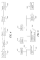
- Fig. 2 is a block diagram of the hanging protocol algorithm.
- Fig. 3 is an example of two input images and the subsequent composed medical image.
- Fig. 4 is a block diagram of a digital medical image acquisition system.
- Fig. 5 is a block diagram of a digital image processor for carrying out the method of the present invention.
- Image processor 408 can take the form of a digital computer, such as illustrated in Fig. 5. In such case, one or more of the steps of said method can be carried out using software routines. Image processor 408 can also include hardware or firmware for carrying out one or more of said method steps. Thus, the steps of the method of the invention can be carried out using software, firmware, and hardware, either alone or in any preferable combination.
- computer readable storage medium can include, specifically, memory 510 and transportable storage medium 524. More generally, computer storage medium may comprise, for example, magnetic storage media, such as magnetic disk (hard drive, floppy disk) or magnetic tape; optical storage media, such as optical disk, optical tape, or machine readable bar code; solid state electronic storage devices, such as random access memory (RAM), read only memory (ROM); or any other physical device or medium which can be employed to store a computer program.
- magnetic storage media such as magnetic disk (hard drive, floppy disk) or magnetic tape
- optical storage media such as optical disk, optical tape, or machine readable bar code
- solid state electronic storage devices such as random access memory (RAM), read only memory (ROM); or any other physical device or medium which can be employed to store a computer program.
- Fig. 1 describes the processing flow of the present invention.
- N digital medical images will be composed on a page.
- the N images are represented in Figure 1 by: Digital Medical Image 1-101, Digital Medical Image 2-103 and Digital Medical Image N-105.
- exam information is provided for each digital medical image.
- the exam information includes items such as the acquisition modality, the body part, procedure, etc. This information is represented in Figure 1 by: Image 1 Exam Information 102, Image 2 Exam Information 104 and Image N Exam Information 106.
- the first processing to be performed is to determine the location of the exposure field in each image. This processing determines the foreground area of the image, which is the area of no direct exposure due to x-ray collimation during the acquisition process.
- the preferred embodiment of the exposure field detection processing is disclosed by Wang, et al., U.S. Patent Application 09/015,656, filed on January 29, 1998. Coupled with the detection is exposure field extraction which creates a new sub-image from the original digital medical image which has the foreground removed and contains only the image data contained in the actual exposure field. This is represented for each digital medical image as follows. Exposure Field Detection 107 is performed on Digital Medical Image 1-101. Then Exposure Field Extraction 110 is performed using the results from Exposure Field Detection 107.
- Exposure Field Detection 108 is performed on Digital Medical Image 2-103. Then Exposure Field Extraction 111 is performed using the results from Exposure Field Detection 108. Likewise, Exposure Field Detection 109 is performed on Digital Medical Image N 105. Then Exposure Field Extraction 112 is performed using the results from Exposure Field Detection 109. Likewise exposure field detection is performed on digital medical images 3 ---N-1 and exposure field extraction is performed respectively on digital medical images 3---N-1.
- Image 1 Exam Information 102 contains the Modality field equal to CR, the Body Part field equal to Hand and the Procedure field equal to AP.
- Image 2 Exam Information 202 contains the Modality field equal to CR, the Body Part field equal to Hand and the Procedure field equal to Oblique.
- Image 3 Exam Information 203 contains the Modality field equal to CR, the Body Part field equal to Hand and the Procedure field equal to Lateral. This information is fed into the Match Template 204 process.
- Match Template 204 uses the Hanging Protocol Template Table 205 to match the exam information provided to a defined template in the table. In this example, the first template in the table matches the exam information provided. Therefore, Match Template 204 provides the following Information to Page Composition 206: the Format field is equal to 3-up Horizontal, the Image 1 position is Left, the Image 2 position is Right and the Image 3 position is Center. This concept can be extended to include more robust hanging protocols by including wildcards "*" in the procedure fields.
- the second line in the Hanging Protocol Template Table 205 is for a CR, Hand image with AP and Lateral views. If two images are presented with AP and Lateral views, this format will be applied. However, if two images are presented with views which are not included in the table, for example, Lateral and Oblique, the format on the following line, with wildcards "*", will be utilized as no direct match without wildcards is provided.
- the Hanging Protocol Algorithm 113 provides layout information to the Page Composition 114 processing module. Using this layout information from the Hanging Protocol Algorithm 113, the Page Composition 114 module takes the sub-images produced by the individual Exposure Field Extraction (110, 111, 112) processing steps and creates the Composed Digital Medical Image 115.
- the image 115 can be presented on an electronic display or printed out on hardcopy, such as film.
- Example Digital Medical Image 1-301 and Example Digital Medical Image 2-303 are provided. Along with these images, Example Image 1 Exam Information 302 and Example Image 2 Exam Information 304 is provided. After performing the processing in the present invention, Example Composed Digital Medical Image 305 is produced.
- multiple exposure fields are produced in a single image. This is particularly common with bone exams using computed radiography or analog film which has been digitized.
- the exposure field detection algorithm of the preferred embodiment can segment multiple exposure fields and produce a sub-image for each exposure field. Exam information must now be provided for each exposure field. Multiple ways for providing this information exist including manual entry by the operator or use of wildcards, as described above for each exposure field.
- image enhancement may be performed on each exposure field sub-image.
- This processing can include applying a blackened surround mask around the exposure field to reduce viewing flare.
- Tonal processing may also be performed such as look-up tables or window center and window width adjustment as is common in medical image processing.
- Adaptive contrast enhancement may also be applied, such as the algorithm disclosed in U.S. Patent 5,978, 518, inventor Oliyide et al., issued November 2, 1999.
- unsharp mask edge enhancement may be performed such as that described in U.S. Patent 5,270,530, inventor Godlewski et al., issued December 14, 1993.
- Default enhancement parameters for all of the above processing algorithms may be stored in the Hanging Protocol Template Table for each set of images in a template.
- Magnification information may also be included in the Hanging Protocol Template Table. This can include methods such as 1:1 magnification, scale to fit or true size. If 1:1 magnification or true size is specified, it is possible for the exposure field sub-image to be larger than the page size allocated to that sub-image. In this case some portion of the image must be clipped or removed from the displayed area on the page. A warning message to the user indicating this condition, including a method for changing the layout is preferred for this case. After the initial page is composed, adjustments to the layout may be desired. The user may manually select an exposure field sub-image through a graphical user interface. Then, the user may move the position, magnification or image enhancement parameters of the selected exposure field from the initial values.
- Some potential methods for selecting the digital medical images to be composed should be noted.
- the user may select multiple images manually through a graphical user interface.
- multiple images may be grouped together as a study or series. Selection of a study or series can be used to specify the digital medical images to be composed on the page.
Landscapes
- Physics & Mathematics (AREA)
- General Physics & Mathematics (AREA)
- Engineering & Computer Science (AREA)
- Theoretical Computer Science (AREA)
- Apparatus For Radiation Diagnosis (AREA)
- Image Processing (AREA)
- Editing Of Facsimile Originals (AREA)
- Ultra Sonic Daignosis Equipment (AREA)
- Television Signal Processing For Recording (AREA)
- Magnetic Resonance Imaging Apparatus (AREA)
Applications Claiming Priority (2)
| Application Number | Priority Date | Filing Date | Title |
|---|---|---|---|
| US71802500A | 2000-11-20 | 2000-11-20 | |
| US718025 | 2000-11-20 |
Publications (2)
| Publication Number | Publication Date |
|---|---|
| EP1207683A2 true EP1207683A2 (de) | 2002-05-22 |
| EP1207683A3 EP1207683A3 (de) | 2006-04-05 |
Family
ID=24884504
Family Applications (1)
| Application Number | Title | Priority Date | Filing Date |
|---|---|---|---|
| EP01204242A Withdrawn EP1207683A3 (de) | 2000-11-20 | 2001-11-05 | Verfahren und System zur Zeitenzusammensetzung von digitalen medizinischen Bildern |
Country Status (2)
| Country | Link |
|---|---|
| EP (1) | EP1207683A3 (de) |
| JP (1) | JP2002238886A (de) |
Cited By (4)
| Publication number | Priority date | Publication date | Assignee | Title |
|---|---|---|---|---|
| EP1424648A3 (de) * | 2002-11-29 | 2006-04-19 | Sectra Imtec AB | Verfahren zum Lesen von Bildern |
| US7657566B2 (en) * | 2006-01-10 | 2010-02-02 | Siemens Aktiengesellschaft | Computer implemented method and system for hanging protocol configuration simulator displaying desired order of medical images data |
| US7831109B2 (en) | 2002-10-01 | 2010-11-09 | Koninklijke Philips Electronics N.V. | Method and device for generating sub-images |
| US11817207B1 (en) * | 2019-12-05 | 2023-11-14 | INMAR Rx SOLUTIONS, INC. | Medication inventory system including image based boundary determination for generating a medication tray stocking list and related methods |
Families Citing this family (3)
| Publication number | Priority date | Publication date | Assignee | Title |
|---|---|---|---|---|
| JP2005073818A (ja) * | 2003-08-29 | 2005-03-24 | Hitachi Medical Corp | 医用画像表示装置、医用画像蓄積検索システム |
| JP4615893B2 (ja) * | 2004-05-17 | 2011-01-19 | ジーイー・メディカル・システムズ・グローバル・テクノロジー・カンパニー・エルエルシー | 超音波撮像装置 |
| JP6129481B2 (ja) * | 2012-04-09 | 2017-05-17 | 東芝メディカルシステムズ株式会社 | X線診断装置及び制御プログラム |
Family Cites Families (3)
| Publication number | Priority date | Publication date | Assignee | Title |
|---|---|---|---|---|
| US5133020A (en) * | 1989-07-21 | 1992-07-21 | Arch Development Corporation | Automated method and system for the detection and classification of abnormal lesions and parenchymal distortions in digital medical images |
| US5764791A (en) * | 1992-03-05 | 1998-06-09 | Fuji Photo Film Co., Ltd. | Method for determining the shape and location of an irradiation field |
| US5734915A (en) * | 1992-11-25 | 1998-03-31 | Eastman Kodak Company | Method and apparatus for composing digital medical imagery |
-
2001
- 2001-11-05 EP EP01204242A patent/EP1207683A3/de not_active Withdrawn
- 2001-11-15 JP JP2001349621A patent/JP2002238886A/ja active Pending
Cited By (5)
| Publication number | Priority date | Publication date | Assignee | Title |
|---|---|---|---|---|
| US7831109B2 (en) | 2002-10-01 | 2010-11-09 | Koninklijke Philips Electronics N.V. | Method and device for generating sub-images |
| EP1424648A3 (de) * | 2002-11-29 | 2006-04-19 | Sectra Imtec AB | Verfahren zum Lesen von Bildern |
| US7162623B2 (en) | 2002-11-29 | 2007-01-09 | Sectra Imtec Ab | Method for reading images |
| US7657566B2 (en) * | 2006-01-10 | 2010-02-02 | Siemens Aktiengesellschaft | Computer implemented method and system for hanging protocol configuration simulator displaying desired order of medical images data |
| US11817207B1 (en) * | 2019-12-05 | 2023-11-14 | INMAR Rx SOLUTIONS, INC. | Medication inventory system including image based boundary determination for generating a medication tray stocking list and related methods |
Also Published As
| Publication number | Publication date |
|---|---|
| EP1207683A3 (de) | 2006-04-05 |
| JP2002238886A (ja) | 2002-08-27 |
Similar Documents
| Publication | Publication Date | Title |
|---|---|---|
| US6434262B2 (en) | Computer-aided diagnosis system and method | |
| US6775399B1 (en) | ROI segmentation image processing system | |
| EP0653726B1 (de) | Verfahren zum Auffinden eines Histogrammgebietes von Interesse zur verbesserten Grauskalawiedergabe von Röntgenbildern | |
| US5982916A (en) | Method and apparatus for automatically locating a region of interest in a radiograph | |
| US7835562B2 (en) | Methods and apparatus for noise reduction filtering of images | |
| EP1501048B1 (de) | Verfahren zum Segmentieren eines Röntgenbildes in diagnostisch relevante und diagnostisch irrelevante Bereiche | |
| JPH06348818A (ja) | ディジタル放射線写真像の前景及び背景自動検出方法 | |
| EP2372649B1 (de) | Verfahren und System zur Definition eines Brustfensters | |
| US7564999B2 (en) | Method for identifying markers in radiographic images | |
| WO2008018998A2 (en) | Method for producing a cropped medical image | |
| US7912263B2 (en) | Method for detecting clipped anatomy in medical images | |
| US7494045B2 (en) | Patient identification method for X-ray film user-interfaces | |
| US20030110178A1 (en) | Method and system of tracking medical films and associated digital images for computer-aided and diagnostic analysis | |
| EP1207683A2 (de) | Verfahren und System zur Zeitenzusammensetzung von digitalen medizinischen Bildern | |
| US6118892A (en) | Method for automatic detection of region of interest for digital x-ray detectors using a filtered histogram | |
| JP4307877B2 (ja) | 画像処理装置及び画像処理方法 | |
| CA2308492C (en) | Computer-aided diagnosis system and method | |
| JP2001285627A (ja) | 医用画像出力システム | |
| US7020343B1 (en) | Method and apparatus for enhancing discrete pixel images by analyzing image structure | |
| EP0544172B1 (de) | Reduzierungsverfahren zur Röntgenbildanzeige | |
| EP1211642A2 (de) | Interaktive Verbesserung von numerischen Vielfachbelichtungsröntgenbildern | |
| JP2002008005A (ja) | 医用画像出力方法および装置 | |
| JP2002135574A (ja) | 画像処理装置、画像処理システム、画像処理方法、及び記憶媒体 | |
| JPH1020422A (ja) | 画像構成判定装置 | |
| JP2002135575A (ja) | 放射線画像出力装置 |
Legal Events
| Date | Code | Title | Description |
|---|---|---|---|
| PUAI | Public reference made under article 153(3) epc to a published international application that has entered the european phase |
Free format text: ORIGINAL CODE: 0009012 |
|
| AX | Request for extension of the european patent |
Free format text: AL;LT;LV;MK;RO;SI |
|
| PUAL | Search report despatched |
Free format text: ORIGINAL CODE: 0009013 |
|
| AK | Designated contracting states |
Kind code of ref document: A3 Designated state(s): AT BE CH CY DE DK ES FI FR GB GR IE IT LI LU MC NL PT SE TR |
|
| AX | Request for extension of the european patent |
Extension state: AL LT LV MK RO SI |
|
| RIC1 | Information provided on ipc code assigned before grant |
Ipc: G06T 7/00 20060101AFI20060215BHEP |
|
| 17P | Request for examination filed |
Effective date: 20060921 |
|
| AKX | Designation fees paid |
Designated state(s): DE FR GB |
|
| RAP1 | Party data changed (applicant data changed or rights of an application transferred) |
Owner name: CARESTREAM HEALTH, INC. |
|
| STAA | Information on the status of an ep patent application or granted ep patent |
Free format text: STATUS: THE APPLICATION IS DEEMED TO BE WITHDRAWN |
|
| 18D | Application deemed to be withdrawn |
Effective date: 20090603 |