EP1247097B1 - Procede de determination de l'efficacite d'une therapie anticancereuse - Google Patents
Procede de determination de l'efficacite d'une therapie anticancereuse Download PDFInfo
- Publication number
- EP1247097B1 EP1247097B1 EP01902043A EP01902043A EP1247097B1 EP 1247097 B1 EP1247097 B1 EP 1247097B1 EP 01902043 A EP01902043 A EP 01902043A EP 01902043 A EP01902043 A EP 01902043A EP 1247097 B1 EP1247097 B1 EP 1247097B1
- Authority
- EP
- European Patent Office
- Prior art keywords
- cells
- chemotherapeutic
- individual
- senescence
- tissue
- Prior art date
- Legal status (The legal status is an assumption and is not a legal conclusion. Google has not performed a legal analysis and makes no representation as to the accuracy of the status listed.)
- Expired - Lifetime
Links
Images



Classifications
-
- G—PHYSICS
- G01—MEASURING; TESTING
- G01N—INVESTIGATING OR ANALYSING MATERIALS BY DETERMINING THEIR CHEMICAL OR PHYSICAL PROPERTIES
- G01N33/00—Investigating or analysing materials by specific methods not covered by groups G01N1/00 - G01N31/00
- G01N33/48—Biological material, e.g. blood, urine; Haemocytometers
- G01N33/50—Chemical analysis of biological material, e.g. blood, urine; Testing involving biospecific ligand binding methods; Immunological testing
- G01N33/53—Immunoassay; Biospecific binding assay; Materials therefor
- G01N33/575—Immunoassay; Biospecific binding assay; Materials therefor for cancer
-
- G—PHYSICS
- G01—MEASURING; TESTING
- G01N—INVESTIGATING OR ANALYSING MATERIALS BY DETERMINING THEIR CHEMICAL OR PHYSICAL PROPERTIES
- G01N33/00—Investigating or analysing materials by specific methods not covered by groups G01N1/00 - G01N31/00
- G01N33/48—Biological material, e.g. blood, urine; Haemocytometers
- G01N33/50—Chemical analysis of biological material, e.g. blood, urine; Testing involving biospecific ligand binding methods; Immunological testing
- G01N33/5005—Chemical analysis of biological material, e.g. blood, urine; Testing involving biospecific ligand binding methods; Immunological testing involving human or animal cells
- G01N33/5008—Chemical analysis of biological material, e.g. blood, urine; Testing involving biospecific ligand binding methods; Immunological testing involving human or animal cells for testing or evaluating the effect of chemical or biological compounds, e.g. drugs, cosmetics
- G01N33/5011—Chemical analysis of biological material, e.g. blood, urine; Testing involving biospecific ligand binding methods; Immunological testing involving human or animal cells for testing or evaluating the effect of chemical or biological compounds, e.g. drugs, cosmetics for testing antineoplastic activity
-
- G—PHYSICS
- G01—MEASURING; TESTING
- G01N—INVESTIGATING OR ANALYSING MATERIALS BY DETERMINING THEIR CHEMICAL OR PHYSICAL PROPERTIES
- G01N33/00—Investigating or analysing materials by specific methods not covered by groups G01N1/00 - G01N31/00
- G01N33/48—Biological material, e.g. blood, urine; Haemocytometers
- G01N33/50—Chemical analysis of biological material, e.g. blood, urine; Testing involving biospecific ligand binding methods; Immunological testing
- G01N33/53—Immunoassay; Biospecific binding assay; Materials therefor
- G01N33/575—Immunoassay; Biospecific binding assay; Materials therefor for cancer
- G01N33/5758—Immunoassay; Biospecific binding assay; Materials therefor for cancer involving compounds serving as markers for tumours, cancers or neoplasias, e.g. cellular determinants, receptors, heat shock/stress proteins, A-protein, oligosaccharides or metabolites
-
- G—PHYSICS
- G01—MEASURING; TESTING
- G01N—INVESTIGATING OR ANALYSING MATERIALS BY DETERMINING THEIR CHEMICAL OR PHYSICAL PROPERTIES
- G01N2333/00—Assays involving biological materials from specific organisms or of a specific nature
- G01N2333/435—Assays involving biological materials from specific organisms or of a specific nature from animals; from humans
- G01N2333/475—Assays involving growth factors
- G01N2333/495—Transforming growth factor [TGF]
-
- G—PHYSICS
- G01—MEASURING; TESTING
- G01N—INVESTIGATING OR ANALYSING MATERIALS BY DETERMINING THEIR CHEMICAL OR PHYSICAL PROPERTIES
- G01N2333/00—Assays involving biological materials from specific organisms or of a specific nature
- G01N2333/82—Translation products from oncogenes
-
- G—PHYSICS
- G01—MEASURING; TESTING
- G01N—INVESTIGATING OR ANALYSING MATERIALS BY DETERMINING THEIR CHEMICAL OR PHYSICAL PROPERTIES
- G01N2800/00—Detection or diagnosis of diseases
- G01N2800/52—Predicting or monitoring the response to treatment, e.g. for selection of therapy based on assay results in personalised medicine; Prognosis
-
- Y—GENERAL TAGGING OF NEW TECHNOLOGICAL DEVELOPMENTS; GENERAL TAGGING OF CROSS-SECTIONAL TECHNOLOGIES SPANNING OVER SEVERAL SECTIONS OF THE IPC; TECHNICAL SUBJECTS COVERED BY FORMER USPC CROSS-REFERENCE ART COLLECTIONS [XRACs] AND DIGESTS
- Y10—TECHNICAL SUBJECTS COVERED BY FORMER USPC
- Y10T—TECHNICAL SUBJECTS COVERED BY FORMER US CLASSIFICATION
- Y10T436/00—Chemistry: analytical and immunological testing
- Y10T436/10—Composition for standardization, calibration, simulation, stabilization, preparation or preservation; processes of use in preparation for chemical testing
- Y10T436/101666—Particle count or volume standard or control [e.g., platelet count standards, etc.]
-
- Y—GENERAL TAGGING OF NEW TECHNOLOGICAL DEVELOPMENTS; GENERAL TAGGING OF CROSS-SECTIONAL TECHNOLOGIES SPANNING OVER SEVERAL SECTIONS OF THE IPC; TECHNICAL SUBJECTS COVERED BY FORMER USPC CROSS-REFERENCE ART COLLECTIONS [XRACs] AND DIGESTS
- Y10—TECHNICAL SUBJECTS COVERED BY FORMER USPC
- Y10T—TECHNICAL SUBJECTS COVERED BY FORMER US CLASSIFICATION
- Y10T436/00—Chemistry: analytical and immunological testing
- Y10T436/10—Composition for standardization, calibration, simulation, stabilization, preparation or preservation; processes of use in preparation for chemical testing
- Y10T436/105831—Protein or peptide standard or control [e.g., hemoglobin, etc.]
-
- Y—GENERAL TAGGING OF NEW TECHNOLOGICAL DEVELOPMENTS; GENERAL TAGGING OF CROSS-SECTIONAL TECHNOLOGIES SPANNING OVER SEVERAL SECTIONS OF THE IPC; TECHNICAL SUBJECTS COVERED BY FORMER USPC CROSS-REFERENCE ART COLLECTIONS [XRACs] AND DIGESTS
- Y10—TECHNICAL SUBJECTS COVERED BY FORMER USPC
- Y10T—TECHNICAL SUBJECTS COVERED BY FORMER US CLASSIFICATION
- Y10T436/00—Chemistry: analytical and immunological testing
- Y10T436/13—Tracers or tags
Definitions
- the invention relates to methods for determining or predicting response to cancer therapy in an individual.
- the invention relates to methods for using image analysis to assess the efficacy of chemotherapeutic and chemopreventative agents in a human in need of treatment with such agents by detecting expression levels of biological makers associated with senescence, apoptosis or terminal differentiation. More specifically, the invention provides methods where the amount of the senescence, apoptosis or terminal differentiation marker is quantitated in tissue and cell samples removed from an individual before and after exposure to the chemotherapeutic or chemopreventative agent.
- a primary goal of cancer therapy is to selectively kill or inhibit the uncontrolled growth of malignant cells while not adversely affecting normal cells.
- Traditional chemotherapeutic agents are highly cytotoxic agents that preferably have greater affinity for malignant cells than for normal cells, or at least preferentially affect malignant cells based on their high rate of metabolic activity. However, these agents often harm normal cells.
- chemotherapeutic drugs can be administered at doses high enough to bring about cell death, such doses typically produce deleterious effects on normal as well as tumor cells. Differentiating agents, and lower doses of chemotherapeutic drugs and agents frequently results in growth arrest rather than cell death; such arrest can be followed by apoptosis and sell death, or continued proliferation once the chemotherapeutic drugs are withdrawn.
- cytotoxic and chemotherapeutic drugs or ionizing radiation may also induce transient growth arrest, a state which depends largely on the function of p53 and a p53-regulated cyclin-dependent kinase inhibitors (such as p16, p27, and p19) or growth inhibitors (such as TGF- ⁇ , IL-4, and IL-6).
- a p53-regulated cyclin-dependent kinase inhibitors such as p16, p27, and p19
- growth inhibitors such as TGF- ⁇ , IL-4, and IL-6
- terminal proliferation arrest may result from terminal differentiation or replicative senescence.
- Senescence is a physiological process that limits the proliferative span of normal cells.
- a commonly-used marker of senescence in human cells is expression of senescence-associated ⁇ -galactosidase (SA- ⁇ -Gal). This protein has been shown to correlate with senescence in aging cell cultures in vitro and with cells in vivo.
- Terminal proliferation arrest in normal cells can be rapidly induced by treatment with DNA-damaging drugs or ⁇ -irradiation and is accompanied by the morphological features of senescence and the induction of SA- ⁇ -Gal. Accelerated senescence is likely to be a protective response of cells to carcinogenic impact.
- Tumor cells appear to have retained the ability to enter senescence and terminal proliferation arrest.
- Treatment of tumor cells with different classes of agents that affect cell differentiation, and anticancer agents readily induces morphological, enzymatic, and other changes characteristic of senescence (such as the up-regulation of p53, p21, p27, p16, TGF- ⁇ , IL-4, IL-6, and SA- ⁇ -Gal).
- This senescence-like phenotype (SLP) distinguishes cells that will become stably growth-arrested from cells that will recover from drug exposure and continue to proliferate.
- SLP senescence-like phenotype
- Apoptosis is generally regarded as an active suicidal response to various physiological or pathological stimuli.
- DNA-damaging agents including X-ray irradiation and several chemotherapeutic drugs (e.g., alkylating agents and topoisomerase II inhibitors) cause necrosis or initiate pathways leading to apoptosis.
- chemotherapeutic drugs e.g., alkylating agents and topoisomerase II inhibitors
- DNA-damaging stimuli can result in an elevation of intracellular p53 protein levels. Increased levels of wild-type p53, in turn, inhibit the cell cycle at G 1 , thus permitting the damaged cell to undergo DNA repair. However, if the damaged cell is unable to undergo DNA repair, p53 can trigger programmed cell death. It is this ability to trigger programmed cell death that contributes to the induction of tumor cell death following exposure to chemotherapeutic agents.
- Increased levels of p53 can also lead to the activation of a number of genes that contain wild-type p53-binding sequences, including the MDM-2 oncogene, Bax, and the WAF1/CIP1 gene.
- the WAF1/CIP1 gene encodes a protein having a M r of 21,000 that associates with cyclin-Cdk complexes and is capable of inhibiting kinase activity associated with these complexes.
- a major target of WAF (p21 or CIP) inhibition is the cyclin E-Cdk2 kinase complex whose activity is required for Go to S phase cell cycle progression.
- the WAF1/CIP1 gene is transcriptionally activated in response to DNA-damaging agents that trigger G 1 arrest or apoptosis in cells with wild-type p53 but not in tumor cells harboring deletions or mutations of the p53 gene.
- WAF1/CIP1 has also been shown to be up-regulated in cells undergoing differentiation or cell cycle arrest by a p53-independent mechanism.
- markers of senescence, apoptosis and terminal proliferative arrest that can be used to detect the effects of chemotherapeutic and chemopreventative drugs and agents. These markers can be used to assess the success or failure of any particular chemotherapeutic or chemopreventative drug or agent, or combination thereof, to affect an anticancer effect on tumor cells in vivo.
- WO 93/03741 discloses a method of determining the effectiveness of a therapeutic agent for the treatment of cancer based on in vitro administration of the therapeutic agent in a cell culture containing viable malignant cells expressing an oncogene product.
- neoadjuvant (or primary) chemotherapy consists of administering drugs as an initial treatment in cancer patients.
- One advantage of such an approach is that, in primary tumors of more than 3 cm this approach permits the use of conservative surgical procedures (as opposed to, e.g., mastectomy in breast cancer patients) in the majority of patients.
- Another advantage is that for many cancers, a partial and/or complete response is achieved in about two-thirds of all cases.
- chemotherapeutic agents in treating particular cancers are unpredictable. In view of this unpredictability, it has not been possible to determine, prior to starting therapy, whether one or more selected agents would be active as anti-tumor agents or to render an accurate prognosis of course of treatment in an individual patient. It would be very desirable to be able to determine the efficacy of a proposed therapeutic agent (or combination of agents) in an individual patient. There is a need in the art for a method of assessing the efficacy of chemotherapeutic programs that is both time- and cost-effective and minimally traumatic to cancer patients.
- the present invention provides methods for determining the response to cancer therapy in an individual.
- the invention specifically provides methods for assessing efficacy of chemotherapeutic and chemopreventative agents in a human in need of treatment with such agents by detecting expression levels of biological makers associated with senescence, apoptosis or terminal differentiation.
- the amount of one or a plurality of senescence, apoptosis or terminal differentiation markers is quantitated in tissue and cell samples removed from an individual before and after exposure to the chemotherapeutic or chemopreventative agent.
- the amount of said marker is determined using image analysis of immunohistochemically-stained tissue or cell samples obtained from a patient tumor.
- response to a chemotherapeutic or chemopreventive agent in an individual is determined by collecting a first tissue or cell sample from the individual before exposing the individual to the chemotherapeutic or chemopreventive agent, collecting a second tissue or cell sample from the individual after exposing the individual to the chemotherapeutic or chemopreventive agent, immunohistochemically staining the first and second tissue or cell samples using a detectably-labeled antibody directed against a biological marker associated with senescence, apoptosis or terminal differentiation, determining the amount of the marker in the first and second tissue or cell samples, and determining whether expression of the biological marker associated with senescence, apoptosis or terminal differentiation was increased following exposure to the chemotherapeutic or chemopreventive agent.
- the detectable label is a chromagen or a fluorophore.
- the methods of the present invention are useful for determining the response to cancer therapy in an individual.
- the present invention provides methods for assessing the efficacy of treating an individual in need of such treatment with a chemotherapeutic or chemopreventive agent, in which the level of expression of a biological marker associated with senescence, apoptosis or terminal differentiation is quantitated in immunohistochemically-stained tissue or cell samples removed from the individual before and after exposure to the chemotherapeutic or chemopreventive agent.
- expression levels are quantitated using computer-aided image analysis system.
- response to a chemotherapeutic or chemopreventive agent in an individual is determined by collecting a first tissue or cell sample from the individual before exposing the individual to the chemotherapeutic or chemopreventive agent, collecting a second tissue or cell sample from the individual after exposing the individual to the chemotherapeutic or chemopreventive agent, immunohistochemically staining the first and second tissue or cell samples using a detectably-labeled antibody directed against a biological marker associated with senescence, apoptosis or terminal differentiation, determining amount of expression of one or a plurality of biological markers associated with senescence, apoptosis or terminal differentiation in the first and second tissue or cell samples, and determining whether expression of the biological marker(s) associated with senescence, apoptosis or terminal differentiation was increased following exposure to the chemotherapeutic or chemopreventive agent.
- biological markers whose expression is induced or increases in cellular senescence, apoptosis and terminal differentiation may include but are not limited to p21, p27, p16, TGF- ⁇ , IL-4, IL-6, and SA- ⁇ -Gal, generally and collectively known as the senescence-like phenotype (SLP).
- SLP senescence-like phenotype
- This senescence-like phenotype (SLP) can be used to distinguish cells with restricted proliferative potential from those that continue to proliferate after drug exposure. suggesting that senescence-like terminal proliferation arrest is an important determinant of treatment response in human cancer.
- SA- ⁇ -Gal + cells and SA- ⁇ -Gal - cells have a similar probability of undergoing mitotic death during drug treatment or within days following release from the drug.
- SLP can be used to distinguish growth-retarded and non-clonogenic cells from recovering and proliferating cells.
- the overall outcome of treatment with chemopreventive or chemotherapeutic drugs is therefore determined by a combination of factors responsible for the induction of cell death (mitotic cell death or apoptosis) and senescence-like terminal proliferation arrest.
- SLP induction may be the primary determinant of treatment outcome for cytostatic differentiating agents such as retinoids, and other chemopreventive agents.
- image analysis may be used to examine therapeutic response by quantitating the expression of proteins that are involved in activated pathways (e . g ., phosphorylated MAP kinase and Akt) in tissues before and after treatment with kinase inhibitors.
- SLP markers such as ⁇ -Gal, p53, and p21 in clinical cancer may provide an important diagnostic approach for monitoring tumor response to different forms of therapy and can be done by calibrated staining for various factors associated with senescence and quantified by microscope based image analysis.
- cells are removed from an individual before and after treatment with a putative anti-cancer agent and the cells are fixed using, e . g ., paraformaldehyde.
- Cell samples are then treated with an organic solvent, such as acetone, formalin, or methanol, to render the cells permeable for immunohistological staining.
- organic solvent such as acetone, formalin, or methanol
- the proteins are identified using suitable antibody systems such as sandwich systems using a primary antibody that specifically binds to the protein of interest and a secondary antibody that binds to the primary antibody.
- the antibody system can then be visualized using suitable stains (Bacus et al ., 1999, Breast J. ).
- suitable stains Bocus et al ., 1999, Breast J. .
- antibodies and other molecules that specifically bind to these target proteins are known in the art, and the methods of the present invention can be practiced using any antibody or other molecule or collection of molecules that specifically binds to any of said biological markers of senescence, apoptosis or terminal differentiation.
- Exemplary primary antibodies that bind to p53, p21, and p16 include the monoclonal antibody Ab-2 (Oncogene Sciences, Cambridge, MA). Similarly, exemplary antibodies that bind to p21 protein include the monoclonal antibody Ab-1 (Oncogene Sciences).
- An exemplary secondary antibody that may be used with either of these primary antibodies is rabbit anti-mouse IgG (Jackson Labs, West Grove, PA). After exposure to antibodies, an enzyme complex such as ABC complex (Vector Labs, Burlington, CA) is added to complete the antibody sandwich system and render it amenable to staining and counterstaining. Image analysis techniques are used to complete the quantitation.
- SA- ⁇ -Gal fresh senescence-associated ⁇ -Gal staining solution (1 mg/ml 5-bromo-4-chloro-3-indolyl ⁇ -D-galactoside (X-Gal), 20 mg/ml dimethylformamide; 40 mM citric acid/ sodium phosphate, pH 6.0; 5 mM potassium ferrocyanide; 5 mM potassium ferricyanide; 150 mM NaCl; 2 mM MgCl 2 .
- proteins associated with senescence or apoptosis are quantitated by image analysis using a suitable primary antibody (such as Ab-2 for p53, or Ab-1 for WAF1, HSP60, or caspase 3), secondary antibody (such as rabbit anti-mouse IgG), and.tertiary ABC complex.
- a suitable primary antibody such as Ab-2 for p53, or Ab-1 for WAF1, HSP60, or caspase 3
- secondary antibody such as rabbit anti-mouse IgG
- secondary ABC complex such as rabbit anti-mouse IgG
- Tertiary ABC complexes are visualized by treating the sections with diaminobenizidine (DAB) stain under appropriate conditions.
- DAB diaminobenizidine
- the tissue is counterstained with another optical enhancement factor, preferably ethyl green.
- the resulting preparation has green nuclei with varying degrees of brown diaminobenzidine (DAB) precipitate localized to the nuclei (and indicating the level of expression of p53, p21, p16, WAF1, HSP60, or caspase-3 protein).
- DAB diaminobenizidine
- the ethyl green stain offers good spectral separation from the DAB precipitate of the immunoperoxidase technique such that different features of the image can be readily separated by filtering at two different wavelengths.
- This allows the image to be digitized into two separate images, one in which all the cell nuclei are optically enhanced (ethyl green or Fast Green) and one in which only those tissue areas with receptor staining (DAB) are optically enhanced.
- the images can be separated by a 600 nanometer (red) filter to produce an image of the counterstained area and a 500 nanometer (green) filter to produce an image of the tissue areas staining with the DAB precipitate.
- an interactive threshold setting technique can be used in which an operator visualizing the images can set a boundary on the areas under consideration. When the boundaries are set, the images are formed by eliminating all parts of the image that are below the threshold in optical density. A threshold is set for the first image, and a second threshold is set for the second image.
- the image processing method then consists of first forming the mask image of the tissues under consideration with the red filter. This mask image is stored and another image for expressed protein quantification is then acquired by using the green filtered version of the same image. Using the filters in combination allows for the optical enhancement of those areas of the tissue mask where tissue components are stained with DAB (which are darkened) and those tissue components having only the green counterstain (which are lightened). An image analysis can then be performed using only those areas of the image that are stained and which are within the mask.
- the amount of target protein is then quantitated by statistical analysis of the differences between the two images. Also, the proportion or percentage of total tissue area stained may be readily calculated, as the area stained above an antibody threshold level in the second image.
- Red and green filters are suitable for practice of the invention.
- a green filter 500 nm is used to determine the total cytoplasmic area stained with red eosin and compared with the blue area stained for SA- ⁇ -Gal (600 nm) as determined using a red filter.
- This implementation shows a convenient and advantageous method for discriminating between two areas having counterstaining. It is recognized that there are various other staining or optical enhancement methods and filtering methods which can be used to optically enhance one particular area or feature over another cell feature (such as Fast green and eosin).
- the specific examples provided herein are suggestive of other equivalents for visualization of cell nuclei containing expressed p53, WAF1 protein, and SA- ⁇ -Gal.
- the percentage of such cells in tissue derived from patients after treatment may be compared to the percentage of such cells in untreated tissue.
- the potential effectiveness of a putative therapeutic or chemopreventive agent against the cancer may be determined. Specifically, if there is a greater percentage of cells expressing p53, WAF1, SA- ⁇ -Gal, HSP60, or caspase 3 in the treated portion of the sample than there is in the untreated portion of the sample, then the therapeutic agent used in the treatment will be predicted to be potentially an effective agent for treating the cancer. Conversely, if there is a substantially equal percentage of cells expressing SA- ⁇ -Gal, p53, WAF1, HSP60, or caspase 3 in both the treated and untreated portions of the sample, or there is less protein expression in the treated portion of the sample, then the putative therapeutic agent will not be predicted to be effective for treatment of the cancer.
- U.S. Patent Nos. 5,252,487 and 5,288,477 disclose one method of quantitating DNA and protein.
- MCF7 cells were treated for 2-3 days with 50 nM doxorubicin or Taxol (doses that induce growth inhibition and cellular death as measured by flow cytometry; as described by, e.g., Dolbeare et al ., 1983, Proc . Natl . Acad . Sci . U.S.A. 80:5573-77) and then were stained for SA- ⁇ -Gal expression 10-11 days after release from the drug.
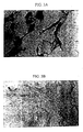
- the results of this treatment on the formation of sub-G 1 fraction and the SA- ⁇ -Gal + phenotype are shown in Figures 1A-1B and 2A-2C.
- Taxol-induced apoptosis is evident by the hypodiploid peak in the flow cytometry histogram. whereas doxorubicin treatment (and treatment with the monoclonal antibody HerceptinTM) resulted in senescence and blocks at G 1 , G 2 , and G 2 M as seen in SA- ⁇ -Gal + cells having the morphological features of senescence ( Figures 2A-2C and Tables I and II).
- MCF7 cells were treated for 3 days with doxorubicin and then stained for p53 and p21 CIPI/WAFI expression. Cells were cultured, stained, and analyzed as described above for SA- ⁇ -Gal using appropriate antibodies. The results of p53 and p21 CIPI/WAFI quantitation are shown in Table III.
- Tissue samples from twenty breast cancer patients was analyzed for p21 expression by image analysis before and after neoadjuvant chemotherapy. Experiments were performed by the Imperial Cancer Research Fund (ICRF; Oxford University, U.K.). The results of this analysis show marked differences in tissue samples collected before and after treatment (Table IV).
Landscapes
- Health & Medical Sciences (AREA)
- Life Sciences & Earth Sciences (AREA)
- Engineering & Computer Science (AREA)
- Immunology (AREA)
- Biomedical Technology (AREA)
- Molecular Biology (AREA)
- Chemical & Material Sciences (AREA)
- Hematology (AREA)
- Urology & Nephrology (AREA)
- Physics & Mathematics (AREA)
- Microbiology (AREA)
- Cell Biology (AREA)
- Food Science & Technology (AREA)
- Medicinal Chemistry (AREA)
- Biotechnology (AREA)
- Analytical Chemistry (AREA)
- Biochemistry (AREA)
- General Health & Medical Sciences (AREA)
- General Physics & Mathematics (AREA)
- Pathology (AREA)
- Bioinformatics & Cheminformatics (AREA)
- Toxicology (AREA)
- Tropical Medicine & Parasitology (AREA)
- Investigating Or Analysing Biological Materials (AREA)
- Medicines That Contain Protein Lipid Enzymes And Other Medicines (AREA)
- Magnetic Resonance Imaging Apparatus (AREA)
- Investigating Or Analysing Materials By Optical Means (AREA)
- Apparatus For Radiation Diagnosis (AREA)
- Pharmaceuticals Containing Other Organic And Inorganic Compounds (AREA)
- Measurement And Recording Of Electrical Phenomena And Electrical Characteristics Of The Living Body (AREA)
- Electrotherapy Devices (AREA)
Claims (6)
- Procédé de détermination de l'efficacité de l'administration d'un agent chimiothérapeutique ou chimiopréventif à un individu, caractérisé en ce que le procédé comprend :(a) la fourniture d'un premier tissu ou échantillon cellulaire ayant été prélevé sur un individu avant exposition de l'individu à l'agent chimiothérapeutique ou chimiopréventif ;(b) la fourniture d'un deuxième tissu ou échantillon cellulaire ayant été prélevé sur un individu après exposition de l'individu à l'agent chimiothérapeutique ou chimiopréventif ;(c) la coloration immunohistochimique des premier et deuxième tissus ou des échantillons cellulaires en utilisant un anticorps marqué de manière détectable dirigé à l'encontre d'un marqueur biologique associé à la sénescence, l'apoptose ou la différentiation terminale ;(d) la mesure de la densité optique des cellules colorées comme à l'étape (c), dans laquelle les cellules colorées sont éclairées par une source lumineuse ayant une longueur d'onde absorbée par le colorant ; et(e) la détermination de l'accroissement éventuel de l'expression du marqueur biologique associé à la sénescence, l'apoptose ou la différentiation terminale à la suite de l'exposition à l'agent chimiothérapeutique ou chimiopréventif.
- Procédé selon la revendication 1, dans lequel le marqueur détectable est un chromagène ou un fluorophore.
- Procédé selon la revendication 1 ou 2, dans lequel le marqueur biologique est p21, p27, p16, TGF-β ou SA-β-Gal.
- Procédé selon l'une quelconque des revendications 1 à 3, dans lequel la quantité de protéine du marqueur biologique est déterminée par essai ELISA.
- Procédé selon l'une quelconque des revendications 1 à 4, dans lequel la densité optique des cellules colorées est obtenue par analyse d'image.
- Procédé selon la revendication 5, dans lequel l'analyse d'image est réalisée en divisant un signal comprenant la densité optique de l'échantillon biologique coloré en une multiplicité de signaux qui sont traités à l'aide de filtres optiques présentant diverses propriétés d'absorption et de transmission de sorte que chaque signal soit spécifique à l'un des multiples colorants utilisés pour colorer les cellules dans l'échantillon biologique.
Priority Applications (1)
| Application Number | Priority Date | Filing Date | Title |
|---|---|---|---|
| DK01902043T DK1247097T3 (da) | 2000-01-12 | 2001-01-12 | Fremgangsmåde til bestemmelse af effektiviteten af cancerterapi |
Applications Claiming Priority (5)
| Application Number | Priority Date | Filing Date | Title |
|---|---|---|---|
| US17651400P | 2000-01-12 | 2000-01-12 | |
| US17651500P | 2000-01-12 | 2000-01-12 | |
| US176514P | 2000-01-12 | ||
| US176515P | 2000-01-12 | ||
| PCT/US2001/001113 WO2001051924A2 (fr) | 2000-01-12 | 2001-01-12 | Procede de determination d'une reaction a une therapie anticancereuse |
Publications (2)
| Publication Number | Publication Date |
|---|---|
| EP1247097A2 EP1247097A2 (fr) | 2002-10-09 |
| EP1247097B1 true EP1247097B1 (fr) | 2004-07-21 |
Family
ID=26872320
Family Applications (2)
| Application Number | Title | Priority Date | Filing Date |
|---|---|---|---|
| EP01942413A Withdrawn EP1247098A1 (fr) | 2000-01-12 | 2001-01-12 | Methode de quantification d'une proteine par analyse d'image |
| EP01902043A Expired - Lifetime EP1247097B1 (fr) | 2000-01-12 | 2001-01-12 | Procede de determination de l'efficacite d'une therapie anticancereuse |
Family Applications Before (1)
| Application Number | Title | Priority Date | Filing Date |
|---|---|---|---|
| EP01942413A Withdrawn EP1247098A1 (fr) | 2000-01-12 | 2001-01-12 | Methode de quantification d'une proteine par analyse d'image |
Country Status (10)
| Country | Link |
|---|---|
| US (2) | US7833698B2 (fr) |
| EP (2) | EP1247098A1 (fr) |
| JP (2) | JP3699399B2 (fr) |
| AT (1) | ATE271691T1 (fr) |
| AU (2) | AU2951801A (fr) |
| CA (2) | CA2397417A1 (fr) |
| DE (1) | DE60104392T2 (fr) |
| DK (1) | DK1247097T3 (fr) |
| MX (2) | MXPA02006843A (fr) |
| WO (2) | WO2001051924A2 (fr) |
Families Citing this family (52)
| Publication number | Priority date | Publication date | Assignee | Title |
|---|---|---|---|---|
| US20050100542A1 (en) * | 1999-10-08 | 2005-05-12 | Young David S. | Cytotoxicity mediation of cells evidencing surface expression of CD44 |
| AU5352901A (en) * | 2000-04-14 | 2001-10-30 | Ventana Med Syst Inc | Method for quantification of akt protein expression |
| US20040085443A1 (en) * | 2000-12-13 | 2004-05-06 | Kallioniemi Olli P | Method and system for processing regions of interest for objects comprising biological material |
| WO2003016867A2 (fr) | 2001-08-21 | 2003-02-27 | Ventana, Medical, Systems, Inc | Methode et essai de quantification permettant d'evaluer l'etat du systeme c-kit/scf/pakt |
| GB0218909D0 (en) * | 2002-08-15 | 2002-09-25 | Qinetiq Ltd | Histological assessment |
| US20030165263A1 (en) * | 2002-02-19 | 2003-09-04 | Hamer Michael J. | Histological assessment |
| US7648678B2 (en) | 2002-12-20 | 2010-01-19 | Dako Denmark A/S | Method and system for pretreatment of tissue slides |
| GB0229734D0 (en) * | 2002-12-23 | 2003-01-29 | Qinetiq Ltd | Grading oestrogen and progesterone receptors expression |
| EP1636585B2 (fr) * | 2003-05-20 | 2012-06-13 | Bayer HealthCare LLC | Diaryl-urees presentant une activite d'inhibition des kinases |
| US20050112622A1 (en) * | 2003-08-11 | 2005-05-26 | Ring Brian Z. | Reagents and methods for use in cancer diagnosis, classification and therapy |
| US20080113874A1 (en) * | 2004-01-23 | 2008-05-15 | The Regents Of The University Of Colorado | Gefitinib sensitivity-related gene expression and products and methods related thereto |
| ES2537631T3 (es) * | 2004-05-27 | 2015-06-10 | The Regents Of The University Of Colorado | Métodos para la predicción del resultado clínico para inhibidores del receptor del factor de crecimiento epidérmico para pacientes de cáncer |
| KR20080003334A (ko) * | 2005-03-11 | 2008-01-07 | 더 리젠트스 오브 더 유니버시티 오브 콜로라도 | 암 세포를 표피 성장 인자 억제제에 대해 감작화시키는히스톤 데아세틸라제 억제제 |
| US7504225B2 (en) | 2005-05-12 | 2009-03-17 | Applied Genomics, Inc. | Reagents and methods for use in cancer diagnosis, classification and therapy |
| US20070091109A1 (en) | 2005-09-13 | 2007-04-26 | Roscoe Atkinson | Image quality |
| US7993918B2 (en) * | 2006-08-04 | 2011-08-09 | Anthrogenesis Corporation | Tumor suppression using placental stem cells |
| ZA200902233B (en) | 2006-10-23 | 2010-07-28 | Anthrogenesis Corp | Methods and compositions for treatment of bone defects with placental cell populations |
| DK2444807T3 (da) | 2006-11-01 | 2014-08-11 | Ventana Med Syst Inc | Mono- og dinitropyrazolhaptenkonjugater |
| US9697582B2 (en) | 2006-11-16 | 2017-07-04 | Visiopharm A/S | Methods for obtaining and analyzing images |
| WO2010017822A1 (fr) | 2008-08-15 | 2010-02-18 | Visiopharm A/S | Procédé et système de détermination d'une cible dans un échantillon biologique par analyse d'image |
| AU2008216748A1 (en) | 2007-02-12 | 2008-08-21 | Anthrogenesis Corporation | Hepatocytes and chondrocytes from adherent placental stem cells; and CD34+, CD45- placental stem cell-enriched cell populations |
| WO2008108059A1 (fr) * | 2007-03-01 | 2008-09-12 | Nec Corporation | Système, procédé et programme pour le diagnostic d'images pathologiques de cancer mammaire, et support d'enregistrement du programme |
| EP2167963B1 (fr) | 2007-05-23 | 2019-04-17 | Ventana Medical Systems, Inc. | Supports polymères pour immunohistochimie et hybridation in situ |
| KR101667960B1 (ko) * | 2007-07-25 | 2016-10-20 | 에자이 알앤드디 매니지먼트 가부시키가이샤 | 암 치료에서 이용하기 위한 멀티키나아제 억제제 |
| US9200253B1 (en) | 2007-08-06 | 2015-12-01 | Anthrogenesis Corporation | Method of producing erythrocytes |
| EP2453234B1 (fr) * | 2007-10-29 | 2014-01-15 | Eisai R&D Management Co., Ltd. | Procédés pour le pronostic de la capacité d'un composé à base d'un analogue de la zéaralénone à traiter le cancer |
| WO2009149013A2 (fr) | 2008-06-05 | 2009-12-10 | Ventana Medical Systems, Inc. | Compositions comprenant des nanomatériaux et procédé d'utilisation de ces compositions pour des processus histochimiques |
| CA2734446C (fr) | 2008-08-22 | 2017-06-20 | Anthrogenesis Corporation | Methodes et compositions pour le traitement de deficits osseux au moyen de populations de cellules placentaires |
| GB2464747B (en) * | 2008-10-10 | 2013-05-15 | Hai Kang Life Corp Ltd | Method for detection of analyte in microarray of samples and apparatus for performing such method |
| EP2387711B1 (fr) * | 2009-01-15 | 2015-04-22 | Laboratory Corporation of America Holdings | Procédés permettant de déterminer la réponse d'un patient par mesure de her-3 |
| WO2010099923A1 (fr) | 2009-03-04 | 2010-09-10 | Roche Diagnostics Gmbh | Serpine b 13 en tant que marqueur du carcinome épidermoïde du poumon |
| WO2010141543A1 (fr) * | 2009-06-02 | 2010-12-09 | Targeted Molecular Diagnostics, Llc | Procédés de détection et de détermination quantitative de la composante p95 de her2/neu (erbb2) |
| US8586360B2 (en) | 2009-07-02 | 2013-11-19 | Anthrogenesis Corporation | Method of producing erythrocytes without feeder cells |
| JP5490568B2 (ja) * | 2010-02-26 | 2014-05-14 | オリンパス株式会社 | 顕微鏡システム、標本観察方法およびプログラム |
| MX2012011543A (es) | 2010-04-08 | 2013-05-06 | Anthrogenesis Corp | Tratamiento de sarcoidosis empleando celulas madre placentarias. |
| US8524239B2 (en) * | 2010-07-09 | 2013-09-03 | The United States of America as represented by the Secrectary, Department of Health and Human Services | Photosensitizing antibody-fluorophore conjugates |
| TW201302793A (zh) | 2010-09-03 | 2013-01-16 | Glaxo Group Ltd | 新穎之抗原結合蛋白 |
| US8969315B2 (en) | 2010-12-31 | 2015-03-03 | Anthrogenesis Corporation | Enhancement of placental stem cell potency using modulatory RNA molecules |
| US9092850B2 (en) * | 2011-01-21 | 2015-07-28 | Carnegie Mellon University | Identifying location biomarkers |
| JP6104896B2 (ja) | 2011-06-01 | 2017-03-29 | アントフロゲネシス コーポレーション | 胎盤幹細胞を使用する疼痛の治療 |
| US20130338014A1 (en) * | 2012-04-05 | 2013-12-19 | Vala Sciences, Inc. | Calibration Standards For Digital Histocytometry |
| EP2864794A4 (fr) * | 2012-06-20 | 2016-03-02 | Merrimack Pharmaceuticals Inc | Quantification de marqueur dans des cellules isolées dans des sections de tissu |
| US10830678B2 (en) | 2014-08-08 | 2020-11-10 | The United States Of America, As Represented By The Secretary, Department Of Health And Human Serv | Photo-controlled removal of targets in vitro and in vivo |
| US10304188B1 (en) | 2015-03-27 | 2019-05-28 | Caleb J. Kumar | Apparatus and method for automated cell analysis |
| CN121270700A (zh) | 2015-08-07 | 2026-01-06 | 美国政府(由卫生和人类服务部的部长所代表) | 用于治疗癌症的针对抑制细胞的近红外光免疫疗法(nir-pit) |
| US11154620B2 (en) | 2015-08-18 | 2021-10-26 | Rakuten Medical, Inc. | Compositions, combinations and related methods for photoimmunotherapy |
| ES2889904T3 (es) | 2015-08-18 | 2022-01-14 | Rakuten Medical Inc | Conjugados de colorante de ftalocianina y su almacenamiento |
| US10846852B2 (en) * | 2016-12-23 | 2020-11-24 | Bio-Rad Laboratories, Inc. | Reduction of background signal in blot images |
| JPWO2018207361A1 (ja) * | 2017-05-12 | 2020-03-12 | オリンパス株式会社 | 細胞画像取得装置 |
| SG11202011538RA (en) | 2018-06-01 | 2020-12-30 | Rakuten Medical Inc | Phthalocyanine dye conjugate compositions |
| WO2020232133A1 (fr) * | 2019-05-14 | 2020-11-19 | Georgetown University | Procédés de traitement de cancers gastro-intestinaux et de leurs tumeurs faisant intervenir une polythérapie |
| CN118609852B (zh) * | 2024-06-09 | 2024-11-22 | 中南大学 | 基于可解释模型预测感染性休克者亚甲蓝治疗响应的方法 |
Family Cites Families (28)
| Publication number | Priority date | Publication date | Assignee | Title |
|---|---|---|---|---|
| US4741043B1 (en) * | 1985-11-04 | 1994-08-09 | Cell Analysis Systems Inc | Method of and apparatus for image analyses of biological specimens |
| US5086476A (en) | 1985-11-04 | 1992-02-04 | Cell Analysis Systems, Inc. | Method and apparatus for determining a proliferation index of a cell sample |
| US5008185A (en) | 1985-11-04 | 1991-04-16 | Cell Analysis Systems, Inc. | Methods and apparatus for the quantitation of nuclear proteins |
| US5109429A (en) | 1985-11-04 | 1992-04-28 | Cell Analysis Systems,Inc. | Apparatus and method for analyses of biological specimens |
| US5202931A (en) * | 1987-10-06 | 1993-04-13 | Cell Analysis Systems, Inc. | Methods and apparatus for the quantitation of nuclear protein |
| US5389523A (en) * | 1988-05-31 | 1995-02-14 | The United States Of Americas, As Represented By The Secretary Of Commerce | Liposome immunoanalysis by flow injection assay |
| US5571714A (en) * | 1988-12-22 | 1996-11-05 | Celtrix Pharmaceuticals, Inc. | Monoclonal antibodies which bind both transforming growth factors β1 and β2 and methods of use |
| EP0378383A3 (fr) * | 1989-01-10 | 1991-09-11 | ARIZONA BOARD OF REGENTS, on behalf of THE UNIVERSITY OF ARIZONA | Méthode et dispositif pour détection, validation et quantification d'antigènes associés à des résistances multiples aux drogues |
| US5784162A (en) * | 1993-08-18 | 1998-07-21 | Applied Spectral Imaging Ltd. | Spectral bio-imaging methods for biological research, medical diagnostics and therapy |
| DE69228300T2 (de) * | 1991-08-22 | 1999-09-23 | Becton Dickinson And Co., Franklin Lakes | Methoden und zusammensetzungen zur krebstherapie und zur vorhersagbarkeit der reaktionen auf diese behandlung |
| EP1134293A3 (fr) * | 1992-03-04 | 2004-01-07 | The Regents of The University of California | Hybridation génomique comparative |
| CA2094341A1 (fr) * | 1992-04-28 | 1993-10-29 | Carl W. Porter | Methodes d'utilisation de la n1-acetyltransferase spermidine/spermine comme indicateur pronostique et/ou marqueur de la reponse tumorale |
| US5536642A (en) * | 1993-09-09 | 1996-07-16 | Barbera-Guillem; Emilio | Diagnostic and prognostic methods for solid non-lymphoid tumors and their metastases |
| US5491069A (en) * | 1994-02-18 | 1996-02-13 | The Regents Of The University Of California | Biomarkers of cell senescence |
| US6463438B1 (en) * | 1994-06-03 | 2002-10-08 | Urocor, Inc. | Neural network for cell image analysis for identification of abnormal cells |
| US5846749A (en) * | 1994-10-12 | 1998-12-08 | The Regents Of The University Of California | Quantitative measurement of tissue protein identified by immunohistochemistry and standardized protein determination |
| US5998151A (en) | 1995-12-01 | 1999-12-07 | The United States Of America As Represented By The Department Of Health And Human Services | Methods for predicting the efficacy of a chemotherapeutic regimen for gastrointestinal cancers using antibodies specific for thymidylate synthase |
| US6165734A (en) | 1995-12-12 | 2000-12-26 | Applied Spectral Imaging Ltd. | In-situ method of analyzing cells |
| US5972622A (en) * | 1996-02-08 | 1999-10-26 | Desjardins; Louise | Method of detecting apoptosis using an anti-human GP46 monoclonal anti-body |
| US5935801A (en) * | 1996-03-29 | 1999-08-10 | Dana-Farber Cancer Institute | Monoclonal antibody that detects apoptotic antigen |
| US6447997B1 (en) * | 1997-07-18 | 2002-09-10 | Research Development Foundation | Gene coding for GADD153 as a clinical diagnostic and prognostic in cancer therapy |
| WO1999016789A1 (fr) * | 1997-09-30 | 1999-04-08 | Peviva Ab | Composes associes a l'apoptose et leur utilisation |
| AU734226B2 (en) * | 1997-11-10 | 2001-06-07 | Regents Of The University Of California, The | Biochemical methods for detecting cervical dysplasia and cancer |
| US5852035A (en) * | 1997-12-12 | 1998-12-22 | Cell Pathways, Inc. | Method for inhibiting neoplastic cells and related conditions by exposure to substituted N- arylmethyl and heterocyclmethyl-1H-pyrazolo (3,4-B) quinolin-4-amines |
| US6350452B1 (en) * | 1998-09-24 | 2002-02-26 | Promega Corporation | Apoptosis marker antibodies and methods of use |
| AU770729B2 (en) * | 1998-10-21 | 2004-02-26 | Steven Jay Smith | Protein quantitation with cell imaging densitometry |
| US6372895B1 (en) * | 2000-07-07 | 2002-04-16 | 3M Innovative Properties Company | Fluorogenic compounds |
| CA2432976A1 (fr) * | 2000-12-21 | 2002-08-08 | Board Of Trustees Of The University Of Illinois | Reactifs et procedes d'identification et de modulation de l'expression de genes de senescence tumorale |
-
2001
- 2001-01-12 AT AT01902043T patent/ATE271691T1/de not_active IP Right Cessation
- 2001-01-12 DE DE60104392T patent/DE60104392T2/de not_active Expired - Lifetime
- 2001-01-12 DK DK01902043T patent/DK1247097T3/da active
- 2001-01-12 JP JP2001552086A patent/JP3699399B2/ja not_active Expired - Fee Related
- 2001-01-12 CA CA002397417A patent/CA2397417A1/fr not_active Abandoned
- 2001-01-12 AU AU29518/01A patent/AU2951801A/en not_active Abandoned
- 2001-01-12 WO PCT/US2001/001113 patent/WO2001051924A2/fr not_active Ceased
- 2001-01-12 CA CA002397772A patent/CA2397772C/fr not_active Expired - Fee Related
- 2001-01-12 EP EP01942413A patent/EP1247098A1/fr not_active Withdrawn
- 2001-01-12 MX MXPA02006843A patent/MXPA02006843A/es unknown
- 2001-01-12 US US09/760,119 patent/US7833698B2/en not_active Expired - Fee Related
- 2001-01-12 MX MXPA02006778A patent/MXPA02006778A/es active IP Right Grant
- 2001-01-12 AU AU27887/01A patent/AU785347B2/en not_active Ceased
- 2001-01-12 US US09/760,120 patent/US6962789B2/en not_active Expired - Lifetime
- 2001-01-12 EP EP01902043A patent/EP1247097B1/fr not_active Expired - Lifetime
- 2001-01-12 JP JP2001552090A patent/JP2003519796A/ja active Pending
- 2001-01-12 WO PCT/US2001/001438 patent/WO2001051928A1/fr not_active Ceased
Also Published As
| Publication number | Publication date |
|---|---|
| DE60104392D1 (de) | 2004-08-26 |
| CA2397417A1 (fr) | 2001-07-19 |
| MXPA02006778A (es) | 2004-04-05 |
| CA2397772A1 (fr) | 2001-07-19 |
| WO2001051928A9 (fr) | 2002-10-24 |
| DK1247097T3 (da) | 2004-11-29 |
| US6962789B2 (en) | 2005-11-08 |
| EP1247098A1 (fr) | 2002-10-09 |
| CA2397772C (fr) | 2007-06-12 |
| WO2001051928A1 (fr) | 2001-07-19 |
| WO2001051924A2 (fr) | 2001-07-19 |
| AU785347B2 (en) | 2007-02-01 |
| ATE271691T1 (de) | 2004-08-15 |
| US20010044124A1 (en) | 2001-11-22 |
| US7833698B2 (en) | 2010-11-16 |
| WO2001051924A3 (fr) | 2002-01-10 |
| DE60104392T2 (de) | 2005-08-11 |
| MXPA02006843A (es) | 2004-04-05 |
| JP3699399B2 (ja) | 2005-09-28 |
| US20010049114A1 (en) | 2001-12-06 |
| AU2788701A (en) | 2001-07-24 |
| JP2003519795A (ja) | 2003-06-24 |
| AU2951801A (en) | 2001-07-24 |
| JP2003519796A (ja) | 2003-06-24 |
| EP1247097A2 (fr) | 2002-10-09 |
Similar Documents
| Publication | Publication Date | Title |
|---|---|---|
| EP1247097B1 (fr) | Procede de determination de l'efficacite d'une therapie anticancereuse | |
| US9625446B2 (en) | Method and quantification assay for determining c-kit/SCF/pAKT status | |
| US6942970B2 (en) | Identifying subjects suitable for topoisomerase II inhibitor treatment | |
| US10365283B2 (en) | Activated HER3 as a marker for predicting therapeutic efficacy | |
| Makris et al. | Quantitative changes in cytological molecular markers during primary medical treatment of breast cancer: a pilot study | |
| US20190351076A1 (en) | Tumor cell xenograft model in zebrafish, and methods of constructing and using the same | |
| Chu et al. | Terpene extract from the stem of Celastrus orbiculatus inhibits actin cytoskeleton remodelling in gastric cancer cells by regulating the protein interaction between PTBP1 and ACTN4 | |
| Pavelic et al. | Retrieval of p53 protein in paraffin-embedded head and neck tumor tissues | |
| Vatne et al. | The prognostic value of p53 in superficially infiltrating transitional cell carcinoma | |
| Cheng et al. | Higher levels of dynamin-related protein 1 are associated with reduced radiation sensitivity of glioblastoma cells | |
| Gebhardt et al. | An assay to measure adriamycin binding in osteosarcoma cells | |
| Sápi et al. | Her-2 oncogene amplification, chromosome 17 and DNA ploidy status in synovial sarcoma | |
| Raza et al. | Observations regarding DNA replication sites in human cells in vivo following infusions of iododeoxyuridine and bromodeoxyuridine | |
| CN119607009B (zh) | 三叶青多糖联用蒽环类化合物在制备三阴性乳腺癌治疗药物中的应用 | |
| Stojanovic-Rundic et al. | p21 does, but p53 does not predict pathological response to preoperative chemoradiotherapy in locally advanced rectal cancer | |
| Toutkaboni et al. | The incidence of mTOR marker in tracheal adenoid cystic carcinoma by immunohistochemical staining | |
| Angi | Identification of biomarkers of metastatic disease in uveal melanoma using proteomic analyses | |
| JP2004131435A (ja) | 抗癌剤のスクリーニング方法及び組織の癌化の判定方法 | |
| WO2019226669A1 (fr) | Identification d'options de traitement pour des patients atteints d'un sclc | |
| AU2002313797A1 (en) | Method and quantification assay for determining c-kit/SCF/pAKT status |
Legal Events
| Date | Code | Title | Description |
|---|---|---|---|
| PUAI | Public reference made under article 153(3) epc to a published international application that has entered the european phase |
Free format text: ORIGINAL CODE: 0009012 |
|
| AK | Designated contracting states |
Kind code of ref document: A2 Designated state(s): AT BE CH CY DE DK ES FI FR GB GR IE IT LI LU MC NL PT SE TR |
|
| AX | Request for extension of the european patent |
Free format text: AL;LT;LV;MK;RO;SI |
|
| 17P | Request for examination filed |
Effective date: 20020801 |
|
| 17Q | First examination report despatched |
Effective date: 20030318 |
|
| RTI1 | Title (correction) |
Free format text: METHOD FOR DETERMINING THE EFFICACY OF CANCER THERAPY |
|
| GRAP | Despatch of communication of intention to grant a patent |
Free format text: ORIGINAL CODE: EPIDOSNIGR1 |
|
| RTI1 | Title (correction) |
Free format text: METHOD FOR DETERMINING THE EFFICACY OF CANCER THERAPY |
|
| GRAS | Grant fee paid |
Free format text: ORIGINAL CODE: EPIDOSNIGR3 |
|
| GRAA | (expected) grant |
Free format text: ORIGINAL CODE: 0009210 |
|
| AK | Designated contracting states |
Kind code of ref document: B1 Designated state(s): AT BE CH CY DE DK ES FI FR GB GR IE IT LI LU MC NL PT SE TR |
|
| PG25 | Lapsed in a contracting state [announced via postgrant information from national office to epo] |
Ref country code: IT Free format text: LAPSE BECAUSE OF FAILURE TO SUBMIT A TRANSLATION OF THE DESCRIPTION OR TO PAY THE FEE WITHIN THE PRESCRIBED TIME-LIMIT;WARNING: LAPSES OF ITALIAN PATENTS WITH EFFECTIVE DATE BEFORE 2007 MAY HAVE OCCURRED AT ANY TIME BEFORE 2007. THE CORRECT EFFECTIVE DATE MAY BE DIFFERENT FROM THE ONE RECORDED. Effective date: 20040721 Ref country code: TR Free format text: LAPSE BECAUSE OF FAILURE TO SUBMIT A TRANSLATION OF THE DESCRIPTION OR TO PAY THE FEE WITHIN THE PRESCRIBED TIME-LIMIT Effective date: 20040721 Ref country code: AT Free format text: LAPSE BECAUSE OF FAILURE TO SUBMIT A TRANSLATION OF THE DESCRIPTION OR TO PAY THE FEE WITHIN THE PRESCRIBED TIME-LIMIT Effective date: 20040721 Ref country code: FI Free format text: LAPSE BECAUSE OF FAILURE TO SUBMIT A TRANSLATION OF THE DESCRIPTION OR TO PAY THE FEE WITHIN THE PRESCRIBED TIME-LIMIT Effective date: 20040721 Ref country code: BE Free format text: LAPSE BECAUSE OF FAILURE TO SUBMIT A TRANSLATION OF THE DESCRIPTION OR TO PAY THE FEE WITHIN THE PRESCRIBED TIME-LIMIT Effective date: 20040721 Ref country code: CH Free format text: LAPSE BECAUSE OF FAILURE TO SUBMIT A TRANSLATION OF THE DESCRIPTION OR TO PAY THE FEE WITHIN THE PRESCRIBED TIME-LIMIT Effective date: 20040721 Ref country code: LI Free format text: LAPSE BECAUSE OF FAILURE TO SUBMIT A TRANSLATION OF THE DESCRIPTION OR TO PAY THE FEE WITHIN THE PRESCRIBED TIME-LIMIT Effective date: 20040721 Ref country code: NL Free format text: LAPSE BECAUSE OF FAILURE TO SUBMIT A TRANSLATION OF THE DESCRIPTION OR TO PAY THE FEE WITHIN THE PRESCRIBED TIME-LIMIT Effective date: 20040721 |
|
| REG | Reference to a national code |
Ref country code: GB Ref legal event code: FG4D |
|
| REG | Reference to a national code |
Ref country code: CH Ref legal event code: EP |
|
| REG | Reference to a national code |
Ref country code: IE Ref legal event code: FG4D |
|
| REF | Corresponds to: |
Ref document number: 60104392 Country of ref document: DE Date of ref document: 20040826 Kind code of ref document: P |
|
| PG25 | Lapsed in a contracting state [announced via postgrant information from national office to epo] |
Ref country code: GR Free format text: LAPSE BECAUSE OF FAILURE TO SUBMIT A TRANSLATION OF THE DESCRIPTION OR TO PAY THE FEE WITHIN THE PRESCRIBED TIME-LIMIT Effective date: 20041021 Ref country code: SE Free format text: LAPSE BECAUSE OF FAILURE TO SUBMIT A TRANSLATION OF THE DESCRIPTION OR TO PAY THE FEE WITHIN THE PRESCRIBED TIME-LIMIT Effective date: 20041021 |
|
| PG25 | Lapsed in a contracting state [announced via postgrant information from national office to epo] |
Ref country code: ES Free format text: LAPSE BECAUSE OF FAILURE TO SUBMIT A TRANSLATION OF THE DESCRIPTION OR TO PAY THE FEE WITHIN THE PRESCRIBED TIME-LIMIT Effective date: 20041101 |
|
| REG | Reference to a national code |
Ref country code: DK Ref legal event code: T3 |
|
| LTIE | Lt: invalidation of european patent or patent extension |
Effective date: 20040721 |
|
| NLV1 | Nl: lapsed or annulled due to failure to fulfill the requirements of art. 29p and 29m of the patents act | ||
| PG25 | Lapsed in a contracting state [announced via postgrant information from national office to epo] |
Ref country code: LU Free format text: LAPSE BECAUSE OF NON-PAYMENT OF DUE FEES Effective date: 20050112 Ref country code: IE Free format text: LAPSE BECAUSE OF NON-PAYMENT OF DUE FEES Effective date: 20050112 Ref country code: CY Free format text: LAPSE BECAUSE OF FAILURE TO SUBMIT A TRANSLATION OF THE DESCRIPTION OR TO PAY THE FEE WITHIN THE PRESCRIBED TIME-LIMIT Effective date: 20050112 |
|
| PG25 | Lapsed in a contracting state [announced via postgrant information from national office to epo] |
Ref country code: MC Free format text: LAPSE BECAUSE OF NON-PAYMENT OF DUE FEES Effective date: 20050131 |
|
| REG | Reference to a national code |
Ref country code: CH Ref legal event code: PL |
|
| ET | Fr: translation filed | ||
| PLBE | No opposition filed within time limit |
Free format text: ORIGINAL CODE: 0009261 |
|
| STAA | Information on the status of an ep patent application or granted ep patent |
Free format text: STATUS: NO OPPOSITION FILED WITHIN TIME LIMIT |
|
| 26N | No opposition filed |
Effective date: 20050422 |
|
| REG | Reference to a national code |
Ref country code: IE Ref legal event code: MM4A |
|
| PG25 | Lapsed in a contracting state [announced via postgrant information from national office to epo] |
Ref country code: PT Free format text: LAPSE BECAUSE OF NON-PAYMENT OF DUE FEES Effective date: 20041221 |
|
| REG | Reference to a national code |
Ref country code: FR Ref legal event code: PLFP Year of fee payment: 16 |
|
| REG | Reference to a national code |
Ref country code: FR Ref legal event code: PLFP Year of fee payment: 17 |
|
| PGFP | Annual fee paid to national office [announced via postgrant information from national office to epo] |
Ref country code: GB Payment date: 20161228 Year of fee payment: 17 Ref country code: DK Payment date: 20161229 Year of fee payment: 17 |
|
| PGFP | Annual fee paid to national office [announced via postgrant information from national office to epo] |
Ref country code: DE Payment date: 20170131 Year of fee payment: 17 Ref country code: FR Payment date: 20170103 Year of fee payment: 17 |
|
| REG | Reference to a national code |
Ref country code: DE Ref legal event code: R119 Ref document number: 60104392 Country of ref document: DE |
|
| REG | Reference to a national code |
Ref country code: DK Ref legal event code: EBP Effective date: 20180131 |
|
| GBPC | Gb: european patent ceased through non-payment of renewal fee |
Effective date: 20180112 |
|
| PG25 | Lapsed in a contracting state [announced via postgrant information from national office to epo] |
Ref country code: FR Free format text: LAPSE BECAUSE OF NON-PAYMENT OF DUE FEES Effective date: 20180131 Ref country code: DE Free format text: LAPSE BECAUSE OF NON-PAYMENT OF DUE FEES Effective date: 20180801 |
|
| REG | Reference to a national code |
Ref country code: FR Ref legal event code: ST Effective date: 20180928 |
|
| PG25 | Lapsed in a contracting state [announced via postgrant information from national office to epo] |
Ref country code: GB Free format text: LAPSE BECAUSE OF NON-PAYMENT OF DUE FEES Effective date: 20180112 |
|
| PG25 | Lapsed in a contracting state [announced via postgrant information from national office to epo] |
Ref country code: DK Free format text: LAPSE BECAUSE OF NON-PAYMENT OF DUE FEES Effective date: 20180131 |