EP1270738A1 - Méthode pour détecter des substances dans des liquides - Google Patents
Méthode pour détecter des substances dans des liquides Download PDFInfo
- Publication number
- EP1270738A1 EP1270738A1 EP01114562A EP01114562A EP1270738A1 EP 1270738 A1 EP1270738 A1 EP 1270738A1 EP 01114562 A EP01114562 A EP 01114562A EP 01114562 A EP01114562 A EP 01114562A EP 1270738 A1 EP1270738 A1 EP 1270738A1
- Authority
- EP
- European Patent Office
- Prior art keywords
- detection
- nucleic acid
- competitor
- pcr
- substance
- Prior art date
- Legal status (The legal status is an assumption and is not a legal conclusion. Google has not performed a legal analysis and makes no representation as to the accuracy of the status listed.)
- Withdrawn
Links
- 239000000126 substance Substances 0.000 title claims abstract description 149
- 238000000034 method Methods 0.000 title claims description 104
- 239000007788 liquid Substances 0.000 title description 2
- 150000007523 nucleic acids Chemical class 0.000 claims abstract description 391
- 108020004707 nucleic acids Proteins 0.000 claims abstract description 384
- 102000039446 nucleic acids Human genes 0.000 claims abstract description 384
- 239000003153 chemical reaction reagent Substances 0.000 claims abstract description 136
- 230000027455 binding Effects 0.000 claims abstract description 113
- 230000010076 replication Effects 0.000 claims abstract description 56
- 238000012360 testing method Methods 0.000 claims abstract description 43
- 238000001514 detection method Methods 0.000 claims description 384
- 239000013642 negative control Substances 0.000 claims description 54
- 108091000054 Prion Proteins 0.000 claims description 32
- 102000029797 Prion Human genes 0.000 claims description 32
- 108090001090 Lectins Proteins 0.000 claims description 18
- 102000004856 Lectins Human genes 0.000 claims description 15
- 239000002523 lectin Substances 0.000 claims description 15
- 239000011159 matrix material Substances 0.000 claims description 15
- 241000221012 Viscum Species 0.000 claims description 14
- 235000014066 European mistletoe Nutrition 0.000 claims description 12
- 235000012300 Rhipsalis cassutha Nutrition 0.000 claims description 12
- 238000013459 approach Methods 0.000 claims description 7
- 239000000470 constituent Substances 0.000 claims 1
- 239000003795 chemical substances by application Substances 0.000 abstract description 12
- 238000003752 polymerase chain reaction Methods 0.000 description 112
- 108020004414 DNA Proteins 0.000 description 38
- 239000000243 solution Substances 0.000 description 28
- YBJHBAHKTGYVGT-ZKWXMUAHSA-N (+)-Biotin Chemical compound N1C(=O)N[C@@H]2[C@H](CCCCC(=O)O)SC[C@@H]21 YBJHBAHKTGYVGT-ZKWXMUAHSA-N 0.000 description 26
- 108010090804 Streptavidin Proteins 0.000 description 25
- 239000012634 fragment Substances 0.000 description 25
- 108010022050 mistletoe lectin I Proteins 0.000 description 24
- 239000000523 sample Substances 0.000 description 23
- 210000002966 serum Anatomy 0.000 description 23
- 230000008569 process Effects 0.000 description 20
- 238000002965 ELISA Methods 0.000 description 19
- 238000003199 nucleic acid amplification method Methods 0.000 description 19
- 230000003321 amplification Effects 0.000 description 17
- 229960002685 biotin Drugs 0.000 description 17
- 239000011616 biotin Substances 0.000 description 17
- 238000009396 hybridization Methods 0.000 description 17
- 239000000203 mixture Substances 0.000 description 16
- 239000000047 product Substances 0.000 description 15
- 239000000427 antigen Substances 0.000 description 14
- 108091007433 antigens Proteins 0.000 description 14
- 102000036639 antigens Human genes 0.000 description 14
- 241000283973 Oryctolagus cuniculus Species 0.000 description 13
- 235000020958 biotin Nutrition 0.000 description 13
- 238000005406 washing Methods 0.000 description 13
- 108091034117 Oligonucleotide Proteins 0.000 description 12
- 210000004556 brain Anatomy 0.000 description 12
- 108060003951 Immunoglobulin Proteins 0.000 description 11
- 230000000295 complement effect Effects 0.000 description 11
- 230000008878 coupling Effects 0.000 description 11
- 238000010168 coupling process Methods 0.000 description 11
- 238000005859 coupling reaction Methods 0.000 description 11
- 102000018358 immunoglobulin Human genes 0.000 description 11
- 230000035945 sensitivity Effects 0.000 description 11
- 239000002253 acid Substances 0.000 description 10
- 230000015572 biosynthetic process Effects 0.000 description 10
- 239000000872 buffer Substances 0.000 description 10
- 108090000623 proteins and genes Proteins 0.000 description 10
- 238000003556 assay Methods 0.000 description 9
- 238000006243 chemical reaction Methods 0.000 description 9
- 230000002860 competitive effect Effects 0.000 description 9
- 239000003599 detergent Substances 0.000 description 9
- 102000004169 proteins and genes Human genes 0.000 description 9
- 238000011002 quantification Methods 0.000 description 9
- 238000012795 verification Methods 0.000 description 9
- 230000036961 partial effect Effects 0.000 description 8
- 238000003786 synthesis reaction Methods 0.000 description 8
- 108010067770 Endopeptidase K Proteins 0.000 description 6
- 208000037265 diseases, disorders, signs and symptoms Diseases 0.000 description 6
- 238000002474 experimental method Methods 0.000 description 6
- 229940072221 immunoglobulins Drugs 0.000 description 6
- 238000004519 manufacturing process Methods 0.000 description 6
- 238000011896 sensitive detection Methods 0.000 description 6
- 150000007513 acids Chemical class 0.000 description 5
- 239000007864 aqueous solution Substances 0.000 description 5
- 150000001875 compounds Chemical class 0.000 description 5
- 238000007796 conventional method Methods 0.000 description 5
- 238000010790 dilution Methods 0.000 description 5
- 239000012895 dilution Substances 0.000 description 5
- 238000011156 evaluation Methods 0.000 description 5
- 239000003550 marker Substances 0.000 description 5
- 238000005259 measurement Methods 0.000 description 5
- 238000002360 preparation method Methods 0.000 description 5
- 238000000746 purification Methods 0.000 description 5
- 241000283690 Bos taurus Species 0.000 description 4
- KCXVZYZYPLLWCC-UHFFFAOYSA-N EDTA Chemical compound OC(=O)CN(CC(O)=O)CCN(CC(O)=O)CC(O)=O KCXVZYZYPLLWCC-UHFFFAOYSA-N 0.000 description 4
- 102000004190 Enzymes Human genes 0.000 description 4
- 108090000790 Enzymes Proteins 0.000 description 4
- 108091093037 Peptide nucleic acid Proteins 0.000 description 4
- JLCPHMBAVCMARE-UHFFFAOYSA-N [3-[[3-[[3-[[3-[[3-[[3-[[3-[[3-[[3-[[3-[[3-[[5-(2-amino-6-oxo-1H-purin-9-yl)-3-[[3-[[3-[[3-[[3-[[3-[[5-(2-amino-6-oxo-1H-purin-9-yl)-3-[[5-(2-amino-6-oxo-1H-purin-9-yl)-3-hydroxyoxolan-2-yl]methoxy-hydroxyphosphoryl]oxyoxolan-2-yl]methoxy-hydroxyphosphoryl]oxy-5-(5-methyl-2,4-dioxopyrimidin-1-yl)oxolan-2-yl]methoxy-hydroxyphosphoryl]oxy-5-(6-aminopurin-9-yl)oxolan-2-yl]methoxy-hydroxyphosphoryl]oxy-5-(6-aminopurin-9-yl)oxolan-2-yl]methoxy-hydroxyphosphoryl]oxy-5-(6-aminopurin-9-yl)oxolan-2-yl]methoxy-hydroxyphosphoryl]oxy-5-(6-aminopurin-9-yl)oxolan-2-yl]methoxy-hydroxyphosphoryl]oxyoxolan-2-yl]methoxy-hydroxyphosphoryl]oxy-5-(5-methyl-2,4-dioxopyrimidin-1-yl)oxolan-2-yl]methoxy-hydroxyphosphoryl]oxy-5-(4-amino-2-oxopyrimidin-1-yl)oxolan-2-yl]methoxy-hydroxyphosphoryl]oxy-5-(5-methyl-2,4-dioxopyrimidin-1-yl)oxolan-2-yl]methoxy-hydroxyphosphoryl]oxy-5-(5-methyl-2,4-dioxopyrimidin-1-yl)oxolan-2-yl]methoxy-hydroxyphosphoryl]oxy-5-(6-aminopurin-9-yl)oxolan-2-yl]methoxy-hydroxyphosphoryl]oxy-5-(6-aminopurin-9-yl)oxolan-2-yl]methoxy-hydroxyphosphoryl]oxy-5-(4-amino-2-oxopyrimidin-1-yl)oxolan-2-yl]methoxy-hydroxyphosphoryl]oxy-5-(4-amino-2-oxopyrimidin-1-yl)oxolan-2-yl]methoxy-hydroxyphosphoryl]oxy-5-(4-amino-2-oxopyrimidin-1-yl)oxolan-2-yl]methoxy-hydroxyphosphoryl]oxy-5-(6-aminopurin-9-yl)oxolan-2-yl]methoxy-hydroxyphosphoryl]oxy-5-(4-amino-2-oxopyrimidin-1-yl)oxolan-2-yl]methyl [5-(6-aminopurin-9-yl)-2-(hydroxymethyl)oxolan-3-yl] hydrogen phosphate Polymers Cc1cn(C2CC(OP(O)(=O)OCC3OC(CC3OP(O)(=O)OCC3OC(CC3O)n3cnc4c3nc(N)[nH]c4=O)n3cnc4c3nc(N)[nH]c4=O)C(COP(O)(=O)OC3CC(OC3COP(O)(=O)OC3CC(OC3COP(O)(=O)OC3CC(OC3COP(O)(=O)OC3CC(OC3COP(O)(=O)OC3CC(OC3COP(O)(=O)OC3CC(OC3COP(O)(=O)OC3CC(OC3COP(O)(=O)OC3CC(OC3COP(O)(=O)OC3CC(OC3COP(O)(=O)OC3CC(OC3COP(O)(=O)OC3CC(OC3COP(O)(=O)OC3CC(OC3COP(O)(=O)OC3CC(OC3COP(O)(=O)OC3CC(OC3COP(O)(=O)OC3CC(OC3COP(O)(=O)OC3CC(OC3COP(O)(=O)OC3CC(OC3CO)n3cnc4c(N)ncnc34)n3ccc(N)nc3=O)n3cnc4c(N)ncnc34)n3ccc(N)nc3=O)n3ccc(N)nc3=O)n3ccc(N)nc3=O)n3cnc4c(N)ncnc34)n3cnc4c(N)ncnc34)n3cc(C)c(=O)[nH]c3=O)n3cc(C)c(=O)[nH]c3=O)n3ccc(N)nc3=O)n3cc(C)c(=O)[nH]c3=O)n3cnc4c3nc(N)[nH]c4=O)n3cnc4c(N)ncnc34)n3cnc4c(N)ncnc34)n3cnc4c(N)ncnc34)n3cnc4c(N)ncnc34)O2)c(=O)[nH]c1=O JLCPHMBAVCMARE-UHFFFAOYSA-N 0.000 description 4
- 239000012491 analyte Substances 0.000 description 4
- 238000004458 analytical method Methods 0.000 description 4
- 230000000903 blocking effect Effects 0.000 description 4
- 238000012512 characterization method Methods 0.000 description 4
- 238000013461 design Methods 0.000 description 4
- 201000010099 disease Diseases 0.000 description 4
- 238000001502 gel electrophoresis Methods 0.000 description 4
- 230000000670 limiting effect Effects 0.000 description 4
- -1 pharmacophores Chemical class 0.000 description 4
- 108090000765 processed proteins & peptides Proteins 0.000 description 4
- 102000004196 processed proteins & peptides Human genes 0.000 description 4
- 235000000346 sugar Nutrition 0.000 description 4
- 239000004094 surface-active agent Substances 0.000 description 4
- 210000001519 tissue Anatomy 0.000 description 4
- 108091028043 Nucleic acid sequence Proteins 0.000 description 3
- HEMHJVSKTPXQMS-UHFFFAOYSA-M Sodium hydroxide Chemical compound [OH-].[Na+] HEMHJVSKTPXQMS-UHFFFAOYSA-M 0.000 description 3
- 210000004369 blood Anatomy 0.000 description 3
- 239000008280 blood Substances 0.000 description 3
- 238000011161 development Methods 0.000 description 3
- 230000029087 digestion Effects 0.000 description 3
- 230000002255 enzymatic effect Effects 0.000 description 3
- 238000000605 extraction Methods 0.000 description 3
- 238000011534 incubation Methods 0.000 description 3
- 238000011835 investigation Methods 0.000 description 3
- 239000002773 nucleotide Substances 0.000 description 3
- 125000003729 nucleotide group Chemical group 0.000 description 3
- 239000013612 plasmid Substances 0.000 description 3
- 230000002829 reductive effect Effects 0.000 description 3
- 108091008146 restriction endonucleases Proteins 0.000 description 3
- 229920006395 saturated elastomer Polymers 0.000 description 3
- 208000008864 scrapie Diseases 0.000 description 3
- 238000000926 separation method Methods 0.000 description 3
- 239000011550 stock solution Substances 0.000 description 3
- 108091032973 (ribonucleotides)n+m Proteins 0.000 description 2
- JCLFHZLOKITRCE-UHFFFAOYSA-N 4-pentoxyphenol Chemical compound CCCCCOC1=CC=C(O)C=C1 JCLFHZLOKITRCE-UHFFFAOYSA-N 0.000 description 2
- 102000002260 Alkaline Phosphatase Human genes 0.000 description 2
- 108020004774 Alkaline Phosphatase Proteins 0.000 description 2
- 108091003079 Bovine Serum Albumin Proteins 0.000 description 2
- 241000283707 Capra Species 0.000 description 2
- 208000010859 Creutzfeldt-Jakob disease Diseases 0.000 description 2
- 102000053602 DNA Human genes 0.000 description 2
- 241000196324 Embryophyta Species 0.000 description 2
- KFZMGEQAYNKOFK-UHFFFAOYSA-N Isopropanol Chemical compound CC(C)O KFZMGEQAYNKOFK-UHFFFAOYSA-N 0.000 description 2
- 241001465754 Metazoa Species 0.000 description 2
- 238000012408 PCR amplification Methods 0.000 description 2
- 102000004160 Phosphoric Monoester Hydrolases Human genes 0.000 description 2
- 108090000608 Phosphoric Monoester Hydrolases Proteins 0.000 description 2
- 229920001213 Polysorbate 20 Polymers 0.000 description 2
- 108020004682 Single-Stranded DNA Proteins 0.000 description 2
- FAPWRFPIFSIZLT-UHFFFAOYSA-M Sodium chloride Chemical compound [Na+].[Cl-] FAPWRFPIFSIZLT-UHFFFAOYSA-M 0.000 description 2
- 239000007984 Tris EDTA buffer Substances 0.000 description 2
- ISAKRJDGNUQOIC-UHFFFAOYSA-N Uracil Chemical compound O=C1C=CNC(=O)N1 ISAKRJDGNUQOIC-UHFFFAOYSA-N 0.000 description 2
- 208000018756 Variant Creutzfeldt-Jakob disease Diseases 0.000 description 2
- BTKMJKKKZATLBU-UHFFFAOYSA-N [2-(1,3-benzothiazol-2-yl)-1,3-benzothiazol-6-yl] dihydrogen phosphate Chemical compound C1=CC=C2SC(C3=NC4=CC=C(C=C4S3)OP(O)(=O)O)=NC2=C1 BTKMJKKKZATLBU-UHFFFAOYSA-N 0.000 description 2
- 239000004480 active ingredient Substances 0.000 description 2
- 239000013543 active substance Substances 0.000 description 2
- 239000011543 agarose gel Substances 0.000 description 2
- 125000003295 alanine group Chemical group N[C@@H](C)C(=O)* 0.000 description 2
- 238000000137 annealing Methods 0.000 description 2
- 239000012472 biological sample Substances 0.000 description 2
- 210000001124 body fluid Anatomy 0.000 description 2
- 239000010839 body fluid Substances 0.000 description 2
- 229940098773 bovine serum albumin Drugs 0.000 description 2
- 208000005881 bovine spongiform encephalopathy Diseases 0.000 description 2
- 210000005013 brain tissue Anatomy 0.000 description 2
- 210000004027 cell Anatomy 0.000 description 2
- 238000010367 cloning Methods 0.000 description 2
- 230000001427 coherent effect Effects 0.000 description 2
- OPTASPLRGRRNAP-UHFFFAOYSA-N cytosine Chemical compound NC=1C=CNC(=O)N=1 OPTASPLRGRRNAP-UHFFFAOYSA-N 0.000 description 2
- 239000003814 drug Substances 0.000 description 2
- 239000000975 dye Substances 0.000 description 2
- 230000000694 effects Effects 0.000 description 2
- 239000012530 fluid Substances 0.000 description 2
- 239000007850 fluorescent dye Substances 0.000 description 2
- PCHJSUWPFVWCPO-UHFFFAOYSA-N gold Chemical compound [Au] PCHJSUWPFVWCPO-UHFFFAOYSA-N 0.000 description 2
- 239000010931 gold Substances 0.000 description 2
- 229910052737 gold Inorganic materials 0.000 description 2
- UYTPUPDQBNUYGX-UHFFFAOYSA-N guanine Chemical compound O=C1NC(N)=NC2=C1N=CN2 UYTPUPDQBNUYGX-UHFFFAOYSA-N 0.000 description 2
- 238000010438 heat treatment Methods 0.000 description 2
- FDGQSTZJBFJUBT-UHFFFAOYSA-N hypoxanthine Chemical compound O=C1NC=NC2=C1NC=N2 FDGQSTZJBFJUBT-UHFFFAOYSA-N 0.000 description 2
- 238000003018 immunoassay Methods 0.000 description 2
- 230000006872 improvement Effects 0.000 description 2
- 230000002452 interceptive effect Effects 0.000 description 2
- 238000002372 labelling Methods 0.000 description 2
- 238000007834 ligase chain reaction Methods 0.000 description 2
- 150000002632 lipids Chemical class 0.000 description 2
- 229920002521 macromolecule Polymers 0.000 description 2
- 230000000873 masking effect Effects 0.000 description 2
- 239000000463 material Substances 0.000 description 2
- 239000002184 metal Substances 0.000 description 2
- 229910052751 metal Inorganic materials 0.000 description 2
- 235000013336 milk Nutrition 0.000 description 2
- 239000008267 milk Substances 0.000 description 2
- 210000004080 milk Anatomy 0.000 description 2
- 230000009871 nonspecific binding Effects 0.000 description 2
- HMFHBZSHGGEWLO-UHFFFAOYSA-N pentofuranose Chemical compound OCC1OC(O)C(O)C1O HMFHBZSHGGEWLO-UHFFFAOYSA-N 0.000 description 2
- YBYRMVIVWMBXKQ-UHFFFAOYSA-N phenylmethanesulfonyl fluoride Chemical compound FS(=O)(=O)CC1=CC=CC=C1 YBYRMVIVWMBXKQ-UHFFFAOYSA-N 0.000 description 2
- 235000010486 polyoxyethylene sorbitan monolaurate Nutrition 0.000 description 2
- 239000000256 polyoxyethylene sorbitan monolaurate Substances 0.000 description 2
- 239000000843 powder Substances 0.000 description 2
- 238000012545 processing Methods 0.000 description 2
- 230000003362 replicative effect Effects 0.000 description 2
- 238000011160 research Methods 0.000 description 2
- 210000003296 saliva Anatomy 0.000 description 2
- 230000009870 specific binding Effects 0.000 description 2
- 238000010186 staining Methods 0.000 description 2
- 239000000758 substrate Substances 0.000 description 2
- 150000008163 sugars Chemical class 0.000 description 2
- RWQNBRDOKXIBIV-UHFFFAOYSA-N thymine Chemical compound CC1=CNC(=O)NC1=O RWQNBRDOKXIBIV-UHFFFAOYSA-N 0.000 description 2
- 210000002700 urine Anatomy 0.000 description 2
- ASJSAQIRZKANQN-CRCLSJGQSA-N 2-deoxy-D-ribose Chemical compound OC[C@@H](O)[C@@H](O)CC=O ASJSAQIRZKANQN-CRCLSJGQSA-N 0.000 description 1
- XMTQQYYKAHVGBJ-UHFFFAOYSA-N 3-(3,4-DICHLOROPHENYL)-1,1-DIMETHYLUREA Chemical compound CN(C)C(=O)NC1=CC=C(Cl)C(Cl)=C1 XMTQQYYKAHVGBJ-UHFFFAOYSA-N 0.000 description 1
- 229930024421 Adenine Natural products 0.000 description 1
- GFFGJBXGBJISGV-UHFFFAOYSA-N Adenine Chemical compound NC1=NC=NC2=C1N=CN2 GFFGJBXGBJISGV-UHFFFAOYSA-N 0.000 description 1
- 108091093088 Amplicon Proteins 0.000 description 1
- 108091023037 Aptamer Proteins 0.000 description 1
- 108090001008 Avidin Proteins 0.000 description 1
- 101001068592 Bos taurus Major prion protein Proteins 0.000 description 1
- 208000014644 Brain disease Diseases 0.000 description 1
- 208000020406 Creutzfeldt Jacob disease Diseases 0.000 description 1
- 208000003407 Creutzfeldt-Jakob Syndrome Diseases 0.000 description 1
- 239000004971 Cross linker Substances 0.000 description 1
- 102000004127 Cytokines Human genes 0.000 description 1
- 108090000695 Cytokines Proteins 0.000 description 1
- HMFHBZSHGGEWLO-SOOFDHNKSA-N D-ribofuranose Chemical compound OC[C@H]1OC(O)[C@H](O)[C@@H]1O HMFHBZSHGGEWLO-SOOFDHNKSA-N 0.000 description 1
- 238000000018 DNA microarray Methods 0.000 description 1
- 208000032274 Encephalopathy Diseases 0.000 description 1
- 108010010803 Gelatin Proteins 0.000 description 1
- 208000003736 Gerstmann-Straussler-Scheinker Disease Diseases 0.000 description 1
- 206010072075 Gerstmann-Straussler-Scheinker syndrome Diseases 0.000 description 1
- 102000003886 Glycoproteins Human genes 0.000 description 1
- 108090000288 Glycoproteins Proteins 0.000 description 1
- UGQMRVRMYYASKQ-UHFFFAOYSA-N Hypoxanthine nucleoside Natural products OC1C(O)C(CO)OC1N1C(NC=NC2=O)=C2N=C1 UGQMRVRMYYASKQ-UHFFFAOYSA-N 0.000 description 1
- 229910019142 PO4 Inorganic materials 0.000 description 1
- 108010043958 Peptoids Proteins 0.000 description 1
- 239000004793 Polystyrene Substances 0.000 description 1
- 108700021402 PrP 27-30 Proteins 0.000 description 1
- PYMYPHUHKUWMLA-LMVFSUKVSA-N Ribose Natural products OC[C@@H](O)[C@@H](O)[C@@H](O)C=O PYMYPHUHKUWMLA-LMVFSUKVSA-N 0.000 description 1
- 238000006993 Weiss annulation reaction Methods 0.000 description 1
- 229960000643 adenine Drugs 0.000 description 1
- 239000002313 adhesive film Substances 0.000 description 1
- 238000001261 affinity purification Methods 0.000 description 1
- 230000004931 aggregating effect Effects 0.000 description 1
- 125000003275 alpha amino acid group Chemical group 0.000 description 1
- 150000001413 amino acids Chemical class 0.000 description 1
- 239000002246 antineoplastic agent Substances 0.000 description 1
- 238000003491 array Methods 0.000 description 1
- 230000008436 biogenesis Effects 0.000 description 1
- 239000012620 biological material Substances 0.000 description 1
- 108091006004 biotinylated proteins Proteins 0.000 description 1
- 238000007413 biotinylation Methods 0.000 description 1
- 230000006287 biotinylation Effects 0.000 description 1
- KGBXLFKZBHKPEV-UHFFFAOYSA-N boric acid Chemical compound OB(O)O KGBXLFKZBHKPEV-UHFFFAOYSA-N 0.000 description 1
- 239000004327 boric acid Substances 0.000 description 1
- 239000007853 buffer solution Substances 0.000 description 1
- 150000001720 carbohydrates Chemical class 0.000 description 1
- 235000014633 carbohydrates Nutrition 0.000 description 1
- 239000000969 carrier Substances 0.000 description 1
- 230000015556 catabolic process Effects 0.000 description 1
- 230000003196 chaotropic effect Effects 0.000 description 1
- 239000011248 coating agent Substances 0.000 description 1
- 238000000576 coating method Methods 0.000 description 1
- 239000000084 colloidal system Substances 0.000 description 1
- 230000000052 comparative effect Effects 0.000 description 1
- 238000010276 construction Methods 0.000 description 1
- 238000011109 contamination Methods 0.000 description 1
- 239000013256 coordination polymer Substances 0.000 description 1
- 229940104302 cytosine Drugs 0.000 description 1
- 230000001472 cytotoxic effect Effects 0.000 description 1
- SUYVUBYJARFZHO-RRKCRQDMSA-N dATP Chemical compound C1=NC=2C(N)=NC=NC=2N1[C@H]1C[C@H](O)[C@@H](COP(O)(=O)OP(O)(=O)OP(O)(O)=O)O1 SUYVUBYJARFZHO-RRKCRQDMSA-N 0.000 description 1
- RGWHQCVHVJXOKC-SHYZEUOFSA-N dCTP Chemical compound O=C1N=C(N)C=CN1[C@@H]1O[C@H](CO[P@](O)(=O)O[P@](O)(=O)OP(O)(O)=O)[C@@H](O)C1 RGWHQCVHVJXOKC-SHYZEUOFSA-N 0.000 description 1
- HAAZLUGHYHWQIW-KVQBGUIXSA-N dGTP Chemical compound C1=NC=2C(=O)NC(N)=NC=2N1[C@H]1C[C@H](O)[C@@H](COP(O)(=O)OP(O)(=O)OP(O)(O)=O)O1 HAAZLUGHYHWQIW-KVQBGUIXSA-N 0.000 description 1
- NHVNXKFIZYSCEB-XLPZGREQSA-N dTTP Chemical compound O=C1NC(=O)C(C)=CN1[C@@H]1O[C@H](COP(O)(=O)OP(O)(=O)OP(O)(O)=O)[C@@H](O)C1 NHVNXKFIZYSCEB-XLPZGREQSA-N 0.000 description 1
- 239000007857 degradation product Substances 0.000 description 1
- 238000006731 degradation reaction Methods 0.000 description 1
- 230000003111 delayed effect Effects 0.000 description 1
- 238000012217 deletion Methods 0.000 description 1
- 230000037430 deletion Effects 0.000 description 1
- 230000001419 dependent effect Effects 0.000 description 1
- 230000006866 deterioration Effects 0.000 description 1
- 238000003745 diagnosis Methods 0.000 description 1
- 238000002408 directed self-assembly Methods 0.000 description 1
- 239000002019 doping agent Substances 0.000 description 1
- 229940079593 drug Drugs 0.000 description 1
- 229940082150 encore Drugs 0.000 description 1
- 238000005516 engineering process Methods 0.000 description 1
- ZMMJGEGLRURXTF-UHFFFAOYSA-N ethidium bromide Chemical compound [Br-].C12=CC(N)=CC=C2C2=CC=C(N)C=C2[N+](CC)=C1C1=CC=CC=C1 ZMMJGEGLRURXTF-UHFFFAOYSA-N 0.000 description 1
- 229960005542 ethidium bromide Drugs 0.000 description 1
- 230000002349 favourable effect Effects 0.000 description 1
- 108020001507 fusion proteins Proteins 0.000 description 1
- 102000037865 fusion proteins Human genes 0.000 description 1
- 239000000499 gel Substances 0.000 description 1
- 239000008273 gelatin Substances 0.000 description 1
- 229920000159 gelatin Polymers 0.000 description 1
- 235000019322 gelatine Nutrition 0.000 description 1
- 235000011852 gelatine desserts Nutrition 0.000 description 1
- 230000002068 genetic effect Effects 0.000 description 1
- 239000011521 glass Substances 0.000 description 1
- PJJJBBJSCAKJQF-UHFFFAOYSA-N guanidinium chloride Chemical compound [Cl-].NC(N)=[NH2+] PJJJBBJSCAKJQF-UHFFFAOYSA-N 0.000 description 1
- 239000005556 hormone Substances 0.000 description 1
- 229940088597 hormone Drugs 0.000 description 1
- 208000010544 human prion disease Diseases 0.000 description 1
- 238000001727 in vivo Methods 0.000 description 1
- 239000012678 infectious agent Substances 0.000 description 1
- 230000002401 inhibitory effect Effects 0.000 description 1
- 238000013101 initial test Methods 0.000 description 1
- 238000003780 insertion Methods 0.000 description 1
- 230000037431 insertion Effects 0.000 description 1
- 230000003993 interaction Effects 0.000 description 1
- 238000004255 ion exchange chromatography Methods 0.000 description 1
- 239000006166 lysate Substances 0.000 description 1
- 108020004999 messenger RNA Proteins 0.000 description 1
- 239000002207 metabolite Substances 0.000 description 1
- 239000011859 microparticle Substances 0.000 description 1
- 239000002086 nanomaterial Substances 0.000 description 1
- 230000004770 neurodegeneration Effects 0.000 description 1
- 208000015122 neurodegenerative disease Diseases 0.000 description 1
- 238000010606 normalization Methods 0.000 description 1
- 238000007899 nucleic acid hybridization Methods 0.000 description 1
- 238000011330 nucleic acid test Methods 0.000 description 1
- SRBFZHDQGSBBOR-UHFFFAOYSA-N oxane-2,3,4,5-tetrol Chemical compound OC1COC(O)C(O)C1O SRBFZHDQGSBBOR-UHFFFAOYSA-N 0.000 description 1
- 150000002972 pentoses Chemical class 0.000 description 1
- 230000000144 pharmacologic effect Effects 0.000 description 1
- NBIIXXVUZAFLBC-UHFFFAOYSA-K phosphate Chemical compound [O-]P([O-])([O-])=O NBIIXXVUZAFLBC-UHFFFAOYSA-K 0.000 description 1
- 239000010452 phosphate Substances 0.000 description 1
- 125000002467 phosphate group Chemical group [H]OP(=O)(O[H])O[*] 0.000 description 1
- 210000002381 plasma Anatomy 0.000 description 1
- 239000004033 plastic Substances 0.000 description 1
- 239000004417 polycarbonate Substances 0.000 description 1
- 229920000515 polycarbonate Polymers 0.000 description 1
- 235000010482 polyoxyethylene sorbitan monooleate Nutrition 0.000 description 1
- 229920000053 polysorbate 80 Polymers 0.000 description 1
- 229920002223 polystyrene Polymers 0.000 description 1
- 238000002731 protein assay Methods 0.000 description 1
- 239000012264 purified product Substances 0.000 description 1
- 239000002516 radical scavenger Substances 0.000 description 1
- 238000003127 radioimmunoassay Methods 0.000 description 1
- 239000011541 reaction mixture Substances 0.000 description 1
- 238000003753 real-time PCR Methods 0.000 description 1
- 230000002441 reversible effect Effects 0.000 description 1
- 238000012552 review Methods 0.000 description 1
- 238000005096 rolling process Methods 0.000 description 1
- 238000001338 self-assembly Methods 0.000 description 1
- 238000007086 side reaction Methods 0.000 description 1
- 239000010703 silicon Substances 0.000 description 1
- 229910052710 silicon Inorganic materials 0.000 description 1
- 239000011780 sodium chloride Substances 0.000 description 1
- VUFNRPJNRFOTGK-UHFFFAOYSA-M sodium;1-[4-[(2,5-dioxopyrrol-1-yl)methyl]cyclohexanecarbonyl]oxy-2,5-dioxopyrrolidine-3-sulfonate Chemical compound [Na+].O=C1C(S(=O)(=O)[O-])CC(=O)N1OC(=O)C1CCC(CN2C(C=CC2=O)=O)CC1 VUFNRPJNRFOTGK-UHFFFAOYSA-M 0.000 description 1
- 239000007790 solid phase Substances 0.000 description 1
- 210000002023 somite Anatomy 0.000 description 1
- 241000894007 species Species 0.000 description 1
- 238000010561 standard procedure Methods 0.000 description 1
- 239000007858 starting material Substances 0.000 description 1
- 125000003396 thiol group Chemical group [H]S* 0.000 description 1
- 229940113082 thymine Drugs 0.000 description 1
- 231100000607 toxicokinetics Toxicity 0.000 description 1
- 229940035893 uracil Drugs 0.000 description 1
- XLYOFNOQVPJJNP-UHFFFAOYSA-N water Substances O XLYOFNOQVPJJNP-UHFFFAOYSA-N 0.000 description 1
Images



Classifications
-
- C—CHEMISTRY; METALLURGY
- C12—BIOCHEMISTRY; BEER; SPIRITS; WINE; VINEGAR; MICROBIOLOGY; ENZYMOLOGY; MUTATION OR GENETIC ENGINEERING
- C12Q—MEASURING OR TESTING PROCESSES INVOLVING ENZYMES, NUCLEIC ACIDS OR MICROORGANISMS; COMPOSITIONS OR TEST PAPERS THEREFOR; PROCESSES OF PREPARING SUCH COMPOSITIONS; CONDITION-RESPONSIVE CONTROL IN MICROBIOLOGICAL OR ENZYMOLOGICAL PROCESSES
- C12Q1/00—Measuring or testing processes involving enzymes, nucleic acids or microorganisms; Compositions therefor; Processes of preparing such compositions
- C12Q1/68—Measuring or testing processes involving enzymes, nucleic acids or microorganisms; Compositions therefor; Processes of preparing such compositions involving nucleic acids
- C12Q1/6804—Nucleic acid analysis using immunogens
-
- C—CHEMISTRY; METALLURGY
- C12—BIOCHEMISTRY; BEER; SPIRITS; WINE; VINEGAR; MICROBIOLOGY; ENZYMOLOGY; MUTATION OR GENETIC ENGINEERING
- C12Q—MEASURING OR TESTING PROCESSES INVOLVING ENZYMES, NUCLEIC ACIDS OR MICROORGANISMS; COMPOSITIONS OR TEST PAPERS THEREFOR; PROCESSES OF PREPARING SUCH COMPOSITIONS; CONDITION-RESPONSIVE CONTROL IN MICROBIOLOGICAL OR ENZYMOLOGICAL PROCESSES
- C12Q1/00—Measuring or testing processes involving enzymes, nucleic acids or microorganisms; Compositions therefor; Processes of preparing such compositions
- C12Q1/68—Measuring or testing processes involving enzymes, nucleic acids or microorganisms; Compositions therefor; Processes of preparing such compositions involving nucleic acids
- C12Q1/6813—Hybridisation assays
- C12Q1/6816—Hybridisation assays characterised by the detection means
- C12Q1/682—Signal amplification
Definitions
- the invention relates to a method for the detection of a substance (analyte), preferably in aqueous solution.
- a general, as yet unsolved problem is low and low concentration Analyte substances from complex media such as blood plasma and to detect serum with high sensitivity and security.
- CM Niemeyer et al., Nucleic Acids Res. 1999, 4553 to 4561 describe a detection method in which the (non-nucleic acid) substance to be detected is labeled with a previously formed supramolecular detection reagent, the detection reagent being a complex by streptavidin oligomerized biotinylated DNA marker fragments and biotinylated antibodies against the substance to be detected.
- the DNA marker fragments linked to the substance to be detected in the detection reagent were detected by PCR (polymerase chain reaction). Methods of this type can be referred to as "immuno-PCR". In comparison to a conventional ELISA, the detection limit could be lowered by a factor of 1000.
- biological matrices biological matrices
- body fluids such as saliva, urine, sperm, spinal fluid or digestions made from biological material such as cell or tissue disruption.
- a method for quantifying low concentration DNA is known.
- the DNA to be quantified and a competitor DNA are replicated (amplified) together.
- the replication reagent comprises oligonucleic acids with which both the detection nucleic acid and the competitor nucleic acid can be replicated.
- the initial concentration of the DNA to be quantified can be calculated from the ratio of the PCR products formed (replicated DNA to be quantified and replicated competitor DNA).
- substances that are detected with the method according to the invention and / or can be quantified include in particular proteins, peptides, glycoproteins, Carbohydrates, lipids and other macromolecules, but also low molecular ones Compounds such as pharmacophores, drugs, doping agents, hormones or other active substances or metabolites.
- binding reagents which, under the selected process conditions, can preferably bind selectively to the substance to be detected.
- Binding reagents can in particular be receptors such as protein A and G and enzymes, but also mono- and / or polyclonal immunoglobulins, in particular those of type G, and their fragments. Such fragments are known in particular under the names Fab, F (ab) 2 , dsFv fragments, scFv fragments and single-chain antibodies.
- binding reagents can also be low molecular weight substances such as peptides, peptoids, haptens and nucleic acid binding reagents such as aptamers.
- the use of polyclonal immunoglobulins as binding reagents is preferred because of their higher stability and often easier availability compared to monoclonal immunoglobulins.
- the binding reagent is a mixture of substances of several substance variants, for example a polyclonal immunoglobulin
- the detection conjugate comprises at least one variant from the total number of substance variants which make up the binding reagent (for example one of the variants of a polyclonal immunoglobulin).
- Nucleic acids in particular detection and competitor nucleic acids
- Nucleic acids in the sense of the detection method are substances which comprise one or more nucleobases (adenine, cytosine, guanine, thymine, uracil) or their functional analogs, such as, for example, hypoxanthine.
- the nucleobases are preferably linked to a sugar, in particular a pentose, pentopyranose or pentofuranose. Ribose and deoxyribose are particularly preferred.
- the sugars in turn are preferably linked to one another via phosphodiester bonds.
- nucleic acid also includes so-called peptide nucleic acids, in which the sugar phosphate backbone is replaced by amino acids which are linked to one another by peptide bonds (PNAs, see, for example, E. Uhlmann, Biol. Chem. 1998, 1045-1052), and Pyranosyl nucleic acids such as p-RNAs (see, for example, DE-A-19741716), and alanyl peptide nucleic acids (Diederichsen, U, pairing properties of alanyl peptide nucleic acids with alternately configured amino acid units as the backbone. Angew. Chem. 1996, 458 to 461) and phosphorothioate nucleic acids.
- a particularly advantageous feature of nucleic acids is that they can be duplicated (replicated, amplified) chemically, biochemically or biologically.
- the detection binding reagent is or will be in the detection method according to the invention linked to the detection nucleic acid to form a detection conjugate. It is preferred if the compounds are under the selected process conditions essentially not solve, that is, that while performing the process disintegrates less than 25% of the bonds.
- the binding preferably takes place via biotin-streptavidin bonds (see, for example, Niemeyer, CM, Adler, M., Gao, F., Chi, L., Bioconjugate Chemistry , "Nanostructured DNA-Protein Networks Consisting of Covalent Oligonucleotide-Streptavidine Conjugates" Bioconjug Chem 12, 364-371, or Diamandis, EP and TK Christopoulos (1991) "The biotin (strept) avidin system: principles and applications in biotechnology.” Clin Chem 37 (5); 625-36).
- biotin-streptavidin binding of the substances involved in the detection conjugate can also do this in other ways be connected to each other.
- they can have covalent bonds be connected to each other.
- biotin streptavidin compounds is preferred, however, because biotin and streptavidin are in high purity low price and available in large quantities, linking both substances with a variety of substances, in particular nucleic acids and immunoglobulins and proteins, is easily possible and because of the biotin-streptavidin binding very stable and easy to handle under normal process conditions is.
- the detection conjugate can be prepared before the detection conjugate Substance binds to the detection binding reagent. That way you can Conjugates from detection binding reagent and detection nucleic acids already be prefabricated before carrying out a verification procedure.
- the connection of detection binding reagent and detection nucleic acid can also during or after the binding of the analyte (the substance to be detected) to the detection binding reagent.
- the competitor nucleic acid is a nucleic acid that is different from the detection nucleic acid is distinguishable, for example based on their length, their nucleobase sequence or due to labels such as fluorophores or radionuclides. To differentiate between detection and competitor nucleic acid however, it is sufficient if they differ in one nucleobase. Detection and competitor nucleic acid preferably differ in that an interface for a restriction enzyme in one of the two is present. After replication, both nudeic acids can then be used of the nucleic acid fragment sizes that they are digested by this restriction enzyme cause to be easily distinguished.
- a method is particularly preferred in which detection and competitor nucleic acids contain different recognition sequences, based on which them by specific nucleic acid hybridization with a complementary one Catcher motif can be recognized and / or separated from each other.
- a recognition sequence is preferably a nucleic acid motif of 15-40 bases Length, whose complementary sequence this with a high hybridization efficiency binds without being unspecific to other nucleic acids contained in the assay to tie. It is also preferred if the hybridization efficiency is the Recognition sequence of the detection and competitor nucleic acid similar to each other are to avoid disruptive influences of different hybridization efficiencies on the Signal intensities in the detection of the amplificates of detection and competitor nucleic acid advantageous to keep low.
- the competitor nucleic acid can coexist with the detection nucleic acid can be replicated by the replication reagent (see below). Compete Detection and competitor nucleic acid for at least one component the replication reagent; this component limits the replication efficiencies both nucleic acids. While with common simultaneous reactions, especially with PCR method with simultaneous replication of an internal standard, the do not mutually influence replication reactions taking place at the same time, make themselves in the parallel competition reaction according to the invention the detection and competitor nucleic acids contain at least one of the components of the replication reagent. This phenomenon is also intercepted or Called competitive reaction.
- replication efficiencies are corresponding dependent on detection and competitor nucleic acid: the higher the Concentration of one nucleic acid, the greater the proportion of the limiting Component used to replicate this nucleic acid, and the more the replication efficiency of this nucleic acid is greater.
- the replication efficiency the other nudeic acid is correspondingly lower because of its share on the limiting component that is used to replicate this other nucleic acid is used is less.
- replication suppresses competitor nucleic acid the replication, especially of non-specifically bound detection nucleic acid and thus lowers the background signal. It promotes replication the detection nucleic acid does not, but actually hinders it.
- the competitor nucleic acid comprises in preferred embodiments of the invention Procedure sections using sections of detection nucleic acid to the extent that they functionally match during replication the detection and competitor nucleic acid with a single oligonucleic acid of the replication reagent can hybridize for replication. In this case, the detection and competitor nucleic acids compete in their replication the oligonucleic acid in question.
- the detection and competitor nucleic acids are one Have a length of 50 to 700 nucleobases. Nucleic acids of such lengths are easy to use and can be replicated relatively easily and safely. It it has been found that primer and competitor nucleic acids match one Replicate and detect lengths of 100 to 300 nucleobases particularly well to let.
- the detection and the competitor nucleic acid differ from each other only in a recognition sequence of 15-40 nucleobases, these differences preferably in a coherent Area spaced from the ends of the detection and competitor nucleic acids occur.
- a coherent end section of at least 8 is preferably correct, preferably from 10 to 40 nucleobase lengths on a corresponding one End in detection and competitor nucleic acid with a maximum of two deviations match.
- a deviation is a base exchange, a deletion or understood the insertion of a nucleobase. It is particularly preferred if this matching area at the 5 'end of the detection and the Competitor nucleic acid begins (primer binding site); is the proof and / or if the competitor nucleic acid has no DNA or RNA, the matching one begins Area preferably at the end that is the 5 'end of a single strand corresponds to linear DNA strands with the same nucleobase sequence.
- pairs of detection and competitor nucleic acid are preferred which have approximately the same replication efficiencies (basic replication efficiencies) in a concentration range of 10-20 to 10-21 mol in the absence of the other nucleic acid of the pair. This simplifies the evaluation of the results of the detection method according to the invention, since no different basic replication efficiencies have to be taken into account.
- the competitor nucleic acid is advantageously homogeneously dissolved in the test mixture in front.
- a number of methods are known for replicating nucleic acids.
- To the known methods include in particular the polymerase chain reaction (PCR), rolling circle amplification (RCA), ligase chain reaction (LCR) and nucleic acid sequence-based amplification (NASBA). These allow even the smallest Amounts of a nucleic acid down to a few hundred, ten or even a single one Replicate molecule. This can result in the presence of a single or fewer Molecules of the detection nucleic acid detected qualitatively and / or quantitatively become.
- Such replication methods take a preferred position a, in the course of which the amount of replicated detection and competitor nucleic acid increases exponentially.
- Replication reagents are substances or mixtures of substances for implementation a replication process.
- PCR methods are particularly suitable and preferred for their detection. These PCR methods are used because of competition from detection and Competitor nucleic acid for one or more limiting components of the Replication reagent also called competitive PCR.
- the implementation of PCR method has long been common practice, so the ones to be set for implementation Process parameters based on simple preliminary tests or based on general knowledge can be easily optimized. Furthermore, the Carrying out such processes requires relatively inexpensive substances as well as easily and quickly available.
- the ones needed to perform replication Devices are also in the majority of molecular biological research institutes and / or working groups already exist.
- Replication reagents for PCR usually include a polymerase, a buffer solution, nucleotides such as deoxy-adenosine triphosphate, deoxy-cytidine triphosphate, deoxy-guanosine triphosphate, deoxy-thymidine triphosphate and one or more nucleic acid primers as starting molecules for nucleic acid replication.
- nucleotides such as deoxy-adenosine triphosphate, deoxy-cytidine triphosphate, deoxy-guanosine triphosphate, deoxy-thymidine triphosphate and one or more nucleic acid primers as starting molecules for nucleic acid replication.
- the detection and competitor nucleic acids compete for the primers of a PCR replication reagent.
- the 5 'end sections of the detection and competitor nucleic acid are usually identical or similar, as described above, the primer concentration during replication being chosen so low that it is the maximum replication efficiency of detection and competitor nucleic acid limited.
- another component of the PCR replication reagent can be limiting for the replication of the detection and competitor nucleic acids. This can in particular be the polymerase and / or one or more nucleotides.
- nucleobase sequences of the detection and competitor nucleic acids can be different from one another; in particular, their homology can be less than 80%, calculated according to the FASTA algorithm by Pearson and Lipman (Improved tools for biological sequence comparison, Proc. Natl. Acad. Sci. USA 1988 Apr; 85 (8): 2444-2448) ,
- the qualitative and preferably quantitative detection of the replicated detection and the replicated competitor nucleic acid can, optionally with switching on a washing step, in many different ways.
- the replicated detection nucleic acid and the replicated competitor nucleic acid be / are radioactively marked and (auto) radiographically proven become.
- the probe can also, as is common in conventional methods, be associated with enzymes that are themselves easily detectable or that generate them of an easily detectable product.
- a method design is preferred in which the replicated detection nucleic acid and the replicated competitor nucleic acid separated and be proven independently of each other. Such a separation can in an advantageously simple manner by gel electrophoresis of the nucleic acids themselves or, if applicable, their degradation products after digestion by one or more restriction enzymes respectively.
- nucleic acids it is particularly preferred to isolate the nucleic acids by hybridization methods based on ELISA or, particularly preferably, ELOSA (U. Reischl, R. Rüger, C. Kessler, Nonradioactive labeling and high-sensitive detection of PCR products, Mol. Biotechnol. 1994, 229-240) to separate and prove.
- ELOSA U. Reischl, R. Rüger, C. Kessler, Nonradioactive labeling and high-sensitive detection of PCR products, Mol. Biotechnol. 1994, 229-240
- the replicated detection and replicated competitor nucleic acids are immobilized by hybridization to specific surface-bound nucleic acids (ELOSA).
- ELOSA specific surface-bound nucleic acids
- the nucleic acids can then be detected using standard detection methods. Initial tests have shown that it is particularly advantageous to label the immobilized detection and competitor nucleic acids with antibodies or other binding molecules, the antibodies comprising units which can generate a detectable signal.
- nucleic acids also biotinylated nucleic acids
- the nucleic acids largely independent of the detection binding reagent and the one to be detected Substance can be selected. This allows those used in the process Standardize nucleic acids in a coordinated manner.
- the Detection conjugate is thus with regard to the detection binding reagent and / or the detection nucleic acid oligomerized.
- the first variant makes it possible advantageously simple way, the stability of the bond between a detection conjugate and to increase a substance to be detected, as in such Conjugate several binding reagents in the immediate vicinity of the one to be detected Substance and can bind to it.
- the second variant increases the likelihood of a detection conjugate using advantageously simple means actually by replicating at least one detection nucleic acid is proven.
- the reagents involved can advantageously be connected in the same way as before described. It is preferred if the detection binding reagents are used are multiple biotinylated and the detection nucleic acids one or more times biotinylated or so covalently bound to streptavidin that its biotin binding capacity is not significantly reduced.
- the detection method is advantageously carried out in such a way that in one Further step e) immobilizes the substance to be detected on a surface becomes.
- the immobilization makes it possible in an advantageously simple manner that Separate the substance to be detected from other substances or use selected substances Places to focus.
- a surface in the sense of such a method according to the invention can be a any surface.
- it can be a glass, silicon or Metal surface or another, preferably for the production of biochips act suitable surface.
- Under the metal surfaces are gold or gold coated Surfaces preferred.
- it can also be plastic surfaces, especially polystyrene or polycarbonate surfaces act as they do in particular in microtiter plate cavities.
- Microparticles act like they do for the extraction of nucleic acids from liquids be used. Surfaces with a nucleic acid are particularly preferred can be connected.
- the surface can contain substances that do not interfere with the execution of the process be coated (blocked).
- the surface can be coated with gelatin, Milk powder and / or bovine serum albumin (BSA) may be coated.
- BSA bovine serum albumin
- the surface can also be provided with nucleic acids, insofar as these carry out the procedure of the verification process.
- Capture conjugates are compounds of at least one first immobilization nucleic acid and at least one immobilization binding reagent for binding to the substance to be detected.
- the two immobilization nucleic acids are nucleic acids of the ones described at the beginning Art. So you can be independent of each other and from the proof and Competitor nucleic acids DNA, RNA or one of their functional analogues (e.g. a peptide nucleic acid).
- the immobilization binding reagent can be selected from the same group are like the detection binding reagent, with those associated with the selection Benefits can also be taken.
- Immobilization binding reagent and second immobilization nucleic acid linkages may correspond to the binding of detection binding reagent and detection nucleic acid.
- the immobilization binding reagent and the first immobilization nucleic acid are linked by biotin-streptavidin bonds.
- the second immobilization nucleic acid can be used in a conventional manner, e.g. be connected to the surface via linker molecules.
- the conjugates of detection binding reagent and detection nucleic acid can be detected in a catcher conjugate in the absence of Substance an immobilization binding reagent with another immobilization binding reagent and / or two or more first immobilization nucleic acids be linked (oligomerized).
- an immobilization binding reagent with another immobilization binding reagent and / or two or more first immobilization nucleic acids be linked (oligomerized).
- the stability of the binding of a capture conjugate to the one to be detected is enhanced by several first and second immobilization nucleic acids can contribute to the immobilization of the catcher conjugate.
- the process also allows capture conjugates bound to the surface detach again and thus regenerate the surface and the capture conjugates, if necessary with the substance to be detected, again in Bring solution.
- the method also enables the order of matching of capture conjugate, detection binding reagents (if necessary with bound Detection nucleic acids) and substance to be detected as well as the one with the second Immobilization nucleic acid associated surface largely free choose and adapt it to the substance to be detected in such a way that a possible low detection limit is reached.
- capture conjugates can also immobilize the Substance to be detected on a surface is advantageously delayed for a long time become.
- the capture conjugates in solution i.e. without the restrictions, to which they are subject in a conventional previous immobilization, bind to the substance to be detected. Both enable improvement the detection limit.
- a DNA-driven immobilization method is already known from CM Niemeyer et al., Analytical Biochemistry 1999, 54-63.
- a biotinylated protein in particular a biotinylated immunoglobulin, is linked via streptavidin to a first biotinylated DNA single strand to form a preconjugate.
- a preselected amount of a second single-stranded nucleic acid is attached to a surface, the second DNA single strand being complementary to the first single strand.
- the protein linked to the first single-stranded DNA is immobilized on the surface.
- a solution of the substance to be detected a mixture of capture and detection conjugates before the Capture conjugates over their first immobilization nucleic acid with that on the surface bound second immobilization nucleic acid can be hybridized.
- a high proportion of capture and detection conjugates can bind to the substance to be detected, so that the detection limit is advantageously lowered.
- substance to be detected by a pre-made mixture of catcher and To convert detection conjugates into a detection complex For frequently to be proven Substances can thus be the number of times the procedure is carried out minimize the required solutions and standardize the solutions themselves and prefabricate.
- Catcher or detection conjugates will be carried out in such a way that a preselected one Amount of detection or immobilization binding reagent and each associated nucleic acid (s) mixed and in a suitable manner with each other get connected. In the end, a proportion of the respective binding reagents not be linked to the corresponding nucleic acid.
- a method is now preferred in which the capture and / or detection conjugate before carrying out steps e) and / or a) not from such immobilization or detection binding reagents that are not separated are linked to the capture or detection conjugate.
- Such an inventive The process is therefore advantageous in a low number of work steps and accordingly quick and easy to carry out. It simplifies in addition, the production of the capture and / or detection conjugates.
- the detection and the immobilization binding reagent is substantially the same as or are identical.
- Two binding reagents are essentially the same if at the same or spatially overlapping points on the one to be detected Bind substance. This is particularly the case if at least the binding reagent regions, those for the detection of the substance to be detected are responsible, identical or very similar.
- a detection method is also preferred in which the amount of replicated Detection and competitor nucleic acids is determined. This can, depending from the chosen method of detection of the nucleic acids, for example by Measurement of the intensity of a coloration caused by the nucleic acids, Blackening of an X-ray film or otherwise.
- the negative control thus lies with the exception of the substance to be detected all substances in the concentrations as they are in the test batch.
- the competitor nucleic acid is in the same Concentration as in the test approach.
- the substance to be detected can be found in the Negative control by an equal volume of water, a buffer or a sample of a biological matrix without replacing the substance to be detected become. Otherwise, negative control and test approach are treated equally, in particular is carried out in a prior to replication of the nucleic acids Washing step at least largely removed such detection nucleic acid is not bound to the substance to be detected in a detection complex.
- negative control and test approach only differ in that the negative control contains no substance to be detected. However, this is not always possible, especially if the substance to be detected is in a complex sample such as blood serum or another biological matrix is present. In this case, it is preferred if the negative control - except in the approach and Absence of the substance to be detected - as little as possible from the test batch different. This can be achieved with blood serum samples, for example that instead of the serum containing the substance to be detected an equal amount of standardized serum (for human serum samples for example "BISEKO" from Biotest) used in the negative control becomes.
- standardized serum for human serum samples for example "BISEKO" from Biotest
- an authentic control is used as the negative control
- Sample of the biological matrix which may be the one to be detected could contain substance, but in the substance to be detected is "masked" by adding binding reagents.
- Such masking can, for example, by adding binding molecules in a small, on comparable spiked samples by equilibrating determined amount, which bind the substance to be detected to the method Prevent binding molecules in the detection conjugate, however, due to their small amount do not or only negligibly influence the matrix properties.
- Such a masking can be used in an experimentally simple manner Generate negative control from the actual sample material.
- partially purified matrix samples can be used as negative controls in which the substance to be detected is removed by special methods has been.
- the amount of nucleic acids replicated in the negative control can be determined by one of the methods described above.
- N T be a measure of the average amount of replicated detection nucleic acid from a test batch
- K T be a measure of the average amount of replicated competitor nucleic acid from the test batch
- N N be a measure of the average amount of replicated detection nucleic acid the negative control
- Q (N T / K T ) / (N N / K N ) where Q is the comparison quotient.
- N T , K T , N N N and K N can in particular be the mean values of double or triple determinations of ELOSA measurements of the respective amounts of nucleic acid.
- N N and K N are mean values from at least triple determinations.
- absolute units especially SI units
- relative units such as color intensity or blackening of an X-ray film can also be used as a measure of the amount of a nucleic acid.
- the comparison quotient can serve as a measure of the success of the replication reaction. If the comparison quotient Q is too low, for example less than 1.5 to 1, then this shows that the concentration of the substance to be detected in the test batch was below the detection limit. The replication of the nucleic acids in the test batch then gave essentially the same result as replication of the Nudeic acids in the negative control.
- the initial concentration the competitor nucleic acid is known in the test batch and negative control, this enables the initial concentration of the detection nucleic acid to be calculated become.
- a mathematical relationship between the comparison quotient Q and the concentration or amount of the substance to be detected is a necessary one Requirement for the quantification method according to the invention Using a negative control, a linear relationship shapes it Quantification advantageously simple.
- the comparison quotient Q increases the sensitivity of the detection method (a definition of the term sensitivity can be found in CP Price, DJ Newman, Principle and Practice of Immunoassay , Macmillan Reference Ltd. 1997: 180ff).
- the detection method according to the invention provides higher sensitivity increases compared to conventional detection methods such as ELISA and the smaller the error ranges, the lower the concentration of the substance to be detected and the more complex the sample matrix. In such cases, detection limits can be so low that even the detection and quantification of homeopathically administered doses of active substances is possible.
- a method according to the invention is also preferred, in which the method to be detected Substance when performing the detection method according to the invention in a biological matrix.
- Biological matrices are substances or mixtures of substances contained in biological samples. In particular are serums. Blood serum, body fluids such as urine, saliva, sperm, spinal fluid, Brain lysates, tissue homogenates and biological digestions Material such as cell or tissue disruption biological matrices, as well like the components of the substances and mixtures listed.
- the invention advantageously allows the substance to be detected Execution of the verification procedure not or not completely of such to have to separate biological matrices. This is the verification procedure Simplified and the losses to be proven during a purification Substances are limited.
- the Sensitivity of detection methods is common when using biological matrices reduced compared to the sensitivity of detection methods in which the substance to be detected is in a pure buffer. Using However, this difficulty arises in the detection method according to the invention not or only to a lesser extent.
- a method according to the invention is preferred in which the method to be detected Substance in aqueous solution forms aggregates.
- Aggregate-forming substances are in particular lipophilic and amphiphilic substances.
- the aggregates can fail in conventional detection methods or this on other Way affect.
- the aggregate formation leads to a Proportion of binding reagent binding sites available in the best case the substance to be detected is hidden (masked) inside the aggregates and is actually not available to bind the binding reagents.
- the detection limit of an invention Process for such substances is advantageously lower than was to be expected. This applies in particular to the case that as evidence and / or Immobilization binding reagent an immunoglobulin, in particular an IgG antibody, or one of the fragments of an immunoglobulin described at the beginning is used.
- the substance to be detected is a prion protein.
- the prion protein (PrP) is considered an important, if not the triggering, communicable agent of neurodegenerative diseases such as scrapie, Creutzfeldt-Jakob disease, bovine spongiform encephalopathy and comparable diseases.
- the term "prion protein” was used by SB Prusiner, Science 1982, 136 to 144, to identify the infectious agent of scrapie disease and has since become the epitome of a whole class of proteins that are comparable in terms of effectiveness (see also Bodemer, Naturwissenschaften 86, 212 ).
- Lectin preferably a mistletoe lectin.
- Lectins such as mistletoe lectins also tend in aqueous solution strongly to form aggregates that bind immunoglobulins Severely restrict (antibodies) in conventional processes and are therefore only detectable in an unfavorably high concentration.
- antibodies antibodies
- Prion proteins and mistletoe lectins are also understood to mean those substances which have arisen from the expression of recombinant prion or lectin genes.
- rViscumin see, for example, Schmidt A, Mockel B, Eck J, Langer M, Gauert M, Zinke H: Cytotoxic activity of recombinant bFGF-rViscumin fusion proteins , Biochem Biophys Res Commun 2000 Oct 22; 277 (2); 499-506 and Eck, J., M. Langer, B. Mockel, A. Baur, M. Rothe, H. Zinke and H.
- Steps a) and optionally e) are carried out essentially free of detergents become.
- Detergents i.e. chaotropic substances such as guanidinium hydrochloride and surfactants and surfactants such as TWEEN-20 and TWEEN-80, are used in conventional processes to cleave aggregates of lipophilic or amphiphilic substances in aqueous solutions. It means essentially free of detergents that the concentration of detergents (surfactants and surfactants) is less than 1 Vol .-%. The concentration is preferably even less than 0.2% by volume, particularly preferably less than 0.1% by volume.
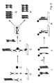
- Figure 1 shows schematically the highly sensitive detection of a substance Coupling with a detection nucleic acid and subsequent enzymatic Amplification of this nucleic acid.
- the substance to be detected (circle) is in a sandwich assay using a first binding molecule (1, Y-shaped shown) isolated from a biological matrix (small squares), while a second binding molecule (2, Y-shaped) is used, the one to be detected Coupling substance with a detection nucleic acid (3).
- PCR Nucleic acid amplification
- FIG. 2 schematically shows the course of the detection while building up a detection complex (E) from a substance to be detected, for example one Antigen (C), a detection reagent oligomer (A), a capture reagent oligomer (B) and a surface that is specific to the scavenger reagent oligomer binds (D).
- the detection reagent oligomer contains antigen-specific Binding molecules (shown in a Y-shape) and a detection nucleic acid (Double arrow).
- the capture reagent oligomer is made from antigen-specific binding molecules (Y-shaped) and nucleic acids (arrow) built up the specific to surface-bound complementary nucleic acids (opposite Arrow) can bind.
- the immobilized containing the detection nucleic acid Detection complex (E) is in the presence of a defined amount a competitor nucleic acid (F) that is different from the detection nucleic acid by the presence of a specific recognition sequence (white rectangle) differs, subjected to a PCR nucleic acid amplification. If originally there is a large amount of antigen (circle), a large amount of detection nucleic acid immobilized and case G occurs: In the PCR product a large amount of the amplified product of the detection nucleic acid and only a small one Amount of amplicon of competitor nucleic acid found. If, however Case H occurs originally: a small amount of antigen is present: in the PCR product only a small amount of the amplificate of the detection nucleic acid and a large amount of the competitor nucleic acid amplificate found.
- the separate detection of the PCR amplificates of detection and competitor nucleic acid For example, they are first of all Hapten (pentagon) marked.
- the marked amplificate mixture (I) is then thermally denatured to the PCR and the labeled single strands (J) through spatially resolved hybridization to complementary oligonucleotides, which either specifically the detection nucleic acid or the recognition sequence of the competitor nucleic acid bind, separated from each other.
- the final detection can then, for example, via an antibody-enzyme conjugate against that into the nucleic acids inserted hapten.
- Figure 4 shows the results of the quantification of rViscumin in BISEKO, a standardized commercially available human serum (see 7th embodiment).
- Curve 1 in Figure A shows the mean values of the signal intensities of the amplificate of the detection nucleic acid (N T ) determined in an ELOSA
- curve 2 in Figure A the mean values of the signal intensities of the amplificate of the competitor nucleic acid (K T ).
- Curve 1 in Figure B shows the values of an ELISA detection method for rViscumin standardized to the negative control
- curves 2 and 3 in Figure B the standardized values of a detection method with (3) and without (2) inclusion of the competitor nucleic acid "KF-1 ".
- the quotient N T / K T was first formed from the signals for detection and competitor nucleic acid according to the procedure shown in FIG. 3, before the comparison quotient (Q) was obtained. was standardized to the negative control.
- mice IgG For the design of the base sequences for the detection and competitor fragments used in competitive nucleic acid amplification, various double-stranded terminal 5'-biotinylated nucleic acid fragments are first examined with regard to their suitability as detection nucleic acid in a highly sensitive protein assay.
- the detection of mouse IgG was carried out with various DNA fragments of the commercially available plasmid M13mp18 (New England Biolabs) according to a known protocol (CM Niemeyer et al., Nucleic Acids Res. 1999, 4553 to 4561).
- a suitable fragment is characterized by the fact that it can detect about 0.01 attomol mouse IgG (see Table 1). Suitability of various DNA fragments for immuno-PCR of mouse IgG.
- the double-stranded detection fragments were determined by quantitative PCR amplified, terminally with known biotinyl substituents derivatized and according to known methods for the synthesis of an oligomer Streptavidin conjugate used (C.M. Niemeyer et al., Nucleic Acids Res. 1999, 4553 to 4561). Due to the high sensitivity when detecting the model antigen DNA-169 was selected as the detection nucleic acid (status of Technology).
- a specific capture nucleic acid was then selected such that they have a high signal intensity in the course of a solid-phase hybridization of the Provides detection fragments. It allowed the use of DNA-169 as Detection -Nucleic acid based on known studies on the hybridization properties single 20 base pair long sections of the nucleic acid will be used (Niemeyer, C.M., L. Boldt, et al. (1999). "Evaluation of single-stranded nucleic acids as carriers in the DNA-directed assembly of macromolecules. " J Biomol Struct Dyn 17 (3): 527-38). In these studies the sequence "N-Es" (see Appendix) showed optimal hybridization properties. Therefore, it was chosen as the recognition sequence of the detection nucleic acid (State of the art).
- the artificial sequence "K-Es” was selected as the recognition sequence for the competitor nucleic acid, which in hybridization experiments according to the procedure described in the literature (Niemeyer, CM et al. (1999), J Biomol Struct Dyn 17 (3): 527- 38) had a hybridization efficiency very similar to sequence "B” (see Table 2). Comparison of the hybridization efficiencies of the recognition sequences of the detection nucleic acid (“N-Es”) and the competitor nucleic acid (“K-Es”). The detection was carried out in each case via an ELOSA using the complementary capture oligonucleotides. Realistic hybridization efficiencies are indicated, the signal obtained for sequence "N-Es" was set to 100%.
- sequence cB 31 Hybridization efficiency to the oligonucleotide complementary to sequence "N-Es" 100% ⁇ 1%
- Hybridization efficiency of the oligonucleotide complementary to sequence "K-Es” ⁇ 1% 108%
- the special primers "K-1" and “K-2” (see Appendix for sequences) at the ends of a 117bp DNA fragment of the plasmid pUC-19 the selected recognition sequence "K-Es" as well as the required primer binding sites of the detection fragment.
- the "KF-1" fragment (sequence, see Appendix) is amplified as a template.
- the reaction mixture was in a 2.5% agarose gel electrophoretically separated, the product band after ethidium bromide staining eluted from the gel.
- the eluate was then used as a template the primer of detection nucleic acid preparative PCR for synthesis a stock solution of competitor nucleic acid "KF-1" performed.
- the Execution of the preparative PCR and the subsequent purification of the Amplified products are carried out according to known methods (C.M. Niemeyer et al., Nucleic Acids Res. 1999, 4553 to 4561)
- the detection nucleic acid "DNA-169” and the competitor nucleic acid “KF-1” became a pair of co-amplifiable nucleic acids, each of which has a specific recognition sequence with similar hybridization properties. By using identical primers and choosing suitable concentration ratios, competitive co-amplification of marker and detection nucleic acid is observed (see Table 3).
- this is due to its co-amplifiability the detection nucleic acid "DNA-169" selected DMA fragment "KF-2" for highly sensitive detection of the model antigen rabbit IgG used.
- This Competitor nucleic acid is both significantly different with a 30 bp difference in length as well as in the primer binding sites from the detection nucleic acid "DNA-169".
- a constant amount of 1 ⁇ 10-20 mol of the competitor nucleic acid was added to a variable amount of the detection nucleic acid “DNA-169” (see first exemplary embodiment) from 1 ⁇ 10 -17 to 1 ⁇ 10 -23 mol per PCR mixture “KF-2" (see Appendix for sequence) was given per PCR batch.
- a PCR was carried out under the known standard conditions (CM Niemeyer et al. , Nucleic Acids Res. 1999, 4553 to 4561) with an annealing temperature of 55 ° C.
- control experiments with different amounts between 1 ⁇ 10 -23 and 1 ⁇ 10 -17 mol were carried out per PCR batch of the competitor nucleic acid "KF-2".
- the quotient was first used for evaluation the signal entities of the amplificate obtained from detection and competition Tor nucleic acid formed. This was true for both the samples containing antigen as well as for a negative control without antigen. In connection the comparison quotient was then formed from these values and with the Signal-to-background value of the detection nucleic acid "DNA-169" in the presence the competitor nucleic acid "KF-2" and without the presence of the competitor nucleic acid compared.
- Test batch to be quantified containing 0.1 attomol rabbit IgG: O T : Sigin intensity of the detection nucleic acid in the PCR-ELOSA (without the presence of "KF-2" in the PCR) 4487 a.u.
- N T signal intensity of the detection nucleic acid in the PCR-ELOSA: (1 ⁇ 10 -20 mol "KF-2" in the PCR) 1525 a.u.
- K T signal intensity of the competitor nucleic acid in the PCR-ELOSA: (1 ⁇ 10 -20 mol "KF-2" in the PCR) 1163 a.u.
- Negative control without rabbit IgG O N : signal intensity of the detection nucleic acid in the PCR-ELOSA (without the presence of "KF-2" in the PCR) 4114 a.u.
- N N signal intensity of the detection nucleic acid in the PCR-ELOSA: (1 ⁇ 10 -20 mol "KF-2" in the PCR) 1324 a.u.
- K N Signal intensity of the competitor nucleic acid in the PCR-ELOSA: (1 ⁇ 10 -20 mol "KF-2" in the PCR) 1569 a.u.
- the comparison quotient Q obtained by forming the partial quotients from the ELOSA values obtained for detection and competitor DNA, yields one with 1.6 significantly higher value for the test mixture containing 0.1 attomol rabbit IgG than the normal determination of the signal-to-background value S.
- the comparison quotient Q obtained by forming the partial quotients from the obtained band intensity values for detection and competitor DNA with 3 a significantly higher value for the test batch containing 1 attomol than the normally customary determination of the signal-to-background value S.
- capture and detection reagent oligomers were used for the detection of PrPc, the non-disease-associated form of the prion protein.
- PrP sc the disease-associated form of the prion protein
- a proteinase K degradation of the PrPc is first carried out in order to then enable detection of the proteinase K-resistant antigen PrPSC with antibodies which recognize both PrP sc and PrPc
- Proteinase K digested samples of bovine brain tissue homogenate were treated with defined concentrations of PrPc as a model system for this procedure.
- the detection was carried out by means of the combined incubation of the samples containing PrPc, catcher reagent oligomer and detection reagent oligomer on a surface coated with a first catcher nucleic acid and subsequent PCR for amplification of the third nucleic acid contained in the detection reagent oligomer in the presence the competitor nucleic acid "KF-1" performed.
- the final detection of the PCR amplificates was carried out by a PCR-ELOSA.
- the non-covalent linkage of the reaction vessels with the first nucleic acid required for immobilization of the capture reagent oligomer was carried out by physisorbing the coupling protein streptavidin (STV) onto the surface to be functionalized and subsequent incubation with biotinylated nucleic acid (Niemeyer, CM, L. Boldt, B Ceyhan and D. Blohm (1999), "DNA-Directed immobilization: efficient, reversible, and site-selective surface binding of proteins by means of covalent DNA-streptavidin conjugates.” Anal Biochem 268 (1): 54-63).
- STV coupling protein streptavidin
- a 0.2 ⁇ M solution of recombinant core-STV (IBA, Göttingen) in PBS buffer (200 mM phosphate (KH 2 PO 4 / K 2 HPO 4 ), pH 7) was prepared.
- 30 ⁇ l of this solution per cavity were applied to top-yield microtiter modules (Nunc), centrifuged for one minute at 4000 rpm in a centrifuge with a microtiter plate rotor, and incubated at 4 ° C. for 72 h. Unbound STV was then removed by washing three times, each for three minutes, with 220 ⁇ l TBS buffer (20 mM Tris-Cl, 150 mM NaCl, pH 7.5) per cavity.
- a blocking step against unspecific interactions with 240 ⁇ l MESTBS buffer (TBS, 45% milk powder (Oxoid), 0.2% NaN 3 , 5 mM EDTA, 1 mg / ml DNA MB grade ( Roche)) per cavity.
- the STV-coated microtiter modules can be stored with this buffer for up to a week.
- the STV-coated and blocked microtiter modules were each used twice washed for one minute and twice for five minutes with 220 ⁇ l TETBS per cavity (hereinafter referred to as: "standard washing step"), and for 30 minutes with 30 ⁇ l per well of the diluted solution of the biotinylated nucleic acid bcA incubated.
- TETBS-Biotin (TETBS, 800 ⁇ M Biotin) washed to the remaining biotin binding ability of the STV-coated modules. That kind prepared microtiter modules are TETBS-Biotin for 12 h at 4 ° C with 100 ⁇ l per well storable.
- binding molecules for the synthesis of detection and capture reagent oligomers a number of possible antibodies are currently available, the some are already commercially available (e.g. 6H4, 5369, 6664, see table 5).
- the biotin / STV system was used to couple the antibody with the nucleic acids of the detection and capture reagent oligomers (Diamandis and Christopoulos 1991).
- the antibody which is only commercially available without biotinylation, was therefore coupled with biotin using a standard method (Meier, T. and F. Fahrenholz (1996), "A laboratory Guide to Biotin-Labeling in Biomolecule Analysis.” BioMethods 7 (Birkhäuser Verlag, Basel), 9-13): To one molar equivalent of antibody was added 10 molar equivalents of sulfo-NHS-biotin (Sigma) and subsequent purification on NAP-10 Sephardex columns (Pharmacia).
- the biotinylated antibody thus obtained was used for the synthesis of the detection and capture reagent.
- Antibodies for the detection of PrP antibody Type species source 6H4 monoclonal mouse Prionics 34C9 monoclonal mouse Prionics 3F4 monoclonal mouse published by Kascsak, RJ, R. Rubenstein, PA Merz, M. Tonna-DeMasi, R. Fersko, RI Carp, HM Wisniewski and H. Diringer (1987), "Mouse polyclonal and monoclonal antibody to scrapie-associated fibril proteins.” J Virol 61 (12): 3688-93 5369 polyclonal goat Biogenesis Ltd. 6664 polyclonal goat Abcam
- a covalent conjugate "HA” was first prepared from STV and a thiolated nucleic acid "A".
- the synthesis of this conjugate using a heterobifunctional chemical crosslinker is described in detail in the literature (Niemeyer, CM, T. Sano, CL Smith and CR Cantor (1994), "Oligonucleotide-directed self-assembly of proteins: semisynthetic DNA-streptavidin hybrid molecules as connectors for the generation of macroscopic arrays and the construction of supramolecular bioconjugates. " Nucleic Acids Res 22 (25): 5530-9).
- a thiol group was first introduced into a commercially available 5'-amino-modified oligonucleotide (Interactiva) and subsequently linked to a maleimide-activated primary amino group of the STV using sulfo-SMCC (Pierce).
- the purified product HA obtained after subsequent ion exchange chromatography purification on an FPLC (Pharmacia) can usually be stored in TE at 4 ° C. for several months.
- a self-assembled aggregate of double biotinylated nucleic acid, tetravalent streptavidin and biotinylated antibody described in the literature was used to link the biotinylated antibody with the third nucleic acid used as a template in the PCR detection (Niemeyer, CM, M. Adler, B. Pignataro , S. Lenhert, S. Gao, L. Chi, H. Fuchs and D. Blohm (1999), "Self-assembly of DNA-streptavidin nanostructures and their use as reagents in immuno-PCR.” Nucleic Acids Res 27 (23 ): 4553-61). The DNA fragment "DNA-169" already described in the first embodiment was used as the nucleic acid.
- bovine PrPc (Prionics, Product No. 03-011) was used.
- Himbial tissue samples were used as the biological matrix Homogenized standard practice and implemented with Prorteinase K. (see Martin, H.D., Blümel, C., Sandmöller, A., Kaiser, F., Kintrup, M., and Fetzer, J., BSE Abericht. LaborPraxis, 2001. 25th year, No. 2: p. 38-59).
- the modules were sealed with adhesive film (plate sealers, Dynex).
- the PCR was carried out in a Gene Amplifying System 9600 (Perkin Bucket) or alternatively in a PTC-200 (MJ-Research) thermal cycler with 96-well Heating block and activated heating lid.
- the PCR was like in the first embodiment described.
- the PCR master mix contained 2 fM of the competitor nucleic acid "KF-1", the Primers “N-1” and “N-2” and digoxigenin-dUTP (Roche).
- the detection of the amplificates of detection and competitor nucleic acid was carried out as already described in the first embodiment, by means of a PCR ELOSA.
- section 3.4 a dilution series of PrPc prepared in proteinase K digested bovine brain homogenate.
- 23.3 ⁇ l of the PrPc solutions were each in 70 ⁇ l TBS buffer with 2 ng / ml of the biotinylated detection antibody and added for one hour Incubated 37 ° C in an Eppendorf thermomixer at 150 rpm.
- Catcher antibodies were used to perform a conventional sandwich assay coated microtiter modules are required.
- top yield Microtiter modules (Nunc) with 30 ⁇ l per cavity of a solution of 2.5 ⁇ g / ml of the respective non-biotinylated antibody in RB buffer (50 mM boric acid / NaOH, pH 9.5) incubated at 4 ° C for 12 hours.
- the step of blocking the antibody-coated Module was created according to the under 3.1.1. described blocking step for STV-coated modules. After that on the Blocking following four-fold washing with TETBS were the modules for one hour at 37 ° C with orbital shaking with 30 ⁇ l / cavity of the solution from PrPc incubated in brain homogenate.
- the modules were added 30 ⁇ l / cavity of a 1: 5000 dilution of an STV alkaline phosphatase conjugate (1000 U / ml before dilution, Roche) in TBS for one hour Incubated at room temperature with orbital shaking. Subsequently, a another standard TETBS wash performed before triple was washed for three minutes with 220 ul / well TBS. After offer of 50 ⁇ l / cavity of the fluorescence-generating substrate AttoPhos TM (Roche) the fluorescence obtained was checked with a VICTOR multi-label counter (Wallac) measured.
- STV alkaline phosphatase conjugate 1000 U / ml before dilution, Roche
- Test batch to be quantified containing 10 pg PrPc in brain homogenate: O T : Signal intensity of the detection nucleic acid in the PCR-ELOSA (without the presence of "KF-1" in the PCR) 5750 a.u. N T : Signal intensity of the detection nucleic acid in the PCR-ELOSA: (2 fM "KF-1" in the PCR) 3820 a.u. K T : signal intensity of the competitor nucleic acid in the PCR-ELOSA: (2 fM "KF-1” in the PCR) 2413 a.u.
- Negative control without PrPc O N signal intensity of the detection nucleic acid in the PCR-ELOSA (without the presence of "KF-1" in the PCR) 5112 a.u.
- N N Signal intensity of the detection nucleic acid in the PCR-ELOSA: (2 fM “KF-1” in the PCR) 2832 a.u.
- K N signal intensity of the competitor nucleic acid in the PCR-ELOSA; (2 sc "KF-1” in the PCR) 3145 a.u.
- the comparison quotient Q obtained by forming the partial quotients from the obtained ELOSA values for detection and competitor DNA, gives a 1.8 significantly higher value for the test batch containing 10 pg PrPc than that Normal determination of the signal-to-background value S.
- Test batch to be quantified containing 1 fg PrP in brain homogenate: N T : Signal intensity of the detection nucleic acid in the PCR-ELOSA: (20 fM “KF-1” in the PCR) 2476 a.u. K T : signal intensity of the competitor nucleic acid in the PCR-ELOSA: (20 fM "KF-1” in the PCR) 16586 a.u. Negative control without PrP: N N : signal intensity of the detection nucleic acid in the PCR-ELOSA: (20 fM "KF-1” in the PCR) 2013 a.u.
- K N Signal intensity of the competitor nucleic acid in the PCR-ELOSA: (20 fM "KF-1" in the PCR) 18409 a.u.
- Signal-to-background value S N T / N N 1.2
- Comparison quotient Q (N T / K T ) / (N N ./ K N ): 1.4
- the comparison quotient Q obtained by forming the partial quotients from the obtained ELOSA values for detection and competitor DNA results in a 1.4 significantly higher value for the test batch containing 1 fg PrP than that Normal determination of the signal-to-background value S.
- the highly sensitive detection of PrP in blood samples is for in vivo diagnosis of TSE diseases of particular interest.
- BISEKO a commercially available standardized human serum (Biotest) used as a biological matrix. 1 ml of the serum was mixed with 10 ⁇ l of a 10 mg / ml proteinase K stock solution (Roche) incubated for 60 min at 56 ° C. The Enzyme was then by adding 100 ul of a 10 mg / ml solution of PMSF (Roche) inactivated in isopropanol.
- This BISEKO solution was as follows used according to the brain homogenate of the third embodiment. The detection of PrPc was in accordance with the 3rd embodiment carried out. The PCR solution was 2fM of the competitor nucleic acid "KF-1" added.
- Test batch to be quantified containing 10 pg PrPc in serum: O T : Signal intensity of the detection nucleic acid in the PCR-ELOSA (without the presence of "KF-1" in the PCR) 613 a.u. N T : Signal intensity of the detection nucleic acid in the PCR-ELOSA: (2 fM "KF-1" in the PCR) 619 a.u. K T : signal intensity of the competitor nucleic acid in the PCR-ELOSA: (2 fM "KF-1" in the PCR) 622 a.u.
- Negative control without PrPc O N signal intensity of the detection nucleic acid in the PCR-ELOSA (without the presence of "KF-1" in the PCR) 542 a.u.
- N N Signal intensity of the detection nucleic acid in the PCR-ELOSA: (2 fM "KF-1” in the PCR) 526 a.u.
- K N Signal intensity of the competitor nucleic acid in the PCR-ELOSA: (2 fM "KF-1” in the PCR) 735 a.u.
- the comparison quotient Q obtained by forming the partial quotients from the obtained ELOSA values for detection and competitor DNA results in a 1.4 higher value for the test preparation containing 10 pg PrPc than that normally usual determination of the signal-to-background value S.
- Proteinase K treated BISEKO used as a biological matrix.
- This BISEKO solution was in the following corresponding to the Him homogenate of the 4th embodiment Releasestzt.
- the detection of PrPc was carried out according to the 4th Exemplary embodiment carried out with the reagent "DRO-2".
- the PCR solution 20fM of the competitor nucleic acid "KF-1" were added.
- Test batch to be quantified containing 1 fg PrP in serum: N T : Signal intensity of the detection nucleic acid in the PCR-ELOSA: (20 fM "KF-1" in the PCR) 130 a.u. K T : signal intensity of the competitor nucleic acid in the PCR-ELOSA: (20 fM "KF-1” in the PCR) 1041 a.u. Negative control without PrP: N N : signal intensity of the detection nucleic acid in the PCR-ELOSA: (20 fM "KF-1” in the PCR) 110 a.u.
- the comparison quotient Q obtained by forming the partial quotients from the ELOSA values obtained for detection and competitor DNA results in a 1.3 higher value for the test batch containing 1 fg PrP than that normally usual determination of the signal-to-background value S.
- rViscumin a new pharmacological active ingredient derived recombinantly from the genetic information of mistletoe lectin, has also proven to be a highly effective anti-tumor agent in animal experiments, even in very low doses (see also Eck, J., M. Langer, B. Mockel, A Baur, M. Rothe, H. Zinke and H. Lentzen (1999), "Cloning of the mistletoe lectin gene and characterization of the recombinant A-chain.” Eur J Biochem 264 (3); 775-84 and Eck, J ., M. Langer, B. Mockel, K. Witthohn, H. Zinke and H.
- the antigen rViscumin (Viscum AG) was stored at a concentration of 2 ng / ⁇ l in aliquots of 10 ⁇ l each at -80 ° C and thawed only immediately before use.
- the rViscumin was diluted to concentrations between 2 ng / ml and 200 fg / ml in BISEKO, a virus-inactivated standardized human serum from the company Biotest, in order to simulate the assay conditions of rViscumin detection from patient serum. Pure BISEKO was used as a negative control.
- 30 ⁇ l of the rViscumin-containing solutions were incubated for 30 min at room temperature in capture antibody-coated modules (see 7.2.).
- the modules were subjected to a standard washing step with TETBS (see 3.1.2.) And then according to the protocol known from the literature (CM Niemeyer et al., Nucleic Acids Res. 1999, 4553 to 4561) with the antirViscumin detection. Reagent oligomer coupled.
- the subsequent PCR step was carried out as in 3.5. described. In this case, in two separate experiments both the competitor nucleic acid "KF-1" was used as the competitor nucleic acid "KF-2" in an amount of from 1x10 -20 mol "KF-1" and 1x10 -20 mol "KF-2" added per PCR batch.
- the primers "N-1” and “N-2” were used to amplify the PCR mixture with “KF-1”; when using “KF-2", the primers "H-1” and “H-2 were also used “used (for sequences see Appendix).
- the ELOSA detection of the amplificates was carried out as under 1.4.
- the 5'biotinylated sequence "H-1” was described as the capture oligonucleotide for "KF-2”.
- "cK-Es” and “cN-Es” were used as capture oligonucleotides, for "KP-1" and for the detection nucleic acid "DNA-169".
- Test batch to be quantified containing 1 attomol (approx. 2 pg / ml) rViscumin:
- O T Signal intensity of the detection nucleic acid in the PCR-ELOSA (without the presence of "KF-1" or "KF-2" in the PCR) 42460 a.u.
- N T signal intensity of the detection nucleic acid in the PCR-ELOSA: (1x10 -20 mol "KF-2" in the PCR) 24426 a.u.
- K T signal intensity of the competitor nucleic acid in the PCR ELOSA (1x10 -20 mol "KF-2" in the PCR) 6381 a.u.
- Negative control without rViscumin O N : signal intensity of the detection nucleic acid in the PCR-ELOSA (without the presence of "KF-1" or "KF-2" in the PCR) 33224 a.u.
- NN Signal intensity of the detection nucleic acid in the PCR-ELOSA: (1x10 -20 mol "KF-2" in the PCR) 18253 a.u.
- K N Signal intensity of the competitor nucleic acid in the PCR-ELOSA: (1x10 -20 mol "KF-2” in the PCR) 8264 a.u.
- the comparison quotient Q obtained by forming the partial quotients from the obtained ELOSA values for detection and competitor DNA, gives a 1.7 significantly higher value for the test batch containing 1 attomol rViscumin than the normal determination of the signal-to-background value S.
- Test batch to be quantified, containing 0.1 attomol rViscumin in serum O T : Signal intensity of the detection nucleic acid in the PCR-ELOSA (without the presence of "KF-1" or "KF-2" in the PCR) 35286 a.u.
- N T Signal intensity of the detection nucleic acid in the PCR-ELOSA: (1x10 -20 mol "KF-1" in the PCR) 16304 a.u.
- K T signal intensity of the competitor nucleic acid in the PCR ELOSA (1x10 -20 mol "KF-1" in the PCR) 2987 a.u.
- Negative control without rViscumin O N : signal intensity of the detection nucleic acid in the PCR-ELOSA (without the presence of "KF-1" or "KF-2" in the PCR) 33224 a.u.
- N N Signal intensity of the detection nucleic acid in the PCR-ELOSA: (1x10 -20 mol "KF-1" in the PCR) 11961 a.u.
- K N signal intensity of the competitor nucleic acid in the PCR ELOSA (1x10 -20 mol "KF-1" in the PCR) 4043 a.u.
- the comparison quotient Q obtained by forming the partial quotients from the obtained ELOSA values for detection and competitor DNA, gives a 1.8 significantly higher value for the test batch containing 0.1 attomol rViscumin than the normal determination of the signal-to-background value S.
- an ELISA there is no rViscumin below under comparable conditions an amount of 10 attomol in 30 ⁇ l BISEKO (approx. 20 pg / ml) more detectable.
- the position of the binding sequence for the PCR primer "N-2" is underlined
- the position of the binding sequence for the primer "N-1” is underlined
- the position of the recognition sequence "N-Es” is highlighted in bold and underlined .
- Competitor nucleic acid (KF1" - artificial) with the primer binding sites "N-2" and "N-1" of the detection nucleic acid:
- the position of the binding sequence for the PCR primer "N-2" is underlined, the position of the binding sequence for the primer “N-1” is double underlined , the position of the recognition sequence "K-Es" is bold and underlined .
Landscapes
- Chemical & Material Sciences (AREA)
- Organic Chemistry (AREA)
- Life Sciences & Earth Sciences (AREA)
- Health & Medical Sciences (AREA)
- Zoology (AREA)
- Proteomics, Peptides & Aminoacids (AREA)
- Engineering & Computer Science (AREA)
- Wood Science & Technology (AREA)
- Analytical Chemistry (AREA)
- Immunology (AREA)
- Microbiology (AREA)
- Molecular Biology (AREA)
- Physics & Mathematics (AREA)
- Biotechnology (AREA)
- Biophysics (AREA)
- Biochemistry (AREA)
- Bioinformatics & Cheminformatics (AREA)
- General Engineering & Computer Science (AREA)
- General Health & Medical Sciences (AREA)
- Genetics & Genomics (AREA)
- Pathology (AREA)
- Measuring Or Testing Involving Enzymes Or Micro-Organisms (AREA)
Priority Applications (1)
| Application Number | Priority Date | Filing Date | Title |
|---|---|---|---|
| EP01114562A EP1270738A1 (fr) | 2001-06-18 | 2001-06-18 | Méthode pour détecter des substances dans des liquides |
Applications Claiming Priority (1)
| Application Number | Priority Date | Filing Date | Title |
|---|---|---|---|
| EP01114562A EP1270738A1 (fr) | 2001-06-18 | 2001-06-18 | Méthode pour détecter des substances dans des liquides |
Publications (1)
| Publication Number | Publication Date |
|---|---|
| EP1270738A1 true EP1270738A1 (fr) | 2003-01-02 |
Family
ID=8177737
Family Applications (1)
| Application Number | Title | Priority Date | Filing Date |
|---|---|---|---|
| EP01114562A Withdrawn EP1270738A1 (fr) | 2001-06-18 | 2001-06-18 | Méthode pour détecter des substances dans des liquides |
Country Status (1)
| Country | Link |
|---|---|
| EP (1) | EP1270738A1 (fr) |
Cited By (5)
| Publication number | Priority date | Publication date | Assignee | Title |
|---|---|---|---|---|
| EP1745157A4 (fr) * | 2004-04-12 | 2008-06-11 | Ohio Med College | Methodes et compositions pour analyser des analytes |
| WO2012049285A1 (fr) * | 2010-10-14 | 2012-04-19 | Octapharma Ag | Méthode d'analyse quantitative par glycosylation de protéines |
| US8304192B2 (en) | 2004-03-05 | 2012-11-06 | University Of Toledo | Methods and compositions for assessing nucleic acids and alleles |
| EP3158089A4 (fr) * | 2014-06-23 | 2018-01-24 | The Board of Trustees of The Leland Stanford Junior University | Coloration sur lames par extension d'amorce |
| US10370698B2 (en) | 2016-07-27 | 2019-08-06 | The Board Of Trustees Of The Leland Stanford Junior University | Highly-multiplexed fluorescent imaging |
Citations (3)
| Publication number | Priority date | Publication date | Assignee | Title |
|---|---|---|---|---|
| WO1998004745A1 (fr) * | 1996-07-31 | 1998-02-05 | Mount Sinai School Of Medicine Of The City University Of New York | Methode d'amplification des acides nucleiques: methode d'amplification avec signal d'hybridation (hsam) |
| WO1999063109A1 (fr) * | 1998-06-04 | 1999-12-09 | Baxter Aktiengesellschaft | Procede permettant de determiner des antigenes |
| DE19941756A1 (de) * | 1999-09-02 | 2001-03-08 | Christof Niemeyer | Verfahren zur Verwendung von oligomeren Nucleinsäure-Protein-Konjugaten als Reagenzien für immunologische Nachweisverfahren insbesondere die Immuno-PCR |
-
2001
- 2001-06-18 EP EP01114562A patent/EP1270738A1/fr not_active Withdrawn
Patent Citations (3)
| Publication number | Priority date | Publication date | Assignee | Title |
|---|---|---|---|---|
| WO1998004745A1 (fr) * | 1996-07-31 | 1998-02-05 | Mount Sinai School Of Medicine Of The City University Of New York | Methode d'amplification des acides nucleiques: methode d'amplification avec signal d'hybridation (hsam) |
| WO1999063109A1 (fr) * | 1998-06-04 | 1999-12-09 | Baxter Aktiengesellschaft | Procede permettant de determiner des antigenes |
| DE19941756A1 (de) * | 1999-09-02 | 2001-03-08 | Christof Niemeyer | Verfahren zur Verwendung von oligomeren Nucleinsäure-Protein-Konjugaten als Reagenzien für immunologische Nachweisverfahren insbesondere die Immuno-PCR |
Non-Patent Citations (9)
| Title |
|---|
| CASE M C ET AL: "Enhanced ultrasensitive detection of structurally diverse antigens using a single immuno-PCR assay protocol", JOURNAL OF IMMUNOLOGICAL METHODS, ELSEVIER SCIENCE PUBLISHERS B.V.,AMSTERDAM, NL, vol. 223, no. 1, 1 February 1999 (1999-02-01), pages 93 - 106, XP004155757, ISSN: 0022-1759 * |
| HENDRICKSON EDWIN R ET AL: "High sensitivity multianalyte immunoassay using covalent DNA-labeled antibodies and polymerase chain reaction", NUCLEIC ACIDS RESEARCH, OXFORD UNIVERSITY PRESS, SURREY, GB, vol. 23, no. 3, 1995, pages 522 - 529, XP002151883, ISSN: 0305-1048 * |
| NIEMEYER C M ET AL: "HYBRIDIZATION CHARACTERISTICS OF BIOMOLECULAR ADAPTORS, COVALENT DNA-STREPTAVIDIN CONJUGATES", BIOCONJUGATE CHEMISTRY, AMERICAN CHEMICAL SOCIETY, WASHINGTON, US, vol. 9, no. 2, 1 March 1998 (1998-03-01), pages 168 - 175, XP000750892, ISSN: 1043-1802 * |
| NIEMEYER CHRISTOF M ET AL: "DNA-directed immobilization: Efficient, reversible, and site-selective surface binding of proteins by means of covalent DNA-streptavidin conjugates", ANALYTICAL BIOCHEMISTRY, ACADEMIC PRESS, SAN DIEGO, CA, US, vol. 268, no. 1, 1 March 1999 (1999-03-01), pages 54 - 63, XP002176566, ISSN: 0003-2697 * |
| NIEMEYER CHRISTOF M ET AL: "Fluorometric polymerase chain reaction (PCR) enzyme-linked immunosorbent assay for quantification of immuno-PCR products in microplates", ANALYTICAL BIOCHEMISTRY, ACADEMIC PRESS, SAN DIEGO, CA, US, vol. 246, no. 1, 1997, pages 140 - 145, XP002151884, ISSN: 0003-2697 * |
| NIEMEYER CHRISTOF M ET AL: "Functionalization of covalent DNA-streptavidin conjugates by means of biotinylated modulator components.", BIOCONJUGATE CHEMISTRY, vol. 10, no. 5, 1999, pages 708 - 719, XP002187381, ISSN: 1043-1802 * |
| NIEMEYER CHRISTOF M ET AL: "Nanostructured DNA-protein aggregates consisting of covalent oligonucleotide-streptavidin conjugates.", BIOCONJUGATE CHEMISTRY, vol. 12, no. 3, May 2001 (2001-05-01), pages 364 - 371, XP002187380, ISSN: 1043-1802 * |
| NIEMEYER CHRISTOF M ET AL: "Self-assembly of DNA-streptavidin nanostructures and their use as reagents in immuno-PCR", NUCLEIC ACIDS RESEARCH, OXFORD UNIVERSITY PRESS, SURREY, GB, vol. 27, no. 23, 1 December 1999 (1999-12-01), pages 4553 - 4561, XP002176567, ISSN: 0305-1048 * |
| REISCHL U ET AL: "QUANTITATIVE PCR A SURVEY OF THE PRESENT TECHNOLOGY", MOLECULAR BIOTECHNOLOGY, TOTOWA, NJ, US, vol. 3, 1995, pages 55 - 71, XP000600241, ISSN: 1073-6085 * |
Cited By (16)
| Publication number | Priority date | Publication date | Assignee | Title |
|---|---|---|---|---|
| US8304192B2 (en) | 2004-03-05 | 2012-11-06 | University Of Toledo | Methods and compositions for assessing nucleic acids and alleles |
| EP1745157A4 (fr) * | 2004-04-12 | 2008-06-11 | Ohio Med College | Methodes et compositions pour analyser des analytes |
| US7476502B2 (en) | 2004-04-12 | 2009-01-13 | Willey James C | Methods and compositions for assaying analytes |
| WO2012049285A1 (fr) * | 2010-10-14 | 2012-04-19 | Octapharma Ag | Méthode d'analyse quantitative par glycosylation de protéines |
| US10017808B2 (en) | 2014-06-23 | 2018-07-10 | The Board Of Trustees Of The Leland Stanford Junior University | On-slide staining by primer extension |
| US9909167B2 (en) | 2014-06-23 | 2018-03-06 | The Board Of Trustees Of The Leland Stanford Junior University | On-slide staining by primer extension |
| US10000796B2 (en) | 2014-06-23 | 2018-06-19 | The Board Of Trustees Of The Leland Stanford Junior University | On-slide staining by primer extension |
| US10006082B2 (en) | 2014-06-23 | 2018-06-26 | The Board Of Trustees Of The Leland Stanford Junior University | On-slide straining by primer extension |
| EP3158089A4 (fr) * | 2014-06-23 | 2018-01-24 | The Board of Trustees of The Leland Stanford Junior University | Coloration sur lames par extension d'amorce |
| US10982263B2 (en) | 2014-06-23 | 2021-04-20 | The Board Of Trustees Of The Leland Stanford Junior University | On-slide staining by primer extension |
| US11299770B2 (en) | 2014-06-23 | 2022-04-12 | The Board Of Trustees Of The Leland Stanford Junior University | On-slide staining by primer extension |
| US11634753B2 (en) | 2014-06-23 | 2023-04-25 | The Board Of Trustees Of The Leland Stanford Junior University | On-slide staining by primer extension |
| US11926865B2 (en) | 2014-06-23 | 2024-03-12 | The Board Of Trustees Of The Leland Stanford Junior University | On-slide staining by primer extension |
| US12331344B2 (en) | 2014-06-23 | 2025-06-17 | The Board Of Trustees Of The Leland Stanford Junior University | On-slide staining by primer extension |
| US10370698B2 (en) | 2016-07-27 | 2019-08-06 | The Board Of Trustees Of The Leland Stanford Junior University | Highly-multiplexed fluorescent imaging |
| US11168350B2 (en) | 2016-07-27 | 2021-11-09 | The Board Of Trustees Of The Leland Stanford Junior University | Highly-multiplexed fluorescent imaging |
Similar Documents
| Publication | Publication Date | Title |
|---|---|---|
| DE69506387T2 (de) | Bindungstest unter verwendung von agenzien mit schwanzgruppen | |
| DE69735532T2 (de) | Bestimmung von antigenen mittels oligonukleotid-antikörper-konjugaten | |
| DE3689412T2 (de) | Verfahren zur durchführung von hybridisierungstests von nukleinsäuren. | |
| DE69614539T2 (de) | Immunoassay und kit mit zwei reagenzien, die vernetzt werden, wenn sie an einem analyten haften | |
| EP1239049B1 (fr) | Détermination de l'eau oxygenée avec des oxidases et des complexes lanthanoide-ligand | |
| DE69033387T2 (de) | Verfahren und satz fur die detektion von ziel nuklein sauren unter verwendung einer fangsonde die uber ein zwischenprotein an eine polystyrene oberflache gekoppelt ist. | |
| DE3888653T2 (de) | Verfahren und Testsatz zum Nachweis einer Nukleinsäure-Sequenz. | |
| DE60317315T2 (de) | Nachweis von dna-bindungsproteinen | |
| DE69414122T2 (de) | Verfahren zum nachweis biologischer wechselwirkungen, insbesondere in rezeptorbindungs-assays | |
| EP2167965B1 (fr) | Procédé de détection d'analytes | |
| EP1249500A1 (fr) | Procédé pour la détermination de la concentration d'un analyte | |
| EP0523557B1 (fr) | Immobilisation des acides nucléiques | |
| DE69924128T2 (de) | Verfahren zur erkennung von verbindungen,die die wechselwirkung von rna und rna-bindungsproteinen beinflussen | |
| EP1270738A1 (fr) | Méthode pour détecter des substances dans des liquides | |
| DE19624562A1 (de) | Verfahren zur Bestimmung von Konzentrationsverhältnissen zweier sich um mindestens ein Nucleotid unterscheidender Nucleinsäuren und Testkit zur Durchführung des Verfahrens | |
| DE10141691A1 (de) | Verdrängungsassay zur Detektion von Ligat-Ligand-Assoziationsereignissen | |
| EP0437774A1 (fr) | Procédé pour la préparation d'acides nucléiques modifiés | |
| DE19859912C2 (de) | Testsystem zur Erkennung verschiedener Marker, seine Herstellung sowie Verwendung | |
| WO2006048164A1 (fr) | Systeme d'essai base sur des aptameres | |
| EP1334208A2 (fr) | Procede pour le marquage de substances chimiques | |
| DE69718624T2 (de) | Nicht-kompetitiver immunoassay mit blockierung von freien bindungsstellen auf der festen phase | |
| DE4212555A1 (de) | Verfahren zum Nachweis von Nukleinsäuren | |
| EP1412528B1 (fr) | Biocapteur et procede de detection de biopolymeres macromoleculaires au moyen d'au moins une unite pour immobiliser des biopolymeres macromoleculaires | |
| EP3790982A1 (fr) | Analyse cellulaire in situ dans un système de culture cellulaire | |
| DE19808003C2 (de) | Verfahren zur Beschleunigung der Dissoziation von Komplexen |
Legal Events
| Date | Code | Title | Description |
|---|---|---|---|
| PUAI | Public reference made under article 153(3) epc to a published international application that has entered the european phase |
Free format text: ORIGINAL CODE: 0009012 |
|
| AK | Designated contracting states |
Kind code of ref document: A1 Designated state(s): AT BE CH CY DE DK ES FI FR GB GR IE IT LI LU MC NL PT SE TR |
|
| AX | Request for extension of the european patent |
Free format text: AL;LT;LV;MK;RO;SI |
|
| 17P | Request for examination filed |
Effective date: 20030702 |
|
| AKX | Designation fees paid |
Designated state(s): AT BE CH CY DE DK ES FI FR GB GR IE IT LI LU MC NL PT SE TR |
|
| RAP1 | Party data changed (applicant data changed or rights of an application transferred) |
Owner name: CHIMERA BIOTEC GMBH |
|
| 17Q | First examination report despatched |
Effective date: 20050311 |
|
| STAA | Information on the status of an ep patent application or granted ep patent |
Free format text: STATUS: THE APPLICATION IS DEEMED TO BE WITHDRAWN |
|
| 18D | Application deemed to be withdrawn |
Effective date: 20050722 |