EP1334981A2 - Méthode de purification de facteurs de croissance des kératinocytes - Google Patents
Méthode de purification de facteurs de croissance des kératinocytes Download PDFInfo
- Publication number
- EP1334981A2 EP1334981A2 EP02028624A EP02028624A EP1334981A2 EP 1334981 A2 EP1334981 A2 EP 1334981A2 EP 02028624 A EP02028624 A EP 02028624A EP 02028624 A EP02028624 A EP 02028624A EP 1334981 A2 EP1334981 A2 EP 1334981A2
- Authority
- EP
- European Patent Office
- Prior art keywords
- kgf
- keratinocyte growth
- growth factor
- amino acid
- seq
- Prior art date
- Legal status (The legal status is an assumption and is not a legal conclusion. Google has not performed a legal analysis and makes no representation as to the accuracy of the status listed.)
- Granted
Links
- 238000000034 method Methods 0.000 title claims description 42
- 210000002510 keratinocyte Anatomy 0.000 title abstract description 11
- 239000003102 growth factor Substances 0.000 title abstract description 10
- 108090000385 Fibroblast growth factor 7 Proteins 0.000 claims description 136
- 102000003972 Fibroblast growth factor 7 Human genes 0.000 claims description 128
- 108090000623 proteins and genes Proteins 0.000 claims description 48
- 102000004169 proteins and genes Human genes 0.000 claims description 41
- 235000018102 proteins Nutrition 0.000 claims description 39
- 235000001014 amino acid Nutrition 0.000 claims description 34
- 125000003275 alpha amino acid group Chemical group 0.000 claims description 20
- 150000001413 amino acids Chemical class 0.000 claims description 20
- 108090000765 processed proteins & peptides Proteins 0.000 claims description 15
- 241000588724 Escherichia coli Species 0.000 claims description 13
- 229920001184 polypeptide Polymers 0.000 claims description 10
- 102000004196 processed proteins & peptides Human genes 0.000 claims description 10
- 238000006467 substitution reaction Methods 0.000 claims description 10
- 238000012217 deletion Methods 0.000 claims description 7
- 230000037430 deletion Effects 0.000 claims description 7
- 238000004191 hydrophobic interaction chromatography Methods 0.000 claims description 7
- NWUYHJFMYQTDRP-UHFFFAOYSA-N 1,2-bis(ethenyl)benzene;1-ethenyl-2-ethylbenzene;styrene Chemical compound C=CC1=CC=CC=C1.CCC1=CC=CC=C1C=C.C=CC1=CC=CC=C1C=C NWUYHJFMYQTDRP-UHFFFAOYSA-N 0.000 claims description 6
- 239000003729 cation exchange resin Substances 0.000 claims description 6
- 235000018417 cysteine Nutrition 0.000 claims description 6
- XUJNEKJLAYXESH-UHFFFAOYSA-N cysteine Natural products SCC(N)C(O)=O XUJNEKJLAYXESH-UHFFFAOYSA-N 0.000 claims description 6
- 239000007800 oxidant agent Substances 0.000 claims description 6
- 108010076504 Protein Sorting Signals Proteins 0.000 claims description 5
- 238000007792 addition Methods 0.000 claims description 5
- 125000000539 amino acid group Chemical group 0.000 claims description 5
- NGDIAZZSCVVCEW-UHFFFAOYSA-M sodium;butyl sulfate Chemical compound [Na+].CCCCOS([O-])(=O)=O NGDIAZZSCVVCEW-UHFFFAOYSA-M 0.000 claims description 5
- 238000011068 loading method Methods 0.000 claims description 4
- 210000004962 mammalian cell Anatomy 0.000 claims description 4
- 239000011159 matrix material Substances 0.000 claims description 4
- RYGMFSIKBFXOCR-UHFFFAOYSA-N Copper Chemical compound [Cu] RYGMFSIKBFXOCR-UHFFFAOYSA-N 0.000 claims description 3
- 108010053070 Glutathione Disulfide Proteins 0.000 claims description 3
- 229910052802 copper Inorganic materials 0.000 claims description 3
- 239000010949 copper Substances 0.000 claims description 3
- 229960003067 cystine Drugs 0.000 claims description 3
- 230000000694 effects Effects 0.000 claims description 3
- 230000007717 exclusion Effects 0.000 claims description 3
- YPZRWBKMTBYPTK-BJDJZHNGSA-N glutathione disulfide Chemical compound OC(=O)[C@@H](N)CCC(=O)N[C@H](C(=O)NCC(O)=O)CSSC[C@@H](C(=O)NCC(O)=O)NC(=O)CC[C@H](N)C(O)=O YPZRWBKMTBYPTK-BJDJZHNGSA-N 0.000 claims description 3
- YPZRWBKMTBYPTK-UHFFFAOYSA-N oxidized gamma-L-glutamyl-L-cysteinylglycine Natural products OC(=O)C(N)CCC(=O)NC(C(=O)NCC(O)=O)CSSCC(C(=O)NCC(O)=O)NC(=O)CCC(N)C(O)=O YPZRWBKMTBYPTK-UHFFFAOYSA-N 0.000 claims description 3
- 125000003396 thiol group Chemical group [H]S* 0.000 claims description 3
- 238000001042 affinity chromatography Methods 0.000 claims description 2
- 230000007935 neutral effect Effects 0.000 claims description 2
- 210000001236 prokaryotic cell Anatomy 0.000 claims 2
- LEVWYRKDKASIDU-QWWZWVQMSA-N D-cystine Chemical compound OC(=O)[C@H](N)CSSC[C@@H](N)C(O)=O LEVWYRKDKASIDU-QWWZWVQMSA-N 0.000 claims 1
- HTTJABKRGRZYRN-UHFFFAOYSA-N Heparin Chemical compound OC1C(NC(=O)C)C(O)OC(COS(O)(=O)=O)C1OC1C(OS(O)(=O)=O)C(O)C(OC2C(C(OS(O)(=O)=O)C(OC3C(C(O)C(O)C(O3)C(O)=O)OS(O)(=O)=O)C(CO)O2)NS(O)(=O)=O)C(C(O)=O)O1 HTTJABKRGRZYRN-UHFFFAOYSA-N 0.000 claims 1
- 210000004978 chinese hamster ovary cell Anatomy 0.000 claims 1
- 239000012504 chromatography matrix Substances 0.000 claims 1
- 229960002897 heparin Drugs 0.000 claims 1
- 229920000669 heparin Polymers 0.000 claims 1
- 125000001360 methionine group Chemical group N[C@@H](CCSC)C(=O)* 0.000 claims 1
- 238000000746 purification Methods 0.000 abstract description 9
- FAPWRFPIFSIZLT-UHFFFAOYSA-M Sodium chloride Chemical compound [Na+].[Cl-] FAPWRFPIFSIZLT-UHFFFAOYSA-M 0.000 description 50
- 210000004027 cell Anatomy 0.000 description 35
- 239000011780 sodium chloride Substances 0.000 description 26
- 108020004414 DNA Proteins 0.000 description 20
- 239000000243 solution Substances 0.000 description 20
- 239000013612 plasmid Substances 0.000 description 19
- 238000003752 polymerase chain reaction Methods 0.000 description 19
- 108091034117 Oligonucleotide Proteins 0.000 description 16
- 239000011347 resin Substances 0.000 description 14
- 229920005989 resin Polymers 0.000 description 14
- 239000002773 nucleotide Substances 0.000 description 12
- 125000003729 nucleotide group Chemical group 0.000 description 12
- 239000000047 product Substances 0.000 description 12
- 239000012634 fragment Substances 0.000 description 11
- 108091028043 Nucleic acid sequence Proteins 0.000 description 10
- 239000002953 phosphate buffered saline Substances 0.000 description 10
- 239000013598 vector Substances 0.000 description 9
- BFNBIHQBYMNNAN-UHFFFAOYSA-N ammonium sulfate Chemical compound N.N.OS(O)(=O)=O BFNBIHQBYMNNAN-UHFFFAOYSA-N 0.000 description 8
- 229910052921 ammonium sulfate Inorganic materials 0.000 description 8
- 239000000872 buffer Substances 0.000 description 8
- 239000000463 material Substances 0.000 description 8
- 108020004705 Codon Proteins 0.000 description 7
- 238000012408 PCR amplification Methods 0.000 description 7
- 238000002523 gelfiltration Methods 0.000 description 7
- 238000009396 hybridization Methods 0.000 description 7
- 230000003647 oxidation Effects 0.000 description 7
- 238000007254 oxidation reaction Methods 0.000 description 7
- 238000002415 sodium dodecyl sulfate polyacrylamide gel electrophoresis Methods 0.000 description 7
- OKKJLVBELUTLKV-UHFFFAOYSA-N Methanol Chemical compound OC OKKJLVBELUTLKV-UHFFFAOYSA-N 0.000 description 6
- 229920002684 Sepharose Polymers 0.000 description 6
- 230000027455 binding Effects 0.000 description 6
- 210000002950 fibroblast Anatomy 0.000 description 6
- 239000012528 membrane Substances 0.000 description 6
- 239000000203 mixture Substances 0.000 description 6
- 239000000523 sample Substances 0.000 description 6
- 241000283707 Capra Species 0.000 description 5
- 238000012300 Sequence Analysis Methods 0.000 description 5
- 238000000137 annealing Methods 0.000 description 5
- 238000010367 cloning Methods 0.000 description 5
- 239000002299 complementary DNA Substances 0.000 description 5
- 239000002609 medium Substances 0.000 description 5
- 238000002360 preparation method Methods 0.000 description 5
- 239000001488 sodium phosphate Substances 0.000 description 5
- 229910000162 sodium phosphate Inorganic materials 0.000 description 5
- RYFMWSXOAZQYPI-UHFFFAOYSA-K trisodium phosphate Chemical compound [Na+].[Na+].[Na+].[O-]P([O-])([O-])=O RYFMWSXOAZQYPI-UHFFFAOYSA-K 0.000 description 5
- 102000018233 Fibroblast Growth Factor Human genes 0.000 description 4
- 108050007372 Fibroblast Growth Factor Proteins 0.000 description 4
- 101001060261 Homo sapiens Fibroblast growth factor 7 Proteins 0.000 description 4
- 102000003960 Ligases Human genes 0.000 description 4
- 108090000364 Ligases Proteins 0.000 description 4
- 239000000020 Nitrocellulose Substances 0.000 description 4
- 230000003321 amplification Effects 0.000 description 4
- 238000005352 clarification Methods 0.000 description 4
- 239000003636 conditioned culture medium Substances 0.000 description 4
- 238000004925 denaturation Methods 0.000 description 4
- 230000036425 denaturation Effects 0.000 description 4
- 238000001962 electrophoresis Methods 0.000 description 4
- 238000000855 fermentation Methods 0.000 description 4
- 230000004151 fermentation Effects 0.000 description 4
- -1 fragments Chemical class 0.000 description 4
- 239000006166 lysate Substances 0.000 description 4
- 229920001220 nitrocellulos Polymers 0.000 description 4
- 238000003199 nucleic acid amplification method Methods 0.000 description 4
- 210000002966 serum Anatomy 0.000 description 4
- 238000005406 washing Methods 0.000 description 4
- 108091032973 (ribonucleotides)n+m Proteins 0.000 description 3
- 229920000936 Agarose Polymers 0.000 description 3
- 239000004475 Arginine Substances 0.000 description 3
- LFQSCWFLJHTTHZ-UHFFFAOYSA-N Ethanol Chemical compound CCO LFQSCWFLJHTTHZ-UHFFFAOYSA-N 0.000 description 3
- PEDCQBHIVMGVHV-UHFFFAOYSA-N Glycerine Chemical compound OCC(O)CO PEDCQBHIVMGVHV-UHFFFAOYSA-N 0.000 description 3
- MTCFGRXMJLQNBG-UHFFFAOYSA-N Serine Natural products OCC(N)C(O)=O MTCFGRXMJLQNBG-UHFFFAOYSA-N 0.000 description 3
- 239000011543 agarose gel Substances 0.000 description 3
- 238000004458 analytical method Methods 0.000 description 3
- ODKSFYDXXFIFQN-UHFFFAOYSA-N arginine Natural products OC(=O)C(N)CCCNC(N)=N ODKSFYDXXFIFQN-UHFFFAOYSA-N 0.000 description 3
- 230000004071 biological effect Effects 0.000 description 3
- 238000004113 cell culture Methods 0.000 description 3
- 238000005119 centrifugation Methods 0.000 description 3
- 238000011210 chromatographic step Methods 0.000 description 3
- 238000010276 construction Methods 0.000 description 3
- 230000003247 decreasing effect Effects 0.000 description 3
- 210000002919 epithelial cell Anatomy 0.000 description 3
- 239000000499 gel Substances 0.000 description 3
- 238000011534 incubation Methods 0.000 description 3
- 230000003993 interaction Effects 0.000 description 3
- 229930027917 kanamycin Natural products 0.000 description 3
- SBUJHOSQTJFQJX-NOAMYHISSA-N kanamycin Chemical compound O[C@@H]1[C@@H](O)[C@H](O)[C@@H](CN)O[C@@H]1O[C@H]1[C@H](O)[C@@H](O[C@@H]2[C@@H]([C@@H](N)[C@H](O)[C@@H](CO)O2)O)[C@H](N)C[C@@H]1N SBUJHOSQTJFQJX-NOAMYHISSA-N 0.000 description 3
- 229960000318 kanamycin Drugs 0.000 description 3
- 229930182823 kanamycin A Natural products 0.000 description 3
- 125000001997 phenyl group Chemical group [H]C1=C([H])C([H])=C(*)C([H])=C1[H] 0.000 description 3
- 108020003175 receptors Proteins 0.000 description 3
- 102000005962 receptors Human genes 0.000 description 3
- 230000000638 stimulation Effects 0.000 description 3
- 238000001262 western blot Methods 0.000 description 3
- NHBKXEKEPDILRR-UHFFFAOYSA-N 2,3-bis(butanoylsulfanyl)propyl butanoate Chemical compound CCCC(=O)OCC(SC(=O)CCC)CSC(=O)CCC NHBKXEKEPDILRR-UHFFFAOYSA-N 0.000 description 2
- QKNYBSVHEMOAJP-UHFFFAOYSA-N 2-amino-2-(hydroxymethyl)propane-1,3-diol;hydron;chloride Chemical compound Cl.OCC(N)(CO)CO QKNYBSVHEMOAJP-UHFFFAOYSA-N 0.000 description 2
- 208000002109 Argyria Diseases 0.000 description 2
- 241000894006 Bacteria Species 0.000 description 2
- 108090000386 Fibroblast Growth Factor 1 Proteins 0.000 description 2
- 108090000379 Fibroblast growth factor 2 Proteins 0.000 description 2
- ZHNUHDYFZUAESO-UHFFFAOYSA-N Formamide Chemical compound NC=O ZHNUHDYFZUAESO-UHFFFAOYSA-N 0.000 description 2
- DHMQDGOQFOQNFH-UHFFFAOYSA-N Glycine Chemical compound NCC(O)=O DHMQDGOQFOQNFH-UHFFFAOYSA-N 0.000 description 2
- 108010031186 Glycoside Hydrolases Proteins 0.000 description 2
- 102000005744 Glycoside Hydrolases Human genes 0.000 description 2
- 102100034343 Integrase Human genes 0.000 description 2
- LEVWYRKDKASIDU-IMJSIDKUSA-N L-cystine Chemical compound [O-]C(=O)[C@@H]([NH3+])CSSC[C@H]([NH3+])C([O-])=O LEVWYRKDKASIDU-IMJSIDKUSA-N 0.000 description 2
- 101100446506 Mus musculus Fgf3 gene Proteins 0.000 description 2
- 125000001429 N-terminal alpha-amino-acid group Chemical group 0.000 description 2
- 241000283973 Oryctolagus cuniculus Species 0.000 description 2
- ISWSIDIOOBJBQZ-UHFFFAOYSA-N Phenol Chemical compound OC1=CC=CC=C1 ISWSIDIOOBJBQZ-UHFFFAOYSA-N 0.000 description 2
- 108010021757 Polynucleotide 5'-Hydroxyl-Kinase Proteins 0.000 description 2
- 102000008422 Polynucleotide 5'-hydroxyl-kinase Human genes 0.000 description 2
- 108010092799 RNA-directed DNA polymerase Proteins 0.000 description 2
- 240000004808 Saccharomyces cerevisiae Species 0.000 description 2
- VYPSYNLAJGMNEJ-UHFFFAOYSA-N Silicium dioxide Chemical compound O=[Si]=O VYPSYNLAJGMNEJ-UHFFFAOYSA-N 0.000 description 2
- IQFYYKKMVGJFEH-XLPZGREQSA-N Thymidine Chemical compound O=C1NC(=O)C(C)=CN1[C@@H]1O[C@H](CO)[C@@H](O)C1 IQFYYKKMVGJFEH-XLPZGREQSA-N 0.000 description 2
- 241000700605 Viruses Species 0.000 description 2
- 239000008365 aqueous carrier Substances 0.000 description 2
- 238000003556 assay Methods 0.000 description 2
- 230000001580 bacterial effect Effects 0.000 description 2
- 239000013592 cell lysate Substances 0.000 description 2
- 238000006243 chemical reaction Methods 0.000 description 2
- 125000000151 cysteine group Chemical group N[C@@H](CS)C(=O)* 0.000 description 2
- 239000006185 dispersion Substances 0.000 description 2
- 239000003814 drug Substances 0.000 description 2
- 108010030074 endodeoxyribonuclease MluI Proteins 0.000 description 2
- 238000002474 experimental method Methods 0.000 description 2
- 239000013613 expression plasmid Substances 0.000 description 2
- 238000009472 formulation Methods 0.000 description 2
- ZDXPYRJPNDTMRX-UHFFFAOYSA-N glutamine Natural products OC(=O)C(N)CCC(N)=O ZDXPYRJPNDTMRX-UHFFFAOYSA-N 0.000 description 2
- 125000000404 glutamine group Chemical class N[C@@H](CCC(N)=O)C(=O)* 0.000 description 2
- 230000002209 hydrophobic effect Effects 0.000 description 2
- 238000000338 in vitro Methods 0.000 description 2
- 238000010348 incorporation Methods 0.000 description 2
- 238000003780 insertion Methods 0.000 description 2
- 230000037431 insertion Effects 0.000 description 2
- 238000002955 isolation Methods 0.000 description 2
- 108010029560 keratinocyte growth factor receptor Proteins 0.000 description 2
- 230000002297 mitogenic effect Effects 0.000 description 2
- 238000010369 molecular cloning Methods 0.000 description 2
- 238000012544 monitoring process Methods 0.000 description 2
- 239000008363 phosphate buffer Substances 0.000 description 2
- 229920000642 polymer Polymers 0.000 description 2
- 230000008569 process Effects 0.000 description 2
- 230000010076 replication Effects 0.000 description 2
- 239000012465 retentate Substances 0.000 description 2
- 239000006228 supernatant Substances 0.000 description 2
- 238000012546 transfer Methods 0.000 description 2
- 230000009466 transformation Effects 0.000 description 2
- 238000013519 translation Methods 0.000 description 2
- 238000000108 ultra-filtration Methods 0.000 description 2
- XLYOFNOQVPJJNP-UHFFFAOYSA-N water Chemical compound O XLYOFNOQVPJJNP-UHFFFAOYSA-N 0.000 description 2
- KIUKXJAPPMFGSW-DNGZLQJQSA-N (2S,3S,4S,5R,6R)-6-[(2S,3R,4R,5S,6R)-3-Acetamido-2-[(2S,3S,4R,5R,6R)-6-[(2R,3R,4R,5S,6R)-3-acetamido-2,5-dihydroxy-6-(hydroxymethyl)oxan-4-yl]oxy-2-carboxy-4,5-dihydroxyoxan-3-yl]oxy-5-hydroxy-6-(hydroxymethyl)oxan-4-yl]oxy-3,4,5-trihydroxyoxane-2-carboxylic acid Chemical compound CC(=O)N[C@H]1[C@H](O)O[C@H](CO)[C@@H](O)[C@@H]1O[C@H]1[C@H](O)[C@@H](O)[C@H](O[C@H]2[C@@H]([C@@H](O[C@H]3[C@@H]([C@@H](O)[C@H](O)[C@H](O3)C(O)=O)O)[C@H](O)[C@@H](CO)O2)NC(C)=O)[C@@H](C(O)=O)O1 KIUKXJAPPMFGSW-DNGZLQJQSA-N 0.000 description 1
- AXAVXPMQTGXXJZ-UHFFFAOYSA-N 2-aminoacetic acid;2-amino-2-(hydroxymethyl)propane-1,3-diol Chemical compound NCC(O)=O.OCC(N)(CO)CO AXAVXPMQTGXXJZ-UHFFFAOYSA-N 0.000 description 1
- QFVHZQCOUORWEI-UHFFFAOYSA-N 4-[(4-anilino-5-sulfonaphthalen-1-yl)diazenyl]-5-hydroxynaphthalene-2,7-disulfonic acid Chemical compound C=12C(O)=CC(S(O)(=O)=O)=CC2=CC(S(O)(=O)=O)=CC=1N=NC(C1=CC=CC(=C11)S(O)(=O)=O)=CC=C1NC1=CC=CC=C1 QFVHZQCOUORWEI-UHFFFAOYSA-N 0.000 description 1
- HRPVXLWXLXDGHG-UHFFFAOYSA-N Acrylamide Chemical compound NC(=O)C=C HRPVXLWXLXDGHG-UHFFFAOYSA-N 0.000 description 1
- 108091003079 Bovine Serum Albumin Proteins 0.000 description 1
- OYPRJOBELJOOCE-UHFFFAOYSA-N Calcium Chemical compound [Ca] OYPRJOBELJOOCE-UHFFFAOYSA-N 0.000 description 1
- 229920002134 Carboxymethyl cellulose Polymers 0.000 description 1
- 102000008186 Collagen Human genes 0.000 description 1
- 108010035532 Collagen Proteins 0.000 description 1
- 241000699802 Cricetulus griseus Species 0.000 description 1
- 102000053602 DNA Human genes 0.000 description 1
- 238000001712 DNA sequencing Methods 0.000 description 1
- 230000006820 DNA synthesis Effects 0.000 description 1
- 239000012591 Dulbecco’s Phosphate Buffered Saline Substances 0.000 description 1
- 102000004190 Enzymes Human genes 0.000 description 1
- 108090000790 Enzymes Proteins 0.000 description 1
- 102000003971 Fibroblast Growth Factor 1 Human genes 0.000 description 1
- 102100031706 Fibroblast growth factor 1 Human genes 0.000 description 1
- 102000003974 Fibroblast growth factor 2 Human genes 0.000 description 1
- 102100024785 Fibroblast growth factor 2 Human genes 0.000 description 1
- 108090000380 Fibroblast growth factor 5 Proteins 0.000 description 1
- 102100028073 Fibroblast growth factor 5 Human genes 0.000 description 1
- 108090000382 Fibroblast growth factor 6 Proteins 0.000 description 1
- 102100028075 Fibroblast growth factor 6 Human genes 0.000 description 1
- WQZGKKKJIJFFOK-GASJEMHNSA-N Glucose Natural products OC[C@H]1OC(O)[C@H](O)[C@@H](O)[C@@H]1O WQZGKKKJIJFFOK-GASJEMHNSA-N 0.000 description 1
- WHUUTDBJXJRKMK-UHFFFAOYSA-N Glutamic acid Natural products OC(=O)C(N)CCC(O)=O WHUUTDBJXJRKMK-UHFFFAOYSA-N 0.000 description 1
- 239000004471 Glycine Substances 0.000 description 1
- 241000238631 Hexapoda Species 0.000 description 1
- CKLJMWTZIZZHCS-REOHCLBHSA-N L-aspartic acid Chemical compound OC(=O)[C@@H](N)CC(O)=O CKLJMWTZIZZHCS-REOHCLBHSA-N 0.000 description 1
- 239000006137 Luria-Bertani broth Substances 0.000 description 1
- 101100446513 Mus musculus Fgf4 gene Proteins 0.000 description 1
- 102100030856 Myoglobin Human genes 0.000 description 1
- 108010062374 Myoglobin Proteins 0.000 description 1
- 108700015679 Nested Genes Proteins 0.000 description 1
- 102000005348 Neuraminidase Human genes 0.000 description 1
- 108010006232 Neuraminidase Proteins 0.000 description 1
- 108700020796 Oncogene Proteins 0.000 description 1
- 108010058846 Ovalbumin Proteins 0.000 description 1
- 102000000447 Peptide-N4-(N-acetyl-beta-glucosaminyl) Asparagine Amidase Human genes 0.000 description 1
- 108010055817 Peptide-N4-(N-acetyl-beta-glucosaminyl) Asparagine Amidase Proteins 0.000 description 1
- 239000001888 Peptone Substances 0.000 description 1
- 229920001213 Polysorbate 20 Polymers 0.000 description 1
- 239000012722 SDS sample buffer Substances 0.000 description 1
- 229920005654 Sephadex Polymers 0.000 description 1
- 239000012507 Sephadex™ Substances 0.000 description 1
- VMHLLURERBWHNL-UHFFFAOYSA-M Sodium acetate Chemical compound [Na+].CC([O-])=O VMHLLURERBWHNL-UHFFFAOYSA-M 0.000 description 1
- 108091081024 Start codon Proteins 0.000 description 1
- 108010090804 Streptavidin Proteins 0.000 description 1
- ZMZDMBWJUHKJPS-UHFFFAOYSA-M Thiocyanate anion Chemical compound [S-]C#N ZMZDMBWJUHKJPS-UHFFFAOYSA-M 0.000 description 1
- 102000009843 Thyroglobulin Human genes 0.000 description 1
- 108010034949 Thyroglobulin Proteins 0.000 description 1
- 102000004338 Transferrin Human genes 0.000 description 1
- 108090000901 Transferrin Proteins 0.000 description 1
- 239000007983 Tris buffer Substances 0.000 description 1
- 241000700618 Vaccinia virus Species 0.000 description 1
- 101710201961 Virion infectivity factor Proteins 0.000 description 1
- JLCPHMBAVCMARE-UHFFFAOYSA-N [3-[[3-[[3-[[3-[[3-[[3-[[3-[[3-[[3-[[3-[[3-[[5-(2-amino-6-oxo-1H-purin-9-yl)-3-[[3-[[3-[[3-[[3-[[3-[[5-(2-amino-6-oxo-1H-purin-9-yl)-3-[[5-(2-amino-6-oxo-1H-purin-9-yl)-3-hydroxyoxolan-2-yl]methoxy-hydroxyphosphoryl]oxyoxolan-2-yl]methoxy-hydroxyphosphoryl]oxy-5-(5-methyl-2,4-dioxopyrimidin-1-yl)oxolan-2-yl]methoxy-hydroxyphosphoryl]oxy-5-(6-aminopurin-9-yl)oxolan-2-yl]methoxy-hydroxyphosphoryl]oxy-5-(6-aminopurin-9-yl)oxolan-2-yl]methoxy-hydroxyphosphoryl]oxy-5-(6-aminopurin-9-yl)oxolan-2-yl]methoxy-hydroxyphosphoryl]oxy-5-(6-aminopurin-9-yl)oxolan-2-yl]methoxy-hydroxyphosphoryl]oxyoxolan-2-yl]methoxy-hydroxyphosphoryl]oxy-5-(5-methyl-2,4-dioxopyrimidin-1-yl)oxolan-2-yl]methoxy-hydroxyphosphoryl]oxy-5-(4-amino-2-oxopyrimidin-1-yl)oxolan-2-yl]methoxy-hydroxyphosphoryl]oxy-5-(5-methyl-2,4-dioxopyrimidin-1-yl)oxolan-2-yl]methoxy-hydroxyphosphoryl]oxy-5-(5-methyl-2,4-dioxopyrimidin-1-yl)oxolan-2-yl]methoxy-hydroxyphosphoryl]oxy-5-(6-aminopurin-9-yl)oxolan-2-yl]methoxy-hydroxyphosphoryl]oxy-5-(6-aminopurin-9-yl)oxolan-2-yl]methoxy-hydroxyphosphoryl]oxy-5-(4-amino-2-oxopyrimidin-1-yl)oxolan-2-yl]methoxy-hydroxyphosphoryl]oxy-5-(4-amino-2-oxopyrimidin-1-yl)oxolan-2-yl]methoxy-hydroxyphosphoryl]oxy-5-(4-amino-2-oxopyrimidin-1-yl)oxolan-2-yl]methoxy-hydroxyphosphoryl]oxy-5-(6-aminopurin-9-yl)oxolan-2-yl]methoxy-hydroxyphosphoryl]oxy-5-(4-amino-2-oxopyrimidin-1-yl)oxolan-2-yl]methyl [5-(6-aminopurin-9-yl)-2-(hydroxymethyl)oxolan-3-yl] hydrogen phosphate Polymers Cc1cn(C2CC(OP(O)(=O)OCC3OC(CC3OP(O)(=O)OCC3OC(CC3O)n3cnc4c3nc(N)[nH]c4=O)n3cnc4c3nc(N)[nH]c4=O)C(COP(O)(=O)OC3CC(OC3COP(O)(=O)OC3CC(OC3COP(O)(=O)OC3CC(OC3COP(O)(=O)OC3CC(OC3COP(O)(=O)OC3CC(OC3COP(O)(=O)OC3CC(OC3COP(O)(=O)OC3CC(OC3COP(O)(=O)OC3CC(OC3COP(O)(=O)OC3CC(OC3COP(O)(=O)OC3CC(OC3COP(O)(=O)OC3CC(OC3COP(O)(=O)OC3CC(OC3COP(O)(=O)OC3CC(OC3COP(O)(=O)OC3CC(OC3COP(O)(=O)OC3CC(OC3COP(O)(=O)OC3CC(OC3COP(O)(=O)OC3CC(OC3CO)n3cnc4c(N)ncnc34)n3ccc(N)nc3=O)n3cnc4c(N)ncnc34)n3ccc(N)nc3=O)n3ccc(N)nc3=O)n3ccc(N)nc3=O)n3cnc4c(N)ncnc34)n3cnc4c(N)ncnc34)n3cc(C)c(=O)[nH]c3=O)n3cc(C)c(=O)[nH]c3=O)n3ccc(N)nc3=O)n3cc(C)c(=O)[nH]c3=O)n3cnc4c3nc(N)[nH]c4=O)n3cnc4c(N)ncnc34)n3cnc4c(N)ncnc34)n3cnc4c(N)ncnc34)n3cnc4c(N)ncnc34)O2)c(=O)[nH]c1=O JLCPHMBAVCMARE-UHFFFAOYSA-N 0.000 description 1
- 238000002835 absorbance Methods 0.000 description 1
- 239000002253 acid Substances 0.000 description 1
- 150000007513 acids Chemical class 0.000 description 1
- 230000001270 agonistic effect Effects 0.000 description 1
- 125000000217 alkyl group Chemical group 0.000 description 1
- 210000004102 animal cell Anatomy 0.000 description 1
- 230000003042 antagnostic effect Effects 0.000 description 1
- 230000000692 anti-sense effect Effects 0.000 description 1
- 235000003704 aspartic acid Nutrition 0.000 description 1
- QVGXLLKOCUKJST-UHFFFAOYSA-N atomic oxygen Chemical compound [O] QVGXLLKOCUKJST-UHFFFAOYSA-N 0.000 description 1
- OQFSQFPPLPISGP-UHFFFAOYSA-N beta-carboxyaspartic acid Natural products OC(=O)C(N)C(C(O)=O)C(O)=O OQFSQFPPLPISGP-UHFFFAOYSA-N 0.000 description 1
- 238000004166 bioassay Methods 0.000 description 1
- 238000012925 biological evaluation Methods 0.000 description 1
- 230000015572 biosynthetic process Effects 0.000 description 1
- 229940098773 bovine serum albumin Drugs 0.000 description 1
- 239000007853 buffer solution Substances 0.000 description 1
- 239000011575 calcium Substances 0.000 description 1
- 229910052791 calcium Inorganic materials 0.000 description 1
- 239000001506 calcium phosphate Substances 0.000 description 1
- 229910000389 calcium phosphate Inorganic materials 0.000 description 1
- 235000011010 calcium phosphates Nutrition 0.000 description 1
- 229940041514 candida albicans extract Drugs 0.000 description 1
- 150000001720 carbohydrates Chemical class 0.000 description 1
- 235000014633 carbohydrates Nutrition 0.000 description 1
- 125000003178 carboxy group Chemical group [H]OC(*)=O 0.000 description 1
- 239000001768 carboxy methyl cellulose Substances 0.000 description 1
- 235000010948 carboxy methyl cellulose Nutrition 0.000 description 1
- 125000002057 carboxymethyl group Chemical group [H]OC(=O)C([H])([H])[*] 0.000 description 1
- 239000008112 carboxymethyl-cellulose Substances 0.000 description 1
- 238000005341 cation exchange Methods 0.000 description 1
- 238000005277 cation exchange chromatography Methods 0.000 description 1
- 125000002091 cationic group Chemical group 0.000 description 1
- 230000010261 cell growth Effects 0.000 description 1
- 230000004663 cell proliferation Effects 0.000 description 1
- 239000001913 cellulose Substances 0.000 description 1
- 229920002678 cellulose Polymers 0.000 description 1
- 238000012512 characterization method Methods 0.000 description 1
- 238000004587 chromatography analysis Methods 0.000 description 1
- FDJOLVPMNUYSCM-WZHZPDAFSA-L cobalt(3+);[(2r,3s,4r,5s)-5-(5,6-dimethylbenzimidazol-1-yl)-4-hydroxy-2-(hydroxymethyl)oxolan-3-yl] [(2r)-1-[3-[(1r,2r,3r,4z,7s,9z,12s,13s,14z,17s,18s,19r)-2,13,18-tris(2-amino-2-oxoethyl)-7,12,17-tris(3-amino-3-oxopropyl)-3,5,8,8,13,15,18,19-octamethyl-2 Chemical compound [Co+3].N#[C-].N([C@@H]([C@]1(C)[N-]\C([C@H]([C@@]1(CC(N)=O)C)CCC(N)=O)=C(\C)/C1=N/C([C@H]([C@@]1(CC(N)=O)C)CCC(N)=O)=C\C1=N\C([C@H](C1(C)C)CCC(N)=O)=C/1C)[C@@H]2CC(N)=O)=C\1[C@]2(C)CCC(=O)NC[C@@H](C)OP([O-])(=O)O[C@H]1[C@@H](O)[C@@H](N2C3=CC(C)=C(C)C=C3N=C2)O[C@@H]1CO FDJOLVPMNUYSCM-WZHZPDAFSA-L 0.000 description 1
- 229920001436 collagen Polymers 0.000 description 1
- 239000012141 concentrate Substances 0.000 description 1
- 230000001143 conditioned effect Effects 0.000 description 1
- 238000012790 confirmation Methods 0.000 description 1
- 238000013480 data collection Methods 0.000 description 1
- 238000013461 design Methods 0.000 description 1
- 239000012470 diluted sample Substances 0.000 description 1
- 239000003085 diluting agent Substances 0.000 description 1
- BVTBRVFYZUCAKH-UHFFFAOYSA-L disodium selenite Chemical compound [Na+].[Na+].[O-][Se]([O-])=O BVTBRVFYZUCAKH-UHFFFAOYSA-L 0.000 description 1
- 239000012153 distilled water Substances 0.000 description 1
- 229940079593 drug Drugs 0.000 description 1
- 230000002500 effect on skin Effects 0.000 description 1
- 239000012636 effector Substances 0.000 description 1
- 238000010828 elution Methods 0.000 description 1
- 239000012149 elution buffer Substances 0.000 description 1
- 239000002158 endotoxin Substances 0.000 description 1
- 210000005175 epidermal keratinocyte Anatomy 0.000 description 1
- 230000008556 epithelial cell proliferation Effects 0.000 description 1
- 238000011067 equilibration Methods 0.000 description 1
- 230000001747 exhibiting effect Effects 0.000 description 1
- 238000001400 expression cloning Methods 0.000 description 1
- 239000013604 expression vector Substances 0.000 description 1
- 238000000605 extraction Methods 0.000 description 1
- 239000000706 filtrate Substances 0.000 description 1
- 238000001914 filtration Methods 0.000 description 1
- 108010074605 gamma-Globulins Proteins 0.000 description 1
- 230000002068 genetic effect Effects 0.000 description 1
- 239000008103 glucose Substances 0.000 description 1
- 235000013922 glutamic acid Nutrition 0.000 description 1
- 239000004220 glutamic acid Substances 0.000 description 1
- 125000000291 glutamic acid group Chemical class N[C@@H](CCC(O)=O)C(=O)* 0.000 description 1
- 108010085617 glycopeptide alpha-N-acetylgalactosaminidase Proteins 0.000 description 1
- 238000010438 heat treatment Methods 0.000 description 1
- 238000004128 high performance liquid chromatography Methods 0.000 description 1
- 239000012456 homogeneous solution Substances 0.000 description 1
- 238000000265 homogenisation Methods 0.000 description 1
- 102000057239 human FGF7 Human genes 0.000 description 1
- 229920002674 hyaluronan Polymers 0.000 description 1
- 229960003160 hyaluronic acid Drugs 0.000 description 1
- ZMZDMBWJUHKJPS-UHFFFAOYSA-N hydrogen thiocyanate Natural products SC#N ZMZDMBWJUHKJPS-UHFFFAOYSA-N 0.000 description 1
- 238000001727 in vivo Methods 0.000 description 1
- 239000004615 ingredient Substances 0.000 description 1
- 238000002347 injection Methods 0.000 description 1
- 239000007924 injection Substances 0.000 description 1
- 239000002054 inoculum Substances 0.000 description 1
- 230000035992 intercellular communication Effects 0.000 description 1
- 238000005342 ion exchange Methods 0.000 description 1
- 238000004255 ion exchange chromatography Methods 0.000 description 1
- 238000009630 liquid culture Methods 0.000 description 1
- 210000004072 lung Anatomy 0.000 description 1
- 238000004519 manufacturing process Methods 0.000 description 1
- 244000005700 microbiome Species 0.000 description 1
- 238000012986 modification Methods 0.000 description 1
- 230000004048 modification Effects 0.000 description 1
- 239000003607 modifier Substances 0.000 description 1
- 238000002703 mutagenesis Methods 0.000 description 1
- 231100000350 mutagenesis Toxicity 0.000 description 1
- 238000007857 nested PCR Methods 0.000 description 1
- 239000003960 organic solvent Substances 0.000 description 1
- 229940092253 ovalbumin Drugs 0.000 description 1
- 210000001672 ovary Anatomy 0.000 description 1
- 230000001590 oxidative effect Effects 0.000 description 1
- 229910052760 oxygen Inorganic materials 0.000 description 1
- 239000001301 oxygen Substances 0.000 description 1
- 102000013415 peroxidase activity proteins Human genes 0.000 description 1
- 108040007629 peroxidase activity proteins Proteins 0.000 description 1
- 239000008194 pharmaceutical composition Substances 0.000 description 1
- 239000000546 pharmaceutical excipient Substances 0.000 description 1
- 239000012071 phase Substances 0.000 description 1
- 229920003023 plastic Polymers 0.000 description 1
- 229920002401 polyacrylamide Polymers 0.000 description 1
- 230000008488 polyadenylation Effects 0.000 description 1
- 239000000256 polyoxyethylene sorbitan monolaurate Substances 0.000 description 1
- 235000010486 polyoxyethylene sorbitan monolaurate Nutrition 0.000 description 1
- 239000002244 precipitate Substances 0.000 description 1
- 238000001556 precipitation Methods 0.000 description 1
- 125000002924 primary amino group Chemical group [H]N([H])* 0.000 description 1
- 230000035755 proliferation Effects 0.000 description 1
- 230000001737 promoting effect Effects 0.000 description 1
- 238000001742 protein purification Methods 0.000 description 1
- 230000002685 pulmonary effect Effects 0.000 description 1
- 238000011160 research Methods 0.000 description 1
- 238000004007 reversed phase HPLC Methods 0.000 description 1
- 210000003705 ribosome Anatomy 0.000 description 1
- 150000003839 salts Chemical class 0.000 description 1
- 230000003248 secreting effect Effects 0.000 description 1
- 239000000377 silicon dioxide Substances 0.000 description 1
- 239000001632 sodium acetate Substances 0.000 description 1
- 235000017281 sodium acetate Nutrition 0.000 description 1
- 229960001471 sodium selenite Drugs 0.000 description 1
- 235000015921 sodium selenite Nutrition 0.000 description 1
- 239000011781 sodium selenite Substances 0.000 description 1
- 239000007787 solid Substances 0.000 description 1
- 239000007790 solid phase Substances 0.000 description 1
- 238000010561 standard procedure Methods 0.000 description 1
- 238000003756 stirring Methods 0.000 description 1
- 238000003860 storage Methods 0.000 description 1
- 238000003786 synthesis reaction Methods 0.000 description 1
- 229940124597 therapeutic agent Drugs 0.000 description 1
- 230000001225 therapeutic effect Effects 0.000 description 1
- 229940104230 thymidine Drugs 0.000 description 1
- 229960002175 thyroglobulin Drugs 0.000 description 1
- 210000001519 tissue Anatomy 0.000 description 1
- 238000013518 transcription Methods 0.000 description 1
- 230000035897 transcription Effects 0.000 description 1
- 238000001890 transfection Methods 0.000 description 1
- 239000012581 transferrin Substances 0.000 description 1
- 238000000844 transformation Methods 0.000 description 1
- QORWJWZARLRLPR-UHFFFAOYSA-H tricalcium bis(phosphate) Chemical compound [Ca+2].[Ca+2].[Ca+2].[O-]P([O-])([O-])=O.[O-]P([O-])([O-])=O QORWJWZARLRLPR-UHFFFAOYSA-H 0.000 description 1
- LENZDBCJOHFCAS-UHFFFAOYSA-N tris Chemical compound OCC(N)(CO)CO LENZDBCJOHFCAS-UHFFFAOYSA-N 0.000 description 1
- 241000701161 unidentified adenovirus Species 0.000 description 1
- 241000701447 unidentified baculovirus Species 0.000 description 1
- 241001515965 unidentified phage Species 0.000 description 1
- 238000011144 upstream manufacturing Methods 0.000 description 1
- 238000005303 weighing Methods 0.000 description 1
- 239000013585 weight reducing agent Substances 0.000 description 1
- 239000012138 yeast extract Substances 0.000 description 1
Images






Classifications
-
- C—CHEMISTRY; METALLURGY
- C07—ORGANIC CHEMISTRY
- C07K—PEPTIDES
- C07K14/00—Peptides having more than 20 amino acids; Gastrins; Somatostatins; Melanotropins; Derivatives thereof
- C07K14/435—Peptides having more than 20 amino acids; Gastrins; Somatostatins; Melanotropins; Derivatives thereof from animals; from humans
- C07K14/475—Growth factors; Growth regulators
- C07K14/50—Fibroblast growth factor [FGF]
Definitions
- the present invention relates to the field of protein purification. Specifically, the present invention relates to the field of purifying keratinocyte growth factors.
- Polypeptide growth factors are important mediators of intercellular communication (Rubin et al. (1989), Proc. Natl. Acad. Sci. USA , 86 :802-806). These molecules are generally released by one cell type and act to influence proliferation of other cell types.
- FGF fibroblast growth factors
- basic fibroblast growth factor bFGF
- acidic fibroblast growth factor aFGF
- int-2 gene product int-2 (Dickson & Peters (1987), Nature, 326 :833)
- hst/kFGF Delli-Bovi et al . (1987), Cell, 50 :729-737, and Yoshida et al. (1987), Proc. Natl.
- KGF keratinocyte growth factor
- hKGF human
- rKGF recombinant polypeptide
- amino acid numbering for molecules described herein shall correspond to that presented for the mature form of the native molecule (i.e., minus the signal sequence), as depicted by amino acids 32 to 194 of SEQ ID NO:2.
- Native KGF may be isolated from natural sources.
- hKGF can be isolated from medium conditioned by an embryonic lung fibroblast cell line (Rubin et al. (1989), supra.
- hKGF were recovered from 10 liters of conditioned medium. These chromatographic steps only recovered 0.8% total hKGF based upon a mitogenic activity assay.
- a further example teaches the use of another chromatographic step using heparin-SepharoseTM affinity and Mono-STM ionexchange chromatographys (Pharmacia, Piscataway, NJ) for isolation of rKGF produced in bacteria (Ron et al. (1993), Journal of Biological Chemistry, 268 :2984-2988).
- keratinocyte growth factors suggest a potential for the application thereof as a drug for promoting specific stimulation of epithelial cell growth. It therefore would be desirable to develop a method or methods for obtaining relatively high levels of homogeneous keratinocyte growth factors to provide sufficient quantities of material for comprehensive in vitro and in vivo biological evaluation and for a potential therapeutic application.
- the present invention is directed to a first method for purifying a keratinocyte growth factor (KGF), the method comprising:
- the invention is further directed to a second method for purifying a keratinocyte growth factor (KGF), the method comprising:
- the cation exchange chromatography step of the first or second methods may be conducted with any suitable buffer (e.g., phosphate buffer saline, sodium acetate or tris-HCL) at a pH of preferably between about 6.8-7.5.
- suitable columns for use in this step include carboxymethyl cellulose, carboxymethyl agarose and sulfated agarose and cellulose columns (e.g.,columns of S-Sepharose Fast FlowTM resin, Mono-STM resin and CM-celluloseTM resin, commercially available from Pharmacia, Piscataway, NJ).
- the flow rate will be variable depending upon the column size.
- the gel filtration step of the first method may be conducted in any suitable buffer (e.g., phosphate buffer saline) at a pH of preferably between about 7.0 and 7.5.
- suitable columns for use in this step include agarose-based, acrylamide-based, silica-based or polymer-based size-exclusion columns (e.g., columns of Sephadex G-75TM resin and Superdex-75TM resin, commercially available from Pharmacia).
- free sulfhydryl groups may be oxidized prior to the hydrophobic interaction step, discussed below. Any manner of oxidation may be employed.
- the protein may be exposed to atmospheric oxygen for a suitable period of time.
- various oxidation procedures may be employed. One such procedure is particularly suited for keratinocyte growth factors wherein one or more cysteine residues, as compared to the native KGF molecule, are deleted or replaced.
- an oxidizing agent e.g., cystamine dihydrochloride or another appropriate oxidizing agent, for instance, cystine, oxidized glutathione or divalent copper
- cystamine dihydrochloride or another appropriate oxidizing agent, for instance, cystine, oxidized glutathione or divalent copper
- the second procedure may be used for oxidizing native KGF and other keratinocyte growth factors with comparable patterns of cysteine residues.
- oxidation may be accomplished by adding an appropriate amount of an ionic strength modifier (e.g., (NH 4 ) 2 SO 4 ), adjusting the pH to preferably between about 7.5-9.5, and holding the temperature at preferably between about 23 ⁇ 5°C for an appropriate period.
- an ionic strength modifier e.g., (NH 4 ) 2 SO 4
- the hydrophobic interaction step of the second method may be conducted by using any suitable buffer (e.g., sodium phosphate) at a pH of preferably between about 6.0-8.0, more preferably about 7.0, and by eluting with a decreasing linear (NH 4 ) 2 SO 4 gradient ranging from 2-0 M.
- suitable columns for use in this step include alkyl or phenyl substituted resins (e.g., a column of Butyl-650M ToyopearlTM resin, commercially available from Tosohaas, Inc., Montgomeryville, PA and columns of phenyl SepharoseTM resin and phenyl SuperoseTM resin, commercially available from Pharmacia).
- keratinocyte growth factor and "KGF” as employed in this description are intended to include, and to mean interchangeably unless otherwise indicated, native KGF and KGF analog proteins (or “muteins”) characterized by a peptide sequence substantially the same as the peptide sequence of native KGF and by retaining some or all of the biological activity of native KGF, particularly non-fibroblast epithelial cell proliferation (e.g., exhibiting at least about 500-fold greater stimulation of BALB/MK keratinocyte cells than that of NIH/3T3 fibroblast cells, and at least about 50-fold greater stimulation of BALB/MK keratinocyte cells than for BS/589 epithelial cells or for CC1208 epithelial cells, as determined by H-thymidine incorporation).
- non-fibroblast epithelial cell proliferation e.g., exhibiting at least about 500-fold greater stimulation of BALB/MK keratinocyte cells than that of NIH/3T3 fibroblast cells, and at least about 50-
- a peptide sequence substantially the same as the peptide sequence of native KGF is meant a peptide sequence which is encoded by a DNA sequence capable of hybridizing to nucleotides 201 to 684 of SEQ ID NO:1, preferably under stringent hybridization conditions.
- the determination of a corresponding amino acid position between two amino acid sequences may be determined by aligning the two sequences to maximize matches of residues including shifting the amino and/or carboxyl terminus, introducing gaps as required and/or deleting residues present as inserts in the candidate.
- Database searches, sequence analysis and manipulations may be performed using one of the well-known and routinely used sequence homology/identity scanning algorithm programs (e.g., Pearson and Lipman (1988), Proc. Natl. Acad. Sci. U.S.A., 85 :2444-2448; Altschul et al. (1990), J. Mol. Biol., 215 :403-410; Lipman and Pearson (1985), Science, 222 :1435 or Devereux et al. (1984), Nuc. Acids Res., 12 :387-395).
- Stringent conditions in the hybridization context, will be stringent combined conditions of salt, temperature, organic solvents and other parameters typically controlled in hybridization reactions.
- Exemplary stringent hybridization conditions are hybridization in 4 X SSC at 62-67° C., followed by washing in 0.1 X SSC at 62-67° C. for approximately an hour.
- exemplary stringent hybridization conditions are hybridization in 45-55% formamide, 4 X SSC at 40-45°C.
- the proteins include allelic variations, or deletion(s), substitution(s) or insertion(s) of amino acids, including fragments, chimeric or hybrid molecules of native KGF.
- KGF includes proteins having residues corresponding to Cys 1 and Cys 15 of SEQ ID NO:2 replaced or deleted, with the resultant molecule having improved stability as compared with the parent molecule (as taught in commonly owned U.S.S.N. 08/487,825, filed on July 7, 1995).
- KGF includes charge-change polypeptides wherein one or more of amino acid residues 41-154 of native KGF (preferably residues Arg 41 , Gln 43 , Lys 55 , Lys 95 , Lys 128 , Asn 137 , Gln 138 , Lys 139 , Arg 144 , Lys 147 , Gln 152 , Lys 153 or Thr 154 ) are deleted or substituted with a neutral residue or negatively charged residue selected to effect a protein with a reduced positive charge (as taught in commonly owned U.S.S.N. 08/323,337, filed on October 13, 1994).
- amino acid residues 41-154 of native KGF preferably residues Arg 41 , Gln 43 , Lys 55 , Lys 95 , Lys 128 , Asn 137 , Gln 138 , Lys 139 , Arg 144 , Lys 147 , Gln 152 , Lys 153 or Thr 154
- KGF includes proteins generated by substituting at least one amino acid having a higher loop-forming potential for at least one amino acid within a loop-forming region of Asn 115 -His 116 -Tyr 117 -Asn 118 -Thr 119 of native KGF (as taught in commonly owned U.S.S.N. 08/323,473, filed on October 13, 1994).
- a still yet further example includes proteins having one or more amino acid substitutions, deletions or additions within a region of 123-133 (amino acids 154-164 of SEQ ID NO:2) of native KGF; these proteins may have agonistic or antagonistic activity.
- Specifically disclosed proteins include the following KGF molecules (referred to by the residue found at that position in the mature protein (minus signal sequence) set forth in SEQ ID NO:2, followed by that amino acid position in parentheses and then either the substituted residue or "-" to designate a deletion): C(1,15)S, ⁇ N15- ⁇ N24, ⁇ N3/C(15)S, ⁇ N3/C(15)-, ⁇ N8/C(15)S, ⁇ N8/C(15)-, C(1,15)S/R(144)E, C(1,15)S/R(144)Q, ⁇ N23/R(144)Q, C(1,15,40)S, C(1,15,102)S, C(1,15,102,106)S, ⁇ N23/N(137)E, ⁇ N23/K(139)E, ⁇ N23/K(139)Q, ⁇ N23/R(144)A, ⁇ N23/R(144)E, ⁇ N23/R(144
- host-vector systems may be utilized to express the KGF protein-coding sequence.
- eucaryotic cell systems such as mammalian cell systems infected with virus (e.g., vaccinia virus, adenovirus, etc.); insect cell systems infected with virus (e.g ., baculovirus); microorganisms such as yeast-containing yeast vectors; or to procaryotic cell systems such as bacteria transformed with bacteriophage DNA, plasmid DNA, or cosmid DNA.
- the expression elements of these vectors vary in their strengths and specificities. Depending on the host-vector system utilized, any one of a number of suitable transcription and translation elements may be used.
- compositions include a suitable, usually chemically-defined, carrier or excipient for the therapeutic agent and, depending on the intended form of administration, other ingredients as well.
- the composition can include aqueous carriers or consist of solid phase formulations in which KGF is incorporated into non-aqueous carriers such as collagens, hyaluronic acid, and various polymers.
- the composition can be suitably formulated to be administered in a variety of ways, including by injection, orally, topically, intranasally and by pulmonary delivery.
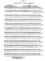
- Figure 1 shows the nucleotide (SEQ ID NO:1) and amino acid (SEQ ID NO:2) sequences of native KGF (the nucleotides encoding the mature form of native KGF is depicted by bases 201 to 684 of SEQ ID NO:1 and the mature form of KGF is depicted by amino acid residues 32 to 194 of SEQ ID NO:2).
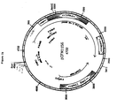
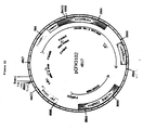
- Figures 2A, 2B and 2C show the plasmid maps of pCFM1156, pCFM1656 and pCFM3102, respectively.
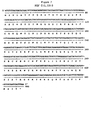
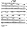
- Figure 3 shows the nucleotide (SEQ ID NO:3) and amino acid (SEQ ID NO:4) sequences of the construct RSH-KGF.
- Figure 4 shows the nucleotide (SEQ ID NO:5) and amino acid (SEQ ID NO:6) sequences of the construct contained in plasmid KGF.
- Figure 5 shows the chemically synthesized OLIGOs (OLIGO#6 through OLIGO#11; SEQ ID NO:12-17, respectively) used to substitute the DNA sequence between a KpnI site and an EcoRI site for a KpnI site (from amino acid positions 46 to 85 of SEQ ID No:6) in the construct contained plasmid KGF to produce the construct in plasmid KGF(dsd).
- Figure 6 shows the chemically synthesized OLIGOs (OLIGO#12 through OLIGO#24; SEQ ID NO:18-30, respectively) used to construct KGF(codon optimized).
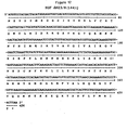
- Figure 7 shows the nucleotide (SEQ ID NO:31) and amino acid sequences (SEQ ID NO:32) of C(1.15)S, a KGF analog having substitutions of serine for cysteine at amino acid positions 1 and 15 of native KGF.
- Figure 8 shows the nucleotide (SEQ ID NO:33) and amino acid sequences (SEQ ID NO:34) of C(1,15)S/R(144)E, a KGF analog having substitutions of serine for cysteine at amino acid positions 1 and 15 and a substitution of glutamic acid for arginine at amino acid position 144 of native KGF.
- Figure 9 shows the nucleotide (SEQ ID NO:35) and amino acid (SEQ ID NO:36) sequences of C(1,15)S/R(144)Q, a KGF analog having substitutions of serine for cysteine at amino acid positions 1 and 15 and a substitution of glutamine for arginine at amino acid position 144 of native KGF.
- Figure 10 shows the nucleotide (SEQ ID NO:37) and amino acid (SEQ ID NO:38) sequences of ⁇ N15, a KGF analog having a deletion of the first 15 amino acids of the N-terminus of native KGF.
- Figure 11 shows the nucleotide (SEQ ID NO:39) and amino acid (SEQ ID NO:40) sequences of ⁇ N23, a KGF analog having a deletion of the first 23 amino acids of the N-terminus of native KGF.
- Figure 12 shows the nucleotide (SEQ ID NO:41) and amino acid (SEQ ID NO:42) sequences of ⁇ N23/R(144)Q, a KGF analog having a deletion of the first 23 amino acids of the N-terminus and a substitution of glutamine for arginine at amino acid position 144 of native KGF.
- RNA isolated from cells known to produce the polypeptide was performed. Initially, cells from a human fibroblast cell line AG1523A (obtained from Human Genetic Mutant Cell Culture Repository Institute For Medical Research, Camden, New Jersey) were disrupted with guanidium thiocyanate, followed by extraction (according to the method of Chomyzinski et al. (1987), Anal. Biochem., 172 :156). Using a standard reverse transcriptase protocol for total RNA, the KGF cDNA was generated.
- AG1523A obtained from Human Genetic Mutant Cell Culture Repository Institute For Medical Research, Camden, New Jersey
- PCR (PCR#1) amplification of the KGF gene was carried out using the KGF cDNA as template and primers OLIGO#1 and OLIGO#2 that encode DNA sequences immediately 5' and 3' of the KGF gene [model 9600 Thermocycler (Perkin-Elmer Cetus, Norwalk, CT); 28 cycles; each cycle consisting of one minute at 94°C for denaturation, two minutes at 60°C for annealing, and three minutes at 72°C for elongation].
- a small aliquot of the PCR#1 product was then used as template for a second KGF PCR (PCR#2) amplification identical to the cycle conditions described above except for a 50°C annealing temperature.
- nested PCR primers were used to create convenient restriction sites at both ends of the KGF gene.
- OLIGO#3 and OLIGO#4 were used to modify the KGF DNA product from PCR#2 to include MluI and BamHI restriction sites at the 5' and 3' ends of the gene, respectively [PCR#3; 30 cycles; each cycle consisting of one minute at 94°C for denaturation, two minutes at 60°C for annealing, and three minutes at 72°C for elongation].
- This DNA was subsequently cut with MluI and BamHI , phenol extracted and ethanol precipitated.
- the PCR DNA product was then cut with NdeI and BsmI and a 311 bp DNA fragment was isolated following electrophoresis through a 4% agarose gel.
- the third fragment used in the ligation was a 1.8 kbp DNA fragment of pCFM1156 cut with NdeI and SstI isolated following electrophoresis through a 1% agarose gel.
- T4 ligase T4 ligase
- transformation, kanamycin selection and DNA sequencing as described above a clone was picked containing the construct in Figure 4, and the plasmid designated KGF.
- the OLIGOs were phosphorylated with T4 polynucleotide kinase and then heat denatured.
- the single-stranded (ss) OLIGOs were then allowed to form a ds DNA fragment by allowing the temperature to slowly decrease to room temperature.
- T4 ligase was then used to covalently link both the internal OLIGO sticky-ends and the whole ds OLIGO fragment to the KGF plasmid cut with KpnI and EcoRI .
- the new plasmid was designated KGF(dsd).
- a completely E. coli codon-optimized KGF gene was constructed by PCR amplification of chemically synthesized OLIGOs #12 through 24.
- OLIGOs #12 through 24 were designed so that the entire DNA sequence encoding native KGF was represented by OLIGOs from either the "Watson” or the "Crick” strand and upon PCR amplification would produce the desired double-stranded DNA sequence ( Figure 6) [PCR#5, Model 9600 thermocycler (Perkin-Elmer Cetus); 21 cycles, each cycle consisting of 31 seconds at 94°C for denaturation, 31 seconds at 50°C for annealing, and 31 seconds at 73°C for elongation; following the 21 cycles the PCR was finished with a final elongation step of 7 minutes].
- Figure 6 [PCR#5, Model 9600 thermocycler (Perkin-Elmer Cetus); 21 cycles, each cycle consisting of 31 seconds at 94°C for denaturation, 31 seconds at 50°C for annealing, and 31 seconds at 73°C for elongation; following the 21 cycles the PCR was finished with a final elongation step of 7 minutes].
- PCR#5 utilized the outside primers (100 pmoles/100 ⁇ l rxn) OLIGO#12 and OLIGO#13 and 1 ⁇ l/100 ⁇ l rxn of a KGF template derived by ligation (by T4 ligase) of OLIGOs #14 through #19 (OLIGOs#15 through OLIGOs#18 were phosphorylated with T4 polynucleotide kinase) using OLIGOs#20 through OLIGOs#24 as band-aid oligos (Jayaraman et al . (1992), Biotechniques, 12 :392) for the ligation.
- the final construct was designated KGF(codon optimized).
- KGF analogs described herein are composed in part from DNA sequences found in KGF(dsd) or KGF(codon optimized), or a combination of the two.
- the sequences are further modified by the insertion into convenient restrictions sites of DNA sequences that encode the particular KGF analog amino acids made utilizing one or more of the above-described techniques for DNA fragment synthesis. Any of the analogs can be generated in their entirety by either of the above described techniques.
- E. coli codons were used where appropriate, although the presence of E. coli optimized codons in part or in toto of any of the genes where examined did not significantly increase the yield of protein that could be obtained from cultured bacterial cells.
- Figures 7 to 12 set forth by convenient example particular KGF analog nucleotide and amino acid sequence constructions: C(1,15)S ( Figure 7); C(1,15)S/R(144)E ( Figure 8); C(1,15)S/R(144)Q ( Figure 9); ⁇ N15 ( Figure 10); ⁇ N23 ( Figure 11) and ⁇ N23/R(144)Q ( Figure 12). All the KGF analog constructions described herein were DNA sequence confirmed.
- the plasmid p3102 can be derived from the plasmid pCFM1656 by making a series of site directed base changes with PCR overlapping oligo mutagenesis. Starting with the BglII site (pCFM1656 plasmid bp #180) immediately 5' to the plasmid replication promoter, P copB , and proceeding toward the plasmid replication genes, the base pair changes are as follows:
- pCFM1156, pCFM1656 and pCFM3102 are very similar to each other and contain many of the same restriction sites.
- the plasmids were chosen by convenience, and the vector DNA components can be easily exchanged for purposes of new constructs.
- the host used for all cloning was E. coli strain FM5 (ATCC: 53911) and the transformations were carried out (according to the method of Hanahan (1983), supra) or by electroelution with a Gene PulserTM transfection apparatus (BioRad Laboraties, Inc., Hercules, CA), according to the manufacturer's protocol.
- a small, freshly-cultured inoculum of the desired recombinant E. coli clone harboring the desired construct on one of the three pCFM vectors was started by transferring 0.1 mL of a frozen glycerol stock of the appropriate strain into a 2 L flask containing 500 mL of Luria broth. The culture was shaken at 30°C for 16 hours. Thereafter the culture was transferred to a 15 L fermentor containing 8 L of sterile batch medium (Tsai, et al. (1987), J. Industrial Microbiol., 2 :181-187).
- Feed batch fermentation starts with the feeding of Feed # 1 medium (Tsai, et al. (1987.), supra).
- Feed # 1 medium (Tsai, et al. (1987.), supra).
- the OD600 reached 35
- expression of the desired KGF analog was induced by rapidly raising the culture temperature to 37°C to allow the amplification of plasmid.
- the culture temperature was quickly raised to 42°C to denature the CI repressor and the addition of Feed 1 was discontinued in favor of Feed 2, the addition rate of which was initiated at 300 mL/hr.
- Feed 2 comprised 175 g/L trypticase-peptone, 87.5 g/L yeast extract, and 260 g/L glucose..
- the culture temperature was decreased to 36°C, where this temperature was then maintained for another 6 hours.
- the fermentation was then halted and the cells were harvested by centrifugation into plastic bags placed within 1 L centrifuge bottles.
- the cells were pelleted by centrifugation at 400 rpm for 60 minutes, after which the supernatants were removed and the cell paste frozen at -90°C.
- native KGF, C(1,15)S, C(1,15)S/R(144)E, C(1,15)S/R(144)Q, ⁇ N15, ⁇ N23, and ⁇ N23/R(144)Q protein were purified using the following procedure.
- Cell paste from a high cell density fermentation was suspended at 4°C in 0.2 M NaCl, 20 mM NaPO 4 , pH 7.5 as a 10-20% solution (weight per volume) using a suitable high shear mixer.
- the suspended cells were then lysed by passing the solution through a homogenizer (APV Gaulin, Inc., Everett, MA) three times.
- the outflowing homogenate was cooled to 4-8'C by using a suitable heat exchanger. Debris was then removed by centrifuging the lysate in a J-6BTM centrifuge (Beckman Instruments, Inc., Brea, CA) equipped with a JS 4.2 rotor at 4,200 rpm for 30-60 min. at 4°C. Supernatants were then carefully decanted and loaded onto a previously prepared 450 mL (5 cm x 23 cm) column of S-Sepharose Fast FlowTM resin (Pharmacia) column equilibrated with 0.2 M NaCl, 20 mM NaPO 4 , pH 7.5 at 4°C.
- the column was washed with five column volumes (2250 mL) of 0.4 M NaCl, 20 mM NaPO 4 , pH 7.5 at 4°C.
- the desired protein was eluted by washing the column with 5 L of 0.5 M NaCl, 20 mM NaPO 4 , pH 7.5. Again, 50 mL fractions were collected and the A 280 of the effluent was continuously monitored. Fractions identified by A 280 as containing eluted material were then analyzed by SDS-PAGE through 14% gels to confirm the presence of the desired polypeptide.
- fractions were collected under constant A 280 monitoring of the effluent. Those fractions containing the protein (determined by 14% SDS-PAGE) were then pooled, followed by concentration through a YM-10 membrane (10,000 molecular weight cutoff) in a 350cc stirring cell (Amicon, Inc. Mayberry, MA) to a volume of 30-40 mL.
- the concentrate was then loaded onto a previously generated 1,300 mL (4.4 cm x 85 cm) column of Superdex-75TM resin (Pharmacia) equilibrated in column buffer comprising 1X PBS (Dulbecco's Phosphate Buffered Saline, "D-PBS,” calcium and magnesium-free) or 0.15 M NaCl, 20 mM NaPO 4 , pH 7.0.
- 1X PBS Dulbecco's Phosphate Buffered Saline, "D-PBS,” calcium and magnesium-free
- 0.15 M NaCl 20 mM NaPO 4 , pH 7.0.
- the protein was eluted from the gel filtration matrix using column buffer. Thereafter, 10 mL fractions were recovered and those containing the analog (determined by 14% SDS-PAGE) were pooled.
- the protein concentration was about 5-10 mg/mL in the resultant pool. All of the above procedures were performed at 4-8°C, unless otherwise specified.
- the cell culture Upon completion of the production phase of a bacterial fermentation, the cell culture was cooled to 4-8°C and the cells were harvested by centrifugation or a similar process. On the basis of the expected yield of protein per unit weight of cell paste and the amount of purified protein required, an appropriate amount of cell paste, by weight, was suspended in a mild buffer solution, 20 mM NaPO 4 , 0.2 M NaCl, pH 7.5, weighing about five times that of the cell paste to be suspended. The cells were dispersed to a homogeneous solution using a high shear mixer. The temperature of the cell paste dispersion was maintained at 4-8°C during homogenization.
- the cells were then lysed by pressure, for example by passing the cell paste dispersion twice through an appropriately sized cell homogenizer.
- the homogenate was kept chilled at 5 ⁇ 3°C.
- a previously prepared depth filter housing (Cuno, Inc., Meriden, CT) equipped with a filter having an appropriate amount of filter surface area, equilibrated with a suitable volume of 0.2 M NaCl, 20 mM NaPO 4 , pH 7.5 was employed.
- the equilibration and clarification were performed at 5 ⁇ 3°C.
- a suitable filter aid was used to pre-coat the filter and be thoroughly mixed with the cell lysate, after which the lysate was clarified by passing the solution through the filter apparatus.
- the filter was washed with 0.2 M NaCl, 20 mM NaPO 4 , pH 7.5.
- the filtrate and any subsequent wash were collected in a chilled container of suitable capacity, all the while being maintained at less than 10°C.
- the lysate was then passed through a previously prepared column of SP-Sepharose Fast Flow containing at least 1 mL of resin per 2 g of cell paste.
- the column of SP-Sepharose Fast Flow was equilibrated with cold (5 ⁇ 3°C), 0.2 M NaCl, 20 mM NaPO 4 , pH 7.5. The temperature of the column was maintained at less than 10°C.
- the clarified lysate (5 ⁇ 3°C) was then loaded onto the ion exchange column, with the absorbance at 280 nm (A 280 ) of eluate being continuously monitored. After sample loading, the column was washed with cold 0.2 M NaCl, 20 mM NaPO 4 , pH 7.5, followed by washing with 0.3 M NaCl, 20 mM NaPO 4 , pH 7.5 at 23 ⁇ 5°C.
- an oxidation step was performed.
- an oxidizing agent e.g., cystamine dihydrochloride or another appropriate oxidizing agent, for instance, cystine, oxidized glutathione or divalent copper
- cystamine dihydrochloride or another appropriate oxidizing agent, for instance, cystine, oxidized glutathione or divalent copper
- the oxidation was conducted at 10 - 30'C for an appropriate period.
- oxidation was accomplished by adding an appropriate amount of (NH 4 ) 2 SO 4 such as 1-2 M (NH 4 ) 2 SO 4 , adjusting the pH to 7.5-9.5, and holding the temperature at 23 ⁇ 5°C for an appropriate period.
- (NH 4 ) 2 SO 4 such as 1-2 M (NH 4 ) 2 SO 4
- the pH of the solution was adjusted to between 6.5 and 9.5. If necessary, solid (NH 4 ) 2 SO 4 was added to the solution to a final concentration of 2 M. To remove particulates, the solution was passed through appropriate clarification filters.
- the filtered, oxidized product was then subjected to hydrophobic interaction chromatography (HIC).
- HIC hydrophobic interaction chromatography
- the HIC matrice was Butyl-650M ToyopearlTM resin (Tosohaas, Inc., Montgomeryville, PA).
- the protein-containing solution was loaded onto the column, which had been previously equilibrated with 2 M (NH 4 ) 2 SO 4 , 0.15 M NaCl, 20 mM NaPO 4 , pH 7.0. After sample loading, the column was washed with 2 M (NH 4 ) 2 SO 4 , 0.15 M NaCl, 20 mM NaPO 4 , pH 7.0.
- the desired protein was then eluted using a decreasing linear (NH 4 ) 2 SO 4 gradient ranging from 2-0 M developed in 0.15 M NaCl, 20 mM NaPO4, pH 7.0.
- NH 4 linear
- fractions were collected. Aliquots of each fraction were then analyzed by SDS-PAGE. Those fractions containing the desired protein were then pooled, thoroughly mixed, and the volume of the pool determined, as was the concentration of the protein therein.
- the pooled HIC protein-containing eluate was then concentrated and the elution buffer exchanged. Typically, proteins were concentrated to 5.0-10.0 mg/mL. Ultrafiltration was conducted using an ultrafiltration system equipped with a PelliconTM cassette system (Millipore, Inc., Bedford, MA) with an appropriately sized cut-off membrane
- the sample was diafiltered against an appropriate buffer.
- the retentate from the concentration step was diafiltered against 0.15 M NaCl, 20 mM NaPO 4 , pH 7.0 until the conductivity of the retentate was within 5% of the conductivity of the 0.15 M NaCl, 20 mM NaPO 4 , pH 7.0 solution.
- the concentrated diafiltered protein-containing sample was passed through a 0.1 ⁇ m PosidyneTM filter (Pall, Inc., Cortland, NY). After determining the protein concentration of the solution and on the basis of the desired concentration of the final bulk product, the solution was diluted with 0.15 M NaCl, 20 mM sodium phosphate, pH 7.0, to the desired final concentration. A final aseptic filtration through a 0.22 ⁇ m filter, was then performed as the final bulk product was transferred to a pyrogen-free container for storage (at about 5°C) for further formulation.
- This example describes the expression, isolation, and characterization of two biologically active recombinant KGF (rKGF) forms produced in a mammalian expression system.
- the human KGF gene was isolated by PCR amplification of cDNA made from normal dermal human fibroblast cells (Clonetec, Inc., Palo Alto, CA). Following the making of cDNA by reverse transcriptase, PCR was used to amplify the KGF gene. OLIGO#25 and OLIGO#26 were used to amplify the gene out of the cDNA and OLIGO#27 and OLIGO#28 were used to place Hind III and Bgl II restriction sites at the fragment ends by a second PCR amplification, as set forth in Figure 1.
- the sense primer, OLIGO#29 included an Xba I site and a consensus Kozak translation sequence (5'-CCACC-3') upstream of the start codon, ATG.
- the antisense primer, OLIGO#30 included a Sal I cloning site and an additional stop codon.
- KGF/pDSR ⁇ 2 DNA Two micrograms of KGF/pDSR ⁇ 2 DNA were then linearized with Pvu I.
- Chinese hamster ovary (CHO) cells seeded the day before at 0.8 x 10 6 cells/60 mm culture dish, were then transfected with the treated DNA using a standard calcium phosphate precipitation method (Bourdrel et al., supra). Two weeks later, individual colonies were picked and transferred into 24-well plates. The conditioned media was considered serum-free when the cells reached confluency and aliquots thereof were analyzed by Western blotting using a polyclonal rabbit antiserum reactive against E. coli-expressed human KGF.
- nitrocellulose membranes were then incubated in 100 mL of 1% normal goat serum in PBS containing VectastainTM biotinylated goat anti-rabbit IgG (secondary antibody, Vector Labs, Burlingame, CA), for 30 minutes at room temperature. After three 10 minute washes in PBS, a 30 minute room temperature incubation was performed in a 100 mL solution of 1% normal goat serum containing streptavidin and biotinylated peroxidase, prepared according to manufacturer's directions (Vector Labs).
- KGF cross-reactive material was visualized by incubation in a mixture of 60 ⁇ L of 30% (w/v) H 2 O 2 in 100 mL of PBS and 50 mg of 4-chloronapthol in 20 mL of methanol. The reaction was stopped by rinsing in water after 10 minutes.
- KGF from 3 L of serum-free conditioned medium was purified applying the medium directly to a cation exchange column (5 x 24 cm) packed with 450 mL of sulfoethyl column of SP-Sepharose Fast Flow(Pharmacia) pre-equilibrated with 20 mM sodium phosphate, pH 7.5. After washing with five column volumes of 20 mM sodium phosphate, 0.2 M NaCl, pH 7.5, rKGF was eluted using a 20 column volume linear gradient of 0.2 to 1.0 M NaCl in 20 mM sodium phosphate, pH 7.5. 50 mL fractions were collected with continuous A 280 monitoring. KGF protein was detected by analyzing aliquots of each fraction by SDS-PAGE.
- SDS-PAGE was performed on an electrophoresis system (Novex, San Diego, CA) using precast 14% Tris-glycine precast gels (according to the method of Laemmli (1970), Nature, 227 :680-685). Samples were mixed with non-reducing SDS sample buffer without heating before loading. The proteins were detected by either Coomassie blue or silver staining. Two late-eluting peaks were seen to contain protein bands corresponding to the 25-29 kDa and 17 kDa bands detected by Western blot. The fractions containing each of these peaks were separately concentrated to a volume of less than 1.0 mL and subjected to gel filtration.
- KGF-a rKGF eluted as a major symmetrical peak
- KGF-b gel filtration resulted in a protein preparation having the expected mobility.
- KGF-a and KGF-b were also analyzed. These analyses were performed on an automatic sequencer (Model 477A or 470A, Applied Biosystems, Inc., Foster City, CA) equipped with a Model 120A on-line PTH-amino acid analyzer and a Model 900A data collection system (according to the method of Lu et al . (1991), J. Biol. Chem., 266 :8102-8107).
- Edman sequence analysis of KGF-a revealed a major N-terminal sequence of X 1 -N-D-M-T-P-E-Q-M-A-T-N-V-X 2 -X 3 -S- (SEQ ID NO:51).
- N-terminal sequence analysis of KGF-b revealed an N-terminal amino acid sequence of S-Y-D-Y-M-E-G-G-D-I-R-V- (SEQ ID NO:52), indicating that it is an N-terminally truncated form of KGF that has been proteolytically cleaved at the Arg 23 -Ser 24 peptide bond.
- KGF-a and KGF-b were subjected to glycosidases (neuraminidase, O-glycanase, and/or N-glycanase), using known techniques (Sasaki et al. (1987), J. Biol. Chem., 262 :12059-12076; Takeuchi et al. (1988), J. Biol. Chem ., 263 :3657-3663; Zsebo et al . (1990), Cell, 63 :195-201). These data indicate that KGF-a contains N- and O-linked carbohydrates, although the lower molecular weight form of KGF-a probably contains only N-linked sugar. Glycosidase treatment did not cause molecular weight reduction for KGF-b, indicating that the molecule is unglycosylated.
- glycosidases neurosidases
- Each KGF analog was diluted and assayed for biological activity by measuring the [ 3 H]-thymidine uptake of Balb/MK cells (according to the method of Rubin et al. (1989), supra).
- the samples were first diluted in a bioassay medium consisting of 50% customer-made Eagle's MEM, 50% customer-made F12, 5 ⁇ g/mL transferrin, 5 ng/mL sodium selenite, 0.0005% HSA and 0.005% Tween 20.
- KGF samples were then added into Falcon primeria 96-well plates seeded with Balb/MK cells. Incorporation of [ 3 H]-Thymidine during DNA synthesis was measured and converted to input native KGF concentration by comparison to a native KGF standard curve. Each of the tested analogs exhibited mitogenic activity.
- KGF receptor membrane preparations prepared from Balb/MK mouse epidermal keratinocytes (by the procedure described by Massague (19932), J. Biol. Chem. , 258 :13614-13620).
- various forms of KGF were diluted with 50 mM Tris-HCl, pH 7.5, containing 0.2% bovine serum albumin so as to range in concentration from 0.8 ng to 100 ng per 50 ⁇ L. They were individually incubated with the membrane preparation (75 ng/mL) and 125 I-labeled E. coli -derived KGF (1.5 ng).
- Receptor binding and competition experiments were performed at 4°C for 16 hr., after which time samples were taken, centrifuged, and washed twice with the above diluent buffer to remove unbound and non-specifically bound, labeled KGF. Samples were then counted for the remaining radioactivity. Competition curves for receptor binding between KGF samples and labeled KGF were constructed by plotting percent uncompetition versus concentrations of each KGF sample. Radioreceptor assay uncompetition experiments indicated that E. coli -derived KGF, KGF-a, and KGF-b have similar receptor binding activity.
Landscapes
- Chemical & Material Sciences (AREA)
- Health & Medical Sciences (AREA)
- Life Sciences & Earth Sciences (AREA)
- Organic Chemistry (AREA)
- Medicinal Chemistry (AREA)
- Toxicology (AREA)
- Biochemistry (AREA)
- Biophysics (AREA)
- General Health & Medical Sciences (AREA)
- Genetics & Genomics (AREA)
- Zoology (AREA)
- Molecular Biology (AREA)
- Proteomics, Peptides & Aminoacids (AREA)
- Gastroenterology & Hepatology (AREA)
- Preparation Of Compounds By Using Micro-Organisms (AREA)
- Peptides Or Proteins (AREA)
- Steroid Compounds (AREA)
- Saccharide Compounds (AREA)
- Investigating Or Analysing Biological Materials (AREA)
- Medicines That Contain Protein Lipid Enzymes And Other Medicines (AREA)
- Medicines Containing Material From Animals Or Micro-Organisms (AREA)
- Pharmaceuticals Containing Other Organic And Inorganic Compounds (AREA)
Applications Claiming Priority (5)
| Application Number | Priority Date | Filing Date | Title |
|---|---|---|---|
| US32333994A | 1994-10-13 | 1994-10-13 | |
| US08/487,830 US6008328A (en) | 1994-10-13 | 1995-06-07 | Method for purifying keratinocyte growth factors |
| US487830 | 1995-06-07 | ||
| EP95936309A EP0785949B1 (fr) | 1994-10-13 | 1995-10-12 | Procede pour purifier des facteurs de croissance des keratinocytes |
| US323339 | 1999-06-01 |
Related Parent Applications (2)
| Application Number | Title | Priority Date | Filing Date |
|---|---|---|---|
| EP95936309A Division EP0785949B1 (fr) | 1994-10-13 | 1995-10-12 | Procede pour purifier des facteurs de croissance des keratinocytes |
| EP95936309.4 Division | 1995-10-12 |
Publications (3)
| Publication Number | Publication Date |
|---|---|
| EP1334981A2 true EP1334981A2 (fr) | 2003-08-13 |
| EP1334981A3 EP1334981A3 (fr) | 2004-05-06 |
| EP1334981B1 EP1334981B1 (fr) | 2011-01-26 |
Family
ID=26983902
Family Applications (2)
| Application Number | Title | Priority Date | Filing Date |
|---|---|---|---|
| EP02028624A Expired - Lifetime EP1334981B1 (fr) | 1994-10-13 | 1995-10-12 | Méthode de purification de facteurs de croissance des kératinocytes |
| EP95936309A Expired - Lifetime EP0785949B1 (fr) | 1994-10-13 | 1995-10-12 | Procede pour purifier des facteurs de croissance des keratinocytes |
Family Applications After (1)
| Application Number | Title | Priority Date | Filing Date |
|---|---|---|---|
| EP95936309A Expired - Lifetime EP0785949B1 (fr) | 1994-10-13 | 1995-10-12 | Procede pour purifier des facteurs de croissance des keratinocytes |
Country Status (18)
| Country | Link |
|---|---|
| EP (2) | EP1334981B1 (fr) |
| JP (1) | JP3877226B2 (fr) |
| CN (1) | CN1161380C (fr) |
| AT (1) | ATE239038T1 (fr) |
| CA (1) | CA2201762C (fr) |
| CZ (1) | CZ98297A3 (fr) |
| DE (2) | DE69530599T2 (fr) |
| DK (1) | DK0785949T3 (fr) |
| ES (1) | ES2197926T3 (fr) |
| HU (1) | HU220584B1 (fr) |
| IL (3) | IL115605A (fr) |
| LU (1) | LU91235I2 (fr) |
| NL (1) | NL300229I2 (fr) |
| NO (1) | NO971567L (fr) |
| PT (1) | PT785949E (fr) |
| SI (1) | SI0785949T1 (fr) |
| SK (1) | SK43097A3 (fr) |
| WO (1) | WO1996011952A1 (fr) |
Cited By (1)
| Publication number | Priority date | Publication date | Assignee | Title |
|---|---|---|---|---|
| EP1429799A4 (fr) * | 2001-08-21 | 2005-12-21 | Chiron Corp | Compositions a base de polypeptide kgf |
Families Citing this family (12)
| Publication number | Priority date | Publication date | Assignee | Title |
|---|---|---|---|---|
| US7232667B2 (en) | 1995-02-14 | 2007-06-19 | Human Genome Sciences, Inc. | Keratinocyte growth factor-2 polynucleotides |
| US6693077B1 (en) | 1995-02-14 | 2004-02-17 | Human Genome Sciences, Inc. | Keratinocyte growth factor-2 |
| US6077692A (en) | 1995-02-14 | 2000-06-20 | Human Genome Sciences, Inc. | Keratinocyte growth factor-2 |
| US6743422B1 (en) | 1996-10-15 | 2004-06-01 | Amgen, Inc. | Keratinocyte growth factor-2 products |
| WO1999032135A1 (fr) | 1997-12-22 | 1999-07-01 | Human Genome Sciences, Inc. | Formulations du facteur-2 de croissance des keratinocytes |
| US6869927B1 (en) | 1997-12-22 | 2005-03-22 | Human Genome Sciences, Inc. | Keratinocyte growth factor-2 formulations |
| CN1491231A (zh) * | 2001-02-06 | 2004-04-21 | Ĭ��ר������˾ | 免疫原性减弱的经修饰角质形成细胞生长因子(kgf) |
| CN102242124B (zh) * | 2010-05-10 | 2013-05-22 | 齐鲁制药有限公司 | 改造的角化细胞生长因子基因及其在酵母中的表达 |
| CN102675449B (zh) * | 2011-03-17 | 2016-06-08 | 重庆富进生物医药有限公司 | 缺失型人角质细胞生长因子-ⅰ二硫键变构体及其用途 |
| CN109402130A (zh) * | 2018-11-23 | 2019-03-01 | 成都中医药大学附属医院 | 一种重组人角质细胞生长因子-1及其制备方法和用途 |
| CN110183529B (zh) * | 2019-05-23 | 2023-06-02 | 重庆派金生物科技有限公司 | 一种缺失型人角质细胞生长因子-1的重组制备方法 |
| KR20230127721A (ko) | 2022-02-25 | 2023-09-01 | 한국해양과학기술원 | 온도안정성을 향상시킨 fgf7 폴리펩타이드 및 그 용도 |
Family Cites Families (7)
| Publication number | Priority date | Publication date | Assignee | Title |
|---|---|---|---|---|
| RU2250213C2 (ru) * | 1989-01-31 | 2005-04-20 | Джеффри С. РУБИН | Выделенный белок фактора роста кератиноцита (kgf), рекомбинантная молекула днк, вектор, способ конструирования клетки-хозяина, способ получения белка, фармацевтическая композиция |
| IE914393A1 (en) * | 1990-12-18 | 1992-07-01 | Amgen Inc | Analogs of acidic fibroblast growth factor having enhanced¹stability and biological activity |
| CA2166278A1 (fr) * | 1993-06-29 | 1995-01-12 | Denis J. Gospodarowicz | Facteur de croissance tronque des keratinocytes, possedant une plus grande activite biologique |
| WO1995008630A1 (fr) * | 1993-09-24 | 1995-03-30 | American Cyanamid Company | Analogues structurels a boucle de surface de facteurs de croissance du fibroblast |
| SI0785950T1 (en) * | 1994-10-13 | 2003-08-31 | Amgen Inc. | Keratinocyte growth factor analogs |
| CA2201944C (fr) * | 1994-10-13 | 2002-12-31 | Tsutomu Arakawa | Analogues du facteur de croissance des keratinocytes ayant une stabilite de temperature amelioree |
| WO1996011949A2 (fr) * | 1994-10-13 | 1996-04-25 | Amgen Inc. | Analogues du facteur de croissance de keratinocytes |
-
1995
- 1995-10-12 SI SI9530657T patent/SI0785949T1/xx unknown
- 1995-10-12 PT PT95936309T patent/PT785949E/pt unknown
- 1995-10-12 ES ES95936309T patent/ES2197926T3/es not_active Expired - Lifetime
- 1995-10-12 JP JP51337696A patent/JP3877226B2/ja not_active Expired - Lifetime
- 1995-10-12 DE DE69530599T patent/DE69530599T2/de not_active Expired - Lifetime
- 1995-10-12 CN CNB951967959A patent/CN1161380C/zh not_active Expired - Lifetime
- 1995-10-12 EP EP02028624A patent/EP1334981B1/fr not_active Expired - Lifetime
- 1995-10-12 DK DK95936309T patent/DK0785949T3/da active
- 1995-10-12 CZ CZ97982A patent/CZ98297A3/cs unknown
- 1995-10-12 IL IL11560595A patent/IL115605A/xx not_active IP Right Cessation
- 1995-10-12 DE DE1995630599 patent/DE122006000019I1/de active Pending
- 1995-10-12 IL IL14757595A patent/IL147575A/en not_active IP Right Cessation
- 1995-10-12 AT AT95936309T patent/ATE239038T1/de active
- 1995-10-12 SK SK430-97A patent/SK43097A3/sk unknown
- 1995-10-12 WO PCT/US1995/013099 patent/WO1996011952A1/fr not_active Ceased
- 1995-10-12 HU HU9701514A patent/HU220584B1/hu unknown
- 1995-10-12 CA CA002201762A patent/CA2201762C/fr not_active Expired - Lifetime
- 1995-10-12 EP EP95936309A patent/EP0785949B1/fr not_active Expired - Lifetime
-
1997
- 1997-04-04 NO NO971567A patent/NO971567L/no unknown
-
2002
- 2002-01-10 IL IL14757502A patent/IL147575A0/xx active IP Right Grant
-
2006
- 2006-04-11 NL NL300229C patent/NL300229I2/nl unknown
- 2006-04-12 LU LU91235C patent/LU91235I2/fr unknown
Non-Patent Citations (19)
| Title |
|---|
| ABRAHAM ET AL., EMBO J., vol. 5, 1986, pages 2523 - 2528 |
| DELLI-BOVI ET AL., CELL, vol. 50, 1987, pages 729 - 737 |
| DICKSON; PETERS, NATURE, vol. 326, 1987, pages 833 |
| FINCH ET AL., SCIENCE, vol. 24, 1989, pages 752 - 755 |
| HABAZZETTL ET AL., NATURE, vol. 359, 1992, pages 855 - 858 |
| JAYARAMAN ET AL., BIOTECHNIQUES, vol. 12, 1992, pages 392 |
| JAYE ET AL., SCIENCE, vol. 233, 1986, pages 541 - 545 |
| MARICS ET AL., ONCOGENE, vol. 4, 1989, pages 335 - 340 |
| MASSAGUE, J. BIOL. CHEM., vol. 258, 1983, pages 13614 - 13620 |
| RON ET AL., THE JOURNAL OF BIOLOGICAL CHEMISTRY, vol. 268, no. 4, 1993, pages 2984 - 2988 |
| RUBIN ET AL., PROC. NATL. ACAD. SCI. USA, vol. 86, 1989, pages 802 - 806 |
| SASAKI ET AL., J. BIOL. CHEM., vol. 262, 1987, pages 12059 - 12076 |
| SUZUKI, M. ET AL., FEBS LETT., vol. 328, 1993, pages 17 - 20 |
| TAKEUCHI ET AL., J. BIOL. CHEM., vol. 263, 1988, pages 3657 - 3663 |
| TSAI ET AL., J. INDUSTRIAL MICROBIOL., vol. 2, 1987, pages 181 - 187 |
| YAN ET AL., IN VITRO CELL. DEV. BIOL., vol. 27A, 1991, pages 437 - 438 |
| YOSHIDA ET AL., PROC. NATL. ACAD. SCI. USA, vol. 84, 1987, pages 7305 - 7309 |
| ZHAN ET AL., MOL. CELL. BIOL., vol. 8, 1988, pages 3487 - 3495 |
| ZSEBO ET AL., CELL, vol. 6, no. 3, 1990, pages 195 - 201 |
Cited By (2)
| Publication number | Priority date | Publication date | Assignee | Title |
|---|---|---|---|---|
| EP1429799A4 (fr) * | 2001-08-21 | 2005-12-21 | Chiron Corp | Compositions a base de polypeptide kgf |
| US7265089B2 (en) | 2001-08-21 | 2007-09-04 | Chiron Corporation | KGF polypeptide compositions |
Also Published As
| Publication number | Publication date |
|---|---|
| EP0785949B1 (fr) | 2003-05-02 |
| ES2197926T3 (es) | 2004-01-16 |
| DK0785949T3 (da) | 2003-08-18 |
| EP0785949A1 (fr) | 1997-07-30 |
| DE122006000019I1 (de) | 2006-06-29 |
| WO1996011952A1 (fr) | 1996-04-25 |
| CZ98297A3 (cs) | 1998-08-12 |
| CA2201762C (fr) | 2001-12-18 |
| SI0785949T1 (en) | 2003-08-31 |
| IL115605A0 (en) | 1996-01-19 |
| IL147575A (en) | 2004-05-12 |
| AU3830695A (en) | 1996-05-06 |
| CA2201762A1 (fr) | 1996-04-25 |
| PT785949E (pt) | 2003-09-30 |
| MX9702481A (es) | 1997-07-31 |
| IL147575A0 (en) | 2002-08-14 |
| AU709362B2 (en) | 1999-08-26 |
| DE69530599T2 (de) | 2003-11-13 |
| EP1334981A3 (fr) | 2004-05-06 |
| ATE239038T1 (de) | 2003-05-15 |
| HU220584B1 (hu) | 2002-03-28 |
| CN1169734A (zh) | 1998-01-07 |
| NO971567D0 (no) | 1997-04-04 |
| NL300229I1 (nl) | 2006-06-01 |
| LU91235I2 (fr) | 2006-11-22 |
| JP3877226B2 (ja) | 2007-02-07 |
| IL115605A (en) | 2003-06-24 |
| CN1161380C (zh) | 2004-08-11 |
| NL300229I2 (nl) | 2006-09-01 |
| HUT76879A (en) | 1997-12-29 |
| JPH10507452A (ja) | 1998-07-21 |
| EP1334981B1 (fr) | 2011-01-26 |
| NO971567L (no) | 1997-06-12 |
| DE69530599D1 (de) | 2003-06-05 |
| SK43097A3 (en) | 1998-10-07 |
Similar Documents
| Publication | Publication Date | Title |
|---|---|---|
| KR100205078B1 (ko) | 염기성 섬유 아세포 성장인자 및 이의 생산방법 | |
| CA2202075C (fr) | Analogues du facteur de croissance de keratinocytes, acides nucleiques encodant ces analogues, procede de fabrication et methodes d'utilisations | |
| AU638402B2 (en) | Analogs of fibroblast growth factor | |
| US7361738B2 (en) | Megakaryocyte stimulating factors | |
| US5726152A (en) | Vascular endothelial cell growth factor II | |
| EP0785949B1 (fr) | Procede pour purifier des facteurs de croissance des keratinocytes | |
| EP0275204A2 (fr) | Préparation du facteur de croissance de fibroblaste | |
| CA2202390C (fr) | Analogues du facteur de croissance des keratinocytes | |
| JP2002509691A (ja) | 生物学的活性が改変された組換えタンパク質多量体の製造及び使用 | |
| US6008328A (en) | Method for purifying keratinocyte growth factors | |
| AU709362C (en) | Method for purifying keratinocyte growth factors | |
| MXPA97002481A (en) | Method for the purification of growth factors of queratinoci | |
| AU745815B2 (en) | Analogs of keratinocyte growth factor, nucleic acids encoding such analogs, processes of making and methods of using | |
| AU5068502A (en) | Analogs of keratinocyte growth factor |
Legal Events
| Date | Code | Title | Description |
|---|---|---|---|
| PUAI | Public reference made under article 153(3) epc to a published international application that has entered the european phase |
Free format text: ORIGINAL CODE: 0009012 |
|
| 17P | Request for examination filed |
Effective date: 20021220 |
|
| AC | Divisional application: reference to earlier application |
Ref document number: 0785949 Country of ref document: EP Kind code of ref document: P |
|
| AK | Designated contracting states |
Designated state(s): AT BE CH DE DK ES FR GB GR IE IT LI LU MC NL PT SE |
|
| AX | Request for extension of the european patent |
Extension state: LT LV SI |
|
| PUAL | Search report despatched |
Free format text: ORIGINAL CODE: 0009013 |
|
| AK | Designated contracting states |
Kind code of ref document: A3 Designated state(s): AT BE CH DE DK ES FR GB GR IE IT LI LU MC NL PT SE |
|
| AX | Request for extension of the european patent |
Extension state: LT LV SI |
|
| AKX | Designation fees paid |
Designated state(s): AT BE CH DE DK ES FR GB GR IE IT LI LU MC NL PT SE |
|
| AXX | Extension fees paid |
Extension state: LT Payment date: 20021220 Extension state: LV Payment date: 20021220 Extension state: SI Payment date: 20021220 |
|
| RAP1 | Party data changed (applicant data changed or rights of an application transferred) |
Owner name: BIOVITRUM AB (PUBL) |
|
| GRAP | Despatch of communication of intention to grant a patent |
Free format text: ORIGINAL CODE: EPIDOSNIGR1 |
|
| GRAJ | Information related to disapproval of communication of intention to grant by the applicant or resumption of examination proceedings by the epo deleted |
Free format text: ORIGINAL CODE: EPIDOSDIGR1 |
|
| GRAP | Despatch of communication of intention to grant a patent |
Free format text: ORIGINAL CODE: EPIDOSNIGR1 |
|
| RAP1 | Party data changed (applicant data changed or rights of an application transferred) |
Owner name: SWEDISH ORPHAN BIOVITRUM AB (PUBL) |
|
| GRAS | Grant fee paid |
Free format text: ORIGINAL CODE: EPIDOSNIGR3 |
|
| GRAA | (expected) grant |
Free format text: ORIGINAL CODE: 0009210 |
|
| AC | Divisional application: reference to earlier application |
Ref document number: 0785949 Country of ref document: EP Kind code of ref document: P |
|
| AK | Designated contracting states |
Kind code of ref document: B1 Designated state(s): AT BE CH DE DK ES FR GB GR IE IT LI LU MC NL PT SE |
|
| AX | Request for extension of the european patent |
Extension state: LT LV SI |
|
| REG | Reference to a national code |
Ref country code: GB Ref legal event code: FG4D |
|
| REG | Reference to a national code |
Ref country code: CH Ref legal event code: EP |
|
| REG | Reference to a national code |
Ref country code: IE Ref legal event code: FG4D |
|
| REF | Corresponds to: |
Ref document number: 69536133 Country of ref document: DE Date of ref document: 20110310 Kind code of ref document: P |
|
| REG | Reference to a national code |
Ref country code: DE Ref legal event code: R096 Ref document number: 69536133 Country of ref document: DE Effective date: 20110310 |
|
| REG | Reference to a national code |
Ref country code: NL Ref legal event code: VDEP Effective date: 20110126 |
|
| LTIE | Lt: invalidation of european patent or patent extension |
Effective date: 20110126 |
|
| PG25 | Lapsed in a contracting state [announced via postgrant information from national office to epo] |
Ref country code: SE Free format text: LAPSE BECAUSE OF FAILURE TO SUBMIT A TRANSLATION OF THE DESCRIPTION OR TO PAY THE FEE WITHIN THE PRESCRIBED TIME-LIMIT Effective date: 20110126 Ref country code: GR Free format text: LAPSE BECAUSE OF FAILURE TO SUBMIT A TRANSLATION OF THE DESCRIPTION OR TO PAY THE FEE WITHIN THE PRESCRIBED TIME-LIMIT Effective date: 20110427 Ref country code: PT Free format text: LAPSE BECAUSE OF FAILURE TO SUBMIT A TRANSLATION OF THE DESCRIPTION OR TO PAY THE FEE WITHIN THE PRESCRIBED TIME-LIMIT Effective date: 20110526 Ref country code: ES Free format text: LAPSE BECAUSE OF FAILURE TO SUBMIT A TRANSLATION OF THE DESCRIPTION OR TO PAY THE FEE WITHIN THE PRESCRIBED TIME-LIMIT Effective date: 20110507 |
|
| PG25 | Lapsed in a contracting state [announced via postgrant information from national office to epo] |
Ref country code: BE Free format text: LAPSE BECAUSE OF FAILURE TO SUBMIT A TRANSLATION OF THE DESCRIPTION OR TO PAY THE FEE WITHIN THE PRESCRIBED TIME-LIMIT Effective date: 20110126 Ref country code: AT Free format text: LAPSE BECAUSE OF FAILURE TO SUBMIT A TRANSLATION OF THE DESCRIPTION OR TO PAY THE FEE WITHIN THE PRESCRIBED TIME-LIMIT Effective date: 20110126 Ref country code: NL Free format text: LAPSE BECAUSE OF FAILURE TO SUBMIT A TRANSLATION OF THE DESCRIPTION OR TO PAY THE FEE WITHIN THE PRESCRIBED TIME-LIMIT Effective date: 20110126 |
|
| PG25 | Lapsed in a contracting state [announced via postgrant information from national office to epo] |
Ref country code: DK Free format text: LAPSE BECAUSE OF FAILURE TO SUBMIT A TRANSLATION OF THE DESCRIPTION OR TO PAY THE FEE WITHIN THE PRESCRIBED TIME-LIMIT Effective date: 20110126 |
|
| PLBE | No opposition filed within time limit |
Free format text: ORIGINAL CODE: 0009261 |
|
| STAA | Information on the status of an ep patent application or granted ep patent |
Free format text: STATUS: NO OPPOSITION FILED WITHIN TIME LIMIT |
|
| 26N | No opposition filed |
Effective date: 20111027 |
|
| REG | Reference to a national code |
Ref country code: DE Ref legal event code: R097 Ref document number: 69536133 Country of ref document: DE Effective date: 20111027 |
|
| PG25 | Lapsed in a contracting state [announced via postgrant information from national office to epo] |
Ref country code: IT Free format text: LAPSE BECAUSE OF FAILURE TO SUBMIT A TRANSLATION OF THE DESCRIPTION OR TO PAY THE FEE WITHIN THE PRESCRIBED TIME-LIMIT Effective date: 20110126 Ref country code: MC Free format text: LAPSE BECAUSE OF NON-PAYMENT OF DUE FEES Effective date: 20111031 |
|
| REG | Reference to a national code |
Ref country code: CH Ref legal event code: PL |
|
| GBPC | Gb: european patent ceased through non-payment of renewal fee |
Effective date: 20111012 |
|
| REG | Reference to a national code |
Ref country code: FR Ref legal event code: ST Effective date: 20120629 |
|
| PG25 | Lapsed in a contracting state [announced via postgrant information from national office to epo] |
Ref country code: CH Free format text: LAPSE BECAUSE OF NON-PAYMENT OF DUE FEES Effective date: 20111031 Ref country code: DE Free format text: LAPSE BECAUSE OF NON-PAYMENT OF DUE FEES Effective date: 20120501 Ref country code: LI Free format text: LAPSE BECAUSE OF NON-PAYMENT OF DUE FEES Effective date: 20111031 |
|
| REG | Reference to a national code |
Ref country code: IE Ref legal event code: MM4A |
|
| REG | Reference to a national code |
Ref country code: DE Ref legal event code: R119 Ref document number: 69536133 Country of ref document: DE Effective date: 20120501 |
|
| PG25 | Lapsed in a contracting state [announced via postgrant information from national office to epo] |
Ref country code: FR Free format text: LAPSE BECAUSE OF NON-PAYMENT OF DUE FEES Effective date: 20111102 Ref country code: GB Free format text: LAPSE BECAUSE OF NON-PAYMENT OF DUE FEES Effective date: 20111012 |
|
| PG25 | Lapsed in a contracting state [announced via postgrant information from national office to epo] |
Ref country code: IE Free format text: LAPSE BECAUSE OF NON-PAYMENT OF DUE FEES Effective date: 20111012 |
|
| PG25 | Lapsed in a contracting state [announced via postgrant information from national office to epo] |
Ref country code: LU Free format text: LAPSE BECAUSE OF NON-PAYMENT OF DUE FEES Effective date: 20111012 |