EP1390516B1 - Novel expression vectors and uses thereof - Google Patents
Novel expression vectors and uses thereof Download PDFInfo
- Publication number
- EP1390516B1 EP1390516B1 EP02722308A EP02722308A EP1390516B1 EP 1390516 B1 EP1390516 B1 EP 1390516B1 EP 02722308 A EP02722308 A EP 02722308A EP 02722308 A EP02722308 A EP 02722308A EP 1390516 B1 EP1390516 B1 EP 1390516B1
- Authority
- EP
- European Patent Office
- Prior art keywords
- vector
- protein
- dna
- dna sequence
- cells
- Prior art date
- Legal status (The legal status is an assumption and is not a legal conclusion. Google has not performed a legal analysis and makes no representation as to the accuracy of the status listed.)
- Expired - Lifetime
Links
- 239000013604 expression vector Substances 0.000 title claims description 15
- 239000013598 vector Substances 0.000 claims description 425
- 108090000623 proteins and genes Proteins 0.000 claims description 391
- 210000004027 cell Anatomy 0.000 claims description 296
- 102000004169 proteins and genes Human genes 0.000 claims description 239
- 108091028043 Nucleic acid sequence Proteins 0.000 claims description 169
- 230000014509 gene expression Effects 0.000 claims description 151
- 238000000034 method Methods 0.000 claims description 70
- 230000027455 binding Effects 0.000 claims description 63
- 101710125507 Integrase/recombinase Proteins 0.000 claims description 56
- 238000004873 anchoring Methods 0.000 claims description 56
- 230000010076 replication Effects 0.000 claims description 43
- 229960005486 vaccine Drugs 0.000 claims description 42
- 241000388186 Deltapapillomavirus 4 Species 0.000 claims description 38
- 241000725303 Human immunodeficiency virus Species 0.000 claims description 37
- 230000004568 DNA-binding Effects 0.000 claims description 35
- 108091032973 (ribonucleotides)n+m Proteins 0.000 claims description 30
- 108091092562 ribozyme Proteins 0.000 claims description 29
- 108090000994 Catalytic RNA Proteins 0.000 claims description 28
- 102000053642 Catalytic RNA Human genes 0.000 claims description 28
- 230000001580 bacterial effect Effects 0.000 claims description 27
- 239000000203 mixture Substances 0.000 claims description 27
- 208000037265 diseases, disorders, signs and symptoms Diseases 0.000 claims description 26
- 239000003814 drug Substances 0.000 claims description 25
- 201000010099 disease Diseases 0.000 claims description 24
- 230000028993 immune response Effects 0.000 claims description 24
- 108010041986 DNA Vaccines Proteins 0.000 claims description 23
- 229940021995 DNA vaccine Drugs 0.000 claims description 23
- 108090000765 processed proteins & peptides Proteins 0.000 claims description 23
- 241000700605 Viruses Species 0.000 claims description 22
- 238000004519 manufacturing process Methods 0.000 claims description 22
- 108091006104 gene-regulatory proteins Proteins 0.000 claims description 18
- 102000034356 gene-regulatory proteins Human genes 0.000 claims description 18
- 238000002360 preparation method Methods 0.000 claims description 18
- 101150020927 Aire gene Proteins 0.000 claims description 16
- 230000000692 anti-sense effect Effects 0.000 claims description 16
- 102000037865 fusion proteins Human genes 0.000 claims description 15
- 241000894006 Bacteria Species 0.000 claims description 14
- 241000588724 Escherichia coli Species 0.000 claims description 14
- 108020001507 fusion proteins Proteins 0.000 claims description 14
- 230000003612 virological effect Effects 0.000 claims description 14
- 208000015181 infectious disease Diseases 0.000 claims description 13
- 230000000394 mitotic effect Effects 0.000 claims description 13
- 241000701822 Bovine papillomavirus Species 0.000 claims description 12
- 102000004127 Cytokines Human genes 0.000 claims description 12
- 108090000695 Cytokines Proteins 0.000 claims description 12
- 101000928549 Homo sapiens Autoimmune regulator Proteins 0.000 claims description 12
- 210000004962 mammalian cell Anatomy 0.000 claims description 12
- 230000001225 therapeutic effect Effects 0.000 claims description 12
- 102100036465 Autoimmune regulator Human genes 0.000 claims description 11
- 206010028980 Neoplasm Diseases 0.000 claims description 11
- 108010008281 Recombinant Fusion Proteins Proteins 0.000 claims description 11
- 229940079593 drug Drugs 0.000 claims description 11
- 239000008194 pharmaceutical composition Substances 0.000 claims description 11
- 102000007056 Recombinant Fusion Proteins Human genes 0.000 claims description 10
- 239000003937 drug carrier Substances 0.000 claims description 10
- 230000002163 immunogen Effects 0.000 claims description 10
- 108010077544 Chromatin Proteins 0.000 claims description 9
- 101710172711 Structural protein Proteins 0.000 claims description 9
- 210000003483 chromatin Anatomy 0.000 claims description 9
- 230000001939 inductive effect Effects 0.000 claims description 9
- 244000052769 pathogen Species 0.000 claims description 9
- 208000035473 Communicable disease Diseases 0.000 claims description 8
- 201000011510 cancer Diseases 0.000 claims description 8
- 230000009395 genetic defect Effects 0.000 claims description 8
- 238000001727 in vivo Methods 0.000 claims description 8
- 239000012678 infectious agent Substances 0.000 claims description 8
- 230000001717 pathogenic effect Effects 0.000 claims description 8
- 229940124597 therapeutic agent Drugs 0.000 claims description 8
- 238000011282 treatment Methods 0.000 claims description 8
- 208000031212 Autoimmune polyendocrinopathy Diseases 0.000 claims description 7
- 238000000302 molecular modelling Methods 0.000 claims description 7
- 239000003795 chemical substances by application Substances 0.000 claims description 6
- 230000002458 infectious effect Effects 0.000 claims description 5
- 230000002401 inhibitory effect Effects 0.000 claims description 5
- 230000001131 transforming effect Effects 0.000 claims description 5
- 241000709661 Enterovirus Species 0.000 claims description 4
- 241000700584 Simplexvirus Species 0.000 claims description 4
- 230000002950 deficient Effects 0.000 claims description 4
- 244000053095 fungal pathogen Species 0.000 claims description 4
- 241000222120 Candida <Saccharomycetales> Species 0.000 claims description 3
- 241000606153 Chlamydia trachomatis Species 0.000 claims description 3
- 241000711549 Hepacivirus C Species 0.000 claims description 3
- 108010050904 Interferons Proteins 0.000 claims description 3
- 102000014150 Interferons Human genes 0.000 claims description 3
- 102000013462 Interleukin-12 Human genes 0.000 claims description 3
- 108010065805 Interleukin-12 Proteins 0.000 claims description 3
- 241000187479 Mycobacterium tuberculosis Species 0.000 claims description 3
- 208000001572 Mycoplasma Pneumonia Diseases 0.000 claims description 3
- 201000008235 Mycoplasma pneumoniae pneumonia Diseases 0.000 claims description 3
- 108700020796 Oncogene Proteins 0.000 claims description 3
- 241000607142 Salmonella Species 0.000 claims description 3
- 229940038705 chlamydia trachomatis Drugs 0.000 claims description 3
- 229940079322 interferon Drugs 0.000 claims description 3
- 230000035800 maturation Effects 0.000 claims description 3
- 210000001236 prokaryotic cell Anatomy 0.000 claims description 3
- 230000012121 regulation of immune response Effects 0.000 claims description 3
- 102000015696 Interleukins Human genes 0.000 claims description 2
- 108010063738 Interleukins Proteins 0.000 claims description 2
- 206010035673 Pneumonia chlamydial Diseases 0.000 claims description 2
- 230000006472 autoimmune response Effects 0.000 claims description 2
- 230000003832 immune regulation Effects 0.000 claims description 2
- 230000003449 preventive effect Effects 0.000 claims description 2
- 208000008128 pulmonary tuberculosis Diseases 0.000 claims description 2
- 239000013612 plasmid Substances 0.000 description 128
- 108020004414 DNA Proteins 0.000 description 95
- 241000699670 Mus sp. Species 0.000 description 49
- 239000000427 antigen Substances 0.000 description 49
- 108091007433 antigens Proteins 0.000 description 45
- 102000036639 antigens Human genes 0.000 description 45
- FAPWRFPIFSIZLT-UHFFFAOYSA-M Sodium chloride Chemical compound [Na+].[Cl-] FAPWRFPIFSIZLT-UHFFFAOYSA-M 0.000 description 33
- 238000001890 transfection Methods 0.000 description 32
- 108091026890 Coding region Proteins 0.000 description 30
- 241000282414 Homo sapiens Species 0.000 description 29
- 108010031111 EBV-encoded nuclear antigen 1 Proteins 0.000 description 28
- 101710192141 Protein Nef Proteins 0.000 description 28
- XLYOFNOQVPJJNP-UHFFFAOYSA-N water Substances O XLYOFNOQVPJJNP-UHFFFAOYSA-N 0.000 description 28
- 238000004458 analytical method Methods 0.000 description 27
- 238000012423 maintenance Methods 0.000 description 27
- 108010048367 enhanced green fluorescent protein Proteins 0.000 description 25
- 239000002609 medium Substances 0.000 description 25
- 239000012634 fragment Substances 0.000 description 24
- 101710177291 Gag polyprotein Proteins 0.000 description 22
- 101710125418 Major capsid protein Proteins 0.000 description 21
- 239000000872 buffer Substances 0.000 description 21
- 108020004999 messenger RNA Proteins 0.000 description 21
- 230000006870 function Effects 0.000 description 20
- 230000001105 regulatory effect Effects 0.000 description 20
- 241000699666 Mus <mouse, genus> Species 0.000 description 19
- 241001631646 Papillomaviridae Species 0.000 description 19
- 230000000694 effects Effects 0.000 description 19
- LFQSCWFLJHTTHZ-UHFFFAOYSA-N Ethanol Chemical compound CCO LFQSCWFLJHTTHZ-UHFFFAOYSA-N 0.000 description 18
- 239000000047 product Substances 0.000 description 18
- 239000011780 sodium chloride Substances 0.000 description 18
- 238000010186 staining Methods 0.000 description 18
- 238000001262 western blot Methods 0.000 description 17
- 210000000805 cytoplasm Anatomy 0.000 description 16
- 238000003556 assay Methods 0.000 description 15
- 210000004940 nucleus Anatomy 0.000 description 15
- 230000004044 response Effects 0.000 description 15
- 239000000523 sample Substances 0.000 description 15
- 210000001744 T-lymphocyte Anatomy 0.000 description 14
- 230000029087 digestion Effects 0.000 description 14
- 230000003053 immunization Effects 0.000 description 14
- 229960000318 kanamycin Drugs 0.000 description 14
- 239000012528 membrane Substances 0.000 description 14
- 239000000243 solution Substances 0.000 description 14
- PEDCQBHIVMGVHV-UHFFFAOYSA-N Glycerine Chemical compound OCC(O)CO PEDCQBHIVMGVHV-UHFFFAOYSA-N 0.000 description 13
- 210000004978 chinese hamster ovary cell Anatomy 0.000 description 13
- 238000010367 cloning Methods 0.000 description 13
- 238000000855 fermentation Methods 0.000 description 13
- 230000004151 fermentation Effects 0.000 description 13
- 238000001415 gene therapy Methods 0.000 description 13
- 238000002649 immunization Methods 0.000 description 13
- 229930027917 kanamycin Natural products 0.000 description 13
- SBUJHOSQTJFQJX-NOAMYHISSA-N kanamycin Chemical compound O[C@@H]1[C@@H](O)[C@H](O)[C@@H](CN)O[C@@H]1O[C@H]1[C@H](O)[C@@H](O[C@@H]2[C@@H]([C@@H](N)[C@H](O)[C@@H](CO)O2)O)[C@H](N)C[C@@H]1N SBUJHOSQTJFQJX-NOAMYHISSA-N 0.000 description 13
- 229930182823 kanamycin A Natural products 0.000 description 13
- 239000003550 marker Substances 0.000 description 13
- 108091008146 restriction endonucleases Proteins 0.000 description 13
- 238000012360 testing method Methods 0.000 description 13
- 238000013518 transcription Methods 0.000 description 13
- 230000035897 transcription Effects 0.000 description 13
- 238000013519 translation Methods 0.000 description 13
- 102000004190 Enzymes Human genes 0.000 description 12
- 108090000790 Enzymes Proteins 0.000 description 12
- 241000701044 Human gammaherpesvirus 4 Species 0.000 description 12
- 101710149951 Protein Tat Proteins 0.000 description 12
- 238000004520 electroporation Methods 0.000 description 12
- 210000004379 membrane Anatomy 0.000 description 12
- 239000013642 negative control Substances 0.000 description 12
- 150000007523 nucleic acids Chemical class 0.000 description 12
- 239000006285 cell suspension Substances 0.000 description 11
- 102000039446 nucleic acids Human genes 0.000 description 11
- 108020004707 nucleic acids Proteins 0.000 description 11
- 102000004196 processed proteins & peptides Human genes 0.000 description 11
- 230000014616 translation Effects 0.000 description 11
- 241000701022 Cytomegalovirus Species 0.000 description 10
- 241001465754 Metazoa Species 0.000 description 10
- 210000000349 chromosome Anatomy 0.000 description 10
- 230000000295 complement effect Effects 0.000 description 10
- 230000001086 cytosolic effect Effects 0.000 description 10
- 238000010790 dilution Methods 0.000 description 10
- 239000012895 dilution Substances 0.000 description 10
- 238000002255 vaccination Methods 0.000 description 10
- WSFSSNUMVMOOMR-UHFFFAOYSA-N Formaldehyde Chemical compound O=C WSFSSNUMVMOOMR-UHFFFAOYSA-N 0.000 description 9
- 241000713772 Human immunodeficiency virus 1 Species 0.000 description 9
- 108700026244 Open Reading Frames Proteins 0.000 description 9
- HEMHJVSKTPXQMS-UHFFFAOYSA-M Sodium hydroxide Chemical compound [OH-].[Na+] HEMHJVSKTPXQMS-UHFFFAOYSA-M 0.000 description 9
- 239000008280 blood Substances 0.000 description 9
- 230000030570 cellular localization Effects 0.000 description 9
- 230000007246 mechanism Effects 0.000 description 9
- 239000002773 nucleotide Substances 0.000 description 9
- 125000003729 nucleotide group Chemical group 0.000 description 9
- 238000003752 polymerase chain reaction Methods 0.000 description 9
- 238000005204 segregation Methods 0.000 description 9
- QKNYBSVHEMOAJP-UHFFFAOYSA-N 2-amino-2-(hydroxymethyl)propane-1,3-diol;hydron;chloride Chemical compound Cl.OCC(N)(CO)CO QKNYBSVHEMOAJP-UHFFFAOYSA-N 0.000 description 8
- 108700006471 APECED Proteins 0.000 description 8
- KCXVZYZYPLLWCC-UHFFFAOYSA-N EDTA Chemical compound OC(=O)CN(CC(O)=O)CCN(CC(O)=O)CC(O)=O KCXVZYZYPLLWCC-UHFFFAOYSA-N 0.000 description 8
- KFZMGEQAYNKOFK-UHFFFAOYSA-N Isopropanol Chemical compound CC(C)O KFZMGEQAYNKOFK-UHFFFAOYSA-N 0.000 description 8
- 108091034117 Oligonucleotide Proteins 0.000 description 8
- 230000005867 T cell response Effects 0.000 description 8
- 210000004369 blood Anatomy 0.000 description 8
- 235000021186 dishes Nutrition 0.000 description 8
- 238000009396 hybridization Methods 0.000 description 8
- 239000007924 injection Substances 0.000 description 8
- 238000002347 injection Methods 0.000 description 8
- 210000000265 leukocyte Anatomy 0.000 description 8
- 230000007774 longterm Effects 0.000 description 8
- 108020005544 Antisense RNA Proteins 0.000 description 7
- 101710205625 Capsid protein p24 Proteins 0.000 description 7
- 238000002965 ELISA Methods 0.000 description 7
- WQZGKKKJIJFFOK-GASJEMHNSA-N Glucose Natural products OC[C@H]1OC(O)[C@H](O)[C@@H](O)[C@@H]1O WQZGKKKJIJFFOK-GASJEMHNSA-N 0.000 description 7
- 108091005904 Hemoglobin subunit beta Proteins 0.000 description 7
- 101710177166 Phosphoprotein Proteins 0.000 description 7
- 101710149279 Small delta antigen Proteins 0.000 description 7
- 230000006052 T cell proliferation Effects 0.000 description 7
- 102100022563 Tubulin polymerization-promoting protein Human genes 0.000 description 7
- 230000004913 activation Effects 0.000 description 7
- 230000008859 change Effects 0.000 description 7
- 239000003184 complementary RNA Substances 0.000 description 7
- 239000000499 gel Substances 0.000 description 7
- 230000012010 growth Effects 0.000 description 7
- 238000011534 incubation Methods 0.000 description 7
- 230000003834 intracellular effect Effects 0.000 description 7
- 238000013411 master cell bank Methods 0.000 description 7
- 230000008488 polyadenylation Effects 0.000 description 7
- 229920001184 polypeptide Polymers 0.000 description 7
- 230000002062 proliferating effect Effects 0.000 description 7
- 238000000638 solvent extraction Methods 0.000 description 7
- 238000010561 standard procedure Methods 0.000 description 7
- 239000008223 sterile water Substances 0.000 description 7
- 238000012546 transfer Methods 0.000 description 7
- 238000005406 washing Methods 0.000 description 7
- 102100038132 Endogenous retrovirus group K member 6 Pro protein Human genes 0.000 description 6
- ZHNUHDYFZUAESO-UHFFFAOYSA-N Formamide Chemical compound NC=O ZHNUHDYFZUAESO-UHFFFAOYSA-N 0.000 description 6
- 102100037850 Interferon gamma Human genes 0.000 description 6
- 108010074328 Interferon-gamma Proteins 0.000 description 6
- 108091005804 Peptidases Proteins 0.000 description 6
- 239000004365 Protease Substances 0.000 description 6
- 102100028748 Transportin-1 Human genes 0.000 description 6
- 238000003776 cleavage reaction Methods 0.000 description 6
- 210000001151 cytotoxic T lymphocyte Anatomy 0.000 description 6
- 238000001514 detection method Methods 0.000 description 6
- 238000002474 experimental method Methods 0.000 description 6
- 238000009472 formulation Methods 0.000 description 6
- 239000008103 glucose Substances 0.000 description 6
- 230000005847 immunogenicity Effects 0.000 description 6
- 230000001965 increasing effect Effects 0.000 description 6
- 230000000977 initiatory effect Effects 0.000 description 6
- 210000003292 kidney cell Anatomy 0.000 description 6
- 230000004807 localization Effects 0.000 description 6
- 235000013336 milk Nutrition 0.000 description 6
- 239000008267 milk Substances 0.000 description 6
- 210000004080 milk Anatomy 0.000 description 6
- 238000000746 purification Methods 0.000 description 6
- 230000007017 scission Effects 0.000 description 6
- 230000000638 stimulation Effects 0.000 description 6
- 108020004705 Codon Proteins 0.000 description 5
- 108700010070 Codon Usage Proteins 0.000 description 5
- 230000004543 DNA replication Effects 0.000 description 5
- 239000007993 MOPS buffer Substances 0.000 description 5
- 239000000020 Nitrocellulose Substances 0.000 description 5
- 102000003992 Peroxidases Human genes 0.000 description 5
- 230000024932 T cell mediated immunity Effects 0.000 description 5
- 210000000612 antigen-presenting cell Anatomy 0.000 description 5
- 201000009771 autoimmune polyendocrine syndrome type 1 Diseases 0.000 description 5
- 230000015572 biosynthetic process Effects 0.000 description 5
- 238000010276 construction Methods 0.000 description 5
- 238000006471 dimerization reaction Methods 0.000 description 5
- 238000001962 electrophoresis Methods 0.000 description 5
- 238000005516 engineering process Methods 0.000 description 5
- 210000003527 eukaryotic cell Anatomy 0.000 description 5
- 108700004026 gag Genes Proteins 0.000 description 5
- 101150098622 gag gene Proteins 0.000 description 5
- PCHJSUWPFVWCPO-UHFFFAOYSA-N gold Chemical compound [Au] PCHJSUWPFVWCPO-UHFFFAOYSA-N 0.000 description 5
- 239000010931 gold Substances 0.000 description 5
- 229910052737 gold Inorganic materials 0.000 description 5
- 238000010166 immunofluorescence Methods 0.000 description 5
- 239000000463 material Substances 0.000 description 5
- 230000035772 mutation Effects 0.000 description 5
- 229920001220 nitrocellulos Polymers 0.000 description 5
- 239000002245 particle Substances 0.000 description 5
- 108040007629 peroxidase activity proteins Proteins 0.000 description 5
- 239000013641 positive control Substances 0.000 description 5
- 235000019419 proteases Nutrition 0.000 description 5
- 210000002966 serum Anatomy 0.000 description 5
- 239000000126 substance Substances 0.000 description 5
- 239000000758 substrate Substances 0.000 description 5
- 239000006228 supernatant Substances 0.000 description 5
- 238000002560 therapeutic procedure Methods 0.000 description 5
- 108091003079 Bovine Serum Albumin Proteins 0.000 description 4
- 101100156151 Bovine papillomavirus type 1 E2 gene Proteins 0.000 description 4
- CURLTUGMZLYLDI-UHFFFAOYSA-N Carbon dioxide Chemical compound O=C=O CURLTUGMZLYLDI-UHFFFAOYSA-N 0.000 description 4
- HEDRZPFGACZZDS-UHFFFAOYSA-N Chloroform Chemical compound ClC(Cl)Cl HEDRZPFGACZZDS-UHFFFAOYSA-N 0.000 description 4
- 108010043121 Green Fluorescent Proteins Proteins 0.000 description 4
- 102000004144 Green Fluorescent Proteins Human genes 0.000 description 4
- 102100021519 Hemoglobin subunit beta Human genes 0.000 description 4
- 108010084873 Human Immunodeficiency Virus nef Gene Products Proteins 0.000 description 4
- 108700020134 Human immunodeficiency virus 1 nef Proteins 0.000 description 4
- 241000701828 Human papillomavirus type 11 Species 0.000 description 4
- TWRXJAOTZQYOKJ-UHFFFAOYSA-L Magnesium chloride Chemical compound [Mg+2].[Cl-].[Cl-] TWRXJAOTZQYOKJ-UHFFFAOYSA-L 0.000 description 4
- CSNNHWWHGAXBCP-UHFFFAOYSA-L Magnesium sulfate Chemical compound [Mg+2].[O-][S+2]([O-])([O-])[O-] CSNNHWWHGAXBCP-UHFFFAOYSA-L 0.000 description 4
- 102000008297 Nuclear Matrix-Associated Proteins Human genes 0.000 description 4
- 108010035916 Nuclear Matrix-Associated Proteins Proteins 0.000 description 4
- 241000283973 Oryctolagus cuniculus Species 0.000 description 4
- ISWSIDIOOBJBQZ-UHFFFAOYSA-N Phenol Chemical compound OC1=CC=CC=C1 ISWSIDIOOBJBQZ-UHFFFAOYSA-N 0.000 description 4
- 101710150344 Protein Rev Proteins 0.000 description 4
- 241000714474 Rous sarcoma virus Species 0.000 description 4
- 108020004440 Thymidine kinase Proteins 0.000 description 4
- 108010067390 Viral Proteins Proteins 0.000 description 4
- JLCPHMBAVCMARE-UHFFFAOYSA-N [3-[[3-[[3-[[3-[[3-[[3-[[3-[[3-[[3-[[3-[[3-[[5-(2-amino-6-oxo-1H-purin-9-yl)-3-[[3-[[3-[[3-[[3-[[3-[[5-(2-amino-6-oxo-1H-purin-9-yl)-3-[[5-(2-amino-6-oxo-1H-purin-9-yl)-3-hydroxyoxolan-2-yl]methoxy-hydroxyphosphoryl]oxyoxolan-2-yl]methoxy-hydroxyphosphoryl]oxy-5-(5-methyl-2,4-dioxopyrimidin-1-yl)oxolan-2-yl]methoxy-hydroxyphosphoryl]oxy-5-(6-aminopurin-9-yl)oxolan-2-yl]methoxy-hydroxyphosphoryl]oxy-5-(6-aminopurin-9-yl)oxolan-2-yl]methoxy-hydroxyphosphoryl]oxy-5-(6-aminopurin-9-yl)oxolan-2-yl]methoxy-hydroxyphosphoryl]oxy-5-(6-aminopurin-9-yl)oxolan-2-yl]methoxy-hydroxyphosphoryl]oxyoxolan-2-yl]methoxy-hydroxyphosphoryl]oxy-5-(5-methyl-2,4-dioxopyrimidin-1-yl)oxolan-2-yl]methoxy-hydroxyphosphoryl]oxy-5-(4-amino-2-oxopyrimidin-1-yl)oxolan-2-yl]methoxy-hydroxyphosphoryl]oxy-5-(5-methyl-2,4-dioxopyrimidin-1-yl)oxolan-2-yl]methoxy-hydroxyphosphoryl]oxy-5-(5-methyl-2,4-dioxopyrimidin-1-yl)oxolan-2-yl]methoxy-hydroxyphosphoryl]oxy-5-(6-aminopurin-9-yl)oxolan-2-yl]methoxy-hydroxyphosphoryl]oxy-5-(6-aminopurin-9-yl)oxolan-2-yl]methoxy-hydroxyphosphoryl]oxy-5-(4-amino-2-oxopyrimidin-1-yl)oxolan-2-yl]methoxy-hydroxyphosphoryl]oxy-5-(4-amino-2-oxopyrimidin-1-yl)oxolan-2-yl]methoxy-hydroxyphosphoryl]oxy-5-(4-amino-2-oxopyrimidin-1-yl)oxolan-2-yl]methoxy-hydroxyphosphoryl]oxy-5-(6-aminopurin-9-yl)oxolan-2-yl]methoxy-hydroxyphosphoryl]oxy-5-(4-amino-2-oxopyrimidin-1-yl)oxolan-2-yl]methyl [5-(6-aminopurin-9-yl)-2-(hydroxymethyl)oxolan-3-yl] hydrogen phosphate Polymers Cc1cn(C2CC(OP(O)(=O)OCC3OC(CC3OP(O)(=O)OCC3OC(CC3O)n3cnc4c3nc(N)[nH]c4=O)n3cnc4c3nc(N)[nH]c4=O)C(COP(O)(=O)OC3CC(OC3COP(O)(=O)OC3CC(OC3COP(O)(=O)OC3CC(OC3COP(O)(=O)OC3CC(OC3COP(O)(=O)OC3CC(OC3COP(O)(=O)OC3CC(OC3COP(O)(=O)OC3CC(OC3COP(O)(=O)OC3CC(OC3COP(O)(=O)OC3CC(OC3COP(O)(=O)OC3CC(OC3COP(O)(=O)OC3CC(OC3COP(O)(=O)OC3CC(OC3COP(O)(=O)OC3CC(OC3COP(O)(=O)OC3CC(OC3COP(O)(=O)OC3CC(OC3COP(O)(=O)OC3CC(OC3COP(O)(=O)OC3CC(OC3CO)n3cnc4c(N)ncnc34)n3ccc(N)nc3=O)n3cnc4c(N)ncnc34)n3ccc(N)nc3=O)n3ccc(N)nc3=O)n3ccc(N)nc3=O)n3cnc4c(N)ncnc34)n3cnc4c(N)ncnc34)n3cc(C)c(=O)[nH]c3=O)n3cc(C)c(=O)[nH]c3=O)n3ccc(N)nc3=O)n3cc(C)c(=O)[nH]c3=O)n3cnc4c3nc(N)[nH]c4=O)n3cnc4c(N)ncnc34)n3cnc4c(N)ncnc34)n3cnc4c(N)ncnc34)n3cnc4c(N)ncnc34)O2)c(=O)[nH]c1=O JLCPHMBAVCMARE-UHFFFAOYSA-N 0.000 description 4
- 230000000890 antigenic effect Effects 0.000 description 4
- 238000013459 approach Methods 0.000 description 4
- 210000003719 b-lymphocyte Anatomy 0.000 description 4
- WQZGKKKJIJFFOK-VFUOTHLCSA-N beta-D-glucose Chemical compound OC[C@H]1O[C@@H](O)[C@H](O)[C@@H](O)[C@@H]1O WQZGKKKJIJFFOK-VFUOTHLCSA-N 0.000 description 4
- 229940041514 candida albicans extract Drugs 0.000 description 4
- 230000001413 cellular effect Effects 0.000 description 4
- 230000009089 cytolysis Effects 0.000 description 4
- 210000004443 dendritic cell Anatomy 0.000 description 4
- 238000011161 development Methods 0.000 description 4
- LOKCTEFSRHRXRJ-UHFFFAOYSA-I dipotassium trisodium dihydrogen phosphate hydrogen phosphate dichloride Chemical compound P(=O)(O)(O)[O-].[K+].P(=O)(O)([O-])[O-].[Na+].[Na+].[Cl-].[K+].[Cl-].[Na+] LOKCTEFSRHRXRJ-UHFFFAOYSA-I 0.000 description 4
- 239000012894 fetal calf serum Substances 0.000 description 4
- 230000037433 frameshift Effects 0.000 description 4
- 239000005090 green fluorescent protein Substances 0.000 description 4
- 210000005260 human cell Anatomy 0.000 description 4
- 230000028996 humoral immune response Effects 0.000 description 4
- 230000008348 humoral response Effects 0.000 description 4
- 238000011081 inoculation Methods 0.000 description 4
- 230000010354 integration Effects 0.000 description 4
- 229940124590 live attenuated vaccine Drugs 0.000 description 4
- 229940023012 live-attenuated vaccine Drugs 0.000 description 4
- 210000004185 liver Anatomy 0.000 description 4
- 210000004698 lymphocyte Anatomy 0.000 description 4
- 230000031864 metaphase Effects 0.000 description 4
- 230000004048 modification Effects 0.000 description 4
- 238000012986 modification Methods 0.000 description 4
- 210000003205 muscle Anatomy 0.000 description 4
- 230000000869 mutational effect Effects 0.000 description 4
- 108700004028 nef Genes Proteins 0.000 description 4
- 101150023385 nef gene Proteins 0.000 description 4
- 210000000299 nuclear matrix Anatomy 0.000 description 4
- 210000003819 peripheral blood mononuclear cell Anatomy 0.000 description 4
- 239000002953 phosphate buffered saline Substances 0.000 description 4
- SCVFZCLFOSHCOH-UHFFFAOYSA-M potassium acetate Chemical compound [K+].CC([O-])=O SCVFZCLFOSHCOH-UHFFFAOYSA-M 0.000 description 4
- 230000035755 proliferation Effects 0.000 description 4
- 239000011541 reaction mixture Substances 0.000 description 4
- 238000011160 research Methods 0.000 description 4
- 230000035939 shock Effects 0.000 description 4
- 230000007480 spreading Effects 0.000 description 4
- 238000003892 spreading Methods 0.000 description 4
- -1 tyrosine hydroxylase Chemical compound 0.000 description 4
- 239000012138 yeast extract Substances 0.000 description 4
- DGVVWUTYPXICAM-UHFFFAOYSA-N β‐Mercaptoethanol Chemical compound OCCS DGVVWUTYPXICAM-UHFFFAOYSA-N 0.000 description 4
- IAKHMKGGTNLKSZ-INIZCTEOSA-N (S)-colchicine Chemical compound C1([C@@H](NC(C)=O)CC2)=CC(=O)C(OC)=CC=C1C1=C2C=C(OC)C(OC)=C1OC IAKHMKGGTNLKSZ-INIZCTEOSA-N 0.000 description 3
- JYCQQPHGFMYQCF-UHFFFAOYSA-N 4-tert-Octylphenol monoethoxylate Chemical compound CC(C)(C)CC(C)(C)C1=CC=C(OCCO)C=C1 JYCQQPHGFMYQCF-UHFFFAOYSA-N 0.000 description 3
- QTBSBXVTEAMEQO-UHFFFAOYSA-N Acetic acid Chemical compound CC(O)=O QTBSBXVTEAMEQO-UHFFFAOYSA-N 0.000 description 3
- 206010067484 Adverse reaction Diseases 0.000 description 3
- 108700042777 Bovine papillomavirus E1 Proteins 0.000 description 3
- 241000282693 Cercopithecidae Species 0.000 description 3
- 102000053602 DNA Human genes 0.000 description 3
- 238000011238 DNA vaccination Methods 0.000 description 3
- 102100025137 Early activation antigen CD69 Human genes 0.000 description 3
- 241000282412 Homo Species 0.000 description 3
- 101000934374 Homo sapiens Early activation antigen CD69 Proteins 0.000 description 3
- 208000026350 Inborn Genetic disease Diseases 0.000 description 3
- GUBGYTABKSRVRQ-QKKXKWKRSA-N Lactose Natural products OC[C@H]1O[C@@H](O[C@H]2[C@H](O)[C@@H](O)C(O)O[C@@H]2CO)[C@H](O)[C@@H](O)[C@H]1O GUBGYTABKSRVRQ-QKKXKWKRSA-N 0.000 description 3
- 241001529936 Murinae Species 0.000 description 3
- 108020002230 Pancreatic Ribonuclease Proteins 0.000 description 3
- 102000005891 Pancreatic ribonuclease Human genes 0.000 description 3
- 108010076039 Polyproteins Proteins 0.000 description 3
- 229920001213 Polysorbate 20 Polymers 0.000 description 3
- 101710185720 Putative ethidium bromide resistance protein Proteins 0.000 description 3
- 108020004511 Recombinant DNA Proteins 0.000 description 3
- 108020005091 Replication Origin Proteins 0.000 description 3
- 238000002105 Southern blotting Methods 0.000 description 3
- 241000251131 Sphyrna Species 0.000 description 3
- 229920002472 Starch Polymers 0.000 description 3
- IQFYYKKMVGJFEH-XLPZGREQSA-N Thymidine Chemical compound O=C1NC(=O)C(C)=CN1[C@@H]1O[C@H](CO)[C@@H](O)C1 IQFYYKKMVGJFEH-XLPZGREQSA-N 0.000 description 3
- 102000006601 Thymidine Kinase Human genes 0.000 description 3
- 208000036142 Viral infection Diseases 0.000 description 3
- 230000009471 action Effects 0.000 description 3
- 230000006838 adverse reaction Effects 0.000 description 3
- 239000011543 agarose gel Substances 0.000 description 3
- 238000010171 animal model Methods 0.000 description 3
- 230000005875 antibody response Effects 0.000 description 3
- 230000002238 attenuated effect Effects 0.000 description 3
- 239000000969 carrier Substances 0.000 description 3
- 230000032823 cell division Effects 0.000 description 3
- 238000001516 cell proliferation assay Methods 0.000 description 3
- 230000036755 cellular response Effects 0.000 description 3
- 238000005119 centrifugation Methods 0.000 description 3
- 150000001875 compounds Chemical class 0.000 description 3
- 238000013270 controlled release Methods 0.000 description 3
- 230000001419 dependent effect Effects 0.000 description 3
- 238000011013 endotoxin removal Methods 0.000 description 3
- 210000002919 epithelial cell Anatomy 0.000 description 3
- 210000003743 erythrocyte Anatomy 0.000 description 3
- 230000002068 genetic effect Effects 0.000 description 3
- 239000001963 growth medium Substances 0.000 description 3
- 210000000987 immune system Anatomy 0.000 description 3
- 238000000338 in vitro Methods 0.000 description 3
- 238000011065 in-situ storage Methods 0.000 description 3
- 238000001802 infusion Methods 0.000 description 3
- 238000001990 intravenous administration Methods 0.000 description 3
- 239000008101 lactose Substances 0.000 description 3
- 239000003446 ligand Substances 0.000 description 3
- 230000000670 limiting effect Effects 0.000 description 3
- 125000005647 linker group Chemical group 0.000 description 3
- 239000002502 liposome Substances 0.000 description 3
- 239000007788 liquid Substances 0.000 description 3
- 239000006166 lysate Substances 0.000 description 3
- 230000001404 mediated effect Effects 0.000 description 3
- 230000001613 neoplastic effect Effects 0.000 description 3
- 230000002023 papillomaviral effect Effects 0.000 description 3
- 230000037361 pathway Effects 0.000 description 3
- 210000005259 peripheral blood Anatomy 0.000 description 3
- 239000011886 peripheral blood Substances 0.000 description 3
- 239000000256 polyoxyethylene sorbitan monolaurate Substances 0.000 description 3
- 235000010486 polyoxyethylene sorbitan monolaurate Nutrition 0.000 description 3
- 239000000843 powder Substances 0.000 description 3
- 238000012552 review Methods 0.000 description 3
- 210000003491 skin Anatomy 0.000 description 3
- 210000000952 spleen Anatomy 0.000 description 3
- 210000004988 splenocyte Anatomy 0.000 description 3
- 239000008107 starch Substances 0.000 description 3
- 235000019698 starch Nutrition 0.000 description 3
- 239000000725 suspension Substances 0.000 description 3
- 210000001519 tissue Anatomy 0.000 description 3
- 231100000331 toxic Toxicity 0.000 description 3
- 230000002588 toxic effect Effects 0.000 description 3
- 239000012137 tryptone Substances 0.000 description 3
- 238000011144 upstream manufacturing Methods 0.000 description 3
- 239000003981 vehicle Substances 0.000 description 3
- 230000009385 viral infection Effects 0.000 description 3
- 229960004854 viral vaccine Drugs 0.000 description 3
- ZTOJFFHGPLIVKC-YAFCTCPESA-N (2e)-3-ethyl-2-[(z)-(3-ethyl-6-sulfo-1,3-benzothiazol-2-ylidene)hydrazinylidene]-1,3-benzothiazole-6-sulfonic acid Chemical compound S\1C2=CC(S(O)(=O)=O)=CC=C2N(CC)C/1=N/N=C1/SC2=CC(S(O)(=O)=O)=CC=C2N1CC ZTOJFFHGPLIVKC-YAFCTCPESA-N 0.000 description 2
- 102000040650 (ribonucleotides)n+m Human genes 0.000 description 2
- VBICKXHEKHSIBG-UHFFFAOYSA-N 1-monostearoylglycerol Chemical compound CCCCCCCCCCCCCCCCCC(=O)OCC(O)CO VBICKXHEKHSIBG-UHFFFAOYSA-N 0.000 description 2
- 208000030507 AIDS Diseases 0.000 description 2
- 102000002260 Alkaline Phosphatase Human genes 0.000 description 2
- 108020004774 Alkaline Phosphatase Proteins 0.000 description 2
- GUBGYTABKSRVRQ-XLOQQCSPSA-N Alpha-Lactose Chemical compound O[C@@H]1[C@@H](O)[C@@H](O)[C@@H](CO)O[C@H]1O[C@@H]1[C@@H](CO)O[C@H](O)[C@H](O)[C@H]1O GUBGYTABKSRVRQ-XLOQQCSPSA-N 0.000 description 2
- 108020004491 Antisense DNA Proteins 0.000 description 2
- 238000012371 Aseptic Filling Methods 0.000 description 2
- 241000972773 Aulopiformes Species 0.000 description 2
- 206010006187 Breast cancer Diseases 0.000 description 2
- 102000000844 Cell Surface Receptors Human genes 0.000 description 2
- 108010001857 Cell Surface Receptors Proteins 0.000 description 2
- 102000012410 DNA Ligases Human genes 0.000 description 2
- 108010061982 DNA Ligases Proteins 0.000 description 2
- 101710177611 DNA polymerase II large subunit Proteins 0.000 description 2
- 101710184669 DNA polymerase II small subunit Proteins 0.000 description 2
- 102000004163 DNA-directed RNA polymerases Human genes 0.000 description 2
- 108090000626 DNA-directed RNA polymerases Proteins 0.000 description 2
- 101150029662 E1 gene Proteins 0.000 description 2
- 238000008157 ELISA kit Methods 0.000 description 2
- 108010042407 Endonucleases Proteins 0.000 description 2
- 102000004533 Endonucleases Human genes 0.000 description 2
- 108010067770 Endopeptidase K Proteins 0.000 description 2
- 241000991587 Enterovirus C Species 0.000 description 2
- 108010008655 Epstein-Barr Virus Nuclear Antigens Proteins 0.000 description 2
- 241000620209 Escherichia coli DH5[alpha] Species 0.000 description 2
- LYCAIKOWRPUZTN-UHFFFAOYSA-N Ethylene glycol Chemical compound OCCO LYCAIKOWRPUZTN-UHFFFAOYSA-N 0.000 description 2
- 241000233866 Fungi Species 0.000 description 2
- 108010010803 Gelatin Proteins 0.000 description 2
- 102100039620 Granulocyte-macrophage colony-stimulating factor Human genes 0.000 description 2
- 208000031886 HIV Infections Diseases 0.000 description 2
- 208000009889 Herpes Simplex Diseases 0.000 description 2
- 208000017604 Hodgkin disease Diseases 0.000 description 2
- 208000010747 Hodgkins lymphoma Diseases 0.000 description 2
- 241000700588 Human alphaherpesvirus 1 Species 0.000 description 2
- 241000701024 Human betaherpesvirus 5 Species 0.000 description 2
- 241000701806 Human papillomavirus Species 0.000 description 2
- MHAJPDPJQMAIIY-UHFFFAOYSA-N Hydrogen peroxide Chemical compound OO MHAJPDPJQMAIIY-UHFFFAOYSA-N 0.000 description 2
- 239000007760 Iscove's Modified Dulbecco's Medium Substances 0.000 description 2
- 239000006391 Luria-Bertani Medium Substances 0.000 description 2
- 108700018351 Major Histocompatibility Complex Proteins 0.000 description 2
- 241000124008 Mammalia Species 0.000 description 2
- 108090000157 Metallothionein Proteins 0.000 description 2
- 241000713333 Mouse mammary tumor virus Species 0.000 description 2
- 101100434583 Mus musculus Aire gene Proteins 0.000 description 2
- 208000037581 Persistent Infection Diseases 0.000 description 2
- ZLMJMSJWJFRBEC-UHFFFAOYSA-N Potassium Chemical compound [K] ZLMJMSJWJFRBEC-UHFFFAOYSA-N 0.000 description 2
- 239000012980 RPMI-1640 medium Substances 0.000 description 2
- 241000606701 Rickettsia Species 0.000 description 2
- PXIPVTKHYLBLMZ-UHFFFAOYSA-N Sodium azide Chemical compound [Na+].[N-]=[N+]=[N-] PXIPVTKHYLBLMZ-UHFFFAOYSA-N 0.000 description 2
- 108090000631 Trypsin Proteins 0.000 description 2
- 102000004142 Trypsin Human genes 0.000 description 2
- 241000700647 Variola virus Species 0.000 description 2
- 229960000583 acetic acid Drugs 0.000 description 2
- 239000013543 active substance Substances 0.000 description 2
- 208000009956 adenocarcinoma Diseases 0.000 description 2
- 230000002411 adverse Effects 0.000 description 2
- 239000000443 aerosol Substances 0.000 description 2
- 230000004075 alteration Effects 0.000 description 2
- 239000003708 ampul Substances 0.000 description 2
- 239000005557 antagonist Substances 0.000 description 2
- 239000003242 anti bacterial agent Substances 0.000 description 2
- 239000003816 antisense DNA Substances 0.000 description 2
- 239000000074 antisense oligonucleotide Substances 0.000 description 2
- 238000012230 antisense oligonucleotides Methods 0.000 description 2
- 230000003115 biocidal effect Effects 0.000 description 2
- 230000000903 blocking effect Effects 0.000 description 2
- 238000004820 blood count Methods 0.000 description 2
- 210000004556 brain Anatomy 0.000 description 2
- 230000007177 brain activity Effects 0.000 description 2
- UDSAIICHUKSCKT-UHFFFAOYSA-N bromophenol blue Chemical compound C1=C(Br)C(O)=C(Br)C=C1C1(C=2C=C(Br)C(O)=C(Br)C=2)C2=CC=CC=C2S(=O)(=O)O1 UDSAIICHUKSCKT-UHFFFAOYSA-N 0.000 description 2
- 210000004899 c-terminal region Anatomy 0.000 description 2
- 239000002775 capsule Substances 0.000 description 2
- 229910002092 carbon dioxide Inorganic materials 0.000 description 2
- 230000003197 catalytic effect Effects 0.000 description 2
- 210000000170 cell membrane Anatomy 0.000 description 2
- 238000006243 chemical reaction Methods 0.000 description 2
- 230000002759 chromosomal effect Effects 0.000 description 2
- 230000002596 correlated effect Effects 0.000 description 2
- 230000000875 corresponding effect Effects 0.000 description 2
- DDRJAANPRJIHGJ-UHFFFAOYSA-N creatinine Chemical compound CN1CC(=O)NC1=N DDRJAANPRJIHGJ-UHFFFAOYSA-N 0.000 description 2
- 239000012228 culture supernatant Substances 0.000 description 2
- 238000011018 current good manufacturing practice Methods 0.000 description 2
- 230000007812 deficiency Effects 0.000 description 2
- 238000013461 design Methods 0.000 description 2
- 230000004069 differentiation Effects 0.000 description 2
- 208000037765 diseases and disorders Diseases 0.000 description 2
- 229910000397 disodium phosphate Inorganic materials 0.000 description 2
- 208000035475 disorder Diseases 0.000 description 2
- 238000009826 distribution Methods 0.000 description 2
- VYFYYTLLBUKUHU-UHFFFAOYSA-N dopamine Chemical compound NCCC1=CC=C(O)C(O)=C1 VYFYYTLLBUKUHU-UHFFFAOYSA-N 0.000 description 2
- 239000012149 elution buffer Substances 0.000 description 2
- 210000002308 embryonic cell Anatomy 0.000 description 2
- 239000002158 endotoxin Substances 0.000 description 2
- 239000003623 enhancer Substances 0.000 description 2
- 108700004025 env Genes Proteins 0.000 description 2
- 210000000981 epithelium Anatomy 0.000 description 2
- 239000006167 equilibration buffer Substances 0.000 description 2
- ZMMJGEGLRURXTF-UHFFFAOYSA-N ethidium bromide Chemical compound [Br-].C12=CC(N)=CC=C2C2=CC=C(N)C=C2[N+](CC)=C1C1=CC=CC=C1 ZMMJGEGLRURXTF-UHFFFAOYSA-N 0.000 description 2
- 235000013861 fat-free Nutrition 0.000 description 2
- 210000002950 fibroblast Anatomy 0.000 description 2
- 238000011049 filling Methods 0.000 description 2
- 239000000706 filtrate Substances 0.000 description 2
- MHMNJMPURVTYEJ-UHFFFAOYSA-N fluorescein-5-isothiocyanate Chemical compound O1C(=O)C2=CC(N=C=S)=CC=C2C21C1=CC=C(O)C=C1OC1=CC(O)=CC=C21 MHMNJMPURVTYEJ-UHFFFAOYSA-N 0.000 description 2
- 239000008273 gelatin Substances 0.000 description 2
- 229920000159 gelatin Polymers 0.000 description 2
- 235000019322 gelatine Nutrition 0.000 description 2
- 235000011852 gelatine desserts Nutrition 0.000 description 2
- 208000016361 genetic disease Diseases 0.000 description 2
- 238000003306 harvesting Methods 0.000 description 2
- 230000004727 humoral immunity Effects 0.000 description 2
- 230000001900 immune effect Effects 0.000 description 2
- 239000007943 implant Substances 0.000 description 2
- 239000000411 inducer Substances 0.000 description 2
- 230000006698 induction Effects 0.000 description 2
- 239000004615 ingredient Substances 0.000 description 2
- 208000032839 leukemia Diseases 0.000 description 2
- 230000021633 leukocyte mediated immunity Effects 0.000 description 2
- 150000002632 lipids Chemical class 0.000 description 2
- 210000005229 liver cell Anatomy 0.000 description 2
- 230000005923 long-lasting effect Effects 0.000 description 2
- 239000012139 lysis buffer Substances 0.000 description 2
- 229910001629 magnesium chloride Inorganic materials 0.000 description 2
- HQKMJHAJHXVSDF-UHFFFAOYSA-L magnesium stearate Chemical compound [Mg+2].CCCCCCCCCCCCCCCCCC([O-])=O.CCCCCCCCCCCCCCCCCC([O-])=O HQKMJHAJHXVSDF-UHFFFAOYSA-L 0.000 description 2
- 229910052943 magnesium sulfate Inorganic materials 0.000 description 2
- 201000001441 melanoma Diseases 0.000 description 2
- 238000002844 melting Methods 0.000 description 2
- 230000008018 melting Effects 0.000 description 2
- 230000011987 methylation Effects 0.000 description 2
- 238000007069 methylation reaction Methods 0.000 description 2
- 239000011859 microparticle Substances 0.000 description 2
- 230000003278 mimic effect Effects 0.000 description 2
- 230000011278 mitosis Effects 0.000 description 2
- 210000000663 muscle cell Anatomy 0.000 description 2
- 238000006386 neutralization reaction Methods 0.000 description 2
- 230000003287 optical effect Effects 0.000 description 2
- 208000003154 papilloma Diseases 0.000 description 2
- 244000045947 parasite Species 0.000 description 2
- 239000008188 pellet Substances 0.000 description 2
- WEXRUCMBJFQVBZ-UHFFFAOYSA-N pentobarbital Chemical compound CCCC(C)C1(CC)C(=O)NC(=O)NC1=O WEXRUCMBJFQVBZ-UHFFFAOYSA-N 0.000 description 2
- 239000000546 pharmaceutical excipient Substances 0.000 description 2
- 239000007981 phosphate-citrate buffer Substances 0.000 description 2
- 239000013600 plasmid vector Substances 0.000 description 2
- 238000007747 plating Methods 0.000 description 2
- 108700004029 pol Genes Proteins 0.000 description 2
- 229920002401 polyacrylamide Polymers 0.000 description 2
- 239000011591 potassium Substances 0.000 description 2
- 229910052700 potassium Inorganic materials 0.000 description 2
- 235000011056 potassium acetate Nutrition 0.000 description 2
- 230000003389 potentiating effect Effects 0.000 description 2
- 230000009696 proliferative response Effects 0.000 description 2
- 108020003175 receptors Proteins 0.000 description 2
- 102000005962 receptors Human genes 0.000 description 2
- 230000003362 replicative effect Effects 0.000 description 2
- 201000009410 rhabdomyosarcoma Diseases 0.000 description 2
- 235000019515 salmon Nutrition 0.000 description 2
- 239000012723 sample buffer Substances 0.000 description 2
- 238000012163 sequencing technique Methods 0.000 description 2
- 230000002269 spontaneous effect Effects 0.000 description 2
- 229960005322 streptomycin Drugs 0.000 description 2
- UCSJYZPVAKXKNQ-HZYVHMACSA-N streptomycin Chemical compound CN[C@H]1[C@H](O)[C@@H](O)[C@H](CO)O[C@H]1O[C@@H]1[C@](C=O)(O)[C@H](C)O[C@H]1O[C@@H]1[C@@H](NC(N)=N)[C@H](O)[C@@H](NC(N)=N)[C@H](O)[C@H]1O UCSJYZPVAKXKNQ-HZYVHMACSA-N 0.000 description 2
- 239000000829 suppository Substances 0.000 description 2
- 230000020382 suppression by virus of host antigen processing and presentation of peptide antigen via MHC class I Effects 0.000 description 2
- 208000024891 symptom Diseases 0.000 description 2
- 239000003826 tablet Substances 0.000 description 2
- 230000002103 transcriptional effect Effects 0.000 description 2
- 230000009466 transformation Effects 0.000 description 2
- 150000003626 triacylglycerols Chemical class 0.000 description 2
- 239000012588 trypsin Substances 0.000 description 2
- 241000701161 unidentified adenovirus Species 0.000 description 2
- 239000011534 wash buffer Substances 0.000 description 2
- 239000008215 water for injection Substances 0.000 description 2
- 238000002424 x-ray crystallography Methods 0.000 description 2
- PGOHTUIFYSHAQG-LJSDBVFPSA-N (2S)-6-amino-2-[[(2S)-5-amino-2-[[(2S)-2-[[(2S)-2-[[(2S)-2-[[(2S)-4-amino-2-[[(2S)-2-[[(2S)-2-[[(2S)-2-[[(2S)-2-[[(2S)-5-amino-2-[[(2S)-5-amino-2-[[(2S)-2-[[(2S)-2-[[(2S)-2-[[(2S,3R)-2-[[(2S)-5-amino-2-[[(2S)-2-[[(2S)-2-[[(2S,3R)-2-[[(2S)-2-[[(2S)-2-[[(2S)-2-[[(2S)-2-[[(2S)-5-amino-2-[[(2S)-1-[(2S,3R)-2-[[(2S)-2-[[(2S)-2-[[(2R)-2-[[(2S)-2-[[(2S)-2-[[2-[[(2S)-2-[[(2S)-2-[[(2S)-2-[[(2S)-1-[(2S)-2-[[(2S)-2-[[(2S)-2-[[(2S)-2-amino-4-methylsulfanylbutanoyl]amino]-3-(1H-indol-3-yl)propanoyl]amino]-5-carbamimidamidopentanoyl]amino]propanoyl]pyrrolidine-2-carbonyl]amino]-3-methylbutanoyl]amino]-4-methylpentanoyl]amino]-4-methylpentanoyl]amino]acetyl]amino]-3-hydroxypropanoyl]amino]-4-methylpentanoyl]amino]-3-sulfanylpropanoyl]amino]-4-methylsulfanylbutanoyl]amino]-5-carbamimidamidopentanoyl]amino]-3-hydroxybutanoyl]pyrrolidine-2-carbonyl]amino]-5-oxopentanoyl]amino]-3-hydroxypropanoyl]amino]-3-hydroxypropanoyl]amino]-3-(1H-imidazol-5-yl)propanoyl]amino]-4-methylpentanoyl]amino]-3-hydroxybutanoyl]amino]-3-(1H-indol-3-yl)propanoyl]amino]-5-carbamimidamidopentanoyl]amino]-5-oxopentanoyl]amino]-3-hydroxybutanoyl]amino]-3-hydroxypropanoyl]amino]-3-carboxypropanoyl]amino]-3-hydroxypropanoyl]amino]-5-oxopentanoyl]amino]-5-oxopentanoyl]amino]-3-phenylpropanoyl]amino]-5-carbamimidamidopentanoyl]amino]-3-methylbutanoyl]amino]-4-methylpentanoyl]amino]-4-oxobutanoyl]amino]-5-carbamimidamidopentanoyl]amino]-3-(1H-indol-3-yl)propanoyl]amino]-4-carboxybutanoyl]amino]-5-oxopentanoyl]amino]hexanoic acid Chemical compound CSCC[C@H](N)C(=O)N[C@@H](Cc1c[nH]c2ccccc12)C(=O)N[C@@H](CCCNC(N)=N)C(=O)N[C@@H](C)C(=O)N1CCC[C@H]1C(=O)N[C@@H](C(C)C)C(=O)N[C@@H](CC(C)C)C(=O)N[C@@H](CC(C)C)C(=O)NCC(=O)N[C@@H](CO)C(=O)N[C@@H](CC(C)C)C(=O)N[C@@H](CS)C(=O)N[C@@H](CCSC)C(=O)N[C@@H](CCCNC(N)=N)C(=O)N[C@@H]([C@@H](C)O)C(=O)N1CCC[C@H]1C(=O)N[C@@H](CCC(N)=O)C(=O)N[C@@H](CO)C(=O)N[C@@H](CO)C(=O)N[C@@H](Cc1cnc[nH]1)C(=O)N[C@@H](CC(C)C)C(=O)N[C@@H]([C@@H](C)O)C(=O)N[C@@H](Cc1c[nH]c2ccccc12)C(=O)N[C@@H](CCCNC(N)=N)C(=O)N[C@@H](CCC(N)=O)C(=O)N[C@@H]([C@@H](C)O)C(=O)N[C@@H](CO)C(=O)N[C@@H](CC(O)=O)C(=O)N[C@@H](CO)C(=O)N[C@@H](CCC(N)=O)C(=O)N[C@@H](CCC(N)=O)C(=O)N[C@@H](Cc1ccccc1)C(=O)N[C@@H](CCCNC(N)=N)C(=O)N[C@@H](C(C)C)C(=O)N[C@@H](CC(C)C)C(=O)N[C@@H](CC(N)=O)C(=O)N[C@@H](CCCNC(N)=N)C(=O)N[C@@H](Cc1c[nH]c2ccccc12)C(=O)N[C@@H](CCC(O)=O)C(=O)N[C@@H](CCC(N)=O)C(=O)N[C@@H](CCCCN)C(O)=O PGOHTUIFYSHAQG-LJSDBVFPSA-N 0.000 description 1
- DDMOUSALMHHKOS-UHFFFAOYSA-N 1,2-dichloro-1,1,2,2-tetrafluoroethane Chemical compound FC(F)(Cl)C(F)(F)Cl DDMOUSALMHHKOS-UHFFFAOYSA-N 0.000 description 1
- CBCKQZAAMUWICA-UHFFFAOYSA-N 1,4-phenylenediamine Chemical compound NC1=CC=C(N)C=C1 CBCKQZAAMUWICA-UHFFFAOYSA-N 0.000 description 1
- NHBKXEKEPDILRR-UHFFFAOYSA-N 2,3-bis(butanoylsulfanyl)propyl butanoate Chemical compound CCCC(=O)OCC(SC(=O)CCC)CSC(=O)CCC NHBKXEKEPDILRR-UHFFFAOYSA-N 0.000 description 1
- ZTOJFFHGPLIVKC-UHFFFAOYSA-N 3-ethyl-2-[(3-ethyl-6-sulfo-1,3-benzothiazol-2-ylidene)hydrazinylidene]-1,3-benzothiazole-6-sulfonic acid Chemical compound S1C2=CC(S(O)(=O)=O)=CC=C2N(CC)C1=NN=C1SC2=CC(S(O)(=O)=O)=CC=C2N1CC ZTOJFFHGPLIVKC-UHFFFAOYSA-N 0.000 description 1
- HQFLTUZKIRYQSP-UHFFFAOYSA-N 3-ethyl-2h-1,3-benzothiazole-6-sulfonic acid Chemical compound OS(=O)(=O)C1=CC=C2N(CC)CSC2=C1 HQFLTUZKIRYQSP-UHFFFAOYSA-N 0.000 description 1
- 101150033839 4 gene Proteins 0.000 description 1
- FWBHETKCLVMNFS-UHFFFAOYSA-N 4',6-Diamino-2-phenylindol Chemical compound C1=CC(C(=N)N)=CC=C1C1=CC2=CC=C(C(N)=N)C=C2N1 FWBHETKCLVMNFS-UHFFFAOYSA-N 0.000 description 1
- FVFVNNKYKYZTJU-UHFFFAOYSA-N 6-chloro-1,3,5-triazine-2,4-diamine Chemical group NC1=NC(N)=NC(Cl)=N1 FVFVNNKYKYZTJU-UHFFFAOYSA-N 0.000 description 1
- 108700001666 APC Genes Proteins 0.000 description 1
- 101150037123 APOE gene Proteins 0.000 description 1
- QTBSBXVTEAMEQO-UHFFFAOYSA-M Acetate Chemical compound CC([O-])=O QTBSBXVTEAMEQO-UHFFFAOYSA-M 0.000 description 1
- 208000024893 Acute lymphoblastic leukemia Diseases 0.000 description 1
- 208000014697 Acute lymphocytic leukaemia Diseases 0.000 description 1
- 208000031261 Acute myeloid leukaemia Diseases 0.000 description 1
- 102100029457 Adenine phosphoribosyltransferase Human genes 0.000 description 1
- 108010024223 Adenine phosphoribosyltransferase Proteins 0.000 description 1
- 229920000936 Agarose Polymers 0.000 description 1
- 108010082126 Alanine transaminase Proteins 0.000 description 1
- 108010088751 Albumins Proteins 0.000 description 1
- 108700028369 Alleles Proteins 0.000 description 1
- 208000024827 Alzheimer disease Diseases 0.000 description 1
- USFZMSVCRYTOJT-UHFFFAOYSA-N Ammonium acetate Chemical compound N.CC(O)=O USFZMSVCRYTOJT-UHFFFAOYSA-N 0.000 description 1
- 239000005695 Ammonium acetate Substances 0.000 description 1
- 201000003076 Angiosarcoma Diseases 0.000 description 1
- 108020000948 Antisense Oligonucleotides Proteins 0.000 description 1
- 102100029470 Apolipoprotein E Human genes 0.000 description 1
- 240000003291 Armoracia rusticana Species 0.000 description 1
- 108010003415 Aspartate Aminotransferases Proteins 0.000 description 1
- 102000004625 Aspartate Aminotransferases Human genes 0.000 description 1
- 206010003571 Astrocytoma Diseases 0.000 description 1
- 208000031504 Asymptomatic Infections Diseases 0.000 description 1
- 201000001320 Atherosclerosis Diseases 0.000 description 1
- 208000023275 Autoimmune disease Diseases 0.000 description 1
- 108090001008 Avidin Proteins 0.000 description 1
- 208000010839 B-cell chronic lymphocytic leukemia Diseases 0.000 description 1
- 210000002237 B-cell of pancreatic islet Anatomy 0.000 description 1
- 230000003844 B-cell-activation Effects 0.000 description 1
- 102100024222 B-lymphocyte antigen CD19 Human genes 0.000 description 1
- 239000007992 BES buffer Substances 0.000 description 1
- 108010077805 Bacterial Proteins Proteins 0.000 description 1
- 208000035143 Bacterial infection Diseases 0.000 description 1
- 206010004146 Basal cell carcinoma Diseases 0.000 description 1
- 206010004593 Bile duct cancer Diseases 0.000 description 1
- 206010005003 Bladder cancer Diseases 0.000 description 1
- 241000283690 Bos taurus Species 0.000 description 1
- 102000004219 Brain-derived neurotrophic factor Human genes 0.000 description 1
- 108090000715 Brain-derived neurotrophic factor Proteins 0.000 description 1
- 208000026310 Breast neoplasm Diseases 0.000 description 1
- OYPRJOBELJOOCE-UHFFFAOYSA-N Calcium Chemical compound [Ca] OYPRJOBELJOOCE-UHFFFAOYSA-N 0.000 description 1
- 241000222122 Candida albicans Species 0.000 description 1
- 241000282465 Canis Species 0.000 description 1
- 241000282472 Canis lupus familiaris Species 0.000 description 1
- 201000009030 Carcinoma Diseases 0.000 description 1
- 102000011727 Caspases Human genes 0.000 description 1
- 108010076667 Caspases Proteins 0.000 description 1
- 206010008342 Cervix carcinoma Diseases 0.000 description 1
- 241000606161 Chlamydia Species 0.000 description 1
- 241000282552 Chlorocebus aethiops Species 0.000 description 1
- 108010089335 Cholecystokinin A Receptor Proteins 0.000 description 1
- 108010089448 Cholecystokinin B Receptor Proteins 0.000 description 1
- 102100034927 Cholecystokinin receptor type A Human genes 0.000 description 1
- 208000005243 Chondrosarcoma Diseases 0.000 description 1
- 201000009047 Chordoma Diseases 0.000 description 1
- 208000006332 Choriocarcinoma Diseases 0.000 description 1
- VYZAMTAEIAYCRO-UHFFFAOYSA-N Chromium Chemical compound [Cr] VYZAMTAEIAYCRO-UHFFFAOYSA-N 0.000 description 1
- 108020004638 Circular DNA Proteins 0.000 description 1
- 241000252203 Clupea harengus Species 0.000 description 1
- 102100022641 Coagulation factor IX Human genes 0.000 description 1
- 101100007328 Cocos nucifera COS-1 gene Proteins 0.000 description 1
- 206010009944 Colon cancer Diseases 0.000 description 1
- 108010071942 Colony-Stimulating Factors Proteins 0.000 description 1
- 108020004635 Complementary DNA Proteins 0.000 description 1
- 108010062580 Concanavalin A Proteins 0.000 description 1
- 241000709687 Coxsackievirus Species 0.000 description 1
- 208000009798 Craniopharyngioma Diseases 0.000 description 1
- 241000699800 Cricetinae Species 0.000 description 1
- 241000699802 Cricetulus griseus Species 0.000 description 1
- 241000724252 Cucumber mosaic virus Species 0.000 description 1
- FBPFZTCFMRRESA-KVTDHHQDSA-N D-Mannitol Chemical compound OC[C@@H](O)[C@@H](O)[C@H](O)[C@H](O)CO FBPFZTCFMRRESA-KVTDHHQDSA-N 0.000 description 1
- 101150074155 DHFR gene Proteins 0.000 description 1
- 102000016928 DNA-directed DNA polymerase Human genes 0.000 description 1
- 108010014303 DNA-directed DNA polymerase Proteins 0.000 description 1
- 239000004338 Dichlorodifluoromethane Substances 0.000 description 1
- 102000015554 Dopamine receptor Human genes 0.000 description 1
- 108050004812 Dopamine receptor Proteins 0.000 description 1
- 241001466953 Echovirus Species 0.000 description 1
- 201000009051 Embryonal Carcinoma Diseases 0.000 description 1
- 108010093099 Endoribonucleases Proteins 0.000 description 1
- 102000002494 Endoribonucleases Human genes 0.000 description 1
- YCAGGFXSFQFVQL-UHFFFAOYSA-N Endothion Chemical compound COC1=COC(CSP(=O)(OC)OC)=CC1=O YCAGGFXSFQFVQL-UHFFFAOYSA-N 0.000 description 1
- 101710121417 Envelope glycoprotein Proteins 0.000 description 1
- 206010014967 Ependymoma Diseases 0.000 description 1
- YQYJSBFKSSDGFO-UHFFFAOYSA-N Epihygromycin Natural products OC1C(O)C(C(=O)C)OC1OC(C(=C1)O)=CC=C1C=C(C)C(=O)NC1C(O)C(O)C2OCOC2C1O YQYJSBFKSSDGFO-UHFFFAOYSA-N 0.000 description 1
- 241000283086 Equidae Species 0.000 description 1
- 208000031637 Erythroblastic Acute Leukemia Diseases 0.000 description 1
- 208000036566 Erythroleukaemia Diseases 0.000 description 1
- 241000701959 Escherichia virus Lambda Species 0.000 description 1
- 208000006168 Ewing Sarcoma Diseases 0.000 description 1
- 108050001049 Extracellular proteins Proteins 0.000 description 1
- 108010076282 Factor IX Proteins 0.000 description 1
- 241000282326 Felis catus Species 0.000 description 1
- 201000008808 Fibrosarcoma Diseases 0.000 description 1
- 229920001917 Ficoll Polymers 0.000 description 1
- 239000004606 Fillers/Extenders Substances 0.000 description 1
- 241000287828 Gallus gallus Species 0.000 description 1
- 102100036016 Gastrin/cholecystokinin type B receptor Human genes 0.000 description 1
- 108700028146 Genetic Enhancer Elements Proteins 0.000 description 1
- 108700039691 Genetic Promoter Regions Proteins 0.000 description 1
- 102000034615 Glial cell line-derived neurotrophic factor Human genes 0.000 description 1
- 108091010837 Glial cell line-derived neurotrophic factor Proteins 0.000 description 1
- 208000032612 Glial tumor Diseases 0.000 description 1
- 206010018338 Glioma Diseases 0.000 description 1
- 235000010469 Glycine max Nutrition 0.000 description 1
- 229940033330 HIV vaccine Drugs 0.000 description 1
- 101150031823 HSP70 gene Proteins 0.000 description 1
- 208000001258 Hemangiosarcoma Diseases 0.000 description 1
- 108010054147 Hemoglobins Proteins 0.000 description 1
- 102000001554 Hemoglobins Human genes 0.000 description 1
- 208000028782 Hereditary disease Diseases 0.000 description 1
- 102000008949 Histocompatibility Antigens Class I Human genes 0.000 description 1
- 108010088652 Histocompatibility Antigens Class I Proteins 0.000 description 1
- 102000018713 Histocompatibility Antigens Class II Human genes 0.000 description 1
- 108010027412 Histocompatibility Antigens Class II Proteins 0.000 description 1
- 101000980825 Homo sapiens B-lymphocyte antigen CD19 Proteins 0.000 description 1
- 101000899111 Homo sapiens Hemoglobin subunit beta Proteins 0.000 description 1
- 101000608935 Homo sapiens Leukosialin Proteins 0.000 description 1
- 108010001336 Horseradish Peroxidase Proteins 0.000 description 1
- 108010048209 Human Immunodeficiency Virus Proteins Proteins 0.000 description 1
- 241000713340 Human immunodeficiency virus 2 Species 0.000 description 1
- 101000742664 Human papillomavirus 11 Regulatory protein E2 Proteins 0.000 description 1
- 101000742658 Human papillomavirus type 1 Regulatory protein E2 Proteins 0.000 description 1
- 101000954458 Human papillomavirus type 1 Replication protein E1 Proteins 0.000 description 1
- 206010020751 Hypersensitivity Diseases 0.000 description 1
- 108010091358 Hypoxanthine Phosphoribosyltransferase Proteins 0.000 description 1
- 102100029098 Hypoxanthine-guanine phosphoribosyltransferase Human genes 0.000 description 1
- XQFRJNBWHJMXHO-RRKCRQDMSA-N IDUR Chemical compound C1[C@H](O)[C@@H](CO)O[C@H]1N1C(=O)NC(=O)C(I)=C1 XQFRJNBWHJMXHO-RRKCRQDMSA-N 0.000 description 1
- DGAQECJNVWCQMB-PUAWFVPOSA-M Ilexoside XXIX Chemical compound C[C@@H]1CC[C@@]2(CC[C@@]3(C(=CC[C@H]4[C@]3(CC[C@@H]5[C@@]4(CC[C@@H](C5(C)C)OS(=O)(=O)[O-])C)C)[C@@H]2[C@]1(C)O)C)C(=O)O[C@H]6[C@@H]([C@H]([C@@H]([C@H](O6)CO)O)O)O.[Na+] DGAQECJNVWCQMB-PUAWFVPOSA-M 0.000 description 1
- 108060003951 Immunoglobulin Proteins 0.000 description 1
- 108700005091 Immunoglobulin Genes Proteins 0.000 description 1
- 108090001061 Insulin Proteins 0.000 description 1
- 108090000467 Interferon-beta Proteins 0.000 description 1
- 108091092195 Intron Proteins 0.000 description 1
- 239000007836 KH2PO4 Substances 0.000 description 1
- 108010025815 Kanamycin Kinase Proteins 0.000 description 1
- ZQISRDCJNBUVMM-UHFFFAOYSA-N L-Histidinol Natural products OCC(N)CC1=CN=CN1 ZQISRDCJNBUVMM-UHFFFAOYSA-N 0.000 description 1
- ZDXPYRJPNDTMRX-VKHMYHEASA-N L-glutamine Chemical compound OC(=O)[C@@H](N)CCC(N)=O ZDXPYRJPNDTMRX-VKHMYHEASA-N 0.000 description 1
- 229930182816 L-glutamine Natural products 0.000 description 1
- ZQISRDCJNBUVMM-YFKPBYRVSA-N L-histidinol Chemical compound OC[C@@H](N)CC1=CNC=N1 ZQISRDCJNBUVMM-YFKPBYRVSA-N 0.000 description 1
- FBOZXECLQNJBKD-ZDUSSCGKSA-N L-methotrexate Chemical compound C=1N=C2N=C(N)N=C(N)C2=NC=1CN(C)C1=CC=C(C(=O)N[C@@H](CCC(O)=O)C(O)=O)C=C1 FBOZXECLQNJBKD-ZDUSSCGKSA-N 0.000 description 1
- 101710192606 Latent membrane protein 2 Proteins 0.000 description 1
- 241000589248 Legionella Species 0.000 description 1
- 208000007764 Legionnaires' Disease Diseases 0.000 description 1
- 208000018142 Leiomyosarcoma Diseases 0.000 description 1
- 241000222722 Leishmania <genus> Species 0.000 description 1
- 241000713666 Lentivirus Species 0.000 description 1
- 206010024305 Leukaemia monocytic Diseases 0.000 description 1
- 102100039564 Leukosialin Human genes 0.000 description 1
- NNJVILVZKWQKPM-UHFFFAOYSA-N Lidocaine Chemical compound CCN(CC)CC(=O)NC1=C(C)C=CC=C1C NNJVILVZKWQKPM-UHFFFAOYSA-N 0.000 description 1
- 102000003960 Ligases Human genes 0.000 description 1
- 108090000364 Ligases Proteins 0.000 description 1
- 206010058467 Lung neoplasm malignant Diseases 0.000 description 1
- 208000008771 Lymphadenopathy Diseases 0.000 description 1
- 208000031422 Lymphocytic Chronic B-Cell Leukemia Diseases 0.000 description 1
- 206010025323 Lymphomas Diseases 0.000 description 1
- 102000043129 MHC class I family Human genes 0.000 description 1
- 108091054437 MHC class I family Proteins 0.000 description 1
- 241000282553 Macaca Species 0.000 description 1
- 229930195725 Mannitol Natural products 0.000 description 1
- 241000712079 Measles morbillivirus Species 0.000 description 1
- 208000007054 Medullary Carcinoma Diseases 0.000 description 1
- 208000000172 Medulloblastoma Diseases 0.000 description 1
- 208000024556 Mendelian disease Diseases 0.000 description 1
- 206010027406 Mesothelioma Diseases 0.000 description 1
- 208000034578 Multiple myelomas Diseases 0.000 description 1
- 241000711386 Mumps virus Species 0.000 description 1
- 101000746372 Mus musculus Granulocyte-macrophage colony-stimulating factor Proteins 0.000 description 1
- 241000204031 Mycoplasma Species 0.000 description 1
- 101710107068 Myelin basic protein Proteins 0.000 description 1
- 208000033776 Myeloid Acute Leukemia Diseases 0.000 description 1
- GXCLVBGFBYZDAG-UHFFFAOYSA-N N-[2-(1H-indol-3-yl)ethyl]-N-methylprop-2-en-1-amine Chemical compound CN(CCC1=CNC2=C1C=CC=C2)CC=C GXCLVBGFBYZDAG-UHFFFAOYSA-N 0.000 description 1
- 108091061960 Naked DNA Proteins 0.000 description 1
- 241000588653 Neisseria Species 0.000 description 1
- 229930193140 Neomycin Natural products 0.000 description 1
- 108010025020 Nerve Growth Factor Proteins 0.000 description 1
- 102000007072 Nerve Growth Factors Human genes 0.000 description 1
- 206010029260 Neuroblastoma Diseases 0.000 description 1
- 108091092724 Noncoding DNA Proteins 0.000 description 1
- 238000000636 Northern blotting Methods 0.000 description 1
- 108091005461 Nucleic proteins Proteins 0.000 description 1
- 201000010133 Oligodendroglioma Diseases 0.000 description 1
- 240000007594 Oryza sativa Species 0.000 description 1
- 235000007164 Oryza sativa Nutrition 0.000 description 1
- 206010033128 Ovarian cancer Diseases 0.000 description 1
- 206010061535 Ovarian neoplasm Diseases 0.000 description 1
- 229910019142 PO4 Inorganic materials 0.000 description 1
- 239000002033 PVDF binder Substances 0.000 description 1
- 108091081548 Palindromic sequence Proteins 0.000 description 1
- 108010067372 Pancreatic elastase Proteins 0.000 description 1
- 206010061902 Pancreatic neoplasm Diseases 0.000 description 1
- 208000030852 Parasitic disease Diseases 0.000 description 1
- 208000018737 Parkinson disease Diseases 0.000 description 1
- 235000019483 Peanut oil Nutrition 0.000 description 1
- 229930182555 Penicillin Natural products 0.000 description 1
- JGSARLDLIJGVTE-MBNYWOFBSA-N Penicillin G Chemical compound N([C@H]1[C@H]2SC([C@@H](N2C1=O)C(O)=O)(C)C)C(=O)CC1=CC=CC=C1 JGSARLDLIJGVTE-MBNYWOFBSA-N 0.000 description 1
- 102000035195 Peptidases Human genes 0.000 description 1
- 239000001888 Peptone Substances 0.000 description 1
- 108010080698 Peptones Proteins 0.000 description 1
- 201000005702 Pertussis Diseases 0.000 description 1
- 208000007641 Pinealoma Diseases 0.000 description 1
- 206010035226 Plasma cell myeloma Diseases 0.000 description 1
- 208000006664 Precursor Cell Lymphoblastic Leukemia-Lymphoma Diseases 0.000 description 1
- 206010060862 Prostate cancer Diseases 0.000 description 1
- 208000000236 Prostatic Neoplasms Diseases 0.000 description 1
- 102100027378 Prothrombin Human genes 0.000 description 1
- 108010094028 Prothrombin Proteins 0.000 description 1
- 206010037075 Protozoal infections Diseases 0.000 description 1
- 238000012180 RNAeasy kit Methods 0.000 description 1
- 241000700157 Rattus norvegicus Species 0.000 description 1
- 208000006265 Renal cell carcinoma Diseases 0.000 description 1
- 241000725643 Respiratory syncytial virus Species 0.000 description 1
- 201000000582 Retinoblastoma Diseases 0.000 description 1
- 208000006257 Rinderpest Diseases 0.000 description 1
- 241000283984 Rodentia Species 0.000 description 1
- 241000702670 Rotavirus Species 0.000 description 1
- 241000710799 Rubella virus Species 0.000 description 1
- 239000011542 SDS running buffer Substances 0.000 description 1
- 201000010208 Seminoma Diseases 0.000 description 1
- VYPSYNLAJGMNEJ-UHFFFAOYSA-N Silicium dioxide Chemical compound O=[Si]=O VYPSYNLAJGMNEJ-UHFFFAOYSA-N 0.000 description 1
- XUIMIQQOPSSXEZ-UHFFFAOYSA-N Silicon Chemical compound [Si] XUIMIQQOPSSXEZ-UHFFFAOYSA-N 0.000 description 1
- 108010071811 Simian immunodeficiency virus Gag protein p27 Proteins 0.000 description 1
- 108020004682 Single-Stranded DNA Proteins 0.000 description 1
- 208000001203 Smallpox Diseases 0.000 description 1
- VMHLLURERBWHNL-UHFFFAOYSA-M Sodium acetate Chemical compound [Na+].CC([O-])=O VMHLLURERBWHNL-UHFFFAOYSA-M 0.000 description 1
- 101710132906 Structural polyprotein Proteins 0.000 description 1
- 229930006000 Sucrose Natural products 0.000 description 1
- CZMRCDWAGMRECN-UGDNZRGBSA-N Sucrose Chemical compound O[C@H]1[C@H](O)[C@@H](CO)O[C@@]1(CO)O[C@@H]1[C@H](O)[C@@H](O)[C@H](O)[C@@H](CO)O1 CZMRCDWAGMRECN-UGDNZRGBSA-N 0.000 description 1
- 241000282887 Suidae Species 0.000 description 1
- 230000006044 T cell activation Effects 0.000 description 1
- 210000000662 T-lymphocyte subset Anatomy 0.000 description 1
- 239000008049 TAE buffer Substances 0.000 description 1
- 108700026226 TATA Box Proteins 0.000 description 1
- 101710109576 Terminal protein Proteins 0.000 description 1
- 208000024313 Testicular Neoplasms Diseases 0.000 description 1
- 206010043376 Tetanus Diseases 0.000 description 1
- 241000248384 Tetrahymena thermophila Species 0.000 description 1
- 108010022394 Threonine synthase Proteins 0.000 description 1
- 108010000499 Thromboplastin Proteins 0.000 description 1
- 102000002262 Thromboplastin Human genes 0.000 description 1
- 238000008050 Total Bilirubin Reagent Methods 0.000 description 1
- 239000007983 Tris buffer Substances 0.000 description 1
- 239000013504 Triton X-100 Substances 0.000 description 1
- 229920004890 Triton X-100 Polymers 0.000 description 1
- 101800002890 Truncated p17 Proteins 0.000 description 1
- 241000223104 Trypanosoma Species 0.000 description 1
- 108010040002 Tumor Suppressor Proteins Proteins 0.000 description 1
- 102000001742 Tumor Suppressor Proteins Human genes 0.000 description 1
- 102000003425 Tyrosinase Human genes 0.000 description 1
- 108060008724 Tyrosinase Proteins 0.000 description 1
- 108091000117 Tyrosine 3-Monooxygenase Proteins 0.000 description 1
- 102000048218 Tyrosine 3-monooxygenases Human genes 0.000 description 1
- 108091023045 Untranslated Region Proteins 0.000 description 1
- 208000006105 Uterine Cervical Neoplasms Diseases 0.000 description 1
- 241000700618 Vaccinia virus Species 0.000 description 1
- 244000000188 Vaccinium ovalifolium Species 0.000 description 1
- 241000870995 Variola Species 0.000 description 1
- 208000014070 Vestibular schwannoma Diseases 0.000 description 1
- 108020005202 Viral DNA Proteins 0.000 description 1
- 108700005077 Viral Genes Proteins 0.000 description 1
- 208000033559 Waldenström macroglobulinemia Diseases 0.000 description 1
- 208000008383 Wilms tumor Diseases 0.000 description 1
- 108010084455 Zeocin Proteins 0.000 description 1
- 230000003187 abdominal effect Effects 0.000 description 1
- 230000001594 aberrant effect Effects 0.000 description 1
- 230000002159 abnormal effect Effects 0.000 description 1
- 230000005856 abnormality Effects 0.000 description 1
- 238000010521 absorption reaction Methods 0.000 description 1
- HGEVZDLYZYVYHD-UHFFFAOYSA-N acetic acid;2-amino-2-(hydroxymethyl)propane-1,3-diol;2-[2-[bis(carboxymethyl)amino]ethyl-(carboxymethyl)amino]acetic acid Chemical compound CC(O)=O.OCC(N)(CO)CO.OC(=O)CN(CC(O)=O)CCN(CC(O)=O)CC(O)=O HGEVZDLYZYVYHD-UHFFFAOYSA-N 0.000 description 1
- 208000004064 acoustic neuroma Diseases 0.000 description 1
- 208000017733 acquired polycythemia vera Diseases 0.000 description 1
- 239000012190 activator Substances 0.000 description 1
- 239000008186 active pharmaceutical agent Substances 0.000 description 1
- 208000021841 acute erythroid leukemia Diseases 0.000 description 1
- 239000002671 adjuvant Substances 0.000 description 1
- 238000000246 agarose gel electrophoresis Methods 0.000 description 1
- 108010050122 alpha 1-Antitrypsin Proteins 0.000 description 1
- 229940024142 alpha 1-antitrypsin Drugs 0.000 description 1
- 108010026331 alpha-Fetoproteins Proteins 0.000 description 1
- VREFGVBLTWBCJP-UHFFFAOYSA-N alprazolam Chemical compound C12=CC(Cl)=CC=C2N2C(C)=NN=C2CN=C1C1=CC=CC=C1 VREFGVBLTWBCJP-UHFFFAOYSA-N 0.000 description 1
- 150000001413 amino acids Chemical class 0.000 description 1
- 229940126575 aminoglycoside Drugs 0.000 description 1
- 229940043376 ammonium acetate Drugs 0.000 description 1
- 235000019257 ammonium acetate Nutrition 0.000 description 1
- 230000003321 amplification Effects 0.000 description 1
- 230000000340 anti-metabolite Effects 0.000 description 1
- 229940088710 antibiotic agent Drugs 0.000 description 1
- 239000002518 antifoaming agent Substances 0.000 description 1
- 230000030741 antigen processing and presentation Effects 0.000 description 1
- 229940100197 antimetabolite Drugs 0.000 description 1
- 239000002256 antimetabolite Substances 0.000 description 1
- 239000002246 antineoplastic agent Substances 0.000 description 1
- 238000011225 antiretroviral therapy Methods 0.000 description 1
- 230000006907 apoptotic process Effects 0.000 description 1
- 239000012062 aqueous buffer Substances 0.000 description 1
- 229940031567 attenuated vaccine Drugs 0.000 description 1
- 238000002555 auscultation Methods 0.000 description 1
- 230000001363 autoimmune Effects 0.000 description 1
- 201000011385 autoimmune polyendocrine syndrome Diseases 0.000 description 1
- 208000022362 bacterial infectious disease Diseases 0.000 description 1
- 230000010310 bacterial transformation Effects 0.000 description 1
- 230000008901 benefit Effects 0.000 description 1
- 201000007180 bile duct carcinoma Diseases 0.000 description 1
- 239000003809 bile pigment Substances 0.000 description 1
- 239000011230 binding agent Substances 0.000 description 1
- 230000004071 biological effect Effects 0.000 description 1
- 230000033228 biological regulation Effects 0.000 description 1
- 238000001815 biotherapy Methods 0.000 description 1
- 201000001531 bladder carcinoma Diseases 0.000 description 1
- 230000036772 blood pressure Effects 0.000 description 1
- 238000009835 boiling Methods 0.000 description 1
- 108010006025 bovine growth hormone Proteins 0.000 description 1
- 210000000481 breast Anatomy 0.000 description 1
- 208000003362 bronchogenic carcinoma Diseases 0.000 description 1
- 239000011575 calcium Substances 0.000 description 1
- 229910052791 calcium Inorganic materials 0.000 description 1
- 239000001506 calcium phosphate Substances 0.000 description 1
- 229910000389 calcium phosphate Inorganic materials 0.000 description 1
- 235000011010 calcium phosphates Nutrition 0.000 description 1
- 238000004364 calculation method Methods 0.000 description 1
- 229940095731 candida albicans Drugs 0.000 description 1
- 201000003984 candidiasis Diseases 0.000 description 1
- 239000001569 carbon dioxide Substances 0.000 description 1
- 229960004424 carbon dioxide Drugs 0.000 description 1
- 230000000747 cardiac effect Effects 0.000 description 1
- 238000004113 cell culture Methods 0.000 description 1
- 230000022131 cell cycle Effects 0.000 description 1
- 230000003915 cell function Effects 0.000 description 1
- 210000003855 cell nucleus Anatomy 0.000 description 1
- 230000004663 cell proliferation Effects 0.000 description 1
- 230000007969 cellular immunity Effects 0.000 description 1
- 239000001913 cellulose Substances 0.000 description 1
- 229920002678 cellulose Polymers 0.000 description 1
- 201000010881 cervical cancer Diseases 0.000 description 1
- 208000019065 cervical carcinoma Diseases 0.000 description 1
- 238000012512 characterization method Methods 0.000 description 1
- 239000003153 chemical reaction reagent Substances 0.000 description 1
- 235000013330 chicken meat Nutrition 0.000 description 1
- HVYWMOMLDIMFJA-DPAQBDIFSA-N cholesterol Chemical compound C1C=C2C[C@@H](O)CC[C@]2(C)[C@@H]2[C@@H]1[C@@H]1CC[C@H]([C@H](C)CCCC(C)C)[C@@]1(C)CC2 HVYWMOMLDIMFJA-DPAQBDIFSA-N 0.000 description 1
- 229910052804 chromium Inorganic materials 0.000 description 1
- 239000011651 chromium Substances 0.000 description 1
- 230000001684 chronic effect Effects 0.000 description 1
- 208000024207 chronic leukemia Diseases 0.000 description 1
- 208000032852 chronic lymphocytic leukemia Diseases 0.000 description 1
- 208000029664 classic familial adenomatous polyposis Diseases 0.000 description 1
- 238000012761 co-transfection Methods 0.000 description 1
- 239000011248 coating agent Substances 0.000 description 1
- 238000000576 coating method Methods 0.000 description 1
- 229960001338 colchicine Drugs 0.000 description 1
- 239000012141 concentrate Substances 0.000 description 1
- 238000011109 contamination Methods 0.000 description 1
- 239000000599 controlled substance Substances 0.000 description 1
- 229940028617 conventional vaccine Drugs 0.000 description 1
- 238000001816 cooling Methods 0.000 description 1
- 230000009146 cooperative binding Effects 0.000 description 1
- 229940109239 creatinine Drugs 0.000 description 1
- 208000002445 cystadenocarcinoma Diseases 0.000 description 1
- 229940127089 cytotoxic agent Drugs 0.000 description 1
- 230000006378 damage Effects 0.000 description 1
- 230000003247 decreasing effect Effects 0.000 description 1
- 230000007547 defect Effects 0.000 description 1
- 238000004925 denaturation Methods 0.000 description 1
- 230000036425 denaturation Effects 0.000 description 1
- 229960000633 dextran sulfate Drugs 0.000 description 1
- 239000008121 dextrose Substances 0.000 description 1
- 238000010586 diagram Methods 0.000 description 1
- PXBRQCKWGAHEHS-UHFFFAOYSA-N dichlorodifluoromethane Chemical compound FC(F)(Cl)Cl PXBRQCKWGAHEHS-UHFFFAOYSA-N 0.000 description 1
- 235000019404 dichlorodifluoromethane Nutrition 0.000 description 1
- 229940042935 dichlorodifluoromethane Drugs 0.000 description 1
- 229940087091 dichlorotetrafluoroethane Drugs 0.000 description 1
- 102000004419 dihydrofolate reductase Human genes 0.000 description 1
- 239000003085 diluting agent Substances 0.000 description 1
- 239000000539 dimer Substances 0.000 description 1
- 206010013023 diphtheria Diseases 0.000 description 1
- BNIILDVGGAEEIG-UHFFFAOYSA-L disodium hydrogen phosphate Chemical compound [Na+].[Na+].OP([O-])([O-])=O BNIILDVGGAEEIG-UHFFFAOYSA-L 0.000 description 1
- VHJLVAABSRFDPM-QWWZWVQMSA-N dithiothreitol Chemical compound SC[C@@H](O)[C@H](O)CS VHJLVAABSRFDPM-QWWZWVQMSA-N 0.000 description 1
- 229960003638 dopamine Drugs 0.000 description 1
- 239000002552 dosage form Substances 0.000 description 1
- 231100000673 dose–response relationship Toxicity 0.000 description 1
- 229940126534 drug product Drugs 0.000 description 1
- 230000009977 dual effect Effects 0.000 description 1
- 208000002169 ectodermal dysplasia Diseases 0.000 description 1
- 208000031068 ectodermal dysplasia syndrome Diseases 0.000 description 1
- 230000008030 elimination Effects 0.000 description 1
- 238000003379 elimination reaction Methods 0.000 description 1
- 239000003995 emulsifying agent Substances 0.000 description 1
- 239000000839 emulsion Substances 0.000 description 1
- 238000005538 encapsulation Methods 0.000 description 1
- 230000012202 endocytosis Effects 0.000 description 1
- 230000002616 endonucleolytic effect Effects 0.000 description 1
- 210000001163 endosome Anatomy 0.000 description 1
- 208000022532 enlargement of lymph nodes Diseases 0.000 description 1
- 230000002255 enzymatic effect Effects 0.000 description 1
- 238000001976 enzyme digestion Methods 0.000 description 1
- 238000003114 enzyme-linked immunosorbent spot assay Methods 0.000 description 1
- 230000010502 episomal replication Effects 0.000 description 1
- 208000037828 epithelial carcinoma Diseases 0.000 description 1
- 235000020774 essential nutrients Nutrition 0.000 description 1
- 229960005542 ethidium bromide Drugs 0.000 description 1
- DNJIEGIFACGWOD-UHFFFAOYSA-N ethyl mercaptane Natural products CCS DNJIEGIFACGWOD-UHFFFAOYSA-N 0.000 description 1
- 230000001747 exhibiting effect Effects 0.000 description 1
- 229960004222 factor ix Drugs 0.000 description 1
- 239000000835 fiber Substances 0.000 description 1
- 235000013312 flour Nutrition 0.000 description 1
- 239000012737 fresh medium Substances 0.000 description 1
- 230000004927 fusion Effects 0.000 description 1
- 230000000799 fusogenic effect Effects 0.000 description 1
- 239000007789 gas Substances 0.000 description 1
- 239000012362 glacial acetic acid Substances 0.000 description 1
- 239000011521 glass Substances 0.000 description 1
- 239000003365 glass fiber Substances 0.000 description 1
- 239000003862 glucocorticoid Substances 0.000 description 1
- YQEMORVAKMFKLG-UHFFFAOYSA-N glycerine monostearate Natural products CCCCCCCCCCCCCCCCCC(=O)OC(CO)CO YQEMORVAKMFKLG-UHFFFAOYSA-N 0.000 description 1
- SVUQHVRAGMNPLW-UHFFFAOYSA-N glycerol monostearate Natural products CCCCCCCCCCCCCCCCC(=O)OCC(O)CO SVUQHVRAGMNPLW-UHFFFAOYSA-N 0.000 description 1
- 230000001456 gonadotroph Effects 0.000 description 1
- 230000005484 gravity Effects 0.000 description 1
- 208000025750 heavy chain disease Diseases 0.000 description 1
- 244000000013 helminth Species 0.000 description 1
- 201000002222 hemangioblastoma Diseases 0.000 description 1
- 208000005252 hepatitis A Diseases 0.000 description 1
- 208000002672 hepatitis B Diseases 0.000 description 1
- 208000010710 hepatitis C virus infection Diseases 0.000 description 1
- 206010073071 hepatocellular carcinoma Diseases 0.000 description 1
- 210000003494 hepatocyte Anatomy 0.000 description 1
- 235000019514 herring Nutrition 0.000 description 1
- 210000003630 histaminocyte Anatomy 0.000 description 1
- 244000052637 human pathogen Species 0.000 description 1
- 239000001257 hydrogen Substances 0.000 description 1
- 150000002431 hydrogen Chemical class 0.000 description 1
- 229910052739 hydrogen Inorganic materials 0.000 description 1
- WGCNASOHLSPBMP-UHFFFAOYSA-N hydroxyacetaldehyde Natural products OCC=O WGCNASOHLSPBMP-UHFFFAOYSA-N 0.000 description 1
- 108010002685 hygromycin-B kinase Proteins 0.000 description 1
- 210000003016 hypothalamus Anatomy 0.000 description 1
- 230000036039 immunity Effects 0.000 description 1
- 238000003018 immunoassay Methods 0.000 description 1
- 238000010185 immunofluorescence analysis Methods 0.000 description 1
- 102000018358 immunoglobulin Human genes 0.000 description 1
- 229940072221 immunoglobulins Drugs 0.000 description 1
- 238000009169 immunotherapy Methods 0.000 description 1
- 230000001976 improved effect Effects 0.000 description 1
- 230000006872 improvement Effects 0.000 description 1
- 238000007901 in situ hybridization Methods 0.000 description 1
- 238000000099 in vitro assay Methods 0.000 description 1
- 238000010874 in vitro model Methods 0.000 description 1
- 229940031551 inactivated vaccine Drugs 0.000 description 1
- 210000003000 inclusion body Anatomy 0.000 description 1
- 206010022000 influenza Diseases 0.000 description 1
- 239000003112 inhibitor Substances 0.000 description 1
- 230000005764 inhibitory process Effects 0.000 description 1
- 230000015788 innate immune response Effects 0.000 description 1
- 238000003780 insertion Methods 0.000 description 1
- 230000037431 insertion Effects 0.000 description 1
- 229960003130 interferon gamma Drugs 0.000 description 1
- 230000016507 interphase Effects 0.000 description 1
- 210000004347 intestinal mucosa Anatomy 0.000 description 1
- 230000007154 intracellular accumulation Effects 0.000 description 1
- 238000007918 intramuscular administration Methods 0.000 description 1
- 239000007927 intramuscular injection Substances 0.000 description 1
- 238000010255 intramuscular injection Methods 0.000 description 1
- 238000007912 intraperitoneal administration Methods 0.000 description 1
- 230000002427 irreversible effect Effects 0.000 description 1
- 150000002576 ketones Chemical class 0.000 description 1
- 210000003734 kidney Anatomy 0.000 description 1
- 238000009533 lab test Methods 0.000 description 1
- 238000011031 large-scale manufacturing process Methods 0.000 description 1
- 229960004194 lidocaine Drugs 0.000 description 1
- 206010024627 liposarcoma Diseases 0.000 description 1
- 239000012160 loading buffer Substances 0.000 description 1
- 239000003589 local anesthetic agent Substances 0.000 description 1
- 239000007937 lozenge Substances 0.000 description 1
- 201000005296 lung carcinoma Diseases 0.000 description 1
- 210000005265 lung cell Anatomy 0.000 description 1
- 208000037829 lymphangioendotheliosarcoma Diseases 0.000 description 1
- 208000012804 lymphangiosarcoma Diseases 0.000 description 1
- 210000005210 lymphoid organ Anatomy 0.000 description 1
- 239000008176 lyophilized powder Substances 0.000 description 1
- 230000006674 lysosomal degradation Effects 0.000 description 1
- 229920002521 macromolecule Polymers 0.000 description 1
- 210000002540 macrophage Anatomy 0.000 description 1
- ZLNQQNXFFQJAID-UHFFFAOYSA-L magnesium carbonate Chemical compound [Mg+2].[O-]C([O-])=O ZLNQQNXFFQJAID-UHFFFAOYSA-L 0.000 description 1
- 239000001095 magnesium carbonate Substances 0.000 description 1
- 229910000021 magnesium carbonate Inorganic materials 0.000 description 1
- 235000019359 magnesium stearate Nutrition 0.000 description 1
- 235000019341 magnesium sulphate Nutrition 0.000 description 1
- 230000036210 malignancy Effects 0.000 description 1
- 208000015486 malignant pancreatic neoplasm Diseases 0.000 description 1
- 210000001161 mammalian embryo Anatomy 0.000 description 1
- 239000000594 mannitol Substances 0.000 description 1
- 235000010355 mannitol Nutrition 0.000 description 1
- 201000006512 mast cell neoplasm Diseases 0.000 description 1
- 208000006971 mastocytoma Diseases 0.000 description 1
- 235000012054 meals Nutrition 0.000 description 1
- 238000005259 measurement Methods 0.000 description 1
- 208000023356 medullary thyroid gland carcinoma Diseases 0.000 description 1
- 206010027191 meningioma Diseases 0.000 description 1
- 208000030159 metabolic disease Diseases 0.000 description 1
- 229960000485 methotrexate Drugs 0.000 description 1
- HPNSFSBZBAHARI-UHFFFAOYSA-N micophenolic acid Natural products OC1=C(CC=C(C)CCC(O)=O)C(OC)=C(C)C2=C1C(=O)OC2 HPNSFSBZBAHARI-UHFFFAOYSA-N 0.000 description 1
- 244000000010 microbial pathogen Species 0.000 description 1
- 230000002906 microbiologic effect Effects 0.000 description 1
- 239000003094 microcapsule Substances 0.000 description 1
- 238000000520 microinjection Methods 0.000 description 1
- 239000002480 mineral oil Substances 0.000 description 1
- 235000010446 mineral oil Nutrition 0.000 description 1
- 239000003226 mitogen Substances 0.000 description 1
- 238000010369 molecular cloning Methods 0.000 description 1
- 201000006894 monocytic leukemia Diseases 0.000 description 1
- 229910000402 monopotassium phosphate Inorganic materials 0.000 description 1
- 235000019796 monopotassium phosphate Nutrition 0.000 description 1
- 239000012120 mounting media Substances 0.000 description 1
- 210000002200 mouth mucosa Anatomy 0.000 description 1
- 210000004877 mucosa Anatomy 0.000 description 1
- 210000004400 mucous membrane Anatomy 0.000 description 1
- 230000003387 muscular Effects 0.000 description 1
- 229960000951 mycophenolic acid Drugs 0.000 description 1
- HPNSFSBZBAHARI-RUDMXATFSA-N mycophenolic acid Chemical compound OC1=C(C\C=C(/C)CCC(O)=O)C(OC)=C(C)C2=C1C(=O)OC2 HPNSFSBZBAHARI-RUDMXATFSA-N 0.000 description 1
- 210000000066 myeloid cell Anatomy 0.000 description 1
- 108010065781 myosin light chain 2 Proteins 0.000 description 1
- 208000001611 myxosarcoma Diseases 0.000 description 1
- 210000000822 natural killer cell Anatomy 0.000 description 1
- 239000006199 nebulizer Substances 0.000 description 1
- 229960004927 neomycin Drugs 0.000 description 1
- 208000025189 neoplasm of testis Diseases 0.000 description 1
- 210000000653 nervous system Anatomy 0.000 description 1
- 230000004770 neurodegeneration Effects 0.000 description 1
- 210000002569 neuron Anatomy 0.000 description 1
- 239000003900 neurotrophic factor Substances 0.000 description 1
- 231100000989 no adverse effect Toxicity 0.000 description 1
- 238000003199 nucleic acid amplification method Methods 0.000 description 1
- 230000030648 nucleus localization Effects 0.000 description 1
- 239000003921 oil Substances 0.000 description 1
- 235000019198 oils Nutrition 0.000 description 1
- 210000004248 oligodendroglia Anatomy 0.000 description 1
- 231100000590 oncogenic Toxicity 0.000 description 1
- 230000002246 oncogenic effect Effects 0.000 description 1
- 210000000056 organ Anatomy 0.000 description 1
- 230000008520 organization Effects 0.000 description 1
- 201000008968 osteosarcoma Diseases 0.000 description 1
- 239000006179 pH buffering agent Substances 0.000 description 1
- 238000002559 palpation Methods 0.000 description 1
- 201000002528 pancreatic cancer Diseases 0.000 description 1
- 208000008443 pancreatic carcinoma Diseases 0.000 description 1
- 208000004019 papillary adenocarcinoma Diseases 0.000 description 1
- 201000010198 papillary carcinoma Diseases 0.000 description 1
- 230000036961 partial effect Effects 0.000 description 1
- 239000000312 peanut oil Substances 0.000 description 1
- 229940049954 penicillin Drugs 0.000 description 1
- 229960001412 pentobarbital Drugs 0.000 description 1
- 235000019319 peptone Nutrition 0.000 description 1
- 238000009527 percussion Methods 0.000 description 1
- 230000002093 peripheral effect Effects 0.000 description 1
- 230000002688 persistence Effects 0.000 description 1
- 239000003208 petroleum Substances 0.000 description 1
- 229940124531 pharmaceutical excipient Drugs 0.000 description 1
- 239000000825 pharmaceutical preparation Substances 0.000 description 1
- CWCMIVBLVUHDHK-ZSNHEYEWSA-N phleomycin D1 Chemical compound N([C@H](C(=O)N[C@H](C)[C@@H](O)[C@H](C)C(=O)N[C@@H]([C@H](O)C)C(=O)NCCC=1SC[C@@H](N=1)C=1SC=C(N=1)C(=O)NCCCCNC(N)=N)[C@@H](O[C@H]1[C@H]([C@@H](O)[C@H](O)[C@H](CO)O1)O[C@@H]1[C@H]([C@@H](OC(N)=O)[C@H](O)[C@@H](CO)O1)O)C=1N=CNC=1)C(=O)C1=NC([C@H](CC(N)=O)NC[C@H](N)C(N)=O)=NC(N)=C1C CWCMIVBLVUHDHK-ZSNHEYEWSA-N 0.000 description 1
- 239000008363 phosphate buffer Substances 0.000 description 1
- 239000002504 physiological saline solution Substances 0.000 description 1
- 239000006187 pill Substances 0.000 description 1
- 208000024724 pineal body neoplasm Diseases 0.000 description 1
- 201000004123 pineal gland cancer Diseases 0.000 description 1
- 230000027086 plasmid maintenance Effects 0.000 description 1
- 229920000729 poly(L-lysine) polymer Polymers 0.000 description 1
- 208000037244 polycythemia vera Diseases 0.000 description 1
- 229920000136 polysorbate Polymers 0.000 description 1
- 229920002981 polyvinylidene fluoride Polymers 0.000 description 1
- 230000023603 positive regulation of transcription initiation, DNA-dependent Effects 0.000 description 1
- GNSKLFRGEWLPPA-UHFFFAOYSA-M potassium dihydrogen phosphate Chemical compound [K+].OP(O)([O-])=O GNSKLFRGEWLPPA-UHFFFAOYSA-M 0.000 description 1
- 239000008057 potassium phosphate buffer Substances 0.000 description 1
- 235000008476 powdered milk Nutrition 0.000 description 1
- 239000002244 precipitate Substances 0.000 description 1
- 238000001556 precipitation Methods 0.000 description 1
- 230000002265 prevention Effects 0.000 description 1
- 125000002924 primary amino group Chemical group [H]N([H])* 0.000 description 1
- 230000001566 pro-viral effect Effects 0.000 description 1
- 230000008569 process Effects 0.000 description 1
- 230000000770 proinflammatory effect Effects 0.000 description 1
- 230000001737 promoting effect Effects 0.000 description 1
- 239000003380 propellant Substances 0.000 description 1
- XJMOSONTPMZWPB-UHFFFAOYSA-M propidium iodide Chemical compound [I-].[I-].C12=CC(N)=CC=C2C2=CC=C(N)C=C2[N+](CCC[N+](C)(CC)CC)=C1C1=CC=CC=C1 XJMOSONTPMZWPB-UHFFFAOYSA-M 0.000 description 1
- QQONPFPTGQHPMA-UHFFFAOYSA-N propylene Natural products CC=C QQONPFPTGQHPMA-UHFFFAOYSA-N 0.000 description 1
- 125000004805 propylene group Chemical group [H]C([H])([H])C([H])([*:1])C([H])([H])[*:2] 0.000 description 1
- 230000001681 protective effect Effects 0.000 description 1
- 230000026447 protein localization Effects 0.000 description 1
- 229940039716 prothrombin Drugs 0.000 description 1
- 229940070741 purified protein derivative of tuberculin Drugs 0.000 description 1
- 230000008707 rearrangement Effects 0.000 description 1
- 230000010837 receptor-mediated endocytosis Effects 0.000 description 1
- 230000006798 recombination Effects 0.000 description 1
- 238000005215 recombination Methods 0.000 description 1
- 230000009467 reduction Effects 0.000 description 1
- 230000009711 regulatory function Effects 0.000 description 1
- 239000003488 releasing hormone Substances 0.000 description 1
- 230000000717 retained effect Effects 0.000 description 1
- 230000001177 retroviral effect Effects 0.000 description 1
- 235000009566 rice Nutrition 0.000 description 1
- 238000009666 routine test Methods 0.000 description 1
- 239000012146 running buffer Substances 0.000 description 1
- CVHZOJJKTDOEJC-UHFFFAOYSA-N saccharin Chemical compound C1=CC=C2C(=O)NS(=O)(=O)C2=C1 CVHZOJJKTDOEJC-UHFFFAOYSA-N 0.000 description 1
- 201000008407 sebaceous adenocarcinoma Diseases 0.000 description 1
- 230000028327 secretion Effects 0.000 description 1
- 239000013049 sediment Substances 0.000 description 1
- 239000006152 selective media Substances 0.000 description 1
- 238000000926 separation method Methods 0.000 description 1
- 210000000717 sertoli cell Anatomy 0.000 description 1
- 239000008159 sesame oil Substances 0.000 description 1
- 235000011803 sesame oil Nutrition 0.000 description 1
- 239000000741 silica gel Substances 0.000 description 1
- 229910002027 silica gel Inorganic materials 0.000 description 1
- 229910052710 silicon Inorganic materials 0.000 description 1
- 239000010703 silicon Substances 0.000 description 1
- 210000002027 skeletal muscle Anatomy 0.000 description 1
- 235000020183 skimmed milk Nutrition 0.000 description 1
- 208000000587 small cell lung carcinoma Diseases 0.000 description 1
- 239000011734 sodium Substances 0.000 description 1
- 229910052708 sodium Inorganic materials 0.000 description 1
- 239000001509 sodium citrate Substances 0.000 description 1
- NLJMYIDDQXHKNR-UHFFFAOYSA-K sodium citrate Chemical compound O.O.[Na+].[Na+].[Na+].[O-]C(=O)CC(O)(CC([O-])=O)C([O-])=O NLJMYIDDQXHKNR-UHFFFAOYSA-K 0.000 description 1
- AJPJDKMHJJGVTQ-UHFFFAOYSA-M sodium dihydrogen phosphate Chemical compound [Na+].OP(O)([O-])=O AJPJDKMHJJGVTQ-UHFFFAOYSA-M 0.000 description 1
- 238000002415 sodium dodecyl sulfate polyacrylamide gel electrophoresis Methods 0.000 description 1
- RYYKJJJTJZKILX-UHFFFAOYSA-M sodium octadecanoate Chemical compound [Na+].CCCCCCCCCCCCCCCCCC([O-])=O RYYKJJJTJZKILX-UHFFFAOYSA-M 0.000 description 1
- 229910000162 sodium phosphate Inorganic materials 0.000 description 1
- 239000002904 solvent Substances 0.000 description 1
- 239000003549 soybean oil Substances 0.000 description 1
- 235000012424 soybean oil Nutrition 0.000 description 1
- 241000894007 species Species 0.000 description 1
- 230000009870 specific binding Effects 0.000 description 1
- 210000004989 spleen cell Anatomy 0.000 description 1
- 239000007921 spray Substances 0.000 description 1
- 206010041823 squamous cell carcinoma Diseases 0.000 description 1
- 210000004085 squamous epithelial cell Anatomy 0.000 description 1
- 230000010473 stable expression Effects 0.000 description 1
- 239000007858 starting material Substances 0.000 description 1
- 239000008227 sterile water for injection Substances 0.000 description 1
- 230000004936 stimulating effect Effects 0.000 description 1
- 238000003860 storage Methods 0.000 description 1
- 238000007920 subcutaneous administration Methods 0.000 description 1
- 239000005720 sucrose Substances 0.000 description 1
- 238000001356 surgical procedure Methods 0.000 description 1
- 238000004114 suspension culture Methods 0.000 description 1
- 238000013268 sustained release Methods 0.000 description 1
- 239000012730 sustained-release form Substances 0.000 description 1
- 201000010965 sweat gland carcinoma Diseases 0.000 description 1
- 208000011580 syndromic disease Diseases 0.000 description 1
- 206010042863 synovial sarcoma Diseases 0.000 description 1
- 238000003786 synthesis reaction Methods 0.000 description 1
- 230000009885 systemic effect Effects 0.000 description 1
- 239000000454 talc Substances 0.000 description 1
- 229910052623 talc Inorganic materials 0.000 description 1
- 230000008685 targeting Effects 0.000 description 1
- 108700004027 tat Genes Proteins 0.000 description 1
- 101150098170 tat gene Proteins 0.000 description 1
- 230000002381 testicular Effects 0.000 description 1
- 201000003120 testicular cancer Diseases 0.000 description 1
- 210000001541 thymus gland Anatomy 0.000 description 1
- 210000001685 thyroid gland Anatomy 0.000 description 1
- 230000009261 transgenic effect Effects 0.000 description 1
- 230000032258 transport Effects 0.000 description 1
- QORWJWZARLRLPR-UHFFFAOYSA-H tricalcium bis(phosphate) Chemical compound [Ca+2].[Ca+2].[Ca+2].[O-]P([O-])([O-])=O.[O-]P([O-])([O-])=O QORWJWZARLRLPR-UHFFFAOYSA-H 0.000 description 1
- CYRMSUTZVYGINF-UHFFFAOYSA-N trichlorofluoromethane Chemical compound FC(Cl)(Cl)Cl CYRMSUTZVYGINF-UHFFFAOYSA-N 0.000 description 1
- 229940029284 trichlorofluoromethane Drugs 0.000 description 1
- 239000001226 triphosphate Substances 0.000 description 1
- 235000011178 triphosphate Nutrition 0.000 description 1
- 125000002264 triphosphate group Chemical class [H]OP(=O)(O[H])OP(=O)(O[H])OP(=O)(O[H])O* 0.000 description 1
- LENZDBCJOHFCAS-UHFFFAOYSA-N tris Chemical compound OCC(N)(CO)CO LENZDBCJOHFCAS-UHFFFAOYSA-N 0.000 description 1
- 210000004881 tumor cell Anatomy 0.000 description 1
- 241001529453 unidentified herpesvirus Species 0.000 description 1
- 241001430294 unidentified retrovirus Species 0.000 description 1
- 208000010570 urinary bladder carcinoma Diseases 0.000 description 1
- 230000002485 urinary effect Effects 0.000 description 1
- 238000005353 urine analysis Methods 0.000 description 1
- 235000013311 vegetables Nutrition 0.000 description 1
- 230000006648 viral gene expression Effects 0.000 description 1
- 210000000605 viral structure Anatomy 0.000 description 1
- 239000013603 viral vector Substances 0.000 description 1
- 238000012800 visualization Methods 0.000 description 1
- 238000009736 wetting Methods 0.000 description 1
- 239000000080 wetting agent Substances 0.000 description 1
- NLIVDORGVGAOOJ-MAHBNPEESA-M xylene cyanol Chemical compound [Na+].C1=C(C)C(NCC)=CC=C1C(\C=1C(=CC(OS([O-])=O)=CC=1)OS([O-])=O)=C\1C=C(C)\C(=[NH+]/CC)\C=C/1 NLIVDORGVGAOOJ-MAHBNPEESA-M 0.000 description 1
Images


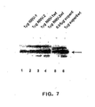


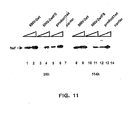


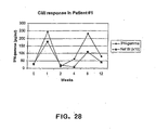


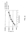


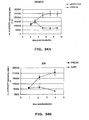
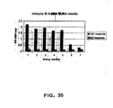


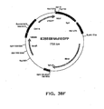


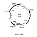


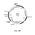

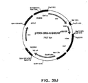
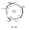


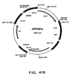
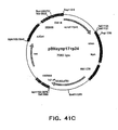


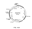
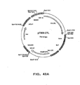


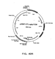


Classifications
-
- C—CHEMISTRY; METALLURGY
- C12—BIOCHEMISTRY; BEER; SPIRITS; WINE; VINEGAR; MICROBIOLOGY; ENZYMOLOGY; MUTATION OR GENETIC ENGINEERING
- C12N—MICROORGANISMS OR ENZYMES; COMPOSITIONS THEREOF; PROPAGATING, PRESERVING, OR MAINTAINING MICROORGANISMS; MUTATION OR GENETIC ENGINEERING; CULTURE MEDIA
- C12N15/00—Mutation or genetic engineering; DNA or RNA concerning genetic engineering, vectors, e.g. plasmids, or their isolation, preparation or purification; Use of hosts therefor
- C12N15/09—Recombinant DNA-technology
- C12N15/63—Introduction of foreign genetic material using vectors; Vectors; Use of hosts therefor; Regulation of expression
- C12N15/79—Vectors or expression systems specially adapted for eukaryotic hosts
- C12N15/85—Vectors or expression systems specially adapted for eukaryotic hosts for animal cells
-
- A—HUMAN NECESSITIES
- A61—MEDICAL OR VETERINARY SCIENCE; HYGIENE
- A61P—SPECIFIC THERAPEUTIC ACTIVITY OF CHEMICAL COMPOUNDS OR MEDICINAL PREPARATIONS
- A61P31/00—Antiinfectives, i.e. antibiotics, antiseptics, chemotherapeutics
-
- A—HUMAN NECESSITIES
- A61—MEDICAL OR VETERINARY SCIENCE; HYGIENE
- A61P—SPECIFIC THERAPEUTIC ACTIVITY OF CHEMICAL COMPOUNDS OR MEDICINAL PREPARATIONS
- A61P31/00—Antiinfectives, i.e. antibiotics, antiseptics, chemotherapeutics
- A61P31/12—Antivirals
-
- A—HUMAN NECESSITIES
- A61—MEDICAL OR VETERINARY SCIENCE; HYGIENE
- A61P—SPECIFIC THERAPEUTIC ACTIVITY OF CHEMICAL COMPOUNDS OR MEDICINAL PREPARATIONS
- A61P31/00—Antiinfectives, i.e. antibiotics, antiseptics, chemotherapeutics
- A61P31/12—Antivirals
- A61P31/14—Antivirals for RNA viruses
- A61P31/18—Antivirals for RNA viruses for HIV
-
- A—HUMAN NECESSITIES
- A61—MEDICAL OR VETERINARY SCIENCE; HYGIENE
- A61P—SPECIFIC THERAPEUTIC ACTIVITY OF CHEMICAL COMPOUNDS OR MEDICINAL PREPARATIONS
- A61P35/00—Antineoplastic agents
-
- A—HUMAN NECESSITIES
- A61—MEDICAL OR VETERINARY SCIENCE; HYGIENE
- A61K—PREPARATIONS FOR MEDICAL, DENTAL OR TOILETRY PURPOSES
- A61K39/00—Medicinal preparations containing antigens or antibodies
- A61K2039/51—Medicinal preparations containing antigens or antibodies comprising whole cells, viruses or DNA/RNA
- A61K2039/53—DNA (RNA) vaccination
-
- A—HUMAN NECESSITIES
- A61—MEDICAL OR VETERINARY SCIENCE; HYGIENE
- A61K—PREPARATIONS FOR MEDICAL, DENTAL OR TOILETRY PURPOSES
- A61K48/00—Medicinal preparations containing genetic material which is inserted into cells of the living body to treat genetic diseases; Gene therapy
-
- C—CHEMISTRY; METALLURGY
- C12—BIOCHEMISTRY; BEER; SPIRITS; WINE; VINEGAR; MICROBIOLOGY; ENZYMOLOGY; MUTATION OR GENETIC ENGINEERING
- C12N—MICROORGANISMS OR ENZYMES; COMPOSITIONS THEREOF; PROPAGATING, PRESERVING, OR MAINTAINING MICROORGANISMS; MUTATION OR GENETIC ENGINEERING; CULTURE MEDIA
- C12N2800/00—Nucleic acids vectors
- C12N2800/10—Plasmid DNA
- C12N2800/108—Plasmid DNA episomal vectors
-
- C—CHEMISTRY; METALLURGY
- C12—BIOCHEMISTRY; BEER; SPIRITS; WINE; VINEGAR; MICROBIOLOGY; ENZYMOLOGY; MUTATION OR GENETIC ENGINEERING
- C12N—MICROORGANISMS OR ENZYMES; COMPOSITIONS THEREOF; PROPAGATING, PRESERVING, OR MAINTAINING MICROORGANISMS; MUTATION OR GENETIC ENGINEERING; CULTURE MEDIA
- C12N2830/00—Vector systems having a special element relevant for transcription
- C12N2830/42—Vector systems having a special element relevant for transcription being an intron or intervening sequence for splicing and/or stability of RNA
-
- C—CHEMISTRY; METALLURGY
- C12—BIOCHEMISTRY; BEER; SPIRITS; WINE; VINEGAR; MICROBIOLOGY; ENZYMOLOGY; MUTATION OR GENETIC ENGINEERING
- C12N—MICROORGANISMS OR ENZYMES; COMPOSITIONS THEREOF; PROPAGATING, PRESERVING, OR MAINTAINING MICROORGANISMS; MUTATION OR GENETIC ENGINEERING; CULTURE MEDIA
- C12N2840/00—Vectors comprising a special translation-regulating system
- C12N2840/20—Vectors comprising a special translation-regulating system translation of more than one cistron
-
- C—CHEMISTRY; METALLURGY
- C12—BIOCHEMISTRY; BEER; SPIRITS; WINE; VINEGAR; MICROBIOLOGY; ENZYMOLOGY; MUTATION OR GENETIC ENGINEERING
- C12N—MICROORGANISMS OR ENZYMES; COMPOSITIONS THEREOF; PROPAGATING, PRESERVING, OR MAINTAINING MICROORGANISMS; MUTATION OR GENETIC ENGINEERING; CULTURE MEDIA
- C12N2840/00—Vectors comprising a special translation-regulating system
- C12N2840/20—Vectors comprising a special translation-regulating system translation of more than one cistron
- C12N2840/203—Vectors comprising a special translation-regulating system translation of more than one cistron having an IRES
Definitions
- Vaccination has proven to be a highly effective and economical method to prevent a disease caused by infectious agents. Since the introduction of the Vaccinia virus as an attenuated vaccine against the smallpox virus (Variola), vaccines against a multitude of human pathogens have been developed and taken into routine use. Today small pox has been eradicated by vaccinations and the same is to be expected shortly for the poliovirus. Several childhood diseases, such as pertussis, diphtheria and tetanus, can be effectively prevented by vaccinations.
- these functional origin fragments contain an E2 binding site, which is essential for the initiation of DNA replication in vivo in most cases (Ustav, E., et al., supra).
- the E2 protein facilitates the first step of the origin recognition by E1. After the initial binding of monomeric E1 to the origin the multimerization of E1 is initiated. This leads to the formation of the complex with the ori melting activity. It has been suggested that E2 has no influence on the following stages of the initiation of the DNA replication [ Lusky, M., et al., Proc Natl Acad Sci USA 91 (1994) 8895-8899 ].
- E2 binding sites in the BPV1 genome and up to four sites in the HPV genomes which play a crucial role in the initiation of viral DNA replication (Ustav, E., et al., supra) and in the regulation of viral gene expression ( Howley, P. M., Papillomavirinae: the viruses and their replication, in Virology, Fields, B. C., Knipe, D. M., Howley, P. M., Eds., Philadelphia: Lippincott-Raven Publishers, 1996. 2. edition, p. 2045 - 2076 ). Structural and mutational analyses have revealed three distinct functional domains in the full size E2 protein.
- the N-terminal part (residues 1 to 210) is an activation domain for transcription and replication. It is followed by the unstructured hinge region (residues 211 to 324) and the carboxy-terminal DNA binding-dimerization domain (residues 325 to 410) [ Dostatni, N., et al., EMBO J 7 (1988) 3807 - 3816 ; Haugen, T., et al.
- the functional elements of the transactivation domain of E2 have a very high structural integrity as confirmed by mutational analysis [ Abroi, A., et al., J Virol 70 (1996) 6169 - 6179 ; Brokaw, J., et al., J Virol 71 (1996) 23 - 29 ; Grossel, M., et al., J Virol 70 (1996) 7264 - 7269 ; Ferguson, M. and Botchan, M., J Virol 70 (1996) 4193-4199 ] and by X-ray crystallography [ Harris, S., and Botchan, M.R., Science 284 (1999) 1673-1677 and Antson, A. et al., Nature 403 (2000) 805-809 ].
- X-ray crystallography shows that the N-terminal domain of the E2 protein forms a dimeric structure, where Arg 37 has an important function in dimer formation (Antson, A., et al., supra).
- an optimal vaccine must produce protective immunity with minimal adverse effects.
- the vaccine should be devoid of components, which are toxic and/or cause symptoms of the disease to the recipient.
- an optimal vaccine must induce a pathogen-specific immune response, i.e. it must elicit a strong and measurable immune response to the desired pathogen without causing an immune response to other components of the vaccine.
- the vector produces several other papilloma proteins, which may elicit undesired immune responses and which induce a risk of the vector's integration in the recipient.
- the vector is replicable, since it contains the E1 gene. Additionally, it is large in size and therefore subject to bacterial modification during preparation.
- vectors containing an MME consisting essentially of ten E2 binding sites are disclosed in some examples. These vectors require the presence of the E1 protein either in the host or in the vector for the expression. This imparts the replication function to the vectors. These vectors also express the E1 protein in addition to the gene of interest and the E2 protein and contain sequences, such as rabbit ⁇ -globin sequences, which are partially homologous to human sequences causing a serious risk of integration to human genome, which reduces the potential of these vectors as DNA vaccines.
- E1 and E2 are the only viral proteins necessary for the episomal long-term replication of the vectors.
- the maintenance function of the BPV1 genome is associated with the presence of minimal ori (MO), which is stated to be necessary, although not sufficient, for the long-term persistence or the stable maintenance of the vectors the cells.
- MO minimal ori
- the cis-elements, i.e. the Minichromosome Maintenance Elements of the BPV1 are stated to be required for the stable replication of BPV1.
- multimeric E2 binding sites (E2BS) are stated to be necessary for the stable maintenance of the vectors.
- Yet another object of the invention is to provide novel vectors, which are suitable for a large-scale production in bacterial cell.
- An additional object of the invention is to provide novel vectors, which are useful as carrier vectors for a gene or genes of interest,
- the present invention discloses novel vectors, which meet the requirements of a carrier vector of a gene or genes of interest or of an optimal DNA vaccination vector and which are preferably devoid of drawbacks and side effects of prior art vectors.
- the present invention discloses novel vectors useful as carrier vectors of a gene or genes of interest, in DNA vaccination and gene therapy and as gene therapeutic agents.
- said vectors are capable of spreading and, if desired, of expressing a gene or genes of interest in an increasing number of cells for an extended time.
- the vectors of the present invention preferably express only a nuclear-anchoring protein, and, if desired, the gene or genes of interest, and optionally a selectable marker. However, they preferably lack any redundant, oncogenically transforming or potentially toxic sequences, thereby avoiding a severe drawback of the vectors previously disclosed or suggested for use as DNA vaccines, i.e. hypersensitivity reactions against other viral components. In certain embodiments of the invention, this is achieved by low level of the expressed nuclear-anchoring protein in the cells.
- the vectors of the present invention induce both humoral and cellular immune responses, where the gene or genes of interest is included in the vector.
- the vectors of the present invention are advantageous for use both in vitro (e.g., in the production level) and in vivo (e.g., vaccination).
- the present invention relates to expression vectors comprising: (a) a DNA sequence encoding a nuclear-anchoring protein operatively linked to a heterologous promoter, said nuclear-anchoring protein comprising (i) a DNA binding domain which binds to a specific DNA sequence, and (ii) a functional domain of the E2 protein of Bovine Papilloma Virus type 1 that binds to a nuclear component, and (b) a multimerized DNA binding sequence for the nuclear anchoring protein, wherein said vector lacks a an origin of replication functional in mammalian cells.
- the nuclear component can be mitotic chromatin, the nuclear matrix, nuclear domain 10 (ND10), or nuclear domain POD.
- the nuclear anchoring-protein is a chromatin-anchoring protein, and said functional domain binds mitotic chromatin.
- the nuclear-anchoring protein contains a hinge or linker region.
- the nuclear-anchoring protein is a natural protein of viral origin.
- the nuclear-anchoring protein is that of a papilloma virus.
- the nuclear-anchoring protein is the E2 protein of Bovine Papilloma Virus type 1.
- the nuclear-anchoring protein is a non-natural protein.
- the nuclear-anchoring protein is a recombinant protein, a fusion protein, or a protein obtained by molecular modeling techniques.
- the recombinant protein, fusion protein, or protein obtained by molecular modeling techniques contains any combination of a DNA binding domain which binds to said specific DNA sequence and a functional domain which binds to a nuclear component, wherein said functional domain which binds to a nuclear component is that of E2 protein of Bovine Papilloma Virus type 1.
- the vector further comprises one or more expression cassettes of a DNA sequence of interest.
- the DNA sequence of interest is that of a bacterium.
- the bacterium is selected from the group consisting of Chlamydia trachomatis, Mycobacterium tuberculosis, and Mycoplasma pneumonia.
- the bacterium is Salmonella.
- the DNA sequence of interest is that of a fungal pathogen.
- the fungal pathogen is Candida albigans.
- the DNA sequence of interest is of HIV origin.
- the DNA sequence of interest encodes a non-structural regulatory protein of HIV.
- the non-structural regulatory protein of HIV is Nef, Tat and/or Rev.
- the non-structural regulatory protein of HIV is Nef.
- the DNA sequence of interest encodes a structural protein of HIV.
- the DNA sequence of interest is the gene encoding HIV gp120/gp160.
- the vector of the invention comprises a first expression cassette comprising a DNA sequence of interest which encodes Nef, Tat and/or Rev, and a second expression cassette comprising a DNA sequence of interest which encodes Nef, Tat and/or Rev.
- the vector of the invention comprises a first expression cassette comprising a DNA sequence of interest which encodes Nef, Tat and/or Rev, and a second expression cassette comprising a DNA sequence of interest which encodes a structural protein of HIV.
- the DNA sequence of interest encodes a protein associated with cancer.
- the DNA sequence of interest encodes a protein associated with immune maturation, regulation of immune responses, or regulation of autoimmune responses.
- the protein is APECED.
- the DNA sequence of interest is the Aire gene.
- the DNA sequence of interest encodes a protein that is defective in any hereditary single gene disease.
- the DNA sequence of interest encodes a macromolecular drug.
- the DNA sequence of interest encodes a cytokine.
- the cytokine is an interleukin selected from the group consisting of IL1, IL2, IL4, IL6 and IL12.
- the DNA sequence of interest encodes an interferon.
- the DNA sequence of interest encodes a biologically active RNA molecule.
- the biologically active RNA molecule is selected from the group consisting of inhibitory antisense and ribozyme molecules.
- the inhibitory antisense or ribozyme molecules antagonize the function of an oncogene.
- a vector of the invention is suitable for the use for the production of a therapeutic macromolecular agent in vivo.
- the invention provides a vector for use as a medicament.
- the invention provides a vector for use as a carrier vector for a gene, genes, or a DNA sequence or DNA sequences of interest, such as a gene, genes, or a DNA sequence or DNA sequences encoding a protein or peptide of an infectious agent, a therapeutic agent, a macromolecular drug, or any combination thereof.
- the invention provides a vector for use as a medicament for treating inherited or acquired genetic defects.
- the invention provides a vector for use as a therapeutic DNA vaccine against an infectious agent.
- the invention provides a vector for use as a therapeutic agent.
- the invention further relates to methods for providing a protein to a subject, said method comprising administering to the subject a vector of the invention, wherein said vector (i) further comprises a second DNA sequence encoding the protein to be provided to the subject, which second DNA sequence is operably linked to a second promoter, and (ii) does not encode Bovine Papilloma Virus protein E1, and wherein said subject does not express Bovine Papilloma Virus protein E1.
- the invention further relates to methods for inducing an immune response to a protein in a subject, said method comprising administering to the subject a vector of the invention wherein said vector (i) further comprises a second DNA sequence encoding said protein, which second DNA sequence is operably linked to a second promoter, and (ii) does not encode Bovine Papilloma Virus protein E1, and wherein said subject does not express Bovine Papilloma Virus protein E1.
- the invention further relates to methods for treating an infectious disease in a subject in need of said treatment, said method comprising administering to said subject a therapeutically effective amount of a vector of the invention, wherein the DNA sequence of interest encodes a protein comprising an immunogenic epitope of an infectious agent.
- the invention further relates to methods for treating an inherited or acquired genetic defect in a subject in need of said treatment, said method comprising: administering to said subject a therapeutically effective amount of a vector of the invention, wherein said DNA sequence of interest encodes a protein which is affected by said inherited or acquired genetic defect.
- the invention further relates to methods for expressing a DNA sequence in a subject, said method comprising administering a vector of the invention to said subject.
- a vector of the invention is used for production of a protein encoded by said DNA sequence of interest in a cell or an organism.
- the invention further provides a method for the preparation of a vector of claim 1, 2, or 17 comprising: (a) cultivating a host cell containing said vector and (b) recovering the vector.
- the method for preparing a vector of the invention further comprises before step (a) a step of transforming said host cell with said vector.
- the host cell is a prokaryotic cell.
- the host cell is an Escherichia coli.
- the invention further relates to a host cell that is characterized by containing a vector of the invention.
- the host cell is a bacterial cell.
- the host cell is a mammalian cell.
- the invention further relates to carrier vectors containing a vector of the invention.
- the invention further relates to a pharmaceutical composition
- a pharmaceutical composition comprising a vector of the invention and a suitable pharmaceutical vehicle.
- the invention further relates to a gene therapeutic agent containing a vector of the invention.
- the invention further relates to a method for the preparation of a DNA vaccine, said method comprising combining a vector of the invention with a suitable pharmaceutical vehicle.
- the invention further relates to a method for the preparation of an agent for use in gene therapy, said method comprising combining a vector of the invention with a suitable pharmaceutical vehicle.
- the present invention is based on the unexpected finding that expression vectors, which carry (A) an expression cassette of a gene of a nuclear-anchoring protein that binds both to (i) a specific DNA sequence and (ii) to a suitable nuclear component and (B) a multimerized DNA binding sequence for said nuclear-anchoring protein as disclosed in claim 1 are capable of spreading in a proliferating cell population.
- nuclear-anchoring proteins include, but are not limited to, chromatin-anchoring proteins, such as the Bovine Papilloma Virus type 1 E2 protein (BPV1 E2; SEQ ID NO: 50).
- the DNA binding sequences can be, but are not limited to, multimerized E2 binding sites.
- nuclear-anchoring protein refers to a protein, which binds to a specific DNA sequence and capable of providing a nuclear compartmentalization function to the vector, i.e., to a protein, which is capable of anchoring or attaching the vector to a specific nuclear compartment.
- the nuclear-anchoring protein is a natural protein. Examples of such nuclear compartments are the mitotic chromatin or mitotic chromosomes, the nuclear matrix, nuclear domains like ND10 and POD etc.
- the nuclear-anchoring protein of the invention is a recombinant protein.
- the nuclear-anchoring protein is a fusion protein, a chimeric protein, or a protein obtained by molecular modeling.
- a fusion protein, or a protein obtained by molecular modeling in connection with the present invention is characterized by its ability to bind to a nuclear component and by its ability to bind sequence-specifically to DNA.
- such a fusion protein is encoded by a vector of the invention which also contains the specific DNA sequence to which the fusion/chimeric protein binds.
- Nuclear components include, but are not limited to chromatin, the nuclear matrix, the ND10 domain and POD.
- the DNA binding domain and the corresponding DNA sequence is preferably non-endogenous to the host cell/host organism.
- Such domains include, but are not limited to, the DNA binding domain of the Bovine Papilloma Virus type 1 (BPV1) E2 protein (SEQ ID NO: 50), Epstein-Barr Virus Nuclear Antigen 1 (EBNA1; SEQ ID NO: 52), and High Mobility Group (HMG) proteins (HMG box).
- the vector of the invention can further comprise a "DNA sequence of interest", that encodes a protein (including a peptide or polypeptide), e.g., that is an immunogen or a therapeutic.
- the DNA sequence of interest encodes a biologically active RNA molecule, such as an antisense RNA molecule or a ribozyme.
- the expression vectors of the invention carrying an expression cassette for a gene of a nuclear-anchoring protein and multimerized binding sites for said nuclear-anchoring protein spread in a proliferating host cell population. This means that a high copy-number of vectors or plasmids are delivered into the target cells and the use of the segregation/partitioning function of the nuclear-anchoring protein and its multimerized binding sites assures the distribution of the vector to the daughter cells during cell division.
- the vector of the invention lacks a papilloma virus origin of replication, providing a vector which lacks an origin of replication functional in mammalian cells.
- the omission of an origin of replication functional in mammalian cells constitutes an improvement over prior art vectors for several reasons.
- Omission of the origin of replication reduces the size of the vector of the invention compared to prior art vectors. Such a reduction in size increases the stability of the vector and facilitates uptake by the host cell.
- Omission of the origin of replication reduces the risk for recombination with the host cell's genome, thereby reducing the risk of unwanted side effects.
- the omission of the origin of replication allows to control the dosage simply by adjusting the amount of vector administered.
- the mechanism of the spreading function is due to the dual function of the E2 protein: the capacity of the E2 protein to attach to mitotic chromosomes through the N-terminal domain of the protein and the sequence-specific binding capacity of the C-terminal domain of the E2 protein, which assures the tethering of vectors, which contain a multimerized E2 binding site, to mitotic chromosomes. A segregation/partitioning function is thus provided to the vectors.
- the expression cassette of a gene of the chromatin-anchoring protein comprises a gene of any suitable protein of cellular, viral or recombinant origin having analogous properties to E2 of the BPV1, i.e., the ability to attach to the mitotic chromatin through one domain and to cooperatively bind DNA through another domain to multimerized binding sites specific for this DNA binding domain.
- the multimerized DNA binding sequences may be composed of any suitable multimeric specific sequences capable of inducing the cooperative binding of the protein to the plasmid, such as those of the EBNA1 or a suitable HMG protein.
- 21x30bp repeats of binding sites for EBNA-1 are localized in the region spanning from nucleotide position 7421 to nucleotide position 8042 of the Epstein-Barr virus genome (SEQ ID NO:51).
- the selectable marker can be any suitable marker allowable in DNA vaccines, such a kanamycin or neomycin, and others.
- other positive and negative selection markers can be included in the vectors of the invention, where applicable.
- the vectors of the present invention are not host specific, since the expression of the nuclear-anchoring protein, such as the E2 protein, is controlled by non-native or heterologous promoters. Depending on the particular promoter chosen, these promoters may be functional in a broad range of mammalian cells or they can be cell or tissue specific. Examples of promoters for the nuclear-anchoring protein, such as for the E2 protein, useful in the vectors of the present invention are thymidine kinase promoters, Human Cytomegalovirus Immediate Early Promoter, Rous Sarcoma Virus LTR and like. For the expression of the gene of interest, preferred promoters are strong promoters assuring high levels of expression of the gene of interest, an example for such a promoter is the Human Cytomegalovirus Immediate Early Promoter.
- a gene, genes or a DNA sequence or DNA sequences to be expressed via a vector of the invention can be any DNA sequence of interest, whose expression is desired.
- the vectors may contain a gene or genes or a DNA sequence or DNA sequences from infectious microbial pathogens, such as viruses, against which live attenuated vaccines or inactivated vaccines cannot be prepared or used.
- DNA sequences of interest include genes or DNA sequences from viruses, such as Human Immunodeficiency Virus (HIV), Herpex Simplex Virus (HSV), Hepatitis C Virus, Influenzae Virus, Enteroviruses etc.; intracellular bacterial, such as Chlamydia trachomatis, Mycobacterium tuberculosis, Mycoplasma pneumonia etc.; extracellular bacteria, such as Salmonella; or fungi, such as Candida albigans.
- viruses such as Human Immunodeficiency Virus (HIV), Herpex Simplex Virus (HSV), Hepatitis C Virus, Influenzae Virus, Enteroviruses etc.
- intracellular bacterial such as Chlamydia trachomatis, Mycobacterium tuberculosis, Mycoplasma pneumonia etc.
- extracellular bacteria such as Salmonella
- fungi such as Candida albigans.
- the vectors contain a gene encoding early regulatory proteins of HIV, i.e. the nonstructural regulatory proteins Nef, Tat or Rev, preferably Nef.
- the vectors of the invention contain genes encoding structural proteins of the HIV.
- the vectors of the present invention contain two or more genes encoding any combination of early regulatory proteins and/or structural proteins of HIV.
- the vaccines of the present invention contain a vector of the present invention or a mixture of said vectors in a suitable pharmaceutical carrier.
- the vaccine may for instance contain a mixture of vectors containing genes for the three different regulatory proteins of the HIV and/or structural proteins of the HIV.
- the vectors of the present invention can be used for the expression of the specific cytokines, like interleukines (IL1, IL2, IL4, IL6, IL12 and others) or interferon, with the aim of modulating the specific immune responses of the organism (immunotherapy) against foreign antigens or boosting of the activity of the immune system against the mutated self-antigens.
- the vectors of the present invention are also useful in complementing malfunctioning of the brain due to the loss of specific dopamine-ergic neurons leading to the irreversible neurodegeneration, which is cause for Parkinson's disease, by expressing genes involved into synthesis of dopamine, like tyrosine hydroxylase, as well as other genes deficiency of which would have the similar effect.
- DNA sequences encoding molecules comprising epitopes of known parasites are used.
- DNA sequences encoding antigenic epitopes may be prepared from parasites including, but not limited to, chlamydia and rickettsia.
- Both constitutive and inducible regulatory regions may be used for expression of the DNA sequence of interest. It may be desirable to use inducible promoters when the conditions optimal for growth of the host cells and the conditions for high level expression of the DNA sequence of interest are different. Examples of useful regulatory regions are provided below (section 5.4.4).
- the vector comprising a DNA sequence of interest operably linked to a regulatory region can be directly introduced into appropriate host cells for expression of the DNA sequence of interest without further cloning.
- a variety of regulatory regions can be used, for example, the SV40 early and late promoters, the cytomegalovirus (CMV) immediate early promoter, and the Rous sarcoma virus long terminal repeat (RSV-LTR) promoter.
- Inducible promoters that may be useful in mammalian cells include but are not limited to those associated with the metallothionein II gene, mouse mammary tumor virus glucocorticoid responsive long terminal repeats (MMTV-LTR), ⁇ -interferon gene, and hsp70 gene [ Williams et al., Cancer Res. 49 (1989) 2735-42 ; Taylor et al., Mol. Cell Biol., 10 (1990) 165-75 ]. It may be advantageous to use heat shock promoters or stress promoters to drive expression of the DNA sequence of interest in recombinant host cells.
- Preferred mammalian host cells include but are not limited to those derived from humans, monkeys and rodents, (see, for example, Kriegler M. in “Gene Transfer and Expression: A Laboratory Manual", New York, Freeman & Co. 1990 ), such as monkey kidney cell line transformed by SV40 (COS-7, ATCC CRL 1651); human embryonic kidney line (293, 293-EBNA, or 293 cells subcloned for growth in suspension culture, Graham et al., J. Gen. Virol., 36 (1977) 59 ; baby hamster kidney cells (BHK, ATCC CCL 10); chinese hamster ovary-cells-DHFR [CHO, Urlaub and Chasin. Proc. Natl. Acad. Sci.
- a vector of the invention comprising an expression cassette of a DNA sequence of interest is administered to a subject to induce an immune response.
- the DNA sequence of interest encodes a protein (for example, a peptide or polypeptide), which induces a specific immune response upon its expression. Examples of such proteins are discussed in section 5.3.
- a vector of the invention for use as a vaccine, methods may be selected from among those known in the art and/or described in section 5.4.6.
- a vector of the invention comprising an expression cassette comprising DNA sequences of interest is administered to treat, or prevent various diseases.
- the DNA sequence of interest may encode a protein and/or a biologically active RNA molecule.
- Gene therapy refers to therapy performed by the administration to a subject of an expressed or expressible DNA sequence.
- the DNA sequences produce their encoded protein or RNA molecule that mediates a therapeutic effect.
- mice mammary tumor virus control region which is active in testicular, breast, lymphoid and mast cells [ Leder et al., Cell 45 (1986) 485-495 ], albumin gene control region which is active in the liver [ Pinkert et al., Genes and Devel. 1 (1987) 268-276 ], alpha-fetoprotein gene control region which is active in the liver [ Krumlauf et al., Mol. Cell. Biol. 5 (1985)1639-1648 ; Hammer et al., Science 235 (1987) 53-58 ; alpha 1-antitrypsin gene control region which is active in the liver [ Kelsey et al., Genes and Devel.
- beta-globin gene control region which is active in myeloid cells [ Mogram et al., Nature 315 (1985) 338-340 ; Kollias et al., Cell 46 (1986) 89-94 ]; myelin basic protein gene control region which is active in oligodendrocyte cells in the brain [ Readhead et al., Cell 48 (1987) 703-712 ]; myosin light chain-2 gene control region which is active in skeletal muscle [ Sani, Nature 314 (1985) 283-286 ], and gonadotropic releasing hormone gene control region which is active in the hypothalamus [ Mason et al., Science 234 (1986)1372-1378 ].
- a vector of the invention contains a DNA sequence of interest that encodes an antisense or ribozyme RNA molecule. Techniques for the production and use of such molecules are well known to those of skill in the art.
- Antisense RNA molecules act to directly block the translation of mRNA by hybridizing to targeted mRNA and preventing protein translation.
- Antisense approaches involve the design of oligonucleotides that are complementary to a target gene mRNA. The antisense oligonucleotides will bind to the complementary target gene mRNA transcripts and prevent translation. Absolute complementarity, although preferred, is not required.
- a sequence "complementary" to a portion of an RNA means a sequence having sufficient complementarity to be able to hybridize with at least the non-polyA portion of an RNA, forming a stable duplex; in the case of double-stranded antisense nucleic acids, a single strand of the duplex DNA may thus be tested, or triplex formation may be assayed.
- the ability to hybridize will depend on both the degree of complementarity and the length of the antisense nucleic acid. Generally, the longer the hybridizing nucleic acid, the more base mismatches with an RNA it may contain and still form a stable duplex (or triplex, as the case may be).
- One skilled in the art can ascertain a tolerable degree of mismatch by use of standard procedures to determine the melting point of the hybridized complex.
- Antisense nucleic acids should be at least six nucleotides in length, and are preferably oligonucleotides ranging from 6 to about 50 nucleotides in length. In specific aspects the oligonucleotide is at least 10 nucleotides, at least 17 nucleotides, at least 25 nucleotides or at least 50 nucleotides. In other embodiments of the invention, the antisense nucleic acids are at least 100, at least 250, at least 500, and at least 1000 nucleotides in length.
- antisense DNA sequences complementary to the target gene coding region sequence could be used, those complementary to the transcribed, untranslated region are most preferred.
- the DNA sequence encoding the biologically active RNA molecule is operatively linked to a strong pol III or pol II promoter.
- a vector of the invention can be introduced, e.g ., such that it is taken up by a cell and directs the transcription of an antisense RNA.
- Such vectors can be constructed by recombinant DNA technology methods standard in the art.
- Expression of the sequence encoding the antisense RNA can be by any promoter known in the art to act in mammalian, preferably human cells.
- Such promoters can be inducible or constitutive.
- Such promoters include but are not limited to: the SV40 early promoter region [ Bernoist and Chambon, Nature 290 (1981) 304-310 ], the promoter contained in the 3 long terminal repeat of Rous sarcoma virus [ Yamamoto, et al., Cell 22 (1980) 787-797 ], the herpes thymidine kinase promoter [ Wagner, et al., Proc. Natl. Acad. Sci. U.S.A. 78 (1981) 1441-1445 ], the regulatory sequences of the metallothionein gene [ Brinster, et al., 1982, Nature 296 (1982) 39-42 ], etc .
- a vector of the invention contains a DNA sequence, which encodes a ribozyme.
- Ribozyme molecules designed to catalytically cleave target gene mRNA transcripts can also be used to prevent translation of a target gene mRNA and, therefore, expression of a target gene product [see, e.g ., PCT International Publication WO90/11364, published October 4, 1990 ; Sarver, et al., Science 247 (1990) 1222-1225 ].
- Ribozymes are enzymatic RNA molecules capable of catalyzing the specific cleavage of RNA. [For a review, see Rossi, Current Biology 4 (1994) 469-471 ]. The mechanism of ribozyme action involves sequence specific hybridization of the ribozyme molecule to complementary target RNA, followed by an endonucleolytic cleavage event. The composition of ribozyme molecules must include one or more sequences complementary to the target gene mRNA, and must include the well known catalytic sequence responsible for mRNA cleavage. For this sequence, see, e.g ., U.S. Patent No. 5,093,246 .
- ribozymes that cleave mRNA at site-specific recognition sequences can be used to destroy target gene mRNAs
- the use of hammerhead ribozymes is preferred.
- Hammerhead ribozymes cleave mRNAs at locations dictated by flanking regions that form complementary base pairs with the target mRNA. The sole requirement is that the target mRNA have the following sequence of two bases: 5'-UG-3'.
- the ribozyme is engineered so that the cleavage recognition site is located near the 5' end of the target gene mRNA, i.e ., to increase efficiency and minimize the intracellular accumulation of non-functional mRNA transcripts.
- the ribozymes of the present invention also include RNA endoribonucleases (hereinafter "Cech-type ribozymes”) such as the one that occurs naturally in Tetrahymena thermophila (known as the IVS, or L-19 IVS RNA) and that has been extensively described by Thomas Cech and collaborators [ Zaug, et al., Science, 224 (1984) 574-578 ; Zaug and Cech, Science, 231 (1986) 470-475 ; Zaug, et al., Nature, 324 (1986) 429-433 ; published International patent application No. WO 88/04300 by University Patents Inc.; Been & Cech, Cell, 47 (1986) 207-216 ].
- Cech-type ribozymes such as the one that occurs naturally in Tetrahymena thermophila (known as the IVS, or L-19 IVS RNA) and that has been extensively described by Thomas Cech and collaborators [ Zaug, et al., Science
- the Cech-type ribozymes have an eight base pair active site, which hybridizes to a target RNA sequence whereafter cleavage of the target RNA takes place.
- the invention encompasses those Cech-type ribozymes, which target eight base-pair active site sequences that are present in the target gene.
- ribozyme can be under the control of a strong constitutive pol III or pol II promoter, so that transfected cells will produce sufficient quantities of the ribozyme to destroy endogenous target gene messages and inhibit translation. Because ribozymes unlike antisense molecules, are catalytic, a lower intracellular concentration is required for efficiency.
- nucleic acid molecules that encode and express target gene polypeptides exhibiting normal target gene activity may, be introduced into cells via gene therapy methods such as those described, below, in Section 5.4.4 that do not contain sequences susceptible to whatever antisense, ribozyme, or triple helix treatments are being utilized.
- the target gene encodes an extracellular protein
- a pharmaceutical of the invention comprises a substantially purified vector of the invention (e.g ., substantially free from substances that limit its effect or produce undesired side-effects).
- the subject to whom the pharmaceutical is administered in the methods of the invention is preferably an animal, including but not limited to animals such as cows, pigs, horses, chickens, cats, dogs, etc ., and is preferably a mammal, and most preferably a human.
- the vector of the invention is directly administered in vivo, where the DNA sequence of interest is expressed to produce the encoded product. This can be accomplished by any of numerous methods known in the art.
- the vectors of the invention can be administered so that the nucleic acid sequences become intracellular.
- the vectors of the invention can be administered by direct injection of naked DNA; use of microparticle bombardment (e.g ., a gene gun; Biolistic, Dupont); coating with lipids or cell-surface receptors ortransfecting agents; encapsulation in microparticles or microcapsules; administration in linkage to a peptide which is known to enter the nucleus; administration in linkage to a ligand subject to receptor-mediated endocytosis [see, e.g ., Wu and Wu, J. Biol. Chem. 262 (1987) 4429-4432 ] (which can be used to target cell types specifically expressing the receptors); etc .
- microparticle bombardment e.g ., a gene gun; Biolistic, Dupont
- coating with lipids or cell-surface receptors ortransfecting agents encapsulation in microparticles or microcapsules
- administration in linkage to a peptide which is known to enter the nucleus administration in linkage to a
- the compound or composition can be delivered in a vesicle, in particular a liposome [see Langer, Science 249 (1990) 1527-1533 ; Treat et al., 1989, in Liposomes in the Therapy of Infectious Disease and Cancer, Lopez-Berestein and Fidler (eds.), Liss, New York, pp. 353- 365 ; Lopez-Berestein, ibid., pp. 317-327 ].
- a liposome see Langer, Science 249 (1990) 1527-1533 ; Treat et al., 1989, in Liposomes in the Therapy of Infectious Disease and Cancer, Lopez-Berestein and Fidler (eds.), Liss, New York, pp. 353- 365 ; Lopez-Berestein, ibid., pp. 317-327 ].
- the vector of the invention is coated with lipids or cell-surface receptors or transfecting agents, or linked to a homeobox- like peptide which is known to enter the nucleus [see e.g ., Joliot et al., Proc. Natl. Acad. Sci. USA 88 (1991) 1864-1868 ], etc .
- nucleic acid-ligand complexes can be formed in which the ligand comprises a fusogenic viral peptide to disrupt endosomes, allowing the nucleic acid to avoid lysosomal degradation.
- the vector of the invention can be targeted in vivo for cell specific uptake and expression, by targeting a specific receptor (see, e.g ., PCT Publications WO 92/06 180 ; WO 92/22635 ; W092/20316 ; W093/14188 , and WO 93/20221 ).
- Methods for use with the invention include, but are not limited to, intradermal, intramuscular, intraperitoneal, intravenous, subcutaneous, intranasal, epidural, and oral routes. Methods for use with the invention further include administration by any convenient route, for example by infusion or bolus injection, by absorption through epithelial or mucocutaneous linings ( e.g ., oral mucosa, rectal and intestinal mucosa, etc.).
- a vector of the invention may be desirable to administer a vector of the invention by injection, by means of a catheter, by means of a suppository, or by means of an implant, said implant being of a porous, non-porous, or gelatinous material, including a membrane, such as a sialastic membrane, or a fiber. Care must be taken to use materials to which the vector does not absorb. Administration can be systemic or local.
- a vector of the invention is administered together with other-biologically active agents such as chemotherapeutic agents or agents that augment the immune system.
- methods for use with the invention include delivery via a controlled release system.
- a pump may be used [see Langer, supra; Sefton, CRC Crit. Ref. Biomed. Eng. 14 (1989) 201 ; Buchwald et al., Surgery 88 (1980) 507 ; Saudek et al., N. Engl. J. Med. 321 (1989) 574 ].
- polymeric materials can be used [see Medical Applications of Controlled Release, 1974, Langer and Wise (eds.), CRC Pres., Boca Raton, Florida ; Controlled Drug Bioavailability, Drug Product Design and Performance, 1984, Smolen and Ball (eds.), Wiley, New York ; Ranger and Peppas, Macromol. Sci. Rev. Macromol. Chem. 23 (1983) 61 ; see also Levy et al., Science 228 (1985) 190 ; During et al., Ann. Neurol. 25 (1989) 351 ; Howard et al., J. Neurosurg. 71 (1989) 105 ].
- compositions of the invention comprise a therapeutically effective amount of a vector of the invention, and a suitable pharmaceutical vehicle.
- suitable pharmaceutical vehicle means approved by a regulatory agency of the Federal or a state government or listed in the U.S. Pharmacopeia or other generally recognized pharmacopeia for use in animals, and more particularly in humans.
- vehicle refers to a diluent, adjuvant, excipient, or vehicle with which the therapeutic is administered.
- suitable pharmaceutical vehicles can be sterile liquids, such as water and oils, including those of petroleum, animal, vegetable or synthetic origin, such as peanut oil, soybean oil, mineral oil, sesame oil and the like. Water is a preferred carrier when the pharmaceutical composition is administered intravenously.
- Saline solutions and aqueous dextrose and glycerol solutions can also be employed as liquid carriers, particularly for injectable solutions.
- suitable pharmaceutical excipients include starch, glucose, lactose, sucrose, gelatin, malt, rice, flour, chalk, silica gel, sodium stearate, glycerol monostearate, talc, sodium chloride, dried skim milk, glycerol, propylene, glycol, water, ethanol and the like.
- the composition if desired, can also contain minor amounts of wetting or emulsifying agents, or pH buffering agents. These compositions can take the form of solutions, suspensions, emulsion, tablets, pills, capsules, powders, sustained-release formulations and the like.
- compositions can be formulated as a suppository, with traditional binders and carriers such as triglycerides.
- Oral formulation can include standard carriers such as pharmaceutical grades of mannitol, lactose, starch, magnesium stearate, sodium saccharine, cellulose, magnesium carbonate, etc. Examples of suitable pharmaceutical carriers are described in "Remington's Pharmaceutical Sciences” by E.W. Martin.
- Such compositions will contain a therapeutically effective amount of the nucleic acid or protein of the invention, preferably in purified form, together with a suitable amount of carrier so as to provide the form for proper administration to the patient.
- the formulation should suit the mode of administration.
- the pharmaceutical of the invention is formulated in accordance with routine procedures as a pharmaceutical composition adapted for intravenous administration to human beings.
- compositions for intravenous administration are solutions in sterile isotonic aqueous buffer.
- the pharmaceutical of the invention may also include a solubilizing agent and a local anesthetic such as lignocaine to ease pain at the site of the injection.
- the ingredients are supplied either separately or mixed together in unit dosage form, for example, as a dry lyophilized powder or water free concentrate in a hermetically sealed container such as an ampoule or sachette indicating the quantity of active agent.
- the pharmaceutical of the invention is to be administered by infusion, it can be dispensed with an infusion bottle containing sterile pharmaceutical grade water or saline.
- an ampoule of sterile water for injection or saline can be provided so that the ingredients may be mixed prior to administration.
- compositions may take the form of tablets or lozenges formulated in conventional manner.
- the compounds for use according to the present invention are conveniently delivered in the form of an aerosol spray presentation from pressurized packs or a nebulizer, with the use of a suitable propellant, e.g., dichlorodifluoromethane, trichlorofluoromethane, dichlorotetrafluoroethane, carbon dioxide or other suitable gas.
- a suitable propellant e.g., dichlorodifluoromethane, trichlorofluoromethane, dichlorotetrafluoroethane, carbon dioxide or other suitable gas.
- the dosage unit may be determined by providing a valve to deliver a metered amount.
- Capsules and cartridges of e.g. gelatin for use in an inhaler or insufflator may be formulated containing a powder mix of the compound and a suitable powder base such as lactose or starch.
- the amount of a vector of the invention which will be effective in the treatment or prevention of the indicated disease, can be determined by standard clinical techniques.
- in vitro assays may optionally be employed to help identify optimal dosage ranges.
- the precise dose to be employed in the formulation will also depend on the route of administration, and the stage of indicated disease, and should be decided according to the judgment of the practitioner and each patient's circumstances. Effective doses may be extrapolated from dose-response curves derived from in vitro or animal model test systems.
- vector plasmids super6 ( Figure 1 ) and super6wt were prepared from previous generation based gene vaccination vectors VI ( Figure 2 ) and Vlwt, respectively.
- Vectors VI and Vlwt are principally synthetic bacterial plasmids that contain a transposon Tn903 derived kanamycin resistance marker gene [ Oka, A., et al., J Mol Biol 147 (1981) 217-226 ] and a modified form of pMB1 replicon [ Yanisch-Perron, C., et al., Gene 33 (1985) 103-119 ] needed for the propagation in Escherichia coli cells.
- Vectors VI and Vlwt also contain a Cytomegalovirus Immediately Early Promoter combined with a HSV1 TK leader sequence and rabbit ⁇ -globin gene sequences, which both are derived from plasmid pCG [ Tanaka, M., et al., 60 (1990) Cell 375-386 ].
- the latter elements are needed for expressing from the nef coding sequence derived from a HAN2 isolate of the HIV-1 strain [ Sauermann, U., et al., AIDS Research. Hum. Retrov. 6 (1990) 813-823 ].
- the expression vectors for the Nef carry clustered ten high affinity E2 binding sites (derived from plasmid pUC1910BS, unpublished) just upstream of the CMV promoter.
- the parent vector VI contains a modified E2 coding sequence: the hinge region of E2 (amino acids 192-311) is replaced with four glycine-alanine repeats from EBNA1 protein of Epstein-Barr Virus [ Baer, R. J., et al., Nature 310 (1984) 207-211 ].
- the protein encoded by this sequence was named as E2d192-311+4G.
- the parent vector Vlwt contains an expression cassette for wild type E2 protein of the bovine papilloma virus type 1 with point mutations introduced for the elimination E3 and E4 open reading frame (ORF) coding capacity by two stop codons into both these ORFs.
- the E2 coding sequences are cloned between a Rous sarcoma virus proviral 5' LTR [ Long, E. O., et al., Hum. Immunol. 31 (1991) 229-235 ] and bovine growth hormone polyadenylation region [ Chesnut, J. D., et al., J Immunol Methods 193 (1996) 17-27 ].
- Plasmid vectors super6 and super6wt were constructed by deleting from the respective parent vectors VI and Vlwt all beta-globin sequences downstream of the nef gene except the second intron of the rabbit beta-globin gene.
- the beta-globin sequences (especially the fragment of the exon) show some homology with sequences in the human beta-globin gene, whereas the intron lacks any significant homology to human genomic sequences.
- the intron was amplified by PCR from the plasmid pCG [ Tanaka, M.
- the expression properties of the Nef and E2- proteins expressed by the plasmid vectors super6 and super6wt were analyzed and compared with the expression properties of the Nef and E2 proteins expressed by VI and Vlwt by Western blotting [ Towbin et al., Proc Natl Acad Sci USA 76 (1979) 4350-4354 ] with monoclonal antibodies against Nef and E2.
- Jurkat cells a human T-cell lymphoblast cell line
- were transfected by electroporation [ Ustav et al. EMBO J 2 (1991) 449-457 ] with 1 ⁇ g of super6, super6WT or equimolar amounts of the plasmids VI, Vlwt.
- the cells were lysed by treating with a sample buffer containing 50mM Tris-HCl pH 6.8; 2% SDS, 0.1 % bromophenol blue, 100mM dithiothreitol, and 10% (v/v) glycerol.
- the lysates were run on a 10% or 12.5% SDS-polyacrylamide gel and subsequently transferred onto a 0.45 ⁇ m PVDF nitrocellulose membrane (Millipore).
- the membrane was first blocked overnight with a blocking solution containing 5% dry milk (fat-free), 0.1 % Tween 20 in 50 mM Tris-HCl pH 7.5; 150mM NaCl and thereafter incubated for 1 h with diluted monoclonal anti-Nef antisera (1:100) or anti-E2-antisera (1:1000) in the blocking solution. After each incubation step, unbound proteins were removed by washing strips three times with TBS - 0.1 % Tween-20.
- the binding of primary immunoglobulins was detected by incubating the strips with horse raddish peroxidase conjugate anti-mouse IgG (Labas, Estonia) followed by visualization using a chemoluminesence detection system (Amersham Pharmacia Biotech, United Kingdom).
- Nef expressed from the plasmids super6, super6wt, VI and Vlwt (lanes 1-4 in Figure 4A ) are quite similar ( Figure 4 , panel A, lanes 1 to 4). Much less protein is produced from plasmid II (lane 5). The expression levels of the Nef protein are higher from vectors containing wtE2 (cf. lane 1 compared with lane 2 and lane 3 compared with lane 4). This is in accordance with the expression levels of E2 and E2d192-311 +4GA proteins from these plasmids ( Figure 4 , panel B).
- the new plasmids were named as the product1 ( Figure 5 ), and product1wt respectively.
- An unsuccessful attempt to reinsert the ten E2 binding sites back into the blunted Nhel site upstream of the CMV promoter of the product1 resulted in vector New Vector NNV, respectively, with only two binding sites integrated in the plasmid.
- the expression properties of the Nef protein from the NNV plasmids i.e. NNV-1, NNV-2, NNVwt and NNV-2wt, after the transfection of Jurkat cells by electroporation at a concentration of 1 ig of the plasmid were analyzed and compared with the expression properties of the Nef proteins from super6 and super6wt by Western blotting essentially as described in Example 1.
- the amounts of super6 and super6wt used for the transfection were 0,95 and 1 ⁇ g, respectively. The results are shown in Figure 7 .
- NNV-1 and NNV-2 vectors have expression potential similar to plasmid super6 as evident from the comparison of lanes 1 and 2 on figure 8 with lane 5.
- the plasmids expressing wt E2 produce more Nef protein than E2d192-311+4GA vectors do (compare lane 1 with lane 3 and lane 2 with lane 4 in figure 7 ).
- vector NNV-2wt was selected for further tests.
- NNV-2wt human T-cell lymphoblasts
- P815 mouse mastocytoma cells
- CHO Choinese Hamster Ovary cells
- RD human embryo rhabdomyosarcoma cells
- Each cell line was transfected with different amounts of the vector DNA by electroporation essentially as described in Example 1. Time-points were taken approximately two and five days after transfection. The results of analyses are presented in figures 8 to10.
- the Jurkat cells were transfected with 0.5 ⁇ g or 2 ⁇ g of the NNV-2wt (lanes 1,2, 8, and 9 in Figure 8 ) and equal amounts of the plasmids NNV-2wtFS (lanes 3, 4, 10, and 11 in Figure 8 ) and product1wt (lanes 5, 6,12, and13 in Figure 8 ) or carrier only (lanes 7 and 14 in Figure 8 ).
- Time-points were taken 44 hours (lanes 1-7) and 114 hours (lanes 8-14) aftertransfection: The expression of the Nef and E2 proteins was analyzed by Western blotting essentially as described in Example 1.
- the P815 cells were transfected with 0.5 ⁇ g or 2 ⁇ g of the NNV-2wt (lanes 1,2, 8, and 9 in Figure 9 ) and equal amounts of the plasmids NNV-2wtFS (lanes 3,4,10, and 11 in Figure 9 ) and product1wt (lanes 5, 6,12, and 13 in Figure 9 ) or carrier only (lanes 7 and 14 in Figure 9 ).
- Time-points were taken 45, hours (lanes 1-7) and 119 hours (lanes 8-14) aftertransfection:
- the expression of the Nef proteins was analyzed by Western blotting essentially as described in Example 1. The blot with anti-E2 antibodies 119h post-transfection is not shown, because no special signal could be detected.
- the expression level of the Nef protein correlated with the expression level of E2 protein in these cells, which confirms the fact that the function of the E2 protein is to activate the transcription and to help the plasmid to be maintained for a longer time in the proliferating cells.
- the CHO cells were transfected with 0.5 ⁇ g or 2 ⁇ g of the NNV-2wt (lanes 1,2, 8, and 9 in Figure 10 ) and equal amounts of the plasmids NNV-2wtFS (lanes 3,4,10, and 11 in Figure 10 ) and product1wt (lanes 5,6,12, and 13 in Figure 10 ) or carrier only (lanes 7 and 14 in Figure 10 ).
- Time-points were taken 48 hours (lanes 1-7) and 114 hours (lanes 8-14) after transfection.
- the expression of the Nef and E2 proteins was analyzed by Western blotting essentially as described in Example 1.
- the RD cells were transfected with 0.5 ⁇ g or 2 ⁇ g of the NNV-2wt (lanes 1,2,8, and 9 in Figure 11 ) and equal amounts of the plasmids NNV-2wtFS (lanes 3,4, 10, and 11 in Figure 11 ) and product1 wt (lanes 5,6,12, and 13 in Figure 11 ) or carrier only (lanes 7 and 14 in Figure 11 ).
- Time-points were taken 39 hours (lanes 1-7) and 110 hours (lanes 8-14) after transfection.
- the expression of the Nef protein was analyzed by Western blotting essentially as described in Example 1.
- RNA from these plasmids was also analyzed using the Northern analysis [ Alwine, J. C, et al., Proc Natl Acad Sci U S A 74 (1977) 5350-5354 ] for the NNV-2wt vector.
- Jurkat and CHO cells were transfected with 2 ⁇ g of the NNV-2wt.
- P815 cells 10 ⁇ g of NNV-2wt were used. The transfections were made essentially as described in Example 1.
- the running buffer contained the same components except formaldehyde.
- the samples were loaded in a buffer containing formamide and formaldehyde.
- RNAs were blotted onto the HybondN+ membrane (Amersham Pharmacia Biotech, United Kingtom) and hybridization with a radio-labeled nef coding sequence, E2 coding sequence or whole vector probes was carried out.
- the RNA from cells transfected with the carrier was used as a control.
- the results of the Northern blot analyses are shown in figure 12 .
- FISH fluoresence in situ hybridisation
- the CHO cells by electroporation with 1 ⁇ g of NNV-2wt or with equimolar amounts of the control plasmids NNV-2wtFS and product1wt (performed essentially as described in Example 1) the cultures were treated with colchicin (Gibco) for arresting the cells in metaphase of the mitosis. Briefly, cells were exposed to colchicine added to medium at final concentration of 0.1 ⁇ g/ml for 1-4 h to block the cell cycle at mitosis.
- colchicin Gabco
- Blocked cells were harvested by a trypsin treatment and suspended in a 0.075M KCI solution, incubated at room temperature for 15 min, and fixed in ice-cold methanol-glacial acetic acid (3:1, vol/vol).
- the spread-out chromosomes at metaphase and nuclei at interphase for fluorescence in situ hybridization analyses were prepared by dropping the cell suspension on wet slides. Several slides from one culture were prepared.
- Hybridization probes were generated by nick-translation, using biotin-16-dUTP as a label and plasmid Product1wt as template.
- a typical nick-translation reaction mixture contained a nick-translation buffer, unlabeled dNTPs, biotin-16-dUTP, and E.coli DNA polymerase.
- Chromosome preparations were denatured at 70°C in 70% formamide (pH 7.0-7.3) for 5 min, then immediately dehydrated in a series of washes (70%, 80%, and 96% ice-cold ethanol washes for 3 min each), and air-dried.
- the hybridization mixture (18 ⁇ l per slide) was composed of 50% formamide in 2xSSC (1xSSC is 0.15 M NaCl plus 0.015 M sodium citrate), 10% dextran sulfate, 150 ng of biotinylated plasmid probe DNA and 10 ⁇ g of herring sperm carrier DNA.
- probe DNA was applied to each slide, sealed under a coverslip, and hybridized for overnight at 37°C in a moist chamber.
- the slides were washed with three changes of 2xSSC, nd 2xSSC containing 0.1 % IGEPAL CA-630 (Sigma Chemical Co.) at 45°C.
- 2xSSC 2xSSC
- nd 2xSSC containing 0.1 % IGEPAL CA-630 (Sigma Chemical Co.) at 45°C.
- slides Prior to the immunofluorescence detection, slides were preincubated for 5 min in PNM a buffer [PN buffer (25.2 g Na 2 HPO 4 •7 H 2 O, 083 g NaH 2 PO4•_ ⁇ H 2 O and 0.6 ml of IGEPAL CA-630 in 1 liter of H 2 O] with 5% nonfat dried milk and 0.02% sodium azide).
- the vector is stable during the passage in Escherichia coli cells: no colonies with re-arrangements were observed when compared with the DNA used for transformation (lane 9).
- the samples were digested with different restriction endonucleases: with Eco81I (Fermentas, Lithuania) that has two recognition sites on the plasmid, with HindIII (Fermentas, Lithuania) that does not cut the NNV-2wt DNA and with DpnI (New England Biolabs, USA) that digest only DNA synthesized in Escherichia coli cells. Restricted DNAs were separated on TAE agarose electrophoresis and analyzed by Southern blotting [ Southern, E.M. J. Mol. Biol. 98 (1975) 503-517 ] with a vector specific radiolabeled probe. The results are illustrated on figure 14 .
- bovine papillomavirus type 1 E2 protein in trans and its multiple binding sites in cis are both necessary and sufficient for the chromatin attachment of the episomal genetic elements.
- the phenomenon is suggested to provide a mechanism for partitioning viral genome during viral infection in the dividing cells [ Ilves, I., et al., J Virol. 73 (1999) 4404-4412 ]. Because both functional elements are also included into our vector system, the aim of this study was analyze the importance of the E2 protein and oligomerized binding sites for maintenance of the transcriptionally active vector element in population of dividing cells.
- the vector gf10bse2 is derived from this plasmid by replacing the Nef coding sequence containing NdeI-Bst1107I fragment with d1 EGFP coding sequence containing fragment from 2wtd1 EGFP, cut out with same enzymes.
- Human and mouse AIRE-gene PCR-products were also digested with NotI restriction enzyme.
- 26 ⁇ l of PCR product 3 ⁇ l of an appropriate enzyme buffer and 10U of NotI restriction enzyme (the buffer and enzyme from MBI Fermentas) was used.
- the digestion was carried out at +37°C for 2 hours, after which digested PCR-products were purified and dissolved in sterile water to a volume of 10 ⁇ l.
- Miniprep DNA preparations from selected colonies were purified using Qiagen's Plasmid Midi Kit and dissolved to a volume of 50 ⁇ l of sterile water. The presence and size of the insert was checked with NotI and BamHI digestion.10 ⁇ l of miniprep DNA was taken for digestion, 5U of NotI and 5U of BamHI enzymes, 2 ml of R+ enzyme buffer and sterile water was added to a final volume of 20 ⁇ l. The digestion was carried out at +37°C for 1 hour.
- the generated vectors were sequenced for approximately 500 bp from both ends to verify the orientation and correctedness of the insert.
- the sequencing was performed using the dideoxy method with PE Biosystem's Big Dye Terminator RR-mix, which contains the four different terminating dideoxynucletide triphosphates labeled with different fluorescent labels.
- Plasmids containing the AIRE gene and AIRE gene fragments were inserted into selected cell lines to check the expression of the protein with Western blot after the transfection.
- Cos-1 cells were harvested with trypsin-EDTA (Bio Whittaker Europe) solution and suspended 10x106 cells/ml into Dulbecco's MEM (Life Technologies) medium and 250 ⁇ l of cell suspension was taken for transfection.
- the transfection of Cos-1 cells was performed using electroporation with 2.5x10⁶ cells, 50 ⁇ g of salmon sperm DNA as a carrier and 5 ⁇ g of appropriate vector.
- the transfections were made with pS6wthAIRE, pS6wtmAIRE, Super6wt, pCAIRE, psiAIRE and pCAIRE S1-4.
- pCAIRE and psiAIRE are positive human AIRE controls
- pCAIRE S1-4 is a positive mouse AIRE control
- Super6wt is a negative control.
- the electroporation was done using Biorad's Gene Pulser with capacitance 960 ⁇ Fd, 240 V and 1 pulse. After the pulse the cells were kept at room temperature for 10 minutes and 400 ⁇ l of medium was added. The cells were transferred to 5 ml of medium and centrifuged for 5 minutes with 1000 rpm. Cells were plated and grown for 3 days at +37°C, 5% CO2.
- the membrane was blocked in 5% milk in TBS (0.05 M Tris-Cl, 0.15 M NaCl, pH 7.5) for 30 minutes at room temperature.
- the membrane was washed two times with 0.1 % Tween in TBS for 5 minutes and once with TBS for 5 minutes.
- the secondary antibody, biotinylated anti-mouse IgG at a dilution of 1:500 in 5% milk in TBS was incubated for 1 hour at room temperature.
- Nef antibody ELISA was performed as previously described (Tähtinen et al., 2001). Briefly, Nunc Maxi Sorp plates were coated with 50 ng of Nef (isolate HAN), blocked with 2% BSA in phosphate buffered saline (PBS), and the sera in a dilution of 1:100 to 1:25000 were added in duplicate wells for an overnight incubation. After extensive washings, the secondary antibody, peroxidase conjugated anti-mouse IgG or IgM (DAKO), was added, and the plates were incubated for two hours and then washed.
- PBS phosphate buffered saline
- T-cell and B-cell assays were performed.
- T cell proliferation assay The spleen cells were suspended to a final concentration of 1 x 10⁶/ml RPMI-1640 (GibcoBRL) supplemented with 10% FCS (GibcoBRL), 1% penicillin-streptomycin (GibcoBRL) and 50 ⁇ M betamercaptoethanol (Sigma). Cells were incubated in microtitre plates at 200 ⁇ l/well with media only or with different stimuli. The final concentrations of stimuli were: Con A 5 ⁇ g/ml, HIV-Nef-protein at a concentration of 1 and 10 ⁇ g/ml, and a negative control antigen HIV-gag at a concentration of 1 and 10 ⁇ g/ml.
- CTL assays Mouse splenocytes were co-cultured with fixed antigen presenting cells (P-815 cells infected with MVA-HIV-nef or control MVA-F6) for five days after which they were tested in a standard 4 hour 51 chromium release assay [ Hiserodt, J., et al., J Immunol 135 (1995) 53-59 ; Lagranderie, M., et al., J Virol 71 (1997) 2303-2309 ) against MVA-HIV-nef infected or control target cells. In CTL assays the specific lysis of 10% or more was considered positive.
- Cytokine assay IFN-gamma and IL-10 were measured from antigen-stimulated cell culture supernatants in order to analyze, whether immunized mice develop a Th1 type or Th2 response. The supernatants were collected from antigen-stimulated cells as described above. Pro-inflammatory cytokines TNF-alfa and IL-10 were measured in the sera of the immunized mice. All cytokines were measured with commercial ELISA kits (Quantikine, R&D Systems).
- Spontaneous proliferation was detected by 3H-thymidine uptake of the cells cultured in the medium only for 6 days.
- dsDNA antibodies Anti-double strand (ds) DNA antibodies.
- dsDNA antibodies were measured in the sera of immunized mice, positive control mice (mrl/Ipr, a generous gift from Dr. Gene Shearer, NIH, USA) and normal mice. The antibodies were assayed with ELISA on poly-L-Lysine bounded lambda phage dsDNA. The results are shown in Tables 2 and 3.
- Table 2 shows complete immunological results of the mice immunized with HIV-Nef plasmid DNA.
- negative control protein HIV-gag did not induce any T cell response. Only the T cells of the mice in the group that had nef-specific proliferation also produced nef-specific IFN-gamma.
- Table 2 Mice HIV-1 HIV-1 IFN-g IL-10 HIV-1 E2 nef gag nef SI* SI Th1 Th2 Ab Ab NNV-Nef 8 1 6 1 - - ++ - 2 8 1 - - ++ - 3 13 2 - - ++ - 4 15 1 - - ++ - 5 7 1 - - ++ - Mean 9.8 1.2 NNV-NEF 0.4 1 24 1 + - + - 2 112 1 + - + - 3 83 1 + - + - 4 73 1 + - + - 5 69 1 + - + - Mean 72.2 1 NNV- ⁇ Nef 8 1 6 1 - - - - 2 nt nt - - - - 3 11 1 -
- the investigational vaccine NNV-2-Nef was prepared according to Example 2 with the Manufacturing License No. LLDnro 756/30/2000 (issued by the Finnish National Agency for Medicines on 21.12.2000).
- the manufacturing processes performed fulfilled the current Good Manufacturing Practices (cGMP) requirements and provided plasmid DNA preparations suitable for use in clinical phase I and II studies.
- the manufacturing process consisted of four steps:
- NNV-2-Nef was produced in E. coli bacteria.
- the Master Cell Banks (MCBs) and Working Cell Banks (WCBs) containing E. coli DH5 alpha T1 phage resistant cell strain were established in accordance with the specific Standard Operating Procedure from pure cultured and released Research Cell Banks.
- HIV-1 infected patients undergoing Highly Active Anti-Retroviral therapy were immunized with the experimental DNA vaccine NN2-Nef, expressing the HIV-1 Nef gene (Clade B).
- NN2-Nef expressing the HIV-1 Nef gene
- the doses were 1 and 20 micrograms/injection.
- Blood samples were drawn at -4, 0, 1, 2, 4, 8 and 12 weeks. The samples were analyzed for humoral (ELISA, Western blot) and cell mediated immune response (T-cell subsets, T-cell proliferation, ELISPOT, cytokine expression, intracellular cytokines).
- a clinical examination was performed to each patient participating the study.
- the clinical examination included a patient interview (anamnesis) and weight determination. Cardiac and pulmonar functions were checked by auscultation and percussion, the blood pressure and heart rate were recorded. Enlargement of lymph nodes, liver and thyroid gland were determined by palpation.
- LPA Lymphocyte proliferation assay
- PBMC Peripheral blood mononuclear cells
- Quadruplicate cultures were then set up in flat-bottomed micro titer plates (1x10 5 PBMC/well) and the cells were incubated for 6 days in the presence or absence of the following stimuli: rNef (0.2, 1 and 5 ⁇ g/ml), GST (0.2, 1 and 5 ⁇ g/ml), purified protein derivative of tuberculin (PPD, 12.5 ⁇ g/ml; Statens Seruminstitut), Candida albicans antigen (20 ⁇ g/ml; Greer Laboratories) and Phytohaemagglutinin (PHA; 5 ⁇ g/ml; Life Technologies).
- PPD purified protein derivative of tuberculin
- PPD purified protein derivative of tuberculin
- Candida albicans antigen (20 ⁇ g/ml; Greer Laboratories
- PHA Phytohaemagglutinin
- results are expresses as delta cpm (cpm in the presence of antigen-cpm without antigen) or as stimulation index (cpm in the presence of antigen/cpm without antigen).
- the type of immune response (Th1/Th2) induced by the vaccine was evaluated by measuring interferon-gamma (IFN- y) released in 6 days old culture supernatant after antigen (rNef, rGST, PPD) or mitogen (PHA) stimulation of PBMC.
- IFN- y interferon-gamma
- PPD antigen
- PHA mitogen
- commercial ELISA kits R&D Quantikine
- the assay employ the quantitative sandwich enzyme immunoassay technique where a monoclonal antibody specific for IFN- y has been coated onto a microplate. Standards and samples are pipetted into the wells and any IFN- y present is bound by the immobilized antibody. After washing away any unbound substances, an enzyme-linked polyclonal antibody specific for IFN- ⁇ is added to the wells. Following a wash to remove any unbound antibody-enzyme reagent, a substrate solution is added to the wells and color develops in proportion to the amount of the cytokine
- IFN- ⁇ response data from patient # 1 is shown in Figure 28 .
- the vaccinee responded to the rNef antigen by marked IFN- ⁇ response correlated with the T-cell proliferation, indicating that the response seen in the vaccinee is in fact of the Th1 type.
- HIV-1 infection is characterized by low or totally lacking cell-mediated immune response towards all HIV proteins.
- the results show that it is possible to induce a robust CMI in such patients with exceptionally low doses of the DNA vaccine NN2-Nef.
- the doses used were minimal to what has generally been required with DNA vaccines. Thus, for instance, Merch announced recently good results with their experimental HIV vaccine but the doses required were from 1000 to 5000 micrograms (IAVI report, 2002).
- EXAMPLE 12 CONSTRUCTION OF THE PLASMID EXPRESSING EPSTEIN-BARR VIRUS (EBV) EBNA-1 PROTEIN AND CONTAINING 20 BINDING SITES FOR EBNA-1 (FR ELEMENT)
- Epstein-Barr virus (EBV) EBNA-1 protein containing 20 binding sites for EBNA-1 (FR element)
- BPV-1 E2 binding sites were first replaced by EBV EBNA-1 binding sites (oriP without DS element).
- Plasmid FRE2d1 EGFP ( Figure 29 ) was constructed by isolating the Xmil(Accl)/Eco321(EcoRV) DNA fragment (blunt-ended with Klenow enzyme) of pEBO LPP plasmid ( Figure 29A ) (the fragment contains 20 binding sites for EBNA-1) and inserting it by blunt end ligation into the Spel/Nhel site of s6E2d1 EGFP ( Figure 29B ) (blunt-ended with Klenow enzyme).
- the constructed plasmid FRE2d1EGFP ( Figure 29 ) was used as a negative control in further experiments. It contains binding sites for EBNA-1 protein instead of the BPV1 E2 10 binding sites, expressing E2, but not EBNA-1.
- the sequence encoding BPV-1 E2 protein in FRE2d1EGFP plasmid was replaced by a sequence encoding EBV EBNA-1 protein as follows.
- the Xmil(Accl)/EcoRI fragment of pEBO LPP plasmid was isolated and blunt-ended with Klenow enzyme and inserted into the XbaI/XbaI site of FRE2d1EGFP plasmid (blunted with Klenow enzyme).
- the vector FRE2d1EGFP was previously grown in Escherichia coli strain DH5 ⁇ lacking Dam - methylation, because one XbaI site is sensitive for methylation.
- the constructed plasmid FREBNAd1EGFP ( Figure 30 ) expresses EBNA-1 protein and contains 20 binding sites for EBNA-1.
- Jurkat, human embryonic kidney cell line 293 (ATCC CRL 1573) and mouse fibroblast cell line 3T6 cells (ATCC CCL 96) were maintained in Iscove's modified Dulbecco's medium (IMDM) supplemented with 10% fetal calf serum (FCS).
- IMDM Iscove's modified Dulbecco's medium
- FCS fetal calf serum
- 75% confluent dishes 293) or 1 ⁇ 4 of 75% confluent dishes (3T6) were used for each transfection, which were carried out by electroporation as follows. Cells were harvested by centrifugation (1000 rpm, 5 min, at 20°C, Jouan CR 422), and resuspended in a complete medium containing 5mM Na-BES buffer (pH 7.5).
- 250 ⁇ l cell of the cell suspension was mixed with 50 ⁇ g of carrier DNA (salmon sperm DNA) and 1 ⁇ g (in the case of Jurkat and 3T6) or 5 ⁇ g (in the case of 293) of plasmid DNA and electroporated at 200 V and 1000 ⁇ F for Jurkat cells, 170 V and 950 ⁇ F for 293 cells and 230 V and 975 ⁇ F for 3T6 cells.
- the transfected Jurkat cells were plated on 6-cm dishes with 5 ml of medium; 1/3 of transfected 293 and 3T6 cells were plated on a 6-cm dishes with 5 ml of medium and 2/3 of the cells were plated on a 10-cm dishes with 10 ml of medium.
- the transfected cells were analysed for the expression of d1EGFP protein (modified enhanced green fluorescent protein). All of the constructed plasmids expressed d1EGFP protein, which was detected by measuring the fluorescence using a flow cytometer. Because of the short half-life of the d1EGFP protein, it does not accumulate, and the expression of this protein reflects the presence of transcriptionally active plasmids in the cells. Becton-Dickinson FACSCalibur system was used. The volume of the Jurkat cell suspension was measured before each time-point (approximately after every 24 hour) and if the volume was less than 5 ml, the missing volume of medium was added. Depending on the cell suspension density the appropriate volume was taken for measuring (1 or 2 ml) and replaced with the same amount of medium. This was later taken into consideration when the dilution was calculated.
- d1EGFP protein modified enhanced green fluorescent protein
- the first time-point 293 cells from the 6-cm dish were suspended in 5 ml of medium for measuring. In every following time-point half of the cells were taken from the 10-cm dish, suspended in 5 ml of medium and then measured. An appropriate volume was added to the rest of the cell suspension.
- 3T6 cells from the 6-cm dish were suspended in 1 ml of trypsine, which was then inactivated with 100 ⁇ l of FCS.
- cells from the 10-cm dish were suspended in 2 ml of trypsin. 1 ml of this suspension was treated as described previously. 9 ml of medium was added to the rest of the suspension. The analyzed cells were taken out of the incubator immediately before the measurement. The appropriate flow speed (500-1000 cells/sec) was determined before each time-point using cells transfected only with carrier DNA as a control. Three different parameters were used to measure size, surface structure and fluorescence of the cells.
- Percentages of the d1EGFP expressing cells were calculated using cells transfected with the carrier only as a negative control for background fluorescence. As shown in Figure 33 , the two vectors were maintained in the cells with different kinetics.
- the number of the d1EGFP expressing cells was calculated taking the dilutions into consideration using cells transfected with the carrier only as a negative control for background fluorescence. As seen from figure 53, the plasmids expressing EBNA-1 and carrying EBNA-1 specific multimeric binding sites are maintained very efficiently in the transfected cells. At day 1 after transfection approximately 8x10 4 cells expressed EGFP. At day 8, in the case of maintenance vector (FREBNAd1EGFP), the number of the plasmid positive d1EGFP expressing cells had increased ten times to 8 x 10 5 .
- the immunogenicity of six different multi-gene vaccine constructs prepared in Example 12, i.e. GTU-1-RNT, GTU-1-TRN, GTU-1-RNT-CTL, GTU-1-TRN-CTL, GTU-1-TRN-optgag-CTL, and GTU-1-TRN-CTL-optgag vectors were tested in mice.
- the vectors were transformed into TOP10 or DH5alpha cells, and the MegaPreps were prepared using commercial Qiagen columns. Endotoxins were removed with Pierce Endotoxin Removal Gel.
- test articles were coated on 1 ⁇ m gold particles according to the instructions given by the manufacturer (Bio-Rad) with slight modifications.
- Balb/c mice were immunized with a Helios Gene Gun using a pressure of 400 psi and 0.5 mg gold/cartridge. Mice were immunized three times at weeks 0,1, and 3. Mice were sacrificed two weeks after the last immunization.
- mice were divided into six test groups (5 mice/group), which received 3 x 1 ⁇ g DNA as follows:
- the humoral response was followed from tail-blood samples from each mouse.
- First pre-immunization sample was taken from anesthetized mice before the first immunization was given.
- Second sample was taken from anesthetized mice before the third immunization.
- whole blood sample was used for white blood cell counting, and serum was collected for humoral immunity tests.
- the blood samples were tested for antibodies with ELISA using a standard procedure.
- Nunc Maxi Sorp plates were coated with 100 ng of Nef, Rev, Tat, Gag, CTL or E2 proteins, blocked and sera at a dilution of 1:100 were added in duplicate wells for an overnight incubation. After washing, the plates were incubated for 2 hours with a diluted (1:500) secondary antibody, peroxidase conjugated anti-mouse IgG (DAKO).
- DAKO peroxidase conjugated anti-mouse IgG
- Color intensity produced from the substrate (2,2'-azino-bis(3-ethylbenzthiazoline-6-sulfonic acid) (ABTS) in phosphate-citrate buffer was measured at 405 nm using Labsystems ELISA-plate reader.
- mice immunized with GTU-1-RNT or GTU-1-RNT-CTL also developed Tat antibodies ( Fig. 36 and Table 4).
- mice immunized with vectors containing the optgag sequence developed also Gag antibodies, but the construct GTU-1-TRN-optgag-CTL was a better antibody inducer that the construct GTU-1-TRN-CTL-optgag ( Figure 37 and Table 4).
- the antibodies induced were mainly of the IgG1 class indicating a Th2 type of response usually seen with gene gun immunization.
- the antibody assays shown below were done from the sera collected when mice were sacrificed.
- EXAMPLE 14 EXPRESSION OF HYBRID PROTEIN EXPRESSING NEF, REV AND TAT IN DIFFERENT COMBINATIONS (MULTIREG)
- GTU-1 vector with a multicloning site ( Figure 38A ) was used as a backbone.
- Intact Nef, Rev and Tat coding sequences were amplified by the polymerase chain reaction (PCR) and attached to each other in various orders to multi-regulatory (multireg) antigen coding reading frames (Nef-Tat-Rev, Tat-Rev-Nef, Rev-Tat-Nef, Tat-Nef-Rev and Rev-Nef-Tat; Sequences Id. No. 1 to 5, respectively). These sequences were cloned to the Bsp119I and NotI sites of the GTU-1 vector.
- Nef protein expressing GTU-2 and GTU-3 vectors ( Figure 38B and 38C ; see also Figure 6B for NNV-2wt)) were also used as backbones for the production of HIV multigene vectors.
- the vector super6wt expressing destabilized enhanced green fluorescent protein or d1EGFP (super6wtd1 EGFP; Figure 17 and Figure 38D ) and plasmid utilizing the EBNA-1 protein and its binding sites (FREBNAd1EGFP; Figure 38E ) were used as a Gene Transfer Unit (GTU) platform.
- a regular cytomegalovirus (CMV) vector NNV-Rev expressing Rev and a plasmid EBNA-1 and E2BS containing d1EGFP plasmid were used as backbones.
- GTU-1, GTU-2 and GTU-3 vectors which express the structural proteins encoded by the gag gene or an artificial polyprotein composed by previously described CTL epitopes, were prepared.
- the dgag contains truncated p17 (start at 13 aa ) +p24+p2+p7 (p1 and p6 are excluded) (Sequence Id. No. 11) of gag region of the Han2 isolate.
- the synp17/24 (Sequence Id. No. 12) codes for the p17+p24 polypeptide of the Han2 HIV-1. The codon usage is modified to be optimal in human cells. Also, previously identified AU rich RNA instability elements were removed by this way.
- the optp17/24 coding (Sequence Id. No. 13) region is very similar to the synp17/24 with the exception that the two synonymous mutations made therein do not change the protein composition but remove a potential splicing donor site.
- a set of the multi-HIV vectors which contain both a multireg antigen and structural antigens as a single polyprotein, were created: pTRN-CTL, pRNT-CTL, pTRN-dgag, pTRN-CTL-dgag, pRNT-CTL-dgag, pTRN-dgag-CTL, pRNT-dgag-CTL, pTRN-optp17/24-CTL, pTRN-CTL-optp17/24, and pRNT-CTL-optp17/24; p2TRN-optp17/24-CTL, p2RNT-optp17/24-CTL, p2TRN-CTL-optp17/24, p2RNT-CTL-optp17/24, p2TRN-CTL-optp17/24, p2TRN-CTL-optp17/24, p2TRN-CT
- the STOP codon was removed from the regulatory multi-antigen coding sequences. Then the structural antigen coding sequences were added by cloning into the NotI site at the end of the frame so that a Notl site was reconstituted. If both CTL and gag were added, the first antigen coding sequence was without the STOP codon.
- the clonings were made in context of GTU-1 and for making the respective GTU-2 (p2%) and GTU-3 (p3...) vectors, the Nef gene in the plasmids GTU-2Nef and GTU-2Nef was replaced using sites for Ndel and Pag I. However, the RNT-optp17/24-CTL antigen was built up directly in GTU-2 vector.
- the HIV multi-antigen was cloned to the vectors super6wtd1EGFP and FREBNAd1EGFP instead of the d1EGFP using sites for Eco105I and NotI (super6wt-RNT-CTL-optp17/24 and FREBNA-RNT-CTL-optp17/24; Figures 43V and 42U , respectively). If indicated, the IRES and mouse mGM-CSF were cloned into the GTU-2 and GTU-3 vectors behind the E2 coding sequence into the sites Mph1103I and Eco91I from pTRN-iE2-rnGMCSF (cut out using same restrictases).
- non-GTU control vector E2BSEBNA-RNT-CTL-optp17/24 ( Figure 42W ) for the system utilizing EBNA-1 (contains EBNA-1 expression cassette with E2 binding sites) was made in a similar way as the FREBNA-RNT-CTL-optp17/24.
- the regular CMV vector pCMV-RNT-CTL-optp17/24 expressing the multi HIV antigen ( Figure 42D ) was made by cloning the multi-HIV coding fragment from respective GTU-1 vector using sites for Ndel and Pag I.
- EXAMPLE 15 EXPRESSION PROPERTIES OF THE MULTIREG ANTIGENS CARRYING ONLY IMMUNODOMINANT REGIONS OF THE REGULATORY PROTEINS.
- MultiREG antigens expressed by the vectors of the invention was studied by in situ immunofluorescence in RD cells using monoclonal antibodies against Nef, Rev and Tat proteins essentially as described in Example 4. The results are summarized in Table 5 and illustrated in figure 45. All antigens that are comprised of intact Nef, Rev and Tat proteins showed exclusive localization in cytoplasm. The aberrant protein initially designed as N(ef)T(at)R(ev), which has a frame-shift before the Rev sequence, showed only the nuclear localization. MultiREG antigens carrying truncated sequences of the regulatory proteins were localized in cytoplasm. In this cases distinct structures like "inclusion bodies" were frequently observed.
- nuclei and cytoplasm inclusions in nuclei and cytoplasm pMV2N11TR only inclusions in nuclei and in cytopi only inclusions in nuclei and in cytopi only inclusions in nuclei and in cytopi.
- the intracellular localization of dgag and p17+p24 proteins was also analyzed in RD cells by immunofluorescence with monoclonal anti p24 antibodies.
- the dgag could not be detected.
- the p17/24 protein showed localization in plasma membranes ( Figure 45). The localization of CTL protein was not analyzed, because no suitable antibody was available.
- the intracellular localization was also analyzed in RD cells by immunofluorescence with anti p24 Mab. Similar to the western blot results in Jurkat cells, the dgag could not be detected.
- the p17/24 protein showed localization in plasma membranes ( Figure 47). The localization of CTL protein was not analyzed caused by lacking of suitable antibody.
- the expression of the MultiHIV antigenes consisting of both, regular multigene together with gag encoded protein and/or CTL multiepitope as single polypeptide was analysed.
- the Western blot shows the expression of several multiHIV-antigenes expressing vectors transfected to the Cos-7 cells. It is clearly seen that the expression levels of all regulatory+structural multi-antigenes are significantly lower than of the RNT or TRN proteins. All tested MultiHIV antigenes migrate in the gel as distinct bands near the position of predicted size (73kD for multireg+CTL; 95kD for multireg+dgag and 120kD for multireg+CTL+dgag).
- the RNT-CTL migrates more slowly than TRN-CTL. Also, in cases of both TRN and RNT constructs, the MultiREG-CTL-dgag combination showed higher expression level than MultiREG-dgag-CTL.
- MultiHIV antigenes More detailed analysis of the multiHIV antigenes was performed in Jurkat cells. For this reason, most of the constructed MultiHIV antigenes (multireg+structural), included the MultiREG+CTL+optp17/24 (with predicted size 113kD) were analyzed by Western blotting using antibodies against different parts of the antigene. The results are presented on Figure 49 are principially similar to those were reported in previous section in case of Cos-7 cells. As it was seen in the previous experiments, the dgag containing multi-antigenes express very low levels of the hybrid protein in Jurkat cells. The expression from the vector pTRNdgag was undetectable on all blots.
- the maintenance of the plasmid in a population of dividing cells was proved using the green fluorescent protein and Nef protein as markers.
- the maintenance of the expression of the RNT-CTL-optp17/24 antigen produced from different GTU or non-GTU vectors was also analyzed.
- GTU-1 p RNT-CTL-optp17/24
- GTU-2 p2 RNT-CTL-optp17/24
- GTU-3 p3 RNT-CTL-optp17124
- super6wt super6wt-RNT-CTL-optp17/24 vectors each utilize the E2 protein and its binding sites for the plasmid maintenance activity.
- EBNA-1 and its binding site utilizing GTU vector FREBNA-RNT-CTL-optp17/24 was included.
- "non-GTU" plasmid containing a mixed pair of the EBNA-1 expression cassette together with E2 binding sites (E2BSEBNA- RNT-CTL-optp17/24) was used.
- regular CMV expression vector pCMV- RNT-CTL-optp17/24 was used.
- Jurkat cells were transfected with equimolar amounts of the plasmids and the antigen expression was studied at 2 and 5 days post-transfection using a monoclonal anti-Nef antibodies. Transfection with carrier DNA only was used as a negative control. The results are presented in Figure 51.
- the expression is detectable only from GTU vectors at the second time-point.
- the antigen expression from the FREBNA- RNT-CTL-optp17/24 was lower at both time-points, because, unlike E2, the EBNA-1 does not have transcription activation ability.
Landscapes
- Health & Medical Sciences (AREA)
- Life Sciences & Earth Sciences (AREA)
- Chemical & Material Sciences (AREA)
- Genetics & Genomics (AREA)
- Organic Chemistry (AREA)
- Engineering & Computer Science (AREA)
- General Health & Medical Sciences (AREA)
- Animal Behavior & Ethology (AREA)
- Chemical Kinetics & Catalysis (AREA)
- Veterinary Medicine (AREA)
- Public Health (AREA)
- Bioinformatics & Cheminformatics (AREA)
- Pharmacology & Pharmacy (AREA)
- Wood Science & Technology (AREA)
- Zoology (AREA)
- Biotechnology (AREA)
- Nuclear Medicine, Radiotherapy & Molecular Imaging (AREA)
- Medicinal Chemistry (AREA)
- Biomedical Technology (AREA)
- General Chemical & Material Sciences (AREA)
- General Engineering & Computer Science (AREA)
- Oncology (AREA)
- Communicable Diseases (AREA)
- Virology (AREA)
- Molecular Biology (AREA)
- Biochemistry (AREA)
- Microbiology (AREA)
- Physics & Mathematics (AREA)
- Plant Pathology (AREA)
- Biophysics (AREA)
- AIDS & HIV (AREA)
- Tropical Medicine & Parasitology (AREA)
- Micro-Organisms Or Cultivation Processes Thereof (AREA)
- Medicines Containing Antibodies Or Antigens For Use As Internal Diagnostic Agents (AREA)
- Medicines That Contain Protein Lipid Enzymes And Other Medicines (AREA)
- Medicines Containing Material From Animals Or Micro-Organisms (AREA)
- Peptides Or Proteins (AREA)
Priority Applications (2)
| Application Number | Priority Date | Filing Date | Title |
|---|---|---|---|
| SI200230827T SI1390516T1 (sl) | 2001-05-03 | 2002-05-03 | Novi ekspresijski vektorji in njihova uporaba |
| CY20091100588T CY1114047T1 (el) | 2001-05-03 | 2009-06-02 | Νεοι φορεις εκφρασης και χρησεις αυτων |
Applications Claiming Priority (5)
| Application Number | Priority Date | Filing Date | Title |
|---|---|---|---|
| FI20010922A FI116851B (fi) | 2001-05-03 | 2001-05-03 | Ilmentämisvektori, sen käyttöjä ja menetelmä sen valmistamiseksi sekä sitä sisältäviä tuotteita |
| FI20010922 | 2001-05-03 | ||
| PCT/FI2002/000379 WO2002090558A1 (en) | 2001-05-03 | 2002-05-03 | Novel expression vectors and uses thereof |
| US138098 | 2002-05-03 | ||
| US10/138,098 US7498314B2 (en) | 2001-05-03 | 2002-05-03 | Expression vectors and uses thereof |
Publications (2)
| Publication Number | Publication Date |
|---|---|
| EP1390516A1 EP1390516A1 (en) | 2004-02-25 |
| EP1390516B1 true EP1390516B1 (en) | 2009-03-18 |
Family
ID=26161168
Family Applications (1)
| Application Number | Title | Priority Date | Filing Date |
|---|---|---|---|
| EP02722308A Expired - Lifetime EP1390516B1 (en) | 2001-05-03 | 2002-05-03 | Novel expression vectors and uses thereof |
Country Status (22)
| Country | Link |
|---|---|
| EP (1) | EP1390516B1 (da) |
| JP (1) | JP5016779B2 (da) |
| CN (1) | CN100430482C (da) |
| AP (1) | AP2368A (da) |
| AT (1) | ATE426035T1 (da) |
| AU (1) | AU2002253198B2 (da) |
| BR (1) | BRPI0209416A8 (da) |
| CA (1) | CA2446260C (da) |
| CZ (1) | CZ301691B6 (da) |
| DE (1) | DE60231613D1 (da) |
| DK (1) | DK1390516T3 (da) |
| EA (1) | EA009388B1 (da) |
| EE (1) | EE05691B1 (da) |
| ES (1) | ES2324095T3 (da) |
| HU (1) | HU227667B1 (da) |
| IL (2) | IL158730A0 (da) |
| MX (1) | MXPA03009978A (da) |
| NZ (1) | NZ529327A (da) |
| PL (1) | PL206097B1 (da) |
| SI (1) | SI1390516T1 (da) |
| SK (1) | SK287471B6 (da) |
| WO (1) | WO2002090558A1 (da) |
Families Citing this family (10)
| Publication number | Priority date | Publication date | Assignee | Title |
|---|---|---|---|---|
| WO2004057005A1 (en) * | 2002-12-20 | 2004-07-08 | Fit Biotech Oyj Plc | Vectors lacking an origin of replication functioning in mammalian cells and expressing growth factors |
| FI116068B (fi) * | 2003-09-15 | 2005-09-15 | Fit Biotech Oyj Plc | Uusi selektiojärjestelmä, siinä käyttökelpoinen vektori, bakteerisolukantoja sekä menetelmä solujen valikoimiseksi |
| US8445431B2 (en) * | 2004-06-01 | 2013-05-21 | The Ohio State University | Ligands having metal binding ability and targeting properties |
| DK2185192T3 (da) | 2007-08-03 | 2019-02-18 | Pasteur Institut | Lentivirale genoverførselsvektorer og deres medicinske anvendelser |
| EP2047861B1 (en) * | 2007-10-12 | 2019-07-31 | Institut Pasteur | Lentiviral gene transfer vectors suitable for iterative administration and their medicinal applications |
| PL2411503T3 (pl) * | 2009-03-27 | 2018-01-31 | Eidgenoessische Technische Hochschule Zuerich | Salmonella enterica prezentująca n-glikan z c. jejuni lub jego pochodne |
| RU2653754C2 (ru) * | 2012-12-06 | 2018-05-14 | Пин Фарма, Инк. | Лечение воспалительных, аутоиммунных и нейродегенеративных нарушений иммуннодепрессивными полипетидами, являющимися производными тат |
| CN107344962B (zh) * | 2016-05-04 | 2021-03-02 | 中国科学院微生物研究所 | 阻遏蛋白、调控元件组和基因表达调控系统及其构建方法 |
| WO2018185110A1 (en) | 2017-04-03 | 2018-10-11 | Fit Biotech Oy | Novel expression vectors and uses thereof |
| WO2025233870A1 (en) * | 2024-05-08 | 2025-11-13 | Engage Biologics Inc. | Non-viral dna expression systems with reduced immunogenicity and methods of use thereof |
Family Cites Families (2)
| Publication number | Priority date | Publication date | Assignee | Title |
|---|---|---|---|---|
| US4987071A (en) | 1986-12-03 | 1991-01-22 | University Patents, Inc. | RNA ribozyme polymerases, dephosphorylases, restriction endoribonucleases and methods |
| JP2000516472A (ja) * | 1996-08-16 | 2000-12-12 | メディカル リサーチ カウンシル | 組織特異的遺伝子発現を与える自己複製型エピソーム性発現ベクター |
-
2002
- 2002-05-03 AT AT02722308T patent/ATE426035T1/de active
- 2002-05-03 CN CNB028133048A patent/CN100430482C/zh not_active Expired - Fee Related
- 2002-05-03 DK DK02722308T patent/DK1390516T3/da active
- 2002-05-03 HU HU0304053A patent/HU227667B1/hu not_active IP Right Cessation
- 2002-05-03 SI SI200230827T patent/SI1390516T1/sl unknown
- 2002-05-03 NZ NZ529327A patent/NZ529327A/en not_active IP Right Cessation
- 2002-05-03 MX MXPA03009978A patent/MXPA03009978A/es active IP Right Grant
- 2002-05-03 CZ CZ20033201A patent/CZ301691B6/cs not_active IP Right Cessation
- 2002-05-03 EE EEP200300483A patent/EE05691B1/xx unknown
- 2002-05-03 ES ES02722308T patent/ES2324095T3/es not_active Expired - Lifetime
- 2002-05-03 SK SK1469-2003A patent/SK287471B6/sk not_active IP Right Cessation
- 2002-05-03 AP APAP/P/2003/002914A patent/AP2368A/en active
- 2002-05-03 DE DE60231613T patent/DE60231613D1/de not_active Expired - Lifetime
- 2002-05-03 WO PCT/FI2002/000379 patent/WO2002090558A1/en not_active Ceased
- 2002-05-03 IL IL15873002A patent/IL158730A0/xx unknown
- 2002-05-03 EA EA200301207A patent/EA009388B1/ru not_active IP Right Cessation
- 2002-05-03 AU AU2002253198A patent/AU2002253198B2/en not_active Ceased
- 2002-05-03 CA CA2446260A patent/CA2446260C/en not_active Expired - Lifetime
- 2002-05-03 BR BRPI0209416A patent/BRPI0209416A8/pt not_active IP Right Cessation
- 2002-05-03 EP EP02722308A patent/EP1390516B1/en not_active Expired - Lifetime
- 2002-05-03 PL PL367126A patent/PL206097B1/pl unknown
- 2002-05-03 JP JP2002587618A patent/JP5016779B2/ja not_active Expired - Fee Related
-
2003
- 2003-11-03 IL IL158730A patent/IL158730A/en active IP Right Grant
Also Published As
Similar Documents
| Publication | Publication Date | Title |
|---|---|---|
| US9725486B2 (en) | Methods of treating HIV diseases using novel expression vectors | |
| AU784605B2 (en) | Chimeric immunogenic compositions and nucleic acids encoding them | |
| CA2258568A1 (en) | Vaccines comprising synthetic genes | |
| AU745805B2 (en) | Compositions and methods for delivery of genetic material | |
| KR20230019450A (ko) | 캡슐화된 rna 레플리콘 및 사용 방법 | |
| EP1390516B1 (en) | Novel expression vectors and uses thereof | |
| KR20040007567A (ko) | 양성쇄 rna 바이러스 게놈으로부터 유도되고이종단백질 생산에 유용한 레플리콘 | |
| AU2002253198A1 (en) | Novel expression vectors and uses thereof | |
| JP2003516741A (ja) | コドン最適化HIV−1Nef及び修飾HIV−1Nefを発現するポリヌクレオチドワクチン | |
| JP2003520786A (ja) | コドン最適化hiv−1polおよび修飾hiv−1polを発現するポリヌクレオチドワクチン | |
| WO1999043841A1 (en) | Self-replicating vector for dna immunization against hiv | |
| HK1063486B (en) | Novel expression vectors and uses thereof | |
| EP0870047B1 (en) | Episomal vector and uses thereof | |
| AU762034B2 (en) | Episomal vector and uses thereof |
Legal Events
| Date | Code | Title | Description |
|---|---|---|---|
| PUAI | Public reference made under article 153(3) epc to a published international application that has entered the european phase |
Free format text: ORIGINAL CODE: 0009012 |
|
| 17P | Request for examination filed |
Effective date: 20031124 |
|
| AK | Designated contracting states |
Kind code of ref document: A1 Designated state(s): AT BE CH CY DE DK ES FI FR GB GR IE IT LI LU MC NL PT SE TR |
|
| AX | Request for extension of the european patent |
Extension state: AL LT LV MK RO SI |
|
| REG | Reference to a national code |
Ref country code: HK Ref legal event code: DE Ref document number: 1063486 Country of ref document: HK |
|
| 17Q | First examination report despatched |
Effective date: 20050808 |
|
| GRAP | Despatch of communication of intention to grant a patent |
Free format text: ORIGINAL CODE: EPIDOSNIGR1 |
|
| GRAS | Grant fee paid |
Free format text: ORIGINAL CODE: EPIDOSNIGR3 |
|
| GRAA | (expected) grant |
Free format text: ORIGINAL CODE: 0009210 |
|
| RAP1 | Party data changed (applicant data changed or rights of an application transferred) |
Owner name: FIT BIOTECH OY |
|
| AK | Designated contracting states |
Kind code of ref document: B1 Designated state(s): AT BE CH CY DE DK ES FI FR GB GR IE IT LI LU MC NL PT SE TR |
|
| AX | Request for extension of the european patent |
Extension state: AL LT LV MK RO SI |
|
| REG | Reference to a national code |
Ref country code: GB Ref legal event code: FG4D |
|
| REG | Reference to a national code |
Ref country code: CH Ref legal event code: EP |
|
| REG | Reference to a national code |
Ref country code: IE Ref legal event code: FG4D |
|
| REF | Corresponds to: |
Ref document number: 60231613 Country of ref document: DE Date of ref document: 20090430 Kind code of ref document: P |
|
| REG | Reference to a national code |
Ref country code: CH Ref legal event code: NV Representative=s name: E. BLUM & CO. AG PATENT- UND MARKENANWAELTE VSP |
|
| REG | Reference to a national code |
Ref country code: PT Ref legal event code: SC4A Free format text: AVAILABILITY OF NATIONAL TRANSLATION Effective date: 20090618 |
|
| REG | Reference to a national code |
Ref country code: SE Ref legal event code: TRGR |
|
| REG | Reference to a national code |
Ref country code: DK Ref legal event code: T3 Ref country code: GR Ref legal event code: EP Ref document number: 20090401561 Country of ref document: GR |
|
| REG | Reference to a national code |
Ref country code: ES Ref legal event code: FG2A Ref document number: 2324095 Country of ref document: ES Kind code of ref document: T3 |
|
| PG25 | Lapsed in a contracting state [announced via postgrant information from national office to epo] |
Ref country code: FI Free format text: LAPSE BECAUSE OF FAILURE TO SUBMIT A TRANSLATION OF THE DESCRIPTION OR TO PAY THE FEE WITHIN THE PRESCRIBED TIME-LIMIT Effective date: 20090318 |
|
| REG | Reference to a national code |
Ref country code: HK Ref legal event code: GR Ref document number: 1063486 Country of ref document: HK |
|
| PLBE | No opposition filed within time limit |
Free format text: ORIGINAL CODE: 0009261 |
|
| STAA | Information on the status of an ep patent application or granted ep patent |
Free format text: STATUS: NO OPPOSITION FILED WITHIN TIME LIMIT |
|
| 26N | No opposition filed |
Effective date: 20091221 |
|
| PG25 | Lapsed in a contracting state [announced via postgrant information from national office to epo] |
Ref country code: IT Free format text: LAPSE BECAUSE OF NON-PAYMENT OF DUE FEES Effective date: 20090503 |
|
| PGRI | Patent reinstated in contracting state [announced from national office to epo] |
Ref country code: IT Effective date: 20110616 |
|
| PGFP | Annual fee paid to national office [announced via postgrant information from national office to epo] |
Ref country code: GR Payment date: 20140528 Year of fee payment: 13 |
|
| PGFP | Annual fee paid to national office [announced via postgrant information from national office to epo] |
Ref country code: CY Payment date: 20140425 Year of fee payment: 13 |
|
| PG25 | Lapsed in a contracting state [announced via postgrant information from national office to epo] |
Ref country code: GR Free format text: LAPSE BECAUSE OF NON-PAYMENT OF DUE FEES Effective date: 20151208 |
|
| PG25 | Lapsed in a contracting state [announced via postgrant information from national office to epo] |
Ref country code: CY Free format text: LAPSE BECAUSE OF NON-PAYMENT OF DUE FEES Effective date: 20150503 |
|
| REG | Reference to a national code |
Ref country code: GR Ref legal event code: ML Ref document number: 20090401561 Country of ref document: GR Effective date: 20151208 |
|
| REG | Reference to a national code |
Ref country code: FR Ref legal event code: PLFP Year of fee payment: 15 |
|
| REG | Reference to a national code |
Ref country code: FR Ref legal event code: PLFP Year of fee payment: 16 |
|
| PGFP | Annual fee paid to national office [announced via postgrant information from national office to epo] |
Ref country code: TR Payment date: 20170406 Year of fee payment: 16 |
|
| REG | Reference to a national code |
Ref country code: FR Ref legal event code: PLFP Year of fee payment: 17 |
|
| PGFP | Annual fee paid to national office [announced via postgrant information from national office to epo] |
Ref country code: PT Payment date: 20180413 Year of fee payment: 17 |
|
| REG | Reference to a national code |
Ref country code: LT Ref legal event code: MM9D Effective date: 20180503 |
|
| PG25 | Lapsed in a contracting state [announced via postgrant information from national office to epo] |
Ref country code: MC Free format text: LAPSE BECAUSE OF NON-PAYMENT OF DUE FEES Effective date: 20180601 |
|
| REG | Reference to a national code |
Ref country code: SI Ref legal event code: KO00 Effective date: 20190108 |
|
| PG25 | Lapsed in a contracting state [announced via postgrant information from national office to epo] |
Ref country code: LU Free format text: LAPSE BECAUSE OF NON-PAYMENT OF DUE FEES Effective date: 20180503 |
|
| PG25 | Lapsed in a contracting state [announced via postgrant information from national office to epo] |
Ref country code: PT Free format text: LAPSE BECAUSE OF NON-PAYMENT OF DUE FEES Effective date: 20191104 |
|
| PGFP | Annual fee paid to national office [announced via postgrant information from national office to epo] |
Ref country code: FR Payment date: 20200312 Year of fee payment: 19 |
|
| PGFP | Annual fee paid to national office [announced via postgrant information from national office to epo] |
Ref country code: IE Payment date: 20200513 Year of fee payment: 19 Ref country code: DE Payment date: 20200422 Year of fee payment: 19 Ref country code: NL Payment date: 20200417 Year of fee payment: 19 Ref country code: CH Payment date: 20200515 Year of fee payment: 19 Ref country code: DK Payment date: 20200512 Year of fee payment: 19 |
|
| PGFP | Annual fee paid to national office [announced via postgrant information from national office to epo] |
Ref country code: SE Payment date: 20200512 Year of fee payment: 19 Ref country code: GB Payment date: 20200422 Year of fee payment: 19 Ref country code: BE Payment date: 20200416 Year of fee payment: 19 Ref country code: IT Payment date: 20200414 Year of fee payment: 19 |
|
| PGFP | Annual fee paid to national office [announced via postgrant information from national office to epo] |
Ref country code: AT Payment date: 20200428 Year of fee payment: 19 |
|
| REG | Reference to a national code |
Ref country code: ES Ref legal event code: FD2A Effective date: 20210927 |
|
| PG25 | Lapsed in a contracting state [announced via postgrant information from national office to epo] |
Ref country code: ES Free format text: LAPSE BECAUSE OF NON-PAYMENT OF DUE FEES Effective date: 20200504 |
|
| REG | Reference to a national code |
Ref country code: DE Ref legal event code: R119 Ref document number: 60231613 Country of ref document: DE |
|
| REG | Reference to a national code |
Ref country code: DK Ref legal event code: EBP Effective date: 20210531 |
|
| REG | Reference to a national code |
Ref country code: SE Ref legal event code: EUG |
|
| REG | Reference to a national code |
Ref country code: CH Ref legal event code: PL |
|
| REG | Reference to a national code |
Ref country code: NL Ref legal event code: MM Effective date: 20210601 |
|
| REG | Reference to a national code |
Ref country code: AT Ref legal event code: MM01 Ref document number: 426035 Country of ref document: AT Kind code of ref document: T Effective date: 20210503 |
|
| GBPC | Gb: european patent ceased through non-payment of renewal fee |
Effective date: 20210503 |
|
| PG25 | Lapsed in a contracting state [announced via postgrant information from national office to epo] |
Ref country code: LI Free format text: LAPSE BECAUSE OF NON-PAYMENT OF DUE FEES Effective date: 20210531 Ref country code: CH Free format text: LAPSE BECAUSE OF NON-PAYMENT OF DUE FEES Effective date: 20210531 Ref country code: AT Free format text: LAPSE BECAUSE OF NON-PAYMENT OF DUE FEES Effective date: 20210503 Ref country code: SE Free format text: LAPSE BECAUSE OF NON-PAYMENT OF DUE FEES Effective date: 20210504 |
|
| REG | Reference to a national code |
Ref country code: BE Ref legal event code: MM Effective date: 20210531 |
|
| PG25 | Lapsed in a contracting state [announced via postgrant information from national office to epo] |
Ref country code: IE Free format text: LAPSE BECAUSE OF NON-PAYMENT OF DUE FEES Effective date: 20210503 Ref country code: GB Free format text: LAPSE BECAUSE OF NON-PAYMENT OF DUE FEES Effective date: 20210503 Ref country code: DK Free format text: LAPSE BECAUSE OF NON-PAYMENT OF DUE FEES Effective date: 20210531 Ref country code: DE Free format text: LAPSE BECAUSE OF NON-PAYMENT OF DUE FEES Effective date: 20211201 |
|
| PG25 | Lapsed in a contracting state [announced via postgrant information from national office to epo] |
Ref country code: NL Free format text: LAPSE BECAUSE OF NON-PAYMENT OF DUE FEES Effective date: 20210601 Ref country code: FR Free format text: LAPSE BECAUSE OF NON-PAYMENT OF DUE FEES Effective date: 20210531 |
|
| PG25 | Lapsed in a contracting state [announced via postgrant information from national office to epo] |
Ref country code: TR Free format text: LAPSE BECAUSE OF NON-PAYMENT OF DUE FEES Effective date: 20180503 |
|
| PG25 | Lapsed in a contracting state [announced via postgrant information from national office to epo] |
Ref country code: BE Free format text: LAPSE BECAUSE OF NON-PAYMENT OF DUE FEES Effective date: 20210531 |
|
| PG25 | Lapsed in a contracting state [announced via postgrant information from national office to epo] |
Ref country code: IT Free format text: LAPSE BECAUSE OF NON-PAYMENT OF DUE FEES Effective date: 20210531 |