EP1400601A1 - Méthode pour détecter multiples variantes d'acides nucléiques - Google Patents
Méthode pour détecter multiples variantes d'acides nucléiques Download PDFInfo
- Publication number
- EP1400601A1 EP1400601A1 EP03255859A EP03255859A EP1400601A1 EP 1400601 A1 EP1400601 A1 EP 1400601A1 EP 03255859 A EP03255859 A EP 03255859A EP 03255859 A EP03255859 A EP 03255859A EP 1400601 A1 EP1400601 A1 EP 1400601A1
- Authority
- EP
- European Patent Office
- Prior art keywords
- capture
- sequence
- wild type
- labeled
- complementary
- Prior art date
- Legal status (The legal status is an assumption and is not a legal conclusion. Google has not performed a legal analysis and makes no representation as to the accuracy of the status listed.)
- Withdrawn
Links
- 238000000034 method Methods 0.000 title claims abstract description 91
- 150000007523 nucleic acids Chemical group 0.000 title claims abstract description 54
- 238000001514 detection method Methods 0.000 title claims description 19
- 108091028043 Nucleic acid sequence Proteins 0.000 title description 6
- 239000000523 sample Substances 0.000 claims abstract description 137
- 238000009396 hybridization Methods 0.000 claims abstract description 84
- 239000000758 substrate Substances 0.000 claims abstract description 53
- 102000039446 nucleic acids Human genes 0.000 claims abstract description 51
- 108020004707 nucleic acids Proteins 0.000 claims abstract description 51
- 239000012491 analyte Substances 0.000 claims abstract description 34
- 239000007787 solid Substances 0.000 claims abstract description 34
- 238000003556 assay Methods 0.000 claims abstract description 31
- 230000007614 genetic variation Effects 0.000 claims abstract description 23
- 230000000295 complement effect Effects 0.000 claims description 55
- 108091093088 Amplicon Proteins 0.000 claims description 37
- 125000003729 nucleotide group Chemical group 0.000 claims description 36
- 239000002773 nucleotide Substances 0.000 claims description 35
- 102000004190 Enzymes Human genes 0.000 claims description 24
- 108090000790 Enzymes Proteins 0.000 claims description 24
- 239000011324 bead Substances 0.000 claims description 24
- 238000003149 assay kit Methods 0.000 claims description 20
- 239000000975 dye Substances 0.000 claims description 16
- 125000006850 spacer group Chemical group 0.000 claims description 13
- 108090000623 proteins and genes Proteins 0.000 claims description 11
- YBJHBAHKTGYVGT-ZKWXMUAHSA-N (+)-Biotin Chemical compound N1C(=O)N[C@@H]2[C@H](CCCCC(=O)O)SC[C@@H]21 YBJHBAHKTGYVGT-ZKWXMUAHSA-N 0.000 claims description 10
- 239000003153 chemical reaction reagent Substances 0.000 claims description 8
- 239000007850 fluorescent dye Substances 0.000 claims description 7
- 230000002068 genetic effect Effects 0.000 claims description 7
- 102000054765 polymorphisms of proteins Human genes 0.000 claims description 7
- 239000011616 biotin Substances 0.000 claims description 5
- 229960002685 biotin Drugs 0.000 claims description 5
- 235000020958 biotin Nutrition 0.000 claims description 5
- 230000002285 radioactive effect Effects 0.000 claims description 5
- 230000015572 biosynthetic process Effects 0.000 claims description 4
- 239000002532 enzyme inhibitor Substances 0.000 claims description 4
- 229910021645 metal ion Inorganic materials 0.000 claims description 4
- 238000003786 synthesis reaction Methods 0.000 claims description 4
- 208000026350 Inborn Genetic disease Diseases 0.000 claims description 3
- 208000016361 genetic disease Diseases 0.000 claims description 3
- 230000001580 bacterial effect Effects 0.000 claims description 2
- 230000002538 fungal effect Effects 0.000 claims description 2
- 230000003612 virological effect Effects 0.000 claims description 2
- 229940125532 enzyme inhibitor Drugs 0.000 claims 1
- 239000012678 infectious agent Substances 0.000 claims 1
- 239000013615 primer Substances 0.000 description 46
- 108091034117 Oligonucleotide Proteins 0.000 description 37
- 108020004414 DNA Proteins 0.000 description 21
- 238000003752 polymerase chain reaction Methods 0.000 description 17
- 108700028369 Alleles Proteins 0.000 description 16
- JLCPHMBAVCMARE-UHFFFAOYSA-N [3-[[3-[[3-[[3-[[3-[[3-[[3-[[3-[[3-[[3-[[3-[[5-(2-amino-6-oxo-1H-purin-9-yl)-3-[[3-[[3-[[3-[[3-[[3-[[5-(2-amino-6-oxo-1H-purin-9-yl)-3-[[5-(2-amino-6-oxo-1H-purin-9-yl)-3-hydroxyoxolan-2-yl]methoxy-hydroxyphosphoryl]oxyoxolan-2-yl]methoxy-hydroxyphosphoryl]oxy-5-(5-methyl-2,4-dioxopyrimidin-1-yl)oxolan-2-yl]methoxy-hydroxyphosphoryl]oxy-5-(6-aminopurin-9-yl)oxolan-2-yl]methoxy-hydroxyphosphoryl]oxy-5-(6-aminopurin-9-yl)oxolan-2-yl]methoxy-hydroxyphosphoryl]oxy-5-(6-aminopurin-9-yl)oxolan-2-yl]methoxy-hydroxyphosphoryl]oxy-5-(6-aminopurin-9-yl)oxolan-2-yl]methoxy-hydroxyphosphoryl]oxyoxolan-2-yl]methoxy-hydroxyphosphoryl]oxy-5-(5-methyl-2,4-dioxopyrimidin-1-yl)oxolan-2-yl]methoxy-hydroxyphosphoryl]oxy-5-(4-amino-2-oxopyrimidin-1-yl)oxolan-2-yl]methoxy-hydroxyphosphoryl]oxy-5-(5-methyl-2,4-dioxopyrimidin-1-yl)oxolan-2-yl]methoxy-hydroxyphosphoryl]oxy-5-(5-methyl-2,4-dioxopyrimidin-1-yl)oxolan-2-yl]methoxy-hydroxyphosphoryl]oxy-5-(6-aminopurin-9-yl)oxolan-2-yl]methoxy-hydroxyphosphoryl]oxy-5-(6-aminopurin-9-yl)oxolan-2-yl]methoxy-hydroxyphosphoryl]oxy-5-(4-amino-2-oxopyrimidin-1-yl)oxolan-2-yl]methoxy-hydroxyphosphoryl]oxy-5-(4-amino-2-oxopyrimidin-1-yl)oxolan-2-yl]methoxy-hydroxyphosphoryl]oxy-5-(4-amino-2-oxopyrimidin-1-yl)oxolan-2-yl]methoxy-hydroxyphosphoryl]oxy-5-(6-aminopurin-9-yl)oxolan-2-yl]methoxy-hydroxyphosphoryl]oxy-5-(4-amino-2-oxopyrimidin-1-yl)oxolan-2-yl]methyl [5-(6-aminopurin-9-yl)-2-(hydroxymethyl)oxolan-3-yl] hydrogen phosphate Polymers Cc1cn(C2CC(OP(O)(=O)OCC3OC(CC3OP(O)(=O)OCC3OC(CC3O)n3cnc4c3nc(N)[nH]c4=O)n3cnc4c3nc(N)[nH]c4=O)C(COP(O)(=O)OC3CC(OC3COP(O)(=O)OC3CC(OC3COP(O)(=O)OC3CC(OC3COP(O)(=O)OC3CC(OC3COP(O)(=O)OC3CC(OC3COP(O)(=O)OC3CC(OC3COP(O)(=O)OC3CC(OC3COP(O)(=O)OC3CC(OC3COP(O)(=O)OC3CC(OC3COP(O)(=O)OC3CC(OC3COP(O)(=O)OC3CC(OC3COP(O)(=O)OC3CC(OC3COP(O)(=O)OC3CC(OC3COP(O)(=O)OC3CC(OC3COP(O)(=O)OC3CC(OC3COP(O)(=O)OC3CC(OC3COP(O)(=O)OC3CC(OC3CO)n3cnc4c(N)ncnc34)n3ccc(N)nc3=O)n3cnc4c(N)ncnc34)n3ccc(N)nc3=O)n3ccc(N)nc3=O)n3ccc(N)nc3=O)n3cnc4c(N)ncnc34)n3cnc4c(N)ncnc34)n3cc(C)c(=O)[nH]c3=O)n3cc(C)c(=O)[nH]c3=O)n3ccc(N)nc3=O)n3cc(C)c(=O)[nH]c3=O)n3cnc4c3nc(N)[nH]c4=O)n3cnc4c(N)ncnc34)n3cnc4c(N)ncnc34)n3cnc4c(N)ncnc34)n3cnc4c(N)ncnc34)O2)c(=O)[nH]c1=O JLCPHMBAVCMARE-UHFFFAOYSA-N 0.000 description 14
- 108020005187 Oligonucleotide Probes Proteins 0.000 description 13
- -1 e.g. Proteins 0.000 description 13
- 239000002751 oligonucleotide probe Substances 0.000 description 13
- 239000000872 buffer Substances 0.000 description 12
- 108010015742 Cytochrome P-450 Enzyme System Proteins 0.000 description 10
- 102000003849 Cytochrome P450 Human genes 0.000 description 10
- 108700024394 Exon Proteins 0.000 description 9
- 230000003321 amplification Effects 0.000 description 8
- 230000027455 binding Effects 0.000 description 8
- 239000000203 mixture Substances 0.000 description 8
- 238000003199 nucleic acid amplification method Methods 0.000 description 8
- 108010001237 Cytochrome P-450 CYP2D6 Proteins 0.000 description 7
- 102000053602 DNA Human genes 0.000 description 7
- 229920000642 polymer Polymers 0.000 description 7
- 102000004169 proteins and genes Human genes 0.000 description 7
- KDCGOANMDULRCW-UHFFFAOYSA-N 7H-purine Chemical compound N1=CNC2=NC=NC2=C1 KDCGOANMDULRCW-UHFFFAOYSA-N 0.000 description 6
- TWRXJAOTZQYOKJ-UHFFFAOYSA-L Magnesium chloride Chemical compound [Mg+2].[Cl-].[Cl-] TWRXJAOTZQYOKJ-UHFFFAOYSA-L 0.000 description 6
- 238000013459 approach Methods 0.000 description 6
- 230000002860 competitive effect Effects 0.000 description 6
- DRAVOWXCEBXPTN-UHFFFAOYSA-N isoguanine Chemical compound NC1=NC(=O)NC2=C1NC=N2 DRAVOWXCEBXPTN-UHFFFAOYSA-N 0.000 description 6
- 239000000463 material Substances 0.000 description 6
- 108091032973 (ribonucleotides)n+m Proteins 0.000 description 5
- 108010014303 DNA-directed DNA polymerase Proteins 0.000 description 5
- 102000016928 DNA-directed DNA polymerase Human genes 0.000 description 5
- 238000011534 incubation Methods 0.000 description 5
- 238000002372 labelling Methods 0.000 description 5
- 238000002844 melting Methods 0.000 description 5
- 230000008018 melting Effects 0.000 description 5
- 239000004005 microsphere Substances 0.000 description 5
- QGKMIGUHVLGJBR-UHFFFAOYSA-M (4z)-1-(3-methylbutyl)-4-[[1-(3-methylbutyl)quinolin-1-ium-4-yl]methylidene]quinoline;iodide Chemical compound [I-].C12=CC=CC=C2N(CCC(C)C)C=CC1=CC1=CC=[N+](CCC(C)C)C2=CC=CC=C12 QGKMIGUHVLGJBR-UHFFFAOYSA-M 0.000 description 4
- 239000003155 DNA primer Substances 0.000 description 4
- 239000004793 Polystyrene Substances 0.000 description 4
- 108010006785 Taq Polymerase Proteins 0.000 description 4
- 238000007796 conventional method Methods 0.000 description 4
- 230000008878 coupling Effects 0.000 description 4
- 238000010168 coupling process Methods 0.000 description 4
- 238000005859 coupling reaction Methods 0.000 description 4
- KWGKDLIKAYFUFQ-UHFFFAOYSA-M lithium chloride Chemical compound [Li+].[Cl-] KWGKDLIKAYFUFQ-UHFFFAOYSA-M 0.000 description 4
- 229910052751 metal Inorganic materials 0.000 description 4
- 239000002184 metal Substances 0.000 description 4
- 230000004048 modification Effects 0.000 description 4
- 238000012986 modification Methods 0.000 description 4
- 108091033319 polynucleotide Proteins 0.000 description 4
- 102000040430 polynucleotide Human genes 0.000 description 4
- 239000002157 polynucleotide Substances 0.000 description 4
- 229920002223 polystyrene Polymers 0.000 description 4
- 239000002987 primer (paints) Substances 0.000 description 4
- XQCZBXHVTFVIFE-UHFFFAOYSA-N 2-amino-4-hydroxypyrimidine Chemical compound NC1=NC=CC(O)=N1 XQCZBXHVTFVIFE-UHFFFAOYSA-N 0.000 description 3
- 102100021704 Cytochrome P450 2D6 Human genes 0.000 description 3
- 108010004729 Phycoerythrin Proteins 0.000 description 3
- 239000004698 Polyethylene Substances 0.000 description 3
- 238000004458 analytical method Methods 0.000 description 3
- 210000004369 blood Anatomy 0.000 description 3
- 239000008280 blood Substances 0.000 description 3
- 210000004027 cell Anatomy 0.000 description 3
- 238000006243 chemical reaction Methods 0.000 description 3
- 238000013461 design Methods 0.000 description 3
- 239000012530 fluid Substances 0.000 description 3
- 238000007403 mPCR Methods 0.000 description 3
- 229910001629 magnesium chloride Inorganic materials 0.000 description 3
- 150000002739 metals Chemical class 0.000 description 3
- 239000000178 monomer Substances 0.000 description 3
- 239000002105 nanoparticle Substances 0.000 description 3
- 125000003835 nucleoside group Chemical group 0.000 description 3
- 238000002360 preparation method Methods 0.000 description 3
- HPZMWTNATZPBIH-UHFFFAOYSA-N 1-methyladenine Chemical compound CN1C=NC2=NC=NC2=C1N HPZMWTNATZPBIH-UHFFFAOYSA-N 0.000 description 2
- RFLVMTUMFYRZCB-UHFFFAOYSA-N 1-methylguanine Chemical compound O=C1N(C)C(N)=NC2=C1N=CN2 RFLVMTUMFYRZCB-UHFFFAOYSA-N 0.000 description 2
- FZWGECJQACGGTI-UHFFFAOYSA-N 2-amino-7-methyl-1,7-dihydro-6H-purin-6-one Chemical compound NC1=NC(O)=C2N(C)C=NC2=N1 FZWGECJQACGGTI-UHFFFAOYSA-N 0.000 description 2
- LRFVTYWOQMYALW-UHFFFAOYSA-N 9H-xanthine Chemical compound O=C1NC(=O)NC2=C1NC=N2 LRFVTYWOQMYALW-UHFFFAOYSA-N 0.000 description 2
- 102000012410 DNA Ligases Human genes 0.000 description 2
- 108010061982 DNA Ligases Proteins 0.000 description 2
- 102100033215 DNA nucleotidylexotransferase Human genes 0.000 description 2
- RTZKZFJDLAIYFH-UHFFFAOYSA-N Diethyl ether Chemical compound CCOCC RTZKZFJDLAIYFH-UHFFFAOYSA-N 0.000 description 2
- ZHNUHDYFZUAESO-UHFFFAOYSA-N Formamide Chemical compound NC=O ZHNUHDYFZUAESO-UHFFFAOYSA-N 0.000 description 2
- BVIAOQMSVZHOJM-UHFFFAOYSA-N N(6),N(6)-dimethyladenine Chemical compound CN(C)C1=NC=NC2=C1N=CN2 BVIAOQMSVZHOJM-UHFFFAOYSA-N 0.000 description 2
- CZPWVGJYEJSRLH-UHFFFAOYSA-N Pyrimidine Chemical compound C1=CN=CN=C1 CZPWVGJYEJSRLH-UHFFFAOYSA-N 0.000 description 2
- 108020004682 Single-Stranded DNA Proteins 0.000 description 2
- PXIPVTKHYLBLMZ-UHFFFAOYSA-N Sodium azide Chemical compound [Na+].[N-]=[N+]=[N-] PXIPVTKHYLBLMZ-UHFFFAOYSA-N 0.000 description 2
- FAPWRFPIFSIZLT-UHFFFAOYSA-M Sodium chloride Chemical compound [Na+].[Cl-] FAPWRFPIFSIZLT-UHFFFAOYSA-M 0.000 description 2
- 108010090804 Streptavidin Proteins 0.000 description 2
- RTAQQCXQSZGOHL-UHFFFAOYSA-N Titanium Chemical compound [Ti] RTAQQCXQSZGOHL-UHFFFAOYSA-N 0.000 description 2
- ISAKRJDGNUQOIC-UHFFFAOYSA-N Uracil Chemical compound O=C1C=CNC(=O)N1 ISAKRJDGNUQOIC-UHFFFAOYSA-N 0.000 description 2
- 241000700605 Viruses Species 0.000 description 2
- DZBUGLKDJFMEHC-UHFFFAOYSA-N acridine Chemical compound C1=CC=CC2=CC3=CC=CC=C3N=C21 DZBUGLKDJFMEHC-UHFFFAOYSA-N 0.000 description 2
- PYMYPHUHKUWMLA-LMVFSUKVSA-N aldehydo-D-ribose Chemical compound OC[C@@H](O)[C@@H](O)[C@@H](O)C=O PYMYPHUHKUWMLA-LMVFSUKVSA-N 0.000 description 2
- 239000003795 chemical substances by application Substances 0.000 description 2
- 238000010367 cloning Methods 0.000 description 2
- OPTASPLRGRRNAP-UHFFFAOYSA-N cytosine Chemical compound NC=1C=CNC(=O)N=1 OPTASPLRGRRNAP-UHFFFAOYSA-N 0.000 description 2
- 229960000633 dextran sulfate Drugs 0.000 description 2
- 238000006073 displacement reaction Methods 0.000 description 2
- 238000002474 experimental method Methods 0.000 description 2
- 238000000684 flow cytometry Methods 0.000 description 2
- UYTPUPDQBNUYGX-UHFFFAOYSA-N guanine Chemical compound O=C1NC(N)=NC2=C1N=CN2 UYTPUPDQBNUYGX-UHFFFAOYSA-N 0.000 description 2
- 229910052739 hydrogen Inorganic materials 0.000 description 2
- 239000001257 hydrogen Substances 0.000 description 2
- FDGQSTZJBFJUBT-UHFFFAOYSA-N hypoxanthine Chemical compound O=C1NC=NC2=C1NC=N2 FDGQSTZJBFJUBT-UHFFFAOYSA-N 0.000 description 2
- 230000000977 initiatory effect Effects 0.000 description 2
- 230000003993 interaction Effects 0.000 description 2
- 229910044991 metal oxide Inorganic materials 0.000 description 2
- 150000004706 metal oxides Chemical class 0.000 description 2
- 239000002777 nucleoside Substances 0.000 description 2
- 229920000915 polyvinyl chloride Polymers 0.000 description 2
- 239000004800 polyvinyl chloride Substances 0.000 description 2
- 235000018102 proteins Nutrition 0.000 description 2
- ZCCUUQDIBDJBTK-UHFFFAOYSA-N psoralen Chemical compound C1=C2OC(=O)C=CC2=CC2=C1OC=C2 ZCCUUQDIBDJBTK-UHFFFAOYSA-N 0.000 description 2
- 125000000714 pyrimidinyl group Chemical group 0.000 description 2
- 230000010076 replication Effects 0.000 description 2
- 230000002441 reversible effect Effects 0.000 description 2
- 239000000243 solution Substances 0.000 description 2
- RWQNBRDOKXIBIV-UHFFFAOYSA-N thymine Chemical compound CC1=CNC(=O)NC1=O RWQNBRDOKXIBIV-UHFFFAOYSA-N 0.000 description 2
- 210000001519 tissue Anatomy 0.000 description 2
- 239000010936 titanium Substances 0.000 description 2
- 229910052719 titanium Inorganic materials 0.000 description 2
- 239000011534 wash buffer Substances 0.000 description 2
- 238000005406 washing Methods 0.000 description 2
- 239000011592 zinc chloride Substances 0.000 description 2
- JIAARYAFYJHUJI-UHFFFAOYSA-L zinc dichloride Chemical compound [Cl-].[Cl-].[Zn+2] JIAARYAFYJHUJI-UHFFFAOYSA-L 0.000 description 2
- SATCOUWSAZBIJO-UHFFFAOYSA-N 1-methyladenine Natural products N=C1N(C)C=NC2=C1NC=N2 SATCOUWSAZBIJO-UHFFFAOYSA-N 0.000 description 1
- WJNGQIYEQLPJMN-IOSLPCCCSA-N 1-methylinosine Chemical compound C1=NC=2C(=O)N(C)C=NC=2N1[C@@H]1O[C@H](CO)[C@@H](O)[C@H]1O WJNGQIYEQLPJMN-IOSLPCCCSA-N 0.000 description 1
- OTTXCOAOKOEENK-UHFFFAOYSA-N 2,2-difluoroethenone Chemical group FC(F)=C=O OTTXCOAOKOEENK-UHFFFAOYSA-N 0.000 description 1
- WYDKPTZGVLTYPG-UHFFFAOYSA-N 2,8-diamino-3,7-dihydropurin-6-one Chemical compound N1C(N)=NC(=O)C2=C1N=C(N)N2 WYDKPTZGVLTYPG-UHFFFAOYSA-N 0.000 description 1
- HLYBTPMYFWWNJN-UHFFFAOYSA-N 2-(2,4-dioxo-1h-pyrimidin-5-yl)-2-hydroxyacetic acid Chemical compound OC(=O)C(O)C1=CNC(=O)NC1=O HLYBTPMYFWWNJN-UHFFFAOYSA-N 0.000 description 1
- SGAKLDIYNFXTCK-UHFFFAOYSA-N 2-[(2,4-dioxo-1h-pyrimidin-5-yl)methylamino]acetic acid Chemical compound OC(=O)CNCC1=CNC(=O)NC1=O SGAKLDIYNFXTCK-UHFFFAOYSA-N 0.000 description 1
- YSAJFXWTVFGPAX-UHFFFAOYSA-N 2-[(2,4-dioxo-1h-pyrimidin-5-yl)oxy]acetic acid Chemical compound OC(=O)COC1=CNC(=O)NC1=O YSAJFXWTVFGPAX-UHFFFAOYSA-N 0.000 description 1
- JKMHFZQWWAIEOD-UHFFFAOYSA-N 2-[4-(2-hydroxyethyl)piperazin-1-yl]ethanesulfonic acid Chemical compound OCC[NH+]1CCN(CCS([O-])(=O)=O)CC1 JKMHFZQWWAIEOD-UHFFFAOYSA-N 0.000 description 1
- QKNYBSVHEMOAJP-UHFFFAOYSA-N 2-amino-2-(hydroxymethyl)propane-1,3-diol;hydron;chloride Chemical compound Cl.OCC(N)(CO)CO QKNYBSVHEMOAJP-UHFFFAOYSA-N 0.000 description 1
- CRYCZDRIXVHNQB-UHFFFAOYSA-N 2-amino-8-bromo-3,7-dihydropurin-6-one Chemical compound N1C(N)=NC(=O)C2=C1N=C(Br)N2 CRYCZDRIXVHNQB-UHFFFAOYSA-N 0.000 description 1
- YCFWZXAEOXKNHL-UHFFFAOYSA-N 2-amino-8-chloro-3,7-dihydropurin-6-one Chemical compound N1C(N)=NC(=O)C2=C1N=C(Cl)N2 YCFWZXAEOXKNHL-UHFFFAOYSA-N 0.000 description 1
- DJGMEMUXTWZGIC-UHFFFAOYSA-N 2-amino-8-methyl-3,7-dihydropurin-6-one Chemical compound N1C(N)=NC(=O)C2=C1N=C(C)N2 DJGMEMUXTWZGIC-UHFFFAOYSA-N 0.000 description 1
- MWBWWFOAEOYUST-UHFFFAOYSA-N 2-aminopurine Chemical compound NC1=NC=C2N=CNC2=N1 MWBWWFOAEOYUST-UHFFFAOYSA-N 0.000 description 1
- ASJSAQIRZKANQN-CRCLSJGQSA-N 2-deoxy-D-ribose Chemical compound OC[C@@H](O)[C@@H](O)CC=O ASJSAQIRZKANQN-CRCLSJGQSA-N 0.000 description 1
- XMSMHKMPBNTBOD-UHFFFAOYSA-N 2-dimethylamino-6-hydroxypurine Chemical compound N1C(N(C)C)=NC(=O)C2=C1N=CN2 XMSMHKMPBNTBOD-UHFFFAOYSA-N 0.000 description 1
- SMADWRYCYBUIKH-UHFFFAOYSA-N 2-methyl-7h-purin-6-amine Chemical compound CC1=NC(N)=C2NC=NC2=N1 SMADWRYCYBUIKH-UHFFFAOYSA-N 0.000 description 1
- KOLPWZCZXAMXKS-UHFFFAOYSA-N 3-methylcytosine Chemical compound CN1C(N)=CC=NC1=O KOLPWZCZXAMXKS-UHFFFAOYSA-N 0.000 description 1
- VXGRJERITKFWPL-UHFFFAOYSA-N 4',5'-Dihydropsoralen Natural products C1=C2OC(=O)C=CC2=CC2=C1OCC2 VXGRJERITKFWPL-UHFFFAOYSA-N 0.000 description 1
- GJAKJCICANKRFD-UHFFFAOYSA-N 4-acetyl-4-amino-1,3-dihydropyrimidin-2-one Chemical compound CC(=O)C1(N)NC(=O)NC=C1 GJAKJCICANKRFD-UHFFFAOYSA-N 0.000 description 1
- LNZURTVCFAYBAC-UHFFFAOYSA-N 5-(1-aminoethyl)-1h-pyrimidine-2,4-dione Chemical compound CC(N)C1=CNC(=O)NC1=O LNZURTVCFAYBAC-UHFFFAOYSA-N 0.000 description 1
- BLXGZIDBSXVMLU-UHFFFAOYSA-N 5-(2-bromoethenyl)-1h-pyrimidine-2,4-dione Chemical compound BrC=CC1=CNC(=O)NC1=O BLXGZIDBSXVMLU-UHFFFAOYSA-N 0.000 description 1
- LQLQRFGHAALLLE-UHFFFAOYSA-N 5-bromouracil Chemical compound BrC1=CNC(=O)NC1=O LQLQRFGHAALLLE-UHFFFAOYSA-N 0.000 description 1
- ZFTBZKVVGZNMJR-UHFFFAOYSA-N 5-chlorouracil Chemical compound ClC1=CNC(=O)NC1=O ZFTBZKVVGZNMJR-UHFFFAOYSA-N 0.000 description 1
- RHIULBJJKFDJPR-UHFFFAOYSA-N 5-ethyl-1h-pyrimidine-2,4-dione Chemical compound CCC1=CNC(=O)NC1=O RHIULBJJKFDJPR-UHFFFAOYSA-N 0.000 description 1
- JDBGXEHEIRGOBU-UHFFFAOYSA-N 5-hydroxymethyluracil Chemical compound OCC1=CNC(=O)NC1=O JDBGXEHEIRGOBU-UHFFFAOYSA-N 0.000 description 1
- KSNXJLQDQOIRIP-UHFFFAOYSA-N 5-iodouracil Chemical compound IC1=CNC(=O)NC1=O KSNXJLQDQOIRIP-UHFFFAOYSA-N 0.000 description 1
- KELXHQACBIUYSE-UHFFFAOYSA-N 5-methoxy-1h-pyrimidine-2,4-dione Chemical compound COC1=CNC(=O)NC1=O KELXHQACBIUYSE-UHFFFAOYSA-N 0.000 description 1
- ZLAQATDNGLKIEV-UHFFFAOYSA-N 5-methyl-2-sulfanylidene-1h-pyrimidin-4-one Chemical compound CC1=CNC(=S)NC1=O ZLAQATDNGLKIEV-UHFFFAOYSA-N 0.000 description 1
- LRSASMSXMSNRBT-UHFFFAOYSA-N 5-methylcytosine Chemical compound CC1=CNC(=O)N=C1N LRSASMSXMSNRBT-UHFFFAOYSA-N 0.000 description 1
- JHEKLAXXCHLMNM-UHFFFAOYSA-N 5-propyl-1h-pyrimidine-2,4-dione Chemical compound CCCC1=CNC(=O)NC1=O JHEKLAXXCHLMNM-UHFFFAOYSA-N 0.000 description 1
- DCPSTSVLRXOYGS-UHFFFAOYSA-N 6-amino-1h-pyrimidine-2-thione Chemical compound NC1=CC=NC(S)=N1 DCPSTSVLRXOYGS-UHFFFAOYSA-N 0.000 description 1
- CZJGCEGNCSGRBI-UHFFFAOYSA-N 6-amino-5-ethyl-1h-pyrimidin-2-one Chemical compound CCC1=CNC(=O)N=C1N CZJGCEGNCSGRBI-UHFFFAOYSA-N 0.000 description 1
- CKOMXBHMKXXTNW-UHFFFAOYSA-N 6-methyladenine Chemical compound CNC1=NC=NC2=C1N=CN2 CKOMXBHMKXXTNW-UHFFFAOYSA-N 0.000 description 1
- FVXHPCVBOXMRJP-UHFFFAOYSA-N 8-bromo-7h-purin-6-amine Chemical compound NC1=NC=NC2=C1NC(Br)=N2 FVXHPCVBOXMRJP-UHFFFAOYSA-N 0.000 description 1
- MSSXOMSJDRHRMC-UHFFFAOYSA-N 9H-purine-2,6-diamine Chemical compound NC1=NC(N)=C2NC=NC2=N1 MSSXOMSJDRHRMC-UHFFFAOYSA-N 0.000 description 1
- ZKHQWZAMYRWXGA-KQYNXXCUSA-J ATP(4-) Chemical compound C1=NC=2C(N)=NC=NC=2N1[C@@H]1O[C@H](COP([O-])(=O)OP([O-])(=O)OP([O-])([O-])=O)[C@@H](O)[C@H]1O ZKHQWZAMYRWXGA-KQYNXXCUSA-J 0.000 description 1
- 229930024421 Adenine Natural products 0.000 description 1
- GFFGJBXGBJISGV-UHFFFAOYSA-N Adenine Chemical compound NC1=NC=NC2=C1N=CN2 GFFGJBXGBJISGV-UHFFFAOYSA-N 0.000 description 1
- ZKHQWZAMYRWXGA-UHFFFAOYSA-N Adenosine triphosphate Natural products C1=NC=2C(N)=NC=NC=2N1C1OC(COP(O)(=O)OP(O)(=O)OP(O)(O)=O)C(O)C1O ZKHQWZAMYRWXGA-UHFFFAOYSA-N 0.000 description 1
- 241000972773 Aulopiformes Species 0.000 description 1
- 241000894006 Bacteria Species 0.000 description 1
- 229920002799 BoPET Polymers 0.000 description 1
- ZOXJGFHDIHLPTG-UHFFFAOYSA-N Boron Chemical compound [B] ZOXJGFHDIHLPTG-UHFFFAOYSA-N 0.000 description 1
- QCMYYKRYFNMIEC-UHFFFAOYSA-N COP(O)=O Chemical class COP(O)=O QCMYYKRYFNMIEC-UHFFFAOYSA-N 0.000 description 1
- 230000007067 DNA methylation Effects 0.000 description 1
- 108010008286 DNA nucleotidylexotransferase Proteins 0.000 description 1
- 102000007260 Deoxyribonuclease I Human genes 0.000 description 1
- 108010008532 Deoxyribonuclease I Proteins 0.000 description 1
- KRHYYFGTRYWZRS-UHFFFAOYSA-M Fluoride anion Chemical compound [F-] KRHYYFGTRYWZRS-UHFFFAOYSA-M 0.000 description 1
- GHASVSINZRGABV-UHFFFAOYSA-N Fluorouracil Chemical compound FC1=CNC(=O)NC1=O GHASVSINZRGABV-UHFFFAOYSA-N 0.000 description 1
- 241000233866 Fungi Species 0.000 description 1
- AEMRFAOFKBGASW-UHFFFAOYSA-N Glycolic acid Polymers OCC(O)=O AEMRFAOFKBGASW-UHFFFAOYSA-N 0.000 description 1
- 241000282412 Homo Species 0.000 description 1
- UGQMRVRMYYASKQ-UHFFFAOYSA-N Hypoxanthine nucleoside Natural products OC1C(O)C(CO)OC1N1C(NC=NC2=O)=C2N=C1 UGQMRVRMYYASKQ-UHFFFAOYSA-N 0.000 description 1
- 229930010555 Inosine Natural products 0.000 description 1
- UGQMRVRMYYASKQ-KQYNXXCUSA-N Inosine Chemical compound O[C@@H]1[C@H](O)[C@@H](CO)O[C@H]1N1C2=NC=NC(O)=C2N=C1 UGQMRVRMYYASKQ-KQYNXXCUSA-N 0.000 description 1
- 102000003960 Ligases Human genes 0.000 description 1
- 108090000364 Ligases Proteins 0.000 description 1
- 239000005041 Mylar™ Substances 0.000 description 1
- SGSSKEDGVONRGC-UHFFFAOYSA-N N(2)-methylguanine Chemical compound O=C1NC(NC)=NC2=C1N=CN2 SGSSKEDGVONRGC-UHFFFAOYSA-N 0.000 description 1
- CBCQWVQNMGNYEO-UHFFFAOYSA-N N(6)-hydroxyadenine Chemical compound ONC1=NC=NC2=C1NC=N2 CBCQWVQNMGNYEO-UHFFFAOYSA-N 0.000 description 1
- 229930182474 N-glycoside Natural products 0.000 description 1
- 239000000020 Nitrocellulose Substances 0.000 description 1
- 101710163270 Nuclease Proteins 0.000 description 1
- 238000012408 PCR amplification Methods 0.000 description 1
- 239000004952 Polyamide Substances 0.000 description 1
- 229920002732 Polyanhydride Polymers 0.000 description 1
- 239000005062 Polybutadiene Substances 0.000 description 1
- 239000002202 Polyethylene glycol Substances 0.000 description 1
- 229920000954 Polyglycolide Polymers 0.000 description 1
- 229920001710 Polyorthoester Polymers 0.000 description 1
- 229920001213 Polysorbate 20 Polymers 0.000 description 1
- 229920001328 Polyvinylidene chloride Polymers 0.000 description 1
- WCUXLLCKKVVCTQ-UHFFFAOYSA-M Potassium chloride Chemical compound [Cl-].[K+] WCUXLLCKKVVCTQ-UHFFFAOYSA-M 0.000 description 1
- 108010076504 Protein Sorting Signals Proteins 0.000 description 1
- 108091028664 Ribonucleotide Proteins 0.000 description 1
- DBMJMQXJHONAFJ-UHFFFAOYSA-M Sodium laurylsulphate Chemical compound [Na+].CCCCCCCCCCCCOS([O-])(=O)=O DBMJMQXJHONAFJ-UHFFFAOYSA-M 0.000 description 1
- 229920002125 Sokalan® Polymers 0.000 description 1
- RYYWUUFWQRZTIU-UHFFFAOYSA-N Thiophosphoric acid Chemical class OP(O)(S)=O RYYWUUFWQRZTIU-UHFFFAOYSA-N 0.000 description 1
- 239000007983 Tris buffer Substances 0.000 description 1
- 125000002777 acetyl group Chemical group [H]C([H])([H])C(*)=O 0.000 description 1
- 230000010933 acylation Effects 0.000 description 1
- 238000005917 acylation reaction Methods 0.000 description 1
- 229960000643 adenine Drugs 0.000 description 1
- 125000001931 aliphatic group Chemical group 0.000 description 1
- 239000002168 alkylating agent Substances 0.000 description 1
- 230000004075 alteration Effects 0.000 description 1
- 229910052782 aluminium Inorganic materials 0.000 description 1
- XAGFODPZIPBFFR-UHFFFAOYSA-N aluminium Chemical compound [Al] XAGFODPZIPBFFR-UHFFFAOYSA-N 0.000 description 1
- 150000001408 amides Chemical class 0.000 description 1
- 150000001412 amines Chemical class 0.000 description 1
- 230000000692 anti-sense effect Effects 0.000 description 1
- 230000005290 antiferromagnetic effect Effects 0.000 description 1
- PYMYPHUHKUWMLA-UHFFFAOYSA-N arabinose Natural products OCC(O)C(O)C(O)C=O PYMYPHUHKUWMLA-UHFFFAOYSA-N 0.000 description 1
- 238000002820 assay format Methods 0.000 description 1
- 125000003236 benzoyl group Chemical group [H]C1=C([H])C([H])=C(C([H])=C1[H])C(*)=O 0.000 description 1
- SRBFZHDQGSBBOR-UHFFFAOYSA-N beta-D-Pyranose-Lyxose Natural products OC1COC(O)C(O)C1O SRBFZHDQGSBBOR-UHFFFAOYSA-N 0.000 description 1
- 229910052796 boron Inorganic materials 0.000 description 1
- 150000004657 carbamic acid derivatives Chemical class 0.000 description 1
- 239000005018 casein Substances 0.000 description 1
- BECPQYXYKAMYBN-UHFFFAOYSA-N casein, tech. Chemical compound NCCCCC(C(O)=O)N=C(O)C(CC(O)=O)N=C(O)C(CCC(O)=N)N=C(O)C(CC(C)C)N=C(O)C(CCC(O)=O)N=C(O)C(CC(O)=O)N=C(O)C(CCC(O)=O)N=C(O)C(C(C)O)N=C(O)C(CCC(O)=N)N=C(O)C(CCC(O)=N)N=C(O)C(CCC(O)=N)N=C(O)C(CCC(O)=O)N=C(O)C(CCC(O)=O)N=C(O)C(COP(O)(O)=O)N=C(O)C(CCC(O)=N)N=C(O)C(N)CC1=CC=CC=C1 BECPQYXYKAMYBN-UHFFFAOYSA-N 0.000 description 1
- 235000021240 caseins Nutrition 0.000 description 1
- 239000013592 cell lysate Substances 0.000 description 1
- 230000008859 change Effects 0.000 description 1
- 239000002738 chelating agent Substances 0.000 description 1
- 230000001268 conjugating effect Effects 0.000 description 1
- 230000009260 cross reactivity Effects 0.000 description 1
- 238000004163 cytometry Methods 0.000 description 1
- 229940104302 cytosine Drugs 0.000 description 1
- SUYVUBYJARFZHO-RRKCRQDMSA-N dATP Chemical compound C1=NC=2C(N)=NC=NC=2N1[C@H]1C[C@H](O)[C@@H](COP(O)(=O)OP(O)(=O)OP(O)(O)=O)O1 SUYVUBYJARFZHO-RRKCRQDMSA-N 0.000 description 1
- SUYVUBYJARFZHO-UHFFFAOYSA-N dATP Natural products C1=NC=2C(N)=NC=NC=2N1C1CC(O)C(COP(O)(=O)OP(O)(=O)OP(O)(O)=O)O1 SUYVUBYJARFZHO-UHFFFAOYSA-N 0.000 description 1
- RGWHQCVHVJXOKC-SHYZEUOFSA-J dCTP(4-) Chemical compound O=C1N=C(N)C=CN1[C@@H]1O[C@H](COP([O-])(=O)OP([O-])(=O)OP([O-])([O-])=O)[C@@H](O)C1 RGWHQCVHVJXOKC-SHYZEUOFSA-J 0.000 description 1
- HAAZLUGHYHWQIW-KVQBGUIXSA-N dGTP Chemical compound C1=NC=2C(=O)NC(N)=NC=2N1[C@H]1C[C@H](O)[C@@H](COP(O)(=O)OP(O)(=O)OP(O)(O)=O)O1 HAAZLUGHYHWQIW-KVQBGUIXSA-N 0.000 description 1
- NHVNXKFIZYSCEB-XLPZGREQSA-N dTTP Chemical compound O=C1NC(=O)C(C)=CN1[C@@H]1O[C@H](COP(O)(=O)OP(O)(=O)OP(O)(O)=O)[C@@H](O)C1 NHVNXKFIZYSCEB-XLPZGREQSA-N 0.000 description 1
- 239000005547 deoxyribonucleotide Substances 0.000 description 1
- 125000002637 deoxyribonucleotide group Chemical group 0.000 description 1
- 230000001419 dependent effect Effects 0.000 description 1
- 238000011161 development Methods 0.000 description 1
- 238000003745 diagnosis Methods 0.000 description 1
- 230000004069 differentiation Effects 0.000 description 1
- 239000003085 diluting agent Substances 0.000 description 1
- 239000004205 dimethyl polysiloxane Substances 0.000 description 1
- 201000010099 disease Diseases 0.000 description 1
- 208000037265 diseases, disorders, signs and symptoms Diseases 0.000 description 1
- NAGJZTKCGNOGPW-UHFFFAOYSA-N dithiophosphoric acid Chemical class OP(O)(S)=S NAGJZTKCGNOGPW-UHFFFAOYSA-N 0.000 description 1
- 229940079593 drug Drugs 0.000 description 1
- 239000003814 drug Substances 0.000 description 1
- 230000036267 drug metabolism Effects 0.000 description 1
- 238000000295 emission spectrum Methods 0.000 description 1
- 238000005516 engineering process Methods 0.000 description 1
- 150000002148 esters Chemical class 0.000 description 1
- 150000002170 ethers Chemical class 0.000 description 1
- 238000000605 extraction Methods 0.000 description 1
- 230000005293 ferrimagnetic effect Effects 0.000 description 1
- 230000005294 ferromagnetic effect Effects 0.000 description 1
- 229960002949 fluorouracil Drugs 0.000 description 1
- 239000011888 foil Substances 0.000 description 1
- 239000012634 fragment Substances 0.000 description 1
- 239000000499 gel Substances 0.000 description 1
- 238000012252 genetic analysis Methods 0.000 description 1
- 238000003205 genotyping method Methods 0.000 description 1
- 239000011521 glass Substances 0.000 description 1
- 150000004820 halides Chemical group 0.000 description 1
- 238000010438 heat treatment Methods 0.000 description 1
- 125000000623 heterocyclic group Chemical group 0.000 description 1
- 230000002209 hydrophobic effect Effects 0.000 description 1
- 125000002887 hydroxy group Chemical group [H]O* 0.000 description 1
- 150000003949 imides Chemical class 0.000 description 1
- 230000000984 immunochemical effect Effects 0.000 description 1
- 230000000415 inactivating effect Effects 0.000 description 1
- 208000015181 infectious disease Diseases 0.000 description 1
- 230000002458 infectious effect Effects 0.000 description 1
- 229960003786 inosine Drugs 0.000 description 1
- 230000000968 intestinal effect Effects 0.000 description 1
- 230000002045 lasting effect Effects 0.000 description 1
- 239000004816 latex Substances 0.000 description 1
- 229920000126 latex Polymers 0.000 description 1
- 210000000265 leukocyte Anatomy 0.000 description 1
- 238000007834 ligase chain reaction Methods 0.000 description 1
- 210000004880 lymph fluid Anatomy 0.000 description 1
- 230000005291 magnetic effect Effects 0.000 description 1
- GLVAUDGFNGKCSF-UHFFFAOYSA-N mercaptopurine Chemical compound S=C1NC=NC2=C1NC=N2 GLVAUDGFNGKCSF-UHFFFAOYSA-N 0.000 description 1
- MYWUZJCMWCOHBA-VIFPVBQESA-N methamphetamine Chemical compound CN[C@@H](C)CC1=CC=CC=C1 MYWUZJCMWCOHBA-VIFPVBQESA-N 0.000 description 1
- IZAGSTRIDUNNOY-UHFFFAOYSA-N methyl 2-[(2,4-dioxo-1h-pyrimidin-5-yl)oxy]acetate Chemical compound COC(=O)COC1=CNC(=O)NC1=O IZAGSTRIDUNNOY-UHFFFAOYSA-N 0.000 description 1
- 230000011987 methylation Effects 0.000 description 1
- 238000007069 methylation reaction Methods 0.000 description 1
- 235000013336 milk Nutrition 0.000 description 1
- 239000008267 milk Substances 0.000 description 1
- 210000004080 milk Anatomy 0.000 description 1
- 230000035772 mutation Effects 0.000 description 1
- MYYQSUKBWORIIV-UHFFFAOYSA-N n-(3-methylbutyl)-2-methylsulfanyl-7h-purin-6-amine Chemical compound CSC1=NC(NCCC(C)C)=C2NC=NC2=N1 MYYQSUKBWORIIV-UHFFFAOYSA-N 0.000 description 1
- FZQMZXGTZAPBAK-UHFFFAOYSA-N n-(3-methylbutyl)-7h-purin-6-amine Chemical compound CC(C)CCNC1=NC=NC2=C1NC=N2 FZQMZXGTZAPBAK-UHFFFAOYSA-N 0.000 description 1
- 238000013188 needle biopsy Methods 0.000 description 1
- 239000013642 negative control Substances 0.000 description 1
- 229920001220 nitrocellulos Polymers 0.000 description 1
- 230000009871 nonspecific binding Effects 0.000 description 1
- 230000000269 nucleophilic effect Effects 0.000 description 1
- 150000003833 nucleoside derivatives Chemical class 0.000 description 1
- 238000002515 oligonucleotide synthesis Methods 0.000 description 1
- 238000005580 one pot reaction Methods 0.000 description 1
- 230000001590 oxidative effect Effects 0.000 description 1
- 230000005298 paramagnetic effect Effects 0.000 description 1
- 239000002245 particle Substances 0.000 description 1
- 244000052769 pathogen Species 0.000 description 1
- 230000001717 pathogenic effect Effects 0.000 description 1
- 239000013610 patient sample Substances 0.000 description 1
- 150000008298 phosphoramidates Chemical class 0.000 description 1
- 150000008300 phosphoramidites Chemical class 0.000 description 1
- 150000003017 phosphorus Chemical class 0.000 description 1
- 229920003023 plastic Polymers 0.000 description 1
- 239000004033 plastic Substances 0.000 description 1
- 229920000729 poly(L-lysine) polymer Polymers 0.000 description 1
- 229920000435 poly(dimethylsiloxane) Polymers 0.000 description 1
- 229920000779 poly(divinylbenzene) Polymers 0.000 description 1
- 229920000747 poly(lactic acid) Polymers 0.000 description 1
- 229920001606 poly(lactic acid-co-glycolic acid) Polymers 0.000 description 1
- 229920003229 poly(methyl methacrylate) Polymers 0.000 description 1
- 239000002745 poly(ortho ester) Substances 0.000 description 1
- 229920002627 poly(phosphazenes) Polymers 0.000 description 1
- 229920002492 poly(sulfone) Polymers 0.000 description 1
- 229920002401 polyacrylamide Polymers 0.000 description 1
- 239000004584 polyacrylic acid Substances 0.000 description 1
- 229920002239 polyacrylonitrile Polymers 0.000 description 1
- 229920002647 polyamide Polymers 0.000 description 1
- 229920002857 polybutadiene Polymers 0.000 description 1
- 229920001610 polycaprolactone Polymers 0.000 description 1
- 239000004632 polycaprolactone Substances 0.000 description 1
- 239000004417 polycarbonate Substances 0.000 description 1
- 229920000515 polycarbonate Polymers 0.000 description 1
- 229920000728 polyester Polymers 0.000 description 1
- 229920000573 polyethylene Polymers 0.000 description 1
- 229920001223 polyethylene glycol Polymers 0.000 description 1
- 229920000139 polyethylene terephthalate Polymers 0.000 description 1
- 239000005020 polyethylene terephthalate Substances 0.000 description 1
- 229920001195 polyisoprene Polymers 0.000 description 1
- 238000006116 polymerization reaction Methods 0.000 description 1
- 230000000379 polymerizing effect Effects 0.000 description 1
- 239000004926 polymethyl methacrylate Substances 0.000 description 1
- 239000000256 polyoxyethylene sorbitan monolaurate Substances 0.000 description 1
- 235000010486 polyoxyethylene sorbitan monolaurate Nutrition 0.000 description 1
- 229920002635 polyurethane Polymers 0.000 description 1
- 239000004814 polyurethane Substances 0.000 description 1
- 229920002689 polyvinyl acetate Polymers 0.000 description 1
- 239000011118 polyvinyl acetate Substances 0.000 description 1
- 229920002102 polyvinyl toluene Polymers 0.000 description 1
- 239000005033 polyvinylidene chloride Substances 0.000 description 1
- 229920002717 polyvinylpyridine Polymers 0.000 description 1
- 239000013641 positive control Substances 0.000 description 1
- 239000003755 preservative agent Substances 0.000 description 1
- 230000002335 preservative effect Effects 0.000 description 1
- 230000008569 process Effects 0.000 description 1
- 125000006239 protecting group Chemical group 0.000 description 1
- IGFXRKMLLMBKSA-UHFFFAOYSA-N purine Chemical compound N1=C[N]C2=NC=NC2=C1 IGFXRKMLLMBKSA-UHFFFAOYSA-N 0.000 description 1
- 239000000376 reactant Substances 0.000 description 1
- 239000011541 reaction mixture Substances 0.000 description 1
- 230000000241 respiratory effect Effects 0.000 description 1
- 239000002336 ribonucleotide Substances 0.000 description 1
- 125000002652 ribonucleotide group Chemical group 0.000 description 1
- 210000003296 saliva Anatomy 0.000 description 1
- 235000019515 salmon Nutrition 0.000 description 1
- 230000028327 secretion Effects 0.000 description 1
- 210000000582 semen Anatomy 0.000 description 1
- 230000035945 sensitivity Effects 0.000 description 1
- 238000012163 sequencing technique Methods 0.000 description 1
- 210000002966 serum Anatomy 0.000 description 1
- 239000011780 sodium chloride Substances 0.000 description 1
- 239000001509 sodium citrate Substances 0.000 description 1
- NLJMYIDDQXHKNR-UHFFFAOYSA-K sodium citrate Chemical compound O.O.[Na+].[Na+].[Na+].[O-]C(=O)CC(O)(CC([O-])=O)C([O-])=O NLJMYIDDQXHKNR-UHFFFAOYSA-K 0.000 description 1
- 241000894007 species Species 0.000 description 1
- 238000010561 standard procedure Methods 0.000 description 1
- 239000000126 substance Substances 0.000 description 1
- 238000006467 substitution reaction Methods 0.000 description 1
- 238000010189 synthetic method Methods 0.000 description 1
- 238000012360 testing method Methods 0.000 description 1
- 238000005382 thermal cycling Methods 0.000 description 1
- ZEMGGZBWXRYJHK-UHFFFAOYSA-N thiouracil Chemical compound O=C1C=CNC(=S)N1 ZEMGGZBWXRYJHK-UHFFFAOYSA-N 0.000 description 1
- 229940113082 thymine Drugs 0.000 description 1
- 229960003087 tioguanine Drugs 0.000 description 1
- 239000003053 toxin Substances 0.000 description 1
- 231100000765 toxin Toxicity 0.000 description 1
- 108700012359 toxins Proteins 0.000 description 1
- 238000013519 translation Methods 0.000 description 1
- 125000004044 trifluoroacetyl group Chemical group FC(C(=O)*)(F)F 0.000 description 1
- LENZDBCJOHFCAS-UHFFFAOYSA-N tris Chemical compound OCC(N)(CO)CO LENZDBCJOHFCAS-UHFFFAOYSA-N 0.000 description 1
- 229940035893 uracil Drugs 0.000 description 1
- XLYOFNOQVPJJNP-UHFFFAOYSA-N water Substances O XLYOFNOQVPJJNP-UHFFFAOYSA-N 0.000 description 1
- 229940075420 xanthine Drugs 0.000 description 1
Images


Classifications
-
- C—CHEMISTRY; METALLURGY
- C12—BIOCHEMISTRY; BEER; SPIRITS; WINE; VINEGAR; MICROBIOLOGY; ENZYMOLOGY; MUTATION OR GENETIC ENGINEERING
- C12Q—MEASURING OR TESTING PROCESSES INVOLVING ENZYMES, NUCLEIC ACIDS OR MICROORGANISMS; COMPOSITIONS OR TEST PAPERS THEREFOR; PROCESSES OF PREPARING SUCH COMPOSITIONS; CONDITION-RESPONSIVE CONTROL IN MICROBIOLOGICAL OR ENZYMOLOGICAL PROCESSES
- C12Q1/00—Measuring or testing processes involving enzymes, nucleic acids or microorganisms; Compositions therefor; Processes of preparing such compositions
- C12Q1/68—Measuring or testing processes involving enzymes, nucleic acids or microorganisms; Compositions therefor; Processes of preparing such compositions involving nucleic acids
- C12Q1/6813—Hybridisation assays
- C12Q1/6827—Hybridisation assays for detection of mutation or polymorphism
Definitions
- This invention relates generally to a method for determining the sequence of a nucleic acid target at a polymorphic site. More specifically, the invention relates to a method of determining sequences using direct hybridization of the nucleic acid target.
- the invention has utility in the fields of diagnostic assays, multiplexing technology, and genetic analysis.
- Assays based on oligonucleotide ligation use two separate oligonucleotide probes designed to hybridize in a tandem arrangement onto the nucleic acid target, thereby forming an oligonucleotide duplex when perfect complementarity exists between both oligonucleotide probes and the target. That is, one end of each oligonucleotide probe is immediately adjacent to the other when the target and pair of oligonucleotide probes are complementary to each other.
- an enzyme e.g., a DNA ligase
- a DNA ligase is added in order to enzymatically ligate, or join, pairs of hybridized oligonucleotide probes adjacent to, although unconnected with, each other on the target.
- ligation succeeds when both oligonucleotides are complementary to the target, but fails (or is less likely to occur) when exact complementarity is absent.
- Subsequent heating results in denaturing of the hybridized strands, causing the original oligonucleotide probes to either join to form a single, larger oligonucleotide, or remain as two separate oligonucleotide probes.
- Primer extension assays represent another technique often used to identify variations of a single oligonucleotide in a target sequence, commonly referred to as a single nucleotide polymorphism, or "SNP.”
- SNP single nucleotide polymorphism
- Primer extension assays have been described in the literature by, for example, Syvänen et al. (1990) Genomics 8(4): 684-692.
- a specifically designed primer is hybridized to the target sequence immediately 3' of the single nucleotidic position of interest.
- a DNA polymerase is added in the presence of an excess of one type of a labeled nucleotide, e.g., a labeled adenosine triphosphate.
- Detection of a labeled primer allows for the determination of the nucleotide at the position of interest in the target, based on the complementary residue of the labeled oligonucleotide added with DNA polymerase.
- both of the above-identified techniques provide the ability to differentiate between variations in a target sequence
- both approaches have significant drawbacks. For example, both steps require the presence of enzymes, e.g., DNA ligase for oligonucleotide ligase assays, and DNA polymerase for primer extension assays. These enzymes increase the overall cost of the assay and require additional steps such as washing, which tend to further complicate the assays. Additionally, these approaches are time consuming and do not lend themselves to multiplexed formats wherein clusters or separate polymorphisms are simultaneously detected.
- a third technique based on competitive hybridization has been suggested to overcome at least some of these shortcomings.
- the competitive hybridization approach uses two oligonucleotide probes, one complementary to the variant of interest and the other complementary to the natural or "wild type" sequence. Based on the hybridization results, it is possible to determine which sequence, i.e., the variant or wild type, is contained in the target.
- a specific example of this technique is described in U.S. Patent No. 6,187,538 to Eastman et al. Other than using a polymerase to amplify the target, no additional enzymes are required.
- Sequence differentiation is based on whether the label from the originally hybridized complementary labeled probe is detected from the captured complex: a decrease in the expected label signal (as determined from a control, for example) indicates that the sequence is the same as the originally labeled probe, since the unlabeled target displaced the labeled oligonucleotide, while no change in label signal indicates a different sequence, as no displacement occurred.
- a primary object of the present invention to address the above-mentioned need in the art by providing a method for detecting the presence or absence of a genetic variation at a polymorphic site in a nucleic acid analyte in a sample, comprising the steps of: (a) preparing labeled amplicons from the nucleic acid analyte contained in the sample; (b) contacting, under hybridization conditions, the labeled amplicons with a plurality of first and second differential hybridization probes to form wild type and variant complexes; (c) contacting, under hybridization conditions, any wild type complexes and variant type complexes formed in step (b) with a plurality of first and second capture probes to form captured wild type and variant complexes; (d) detecting and counting any captured wild type complexes and captured variant complexes formed in step (c); and (e) determining the presence or absence of the genetic variation by comparing the relative amounts of the captured wild type and captured variant complexes detected in step (d),
- Another object of the invention is to provide such a method wherein the solid substrates are beads.
- Still another object of the invention is to provide such a method wherein each detectable signal is based on a ratio of two different fluorescent dyes.
- the invention provides a method for detecting the presence or absence of a genetic variation at a polymorphic site in a nucleic acid analyte in a sample.
- the method includes the step of preparing labeled amplicons from the nucleic acid analyte contained in the sample.
- the nucleic acid amplification step is carried out using conventional polymerase chain reaction (PCR) techniques, typically with the aid of a labeled primer such as a biotinylated primer.
- PCR polymerase chain reaction
- each probe in the plurality of first differential hybridization probes is comprised of a first capture sequence portion and a region that is complementary to the polymorphic site corresponding to a wild type sequence.
- Each probe in the plurality of second differential hybridization probes is comprised of a second capture portion that is different from the first capture portion.
- each second differential hybridization probe also comprises a region that is complementary to the polymorphic site corresponding to a variation sequence.
- wild type complexes are formed between the first hybridization probes and labeled amplicons having the polymorphic site corresponding to the wild type sequence.
- variant complexes are formed between the second hybridization probes and labeled amplicons having the polymorphic site corresponding to the variant sequence.
- the wild type and/or variant complexes are contacted under hybridization conditions with a plurality of first and second capture probes.
- the first capture probes are each comprised of a region that is complementary to the first capture sequence portion.
- each first capture probe is attached to a single solid substrate having a first detectable signal.
- the second capture probes are each comprised of a region that is complementary to the second capture sequence portion.
- each second capture probe is attached to a single solid substrate having a second detectable signal.
- the captured complexes, wild type and/or variant are then detected and counted.
- By comparing the relative amounts of the captured wild type and captured variant complexes one can determine whether the nucleic acid sample is positive or negative for the genetic variation being sought. Specifically, a greater amount of captured wild type complexes is indicative of the absence of the genetic variation, while a greater amount of captured variant complexes is indicative of the presence of the variation.
- a heterozygous condition may exist in the sample when an equal or substantially equal amount of both types of complexes are detected.
- an assay kit comprising: (a) a plurality of first differential hybridization probes each comprised of a first capture portion and a region that is complementary to the polymorphic site corresponding to a wild type sequence; (b) a plurality of second differential hybridization probes each comprised of a second capture portion different from the first capture portion and a region that is complementary to the polymorphic site corresponding to a variation sequence; (c) a plurality of first solid substrates each (i) comprised of an attached first capture probe that is complementary to the first capture portion and (ii) having a first detectable signal; and (d) a plurality of second solid substrates each (i) comprised of an attached second capture probe that is complementary to the second capture portion and (ii) having a second detectable signal.
- the assay kit optionally includes additional components, such as a polymerase for amplifying nucleic acids; primers (preferably labeled) for carrying out PCR; and instructions for carrying out the assay.
- the present method provides the ability to detect all types of genetic variations, including single nucleotide polymorphisms as well as individual or clusters of polymorphisms.
- the inventive method does not require the use of enzymes once amplification of the nucleic acid target takes place.
- the present method can be carried out simultaneously with different probes in a "one-pot" approach, thereby allowing for convenient multiplexing.
- the method and components used therein are well suited for large-scale, commercial applications. For example, uniquely fluorescent beads containing known capture probe sequences attached thereto can be adapted for the detection of any number of genetic assays, since only the differential hybridization probes need to be adjusted without attaching a unique probe to the bead.
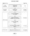
- FIG. 1 illustrates the time allotted for each step of the SNP assay.
- FIG. 2 shows the design of the primers, probes, and capture sequences of the CYP2D6 SNP Assay method (SEQ ID NOS: 17-22, 5-6, 23-24, 7-8 and 25-26, respectively, in order of appearance).
- FIG. 3 schematically illustrates the primers and probes used to detect an SNP in multiplex mode, and using LuminexTM microspheres.
- FIG. 4 is a graph bar demonstrating the cytochrome P450 genotyping results of a multiplex SNP assay conducted on four individual patient samples.
- nucleoside and nucleotide refer to nucleosides and nucleotides containing not only the conventional purine and pyrimidine bases, i.e., adenine (A), thymine (T), cytosine (C), guanine (G), and uracil (U), but also modified nucleosides and nucleotides.
- A adenine
- T thymine
- C cytosine
- G guanine
- U uracil
- Such modifications include, but are not limited to, methylation or acylation of a purine or pyrimidine moiety, substitution of a different heterocyclic ring structure for a pyrimidine ring or for one or both rings in the purine ring system, and protection of one or more functionalities, e.g., using a protecting group such as acetyl, difluoroacetyl, trifluoroacetyl, isobutyryl, benzoyl, and the like.
- a protecting group such as acetyl, difluoroacetyl, trifluoroacetyl, isobutyryl, benzoyl, and the like.
- Modified nucleosides and nucleotides also include modifications on the sugar moiety, e.g., wherein one or more of the hydroxyl groups are replaced with halide and/or hydrocarbyl substituents (typically aliphatic groups, in the latter case), or are functionalized as ethers, amines, or the like.
- Common analogs include, but are not limited to, 1-methyladenine, 2-methyladenine, N 6 -methyladenine, N 6 -isopentyl-adenine, 2-methylthio-N 6 -isopentyladenine, N,N-dimethyladenine, 8-bromoadenine, 2-thiocytosine, 3-methylcytosine, 5-methylcytosine, 5-ethylcytosine, 4-acetylcytosine, 1-methylguanine, 2-methylguanine, 7-methylguanine, 2,2-dimethylguanine, 8-bromo-guanine, 8-chloroguanine, 8-aminoguanine, 8-methylguanine, 8-thioguanine, 5-fluoro-uracil, 5-bromouracil, 5-chlorouracil, 5-iodouracil, 5-ethyluracil, 5-propyluracil, 5-methoxyuracil, 5-hydroxymethyluracil, 5-
- oligonucleotide encompasses polydeoxyribonucleotides (containing 2-deoxy-D-ribose), polyribonucleotides (containing D-ribose), any other type of polynucleotide that is an N-glycoside of a purine or pyrimidine base, and other polymers containing nonnucleotidic backbones (e.g., protein nucleic acids and synthetic sequence-specific nucleic acid polymers commercially available from the Anti-Gene Development Group, Corvallis, Oregon, as NeugeneTM polymers) or nonstandard linkages, providing that the polymers contain nucleobases in a configuration that allows for base pairing and base stacking, such as is found in DNA and RNA.
- nonnucleotidic backbones e.g., protein nucleic acids and synthetic sequence-specific nucleic acid polymers commercially available from the Anti-Gene Development Group, Corvallis, Oregon, as NeugeneTM polymers
- oligonucleotides herein include double- and single-stranded DNA, as well as double- and single-stranded RNA and DNA:RNA hybrids, and also include known types of modified oligonucleotides, such as, for example, oligonucleotides wherein one or more of the naturally occurring nucleotides is substituted with an analog; oligonucleotides containing internucleotide modifications such as, for example, those with uncharged linkages (e.g., methyl phosphonates, phosphotriesters, phosphoramidates, carbamates, etc.), negatively charged linkages (e.g., phosphorothioates, phosphorodithioates, etc.), and positively charged linkages (e.g., aminoalkylphosphoramidates, aminoalkylphosphotriesters), those containing pendant moieties, such as, for example, proteins (including nucleases, toxins, antibodies, signal peptides, poly-L-lysine
- PCR polymerase chain reaction
- PCR polymerase chain reaction
- the reaction mixture is then returned to polymerizing conditions (e.g., by lowering the temperature, inactivating a denaturing agent, or adding more polymerase), and a second cycle is initiated.
- the second cycle provides the two original strands, the two long products from cycle 1, two new long products (replicated from the original strands), and two "short products” replicated from the long products.
- the short products have the sequence of the target sequence (sense or antisense) with a primer at each end.
- an additional two long products are produced, and a number of short products equal to the number of long and short products remaining at the end of the previous cycle.
- the number of short products grows exponentially with each cycle. This amplification of a specific analyte sequence allows the detection of extremely small quantities of DNA.
- 3SR refers to a method of target nucleic acid amplification also known as the "self-sustained sequence replication" system as described in European Patent Publication No. 373,960 (published Jun. 20, 1990).
- LCR refers to a method of target nucleic acid amplification also known as the "ligase chain reaction” as described by Barany (1991), Proc. Nat. Acad. Sci . 88:189-193.
- probe refers to a structure comprised of a polynucleotide that forms a hybrid structure with a target sequence contained in a molecule (a "target molecule") in a sample undergoing analysis, due to complementarity of at least one sequence in the probe with the target sequence.
- the nucleotides of any particular probe may be deoxyribonucleotides, ribonucleotides, and/or synthetic nucleotide analogs.
- primer refers to a molecule that comprises an oligonucleotide, whether produced naturally as in a purified restriction digest or produced synthetically, which is capable of acting as a point of initiation of synthesis when placed under conditions in which synthesis of a primer extension product that is complementary to a nucleic acid strand is induced, i.e., in the presence of appropriate nucleotides and an agent for polymerization such as a DNA polymerase in an appropriate buffer and at a suitable temperature.
- Preferred primers for use herein are the dual-purpose primers described in European Patent Application No. to Quinn et al. for "Dual-Purpose Primers and Probes for Providing Enhanced Hybridization Assays by Disruption of Secondary Structure Formation," filed on even date herewith.
- binding sequences need not have perfect complementarity to provide stable hybrids. In many situations, stable hybrids will form where fewer than about 10% of the bases are mismatches, ignoring loops of four or more nucleotides. However, the high sensitivity of this assay will usually require that the probes that hybridize to the first and second variants of the target nucleotide sequence and the control nucleotide sequence have 100% homology to their targets.
- the term “complementary” intends to refer to an oligonucleotide that forms a stable duplex with its "complement” under assay conditions, generally where there is about 100% homology
- the term “substantially complementary” intends to refer to an oligonucleotide that forms a stable duplex with its "complement” under assay conditions, generally where there is about 90% or greater homology.
- hybridizing conditions is intended to mean those conditions of time, temperature, and pH, and the necessary amounts and concentrations of reactants and reagents, sufficient to allow at least a portion of complementary sequences to anneal with each other.
- time, temperature, and pH conditions required to accomplish hybridization depend on the size of the oligonucleotide probe to be hybridized, the degree of complementarity between the oligonucleotide probe and the target, and the presence of other materials in the hybridization reaction admixture.
- the actual conditions necessary for each hybridization step are well known in the art or can be determined without undue experimentation.
- Typical hybridizing conditions include the use of solutions buffered to a pH from about 7 to about 8.5 and temperatures of from about 30°C to about 60°C, preferably from about 37°C to about 55°C for a time period of from about one second to about one day, preferably from about 15 minutes to about 16 hours, and most preferably from about 15 minutes to about three hours.
- Hybridization conditions also include an effective buffer. Any buffer that is compatible, i.e., chemically inert, with respect to the probes and other components, yet still allows for hybridization between complementary base pairs, can be used.
- One particularly preferred buffer comprises 3X SSC, 50% formamide, 10% dextran sulfate (MW 500,000), 0.2% casein, 10 ⁇ g/mL poly A, and 100 ⁇ g/mL denatured salmon sperm DNA, wherein 1X SSC is 0.15 M sodium chloride and 0.015 M sodium citrate.
- Another particularly preferred buffer comprises 5X SSC, 0.1 to 0.3% sodium dodecyl sulfate, 10% dextran sulfate, 1 mM ZnCl 2 , and 10 mM MgCl 2 , wherein 1X SSC is as defined above.
- Other suitable buffers are known to those of ordinary skill in the art.
- Patient refers to an organism, preferably mammalian, from which the nucleic acid analyte is obtained.
- Preferred patients are mammalian organisms, e.g., humans.
- sample refers to a fluid or tissue obtained from an organism (e.g., a mammalian organism such as a human) that contains the nucleic acid analyte to be characterized.
- organism e.g., a mammalian organism such as a human
- samples include, without limitation: blood; plasma; serum; spinal fluid; lymph fluid; cell lysates; semen; secretions of the skin or respiratory, intestinal, or genitourinary tracts; tears; saliva; milk; and white blood cells.
- the term "attached” refers to coupling by covalent or non-covalent interactions (e.g., hydrophobic interactions, hydrogen bonds, etc.). Covalent bonds include, for example, ester, ether, phosphoester, amide, imide, carbon-sulphur bonds, carbon-phosphorous bonds, and the like. Methods for attaching oligonucleotides to substrates are known in the art and include, for example, blotting of the oligonucleotide onto the substrate.
- substrate refers to any solid or semi-solid surface to which a desired oligonucleotide may be anchored.
- Suitable substrates include any material that can immobilize an oligonucleotide and encompass, for example, glass, nitrocellulose, plastics including polyvinyl chloride (e.g., in sheets or microtiter wells), polystyrene latex (e.g., in beads or microtiter plates), polyvinylidine fluoride (e.g., in microtiter plates), polystyrene (e.g., in beads), metal, polymer gels, and the like.
- polyvinyl chloride e.g., in sheets or microtiter wells
- polystyrene latex e.g., in beads or microtiter plates
- polyvinylidine fluoride e.g., in microtiter plates
- polystyrene e.g., in beads
- metal polymer gels, and the
- Optional or “optionally” means that the subsequently described event or circumstance may or may not occur, and that the description includes instances where the said event or circumstance occurs as well as instances where it does not.
- reference to an "optional component" in an assay kit means that such a component may or may not be present in the assay kit, and the description includes assay kits wherein the component is present and assay kits wherein the component is not present.
- the method of this invention comprises a means for detecting the presence or absence of a genetic variation, e.g., mutation, in a nucleic acid analyte obtained from a sample.
- a genetic variation e.g., mutation
- the sequence itself is considered “polymorphic,” and the site at which the alteration occurs is referred to as a "polymorphic site.”
- the nucleic acid analyte of interest is a portion of genetic material that comprises or is suspected to comprise a polymorphic site.
- the method includes the preparation of labeled amplicons from the nucleic acid analyte contained in the sample.
- the actual steps of preparing labeled amplicons are well known to those of ordinary skill in the art.
- the nucleic acid analyte of interest contained in the sample is often located within a cell or virus.
- the nucleic acid analyte can be DNA, RNA, or some other nucleic acid-containing polymer, which can either be in single-stranded or double-stranded form.
- Cells containing the nucleic acid analyte of interest can be obtained from biological tissue or fluid samples using conventional techniques such as, for example, needle biopsy or swabbing.
- the cells so obtained may contain genetic material of the host, or they may contain non-host genetic material.
- samples obtained from a human individual may contain human genetic material and/or the genetic material of an infectious pathogen such as a bacterium, virus, or fungus.
- the nucleic acid analyte is then amplified, i.e., copied, using conventional techniques.
- amplification method any known amplification method can be used, preferred methods include PCR, 3SR, and LCR, with PCR being most preferred.
- PCR amplification is carried out in a mixture comprising the nucleic acid analyte, a polymerase (e.g., Taq polymerase), oligonucleotide primers, an excess of the four oligonucleotide (dNTP) monomers, and water that contains a buffer suitable for carrying out PCR (including conventional buffers comprising Tris-HCl, KCl, and MgCl 2 ).
- a buffer suitable for carrying out PCR including conventional buffers comprising Tris-HCl, KCl, and MgCl 2 .
- the mixture is then exposed to a series of replication cycles based on temperature. For example, the mixture is heated to about 94-96°C for several minutes during which time any double-stranded DNA is denatured into single-stranded DNA.
- the temperature of the mixture is lowered to about 50-65°C, during which time the oligonucleotide primers hybridize via hydrogen bonds to complementary sequences. Finally, the temperature of the mixture is increased to about 72°C, during which time the polymerase binds and extends a complementary strand from each primer. Since the sequence being amplified doubles after each sample, a theoretical amplification of one billion can be attained, thereby providing ample nucleic acid analyte for the present method.
- primers suitable for amplifying the nucleic acid analyte may be used in the PCR process.
- Primers are relatively short oligonucleotides (e.g., 10 - 30 bases in length) that are complementary to a portion of the nucleic acid to be amplified.
- the primers are annealed to the denatured nucleic acid and provide an initiation site for elongation of the new DNA molecule.
- Some primers can be specific to a particular nucleic acid sequence, while others are universal in nature, such that they anneal at locations found throughout the genome of a single organism or in the genomes of several organisms.
- Suitable PCR primers are prepared by means known to those of ordinary skill in the art, for example, by cloning and performing restriction of appropriate sequences, or by direct chemical synthesis. For example, one may employ the phosphotriester method described by S. A. Narang et al. (1979) Meth. Enzymol . 68:90, and U.S. Patent No. 4,356,270 to Itakura. Primers are also available from Sigma-Aldrich, Co. (St. Louis, Missouri) and other commercial suppliers. Other primers may also be used, such as those designed to hybridize to other portions of the nucleic acid analyte in order to avoid undesired secondary structure.
- the amplified nucleic acid analyte referred to herein as the "amplicon," must be labeled.
- labeled primers are used during the amplification step, thereby resulting in labeled amplicons as the labeled primers are incorporated into the amplicons.
- Labeled primers are available from commercial suppliers such as CPG, Inc. (Lincoln Park, New Jersey), or they can be prepared via conjugating a label to an unlabeled primer. Labeling oligonucleotides such as primers can be carried out using conventional coupling procedures. Specific coupling procedures, however, will vary depending on the reactive group or groups present on the label and/or on the oligonucleotide.
- nick translation procedures known to those of skill in the art are available for substituting an unlabeled nucleotide with a labeled nucleotide through the use of DNase I and other enzymes, thereby providing a labeled amplicon.
- Another method for labeling includes the addition of a labeled deoxynucleotide terminal deoxynucleotidyl transferase (TdT, available from commercial suppliers such as PanVera Corp., Madison, Wisconsin), a DNA polymerase that can catalyze the addition of labeled deoxynucleotides to the 3'-end of DNA fragments.
- TdT labeled deoxynucleotide terminal deoxynucleotidyl transferase
- DNA polymerase a DNA polymerase that can catalyze the addition of labeled deoxynucleotides to the 3'-end of DNA fragments.
- a second technique for preparing labeled amplicons involves a separate labeling step wherein unlabeled amplicons are subsequently coupled to a label.
- the techniques described above with respect to labeling primers can also be used to attach labels to unlabeled amplicons.
- any type of label can be attached to the amplicon.
- Preferred labels include those moieties detectable by spectroscopic, photochemical, biochemical, immunochemical, or chemical means. Such labels include, without limitation, fluorescers, chemiluminescers, dyes, biotin, haptens, enzymes, enzyme substrates, enzyme cofactors, enzyme inhibitors, enzyme subunits, metal ions, electron-dense reagents, and radioactive isotopes (e.g., 32 P).
- the label moiety can be directly or indirectly attached to the amplicon.
- the label should be selected to withstand denaturing conditions if it is to be attached directly to the primer. It is preferred, although not necessary, that the label be biotin, which can be detected via binding with streptavidin coupled to a fluorescer, e.g., a streptavidin-phycoerythrin conjugate.
- the labeled amplicons are contacted with a plurality of first and second differential hybridization probes under conditions that permit hybridization.
- the first differential hybridization probe comprises two binding regions: a first capture sequence portion and a region that is complementary to the polymorphic site corresponding to a wild type sequence.
- the first hybridization probe further comprises a spacer portion.
- wild type complexes are formed between the first hybridization probe and the nucleic acid analyte when the polymorphic site of nucleic acid analyte is in the "natural" or wild type form.
- second differential hybridization probes comprising two binding regions and an optional spacer portion are also contacted with the labeled amplicons under hybridization conditions.
- the two binding regions differ from the first differential hybridization probes in that the second hybridization probes each comprise a second capture sequence portion and a region that is complementary to the polymorphic site corresponding to a variant sequence. Consequently, any of the labeled amplicons made from a nucleic acid analyte having the variant sequence at the polymorphic site will hybridize to the corresponding complementary region of the second differential hybridization probe, thereby forming variant complexes.
- the optional spacer portion in the hybridization probes can be located at any part of the probe. It is preferred, however, that the spacer portion be located between the capture sequence portion and the region that is complementary to the polymorphic site.
- the spacer can comprise a series of nucleotides specifically designed not to hybridize or interfere with the hybridization events necessary for carrying out the present method, i.e., designed not to interfere with binding to other probes.
- nonnucleotidic spacers may be used. Methods suitable for preparing the optional spacer are known from the literature. The preparation of polyethylene glycol spacers is described, for example, by Kern et al. (1979) Makromol. Chem . 150:2539. Other spacers can be prepared in a similar manner.
- each of the first and second hybridization probes and the time required for hybridization can be determined experimentally, but it is preferred that a molar excess of each probe type be used. Generally, however, each of the first and second hybridization probes are added in an amount of from about 0.01 pmoles to about 100 pmoles, with a hybridization incubation period of from about one minute to about one day. It is preferred, however, that each of the first and second hybridization probes be added in an amount of from about 0.1 pmoles to about 10 pmoles for each type of probe, and allowed to incubate for a period of time lasting from about five minutes to about thirty minutes.
- variant complexes and/or wild type complexes will have formed (as discussed above), depending upon the polymorphic form of the nucleic acid analyte.
- These complexes, if any, are then "captured” using capture probes coupled to a solid substrate or a plurality of solid substrates.
- Each wild type complex is captured by a first capture probe comprised of a region that is complementary to the first capture sequence portion of the first differential hybridization probe.
- the first capture probe is coupled to a solid substrate.
- a plurality of first capture probes is used in order to capture substantially all of the wild type complexes.
- the variant complexes are captured in a similar way, recognizing that a plurality of second capture probes, each comprised of a region that is complementary to the second capture sequence portion and each attached to a plurality of second solid substrates, is used to capture the variant sequences.
- the preferred molar amounts and times used for capturing the complexes are the same as those disclosed above for forming the complexes.
- the complexes so captured through this step are referred to as "captured wild type complexes" and “captured variant complexes.”
- Detection and counting of the captured wild type complexes and captured variant complexes can take place using any conventional method.
- the captured complexes comprise a label (from the labeled amplicon) and a solid substrate (from the capture probe) having a detectable signal
- detection generally proceeds by detecting both the label and the signal.
- the labeling can be accomplished by any art-known means and is dependent upon the nature of the label.
- fluorescers a large number of fluorometers are available.
- chemiluminescers luminometers or films are available.
- enzymes a fluorescent, chemiluminescent, or colored product can be provided and determined fluorometrically, luminometrically, spectrophotometrically, or visually (preferably with the aid of a microscope).
- a biotinylated label is detected by adding a streptavidin-phycoerythrin conjugate, followed by detection of phycoerythrin-induced fluorescence.
- Each signal provided by the solid substrate is detected by any conventional means.
- the signal can be based on the shape of the substrate, wherein the first detectable signal and second detectable signal are differentiated by different shapes of the corresponding solid substrate.
- each set of solid substrates e.g., the plurality of first solid substrates, generates a signal unique to the plurality.
- each of the first solid substrates has a first detectable signal and each of the second solid substrates has a second detectable signal.
- the first and second detectable signals can be provided by moieties selected from the groups consisting of fluorescers, chemiluminescers, dyes, biotin, haptens, enzymes, enzyme substrates, enzyme cofactors, enzyme inhibitors, enzyme subunits, metal ions, electron-dense reagents, and radioactive isotopes.
- the solid substrates be beads, wherein a detectable signal is provided by one or more fluorescent dyes.
- a detectable signal is provided by one or more fluorescent dyes.
- suitably dyed substrates e.g., beads
- the solid substrates comprise a combination of fluorescent or colored dyes.
- Preferred dyes include cyanine dyes that are characterized by having emission wavelengths of between 550 nm to 900 nm. Some cyanine dyes have a blue to blue-green fluorescence, while others have green to yellow-green fluorescence. Still other cyanine dyes have red or even infrared fluorescence.
- the cyanine dye can be covalently attached to the substrate or adsorbed, e.g., "stained,” thereon.
- the substrate can have attached thereto one or more populations of fluorescently stained nanoparticles, wherein all nanoparticles in a given population have the same dye concentration.
- By varying the quantity and ratio of different dyes specific to different populations of nanoparticles associated with the substrate it is possible to establish and distinguish a large number of discreet populations of substrates with unique emission spectra.
- Such uniquely labeled substrates are particularly useful for multiplex analysis of sequences, and can be conveniently detected and analyzed using flow cytometry.
- Such beads are also available from commercial suppliers such as, for example, Luminex Corporation (Austin, Texas).
- a flow cytometer linked with one or more detecting means is used for detecting the complexes, although other means for detecting and counting the captured complexes can also be used, depending on the type of label and signal.
- the complexes in the hybridization solution are passed through the flow cytometer, thereby allowing the detection of each complex.
- the flow cytometer is linked with a first detecting means for detecting the label (of the labeled amplicon) as well as a second detecting means for detecting the signal associated with the solid substrate.
- Suitable equipment and methods for detecting the labels and signals using flow cytometry, and having the ability to perform multiplexing analysis, are described in U.S. Patent Nos. 5,981,180 to Chandler et al., and 6,046,807 and 6,139,800 to Chandler.
- Commercially available systems are also available from Luminex Corp. (Austin, Texas) and include, for example, the LuminexTM 100 machine.
- Substrates having a diameter of less than one millimeter can be used in flow cytometers, although other sized particles can be used as well. It is preferred, however, that substrates that are spherical in shape, e.g., beads, be used. Such beads have a size in the range of from about 0.1 to 1,000 ⁇ m, preferably 1 to 100 ⁇ m, more preferably 2 to 50 ⁇ m, still more preferably 3 to 25 ⁇ m, with beads having a diameter of from about 6 to 12 ⁇ m being most preferred. Beads of this size are suited for use in flow cytometers, thereby providing a facile means for detecting and counting the complexes.
- substrates that are spherical in shape e.g., beads.
- Such beads have a size in the range of from about 0.1 to 1,000 ⁇ m, preferably 1 to 100 ⁇ m, more preferably 2 to 50 ⁇ m, still more preferably 3 to 25 ⁇ m, with beads having a diameter of from about 6 to
- the solid substrates are preferably, although not necessarily, made of a polymeric material such as polystyrene.
- a polymeric material such as polystyrene.
- Other usable polymeric materials include brominated polystyrene, polyacrylic acid, polyacrylonitrile, polyamide, polyacrylamide, polyacrolein, polybutadiene, polycaprolactone, polycarbonate, polyester, polyethylene, polyethylene terephthalate, polydimethylsiloxane, polyisoprene, polyurethane, polyvinyl acetate, polyvinylchloride, polyvinylpyridine, polyvinylbenzylchloride, polyvinyltoluene, polyvinylidene chloride, polydivinylbenzene, polymethylmethacrylate, polylactide, polyglycolide, poly(lactide-co-glycolide), polyanhydride, polyorthoester, polyphosphazene, polyphosoph
- the presence or absence of the genetic variation can be determined. Specifically, a greater amount of captured wild type complexes is indicative of the absence of the genetic variation, whereas a greater amount of captured variant complexes is indicative of the presence of the variation. Relative comparisons such as these are sufficient to determine the sequence at a known polymorphic site. Additional techniques can be used, however, to obtain even more information.
- ratios can be developed to determine whether the sample contained a mixture of both the wild type and variant sequences.
- the results can be expressed as the ratio of the log net signals, wherein cutoffs can be used to identify what sequence or combination of sequences is contained in the sample.
- An example of such a ratio is provided in Table 1.
- results can be categorized in order to establish whether the sample predominantly or exclusively contains a single sequence type.
- results can be obtained wherein a mixture of two types of sequences is contained in a sample, e.g., when the sample is taken from an organism possessing the heterozygous condition.
- the invention also provides assay kits for carrying out the method described herein.
- the assay kits comprise in a packaged combination: (a) a plurality of first differential hybridization probes each comprised of a first capture sequence portion and a region that is complementary to the polymorphic site corresponding to a wild type sequence; (b) a plurality of second differential hybridization probes each comprised of a second capture sequence portion different from the first capture sequence portion and a region that is complementary to the polymorphic site corresponding to a variation sequence; (c) a plurality of first solid substrates each (i) comprised of an attached first capture probe complementary to the first capture region and (ii) having a first detectable signal; and (d) a plurality of second solid substrates each (i) comprised of an attached second capture probe complementary to the second capture region and (ii) having a second detectable signal.
- the kit may also contain a polymerase such as a DNA polymerase, Taq polymerase, or similar polymerase for creating amplicons.
- Primers preferably labeled, for carrying out PCR may also be included in the assay kits.
- the primers are preferably biotinylated primers.
- Hybridization buffers, enzyme substrates, negative controls, positive controls, and written instructions for carrying out the method described herein may also be conveniently included in the assay kit.
- oligonucleotides included with the kits and used in the methods described herein are known to those of ordinary skill in the art. Thus, conventional techniques are used to prepare the necessary primers, first hybridization probes, second hybridization probes, first capture probes, second capture probes, and so forth.
- the oligonucleotides can be prepared by oligonucleotide synthesis or by cloning, with the former preferred.
- Direct synthetic methods typically involve sequential addition of 3'-blocked and 5'-blocked nucleotide monomers to the terminal 5'-hydroxyl group of a growing oligonucleotide chain, wherein each addition is effected by nucleophilic attack of the terminal 5'-hydroxyl group of the growing chain on the 3'-position of the added monomer, which is typically a phosphorus derivative such as a phosphotriester, phosphoramidite, or the like.
- a phosphorus derivative such as a phosphotriester, phosphoramidite, or the like.
- the actual sequences of the oligonucleotides are determined using standard techniques known in the art. For each of the differential hybridization probes, it is first necessary to determine the sequence for each polymorphic site, i.e., the wild type and variant sequences.
- the wild type and variant sequences can be determined manually by first obtaining a sample of the nucleic acid analyte containing the polymorphic site of interest, and then sequencing the nucleic acid analyte using a commercial sequencer such as the Global IR 2 System (LI-COR, Inc., Lincoln, Kansas).
- the sequence of many nucleic analytes can be determined by referring to the relevant texts and databases providing wild type and variant sequences, such as the GenBank database (Bethesda, Maryland).
- the corresponding complementary sequence is included in the appropriate differential hybridization probe. That is, a region complementary to the polymorphic site corresponding to the wild type sequence is included in the first differential hybridization probe, and a region that is complementary to the variant sequence is included in the second differential hybridization probe.
- sequences used for capturing i.e., the sequences of the capture portions and the regions on the capture probes complementary thereto, can be directly attached to the solid substrate, or may be spaced therefrom by a spacer.
- spacers discussed above with respect to the differential hybridization probes can be used between the solid substrate and the capture sequence.
- natural nucleotides can be substituted with non-natural nucleotides such as iso-C and iso- G, and the sequences designed to hybridize to the capture regions can have the corresponding complementary non-natural nucleotides to enhance specificity and reduce background of the capture hybridization step. Methods for using the non-natural nucleotides to reduce nonspecific hybridization are described in U.S. Patent No. 6,232,462 to Collins et al.
- Those regions of the oligonucleotides that are intended to be involved with binding will each be at least 15 nucleotides, usually at least 20 nucleotides, and not more than about 100 nucleotides.
- the binding sequences will be approximately 25 nucleotides in length.
- the methods described herein have a vast number of uses.
- the method provides the ability to detect nucleic acid variants, e.g., SNPs, and other useful genetic or epigenomic data such a genetic subtype.
- the method can be easily adapted into a multiplex format when the appropriate probes and substrates are used. That is, multiplexed approaches based on the described method will include a different set of first and second hybridization probes, first and second capture probes, and substrates providing a first and second detectable signal for each polymorphic site of interest. Further, the present invention can simultaneously detect clusters of polymorphisms when the appropriate probes and substrates are used.
- the method provides the ability to proportionately quantify the relative amounts of a variant type to wild type.
- the addition of appropriate controls may provide additional quantitative information.
- the distribution and extent of DNA methylation within the nucleic acid target can also be determined using the present method.
- the present method can detect the presence of any type of genetic variation and the invention is not limited in this regard.
- Types of genetic variations particularly suited for detection using the present method include, without limitation, single nucleotide polymorphisms, clusters of polymorphisms, genetic disorders, allelic variations, exon sequences, intron sequences, and genetic subtypes.
- the cytochrome P450 CYP2D6 subtype is known to have four different SNPs, one at each of four different exons within the cytochrome P450 CYP2D6 subtype. These variations of the cytochrome P450 CYP2D6 subtype are responsible for differential rates of drug metabolism.
- the present method can detect which variant of the cytochrome P450 CYP2D6 subtype occurs in an individual thereby assisting in the determination of how well the individual will metabolize a drug.
- the nucleic acid analyte can be obtained from nearly any source.
- the nucleic acid analyte can be obtained from a gene belonging to a patient or can be taken from a viral, bacterial, or fungal organism. Other sources will be evident to one of ordinary skill in the art.
- Hybsimulator was found to be the preferred software for Tm prediction.
- human genomic DNA was extracted from 200 ⁇ L of EDTAtreated whole blood using QIAamp® DNA Blood Mini Kit as described by the manufacturer (Qiagen).
- allele specific hybridization probes were designed for use in a multiplexing mode.
- the presence or absence of a SNP in each of exons 1, 2, 6, and 9 was determined using allele specific hybridization probes comprising a sequence complementary to either the wild type or mutant allele, and further comprising unique capture sequences that are complementary to sequences attached to LuminexTM beads having unique color codes; the color detected indicating the identity of the allele specific sequence, and thus the presence of the wild type or mutant sequence in the target nucleotide sequence.
- the capture sequences were designed to minimize the potential non-specific binding to genomic DNA sequences that may be present in the amplified sequences by incorporating iso-cytosine and iso-guanine into the capture probes and complementary sequences attached to the LuminexTM beads.
- the sequences shown below were designed and tested for their ability to discriminate between wild type and mutant cytochrome P450 genotypes in a sample of amplified genomic DNA, using the primers shown above to amplify the sequences of interest.
- the Tm for melting of each allele specific oligonucleotide sequence - target nucleotide sequence duplex is shown, relative to wild type sequences.
- the allele specific hybridization probes designed to hybridize with wild type amplicon sequences are at least 10°C more stable (melting at least 10°C higher) than those allele specific hybridization probes designed to hybridize with mutant amplicon sequences. These melting temperatures are consistent with the predicted difference in melting temperatures for one base pair mismatched sequences of about 10°C.
- the allele specific hybridization probe for mutant amplicon sequences will be more stable by at least 10°C when the mutant amplicon sequences are present.
- J iso-guanine
- F iso-cytosine.
- the fourth step in the detection of wild type or mutant alleles for exons 1, 2, 6, and 9 of cytochrome P450 CPY2D6, is the carrying out of a singleplex or multiplex polymerase chain reaction.
- the singleplex or multiplex PCR was carried out using 2 ⁇ L (50-70 ng) isolated genomic DNA. Twenty-five microliter reaction volumes were prepared containing 1x Titanium Taq PCR buffer; 2.5 mM each dNTP's (dATP, dCTP, dGTP, and dTTP); 0.2 ⁇ M each forward (biotinylated) and reverse primers; and 2.5 U Titanium Taq DNA polymerase.
- a PE 9600 thermocycler was used. The thermal cycling conditions were 94°C for 2 min followed by 30 cycles of 95°C for 30 see, 68°C for 1 minute, followed by a final extension of 5 min at 68°C.
- the final step in the detection of wild type or mutant alleles for exons 1, 2, 6, and 9 of cytochrome P450 CPY2D6, is the SNP Assay, shown in FIG. 1 and carried out as follows. After the PCR reaction was complete, 25 ⁇ L of multiplex working reagent was added to the individual wells containing the multiplex PCR products. The plate was sealed with mylar and the incubation was continued in the PE 9600 thermocycler.
- the multiplex working reagent contained 0.1 pmol each allele specific hybridization probe (ASH) for the four SNP regions and 2000 each individual LuminexTM microspheres per 25 ⁇ L of 50 mM Hepes containing 500 mM LiCl, 1 % LDS, 1% BSA, 10 mM MgCl 2 , 0.01 mM ZnCl 2 and 0.5% sodium azide and Proclin-300 as a preservative (HIV 3.0 Label Diluent, Bayer Diagnostics), having a final pH of 7.5.
- ASH allele specific hybridization probe
- the PCR plate with the multiplex ASH working reagent was incubated for 10 minutes at 95°C to dissociate double stranded DNA followed by a 30 minute incubation at 50°C to achieve allele specific hybridization.
- the plate was removed and 100 ⁇ L of wash buffer was added to each well (HIV 3.0 Wash A, Bayer Diagnostics).
- the contents of the wells were transferred to a 96 well pre-wetted filter plate (MultiScreen®-BV 1.2 ⁇ m, Millipore, Bedford MA).
- the wash buffer was pulled through with gentle vacuum and a 200 ⁇ L wash was repeated.
- microspheres were resuspended in a 50 ⁇ L streptavidin phycoerythrin, (0.05 ⁇ g/50 ⁇ L) TTL buffer (50 mM Tris, 400 mM LiCl, 0.1% Tween-20, pH 8.0) mixture. The plate was then wrapped in aluminum foil and incubated for 15 minutes at 25°C with mild shaking (Titer Plate shaker, Labline Instruments). The previous wash step was repeated and the microspheres were resuspended in 80 ⁇ L of TTL buffer and read on the LuminexTM 100 in which the presence of phycoerythrin (and hence the original forward primer) and the microspheres specific for each particular ASH were detected.
- FIG. 2 shows the capture region for the wild type and variant discrimination probes for each of exons 1, 2, 6, and 9 of the cytochrome P450 CPY2D6 gene;
- FIG. 3 shows how the SNP assay results in the different color LuminexTM beads; and
- FIG. 4 shows the results of the SNP P450 Assay for four individual patients.
Landscapes
- Chemical & Material Sciences (AREA)
- Organic Chemistry (AREA)
- Life Sciences & Earth Sciences (AREA)
- Zoology (AREA)
- Wood Science & Technology (AREA)
- Proteomics, Peptides & Aminoacids (AREA)
- Health & Medical Sciences (AREA)
- Engineering & Computer Science (AREA)
- Microbiology (AREA)
- Biochemistry (AREA)
- Physics & Mathematics (AREA)
- Molecular Biology (AREA)
- Biotechnology (AREA)
- Biophysics (AREA)
- Analytical Chemistry (AREA)
- Immunology (AREA)
- Bioinformatics & Cheminformatics (AREA)
- General Engineering & Computer Science (AREA)
- General Health & Medical Sciences (AREA)
- Genetics & Genomics (AREA)
- Measuring Or Testing Involving Enzymes Or Micro-Organisms (AREA)
- Investigating Or Analysing Biological Materials (AREA)
- Investigating Or Analysing Materials By The Use Of Chemical Reactions (AREA)
Applications Claiming Priority (2)
| Application Number | Priority Date | Filing Date | Title |
|---|---|---|---|
| US41247702P | 2002-09-20 | 2002-09-20 | |
| US412477P | 2002-09-20 |
Publications (1)
| Publication Number | Publication Date |
|---|---|
| EP1400601A1 true EP1400601A1 (fr) | 2004-03-24 |
Family
ID=31946986
Family Applications (1)
| Application Number | Title | Priority Date | Filing Date |
|---|---|---|---|
| EP03255859A Withdrawn EP1400601A1 (fr) | 2002-09-20 | 2003-09-18 | Méthode pour détecter multiples variantes d'acides nucléiques |
Country Status (3)
| Country | Link |
|---|---|
| US (1) | US20040157238A1 (fr) |
| EP (1) | EP1400601A1 (fr) |
| JP (1) | JP2004113241A (fr) |
Cited By (5)
| Publication number | Priority date | Publication date | Assignee | Title |
|---|---|---|---|---|
| WO2007082881A3 (fr) * | 2006-01-17 | 2007-09-07 | Glaxosmithkline Biolog Sa | Analyse et materiel correspondant |
| WO2008111741A1 (fr) * | 2007-03-14 | 2008-09-18 | Seegene, Inc. | Méthode de détection de variations de nucléotides au moyen d'amorces marquées |
| US20100221702A1 (en) * | 2005-06-07 | 2010-09-02 | Eragen Biosciences, Inc. | Methods for detection and typing of nucleic acids |
| WO2011050278A1 (fr) * | 2009-10-23 | 2011-04-28 | Eragen Biosciences, Inc. | Amorces d'amplification avec des bases non-standard ayant une spécificité de réaction augmentée |
| IT201800005685A1 (it) * | 2018-05-24 | 2019-11-24 | Kit e procedimento per la rivelazione colorimetrica di un acido nucleico bersaglio in un campione. |
Families Citing this family (8)
| Publication number | Priority date | Publication date | Assignee | Title |
|---|---|---|---|---|
| WO2005086679A2 (fr) * | 2004-03-05 | 2005-09-22 | Medical College Of Ohio | Procedes et compositions pour l'evaluation d'acides nucleiques et d'alleles |
| DK2302070T3 (da) | 2005-06-23 | 2012-11-26 | Keygene Nv | Strategier til identifikation og detektion af polymorfismer med højt gennemløb |
| US10316364B2 (en) | 2005-09-29 | 2019-06-11 | Keygene N.V. | Method for identifying the source of an amplicon |
| WO2007037678A2 (fr) | 2005-09-29 | 2007-04-05 | Keygene N.V. | Criblage a haut debit de populations mutagenisees |
| CN103937899B (zh) | 2005-12-22 | 2017-09-08 | 凯津公司 | 用于基于aflp的高通量多态性检测的方法 |
| EP3239304B1 (fr) | 2006-04-04 | 2020-08-19 | Keygene N.V. | Détection à haut débit de marqueurs moléculaires sur la base d'aflp et de séquençage à haut débit |
| US11976330B2 (en) * | 2017-04-06 | 2024-05-07 | The University Of Vermont And State Agricultural College | MiRNA signature expression in cancer |
| CN116790740A (zh) * | 2023-04-27 | 2023-09-22 | 中国人民解放军总医院第一医学中心 | 一种新型的常见耳聋基因拷贝数检测诊断芯片构建方法 |
Citations (4)
| Publication number | Priority date | Publication date | Assignee | Title |
|---|---|---|---|---|
| EP1023464A1 (fr) * | 1997-10-14 | 2000-08-02 | Luminex Corporation | Particules fluorescentes de precision, et procede de fabrication et mode d'utilisation associes |
| US6235465B1 (en) * | 1991-12-23 | 2001-05-22 | Bayer Corporation | HTLV-1 probes for use in solution phase sandwich hybridization assays |
| WO2001048244A2 (fr) * | 1999-12-28 | 2001-07-05 | Glaxo Group Limited | Detection de polymorphismes de nucleotides a base simple |
| WO2002061121A2 (fr) * | 2001-01-29 | 2002-08-08 | Syngenta Participations Ag | Methodes d'analyse des acides nucleiques |
Family Cites Families (15)
| Publication number | Priority date | Publication date | Assignee | Title |
|---|---|---|---|---|
| US5030557A (en) * | 1987-11-24 | 1991-07-09 | Ml Technology Venture | Means and method for enhancing nucleic acid hybridization |
| US6187538B1 (en) * | 1993-12-13 | 2001-02-13 | Bayer Corporation | Differential hybridization for relative quantification of variant populations |
| US5681702A (en) * | 1994-08-30 | 1997-10-28 | Chiron Corporation | Reduction of nonspecific hybridization by using novel base-pairing schemes |
| US5888731A (en) * | 1995-08-30 | 1999-03-30 | Visible Genetics Inc. | Method for identification of mutations using ligation of multiple oligonucleotide probes |
| US5981180A (en) * | 1995-10-11 | 1999-11-09 | Luminex Corporation | Multiplexed analysis of clinical specimens apparatus and methods |
| US5736330A (en) * | 1995-10-11 | 1998-04-07 | Luminex Corporation | Method and compositions for flow cytometric determination of DNA sequences |
| US5884323A (en) * | 1995-10-13 | 1999-03-16 | 3Com Corporation | Extendible method and apparatus for synchronizing files on two different computer systems |
| US6265152B1 (en) * | 1995-12-22 | 2001-07-24 | Visible Genetics Inc. | Method and kit for evaluation of HIV mutations |
| AU2189397A (en) * | 1996-02-08 | 1997-08-28 | Affymetrix, Inc. | Chip-based speciation and phenotypic characterization of microorganisms |
| US6139800A (en) * | 1997-06-23 | 2000-10-31 | Luminex Corporation | Interlaced lasers for multiple fluorescence measurement |
| US6297018B1 (en) * | 1998-04-17 | 2001-10-02 | Ljl Biosystems, Inc. | Methods and apparatus for detecting nucleic acid polymorphisms |
| ATE239801T1 (de) * | 1998-01-22 | 2003-05-15 | Luminex Corp | Mikropartikel mit multiplen fluoreszenz-signalen |
| US6046807A (en) * | 1998-05-14 | 2000-04-04 | Luminex Corporation | Diode laser based measurement apparatus |
| US6268147B1 (en) * | 1998-11-02 | 2001-07-31 | Kenneth Loren Beattie | Nucleic acid analysis using sequence-targeted tandem hybridization |
| US6753143B2 (en) * | 2000-05-01 | 2004-06-22 | Clinical Micro Sensors, Inc. | Target analyte detection using asymmetrical self-assembled monolayers |
-
2003
- 2003-09-15 US US10/666,744 patent/US20040157238A1/en not_active Abandoned
- 2003-09-18 EP EP03255859A patent/EP1400601A1/fr not_active Withdrawn
- 2003-09-19 JP JP2003329192A patent/JP2004113241A/ja not_active Withdrawn
Patent Citations (4)
| Publication number | Priority date | Publication date | Assignee | Title |
|---|---|---|---|---|
| US6235465B1 (en) * | 1991-12-23 | 2001-05-22 | Bayer Corporation | HTLV-1 probes for use in solution phase sandwich hybridization assays |
| EP1023464A1 (fr) * | 1997-10-14 | 2000-08-02 | Luminex Corporation | Particules fluorescentes de precision, et procede de fabrication et mode d'utilisation associes |
| WO2001048244A2 (fr) * | 1999-12-28 | 2001-07-05 | Glaxo Group Limited | Detection de polymorphismes de nucleotides a base simple |
| WO2002061121A2 (fr) * | 2001-01-29 | 2002-08-08 | Syngenta Participations Ag | Methodes d'analyse des acides nucleiques |
Non-Patent Citations (5)
| Title |
|---|
| ARMSTRONG B ET AL: "SUSPENSON ARRAYS FOR HIGH THROUGHPUT, MULTIPLEXED SINGLE NUCLEOTIDE POLYMORPHISM GENOTYPING", CYTOMETRY, ALAN R. LISS, INC, XX, vol. 40, no. 2, 1 June 2000 (2000-06-01), pages 102 - 108, XP001106772, ISSN: 0196-4763 * |
| CHEN J ET AL: "A MICROSPHERE-BASED ASSAY FOR MULTIPLEXED SINGLE NUCLEOTIDE POLYMORPHISM ANALYSIS USING SINGLE BASE CHAIN EXTENSION", GENOME RESEARCH, COLD SPRING HARBOR LABORATORY PRESS, US, vol. 10, no. 4, April 2000 (2000-04-01), pages 549 - 557, XP000927257, ISSN: 1088-9051 * |
| IANNONE M A ET AL: "MULTIPLEXED SINGLE NUCLEOTIDE POLYMORPHISM GENOTYPING BY OLIGONUCLEOTIDE LIGATION AND FLOW CYTOMETRY", CYTOMETRY, ALAN R. LISS, INC, XX, vol. 39, no. 2, 2000, pages 131 - 140, XP001073442, ISSN: 0196-4763 * |
| WEDEMEYER N ET AL: "Flow cytometry: An 'old' tool for novel applications in medical genetics", CLINICAL GENETICS, vol. 60, no. 1, July 2001 (2001-07-01), pages 1 - 8, XP002259894, ISSN: 0009-9163 * |
| YE F ET AL: "FLUORESCENT MICROSPHERE-BASED READOUT TECHNOLOGY FOR MULTIPLEXED HUMAN SINGLE NUCLEOTIDE POLYMORPHISM ANALYSIS AND BACTERIAL INDENTIFICATION", HUMAN MUTATION, WILEY-LISS, NEW YORK, NY, US, vol. 17, no. 4, 2001, pages 305 - 316, XP001118024, ISSN: 1059-7794 * |
Cited By (12)
| Publication number | Priority date | Publication date | Assignee | Title |
|---|---|---|---|---|
| US20100221702A1 (en) * | 2005-06-07 | 2010-09-02 | Eragen Biosciences, Inc. | Methods for detection and typing of nucleic acids |
| US8293472B2 (en) * | 2005-06-07 | 2012-10-23 | Luminex Corporation | Methods for detection and typing of nucleic acids |
| US8936913B2 (en) | 2005-06-07 | 2015-01-20 | Luminex Corporation | Methods for detection and typing of nucleic acids |
| US9562261B2 (en) | 2005-06-07 | 2017-02-07 | Luminex Corporation | Methods for detection and typing of nucleic acids |
| US10385412B2 (en) | 2005-06-07 | 2019-08-20 | Luminex Corporation | Methods for detection and typing of nucleic acids |
| WO2007082881A3 (fr) * | 2006-01-17 | 2007-09-07 | Glaxosmithkline Biolog Sa | Analyse et materiel correspondant |
| CN101541974B (zh) * | 2006-01-17 | 2013-06-12 | 葛兰素史密丝克莱恩生物有限公司 | 杂交探针测定和阵列 |
| WO2008111741A1 (fr) * | 2007-03-14 | 2008-09-18 | Seegene, Inc. | Méthode de détection de variations de nucléotides au moyen d'amorces marquées |
| WO2011050278A1 (fr) * | 2009-10-23 | 2011-04-28 | Eragen Biosciences, Inc. | Amorces d'amplification avec des bases non-standard ayant une spécificité de réaction augmentée |
| US8440406B2 (en) | 2009-10-23 | 2013-05-14 | Luminex Corporation | Amplification primers with non-standard bases for increased reaction specificity |
| IT201800005685A1 (it) * | 2018-05-24 | 2019-11-24 | Kit e procedimento per la rivelazione colorimetrica di un acido nucleico bersaglio in un campione. | |
| WO2019224731A1 (fr) * | 2018-05-24 | 2019-11-28 | Fondazione Istituto Italiano Di Tecnologia | Kit et procédé de détection colorimétrique d'un acide nucléique cible dans un échantillon |
Also Published As
| Publication number | Publication date |
|---|---|
| US20040157238A1 (en) | 2004-08-12 |
| JP2004113241A (ja) | 2004-04-15 |
Similar Documents
| Publication | Publication Date | Title |
|---|---|---|
| EP1076721B1 (fr) | Generation de produits d'amplification de l'acide nucleique par utilisation de concentrations inegales d'amorces | |
| US6232455B1 (en) | Nucleic acid primers and probes for detecting HIV-1 and HIV-2 | |
| US7250252B2 (en) | Amplification based polymorphism detection | |
| EP1428892B1 (fr) | Amorces et sondes à double usage pour l'amélioration d'essais d'hybridations parla perturbation de la formation de structures secondaires | |
| EP1400601A1 (fr) | Méthode pour détecter multiples variantes d'acides nucléiques | |
| EP2598656B1 (fr) | Acides nucléiques témoins pour de multiples paramètres | |
| WO2007083928A1 (fr) | Puce à adn permettant le diagnostic d'une dystrophie cornéenne | |
| WO2008076856A2 (fr) | Réactifs et procédés permettant de détecter des polymorphismes de cyp2d6 | |
| EP2215245A2 (fr) | Épreuve biologique de détection de cible s'effectuant en une seule étape | |
| US8198422B2 (en) | High-risk human papillomavirus detection | |
| US20060172284A1 (en) | Highly orthogonal universal sequences for use in nucleic acid assays | |
| US20010039012A1 (en) | Methods for diagnostic screening | |
| EP2271778B1 (fr) | Procédé de détection du virus de l'herpès dans un échantillon a tester | |
| EP4182482A1 (fr) | Détection du mycoplasma genitalium résistant aux macrolides | |
| EP2002020A2 (fr) | Détection de polymorphismes de nucléotide simple à partir d'adn génomique non amplifié | |
| WO2000011215A2 (fr) | Procede de depistage diagnostique | |
| US7211381B1 (en) | β2 andrenergic polymorphism detection | |
| CN120457217A (zh) | 用于检测和分析单纯疱疹病毒1型(hsv-1)、单纯疱疹病毒2型(hsv-2)和水痘带状疱疹病毒(vzv)的组合物和方法 | |
| EP1330549A2 (fr) | Genotypage a l'aide de sondes partiellement complementaires |
Legal Events
| Date | Code | Title | Description |
|---|---|---|---|
| PUAI | Public reference made under article 153(3) epc to a published international application that has entered the european phase |
Free format text: ORIGINAL CODE: 0009012 |
|
| AK | Designated contracting states |
Kind code of ref document: A1 Designated state(s): AT BE BG CH CY CZ DE DK EE ES FI FR GB GR HU IE IT LI LU MC NL PT RO SE SI SK TR |
|
| AX | Request for extension of the european patent |
Extension state: AL LT LV MK |
|
| 17P | Request for examination filed |
Effective date: 20040924 |
|
| AKX | Designation fees paid |
Designated state(s): AT BE BG CH CY CZ DE DK EE ES FI FR GB GR HU IE IT LI LU MC NL PT RO SE SI SK TR |
|
| 17Q | First examination report despatched |
Effective date: 20050301 |
|
| RAP1 | Party data changed (applicant data changed or rights of an application transferred) |
Owner name: BAYER HEALTHCARE LLC |
|
| 17Q | First examination report despatched |
Effective date: 20050301 |
|
| STAA | Information on the status of an ep patent application or granted ep patent |
Free format text: STATUS: THE APPLICATION IS DEEMED TO BE WITHDRAWN |
|
| 18D | Application deemed to be withdrawn |
Effective date: 20070718 |