EP1409727B1 - Verfahren zum qualitativen und/oder quantitativen nachweis von zellen - Google Patents
Verfahren zum qualitativen und/oder quantitativen nachweis von zellen Download PDFInfo
- Publication number
- EP1409727B1 EP1409727B1 EP02732726A EP02732726A EP1409727B1 EP 1409727 B1 EP1409727 B1 EP 1409727B1 EP 02732726 A EP02732726 A EP 02732726A EP 02732726 A EP02732726 A EP 02732726A EP 1409727 B1 EP1409727 B1 EP 1409727B1
- Authority
- EP
- European Patent Office
- Prior art keywords
- cells
- antibodies
- mrna
- portions
- combination
- Prior art date
- Legal status (The legal status is an assumption and is not a legal conclusion. Google has not performed a legal analysis and makes no representation as to the accuracy of the status listed.)
- Expired - Lifetime
Links
Images


Classifications
-
- G—PHYSICS
- G01—MEASURING; TESTING
- G01N—INVESTIGATING OR ANALYSING MATERIALS BY DETERMINING THEIR CHEMICAL OR PHYSICAL PROPERTIES
- G01N33/00—Investigating or analysing materials by specific methods not covered by groups G01N1/00 - G01N31/00
- G01N33/48—Biological material, e.g. blood, urine; Haemocytometers
- G01N33/50—Chemical analysis of biological material, e.g. blood, urine; Testing involving biospecific ligand binding methods; Immunological testing
- G01N33/53—Immunoassay; Biospecific binding assay; Materials therefor
- G01N33/575—Immunoassay; Biospecific binding assay; Materials therefor for cancer
- G01N33/5758—Immunoassay; Biospecific binding assay; Materials therefor for cancer involving compounds serving as markers for tumours, cancers or neoplasias, e.g. cellular determinants, receptors, heat shock/stress proteins, A-protein, oligosaccharides or metabolites
- G01N33/5759—Immunoassay; Biospecific binding assay; Materials therefor for cancer involving compounds serving as markers for tumours, cancers or neoplasias, e.g. cellular determinants, receptors, heat shock/stress proteins, A-protein, oligosaccharides or metabolites involving compounds localised on the membrane of tumour or cancer cells
-
- C—CHEMISTRY; METALLURGY
- C07—ORGANIC CHEMISTRY
- C07K—PEPTIDES
- C07K16/00—Immunoglobulins [IG], e.g. monoclonal or polyclonal antibodies
- C07K16/18—Immunoglobulins [IG], e.g. monoclonal or polyclonal antibodies against material from animals or humans
- C07K16/28—Immunoglobulins [IG], e.g. monoclonal or polyclonal antibodies against material from animals or humans against receptors, cell surface antigens or cell surface determinants
- C07K16/30—Immunoglobulins [IG], e.g. monoclonal or polyclonal antibodies against material from animals or humans against receptors, cell surface antigens or cell surface determinants from tumour cells
-
- G—PHYSICS
- G01—MEASURING; TESTING
- G01N—INVESTIGATING OR ANALYSING MATERIALS BY DETERMINING THEIR CHEMICAL OR PHYSICAL PROPERTIES
- G01N33/00—Investigating or analysing materials by specific methods not covered by groups G01N1/00 - G01N31/00
- G01N33/48—Biological material, e.g. blood, urine; Haemocytometers
- G01N33/50—Chemical analysis of biological material, e.g. blood, urine; Testing involving biospecific ligand binding methods; Immunological testing
- G01N33/53—Immunoassay; Biospecific binding assay; Materials therefor
- G01N33/543—Immunoassay; Biospecific binding assay; Materials therefor with an insoluble carrier for immobilising immunochemicals
- G01N33/54313—Immunoassay; Biospecific binding assay; Materials therefor with an insoluble carrier for immobilising immunochemicals the carrier being characterised by its particulate form
- G01N33/54326—Magnetic particles
-
- G—PHYSICS
- G01—MEASURING; TESTING
- G01N—INVESTIGATING OR ANALYSING MATERIALS BY DETERMINING THEIR CHEMICAL OR PHYSICAL PROPERTIES
- G01N33/00—Investigating or analysing materials by specific methods not covered by groups G01N1/00 - G01N31/00
- G01N33/48—Biological material, e.g. blood, urine; Haemocytometers
- G01N33/50—Chemical analysis of biological material, e.g. blood, urine; Testing involving biospecific ligand binding methods; Immunological testing
- G01N33/53—Immunoassay; Biospecific binding assay; Materials therefor
- G01N33/569—Immunoassay; Biospecific binding assay; Materials therefor for microorganisms, e.g. protozoa, bacteria, viruses
- G01N33/56966—Animal cells
Definitions
- the present invention relates to a method for Qualitative and / or quantitative detection of cells in a sample. Such detection methods are especially in diagnostics or treatment control needed by tumors. Because in the It is very important for cancer prevention or follow-up Importance of malignant tumors or recurrent malignancies Tumors based on the occurrence of metastatic tumor cells in the blood early to be able to prove. The However, the present method is not used only here but can basically prove it and for the detection of rare cells in biological Cells containing samples are used. This For example, it can also be used to detect fetal Cells in maternal blood or for the detection of Stem cells take place.
- Testicular cancer accounts for less than 2% of all malignant Neoplasms of tumors in men responsible. However, it is 20-30% of all cancers under testimony of men under 40 years old. The Number of annual new cases, for example in the Federal Republic of Germany amounts to approx. 3600, whereby About 240 men die from testicular cancer. The highest Incidence is found between the 25th and 40th Age. Through the progress in the oncological Today, over 90% of sufferers can receive therapy be cured in the long term. The high survival rates are justified in the pronounced effectiveness cis-platinum-based chemotherapy.
- the colorectal primary tumor is tumor progression after resection primarily on residual Attributed to tumor cells. These cells are pre- or detached intraoperatively from the primary tumor and get the opportunity for scattering throughout Organism.
- tumor markers Protein level immunological or enzymatic
- these detection methods are for the Tumor diagnostics or treatment control / aftercare for tumors only conditionally suitable, since increased tumor marker values in body fluids also by non-tumorous Diseases such as inflammation of the gastrointestinal tract, liver cirrhosis, viral infections or heavy smoking can be caused.
- EP 0 520 794 B1 discloses such a method in the metastases in body tissues or Liquids are detected. This will be nucleic acids recorded, for example by means of duplication by polymerase chain reaction. The procedure is now crucial that the proven Nucleic acid sequence in cells of the tissue of origin a tumor is expressed, i. in tumor cells and Marker-dependent also in the healthy cells of the tissue of origin. Another condition is that this sequence not normally expressed in those cells whose tissue is being examined. Will be so a corresponding sequence in the examined sample found, it must be removed from, i. metastasizing Cells of a non-local tumor originate. Thus, this method is ultimately based on the Detection of cells in the blood sample of healthy Persons should not occur.
- WO 98/12227 A1 discloses a method for detection the expression of the GP-54 antigen. To do this first immunologically using a sample of cells two against different epitopes of the GP-54 antigen directed antibodies isolated. For these isolated Cells then become proof of expression from GP-54 by analysis of mRNA.
- WO 97/37226 A1 discloses a method for obtaining of tumor cells or fragments of tumor cells by means of an antibody or an antibody mixture. The analysis of the cells or cell fragments obtained in this way takes place via optical procedures, which it the Viewers allow tumor cells from normal cells to distinguish.
- WO 96/29430 A1 discloses the detection of tumor cells with a multi-marker assay.
- two or more mRNA markers in samples for example Blood samples, examined.
- the results of this molecular biological multi-marker detection with a statistically significant reference group of normal and melanoma or breast cancer patients compared and based on this comparison one Correlation of the number and type of markers with different ones clinical conditions.
- This procedure is also used in occult tumors Patients with no clinical signs are present for a tumor.
- a second step will be on molecular biological Base with a predefined combination detected by detection reagents at least one marker, which is specific to the cells sought or to be found in these preferential, so that Here again specifically selected the selected cells become. So this is a combined one molecular biological detection.
- the present invention is a proof via a combination of immunological parameters with evidence of a combination of molecular biological To combine parameters. Surprisingly result in this very excellent Detection results with which in detection areas is advanced, all known techniques Previously not accessible from the prior art. Thus, concentrations of wanted cells in Blood samples down to two cells per 5 milliliters yet to be proven. Such specificity and sensitivity is heretofore known in the art not reached.
- Eukaryotic cells carry a variety of different Molecules on their cell surface. Corresponding the origin and function of the individual Cell differs the combination of expressed surface molecules, so that cell type specific Patterns arise. To recognize this Cell-type-specific patterns are used antibodies. Antibodies bind to it with high specificity Antigen, here on selected surface molecules. This property is used to cells by means of specific antibody binding by cell type specific Recognize patterns and from each other differ.
- the expression differentiates special Surface proteins of tumor cells of non-transmembrane cells of this cell type.
- the detection of the markers is a selection of the target cells about the binding of different antibodies the searched cells ahead. Because the expression special Surface proteins distinguishes one cells Type of cells of a different type. So different for example, the expression of special surface proteins Tumor cells from non-transformed Cells of this cell type.
- tumor cells also of the blood cell typical Differentiates patterns, can tumor cells be distinguished in the blood.
- To identify tumor cells be antibodies that those special Specifically recognize surface proteins as tools used. The specific antibody binding becomes for different analysis and separation methods harnessed.
- a blood sample containing target cells is using antibody-coupled magnetic particles offset; then particles and blood become relative to each other moved, for example, by "over-head rotation" in a closed container Samples or due to movement of the particles due alternating magnetic fields. Those target cells by one of the solid phase-bound antibodies recognized and to be firmly bound, follow the movement of Particle. This makes it possible when creating a magnetic field, the particles with the attached Extract cells from the blood (e.g. the wall of the separation vessel). The target cell-depleted in this way Blood can be against other solutions exchanged, with the over magnetic particles separated cells until turning off / removing the Magnetic field remain on site and for other applications be available.
- antibody mixtures which are specific for the detection of the tumor cells are advantageously used.
- a combination of the antibodies MOC-31 and Ber-EP4 is suitable for detecting tumor cells in the blood.
- Antibody mixture antigen clone concentration Epith.Rel.Antigen MOC-31 (Novocastra) 1.25 ⁇ l / 10 8 cells
- Epithelial antigen Ber-EP 4 (DAKO) 0.924 ⁇ g / 10 6 cells
- Such antibody mixtures show in comparison to each separately used antibodies in the Cell recognition and cell separation regardless of the applied Method an increased sensitivity.
- the present invention is still substantially based that not about immunological or enzymatic Level cell markers in the blood of patients be proven, but that as a molecular biological Marker the mRNA (messenger ribonucleic acid) of sought cells in a sample, for example a blood sample is detected.
- mRNA messenger ribonucleic acid
- the expression of the genes mentioned in Table 2 is detected.
- the detection can be carried out for two markers or for any number of these tumor markers in combination with each other.
- the kit of the invention may therefore contain oligonucleotide pairs for two or any selection or even all of the tumor markers.
- a peripheral blood sample was taken and added to this a defined number of target cells, for example, 2, 10, 100 cells of a given one Tumor type.
- antibodies were coupled to magnetic particles.
- the antibodies used in the following in Table 3 were used as antibodies.
- antigen clone company Epithelial membrane antigen GP1.4 Novocastra Epithelial antigen MOC-31
- Novocastra Epithelial antigen Ber-EP4 DAKO Muc 1 HMPV.2 Pharmingen PLAP 8B6 Cymbus Biotechnology LTD
- the magnetic particles were used at a particle concentration of 4 ⁇ 10 8 beads / ml (CELLection TM Pan Mouse IgG Kit, Dynal). The ratios between the antibody concentration and the antibodies coupled thereto are shown in Table 4.
- clone Antibody concentration ⁇ l antibody / 25 ⁇ l particle BerEP4 0.1 mg / ml 4 ⁇ l HMPV.2 0.5 mg / ml 4 ⁇ l MOC31 kA dilution s.
- Manufacturer's information (lyophilisate) 4 ⁇ l 8B6 0.1 mg / ml 1 ⁇ l 131-11741 0.5 mg / ml 4 ⁇ l E29 0.1 mg / ml 4 ⁇ l
- the prepared magnetic particles were added to the blood depending on the experimental approach and detection system.
- the corresponding addition of antibody-coupled magnetic particles per ml of blood at a starting concentration of 4 ⁇ 10 8 beads / ml of particles is reproduced in Table 5.
- the magnetic particles optionally as cell-antibody-magnetic particle complexes templates, by means of a magnetic particle concentrator (MPC®-S, Dynal company) Washed 3 times with PBS (phosphate buffer saline) and then attach the adherent cells accordingly of the RNA isolation protocol described below treated.
- MPC®-S magnetic particle concentrator
- PBS phosphate buffer saline
- erythrocytes Here is a first relative accumulation of tumor cells achieved by depletion of the erythrocytes.
- a hypotonic erythrocyte lysis buffer mixed and left for 30 minutes at room temperature incubated.
- the remaining nucleated Cells are centrifuged and resuspended in PBS / BSA.
- the cells thus obtained are subsequently incubated with antibodies containing a fluorophore are marked.
- the target cells by binding to an antibody is fluorescently labeled then separated via FACS.
- mRNA isolation e.g. B. by means Oligo (dT) -coupled magnetic particles, Dynabeads® mRNA Direct TM Micro Kit, (Dynal)). This isolation is carried out according to the in the Kit specified manufacturer's information.
- RNA isolation As another alternative to RNA isolation, the isolated cells by adding Trizol reagent (Gibco BRL, NY, USA) lysed and pipetted homogenized. After subsequent chloroform extraction the RNA-containing aqueous phase is in isopropanol precipitated at -80 ° C. After washing twice and centrifuging in 80% ethanol becomes the pellet dried in air and then in RNase resuspended in free water. This work-up step also takes place according to conventional protocols.
- Trizol reagent Gibco BRL, NY, USA
- RNA isolation of the RNA is followed by a reverse Transcription, in which the mRNA is rewritten into cDNA becomes.
- RNA is denatured in a corresponding volume of water according to the reaction mixture in Table 6 together with oligo (dT) 15 primers (Promega, Mannheim) for 5 minutes at 65 ° C and then incubated directly on ice.
- oligo (dT) 15 primers Promega, Mannheim
- cDNA Synthesis is carried out in a 20 ⁇ l reaction mixture components volume final concentration RNA / mRNA 10 ⁇ l - 10x RT buffer 2 ⁇ l 1 x dNTP mix (5 mM each) 2 ⁇ l 0.5 mM each Oligo (dT) primer (10 ⁇ M) 2 ⁇ l 1 ⁇ M RNase inhibitor 1 ⁇ l 0.5 units / ⁇ l Reverse transcriptase 1 ⁇ l 4 U RNase-free water ad 20 ⁇ l
- the cDNA synthesis was carried out at 37 ° C for one hour with subsequent inactivation of the reverse transcriptase by heating for 5 minutes at 95 ° C and subsequent cooling on ice.
- This was a Sensiscript Reverse Transcriptase Kit, Qiagen, Hilden according to protocols specified there.
- oligo (dT) -coupled magnetic particles already fails to isolate mRNA then the addition of oligo (dT) primers, i. the oligo (dT) linker also serves as a primer for the reverse transcription, in which case the magnetic particles remain in the batch.
- PCR polymerase chain reaction
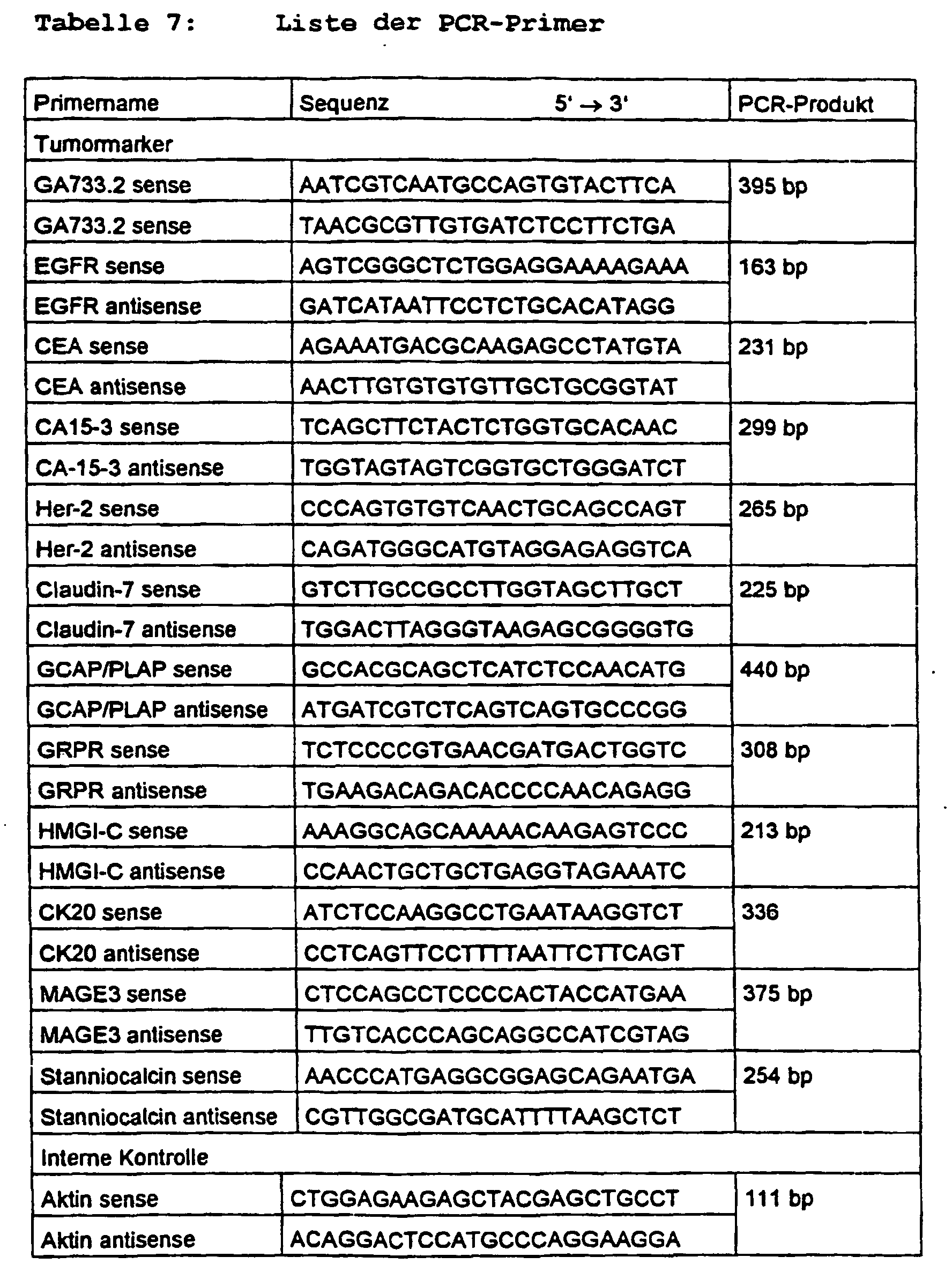
- oligonucleotides listed in Table 7 were used as PCR primers for the amplification of cDNA according to different marker genes, as indicated in the first column.
- Table 7 contains in the first column the indication of the tumor marker to be detected, whereby in each case two oligonucleotides (sense and antisense) are indicated as primer pair.
- the length of the PCR product generated by the primers shown in column two is given in column three.
- the PCR was carried out using the approach given in Table 8. PCR Approach The PCR synthesis was carried out in a 50 ⁇ l reaction mixture components volume final concentration cDNA 6 ⁇ l 10 x PCR buffer 5 ⁇ l 1 x dNTP mix 1 ⁇ l 200 ⁇ M each primer s.
- Tables 7 and 9 Taq DNA polymerase 0.5 ⁇ l 2.5 U [Q-Solution 10 ⁇ l] H 2 O ad 50 ⁇ l
- Table 9 gives a list of the specific primer combination and primer concentrations as the final concentration in the PCR approach.
- a multiplex combination to these primers is shown by way of example for the various tumor types breast cancer, colon cancer and testicular cancer, as shown in Table 9.
- PCR conditions (number of cycles, cycles, etc.) are given in Tables 10 and 11. PCR conditions Vorabdenaturierung 95 ° C 15 min cycle 1. denaturation 94 ° C 1 min Second annealing x ° C 1 min (see Table 11) Third extension 72 ° C 1 min Final Extension 72 ° C 10 min 4 ° C break Multiplex-specific annealing temperature and number of cycles marker Breast Cancer 1 Colon Cancer 1 Testicular Cancer 1 Annealing temperature 60 ° C 58 ° C 58 ° C number of cycles 35 40 40 (Thermocycler: PCT 200, Biozym)
- the amplified cDNAs thus generated were electrophoresed separated by a Bioanalyzer 2100 (Agilent company). To this was added 1 ⁇ l of the PCR product in the bioanalyzer on a DNA chip (500) separated and the separation result is documented electronically. In this way, Figures 1-10 were created.
- PCR product 25 ⁇ l is transferred over Separated 2.5% agarose gel and the DNA bands stained with ethidium bromide.
- the documentation is done e.g. with the help of the company's DUO Store System Intas.
- a fragment analysis may continue, for example using an ABI Prism 310 Genetic Analyzer (PE Applied Biosystem, Rothstadt) be performed.
- ABI Prism 310 Genetic Analyzer PE Applied Biosystem, Rothstadt
- fluorescently labeled primers used.
- FIGS. 1A and 1B now each show electrophoretic Separations, with the individual bands here and below labeled with the same terms are.
- the term ladder refers to calibrators from 50-600bp length, with RT-Ko is a control designated, which contained no mRNA, with PCR Ko is a control measurement called that no cDNA prior to PCR.
- Blood is the Blood sample without inoculated tumor cells, with 10 Z die Blood sample with 10 inoculated tumor cells per milliliter and with 100Z inoculated the blood sample with 100 Tumor cells per milliliter or per 5 milliliters designated.
- cell line is a control measurement with a large cell number of the tumor cell line in the Sample designated.
- FIG. 2 now shows results of detection methods, in which in Figure 2A no preselection by means of antibody labeling and in Figure 2B a preselection means Antibody labeling was performed.
- FIG 2 are combinations of antibodies HMPV.2, Ber-Ep 4 and GP1.4 as a combination of two or all three antibodies were determined.
- GP1.4 as a combination of two or all three antibodies were determined.
- a background expression for some of the mRNA markers (GA 733.2, CA 15.3 and Her 2 / new).
- FIG. 4 shows the detection of colon cancer cells which were inoculated into the blood of a healthy person.
- FIG. 4 shows Immediately, that the use of a combination the antibody Ber-Ep 4 and MOC-31 to an improved Sensitivity to the mRNA marker EGF-R leads.
- a detection sensitivity of 100 cells per milliliter of blood achieved while at Use only one antibody sensitivity at about 1000 cells per milliliter of blood.
- Figure 5 shows an attempt in which by means of a Antibody combination of Ber-Ep 4, HMPV.2 and GP1.4, those labeled with at least one of the antibodies Cells were selected and then on the mRNA marker GA 733.2, CA 15.3, Her 2 and claudin 7 were examined. However, here was the blood of the Healthy no tumor cell added but defined Amounts of epithelial cells. As shown in Figure 5 directly only two of the markers show in a very high number of epithelial cells a positive result.
- FIG. 6 shows tumor cells of two different types Breast cancer cell lines (MCF-7 and SKBR-3) of a blood sample added.
- MCF-7 and SKBR-3 breast cancer cell lines
- the antibodies Ber-Ep 4, HMPV.2 and GP1.4 used in combination.
- the four mRNA markers GA 733.2, CA 15.3, Her 2 and Claudin 7 each secured down to 10 cells the individual cell lines per milliliter detection ensures the breast cancer cells.
- An unspecific Reaction in blood without breast cancer cells occurred not up.
- the tumor markers GA 733.2 and CA 15.3 for cell line 2 (SKBR-3) more sensitive
- the tumor marker Her 2 for the cell line 1 (MCF-7) is more sensitive. So can the individual Subtypes of breast cancer cells based on the distinguished marker pattern from each other become.
- breast cancer cells are different Cell lines (MCF-7) in Figure 7A and SKBR 3 in Figure 7B inoculated in blood.
- MCF-7 Cell lines
- the cells from the blood sample became the antibodies Ber-Ep 4, HMPV.2 and GPl.4 used in combination.
- mRNA markers GA 733.2, CA 15.3 Her 2 and Claudin 7 in each case at least one the marker down to 2 cells per 5 milliliters positive reacts without the blood without tumor cells would provide an expression background.
- mRNA markers GA 733.2, CA 15.3 Her 2 and Claudin 7 in each case at least one the marker down to 2 cells per 5 milliliters positive reacts without the blood without tumor cells would provide an expression background.
- Both cell lines would be selected by the marker combination in any case down to 2 cells per 5 milliliters of blood ensures recognition of both cell lines, d. H. the detection of breast cancer cells in the blood would be independent of the cell type of the breast cancer cell line possible with high sensitivity.
- Figure 8 shows the detection of colon cancer cells, the were inoculated in blood.
- the selection took place the cells with the two antibodies Ber-Ep 4 and MOC 31st
- the molecular biological detection step took place with the mRNA markers GA 733.2, CEA and EGF-R. Two tumor cells were detectable in 5 milliliters of blood.
- FIG. 9 again shows a measurement with a combination from three antibodies Ber-Ep 4, MOC-31 and 8B6 and the mRNA markers GCAP / PLAP, GA 733.2, GRPR and HMGI-C to blood into which testicular cancer cells are inoculated were.
- FIG. 10 shows the separation of rare ones Cells from the cell mixture of a biopsy material.
- biopsy material from breast tissue containing tumor tissue with a suspected primary tumor, mechanically isolated and over gauze of cell debris, Connective tissue etc. separated.
- the won Cell mixture containing both cells of the suspected tumor tissue as well as cells of the surrounding healthy tissue contained a cell selection with a antibody mixture coupled to a solid phase (magnetic particles with antibodies GP1.4, HMPV.2 and Ber-Ep 4) and after incubation to prepare the Antigen antibody binding magnetically separated.
- a mRNA-proof concerning the Marker GA 733.2 and Her 2 As control was at the same time a cell line of a breast cancer in parallel also determined. As can be seen, takes place positive proof that the biopsy actually has a Breast cancer tumor included.
- FIGS. 8 and 9 with "positive control” show results from Samples with cell lines HT 29 for intestinal tumor (FIG. 8) or tera / l for testicular tumor (FIG. 9).
Landscapes
- Health & Medical Sciences (AREA)
- Life Sciences & Earth Sciences (AREA)
- Immunology (AREA)
- Chemical & Material Sciences (AREA)
- Engineering & Computer Science (AREA)
- Molecular Biology (AREA)
- Hematology (AREA)
- Urology & Nephrology (AREA)
- Biomedical Technology (AREA)
- Cell Biology (AREA)
- Biochemistry (AREA)
- Medicinal Chemistry (AREA)
- General Health & Medical Sciences (AREA)
- Physics & Mathematics (AREA)
- Food Science & Technology (AREA)
- Analytical Chemistry (AREA)
- Microbiology (AREA)
- Biotechnology (AREA)
- General Physics & Mathematics (AREA)
- Pathology (AREA)
- Organic Chemistry (AREA)
- Proteomics, Peptides & Aminoacids (AREA)
- Genetics & Genomics (AREA)
- Biophysics (AREA)
- Zoology (AREA)
- Tropical Medicine & Parasitology (AREA)
- Virology (AREA)
- Measuring Or Testing Involving Enzymes Or Micro-Organisms (AREA)
- Investigating Or Analysing Biological Materials (AREA)
- Investigating Or Analysing Materials By The Use Of Chemical Reactions (AREA)
Description
| Antikörper-Mischung | ||
| Antigen | Klon | Konzentration |
| Epith.Rel.Antigen | MOC-31 (Fa. Novocastra) | 1,25 µl/108 Zellen |
| Epitheliales Antigen | Ber-EP 4 (Fa. DAKO) | 0,924 µg/106 Zellen |
| Gen bzw. Genprodukt | Gen | Alernativ- bezeichn.. |
| Human carninoma-associated antigen GA733-2 gene | GA733-2 | GA733.2 |
| Human epidermal growth factor receptor (EGFR) gene | EGFR | EGFR |
| Human carcinoembryonic antigen (CEA) gene | CEA | CEA |
| Homo sapiens mucin 1 (MUC1) | MUC1 | CA15-3 |
| Homo sapiens C-erb B2/neu protein (ERBB2) gene | HER-2/neu | HER-2 |
| Homo sapiens claudin 7 (CLDN7), mRNA | claudin7 (CLDN7) | Claudin-7 |
| Alkaline phosphatase, placental-like (Nago isozyme), (Germ-cell alkaline phosphatase), (PLAP-like) | ALPPL2 (GCAP) | PLAP |
| Homo sapiens gastrin-releasing peptide receptor (GPPR) gene | GRPR | GRPR |
| Homo sapiens high-mobility group (nonhistone chromosomal) protein isoform I-C (HMGIC), mRNA | HMGIC | HMGI-C |
| Homo sapiens gene for cytokeratin 20 | CK20 | CK20 |
| Human MAGE-3 antigen (MAGE-3) gene | MAGE-3 | MAGE-3 |
| Homo sapiens stanniocalcin 1 (STC1) gene | Stanniocalcin 1 (STC1) | Stanniocalcin |
- GA733.2
- GCAP/PLAP
- HMGI-C
- GRPR,
| Antigen | Klon | Firma |
| Epitheliales Membran Antigen | GP1.4 | Novocastra |
| Epitheliales Antigen | MOC-31 | Novocastra |
| Epitheliales Antigen | Ber-EP4 | DAKO |
| Muc 1 | HMPV.2 | Pharmingen |
| PLAP | 8B6 | Cymbus Biotechnology LTD |
| Epitheliales Membran Antigen | E29 | DAKO |
| Epitheliales Membran Antigen | 131-11741 | HISS |
| Klon | Antikörperkonzentration | µl Antikörper/25 µl Partikel |
| BerEP4 | 0,1 mg/ml | 4 µl |
| HMPV.2 | 0,5 mg/ml | 4 µl |
| MOC31 | k.A. Verdünnung s. Herstellerangaben (Lyophilisat) | 4 µl |
| GP1.4 | k.A. Verdünnung s. Herstellerangaben (Lyophilisat) | 4 µl |
| 8B6 | 0,1 mg/ml | 1 µl |
| 131-11741 | 0,5 mg/ml | 4 µl |
| E29 | 0,1 mg/ml | 4 µl |

| Komponenten der cDNS-Synthese Die cDNS-Synthese erfolgt in einem 20 µl-Reaktionsansatz | ||
| Komponenten | Volumen | Endkonzentration |
| RNS/mRNS | 10 µl | - |
| 10 x RT-Puffer | 2 µl | 1 x |
| dNTP-Mix (je 5 mM) | 2 µl | jeweils 0.5 mM |
| Oligo(dT)-Primer (10 µM) | 2 µl | 1 µM |
| RNase-Inhibitor | 1 µl | 0.5 Units/µl |
| Reverse Transkriptase | 1 µl | 4 U |
| RNase-freies Wasser | ad 20 µl |
| PCR-Ansatz Die PCR-Synthese erfolgte in einem 50 µl-Rekationsansatz | ||
| Komponenten | Volumen | Endkonzentration |
| cDNS | 6 µl | |
| 10 x PCR-Puffer | 5 µl | 1 x |
| dNTP-Mix | 1 µl | jeweils 200 µM |
| Primer | s. Tabellen 7 und 9 | |
| Taq-DNA Polymerase | 0,5 µl | 2.5 U |
| [Q-Solution | 10 µl] | |
| H2O | ad 50 µl |

| PCR-Bedingungen | ||
| Vorabdenaturierung | 95 °C 15 min | |
| Zyklus | ||
| 1. | Denaturierung | 94 °C 1 min |
| 2. | Annealing | x °C 1 min (s. Tabelle 11) |
| 3. | Extension | 72 °C 1 min |
| Finale Extension | 72 °C 10 min | |
| 4 °C Pause |
| Multiplex-spezifische Annealingtemperatur und Zyklenzahl | |||
| Marker | Brustkrebs-1 | Darmkrebs-1 | Hodenkrebs-1 |
| Annealing-Temperatur | 60 °C | 58 °C | 58 °C |
| Zyklenzahl | 35 | 40 | 40 |
| (Thermocycler: PCT 200; Biozym) |
Claims (28)
- Verfahren zum qualitativen und/oder quantitativen Nachweis von vorbestimmten biologischen Zellen aus bzw. in einer biologische Zellen enthaltenden Probe, wobei
die Probe mit einer vorbestimmten Kombination von mindestens zwei Antikörpern und/oder Antikörperderivaten, die mit ihren Bindungsstellen an verschiedene auf den nachzuweisenden Zellen präferentiell vorhandene Epitope binden, und/oder mit mindestens einem bispezifischen Antikörper und/oder Antikörperderivat, der/das mit seinen beiden Bindungsstellen an unterschiedliche auf den nachzuweisenden Zellen präferentiell vorhandene Epitope bindet, versetzt wird,
die mit mindestens einem der Antikörper und/oder Antikörperderivate markierten Zellen aus der Probe abgetrennt werden,
und die abgetrennten Zellen mit einer vorbestimmten Kombination mindestens zweier molekularbiologischer Nachweisreagentien auf die Expression einer vorbestimmten Kombination mindestens zweier mRNA-Abschnitte geprüft werden, deren Expression in den nachzuweisenden Zellen präferentiell erfolgt, und
erfasst wird, ob zumindest einer der mRNA-Abschnitte exprimiert ist. - Verfahren nach dem vorhergehenden Anspruch dadurch gekennzeichnet, dass die Abtrennung der markierten Zellen in Flüssigphase oder Festphase erfolgt.
- Verfahren nach einem der vorhergehenden Ansprüche, dadurch gekennzeichnet, dass auf Festphasen gekoppelte Antikörper oder Antikörperderivate verwendet werden, um die Zielzellen aus der Probe abzutrennen.
- Verfahren nach einem der vorhergehenden Ansprüche, dadurch gekennzeichnet, dass mit Fluorophoren markierte Antikörper oder Antikörperderivate verwendet werden und die Abtrennung der markierten Zellen aus der Probe mittels Durchflußzytometrie (fluoreszenzassoziierte Zelltrennung, FACS) erfolgt..
- Verfahren nach einem der vorhergehenden Ansprüche, dadurch gekennzeichnet; dass mit magnetischen bzw. pseudomagnetischen Partikeln gekoppelte Antikörper oder Antikörperderivate verwendet werden und zur Abtrennung der markierten Zellen aus der Probe die magnetischen bzw. pseudomagnetischen, antikörpergekoppelten Partikel nach der Mischung mit der Probe magnetisch von der Probe getrennt werden.
- Verfahren nach einem der vorhergehenden Ansprüche, dadurch gekennzeichnet, dass die Antikörper oder Antikörperderivate Bindungsstellen aufweisen, die an Tumorzellen binden.
- Verfahren nach einem der vorhergehenden Ansprüche, dadurch gekennzeichnet, dass die Antikörper oder Antikörperderivate Bindungsstellen aufweisen, die an Zellen eines oder mehrerer bestimmter Tumortypen oder -untertypen binden.
- Verfahren nach einem der vorhergehenden Ansprüche, dadurch gekennzeichnet, dass zur Abtrennung von Tumorzellen oder Zellen eines bestimmten Tumortyps oder -subtyps die Antikörper oder Antikörperderivate Bindungsstellen aufweisen, die an Epitope eines epithelialen Antigens, eines epithelialen Membranantigens, des Antigens MUC1 und/oder des Antigens PLAP binden.
- Verfahren nach einem der vorhergehenden Ansprüche, dadurch gekennzeichnet, dass mindestens einer der Antikörper GP1.4, MOC-31, Ber-EP4, HMPV.2, 8B6, E29 und/oder 131-11741 verwendet wird.
- Verfahren nach einem der vorhergehenden Ansprüche, dadurch gekennzeichnet, dass zur Abtrennung von Tumorzellen allgemein oder eines bestimmten Typs oder Subtyps eine Kombination von Antikörpern verwendet wird, die die Antikörper Ber-EP4 und M0C31 oder mindestens zwei der Antikörper HMPV.2, GP1.4 und Ber-EP4 enthält.
- Verfahren nach einem der vorhergehenden Ansprüche, dadurch gekennzeichnet, dass zur Abtrennung von Brusttumorzellen eine Kombination von Antikörpern verwendet wird, die mindestens zwei der Antikörper 131-11741, GP1.4, E29 und HMPV.2 oder mindestens zwei der Antikörper HIKEIV.2, GP1.4 und Ber-EP4 enthält.
- Verfahren nach einem der vorhergehenden Ansprüche, dadurch gekennzeichnet, dass zur Abtrennung von Darmtumorzellen eine Kombination von Antikörpern verwendet wird, die die Antikörper Ber-EP4 und MOC-31 enthält.
- Verfahren nach einem der vorhergehenden Ansprüche, dadurch gekennzeichnet, dass zur Abtrennung von Hodentumorzellen eine Kombination von Antikörpern verwendet wird, die mindestens zwei der Antikörper MOC31, Ber-EP4 und 8B6 enthält.
- Verfahren nach einem der vorhergehenden Ansprüche, dadurch gekennzeichnet, dass zum Nachweis von Tumorzellen oder Zellen eines bestimmten Tumortyps oder -subtyps eine Kombination von mRNS-Abschnitten geprüft wird, die mRNS-Abschnitte korrespondierend zu Sequenzabschnitten mindestens zwei der Gene GA733.2, EGFR, CEA, HER2/neu, Claudin-7 (CLDN7), GCAP(ALPPL2)/ALPP, GRPR, HMGIC, CK20, MAGE3, MUC1 und Stanniocalcin (STC1) enthält.
- Verfahren nach dem vorhergehenden Anspruch, dadurch gekennzeichnet, dass zum Nachweis von Tumorzellen oder Zellen eines bestimmten Tumortyps oder -subtyps eine Kombination von mRNS-Abschnitten geprüft wird, die mRNS-Abschnitte korrespondierend zu Sequenzabschnitten mindestens zwei der Gene EGFR, GA733.2 und HER-2/NEU enthält.
- Verfahren nach Anspruch 14, dadurch gekennzeichnet, dass zum Nachweis von Brusttumorzellen eine Kombination von mRNS-Abschnitten verwendet wird, die mRNS-Abschnitte korrespondierend zu Sequenzabschnitten mindestens zwei der Gene GA733.2, MUC1, Her-2/neu, Claudin7, CK20, PIAGE3, Stanniocalcin, EGFR und CEA enthält.
- Verfahren Anspruch 16, dadurch gekennzeichnet, dass zum Nachweis von Brusttumorzellen eine Kombination von mRNS-Abschnitten verwendet wird, die mRNS-Abschnitte korrespondierend zu Sequenzabschnitten der beiden Gene GA733.12 und MUC1, korrespondierenden zu Sequenzabschnitten der beiden Gene Her-2/neu und Claudin7, korrespondierend zu Sequenzabschnitten mindestens zwei der Gene CK20, MAGE-3 und MUC1 und/oder korrespondierend zu Sequenzabschnitten mindestens zwei der Gene Stanniocalcin, EGFR und CEA enthält.
- Verfahren nach Anspruch 14, dadurch gekennzeichnet, dass zum Nachweis von Darmtumorzellen eine Kombination von mRNS-Abschnitten verwendet wird, die mRNS-Abschnitte korrespondierend zu Sequenzabschnitten mindestens zwei der Gene CK20, EGFR, GA733.2, CEA und Stanniocalcin enthält.
- Verfahren nach Anspruch 18, dadurch gekennzeichnet, dass zum Nachweis von Darmtumorzellen eine Kombination von mRNS-Abschnitten verwendet wird, die mRNS-Abschnitte korrespondierend zu Sequenzabschnitten mindestens zwei der Gene CK20, EGFR, CEA und Stanniocalcin und/oder korrespondierend zu Sequenzabschnitten mindestens zwei der Gene EGFR, CEA und GA733.2 enthält.
- Verfahren nach Anspruch 14, dadurch gekennzeichnet, dass zum Nachweis von Hodentumorzellen eine Kombination von mRNS-Abschnitten verwendet wird, die mRNS-Abschnitte korrespondierend zu Sequenzabschnitten mindestens zwei der Gene ALPP/ALPPL2 (GCAP), CGA733.2(=EGP-40), HMGI-C, GRPR enthält.
- Verfahren, nach einem der vorhergehenden Ansprüche, dadurch gekennzeichnet, dass die mRNS-Abschnitte unter Verwendung von Polymerasekettenreaktion (PCR), LCR, NASBA RT-PCR und/oder Hybridisierungsverfahren vervielfältigt und/oder nachgewiesen werden.
- Verfahren nach einem der vorhergehenden Ansprüche, dadurch gekennzeichnet, dass die mRNS der abgetrennten Zellen in cDNS revers transkribiert, die cDNS vervielfältigt und anschließend das Vorhandensein oder Fehlen des nachzuweisenden mRNS-Abschnittes erfasst wird.
- Verfahren nach dem vorhergehenden Anspruch, dadurch gekennzeichnet, dass die vervielfältigte cDNS durch geeignete Restriktionsenzyme verdaut und anhand der erzeugten cDNS-Bruchstücke das Vorhandensein oder Fehlen der nachzuweisenden mRNS erfasst wird (Fragmentanalyse).
- Verfahren nach einem der beiden vorhergehenden Ansprüche, dadurch gekennzeichnet, dass die der nachzuweisenden mRNS entsprechende cDNS mittels fluoreszenzbasierter Echtzeit -PCR bestimmt wird.
- Verfahren nach einem der vorhergehenden Ansprüche, dadurch gekennzeichnet, dass als interne Kontrolle die mRNS des Proteins β-Aktin erfasst wird.
- Verwendung eines Verfahrens nach einem der vorhergehenden Ansprüche zum Nachweis von Zellen seltenen Typs in Suspensionen und Zellmischungen, insbesondere von Tumorzellen, Epithelzellen und/oder Endothelzellen in Körperflüssigkeiten, peripherem Blut, Sputum, Ascites, Lymphe, Urin, Knochenmark und/oder Biopsiematerial und/oder von fötalen Zellen in Amnionflüssigkeit oder maternalem peripherem Blut.
- Verwendung nach dem vorhergehenden Anspruch zur Diagnostik und/oder Behandlungskontrolle von Tumorerkrankungen.
- Verwendung nach dem vorhergehenden Anspruch zur Diagnostik und/oder Behandlungskontrolle von Hodentumor, Brusttumor und/oder Darmtumor.
Priority Applications (1)
| Application Number | Priority Date | Filing Date | Title |
|---|---|---|---|
| EP02732726A EP1409727B1 (de) | 2001-09-06 | 2002-05-17 | Verfahren zum qualitativen und/oder quantitativen nachweis von zellen |
Applications Claiming Priority (12)
| Application Number | Priority Date | Filing Date | Title |
|---|---|---|---|
| DE10143776 | 2001-09-06 | ||
| DE10143691 | 2001-09-06 | ||
| DE10143775A DE10143775A1 (de) | 2001-09-06 | 2001-09-06 | Verfahren und Kit zur Diagnostik oder Behandlungskontrolle von Darmkrebs |
| DE10143699 | 2001-09-06 | ||
| DE10143691 | 2001-09-06 | ||
| DE10143699 | 2001-09-06 | ||
| DE10143776A DE10143776A1 (de) | 2001-09-06 | 2001-09-06 | Verfahren und Kit zur Diagnostik oder Behandlungskontrolle von Brustkrebs |
| DE10143775 | 2001-09-06 | ||
| WOPCT/EP01/13606 | 2001-11-22 | ||
| PCT/EP2001/013606 WO2003044224A1 (de) | 2001-11-22 | 2001-11-22 | Diagnose-kit, dns-chip sowie verfahren zur diagnostik oder behandlungskontrolle bei hodenkrebs |
| EP02732726A EP1409727B1 (de) | 2001-09-06 | 2002-05-17 | Verfahren zum qualitativen und/oder quantitativen nachweis von zellen |
| PCT/EP2002/005489 WO2003023057A2 (de) | 2001-09-06 | 2002-05-17 | Verfahren und diagnose-kit zur selektionierung und/oder zum qualitativen und/oder quantitativen nachweis von zellen |
Publications (2)
| Publication Number | Publication Date |
|---|---|
| EP1409727A2 EP1409727A2 (de) | 2004-04-21 |
| EP1409727B1 true EP1409727B1 (de) | 2005-11-09 |
Family
ID=27512423
Family Applications (1)
| Application Number | Title | Priority Date | Filing Date |
|---|---|---|---|
| EP02732726A Expired - Lifetime EP1409727B1 (de) | 2001-09-06 | 2002-05-17 | Verfahren zum qualitativen und/oder quantitativen nachweis von zellen |
Country Status (9)
| Country | Link |
|---|---|
| US (1) | US7507528B2 (de) |
| EP (1) | EP1409727B1 (de) |
| JP (1) | JP4336198B2 (de) |
| AT (1) | ATE309393T1 (de) |
| AU (1) | AU2002304640A1 (de) |
| CA (1) | CA2466896A1 (de) |
| DE (1) | DE50204883D1 (de) |
| ES (1) | ES2253533T3 (de) |
| WO (1) | WO2003023057A2 (de) |
Cited By (10)
| Publication number | Priority date | Publication date | Assignee | Title |
|---|---|---|---|---|
| US8021614B2 (en) | 2005-04-05 | 2011-09-20 | The General Hospital Corporation | Devices and methods for enrichment and alteration of cells and other particles |
| US8137912B2 (en) | 2006-06-14 | 2012-03-20 | The General Hospital Corporation | Methods for the diagnosis of fetal abnormalities |
| US8168389B2 (en) | 2006-06-14 | 2012-05-01 | The General Hospital Corporation | Fetal cell analysis using sample splitting |
| US8195415B2 (en) | 2008-09-20 | 2012-06-05 | The Board Of Trustees Of The Leland Stanford Junior University | Noninvasive diagnosis of fetal aneuploidy by sequencing |
| US8304230B2 (en) | 2002-09-27 | 2012-11-06 | The General Hospital Corporation | Microfluidic device for cell separation and uses thereof |
| US8921102B2 (en) | 2005-07-29 | 2014-12-30 | Gpb Scientific, Llc | Devices and methods for enrichment and alteration of circulating tumor cells and other particles |
| US10167517B2 (en) | 2007-07-06 | 2019-01-01 | The Regents Of The University Of Michigan | MIPOL1-ETV1 gene rearrangements |
| US10190173B2 (en) | 2005-09-12 | 2019-01-29 | The Regents Of The University Of Michigan | Recurrent gene fusions in prostate cancer |
| US10591391B2 (en) | 2006-06-14 | 2020-03-17 | Verinata Health, Inc. | Diagnosis of fetal abnormalities using polymorphisms including short tandem repeats |
| US10704090B2 (en) | 2006-06-14 | 2020-07-07 | Verinata Health, Inc. | Fetal aneuploidy detection by sequencing |
Families Citing this family (77)
| Publication number | Priority date | Publication date | Assignee | Title |
|---|---|---|---|---|
| US6692952B1 (en) * | 1999-11-10 | 2004-02-17 | Massachusetts Institute Of Technology | Cell analysis and sorting apparatus for manipulation of cells |
| JP4336198B2 (ja) | 2001-09-06 | 2009-09-30 | アトナーゲン アクチエンゲゼルシャフト | 細胞の選択、および/または定性および/または定量検出のための方法および診断キット |
| DE10143775A1 (de) * | 2001-09-06 | 2003-04-10 | Adnagen Ag | Verfahren und Kit zur Diagnostik oder Behandlungskontrolle von Darmkrebs |
| US20040171091A1 (en) * | 2003-02-27 | 2004-09-02 | Cell Work, Inc. | Standardized evaluation of therapeutic efficacy based on cellular biomarkers |
| WO2004113877A1 (en) * | 2003-06-13 | 2004-12-29 | The General Hospital Corporation | Microfluidic systems for size based removal of red blood cells and platelets from blood |
| EP1694865A4 (de) * | 2003-12-12 | 2007-06-06 | Bayer Pharmaceuticals Corp | Verfahren zur vorhersage und prognose von krebs und überwachung der krebstherapie |
| AU2005218622A1 (en) * | 2004-03-03 | 2005-09-15 | Living Microsystems | Magnetic device for isolation of cells and biomolecules in a microfluidic environment |
| EP1874920A4 (de) * | 2005-04-05 | 2009-11-04 | Cellpoint Diagnostics | Vorrichtungen und verfahren zur anreicherung und veränderung zirkulierender tumorzellen und anderer partikel |
| US20070026413A1 (en) * | 2005-07-29 | 2007-02-01 | Mehmet Toner | Devices and methods for enrichment and alteration of circulating tumor cells and other particles |
| US20070026415A1 (en) * | 2005-07-29 | 2007-02-01 | Martin Fuchs | Devices and methods for enrichment and alteration of circulating tumor cells and other particles |
| US20070026417A1 (en) * | 2005-07-29 | 2007-02-01 | Martin Fuchs | Devices and methods for enrichment and alteration of circulating tumor cells and other particles |
| US20070026414A1 (en) * | 2005-07-29 | 2007-02-01 | Martin Fuchs | Devices and methods for enrichment and alteration of circulating tumor cells and other particles |
| US20090181421A1 (en) * | 2005-07-29 | 2009-07-16 | Ravi Kapur | Diagnosis of fetal abnormalities using nucleated red blood cells |
| US20070026416A1 (en) * | 2005-07-29 | 2007-02-01 | Martin Fuchs | Devices and methods for enrichment and alteration of circulating tumor cells and other particles |
| US20070059680A1 (en) * | 2005-09-15 | 2007-03-15 | Ravi Kapur | System for cell enrichment |
| US9957569B2 (en) | 2005-09-12 | 2018-05-01 | The Regents Of The University Of Michigan | Recurrent gene fusions in prostate cancer |
| US20070059719A1 (en) * | 2005-09-15 | 2007-03-15 | Michael Grisham | Business methods for prenatal Diagnosis |
| US20070059718A1 (en) * | 2005-09-15 | 2007-03-15 | Mehmet Toner | Systems and methods for enrichment of analytes |
| US20070059716A1 (en) * | 2005-09-15 | 2007-03-15 | Ulysses Balis | Methods for detecting fetal abnormality |
| US20070059683A1 (en) * | 2005-09-15 | 2007-03-15 | Tom Barber | Veterinary diagnostic system |
| US20070059774A1 (en) * | 2005-09-15 | 2007-03-15 | Michael Grisham | Kits for Prenatal Testing |
| US20070059781A1 (en) * | 2005-09-15 | 2007-03-15 | Ravi Kapur | System for size based separation and analysis |
| EP1803822A1 (de) * | 2005-12-30 | 2007-07-04 | Adnagen AG | Verfahren zur individuellen Charakterisierung therapeutischer Zielmoleküle und seine Anwendung |
| US20080124721A1 (en) * | 2006-06-14 | 2008-05-29 | Martin Fuchs | Analysis of rare cell-enriched samples |
| EP2544006A1 (de) * | 2007-06-04 | 2013-01-09 | Diagnoplex | Biomarkerkombinationen für Kolorektalkrebs |
| CA2814246A1 (en) | 2007-07-06 | 2009-01-15 | The Regents Of The University Of Michigan | Solute carrier family 45 member 3 (slc45a3) and erg family gene fusions in prostate cancer |
| EP2037265A1 (de) | 2007-09-17 | 2009-03-18 | Adnagen AG | Verfahren zur Isolierung und/oder Anreicherung von Zellen an festen Phasen |
| CA2725978A1 (en) | 2008-05-28 | 2009-12-03 | Genomedx Biosciences, Inc. | Systems and methods for expression-based discrimination of distinct clinical disease states in prostate cancer |
| EP2382328A2 (de) | 2009-01-09 | 2011-11-02 | The Regents of the University of Michigan | Rezidivierende genfusionen bei krebs |
| WO2010114912A1 (en) * | 2009-03-31 | 2010-10-07 | University Of North Carolina At Greensboro | Minimally invasive assessment of ige mediated allergy |
| CA2769219A1 (en) | 2009-08-04 | 2011-02-10 | E.I. Du Pont De Nemours And Company | Process and device for collecting nucleic acids of microorganisms from a particulate sample |
| WO2011034906A2 (en) | 2009-09-17 | 2011-03-24 | The Regents Of The University Of Michigan | Recurrent gene fusions in prostate cancer |
| JP5234666B2 (ja) * | 2009-12-11 | 2013-07-10 | 国立大学法人大阪大学 | 乾癬の判定のための測定方法 |
| US8187979B2 (en) * | 2009-12-23 | 2012-05-29 | Varian Semiconductor Equipment Associates, Inc. | Workpiece patterning with plasma sheath modulation |
| NZ607234A (en) | 2010-07-19 | 2014-08-29 | Univ Leland Stanford Junior | Methods and systems for analysis of single cells |
| CA3012765A1 (en) | 2010-11-19 | 2012-05-24 | The Regents Of The University Of Michigan | Ncrna and uses thereof |
| US8945556B2 (en) | 2010-11-19 | 2015-02-03 | The Regents Of The University Of Michigan | RAF gene fusions |
| US20130267443A1 (en) | 2010-11-19 | 2013-10-10 | The Regents Of The University Of Michigan | ncRNA AND USES THEREOF |
| CN102565404B (zh) * | 2010-12-24 | 2014-04-16 | 牛刚 | 一种检测血液中游离大肠癌细胞标志物的试剂盒 |
| CN102109525B (zh) * | 2011-01-26 | 2013-10-16 | 牛刚 | 一种检测血液中游离乳腺癌细胞标志物的试剂盒 |
| US9174216B2 (en) | 2013-03-13 | 2015-11-03 | DeNovo Science, Inc. | System for capturing and analyzing cells |
| CA2842359A1 (en) | 2011-08-01 | 2013-02-07 | Denovo Sciences | Cell capture system and method of use |
| US10466160B2 (en) | 2011-08-01 | 2019-11-05 | Celsee Diagnostics, Inc. | System and method for retrieving and analyzing particles |
| US9404864B2 (en) | 2013-03-13 | 2016-08-02 | Denovo Sciences, Inc. | System for imaging captured cells |
| WO2013067413A1 (en) | 2011-11-04 | 2013-05-10 | Gen-Probe Incorporated | Molecular assay reagents and methods |
| JP2014533100A (ja) | 2011-11-04 | 2014-12-11 | オスロ ウニヴェルスィテーツスィーケフース ハーエフOslo Universitetssykehus Hf | 結腸直腸癌の分析のための方法およびバイオマーカー |
| US20130189679A1 (en) | 2011-12-20 | 2013-07-25 | The Regents Of The University Of Michigan | Pseudogenes and uses thereof |
| EP2802673B1 (de) | 2012-01-09 | 2019-07-03 | Oslo Universitetssykehus HF | Verfahren und biomarker zur analyse von kolorektalkarzinomen |
| ES2529419T3 (es) | 2012-01-12 | 2015-02-19 | Adnagen Gmbh | Método para la cuantificación, caracterización genética cualitativa y caracterización de la expresión génica de células predeterminadas |
| US9506117B2 (en) | 2012-02-21 | 2016-11-29 | Oslo Universitetssykehus Hf | Methods and biomarkers for detection and prognosis of cervical cancer |
| ES3015226T3 (en) | 2012-08-16 | 2025-04-30 | Veracyte Sd Inc | Prostate cancer prognosis using biomarkers |
| RU2522923C1 (ru) * | 2012-10-23 | 2014-07-20 | Федеральное Государственное Автономное Образовательное Учреждение Высшего Профессионального Образования "Московский Физико-Технический Институт (Государственный Университет)" | Комплексный способ определения циркулирующих опухолевых клеток в крови больных раком молочной железы |
| US9752181B2 (en) | 2013-01-26 | 2017-09-05 | Denovo Sciences, Inc. | System and method for capturing and analyzing cells |
| JP6673698B2 (ja) | 2013-02-15 | 2020-03-25 | エクソサム ダイアグノスティクス,インコーポレイティド | 新規のegfr変異体 |
| US9707562B2 (en) | 2013-03-13 | 2017-07-18 | Denovo Sciences, Inc. | System for capturing and analyzing cells |
| US9856535B2 (en) | 2013-05-31 | 2018-01-02 | Denovo Sciences, Inc. | System for isolating cells |
| US10391490B2 (en) | 2013-05-31 | 2019-08-27 | Celsee Diagnostics, Inc. | System and method for isolating and analyzing cells |
| AU2014334531B2 (en) | 2013-10-18 | 2019-05-16 | The Regents Of The University Of Michigan | Systems and methods for determining a treatment course of action |
| WO2015107430A2 (en) | 2014-01-16 | 2015-07-23 | Oslo Universitetssykehus Hf | Methods and biomarkers for detection and prognosis of cervical cancer |
| US10760109B2 (en) | 2014-06-06 | 2020-09-01 | The Regents Of The University Of Michigan | Compositions and methods for characterizing and diagnosing periodontal disease |
| US20170233826A1 (en) * | 2014-09-18 | 2017-08-17 | Adnagen Gmbh | Mrna and/or protein of ercc1 isoform 3 for use in diagnosing a resistance against a therapeutic agent and method for diagnosing a resistance against a therapeutic agent using said mrna and/or protein |
| US11414708B2 (en) | 2016-08-24 | 2022-08-16 | Decipher Biosciences, Inc. | Use of genomic signatures to predict responsiveness of patients with prostate cancer to post-operative radiation therapy |
| WO2018127786A1 (en) | 2017-01-06 | 2018-07-12 | Oslo Universitetssykehus Hf | Compositions and methods for determining a treatment course of action |
| AU2018210695B2 (en) | 2017-01-20 | 2024-07-18 | The University Of British Columbia | Molecular subtyping, prognosis, and treatment of bladder cancer |
| WO2018165600A1 (en) | 2017-03-09 | 2018-09-13 | Genomedx Biosciences, Inc. | Subtyping prostate cancer to predict response to hormone therapy |
| US11078542B2 (en) | 2017-05-12 | 2021-08-03 | Decipher Biosciences, Inc. | Genetic signatures to predict prostate cancer metastasis and identify tumor aggressiveness |
| AU2018310987A1 (en) | 2017-08-04 | 2020-02-27 | Decipher Biosciences, Inc. | Use of immune cell-specific gene expression for prognosis of prostate cancer and prediction of responsiveness to radiation therapy |
| WO2019046307A1 (en) | 2017-08-29 | 2019-03-07 | Celsee Diagnostics, Inc. | SYSTEM AND METHOD FOR ISOLATING AND ANALYZING CELLS |
| CN107843731B (zh) * | 2017-09-14 | 2020-01-03 | 北京牛牛基因技术有限公司 | 一种检测外周血中胰腺癌细胞标志物的试剂盒 |
| US12139764B2 (en) | 2018-11-14 | 2024-11-12 | Arizona Board Of Regents On Behalf Of The University Of Arizona | Systems and methods for characterizing and treating breast cancer |
| US10633693B1 (en) | 2019-04-16 | 2020-04-28 | Celsee Diagnostics, Inc. | System and method for leakage control in a particle capture system |
| US11273439B2 (en) | 2019-05-07 | 2022-03-15 | Bio-Rad Laboratories, Inc. | System and method for target material retrieval from microwells |
| US11578322B2 (en) | 2019-05-07 | 2023-02-14 | Bio-Rad Laboratories, Inc. | System and method for automated single cell processing |
| EP3982716A4 (de) | 2019-06-14 | 2023-09-06 | Bio-Rad Laboratories, Inc. | System und verfahren für automatisierte einzelzellenverarbeitung und -analyse |
| US11504719B2 (en) | 2020-03-12 | 2022-11-22 | Bio-Rad Laboratories, Inc. | System and method for receiving and delivering a fluid for sample processing |
| WO2023021330A1 (en) | 2021-08-16 | 2023-02-23 | University Of Oslo | Compositions and methods for determining a treatment course of action |
| CN118369438A (zh) | 2021-12-10 | 2024-07-19 | 伯乐实验室有限公司 | 用形态可调节功能化颗粒进行样品处理的组合物、方法和系统 |
Family Cites Families (23)
| Publication number | Priority date | Publication date | Assignee | Title |
|---|---|---|---|---|
| SU943280A1 (ru) | 1978-05-29 | 1982-07-15 | Институт биоорганической химии им.М.М.Шемякина | Способ консервации иммобилизованных ферментов |
| JPS61189454A (ja) | 1985-02-18 | 1986-08-23 | Fujisawa Pharmaceut Co Ltd | 安定化された固定化抗体 |
| DE3811659C1 (de) | 1988-04-07 | 1989-11-16 | Henning Berlin Gmbh Chemie- Und Pharmawerk, 1000 Berlin, De | |
| US5336760A (en) | 1989-09-14 | 1994-08-09 | Baxter International Inc. | Method and useful apparatus for preparing pharmaceutical compositions |
| AU662906B2 (en) | 1991-06-26 | 1995-09-21 | F. Hoffmann-La Roche Ag | Methods for detection of carcinoma metastases by nucleic acid amplification |
| US6165467A (en) | 1991-07-20 | 2000-12-26 | Yoshihide Hagiwara | Stabilized human monoclonal antibody preparation |
| WO1994007138A1 (en) | 1992-09-14 | 1994-03-31 | Fodstad Oystein | Detection of specific target cells in specialized or mixed cell population and solutions containing mixed cell populations |
| WO1996026011A1 (en) | 1995-02-21 | 1996-08-29 | Siddiqi Iqbal W | Apparatus and method for mixing and separation employing magnetic particles |
| ATE270712T1 (de) * | 1995-03-17 | 2004-07-15 | Wayne John Cancer Inst | Nachweis von melanomenmetastasen unter verwendung eines mehrfachmarker-tests |
| WO1997007242A1 (en) | 1995-08-16 | 1997-02-27 | Steven Lehrer | Method for detecting circulating breast cancer cells |
| GB9518156D0 (en) * | 1995-09-06 | 1995-11-08 | Medical Res Council | Method of isolating cells |
| ES2347854T5 (es) | 1996-03-26 | 2014-07-24 | Michael S. Kopreski | Método que permite el uso de RNA extracelular extraído de plasma o suero para detectar, monitorizar o evaluar un cáncer |
| US5891651A (en) * | 1996-03-29 | 1999-04-06 | Mayo Foundation For Medical Education And Research | Methods of recovering colorectal epithelial cells or fragments thereof from stool |
| WO1997038313A1 (en) | 1996-04-05 | 1997-10-16 | The Johns Hopkins University | A method of enriching rare cells |
| WO1998012227A1 (en) * | 1996-09-19 | 1998-03-26 | Diagnocure Inc. | Recombinant single chain antibodies directed against the gp54 cancer marker, composition comprising same and use thereof |
| DE19736691A1 (de) * | 1997-08-22 | 1999-02-25 | Michael Prof Dr Med Giesing | Verfahren zur Charakterisierung und Identifizierung disseminierter und metastasierter Krebszellen |
| CA2322282A1 (en) | 1998-02-27 | 1999-09-02 | Cli Oncology, Inc. | Method and compositions for differential detection of primary tumor cells and metastatic cells |
| JP2000254472A (ja) | 1999-03-15 | 2000-09-19 | Toshiba Corp | 攪拌装置と攪拌方法 |
| US6558929B2 (en) * | 2000-09-15 | 2003-05-06 | Fraunhofer-Gesellschaft Zur Forderung Der Angewandten Forshung E.V. | PCR reaction mixture for fluorescence-based gene expression and gene mutation analyses |
| EP1409746A2 (de) | 2001-09-06 | 2004-04-21 | Adnagen AG | Verfahren und kit zur diagnostik oder behandlungskontrolle von brustkrebs |
| EP1409745B1 (de) | 2001-09-06 | 2007-04-11 | Adnagen AG | Verfahren und kit zur diagnostik oder behandlungskontrolle von darmkrebs |
| JP4336198B2 (ja) | 2001-09-06 | 2009-09-30 | アトナーゲン アクチエンゲゼルシャフト | 細胞の選択、および/または定性および/または定量検出のための方法および診断キット |
| US20050118591A1 (en) | 2001-11-22 | 2005-06-02 | Adnagen Ag | Diagnosis kit, dna chip, and methods for diagnosing or supervising the treatment of testicular cancer |
-
2002
- 2002-05-17 JP JP2003527120A patent/JP4336198B2/ja not_active Expired - Lifetime
- 2002-05-17 CA CA002466896A patent/CA2466896A1/en not_active Abandoned
- 2002-05-17 WO PCT/EP2002/005489 patent/WO2003023057A2/de not_active Ceased
- 2002-05-17 ES ES02732726T patent/ES2253533T3/es not_active Expired - Lifetime
- 2002-05-17 AT AT02732726T patent/ATE309393T1/de active
- 2002-05-17 EP EP02732726A patent/EP1409727B1/de not_active Expired - Lifetime
- 2002-05-17 AU AU2002304640A patent/AU2002304640A1/en not_active Abandoned
- 2002-05-17 DE DE50204883T patent/DE50204883D1/de not_active Expired - Lifetime
- 2002-05-17 US US10/488,729 patent/US7507528B2/en not_active Expired - Lifetime
Cited By (29)
| Publication number | Priority date | Publication date | Assignee | Title |
|---|---|---|---|---|
| US8895298B2 (en) | 2002-09-27 | 2014-11-25 | The General Hospital Corporation | Microfluidic device for cell separation and uses thereof |
| US10081014B2 (en) | 2002-09-27 | 2018-09-25 | The General Hospital Corporation | Microfluidic device for cell separation and uses thereof |
| US8986966B2 (en) | 2002-09-27 | 2015-03-24 | The General Hospital Corporation | Microfluidic device for cell separation and uses thereof |
| US8304230B2 (en) | 2002-09-27 | 2012-11-06 | The General Hospital Corporation | Microfluidic device for cell separation and uses thereof |
| US8372579B2 (en) | 2002-09-27 | 2013-02-12 | The General Hospital Corporation | Microfluidic device for cell separation and uses thereof |
| US9174222B2 (en) | 2005-04-05 | 2015-11-03 | The General Hospital Corporation | Devices and method for enrichment and alteration of cells and other particles |
| US8021614B2 (en) | 2005-04-05 | 2011-09-20 | The General Hospital Corporation | Devices and methods for enrichment and alteration of cells and other particles |
| US8585971B2 (en) | 2005-04-05 | 2013-11-19 | The General Hospital Corporation | Devices and method for enrichment and alteration of cells and other particles |
| US8921102B2 (en) | 2005-07-29 | 2014-12-30 | Gpb Scientific, Llc | Devices and methods for enrichment and alteration of circulating tumor cells and other particles |
| US10190173B2 (en) | 2005-09-12 | 2019-01-29 | The Regents Of The University Of Michigan | Recurrent gene fusions in prostate cancer |
| US8372584B2 (en) | 2006-06-14 | 2013-02-12 | The General Hospital Corporation | Rare cell analysis using sample splitting and DNA tags |
| US8168389B2 (en) | 2006-06-14 | 2012-05-01 | The General Hospital Corporation | Fetal cell analysis using sample splitting |
| US11781187B2 (en) | 2006-06-14 | 2023-10-10 | The General Hospital Corporation | Rare cell analysis using sample splitting and DNA tags |
| US9017942B2 (en) | 2006-06-14 | 2015-04-28 | The General Hospital Corporation | Rare cell analysis using sample splitting and DNA tags |
| US11674176B2 (en) | 2006-06-14 | 2023-06-13 | Verinata Health, Inc | Fetal aneuploidy detection by sequencing |
| US9273355B2 (en) | 2006-06-14 | 2016-03-01 | The General Hospital Corporation | Rare cell analysis using sample splitting and DNA tags |
| US9347100B2 (en) | 2006-06-14 | 2016-05-24 | Gpb Scientific, Llc | Rare cell analysis using sample splitting and DNA tags |
| US10704090B2 (en) | 2006-06-14 | 2020-07-07 | Verinata Health, Inc. | Fetal aneuploidy detection by sequencing |
| US10591391B2 (en) | 2006-06-14 | 2020-03-17 | Verinata Health, Inc. | Diagnosis of fetal abnormalities using polymorphisms including short tandem repeats |
| US8137912B2 (en) | 2006-06-14 | 2012-03-20 | The General Hospital Corporation | Methods for the diagnosis of fetal abnormalities |
| US10155984B2 (en) | 2006-06-14 | 2018-12-18 | The General Hospital Corporation | Rare cell analysis using sample splitting and DNA tags |
| US10167517B2 (en) | 2007-07-06 | 2019-01-01 | The Regents Of The University Of Michigan | MIPOL1-ETV1 gene rearrangements |
| US8682594B2 (en) | 2008-09-20 | 2014-03-25 | The Board Of Trustees Of The Leland Stanford Junior University | Noninvasive diagnosis of fetal aneuploidy by sequencing |
| US9404157B2 (en) | 2008-09-20 | 2016-08-02 | The Board Of Trustees Of The Leland Stanford Junior University | Noninvasive diagnosis of fetal aneuploidy by sequencing |
| US10669585B2 (en) | 2008-09-20 | 2020-06-02 | The Board Of Trustees Of The Leland Stanford Junior University | Noninvasive diagnosis of fetal aneuploidy by sequencing |
| US9353414B2 (en) | 2008-09-20 | 2016-05-31 | The Board Of Trustees Of The Leland Stanford Junior University | Noninvasive diagnosis of fetal aneuploidy by sequencing |
| US8195415B2 (en) | 2008-09-20 | 2012-06-05 | The Board Of Trustees Of The Leland Stanford Junior University | Noninvasive diagnosis of fetal aneuploidy by sequencing |
| US8296076B2 (en) | 2008-09-20 | 2012-10-23 | The Board Of Trustees Of The Leland Stanford Junior University | Noninvasive diagnosis of fetal aneuoploidy by sequencing |
| US12054777B2 (en) | 2008-09-20 | 2024-08-06 | The Board Of Trustees Of The Leland Standford Junior University | Noninvasive diagnosis of fetal aneuploidy by sequencing |
Also Published As
| Publication number | Publication date |
|---|---|
| AU2002304640A1 (en) | 2003-03-24 |
| DE50204883D1 (de) | 2005-12-15 |
| WO2003023057A3 (de) | 2003-12-18 |
| US20050042685A1 (en) | 2005-02-24 |
| US7507528B2 (en) | 2009-03-24 |
| CA2466896A1 (en) | 2003-03-20 |
| JP2005502867A (ja) | 2005-01-27 |
| ES2253533T3 (es) | 2006-06-01 |
| WO2003023057A2 (de) | 2003-03-20 |
| JP4336198B2 (ja) | 2009-09-30 |
| ATE309393T1 (de) | 2005-11-15 |
| EP1409727A2 (de) | 2004-04-21 |
Similar Documents
| Publication | Publication Date | Title |
|---|---|---|
| WO2003023057A2 (de) | Verfahren und diagnose-kit zur selektionierung und/oder zum qualitativen und/oder quantitativen nachweis von zellen | |
| DE69635377T2 (de) | Effiziente anreicherung und detektion von ausgesäten tumorzellen | |
| DE10143776A1 (de) | Verfahren und Kit zur Diagnostik oder Behandlungskontrolle von Brustkrebs | |
| Libregts et al. | Flow cytometric analysis of extracellular vesicle subsets in plasma: impact of swarm by particles of non‐interest | |
| Basiji et al. | Cellular image analysis and imaging by flow cytometry | |
| EP2807485B1 (de) | Verfahren zum nachweis 5t4-positiver zirkulierender tumorzellen und verfahren zur diagnose von 5t4-positivem krebs bei einem säuger | |
| DE69501161T2 (de) | Verfahren zum nachweis und/oder zur bestimmung und/oder zur isolierung von apoptotischen zellen in oder aus einer probe | |
| DE69230890T2 (de) | Bestimmung von nukleären matrixproteinen in flüssigkeiten | |
| EP1262776A2 (de) | Verfahren zum quantitativen Nachweis vitaler ephithelialer Tumorzellen in einer Körperflüssigkeit | |
| AU2024205081B2 (en) | Methods of using giant cell nucleic acid characterization in cancer screening, diagnostics, treatment and recurrence | |
| WO2000026666A9 (en) | Multiple marker characterization of single cells | |
| DE69821604T2 (de) | Diagnose von epithelzellabnormitäten | |
| DE69822149T2 (de) | Erkennung von tmorspezifische genprodukte | |
| EP1409745A2 (de) | Verfahren und kit zur diagnostik oder behandlungskontrolle von darmkrebs | |
| EP1409746A2 (de) | Verfahren und kit zur diagnostik oder behandlungskontrolle von brustkrebs | |
| EP1529206A1 (de) | Verfahren zum immunzytologischen oder molekularen nachweis von disseminierten tumorzellen aus einer körperflüssigkeit und dazu geeigneter kit | |
| DE19859912C2 (de) | Testsystem zur Erkennung verschiedener Marker, seine Herstellung sowie Verwendung | |
| DE60031521T2 (de) | Produkte und verfahren für einzelwert- und vielfachwert-phänotypisierung von zellen | |
| DE69725297T2 (de) | Immuno-magnetische zelltrennung zur erkennung von krebsmetastasenbildung-assozierten genen | |
| DE102005052384A1 (de) | Verfahren zur Erkennung, Markierung und Behandlung von epithelialen Lungentumorzellen sowie Mittel zur Durchführung des Verfahrens | |
| Buttke et al. | Positive selection of mouse B and T lymphocytes and analysis of isolated populations by flow cytometry | |
| DE4208422C2 (de) | ||
| Kaplan et al. | Acridine orange fluorochrome in the study of normal and malignant epithelium of the uterine cervix | |
| CN103608681A (zh) | 用于检测肿瘤细胞的类固醇受体测定 | |
| DE10143775A1 (de) | Verfahren und Kit zur Diagnostik oder Behandlungskontrolle von Darmkrebs |
Legal Events
| Date | Code | Title | Description |
|---|---|---|---|
| PUAI | Public reference made under article 153(3) epc to a published international application that has entered the european phase |
Free format text: ORIGINAL CODE: 0009012 |
|
| 17P | Request for examination filed |
Effective date: 20040127 |
|
| AK | Designated contracting states |
Kind code of ref document: A2 Designated state(s): AT BE CH CY DE DK ES FI FR GB GR IE IT LI LU MC NL PT SE TR |
|
| AX | Request for extension of the european patent |
Extension state: AL LT LV MK RO SI |
|
| 17Q | First examination report despatched |
Effective date: 20040430 |
|
| RIN1 | Information on inventor provided before grant (corrected) |
Inventor name: ALBERT, WINFRIED Inventor name: KREHAN, ALF-ANDREAS Inventor name: WASCHUETZA, STEFANIE Inventor name: STEFFENS, PIA |
|
| RIN1 | Information on inventor provided before grant (corrected) |
Inventor name: KREHAN, ALF-ANDREAS Inventor name: ALBERT, WINFRIED Inventor name: WASCHUETZA, STEFANIE Inventor name: STEFFENS, PIA |
|
| GRAP | Despatch of communication of intention to grant a patent |
Free format text: ORIGINAL CODE: EPIDOSNIGR1 |
|
| RTI1 | Title (correction) |
Free format text: METHOD FOR QUALITATIVE AND/OR QUANTITATIVE DETECTION OF CELLS |
|
| GRAS | Grant fee paid |
Free format text: ORIGINAL CODE: EPIDOSNIGR3 |
|
| GRAA | (expected) grant |
Free format text: ORIGINAL CODE: 0009210 |
|
| AK | Designated contracting states |
Kind code of ref document: B1 Designated state(s): AT BE CH CY DE DK ES FI FR GB GR IE IT LI LU MC NL PT SE TR |
|
| PG25 | Lapsed in a contracting state [announced via postgrant information from national office to epo] |
Ref country code: NL Free format text: LAPSE BECAUSE OF FAILURE TO SUBMIT A TRANSLATION OF THE DESCRIPTION OR TO PAY THE FEE WITHIN THE PRESCRIBED TIME-LIMIT Effective date: 20051109 Ref country code: FI Free format text: LAPSE BECAUSE OF FAILURE TO SUBMIT A TRANSLATION OF THE DESCRIPTION OR TO PAY THE FEE WITHIN THE PRESCRIBED TIME-LIMIT Effective date: 20051109 |
|
| REG | Reference to a national code |
Ref country code: GB Ref legal event code: FG4D Free format text: NOT ENGLISH |
|
| REG | Reference to a national code |
Ref country code: CH Ref legal event code: EP |
|
| REG | Reference to a national code |
Ref country code: IE Ref legal event code: FG4D Free format text: LANGUAGE OF EP DOCUMENT: GERMAN |
|
| REF | Corresponds to: |
Ref document number: 50204883 Country of ref document: DE Date of ref document: 20051215 Kind code of ref document: P |
|
| PG25 | Lapsed in a contracting state [announced via postgrant information from national office to epo] |
Ref country code: SE Free format text: LAPSE BECAUSE OF FAILURE TO SUBMIT A TRANSLATION OF THE DESCRIPTION OR TO PAY THE FEE WITHIN THE PRESCRIBED TIME-LIMIT Effective date: 20060209 Ref country code: GR Free format text: LAPSE BECAUSE OF FAILURE TO SUBMIT A TRANSLATION OF THE DESCRIPTION OR TO PAY THE FEE WITHIN THE PRESCRIBED TIME-LIMIT Effective date: 20060209 Ref country code: DK Free format text: LAPSE BECAUSE OF FAILURE TO SUBMIT A TRANSLATION OF THE DESCRIPTION OR TO PAY THE FEE WITHIN THE PRESCRIBED TIME-LIMIT Effective date: 20060209 |
|
| REG | Reference to a national code |
Ref country code: CH Ref legal event code: NV Representative=s name: TROESCH SCHEIDEGGER WERNER AG |
|
| GBT | Gb: translation of ep patent filed (gb section 77(6)(a)/1977) |
Effective date: 20060221 |
|
| PG25 | Lapsed in a contracting state [announced via postgrant information from national office to epo] |
Ref country code: PT Free format text: LAPSE BECAUSE OF FAILURE TO SUBMIT A TRANSLATION OF THE DESCRIPTION OR TO PAY THE FEE WITHIN THE PRESCRIBED TIME-LIMIT Effective date: 20060410 |
|
| NLV1 | Nl: lapsed or annulled due to failure to fulfill the requirements of art. 29p and 29m of the patents act | ||
| PG25 | Lapsed in a contracting state [announced via postgrant information from national office to epo] |
Ref country code: MC Free format text: LAPSE BECAUSE OF NON-PAYMENT OF DUE FEES Effective date: 20060531 |
|
| REG | Reference to a national code |
Ref country code: ES Ref legal event code: FG2A Ref document number: 2253533 Country of ref document: ES Kind code of ref document: T3 |
|
| ET | Fr: translation filed | ||
| PLBE | No opposition filed within time limit |
Free format text: ORIGINAL CODE: 0009261 |
|
| STAA | Information on the status of an ep patent application or granted ep patent |
Free format text: STATUS: NO OPPOSITION FILED WITHIN TIME LIMIT |
|
| 26N | No opposition filed |
Effective date: 20060810 |
|
| PG25 | Lapsed in a contracting state [announced via postgrant information from national office to epo] |
Ref country code: TR Free format text: LAPSE BECAUSE OF FAILURE TO SUBMIT A TRANSLATION OF THE DESCRIPTION OR TO PAY THE FEE WITHIN THE PRESCRIBED TIME-LIMIT Effective date: 20051109 |
|
| PG25 | Lapsed in a contracting state [announced via postgrant information from national office to epo] |
Ref country code: CY Free format text: LAPSE BECAUSE OF FAILURE TO SUBMIT A TRANSLATION OF THE DESCRIPTION OR TO PAY THE FEE WITHIN THE PRESCRIBED TIME-LIMIT Effective date: 20051109 |
|
| REG | Reference to a national code |
Ref country code: FR Ref legal event code: PLFP Year of fee payment: 15 |
|
| REG | Reference to a national code |
Ref country code: FR Ref legal event code: PLFP Year of fee payment: 16 |
|
| REG | Reference to a national code |
Ref country code: FR Ref legal event code: PLFP Year of fee payment: 17 |
|
| PGFP | Annual fee paid to national office [announced via postgrant information from national office to epo] |
Ref country code: LU Payment date: 20210519 Year of fee payment: 20 |
|
| PGFP | Annual fee paid to national office [announced via postgrant information from national office to epo] |
Ref country code: FR Payment date: 20210520 Year of fee payment: 20 Ref country code: DE Payment date: 20210520 Year of fee payment: 20 Ref country code: IT Payment date: 20210527 Year of fee payment: 20 |
|
| PGFP | Annual fee paid to national office [announced via postgrant information from national office to epo] |
Ref country code: IE Payment date: 20210519 Year of fee payment: 20 Ref country code: AT Payment date: 20210520 Year of fee payment: 20 Ref country code: BE Payment date: 20210519 Year of fee payment: 20 Ref country code: CH Payment date: 20210519 Year of fee payment: 20 Ref country code: GB Payment date: 20210520 Year of fee payment: 20 |
|
| PGFP | Annual fee paid to national office [announced via postgrant information from national office to epo] |
Ref country code: ES Payment date: 20210721 Year of fee payment: 20 |
|
| REG | Reference to a national code |
Ref country code: DE Ref legal event code: R071 Ref document number: 50204883 Country of ref document: DE |
|
| REG | Reference to a national code |
Ref country code: CH Ref legal event code: PL |
|
| REG | Reference to a national code |
Ref country code: GB Ref legal event code: PE20 Expiry date: 20220516 |
|
| REG | Reference to a national code |
Ref country code: BE Ref legal event code: MK Effective date: 20220517 |
|
| REG | Reference to a national code |
Ref country code: IE Ref legal event code: MK9A |
|
| REG | Reference to a national code |
Ref country code: AT Ref legal event code: MK07 Ref document number: 309393 Country of ref document: AT Kind code of ref document: T Effective date: 20220517 |
|
| PG25 | Lapsed in a contracting state [announced via postgrant information from national office to epo] |
Ref country code: IE Free format text: LAPSE BECAUSE OF EXPIRATION OF PROTECTION Effective date: 20220517 Ref country code: GB Free format text: LAPSE BECAUSE OF EXPIRATION OF PROTECTION Effective date: 20220516 |
|
| REG | Reference to a national code |
Ref country code: ES Ref legal event code: FD2A Effective date: 20220804 |
|
| PG25 | Lapsed in a contracting state [announced via postgrant information from national office to epo] |
Ref country code: ES Free format text: LAPSE BECAUSE OF EXPIRATION OF PROTECTION Effective date: 20220518 |