EP1415156B1 - Verfahren und zusammensetzungen zum nachweis des aktivierungszustands multipler proteine in einzelzellen - Google Patents
Verfahren und zusammensetzungen zum nachweis des aktivierungszustands multipler proteine in einzelzellen Download PDFInfo
- Publication number
- EP1415156B1 EP1415156B1 EP02805693A EP02805693A EP1415156B1 EP 1415156 B1 EP1415156 B1 EP 1415156B1 EP 02805693 A EP02805693 A EP 02805693A EP 02805693 A EP02805693 A EP 02805693A EP 1415156 B1 EP1415156 B1 EP 1415156B1
- Authority
- EP
- European Patent Office
- Prior art keywords
- cells
- protein
- activation state
- isoform
- kinase
- Prior art date
- Legal status (The legal status is an assumption and is not a legal conclusion. Google has not performed a legal analysis and makes no representation as to the accuracy of the status listed.)
- Expired - Lifetime
Links
- 108090000623 proteins and genes Proteins 0.000 title claims abstract description 358
- 102000004169 proteins and genes Human genes 0.000 title claims abstract description 355
- 230000004913 activation Effects 0.000 title claims abstract description 278
- 238000000034 method Methods 0.000 title claims abstract description 170
- 239000000203 mixture Substances 0.000 title abstract description 29
- 230000000694 effects Effects 0.000 claims abstract description 172
- 238000000684 flow cytometry Methods 0.000 claims abstract description 81
- 239000012867 bioactive agent Substances 0.000 claims abstract description 21
- 238000012216 screening Methods 0.000 claims abstract description 16
- 210000004027 cell Anatomy 0.000 claims description 516
- 108091000080 Phosphotransferase Proteins 0.000 claims description 217
- 102000020233 phosphotransferase Human genes 0.000 claims description 217
- 239000000758 substrate Substances 0.000 claims description 132
- 108010029485 Protein Isoforms Proteins 0.000 claims description 101
- 102000001708 Protein Isoforms Human genes 0.000 claims description 101
- 230000027455 binding Effects 0.000 claims description 94
- 238000009739 binding Methods 0.000 claims description 94
- 102000011727 Caspases Human genes 0.000 claims description 59
- 108010076667 Caspases Proteins 0.000 claims description 59
- 239000003795 chemical substances by application Substances 0.000 claims description 56
- 102000043136 MAP kinase family Human genes 0.000 claims description 50
- 108091054455 MAP kinase family Proteins 0.000 claims description 50
- 230000006907 apoptotic process Effects 0.000 claims description 36
- 238000012986 modification Methods 0.000 claims description 31
- 230000004048 modification Effects 0.000 claims description 30
- 102000001253 Protein Kinase Human genes 0.000 claims description 24
- 108060006633 protein kinase Proteins 0.000 claims description 22
- 238000002866 fluorescence resonance energy transfer Methods 0.000 claims description 21
- 101000950669 Homo sapiens Mitogen-activated protein kinase 9 Proteins 0.000 claims description 14
- 102100037809 Mitogen-activated protein kinase 9 Human genes 0.000 claims description 13
- 239000007850 fluorescent dye Substances 0.000 claims description 13
- 230000003213 activating effect Effects 0.000 claims description 10
- 108010064600 Intercellular Adhesion Molecule-3 Proteins 0.000 claims description 5
- 102100037871 Intercellular adhesion molecule 3 Human genes 0.000 claims description 5
- 239000003102 growth factor Substances 0.000 claims description 5
- 102100037872 Intercellular adhesion molecule 2 Human genes 0.000 claims description 4
- 125000005340 bisphosphate group Chemical group 0.000 claims description 4
- 101710148794 Intercellular adhesion molecule 2 Proteins 0.000 claims description 3
- 210000004962 mammalian cell Anatomy 0.000 claims description 3
- 108010001857 Cell Surface Receptors Proteins 0.000 claims description 2
- 101100297694 Arabidopsis thaliana PIP2-7 gene Proteins 0.000 claims 2
- 101150037263 PIP2 gene Proteins 0.000 claims 2
- 101100456541 Saccharomyces cerevisiae (strain ATCC 204508 / S288c) MEC3 gene Proteins 0.000 claims 2
- 101100262439 Saccharomyces cerevisiae (strain ATCC 204508 / S288c) UBA2 gene Proteins 0.000 claims 2
- 101100483663 Saccharomyces cerevisiae (strain ATCC 204508 / S288c) UFD1 gene Proteins 0.000 claims 2
- 102000006240 membrane receptors Human genes 0.000 claims 1
- 238000011282 treatment Methods 0.000 abstract description 31
- 238000012544 monitoring process Methods 0.000 abstract description 11
- 201000010099 disease Diseases 0.000 abstract description 10
- 208000037265 diseases, disorders, signs and symptoms Diseases 0.000 abstract description 10
- 235000018102 proteins Nutrition 0.000 description 280
- 238000001994 activation Methods 0.000 description 226
- 238000006366 phosphorylation reaction Methods 0.000 description 101
- 230000026731 phosphorylation Effects 0.000 description 100
- 239000000523 sample Substances 0.000 description 81
- 101000840293 Homo sapiens Interferon-induced protein 44 Proteins 0.000 description 77
- 101000582546 Homo sapiens Methylosome protein 50 Proteins 0.000 description 77
- 102100033810 RAC-alpha serine/threonine-protein kinase Human genes 0.000 description 76
- 108091008611 Protein Kinase B Proteins 0.000 description 75
- 102000017420 CD3 protein, epsilon/gamma/delta subunit Human genes 0.000 description 74
- 108050005493 CD3 protein, epsilon/gamma/delta subunit Proteins 0.000 description 74
- 101000914514 Homo sapiens T-cell-specific surface glycoprotein CD28 Proteins 0.000 description 74
- 102100027213 T-cell-specific surface glycoprotein CD28 Human genes 0.000 description 74
- 238000002474 experimental method Methods 0.000 description 69
- -1 phospho Chemical class 0.000 description 60
- 210000001744 T-lymphocyte Anatomy 0.000 description 57
- 230000000638 stimulation Effects 0.000 description 53
- HKSZLNNOFSGOKW-UHFFFAOYSA-N ent-staurosporine Natural products C12=C3N4C5=CC=CC=C5C3=C3CNC(=O)C3=C2C2=CC=CC=C2N1C1CC(NC)C(OC)C4(C)O1 HKSZLNNOFSGOKW-UHFFFAOYSA-N 0.000 description 51
- HKSZLNNOFSGOKW-FYTWVXJKSA-N staurosporine Chemical compound C12=C3N4C5=CC=CC=C5C3=C3CNC(=O)C3=C2C2=CC=CC=C2N1[C@H]1C[C@@H](NC)[C@@H](OC)[C@]4(C)O1 HKSZLNNOFSGOKW-FYTWVXJKSA-N 0.000 description 51
- CGPUWJWCVCFERF-UHFFFAOYSA-N staurosporine Natural products C12=C3N4C5=CC=CC=C5C3=C3CNC(=O)C3=C2C2=CC=CC=C2N1C1CC(NC)C(OC)C4(OC)O1 CGPUWJWCVCFERF-UHFFFAOYSA-N 0.000 description 51
- 102000002574 p38 Mitogen-Activated Protein Kinases Human genes 0.000 description 47
- 108010068338 p38 Mitogen-Activated Protein Kinases Proteins 0.000 description 45
- 108090000765 processed proteins & peptides Proteins 0.000 description 45
- 230000003834 intracellular effect Effects 0.000 description 43
- 239000013598 vector Substances 0.000 description 40
- 108010055717 JNK Mitogen-Activated Protein Kinases Proteins 0.000 description 36
- 238000004458 analytical method Methods 0.000 description 35
- 230000014509 gene expression Effects 0.000 description 35
- 230000006870 function Effects 0.000 description 34
- 108090000430 Phosphatidylinositol 3-kinases Proteins 0.000 description 33
- 102000003993 Phosphatidylinositol 3-kinases Human genes 0.000 description 33
- 101000716102 Homo sapiens T-cell surface glycoprotein CD4 Proteins 0.000 description 32
- 108091007960 PI3Ks Proteins 0.000 description 32
- 102100036011 T-cell surface glycoprotein CD4 Human genes 0.000 description 32
- 230000003993 interaction Effects 0.000 description 31
- 150000007523 nucleic acids Chemical class 0.000 description 31
- 230000019491 signal transduction Effects 0.000 description 31
- IAZDPXIOMUYVGZ-UHFFFAOYSA-N Dimethylsulphoxide Chemical compound CS(C)=O IAZDPXIOMUYVGZ-UHFFFAOYSA-N 0.000 description 30
- 102000039446 nucleic acids Human genes 0.000 description 30
- 108020004707 nucleic acids Proteins 0.000 description 30
- 102000004196 processed proteins & peptides Human genes 0.000 description 30
- 238000003119 immunoblot Methods 0.000 description 29
- 230000011664 signaling Effects 0.000 description 28
- 210000003819 peripheral blood mononuclear cell Anatomy 0.000 description 27
- 238000003556 assay Methods 0.000 description 26
- 238000001514 detection method Methods 0.000 description 24
- 102000008130 Cyclic AMP-Dependent Protein Kinases Human genes 0.000 description 22
- 239000011324 bead Substances 0.000 description 22
- 235000001014 amino acid Nutrition 0.000 description 21
- 230000001640 apoptogenic effect Effects 0.000 description 21
- 102000004190 Enzymes Human genes 0.000 description 20
- 108090000790 Enzymes Proteins 0.000 description 20
- 150000001413 amino acids Chemical class 0.000 description 20
- 230000001413 cellular effect Effects 0.000 description 20
- 229940088598 enzyme Drugs 0.000 description 20
- 230000000875 corresponding effect Effects 0.000 description 19
- 229920001184 polypeptide Polymers 0.000 description 19
- 108090000315 Protein Kinase C Proteins 0.000 description 18
- 102000003923 Protein Kinase C Human genes 0.000 description 18
- 230000006044 T cell activation Effects 0.000 description 18
- 239000000427 antigen Substances 0.000 description 18
- 102000036639 antigens Human genes 0.000 description 18
- 108091007433 antigens Proteins 0.000 description 18
- 102100021824 COP9 signalosome complex subunit 5 Human genes 0.000 description 17
- 102100034025 Cytohesin-1 Human genes 0.000 description 17
- 101000896048 Homo sapiens COP9 signalosome complex subunit 5 Proteins 0.000 description 17
- 102100032028 Non-receptor tyrosine-protein kinase TYK2 Human genes 0.000 description 17
- 102000035195 Peptidases Human genes 0.000 description 17
- 108091005804 Peptidases Proteins 0.000 description 17
- 239000004365 Protease Substances 0.000 description 17
- 238000004132 cross linking Methods 0.000 description 17
- 108010036401 cytohesin-1 Proteins 0.000 description 17
- 230000001177 retroviral effect Effects 0.000 description 17
- 101000844245 Homo sapiens Non-receptor tyrosine-protein kinase TYK2 Proteins 0.000 description 16
- 108010002350 Interleukin-2 Proteins 0.000 description 16
- 102000000588 Interleukin-2 Human genes 0.000 description 16
- 102000004160 Phosphoric Monoester Hydrolases Human genes 0.000 description 16
- 108090000608 Phosphoric Monoester Hydrolases Proteins 0.000 description 16
- 239000003153 chemical reaction reagent Substances 0.000 description 16
- 239000012528 membrane Substances 0.000 description 16
- 238000010186 staining Methods 0.000 description 16
- 102000004127 Cytokines Human genes 0.000 description 15
- 108090000695 Cytokines Proteins 0.000 description 15
- 239000002245 particle Substances 0.000 description 15
- 210000002966 serum Anatomy 0.000 description 15
- 230000002424 anti-apoptotic effect Effects 0.000 description 14
- 150000001875 compounds Chemical class 0.000 description 14
- 239000000284 extract Substances 0.000 description 14
- 239000003112 inhibitor Substances 0.000 description 14
- PGHMRUGBZOYCAA-ADZNBVRBSA-N ionomycin Chemical compound O1[C@H](C[C@H](O)[C@H](C)[C@H](O)[C@H](C)/C=C/C[C@@H](C)C[C@@H](C)C(/O)=C/C(=O)[C@@H](C)C[C@@H](C)C[C@@H](CCC(O)=O)C)CC[C@@]1(C)[C@@H]1O[C@](C)([C@@H](C)O)CC1 PGHMRUGBZOYCAA-ADZNBVRBSA-N 0.000 description 14
- PGHMRUGBZOYCAA-UHFFFAOYSA-N ionomycin Natural products O1C(CC(O)C(C)C(O)C(C)C=CCC(C)CC(C)C(O)=CC(=O)C(C)CC(C)CC(CCC(O)=O)C)CCC1(C)C1OC(C)(C(C)O)CC1 PGHMRUGBZOYCAA-UHFFFAOYSA-N 0.000 description 14
- 239000000463 material Substances 0.000 description 14
- 102100025137 Early activation antigen CD69 Human genes 0.000 description 13
- 102100020903 Ezrin Human genes 0.000 description 13
- 101000934374 Homo sapiens Early activation antigen CD69 Proteins 0.000 description 13
- 108010055671 ezrin Proteins 0.000 description 13
- 239000000047 product Substances 0.000 description 13
- 108090000672 Annexin A5 Proteins 0.000 description 12
- 102000004121 Annexin A5 Human genes 0.000 description 12
- 102100024222 B-lymphocyte antigen CD19 Human genes 0.000 description 12
- 102000001267 GSK3 Human genes 0.000 description 12
- 108060006662 GSK3 Proteins 0.000 description 12
- 101000980825 Homo sapiens B-lymphocyte antigen CD19 Proteins 0.000 description 12
- 108010089430 Phosphoproteins Proteins 0.000 description 12
- 102000007982 Phosphoproteins Human genes 0.000 description 12
- 210000003719 b-lymphocyte Anatomy 0.000 description 12
- NFVJNJQRWPQVOA-UHFFFAOYSA-N n-[2-chloro-5-(trifluoromethyl)phenyl]-2-[3-(4-ethyl-5-ethylsulfanyl-1,2,4-triazol-3-yl)piperidin-1-yl]acetamide Chemical compound CCN1C(SCC)=NN=C1C1CN(CC(=O)NC=2C(=CC=C(C=2)C(F)(F)F)Cl)CCC1 NFVJNJQRWPQVOA-UHFFFAOYSA-N 0.000 description 12
- 239000013642 negative control Substances 0.000 description 12
- 238000000159 protein binding assay Methods 0.000 description 12
- 101000946843 Homo sapiens T-cell surface glycoprotein CD8 alpha chain Proteins 0.000 description 11
- 102100034922 T-cell surface glycoprotein CD8 alpha chain Human genes 0.000 description 11
- 239000004473 Threonine Substances 0.000 description 11
- 230000005754 cellular signaling Effects 0.000 description 11
- 230000005284 excitation Effects 0.000 description 11
- 238000000021 kinase assay Methods 0.000 description 11
- 238000004519 manufacturing process Methods 0.000 description 11
- 235000008521 threonine Nutrition 0.000 description 11
- AYFVYJQAPQTCCC-GBXIJSLDSA-N L-threonine Chemical compound C[C@@H](O)[C@H](N)C(O)=O AYFVYJQAPQTCCC-GBXIJSLDSA-N 0.000 description 10
- AYFVYJQAPQTCCC-UHFFFAOYSA-N Threonine Natural products CC(O)C(N)C(O)=O AYFVYJQAPQTCCC-UHFFFAOYSA-N 0.000 description 10
- 102100023132 Transcription factor Jun Human genes 0.000 description 10
- 238000013459 approach Methods 0.000 description 10
- 230000008859 change Effects 0.000 description 10
- 238000005259 measurement Methods 0.000 description 10
- 239000000126 substance Substances 0.000 description 10
- 238000012546 transfer Methods 0.000 description 10
- 238000012384 transportation and delivery Methods 0.000 description 10
- 239000003981 vehicle Substances 0.000 description 10
- 101000581981 Homo sapiens Neural cell adhesion molecule 1 Proteins 0.000 description 9
- 230000004069 differentiation Effects 0.000 description 9
- 239000000975 dye Substances 0.000 description 9
- 238000005516 engineering process Methods 0.000 description 9
- GNBHRKFJIUUOQI-UHFFFAOYSA-N fluorescein Chemical compound O1C(=O)C2=CC=CC=C2C21C1=CC=C(O)C=C1OC1=CC(O)=CC=C21 GNBHRKFJIUUOQI-UHFFFAOYSA-N 0.000 description 9
- 230000006698 induction Effects 0.000 description 9
- 230000001404 mediated effect Effects 0.000 description 9
- 125000003729 nucleotide group Chemical group 0.000 description 9
- 230000037361 pathway Effects 0.000 description 9
- 230000001105 regulatory effect Effects 0.000 description 9
- 230000004044 response Effects 0.000 description 9
- 238000001262 western blot Methods 0.000 description 9
- YBJHBAHKTGYVGT-ZKWXMUAHSA-N (+)-Biotin Chemical compound N1C(=O)N[C@@H]2[C@H](CCCCC(=O)O)SC[C@@H]21 YBJHBAHKTGYVGT-ZKWXMUAHSA-N 0.000 description 8
- HKWJHKSHEWVOSS-OMDJCSNQSA-N 1,2-dihexadecanoyl-sn-glycero-3-phospho-(1D-myo-inositol-3,4-bisphosphate) Chemical compound CCCCCCCCCCCCCCCC(=O)OC[C@@H](OC(=O)CCCCCCCCCCCCCCC)COP(O)(=O)O[C@H]1[C@H](O)[C@@H](O)[C@H](OP(O)(O)=O)[C@@H](OP(O)(O)=O)[C@H]1O HKWJHKSHEWVOSS-OMDJCSNQSA-N 0.000 description 8
- 102100035427 Forkhead box protein O1 Human genes 0.000 description 8
- 101000877727 Homo sapiens Forkhead box protein O1 Proteins 0.000 description 8
- 101001050288 Homo sapiens Transcription factor Jun Proteins 0.000 description 8
- CZQHHVNHHHRRDU-UHFFFAOYSA-N LY294002 Chemical compound C1=CC=C2C(=O)C=C(N3CCOCC3)OC2=C1C1=CC=CC=C1 CZQHHVNHHHRRDU-UHFFFAOYSA-N 0.000 description 8
- 102000004232 Mitogen-Activated Protein Kinase Kinases Human genes 0.000 description 8
- 108090000744 Mitogen-Activated Protein Kinase Kinases Proteins 0.000 description 8
- 102100027347 Neural cell adhesion molecule 1 Human genes 0.000 description 8
- 108010004729 Phycoerythrin Proteins 0.000 description 8
- 108010038512 Platelet-Derived Growth Factor Proteins 0.000 description 8
- 102000010780 Platelet-Derived Growth Factor Human genes 0.000 description 8
- 108010016672 Syk Kinase Proteins 0.000 description 8
- 102000000551 Syk Kinase Human genes 0.000 description 8
- 230000015572 biosynthetic process Effects 0.000 description 8
- 230000001419 dependent effect Effects 0.000 description 8
- 238000001114 immunoprecipitation Methods 0.000 description 8
- 230000001965 increasing effect Effects 0.000 description 8
- 238000002955 isolation Methods 0.000 description 8
- 239000003446 ligand Substances 0.000 description 8
- 210000004698 lymphocyte Anatomy 0.000 description 8
- 239000003068 molecular probe Substances 0.000 description 8
- 239000002773 nucleotide Substances 0.000 description 8
- 150000003916 phosphatidylinositol 3,4,5-trisphosphates Chemical class 0.000 description 8
- 235000004400 serine Nutrition 0.000 description 8
- VBEQCZHXXJYVRD-GACYYNSASA-N uroanthelone Chemical compound C([C@@H](C(=O)N[C@H](C(=O)N[C@@H](CS)C(=O)N[C@@H](CC(N)=O)C(=O)N[C@@H](CS)C(=O)N[C@H](C(=O)N[C@@H]([C@@H](C)CC)C(=O)NCC(=O)N[C@@H](CC=1C=CC(O)=CC=1)C(=O)N[C@@H](CO)C(=O)NCC(=O)N[C@@H](CC(O)=O)C(=O)N[C@@H](CCCNC(N)=N)C(=O)N[C@@H](CS)C(=O)N[C@@H](CCC(N)=O)C(=O)N[C@@H]([C@@H](C)O)C(=O)N[C@@H](CCCNC(N)=N)C(=O)N[C@@H](CC(O)=O)C(=O)N[C@@H](CC(C)C)C(=O)N[C@@H](CCCNC(N)=N)C(=O)N[C@@H](CC=1C2=CC=CC=C2NC=1)C(=O)N[C@@H](CC=1C2=CC=CC=C2NC=1)C(=O)N[C@@H](CCC(O)=O)C(=O)N[C@@H](CC(C)C)C(=O)N[C@@H](CCCNC(N)=N)C(O)=O)C(C)C)[C@@H](C)O)NC(=O)[C@H](CO)NC(=O)[C@H](CC(O)=O)NC(=O)[C@H](CC(C)C)NC(=O)[C@H](CO)NC(=O)[C@H](CCC(O)=O)NC(=O)[C@@H](NC(=O)[C@H](CC=1NC=NC=1)NC(=O)[C@H](CCSC)NC(=O)[C@H](CS)NC(=O)[C@@H](NC(=O)CNC(=O)CNC(=O)[C@H](CC(N)=O)NC(=O)[C@H](CC(C)C)NC(=O)[C@H](CS)NC(=O)[C@H](CC=1C=CC(O)=CC=1)NC(=O)CNC(=O)[C@H](CC(O)=O)NC(=O)[C@H](CC=1C=CC(O)=CC=1)NC(=O)[C@H](CO)NC(=O)[C@H](CO)NC(=O)[C@H]1N(CCC1)C(=O)[C@H](CS)NC(=O)CNC(=O)[C@H]1N(CCC1)C(=O)[C@H](CC=1C=CC(O)=CC=1)NC(=O)[C@H](CO)NC(=O)[C@@H](N)CC(N)=O)C(C)C)[C@@H](C)CC)C1=CC=C(O)C=C1 VBEQCZHXXJYVRD-GACYYNSASA-N 0.000 description 8
- MTCFGRXMJLQNBG-REOHCLBHSA-N (2S)-2-Amino-3-hydroxypropansäure Chemical compound OC[C@H](N)C(O)=O MTCFGRXMJLQNBG-REOHCLBHSA-N 0.000 description 7
- 102400001368 Epidermal growth factor Human genes 0.000 description 7
- 101800003838 Epidermal growth factor Proteins 0.000 description 7
- 108010064593 Intercellular Adhesion Molecule-1 Proteins 0.000 description 7
- 102100037877 Intercellular adhesion molecule 1 Human genes 0.000 description 7
- MTCFGRXMJLQNBG-UHFFFAOYSA-N Serine Natural products OCC(N)C(O)=O MTCFGRXMJLQNBG-UHFFFAOYSA-N 0.000 description 7
- 238000006243 chemical reaction Methods 0.000 description 7
- 238000003776 cleavage reaction Methods 0.000 description 7
- 230000002596 correlated effect Effects 0.000 description 7
- 230000001086 cytosolic effect Effects 0.000 description 7
- 230000007423 decrease Effects 0.000 description 7
- 238000009826 distribution Methods 0.000 description 7
- 239000012636 effector Substances 0.000 description 7
- 229940116977 epidermal growth factor Drugs 0.000 description 7
- 125000000524 functional group Chemical group 0.000 description 7
- 108020001507 fusion proteins Proteins 0.000 description 7
- 102000037865 fusion proteins Human genes 0.000 description 7
- 230000001939 inductive effect Effects 0.000 description 7
- 230000005764 inhibitory process Effects 0.000 description 7
- 102000006495 integrins Human genes 0.000 description 7
- 108010044426 integrins Proteins 0.000 description 7
- 125000005647 linker group Chemical group 0.000 description 7
- 239000007788 liquid Substances 0.000 description 7
- 239000012660 pharmacological inhibitor Substances 0.000 description 7
- 102000005962 receptors Human genes 0.000 description 7
- 108020003175 receptors Proteins 0.000 description 7
- 230000004083 survival effect Effects 0.000 description 7
- WCKQPPQRFNHPRJ-UHFFFAOYSA-N 4-[[4-(dimethylamino)phenyl]diazenyl]benzoic acid Chemical compound C1=CC(N(C)C)=CC=C1N=NC1=CC=C(C(O)=O)C=C1 WCKQPPQRFNHPRJ-UHFFFAOYSA-N 0.000 description 6
- 108010059108 CD18 Antigens Proteins 0.000 description 6
- 102000016289 Cell Adhesion Molecules Human genes 0.000 description 6
- 108010067225 Cell Adhesion Molecules Proteins 0.000 description 6
- 108010043121 Green Fluorescent Proteins Proteins 0.000 description 6
- 102000004144 Green Fluorescent Proteins Human genes 0.000 description 6
- 102100029607 Interferon-induced protein 44 Human genes 0.000 description 6
- 102000004388 Interleukin-4 Human genes 0.000 description 6
- 108090000978 Interleukin-4 Proteins 0.000 description 6
- 239000005089 Luciferase Substances 0.000 description 6
- 101001117144 Saccharomyces cerevisiae (strain ATCC 204508 / S288c) [Pyruvate dehydrogenase (acetyl-transferring)] kinase 1, mitochondrial Proteins 0.000 description 6
- 102000040945 Transcription factor Human genes 0.000 description 6
- 108091023040 Transcription factor Proteins 0.000 description 6
- 125000003275 alpha amino acid group Chemical group 0.000 description 6
- 238000003491 array Methods 0.000 description 6
- 230000004071 biological effect Effects 0.000 description 6
- 210000004369 blood Anatomy 0.000 description 6
- 239000008280 blood Substances 0.000 description 6
- 239000013592 cell lysate Substances 0.000 description 6
- 239000002299 complementary DNA Substances 0.000 description 6
- 231100000673 dose–response relationship Toxicity 0.000 description 6
- 230000009977 dual effect Effects 0.000 description 6
- 238000000338 in vitro Methods 0.000 description 6
- 239000002777 nucleoside Substances 0.000 description 6
- 230000008569 process Effects 0.000 description 6
- 230000007017 scission Effects 0.000 description 6
- 230000009870 specific binding Effects 0.000 description 6
- 230000004936 stimulating effect Effects 0.000 description 6
- 108010063503 Actinin Proteins 0.000 description 5
- 102000010825 Actinin Human genes 0.000 description 5
- 102100021569 Apoptosis regulator Bcl-2 Human genes 0.000 description 5
- 108091003079 Bovine Serum Albumin Proteins 0.000 description 5
- 108020004414 DNA Proteins 0.000 description 5
- 101001057504 Homo sapiens Interferon-stimulated gene 20 kDa protein Proteins 0.000 description 5
- 101001055144 Homo sapiens Interleukin-2 receptor subunit alpha Proteins 0.000 description 5
- 102100027268 Interferon-stimulated gene 20 kDa protein Human genes 0.000 description 5
- 108010002386 Interleukin-3 Proteins 0.000 description 5
- OUYCCCASQSFEME-QMMMGPOBSA-N L-tyrosine Chemical compound OC(=O)[C@@H](N)CC1=CC=C(O)C=C1 OUYCCCASQSFEME-QMMMGPOBSA-N 0.000 description 5
- 102000003945 NF-kappa B Human genes 0.000 description 5
- 108010057466 NF-kappa B Proteins 0.000 description 5
- 206010028980 Neoplasm Diseases 0.000 description 5
- 108091093037 Peptide nucleic acid Proteins 0.000 description 5
- 102000012479 Serine Proteases Human genes 0.000 description 5
- 108010022999 Serine Proteases Proteins 0.000 description 5
- 201000011510 cancer Diseases 0.000 description 5
- 150000001720 carbohydrates Chemical class 0.000 description 5
- 235000014633 carbohydrates Nutrition 0.000 description 5
- 125000003178 carboxy group Chemical group [H]OC(*)=O 0.000 description 5
- 230000022131 cell cycle Effects 0.000 description 5
- 210000000170 cell membrane Anatomy 0.000 description 5
- 230000021615 conjugation Effects 0.000 description 5
- 230000008878 coupling Effects 0.000 description 5
- 238000010168 coupling process Methods 0.000 description 5
- 238000005859 coupling reaction Methods 0.000 description 5
- 210000002950 fibroblast Anatomy 0.000 description 5
- 238000011534 incubation Methods 0.000 description 5
- 230000010354 integration Effects 0.000 description 5
- 238000002372 labelling Methods 0.000 description 5
- 210000000207 lymphocyte subset Anatomy 0.000 description 5
- 239000003550 marker Substances 0.000 description 5
- 230000007246 mechanism Effects 0.000 description 5
- 210000001616 monocyte Anatomy 0.000 description 5
- 150000003833 nucleoside derivatives Chemical class 0.000 description 5
- 230000002018 overexpression Effects 0.000 description 5
- 210000005259 peripheral blood Anatomy 0.000 description 5
- 239000011886 peripheral blood Substances 0.000 description 5
- DCWXELXMIBXGTH-UHFFFAOYSA-N phosphotyrosine Chemical compound OC(=O)C(N)CC1=CC=C(OP(O)(O)=O)C=C1 DCWXELXMIBXGTH-UHFFFAOYSA-N 0.000 description 5
- 102000034285 signal transducing proteins Human genes 0.000 description 5
- 108091006024 signal transducing proteins Proteins 0.000 description 5
- 239000007787 solid Substances 0.000 description 5
- 238000003786 synthesis reaction Methods 0.000 description 5
- 235000002374 tyrosine Nutrition 0.000 description 5
- YKJYKKNCCRKFSL-RDBSUJKOSA-N (-)-anisomycin Chemical compound C1=CC(OC)=CC=C1C[C@@H]1[C@H](OC(C)=O)[C@@H](O)CN1 YKJYKKNCCRKFSL-RDBSUJKOSA-N 0.000 description 4
- 108091032973 (ribonucleotides)n+m Proteins 0.000 description 4
- LMDZBCPBFSXMTL-UHFFFAOYSA-N 1-ethyl-3-(3-dimethylaminopropyl)carbodiimide Chemical compound CCN=C=NCCCN(C)C LMDZBCPBFSXMTL-UHFFFAOYSA-N 0.000 description 4
- LIZDKDDCWIEQIN-UHFFFAOYSA-N 6-[2-[5-(3-ethyl-1,1-dimethyl-6,8-disulfobenzo[e]indol-2-ylidene)penta-1,3-dienyl]-1,1-dimethyl-6,8-disulfobenzo[e]indol-3-ium-3-yl]hexanoate Chemical compound C1=CC2=C(S(O)(=O)=O)C=C(S(O)(=O)=O)C=C2C(C2(C)C)=C1N(CC)\C2=C\C=C\C=C\C1=[N+](CCCCCC([O-])=O)C2=CC=C(C(=CC(=C3)S(O)(=O)=O)S(O)(=O)=O)C3=C2C1(C)C LIZDKDDCWIEQIN-UHFFFAOYSA-N 0.000 description 4
- 108010085238 Actins Proteins 0.000 description 4
- 102000007469 Actins Human genes 0.000 description 4
- IKYJCHYORFJFRR-UHFFFAOYSA-N Alexa Fluor 350 Chemical compound O=C1OC=2C=C(N)C(S(O)(=O)=O)=CC=2C(C)=C1CC(=O)ON1C(=O)CCC1=O IKYJCHYORFJFRR-UHFFFAOYSA-N 0.000 description 4
- WEJVZSAYICGDCK-UHFFFAOYSA-N Alexa Fluor 430 Chemical compound CC[NH+](CC)CC.CC1(C)C=C(CS([O-])(=O)=O)C2=CC=3C(C(F)(F)F)=CC(=O)OC=3C=C2N1CCCCCC(=O)ON1C(=O)CCC1=O WEJVZSAYICGDCK-UHFFFAOYSA-N 0.000 description 4
- ZAINTDRBUHCDPZ-UHFFFAOYSA-M Alexa Fluor 546 Chemical compound [H+].[Na+].CC1CC(C)(C)NC(C(=C2OC3=C(C4=NC(C)(C)CC(C)C4=CC3=3)S([O-])(=O)=O)S([O-])(=O)=O)=C1C=C2C=3C(C(=C(Cl)C=1Cl)C(O)=O)=C(Cl)C=1SCC(=O)NCCCCCC(=O)ON1C(=O)CCC1=O ZAINTDRBUHCDPZ-UHFFFAOYSA-M 0.000 description 4
- YKJYKKNCCRKFSL-UHFFFAOYSA-N Anisomycin Natural products C1=CC(OC)=CC=C1CC1C(OC(C)=O)C(O)CN1 YKJYKKNCCRKFSL-UHFFFAOYSA-N 0.000 description 4
- 102100032912 CD44 antigen Human genes 0.000 description 4
- 102000005600 Cathepsins Human genes 0.000 description 4
- 108010084457 Cathepsins Proteins 0.000 description 4
- 102000005927 Cysteine Proteases Human genes 0.000 description 4
- 108010005843 Cysteine Proteases Proteins 0.000 description 4
- 238000002965 ELISA Methods 0.000 description 4
- 101000868273 Homo sapiens CD44 antigen Proteins 0.000 description 4
- 101000655398 Homo sapiens General transcription factor IIH subunit 2 Proteins 0.000 description 4
- 101001018097 Homo sapiens L-selectin Proteins 0.000 description 4
- 102100039897 Interleukin-5 Human genes 0.000 description 4
- 108010002616 Interleukin-5 Proteins 0.000 description 4
- 102100033467 L-selectin Human genes 0.000 description 4
- 102000019149 MAP kinase activity proteins Human genes 0.000 description 4
- 108040008097 MAP kinase activity proteins Proteins 0.000 description 4
- 241000699666 Mus <mouse, genus> Species 0.000 description 4
- PXHVJJICTQNCMI-UHFFFAOYSA-N Nickel Chemical compound [Ni] PXHVJJICTQNCMI-UHFFFAOYSA-N 0.000 description 4
- 102000004503 Perforin Human genes 0.000 description 4
- 108010056995 Perforin Proteins 0.000 description 4
- KHGNFPUMBJSZSM-UHFFFAOYSA-N Perforine Natural products COC1=C2CCC(O)C(CCC(C)(C)O)(OC)C2=NC2=C1C=CO2 KHGNFPUMBJSZSM-UHFFFAOYSA-N 0.000 description 4
- 108700019535 Phosphoprotein Phosphatases Proteins 0.000 description 4
- 102000045595 Phosphoprotein Phosphatases Human genes 0.000 description 4
- 101150094745 Ptk2b gene Proteins 0.000 description 4
- 101150110875 Syk gene Proteins 0.000 description 4
- IOUUIFSIQMVYKP-UHFFFAOYSA-N Tetradecyl acetate Chemical compound CCCCCCCCCCCCCCOC(C)=O IOUUIFSIQMVYKP-UHFFFAOYSA-N 0.000 description 4
- 108060008682 Tumor Necrosis Factor Proteins 0.000 description 4
- 102000000852 Tumor Necrosis Factor-alpha Human genes 0.000 description 4
- 230000009471 action Effects 0.000 description 4
- 150000001412 amines Chemical class 0.000 description 4
- 210000000612 antigen-presenting cell Anatomy 0.000 description 4
- 230000033228 biological regulation Effects 0.000 description 4
- 229960002685 biotin Drugs 0.000 description 4
- 235000020958 biotin Nutrition 0.000 description 4
- 239000011616 biotin Substances 0.000 description 4
- 230000006287 biotinylation Effects 0.000 description 4
- 238000007413 biotinylation Methods 0.000 description 4
- 229940098773 bovine serum albumin Drugs 0.000 description 4
- 210000004899 c-terminal region Anatomy 0.000 description 4
- 244000309466 calf Species 0.000 description 4
- 102000028861 calmodulin binding Human genes 0.000 description 4
- 108091000084 calmodulin binding Proteins 0.000 description 4
- 239000013043 chemical agent Substances 0.000 description 4
- 238000004624 confocal microscopy Methods 0.000 description 4
- YPHMISFOHDHNIV-FSZOTQKASA-N cycloheximide Chemical compound C1[C@@H](C)C[C@H](C)C(=O)[C@@H]1[C@H](O)CC1CC(=O)NC(=O)C1 YPHMISFOHDHNIV-FSZOTQKASA-N 0.000 description 4
- 230000003247 decreasing effect Effects 0.000 description 4
- 230000002255 enzymatic effect Effects 0.000 description 4
- MHMNJMPURVTYEJ-UHFFFAOYSA-N fluorescein-5-isothiocyanate Chemical compound O1C(=O)C2=CC(N=C=S)=CC=C2C21C1=CC=C(O)C=C1OC1=CC(O)=CC=C21 MHMNJMPURVTYEJ-UHFFFAOYSA-N 0.000 description 4
- 230000012010 growth Effects 0.000 description 4
- 239000001963 growth medium Substances 0.000 description 4
- 125000002887 hydroxy group Chemical group [H]O* 0.000 description 4
- 239000012133 immunoprecipitate Substances 0.000 description 4
- 238000001727 in vivo Methods 0.000 description 4
- 239000000411 inducer Substances 0.000 description 4
- 208000015181 infectious disease Diseases 0.000 description 4
- 238000011246 intracellular protein detection Methods 0.000 description 4
- 238000010212 intracellular staining Methods 0.000 description 4
- 238000003674 kinase activity assay Methods 0.000 description 4
- 229940043355 kinase inhibitor Drugs 0.000 description 4
- 238000010606 normalization Methods 0.000 description 4
- 229930192851 perforin Natural products 0.000 description 4
- 230000008823 permeabilization Effects 0.000 description 4
- 239000003757 phosphotransferase inhibitor Substances 0.000 description 4
- 239000013641 positive control Substances 0.000 description 4
- 239000002243 precursor Substances 0.000 description 4
- 230000006337 proteolytic cleavage Effects 0.000 description 4
- 239000011541 reaction mixture Substances 0.000 description 4
- PYWVYCXTNDRMGF-UHFFFAOYSA-N rhodamine B Chemical compound [Cl-].C=12C=CC(=[N+](CC)CC)C=C2OC2=CC(N(CC)CC)=CC=C2C=1C1=CC=CC=C1C(O)=O PYWVYCXTNDRMGF-UHFFFAOYSA-N 0.000 description 4
- 230000007781 signaling event Effects 0.000 description 4
- MPLHNVLQVRSVEE-UHFFFAOYSA-N texas red Chemical compound [O-]S(=O)(=O)C1=CC(S(Cl)(=O)=O)=CC=C1C(C1=CC=2CCCN3CCCC(C=23)=C1O1)=C2C1=C(CCC1)C3=[N+]1CCCC3=C2 MPLHNVLQVRSVEE-UHFFFAOYSA-N 0.000 description 4
- OUYCCCASQSFEME-UHFFFAOYSA-N tyrosine Natural products OC(=O)C(N)CC1=CC=C(O)C=C1 OUYCCCASQSFEME-UHFFFAOYSA-N 0.000 description 4
- 125000001493 tyrosinyl group Chemical group [H]OC1=C([H])C([H])=C(C([H])=C1[H])C([H])([H])C([H])(N([H])[H])C(*)=O 0.000 description 4
- 239000013603 viral vector Substances 0.000 description 4
- 238000005406 washing Methods 0.000 description 4
- SJQRQOKXQKVJGJ-UHFFFAOYSA-N 5-(2-aminoethylamino)naphthalene-1-sulfonic acid Chemical compound C1=CC=C2C(NCCN)=CC=CC2=C1S(O)(=O)=O SJQRQOKXQKVJGJ-UHFFFAOYSA-N 0.000 description 3
- 102100034540 Adenomatous polyposis coli protein Human genes 0.000 description 3
- 239000012099 Alexa Fluor family Substances 0.000 description 3
- 239000004475 Arginine Substances 0.000 description 3
- 210000001266 CD8-positive T-lymphocyte Anatomy 0.000 description 3
- 102000000584 Calmodulin Human genes 0.000 description 3
- 108010041952 Calmodulin Proteins 0.000 description 3
- 108091035707 Consensus sequence Proteins 0.000 description 3
- 238000012413 Fluorescence activated cell sorting analysis Methods 0.000 description 3
- 102000001398 Granzyme Human genes 0.000 description 3
- 108060005986 Granzyme Proteins 0.000 description 3
- 101000924577 Homo sapiens Adenomatous polyposis coli protein Proteins 0.000 description 3
- 101000971171 Homo sapiens Apoptosis regulator Bcl-2 Proteins 0.000 description 3
- 101000945272 Homo sapiens Phosphorylase b kinase regulatory subunit alpha, liver isoform Proteins 0.000 description 3
- 101001051777 Homo sapiens Protein kinase C alpha type Proteins 0.000 description 3
- 108091022875 Microtubule Proteins 0.000 description 3
- 102000029749 Microtubule Human genes 0.000 description 3
- 241000713869 Moloney murine leukemia virus Species 0.000 description 3
- 108010025020 Nerve Growth Factor Proteins 0.000 description 3
- 102100023050 Nuclear factor NF-kappa-B p105 subunit Human genes 0.000 description 3
- 108091034117 Oligonucleotide Proteins 0.000 description 3
- 229910019142 PO4 Inorganic materials 0.000 description 3
- 108010033276 Peptide Fragments Proteins 0.000 description 3
- 102000007079 Peptide Fragments Human genes 0.000 description 3
- 102100033548 Phosphorylase b kinase regulatory subunit alpha, liver isoform Human genes 0.000 description 3
- 102100024924 Protein kinase C alpha type Human genes 0.000 description 3
- 108010090804 Streptavidin Proteins 0.000 description 3
- IYOZTVGMEWJPKR-IJLUTSLNSA-N Y-27632 Chemical compound C1C[C@@H]([C@H](N)C)CC[C@@H]1C(=O)NC1=CC=NC=C1 IYOZTVGMEWJPKR-IJLUTSLNSA-N 0.000 description 3
- 238000002835 absorbance Methods 0.000 description 3
- 239000002253 acid Substances 0.000 description 3
- 230000004075 alteration Effects 0.000 description 3
- 208000034615 apoptosis-related disease Diseases 0.000 description 3
- ODKSFYDXXFIFQN-UHFFFAOYSA-N arginine Natural products OC(=O)C(N)CCCNC(N)=N ODKSFYDXXFIFQN-UHFFFAOYSA-N 0.000 description 3
- 230000024245 cell differentiation Effects 0.000 description 3
- 238000000576 coating method Methods 0.000 description 3
- 235000018417 cysteine Nutrition 0.000 description 3
- 238000012217 deletion Methods 0.000 description 3
- 230000037430 deletion Effects 0.000 description 3
- 238000011161 development Methods 0.000 description 3
- 230000018109 developmental process Effects 0.000 description 3
- 238000010586 diagram Methods 0.000 description 3
- 238000004141 dimensional analysis Methods 0.000 description 3
- 238000010494 dissociation reaction Methods 0.000 description 3
- 230000005593 dissociations Effects 0.000 description 3
- VJJPUSNTGOMMGY-MRVIYFEKSA-N etoposide Chemical compound COC1=C(O)C(OC)=CC([C@@H]2C3=CC=4OCOC=4C=C3[C@@H](O[C@H]3[C@@H]([C@@H](O)[C@@H]4O[C@H](C)OC[C@H]4O3)O)[C@@H]3[C@@H]2C(OC3)=O)=C1 VJJPUSNTGOMMGY-MRVIYFEKSA-N 0.000 description 3
- 229960005420 etoposide Drugs 0.000 description 3
- 238000000799 fluorescence microscopy Methods 0.000 description 3
- 239000012634 fragment Substances 0.000 description 3
- 230000002401 inhibitory effect Effects 0.000 description 3
- 230000000968 intestinal effect Effects 0.000 description 3
- 230000004807 localization Effects 0.000 description 3
- DLBFLQKQABVKGT-UHFFFAOYSA-L lucifer yellow dye Chemical compound [Li+].[Li+].[O-]S(=O)(=O)C1=CC(C(N(C(=O)NN)C2=O)=O)=C3C2=CC(S([O-])(=O)=O)=CC3=C1N DLBFLQKQABVKGT-UHFFFAOYSA-L 0.000 description 3
- 239000006166 lysate Substances 0.000 description 3
- 210000003071 memory t lymphocyte Anatomy 0.000 description 3
- 210000004688 microtubule Anatomy 0.000 description 3
- 238000002823 phage display Methods 0.000 description 3
- 125000002467 phosphate group Chemical group [H]OP(=O)(O[H])O[*] 0.000 description 3
- 108091005981 phosphorylated proteins Proteins 0.000 description 3
- 230000000704 physical effect Effects 0.000 description 3
- 229920000642 polymer Polymers 0.000 description 3
- 229920002981 polyvinylidene fluoride Polymers 0.000 description 3
- 238000010791 quenching Methods 0.000 description 3
- 230000000171 quenching effect Effects 0.000 description 3
- 230000005855 radiation Effects 0.000 description 3
- 238000011160 research Methods 0.000 description 3
- 230000000284 resting effect Effects 0.000 description 3
- 239000007790 solid phase Substances 0.000 description 3
- 230000003595 spectral effect Effects 0.000 description 3
- 238000010561 standard procedure Methods 0.000 description 3
- 230000002459 sustained effect Effects 0.000 description 3
- ABZLKHKQJHEPAX-UHFFFAOYSA-N tetramethylrhodamine Chemical compound C=12C=CC(N(C)C)=CC2=[O+]C2=CC(N(C)C)=CC=C2C=1C1=CC=CC=C1C([O-])=O ABZLKHKQJHEPAX-UHFFFAOYSA-N 0.000 description 3
- 238000004448 titration Methods 0.000 description 3
- 230000005945 translocation Effects 0.000 description 3
- 230000003612 virological effect Effects 0.000 description 3
- QDLHCMPXEPAAMD-QAIWCSMKSA-N wortmannin Chemical compound C1([C@]2(C)C3=C(C4=O)OC=C3C(=O)O[C@@H]2COC)=C4[C@@H]2CCC(=O)[C@@]2(C)C[C@H]1OC(C)=O QDLHCMPXEPAAMD-QAIWCSMKSA-N 0.000 description 3
- QDLHCMPXEPAAMD-UHFFFAOYSA-N wortmannin Natural products COCC1OC(=O)C2=COC(C3=O)=C2C1(C)C1=C3C2CCC(=O)C2(C)CC1OC(C)=O QDLHCMPXEPAAMD-UHFFFAOYSA-N 0.000 description 3
- QUDAEJXIMBXKMG-UHFFFAOYSA-N 1-[2-[[2-[2-[[6-amino-2-[[2-[[2-amino-5-(diaminomethylideneamino)pentanoyl]amino]-5-(diaminomethylideneamino)pentanoyl]amino]hexanoyl]amino]propanoylamino]-3-hydroxypropanoyl]amino]acetyl]pyrrolidine-2-carboxylic acid Chemical compound NC(N)=NCCCC(N)C(=O)NC(CCCN=C(N)N)C(=O)NC(CCCCN)C(=O)NC(C)C(=O)NC(CO)C(=O)NCC(=O)N1CCCC1C(O)=O QUDAEJXIMBXKMG-UHFFFAOYSA-N 0.000 description 2
- 102100037263 3-phosphoinositide-dependent protein kinase 1 Human genes 0.000 description 2
- ZMERMCRYYFRELX-UHFFFAOYSA-N 5-{[2-(iodoacetamido)ethyl]amino}naphthalene-1-sulfonic acid Chemical compound C1=CC=C2C(S(=O)(=O)O)=CC=CC2=C1NCCNC(=O)CI ZMERMCRYYFRELX-UHFFFAOYSA-N 0.000 description 2
- GHUXAYLZEGLXDA-UHFFFAOYSA-N 8-azido-5-ethyl-6-phenylphenanthridin-5-ium-3-amine;bromide Chemical compound [Br-].C12=CC(N=[N+]=[N-])=CC=C2C2=CC=C(N)C=C2[N+](CC)=C1C1=CC=CC=C1 GHUXAYLZEGLXDA-UHFFFAOYSA-N 0.000 description 2
- 239000012103 Alexa Fluor 488 Substances 0.000 description 2
- 239000012109 Alexa Fluor 568 Substances 0.000 description 2
- 239000012110 Alexa Fluor 594 Substances 0.000 description 2
- 239000012112 Alexa Fluor 633 Substances 0.000 description 2
- 239000012115 Alexa Fluor 660 Substances 0.000 description 2
- 239000012116 Alexa Fluor 680 Substances 0.000 description 2
- 108090001008 Avidin Proteins 0.000 description 2
- 101150017888 Bcl2 gene Proteins 0.000 description 2
- 102100023995 Beta-nerve growth factor Human genes 0.000 description 2
- 102100027207 CD27 antigen Human genes 0.000 description 2
- 101100026251 Caenorhabditis elegans atf-2 gene Proteins 0.000 description 2
- 101800001318 Capsid protein VP4 Proteins 0.000 description 2
- 108010045171 Cyclic AMP Response Element-Binding Protein Proteins 0.000 description 2
- 102000005636 Cyclic AMP Response Element-Binding Protein Human genes 0.000 description 2
- 239000006144 Dulbecco’s modified Eagle's medium Substances 0.000 description 2
- KCXVZYZYPLLWCC-UHFFFAOYSA-N EDTA Chemical compound OC(=O)CN(CC(O)=O)CCN(CC(O)=O)CC(O)=O KCXVZYZYPLLWCC-UHFFFAOYSA-N 0.000 description 2
- 102100035416 Forkhead box protein O4 Human genes 0.000 description 2
- 102000038624 GSKs Human genes 0.000 description 2
- 108091007911 GSKs Proteins 0.000 description 2
- 102000013446 GTP Phosphohydrolases Human genes 0.000 description 2
- 108091006109 GTPases Proteins 0.000 description 2
- 102000006354 HLA-DR Antigens Human genes 0.000 description 2
- 108010058597 HLA-DR Antigens Proteins 0.000 description 2
- SQUHHTBVTRBESD-UHFFFAOYSA-N Hexa-Ac-myo-Inositol Natural products CC(=O)OC1C(OC(C)=O)C(OC(C)=O)C(OC(C)=O)C(OC(C)=O)C1OC(C)=O SQUHHTBVTRBESD-UHFFFAOYSA-N 0.000 description 2
- 101000600756 Homo sapiens 3-phosphoinositide-dependent protein kinase 1 Proteins 0.000 description 2
- 101000914511 Homo sapiens CD27 antigen Proteins 0.000 description 2
- 101100005713 Homo sapiens CD4 gene Proteins 0.000 description 2
- 101100391199 Homo sapiens FOXO4 gene Proteins 0.000 description 2
- 101000599858 Homo sapiens Intercellular adhesion molecule 2 Proteins 0.000 description 2
- 101000608935 Homo sapiens Leukosialin Proteins 0.000 description 2
- 101000917858 Homo sapiens Low affinity immunoglobulin gamma Fc region receptor III-A Proteins 0.000 description 2
- 101000917839 Homo sapiens Low affinity immunoglobulin gamma Fc region receptor III-B Proteins 0.000 description 2
- 101000946889 Homo sapiens Monocyte differentiation antigen CD14 Proteins 0.000 description 2
- 101001117146 Homo sapiens [Pyruvate dehydrogenase (acetyl-transferring)] kinase isozyme 1, mitochondrial Proteins 0.000 description 2
- 108010001336 Horseradish Peroxidase Proteins 0.000 description 2
- OAKJQQAXSVQMHS-UHFFFAOYSA-N Hydrazine Chemical compound NN OAKJQQAXSVQMHS-UHFFFAOYSA-N 0.000 description 2
- 108010021625 Immunoglobulin Fragments Proteins 0.000 description 2
- 102000008394 Immunoglobulin Fragments Human genes 0.000 description 2
- 108010034219 Insulin Receptor Substrate Proteins Proteins 0.000 description 2
- 102100025087 Insulin receptor substrate 1 Human genes 0.000 description 2
- 102000003814 Interleukin-10 Human genes 0.000 description 2
- 108090000174 Interleukin-10 Proteins 0.000 description 2
- XUJNEKJLAYXESH-REOHCLBHSA-N L-Cysteine Chemical compound SC[C@H](N)C(O)=O XUJNEKJLAYXESH-REOHCLBHSA-N 0.000 description 2
- 102100039564 Leukosialin Human genes 0.000 description 2
- 102100029185 Low affinity immunoglobulin gamma Fc region receptor III-B Human genes 0.000 description 2
- 108060001084 Luciferase Proteins 0.000 description 2
- TWRXJAOTZQYOKJ-UHFFFAOYSA-L Magnesium chloride Chemical compound [Mg+2].[Cl-].[Cl-] TWRXJAOTZQYOKJ-UHFFFAOYSA-L 0.000 description 2
- 102000018697 Membrane Proteins Human genes 0.000 description 2
- 108010052285 Membrane Proteins Proteins 0.000 description 2
- 102100035877 Monocyte differentiation antigen CD14 Human genes 0.000 description 2
- 239000000020 Nitrocellulose Substances 0.000 description 2
- 229930040373 Paraformaldehyde Natural products 0.000 description 2
- BDJDTKYGKHEMFF-UHFFFAOYSA-M QSY7 succinimidyl ester Chemical compound [Cl-].C=1C=C2C(C=3C(=CC=CC=3)S(=O)(=O)N3CCC(CC3)C(=O)ON3C(CCC3=O)=O)=C3C=C\C(=[N+](\C)C=4C=CC=CC=4)C=C3OC2=CC=1N(C)C1=CC=CC=C1 BDJDTKYGKHEMFF-UHFFFAOYSA-M 0.000 description 2
- 102100033479 RAF proto-oncogene serine/threonine-protein kinase Human genes 0.000 description 2
- 241000242739 Renilla Species 0.000 description 2
- 102100027609 Rho-related GTP-binding protein RhoD Human genes 0.000 description 2
- FAPWRFPIFSIZLT-UHFFFAOYSA-M Sodium chloride Chemical compound [Na+].[Cl-] FAPWRFPIFSIZLT-UHFFFAOYSA-M 0.000 description 2
- 102100025237 T-cell surface antigen CD2 Human genes 0.000 description 2
- 210000000662 T-lymphocyte subset Anatomy 0.000 description 2
- 108010018242 Transcription Factor AP-1 Proteins 0.000 description 2
- 102000005747 Transcription Factor RelA Human genes 0.000 description 2
- 108010031154 Transcription Factor RelA Proteins 0.000 description 2
- 239000007983 Tris buffer Substances 0.000 description 2
- 102000004243 Tubulin Human genes 0.000 description 2
- 108090000704 Tubulin Proteins 0.000 description 2
- ISAKRJDGNUQOIC-UHFFFAOYSA-N Uracil Chemical compound O=C1C=CNC(=O)N1 ISAKRJDGNUQOIC-UHFFFAOYSA-N 0.000 description 2
- 241000269370 Xenopus <genus> Species 0.000 description 2
- FKAWLXNLHHIHLA-YCBIHMBMSA-N [(2r,3r,5r,7r,8s,9s)-2-[(1s,3s,4s,5r,6r,7e,9e,11e,13z)-14-cyano-3,5-dihydroxy-1-methoxy-4,6,8,9,13-pentamethyltetradeca-7,9,11,13-tetraenyl]-9-[(e)-3-[2-[(2s)-4-[[(2s,3s,4s)-4-(dimethylamino)-2,3-dihydroxy-5-methoxypentanoyl]amino]butan-2-yl]-1,3-oxazol-4 Chemical compound O1C([C@@H](C)CCNC(=O)[C@@H](O)[C@@H](O)[C@H](COC)N(C)C)=NC(\C=C\C[C@H]2[C@H]([C@H](O)C[C@]3(O2)C([C@@H](OP(O)(O)=O)[C@@H]([C@H](C[C@H](O)[C@H](C)[C@H](O)[C@H](C)\C=C(/C)\C(\C)=C\C=C\C(\C)=C/C#N)OC)O3)(C)C)C)=C1 FKAWLXNLHHIHLA-YCBIHMBMSA-N 0.000 description 2
- JLCPHMBAVCMARE-UHFFFAOYSA-N [3-[[3-[[3-[[3-[[3-[[3-[[3-[[3-[[3-[[3-[[3-[[5-(2-amino-6-oxo-1H-purin-9-yl)-3-[[3-[[3-[[3-[[3-[[3-[[5-(2-amino-6-oxo-1H-purin-9-yl)-3-[[5-(2-amino-6-oxo-1H-purin-9-yl)-3-hydroxyoxolan-2-yl]methoxy-hydroxyphosphoryl]oxyoxolan-2-yl]methoxy-hydroxyphosphoryl]oxy-5-(5-methyl-2,4-dioxopyrimidin-1-yl)oxolan-2-yl]methoxy-hydroxyphosphoryl]oxy-5-(6-aminopurin-9-yl)oxolan-2-yl]methoxy-hydroxyphosphoryl]oxy-5-(6-aminopurin-9-yl)oxolan-2-yl]methoxy-hydroxyphosphoryl]oxy-5-(6-aminopurin-9-yl)oxolan-2-yl]methoxy-hydroxyphosphoryl]oxy-5-(6-aminopurin-9-yl)oxolan-2-yl]methoxy-hydroxyphosphoryl]oxyoxolan-2-yl]methoxy-hydroxyphosphoryl]oxy-5-(5-methyl-2,4-dioxopyrimidin-1-yl)oxolan-2-yl]methoxy-hydroxyphosphoryl]oxy-5-(4-amino-2-oxopyrimidin-1-yl)oxolan-2-yl]methoxy-hydroxyphosphoryl]oxy-5-(5-methyl-2,4-dioxopyrimidin-1-yl)oxolan-2-yl]methoxy-hydroxyphosphoryl]oxy-5-(5-methyl-2,4-dioxopyrimidin-1-yl)oxolan-2-yl]methoxy-hydroxyphosphoryl]oxy-5-(6-aminopurin-9-yl)oxolan-2-yl]methoxy-hydroxyphosphoryl]oxy-5-(6-aminopurin-9-yl)oxolan-2-yl]methoxy-hydroxyphosphoryl]oxy-5-(4-amino-2-oxopyrimidin-1-yl)oxolan-2-yl]methoxy-hydroxyphosphoryl]oxy-5-(4-amino-2-oxopyrimidin-1-yl)oxolan-2-yl]methoxy-hydroxyphosphoryl]oxy-5-(4-amino-2-oxopyrimidin-1-yl)oxolan-2-yl]methoxy-hydroxyphosphoryl]oxy-5-(6-aminopurin-9-yl)oxolan-2-yl]methoxy-hydroxyphosphoryl]oxy-5-(4-amino-2-oxopyrimidin-1-yl)oxolan-2-yl]methyl [5-(6-aminopurin-9-yl)-2-(hydroxymethyl)oxolan-3-yl] hydrogen phosphate Polymers Cc1cn(C2CC(OP(O)(=O)OCC3OC(CC3OP(O)(=O)OCC3OC(CC3O)n3cnc4c3nc(N)[nH]c4=O)n3cnc4c3nc(N)[nH]c4=O)C(COP(O)(=O)OC3CC(OC3COP(O)(=O)OC3CC(OC3COP(O)(=O)OC3CC(OC3COP(O)(=O)OC3CC(OC3COP(O)(=O)OC3CC(OC3COP(O)(=O)OC3CC(OC3COP(O)(=O)OC3CC(OC3COP(O)(=O)OC3CC(OC3COP(O)(=O)OC3CC(OC3COP(O)(=O)OC3CC(OC3COP(O)(=O)OC3CC(OC3COP(O)(=O)OC3CC(OC3COP(O)(=O)OC3CC(OC3COP(O)(=O)OC3CC(OC3COP(O)(=O)OC3CC(OC3COP(O)(=O)OC3CC(OC3COP(O)(=O)OC3CC(OC3CO)n3cnc4c(N)ncnc34)n3ccc(N)nc3=O)n3cnc4c(N)ncnc34)n3ccc(N)nc3=O)n3ccc(N)nc3=O)n3ccc(N)nc3=O)n3cnc4c(N)ncnc34)n3cnc4c(N)ncnc34)n3cc(C)c(=O)[nH]c3=O)n3cc(C)c(=O)[nH]c3=O)n3ccc(N)nc3=O)n3cc(C)c(=O)[nH]c3=O)n3cnc4c3nc(N)[nH]c4=O)n3cnc4c(N)ncnc34)n3cnc4c(N)ncnc34)n3cnc4c(N)ncnc34)n3cnc4c(N)ncnc34)O2)c(=O)[nH]c1=O JLCPHMBAVCMARE-UHFFFAOYSA-N 0.000 description 2
- 150000007513 acids Chemical class 0.000 description 2
- 239000012190 activator Substances 0.000 description 2
- 230000001464 adherent effect Effects 0.000 description 2
- 230000000692 anti-sense effect Effects 0.000 description 2
- 125000003118 aryl group Chemical group 0.000 description 2
- 230000000712 assembly Effects 0.000 description 2
- 238000000429 assembly Methods 0.000 description 2
- 230000001580 bacterial effect Effects 0.000 description 2
- 230000008901 benefit Effects 0.000 description 2
- DHCLVCXQIBBOPH-UHFFFAOYSA-N beta-glycerol phosphate Natural products OCC(CO)OP(O)(O)=O DHCLVCXQIBBOPH-UHFFFAOYSA-N 0.000 description 2
- GHRQXJHBXKYCLZ-UHFFFAOYSA-L beta-glycerolphosphate Chemical compound [Na+].[Na+].CC(CO)OOP([O-])([O-])=O GHRQXJHBXKYCLZ-UHFFFAOYSA-L 0.000 description 2
- 239000011230 binding agent Substances 0.000 description 2
- 230000000903 blocking effect Effects 0.000 description 2
- 239000000872 buffer Substances 0.000 description 2
- 150000001718 carbodiimides Chemical class 0.000 description 2
- 238000004113 cell culture Methods 0.000 description 2
- 230000030833 cell death Effects 0.000 description 2
- 230000033077 cellular process Effects 0.000 description 2
- 238000012512 characterization method Methods 0.000 description 2
- 239000013000 chemical inhibitor Substances 0.000 description 2
- 238000004587 chromatography analysis Methods 0.000 description 2
- 238000000749 co-immunoprecipitation Methods 0.000 description 2
- 239000011248 coating agent Substances 0.000 description 2
- 239000003086 colorant Substances 0.000 description 2
- ZYGHJZDHTFUPRJ-UHFFFAOYSA-N coumarin Chemical compound C1=CC=C2OC(=O)C=CC2=C1 ZYGHJZDHTFUPRJ-UHFFFAOYSA-N 0.000 description 2
- XUJNEKJLAYXESH-UHFFFAOYSA-N cysteine Natural products SCC(N)C(O)=O XUJNEKJLAYXESH-UHFFFAOYSA-N 0.000 description 2
- 230000016396 cytokine production Effects 0.000 description 2
- OPTASPLRGRRNAP-UHFFFAOYSA-N cytosine Chemical compound NC=1C=CNC(=O)N=1 OPTASPLRGRRNAP-UHFFFAOYSA-N 0.000 description 2
- 210000000172 cytosol Anatomy 0.000 description 2
- 231100000433 cytotoxic Toxicity 0.000 description 2
- 230000001472 cytotoxic effect Effects 0.000 description 2
- 230000034994 death Effects 0.000 description 2
- 238000002224 dissection Methods 0.000 description 2
- 230000003828 downregulation Effects 0.000 description 2
- 230000009144 enzymatic modification Effects 0.000 description 2
- 238000006911 enzymatic reaction Methods 0.000 description 2
- 230000005281 excited state Effects 0.000 description 2
- 238000002189 fluorescence spectrum Methods 0.000 description 2
- 230000002538 fungal effect Effects 0.000 description 2
- 150000004676 glycans Chemical class 0.000 description 2
- 239000005090 green fluorescent protein Substances 0.000 description 2
- UYTPUPDQBNUYGX-UHFFFAOYSA-N guanine Chemical compound O=C1NC(N)=NC2=C1N=CN2 UYTPUPDQBNUYGX-UHFFFAOYSA-N 0.000 description 2
- 238000004128 high performance liquid chromatography Methods 0.000 description 2
- 235000003642 hunger Nutrition 0.000 description 2
- 230000007062 hydrolysis Effects 0.000 description 2
- 238000006460 hydrolysis reaction Methods 0.000 description 2
- 230000002621 immunoprecipitating effect Effects 0.000 description 2
- 230000002458 infectious effect Effects 0.000 description 2
- 230000000977 initiatory effect Effects 0.000 description 2
- CDAISMWEOUEBRE-GPIVLXJGSA-N inositol Chemical group O[C@H]1[C@H](O)[C@@H](O)[C@H](O)[C@H](O)[C@@H]1O CDAISMWEOUEBRE-GPIVLXJGSA-N 0.000 description 2
- 229960000367 inositol Drugs 0.000 description 2
- 230000037041 intracellular level Effects 0.000 description 2
- 230000031146 intracellular signal transduction Effects 0.000 description 2
- 230000004068 intracellular signaling Effects 0.000 description 2
- DRAVOWXCEBXPTN-UHFFFAOYSA-N isoguanine Chemical compound NC1=NC(=O)NC2=C1NC=N2 DRAVOWXCEBXPTN-UHFFFAOYSA-N 0.000 description 2
- 150000002632 lipids Chemical class 0.000 description 2
- 210000002540 macrophage Anatomy 0.000 description 2
- 239000006249 magnetic particle Substances 0.000 description 2
- 239000011159 matrix material Substances 0.000 description 2
- 239000003226 mitogen Substances 0.000 description 2
- 230000004001 molecular interaction Effects 0.000 description 2
- 210000000822 natural killer cell Anatomy 0.000 description 2
- 229930014626 natural product Natural products 0.000 description 2
- 229940053128 nerve growth factor Drugs 0.000 description 2
- 229910052759 nickel Inorganic materials 0.000 description 2
- 229920001220 nitrocellulos Polymers 0.000 description 2
- 238000012758 nuclear staining Methods 0.000 description 2
- 230000005937 nuclear translocation Effects 0.000 description 2
- 125000003835 nucleoside group Chemical group 0.000 description 2
- 210000004940 nucleus Anatomy 0.000 description 2
- 230000030648 nucleus localization Effects 0.000 description 2
- 229920002866 paraformaldehyde Polymers 0.000 description 2
- YBYRMVIVWMBXKQ-UHFFFAOYSA-N phenylmethanesulfonyl fluoride Chemical compound FS(=O)(=O)CC1=CC=CC=C1 YBYRMVIVWMBXKQ-UHFFFAOYSA-N 0.000 description 2
- 235000021317 phosphate Nutrition 0.000 description 2
- 239000002953 phosphate buffered saline Substances 0.000 description 2
- 150000003905 phosphatidylinositols Chemical class 0.000 description 2
- 150000003904 phospholipids Chemical class 0.000 description 2
- 108060006184 phycobiliprotein Proteins 0.000 description 2
- 229920003023 plastic Polymers 0.000 description 2
- 239000004033 plastic Substances 0.000 description 2
- 229920001282 polysaccharide Polymers 0.000 description 2
- 239000005017 polysaccharide Substances 0.000 description 2
- 238000002360 preparation method Methods 0.000 description 2
- 125000002924 primary amino group Chemical group [H]N([H])* 0.000 description 2
- 238000012545 processing Methods 0.000 description 2
- 235000013930 proline Nutrition 0.000 description 2
- 230000004952 protein activity Effects 0.000 description 2
- 238000003498 protein array Methods 0.000 description 2
- 239000011546 protein dye Substances 0.000 description 2
- 230000004853 protein function Effects 0.000 description 2
- UYLQOGTYNFVQQX-UHFFFAOYSA-N psi-tectorigenin Natural products COC1=C(O)C=C(O)C(C2=O)=C1OC=C2C1=CC=C(O)C=C1 UYLQOGTYNFVQQX-UHFFFAOYSA-N 0.000 description 2
- 150000003212 purines Chemical class 0.000 description 2
- BBEAQIROQSPTKN-UHFFFAOYSA-N pyrene Chemical compound C1=CC=C2C=CC3=CC=CC4=CC=C1C2=C43 BBEAQIROQSPTKN-UHFFFAOYSA-N 0.000 description 2
- 230000002829 reductive effect Effects 0.000 description 2
- 238000002165 resonance energy transfer Methods 0.000 description 2
- 238000003757 reverse transcription PCR Methods 0.000 description 2
- 229920006395 saturated elastomer Polymers 0.000 description 2
- CDAISMWEOUEBRE-UHFFFAOYSA-N scyllo-inosotol Natural products OC1C(O)C(O)C(O)C(O)C1O CDAISMWEOUEBRE-UHFFFAOYSA-N 0.000 description 2
- 230000028327 secretion Effects 0.000 description 2
- 230000035945 sensitivity Effects 0.000 description 2
- 238000000926 separation method Methods 0.000 description 2
- 238000013207 serial dilution Methods 0.000 description 2
- 150000003384 small molecules Chemical class 0.000 description 2
- 238000002415 sodium dodecyl sulfate polyacrylamide gel electrophoresis Methods 0.000 description 2
- 230000037351 starvation Effects 0.000 description 2
- 210000000130 stem cell Anatomy 0.000 description 2
- 108010051423 streptavidin-agarose Proteins 0.000 description 2
- 239000006228 supernatant Substances 0.000 description 2
- 230000004654 survival pathway Effects 0.000 description 2
- OBBCRPUNCUPUOS-UHFFFAOYSA-N tectorigenin Chemical compound O=C1C2=C(O)C(OC)=C(O)C=C2OC=C1C1=CC=C(O)C=C1 OBBCRPUNCUPUOS-UHFFFAOYSA-N 0.000 description 2
- RWQNBRDOKXIBIV-UHFFFAOYSA-N thymine Chemical compound CC1=CNC(=O)NC1=O RWQNBRDOKXIBIV-UHFFFAOYSA-N 0.000 description 2
- 210000001519 tissue Anatomy 0.000 description 2
- 238000012549 training Methods 0.000 description 2
- 230000009466 transformation Effects 0.000 description 2
- 238000013519 translation Methods 0.000 description 2
- 239000001226 triphosphate Substances 0.000 description 2
- 235000011178 triphosphate Nutrition 0.000 description 2
- LENZDBCJOHFCAS-UHFFFAOYSA-N tris Chemical compound OCC(N)(CO)CO LENZDBCJOHFCAS-UHFFFAOYSA-N 0.000 description 2
- 229940121358 tyrosine kinase inhibitor Drugs 0.000 description 2
- 239000005483 tyrosine kinase inhibitor Substances 0.000 description 2
- GXFZCDMWGMFGFL-KKXMJGKMSA-N (+)-Tubocurarine chloride hydrochloride Chemical compound [Cl-].[Cl-].C([C@H]1[N+](C)(C)CCC=2C=C(C(=C(OC3=CC=C(C=C3)C[C@H]3C=4C=C(C(=CC=4CC[NH+]3C)OC)O3)C=21)O)OC)C1=CC=C(O)C3=C1 GXFZCDMWGMFGFL-KKXMJGKMSA-N 0.000 description 1
- 102000001556 1-Phosphatidylinositol 4-Kinase Human genes 0.000 description 1
- 108010029190 1-Phosphatidylinositol 4-Kinase Proteins 0.000 description 1
- WZZBNLYBHUDSHF-DHLKQENFSA-N 1-[(3s,4s)-4-[8-(2-chloro-4-pyrimidin-2-yloxyphenyl)-7-fluoro-2-methylimidazo[4,5-c]quinolin-1-yl]-3-fluoropiperidin-1-yl]-2-hydroxyethanone Chemical compound CC1=NC2=CN=C3C=C(F)C(C=4C(=CC(OC=5N=CC=CN=5)=CC=4)Cl)=CC3=C2N1[C@H]1CCN(C(=O)CO)C[C@@H]1F WZZBNLYBHUDSHF-DHLKQENFSA-N 0.000 description 1
- CNWINRVXAYPOMW-FCNJXWMTSA-N 1-stearoyl-2-arachidonoyl-sn-glycero-3-phospho-1D-myo-inositol 4,5-biphosphate Chemical compound CCCCC\C=C/C\C=C/C\C=C/C\C=C/CCCC(=O)O[C@H](COC(=O)CCCCCCCCCCCCCCCCC)COP(O)(=O)O[C@@H]1[C@H](O)[C@H](O)[C@@H](OP(O)(O)=O)[C@H](OP(O)(O)=O)[C@H]1O CNWINRVXAYPOMW-FCNJXWMTSA-N 0.000 description 1
- FUFLCEKSBBHCMO-UHFFFAOYSA-N 11-dehydrocorticosterone Natural products O=C1CCC2(C)C3C(=O)CC(C)(C(CC4)C(=O)CO)C4C3CCC2=C1 FUFLCEKSBBHCMO-UHFFFAOYSA-N 0.000 description 1
- VGIRNWJSIRVFRT-UHFFFAOYSA-N 2',7'-difluorofluorescein Chemical compound OC(=O)C1=CC=CC=C1C1=C2C=C(F)C(=O)C=C2OC2=CC(O)=C(F)C=C21 VGIRNWJSIRVFRT-UHFFFAOYSA-N 0.000 description 1
- 150000003923 2,5-pyrrolediones Chemical class 0.000 description 1
- XQCZBXHVTFVIFE-UHFFFAOYSA-N 2-amino-4-hydroxypyrimidine Chemical compound NC1=NC=CC(O)=N1 XQCZBXHVTFVIFE-UHFFFAOYSA-N 0.000 description 1
- LLTDOAPVRPZLCM-UHFFFAOYSA-O 4-(7,8,8,16,16,17-hexamethyl-4,20-disulfo-2-oxa-18-aza-6-azoniapentacyclo[11.7.0.03,11.05,9.015,19]icosa-1(20),3,5,9,11,13,15(19)-heptaen-12-yl)benzoic acid Chemical compound CC1(C)C(C)NC(C(=C2OC3=C(C=4C(C(C(C)[NH+]=4)(C)C)=CC3=3)S(O)(=O)=O)S(O)(=O)=O)=C1C=C2C=3C1=CC=C(C(O)=O)C=C1 LLTDOAPVRPZLCM-UHFFFAOYSA-O 0.000 description 1
- WOVKYSAHUYNSMH-RRKCRQDMSA-N 5-bromodeoxyuridine Chemical compound C1[C@H](O)[C@@H](CO)O[C@H]1N1C(=O)NC(=O)C(Br)=C1 WOVKYSAHUYNSMH-RRKCRQDMSA-N 0.000 description 1
- GFFGJBXGBJISGV-UHFFFAOYSA-N Adenine Chemical compound NC1=NC=NC2=C1N=CN2 GFFGJBXGBJISGV-UHFFFAOYSA-N 0.000 description 1
- 229930024421 Adenine Natural products 0.000 description 1
- 241000243290 Aequorea Species 0.000 description 1
- 229920000936 Agarose Polymers 0.000 description 1
- 230000007730 Akt signaling Effects 0.000 description 1
- 102000002260 Alkaline Phosphatase Human genes 0.000 description 1
- 108020004774 Alkaline Phosphatase Proteins 0.000 description 1
- 108010032595 Antibody Binding Sites Proteins 0.000 description 1
- 102000006306 Antigen Receptors Human genes 0.000 description 1
- 108010083359 Antigen Receptors Proteins 0.000 description 1
- 108020005544 Antisense RNA Proteins 0.000 description 1
- 241001120493 Arene Species 0.000 description 1
- 102100022717 Atypical chemokine receptor 1 Human genes 0.000 description 1
- 230000003844 B-cell-activation Effects 0.000 description 1
- 238000000035 BCA protein assay Methods 0.000 description 1
- 108010029697 CD40 Ligand Proteins 0.000 description 1
- 102100032937 CD40 ligand Human genes 0.000 description 1
- 108010084313 CD58 Antigens Proteins 0.000 description 1
- 101100289995 Caenorhabditis elegans mac-1 gene Proteins 0.000 description 1
- OYPRJOBELJOOCE-UHFFFAOYSA-N Calcium Chemical compound [Ca] OYPRJOBELJOOCE-UHFFFAOYSA-N 0.000 description 1
- VTYYLEPIZMXCLO-UHFFFAOYSA-L Calcium carbonate Chemical compound [Ca+2].[O-]C([O-])=O VTYYLEPIZMXCLO-UHFFFAOYSA-L 0.000 description 1
- OKTJSMMVPCPJKN-UHFFFAOYSA-N Carbon Chemical group [C] OKTJSMMVPCPJKN-UHFFFAOYSA-N 0.000 description 1
- 102000000844 Cell Surface Receptors Human genes 0.000 description 1
- 229930186147 Cephalosporin Natural products 0.000 description 1
- 241001111317 Chondrodendron tomentosum Species 0.000 description 1
- MFYSYFVPBJMHGN-ZPOLXVRWSA-N Cortisone Chemical compound O=C1CC[C@]2(C)[C@H]3C(=O)C[C@](C)([C@@](CC4)(O)C(=O)CO)[C@@H]4[C@@H]3CCC2=C1 MFYSYFVPBJMHGN-ZPOLXVRWSA-N 0.000 description 1
- MFYSYFVPBJMHGN-UHFFFAOYSA-N Cortisone Natural products O=C1CCC2(C)C3C(=O)CC(C)(C(CC4)(O)C(=O)CO)C4C3CCC2=C1 MFYSYFVPBJMHGN-UHFFFAOYSA-N 0.000 description 1
- 239000008709 Curare Substances 0.000 description 1
- 102000001189 Cyclic Peptides Human genes 0.000 description 1
- 108010069514 Cyclic Peptides Proteins 0.000 description 1
- 238000000018 DNA microarray Methods 0.000 description 1
- 102000010170 Death domains Human genes 0.000 description 1
- 108050001718 Death domains Proteins 0.000 description 1
- SHIBSTMRCDJXLN-UHFFFAOYSA-N Digoxigenin Natural products C1CC(C2C(C3(C)CCC(O)CC3CC2)CC2O)(O)C2(C)C1C1=CC(=O)OC1 SHIBSTMRCDJXLN-UHFFFAOYSA-N 0.000 description 1
- LTMHDMANZUZIPE-AMTYYWEZSA-N Digoxin Natural products O([C@H]1[C@H](C)O[C@H](O[C@@H]2C[C@@H]3[C@@](C)([C@@H]4[C@H]([C@]5(O)[C@](C)([C@H](O)C4)[C@H](C4=CC(=O)OC4)CC5)CC3)CC2)C[C@@H]1O)[C@H]1O[C@H](C)[C@@H](O[C@H]2O[C@@H](C)[C@H](O)[C@@H](O)C2)[C@@H](O)C1 LTMHDMANZUZIPE-AMTYYWEZSA-N 0.000 description 1
- 206010061818 Disease progression Diseases 0.000 description 1
- 101000715161 Drosophila melanogaster Transcription initiation factor TFIID subunit 9 Proteins 0.000 description 1
- 102100031480 Dual specificity mitogen-activated protein kinase kinase 1 Human genes 0.000 description 1
- 101710146526 Dual specificity mitogen-activated protein kinase kinase 1 Proteins 0.000 description 1
- 241000196324 Embryophyta Species 0.000 description 1
- 101710202200 Endolysin A Proteins 0.000 description 1
- 102000010911 Enzyme Precursors Human genes 0.000 description 1
- 108010062466 Enzyme Precursors Proteins 0.000 description 1
- XZWYTXMRWQJBGX-VXBMVYAYSA-N FLAG peptide Chemical compound NCCCC[C@@H](C(O)=O)NC(=O)[C@H](CC(O)=O)NC(=O)[C@H](CC(O)=O)NC(=O)[C@H](CC(O)=O)NC(=O)[C@H](CC(O)=O)NC(=O)[C@H](CCCCN)NC(=O)[C@@H](NC(=O)[C@@H](N)CC(O)=O)CC1=CC=C(O)C=C1 XZWYTXMRWQJBGX-VXBMVYAYSA-N 0.000 description 1
- 108010020195 FLAG peptide Proteins 0.000 description 1
- 108091006020 Fc-tagged proteins Proteins 0.000 description 1
- 102000016621 Focal Adhesion Protein-Tyrosine Kinases Human genes 0.000 description 1
- 108010067715 Focal Adhesion Protein-Tyrosine Kinases Proteins 0.000 description 1
- 108091006027 G proteins Proteins 0.000 description 1
- 230000004668 G2/M phase Effects 0.000 description 1
- 102000030782 GTP binding Human genes 0.000 description 1
- 108091000058 GTP-Binding Proteins 0.000 description 1
- 101000888807 Gallus gallus Glutamine synthetase Proteins 0.000 description 1
- 102100033295 Glial cell line-derived neurotrophic factor Human genes 0.000 description 1
- 108091010837 Glial cell line-derived neurotrophic factor Proteins 0.000 description 1
- 108010015776 Glucose oxidase Proteins 0.000 description 1
- 239000004366 Glucose oxidase Substances 0.000 description 1
- 102000053187 Glucuronidase Human genes 0.000 description 1
- 108010060309 Glucuronidase Proteins 0.000 description 1
- 102000005720 Glutathione transferase Human genes 0.000 description 1
- 108010070675 Glutathione transferase Proteins 0.000 description 1
- 239000004471 Glycine Substances 0.000 description 1
- 102000003886 Glycoproteins Human genes 0.000 description 1
- 108090000288 Glycoproteins Proteins 0.000 description 1
- HVLSXIKZNLPZJJ-TXZCQADKSA-N HA peptide Chemical compound C([C@@H](C(=O)N[C@@H](CC(O)=O)C(=O)N[C@@H](C(C)C)C(=O)N1[C@@H](CCC1)C(=O)N[C@@H](CC(O)=O)C(=O)N[C@@H](CC=1C=CC(O)=CC=1)C(=O)N[C@@H](C)C(O)=O)NC(=O)[C@H]1N(CCC1)C(=O)[C@@H](N)CC=1C=CC(O)=CC=1)C1=CC=C(O)C=C1 HVLSXIKZNLPZJJ-TXZCQADKSA-N 0.000 description 1
- MCAHMSDENAOJFZ-UHFFFAOYSA-N Herbimycin A Natural products N1C(=O)C(C)=CC=CC(OC)C(OC(N)=O)C(C)=CC(C)C(OC)C(OC)CC(C)C(OC)C2=CC(=O)C=C1C2=O MCAHMSDENAOJFZ-UHFFFAOYSA-N 0.000 description 1
- 241000238631 Hexapoda Species 0.000 description 1
- LYCVKHSJGDMDLM-LURJTMIESA-N His-Gly Chemical compound OC(=O)CNC(=O)[C@@H](N)CC1=CN=CN1 LYCVKHSJGDMDLM-LURJTMIESA-N 0.000 description 1
- 101001069718 Homo sapiens 26S proteasome regulatory subunit 10B Proteins 0.000 description 1
- 101000678879 Homo sapiens Atypical chemokine receptor 1 Proteins 0.000 description 1
- 101000945708 Homo sapiens Cyclin-dependent kinase 20 Proteins 0.000 description 1
- 101000935040 Homo sapiens Integrin beta-2 Proteins 0.000 description 1
- 101001063392 Homo sapiens Lymphocyte function-associated antigen 3 Proteins 0.000 description 1
- 101000598421 Homo sapiens Nucleoporin Nup43 Proteins 0.000 description 1
- 101001051767 Homo sapiens Protein kinase C beta type Proteins 0.000 description 1
- 102000003839 Human Proteins Human genes 0.000 description 1
- 108090000144 Human Proteins Proteins 0.000 description 1
- 241000713772 Human immunodeficiency virus 1 Species 0.000 description 1
- PMMYEEVYMWASQN-DMTCNVIQSA-N Hydroxyproline Chemical compound O[C@H]1CN[C@H](C(O)=O)C1 PMMYEEVYMWASQN-DMTCNVIQSA-N 0.000 description 1
- UGQMRVRMYYASKQ-KQYNXXCUSA-N Inosine Chemical compound O[C@@H]1[C@H](O)[C@@H](CO)O[C@H]1N1C2=NC=NC(O)=C2N=C1 UGQMRVRMYYASKQ-KQYNXXCUSA-N 0.000 description 1
- 229930010555 Inosine Natural products 0.000 description 1
- 102100025390 Integrin beta-2 Human genes 0.000 description 1
- 108010020950 Integrin beta3 Proteins 0.000 description 1
- 102000008607 Integrin beta3 Human genes 0.000 description 1
- 102000008070 Interferon-gamma Human genes 0.000 description 1
- 108010074328 Interferon-gamma Proteins 0.000 description 1
- 108010002352 Interleukin-1 Proteins 0.000 description 1
- 108010044467 Isoenzymes Proteins 0.000 description 1
- ONIBWKKTOPOVIA-BYPYZUCNSA-N L-Proline Chemical compound OC(=O)[C@@H]1CCCN1 ONIBWKKTOPOVIA-BYPYZUCNSA-N 0.000 description 1
- RHGKLRLOHDJJDR-BYPYZUCNSA-N L-citrulline Chemical compound NC(=O)NCCC[C@H]([NH3+])C([O-])=O RHGKLRLOHDJJDR-BYPYZUCNSA-N 0.000 description 1
- ZDXPYRJPNDTMRX-VKHMYHEASA-N L-glutamine Chemical compound OC(=O)[C@@H](N)CCC(N)=O ZDXPYRJPNDTMRX-VKHMYHEASA-N 0.000 description 1
- 229930182816 L-glutamine Natural products 0.000 description 1
- JTTHKOPSMAVJFE-VIFPVBQESA-N L-homophenylalanine Chemical compound OC(=O)[C@@H](N)CCC1=CC=CC=C1 JTTHKOPSMAVJFE-VIFPVBQESA-N 0.000 description 1
- GDBQQVLCIARPGH-UHFFFAOYSA-N Leupeptin Natural products CC(C)CC(NC(C)=O)C(=O)NC(CC(C)C)C(=O)NC(C=O)CCCN=C(N)N GDBQQVLCIARPGH-UHFFFAOYSA-N 0.000 description 1
- 102100030984 Lymphocyte function-associated antigen 3 Human genes 0.000 description 1
- 108010068304 MAP Kinase Kinase 4 Proteins 0.000 description 1
- 102000002569 MAP Kinase Kinase 4 Human genes 0.000 description 1
- 229940124647 MEK inhibitor Drugs 0.000 description 1
- 108060003100 Magainin Proteins 0.000 description 1
- 101000910258 Melon necrotic spot virus Capsid protein Proteins 0.000 description 1
- 241001465754 Metazoa Species 0.000 description 1
- 241000714177 Murine leukemia virus Species 0.000 description 1
- 102100038895 Myc proto-oncogene protein Human genes 0.000 description 1
- 101710135898 Myc proto-oncogene protein Proteins 0.000 description 1
- 108010006401 NF-kappa B p50 Subunit Proteins 0.000 description 1
- 229910020700 Na3VO4 Inorganic materials 0.000 description 1
- RHGKLRLOHDJJDR-UHFFFAOYSA-N Ndelta-carbamoyl-DL-ornithine Natural products OC(=O)C(N)CCCNC(N)=O RHGKLRLOHDJJDR-UHFFFAOYSA-N 0.000 description 1
- 102000007072 Nerve Growth Factors Human genes 0.000 description 1
- 239000004677 Nylon Substances 0.000 description 1
- BZQFBWGGLXLEPQ-UHFFFAOYSA-N O-phosphoryl-L-serine Natural products OC(=O)C(N)COP(O)(O)=O BZQFBWGGLXLEPQ-UHFFFAOYSA-N 0.000 description 1
- 102000015636 Oligopeptides Human genes 0.000 description 1
- 108010038807 Oligopeptides Proteins 0.000 description 1
- 239000012828 PI3K inhibitor Substances 0.000 description 1
- 101150012195 PREB gene Proteins 0.000 description 1
- 102000001938 Plasminogen Activators Human genes 0.000 description 1
- 108010001014 Plasminogen Activators Proteins 0.000 description 1
- 102100030264 Pleckstrin Human genes 0.000 description 1
- 239000004793 Polystyrene Substances 0.000 description 1
- ONIBWKKTOPOVIA-UHFFFAOYSA-N Proline Natural products OC(=O)C1CCCN1 ONIBWKKTOPOVIA-UHFFFAOYSA-N 0.000 description 1
- 229940124158 Protease/peptidase inhibitor Drugs 0.000 description 1
- 102100027584 Protein c-Fos Human genes 0.000 description 1
- 102100024923 Protein kinase C beta type Human genes 0.000 description 1
- 229940116193 Protein phosphatase inhibitor Drugs 0.000 description 1
- 108010071563 Proto-Oncogene Proteins c-fos Proteins 0.000 description 1
- 108010001859 Proto-Oncogene Proteins c-rel Proteins 0.000 description 1
- 102000000850 Proto-Oncogene Proteins c-rel Human genes 0.000 description 1
- 241001343656 Ptilosarcus Species 0.000 description 1
- 101710113459 RAC-alpha serine/threonine-protein kinase Proteins 0.000 description 1
- 101150071831 RPS6KA1 gene Proteins 0.000 description 1
- 108010008281 Recombinant Fusion Proteins Proteins 0.000 description 1
- 102000007056 Recombinant Fusion Proteins Human genes 0.000 description 1
- 201000000582 Retinoblastoma Diseases 0.000 description 1
- 108091028664 Ribonucleotide Proteins 0.000 description 1
- 235000011449 Rosa Nutrition 0.000 description 1
- 239000012722 SDS sample buffer Substances 0.000 description 1
- 229940124639 Selective inhibitor Drugs 0.000 description 1
- 102100030070 Serine/threonine-protein kinase Sgk1 Human genes 0.000 description 1
- 241000700584 Simplexvirus Species 0.000 description 1
- 108020004459 Small interfering RNA Proteins 0.000 description 1
- PJANXHGTPQOBST-VAWYXSNFSA-N Stilbene Natural products C=1C=CC=CC=1/C=C/C1=CC=CC=C1 PJANXHGTPQOBST-VAWYXSNFSA-N 0.000 description 1
- 229940126530 T cell activator Drugs 0.000 description 1
- 230000006052 T cell proliferation Effects 0.000 description 1
- 108010083312 T-Cell Antigen Receptor-CD3 Complex Proteins 0.000 description 1
- 101710165202 T-cell surface antigen CD2 Proteins 0.000 description 1
- 238000012288 TUNEL assay Methods 0.000 description 1
- 108010010057 TYK2 Kinase Proteins 0.000 description 1
- 101710150448 Transcriptional regulator Myc Proteins 0.000 description 1
- 101710157927 Translationally-controlled tumor protein Proteins 0.000 description 1
- 229920004890 Triton X-100 Polymers 0.000 description 1
- 102000004142 Trypsin Human genes 0.000 description 1
- 108090000631 Trypsin Proteins 0.000 description 1
- 101710100170 Unknown protein Proteins 0.000 description 1
- 241000256856 Vespidae Species 0.000 description 1
- 241000863480 Vinca Species 0.000 description 1
- ZKHQWZAMYRWXGA-KNYAHOBESA-N [[(2r,3s,4r,5r)-5-(6-aminopurin-9-yl)-3,4-dihydroxyoxolan-2-yl]methoxy-hydroxyphosphoryl] dihydroxyphosphoryl hydrogen phosphate Chemical compound C1=NC=2C(N)=NC=NC=2N1[C@@H]1O[C@H](COP(O)(=O)OP(O)(=O)O[32P](O)(O)=O)[C@@H](O)[C@H]1O ZKHQWZAMYRWXGA-KNYAHOBESA-N 0.000 description 1
- 230000002159 abnormal effect Effects 0.000 description 1
- 238000000862 absorption spectrum Methods 0.000 description 1
- DPKHZNPWBDQZCN-UHFFFAOYSA-N acridine orange free base Chemical compound C1=CC(N(C)C)=CC2=NC3=CC(N(C)C)=CC=C3C=C21 DPKHZNPWBDQZCN-UHFFFAOYSA-N 0.000 description 1
- 239000000654 additive Substances 0.000 description 1
- 230000000996 additive effect Effects 0.000 description 1
- 229960000643 adenine Drugs 0.000 description 1
- 239000000853 adhesive Substances 0.000 description 1
- 230000001070 adhesive effect Effects 0.000 description 1
- 238000001261 affinity purification Methods 0.000 description 1
- 239000000556 agonist Substances 0.000 description 1
- 150000001298 alcohols Chemical class 0.000 description 1
- 150000001299 aldehydes Chemical class 0.000 description 1
- PPQRONHOSHZGFQ-LMVFSUKVSA-N aldehydo-D-ribose 5-phosphate Chemical group OP(=O)(O)OC[C@@H](O)[C@@H](O)[C@@H](O)C=O PPQRONHOSHZGFQ-LMVFSUKVSA-N 0.000 description 1
- 150000001335 aliphatic alkanes Chemical class 0.000 description 1
- 229930013930 alkaloid Natural products 0.000 description 1
- 150000001336 alkenes Chemical class 0.000 description 1
- 150000001345 alkine derivatives Chemical class 0.000 description 1
- 125000000217 alkyl group Chemical group 0.000 description 1
- 108010004469 allophycocyanin Proteins 0.000 description 1
- 150000001408 amides Chemical class 0.000 description 1
- 125000000539 amino acid group Chemical group 0.000 description 1
- 125000003277 amino group Chemical group 0.000 description 1
- 239000003098 androgen Substances 0.000 description 1
- 229940030486 androgens Drugs 0.000 description 1
- 239000005557 antagonist Substances 0.000 description 1
- 230000000845 anti-microbial effect Effects 0.000 description 1
- 230000000259 anti-tumor effect Effects 0.000 description 1
- 238000003782 apoptosis assay Methods 0.000 description 1
- 150000004945 aromatic hydrocarbons Chemical class 0.000 description 1
- 101150003525 atf-2 gene Proteins 0.000 description 1
- 230000002238 attenuated effect Effects 0.000 description 1
- 150000001540 azides Chemical class 0.000 description 1
- 210000000649 b-lymphocyte subset Anatomy 0.000 description 1
- DZBUGLKDJFMEHC-UHFFFAOYSA-N benzoquinolinylidene Natural products C1=CC=CC2=CC3=CC=CC=C3N=C21 DZBUGLKDJFMEHC-UHFFFAOYSA-N 0.000 description 1
- 102000023732 binding proteins Human genes 0.000 description 1
- 108091008324 binding proteins Proteins 0.000 description 1
- 230000000975 bioactive effect Effects 0.000 description 1
- 239000003124 biologic agent Substances 0.000 description 1
- 238000000225 bioluminescence resonance energy transfer Methods 0.000 description 1
- 238000011237 bivariate analysis Methods 0.000 description 1
- 108091005948 blue fluorescent proteins Proteins 0.000 description 1
- 239000011575 calcium Substances 0.000 description 1
- 229910052791 calcium Inorganic materials 0.000 description 1
- 229930182747 calyculin Natural products 0.000 description 1
- 238000005251 capillar electrophoresis Methods 0.000 description 1
- 229910052799 carbon Inorganic materials 0.000 description 1
- 125000002915 carbonyl group Chemical group [*:2]C([*:1])=O 0.000 description 1
- 150000001732 carboxylic acid derivatives Chemical class 0.000 description 1
- 125000002843 carboxylic acid group Chemical group 0.000 description 1
- 150000001735 carboxylic acids Chemical class 0.000 description 1
- 239000005018 casein Substances 0.000 description 1
- BECPQYXYKAMYBN-UHFFFAOYSA-N casein, tech. Chemical compound NCCCCC(C(O)=O)N=C(O)C(CC(O)=O)N=C(O)C(CCC(O)=N)N=C(O)C(CC(C)C)N=C(O)C(CCC(O)=O)N=C(O)C(CC(O)=O)N=C(O)C(CCC(O)=O)N=C(O)C(C(C)O)N=C(O)C(CCC(O)=N)N=C(O)C(CCC(O)=N)N=C(O)C(CCC(O)=N)N=C(O)C(CCC(O)=O)N=C(O)C(CCC(O)=O)N=C(O)C(COP(O)(O)=O)N=C(O)C(CCC(O)=N)N=C(O)C(N)CC1=CC=CC=C1 BECPQYXYKAMYBN-UHFFFAOYSA-N 0.000 description 1
- 235000021240 caseins Nutrition 0.000 description 1
- 230000015556 catabolic process Effects 0.000 description 1
- 230000003197 catalytic effect Effects 0.000 description 1
- 238000006555 catalytic reaction Methods 0.000 description 1
- 230000025084 cell cycle arrest Effects 0.000 description 1
- 230000009743 cell cycle entry Effects 0.000 description 1
- 230000018486 cell cycle phase Effects 0.000 description 1
- 230000006369 cell cycle progression Effects 0.000 description 1
- 230000003915 cell function Effects 0.000 description 1
- 239000008004 cell lysis buffer Substances 0.000 description 1
- 239000002771 cell marker Substances 0.000 description 1
- 230000036978 cell physiology Effects 0.000 description 1
- 230000003833 cell viability Effects 0.000 description 1
- 238000003570 cell viability assay Methods 0.000 description 1
- 230000004098 cellular respiration Effects 0.000 description 1
- 238000005119 centrifugation Methods 0.000 description 1
- 229940124587 cephalosporin Drugs 0.000 description 1
- 150000001780 cephalosporins Chemical class 0.000 description 1
- 238000010382 chemical cross-linking Methods 0.000 description 1
- 125000003636 chemical group Chemical group 0.000 description 1
- 238000001311 chemical methods and process Methods 0.000 description 1
- 238000007385 chemical modification Methods 0.000 description 1
- 239000007795 chemical reaction product Substances 0.000 description 1
- 229960002173 citrulline Drugs 0.000 description 1
- 235000013477 citrulline Nutrition 0.000 description 1
- 238000009643 clonogenic assay Methods 0.000 description 1
- 231100000096 clonogenic assay Toxicity 0.000 description 1
- 238000004891 communication Methods 0.000 description 1
- 230000006854 communication Effects 0.000 description 1
- 230000000052 comparative effect Effects 0.000 description 1
- 230000000295 complement effect Effects 0.000 description 1
- 239000003184 complementary RNA Substances 0.000 description 1
- 230000006552 constitutive activation Effects 0.000 description 1
- 230000030944 contact inhibition Effects 0.000 description 1
- 229910000366 copper(II) sulfate Inorganic materials 0.000 description 1
- JZCCFEFSEZPSOG-UHFFFAOYSA-L copper(II) sulfate pentahydrate Chemical compound O.O.O.O.O.[Cu+2].[O-]S([O-])(=O)=O JZCCFEFSEZPSOG-UHFFFAOYSA-L 0.000 description 1
- 229960004544 cortisone Drugs 0.000 description 1
- 108091008034 costimulatory receptors Proteins 0.000 description 1
- 229960000956 coumarin Drugs 0.000 description 1
- 235000001671 coumarin Nutrition 0.000 description 1
- 238000012864 cross contamination Methods 0.000 description 1
- DMSZORWOGDLWGN-UHFFFAOYSA-N ctk1a3526 Chemical compound NP(N)(N)=O DMSZORWOGDLWGN-UHFFFAOYSA-N 0.000 description 1
- 150000001923 cyclic compounds Chemical class 0.000 description 1
- 150000001945 cysteines Chemical class 0.000 description 1
- 230000009089 cytolysis Effects 0.000 description 1
- 229940104302 cytosine Drugs 0.000 description 1
- 230000003436 cytoskeletal effect Effects 0.000 description 1
- 210000001151 cytotoxic T lymphocyte Anatomy 0.000 description 1
- 238000002784 cytotoxicity assay Methods 0.000 description 1
- 231100000263 cytotoxicity test Toxicity 0.000 description 1
- 238000007405 data analysis Methods 0.000 description 1
- 238000013480 data collection Methods 0.000 description 1
- 230000002950 deficient Effects 0.000 description 1
- 238000006731 degradation reaction Methods 0.000 description 1
- 238000000326 densiometry Methods 0.000 description 1
- 230000001627 detrimental effect Effects 0.000 description 1
- 229950006137 dexfosfoserine Drugs 0.000 description 1
- QONQRTHLHBTMGP-UHFFFAOYSA-N digitoxigenin Natural products CC12CCC(C3(CCC(O)CC3CC3)C)C3C11OC1CC2C1=CC(=O)OC1 QONQRTHLHBTMGP-UHFFFAOYSA-N 0.000 description 1
- SHIBSTMRCDJXLN-KCZCNTNESA-N digoxigenin Chemical compound C1([C@@H]2[C@@]3([C@@](CC2)(O)[C@H]2[C@@H]([C@@]4(C)CC[C@H](O)C[C@H]4CC2)C[C@H]3O)C)=CC(=O)OC1 SHIBSTMRCDJXLN-KCZCNTNESA-N 0.000 description 1
- LTMHDMANZUZIPE-PUGKRICDSA-N digoxin Chemical compound C1[C@H](O)[C@H](O)[C@@H](C)O[C@H]1O[C@@H]1[C@@H](C)O[C@@H](O[C@@H]2[C@H](O[C@@H](O[C@@H]3C[C@@H]4[C@]([C@@H]5[C@H]([C@]6(CC[C@@H]([C@@]6(C)[C@H](O)C5)C=5COC(=O)C=5)O)CC4)(C)CC3)C[C@@H]2O)C)C[C@@H]1O LTMHDMANZUZIPE-PUGKRICDSA-N 0.000 description 1
- 229960005156 digoxin Drugs 0.000 description 1
- LTMHDMANZUZIPE-UHFFFAOYSA-N digoxine Natural products C1C(O)C(O)C(C)OC1OC1C(C)OC(OC2C(OC(OC3CC4C(C5C(C6(CCC(C6(C)C(O)C5)C=5COC(=O)C=5)O)CC4)(C)CC3)CC2O)C)CC1O LTMHDMANZUZIPE-UHFFFAOYSA-N 0.000 description 1
- 238000007865 diluting Methods 0.000 description 1
- 238000010790 dilution Methods 0.000 description 1
- 239000012895 dilution Substances 0.000 description 1
- 230000003292 diminished effect Effects 0.000 description 1
- NAGJZTKCGNOGPW-UHFFFAOYSA-K dioxido-sulfanylidene-sulfido-$l^{5}-phosphane Chemical compound [O-]P([O-])([S-])=S NAGJZTKCGNOGPW-UHFFFAOYSA-K 0.000 description 1
- 230000005750 disease progression Effects 0.000 description 1
- PMMYEEVYMWASQN-UHFFFAOYSA-N dl-hydroxyproline Natural products OC1C[NH2+]C(C([O-])=O)C1 PMMYEEVYMWASQN-UHFFFAOYSA-N 0.000 description 1
- 238000012137 double-staining Methods 0.000 description 1
- 229940079593 drug Drugs 0.000 description 1
- 239000003814 drug Substances 0.000 description 1
- 238000001962 electrophoresis Methods 0.000 description 1
- 239000002158 endotoxin Substances 0.000 description 1
- 108010048367 enhanced green fluorescent protein Proteins 0.000 description 1
- 238000007824 enzymatic assay Methods 0.000 description 1
- 239000002532 enzyme inhibitor Substances 0.000 description 1
- YQGOJNYOYNNSMM-UHFFFAOYSA-N eosin Chemical compound [Na+].OC(=O)C1=CC=CC=C1C1=C2C=C(Br)C(=O)C(Br)=C2OC2=C(Br)C(O)=C(Br)C=C21 YQGOJNYOYNNSMM-UHFFFAOYSA-N 0.000 description 1
- WTOSNONTQZJEBC-UHFFFAOYSA-N erythrosin Chemical compound OC(=O)C1=CC=CC=C1C(C1C(C(=C(O)C(I)=C1)I)O1)=C2C1=C(I)C(=O)C(I)=C2 WTOSNONTQZJEBC-UHFFFAOYSA-N 0.000 description 1
- 150000002148 esters Chemical class 0.000 description 1
- 229940011871 estrogen Drugs 0.000 description 1
- 239000000262 estrogen Substances 0.000 description 1
- 150000002170 ethers Chemical class 0.000 description 1
- ZMMJGEGLRURXTF-UHFFFAOYSA-N ethidium bromide Chemical compound [Br-].C12=CC(N)=CC=C2C2=CC=C(N)C=C2[N+](CC)=C1C1=CC=CC=C1 ZMMJGEGLRURXTF-UHFFFAOYSA-N 0.000 description 1
- 229960005542 ethidium bromide Drugs 0.000 description 1
- DEFVIWRASFVYLL-UHFFFAOYSA-N ethylene glycol bis(2-aminoethyl)tetraacetic acid Chemical compound OC(=O)CN(CC(O)=O)CCOCCOCCN(CC(O)=O)CC(O)=O DEFVIWRASFVYLL-UHFFFAOYSA-N 0.000 description 1
- 230000001747 exhibiting effect Effects 0.000 description 1
- 238000010195 expression analysis Methods 0.000 description 1
- 239000013604 expression vector Substances 0.000 description 1
- 239000012894 fetal calf serum Substances 0.000 description 1
- 239000000835 fiber Substances 0.000 description 1
- 239000012530 fluid Substances 0.000 description 1
- GVEPBJHOBDJJJI-UHFFFAOYSA-N fluoranthrene Natural products C1=CC(C2=CC=CC=C22)=C3C2=CC=CC3=C1 GVEPBJHOBDJJJI-UHFFFAOYSA-N 0.000 description 1
- 238000001506 fluorescence spectroscopy Methods 0.000 description 1
- 238000012757 fluorescence staining Methods 0.000 description 1
- 238000001943 fluorescence-activated cell sorting Methods 0.000 description 1
- 238000001215 fluorescent labelling Methods 0.000 description 1
- 102000034287 fluorescent proteins Human genes 0.000 description 1
- 108091006047 fluorescent proteins Proteins 0.000 description 1
- 238000005194 fractionation Methods 0.000 description 1
- 239000003517 fume Substances 0.000 description 1
- 238000007306 functionalization reaction Methods 0.000 description 1
- 239000007789 gas Substances 0.000 description 1
- 238000001502 gel electrophoresis Methods 0.000 description 1
- 238000002523 gelfiltration Methods 0.000 description 1
- 238000001415 gene therapy Methods 0.000 description 1
- 229940045109 genistein Drugs 0.000 description 1
- TZBJGXHYKVUXJN-UHFFFAOYSA-N genistein Natural products C1=CC(O)=CC=C1C1=COC2=CC(O)=CC(O)=C2C1=O TZBJGXHYKVUXJN-UHFFFAOYSA-N 0.000 description 1
- 235000006539 genistein Nutrition 0.000 description 1
- ZCOLJUOHXJRHDI-CMWLGVBASA-N genistein 7-O-beta-D-glucoside Chemical compound O[C@@H]1[C@@H](O)[C@H](O)[C@@H](CO)O[C@H]1OC1=CC(O)=C2C(=O)C(C=3C=CC(O)=CC=3)=COC2=C1 ZCOLJUOHXJRHDI-CMWLGVBASA-N 0.000 description 1
- 239000011521 glass Substances 0.000 description 1
- 229940116332 glucose oxidase Drugs 0.000 description 1
- 235000019420 glucose oxidase Nutrition 0.000 description 1
- 125000005179 haloacetyl group Chemical group 0.000 description 1
- 238000003306 harvesting Methods 0.000 description 1
- 231100001261 hazardous Toxicity 0.000 description 1
- MCAHMSDENAOJFZ-BVXDHVRPSA-N herbimycin Chemical compound N1C(=O)\C(C)=C\C=C/[C@H](OC)[C@@H](OC(N)=O)\C(C)=C\[C@H](C)[C@@H](OC)[C@@H](OC)C[C@H](C)[C@@H](OC)C2=CC(=O)C=C1C2=O MCAHMSDENAOJFZ-BVXDHVRPSA-N 0.000 description 1
- 125000004404 heteroalkyl group Chemical group 0.000 description 1
- 125000001072 heteroaryl group Chemical group 0.000 description 1
- 125000005842 heteroatom Chemical group 0.000 description 1
- 150000002391 heterocyclic compounds Chemical class 0.000 description 1
- 125000000623 heterocyclic group Chemical group 0.000 description 1
- 238000013537 high throughput screening Methods 0.000 description 1
- 235000014304 histidine Nutrition 0.000 description 1
- 150000002411 histidines Chemical class 0.000 description 1
- 229940088597 hormone Drugs 0.000 description 1
- 239000005556 hormone Substances 0.000 description 1
- 102000056475 human ICAM2 Human genes 0.000 description 1
- 102000049018 human NCAM1 Human genes 0.000 description 1
- 229910052739 hydrogen Inorganic materials 0.000 description 1
- 239000001257 hydrogen Substances 0.000 description 1
- 230000003301 hydrolyzing effect Effects 0.000 description 1
- 230000002209 hydrophobic effect Effects 0.000 description 1
- 229960002591 hydroxyproline Drugs 0.000 description 1
- 238000003384 imaging method Methods 0.000 description 1
- 125000001841 imino group Chemical group [H]N=* 0.000 description 1
- 230000028993 immune response Effects 0.000 description 1
- 210000000987 immune system Anatomy 0.000 description 1
- 230000000984 immunochemical effect Effects 0.000 description 1
- 238000003318 immunodepletion Methods 0.000 description 1
- 210000000428 immunological synapse Anatomy 0.000 description 1
- 238000013394 immunophenotyping Methods 0.000 description 1
- 238000009169 immunotherapy Methods 0.000 description 1
- 206010022000 influenza Diseases 0.000 description 1
- 238000013383 initial experiment Methods 0.000 description 1
- 229960003786 inosine Drugs 0.000 description 1
- 102000017776 integrin beta chain Human genes 0.000 description 1
- 108060004057 integrin beta chain Proteins 0.000 description 1
- 238000009830 intercalation Methods 0.000 description 1
- 230000002452 interceptive effect Effects 0.000 description 1
- 229960003130 interferon gamma Drugs 0.000 description 1
- 230000004073 interleukin-2 production Effects 0.000 description 1
- 238000011835 investigation Methods 0.000 description 1
- ZBKFYXZXZJPWNQ-UHFFFAOYSA-N isothiocyanate group Chemical group [N-]=C=S ZBKFYXZXZJPWNQ-UHFFFAOYSA-N 0.000 description 1
- 150000002576 ketones Chemical class 0.000 description 1
- GDBQQVLCIARPGH-ULQDDVLXSA-N leupeptin Chemical compound CC(C)C[C@H](NC(C)=O)C(=O)N[C@@H](CC(C)C)C(=O)N[C@H](C=O)CCCN=C(N)N GDBQQVLCIARPGH-ULQDDVLXSA-N 0.000 description 1
- 108010052968 leupeptin Proteins 0.000 description 1
- 238000012417 linear regression Methods 0.000 description 1
- 238000011068 loading method Methods 0.000 description 1
- 230000033001 locomotion Effects 0.000 description 1
- 238000004020 luminiscence type Methods 0.000 description 1
- HWYHZTIRURJOHG-UHFFFAOYSA-N luminol Chemical compound O=C1NNC(=O)C2=C1C(N)=CC=C2 HWYHZTIRURJOHG-UHFFFAOYSA-N 0.000 description 1
- 239000012139 lysis buffer Substances 0.000 description 1
- 229910001629 magnesium chloride Inorganic materials 0.000 description 1
- 238000007898 magnetic cell sorting Methods 0.000 description 1
- 238000007885 magnetic separation Methods 0.000 description 1
- 239000006148 magnetic separator Substances 0.000 description 1
- 238000012423 maintenance Methods 0.000 description 1
- 238000013507 mapping Methods 0.000 description 1
- 238000004949 mass spectrometry Methods 0.000 description 1
- 108010019084 mastoparan Proteins 0.000 description 1
- MASXKPLGZRMBJF-MVSGICTGSA-N mastoparan Chemical compound CC[C@H](C)[C@H](N)C(=O)N[C@@H](CC(N)=O)C(=O)N[C@@H](CC(C)C)C(=O)N[C@@H](CCCCN)C(=O)N[C@@H](C)C(=O)N[C@@H](CC(C)C)C(=O)N[C@@H](C)C(=O)N[C@@H](C)C(=O)N[C@@H](CC(C)C)C(=O)N[C@@H](C)C(=O)N[C@@H](CCCCN)C(=O)N[C@@H](CCCCN)C(=O)N[C@@H]([C@@H](C)CC)C(=O)N[C@@H](CC(C)C)C(N)=O MASXKPLGZRMBJF-MVSGICTGSA-N 0.000 description 1
- 239000002609 medium Substances 0.000 description 1
- 238000000386 microscopy Methods 0.000 description 1
- 230000005012 migration Effects 0.000 description 1
- 238000013508 migration Methods 0.000 description 1
- 230000003278 mimic effect Effects 0.000 description 1
- 238000002156 mixing Methods 0.000 description 1
- 210000005087 mononuclear cell Anatomy 0.000 description 1
- 238000011527 multiparameter analysis Methods 0.000 description 1
- 230000006576 neuronal survival Effects 0.000 description 1
- 239000003900 neurotrophic factor Substances 0.000 description 1
- 230000007935 neutral effect Effects 0.000 description 1
- 231100000252 nontoxic Toxicity 0.000 description 1
- 230000003000 nontoxic effect Effects 0.000 description 1
- 229920001778 nylon Polymers 0.000 description 1
- 238000006384 oligomerization reaction Methods 0.000 description 1
- 210000000287 oocyte Anatomy 0.000 description 1
- 230000003287 optical effect Effects 0.000 description 1
- 238000005457 optimization Methods 0.000 description 1
- 150000002894 organic compounds Chemical class 0.000 description 1
- 150000002902 organometallic compounds Chemical group 0.000 description 1
- 239000000137 peptide hydrolase inhibitor Substances 0.000 description 1
- 239000000816 peptidomimetic Substances 0.000 description 1
- 239000002831 pharmacologic agent Substances 0.000 description 1
- NBIIXXVUZAFLBC-UHFFFAOYSA-K phosphate Chemical compound [O-]P([O-])([O-])=O NBIIXXVUZAFLBC-UHFFFAOYSA-K 0.000 description 1
- 239000010452 phosphate Substances 0.000 description 1
- 150000004713 phosphodiesters Chemical group 0.000 description 1
- 229940043441 phosphoinositide 3-kinase inhibitor Drugs 0.000 description 1
- 239000003934 phosphoprotein phosphatase inhibitor Substances 0.000 description 1
- BZQFBWGGLXLEPQ-REOHCLBHSA-N phosphoserine Chemical compound OC(=O)[C@@H](N)COP(O)(O)=O BZQFBWGGLXLEPQ-REOHCLBHSA-N 0.000 description 1
- USRGIUJOYOXOQJ-GBXIJSLDSA-N phosphothreonine Chemical compound OP(=O)(O)O[C@H](C)[C@H](N)C(O)=O USRGIUJOYOXOQJ-GBXIJSLDSA-N 0.000 description 1
- 230000035479 physiological effects, processes and functions Effects 0.000 description 1
- 229940127126 plasminogen activator Drugs 0.000 description 1
- 108010026735 platelet protein P47 Proteins 0.000 description 1
- 230000010287 polarization Effects 0.000 description 1
- 229920002704 polyhistidine Polymers 0.000 description 1
- 102000040430 polynucleotide Human genes 0.000 description 1
- 108091033319 polynucleotide Proteins 0.000 description 1
- 239000002157 polynucleotide Substances 0.000 description 1
- 229920002223 polystyrene Polymers 0.000 description 1
- 230000004481 post-translational protein modification Effects 0.000 description 1
- 230000003389 potentiating effect Effects 0.000 description 1
- 238000002203 pretreatment Methods 0.000 description 1
- 150000003141 primary amines Chemical class 0.000 description 1
- 210000001948 pro-b lymphocyte Anatomy 0.000 description 1
- 230000005522 programmed cell death Effects 0.000 description 1
- 230000035755 proliferation Effects 0.000 description 1
- 150000003148 prolines Chemical class 0.000 description 1
- 238000002731 protein assay Methods 0.000 description 1
- 235000004252 protein component Nutrition 0.000 description 1
- 238000002331 protein detection Methods 0.000 description 1
- 108020001580 protein domains Proteins 0.000 description 1
- 239000003531 protein hydrolysate Substances 0.000 description 1
- 230000006916 protein interaction Effects 0.000 description 1
- 230000009822 protein phosphorylation Effects 0.000 description 1
- 230000007026 protein scission Effects 0.000 description 1
- 230000002488 pyknotic effect Effects 0.000 description 1
- 150000003230 pyrimidines Chemical class 0.000 description 1
- 239000002096 quantum dot Substances 0.000 description 1
- 230000002285 radioactive effect Effects 0.000 description 1
- 102000016914 ras Proteins Human genes 0.000 description 1
- 108010014186 ras Proteins Proteins 0.000 description 1
- 238000011084 recovery Methods 0.000 description 1
- 230000007115 recruitment Effects 0.000 description 1
- 230000008844 regulatory mechanism Effects 0.000 description 1
- 230000008521 reorganization Effects 0.000 description 1
- 230000003252 repetitive effect Effects 0.000 description 1
- 230000010076 replication Effects 0.000 description 1
- 238000003571 reporter gene assay Methods 0.000 description 1
- 229920005989 resin Polymers 0.000 description 1
- 239000011347 resin Substances 0.000 description 1
- 230000000717 retained effect Effects 0.000 description 1
- 238000004007 reversed phase HPLC Methods 0.000 description 1
- 230000002441 reversible effect Effects 0.000 description 1
- 238000012552 review Methods 0.000 description 1
- 239000002336 ribonucleotide Substances 0.000 description 1
- 125000002652 ribonucleotide group Chemical group 0.000 description 1
- 150000003290 ribose derivatives Chemical group 0.000 description 1
- 238000003345 scintillation counting Methods 0.000 description 1
- 238000012764 semi-quantitative analysis Methods 0.000 description 1
- 238000011896 sensitive detection Methods 0.000 description 1
- 125000003607 serino group Chemical class [H]N([H])[C@]([H])(C(=O)[*])C(O[H])([H])[H] 0.000 description 1
- 230000007727 signaling mechanism Effects 0.000 description 1
- 239000011734 sodium Substances 0.000 description 1
- 239000011780 sodium chloride Substances 0.000 description 1
- 239000002195 soluble material Substances 0.000 description 1
- 238000011895 specific detection Methods 0.000 description 1
- 102000009076 src-Family Kinases Human genes 0.000 description 1
- 108010087686 src-Family Kinases Proteins 0.000 description 1
- 230000000087 stabilizing effect Effects 0.000 description 1
- 150000003431 steroids Chemical class 0.000 description 1
- PJANXHGTPQOBST-UHFFFAOYSA-N stilbene Chemical compound C=1C=CC=CC=1C=CC1=CC=CC=C1 PJANXHGTPQOBST-UHFFFAOYSA-N 0.000 description 1
- 235000021286 stilbenes Nutrition 0.000 description 1
- 238000003860 storage Methods 0.000 description 1
- 229960005322 streptomycin Drugs 0.000 description 1
- 230000004960 subcellular localization Effects 0.000 description 1
- 238000006467 substitution reaction Methods 0.000 description 1
- 235000000346 sugar Nutrition 0.000 description 1
- 150000003461 sulfonyl halides Chemical class 0.000 description 1
- 230000001360 synchronised effect Effects 0.000 description 1
- 230000008685 targeting Effects 0.000 description 1
- 238000012360 testing method Methods 0.000 description 1
- 125000003698 tetramethyl group Chemical group [H]C([H])([H])* 0.000 description 1
- 238000002560 therapeutic procedure Methods 0.000 description 1
- 125000003396 thiol group Chemical group [H]S* 0.000 description 1
- 150000003573 thiols Chemical group 0.000 description 1
- RYYWUUFWQRZTIU-UHFFFAOYSA-K thiophosphate Chemical compound [O-]P([O-])([O-])=S RYYWUUFWQRZTIU-UHFFFAOYSA-K 0.000 description 1
- 150000003588 threonines Chemical class 0.000 description 1
- 229940113082 thymine Drugs 0.000 description 1
- 230000036962 time dependent Effects 0.000 description 1
- 238000012876 topography Methods 0.000 description 1
- 239000003053 toxin Substances 0.000 description 1
- 231100000765 toxin Toxicity 0.000 description 1
- FGMPLJWBKKVCDB-UHFFFAOYSA-N trans-L-hydroxy-proline Natural products ON1CCCC1C(O)=O FGMPLJWBKKVCDB-UHFFFAOYSA-N 0.000 description 1
- 238000013518 transcription Methods 0.000 description 1
- 230000035897 transcription Effects 0.000 description 1
- 230000002103 transcriptional effect Effects 0.000 description 1
- 108091008023 transcriptional regulators Proteins 0.000 description 1
- 230000007704 transition Effects 0.000 description 1
- 125000002264 triphosphate group Chemical class [H]OP(=O)(O[H])OP(=O)(O[H])OP(=O)(O[H])O* 0.000 description 1
- UNXRWKVEANCORM-UHFFFAOYSA-N triphosphoric acid Chemical compound OP(O)(=O)OP(O)(=O)OP(O)(O)=O UNXRWKVEANCORM-UHFFFAOYSA-N 0.000 description 1
- IHIXIJGXTJIKRB-UHFFFAOYSA-N trisodium vanadate Chemical compound [Na+].[Na+].[Na+].[O-][V]([O-])([O-])=O IHIXIJGXTJIKRB-UHFFFAOYSA-N 0.000 description 1
- 239000012588 trypsin Substances 0.000 description 1
- 150000004917 tyrosine kinase inhibitor derivatives Chemical class 0.000 description 1
- 150000003668 tyrosines Chemical class 0.000 description 1
- 238000011144 upstream manufacturing Methods 0.000 description 1
- 229940035893 uracil Drugs 0.000 description 1
- 230000000007 visual effect Effects 0.000 description 1
- 108091005957 yellow fluorescent proteins Proteins 0.000 description 1
- 108010065816 zeta chain antigen T cell receptor Proteins 0.000 description 1
- 150000003952 β-lactams Chemical class 0.000 description 1
Images
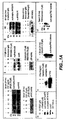


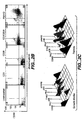


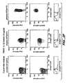
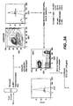

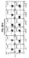


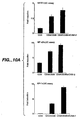
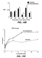

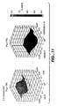


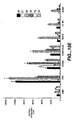


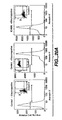
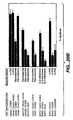


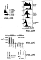

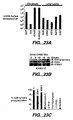


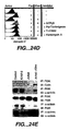


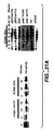


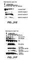


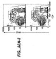
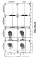
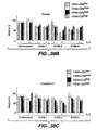

Classifications
-
- G—PHYSICS
- G01—MEASURING; TESTING
- G01N—INVESTIGATING OR ANALYSING MATERIALS BY DETERMINING THEIR CHEMICAL OR PHYSICAL PROPERTIES
- G01N33/00—Investigating or analysing materials by specific methods not covered by groups G01N1/00 - G01N31/00
- G01N33/48—Biological material, e.g. blood, urine; Haemocytometers
- G01N33/50—Chemical analysis of biological material, e.g. blood, urine; Testing involving biospecific ligand binding methods; Immunological testing
- G01N33/68—Chemical analysis of biological material, e.g. blood, urine; Testing involving biospecific ligand binding methods; Immunological testing involving proteins, peptides or amino acids
- G01N33/6803—General methods of protein analysis not limited to specific proteins or families of proteins
- G01N33/6845—Methods of identifying protein-protein interactions in protein mixtures
-
- C—CHEMISTRY; METALLURGY
- C12—BIOCHEMISTRY; BEER; SPIRITS; WINE; VINEGAR; MICROBIOLOGY; ENZYMOLOGY; MUTATION OR GENETIC ENGINEERING
- C12Q—MEASURING OR TESTING PROCESSES INVOLVING ENZYMES, NUCLEIC ACIDS OR MICROORGANISMS; COMPOSITIONS OR TEST PAPERS THEREFOR; PROCESSES OF PREPARING SUCH COMPOSITIONS; CONDITION-RESPONSIVE CONTROL IN MICROBIOLOGICAL OR ENZYMOLOGICAL PROCESSES
- C12Q1/00—Measuring or testing processes involving enzymes, nucleic acids or microorganisms; Compositions therefor; Processes of preparing such compositions
- C12Q1/48—Measuring or testing processes involving enzymes, nucleic acids or microorganisms; Compositions therefor; Processes of preparing such compositions involving transferase
- C12Q1/485—Measuring or testing processes involving enzymes, nucleic acids or microorganisms; Compositions therefor; Processes of preparing such compositions involving transferase involving kinase
-
- G—PHYSICS
- G01—MEASURING; TESTING
- G01N—INVESTIGATING OR ANALYSING MATERIALS BY DETERMINING THEIR CHEMICAL OR PHYSICAL PROPERTIES
- G01N21/00—Investigating or analysing materials by the use of optical means, i.e. using sub-millimetre waves, infrared, visible or ultraviolet light
- G01N21/62—Systems in which the material investigated is excited whereby it emits light or causes a change in wavelength of the incident light
- G01N21/63—Systems in which the material investigated is excited whereby it emits light or causes a change in wavelength of the incident light optically excited
- G01N21/64—Fluorescence; Phosphorescence
- G01N21/6428—Measuring fluorescence of fluorescent products of reactions or of fluorochrome labelled reactive substances, e.g. measuring quenching effects, using measuring "optrodes"
-
- G—PHYSICS
- G01—MEASURING; TESTING
- G01N—INVESTIGATING OR ANALYSING MATERIALS BY DETERMINING THEIR CHEMICAL OR PHYSICAL PROPERTIES
- G01N33/00—Investigating or analysing materials by specific methods not covered by groups G01N1/00 - G01N31/00
- G01N33/48—Biological material, e.g. blood, urine; Haemocytometers
- G01N33/50—Chemical analysis of biological material, e.g. blood, urine; Testing involving biospecific ligand binding methods; Immunological testing
- G01N33/5005—Chemical analysis of biological material, e.g. blood, urine; Testing involving biospecific ligand binding methods; Immunological testing involving human or animal cells
- G01N33/5008—Chemical analysis of biological material, e.g. blood, urine; Testing involving biospecific ligand binding methods; Immunological testing involving human or animal cells for testing or evaluating the effect of chemical or biological compounds, e.g. drugs, cosmetics
-
- G—PHYSICS
- G01—MEASURING; TESTING
- G01N—INVESTIGATING OR ANALYSING MATERIALS BY DETERMINING THEIR CHEMICAL OR PHYSICAL PROPERTIES
- G01N33/00—Investigating or analysing materials by specific methods not covered by groups G01N1/00 - G01N31/00
- G01N33/48—Biological material, e.g. blood, urine; Haemocytometers
- G01N33/50—Chemical analysis of biological material, e.g. blood, urine; Testing involving biospecific ligand binding methods; Immunological testing
- G01N33/5005—Chemical analysis of biological material, e.g. blood, urine; Testing involving biospecific ligand binding methods; Immunological testing involving human or animal cells
- G01N33/5008—Chemical analysis of biological material, e.g. blood, urine; Testing involving biospecific ligand binding methods; Immunological testing involving human or animal cells for testing or evaluating the effect of chemical or biological compounds, e.g. drugs, cosmetics
- G01N33/502—Chemical analysis of biological material, e.g. blood, urine; Testing involving biospecific ligand binding methods; Immunological testing involving human or animal cells for testing or evaluating the effect of chemical or biological compounds, e.g. drugs, cosmetics for testing non-proliferative effects
- G01N33/5041—Chemical analysis of biological material, e.g. blood, urine; Testing involving biospecific ligand binding methods; Immunological testing involving human or animal cells for testing or evaluating the effect of chemical or biological compounds, e.g. drugs, cosmetics for testing non-proliferative effects involving analysis of members of signalling pathways
-
- G—PHYSICS
- G01—MEASURING; TESTING
- G01N—INVESTIGATING OR ANALYSING MATERIALS BY DETERMINING THEIR CHEMICAL OR PHYSICAL PROPERTIES
- G01N33/00—Investigating or analysing materials by specific methods not covered by groups G01N1/00 - G01N31/00
- G01N33/48—Biological material, e.g. blood, urine; Haemocytometers
- G01N33/50—Chemical analysis of biological material, e.g. blood, urine; Testing involving biospecific ligand binding methods; Immunological testing
- G01N33/5005—Chemical analysis of biological material, e.g. blood, urine; Testing involving biospecific ligand binding methods; Immunological testing involving human or animal cells
- G01N33/5091—Chemical analysis of biological material, e.g. blood, urine; Testing involving biospecific ligand binding methods; Immunological testing involving human or animal cells for testing the pathological state of an organism
-
- G—PHYSICS
- G01—MEASURING; TESTING
- G01N—INVESTIGATING OR ANALYSING MATERIALS BY DETERMINING THEIR CHEMICAL OR PHYSICAL PROPERTIES
- G01N33/00—Investigating or analysing materials by specific methods not covered by groups G01N1/00 - G01N31/00
- G01N33/48—Biological material, e.g. blood, urine; Haemocytometers
- G01N33/50—Chemical analysis of biological material, e.g. blood, urine; Testing involving biospecific ligand binding methods; Immunological testing
- G01N33/5005—Chemical analysis of biological material, e.g. blood, urine; Testing involving biospecific ligand binding methods; Immunological testing involving human or animal cells
- G01N33/5094—Chemical analysis of biological material, e.g. blood, urine; Testing involving biospecific ligand binding methods; Immunological testing involving human or animal cells for blood cell populations
-
- G—PHYSICS
- G01—MEASURING; TESTING
- G01N—INVESTIGATING OR ANALYSING MATERIALS BY DETERMINING THEIR CHEMICAL OR PHYSICAL PROPERTIES
- G01N33/00—Investigating or analysing materials by specific methods not covered by groups G01N1/00 - G01N31/00
- G01N33/48—Biological material, e.g. blood, urine; Haemocytometers
- G01N33/50—Chemical analysis of biological material, e.g. blood, urine; Testing involving biospecific ligand binding methods; Immunological testing
- G01N33/53—Immunoassay; Biospecific binding assay; Materials therefor
- G01N33/5302—Apparatus specially adapted for immunological test procedures
-
- G—PHYSICS
- G01—MEASURING; TESTING
- G01N—INVESTIGATING OR ANALYSING MATERIALS BY DETERMINING THEIR CHEMICAL OR PHYSICAL PROPERTIES
- G01N33/00—Investigating or analysing materials by specific methods not covered by groups G01N1/00 - G01N31/00
- G01N33/48—Biological material, e.g. blood, urine; Haemocytometers
- G01N33/50—Chemical analysis of biological material, e.g. blood, urine; Testing involving biospecific ligand binding methods; Immunological testing
- G01N33/53—Immunoassay; Biospecific binding assay; Materials therefor
- G01N33/543—Immunoassay; Biospecific binding assay; Materials therefor with an insoluble carrier for immobilising immunochemicals
- G01N33/54313—Immunoassay; Biospecific binding assay; Materials therefor with an insoluble carrier for immobilising immunochemicals the carrier being characterised by its particulate form
-
- G—PHYSICS
- G01—MEASURING; TESTING
- G01N—INVESTIGATING OR ANALYSING MATERIALS BY DETERMINING THEIR CHEMICAL OR PHYSICAL PROPERTIES
- G01N33/00—Investigating or analysing materials by specific methods not covered by groups G01N1/00 - G01N31/00
- G01N33/48—Biological material, e.g. blood, urine; Haemocytometers
- G01N33/50—Chemical analysis of biological material, e.g. blood, urine; Testing involving biospecific ligand binding methods; Immunological testing
- G01N33/53—Immunoassay; Biospecific binding assay; Materials therefor
- G01N33/573—Immunoassay; Biospecific binding assay; Materials therefor for enzymes or isoenzymes
-
- G—PHYSICS
- G01—MEASURING; TESTING
- G01N—INVESTIGATING OR ANALYSING MATERIALS BY DETERMINING THEIR CHEMICAL OR PHYSICAL PROPERTIES
- G01N33/00—Investigating or analysing materials by specific methods not covered by groups G01N1/00 - G01N31/00
- G01N33/48—Biological material, e.g. blood, urine; Haemocytometers
- G01N33/50—Chemical analysis of biological material, e.g. blood, urine; Testing involving biospecific ligand binding methods; Immunological testing
- G01N33/58—Chemical analysis of biological material, e.g. blood, urine; Testing involving biospecific ligand binding methods; Immunological testing involving labelled substances
- G01N33/582—Chemical analysis of biological material, e.g. blood, urine; Testing involving biospecific ligand binding methods; Immunological testing involving labelled substances with fluorescent label
-
- G—PHYSICS
- G01—MEASURING; TESTING
- G01N—INVESTIGATING OR ANALYSING MATERIALS BY DETERMINING THEIR CHEMICAL OR PHYSICAL PROPERTIES
- G01N21/00—Investigating or analysing materials by the use of optical means, i.e. using sub-millimetre waves, infrared, visible or ultraviolet light
- G01N21/62—Systems in which the material investigated is excited whereby it emits light or causes a change in wavelength of the incident light
- G01N21/63—Systems in which the material investigated is excited whereby it emits light or causes a change in wavelength of the incident light optically excited
- G01N21/64—Fluorescence; Phosphorescence
- G01N21/6428—Measuring fluorescence of fluorescent products of reactions or of fluorochrome labelled reactive substances, e.g. measuring quenching effects, using measuring "optrodes"
- G01N2021/6439—Measuring fluorescence of fluorescent products of reactions or of fluorochrome labelled reactive substances, e.g. measuring quenching effects, using measuring "optrodes" with indicators, stains, dyes, tags, labels, marks
- G01N2021/6441—Measuring fluorescence of fluorescent products of reactions or of fluorochrome labelled reactive substances, e.g. measuring quenching effects, using measuring "optrodes" with indicators, stains, dyes, tags, labels, marks with two or more labels
-
- G—PHYSICS
- G01—MEASURING; TESTING
- G01N—INVESTIGATING OR ANALYSING MATERIALS BY DETERMINING THEIR CHEMICAL OR PHYSICAL PROPERTIES
- G01N2333/00—Assays involving biological materials from specific organisms or of a specific nature
- G01N2333/90—Enzymes; Proenzymes
- G01N2333/91—Transferases (2.)
- G01N2333/912—Transferases (2.) transferring phosphorus containing groups, e.g. kinases (2.7)
- G01N2333/91205—Phosphotransferases in general
-
- G—PHYSICS
- G01—MEASURING; TESTING
- G01N—INVESTIGATING OR ANALYSING MATERIALS BY DETERMINING THEIR CHEMICAL OR PHYSICAL PROPERTIES
- G01N2333/00—Assays involving biological materials from specific organisms or of a specific nature
- G01N2333/90—Enzymes; Proenzymes
- G01N2333/914—Hydrolases (3)
- G01N2333/948—Hydrolases (3) acting on peptide bonds (3.4)
- G01N2333/95—Proteinases, i.e. endopeptidases (3.4.21-3.4.99)
- G01N2333/964—Proteinases, i.e. endopeptidases (3.4.21-3.4.99) derived from animal tissue
- G01N2333/96425—Proteinases, i.e. endopeptidases (3.4.21-3.4.99) derived from animal tissue from mammals
- G01N2333/96427—Proteinases, i.e. endopeptidases (3.4.21-3.4.99) derived from animal tissue from mammals in general
- G01N2333/9643—Proteinases, i.e. endopeptidases (3.4.21-3.4.99) derived from animal tissue from mammals in general with EC number
- G01N2333/96466—Cysteine endopeptidases (3.4.22)
-
- G—PHYSICS
- G01—MEASURING; TESTING
- G01N—INVESTIGATING OR ANALYSING MATERIALS BY DETERMINING THEIR CHEMICAL OR PHYSICAL PROPERTIES
- G01N2405/00—Assays, e.g. immunoassays or enzyme assays, involving lipids
-
- G—PHYSICS
- G01—MEASURING; TESTING
- G01N—INVESTIGATING OR ANALYSING MATERIALS BY DETERMINING THEIR CHEMICAL OR PHYSICAL PROPERTIES
- G01N2440/00—Post-translational modifications [PTMs] in chemical analysis of biological material
- G01N2440/14—Post-translational modifications [PTMs] in chemical analysis of biological material phosphorylation
-
- Y—GENERAL TAGGING OF NEW TECHNOLOGICAL DEVELOPMENTS; GENERAL TAGGING OF CROSS-SECTIONAL TECHNOLOGIES SPANNING OVER SEVERAL SECTIONS OF THE IPC; TECHNICAL SUBJECTS COVERED BY FORMER USPC CROSS-REFERENCE ART COLLECTIONS [XRACs] AND DIGESTS
- Y10—TECHNICAL SUBJECTS COVERED BY FORMER USPC
- Y10S—TECHNICAL SUBJECTS COVERED BY FORMER USPC CROSS-REFERENCE ART COLLECTIONS [XRACs] AND DIGESTS
- Y10S435/00—Chemistry: molecular biology and microbiology
- Y10S435/973—Simultaneous determination of more than one analyte
-
- Y—GENERAL TAGGING OF NEW TECHNOLOGICAL DEVELOPMENTS; GENERAL TAGGING OF CROSS-SECTIONAL TECHNOLOGIES SPANNING OVER SEVERAL SECTIONS OF THE IPC; TECHNICAL SUBJECTS COVERED BY FORMER USPC CROSS-REFERENCE ART COLLECTIONS [XRACs] AND DIGESTS
- Y10—TECHNICAL SUBJECTS COVERED BY FORMER USPC
- Y10T—TECHNICAL SUBJECTS COVERED BY FORMER US CLASSIFICATION
- Y10T436/00—Chemistry: analytical and immunological testing
- Y10T436/10—Composition for standardization, calibration, simulation, stabilization, preparation or preservation; processes of use in preparation for chemical testing
- Y10T436/101666—Particle count or volume standard or control [e.g., platelet count standards, etc.]
-
- Y—GENERAL TAGGING OF NEW TECHNOLOGICAL DEVELOPMENTS; GENERAL TAGGING OF CROSS-SECTIONAL TECHNOLOGIES SPANNING OVER SEVERAL SECTIONS OF THE IPC; TECHNICAL SUBJECTS COVERED BY FORMER USPC CROSS-REFERENCE ART COLLECTIONS [XRACs] AND DIGESTS
- Y10—TECHNICAL SUBJECTS COVERED BY FORMER USPC
- Y10T—TECHNICAL SUBJECTS COVERED BY FORMER US CLASSIFICATION
- Y10T436/00—Chemistry: analytical and immunological testing
- Y10T436/25—Chemistry: analytical and immunological testing including sample preparation
Definitions
- the invention relates generally to the field of protein detection using flow cytometry. More specifically, the invention relates to simultaneously detecting the activation states of multiple proteins in single cells using flow cytometry and, more particularly, using polychromatic flow cytometry.
- Proteins are the major components of cells. The spatiotemporal expression pattern and the subcellular localization of proteins determines the shape, structure, and function of cells. Proteins are assembled from 20 different amino acids, each with a distinct side chain and chemical property. This provides for enormous variety in the chemical properties of proteins and the activities they exhibit.
- proteins are dynamically regulated such that their activity is altered in response to certain intrinsic or extrinsic cues.
- One form of regulation involves covalent modification of the regulated protein.
- An example of such a covalent modification is the substitution of a phosphate group for an hydroxyl group in the side chain of an amino acid (phosphorylation).
- phosphorylation is often associated with an alteration in the activity of a protein.
- Protein kinases can recognize specific protein substrates and catalyze the phosphorylation of serine, threonine, or tyrosine residues on their protein substrates. Such substrate proteins capable of being phosphorylated are referred to as phosphoproteins.
- a substrate protein may have its phosphorylated residue converted back to an hydroxylated one by the action of a protein phosphatase which specifically recognizes the substrate protein.
- Protein phosphatases catalyze the replacement of phosphate groups by hydroxyl groups on serine, threonine, or tyrosine residues. Through the action of kinases and phosphatases a protein may be reversibly phosphorylated on a multiplicity of residues and its activity may be regulated thereby.
- Many intracellular kinases and phosphatases transduce signals in response to extracellular signals that stimulate receptors at the cell surface.
- extracellular signals induce changes in the phosphorylation and activation states of many proteins in receptive cells.
- many mitogens induce JNK and MAPK signaling activity which causes cell to proliferate.
- neurotrophic factors have been shown to induce AKT signaling activity which supports neuronal survival.
- many diseases, such as cancer are associated with an alteration in the activation state of many signaling proteins. In many instances, the signaling activity observed in cancer cells is reminiscent of growth factor stimulated signaling activity in non-cancer cells.
- lipids of the cell membrane are also substrates for kinases.
- PI3K phosphatidylinositol 3-kinase
- PIP2 phosphatidylinositol 4-kinase which catalyzes phosphorylation of phosphatidylinositol at the 4' and 5' positions of the inositol moiety.
- PI3K regulates a variety of cellular functions and is activated by growth factors such as nerve growth factor (NGF), platelet derived growth factor (PDGF), insulin receptor substrate 1 (IRS-1) and CD28. Many of the proteins that activate PI3K also activate GTPases, predominantly the ras family, which in turn activate PI3K by binding to its p110 subunit. The direct pathway from tyrosine phosphorylated receptors to PI3K and activation of PI3K by GTPase proteins function synergistically.
- PI3K is a heterodimeric enzyme composed of a catalytic subunit (p110) and a regulatory subunit. Several genes encoding regulatory subunits of PI3K have been identified, including p85 ⁇ and p85 ⁇ . In addition, a number of splice variants of p85 ⁇ are known (including p55 ⁇ and p50 ⁇ ).
- kinase signaling cascades play an important role in nearly every critical decision process in cells (for review see Hunter, Cell 100:113-127, 2000 ). Determining the role of specific signal transduction pathways in a given system has been aided by the advent of pharmacological inhibitors for specific kinases.
- monitoring activities for such kinases for example, protein kinase B/AKT, cJun-N-terminal kinase (JNK), p38 mitogen activated protein kinase (p38), p44/42 ERK1/2 (ERK), PKA, PKC or TYK2, is usually dependent upon in vitro kinase assays or more recently by immunoblotting to determine the phosphorylation state of these proteins using phospho-specific antibodies. To date it has not been possible to correlate rare subpopulations of cells with the activation state of kinases in important signaling pathways.
- proteolytic cleavage of a peptide bond Another form of protein regulation involves proteolytic cleavage of a peptide bond. While random or misdirected proteolytic cleavage may be detrimental to the activity of a protein, many proteins are activated by the action of proteases that recognize and cleave specific peptide bonds. Many proteins derive from precursor proteins, or pro-proteins, which give rise to a mature isoform of the protein following proteolytic cleavage of specific peptide bonds. Many growth factors are synthesized and processed in this manner, with a mature isoform of the protein typically possessing a biological activity not exhibited by the precursor form.
- caspases are an important class of proteases that mediate programmed cell death (referred to in the art as "apoptosis").
- Caspases are constitutively present in most cells, residing in the cytosol as a single chain proenzyme. These are activated to fully functional proteases by a first proteolytic cleavage to divide the chain into large and small caspase subunits and a second cleavage to remove the N-terminal domain. The subunits assemble into a tetramer with two active sites ( Green, Cell 94:695-698, 1998 ).
- antibodies may be used to determine the presence of a specific isoform of a protein, especially, when the isoform possesses an antigen not present in other isoforms of the protein, or when the isoform lacks an antigen not present in other isoforms of the protein.
- the isoform can be antigenically distinct from other isoforms of the same protein.
- the antigenically distinct isoform may uniquely possess or lack a covalently attached moiety, or may be in a conformation distinct from other isoforms and thereby present the same amino acid sequence in an antigenically distinguishable way.
- PI3K activity has typically involved immunoprecipitation of PI3K and an in vitro enzymatic assay using phospholipids and labeled ATP.
- antibodies that specifically bind to phosphatidylinositol 4,5-diphosphate and phosphatidylinositol 3,4,5-triphosphate have been developed (Molecular Probes, A-21327-anti-phosphatidylinositol 4,5-diphosphate, mouse IgM monoclonal 2C11; A-21328 anti-phosphatidylinositol 3,4,5-triphosphate, mouse IgM monoclonal RC6F8).
- PIP2 PI3K substrate
- PIP33 product
- proteins involved in signal transduction can often exist in two or more forms, and it is usually a particular form of the signaling protein that transmits a signal that mediates the effects of a signaling pathway. Conversion from one form to another is often used to turn a signal on or off through a change in protein activity.
- proteins of interest are first separated by electrophoresis and then transferred and immobilized onto a nitrocellulose or a polyvinylidene difluoride (PVDF) membrane.
- PVDF polyvinylidene difluoride
- phage display screening of a protein expression library several hundred thousand proteins expressed by phages are immobilized on membranes.
- proteins are immobilized non-covalently.
- the protein of interest is then selected by its unique property, i.e., interaction with an antibody.
- agents e.g., antibodies, ligands
- solid supports e.g., agarose beads
- proteins retain their abilities of interacting with other proteins or ligands after immobilization.
- immobilization of a multiplicity of proteins from a sample the problems of simultaneous detection of protein expression, protein form, and protein level for a multiplicity of proteins remains.
- WO 99/44067 describes a method for the flow cytometric determination of the phosphorylation status of retinoblastoma susceptibility gene protein (pRb) in individual cells, including the measurement of the hypophosphorylated active form of pRb as a proportion of total cellular pRb, as well as allowing for the correlation of pRb phosphorylation with cell cycle phase and with protein components of the cell.
- pRb retinoblastoma susceptibility gene protein
- an object of the present invention is to overcome the problems described above. Accordingly, the present invention provides an approach for the simultaneous determination of the activation states of a plurality of proteins in single cells. This approach permits the rapid detection of heterogeneity in a complex cell population based on protein activation states, and the identification of cellular subsets that exhibit correlated changes in protein activation within the cell population, Moreover, this approach allows the correlation of cellular activities or properties, such as surface molecule expression or cell granularity, with protein activation at the single cell level.
- the present invention provides methods and compositions for simultaneously detecting the activation state of a plurality of activatable proteins in single cells using flow cytometry.
- the invention further provides methods and compositions of screening for bioactive agents capable of coordinately modulating the activity or activation state of a plurality of activatable proteins in single cells.
- the methods and compositions can be used to determine the protein activation profile of a cell for predicting or diagnosing a disease state, and for monitoring treatment of a disease state.
- the methods and compositions of the present invention can be used optionally to sequentially detect the activation state of a plurality of activatable proteins in single cells.
- the methods and compositions of the present invention can be used optionally detect the activation state of a single protein or modulate the activity or activation state of a single protein.
- the invention provides populations of cells, single cells, cell lysates, proteins, and samples comprising populations of cells, single cells, cell lysates, proteins useful in the methods of the present invention.
- the invention provides activatable proteins and activation state-specific antibodies that bind to a specific isoform of an activatable protein.
- the activation state-specific antibodies are conjugated to a label, preferably a fluorescent label, and more preferably a FRET label.
- the invention provides methods of detecting the activation state of at least a first and a second activatable protein in single cells, the method comprising the steps of: a) providing a population of cells comprising the first and the second activatable proteins; b) contacting the population of cells with a plurality of activation state-specific antibodies, wherein the plurality of activation state-specific antibodies comprise: i) at least one first activation state-specific antibody that is capable of binding to a corresponding isoform of the first activable protein in the population of cells; and ii) at least one second activation state-specific antibody that is capable of binding to a corresponding isoform of the second activatable protein in the population of cells; and c) using flow cytometry to detect the binding of the first and second activation state-specific antibodies in single cells of the population of cells, wherein the binding of the first activation state-specific antibody is indicative of a specific activation state of the first activatable protein, and the binding of the second activation state-specific antibody is indicative of
- the first activatable protein is a kinase. Also in a further aspect, the first activatable protein is a caspase.
- the first activatable protein is a first kinase and the second activatable protein is a second kinase.
- the isoform of the first kinase is a first phosphorylated kinase
- the isoform of the second kinase is a second phosphorylated kinase.
- the first activation site-specific antibody binds to the first phosphorylated kinase
- the second activation site-specific antibody binds the second phorphorylated kinase.
- the first activatable protein is a first caspase and the second activatable protein is a second caspase.
- the isoform of the first caspase is a cleaved product of a first pro-caspase
- the isoform of the second caspase is a cleaved product of a second pro-caspase.
- the plurality of activation site-specific antibodies comprise a first activation site-specific antibody that binds to the isoform of the first caspase, and a second activation site-specific antibody that binds to the isoform of the second caspase.
- the invention provides methods of detecting the activation state of at least a first activatable protein in single cells, the method comprising the steps of: a) providing a population of cells comprising at least the first activatable protein; b) contacting the population of cells with a plurality of substrates; wherein the plurality of substrates comprise at least a first substrate that is capable of being modified by a corresponding isoform of the first activatable protein in the population of cells; and c) using flow cytometry to detect the modification of the first substrate in single cells of the population of cells, wherein the modification is indicative of a specific activation state of the first activatable protein.
- the population of cells further comprises a second activatable protein
- the plurality of substrates further comprise a second substrate that is capable of being modified by a corresponding isoform of the second activable protein in the population of cells
- step c) further comprises using the flow cytometry to detect the modification of the second substrate in single cells of the population of cells, wherein the modification of the second substrate is indicative of a specific activation state of the second activatable protein.
- the invention provides methods of detecting a protein activation state profile of single cells based on the activation state of at least a first activatable protein in the cells, the method comprising the steps of: a) providing a population of cells comprising at least the first activatable protein; b) contacting the population of cells with a plurality of substrates; wherein the plurality of substrates comprise at least a first substrate that is capable of being modified by a corresponding isoform of the first activatable protein in the population of cells; c) contacting the population of cells with a plurality of activation state-specific antibodies, wherein the activation state-specific antibodies comprise at least one first activation state-specific antibody that is capable of binding to a corresponding isoform of the first activatable protein in the population of cells; and d) using flow cytometry to simultaneously detect: i) the binding of the first activation state-specific antibody in single cells of the population of cells, wherein the binding of the first activation state-specific antibody is indicative of a specific activation
- the population of cells further comprises a second activatable protein
- the plurality of substrates further comprises a second substrate that is capable of being modified by a corresponding isoform of the second activatable protein in the population of cells
- step d) further comprises using the flow cytometry to detect the modification of the second substrate in the single cells, wherein the modification is indicative of a specific activation state of the the second activatable protein.
- the plurality of activation state-specific antibodies further comprises a second activation state-specific antibody that is capable of binding to a corresponding isoform of the second activatable protein in the population of cells
- step c) further comprises using the flow cytometry to detect the binding of the second activation state-specific antibody the single cells of the population of cells, wherein the binding of the second activation state-specific antibody is indicative of the specific activation state of the second activatable protein.
- the invention provides methods of screening for a bioactive agent capable of modulating the activity of at least a first activatable protein in cells, the method comprising: a) providing a population of cells, each of the cells comprising at least the a first activable protein, a second activatable protein, and a third activatable protein, wherein the first activable protein can activate the second activatable protein thereby forming a specific isoform of the second activable protein (isoform-2), and wherein the second activable protein can activate the third activatable protein thereby forming a specific isoform of the third activatable protein (isoform-3); b) contacting the cells with a second activation state-specific antibody, a third activation state-specific antibody, and a candidate bioactive agent, wherein the second activation state-specific antibody is capable of binding to the isoform-2, and wherein the third activation state-specific antibody is capable of binding to the isoform-3; c) using fluorescent activated cell sorting (FACS) to sort single
- the first activatable protein is activated by an activating agent
- the first activatable protein is a caspase.
- the first activatable protein is a kinase; in an additional aspect, the kinase is PI3K; and in a further aspect the PI3K is activated by a growth factor or by activation of a cell surface receptor.
- the kinase is PI3K; the second activatable protein is PIP2; the third activatable protein is PIP3; the isoform-2 is PIP 4,5 bisphosphate; and the isoform-3 is PIP 3,4,5.
- the first activatable protein is ICAM-2; the activity is apoptosis; the second activatable protein is PIP2; the third activatable protein is PIP3; the isoform-2 is PIP 4,5 bisphosphate; and the isoform-3 is PIP 3,4,5.
- step a) further comprises clustering the ICAM-2 with ICAM-3.
- the plurality of activation state-specific antibodies comprise at least one antibody selected from a group of antibodies consisting of: anti- AKT-phospho-Ser473, anti-AKT phospho-Thr308, anti-p44142 MAPK phospho-Thr202/Tyr204, anti-TYK2 phospho-Tyr1054/1055, anti-p38 MAPK phospho-Thr180/Tyr182, anti-JNK/SAPK phospho-Thr183/Tyr185, anti-phospho-tyrosine, anti-phospho-threonine, anti-PIP2, and anti-PIP3.
- a group of antibodies consisting of: anti- AKT-phospho-Ser473, anti-AKT phospho-Thr308, anti-p44142 MAPK phospho-Thr202/Tyr204, anti-TYK2 phospho-Tyr1054/1055, anti-p38 MAPK phospho-Thr180/Tyr182, anti-JNK/SAPK phospho-Thr183/Tyr185, anti-phospho-tyrosine
- step a) further comprises contacting the population of cells with an agent that induces the activation of at least the first activatable protein.
- step a) further comprises contacting the population of cells with an agent that induces the activation of at least one of the first and the second activatable proteins.
- step a) further comprises contacting the population of cells with an agent that induces the activation of at least one of the first, the second, and the third activatable proteins.
- the methods further comprises sorting the single cells based on the activation state of the first activatable protein, and the activation state of the second activatable protein.
- the first activation state-specific antibody comprises a first label
- the second activation state-specific antibody comprises a second fluorescent label
- the sorting is by fluorescent activated cell sorting (FACS).
- the first activation state-specific antibody comprises a first FRET label; the second activation state-specific antibody comprises a second FRET label and the sorting is by fluorescent activated cell sorting (FACS).
- FACS fluorescent activated cell sorting
- the first activation state-specific antibody comprises a FRET label
- the second activation state-specific antibody comprises a label
- the sorting is by fluorescent activated cell sorting (FACS).
- step a) further comprises fixing the cells.
- the cells are mammalian cells.
- Intracelluar assays of signaling systems have been limited by an inability to correlate functional subsets of cells in complex populations based on the activity of signaling agents, for example, the activity of kinases. Such correlations are important for distinguishing changes in signaling status that arise in rare cell subsets during signaling or in disease manifestations.
- the present invention solves these problems by providing methods and compositions for simultaneously detecting the activation state of a plurality of activatable proteins in single cells using flow cytometry.
- the invention further provides methods and compositions of screening for bioactive agents capable of coordinately modulating the activity or activation state of a plurality of activatable proteins in single cells.
- the methods and compositions can be used to determine the protein activation profile of a cell for predicting or diagnosing a disease state, and for monitoring treatment of a disease state. Further, the methods and compositions of the present invention can be used optionally to sequentially detect the activation state of a plurality of activatable proteins in single cells. In addition, the methods and compositions of the present invention can be used optionally to detect the activation state of a single protein or modulate the activity or activation state of a single protein in single cells.
- the methods and compositions of the present invention may be used to detect any particular protein isoform in a sample that is antigenically detectable and antigenically distinguishable from other isoforms of the protein which are present in the sample.
- the activation state-specific antibodies of the present invention can be used in the present methods to identify distinct signaling cascades of a subset or subpopulation of complex cell populations; and the ordering of protein activation (e.g., kinase activation) in potential signaling hierarchies.
- the use of flow cytometry particularly polychromatic flow cytometry, permits the multi-dimensional analysis and functional assessment of the signaling pathway in single cells.
- activation state-specific antibody or “activation state antibody” or grammatical equivalents thereof, refer to an antibody that specifically binds to a corresponding and specific antigen.
- the corresponding and specific antigen is a specific isoform of an activable protein.
- the binding of the activation state-specific antibody is indicative of a specific activation state of a specific activatable protein.
- the binding of an activation state-specific antibody to a corresponding isoform of an activable protein is indicative of the identity of the activatable protein and of the activation state of the activatable protein.
- the activation state-specific antibody is a peptide comprising a recognition structure that binds to a target structure on an activatable protein.
- recognition structures are well known in the art and can be made using methods known in the art, including by phage display libraries (see e.g., Gururaja et al. Chem. Biol. (2000) 7:515-27 ; Houimel et al., Eur. J. Immunol. (2001) 31:3535-45 ; Cochran et al. J. Am. Chem. Soc. (2001) 123:625-32 ; Houimel et al. Int. J. Cancer (2001) 92:748-55 .
- the activation state-specific antibody comprises the following recognition structure: SKVILFE-random peptide loop- SKVILFE.
- Antibodies having such recognition structures can bind with high affinity to specific target structures.
- fluorophores can be attached to such antibodies for use in the methods of the present invention.
- the activation state-specific antibody is a protein that only binds to an isoform of a specific activatable protein that is phosphorylated and does not bind to the isoform of this activable protein when it is not phosphorylated or nonphosphorylated.
- the activation state-specific antibody is a protein that only binds to an isoform of an activatable protein that is intracellular and not extracellular, or vice versa.
- the recognition structure is an anti-laminin single-chain antibody fragment (scFv) (see e.g., Sanz et al., Gene Therapy (2002) 9:1049-53 ; Tse et al., J. Mol. Biol. (2002) 317:85-94 ).
- scFv anti-laminin single-chain antibody fragment
- polypeptide and protein may be used interchangeably and mean at least two covalently attached amino acids, which includes proteins, polypeptides, oligopeptides and peptides.
- the protein may be made up of naturally occurring amino acids and peptide bonds, or synthetic peptidomimetic structures.
- amino acid or “peptide residue”, as used herein means both naturally occurring and synthetic amino acids. For example, homo-phenylalanine, citrulline and noreleucine are considered amino acids for the purposes of the invention.
- Amino acid also includes imino acid residues such as proline and hydroxyproline.
- the side chains may be in either the (R) or the (S) configuration. In the preferred embodiment, the amino acids are in the (S) or L-configuration. If non-naturally occurring side chains are used, non-amino acid substituents may be used, for example to prevent or retard in vivo degradation.
- an "activatable protein” or “substrate” or “substrate protein” or “protein substrate” grammatical equivalents thereof refers to a protein that has at least one isoform (and in some cases two or more isoforms) that corresponds to a specific form of the protein having a particular biological, biochemical, or physical property, e.g., an enzymatic activity, a modification (e.g., post-translational modification), or a conformation.
- the activable protein can be activated or nonactivated with respect to a particular biological activity, modification, or conformation.
- the "activated" or “active” form of the activatable protein has the particular biological activity, modification, or conformation
- the "non-activated” or “non-active” form of the activatable protein does not have (or has a lesser or diminished level of) the particular biological activity, modification, or conformation, respectively.
- there may be more than one isoform associated with activity or activation state for example, there may be an isoform associated with an "open” conformation available for substrate binding, a second "transition state” isoform, and an isoform devoid of activity (e.g., where the activity is inhibited).
- activatable proteins include, but are not limited to, phospho-proteins and phospholipids.
- activatable proteins include, but are not limited to, kinases, phosphatases, PIP2, PIP3, and proteases such as cysteine and serine proteases, including, but not limited to caspases, cathepsins and a variety of well known serine proteases.
- the biological, biochemical, or physical property (e.g. enzymatic activity, modification, or conformation) of the activatable protein is inducible or "activatable” by an activating agent or by cell signaling events.
- activating agents include, but are not limited to, kinases, phosphatases, proteases (e.g., caspases), and hormones.
- cell signaling events include, but are not limited to, receptor clustering or binding of a cognate molecule or ligand.
- an "isoform” or grammatical equivalents thereof refers to a form of an activatable protein having a specific, and preferably detectable, biological activity, modification, or conformation.
- the isoform can be an activated (or active) form, or non-activated (or not active) form of an activatable protein.
- the binding of an activation state-specific antibody to a corresponding isoform of an activable protein is indicative of the identity of the activatable protein and of the activation state of the activatable protein.
- the invention provides methods for determining a protein isoform profile which comprise determining the presence of an isoform of an activatable protein that is activated (or activated isoform).
- the activated isoform or activated state of an activable protein is a form of the activable protein having a particular or specific biological, biochemical, or physical property that is not possessed by at least one other isoform of activatable protein.
- properties include, but are not limited to, enzymatic activity (e.g., kinase activity and protease activity), and protein binding activity.
- enzymatic activity e.g., kinase activity and protease activity
- protein binding activity e.g., protein binding activity.
- Such particular or specific properties or activities are associated with an activated isoform of an activatable protein.
- Such properties or activities are sometimes referred to herein as activation state activities.
- activation state activity is kinase activity for an activated protein kinase.
- a protein kinase may refer to a protein that when activated is capable of catalyzing the phosphorylation of amino acids, or derivatives thereof, which possess an hydroxyl group.
- Preferred kinases are those which are capable of catalyzing the phosphorylation of serine, threonine, and tyrosine residues.
- Kinase activity may be determined by supplying a substrate for phosphorylation by kinase, a source of phosphate usable by kinase, and determining the phosphorylation of substrate in the presence of kinase.
- protease activity for an activated protease may refer to a protein that when activated is capable of hydrolyzing a peptide bond within a polypeptide comprising an amino acid sequence.
- Preferred proteases are the family of cysteine proteases known as caspases. Caspase activity may be determined by providing a substrate of a caspase and determining the cleavage of substrate in the presence of caspase.
- Other cysteine proteases having activations states that may be monitored using the methods of the invention include, but are not limited to, cathespins.
- an additional class of proteases that may be monitored using the methods of the invention include serine proteases, of which a wide variety of suitable classes are known.
- an activated isoform of an activatable protein is distinguishable from the antigenicity of non-activated isoform of an activatable protein or from the antigenicity of an isoform of a different activation state.
- an activated isoform of a protein possesses an epitope that is absent in a non-activated isoform of a protein, or vice versa.
- this difference is due to covalent addition of moieties to a protein, such as phosphate moieties, or due to a structural change in a protein, as through protein cleavage, or due to an otherwise induced conformational change in a protein which causes the protein to present the same sequence in an antigenically distinguishable way.
- such a conformational change causes an activated isoform of a protein to present at least one epitope that is not present in a non-activated isoform, or to not present at least one epitope that is presented by a non-activated isoform of the protein.
- the epitopes for the distinguishing antibodies are centered around the active site of the enzyme, although as is known in the art, conformational changes in one area of a protein may cause alterations in different areas of the protein as well.
- kinases are enzymes that catalyze protein phosphorylation on serine, threonine, or tyrosine residues. The majority of kinases are phosphoproteins that are reversibly phosphorylated as a form of activity regulation. In the phosphorylated form (phosphorylated isoform), protein kinases are typically enzymatically active kinases. In the un-phosphorylated form, protein kinases are typically enzymatically inactive.
- antibodies for use in the present invention include: phospho-AKT Ser473 monoclonal anti-4E2, phospho-p44/42 MAP kinase (Thr202/Tyr204) monoclonal antibody, phospho-TYK2 (Tyr1054/1055) antibody, phospho-p38 MAP kinase (Thr180/Tyr182) monoclonal antibody 28B10, phospho-PKC-PAN substrate antibody, phospho-PKA-substrate, phospho-SAPK/JNK (Thr183/Tyr185) G9 monoclonal antibody, phospho-tyrosine monoclonal antibody (P-tyr-100), p44/42 MAPK, p38 MAPK, JNK/SAPK, and phospho-AKT-Thr308.
- the present invention provides methods for the determination of a kinase activation state profile for a sample which comprise simultaneously determining the presence of activated isoforms of a multiplicity of kinases using a multiplicity of antibodies that specifically bind to active, phosphorylated isoforms of the multiplicity of kinases.
- caspases Another example of proteins that are capable of existing in a non-activated isoform and an activated isoform, which isoforms are antigenically distinguishable, are caspases.
- Caspases are proteases that catalyze the hydrolysis of peptide bonds within proteins.
- Caspases are synthesized as pro-caspases which are proteolytically inactive precursors of caspases.
- a pro-caspase is specifically cleaved by protease to yield an active caspase.
- proteases including both cysteine and serine proteases such as cathepsins, tissue plasminogen activators, etc., that are known to exist as pro-proteases, as is well known in the art.
- the present invention provides methods for the determination of a caspase activation state profile for a sample which comprise simultaneously determining the presence of activated isoforms of a multiplicity of caspases using a multiplicity of antibodies that specifically bind to a multiplicity of caspases but not to the correlating pro-caspases from which they may be derived.
- a multiplicity of antibodies that specifically bind to a multiplicity of caspases but not to the correlating pro-caspases from which they may be derived.
- antibodies which specifically bind to caspases but do not specifically bind to pro-caspases from which caspases are derived are useful in the methods of the invention. Many such antibodies are commercially available (for example, see Cell Signaling Technology, www.cellsignal.com ).
- kinase activation Additional means for determining kinase activation are provided by the present invention.
- Substrates that are specifically recognized by protein kinases and phosphorylated thereby are known.
- Antibodies that specifically bind to such phosphorylated substrates but do not bind to such non-phosphorylated substrates may be used to determine the presence of activated kinase in a sample.
- the present invention provides methods for determining a kinase activation state profile for a sample, comprising providing a population of cells comprising a plurality of activatable proteins and contacting the cells with at least one non-phosphorylated protein kinase substrate under conditions which provide for phosphorylation of the substrate by activated kinase if present, and determining the presence of phosphorylated protein kinase substrate using a phospho-substrate antibody that binds specifically to the phosphorylated substrate.
- the present invention provides methods for determining the activation state of one or more protein kinases and one or more caspases in a sample by combining the methods described for determining the activation state profile of each provided herein.
- the sample comprises a population of cells, a cell, cell lysate, or proteins.
- the invention provides methods utilizing any and all other combinations of kinases and proteases (e.g., kinases and caspases, kinases and cathepsins, kinases and serine proteases, caspases and cathepsins, etc.).
- kinases and proteases e.g., kinases and caspases, kinases and cathepsins, kinases and serine proteases, caspases and cathepsins, etc.
- a protein activation state profile is determined using a multiplicity of activation state antibodies that are immobilized.
- Antibodies may be non-diffusibly bound to an insoluble support having isolated sample receiving areas (e.g. a microtiter plate, an array, etc.).
- the insoluble supports may be made of any composition to which the compositions can be bound, is readily separated from soluble material, and is otherwise compatible with the overall method of screening.
- the surface of such supports may be solid or porous and of any convenient shape.
- suitable insoluble supports include microtiter plates, arrays, membranes, and beads. These are typically made of glass, plastic (e.g., polystyrene), polysaccharides, nylon or nitrocellulose, teflonTM, etc.
- Microtiter plates and arrays are especially convenient because a large number of assays can be carried out simultaneously, using small amounts of reagents and samples. In some cases magnetic beads and the like are included.
- the particular manner of binding of the composition is not crucial so long as it is compatible with the reagents and overall methods of the invention, maintains the activity of the composition and is nondiffusable.
- Preferred methods of binding include the use of antibodies (which do not sterically block either the ligand binding site or activation sequence when the protein is bound to the support), direct binding to "sticky" or ionic supports, chemical crosslinking, the synthesis of the antibody on the surface, etc. Following binding of the antibody, excess unbound material is removed by washing. The sample receiving areas may then be blocked through incubation with bovine serum albumin (BSA), casein or other innocuous protein or other moiety.
- BSA bovine serum albumin
- an epitope-recognizing fragment of an activation state antibody rather than the whole antibody is used.
- the epitope-recognizing fragment is immobilized.
- the antibody light chain which recognizes an epitope is used.
- a recombinant nucleic acid encoding a light chain gene product which recognizes an epitope may be used to produce such an antibody fragment by recombinant means well known in the art.
- a chip analogous to a DNA chip can be used in the methods of the present invention.
- Arrayers and methods for spotting nucleic acid to a chip in a prefigured array are known.
- protein chips and methods for synthesis are known. These methods and materials may be adapted for the purpose of affixing activation state antibodies to a chip in a prefigured array.
- such a chip comprises a multiplicity of kinase activation state antibodies, and is used to determine a kinase activation state profile for a sample.
- a sample is a cell extract.
- detection of activated kinase is by "sandwich assay" as known in the art. Briefly, a sample, preferably a cell extract, is passed over a the chip under conditions which allow the multiplicity of immobilized kinase activation state antibodies to simultaneously bind to a multiplicity of activated kinases if present in the sample.
- the immobilized antibody-kinase complex is optionally washed and contacted with a second plurality of antibodies comprising non-activation state antibodies that are capable of specifically binding to activated kinases while the kinases are specifically bound to kinase activation state specific antibodies.
- non-activation state specific antibodies specifically bind to activated kinases via an epitope that is not recognized by the kinase activation state specific antibody. Binding of the non-activation state specific antibodies to the activation state antibody-activated kinase complex is determined and reveals the presence of activated kinase in sample.
- the determination of binding of second antibody in the sandwich assay can be accomplished in many different ways.
- the multiplicity of non-activation state specific antibodies are uniquely labeled to facilitate detection.
- one or more components of the methods of the present invention comprise a tag.
- tag is meant an attached molecule or molecules useful for the identification or isolation of the attached molecule(s), which are preferably substrate molecules.
- a tag can be an attachment tag or a label tag.
- Components having a tag are referred to as "tag-X", wherein X is the component
- the tag is covalently bound to the attached component
- the tags will be numbered for identification, for example, “tag1-antibody”, “tag1-protein”, and "tag1-substrate”.
- Components may comprise more than one tag, in which case each tag will be numbered, for example "tag 1,2-antibody”, “tag1,2-protein”, and “tag1,2-substrate.
- Preferred tags include, but are not limited to, a label, a partner of a binding pair, and a surface substrate binding molecule (or attachment tag). As will be evident to the skilled artisan, many molecules may find use as more than one type of tag, depending upon how the tag is used.
- label is meant a molecule that can be directly (i.e ., a primary label) or indirectly (i.e ., a secondary label) detected; for example a label can be visualized and/or measured or otherwise identified so that its presence or absence can be known.
- a compound can be directly or indirectly conjugated to a label which provides a detectable signal, e.g. radioisotope, fluorescers, enzyme, antibodies, particles such as magnetic particles, chemiluminescers, or specific binding molecules, etc.
- Specific binding molecules include pairs, such as biotin and streptavidin, digoxin and antidigoxin etc.
- Preferred labels include, but are not limited to, fluorescent labels, label enzymes and radioisotopes.
- fluorescent label any molecule that may be detected via its inherent fluorescent properties.
- Suitable fluorescent labels include, but are not limited to, fluorescein, rhodamine, tetramethylrhodamine, eosin, erythrosin, coumarin, methyl-coumarins, pyrene, Malacite green, stilbene, Lucifer Yellow, Cascade BlueTM, Texas Red, IAEDANS, EDANS, BODIPY FL, LC Red 640, Cy 5, Cy 5.5, LC Red 705 and Oregon green.
- Suitable optical dyes are described in the 1996 Molecular Probes Handbook by Richard P. Haugland, hereby expressly incorporated by reference.
- Suitable fluorescent labels also include, but are not limited to, green fluorescent protein (GFP; Chalfie, et al., Science 263(5148):802-805 (Feb 11, 1994 ); and EGFP; Clontech - Genbank Accession Number U55762 ), blue fluorescent protein (BFP; 1. Quantum Biotechnologies, Inc. 1801 de Maisonneuve Blvd. West, 8th Floor, Montreal (Quebec) Canada H3H 1J9; 2. Stauber, R. H. Biotechniques 24(3):462-471 (1998 ); 3. Heim, R. and Tsien, R. Y. Curr. Biol. 6:178-182 (1996 )), enhanced yellow fluorescent protein (EYFP; 1.
- GFP green fluorescent protein
- EGFP blue fluorescent protein
- EYFP enhanced yellow fluorescent protein
- preferred labels for use in the present invention include: Alexa-Fluor dyes (Alexa Fluor 350, Alexa Fluor 430, Alexa Fluor 488, Alexa Fluor 546, Alexa Fluor 568, Alexa Fluor 594, Alexa Fluor 633, Alexa Fluor 660, Alexa Fluor 680), Cascade Blue, Cascade Yellow and R-phycoerythrin (PE) (Molecular Probes) (Eugene, Oregon), FITC, Rhodamine, and Texas Red (Pierce, Rockford, Illinois), Cy5, Cy5.5, Cy7 (Amersham Life Science, Pittsburgh, Pennsylvania).
- Alexa-Fluor dyes Alexa Fluor 350, Alexa Fluor 430, Alexa Fluor 488, Alexa Fluor 546, Alexa Fluor 568, Alexa Fluor 594, Alexa Fluor 633, Alexa Fluor 660, Alexa Fluor 680
- Cascade Blue Cascade Yellow and R-phycoerythrin (PE) (Molecular Probes
- Tandem conjugate protocols for Cy5PE, Cy5.5PE, Cy7PE, Cy5.5APC, Cy7APC can be found at wuvw.drmr.com/abcon. Quantitation of fluorescent probe conjugation may be assessed to determine degree of labeling and protocols including dye spectral properties can be found at www.metazoa.com/UPL3419.
- multiple fluorescent labels are employed in the methods and compositions of the present invention.
- at least two fluorescent labels are used which are members of a fluorescence resonance energy transfer (FRET) pair.
- FRET is phenomenon known in the art wherein excitation of one fluorescent dye is transferred to another without emission of a photon.
- a FRET pair consists of a donor fluorophore and an acceptor fluorophore.
- the fluorescence emission spectrum of the donor and the fluorescence absorption spectrum of the acceptor must overlap, and the two molecules must be in close proximity.
- the distance between donor and acceptor at which 50% of donors are deactivated (transfer energy to the acceptor) is defined by the Förster radius (R o ), which is typically 10-100 ⁇ .
- Changes in the fluorescence emission spectrum comprising FRET pairs can be detected, indicating changes in the number of that are in close proximity (i.e ., within 100 ⁇ of each other). This will typically result from the binding or dissociation of two molecules, one of which is labeled with a FRET donor and the other of which is labeled with a FRET acceptor, wherein such binding brings the FRET pair in close proximity. Binding of such molecules will result in an increased fluorescence emission of the acceptor and/or quenching of the fluorescence emission of the donor.
- FRET pairs (donor/acceptor) useful in the invention include, but are not limited to, EDANS/fluorescien, IAEDANS/fluorescein, fluorescein/tetramethylrhodamine, fluorescein/LC Red 640, fluorescein/Cy 5, fluorescein/Cy 5.5 and fluorescein/LC Red 705.
- a fluorescent donor molecule and a nonfluorescent acceptor molecule may be employed.
- fluorescent emission of the donor will increase when quencher is displaced from close proximity to the donor and fluorescent emission will decrease when the quencher is brought into close proximity to the donor.
- Useful quenchers include, but are not limited to, TAMRA, DABCYL, QSY 7 and QSY 33.
- Useful fluorescent donor/quencher pairs include, but are not limited to EDANS/DABCYL, Texas Red/DABCYL, BODIPY/DABCYL, Lucifer yellow/DABCYL, coumarin/DABCYL and fluorescein/QSY 7 dye.
- FRET and fluorescence quenching allow for monitoring of binding of labeled molecules over time, providing continuous information regarding the time course of binding reactions.
- label enzyme substrates such as Luminol (available from Pierce Chemical Co.) have been developed that produce fluorescent reaction products.
- Methods for identifying label enzymes with label enzyme substrates are well known in the art and many commercial kits are available. Examples and methods for the use of various label enzymes are described in Savage et al., Previews 247:6-9 (1998 ), Young, J. Virol. Methods 24:227-236 (1989 ), which are each hereby incorporated by reference in their entirety.
- radioisotope any radioactive molecule. Suitable radioisotopes for use in the invention include, but are not limited to 14 C, 3 H, 32 P, 33 P, 35 S, 125 I, and 131 I. The use of radioisotopes as labels is well known in the art.
- labels may be indirectly detected, that is, the tag is a partner of a binding pair.
- partner of a binding pair is meant one of a first and a second moiety, wherein the first and the second moiety have a specific binding affinity for each other.
- Suitable binding pairs for use in the invention include, but are not limited to, antigens/antibodies (for example, digoxigenin/anti-digoxigenin, dinitrophenyl (DNP)/anti-DNP, dansyl-X-anti-dansyl, Fluorescein/anti-fluorescein, lucifer yellow/anti-lucifer yellow, and rhodamine anti-rhodamine), biotin/avid (or biotin/streptavidin) and calmodulin binding protein (CBP)/calmodulin.
- antigens/antibodies for example, digoxigenin/anti-digoxigenin, dinitrophenyl (DNP)/anti-DNP, dansyl-X-anti-dansyl, Fluorescein/anti-fluorescein, lucifer yellow/anti-lucifer yellow, and rhodamine anti-rhodamine
- biotin/avid or biotin/streptavidin
- binding pair pairs include polypeptides such as the FLAG-peptide [ Hopp et al., Biotechnology, 6:1204-1210 (1988 )]; the KT3 epitope peptide [ Martin et al., Science, 255:192-194 (1992 )]; tubulin epitope peptide [ Skinner et al., J. Biol. Chem., 266:15163-15166 (1991 )]; and the T7 gene 10 protein peptide tag [ Lutz-Freyermuth et al., Proc. Natl. Acad. Sci. USA, 87:6393-6397 (1990 )] and the antibodies each thereto.
- binding pair partners may be used in applications other than for labeling, as is described herein.
- a partner of one binding pair may also be a partner of another binding pair.
- an antigen first moiety
- first moiety may bind to a first antibody (second moiety) which may, in turn, be an antigen for a second antibody (third moiety).
- second moiety an antigen for a second antibody (third moiety).
- a partner of a binding pair may comprise a label, as described above. It will further be appreciated that this allows for a tag to be indirectly labeled upon the binding of a binding partner comprising a label. Attaching a label to a tag which is a partner of a binding pair, as just described, is referred to herein as "indirect labeling".
- surface binding substrate molecules useful in the present invention include, but are not limited to, polyhistidine structures (His-tags) that bind nickel substrates, antigens that bind to surface substrates comprising antibody, haptens that bind to avidin substrate (e.g., biotin) and CBP that binds to surface substrate comprising calmodulin.
- His-tags polyhistidine structures
- antigens that bind to surface substrates comprising antibody
- haptens that bind to avidin substrate (e.g., biotin)
- CBP that binds to surface substrate comprising calmodulin.
- tag-components of the invention can be made in various ways, depending largely upon the form of the tag.
- Components of the invention and tags are preferably attached by a covalent bond.
- tag-polypeptides by recombinant means when the tag is also a polypeptide is described below.
- Production of tag-labeled proteins is well known in the art and kits for such production are commercially available (for example, from Kodak and Sigma).
- Examples of tag labeled proteins include, but are not limited to, a Flag-polypeptide and His-polypeptide. Methods for the production and use of tag-labeled proteins are found, for example, in Winston et al., Genes and Devel. 13:270-283 (1999 ).
- Biotinylation of target molecules and substrates is well known, for example, a large number of biotinylation agents are known, including amine-reactive and thiol-reactive agents, for the biotinylation of proteins, nucleic acids, carbohydrates, carboxylic acids; see chapter 4, Molecular Probes Catalog, Haugland, 6th Ed. 1996 , hereby incorporated by reference.
- a biotinylated substrate can be attached to a biotinylated component via avidin or streptavidin.
- haptenylation reagents are also known (Id.).
- the tag is functionalized to facilitate covalent attachment
- the covalent attachment of the tag may be either direct or via a linker.
- the linker is a relatively short coupling moiety, that is used to attach the molecules.
- a coupling moiety may be synthesized directly onto a component of the invention and contains at least one functional group to facilitate attachment of the tag.
- the coupling moiety may have at least two functional groups, which are used to attach a functionalized component to a functionalized tag, for example.
- the linker is a polymer.
- covalent attachment is accomplished either directly, or through the use of coupling moieties from the component or tag to the polymer.
- the covalent attachment is direct, that is, no linker is used.
- the component preferably contains a functional group such as a carboxylic acid which is used for direct attachment to the functionalized tag.
- the component and tag may be attached in a variety of ways, including those listed above.
- the tag is attached to the amino or carboxl terminus of the polypeptide.
- the tag is functionalized to facilitate covalent attachment, as is generally outlined above.
- tags are commercially available which contain functional groups, including, but not limited to, isothiocyanate groups, amino groups, haloacetyl groups, maleimides, succinimidyl esters, and sulfonyl halides, all of which may be used to covalently attach the tag to a second molecule, as is described herein.
- the choice of the functional group of the tag will depend on the site of attachment to either a linker, as outlined above or a component of the invention.
- amino modified or hydrazine modified tags will be used for coupling via carbodiimide chemistry, for example using 1-ethyl-3-(3-dimethylaminopropyl)-carbodiimide (EDAC) as is known in the art (see Set 9 and Set 11 of the Molecular Probes Catalog, supra; see also the Pierce 1994 Catalog and Handbook, pages T-155 to T-200 , both of which are hereby incorporated by reference).
- EDAC 1-ethyl-3-(3-dimethylaminopropyl)-carbodiimide
- the carbodiimide is first attached to the tag, such as is commercially available for many of the tags described herein.
- Antibody conjugation may be performed using standard procedures ( http://drmr.com.abcon ) or by using protein-protein/protein-dye crosslinking kits from Molecular Probes (Eugene, Oregon).
- first activation state antibody recognizing a first activated kinase comprises a first label
- second activation state antibody recognizing a second activated kinase comprises a second label, wherein the first and second label are detectable and distinguishable, making the first antibody and the second antibody uniquely labeled.
- Non-activation state antibodies may also be used in the present invention.
- non-activation state antibodies bind to epitopes in both activated and non-activated forms of a protein. Such antibodies may be used to determine the amount of non-activated plus activated protein in a sample.
- non-activation state antibodies bind to epitopes present in non-activated forms of a protein but absent in activated forms of a protein. Such antibodies may be used to determine the amount of non-activated protein in a sample.
- Both types of non-activation state antibodies may be used to determine if a change in the amount of activation state protein, for example from samples before and after treatment with a candidate bioactive agent as described herein, coincide with changes in the amount of non-activation state protein.
- such antibodies can be used to determine whether an increase in activated protein is due to activation of non-activation state protein, or due to increased expression of protein, or both.
- antibodies are immobilized using beads analogous to those known and used for standardization in flow cytometry. Attachment of a multiplicity of activation state specific antibodies to beads may be done by methods known in the art and/or described herein. Such conjugated beads may be contacted with sample, preferably cell extract, under conditions which allow for a multiplicity of activated kinases, if present, to bind to the multiplicity of immobilized antibodies.
- a second multiplicity of antibodies comprising non-activation state antibodies which are uniquely labeled may be added to the immobilized activation state specific antibody-activated kinase complex and the beads may be sorted by FACS on the basis of the presence of each label, wherein the presence of label indicates binding of corresponding second antibody and the presence of corresponding activated kinase.
- the present invention provides methods for determining a protein activation state profile for a single cell.
- the methods comprise sorting cells by FACS on the basis of the activation state of at least two proteins.
- Activation state-specific antibodies are used to sort cells on the basis of protein activation state.
- FACS systems are used or systems dedicated to high throughput screening, e.g 96 well or greater microtiter plates.
- Methods of performing assays on fluorescent materials are well known in the art and are described in, e.g., Lakowicz, J. R., Principles of Fluorescence Spectroscopy, New York: Plenum Press (1983 ); Herman, B., Resonance energy transfer microscopy, in: Fluorescence Microscopy of Living Cells in Culture, Part B, Methods in Cell Biology, vol. 30, ed. Taylor, D. L.
- flow cytometry is used to detect fluorescence.
- Other methods of detecting fluorescence may also be used, e.g., Quantum dot methods (see, e.g., Goldman et al., J. Am. Chem. Soc. (2002) 124:6378-82 ; Pathak et al. J Am. Chem. Soc. (2001) 123:4103-4 ; and Remacle et al., Proc. Natl. Sci. USA (2000) 18:553-8 ).
- FRET is used as a way of monitoring the activation state of proteins inside a cell.
- the degree of FRET can be determined by any spectral or fluorescence lifetime characteristic of the excited construct, for example, by determining the intensity of the fluorescent signal from the donor, the intensity of fluorescent signal from the acceptor, the ratio of the fluorescence amplitudes near the acceptor's emission maxima to the fluorescence amplitudes near the donor's emission maximum, or the excited state lifetime of the donor.
- cleavage of the linker can increase the intensity of fluorescence from the donor, decreases the intensity of fluorescence from the acceptor, decreases the ratio of fluorescence amplitudes from the acceptor to that from the donor, and increases the excited state lifetime of the donor.
- changes in the degree of FRET are determined as a function of the change in the ratio of the amount of fluorescence from the donor and acceptor moieties, a process referred to as "ratioing.”
- ratioing Changes in the absolute amount of substrate, excitation intensity, and turbidity or other background absorbances in the sample at the excitation wavelength affect the intensities of fluorescence from both the donor and acceptor approximately in parallel. Therefore the ratio of the two emission intensities is a more robust and preferred measure of cleavage than either intensity alone.
- the ratio-metric fluorescent reporter system described herein has significant advantages over existing reporters for protein integration analysis, as it allows sensitive detection and isolation of both expressing and non-expressing single living cells.
- the assay system uses a non-toxic, non-polar fluorescent substrate which is easily loaded and then trapped intracellularly. Modification of the fluorescent substrate by a cognate protein yields a fluorescent emission shift as substrate is converted to product.
- the reporter readout is ratiometric it is unique among reporter protein assays in that it controls for variables such as the amount of substrate loaded into individual cells.
- the stable, easily detected, intracellular readout eliminates the need for establishing clonal cell lines prior to expression analysis. This system and other analogous flow sorting systems can be used to isolate cells having a particular protein activation profile from pools of millions of viable cells.
- a FACS cell sorter e.g. a FACSVantage.TM. Cell Sorter, Becton Dickinson Immunocytometry Systems, San Jose, Calif.
- the cells are first contacted with fluorescent-labeled activation state-specific antibodies directed against specific isoforms of specific activatable proteins.
- the amount of bound antibody on each cell can be measured by passing droplets containing the cells through the cell sorter. By imparting an electromagnetic charge to droplets containing the positive cells, the cells can be separated from other cells. The positively selected cells can then be harvested in sterile collection vessels.
- positive cells can be sorted using magnetic separation of cells based on the presence of an isoform of an activatable protein.
- cells to be positively selected are first contacted with specific binding agent (e.g., an antibody or reagent that binds an isoform of an activatable protein).
- specific binding agent e.g., an antibody or reagent that binds an isoform of an activatable protein.
- retrievable particles e.g., magnetically responsive particles
- the cell-binding agent-particle complex can then be physically separated from non-positive or non-labeled cells, for example, using a magnetic field.
- magnetically responsive particles the positive or labeled cells can be retained in a container using a magnetic filed while the negative cells are removed.
- methods for the determination of a kinase activation state profile for a single cell comprise providing a population of cells and sorting the population of cells by FACS.
- cells are separated on the basis of the activation state of at least two kinases.
- a multiplicity of kinase activation state antibodies (sometimes referred to herein as kinase activation state specific antibodies) are used to simultaneously determine the activation state of a multiplicity of kinases.
- kinase activation state antibodies anti- AKT-phospho-Ser473, anti-AKT phospho-Thr308, anti-p44/42 MAPK phospho-Thr202/Tyr204, anti-TYK2 phospho-Tyr1054/1055, anti-p38 MAPK phospho-Thr180/Tyr182, anti-JNK/SAPK phospho-Thr183/Tyr185, anti-phospho-tyrosine, anti-phospho-threonine.
- the methods provide for the detection of activated kinases in single cells using specific substrates for kinases and phospho-substrate antibodies as described herein.
- cells are contacted with at least two such substrates, contacted with phospho-substrate antibodies, and then sorted on the basis of kinase activation using a FACS machine.
- PKC substrate PAN phospho-substrate which is a substrate for ⁇ , ⁇ I, ⁇ II, and ⁇ isoforms of PKC only when phosphorylated at a carboxy terminal residue homologous to Ser660 of PKC ⁇ II
- phospho-substrate antibodies that specifically bind to PKA phospho-substrate (consensus phosphorylation site of PKA phosphorylation of threonine with arginine at -3 position).
- a combination of kinase activation state antibodies that specifically bind to phosphorylated kinases, specific substrates for kinases, and phospho-substrate antibodies are used to determine a kinase activation state profile for a single cell.
- the methods comprise sorting cells on the basis of the activation state of at least two kinases using a FACS machine. Activation of at least two kinases is determined using at least one kinase activation state antibody and at least one specific substrate for a kinase and at least one phospho-substrate antibody for the measurement of the at least one phosphorylated substrate.
- a kinase activation state antibody is selected from the group consisting of anti-AKT-phospho-Ser473, anti-AKT phospho-Thr308, anti-p44/42 MAPK phospho-Thr202/Tyr204, anti-TYK2 phospho-Tyr1054/1055, anti-p38 MAPK phospho-Thr180/Tyr182, anti-JNK/SAPK phospho-Thr183/Tyr185, anti-phospho-tyrosine, anti-phospho-threonine.
- a phospho-substrate antibody is selected from an antibody that specifically binds to PKC substrate PAN phospho-substrate (which is a substrate for ⁇ , ⁇ I, ⁇ II, and ⁇ isoforms of PKC only when phosphorylated at a carboxy terminal residue homologous to Ser660 of PKC ⁇ II), and an antibody that specifically binds to PKA phospho-substrate (consensus phosphorylation site of PKA phosphorylation of threonine with arginine at -3 position).
- PAN phospho-substrate which is a substrate for ⁇ , ⁇ I, ⁇ II, and ⁇ isoforms of PKC only when phosphorylated at a carboxy terminal residue homologous to Ser660 of PKC ⁇ II
- PKA phospho-substrate consistensus phosphorylation site of PKA phosphorylation of threonine with arginine at -3 position
- cell sorting by FACS on the basis of the activation state of at least two proteins, preferably kinases and/or caspases, is combined with a determination of other FACS readable outputs, such as the presence of surface markers, granularity and cell size to provide a correlation between the activation state of a multiplicity of proteins and other cell qualities measurable by FACS for single cells.
- the present invention also provides for the ordering of protein activation events in signal transduction.
- the present invention allows the artisan to construct a kinase activation heirarchy based on the correlation of levels of activation of a multiplicity of kinases within single cells.
- the present invention may also be used to determine the presence of cellular subsets, based on correlated protein activation, preferably kinase activation, within complex cellular mixtures such as peripheral blood mononuclear cells. These subsets may represent different differentiation or activation states or different cell lineages or sublineages.
- the present invention provides methods for the analysis of signal transduction in single cells, where the activated state of the signal transducing proteins involved is antigenically distinguishable from a non-activated state.
- these methods provide for the identification of distinct signaling cascades for both artificial and stimulatory conditions in complex cell populations, such a peripheral blood mononuclear cells, or naive and memory lymphocytes.
- the methods provided herein may also involve the use of specific inhibitors of particular kinases.
- the methods provided herein may also involve the use of specific inhibitors of caspases.
- the methods provided herein may also involve the use of other pharmacological inhibitors of signaling pathways. These inhibitors may be used as controls to ensure that antibodies specifically bind to activated isoforms of proteins.
- an inhibitor of a kinase known to phosphorylate and activate a second kinase may be used to inhibit phosphorylation of the second kinase and examine whether an antibody specifically recognizes a phosphorylated isoform of the second kinase.
- the inhibitors may be used to further probe signaling pathways and correlations in protein activity, particularly in single cells.
- methods for the determination of a caspase activation state profile for a single cell comprise providing a population of cells and sorting the population of cells by FACS. Preferably, cells are separated on the basis of the activation state of at least one caspase.
- caspase cleavage product antibodies as described herein are used to determine the activation state of at least one caspase.
- a method for screening for a bioactive agent capable of modulating kinase activity comprises contacting a cell with a candidate bioactive agent and determining kinase activation in said cell by cell sorting said cell by FACS.
- the method comprises contacting a plurality of cells with a plurality of candidate bioactive agents and sorting the cells by FACS on the basis of the activation of at least one kinase.
- a method for screening for a bioactive agent capable of modulating caspase activity comprises contacting a cell with a candidate bioactive agent and determining caspase activation in said cell by cell sorting said cell by FACS.
- the method comprises contacting a plurality of cells with a plurality of candidate bioactive agents and sorting the cells by FACS on the basis of the activation of at least one caspase.
- candidate bioactive agent By “candidate bioactive agent”, “candidate agents, “candidate modulator”, “candidate modulating agent”, or “exogeneous compound” or grammatical equivalents herein is meant any molecule, e.g., protein, small organic molecule, carbohydrates (including polysaccharides), polynucleotide, lipids, etc.
- assay mixtures can be run in parallel with different agent concentrations to obtain a differential response to the various concentrations.
- these concentrations can serve as a negative control, i.e., at zero concentration or below the level of detection.
- positive controls can be used.
- candidate agents encompass numerous chemical classes.
- the candidate agents are small molecules.
- the candidate agents are organic molecules, particularly small organic molecules, comprising functional groups necessary for structural interaction with proteins, particularly hydrogen bonding, and typically include at least an amine, carbonyl, hydroxyl or carboxyl group, preferably at least two of the functional chemical groups.
- the candidate agents often comprise cyclical carbon or heterocyclic structures and/or aromatic or polyaromatic structures substituted with one or more chemical functional groups.
- Candidate agents are obtained from a wide variety of sources, as will be appreciated by those in the art, including libraries of synthetic or natural compounds. As will be appreciated by those in the art, the present invention provides a rapid and easy method for screening any library of candidate modulators, including the wide variety of known combinatorial chemistry-type libraries.
- candidate agents are synthetic compounds. Any number of techniques are available for the random and directed synthesis of a wide variety of organic compounds and biomolecules, including expression of randomized oligonucleotides. See for example WO 94/24314 , hereby expressly incorporated by reference, which discusses methods for generating new compounds, including random chemistry methods as well as enzymatic methods. As described in WO 94/24314 , one of the advantages of the present method is that it is not necessary to characterize the candidate agent prior to the assay. Using the methods of the present invention, any candidate agents can be screened for the ability to modulate (e.g., increase or decease) the activity of an activatable protein. In addition, as is known in the art, coding tags using split synthesis reactions may be used to essentially identify the chemical moieties tested.
- a preferred embodiment utilizes libraries of natural compounds, as candidate agents, in the form of bacterial, fungal, plant and animal extracts that are available or readily produced.
- candidate agents include proteins, nucleic acids, and chemical moieties.
- the candidate agents are proteins, as defined above.
- the candidate agents are naturally occurring proteins or fragments of naturally occurring proteins.
- cellular extracts containing proteins, or random or directed digests of proteinaceous cellular extracts may be tested, as is more fully described below.
- libraries of prokaryotic and eukaryotic proteins may be made for screening against any number of candidate agents.
- Particularly preferred in this embodiment are libraries of bacterial, fungal, viral, and mammalian proteins, with the latter being preferred, and human proteins being especially preferred.
- the candidate agents are peptides of from about 2 to about 50 amino acids, with from about 5 to about 30 amino acids being preferred, and from about 8 to about 20 being particularly preferred.
- the peptides may be digests of naturally occurring proteins as is outlined above, random peptides, or "biased” random peptides.
- randomized or grammatical equivalents herein is meant that each nucleic acid and peptide consists of essentially random nucleotides and amino acids, respectively. Since generally these random peptides (or nucleic acids, discussed below) are chemically synthesized, they may incorporate any nucleotide or amino acid at any position.
- the synthetic process can be designed to generate randomized proteins or nucleic acids, to allow the formation of all or most of the possible combinations over the length of the sequence, thus forming a library of randomized candidate bioactive proteinaceous agents.
- the library should provide a sufficiently structurally diverse population of randomized agents to effect a probabilistically sufficient range of diversity to allow interaction with a particular activatable protein. Accordingly, an interaction library must be large enough so that at least one of its members will have a structure that interacts with an activatable protein or other specific components of the signal transduction pathway involving the activable protein. Although it is difficult to gauge the required absolute size of an interaction library, nature provides a hint with the immune response: a diversity of 10 7 -10 8 different antibodies provides at least one combination with sufficient affinity to interact with most potential antigens faced by an organism.
- a library size of 10 7 to 10 8 is sufficient to find structures with affinity for a target
- a library of all combinations of a peptide 7 to 20 amino acids in length such as generally proposed herein, has the potential to code for 20 7 (10 9 ) to 20 20 .
- the present methods allow a "working" subset of a theoretically complete interaction library for 7 amino acids, and a subset of shapes for the 20 20 library.
- at least 10 6 , preferably at least 10 7 , more preferably at least 10 8 and most preferably at least 10 9 different sequences are simultaneously analyzed in the subject methods. Preferred methods maximize library size and diversity.
- the library is fully randomized, with no sequence preferences or constants at any position.
- the library is biased. That is, some positions within the sequence are either held constant, or are selected from a limited number of possibilities.
- the nucleotides or amino acid residues are randomized within a defined class, for example, of hydrophobic amino acids, hydrophilic residues, sterically biased (either small or large) residues, towards the creation of cysteines, for cross-linking, prolines for SH-3 domains, serines, threonines, tyrosines or histidines for phosphorylation sites, etc., or to purines, etc.
- the bias is towards peptides or nucleic acids that interact with known classes of molecules.
- the candidate agent is a peptide
- a short region from the HIV-1 envelope cytoplasmic domain has been previously shown to block the action of cellular calmodulin.
- Regions of the Fas cytoplasmic domain which shows homology to the mastoparan toxin from Wasps, can be limited to a short peptide region with death-inducing apoptotic or G protein inducing functions.
- Magainin a natural peptide derived from Xenopus, can have potent anti-tumor and anti-microbial activity.
- Short peptide fragments of a protein kinase C isozyme (ßPKC) have been shown to block nuclear translocation of ßPKC in Xenopus oocytes following stimulation.
- short SH-3 target peptides have been used as psuedosubstrates for specific binding to SH-3 proteins. This is of course a short list of available peptides with biological activity, as the literature is dense in this area. Thus, there is much precedent for the potential of small peptides to have activity on intracellular signaling cascades.
- agonists and antagonists of any number of molecules may be used as the basis of biased randomization of candidate modulators as well.
- a number of molecules or protein domains are suitable as starting points for the generation of biased randomized candidate modulators.
- a large number of small molecule domains are known, that confer a common function, structure or affinity.
- areas of weak amino acid homology may have strong structural homology.
- a number of these molecules, domains, and/or corresponding consensus sequences are known, including, but are not limited to, SH-2 domains, SH-3 domains, Pleckstrin, death domains, protease cleavage/recognition sites, enzyme inhibitors, enzyme substrates, and Traf.
- the candidate modulating agent is a polypeptide.
- the polypeptide is a cyclic peptide having at least 4 to 20 amino acids.
- the polypeptide is a catalytically inactive polypeptide. Examples of catalytically inactive polypeptides include, but are not limited to, catalytically inactive activable proteins and, more specifically a catalytically inactive kinases (e.g., PI3K) or caspases.
- the candidate modulating agent is peptide fragment of an activatable protein, wherein the peptide fragment comprises an amino acid sequence that is a subsequence of the full-length amino acid sequence of the activable protein.
- the candidate agents are nucleic acids.
- nucleic acid or “oligonucleotide” or grammatical equivalents herein means at least two nucleotides covalently linked together.
- a nucleic acid of the present invention will generally contain phosphodiester bonds, although in some cases, as outlined below, nucleic acid analogs are included that may have alternate backbones, comprising, for example, phosphoramide ( Beaucage et al., Tetrahedron 49(10):1925 (1993 ) and references therein; Letsinger, J. Org. Chem. 35:3800 (1970 ); Sblul et al., Eur. J. Biochem.
- nucleic acids containing one or more carbocyclic sugars are also included within the definition of nucleic acids (see Jenkins et al., Chem. Soc. Rev. (1995) pp169-176 ). Several nucleic acid analogs are described in Rawls, C & E News June 2, 1997 page 35 . These modifications of the ribose-phosphate backbone may be done to facilitate the addition of additional moieties such as labels, or to increase the stability and half-life of such molecules in physiological environments.
- nucleic acid analogs may find use in the present invention.
- mixtures of naturally occurring nucleic acids and analogs can be made.
- mixtures of different nucleic acid analogs, and mixtures of naturally occurring nucleic acids and analogs may be made.
- Particularly preferred are peptide nucleic acids (PNA) which includes peptide nucleic acid analogs.
- PNA peptide nucleic acids
- These backbones are substantially non-ionic under neutral conditions, in contrast to the highly charged phosphodiester backbone of naturally occurring nucleic acids.
- the nucleic acids may be single stranded or double stranded, as specified, or contain portions of both double stranded or single stranded sequence.
- the nucleic acid may be DNA, both genomic and cDNA, RNA or a hybrid, where the nucleic acid contains any combination of deoxyribo- and ribo-nucleotides, and any combination of bases, including uracil, adenine, thymine, cytosine, guanine, inosine, xathanine hypoxathanine, isocytosine, isoguanine, etc.
- nucleoside includes nucleotides and nucleoside and nucleotide analogs, and modified nucleosides such as amino modified nucleosides.
- nucleoside includes non-naturally occurring analog structures. Thus for example the individual units of a peptide nucleic acid, each containing a base, are referred to herein as a nucleoside.
- nucleic acid candidate agent may be naturally occurring nucleic acids, random nucleic acids, or "biased" random nucleic acids.
- digests of prokaryotic or eukaryotic genomes may be used as is outlined above for proteins.
- the ultimate expression product is a nucleic acid
- at least 10, preferably at least 12, more preferably at least 15, most preferably at least 21 nucleotide positions need to be randomized, with more preferable if the randomization is less than perfect.
- at least 5, preferably at least 6, more preferably at least 7 amino acid positions need to be randomized; again, more are preferable if the randomization is less than perfect
- the candidate modulating agent is a mutant cDNA encoding a catalytically inactive polypeptide.
- catalytically inactive polypeptides include, but are not limited to, catalytically-inactive-activatable-proteins and, more specifically, catalytically inactive kinases (e.g., PI3K) or caspases.
- the candidate modulating agent is an RNA, for example an antisense RNA or siRNA (small inhibitory RNA).
- siRNA small inhibitory RNA
- the siRNA cleaves RNA encoding an activatable protein.
- the siRNAs can be prepared using the methods described herein and known in the art.
- the candidate agents are organic moieties.
- candidate agents are synthesized from a series of substrates that can be chemically modified. "Chemically modified” herein includes traditional chemical reactions as well as enzymatic reactions.
- These substrates generally include, but are not limited to, alkyl groups (including alkanes, alkenes, alkynes and heteroalkyl), aryl groups (including arenes and heteroaryl), alcohols, ethers, amines, aldehydes, ketones, acids, esters, amides, cyclic compounds, heterocyclic compounds (including purines, pyrimidines, benzodiazepins, beta-lactams, tetracylines, cephalosporins, and carbohydrates), steroids (including estrogens, androgens, cortisone, ecodysone, etc.), alkaloids (including ergots, vinca, curare, pyrollizdine, and mitomycines), organometallic compounds, hetero-atom bearing compounds, amino acids, and nucleosides. Chemical (including enzymatic) reactions may be done on the moieties to form new substrates or candidate agents which can then be tested using the present invention.
- alkyl groups including alkanes,
- the library of candidate agents used may include only one type of agent (i.e. peptides), or multiple types (peptides and organic agents).
- steps of the assays provided herein can vary in order. It is also understood, however, that while various options (of compounds, properties selected or order of steps) are provided herein, the options are also each provided individually, and can each be individually segregated from the other options provided herein. Moreover, steps which are obvious and known in the art that will increase the sensitivity of the assay are intended to be within the scope of this invention. For example, there may be additionally washing steps, blocking steps, etc.
- the reaction mixture or cells are contained in a well of a 96 well plate or other commercially available multiwell plate.
- the reaction mixture or cells are in a FACS machine.
- Other multiwell plates useful in the present invention include, but are not limited to 384 well plates and 1536 well plates. Still other vessels for containing the reaction mixture or cells and useful in the present invention will be apparent to the skilled artisan.
- the addition of the components of the assay for detecting the activation state or activity of an activatable protein, or modulation of such activation state or activity may be sequential or in a predetermined order or grouping under conditions appropriate for the activity that is assayed for. Such conditions are described here and known in the art. Moreover, further guidance is provided below (see, e.g., in the Examples).
- the methods of the invention include the use of liquid handling components.
- the liquid handling systems can include robotic systems comprising any number of components.
- any or all of the steps outlined herein may be automated; thus, for example, the systems may be completely or partially automated.
- Fully robotic or microfluidic systems include automated liquid-, particle-, cell- and organism-handling including high throughput pipetting to perform all steps of screening applications.
- This includes liquid, particle, cell, and organism manipulations such as aspiration, dispensing, mixing, diluting, washing, accurate volumetric transfers; retrieving, and discarding of pipet tips; and repetitive pipetting of identical volumes for multiple deliveries from a single sample aspiration.
- These manipulations are cross-contamination-free liquid, particle, cell, and organism transfers.
- This instrument performs automated replication of microplate samples to filters, membranes, and/or daughter plates, high-density transfers, full-plate serial dilutions, and high capacity operation.
- chemically derivatized particles, plates, cartridges, tubes, magnetic particles, or other solid phase matrix with specificity to the assay components are used.
- the binding surfaces of microplates, tubes or any solid phase matrices include non-polar surfaces, highly polar surfaces, modified dextran coating to promote covalent binding, antibody coating, affinity media to bind fusion proteins or peptides, surface-fixed proteins such as recombinant protein A or G, nucleotide resins or coatings, and other affinity matrix are useful in this invention.
- platforms for multi-well plates, multi-tubes, holders, cartridges, minitubes, deep-well plates, microfuge tubes, cryovials, square well plates, filters, chips, optic fibers, beads, and other solid-phase matrices or platform with various volumes are accommodated on an upgradable modular platform for additional capacity.
- This modular platform includes a variable speed orbital shaker, and multi-position work decks for source samples, sample and reagent dilution, assay plates, sample and reagent reservoirs, pipette tips, and an active wash station.
- thermocycler and thermoregulating systems are used for stabilizing the temperature of heat exchangers such as controlled blocks or platforms to provide accurate temperature control of incubating samples from 0°C to 1000°C.
- interchangeable pipet heads single or multi-channel with single or multiple magnetic probes, affinity probes, or pipetters robotically manipulate the liquid, particles, cells, and organisms.
- Multi-well or multi-tube magnetic separators or platforms manipulate liquid, particles, cells, and organisms in single or multiple sample formats.
- the instrumentation will include a detector, which can be a wide variety of different detectors, depending on the labels and assay.
- useful detectors include a microscope(s) with multiple channels of fluorescence; plate readers to provide fluorescent, ultraviolet and visible spectrophotometric detection with single and dual wavelength endpoint and kinetics capability, fluroescence resonance energy transfer (FRET), luminescence, quenching, two-photon excitation, and intensity redistribution; CCD cameras to capture and transform data and images into quantifiable formats; and a computer workstation.
- the detecting is by FACS.
- the detecting is by high pressure liquid chromatography (HPLC), for example, reverse phase HPLC, and in a further aspect, the detecting is by mass spectromety.
- HPLC high pressure liquid chromatography
- the living cells may be grown under controlled growth conditions, with controls for temperature, humidity, and gas for time series of the live cell assays. Automated transformation of cells and automated colony pickers may facilitate rapid screening of desired cells.
- Flow cytometry or capillary electrophoresis formats can be used for individual capture of magnetic and other beads, particles, cells, and organisms.
- the flexible hardware and software allow instrument adaptability for multiple applications.
- the software program modules allow creation, modification, and running of methods.
- the system diagnostic modules allow instrument alignment, correct connections, and motor operations.
- the customized tools, labware, and liquid, particle, cell and organism transfer patterns allow different applications to be performed.
- the database allows, method and parameter storage. Robotic and computer interfaces allow communication between instruments.
- the robotic apparatus includes a central processing unit which communicates with a memory and a set of input/output devices (e.g., keyboard, mouse, monitor, printer, etc.) through a bus. Again, as outlined below, this may be in addition to or in place of the CPU for the multiplexing devices of the invention.
- a central processing unit which communicates with a memory and a set of input/output devices (e.g., keyboard, mouse, monitor, printer, etc.) through a bus.
- input/output devices e.g., keyboard, mouse, monitor, printer, etc.
- this may be in addition to or in place of the CPU for the multiplexing devices of the invention.
- the general interaction between a central processing unit, a memory, input/output devices, and a bus is known in the art. Thus, a variety of different procedures, depending on the experiments to be run, are stored in the CPU memory.
- robotic fluid handling systems can utilize any number of different reagents, including buffers, reagents, samples, washes, assay components such as label probes, etc.
- Intracellular assays of signaling systems has been limited by an inability to correlate functional subsets of cells in complex populations based on kinase activity. Such correlations could be important to distinguish changes in signaling status that arise in rare cell subsets during signaling or in disease manifestation.
- the present inventors demonstrate the ability to simultaneously detect the kinase activities of members of the mitogen activated protein kineses family (p38 MAPK, p44/42 MAPK, JNK/SAPK), members of cell survival pathways (PKA, AKT/PKB), and members of T-cell activation pathways (TYK2, PKC) in subpopulations of complex cell populations by multi-parameter flow cytometric analysis.
- the present inventors demonstrate the utility of these probes in identifying distinct signaling cascades for: 1) both artificial and physiological stimulatory conditions of peripheral blood mononuclear cells (PBMC); 2) cytokine stimulation in human memory and naive lymphocyte subsets as identified by 5 differentiation markers; and 3) ordering of kinase activation in potential signaling hierarchies.
- PBMC peripheral blood mononuclear cells
- PFC-KA Polychromatic flow cytometric kinase activity measurements
- Multiparameter flow cytometric analysis allows for small subpopulations - representing different cellular subsets, differentiation or activation states - to be discerned using cell surface markers
- Downstream activation of signaling pathways can be read out by reporter systems that include intracellular detection of enzyme activity for pgatactoside 1,2 , and ⁇ -glucuronidase 3 to study transcription from defined promoter(s) and green fluorescent proteins (GFP). Interactions between proteins can be determined by both Fluorescence Resononance Energy Transfer and Bioluminescence Resonance Energy Transfer 4,5,6,7 . In either of these latter cases, the activation of the signaling system is determined through overexpression of reporter proteins-as such the endogenous kinase activation states are not accessible.
- kinase signaling cascades play an important role in nearly every critical decision process in cells 8 . Determining the role of specific signal transduction pathways in a given system has been aided by the advent of pharmacological inhibitors for specific kinases. However, monitoring activities for such kinases as Protein Kinase B/AKT, c-Jun-N-terminal Kinase (JNK), p38 mitogen activated protein kinase (p38), p44/42 ERK1/2 (ERK) activity, PKA activity, PKC activity, or TYK2 activity is usually dependent upon in vitro kinase assays or, more recently, by immunoblotting to determine their phosphorylation state using phospho-specific antibodies.
- JNK c-Jun-N-terminal Kinase
- p38 p38 mitogen activated protein kinase
- ERK1/2 ERK1/2
- Antibodies exhibiting specificity for the phosphorylated state of the kinase were: AKT phospho-Ser473, AKT phospho-Thr308, p44/42 MAPK phosphoThr202 Tyr204, TYK2 phospho-Tyr1054/1055, p38 MAPK phosho-Thr1BO Tyr182, PKC-PAN phospho-substrate (detects PKC ⁇ , ⁇ 1, ⁇ 11, and ⁇ isoforms only when phosphorylated at a carboxy terminal residue homologous to seer660 of PKC ⁇ 11), PKA phospho-substrate (consensus phosphorylation site of PKA phosphorylation of threonine with arginine at -3 position), JNK/SAPK phospho-Thr183/Tyr185, phosphotyrosine (p-tyr-100) and phosho-threonine. All kinase activation states were verified using specific inhibitors for the upstream kinase known to
- a FACS dose response curve demonstrates there is excellent p44/42 activity to fluorescence correlation ( FIG. 1B , bottom right panel). Furthermore, the mean fluorescence intensity values of the phospho-p44/42 probe had a correlation coefficient of 0.96 with the densitometry values of phosphorylated p44/42 in a dose response curve, validating the kinase probes as being able to measure small differences in kinase activity ( Fig.1 D) .
- PBMC peripheral blood mononuclear cells
- FIG. 2A illustrates that primary B and T cell have inherent differences with respect to kinase activity of p38 MAPK and p44/42 MAPK in an unstimulated state, and respond differently to the various stimulatory conditions applied. Dramatic differences for T cells in both p38 and p44/42 activity can be seen when comparing PMA/ionomycin and CD3/CD28 stimulation, suggesting that CD3/CD28 does not engage full activation of CD4+ T cells along the p38 or p44/42 pathway.
- CD19+ B cells Some activation of the CD19+ B cells is observed using combinations of CD3 and CD28 that we presume is due to T cell activation and their production of stimulatory cytokines: for B cells. Alternatively other T cell helper interactions with B cells could lead to the B cell activations observed with CD3 and CD28.
- CD69 an early T cell activator marker 10,11 as a surface antigen to distinguish between activated and non-activated Jurkat cells
- CD69 expression was plotted as a function of p44/42 activity to a variety of T cell stimulatory agents ( Fig. 2B ).
- the differences observed in kinase activity and CD69 expression when comparing CD3/CD28 stimulation to that of PMA/ionomycin (artificial) would suggest that other co-stimulatory molecules exist and are necessary for optimal T cell activation with respect to p44/42 MAPK.
- kinase-activity gating analysis both traditional histogram analysis and kinase-activity gating analysis was undertaken and is presented as follows.
- Jurkat cells in either growth media or stimulated with PMA/ionomycin were assessed for multiple intracellular kinase activity and CD69 expression ( Fig. 2C ).
- Basal activity of PKC, p38 and p44/42 is observed, however, kinase activity for JNK, p38, p44/42, PKC, and PKA is dramatically altered for PMA/ionomycin stimulated cells ( Fig. 2C ).
- This is congruent with the observations for the CD69+ PBMC data ( Fig. 2B ).
- monitoring phosphorylation events in intact cells makes it possible to correlate activity with other parameters such as surface molecule expression, cell size, or cell granularity approaches not possible with conventional lysate analysis of kinase phosphorylation states.
- Cell granularity is a function of surface molecule distribution, cell structure and conformation; and-can be influenced by internal cellular signals that result in outward cell surface modulation (events that are common for instance in the field of integrin "inside-out" signaling 12,13,14,15 ).
- Contour plots depict a shift from non-phosphorylated to phosphorylated p38 as the granularity of the cell increases ( Fig. 2D ).
- a direct correlation between elevated p44/42 kinase activity and increased cell size was observed when kinase activity was monitored as a function of cell size (data not shown).
- IL-1 ⁇ stimulation concomitant activation of p38 and p44/42 is observed when activation of TYK2 begins to be detected.
- AKT and PKA activation a distinct profile is observed with respect to AKT and PKA activation, as the shift in detected phosphorylated AKT is correlated to lower levels of phospho-TYK2 and PKA phosphorylation of PKA substrates.
- the utility of these probes to spatially resolve as many as five simultaneous kinase activations allows global assessment of signaling pathways.
- population subsets can be distinguished based upon intracellular kinase activity and be correlated for phenotype by using multiple surface markers- (alternative data analysis not presented here).
- PBMC peripheral blood monocytes
- Figure 4 demonstrates the power of polychromatic flow cytometry based kinase assays to identify functional differences in naive and memory T cells in their response to IL -1 ⁇ , IL-1 ⁇ , and IL-2 as measured by the phospho-kinase profiles.
- This result could be due to EGF-induced binding by an unknown protein to the site recognized by the p44/42 antibody probe, or a change in the structure of the p44/42 molecule that occludes the antibody binding site.
- p38 we observed a strong increase after anisomycin stimulation in the levels of apparent p38 (or loss of a bound protein that occludes the normalization antibody recognition site) as well as a subset of cells that show increased phosphorylation of p38.
- JNK three distinct populations were observed before stimulation and four distinct populations could be visualized after stimulation.
- plots could depict two phosphorylation events of the p46 and p54 isoforms of JNK/SAPK, phosphorylation of JNK coupled by a rapid onset of JNK or p54 isoform expression, or if phosphorylation alters the capacity for interacting proteins to occlude/expose the antibody recognition site(s).
- AKT For AKT, two distinct cellular populations that differed from the unstimulated state appeared after PDGF stimulation of Jurkat cells. The population of cells that had a higher AKT fluorescence intensity existed in both a mono and bi-phosphorylated state (Fig. 5A lower panel).
- kinase specific probes are the following: (1) To obtain a unique signaling profile for a stimulus to understand the biology of various cellular processes (2) To monitor signaling mechanisms in rare cell populations in the development, differentiation, or maintenance of cellular fates, (3) The potential to utilize these kinase probes (and others) in high-throughput FACS based screens for pharmacological inhibitors of specific kinase activity within mammalian cells, (4) the potential to temporally and spatially resolve kinase activation cascades on a global scale, and (5) the possibility that binding partners might be inferred before and after stimulation with appropriate antibody sets.
- intracellular kinase probes can contribute to the elucidation of signaling networks and in combination with polychromatic flow cytometry (11-color, 13 parameter) technology 9 could aid an understanding of the complexity of intracellular signals in the immune system. Furthermore, the utility of these probes might be extended to clinical settings to determine intracellular knase activities for cells derived from diseased patients as a diagnostic indicator of disease progression.
- Jurkat T cells were maintained in RPML1640, 10% FCS 1% PSQ (10% fetal calf serum, 1% penicillin-streptomycin (1000 units/ml and 2mM L-glutamine PSQ).
- NIH3T3 cells were maintained in DMEM (Dulbecco modified eagle medium), 10% donar calf serum, 1% PSQ.
- mononuclear cells were isolated from blood of healthy donors (Stanford Blood Bank, Stanford, California). Human peripheral blood monocytes were obtained by Ficoll-plaque density centrifugation (Amersham Pharmacia, Uppsala, Sweden) of whole blood and depletion of adherent cells by adherence to plastic culture dishes.
- Magnetically activated cell sorting (MACS, Miltenyl Biotech, Baraisch-Gladbach, Germany) was used in some experiments as noted to enrich for CD3+ T cells by negative isolation using combinations of CD16, CD14, CD44, HLA-DR, or CD19 biotinylated antibodies (Dynal, Oslo, Norway) and streptavidin-magnetic beads (Dynal, Oslo, Norway). Chemical and biological agents used were either obtained form Sigma (St.
- kinase activity inhibition was done with the appropriate kinase inhibitor added 30 minutes prior to being stimulated by the kinase activating agent. Stimulation by either kinase activating agent or exogenous stimuli was performed for 30 minutes before cells were prepared for flow cytometry. Stimulation of Jurkat cells with PMA/ionomycin was for 2 hours at 1 ⁇ M. Stimulation of PBMC was performed for 12 hours prior to 11-color analysis at 10 ng/ml of indicated cytokine.
- Cell extracts were prepared by washing 2x10 6 cells in ice cold PBS and harvesting in lysis buffer (20mM Tris pH 7.5, 150 mM NaCl 1 mM EDTA 1 mM EGTA, 1% Triton X100, 2.5 mM Na 2 PO 4 , 1 mM ⁇ -glycerolphosphate, 1mM Na3V04,1 ug/ml Leupeptin, 1mM PMSF, protease inhibitor cocktail tablet (Boehringer Mannheim, Basel, Switzerland).
- lysis buffer 20mM Tris pH 7.5, 150 mM NaCl 1 mM EDTA 1 mM EGTA, 1% Triton X100, 2.5 mM Na 2 PO 4 , 1 mM ⁇ -glycerolphosphate, 1mM Na3V04,1 ug/ml Leupeptin, 1mM PMSF, protease inhibitor cocktail tablet (Boehringer Mannheim, Basel, Switzerland).
- Extracts were centrifuged 14,000 RPM for 5 min at 4c and cell lysates (20 g as determine by BCA protein assay (Pierce, Rockford, lilinois) were fractionated on 12% or 15% SDS-polyacrylamide gel electrophoresis and transferred to PVDF membranes using standard procedures.
- Antibodies used are indicated adjacent to blots and were purchased from Cell Signaling Technologies) (Beverly, Massachusetts). Cell treatments are indicated where appropriate.
- Kinase activity assay protocol was similar for AKT, p38, p44/42, JNK and substituted reagents used for respective kinase assay is noted. All reagents used for kinase activity assays were obtained from Cell Signaling Technologies (Beverly, Massachusetts). Kinase activity was detected by immunoprecipitation of kinase AKT, p38, p44/42, or JNK using immobilized AKT1G1 monoclonal antibody, immobilized phospho-p38 MAPK monoclonal antibody, immobilized phospho-p44/42 monoclonal antibody, or c-Jun fusion protein beads respectively, from cells.
- Immobilized kinase was used in a kinase assay with GSK3 fusion protein, ATF-2 fusion protein, Elk-1 fusion protein, or c-Jun fusion protein beads respectively.
- 2X10 6 NIH3T3 or 2x10 6 Jurkat cell were washed twice in PBS and lists were incubated with immobilized kinase antibody or beads (1:200) at 4° c with gentle rocking motion for 2 hrs.
- Immunocomplexes were washed 4X with cell lysis buffer and resuspended in 40 ⁇ l kinase buffer-(25-mM Tris pH-7.5, 5 mM ⁇ -glycerolphosphate, 2mM DTT, 0.1 mM Na 3 VO 4 , 10 mM MgCl 2 ) supplemented with 200 ⁇ M ATP and 1 ⁇ g of fusion protein (except for JNK activity assay, in which ATP was-sufficient to induce activation) for 30 min at 30°C.
- Antibody conjugation was done using standard procedures (http://drmr.com.abcon) or by using protein-protein/protein-dye crosslinking kits from Molecular Probes (Eugene, Oregon). Phospho-specific and non-phospho specific antibodies were from Cell Signaling Technologies (Beverly, Massachusetts) or as indicated.
- Antibodies conjugated were the following: phospho-AKT Ser473 monoclonal anti-4E2, phospho p44/42 MAP Kinase (Thr202/Tyr204) monoclonal antibody, phospho-TYK2 (Tyr1054/1055) antibody, phosho-p38 MAP Kinase (Thr180/Tyr182) monoclonal antibody 28B10, phospho-PKC-PAN substrate antibody, phospho-PKA-substrate, phospho-SAPK JNK (Thr183 Tyr 185) G9 monoclonal antibody, phospho-tyrosine monoclonal antibody (P-tyr-100), p44/42 MAPK, p38 MAPK, JNK/SAPK, and phosphoAKTthr308.
- Anti-TYK2 was obtained from Santa Cruz Biotechnologies (Santa Cruz, California).
- Phospho-PYK2 (pY402) antibody and phospho-FAK (pY397) were obtained from Biosource (Carillo, California).
- Alexa-Fluor dyes Alexa Fluor 350, Alexa Fluor 430, Alexa Fluor 488, Alexa Fluor 546, Alexa Fluor 568, Alexa Fluor 594, Alexa Fluor 633, Alexa Fluor 660 Alexa Fluor 680
- Cascade Blue, Cascade Yellow and Rphycoerythrin (PE) were purchased from Molecular Probes (Eugene, Oregon).
- FITC, Rhodamine, and Texas Red were purchased from Pierce (Rockford, Illinois).
- Cy5 Cy5.5
- Cy7 were obtained from Amersham Life Science (Pittsburgh, Pennsylvania). Tandem conjugate protocols for Cy5PE, Cy5.5PE, Cy7PE, Cy5.5APC, Cy7APC can be found at www.drmr.com/abcon. Quantitation of fluorescent probe conjugation was assessed to determine degree of labeling and protocols including dye spectral properties can be found at www.metazoa.com/UPL3419. In general, probes used had 29 moles of fluorescent dye per mole of protein.
- Intracellular and extracell ⁇ lar staining was initially performed as described (www.metezoa.com ⁇ UPL3287) but modified. Approximately 1-10 x 10 7 peripheral blood mononuclear cells were obtained by magnetically activated cell sorting, treated with a stimulatory condition for 12 hours, and prepared for flow cytometry. Surface stained for 20 minutes, and divided into aliquots for intracellular stain reagents (see below for details). Intracellular staining was achieved by permeabilizing with CytoFix/CytoPerm buffer (PharMingen, La Jolla, California), intracellular stained for 30 minutes on ice, and final resuspension in 1% paraformaldehyde. Isotype control match antibodies were used for stain controls.
- Purified antibodies to cell surface markers listed in Table 1 in addition to the following where either obtained from PharMingen (La Jolla, California) or conjugated to the indicated fluorophore in the Herzenberg laboratory (Department of Genetics, Stanford University): CD69-APC, CD69-PE, CD4-FITC, CD4-APC, CD4-PE, CD3-APC, CD3-PE, CD3-PERCP, CD19-FITC, CD19-APC.
- PBMC peripheral blood mononuclear cells
- conjugates comprised of phycobiliproteins such as phycoerythrin (PE) and allophycocyanin (APC), required significant optimization of permeabilization conditions perhaps because phycobiliproteins are large and might contribute to background binding to intracellular surfaces.
- PE phycoerythrin
- APC allophycocyanin
- Top table shows single, 3, and 4 color combinations were used for Figure 1 and 2A-D .
- the antibody-dye sets for these can be found in rows 1, 2, and 3 above.
- the antibody-dye combinations are grouped according to their excitation wavelength as determined by the laser line applied during the experiment 11-color staining combinations (rows 4, 5, 6) were used to generate the data in Figures 2E-F , 3 , 4 , and 5 .
- Regions left blank indicate no reagent used.
- Conditions used ethidium monoazide bromide (EMA) to identify dead cells by intercalating into DNA and forming a covalent adduct prior to permeabilization of the cells. Serum underlays, as well as forward and side scatter gating were used to physically remove and identify dead cells, respectively ( Fig. 4 ).
- EMA ethidium monoazide bromide
- Bottom table illustrates Alexa fluorescent dye labeled probes used in these experiments.
- the various kinases were conjugated with one or more of 7 different Phospho-specific antibodies labeled with Alexa dyes are referred in the above tables as p-probe to indicate phospho specific probe was used in that color. They were applied in the various stains in the top section of Table 1 to allow for complementary dye combinations for multiparameter analysis.
- Table 2 shows agents used to induce and inhibit specific kinase activity in serum starved Jurkat cells are denoted 3,20-32 . 24 hour serum starvation reduced background phosphorylation significantly. Concentrations used other than those depicted is stated where appropriate. See Methods for treatment protocols.
- Soluble ICAM-2 upon interacting with LFA-1, initiated phosphorylation and release of LFA-1 binding proteins cytohesin-1 and JAB1, which induced activation of the p44/42 MAPK pathway and cJun respectively.
- sICAM-2 enforced rapid kinetics of multiple active kinases (p44/42, p38, JNK MAPKs, Ick, AKT, PLCg) when combined with CD3/CD28 stimulation, lowering the CD3/CD28 stimulus threshold by 10,000 fold as determined by activation markers CD25 and CD69.
- CD3/CD28/LFA-1 signal integration resulted in enhanced nuclear localization of NF-kB, Creb, Atf2, c-Fos, and c-Rel.
- this Example demonstrates that polychromatic flow cytometry based on the methods and compositions of the present invention allow for the rapid identification of specific lymphocyte subsets with simultaneous measurement of multiple intracellular kinase activities.
- This approach provides functional signaling information on unique cellular processes, for example, T cell activation, at the single cell level.
- T-cell activation is a multi-step process involving integration of antigen receptor (TCR-CD3) signals with co-stimulatory signals such as B7/CD28 (Croft and Dubey, 1997). Engagement of the antigen-specific TCR-CD3 complex with a recognized peptide-MHC complex on antigen-presenting cells (APC) is necessary for in vivo T-cell activation.
- TCR-CD3 signals antigen receptor signals
- co-stimulatory signals such as B7/CD28
- APC antigen-presenting cells
- T-cell surface molecules LFA-1 CD11a/CD18
- IAMs intercellular adhesion molecule ligands
- Insect and fibroblast model systems utilized to study T cell activation have demonstrated that increasing antigen density by more than 10,000-fold fail to initiate na ⁇ ve CD4+T cell proliferation or cytokine synthesis in the absence of an ICAM/LFA-1 interaction (Abraham et al., 1999; Ragazzo et al., 2001).
- ICAM-1/LFA-1 interaction can promote the appearance of the hyper-phosphorylated p23 form of the TCR ⁇ chain (Ragazzo et al., 2001).
- LFA-1/ICAM interaction can lead to sustained intracellular calcium response, and increased inositol phospholipid hydrolysis (van Seventer 1992; Wulfing 1998; Rovere 1996), the specific functional consequences for an LFA-1 mediated "co-stimulatory" signal are not fully understood. Furthermore, these latter studies downplay a suggestion by some that LFA-1/ICAM interactions simply allow for longer TCR engagement and rather suggested to us that the LFA-1/ICAM interaction is capable by itself of initiating important intracellular signaling events.
- CD3, CD28, and LFA-1 converge to mediate activation of multiple pathways (MAPKs, PKC, Src kinases, and focal adhesion kinases) which enhanced NFAT, NF- ⁇ B, and AP-1 activities, cell cycle entry, and expression of IL-2, CD25, and CD69.
- CD3/CD28/LFA-1 stimulus polarized T cell differentiation towards a TH1 phenotype as compared to the TH2 phenotype observed for a CD3/CD28 stimulus.
- the additional LFA-1 signal contributes functionally to both T cell activation and effector cell differentiation.
- ICAM-2/LFA-1 interaction induces release and phosphorylation of JAB1 and cytohesin-1
- Intracellular delivery of an ⁇ -cytohesin-1 antibody to the C-terminal blocked the LFA-1 induced p44/42 MAPK activity, a property shared by intracellular delivery of a peptide corresponding to the cytohesin-1 C-terminal ( Figure 6E ).
- Intracellular delivery of ⁇ -JAB1 or an IgG control affected the LFA-1 induced p44/42 MAPK activation ( Figure 6E ).
- intracellular delivery of antibodies directed at the cytohesin-1 N-terminal induced p44/42 MAPK activation ( Figure 6E ).
- Enrichment of primary na ⁇ ve T cells was performed by magnetically activated cell sorting using markers CD8, CD19, CD16, CD14, HLA-DR, and CD44 to remove cytotoxic T cells, B cells, NK cells, macrophages, and activated T cells from human PBMC respectively.
- equivalent protein lysates were labeled with Alexa532 or Alexa680, and the samples were co-hybridized to the array.
- the fluorescent intensities for both dyes were read by a fluorescent plate reader, and the log ratios were computed to obtain a differential signal between the stimulations tested.
- the fluorescent intensities for both channels is plotted on an XY coordinate grid ( Figure 7A ).
- the fold induction in phosphorylation is color coded to represent fold change (from red being 0 fold induction to green being 5 fold induction) and is displayed on the plotted fluorescence intensity coordinates yielded for CD3/CD28 and CD3/CD28/LFA-1 stimulation ( Figure 7A ).
- Analysis identified a 2-5 fold phospho-induction of MAPK kinases (p44/p42, p38, JNK, raf, mek1/2, mek3/6, rsk, raf), src related kinases (src, pyk2), and integrin signaling kinases (FAK), among others ( Figure 7A ).
- T cell activation requires cell cycle entry and subsequent cell proliferation, which is an important downstream outcome of cellular activity induced by signal transduction pathways.
- Cellular proliferation and concomitant cell cycle initiation was measured by flow cytometry staining for DNA content and Ki-67 proliferation antigen.
- Figure 7C demonstrates cell cycle entry after 12 hours in CD3/CD28/LFA-1 stimulus as noted by >2n DNA content and expression of nuclear antigen Ki-67. Proliferation associated antigen Ki-67 and total DNA content quantify the cells residing within different cell cycle phases (Endl et al., 2001).
- CD3/CD28/LFA-1 stimulation invokes far greater percentages of cells in the S and G2/M phase than CD3/CD28 stimulation.
- a more profound cell cycle entry is observed in CD3/CD28/LFA-1 vs. CD3/CD28 stimulated cells.
- FIG. 8 represents a flow diagram of the various techniques multiplexed for functional analysis and these data sets were cross-referenced for correlatives of cell surface receptor activation, intracellular signaling, and functional outcomes.
- Enrichment of primary na ⁇ ve T cells was performed by magnetically activated cell sorting (MACS negative depletion) using markers CD8, CD19, CD16, CD14, HLA-DR, and CD44 to remove cytotoxic T cells, B cells, NK cells, macrophages, and activated T cells from PBMC respectively ( Figure 8 ).
- markers CD8 - CD45RA + CD62L + CD11a dim CD27 + CD28 + were utilized.
- CD3, CD28, and LFA-1 single and combined stimulation was performed on enriched non-activated T cells.
- Mature na ⁇ ve T cells as determined by 8 immunophenotypical markers, were analyzed for CD69, CD25, and IL-2 expression, simultaneously by 13-dimensional flow cytometry as expression of these markers defines an activated T cell (Yu et al., 2000; Zola, 2000).
- Flow cytometric kinetic experiments identified signaling pathways as tested for individual sitmulation to peak and subside within 0.5-4 hours post stimulation and were unique to each phospho-kinase tested (data not shown).
- CD3/CD28 co-stimulation induced more CD69 activation, but the levels of phospho-p44/42 MAPK returned back to a basal level ( Figure 8 , panel II).
- optimal activation of na ⁇ ve T cells (CD3 + CD4 + CD8 - CD45RA + CD62L + CD11a dim CD27 + CD28 + ) was observed in the presence of LFA-1 plus CD3/CD28 stimulation at 8 hours ( Figure 8 , panel III).
- sICAM-2 gave rise to some phospho-p44/42 induction and intermediate CD69 induction whereas CD28 stimulation alone was incapable of inducing either marker to express (data not shown, Reviewers please see Supplementary Figure 7A ) .
- titration of LFA-1 and CD3/CD28 stimulation increased production of intracellular IL-2, depicting a threshold for IL-2 production at 0.1 ⁇ g/ml of a 1:1:1 ratio of CD3/CD28/LFA-1 stimulation ( Figure 9A , right panel). Similar levels of CD3 and CD28 titrations showed lower levels of detected intracellular IL-2 ( Figure 9A , left panel). Higher levels than 0.1 ⁇ g/ml of CD3/CD28/LFA-1 demonstrated a linear response in concentration of stimulus to that of IL-2 production ( Figure 9A ).
- CD25 expression is maintained in the CD3/CD28/LFA-1 stimulated T cells at 48 hours, contrasting to a decrease observed in the CD3/CD28 stimulated cell population (data not shown, Reviewers please see Supplementary Figure 7B ).
- the LFA-1 stimulus positively contributes to overcome the threshold of IL-2 production, allowing T cells to become activated more proficiently and with considerably enhanced longevity.
- Stimulus through LFA-1 contributes to transcription factor nuclear translocation and activity
- Transcription factor nuclear localization and activation profiling was performed in single CD3, CD28 and LFA-1 stimulations, as well as CD3/CD28 vs. CD3/CD28/LFA-1 stimulation.
- Luciferase reporter constructs were used to verify NFAT, NF- ⁇ B, and AP-1 activities.
- CD3/CD28/LFA-1 stimulus presented higher fold inductions of NFAT, NF- ⁇ B, and AP-1 transcriptional activity than CD3 or CD3/CD28 stimulation ( Figure 10A ).
- Nuclear extracts of stimulated na ⁇ ve CD4 T cells were bound to 96-well plates spotted with consensus sequences to the transcription factors NF- ⁇ B p65, NF- ⁇ B p50, cFos, CREB, aft2, and cRel. Detection of bound transcription factors was performed by standard ELISA techniques.
- CD3/CD28 stimulus induced nuclear localization of all transcription factors tested here, with somewhat higher levels detected for the CD3/CD28/LFA-1 stimulus (10ure 5B).
- the presence of the LFA-1 stimulus was slightly additive with CD3/CD28 in enhancing the levels of nuclear localized transcription factors.
- Titration experiments of NFAT activity verified that the LFA-1 signaling cannot be compensated for by increased concentrations of CD3/CD28 ( Figure 10C ), results consistent with the expression of IL-2 (see Figure 9A ).
- the addition of the LFA-1 signaling integrates with that of CD3 and CD28 co-stimulation to enhance gene expression of at least the T cell transcription activities tested here.
- Activated T cells differentiate to either TH1 or TH2 effector T cells and can be preferentially directed towards one pathway by cytokines or additional stimuli (Asnagli and Murphy, 2001; Moser and Murphy, 2000). Effector cells can be identified by their cytokine secretion profile.
- TH1 cells are characterized by high IFN ⁇ and low IL-4, whereas TH2 cells are characterized by high IL-4 and low IFNy (Murphy et al., 2000).
- T cell activation requires cell-to-cell contact, an adhesion process primarily mediated by Leukocyte Function-Antigen 1 (LFA-1). Adhesive events involved in T cell contact have largely been unrecognized as transducing signals that may enhance T cell functions. However, there is increasing evidence for the importance of adhesion mediated events in optimal T cell activation (Neeson et al., 2000; Somersalo et al., 1995; Wulfing et al., 1998).
- LFA-1 Leukocyte Function-Antigen 1
- Cyohesin-1 is a guanine exchange factor that has recently been identified to be phosphorylated by PKC ⁇ in vitro (Dierks et al., 2001) and enhances LFA-1 mediated leukocyte adhesion in T cell lines (Weber et al., 2001). Recently, a study identified the human herpesvirus 8 protein kaposin A as capable of mediating signaling by recruitment of cytohesin-1 and that a cytohesin-1 mutant (defective in guanine nucleotide exchange) was defective in p44/42 MAPK activation (Kliche et al., 2001).
- JAB1 another LFA-1 integrin binding protein, contains PKC serine consensus sites by sequence analysis, and has been identified as a mediator of cJun phosphorylation and AP-1 activity in cell culture models (Bianchi et al., 2000). Therefore these studies corroborate the direct observation that JAB1 and cytohesin-1 are involved in signaling upon LFA-1 stimulation in na ⁇ ve CD4 T cells.
- LFA-1 signaling events were analyzed by Multidimensional analysis of LFA-1 signaling events, in conjunction with CD3/CD28 stimulation, indicated that T cell activation was optimal upon triple stimulation.
- Differential phospho-protien analysis suggested that LFA-1 signaling induced phosphorylation of many signaling events.
- the integration of these signaling events mediated by LFA-1 in addition to CD3/CD28 costimulation is exemplified by the enhanced NFAT, NF- ⁇ B, and AP-1 activities.
- the presence of LFA-1 signaling lowered the threshold for IL-2, CD25, and CD69 expression (see Figure 9 ) and initiated cell cycle progressions.
- the levels of nuclear translocated transcription factors as tested were greater in the CD3/CD28/LFA-1 stimulus vs.
- CD3/CD28 demonstrating the result of multiple kinase cascades.
- the global analysis of transcriptional activity is expected to yield insights into the consequences of signal integration for factors not tested here as phosphorylation of other transcription factors, namely cJun, Creb, Rsk, JNK, p44/42, and elk were also observed (see Figure 7A ).
- Consequences of the LFA-1 integrin signaling included kinetically enhanced na ⁇ ve T cell activation, lowering of the stimulus threshold for T cell activation, and apparent polarization of T cells to favor a TH1 phenotype, as determined by low IL-4 and high IFN ⁇ levels.
- T cell lines expressing a constitutively active LFA-1 respond to sub-threshold stimuli (Kaplan et al., 2000(Damle et al., 1993) and blocking LFA-1/ICAM interactions has been demonstrated to favor TH2 cytokine production (Salomon and Bluestone, 1998). Therefore these results reconcile with the evidence provided here that LFA-1 plays an active role in T cell activation in addition to its adhesive properties.
- LFA-1 knockout mice will not only have impaired immune responses, but will be deficient in the ability to surmount an inflammatory response or recover from infections necessitating a TH1 response for clearance.
- Studies utilizing CD18 knockout mice, the ⁇ 2 integrin of LFA-1 do indeed show impaired responses in a delayed-type hypersensitivity (DTH) inflammation model (Grabbe et al., 2002), as well as a defect in the induction of peripheral immune responses (Scharffetter-Kochanek et al., 1998).
- DTH delayed-type hypersensitivity
- LFA-1 contributes significantly to na ⁇ ve T cell functionality as demonstrated by multiple independent assays. Most importantly, it greatly potentiates CD3/CD28 costimulation allowing for T cell maturation (as measured by CD69/CD25 expression, IL-2 secretion and progression to the S+G2/M phases of the cell cycle. Under the defined circumstances it also appears to contribute to commitment decisions as measured by the enhanced expression of TH1 cytokines. Underlying these outcomes we correlated these results with stimulation of multiple transcription factors and kinases, such as p44/p42, known to play roles in expression of these genes. Inhibition of LFA-1 by small molecules has also shown efficacy in various inflammation murine models although the molecular mechanisms for these observations remain unknown.
- the small molecule LFA-1 antagonist BIX 642 inhibited the DTH response in a trans-vivo DTH model (Winquist et al., 2001). Recently, an oral LFA-1 inhibitor LFA703 suppressed the inflammatory response in a murine model of peritonitis (Weitz-Schmidt et al., 2001).
- LAD Leukocyte Adhesion Deficient
- p-Ab phospho-antibodies
- CST Cell Signaling Technologies
- p-Raf1 Ser259
- p-MEK1/2 Ser217/221
- p-p44/42 MAP kinase Thr202/Tyr204
- p-p44/42 MAPK Thr202/Tyr204
- p-Elk-1 Ser383
- p-p38 MAP kinase Thr180/Tyr182
- p-cJun S63
- non-phospho specific antibodies to the above proteins were also obtained from CST.
- Anti-Ki-67 mAb Transduction Laboratories).
- Alexa-Fluor dyes 350, 430, 488, 532, 546, 568, 594, 633, 660, 680
- Cascade Blue Cascade Yellow and R-phycoerythrin (PE) were purchased from Molecular Probes (Eugene, Oregon).
- FITC and Texas Red were purchased from Pierce (Rockford, Illinois). Cy5, Cy5.5, Cy7 were obtained from Amersham Life Science (Pittsburgh, Pennsylvania).
- Tandem conjugate protocols for Cy5PE, Cy5.5PE, Cy7PE, Cy5.5APC, Cy7APC can be found at www.drmr.com/abcon.
- Antibodies to CD62L, CD28, CD27, CD45RA, CD11a, CD3, CD4, CD8, IL-2, IL-4, IFN ⁇ (PharMingen) were conjugated to FITC, PE, Cy5PE, Cy5.5PE, Cy7PE, Cy5.5APC, Cy7APC, cascade blue, cascade yellow, or alexa fluor dyes as needed. Quantitation of fluorescent probe conjugation was assessed to determine degree of labeling and protocols including dye spectral properties can be found at www.metazoa.com/UPL3419.
- Jurkat T-cells were maintained in RPMI-1640, 10% FCS, 1% PSQ. Cells were maintained at 5% CO 2 /37°C humidified incubator and serum starved 12 hours for phospho analysis.
- mononuclear cells were isolated from blood of healthy donors (Stanford Blood Bank, Stanford, California).
- Human peripheral blood monocytes were obtained by Ficoll-plaque density centrifugation (Amersham Pharmacia, Uppsala, Sweden) of whole blood and depletion of adherent cells by adherence to plastic culture dishes. Isolated cells were maintained in complete media.
- Magnetically activated cell sorting was to enrich for na ⁇ ve CD4 T cells by negative isolation using combinations of CD16, CD14, CD44, HLA-DR, CD8 or CD19 biotinylated antibodies (Dynal, Oslo, Norway) and streptavidin-magnetic beads (Dynal, Oslo, Norway).
- Cell extracts were prepared by washing 2x10 6 cells (treated as indicated) in ice cold PBS and harvesting in lysis buffer (20 mM Tris pH 7.5, 150 mM NaCl 1 mM EDTA 1 mM EGTA, 1% Triton X-100, 2.5 mM Na 2 PO 4 , 1 mM ⁇ -glycerolphosphate, 1 mM Na 3 V0 4 , 1 ⁇ g/ml Leupeptin, 1 mM PMSF, protease inhibitor cocktail tablet (Boehringer Mannheim). Extracts were centrifuged 14,000 RPM (5 min, 4°C) and 10 ⁇ g (BCA protein assay (Pierce)) were immunoblotted using standard procedures.
- lysis buffer 20 mM Tris pH 7.5, 150 mM NaCl 1 mM EDTA 1 mM EGTA, 1% Triton X-100, 2.5 mM Na 2 PO 4 , 1 mM ⁇ -glyce
- IP Immunoprecipitations
- Intracellular and extracellular staining was performed as described for phospho-staining (Perez and Nolan, 2002). Approximately 1-10 x 10 7 peripheral blood mononuclear cells (unactivated CD4+ T cells) were obtained by magnetically activated cell sorting (negative isolation), treated with a stimulatory condition for indicated time, and prepared for flow cytometry. Likewise Brefaldin A (10 ⁇ M) was added for intracellular cytokine detection for 6 hours (or as indicated). Isotype control match antibodies were used for stain controls. Kinetic analyses was done on synchronized 96-wells and directly fixed as described (Perez and Nolan, Submitted).
- Cytometric bead arrays were from PharMingen. Ratios of intracellular flow cytometric measurements were calculated from the following equation where MFI equals mean fluorescent intensity: Log [(MFI experimental -MFI isotype mAb )/(MFI control -MFI isotype mAb )]. Values were then designated a color intensity of two chosen colors to depict high and low, and displayed where appropriate.
- Protein A coated 96 well plates were coated with phospho and non-phospho specific antibodies (1 ⁇ g/ml) for 1 hr. Plates were washed twice with PBS, and antibodies were covalently coupled with dissuccimidyl succinate as suggested by manufacturer (Pierce). Plates were washed three times (PBS) and blocked with 4% BSA (in PBS, 30 min). Protein samples from stimulated CD4+ na ⁇ ve T cells were quantified and equivalent amounts conjugated to either succimydyl ester reactive Alexa 532 and 680 dyes in 0.1 M pH8.0 sodium bicarbonate. After 1 hr the reactions were quenched (100 ⁇ L 1M Tris pH 8.0) and washed in centricon YM-10 spin columns.
- Mercury TransFactor kits (Clontech) spotted for consensus sequences for NF- ⁇ B p50, NF- ⁇ B p65, c-Rel, c-Fos, CREB, and ATF2 were used to hybridize transcription factors from nuclear extracts of 1X10 7 stimulated CD4+ na ⁇ ve T cells (prepared as suggested by manufacturer). Detection of bound protein to DNA complex was made by standard ELISA methods.
- Antibodies were buffer exchanged into PBS by spin dialysis (centricon YM-100 spin columns) and mixed in 1:20 ratio of Fugene (Roche) for 30 min. Liposome/antibody complex was mixed with 1X10 5 cells in 50 ⁇ L, serum free media for 1 hr. Cells were washed twice (RPMI/4%FCS) and used for cellular assays within 2-4 hrs. Transfection efficiencies using FACS analysis of IgG-FITC was 30-40%.
- NF- ⁇ B driven luciferase reporter gene gift of S. Kinoshita, Stanford University
- AP-1 driven luciferase reporter gene gift of J.F. Fortin, Stanford University
- Fugene 6 reagent Boehringer Mannheim
- NFAT-luciferase transfected cells were a gift of J.F. Fortin. Twenty-four hours after transfection, cells were stimulated as indicated for 6 hr at 37°C. Luciferase activity was analyzed in cell lysates of 1 x 10 4 cells (Promega).
- ICAM-2/LFA-1 activates p44/42 MAPK through PYK2 and SYK
- the present inventors demonstrate that Leukocyte Function Antigen-1-(LFA-1) was found to activate the RAS/RAF/MEK/MAPK cascade upon engagement with ICAM-2. Dissection of the signaling pathway in revealed the ICAM-2/LFA-1 interaction depended on both PYK2 and SYK tyrosine kineses. Both PYK2 and SYK were found to associate with LFA-1 only after interaction with ICAM-2 and were rapidly re-distributed to the plasma membrane with concomitant phosphorylation of PY[C2 and SYK as revealed by biochemical analysis and confocal microscopy.
- Leukocyte mediated functions are largely governed by their ability to localize to appropriate tissue compartments via adhesion molecule recognition of ligands on target tissues.
- Targeting of: leukocytes to distinct cellular environments is of pivotal importance in cellular processes such as blood clot formation (Bowes et al., 1995; Nagaoka et al., 2000; Schleef et al., 2001), immune surveillance (Patarroyo and Makgoba, 1989; Plate et al., 2000), inflammatory responses (Chihara et al., 2000; Cid et al., 2000; Krull et al., 1999; Zhang et al., 1999), angiogenesis (Griffoen et al., 1998; Melder et al., 1996; Patey et al., 1996; Radisavljevic et al., 2000; Verkarre et al., 1999), and cellular migration (All et al., 2000; Sans
- Leukocyte targeting and adhesion is primarily mediated by the a and ⁇ integrin subunits and their intercellular adhesion molecule (ICAMs) -1, -2, -3, -4, -5 ligands (Douglas et al., 2000; Kruger et al., 2001; Sixt et al., 2001; Tian et al., 2000; van den Berg et al., 2001; Wang and Springer, 1998).
- IAMs intercellular adhesion molecule
- the ⁇ and ⁇ integrin subunits form heterodimeric ICAM receptors such as leukocyte function antigen-1 (LFA-1, CD11a/CD18, or ⁇ L ⁇ B2), MAC-1 (macrophage receptor 1, CD11 ⁇ /CD18, or ⁇ M ⁇ B2), CD11 c/CD18 ( ⁇ X ⁇ B2 or p150,95) and recently identified CD11d/CD18 ( ⁇ D ⁇ B2) (Binnerts and van Kooyk, 1999; de Fougerolles et al., 1995; Gahmberg et al., 1998; Hayflick et al., 1998; Hermand et al., 2000).
- LFA-1 leukocyte function antigen-1
- CD11a/CD18 or ⁇ L ⁇ B2
- MAC-1 macrophage receptor 1
- CD11 c/CD18 ⁇ X ⁇ B2 or p150,95
- CD11d/CD18 ⁇ D ⁇
- the ICAMs are members of the immunoglobin superfamily, a subclass of the adhesion molecules that include cadherins, integrins, and selecting. ICAMs show differences in their cell type distribution- which is believed to reflect their varying functions. In addition, they can be differentially induced by various inflammatory cytokines (Hayflick et al., 1998; Simmons, 1995; Somersalo, 1996).
- LAD I leukocyte adhesion deficiency type I
- Deficiencies or augmentation of cellular adhesion properties are both detrimental and life-threatening, as exemplified in a variety of immunopathologies such as leukocyte adhesion deficiency type I (LAD I, allergic encephalomyelitis, tumor vasculature, vascular permeability diseases, hypertension, severe sepsis syndrome/septic shock, tumor metastasis, and osteoporosis among others
- LAD I leukocyte adhesion deficiency type I
- ICAM-2 can illicit a pro-survival signal upon surface clustering on human B cells rendering protection against FAS and TNFa induced apoptosis.
- ICAM-1 has been showr! to transmit an intracellular signal upon interaction with the extracellular matrix (Pluskota and D'Souza, 2000) and mice deficient in ICAM receptors or ICAM molecules have irregularities in a functional immune system (Ding et al., 1999; Gerwin et al., 1999; Igietseme et al., 1999; Schneeberger et al., 200p; van Den Engel et al., 2000).
- ICAM-2/LFA-1 induced p44/42 MAPK activity was dependent on proline-rich tyrosine kinase 2 (PYK2) and spleen tyrosine kinase (SYK), members of the Src family non-receptor tyrosine kineses, and p44/42 activity was abrogated with specific pharmacological inhibitors piceatannol and tyrphostin A9 respectively.
- Confocal microscopy reveals that there is a re-distribution of both PYK2 and SYK to the cellular membrane upon LFA-1 engagement with ICAM-2 and that thisinteraction yields phosphorylation of PYK2.
- PYK2 and SYK co immunoprecipated with LFA-1 only after engagement of ICAM-2, indicating a ligand induced conformational change was responsible for the interaction.
- ICAM-2 clustering activates the RAFIMEK/ERK pathway
- ICAM-2 The extracellular portion of ICAM-2 was sufficient to induce MAPK activity as both an ICAM-2 C-terminal truncated molecule (ICAM2-AC) and full length ICAM-2 induced activation of p44/p42 MAPK when clustered on the cell surface.
- ICAM2-DC molecule was not capable of transducing the PI3K/AKT intracellular signal characterized to ICAM-2 in a separate study.
- Expression of human ICAM-2 and ICAM2-AC was verified to target to the cellular membrane by flow cytometry ( Figure 13A ) and was confirmed by western blot analysis to present a similar electrophoretic mobility as the endogenous ICAM-2 protein in Jurkat cells ( Figure 13B ).
- ICAM-2 clustering initiated activation of the RAF/MEK/ERK cascade (p44/42 MAPK pathway) in NIH3T3 cells as a function of time as observed by immunoblotting for each respective kinase's active phosphorylated state.
- ERK kinase activity led, as expected, to phosphorylation of Elk-1. Phosphorylation of Elk-1 has been demonstrated to be sufficient for its activation and nuclear translocation (Wang and Prywes, 2000).
- other p44/42 MAPK activated proteins capable of transcription activity namely Rsk-1 ( Figure 13 C) and CREB (data not shown), were also phosphorylated as a function ICAM-2 clustering.
- ICAM-2 induced phosphorylation of p44/42 MAPK was verified by kinase activity assays and was present in both ICAM-2 and ICAM2-AC transduced fibroblasts, and by endogenous ICAM-2 in Jurkat T cells when crosslinked ( Figure 13D ). Furthermore, activation of JNK, p38 MAPK or activities of MEK3/6, or MEK 4/7 were not observed following ICAM-2 crosslinking (data not shown). Verification of the truncated ICAM2-AC expression by intracellular flow cytometry (see Figure 13A ) and its apparent inability to induce an intracellular signal led us to believe that the p44/42 MAPK activity observed was due by a surface-bound ICAM-2 interacting protein (discussed below).
- ICAM-2 and ICAM2- ⁇ C expressing cells were not found to be transformant, a property of cells that have lost contact inhibition, using a top agar transformation assay and did not form tumors when subcutaneously implanted in nude mice (data not shown). Irrespective of this, both ICAM-2 and ICAM2- ⁇ C expressing cells displayed enhanced growth kinetics over vector control cells ( Figure-14C). Since the ICAM2- ⁇ C construct elicited p44/42 MAPK activity and was able to initiate a robust cell cycle re-entry in comparison to vector controls, this suggested that an ICAM-2 surface interacting protein was responsible for the activation of p44/42 MAPK.
- Figure 14A illustrates a potential role for cytoskeleton-mediated regulation of ICAM surface protein signal transduction (Heiska et al., 1998; Heiska et al., 1996; van Kooyk et al., 1999).
- ICAM-2 interacts with ⁇ integrins 1, 2, and 3
- ICAM-2 A soluble form of ICAM-2 was used to verify the ability of ICAM-2 to bind to the cell surface.
- Full length ICAM-2 was immuno-depleted from ICAM-2 transduced cell lysates and conjugated to FITC ( Figure 15A ).
- the ICAM-2-FITC probe was used to stain the surface of NIH3T3 fibroblast cells, Jurkat T cells, and BaF3 proB cells ( Figure 14B ) on the presumption that ICAM-2 will bind, at least to its normal ligand partner ,B2 integrin on these cells.
- Pre-treatment of cells with the protease trypsin drastically decreased ICAM-2-FITC binding, suggesting that ICAM-2 was binding to a surface bound protein.
- the ICAM-2-FITC probe was found to be partially competed by antibodies to ⁇ 2-integrin on NIH3T3 fibroblasts, a cell line that lacks LFA-1 ⁇ expression (data not shown). ICAM-2-FITC binding was confirmed by epifluorescence microscopy and was titrateable and inhibited with an unlabeled control in a competition assay (data not shown). Since ICAM2-FITC binding was not completely inhibited by anti- ⁇ 2 integrin antibodies in NIH3T3, in addition to these cells lacking expression of LFA-1 ⁇ , it suggested that an additional molecule(s) capable of binding ICAM-2 existed on the surface of these cells.
- ICAM-2 A two-column affnity chromatography approach was utilized to determine surface bound target molecules capable of binding ICAM-2.
- ICAM-2, ICAM2-AC, and vector control cell surface proteins were biotinylated and subjected to affnity chromatography on an ICAM-2 coupled solid support, and subsequently passed through a streptavdin agarose support.
- a two-column system was preferred in order to identify proteins that were both surface expressed (biotinylated) and ICAM-2 binding.
- Final protein elusions were tested for both biotinylation by immunoblotting techniques and -ICAM-2 interaction by the use of the ICAM-2FITC probe in a farwestem approach ( Figure 15A ).
- Protein bands of 112, 95, 67 kilodaltons (kDa) were gel eluted and sequenced by MALDI-MS.
- Immunoblotting the final two-column elusions confirmed ⁇ 1, ⁇ 2, and ⁇ 3 integrins to be both surface expressed (biotinylated) and ICAM-2 interacting ( Figure 15C ).
- ⁇ 4, ⁇ 5, and ⁇ 6 integrins were not identified in immunoblotting final two-column elusions (data not shown).
- Performing reciprocal column systems i.e. streptavidin-agarose support followed by ICAM-2 affnity column
- ICAM-2 immunoblots on ⁇ 1, ⁇ 2, and, ⁇ 3 integrin immunoprecipitations confirmed these results (data not shown).
- ICAM2 protein did not interact with the ICAM-2 coupled solid support, discrediting the possibility that ICAM-2 can dimerize, at least under the non-reducing conditions used here (data not shown).
- Monoclonal antibody (mAb) blocking experiments were used to distinguish the contribution of the integrin binding partners to the ICAM-2 induced p44/42 MAPK activation.
- Pretreatment of ICAM-2, ICAM2-AC, and vector control cells with mAbs for ⁇ 1, ⁇ 2, and ⁇ 3 integrin prior to ICAM-2 clustering demonstrated occlusion of ⁇ 2 integrin to selectively inhibit ICAM-2 induced p44/42 MAPK activity ( Figure 15C ).
- Unexpected results were observed by using mAbs to ⁇ 1 and ⁇ 3, in that both partial inhibition and induction of p44/42 MAPK activity was observed (elaborated in the discussion).
- ICAM-2's natural receptor Leukocyte Function-Antigen-1 (LFA-1) is an ⁇ , ⁇ heterodimeric receptor composed of both ⁇ 2 and ⁇ L integrins (CD11 a) (Kaplan et al., 2000). Therefore, we decided to test the ICA ⁇ 2 induced p44/42 MAPK activation in Jurkat T-cells, which endogenously express the low levels of the LFA-1 receptor subunits, with a biochemically purified soluble ICAM-2 protein.
- ICAM-2/LFA-1 engagement induces p44/42 MAPK activity through PYK2 and SYK
- LFA-1 receptor integrity was found to be stabilized by treatment with various T cell stimulants in addition to soluble ICAM-2 resulting in an enhanced interaction between ⁇ 2 and ⁇ L integrin.
- ICAM-2/LFA-1 interaction transmits distinct signals to both contacting cells
- LFA-1 and ICAM- have been implicated in cellular adhesion of leukocytes, in essential processes that include lymphocyte traffficking (Etienne-Manneville et al., 2000; Sato et al., 2000; Sigal et al., 2000), stabilizing T-cell:antigen-presenting cell contact (Hwang et al., 2000; Neeson et al., 2000), metastatic spread (Dosquet et al., 1997; Griffoen et al., 1996; Griffoen et al., 1996; Regidor et al., 1998; Tang and Honn, 1994), and organ transplant rejection (Rentsch et al., 2000).
- NIH3T3 cells were engineered by retroviral transduction to overexpress human ICAM-2 and were used as a presenting cell to Jurkat T cells.
- NIH3T3 lack expression of costimulatory molecules CD86 and CD80, and express low levels of murine ICAM1,-3 as determined by flow cytometry (data not shown).
- the engineered ICAM-2 overexpressing cells present 106 fold surface human ICAM-2 protein compared to vector control as determined by quantitative flow cytometry.
- ICAM-2 cells and vector control cells were mixed with Jurkat T cells and prepared for flow cytometry by surface staining for T cell markers CD3 and CD4, and intracellular detection of phosphorylated p44/42 and AKT.
- Figure 18 displays a FACS plot of both ICAM-2 (CD3 CD4-) and Jurkat cells (CD3+CD4+) as distinguished by CD4 and CD3 markers. Forward and scatter voltages were experimentally determined to allow visualization of both cellular populations denoting the autofluorescence of fibroblast cells. Intracellular kinase activities for both AKT and p44/42 present two distinguishable peaks that can be attributed to distinct cell populations- only single peaks were observed in homogenous control populations (data not shown). AKT high activity is observed primarily in CD3/CD4 double negative cells while ERK high activity is observed primarily in CD3/CD4 double positive cells. Therefore, ICAM-2/LFA-1 interaction between a presenting cell and a leukocyte activates AKT and p44/42 within each cell respectively.
- ICAM-2/LFA-1 interaction activates the p44/p42 MAPK cascade in LFA-1 expressing cells and can be blocked by antibodies to both aL and 02 integrins
- LFA-1 transmits a signal to PYK2 and SYK upon ligand binding
- ICAM-2/LFA-1 engagement activates AKT in ICAM2 expressing cells and p44/42 in LFA-1 expressing cells, a concerted transstimulation to contacting cells upon celLto-cell contact respectively.
- ICAM-2 can intracellularly activate the PKB/AKT pathway protecting fibroblasts, T and B cells from apoptosis. Recently, it was shown that ICAM-1 interactions with fibrinogen can activate ERK1/2 and regulate endothelial cell survival (Pluskota and D'Souza, 2000). Therefore it is likely that ICAMs can transduce specific signals to their host cell upon engagement of LFA-1. Since the ICAMs are members of immunoglobulin superfamily, extensive work has been focused around their adhesive properties in cell-to-cell contact and lymphocyte recirculation (EtienneManneville et al., 2000; Geijtenbeek et al., 2000; Gerwin et al., 1999).
- ICAM-1 is expressed at low levels and is upregulated by inflammatory cytokines on leukocytes, endothelial cells, fibroblasts, and dendritic cells in contrast to ICAM-2 which is constitutively expressed on leukocytes, endothelial cells, and platelets (Staunton et al., 1989; Xu et al., 1996) and overexpressed in many lymphoprolferative diseases (Eichelmann et al., 1992; Molica et al., 1996).
- Adhesion properties attributed to the ICAMs include cell migration (Ding et al., 1999; Nagaoka et al., 2000; Somersalo, 1996), lymphocyte traffficking (Devine et al., 1996; Geljtenbeek et al., 2000; Jutila, 1992), enhancing the avidity of the interaction between a T cell and antigen-presenting cell (APC) (Kotovuori et al., 1g99; van Kooyk and Figdor, 2000; VanCompemolle et al., 2001; Wulfing et al., 1998) and contribute to the pathogenecity of viral infections (Belle and Rossmann, 1999; Hioe et al., 2001; Nagaoka et al., 2000; Shigeta, 1998).
- APC antigen-presenting cell
- integrins more commonly known by their CD number as leukocyte antigens - ⁇ 1 (CD29), ⁇ 2 (CD18), ⁇ 3 (CD61) ⁇ 4 (CD 104) ⁇ 1-6 (CD49a-f respectively in addition to the ICAMs ICAM-1 (CD54), ICAM-2 (CD102), ICAM-3 (CD50) indirectly suggest that these molecules may possess specific signaling roles with functional consequences in lymphocyte populations.
- PYK2 also known as CAK-B or RAFTK
- RAFTK is a second member of the focal adhesion kinase family and activated by many types of cell surface receptors that include integrins, cytokines, immune receptors, stress stimuli, and PKC activation (Avraham et al., 2000; Hauck et al., 2000).
- PYK2 has been associated with homotypic adhesion mediated by an LFA-1/ICAM-1 interaction in B cells (McDonald et al., 2000), and has also been implicated to be positively regulated by ⁇ 1 and ⁇ 3 integrin in myeloid cells (Litvak et al., 2000; Miura et al., 2000; van Seventer et al., 1998). Positive regulation of PYK2 by ⁇ 2 integrins has been reported in regards to cell spreading and migration in macrophages (Duong and Rodan, 2000) and to induce apoptosis in neutrophils (Avdi et al., 2001).
- PYK2 activation of PYK2 has been stated to be downstream of SYK in FceRI signaling in basophils (Okazaki et al., 1997). Therefore PYK2 is recently being regarded as a key signaling molecule in transmiting adhesion mediated events intracellularly.
- SYK is a spleen non-receptor tyrosine kinase that is essential in signal transmission of ⁇ III ⁇ 3 inside out signaling (Saci et al., 2000), has roles in Fc ⁇ RI signaling (Jabril-Cuenod et al., 1996), and phosphorylated by integrin mediated signaling (Miller et al., 1999; Moores et al., 2000).
- PYK2 and SYK phosphorylation events can be categorized by stimulatory conditions and present differential outcomes among different cell types: (1) signaling events involving ⁇ integrins with regards to B cell signaling (Babe et al., 2001; Li et al., 2001); (2) events involving ⁇ 2 integrins with regards to cellular migration and invasion (Holly et al., 2000); (3) activation induced by immune receptors CD2 and CD28 (Fukai et al., 2000; Tsuchida et al., 1999).
- PYK2 and SYK interactions have been reported in G protein coupled MAPK activity in PC12 cells (Dikic et al., 1996) and activation of HL40 cells (Miura et al., 2000), supporting the notion of PYK2 and SYK interactions in cellular processes.
- Activation of the RAF/MEK/ERK cascade was monitored as a function of time per induction of ICAM-2 clustering.
- MAPK activation by these specific integrins in the fibroblast cell line used since this was not observed in Jurkat cells.
- the results of ⁇ 1 and ⁇ 3 mAb treatment can be accounted for by at least three possible interpretations on the ICAM-2 and ICAM2- ⁇ C expressing NIH3T3: (1) partial ICAM-2/partner inhibition; (2) partial integrin activation by antibody clustering; (3) combination of 1 and 2.
- Beta-integrins 1 and 3 have been reported to interact with Ras via G-proteins, and been implicated in the cellular adhesion/ECM signal transduction pathways resulting in p44/42 MAPK activity in adherent cell lines (Yokosaki et al., 1996; Zhu et al., 1999).
- integrin clustering initiated by extracellular matrix attachment can initiate specific signals as observed by antibody clustering (Chen et al., 1996; Hotchin and Hall, 1995).
- LFA-1 is capable of initiating a signal to p44/42 MAPK after binding to its ligand ICAM-2.
- ICAM-1 and ICAM-3 also bind LFA-1, further studies are warranted to generalize the LFA-1 induced signal to binding of all its cognate ligands, in addition to elucidating signaling pathways for other ICAM-2 receptors such as DC-SIGN.
- CST Cell Signaling Technologies
- anti- ⁇ 1 monoclonal anti- ⁇ 2 monoclonal, anti- ⁇ 3 monoclonal, anti- ⁇ L monoclonal, anti- ⁇ 4 monoclonal, anti- ⁇ 5 monoclonal, anti- ⁇ 6 monoclonal, anti-LFA-1 monoclonal, anti- ⁇ 5 monoclonal, anti- ⁇ 1 monoclonal, anti- ⁇ 4 monoclonal, anti-PYK2, anti-SYK, anti-CD3.
- ICAM-2 monoclonal IC2/2 Research Diagnostics
- ICAM-2 Cterminal polyclonal Santa Cruz Biotechnologies; SCBT
- anti-PYK2p402 Biosource
- anti-phosphotyrosine-FITC CST
- CD3-APC PharMingen
- CD4 PerCP PharMingen
- CD80-PE PharMingen
- CD86-FITC PharMingen
- CD54PE PharMingen
- CD50 PharMingen
- CD102-FITC Research Diagnostics
- anti-rabbit IgG HRP conjugated CST
- anti-mouse IgG HRP conjugated SCBT
- anti-mouse IgG FITC conjugated PharMingen
- anti-goat rhodamine conjugated SCBT
- anti-mouse Alexa Fluor 488 conjugated Molecular Probes
- anti-rabbit Alexa Fluor 488 Molecular Probes
- SCBT streptavidin-FITC
- FITC
- ICAM2 cDNA Full length ICAM2 cDNA was obtained from a retroviral cDNA library produced from Jurkat cDNA and cloned into retroviral vector PBM-Z-IN backbone (Kinoshita et al., 1998) at BamHI/Sal1 site. ICAM2-AC was generated by PCR and cloned into the BamHI/Sal1 site. Retroviral constructs containing ICAM-2 or ICAM2-AC were used to transduce NIH3T3 murine fibroblast.
- NIH3T3 cells were maintained in DMEM, 10% DCS, 1% PSQ (Duelbecco Modified Eagle Media, 10% Donor calf serum, 1% penicillinstreptomycin (1000 units/ml and 2mM L-glutamine PSQ).
- Jurkat T-cells were maintained in RPMI-1640, 10% FCS, 1% PSQ.
- BaF3 pro-B-cells were maintained in RPML1640, 10% FCS, 1% PSQ, 400 U/ml IL-3 (Peprotech).
- HL60 myeloid leukemia cells were maintained in Opti-Mem 1, 10% FCS, 1% PSQ.
- 293T human fibroblast cells were maintained in DMEM, 10% FCS, 1% PSQ.
- Transduced cells were maintained at 5% CO2 /37°C humidified incubator. Transduced cells were maintained in 500 1 ⁇ g/ml G418 (Sigma) and proper expression of construct was verified by western blots and FACS analysis of full length ICAM-2 and IC ⁇ M2-AC as indicated.
- Vector controls consisted of pBMN-Z-IN (empty vector) transduction as indicated. The infection frequency of pBMN-Z-IN was 48% +/- 90 for three independent viral transductions. Continual neomycin selection yielded homogenous ICAM-2, ICAM2- ⁇ C, or control vector populations as routinely monitored by flow cytometry for protein expression.
- Antibody cross-linking experiments were performed using a mouse monoclonal anti-ICAM-2 that recognizes the N-terminal region (extracellular domain) (IC2/2 Research Diagnostics) at 10 ⁇ g/ml.
- Spin dialysis Biorad
- Anti-mouse IgG (Sigma) was used as a control antibody in crosslinking experiments.
- 106 cells as indicated were serum starved for 3 hours or 4 hours respectively.
- Cells were incubated with either ICAM-2 (10 ⁇ g/ml) or mouse IgG (10 ⁇ g/ml) at 37°C for the indicated time. Time points began post serum starvation to attenuate most of the signaling pathways or as indicated.
- Cells extracts were taken and subjected to either immunoprecipitation or immunoblotting.
- ICAM-2, ICAM2- ⁇ C, and vector control were biotinylated using sulfo-NHS-LC-Biotin (Pierce) as suggested by manufacturer and verified by flow cytometry and western blot techniques (data not shown).
- An ICAM-2 solid support was synthesized by coupling an anti-ICAM2 C-terminal antibody to an Affi-Prep 10 solid support (Biorad) as stated by manufacturer's instruction.
- the anti-ICAM-2 support was used to isolate ICAM-2 from retrovirally transduced ICAM-2 expressing NIH3T3 cells, thoroughly washed, and antibody/protein complex stabilized by chemical crosslinking with bis(sulfosuccinimidyl) suberate (Pierce) as suggested by manufacturer.
- Cell lysates were then passaged over the column and were initially subjected to a step gradient elusion using increments of 100, 250, 500 and 1 mM NaCl (in Tris pH 6.8) to elute binding proteins.
- Dual column system utilized the ICAM-2 solid support, binding proteins eluted with 500 mM NaCl (in Tris pH 6.8), concentrated (Centricon 3 kDa cutoff), dialyzed using dialysis membrane (3 kDa cutoff) in PBS pH 7.4 overnight, and depleted for biotinylated proteins by streptavidin-agarose. Further procedure was similar to immunoprecipitation protocol described below.
- Interacting proteins that survived the procedure were reduced in SDS-PAGE buffer, boiled for 5 minutes, and subjected to SDS-PAGE analysis. Bands appearing were gel eluted and subjected to protein sequencing by MALDI-MS. Verification of the identity of the interacting proteins was done using monoclonal antibodies as indicated in immunoblots.
- ICAM-2-FITC generation was achieved by immunodepleting ICAM-2, antibody complex stripped by using 4.5 M NaCl (in Tris pH 6.8), concentrated, dialyzed, and chemically conjugated to NHS-Fluorescein (Pierce) as suggested by manufacturer. ICAM-2-FITC probe was stored in PBS containing 0.1 % azide.
- Intracellular and extracellular staining was performed as described (http://www.metazoa.com/UPL3287).
- Intracellular probes for AKT and p44/42 MAPK activity was made by conjugating monoclonal anti-AKTser473 antibody or monoclonal anti-p44/42 phospho (Thr202/Tyr204) (Cell Signaling Technology) to Alexa-568 dye or Alexa Fluor 488 (Molecular Probes), using Alexa Fluor protein conjugation kit (Molecular Probes) respectively.
- Phospho specificity was tested by western blotting and FACS analysis to a variety of PI3K activators (platelet derived growth factor) and inhibitors (LY294002) for AKT activity, and to epidermal growth factor and MEK1/2 inhibitors U0126 (CST) for p44/42 MAPK activity (data not shown).
- Intracellular staining by phospho-AKTser473Alexa568 and phospho-p44/42-Alexa488 reflected AKT kinase and p44/42 MAPK activity in Jurkat cells when stimulated and inhibited prior to stimulation. Quantitative FACS analysis was performed as described (Davis et al., 1998; lyer et al., 1998; Lenkei et al., 1998).
- R-phycoerythrin (Molecular Probes) was conjugated to indicated antibody as suggested by manufacturer of protein cross-linking kit (Molecular Probes), tested for proper stochiometry (data not shown) and quantitated using QuantiBRITE-PE beads (BD systems).
- a quantitative calibration curve was generated using QuantiBRITE-PE beads that contain a known amount of PE molecule/bead.
- Jurkat cells were treated as indicated and adhered to poly-L-lysine (Sigma) coated sterilized coverslips (10 mg/ml, 30 minutes) by mild centrifugation (1000 RPM, 10 minutes), washed twice in phosphate buffered saline pH 7.4 (PBS) and fixed in 2.7% paraformaldehyde (in PBS).
- Cells were permeabilized for 5 minutes with 0.1 % saponin (in PBS), washed twice in PBS, blocked in 4% bovine serum albumin (BSA, in PBS), and subjected to antibody incubation: (primary at 0.1 mg/ml, secondary at 1:1000 dilution in 1% BSA, with several washing steps in between).
- MAPK activity was detected by immunoprecipitation of active phosphorylated p44/42 from cells and used in a kinase assay with Elk-1 fusion protein (Cell Signaling Technologies; CST). 2X10 6 NIH3T3 or Jurkat cells were incubated with immobilized phospho-p44/42 monoclonal antibody (mAb) (1:200, CST) at 4°C with gentle rocking motion for 2 hrs.
- mAb monoclonal antibody
- Immunocomplexes were washed 4X with cell lysis buffer and resuspended in 40 ⁇ l kinase buffer (25 mM Tris pH 7.5, 5 mM , ⁇ -glycerolphosphate, 2mM DTT, 0.1 mM Na 3 VO 4 , 10 mM MgCl2) supplemented with 200 ⁇ M ATP and 1 ⁇ g of Elk-1 fusion protein (CST) for 30 min at 30°C.
- CST Elk-1 fusion protein
- Cell extracts were prepared by washing 2X10 6 cells (treated as indicated) in ice cold PBS and harvesting in lysis buffer (20 mM Tris pH 7.5,150 mM NaCl 1 mM EDTA 1 mM EGTA, 1 % Triton X-100, 2.5 mM Na 2 PO 4 , 1 mM, ⁇ -glycerolphosphate, 1 mM Na 3 VO 4 , 1 ,ug/ml Leupeptin, 1 mM PMSF, protease inhibitor cocktail tablet (Boehringer Mannheim)).
- lysis buffer 20 mM Tris pH 7.5,150 mM NaCl 1 mM EDTA 1 mM EGTA, 1 % Triton X-100, 2.5 mM Na 2 PO 4 , 1 mM, ⁇ -glycerolphosphate, 1 mM Na 3 VO 4 , 1 ,ug/ml Leupeptin, 1 mM PMSF, protease inhibitor
- Extracts were centrifuged 14,000 RPM for 5 min at 4°C and cell lysates (20 ⁇ g as determine by BCA protein assay (Pierce)) were fractionated on 15% SDS-polyacrylamide gel electrophoresis and transferred to PVDF membranes using standard procedures.
- Immunoprecipitations were pre-cleared with protein A/G plus-agarose beads (Santa Cruz Biotechnologies; SCBT). IP was incubated for 1 hr with primary antibody, 1 hr with protein A/G plus-agarose beads and washed 4X with lysis buffer. Blots were incubated with the indicated antibodies and developed using ECL (Amersham). Fluorescent blots were visualized on a Kodak Digital Image station 440 C station.
- Immunoblots stripped and reprobed were done by incubating with stripping buffer (62.5 mM Tris, pH 6.8, 10% SDS, 1%, Bmercaptoethanol) for 30 minutes at 55°C and subjected to a repeat of the entire immunoblot protocol.
- stripping buffer (62.5 mM Tris, pH 6.8, 10% SDS, 1%, Bmercaptoethanol) for 30 minutes at 55°C and subjected to a repeat of the entire immunoblot protocol.
- the present inventors using the methods and compositions of the present invention, the present inventors (also referred to herein as "we") using a retroviral cDNA library in a functional genetic screen, identified intracellular adhesion molecule-2 (ICAM-2), a member of the immunoglobin-family of adhesion molecules, as a potent inhibitor of several activators of apoptosis in varying cell types and induction settings.
- IAM-2 intracellular adhesion molecule-2
- the anti-apoptotic effect was mapped to the activation of the PI3K/AKT pathway.
- ICAM-2 induced tyrosine phosphorylation of ezrin recruited PI3 kinase to the membrane and resulted in phosphatidylinositol 3,4,5 production, PDK-1 activity, AKT membrane translocation and activation and subsequent phosphorylation of AKT targets BAD, GSK3, FKHR and AFX.
- the ICAM-2 mediated survival function was abrogated by pharmacological inhibitors of Src (herbimycin A), Rho-dependent kineses (Y-27632), phosphoinositide turnover (Psi-tectorigenin), and of P13 kinase (wortmannin and LY294002), indicating activation of AKT and its downstream effectors were dependent on P13K recruitment to the plasma membrane by an ICAM-2 induced ezrin activation.
- a cell's commitment to cellular proliferation or programmed cell death is a balance of the survival and death signals communicated from both the immediate environs, such as extracellular matrix (ECM) interactions, cell-cell contact, or more distant sites through endocrine and paracrine factors (Ruoslahti and Reed, 1994).
- ECM extracellular matrix
- Survival pathways are proposed to function during early development where apoptosis plays a key role in shaping organogenesis (Raff, 1992) and are eminent in the maturation of the immune system.
- Clonal selection of antibody forming cells and maintenance of memory B cells are essential processes that require activation of cell survival pathways.
- ECM survival signals are presumably mediated by activation of cell-surface receptors via integrin clustering, contributes to the specificity of cell cell/cell-matrix interactions, and deregulation of ECM-integrin signaling leads to anchorage-independent growth (Hulleman and Boonstra, 2001). Identification of analogous molecules in lymphoid cells as those that can antagonize cell death or override growth inhibition programs would be pivotal in understanding intrinsic cell survival programs that can go awry in lymphatic disorders and possibly contribute to metastatic spread.
- ICAM-2 (CD102) is a member of the Ig-superfamily of cell surface proteins and mediates leukocyte binding to LFA-1 (CD11a/CD18) and MAC-1 (CD11 b/CD18) (Van Kooyk and Figdor, 2000).
- ICAM-2 As a cell surface adhesion member involved in leukocyte recruitment in tissues, ICAM-2 is expressed in low levels on most leukocytes including T and B lymphocytes, monocytes, platelets and early CD34 + hematopoetic progenitor cells (de Fougerolles et al., 1991; Diacovo et al., 1994; Nortamo et al., 1991). In addition, ICAM-2 constitutive expression has been observed on all vascular endothelium (de Fougerolles et al., 1991). Based on the expression pattern of ICAM-2, it has been postulated to be involved in lymphocyte recirculation, trafficking, and extravasation.
- ICAM-2 deficient mice support this by demonstrating that eosinophil trafficking is augmented in inflammatory responses (Gerwin et al., 1999). Surprisingly, this phenotype is relatively mild considering the range of cell types upon which ICAM-2 is expressed. It suggests that the roles ICAM-2 plays are subtle or are redundant with other pathways. Thus, the ICAM-2 deficient mice do not suffficiently detail the roles of ICAM-2, warranting further characterizations of the mechanisms by which ICAM-2 acts are needed.
- ICAM-2 In contrast to ICAM-2's putative role as an adhesion molecule, it is surprising to find ICAM-2 highly expressed on both endothelial cells in lymphomas and CD5 + B cells from B-cell chronic lymphatic leukemia (B-CLL) patients (Molica et al., 1996; Renkonen et al., 1992). Lymphoproliferative diseases inevitably inhibit adhesion dependency-loss of adhesion is one of the primary means by which metastatic spread occurs. However, recent gene expression profiling of large B-cell lymphomas from patients indicated an elevated expression of ICAM-2 (Alizadeh et al., 2000).
- ICAM-2 is the only ICAM (1-5) that maps to chromosome 1 7q23-25 (Sansom et al., 1991), a segment associated with genomic instability and recently identified as a segment of high aberration in various cancers (Dion et al., 2000; Dobo et al., 1995; Russell et al., 2000; Sugai et al., 2000).
- ICAM-2's role and molecular characterization in the progression of lymphatic disease states has not been addressed.
- ICAM-2 mediates a survival signal suffficient to block apoptosis by activation of the P13K/AKT pathway, being contingent on ezrin activation by Src tyrosine phosphorylation and Rho-kinase dependent threonine phosphorylation. Ezrin activation recruited P13K to the cell membrane in lymphoid cells, thereby initiating activation of the PI3K/AKT pathway. Furthermore, development of prob to measure intracellular AKT and PI3K activity by flow cytometry allowed for the simultaneous measurement of kinase activity as a function of ICAM-2 clustering on human peripheral blood monocytes (PBMC).
- PBMC peripheral blood monocytes
- Multi-parameter FACS analysis confirmed ICAM-2 clustering led to phosphatidylinositol 3,4,5 production and AKT activity in CD19+ primary human cells and subsequent protection from Fas and TNFa mediated apoptosis.
- activation of the P13K/AKT pathway was shared by ICAM-3 and not by ICA ⁇ 1, CD43, CD44 indicating that molecular differences exist amongst the 19 adhesion molecules that are capable of binding ezrin (Serrador et al., 1997; Yonemura and Tsukita, 1999).
- Ligation of ICAM-2 by its endogenous receptor LFA-1 was observed to induce AKT activity, illustrating that the ICAM-2/LFA-1 interaction is capable of activating the PI3K/AKT pathway.
- FIG. 19A cDNAs from a malignant cell type (Jurkat T cell leukemia) were screened for anti-apoptotic function in a recipient non-malignant (fibroblast) cell. Apoptosis was induced with staurosporine (STP), a kinase-inhibiting plant-derived alkaloid, and a potent inducer of caspase-dependent cell death in most cell types.
- STP staurosporine
- STP a kinase-inhibiting plant-derived alkaloid
- Induction conditions were determined that limited the level of spontaneous background while maximizing the extent of apoptotic death (minimal background of one surviving cell in a STP-treated mock library control in ⁇ 104 starting cells).
- 10 7 NIH3T3 fibroblasts were infected with a retroviral cDNA library derived from Jurkat cells at an infection frequency of 40% to limit the number of integrations to a single event per cell.
- Control retroviruses were prepared expressing LacZ- or Bcl2, and infected into a similar number of NIH3T3 cells.
- Apoptosis was induced in the transduced cell culture by staurosporine treatment and the cells were then allowed to recover ( Figure 19A ).
- Surviving cell clones were expanded, replated and retreated with staurosporine.
- the complexity of the expressed cDNAs in the surviving population was assessed with RT-PCR. A few major bands were observed following three rounds of selection ( Figure 19B ).
- MMLV Moloney murine leukemia virus
- ICAM-2 mediates a survival signal
- the ICAM-2 cDNA was cloned into a retroviral vector (pBMN-Z-IN) that carries an internal ribosome entry site (IRES) upstream of the neomycin resistance gene.
- pBMN-Z-IN retroviral vector
- ICAM-2 internal ribosome entry site
- NIH3T3 fibroblasts were infected with retroviruses capable of expressing the ICAM-2 gene, along with appropriate control vectors pBMN-Z-IN (vector control) and pBMN-Z-IN-GFP (GFP vector control).
- ICAM-2 cells 60 kD ICAM-2 glycoprotein in neomycin-selected NIH3T3 fibroblasts infected with the pBMN-Z-IN-ICAM-2 construct (referred to as ICAM-2 cells) was verified by immunoblotting ( Figure 19C ), flow cytometry ( Figure 19 D3A), and immunoprecipitation ( Figure 22A ). The expressed ICAM-2 protein co-migrated with the native protein from Jurkat T cells (data not shown).
- Staurosporine treatment is thought to mimic factor-withdrawal in cells (Raff, 1992), and induces apoptosis by inhibiting PKC, PKA, and PKG at nanomolar concentrations among other kineses (Meggio et al., 1995).
- 1CAM-2 expression in NIH3T3 cells limited the extent of apoptosis induced by staurosporine treatment as determined by an annexin-V binding assay ( Figure 20A ), pyknotic nuclei count ( Figure 20B ) and the BrdU TUNEL assay (data not shown, Reviewers see Supplementary Figure 19A-B ) confirming the clone's selection-by-phenotype in the screen.
- ICAM-2 overexpressing Jurkat T cells did not inhibit apoptosis across a range of concentrations of a monoclonal antibody to the Fas receptor, contrasting inhibition of staurosporine induced apoptosis ( Figure 19E , panel 2-3, elaborated below).
- ICAM-2 expression in the IL-3-dependent pro-B cell line BaF3 resulted in a delayed onset of apoptosis following IL-3 deprivation ( Figure 20B ).
- ICAM-2 demonstrated a potent inhibition of apoptosis through the Fas- pathway in these cells ( Figure 20B ).
- ICAM-2 survival function maps to a membrane-cytoskeleton linker protein binding motif ICAM-2 is regarded as an adhesion molecule and has been shown to be interconnected with the actin cytoskeleton by linker proteins a-actinin and ezrin, both of which bind the ICAM-2 cytoplasmic domain.
- Retroviral constructs encoding ICAM-2, a C-terminal deletion mutant (ICAM2-AC) and a full-length ICAM-2 protein with the mutated a-actinin binding motif (ICAM2C scrambled) were used to transduce NIH3T3 cells ( Figure 20C ).
- ICAM-2 imparted potent long-term survival on NIH3T3 cells and the survival signal was lost when the terminal deletion of ICAM-2 (ICAM2-AC) was expressed in cells ( Figure 20C ).
- ICAM2-AC terminal deletion of ICAM-2
- Mutation of the a-actinin binding site in the ICAM-2 cytoplasmic tail also did not provide any protection against etoposide or staurosporine treatments.
- the motif within ICAM-2s ⁇ terminal is identified as an a-actinin binding site, recent studies have shown that the positively charged amino acid cluster the cytoplasmic domain can also interact with ezrin, a member of the ezrin/radixin/moesin (ERM) cytoplasmic linker protein family (Yonemura et al., 1998).
- ICAM-2 transduced NIH3T3 with LY294002 abrogated ICAM-2's anti-apoptotic effect as measured by both annexin-V binding and BrdU TUNEL assay using flow cytometry ( Figure 21A , data not shown, reviewers see Supplementary Figure 19 ).
- Anti-apoptotic effect was not abrogated with inhibitors to p44/42 MAPK (U0126 and PD98059), p38 MAPK (SB 203580), PCK isozymes (bisindolymaleimide I and 11), or PKA (H-89), excluding the involvement of these kineses in the signal transduction pathway (data not shown).
- PIP3 single cell based phosphatidylinositol 3,4,5 triphosphate
- PIP2 phosphatidylinositol 4,5 bisphosphate
- ICAM-2 cells presented uniformly high levels of PIP3 versus the asynchronous production of PIP3 in vector controls and were inhibited by serum starvation and incubation with LY294002 ( Figure 22B ).
- PIP3 serves as a secondary messenger to recruit pleckstrin domain containing proteins, such as AKT, to the plasma membrane, and also serves to activate others, namely phosphatidylinositoLdependent kineses (PDK-1 and PDK-2) (Blume-Jensen and Hunter, 2001).
- PDK-1 and PDK-2 phosphatidylinositoLdependent kineses
- PI3 kinase is known to regulate apoptosis through its downstream effector AKT (PKB) kinase (Downward, 1998).
- AKT kinase has a potent anti-apoptotic effect downstream of diverse apoptotic stimuli, including staurosporine-(Marte and Downward, 1997)(data not shown).
- a target of AKT kinase is the pro-apoptotic Bch2 family member, BAD which is normally held in an inactive state by phosphorylation of serines 112 and 136, and is bound to 14-3-3. Upon induction of apoptosis, BAD is dephosphorylated at both ser112 and ser136, and dimerizes with Bc1-2.
- BAD is dephosphorylated following staurosporine-treatment of ICAM2-AC and mock-transduced NIH3T3 cells, concomitant with the induction of apoptosis ( Figure 22F ); however, ICAM-2 expression results in the maintenance of BADphosphorylation consistent ( Figure 22F ) with sustained AKT kinase activity ( Figure 22E ).
- BAD itself can integrate a number of signal transduction pathways with a similar outcome, possessing 3 phosphorylation sites that can be phosphorylated independent of one another: ser112 mediated by active p90Rsk1, ser136 mediated by active AKT, and ser155 mediated by both active PKA and active p90Rsk1.
- ICAM-2 and its role in connection to BAD ser112 phosphorylation is to be described elsewhere (Perez and Nolan, manuscript in preparation). Phosphorylation of BAD ser155 was not observed (data not shown).
- the anti-apoptotic signal had little apparent effects on the anti-apoptotic proteins Bch2 and Bc ⁇ XL ( Figure 22E ) and was found to be independent of caspase inhibitory proteins, cell cycle regulators, anti-apoptotic pathways using Bc1-2 or additional cell survival programs (data not shown). This indicated that typical anti-apoptotic pathways did not mediate the survival signal (data not shown).
- Another target of AKT kinase involved in anti-apoptotic signaling, GSK3 was found to be phosphorylated in ICAM-2 expressing NIH3T3 and probably contributes to the anti-apoptotic effect ( Figure 22F , bottom panel). Additional AKT substrates, namely p70S6K and FKHR were also found to be phosphorylated in addition to a decreased detection of cleaved caspase products in ICAM-2 cells induced to apoptose (data not shown).
- ICAM-2 crosslinking confirms a role for endogenous ICAM-2 in providing an anti-apoptotic signal
- the ICAM-2-dependent elevation of activated AKT levels in fibroblasts is independent of ligand interactions as determined by the fact that ICAM-2 AN can protect cells from apoptosis and ICAM2-AC cannot activate AKT. This is indicative of the signaling mechanism driven by oligomerization of surface ICAM -2 molecules, a common modus operandi for adhesion molecules.
- the surface oligomers may create interaction sites within their cytoplasmic domains, recruiting signaling molecules to participate in membrane proximal interactions.
- ezrin becomes tyrosine phosphorylated upon interacting with ICAM-2 as a time delay is detected upon co-immunoprecipitation, indicating that phosphorylation is mediated by a membrane proximal protein tyrosine kinase, since ICAM-2 lacks tyrosine kinase activity (data not shown).
- Ezrin tyrosine phosphorylation and concomitant association with p85 was observed as a function of ICAM-2 cross-linking, with a notable enhancement of ezrin threonine phosphorylation ( Figure 23D ) effects of which were not shared by a-actinin (data not shown).
- ICAM-2 clustering by virtue of antibody crosslinking is necessary for imparting the cell survival signal and that ICAM-2 overexpression in this cell type is not suffficient to block Fas induced apoptosis.
- the abated ICAM-2 induced cell survival signal by compounds GTP ⁇ S, Psi-tectorigenin, Y-27632, and herbimycin A in lymphoid cells contrasts the mild effects observed in ICAM-2 overexpressing NIH3T3 cell types (data not shown; Reviewers see Supplementary Figure 20B ).
- ezrin is the predominant linker between ICAM-2 and PI3K in lymphoid cells, since these compounds affect the activation of ezrin and not a-actinin (Freeman et al., 2000; Izaguirre et al., 1999; Morishige et al., 2001; Nakamura et al., 2000).
- ICAM-2 protects primary CD19+ B cells from TNFa and Fas induced apoptosis via PI3KIAKT activation and is distinct from ICAM-1, ICAM-3, CD43, and CD44
- Intracellular AKT kinase activity measurements were achieved by development of a phospho-specific fluorescent probe for AKT-phospho ser473 phosphorylation site on AKT detectable by FACS (Perez and Nolan, submitted). Both CD4+ T cells and CD19+ B cells exhibited AKT activity when ICAM-2 was clustered and induced AKT activity preferentially protected B cells from apoptosis to Fas and TNF-a induced apoptosis ( Figure 25A ). There was little apparent antiapoptotic effect on CD4+ T cells perhaps due to the fact that these T cells were not strongly induced by Fas or TNF-a to undergo apoptosis with our conditions and require prior stimulation with IL-2 or PMA/ionomycin (data not shown).
- ICAM-3 was found to increase both PIP3 and PIP2, with no effect on phosphoinositide levels by ICAM1 ( Figure 25B ).
- ICAM-2, -3, CD44, and CD43 were found to activate AKT in primary CD4+ and CD19+ cells to varying extents ( Figure 25B , lower panel), only ICAM-2 and ICAM-3 effectively blocked TNFa induced apoptosis ( Figure 25C ) and Fas induced apoptosis (data not shown) in CD19+ B cells, indicative of functionally distinct properties possessed by ICAM-3, CD44, and CD43.
- ICAM-2 anti-apoptotic effect was abrogated by incubation with Psitectorigenin and Y-27632 in CD19+ human B cells ( Figure 25E ), verifying the Rhokinase and phosphoinositide dependency in primary human cells. Incubation with GTP yS loading conditions and herbimycin A induced apoptosis in primary cells for unknown reasons (data not shown).
- ICAM-2 oligomerization leads to the apparent membrane recruitment of PI3-kinase, results in PIP3 production, and subsequent activation of AKT kinase, initiating a potent inhibition of apoptosis in Jurkat T cells, BaF3 pro-B cells, and human primary lymphoid cells.
- the overlay assay was designed to identify AKT activity in the ICA ⁇ 2 cells after one hour blockade of PI3K at a concentration (2 ⁇ m) close to the IC 50 of the compound. This time period and concentration was found to block AKT activity in control experiments and be inducible upon ICAM-2 crossinking (data not shown).
- the overlay assay takes advantage of our recently developed FACS based AKT assay, in conjunction with immunophenotypic markers to identify which specific cells in a heterogeneous mixture present AKT kinase activity.
- Jurkat cells express low levels of LFA-1 and no detectable levels of Mac-1, two endogenous receptors for ICAM-2 (data not shown).
- ICAM-2 cells express full length human ICAM-2, and these NIH3T3 derived cells express low levels of mouse ICAM-1, -2 and no detectable levels of mouse ICAM-3 (data not shown), interactions of which are thought to be insignificant with human LFA-1, given the species and stoichiometric ratios of human ICAM-2 to murine ICAM-1, -2(see Figure 23C , data not shown).
- mAb monoclonal antibodies
- ICAM-2 induced AKT activation is a survival signal that can render a cell resistant to chemical (staurosporine and etoposide), and physiological (TNFa and Fas ligand) apoptotic inducers and is contingent on ezrin activation, a tyrosine phosphorylation event requiring both src tyrosine kinase and Rho-dependent serine/threonine kinase.
- a-actinin has been shown to interact directly with PI3 kinase through the p85 SH3-domain (Shibasaki et al., 1994) and was found to co-immunoprecipitate with PI3K in ICAM-2 overexpressing transduced NIH3T3 and associate with PI3K in ICAM-2 crosslinked Jurkat cells ( Figure 24E ).
- We did not observe phosphorylation of a-actinin data not shown), but did observe ezrin phosphorylation as a function of CAM-2 crosslinking ( Figure 23B ), suggestive of an ICAM-2 induced ezrin activation mechanism.
- the ICAM-2 cytoplasmic domain has been found to bind ezrin, a member of the ezrin/radixin/moesin (ERM) family of membrane-cytoskeleton linker proteins (Helander et al., 1996) and notably bound ICAM-2 in contrast to ⁇ -actinin in our experiments ( Figure 22A ).
- ERM-proteins regulate cell morphology, adhesion and growth by promoting membrane -cytoplasmic linkages (Tsukita and Yonemura, 1997) and include the tumor suppressor, merlin/schwannomin, implicated in neurofibroblastomatosis type II.
- ezrin and other ERM proteins have been implicated in different diseases for their modulation of adhesion molecule distribution (Ichikawa et al., 1998; Stokowski and Cox, 2000; Turunen et al., 1998; Vinores et al., 1995).
- exrin overexpression has been shown to induce ICAM-2 clusterring (Helander et al., 1996) suggesting that inappropriate surface protein clustering and subsequent signaling can be strongly influenced by ERM proteins.
- Additional studies are needed to address if the deregulation of the ICAM-2/ezrin protein stoichiometry by mutually exclusive pathways can impart similar cell survival capabilities.
- Ezrin has recently been reported to mediate adhesion dependent survival in epithelial cells through a direct interaction with PI3-kinase and pursuant AKT activation (Gautreau et al., 1999).
- ICAM-2 clustering induced ezrin tyrosine phosphorylation and enhanced threonine phosphorylation, concomitant with p85 and PI3K association in Jurkat T cells ( Figure 23D and 24E ).
- This was suffficient to translocate P13K to the plasma membrane ( Figure 23F ) and initiate production of PIP3 as measured directly by single cell analysis within primary cells ( Figure 25B ) and indirectly through a PDK-1 activity assay ( Figure 23E ).
- RhoGDI Rho-GDP-dissociation inhibitor
- Rho-GDP-dissociation inhibitor has been observed to bind only activated ERM proteins (Takahashi et al., 1998), suggesting that dissociation of Rho/RhoGDI is a prerequisite for Rho kinase activation (Straw et al., 1998; Yasui et al., 1998).
- Dissociation of RhoGDI is a prerequisite for activation of Rhodependent pathways, including ROCK, and is dependent for GTP to GDP conversion.
- inhibitors of src/p62 yes similarly antagonized the ICAM-2 induced tyrosine phosphorylation.
- PCK isozymes As PKC isozyme inhibitors bisindolymaleimide I, bisindolymaleimide II, and staurosporine did not abrogate the ICAM-2 induced survival signal and suggest that the PCK ⁇ and PCK ⁇ dependent threonine phosphorylation of ezrin for its activation as reported by others contributes-minimally in this system (Ng et al., 2001; Pietromonaco et al., 1998).
- ezrin tyrosine residue Y353 to phenylalanine has been shown to abrogate PI3K binding in epithelial cells (Gautreau et al., 1999), suggesting that kineses involved in phosphorylating Y353 may be key regulators in ezrins capability to activate the PI3K/AKT pathway.
- tyrosine residues (Y154 and Y353) have been observed to be phosphorylated by several membrane proteins possessing tyrosine kinase activity, namely platelet derived growth factor receptor (PDGF-R), epidermal growth factor receptor (EGF-R3 and hepatocyte growth factor (HGF); however, mutants lacking both tyrosine residues are still observed to be tyrosine phosphorylated, with various augmentation in signaling properties (Bretscher et al., 2000; Crepaldi et al., 1997; Jiang et al., 1995).
- PDGF-R platelet derived growth factor receptor
- EGF-R3 epidermal growth factor receptor
- HGF hepatocyte growth factor
- ICAM-2 PI3K/AKT survival signal requirements for Rho-dependent kineses and phosphoinositides is apparent in Jurkat cells and in primary B cells ( Figure 24D , 26C), however these requirements are less constraining in ICAM-2 overexpressing cells (data not shown, Reviewers see Supplementary Figure 20B ) illustrating differences between the ICAM-2 overexpression and ICAM-2 clustering induced activation of the PI3K/AKT pathway.
- the PI3K/AKT signaling system is a general mediator of extracellular stimuli, including growth factors, cytokines, as well as adhesion to extracellular matrices (Downward, 1998).
- the activation of AKT is dependent on phosphorylation at two sites, threonine-308 by PDK-1 and serine-473 by an unidentified PDK-2 (Downward, 1998).
- the amino terminus of AKT contains a pleckstrin homology (PH) domain that is thought to bind directly to phospholipid products of PI3K.
- PH pleckstrin homology
- ICAM-2 induced activation of AKT kinase results in the activation of several downstream effectors as detected by phosphorylation of BAD, GSK3, FKHR and AFX, all of which can contribute to favor cell survival (Brunet et al., 1999; Graff et al., 2000; Pap and Cooper, 1998; Zha et al., 1996).
- BAD BAD
- GSK3, FKHR FKHR
- the data presented here illustrates that molecular differences exist between the induction of a cell survival signal in an ICAM-2 overexpression and ICAM-2 clustering model and further suggest that these processes may be cell type dependent.
- the anti-apoptotic effects mediated by ICAM-2 overexpression and by ICAM-2 clustering illustrate that inappropriate ICAM-2 expression may confer resistance to apoptotic stimuli but more importantly, ICAM-2 oligomerization can initiate a cell survival signal upon contact with LFA-1 bearing cells.
- This activation of the PI3K/AKT pathway by ICAM-2 can be viewed as both anti-apoptotic, if a cell where in an environment that predisposed it to apoptose, or as a cell survival signal that is mediated by cell-to-cell contact.
- ICAM-2 As an adhesion molecule, ICAM-2 has been attributed a role in lymphocyte recirculation and a role in enhancing and stabilizing T-cell:B-cell contact via interactions with its receptor LFA-1 (Damle et al., 1992; Douglas et al., 2000). Here, we suggest that in addition to possessing adhesion molecule properties, ICAM-2 is capable of transmitting an intracellular signal when LFA-1 engagement clusters it on the cell surface.
- ICAM-2 clustering The significance of ICAM-2 clustering is twofold (1) by virtue of its overexpression it could lead to apoptotic resistance of cells otherwise destined to die by apoptosis; (2) receptor-mediated engagement of ICAM-2 can mediate cell survival selectively to cells that engage LFA-1 bearing cells, a process that occurs at the immunological synapse (Perez and Nolan, manuscript in preparation).
- ICAM-2 clustering as induced by antibody crosslinking or by engagement of its natural receptor LFA-1 presents a regulateable cell survival signal that is only initiated when an ICAM-2 cell comes in contact with an LFA-1 expressing cells.
- Such cell-to-cell contact is of functional necessity in T cell activation (Perez and Nolan, manuscript submitted) and may be important in other ICAM-2 receptor interactions such as those recently reported between ICAM-2/DC-SIGN interactions in lymphocyte extravasation and ICAM/LFA-1 enhanced HIV infection/transmission (Barbeau et al., 1998; Butini et al., 1994; Geijtenbeek et al., 2000; Hioe et al., 2001).
- ICAM-2 overexpression could lead to tumor drug resistance and cytoproliferative disorders such as B-cell chronic lymphatic leukemia (B-CLL) since it has been reported that ICAM-2 is highly expressed on CD5 + B cells from B-CLL patients (Molica et al., 1996) and on large B-cell lymphomas derived from patients (Alizadeh et al., 2000).
- B-CLL B-cell chronic lymphatic leukemia
- ICAM-2 mediated PI3K activation may likely regulate the endothelial transmigration of eosinophils (Gerwin et al., 1999) and potentially dendritic cells (via DC-SIGN) (Geijtenbeek et al., 2000), in addition to lymphocyte extravasation, two pivotal cell-contact dependent processes.
- ICAM-2 overexpression and clustering can provide an anti-apoptotic signal in cell lines and primary human B cells rendering them resistant to physiological death programs induced by TNFa and Fas ligand.
- ICAM-3, CD43, and CD44 induced AKT activity in primary cells as determined by single cell analysis, only ICAM-2 and ICAM-3 were able to block apoptosis to FAS and TNFa, with CD44 and CD43 actually perpetuating cell death.
- ICAM-3 crosslinking intensified PIP2 production in addition to a temporal delay in AKT activation (data not shown), which differed from ICAM-2 crosslinking and suggests that ICAM-3 is affecting other signaling pathways.
- ICAM-1 was completely dissimilar to ICAM-2 in the experiments conducted.
- ICAM-2 can exert cell survival signaling, ectopically and endogenously, as well as elicit an anti-apoptotic phenotype in a variety of cell lines and human primary cells suggests that deregulation or induced clustering is likely to contribute significantly to the anti-apoptotic phenotype that leads to tumor progression and/or cell survival.
- anti-phospho-AKT-ser473 mAb anti-phospho-AKT-thr308, anti-AKT, anti-phospho FKHR, anti-FKHR, anti-phospho-Gsk3 atop21, anti-cleaved PARP, anti-cleaved caspase 9, anti-cleaved caspase 3, anti-cleaved caspase 7, anti-caspase 9, anti-caspase 7, anti-pAFX, anti-PARP, anti-BAD, anti-BADser112, anti-BADser136, anti-phospho-BADser155, anti-rabbit-HRP.
- Anti-ICAM-2 IC2/2 mAb, anti-ICAM-2-FITC mAb (IC2/2) were purchased from Research Diagnostics (Flanders, NJ)
- Anti-ezrin mAb, anti-PI3K were purchased from Transduction Laboratories (San Diego, CA). Anti-PDK-phospho-serine, anti-PDK-phospho-threonine, Y-27362, U0126, bisindolymaleimide I and II, psi-tectorigenin, etoposide, PD98059, LY294002, TNF ⁇ , IL-2 were purchased from Calbiochem (San Diego, CA). Staurosporine, wortmannin, acridine orange, ethidium bromide, propidium iodide, GTP ⁇ S were purchased from Sigma (St. Louis, MO).
- nexin-V-FITC An nexin-V-FITC, anti-rabbit PE, anti-mouse-F ITC, anti-goat F ITC, anti-goat texas red, Streptavidin-PE, were from BD Biosciences (San Jose, CA).
- CD4-APC, CD19-PerCP, anti-ICAM-1, anti-CAM-3, anti-CD43, anti-CD44, ANTI-LFA-1, anti-Mac1, were purchased from PharMingen (San Diego, CA). Additional reagents are described where appropriate.
- a cDNA library was prepared from Jurkat T cells in the pBabe-S vector (Hitoshi et al., 1998). The library (2x10 6 primary transformants) was used to transfect ⁇ 10 7 PHOENIX-E packaging cells. The pBMN-LacZ control vector was spiked into the library at 1:10 to monitor the transfection/infection efficiency. Viral supernatants were harvested and used to infect ⁇ 10 7 exponentially growing NIH3T3 cells; 48 hours post infection the cells were split and analyzed for ⁇ -galactosidase activity.
- the library-transduced NIH3T3 cells were plated at - 25% confluency; 24 hours later the medium was replaced with DMEM containing 1 ⁇ M staurosporine (STP). After 24 hours treatment the STP-containing medium was replaced with complete DMEM/10% donor bovine serum. S urviving cells were allowed to reach ⁇ 80% confluency (7 days). Survivor cells were retreated as described two additional rounds.
- RNA was prepared by the acid-guanidium-phenol-chloroform method as described (Chomczynski and Sacchi, 1987).
- cDNA was prepared from 4 ⁇ g heat-denatured total RNA in a 50 ⁇ l reaction volume (Promega, Madison, WI) and incubated at 42°C and 55°C for 45 minutes.
- the cDNA reaction products (5 ⁇ l) were amplified (primers: 5'-GATCCTCCCTTTATCCAG; 5'-GAATGAAAGACCCCACCTGT) in a 50 ⁇ l PCR reaction mix (Perkin-Elmer, Foster City, CA) at 95°C/30sec;55°C/60sec; 72°C/2min for 25 cycles.
- ICAM2 cDNA was cloned into retroviral vector PBM-Z-IN backbone (Kinoshita et al., 1998) at BamHI/Sal1 site.
- ICAM2- ⁇ C, ICAM2- ⁇ N and ICAM2-C-terminal scrambled were generated by PCR and cloned into the BamHI/Sal1 site.
- NIH3T3 murine fibroblast were maintained in DMEM, 10% DCS, 1% PSQ (Duelbecco Modified Eagle Media, 10%Donar calf serum, 1 % penicillin-streptomycin (1000 units/ml and 2mM L-glutamine PSQ).
- Jurkat T-cells and CH27 transformed B-cells were maintained in RPMI-1640, 10% FCS, 1% PSQ.
- BaF3 pro-B-cells were maintained in RPMI-1640, 10% FCS, 1% PSQ, 400 U/ml IL-3 (Peprotech). 70Z/3 pre-B-cells were maintained in RPMI-1640, 10% FCS, 1 % PSQ, 50 ⁇ m ⁇ -mercaptoethanol.
- HL60 myeloid leukemia cells were maintained in Opti-Mem I, 10% FCS, 1% PSQ.
- 293T human fibroblast cells were maintained in DMEM, 10% FCS, 1%PSQ.
- Cells were maintained at 5% CO 2 /37°C humidified incubator.
- Transduced cells were maintained in 500 ⁇ g/ml G418 (Sigma) and proper expression of construct was verified by western blots and FACS analysis of full length ICAM-2, ICAM-2 ⁇ C, ICAM- ⁇ N as indicated. Staurosporine treatment was for 24 hrs at 1 ⁇ m (unless otherwise indicated).
- LY294002 treatment was at 10 ⁇ m 30 minutes before any subsequent treatment Wortmannin treatment was for 24 hrs at 100 nM.
- Vector controls consisted of pBMN-Z-IN (empty vector) transduction or pBMN-Z-IN-GFP as indicated.
- the infection frequency of pBMN-Z-IN-GFP was 48% ⁇ 10 for three independent viral transductions.
- Continual neomycin selection yielded homogenous ICAM-2, ICAM2- ⁇ C, ICAM2- ⁇ N, or control vector populations as routinely monitored by flow cytometry for protein expression.
- mononuclear cells were isolated from human peripheral blood by Ficoll-plaque density centrifugation and depletion of adherent cells on adherent plastic culture dishes. Isolated cells were maintained in complete media and analyzed by FACS for CD4, CD3, CD19, and ICAM-2 expression.
- Antibody cross-linking experiments were performed using mAbs to anti-ICAM-1, anti-ICAM-2, anti- ICAM-3, anti-CD43, or anti -CD44 that recognizes the N-terminal region (extracellular domain) at 10 ⁇ g/ml.
- Spin dialysis Biorad
- Anti-mouse IgG (Sigma) was used as a control antibody in cross-linking experiments.
- 1x10 6 Jurkat, 1x10 6 BaF3, ar 1 x 10 7 PBMC cells were serum starved for 4 hours respectively (except PBMC).
- Cells were incubated with either mAb (10 ⁇ g/ml) or mouse IgG (10 ⁇ g/ml) at 37°C for the indicated time. Time points began post serum starvation to attenuate most of the signaling pathways or as indicated. Serum starvation of 4 hrs lacked biochemical initiations of apoptosis as tested by caspase 3 activity, cell cycle analysis, annexin V/PI staining (data not shown). Cells extracts were taken and subjected to either immunoprecipitation or immunoblotting were appropriate. Primary cells were subjected to apoptotic treatment followed by preparation for flow cytometry.
- GTP ⁇ S was made cell permeant by pre-incubating compound with Fugene (Roche Biochemicals) in a 1:4 molar ratio for 30 minutes at 25°C prior to being applied to cells.
- Fugene Fugene
- Liposome mediated delivery of radiolabeled/fluorescent triphosphates yields high cell incorporation of highly charged molecular species (Perez, O,D unpublished results). Apoptosis assays
- Apoptosis was determined either by counting pyknotic nuclei, annexin-V binding by flow cytometry, or TUNEL BrdU staining by flow cytometry as indicated. Apoptosis was induced either by staurosporine (1 ⁇ m), treatment with anti-mouse Fas (5 ⁇ /ml Santa Cruz Biotechnology (SCB), anti-human Fas (5 ⁇ g/ml Jo2 Biomed) or anti-human Fas (10 ng/ml CH11 Kamiya Biomedical) as indicated in appropriate experiment, IL-3 withdrawal- (24 hrs) or as indicated. For pyknolic nuclei count, NIH3T3 cells were grown on glass coverslips and assayed for apoptosis.
- Suspension cells were analyzed by nuclear staining with 25 ⁇ M acridine orange/25 ⁇ m ethidium bromide or 5 ⁇ g/ml Hoechst 33342 (Molecular Probes, Eugene, OR). Apoptosis was identified by the presence of characteristic pyknotic nuclei as described (Jacobson and Raff, 1995). At least 5 individual fields were photographed and counted using a Zeiss axioscope fluorescence microscope. For the survival assay 10 6 NIH3T3 cells were plated in 10 cm dishes and incubated overnight at 37°C. The cells were treated with 1 ⁇ M staurosporine for 24 hours. The cells were washed and cultured for 24 hours in complete medium.
- the cells were trypsinized and viable cells counted following trypan blue staining. Untreated cells were counted at the time of treatment. % survival represents (number of viable cells post-treatment)/(number of viable cells pretreatment).
- FACS based annexin-V-FITC/PI staining was done as described (PharMingen). TUNEL assay was performed as described by manufacturer of Apoptosis BrdU kit (Promega). Flow cytometry data acquisition was performed 24 post staurosporine treatment (1 ⁇ M) for both BrdU (10,000 events collected) and pyknotic nuclei count and 12 hr post-staurosporine treatment for annexin V binding (100,000 events collected). Annexin-V positive cells will have compromised membranes after 24 hr treatment. Flow cytometry analysis was performed on a BD FACSCalibur machine with CELLQuest and analyzed using FlowJo software (Tree Star).
- PDK-1 activity was determined by a PDK-1 immunoprecipiation kinase assay kit (Upstate Biotecnology, Lake Placid, NY) as recommended by manufacturer. In brief, PDK-1 was immunoprecipitated from 1 x 10 6 treated cells, incubated with inactivated SGK enzyme, and detection of ( ⁇ - 32 P) ATP incorporation to a SGK peptide substrate was measured by scintillation counting. PI3K activity was detected by developed PI3K FACS based assay (see below). AKT activity was detected by immunoprecipitation of AKT1 from cells and used in a kinase assay with GSK3 fusion protein (CST).
- CST GSK3 fusion protein
- Immunocomplexes were washed 4X with cell lysis buffer and resuspended in 40 ,ul kinase buffer (25 mM Tris pH 7.5, 5 mM p-glycerolphosphate, 2mM DTT, 0.1 mM Na 3 VO 4 , 10 mM MgCl 2 ) supplemented with 200 ⁇ M ATP and 1 ⁇ g of GSK-3 fusion protein (CST) for 30 min at 30°C.
- CST GSK-3 fusion protein
- Triton-X-100 fractionation was carried out as described (Ref xxx).
- Cell extracts were prepared by washing 2 x 10 6 cells in ice cold PBS and harvesting in lysis buffer (20 mM Tris pH 7.5, 150 mM NaCl 1 mM EDTA 1 mM EGTA, 1% Triton X100, 2.5 mM Na 2 PO 4 , 1 mM ⁇ -glycerolphosphate, 1 mM Na 3 VO 4 , 1 ⁇ g/ml Leupeptin, 1 mM PMSF, protease inhibitor cocktail tablet (Boehringer Mannheim)).
- Extracts were centrifuged 14,000 RPM for 5 min at 4 c and cell lysates (20 ⁇ g as determine by BCA protein assay (Pierce)) were fractionated on 12% or 15% SDS-polyacrylamide gel electrophoresis and transferred to PVDF membranes using standard procedures. Immunoprecipitations were pre-cleared with protein A/G plus-agarose beads (SBC). IP was incubated for 1 hr or overnight with primary antibody, 1 hr with protein A/G plus-agarose beads and washed 4X with lysis buffer. Blots were incubated with the indicated antibodies.
- SBC protein A/G plus-agarose beads
- Intracellular and extracellular staining was performed as described (www.metazoa.com ⁇ UPL3287) using indicated antibodies.
- Intracellular probe for AKT activity was made by conjugating monoclonal anti-AKTser473 antibody (Cell Signaling Technology) to Alexa-568 dye (Molecular Probes), using Alexa-568 protein conjugation kit (Molecular Probes). Phospho specificity was tested by western blotting and FACS analysis to a yariety of PI3K activators and inhibitors (data not shown, Perez and Nolan, submitted).
- Intracellular probe for phosphatidylinositol 3,4,5 triphosphate, and phosphatidylinositol 4,5 bisphosphate was made by conjugating mAb to PIP3 or PIP2 to alexa fluor 488 or alexa fluor 633 using alexa fluor protein conjugation kits (Molecular Probes).
- PIP3 and PIP2 FACS based measurements reflected PI3K and PI4K kinase activity (Perez and Nolan, manuscript in preparation).
- Quantitative FACS analysis was performed as described (Davis et al., 1998; lyer et al., 1998; Lenkei et al., 1998).
- Ilphycoerythrin (PE) (Molecular Probes) was conjugated to a anti-ICAM2 N-terminal monoclonal antibody (Research Diagnostics IC2/2) as suggested by manufacturer of protein cross-linking kit (Molecular Probes), tested for proper stoichiometry (data not shown) and quantitated using Quantibrite-PE beads (BD systems).
- a quantitative calibration curve was generated using Quantibrite-PE beads that contain a known amount of PE molecule/bead.
- PE fluorescence is determined by taking the geometric mean of the PE channel. Quantitation is valid only for antibodies directly conjugated to PK Using saturating amounts of antibody and thorough washing ensures all surface antigens are bound. Preparation of samples on ice reduces antibody internalization. Surface antigens of cells for an unknown cell population are determined by computing the PE geometric mean using the calibration curve. Quantitative FACS analysis provides approximate estimates for surface antigen expression. Numbers plotted represent relative surface molecules and not absolute numbers since antibody valency was not investigated for the monoclonal antibodies used.
- Flow cytometry analysis was performed on a BD FACSCalibur machine and analyzed using FlowJo software (Tree Star). CellQuest was used for quantitative flow cytometry and linear regression analysis of Quantibrite-PE beads.
- Flow cytometry data are representative of 3 independent experiments of 10 6 cells/sample analyzed. 100,000 events were collected or otherwise noted and calibrated using Calibrite beads (BD systems). Data plotted in bar graph format is expressed as the mean (bar) ⁇ SD of triplicate experiments.
- the present inventors using the methods and compositions of the present invention, the present inventors (also referred to herein as "we") using a retroviral cDNA library in a functional genetic screen identified intracellular adhesion molecule-2 (ICAM-2), a member of the immunoglobin-family of adhesion molecules, as a potent inhibitor of several activators of apoptosis.
- ICAM-2 expression blocked the onset of apoptosis in several cell types and induction settings. ICAM-2's anti-apoptotic effect was mapped to the activation of the PI3K/AKT pathway and resulted in subsequent phosphorylation of BAD, GSK3, and FKHR.
- ICAM-2's cell survival signal was found to be independent of caspase inhibitory proteins, cell cycle regulators, or anti-apoptotic pathways using Bcl-2.
- the survival function was dependent on the cytoplasmic tail of ICAM-2, a region that bound membrane-cytoskeleton linker proteins ⁇ -actinin and ezrin.
- the ICAM-2 mediated survival function was abrogated by two pharmacological inhibitors of P13 kinase, wortmannin and LY294002, indicating activation of AKT and its downstream effectors were dependent on P13K recruitment to the plasma membrane.
- Apoptosis is a distinct form of cell death that is initiated by a number of events including DNA damage, growth factor deprivation, receptor-mediated death signaling, or by xenobiotic induction, among others. Regulation of apoptotic signaling pathways is largely governed by a cell's ability to sense its environment. Cellular apoptotic death can occur by many paths with distinct apoptotic stimuli initiating varied cell death mechanisms. For instance, chemically induced apoptosis occurs with the release of cytochrome C and activation of caspase 9 (Budihardjo et al., 1999).
- Receptor induced apoptosis (Fas/TNF ⁇ ) is mediated through activation of caspase 8 (Holmstrom and Eriksson, 2000; Kruidering and Evan, 2000; Rath and Aggarwal, 1999; Schneider and Tschopp, 2000) and can operate independent of the mitochondria (Gott Kunststoff, 2000; Kroemer, 1999; Kuida, 2000). Both caspase 8 and 9 converge on effector caspases 3 and 7 to mediate the characteristic hallmarks of apoptosis (Earnshaw et al., 1999; Grutter, 2000).
- a cell's commitment to cellular proliferation or programmed cell death is a balance of the survival and death signals communicated from both the immediate environs, such as extracellular matrix (ECM) interactions, cell-cell contact, or more distant sites through endocrine and paracrine factors (Ruoslahti and Reed, 1994).
- ECM extracellular matrix
- Survival pathways are proposed to function during early development where apoptosis plays a key role in shaping organogenesis (Raff, 1992).
- the lack of survival signals is exemplified in the phenomenon of anoikis, where epithelial cells undergo apoptosis when ECM-attachment is blocked (Frisch and Ruoslahti, 1997).
- ECM survival signals are presumably mediated by activation of cell-surface receptors via integrin clustering, contributes to the specificity of cell-cell/cell-matrix interactions, and prevents unwarranted cell migration.
- ECM-interactions are required for growth factor signaling in adherent cells (Hulleman and Boonstra, 2001).
- Deregulation of ECM-integrin signaling leads to anchorage-independent growth, which correlates with malignancy.
- deregulation of ECM signaling can desensitize a cell to apoptotic stimuli and contribute to a malignant phenotype that allows cells to survive in foreign milieus and under adverse conditions.
- ICAM-2 (CD102) is a member of the Ig-superfamily of cell surface proteins and mediates leukocyte binding to LFA-1 (CD11a/CD18) and MAC-1 (CD11b/CD18) whose expression is restricted to leukocytes (van Kooyk and Figdor, 2000).
- ICAM-2 As a cell surface adhesion member involved in leukocyte recruitment in tissues, ICAM-2 is expressed in low levels on most leukocytes including T and B lymphocytes, monocytes, platelets and early CD34+ hematopoetic progenitor cells (de Fougerolles et al., 1991; Diacovo et al., 1994; Nortamo et al., 1991). In addition, ICAM-2 constitutive expression has been observed on all vascular endothelium (de Fougerolles et al., 1991). Based on the expression pattern of ICAM-2, it has been hypothesized to be involved in leukocyte recirculation.
- ICAM-2 In contrast to ICAM-2's putative role as an adhesion molecule, it is surprising to find ICAM-2 highly expressed on both endothelial cells in lymphomas and CD5 + B cells from B-cell chronic lymphatic leukemia (B-CLL) patients (Molica et al., 1996; Renkonen et al., 1992). Lymphoproliferative diseases inevitably inhibit adhesion dependency-loss of adhesion is one of the primary means by which metastatic spread occurs. However, recent gene expression profiling of large B-cell lymphomas from patients indicated an elevated expression of ICAM-2 (Alizadeh et al., 2000).
- FIG. 26A To identify genes that alter signaling in apoptosis we devised a retroviral library approach to screen for cDNAs that encode anti-apoptotic molecules ( Figure 26A ).
- cDNAs from a malignant cell type Jurkat T cell leukemia
- fibroblast non-malignant
- Apoptosis was induced with staurosporine (STP), a kinase-inhibiting plant-derived alkaloid, activity, and a potent inducer of caspase-dependent cell death in most cell types.
- STP staurosporine
- Induction conditions were determined that limited the level of spontaneous background while maximizing the extent of apoptotic death (minimal background of one surviving cell in a STP-treated mock library control in ⁇ 10 4 starting cells).
- 10 7 NIH3T3 fibroblasts were infected with a retroviral cDNA library derived from Jurkat cells at an infection frequency of 40% to limit the number of integrations to a single event per cell.
- Control retroviruses were prepared, expressing LacZ- or Bcl-2, and infected into a similar number of NIH3T3 cells. Apoptosis was induced in the transduced cell culture by staurosporine treatment and the cells were then allowed to recover ( Figure 26A ).
- MMLV Moloney murine leukemia virus
- ICAM-2 mediates a survival signal
- the ICAM-2 cDNA was cloned into a retroviral vector (pBMN-Z-IN) that carries an internal ribosome entry site (IRES) upstream of the neomycin resistance gene.
- pBMN-Z-IN retroviral vector
- ICAM-2 internal ribosome entry site
- NIH3T3 fibroblasts were infected with retroviruses capable of expressing the ICAM-2 gene, along with appropriate control vectors pBMN-Z-IN (vector control) and pBMN-Z-IN-GFP (GFP vector control).
- ICAM-2 60 kD ICAM-2 glycoprotein in neomycin-selected NIH3T3 fibroblasts infected with the pBMN-Z-IN-ICAM-2 construct (referred to as ICAM-2) was verified by immunoblotting ( Figure 27A ), flow cytometry ( Figure 27B , 28B ), and immunoprecipitation ( Figure 28C ).
- the expressed ICAM-2 protein co-migrated with the native protein from Jurkat T cells (data not shown).
- Staurosporine treatment is thought to mimic factor-withdrawal in cells (Raff, 1992), and induces apoptosis.
- ICAM-2 expression in NIH3T3 cells limited the extent of apoptosis induced by staurosporine treatment as determined by pyknotic nuclei count ( Figure 27C, 27D ) as well as an annexin-V binding assay ( Figure 27E ) and the BrdU TUNEL assay ( Figure 29B ) confirming the clone's selection by phenotype in the screen.
- Fas induced apoptosis in ICAM-2 expressing Jurkat T cells was not inhibited across a range of concentrations of a monoclonal antibody to the Fas receptor ( Figure 27C , panel 3).
- staurosporine mimics factor withdrawal
- ICAM-2 expression in the IL-3-dependent pro-B cell line BaF3 resulted in a delayed onset of apoptosis following IL-3 deprivation ( Figure 28A ).
- ICAM-2 had been regarded as an adhesion molecule and has been shown to be interconnected with the actin cytoskeleton by linker proteins ⁇ -actinin and ezrin, which bind the ICAM-2 cytoplasmic domain. (Heiska et al., 1996; Helander et al., 1996). To assess the potential role of ⁇ -actinin in mediating the ICAM-2 antiapoptotic signal, the ⁇ -actinin recognition sequence was changed to a scrambled sequence unable to bind ⁇ -actinin in vitro (Heiska et al., 1996).
- Retroviral constructs encoding ICAM-2, a C-terminal deletion mutant (ICAM2- ⁇ C) and a full-length ICAM-2 protein with the mutated ⁇ -actinin binding motif (ICAM2-C scrambled) were used to transduce NIH3T3 cells ( Figure 28B ).
- the infected cells were sorted by FACS using an ICAM-2 monoclonal antibody (IC2/2) recognizing only native N-terminal ICAM-2 (de Fougerolles et al., 1991). This verified that ICAM-2, ICAM2- ⁇ C, or ICAM2-C-scrambled proteins were properly folded and targeted to the outer face of the plasma membrane.
- the NIH3T3 cells expressing the ICAM-2 mutants were assayed for survival following both etoposide treatment (another potent inducer of apoptosis in NIH3T3 cells) and staurosporine treatment.
- ICAM-2 imparted potent long-term survival on NIH3T3 cells and the survival signal was lost when the C-terminal deletion of ICAM-2 (ICAM2- ⁇ C) was expressed in cells ( Figure 28B ).
- ICAM2- ⁇ C C-terminal deletion of ICAM-2
- Mutation of the ⁇ -actinin binding site in the ICAM2 cytoplasmic tail also did not provide any protection against etoposide or staurosporine treatments.
- both ⁇ -actinin and ezrin co-immunoprecipated only with full length ICAM-2 ( Figure 28C ), supporting the postulate that these cytoskeleton linker proteins were necessary to relay an anti-apoptotic signal.
- ICAM-2 signals through AKT kinase
- PI3 kinase is known to regulate apoptosis through its downstream effector AKT (PKB) kinase (Downward, 1998).
- AKT kinase has a potent anti-apoptotic effect downstream of diverse apoptotic stimuli, including staurosporine (Marte and Downward, 1997) (data not shown).
- ICAM-2 expressing fibroblasts for AKT kinase activity.
- NIH3T3 fibroblasts expressing ICAM-2 showed an elevated level of activated AKT ( Figure 30A ). This increase in AKT kinase activity was not seen in ICAM2- ⁇ C or mock transduced cells.
- a target of AKT kinase is the pro-apoptotic BcL2 family member, BAD.
- BAD is normally held in an inactive state by phosphorylation of serines 112 and 136, and is bound to 14-3-3.
- BAD is dephosphorylated at both ser112 and ser136, and dimerizes with Bc1-2.
- BAD has 3 phosphorylation sites, ser112 mediated by active p90Rsk1, ser136 mediated by active AKT, and ser155 mediated by both active PKA and active p90Rsk1.
- ICAM-2 and its role in connection to BAD ser112 phosphorylation is to be described elsewhere (Perez and Nolan, manuscript in preparation). Phosphorylation of BAD ser155 was not observed (data not shown).
- Another target of AKT kinase involved in anti-apoptotic signaling, GSK3 was found to be phosphorylated in ICAM-2 expressing NIH3T3 and probably contribute to the anti-apoptotic effect ( Figure 30B , bottom panel).
- the anti-apoptotic signal had no apparent effects on the anti-apoptotic proteins Bc1-2 and BCI-X L indicating that typical anti-apoptotic pathways did not mediate the survival signal ( Figure 30C ).
- ICAM-2 crosslinking confirms a role for endogenous ICAM-2 in providing an anti-apoptotic signal
- ICAM-2-dependent elevation of activated AKT levels in fibroblasts is independent of ligand -- interactions-as-determined- by the fact that ICAM-2 ⁇ N can protect cells from apoptosis and ICAM2- ⁇ C cannot activate AKT.
- This is indicative of the signaling mechanism driven by oligomerization of surface ICAM-2 molecules, a common modus operandi for adhesion molecules.
- the surface oligomers create interaction sites within their cytoplasmic domains, recruiting signaling molecules to participate in membrane proximal interactions.
- ICAM-2 antibody clustering can initiate a cell survival signal able to overcome the apoptotic machinery after induction of an apoptotic program.
- Jurkat T cells were treated with staurosporine for one hour to initiate an apoptotic program and then challenged to survive by ICAM-2 antibody clustering. After 1 hour of staurosporine treatment, hallmarks of apoptosis initiation were evident as detected by cleavage of PARP, cleavage of caspase 9, and cleavage of effector caspases 3 and 7 ( Figure 31E ).
- human PBMC were crosslinked for ICAM-2 prior to treatment with TNF ⁇ and anti-Fas monoclonal antibody.
- Treated PBMC were gated by immunophenotype for CD4 and CD19 and subsequently assayed for AKT activity and apoptosis by muHi-parameter FACS.
- Intracellular AKT kinase activity measurements were achieved by development of a phospho-specific fluorescent probe for AKT-phospho ser473 phosphorylation site on AKT detectable by FACS (Perez and Nolan, submitted).
- ICAM-2+/CD4+ T cells and ICAM-2+/CD19+ B cells exhibited AKT activity when ICAM-2 was clustered ( Figure 32A ).
- ICAM-2 induced AKT activity protected B cells from apoptosis to Fas and TNF- ⁇ induced apoptosis ( Figure 32B ).
- There was little apparent anti-apoptotic effect on CD4+ T cells ( Figure 32B ) perhaps due to the fact that these T cells were not strongly induced by Fas or TNF- ⁇ to undergo apoptosis with our conditions.
- ICAM-2 oligomerization leads to the apparent membrane recruitment of PI3-kinase and subsequent activation of AKT kinase, resulting in a potent inhibition of apoptosis in Jurkat T cells, BaF3 pro-B cells, and human primary T and B cells.
- ICAM-2 induced AKT activation is a survival signal that can render a cell resistant to chemical (staurosporine and etoposide), and physiological (TNF ⁇ and Fas ligand) apoptotic inducers.
- ICAM-2 co-immunoprecipitated with both ⁇ -actinin and ezrin, demonstrating interactions with critical membrane-cytoskeletal linker proteins.
- ICAM-2's anti-apoptotic effect was dependent on the 21 amino acid cytoplasmic domain known to bind ⁇ -actinin.
- ⁇ -actinin was shown to interact directly with PI3 kinase through the p85 SH3-domain (Shibasaki et al., 1994).
- the ICAM-2 cytoplasmic domain binds to members of the ezrin/radixin/moesin (ERM) family of membrane-cytoskeleton linker proteins (Helander et al., 1996).
- ERM-proteins regulate cell morphology, adhesion and growth by promoting membrane cytoplasmic linkages (Tsukita and Yonemura, 1997) and include the tumor suppressor, merlin/schwannomin, implicated in neurofibroblastomatosis type II.
- ezrin and other ERM proteins have been implicated in different diseases for their modulation of adhesion molecule distribution (Ichikawa et al., 1998; Stokowski and Cox, 2000; Turunen et al., 1998; Vinores et al., 1995).
- Adhesion dependent survival was recently reported to be mediated by ezrin in epithelial cells through a direct interaction with PI3-kinase and pursuant AKT activation (Gautreau et al., 1999).
- ICAM-2 clustering was induced by errin overexpression (Helander et al., 1996) suggesting that inappropriate surface protein clustering and subsequent signaling can be strongly influenced by ERM proteins.
- membrane-cytoskeletal linker proteins such as ⁇ -actinin and/or ezrin
- the PI3K/AKT signaling system is a general mediator of extracellular stimuli, including growth factors, cytokines, as well as adhesion to extracellular matrices (Downward, 1998).
- the activation of AKT is dependent on phosphorylation at two sites, threonine-308 and serine-473 (Downward, 1998).
- the amino terminus of AKT contains a pleckstrin homology domain that is thought to bind directly to phospholipid products of PI3K. This binding recruits AKT to the membrane that has been proposed to induce a conformational change allowing phosphorylation at threonine-308 and serine-473 by PDKI and PDKII respectively (Alessi et al., 1996).
- Dually phosphorylated AKT is then active and can phosphorylate a number of downstream effectors. Another mode by which AKT is thought to inhibit cell death is by preventing release of cytochrome C from mitochondria (Kennedy et al., 1999). Activation of AKT kinase induced by ICAM-2 clustering resulted in activation of several downstream effectors as detected by phosphorylation of BAD, GSK3, FKHR and AFX. Phosphorylation of BAD at ser-136 results in binding to 14-3-3 instead of BCL-X L (Zha et al., 1996), shifting the equilibrium of Bc1-2 family members to favor cell survival.
- GSK3 activity has been shown to be necessary for apoptosis resulting from PI3K inhibition (Pap and Cooper, 1998). Phosphorylation of GSK3 inhibits its activity and therefore would present an anti-apoptotic signal. Phosphorylation of FKHR inhibits its ability to translocate to the nucleus and initiate FKHR-dependent transcription. Within the nucleus, dephosphorylated FKHR can induce target genes such as Fas ligand in certain cells and trigger apoptosis (Brunet et al., 1999). Phosphorylation of AFX inhibits transcriptional activation of p27, a cell cycle inhibitor (Graff et al., 2000).
- AKT has the ability to phosphorylate and inactivate caspase 9 (human caspase 9), phosphorylate eNOS (endothelial cells) and promote angiogenesis of vascular endothelium, and potentially other substrates (Cardone et al., 1998; Kureishi et al., 2000).
- AKT hyperactivity has been observed in a number of disorders (Haas-Kogan et al., 1998) (Kulik et al., 1997; Shan et al., 2000; Wick et al., 1999). All these data support the notion that ICAM-2 activation would be expected to lead to an extremely powerful anti-apoptotic signal in cells.
- a model of ICAM2's interaction with the P13K/AKT pathway is depicted in Figure 32C .
- ICAM-2 anti-apoptotic effects mediated by ICAM-2 overexpression and by ICAM-2 clustering suggest that inappropriate ICAM-2 expression contributes an anti-apoptotic phenotype.
- ICAM-2 As an adhesion molecule, ICAM-2 has been attributed a role in lymphocyte recirculation and through interactions with its receptor LFA-1, a role in enhancing and stabilizing T-cell:B-cell contact (Damle et al., 1992; Douglas et al., 2000).
- ICAM-2 is capable of transmitting an intracellular signal when it is clustered on the cell surface.
- ICAM-2 clustering by virtue of its overexpression in a cancer or by receptor-mediated engagement, could lead to survival in cells otherwise destined to die by apoptosis. This could lead to tumor drug resistance and cytoproliferative disorders such as B-cell chronic lymphatic leukemia (B-CLL).
- B-CLL B-cell chronic lymphatic leukemia
- ICAM-2 is highly expressed on CD5 + B cells from B-CLL patients (Molica et al., 1996) and on large B-cell lymphomas derived from patients (Alizadeh et al., 2000) indicating that ICAM-2 or other adhesion molecules may have a role in certain classes of leukemias.
- ICAM-2 mediated PI3K activation may likely regulate the endothelial transmigration of eosinophils (Gerwin et al., 1999), and potentially dendritic cells (via DC-SIGN) (Geijtenbeek et al., 2000).
- ICAM-2 overexpression and clustering can provide an, anti-apoptotic signal in B cells rendering them resistant to physiological death 2 programs induced by TNF ⁇ and Fas ligand.
- ICAM-2 expression is upregulated in many tissue culture cell lines in comparison to their normal counterparts, suggesting that cells in an ex vivo context need to initiate cell survival programs (de Fougerolles et al., 1991).
- the work presented herein attributes a previously unrecognized function for ICAM-2.
- ICAM-2 can exert cell survival signaling, ectopically and endogenously, as well as elicit an anti-apoptotic phenotype in a variety of cell lines and human primary cells suggests that deregulation or induced clustering is likely to contribute significantly to the anti-apoptotic phenotype that leads to tumor progression.
- pathways involved in the regulation of ICAM-2 expression and distribution or other adhesion molecules could be important in the development of anti-tumor therapies.
- a cDNA library was prepared from Jurkat T cells in the pBabe-S vector (Hitoshi et al., 1998). The library (2x10 6 primary transformants) was used to transfect ⁇ 10 7 PHOENIX-E packaging cells. The pBMN-LacZ control vector was spiked into the library at 1:10 to monitor the transfection/infection efficiency. Viral supernatants were harvested and used to infect ⁇ 10 7 exponentially growing NIH3T3 cells; 48 hours post infection the cells were split and analyzed for ⁇ -galactosidase activity.
- the literary transduced NIH3T3 cells were plated at - 25% confluency; 24 hours later the medium was replaced with DMEM containing 1 ⁇ M staurosporine (STP; Sigma, St. Louis MO). After 24 hours treatment the STP-containing medium was replaced with complete DMEM/10% donor bovine serum. Surviving cells were allowed to reach ⁇ 80% confluency (7 days). Survivor cells were retreated as described two additional rounds.
- STP 1 ⁇ M staurosporine
- RNA was prepared by the acid-guanidium-phenol-chloroform method as described (Chomczynski and Sacchi, 1987).
- cDNA was prepared from 4 ⁇ g heat-denatured total RNA in a 50 ⁇ l reaction volume (Promega, Madison, WI) and incubated at 42°C and 55°C for 45 minutes.
- the cDNA reaction products (5 ⁇ l) were amplified (primers: 5'-GATCCTCCCITTATCCAG; 5'GAATGAAAGACCCCACCTGT) in a 50 ⁇ l PCR reaction mix (Perkin-Elmer, Foster City, CA) at 95°C/30sec; 55°C/60sec; 72C/2min for 25 cycles.
- ICAM2 cDNA was cloned into retroviral vector PBM-Z-IN backbone (Kinoshita et al., 1998) at BamHI/Sal1 site.
- ICAM2-AC, ICAM2-AN and ICAM2-C-terminal scrambled were generated by PCR and cloned into the BamHl/Sal1 site.
- NIH3T3 murine fibroblast were maintained in DMEM, 10% DCS, 1% PSQ (Duelbecco Modified Eagle Media, 10%Donar calf serum, 1 % penicilli nstreptomycin (1000 units/ml and 2mM L-glutamine PSQ).
- Jurkat T-cells and CH27 transformed B-cells were maintained in RPMI-1640, 10% FCS, 1% PSQ.
- BaF3 pro-B-cells were maintained in RPMk1640, 10% FCS, 1% PSQ, 400 U/ml IL-3 (Peprotech). 70Z/3 pre-B-cells were maintained in RPMI-1640, 10% FCS, 1 % PSQ, 50 ⁇ m ⁇ -mercaptoethanol.
- HL60 myeloid leukemia cells were maintained in Opti-Mem I, 10% FCS, 1% PSQ.
- 293T human fibroblast cells were maintained in DMEM, 10% FCS, 1%PSQ.
- Cells were maintained at 5% CO 2 /37°C humidified incubator.
- Transduced cells were maintained in 500 ⁇ g/ml G418 (Sigma) and proper expression of construct was verified by western blots and FACS analysis of full length ICAM-2, ICAM-2 ⁇ C, ICAM- ⁇ N as indicated. Staurosporine (Sigma) treatment was for 24 hrs at 1 ⁇ M (unless otherwise indicated).
- LY294002 New England Biolabs
- Wortmannin (Sigma) treatment was for 24 hrs at 100 nM.
- Chemicals were dissolved in DMSO (final being 1:1000 for solvent dilution) and vector controls were incubated with 1 % DMSO as negative control.
- Vector controls consisted of pBMN-Z-IN (empty vector) transduction or pBMN-Z-IN-GFP (http://www.stanford.edu/group/nolan/plasmid-maps/pmaps.html) as indicated.
- the infection frequency of pBMN-Z-IN-GFP was 48% ⁇ 10 for three independent viral transductions.
- ICAM-2 homogenous ICAM-2, ICAM2- ⁇ C, ICAM2- ⁇ N, or control vector populations as routinely monitored by flow cytometry for protein expression.
- mononuclear cells were isolated from human peripheral blood by Ficoll-plaque density centrifugation and depletion of adherent cells on adherent plastic culture dishes. Isolated cells were maintained in complete media and analyzed by FACS for CD4, CD3, CD19, and ICAM-2 expression.
- Antibody cross-linking experiments were performed using a mouse monoclonal anti-ICAM-2 that recognizes the N-terminal region (extracellular domain) at 10 ⁇ g/ml.
- Spin dialysis Biorad
- Anti-mouse IgG (Sigma) was used as a control antibody in cross-linking experiments.
- 10 6 Jurkat or BaF3 cells were serum starved for 3 hours or 4 hours respectively. Cells were incubated with either ICAM-2 (10 ⁇ g/ml) or mouse IgG (10 ⁇ g/ml) at 37°C for the indicated time.
- Apoptosis was determined either by counting pyknotic nuclei, annexin-V binding by flow cytometry, or TUNEL BrdU staining by flow cytometry as indicated. Apoptosis was induced either by staurosporine (1 1lm), treatment with anti-mouse Fas (5 ⁇ g/ml Santa Cruz Biotechnology (SCB), anti-human Fas (5 ⁇ g/ml Jo2 Biomed) or anti-human Fas (10 ng/ml CH11 Kamiya Biomedical) as indicated in appropriate experiment, IL-3 withdrawal (24 hrs) or as indicated. For pyknotic nuclei count, NIH3T3 cells were grown on glass coverslips and assayed for apoptosis.
- Suspension cells were analyzed by nuclear staining with 25 ⁇ M acridine orange/25 ⁇ m ethidium bromide or 5 ⁇ g/ml Hoechst 33342 (Molecular Probes, Eugene, OR). Apoptosis was identified by the presence of characteristic pyknotic nuclei as described (Jacobson and Raff, 1995). At least 5 individual fields were photographed and counted using a Zeiss axioscope fluorescence microscope. For the survival assay 10 6 NIH3T3 cells were plated in 10 cm dishes and incubated overnight at 37°C. The cells were treated with 1 ⁇ M staurosporine for 24 hours. The cells were washed and cultured for 24 hours in complete medium.
- the cells were trypsinized and viable cells counted following trypan blue staining. Untreated cells were counted at the time of treatment % survival represents (number of viable cells post-treatment)/(number of viable cells pretreatment).
- FACS based annexin-V-FITC (SCB)/Pl (Clontech) staining was done as described (PharMingen). TUNEL assay was performed as described by manufacturer of Apoptosis BrdU kit (Promega).
- Flow cytometry data acquisition was performed 24 post staurosporine treatment (1 ⁇ M) for both BrdU (10,000 events collected) and pyknotic nuclei count and 12 hr post-staurosporine treatment for annexin V binding (100,000 events collected). Annexin-V positive cells will have compromised membranes after 24 hr treatment.
- Flow cytometry analysis was performed on a BD FACSCalibur machine with CELLQuest and analyzed using FlowJo software (Tree Star).
- AKT activity was detected by immunoprecipitation of AKT1 from cells and used in a kinase assay with GSK3 fusion protein (NEB). 2 x 10 6 NIH3T3, 2 x 10 6 Jurkat, or 5 x 10 5 BaF3 cells were incubated with immobilized AKT 1G1 monoclonal antibody (mAb) (1:200, NEB) at 4°C with gentle rocking motion for 2 hrs.
- mAb monoclonal antibody
- Immunocomplexes were washed 4X with cell lysis buffer and resuspended in 40 ⁇ l kinase buffer (25 mM Tris pH 7.5, 5 mM ⁇ -glycerolphosphate, 2mM DTT, 0.1 mM Na 3 VO 4 , 10 mM MgCl 2 ) supplemented with 200 ⁇ M ATP and 1 ⁇ g of GSK-3 fusion protein (NEB) for 30 min at 30°C.
- Cell extracts were prepared by washing 2 x 10 6 cells in ice cold PBS and harvesting in lysis buffer (20 mM Tris pH 7.5, 150 mM NaCl 1 mM EDTA 1 mM EGTA, 1% Triton X-100, 2.5 mM Na 2 PO 4 , 1 mM ⁇ -glycerolphosphate, 1 mM Na 3 VO 4 , 1 ⁇ g/ml Leupeptin, 1 mM PMSF, protease inhibitor cocktail tablet (Boehringer Mannheim)).
- Extracts were centrifuged 14,000 RPM for 5 min at 4 c and cell lysates (20 ⁇ g as determine by BCA protein assay (Pierce)) were fractionated on 12% or 15% SDS-polyacrylamide gel electrophoresis and transferred to PVDF membranes using standard procedures. Immunoprecipitations were pre-cleared with protein A/G plus-agarose beads (SBC). IP was incubated for 1 hr or overnight with primary antibody, 1 hr with protein A/G plus-agarose beads and washed 4X with lysis buffer.
- SBC protein A/G plus-agarose beads
- BAD phosphorylation was determined by immunoprecipitating total BAD (1:200 NEB) and immunoblotting with anti-BADser112 (1:1000 NEB) and antiBADser136 (1:1000 NEB) as recommended by manufacturer of BAD phosphorylation detection kit (NEB). Secondary antibodies used: anti-rabbit HRP (1:5000 NEB), anti-mouse HRP or anti-goat HRP 1:5000, SBC) and visualized using ECL detection (Amersham) or with a Kodak Digital Science 440C station. Blots are representative of at least 3 independent experiments.
- Antibodies used anti-ICAM2 N-terminal (SBC), anti-ICAM2 N-terminal (SBC), anti-ICAM2-FITC mAb (BD Biosciecnes/Fisher), annexin-V-FITC (BD Biosciences), annexin-V-biotin (SBC), PI (Clonetech).
- Phospho specificity was tested by western blotting and FACS analysis to a variety of PI3K activators and inhibitors (data not shown). Intracellular staining by phospho-AKTser473-Alexa568 reflected AKT kinase activity when NIH3T3 cells were stimulated with platelet derived growth factor (Sigma) and inhibited by LY294002 prior to stimulation. Quantitative FACS analysis was performed as described (Davis et al., 1998; lyer et al., 1998; Lenkei et al., 1998).
- R-phycoerythrin (Molecular Probes) was conjugated to a anti-ICAM2 N-terminal monoclonal antibody (Research Diagnostics IC2/2) as suggested by manufacturer of protein cross-linking kit (Molecular Probes), tested for proper stochiometry (data not shown) and quantitated using Quantibrite-PE beads (BD systems).
- a quantitative calibration curve was generated using Quantibrite-PE beads that contain a known amount of PE molecule/bead.
- PE fluorescence is determined by taking the geometric mean of the PE channel. Quantitation is valid only for antibodies directly conjugated to PK. Using saturating amounts of antibody and thorough washing ensures all surface antigens are bound. Preparation of samples on ice reduces antibody internalization. Surface antigens of cells for an unknown cell population are determined by computing the PE geometric mean using the calibration curve. Quantitative FACS analysis provides approximate estimates for surface antigen expression. Numbers plotted represent relative surface molecules and not absolute numbers since antibody valency was not investigated for the monoclonal antibodies used.
- Flow cytometry analysis was performed on a BD FACSCalibur machine and analyzed using FlowJo software (Tree Star). CellQuest was used for quantitative flow cytometry and linear regression analysis of Quantibrite-PE beads.
- Flow cytometry data are representative of 3 independent experiments of 10 6 cells/sample analyzed. 100,000 events were collected or otherwise noted and calibrated using Calibrite beads (BD systems). Data plotted in bar graph format is expressed as the mean (bar) ⁇ SD of triplicate experiments.
- the present inventors show that Leukocyte Function Antigen-1 (LFA-1) is essential in the formation of immune cell synapses and has a role in the pathophysiology of various autoimmune diseases.
- LFA-1 Leukocyte Function Antigen-1
- the present inventors demonstrate that ICAM-2 induced an LFA-1 signal transduction pathway that is linked to receptor clustering and activation by both the microtubule and actin cytoskeleton.
- ICAM-2 exhibited a 21.7 pM/cell binding affinity as determined by single cell analysis.
- ICAM-2/LFA-1 engagement induced activation of PKC and a reorganization of both the actin and microtubule cytoskeleton.
- ICAM-2 mediated human CD56 + CD8 + perforin release and resultant cytotoxicity to target leukemia cells.
- ICAM-3 was found to be most similar to ICAM-2's effect and dissimilar to ICAM-1.
- ICAM-2 pre-activated human PBMC ICAM-2 > ICAM-3 » ICAM-1 in mediating perforin release of a CD56 + CD8 med population. All ICAMs contributed to perforin and granzyme-A loss in CD56 + CD8 high populations.
- Leukocyte Function Antigen -1 is an ⁇ , ⁇ heterodimer integrin involved in leukocyte adhesion (van Kooyk and Figdor, 2000). At present, it is well understood that LFA-1 participates in lymphocyte adhesion, with prominent roles in the formation of the immunological synapse (Dustin and Shaw, 1999), and lymphocyte extravasation and recirculation (Volkov et al., 2001). LFA-1 adhesion is governed by the intercellular adhesion molecule (ICAMs) -1, -2, and -3 ligands (van Kooyk and Figdor, 2000).
- IAMs intercellular adhesion molecule
- LFA-1 Leukocyte Adhesion Deficiency disorder
- a syndrome in which the LFA-1 integrin is mutated or missing suffer sever recurrent bacterial infections and impaired overall immunity
- the LFA-1 knockout mouse has suggested that LFA-1 may have a potential role in mediating tumor regression in adoptive immunotherapy (Mukai et al., 1999; Nishimura et al., 1999).
- these studies genetically link a lymphocyte adhesion molecule with impaired immune function, the molecular details that mediate these immunopathologies are less well understood. Investigations of LFA-1 have primarily focused on the integrin's adhesive role.
- the microtubule cytoskeleton constrained the LFA-1 conformational change (activation), an event that preceded LFA-1 clustering as measured by multiparameter flow cytometry.
- the induced LFA-1 activation led to the activation of the p44/42 Mitogen Activated Protein Kinase pathway (MAPK; RAF/MEKIERK), an event that was dependent on both Pyk2 and Syk kinase activities.
- MAPK Mitogen Activated Protein Kinase pathway
- the present inventors investigated these molecular details of the ICAM-2 mediated LFA-1 activation in the adhesion between cytotoxic T cells and a target leukemia cell, an event that requires cell-to-cell contact.
- ICAM-2 stimulation of human CD56 + CD8 + T cells could induce perforin/granzyme-A mediated cytotoxicity of leukemia cells.
- This directed killing was shared by ICAM-3 and to a lesser extent by ICAM-1, two other LFA-1 ligands.
- Recombinant ICAM-2 promotes LFA-1 mediated adhesion
- the present inventors chose a model Jurkat T cell line as a system to initially dissect the LFA-1 signaling mechanism and then verified the findings in human T cells.
- a biochemically purified ICAM-2 protein was produced to study ICAM-2/LFA-1 interactions in the absence of other ligands ( Reviewers please see Supplementary Figure 33A-B ) .
- ICAM-2FC protein Biochemical analysis of ICAM-2FC protein was consistent with the expected molecular weight of the fusion protein (76 kD, Figure 33A ) and purified human ICAM-2 displayed a molecular weight of 72-74 kD ( Figure 33A ). This size was similar to the 75 kD ICAM-2 purified from Jurkat T cells (data not shown).
- the present inventors generated a FITC conjugated ICAM-2 (ICAM-2-FITC) to study LFA-1 receptor dynamics by flow cytometry and laser scanning confocal microscopy (LSCM) ( Reviewers please see Supplementary Figure 33C ).
- ICAM-2-FITC FITC conjugated ICAM-2
- LSCM laser scanning confocal microscopy
- Curve fit analysis indicated a dissociation constant of 0.21 ⁇ 0.07 ⁇ M/10 4 cells ( Figure 33D ). This value equates to 21.7 pM/cell, representing the first ligand binding measurements reported for ICAM-2 within the physiological context of cell surface LFA-1. Thus, quantitative single cell analysis of ICAM-2 ligand binding suggests strong binding at physiological temperatures.
- Soluble ICAM-2 induces LFA-1 clustering and cytoskeleton polarization
- the present inventors investigated if LFA engagement altered cytoskeletal structures and observed a reorganization of both the actin and microtubule cytoskeleton upon ICAM-2 stimulus ( Figure 34A ).
- the present inventors monitored the cytoskeletal architecture by flow cytometry and observed a simultaneous change in the actin and microtubule organization upon ICAM-2 binding ( Figure 34B ), an effect consistent with depolymerization.
- ICAM-2 treatment induced a rapid clustering of LFA-1 within one minute, with multiple clustering events at five minutes ( Figure 34C , left panel).
- Using the 1CAM-2-FITC ligand to visualize the cell surface, indicated that the ICAM-2 ligand induced clustering of the LFA-1 receptor ( Figure 34C , left panel).
- the present inventors assessed LFA-1 receptor dynamics by multiparameter flow cytometry upon ICAM-2 binding to correlate LFA-1 activation and clustering.
- FFP distributed and focalized fluorescence pulses
- Incubation of ICAM-2 at 37°C vs. 4°C displayed a decrease in the FFP, an effect that was greatly enhanced upon cytochalisin D treatment ( Figure 34D ).
- ICAM-2-FITC surface binding was monitored by the fluorescence intensity and normalized against the time-of-flight (TOF) of the fluorescence pulse (FP).
- TOF time-of-flight
- ICAM-2 adhesion induces a conformational change in LFA-1 that is regulated by the microtubule cytoskeleton.
- the enhanced ICAM-2 adhesion and induced LFA-1 clustering is reflective of overall increased avidity for the ICAM-2 ligand, it does not necessarily reflect an LFA-1 activation state (high affinity state) (McDowall et al., 1998).
- LFA-1 activation Upon LFA-1 activation, a conformational change exposes an epitope that is recognized by the mAb24 antibody (Neeson et al., 2000).
- a mAb24-Alexa633 conjugate was used to assess the activation state of LFA-1 upon ICAM-2 stimulus by flow cytometry. Unstimulated cells did not display mAb24 binding, contrasting the induction observed with PMA treatment ( Figure 34F ).
- ICAM-2 stimulated cells displayed a bimodal population in active LFA-1, an effect that was attenuated by cytochalisin D ( Figure 34F ).
- disrupting the actin cytoskeleton via cytochalisin D diminished the ICAM-2 induced LFA-1 activation, although it enhanced LFA-1 receptor clustering and subsequent ICAM-2 binding (see Figure 34D ). Therefore, the actin and microtubule cytoskeletal network differentially impact LFA-1 activity and avidity.
- the present inventors monitored LFA-1 activation and clustering simultaneously as a function of ICAM-2 stimulus per time by flow cytometry.
- Correlating the mean fluorescence of mAb24 antibody with the LFA-1 clustering value revealed LFA-1 activation preceeded LFA-1 clustering ( Figure 34G ) within 30 seconds there was a significant increase in binding of the mAb24 antibody but only a modest increase in clustering.
- the relative binding of mAb24 increased somewhat but there was a significant increase in the clustering value ( Figure 34G ).
- these results suggest that the ICAM-2 ligand induced activation of LFA-1 is followed by subsequent LFA-1-clustering.
- the present inventors observed that treating cells with a PKC inhibitor, bisindolymaleimide I (BIM I), inhibited ICAM-2 induced LFA-1 activation as measured by using mAb24 binding ( Figure 35A ).
- ICAM-2 adhesion as measured by the binding of ICAM-2-FITC, was not affected ( Figure 35A ).
- ICAM-2 induced a calcium influx, a component necessary in PKC activation (data not shown).
- these observations suggest that the ICAM-2 ligand induced exposure of the mAb24 neoepitope triggers a PKC dependent intracellular signaling event.
- ICAM-2 Induces p44/42 MAPK activity through LFA-1
- the present inventors undertook flow cytometric based p44/42 MAPK kinase inhibition and activation profiling to identify necessary components for LFA-1 signaling.
- PKC inhibitor BIM I cytoskeletal disrupting agents cytochalisin D, taxol, nocodozole, and sequestering of divalent cations by EDTA diminished the ICAM-2 induced p44/42 MAPK signal ( Figure 35D ), suggesting that the ligand-induced events of LFA-1 are mechanically linked to signal transduction by the actin-microtubule cytoskeleton.
- Tyrphostin A9 and piceatannol specific inhibitors of proline-tyrosine kinase 2 (Pyk2) and Spleen-tyrosine kinase (Syk), respectively (Avdi et al., 2001; Fuortes et al., 1999) abrogated the ICAM-2 induced activation of p44/42 MAPK and its upstream activator Raf-1 ( Figure 36A ).
- NK natural killer cells
- CTL cytotoxic T lymphocytes
- NK cells CD16 + and subsets therein
- CD4 + TH1 cells CD4 + TH1 cells
- the CD56 + CDS med population displayed a slight increase (1.5-2 fold) in the frequency of the perforin negative population for ICAM-2 and ICAM-3 (21.5% ICAM-2 > 19.8% ICAM-3 > 13.7% ICAM-1) ( Figure 38A panel III).
- the CD56 + CD8 high population displayed a loss in both granzyme-A and perforin for ICAM-1, -2, -3 stimulations compared to unstimulated with a significant loss in the granzyme-A negative population for ICAM-2 (68.3%) compared to ICAM-1 (4.12%) or ICAM-3 (3.07%) ( Figure 38A , panel IV).
- the CD56-CD8 high also displayed a loss of both granzyme-A and perforin by all ICAM stimulations ( Figure 38A panel V). Since it was not possible to positively identify the subsets within the CD56 - CD8 - population, they were omitted from analysis.
- ICAM-2 and ICAM-3 mediated loss of granzyme-A and perforin to a greater extent than ICAM-1 ( Figures 38B-C ). Additionally, in IL-2 pre-activated cells, differences where seen with the ICAM stimulations: ICAM-2 > ICAM-3 >> ICAM-1 displayed a loss of perforin, particularly in the CD56 + CD8 med/high populations ( Figure 38B ). ICAM-2 and ICAM-3 also induced perforin loss in the CD56 + CD8 low , however ICAM-2 required preactivation by IL-2 ( Figure 38B ).
- ICAM-1, -2, and -3 stimulation of cytolytic activity in CD56CD8 subsets All three ICAMs mediated perforin release in the CD56-CD8 high populations.
- ICAM-2 can induce LFA-1 clustering, activation, and cytoskeletal reorganization in the absence of exogenous activators such as cytokines or TCR signaling; (2) LFA-1 transmits a signal to the p44/42 MAPK pathway involving PKC, Pyk2, and Syk upon ligand binding; and (3) LFA-1 receptor dynamics are mechanically coupled to signal transduction by both the actin and microtubule cytoskeleton network.
- the physiological outcome of these molecular events triggered perforin and granzyme A mediated CD56 + CD8 + T cell cytotoxicity that were mostly shared by ICAM-2 and ICAM-3 but not ICAM-1.
- ⁇ 2 integrin signaling mechanisms vary depending on the system of study and are centered on adhesive roles in cell morphology and motility (Dib, 2000). ⁇ 2 integrin signaling has been shown to involve cytoskeletal reorganization via tyrosine phosphorylation of paxillin, vav, and GTPase activating proteins among others (Fuortes et al., 1994; Zheng et al., 1996).
- LFA-1 mediated leukocyte adhesion CD11a/CD18
- TCR signaling can activate LFA-1
- chemokines in the absence of TCR signaling, can serve as activators of LFA-1 - during lymphocyte/endothelial contact (Constantin et al., 2000).
- LFA-1 integrin adhesion, clustering, and activation are coupled to intracellular signaling events, in the absence of external (chemokine) or internal (TCR or costimulatory molecule) stimulation.
- a synthesized peptide of ICAM-2's first Ig domain can induce LFA-1 mediated adhesion at high concentrations (62 ⁇ M), which was comparable to a 48-fold lower ICAM-2 soluble protein concentration (1.3 ⁇ M) in a bulk cellular adhesion assay (Kotovuori et al., 1999).
- P1-binding did not induce the active conformation of LFA-1 and did not induce calcium influx (Kotovuori et al., 1999), whereas full length ICAM-2 binding resulted in active LFA-1 (see Figure 34D ) and a calcium influx event (data not shown).
- the calculated ICAM-2 affinity of 217 ⁇ 66 nM contrasts the 605 ⁇ 55 nM k D reported using BlAcore analysis of an engineered "active" locked I domain of LFA-1 (Shimaoka et al., 2001).
- the reported affinities for ICAM-2 binding here take advantage of single cell resolution within a physiological context, something not possible utilizing purified or genetically engineered LFA-1. The differences observed for peptide vs.
- Syk is a tyrosine kinase essential in ⁇ III ⁇ 3 signaling (Saci et al., 2000), and links Fc ⁇ RI signaling to the ras/MAPK pathway (Jabril-Cuenod et al., 1996). Inhibition or ablation of Syk, either by pharmacological means (via inhibition by piceatannol), biochemical means (dominant negative Syk), or genetic means (Syk -/- mice) inhibits natural cytotoxicity (Brumbaugh et al., 1997; Colucci et al., 1999).
- LFA-1 activation signaling to Syk a kinase that has been shown to be important for NK cell function, provides a biochemical link between surface integrin activation and effector cell function.
- the present inventors demonstrated that both Pyk2 and Syk are necessary in ICAM-2 induced LFA-1 signaling to Raf-1, the upstream kinase in the p44/42 MAPK (RAF/MEK/ERK) cascade. Inhibition of p44/42 MAPK did not prevent the occurrence of CD56 + CD8 + cell conjugation.
- immunofluorescence analysis it has been shown that treatment of the NK leukemic cell line YT with the p44/42 MAPK inhibitor PD98059 inhibits perforin redistribution to the site of effector-target cell contact (Wei et al., 1998).
- the p44/42 MAPK pathway has been shown to be important in the regulation of cytoxicity in natural killer cells (Jiang et al., 2000).
- the p44/42 MAPK pathway here demonstrated to become active upon LFA-1/ICAM-2 binding, has been shown to be connected to at least perforin granule exocytosis.
- the LFA-1 signaling pathway as elicited by ICAM-2 contains signaling junctures that map to both the effector-target cell adhesion event and activation of cytolytic machinery in the human CD56 + CD8 + cytotoxic T cell population.
- ICAM-2 was similar to ICAM-3 in mediating cytolytic activity as evidenced by release of perforin and granzyme-A in effector-cell conjugation, effects of which contrasted ICAM-1 (see Figure 38 ).
- ICAM-2 intracellular signaling mechanism that also differed from that of ICAM-1 (Perez et al., 2002).
- the results do not exclude the possibility of ICAM-2 stimulating other yet to be identified cytotoxic capable subsets, as high cytolytic activity was observed in bulk PBMC (see Figure 37 ).
- NK cell cytotoxicity is defective in NK cells from LAD patients (Shibuya et al., 1999). It has only recently been shown that the directed killing of cytotoxic T lymphocytes involves polarization of the microtubule-organizing center (MTOC) towards LFA-1 at the CTL-target site (Kuhn and Poenie, 2002), an indication that LFA-1 may possess a functional role other than strictly adhesion.
- MTOC microtubule-organizing center
- ICAM-2 as an LFA-1 ligand, can mediate activation and clustering of the LFA-1 receptor-an event that in turn polarizes the microtubule and actin cytoskeleton and activates the p44/42 MAPK pathway. These events were found to be necessary for effector target cell binding of CD56 + CD8 + T cells, and perforin/granzyme A mediated cytolytic activity. This effect was shared by ICAM-3.
- the mechanisms governing LFA-1 receptor dynamics and intracellular signaling reported here suggest LFA-1 signaling functionally contributes in CD56 + CD8 + cytolytic activity in addition to possessing an adhesive role upon which other molecular interactions occur.
- Improper localization of the MTOC has been shown to inhibit exocytosis of lytic granules in CD8 + tumor infiltrating T cells, thereby ablating perforin mediated cytolytic activity necessary for a CTL response in murine tumor models (Radoja et al., 2001 b).
- defective CD8 + tumor infiltrating T cells can effectively mediate cell killing in vitro (Radoja et al., 2001 a), suggesting tumor mediated inhibitory mechanisms exist within the tumor microenvironment
- the production of soluble ICAMs (1 and 3) has been observed in sera from cancer and autoimmunity patients, though analysis has not been extended to ICAM-2 (Bloom et al., 2002).
- Perforin-CY5 and CD8-CY5PE gift from the Sea Laboratory, Stanford University).
- ICAM-2 mAb and ICAM-2-FITC (IC2/2 Research Diagnostics): Anti-phospho PYK2(Y402), anti-phospho-p44/42 (pT185Py187) (Biosource). Anti-phospho PKC ⁇ / ⁇ (Thr638), anti-phospho-Syk(Tyr5251526), anti-phosphoRaf1(Ser259) (Cell Signaling Technologies). Protein and chemical reagents used: fluorescein isothiocyanate (FITC) (Pierce), Alexa fluor dye series 488, 546, 568, 633, taxol-alexa546, phalloidin-alexa633, and CFSE (Molecular Probes).
- FITC fluorescein isothiocyanate
- Alexa fluor dye series 488 546, 568, 633
- taxol-alexa546, phalloidin-alexa633, and CFSE Molecular Probes.
- Tyrphostin A9 and 18, SB203580 piceatannol, bisindolylmaleimide I and II, herbimycin A (Calbiochem). Emodin, genistein, DMSO, PMA, PHA, staurosporine, ionomycin, propidium iodide, cytochalisin D (Sigma). Protein A/G agarose (SCBT). Recombinant human IL-2 (Roche), recombinant human ICAM-1-FC, ICAM2-FC, ICAM3-FC (Genzyme). Secondary antibodies to mouse and rabbit IgG (Santa Cruz). Mock treatments consisted of mouse IgG (for antibodies), 1% BSA (for proteins), or 0.001% DMSO vehicle (for chemicals).
- NIH3T3 cells were maintained in DMEM, 10% DCS, 1% PSQ (Duelbecco Modified Eagle Media, 10% Donor calf serum, 1% penicillin-streptomycin (1000 units/ml and 2mM L-glutamine PSQ).
- Jurkat T-cells were maintained in RPMI-1640, 10% FCS, 1% PSQ at 1 X10 5 cells/ml and serum starved 12 hours for all functional assays. Cells were maintained at 5% CO 2 /37°C humidified incubator.
- Human peripheral blood monocytes were obtained by Ficoll-plaque density centrifugation (Amersham Pharmacia, Uppsala, Sweden) of whole blood from healthy donors (Stanford Blood Bank) and depleted for adherent cells. Magnetically activated cell sorting was used to negatively isolate na ⁇ ve CD8 + T cells for studies as indicated (Dynal, Oslo, Norway).
- ICAM2 cDNA Full length ICAM2 cDNA was obtained from Jurkat cells and cloned into retroviral vector PBM-Z-IN at the BamHI/Sal1 site as described (Perez et al., 2002). Human ICAM-2 was overexpressed in NIH3T3 cells by retroviral infection and harvested by immunoaffinity chromatography. ICAM-2 was affinity purified using a two step lysing procedure and subsequent purification on an anti-ICAM-2 solid support.
- Cells were lysed in buffer A (20 mM Tris pH 7.5, 150 mM NaCl 1 mM EDTA 1 mM EGTA, 0.1% NP40, 2.5 mM Na 2 PO 4 , 1 mM ⁇ -glycerolphosphate, 1 mM Na 3 V0 4 , 1 ⁇ g/ml Leupeptin, 1 mM PMSF, protease inhibitor cocktail tablet (Boehringer Mannheim) for 5 min 4°C, and subsequently permeabilized with 50% v/v with buffer B (Buffer A plus 1% Triton-X-100) for 30 min 4°C. Supernatant was harvested by centrifugation-(14,000-RPM,-5 min; 4°C).
- buffer A 20 mM Tris pH 7.5, 150 mM NaCl 1 mM EDTA 1 mM EGTA, 0.1% NP40, 2.5 mM Na 2 PO 4 , 1 mM ⁇ -glycerolphosphate, 1
- An Anti-ICAM- 2 pAb to the C-terminal (4 mgs, Santa Cruz) was conjugated to an Affi-Gel Hz activated support (Biorad) as suggested by manufacturer: This support couples lg molecules via the FC region, resulting in higher antigen binding capacity. Batch lysate of harvested supernatant was performed (4°C, for 2 hrs), and washed 4 times in buffer C (0.1 % Tween-20, PBS pH 7.4).
- ICAM-2 protein was eluted by 4.5 M NaCl (in Tris pH 6.8), dialyzed overnight (in PBS pH 7.4, 0.001 % azide, 0.01 % glycerol, 4°C), concentrated using size exclusion spin chromatography and stabilized using 0.01% glycerol.
- Anti-ICAM-2 solid support was re-equilibrated in buffer C, stored in 0.001 % thimerosol and reused up to 3 times. Purity was >98% as assessed by coomasie gel. Size exclusion chromatography removed higher molecular weight aggregates and were not observed on purified ICAM-2 by native gel electrophoresis. 20 mgs were purified by this method and used for this study.
- ICAM-2-FITC synthesis was achieved by chemical conjugation to NHS-Fluorescein (Pierce) and unreactive dye was removed by gel filtration. ICAM-2-FITC probe did not integrate into trypsinized Jurkat cells or bind when blocked by LFA-1 antibody clones TS1/22 or TS1/18 (Developmental Hybridoma Studies Bank) or unlabeled ICAM-2 protein as determined by flow cytometry. ICAM-2-FITC binding was not blocked by ⁇ 2 integrin clone CT104 (Santa Cruz). Purified ICAM-2 was comparable to human recombinant ICAM-2FC fusion protein purified from NSO murine myeloma cells (Genzyme).
- ICAM-1 FC and ICAM-3FC were also purified from NSO cells (Genzyme). Proteins were spun at 14,000 RPM, 5 min prior to use. 1 mg of ICAM-2 protein was conjugated to 2X10 8 epoxy activated beads as suggested by manufacturer (Dynal). 4X10 5 beads containing a total of 2 ⁇ g ICAM-2 protein were used as indicated. Gel imaging was performed on a VersaDoc machine (Biorad) and analyzed using Quantity One quantitation software (Biorad).
- Intracellular and extracellular staining was performed as described (Perez and Nolan, 2002). Intracellular probes for active kinases were made by conjugating phospho-specific antibodies to the Alexa Fluor dye series as described and used in phospho-protein optimized conditions (Perez and Nolan, 2002). Kinetic analyses was performed by direct application of fixation buffer in time synchronized 96-wells maintained at 37°C. Intracellular actin and microtubule staining was performed using phalloidin-Alexa633 and taxol-Alexa546 dyes (Molecular Probes). Adhesion and clustering assays were performed using ICAM-2-FITC as described in text.
- LFA-1 activation was assessed by either mAb24-Alexa633 or mAb24-Alexa546 conjugate, surface stained at 37°C.
- Flow cytometry data are representative of 3 independent experiments of 10 6 cells/sample. 10-50,000 events were collected and manually calibrated on a FACSCalibur machine. Data plotted in bar graph format is expressed as geometric mean fluorescence intensity (MFI) and normalized for isotype controls. Log ratios are defined as the MFI of stimulus to the MFI of unstimutated cells. Data was analyzed using Flowjo software (Treestar).
- Percentage of ICAM-2-FITC binding was expressed as 100*((MFI exp - MFI ctl )/(MFI final - MFI ctl )), where MFI exp equals the mean fluorescent intensity of experimental concentration, MFI ctl equals mean fluorescent intensity of unstained cells, MFI final equals mean fluorescent intensity of final concentration that saturated binding.
- the samples were incubated with final concentrations as indicted in Figure for 30 min at 37°C in 50 ⁇ L staining media (def RPMI, 4%FCS), washed 1X (500 ⁇ L, PBS pH 7.4, containing 1mM EDTA), and resuspended in 100 ⁇ L (1% paraformaldehyde).
- Jurkat cells were treated as indicated and adhered to poly-L-lysine (Sigma) coated sterilized coverslips (1 mg/ml, 30 min) by mild centrifugation (1000 RPM, 10 min), washed twice in phosphate buffered saline pH 7.4 (PBS) and fixed in 2.7% paraformaldehyde (in PBS). Cells were permeabilized (5 min, 0.1% Triton-X-100 in PBS), washed twice in PBS, blocked in 4% FCS, and subjected to antibody or intracellular staining as indicated. Stained coverslips were mounted and visualized using a Zeiss laser scanning confocal microscope 510.
- Cell extracts were prepared by washing 2x10 6 cells (treated as indicated) in ice cold PBS and harvesting in lysis buffer (20 mM Tris pH 7.5, 150 mM NaCl 1 mM EDTA 1 mM EGTA, 1% Triton X-100, 2.5 mM Na 2 PO 4 , 1 mM ⁇ -glycerolphosphate, 1 mM NaV0 4 , 1 ⁇ g/ml Leupeptin, 1 mM PMSF, protease inhibitor cocktail tablet (Boehringer Mannheim). Extracts were centrifuged 14,000 RPM (5 min, 4°C) and 10-20 ⁇ g (BCA protein assay (Pierce)) were immunoblotted using standard procedures.
- IP Immunoprecipitations
- Target cell lysis was measured by flow cytoemtric based detection of CFSE labeled HL60 cells.
- HL60 cells were labeled with 1 ⁇ g of CFSE (30 min, 37°C).
- Targets were washed twice and mock treated, IL-2 activated (100 U/ml), CD3/CD28 activated (1 ⁇ g/ml), or treated with ICAM2 beads or soluble ICAM-1, -2, or 3 (10 ⁇ g/ml, 30 min, 37°C) before plating at 10 4 target cells/well of a 96-well round bottom plate.
- CTLs were added at 50:1, 25:1, and 12.5:1 E:T ratio, and incubated at 37°C for 4 hrs.
- MFI refers to mean fluorescent intensity of flow cytoemtric based intracellular detection.
- Cell conjugates were determined by flow cytometry as described (Morgan et al., 2001). Chemical inhibition was done at 10 ⁇ M of indicated compound (30 min, 37°C) prior to stimulation as indicated. All experiments were performed in triplicate.
Landscapes
- Health & Medical Sciences (AREA)
- Life Sciences & Earth Sciences (AREA)
- Engineering & Computer Science (AREA)
- Immunology (AREA)
- Chemical & Material Sciences (AREA)
- Molecular Biology (AREA)
- Biomedical Technology (AREA)
- Urology & Nephrology (AREA)
- Hematology (AREA)
- Physics & Mathematics (AREA)
- General Health & Medical Sciences (AREA)
- Biochemistry (AREA)
- Analytical Chemistry (AREA)
- Biotechnology (AREA)
- Microbiology (AREA)
- Cell Biology (AREA)
- Pathology (AREA)
- General Physics & Mathematics (AREA)
- Medicinal Chemistry (AREA)
- Food Science & Technology (AREA)
- Tropical Medicine & Parasitology (AREA)
- Bioinformatics & Cheminformatics (AREA)
- Organic Chemistry (AREA)
- Proteomics, Peptides & Aminoacids (AREA)
- Toxicology (AREA)
- Wood Science & Technology (AREA)
- Zoology (AREA)
- Biophysics (AREA)
- Physiology (AREA)
- General Engineering & Computer Science (AREA)
- Ecology (AREA)
- Genetics & Genomics (AREA)
- Bioinformatics & Computational Biology (AREA)
- Chemical Kinetics & Catalysis (AREA)
- Optics & Photonics (AREA)
- Nuclear Medicine, Radiotherapy & Molecular Imaging (AREA)
- Measuring Or Testing Involving Enzymes Or Micro-Organisms (AREA)
- Investigating Or Analysing Biological Materials (AREA)
- Investigating Or Analysing Materials By The Use Of Chemical Reactions (AREA)
Claims (32)
- Verfahren zur Detektion des Aktivierungszustands von zumindest einem ersten und einem zweiten aktivierbaren Protein in einzelnen Zellen, wobei das Verfahren folgende Schritte umfasst:a) das Bereitstellen einer Population an Zellen, welche die ersten und zweiten aktivierbaren Proteine umfasst;b) das Kontaktieren der Population an Zellen mit einer Vielzahl an Aktivierungszustand-spezifischen Antikörpern,
worin die Vielzahl an Aktivierungszustand-spezifischen Antikörpern Folgendes umfasst:i) zumindest einen ersten Aktivierungszustand-spezifischen Antikörper, der in der Lage ist, an eine entsprechende Isoform des ersten aktivierbaren Proteins in der Population an Zellen zu binden; undii) zumindest einen zweiten Aktivierungszustand-spezifischen Antikörper, der in der Lage ist, an die entsprechende Isoform des zweiten aktivierbaren Proteins in der Population an Zellen zu binden; sowiec) die Verwendung von Durchflusszytometrie zur Detektion der Bindung der ersten und zweiten Aktivierungszustand-spezifischen Antikörper in einzelnen Zellen der Zellpopulation, worin die Bindung des ersten Aktivierungszustand-spezifischen Antikörpers ein Indikator für einen spezifischen Aktivierungszustand des ersten aktivierbaren Proteins ist und die Bindung des zweiten Aktivierungszustand-spezifischen Antikörpers ein Indikator für einen spezifischen Aktivierungszustand des zweiten aktivierbaren Proteins ist. - Verfahren nach Anspruch 1, worin das erste aktivierbare Protein eine Kinase ist.
- Verfahren nach Anspruch 1, worin das erste aktivierbare Protein eine Caspase ist.
- Verfahren nach Anspruch 1, worin das erste aktivierbare Protein eine erste Kinase und das zweite aktivierbare Protein eine zweite Kinase ist.
- Verfahren nach Anspruch 4, worin die Isoform der ersten Kinase eine erste phosphorylierte Kinase ist und die Isoform der zweiten Kinase eine zweite phosphorylierte Kinase ist.
- Verfahren nach Anspruch 5, worin der erste Aktivierungsstellen-spezifische Antikörper an die erste phosphorylierte Kinase bindet und der zweite Aktivierungsstellen-spezifische Antikörper an die zweite phosphorylierte Kinase bindet.
- Verfahren nach Anspruch 1, worin das erste aktivierbare Protein eine erste Caspase ist und das zweite aktivierbare Protein eine zweite Caspase ist.
- Verfahren nach Anspruch 7, worin die Isoform der ersten Caspase ein gespaltenes Produkt einer ersten Pro-Caspase ist und die Isoform der zweiten Caspase ein gespaltenes Produkt einer zweiten Pro-Caspase ist.
- Verfahren nach Anspruch 8, worin die Vielzahl an Aktivierungsstellen-spezifischen Antikörpern einen ersten Aktivierungsstellen-spezifischen Antikörper, der an die Isoform der ersten Caspase bindet, sowie einen zweiten Aktivierungsstellen-spezifischen Antikörper, der an die Isoform der zweiten Caspase bindet, umfasst.
- Verfahren zur Detektion des Aktivierungszustands von zumindest einem ersten aktivierbaren Protein in einzelnen Zellen, wobei das Verfahren folgende Schritte umfasst:a) das Bereitstellen einer Population an Zellen, die zumindest das erste aktivierbare Protein umfasst;b) das Kontaktieren der Population an Zellen mit einer Vielzahl an Substraten,
worin die Vielzahl an Substraten zumindest ein erstes Substrat umfasst, das in der Lage ist, durch eine entsprechende Isoform des ersten aktivierbaren Proteins in der Population an Zellen modifiziert zu werden; sowiec) die Verwendung von Durchflusszytometrie zur Detektion der Modifikation des ersten Substrats in einzelnen Zellen der Population an Zellen, worin die Modifikation ein Indikator für einen spezifischen Aktivierungszustand des ersten aktivierbaren Proteins ist. - Verfahren nach Anspruch 10, worin die Population an Zellen weiters ein zweites aktivierbares Protein umfasst, worin die Vielzahl an Substraten weiters ein zweites Substrat umfasst, das in der Lage ist, durch eine entsprechende Isoform des zweiten aktivierbaren Proteins in der Population an Zellen modifiziert zu werden, worin Schritt c) weiters die Verwendung der Durchflusszytometrie umfasst, um die Modifikation des zweiten Substrats in einzelnen Zellen der Zellpopulation zu detektieren, worin die Modifikation des zweiten Substrats ein Indikator für einen spezifischen Aktivierungszustand des zweiten aktivierbaren Proteins ist.
- Verfahren zur Detektion eines Protein-Aktivierungszustandsprofils von einzelnen Zellen, basierend auf dem Aktivierungszustand zumindest eines ersten aktivierbaren Proteins in den Zellen, wobei das Verfahren folgende Schritte umfasst:a) das Bereitstellen einer Population an Zellen, die zumindest das erste aktivierbare Protein umfasst;b) das Kontaktieren der Population an Zellen mit einer Vielzahl an Substraten,
worin die Vielzahl an Substraten zumindest ein erstes Substrat umfasst, das in der Lage ist, durch eine entsprechende Isoform des ersten aktivierbaren Proteins in der Population an Zellen modifiziert zu werden;c) das Kontaktieren der Population an Zellen mit einer Vielzahl an Aktivierungszustand-spezifischen Antikörpern,
worin die Aktivierungszustand-spezifischen Antikörper zumindest einen Aktivierungszustand-spezifischen Antikörper umfassen, der in der Lage ist, an eine entsprechende Isoform des ersten aktivierbaren Proteins in der Population an Zellen zu binden, sowied) die Verwendung von Durchflusszytometrie zur gleichzeitigen Detektion:i) der Bindung des ersten Aktivierungszustand-spezifischen Antikörpers in einzelnen Zellen der Zellpopulation,
worin die Bindung des ersten Aktivierungszustand-spezifischen Antikörpers ein Indikator für einen spezifischen Aktivierungszustand des ersten aktivierbaren Proteins ist; sowieii) der Modifikation des ersten Substrats der einzelnen Zellen,
worin die Modifikation ein Indikator für den spezifischen Aktivierungszustand des ersten aktivierbaren Proteins ist. - Verfahren nach Anspruch 12, worin die Zellpopulation weiters ein zweites aktivierbares Protein umfasst, worin die Vielzahl an Substraten weiters ein zweites Substrat umfasst, das in der Lage ist, von einer entsprechenden Isoform des zweiten aktivierbaren Proteins in der Population an Zellen modifiziert zu werden, sowie Schritt d) weiters die Verwendung der Durchflusszytometrie zur Detektion der Modifikation des zweiten Substrats in einzelnen Zellen umfasst, worin die Modifikation ein Indikator für einen spezifischen Aktivierungszustand des zweiten aktivierbaren Proteins ist.
- Verfahren nach Anspruch 13, worin die Vielzahl an Aktivierungszustand-spezifischen Antikörpern weiters einen zweiten Aktivierungszustand-spezifischen Antikörper umfasst, der in der Lage ist, an eine entsprechende Isoform des zweiten aktivierbaren Proteins in der Population an Zellen zu binden, und worin Schritt c) weiters die Verwendung der Durchflusszytometrie zur Detektion der Bindung des zweiten Aktivierungszustand-spezifischen Antikörpers der einzelnen Zellen der Zellpopulation umfasst, worin die Bindung des zweiten Aktivierungszustand-spezifischen Antikörpers ein Indikator für den spezifischen Aktivierungszustand des zweiten aktivierbaren Proteins ist.
- Verfahren zum Screening eines bioaktiven Mittels, das in der Lage ist, die Aktivität von zumindest einem ersten aktivierbaren Protein in Zellen zu modulieren, wobei das Verfahren Folgendes umfasst:a) das Bereitstellen einer Population an Zellen, wobei jede der Zellen zumindest ein erstes aktivierbares Protein, ein zweites aktivierbares Protein und ein drittes aktivierbares Protein umfasst,
worin das erste aktivierbare Protein das zweite aktivierbare Protein aktivieren kann, wodurch eine spezifische Isoform des zweiten aktivierbaren Proteins (Isoform-2) gebildet wird, und
worin das zweite aktivierbare Protein das dritte aktivierbare Protein aktivieren kann, wodurch eine spezifische Isoform des dritten aktivierbaren Proteins (Isoform-3) gebildet wird;b) das Kontaktieren der Zellen mit einem zweiten Aktivierungszustand-spezifischen Antikörper, einem dritten Aktivierungszustand-spezifischen Antikörper und einem bioaktiven Kandidaten-Mittel,
worin der zweite Aktivierungszustand-spezifische Antikörper in der Lage ist, an die Isoform-2 zu binden, und
worin der dritte Aktivierungszustand-spezifische Antikörper in der Lage ist, an die Isoform-3 zu binden;c) die Verwendung von fluoreszenzaktiviertem Zellsortieren (FACS), um einzelne Zellen der Zellpopulation basierend auf der Gegenwart von Isoform-2 und Isoform-3 zu sortieren; sowied) das Bestimmen des Verhältnisses der Isoform-2 zur Isoform-3 in den einzelnen Zellen in Gegenwart und Abwesenheit des bioaktiven Kandidatenmittels,worin ein Unterschied im Verhältnis der Isoform-2 zur Isoform-3 in Gegenwart und Abwesenheit des bioaktiven Kandidatenmittels ein Indikator für die Fähigkeit des bioaktiven Kandidatenmittels ist, die Aktivität des ersten aktivierbaren Proteins zu modifizieren. - Verfahren nach Anspruch 15, worin das erste aktivierbare Protein durch ein aktivierendes Mittel aktiviert wird.
- Verfahren nach Anspruch 15, worin das erste aktivierbare Protein eine Caspase ist.
- Verfahren nach Anspruch 15, worin das erste aktivierbare Protein eine Kinase ist.
- Verfahren nach Anspruch 18, worin die Kinase PI3K ist, worin das zweite aktivierbare Protein PIP2 ist, worin das dritte aktivierbare Protein PIP3 ist, worin die Isoform-2 PIP-4,5-Bisphosphat ist und worin die Isoform-3 PIP-3,4,5 ist.
- Verfahren nach Anspruch 19, worin die PI3K durch einen Wachstumsfaktor oder durch Aktivierung eines Zelloberflächenrezeptors aktiviert wird.
- Verfahren nach Anspruch 20, worin das erste aktivierbare Protein ICAM-2 ist, worin die Aktivität Apoptose ist, worin das zweite aktivierbare Protein PIP2 ist, worin das dritte aktivierbare Protein PIP3 ist, worin die Isoform-2 PIP-4,5-Bisphosphat ist und worin die Isoform-3 PIP-3,4,5 ist.
- Verfahren nach Anspruch 21, worin Schritt a) weiters das Clustern des ICAM-2 mit ICAM-3 umfasst.
- Verfahren nach einem der Ansprüche 1-9 und 12-14, worin die Vielzahl an Aktivierungszustand-spezifischen Antikörpern zumindest einen Antikörper umfasst, der aus einer Gruppe an Antikörpern ausgewählt wurde, die aus Folgendem besteht: Anti-AKT-Phospho-Ser473, Anti-AKT-Phospho-Thr308, Anti-p44/42-MAPK-Phospho-Thr202/Tyr204, Anti-TYK2-Phospho-Tyr1054/1055, Anti-p38-MAPK-Phospho-Thr180/Tyr182, Anti-JNK/SAPK-Phospho-Thr183/Tyr185, Anti-Phospho-Tyrosin, Anti-Phospho-Threonin, Anti-PIP2 und Anti-PIP3.
- Verfahren nach einem der Ansprüche 1-22, worin Schritt a) weiters das Kontaktieren der Population an Zellen mit einem Mittel umfasst, das die Aktivierung von zumindest dem ersten aktivierbaren Protein induziert.
- Verfahren nach einem der Ansprüche 1-9, 11 und 13-22, worin Schritt a) weiters das Kontaktieren der Population an Zellen mit einem Mittel umfasst, das die Aktivierung von zumindest einem der ersten und zweiten aktivierbaren Proteine induziert.
- Verfahren nach einem der Ansprüche 15-22, worin Schritt a) weiters das Kontaktieren der Population an Zellen mit einem Mittel umfasst, das die Aktivierung von zumindest einem der ersten, der zweiten und der dritten aktivierbaren Proteine induziert.
- Verfahren nach einem der Ansprüche 1-9, 11 und 13-22, worin das Verfahren weiters das Sortieren der einzelnen Zellen basierend auf dem Aktivierungszustand des ersten aktivierbaren Proteins sowie dem Aktivierungszustand des zweiten aktivierbaren Proteins umfasst.
- Verfahren nach einem der Ansprüche 1-9 und 14-22, worin der erste Aktivierungszustand-spezifische Antikörper eine erste fluoreszierende Markierung umfasst, worin der zweite Aktivierungszustand-spezifische Antikörper eine zweite fluoreszierende Markierung umfasst und worin das Sortieren durch fluoreszenzaktiviertes Zellsortieren (FACS) erfolgt.
- Verfahren nach einem der Ansprüche 1-9 und 14-22, worin der erste Aktivierungszustand-spezifische Antikörper eine erste FRET-Markierung umfasst, worin der zweite Aktivierungszustand-spezifische Antikörper eine zweite FRET-Markierung umfasst und worin das Sortieren durch fluoreszenzaktiviertes Zellsortieren (FACS) erfolgt.
- Verfahren nach einem der Ansprüche 1-9 und 14-22, worin der erste Aktivierungszustand-spezifische Antikörper eine FRET-Markierung umfasst, worin der zweite Aktivierungszustand-spezifische Antikörper eine Markierung umfasst und worin das Sortieren durch fluoreszenzaktiviertes Zellsortieren (FACS) erfolgt.
- Verfahren nach einem der Ansprüche 1-22, worin Schritt a) weiters das Fixieren der Zellen umfasst.
- Verfahren nach einem der Ansprüche 1-22, worin die Zellen Säugetierzellen sind.
Applications Claiming Priority (5)
| Application Number | Priority Date | Filing Date | Title |
|---|---|---|---|
| US30443401P | 2001-07-10 | 2001-07-10 | |
| US304434P | 2001-07-10 | ||
| US31014101P | 2001-08-02 | 2001-08-02 | |
| US310141P | 2001-08-02 | ||
| PCT/US2002/022328 WO2003067210A2 (en) | 2001-07-10 | 2002-07-10 | Methods and compositions for detecting the activation state of the multiple proteins in single cells |
Publications (3)
| Publication Number | Publication Date |
|---|---|
| EP1415156A2 EP1415156A2 (de) | 2004-05-06 |
| EP1415156A4 EP1415156A4 (de) | 2006-08-09 |
| EP1415156B1 true EP1415156B1 (de) | 2009-09-02 |
Family
ID=27737231
Family Applications (1)
| Application Number | Title | Priority Date | Filing Date |
|---|---|---|---|
| EP02805693A Expired - Lifetime EP1415156B1 (de) | 2001-07-10 | 2002-07-10 | Verfahren und zusammensetzungen zum nachweis des aktivierungszustands multipler proteine in einzelzellen |
Country Status (7)
| Country | Link |
|---|---|
| US (6) | US7563584B2 (de) |
| EP (1) | EP1415156B1 (de) |
| JP (1) | JP4401172B2 (de) |
| AT (1) | ATE441857T1 (de) |
| AU (1) | AU2002365421A1 (de) |
| DE (1) | DE60233574D1 (de) |
| WO (1) | WO2003067210A2 (de) |
Families Citing this family (66)
| Publication number | Priority date | Publication date | Assignee | Title |
|---|---|---|---|---|
| US20090118139A1 (en) * | 2000-11-07 | 2009-05-07 | Caliper Life Sciences, Inc. | Microfluidic method and system for enzyme inhibition activity screening |
| EP1415156B1 (de) | 2001-07-10 | 2009-09-02 | The Board Of Trustees Of The Leland Stanford Junior University | Verfahren und zusammensetzungen zum nachweis des aktivierungszustands multipler proteine in einzelzellen |
| US7381535B2 (en) * | 2002-07-10 | 2008-06-03 | The Board Of Trustees Of The Leland Stanford Junior | Methods and compositions for detecting receptor-ligand interactions in single cells |
| US7695926B2 (en) | 2001-07-10 | 2010-04-13 | The Board Of Trustees Of The Leland Stanford Junior University | Methods and compositions for detecting receptor-ligand interactions in single cells |
| US7393656B2 (en) * | 2001-07-10 | 2008-07-01 | The Board Of Trustees Of The Leland Stanford Junior University | Methods and compositions for risk stratification |
| US20040197836A1 (en) * | 2003-04-04 | 2004-10-07 | Hashemi Brian B. | Measurement of F-actin in whole blood cellular subsets |
| EP1692153A4 (de) * | 2003-07-03 | 2007-03-21 | Univ Pennsylvania | Inhibierung der expression von syk-kinase |
| WO2006017549A2 (en) * | 2004-08-03 | 2006-02-16 | Vertex Pharmaceuticals Incoporated | Cell-based kinase assay |
| AU2006206159A1 (en) * | 2005-01-24 | 2006-07-27 | Massachusetts Institute Of Technology | Method for modeling cell signaling systems by means of bayesian networks |
| US8214157B2 (en) | 2006-03-31 | 2012-07-03 | Nodality, Inc. | Method and apparatus for representing multidimensional data |
| AU2007277050A1 (en) * | 2006-07-28 | 2008-01-31 | The Research Foundation Of State University Of New York | Methods and kits for measurement of lymphocyte function |
| CN101802182A (zh) | 2007-08-21 | 2010-08-11 | 诺达利蒂公司 | 用于诊断、预后和治疗方法的方法 |
| EP2214824A4 (de) * | 2007-11-28 | 2015-08-19 | Smart Tube Inc | Vorrichtungen, systeme und verfahren zur entnahme, stimulation, stabilisierung und analyse einer biologischen probe |
| US20090269800A1 (en) * | 2008-04-29 | 2009-10-29 | Todd Covey | Device and method for processing cell samples |
| US20090269773A1 (en) * | 2008-04-29 | 2009-10-29 | Nodality, Inc. A Delaware Corporation | Methods of determining the health status of an individual |
| US20090291458A1 (en) * | 2008-05-22 | 2009-11-26 | Nodality, Inc. | Method for Determining the Status of an Individual |
| US9183237B2 (en) | 2008-07-10 | 2015-11-10 | Nodality, Inc. | Methods and apparatus related to gate boundaries within a data space |
| US20100030719A1 (en) * | 2008-07-10 | 2010-02-04 | Covey Todd M | Methods and apparatus related to bioinformatics data analysis |
| GB2474613A (en) * | 2008-07-10 | 2011-04-20 | Nodality Inc | Methods and apparatus related to management of experiments |
| US8399206B2 (en) | 2008-07-10 | 2013-03-19 | Nodality, Inc. | Methods for diagnosis, prognosis and methods of treatment |
| US8227202B2 (en) | 2008-07-10 | 2012-07-24 | Nodality, Inc. | Methods for diagnosis, prognosis and methods of treatment |
| US20100014741A1 (en) * | 2008-07-10 | 2010-01-21 | Banville Steven C | Methods and apparatus related to gate boundaries within a data space |
| CN102144161B (zh) * | 2008-09-04 | 2016-02-17 | 贝克曼考尔特公司 | 泛激酶活化与信号通路的评估 |
| EP2347352B1 (de) * | 2008-09-16 | 2019-11-06 | Beckman Coulter, Inc. | Interaktiver baum-plot für durchflusscytometriedaten |
| WO2010045651A1 (en) * | 2008-10-17 | 2010-04-22 | Nodality, Inc. | Methods for analyzing drug response |
| US9034257B2 (en) | 2008-10-27 | 2015-05-19 | Nodality, Inc. | High throughput flow cytometry system and method |
| US8309306B2 (en) | 2008-11-12 | 2012-11-13 | Nodality, Inc. | Detection composition |
| US20100209929A1 (en) * | 2009-01-14 | 2010-08-19 | Nodality, Inc., A Delaware Corporation | Multiple mechanisms for modulation of jak/stat activity |
| US20100204973A1 (en) * | 2009-01-15 | 2010-08-12 | Nodality, Inc., A Delaware Corporation | Methods For Diagnosis, Prognosis And Treatment |
| US20100233733A1 (en) * | 2009-02-10 | 2010-09-16 | Nodality, Inc., A Delaware Corporation | Multiple mechanisms for modulation of the pi3 kinase pathway |
| US20100215644A1 (en) * | 2009-02-25 | 2010-08-26 | Nodality, Inc. A Delaware Corporation | Analysis of nodes in cellular pathways |
| PT3299800T (pt) * | 2009-03-16 | 2020-03-04 | Alere Tech Gmbh | Dispositivo microfluídico |
| US8242248B2 (en) | 2009-03-23 | 2012-08-14 | Nodality, Inc. | Kits for multiparametric phospho analysis |
| US8187885B2 (en) | 2009-05-07 | 2012-05-29 | Nodality, Inc. | Microbead kit and method for quantitative calibration and performance monitoring of a fluorescence instrument |
| US20100297676A1 (en) * | 2009-05-20 | 2010-11-25 | Nodality, Inc. | Methods for diagnosis, prognosis and methods of treatment |
| CN102625932A (zh) * | 2009-09-08 | 2012-08-01 | 诺达利蒂公司 | 细胞网络分析 |
| US9459246B2 (en) | 2009-09-08 | 2016-10-04 | Nodality, Inc. | Induced intercellular communication |
| WO2012009578A2 (en) | 2010-07-14 | 2012-01-19 | The Curators Of The University Of Missouri | Nanopore-facilitated single molecule detection of nucleic acids |
| US8956859B1 (en) | 2010-08-13 | 2015-02-17 | Aviex Technologies Llc | Compositions and methods for determining successful immunization by one or more vaccines |
| US8541382B2 (en) | 2010-11-13 | 2013-09-24 | Sirbal Ltd. | Cardiac glycoside analogs in combination with emodin for cancer therapy |
| US9095606B1 (en) | 2010-11-13 | 2015-08-04 | Sirbal Ltd. | Molecular and herbal combinations for treating psoriasis |
| US8597695B1 (en) | 2010-11-13 | 2013-12-03 | Sirbal Ltd. | Herbal combinations for treatment of a skin condition |
| US9066974B1 (en) | 2010-11-13 | 2015-06-30 | Sirbal Ltd. | Molecular and herbal combinations for treating psoriasis |
| US9005909B2 (en) * | 2011-01-06 | 2015-04-14 | Rigel Pharmaceuticals, Inc. | Whole blood assay for measuring AMPK activation |
| JP5862671B2 (ja) * | 2011-08-23 | 2016-02-16 | 日本電気株式会社 | 情報処理システム、情報処理方法、情報処理装置およびその制御方法と制御プログラム |
| WO2013036620A1 (en) * | 2011-09-06 | 2013-03-14 | Becton, Dickinson And Company | Methods and compositions for cytometric detection of rare target cells in a sample |
| EP2771043A4 (de) | 2011-10-28 | 2015-04-29 | Presage Biosciences Inc | Wirkstofffreisetzungsverfahren |
| EP2623978A1 (de) * | 2012-02-03 | 2013-08-07 | Charité - Universitätsmedizin Berlin | CD8+ T-Zell-Subpopulationen als Indikator zur Vorhersage von verzögerter Bruchheilung |
| WO2013188756A1 (en) | 2012-06-15 | 2013-12-19 | The University Of Chicago | Oligonucleotide-mediated quantitative multiplexed immunoassays |
| WO2014099725A1 (en) | 2012-12-17 | 2014-06-26 | Genomatica, Inc. | Microorganisms and methods for enhancing the availability of reducing equivalents in the presence of methanol, and for producing adipate, 6-aminocaproate, hexamethylenediamine or caprolactam related thereto |
| AU2014227754B2 (en) | 2013-03-15 | 2018-03-01 | The Curators Of The University Of Missouri | Encoded nanopore sensor for multiplex nucleic acids detection |
| WO2015082950A1 (en) | 2013-12-02 | 2015-06-11 | Sirbal Ltd. | Herbal combinations for treatment of a skin condition |
| CA2945263A1 (en) | 2014-04-09 | 2015-10-15 | Christopher Rudd | Use of gsk-3 inhibitors or activators which modulate pd-1 or t-bet expression to modulate t cell immunity |
| US10584366B2 (en) | 2014-12-03 | 2020-03-10 | IsoPlexis Corporation | Analysis and screening of cell secretion profiles |
| WO2016154620A1 (en) | 2015-03-26 | 2016-09-29 | University Of Houston System | Integrated functional and molecular profiling of cells |
| EP3328498B1 (de) | 2015-07-29 | 2021-05-05 | Sirbal Ltd. | Medizinisches kit enthaltend kräuterkombinationen zur behandlung von psoriasis |
| JP7128121B2 (ja) | 2016-06-28 | 2022-08-30 | ヴェンタナ メディカル システムズ, インク. | Ihc及びishアッセイにおけるシグナル増幅のためのクリックケミストリーの応用 |
| WO2018002015A1 (en) * | 2016-06-28 | 2018-01-04 | Ventana Medical Systems, Inc. | New colors for chromogenic ihc and ish staining with multi-dye quinone methide and tyramide conjugates |
| WO2018049418A1 (en) | 2016-09-12 | 2018-03-15 | IsoPlexis Corporation | System and methods for multiplexed analysis of cellular and other immunotherapeutics |
| EP3538891B1 (de) | 2016-11-11 | 2022-01-05 | Isoplexis Corporation | Zusammensetzungen und verfahren zur gleichzeitigen genomischen, transkriptomischen und proteomischen analyse von einzelzellen |
| US11993766B2 (en) | 2018-09-21 | 2024-05-28 | Bruker Cellular Analysis, Inc. | Functionalized well plate, methods of preparation and use thereof |
| CN109557296B (zh) * | 2018-11-22 | 2022-05-20 | 珠海澳加动力生物科技有限公司 | 一种循环检测肿瘤细胞药物敏感性的方法 |
| CN109975537B (zh) * | 2019-04-09 | 2022-05-24 | 上海药明生物技术有限公司 | 一种检测tim-3抗体活性的试剂盒和方法 |
| KR102264116B1 (ko) * | 2019-06-19 | 2021-06-14 | 한국과학기술연구원 | 세포 소포체 내 g-단백질 연결 수용체(gpcr)의 실시간 활성 상태 분석 방법 및 컴퓨터 프로그램 |
| JP2023513578A (ja) * | 2020-02-11 | 2023-03-31 | アブサイ コーポレーション | 近接アッセイ |
| US20230265509A1 (en) * | 2022-02-18 | 2023-08-24 | David R. Kaplan | Novel targets for the treatment and diagnosis of patients with atherosclerosis and enhanced risk of end organ damage |
Family Cites Families (109)
| Publication number | Priority date | Publication date | Assignee | Title |
|---|---|---|---|---|
| US4469863A (en) | 1980-11-12 | 1984-09-04 | Ts O Paul O P | Nonionic nucleic acid alkyl and aryl phosphonates and processes for manufacture and use thereof |
| US4568649A (en) | 1983-02-22 | 1986-02-04 | Immunex Corporation | Immediate ligand detection assay |
| US5235033A (en) | 1985-03-15 | 1993-08-10 | Anti-Gene Development Group | Alpha-morpholino ribonucleoside derivatives and polymers thereof |
| US5034506A (en) | 1985-03-15 | 1991-07-23 | Anti-Gene Development Group | Uncharged morpholino-based polymers having achiral intersubunit linkages |
| ATE81724T1 (de) | 1987-11-09 | 1992-11-15 | Becton Dickinson Co | Verfahren zur analyse haematopoietischer zellen in einer probe. |
| US5216141A (en) | 1988-06-06 | 1993-06-01 | Benner Steven A | Oligonucleotide analogs containing sulfur linkages |
| US4979824A (en) | 1989-05-26 | 1990-12-25 | Board Of Trustees Of The Leland Stanford Junior University | High sensitivity fluorescent single particle and single molecule detection apparatus and method |
| US5216414A (en) | 1989-11-20 | 1993-06-01 | Sharp Kabushiki Kaisha | Color liquid crystal display device |
| US5386023A (en) | 1990-07-27 | 1995-01-31 | Isis Pharmaceuticals | Backbone modified oligonucleotide analogs and preparation thereof through reductive coupling |
| US5602240A (en) | 1990-07-27 | 1997-02-11 | Ciba Geigy Ag. | Backbone modified oligonucleotide analogs |
| US5199576A (en) * | 1991-04-05 | 1993-04-06 | University Of Rochester | System for flexibly sorting particles |
| US5234816A (en) | 1991-07-12 | 1993-08-10 | Becton, Dickinson And Company | Method for the classification and monitoring of leukemias |
| US5644048A (en) | 1992-01-10 | 1997-07-01 | Isis Pharmaceuticals, Inc. | Process for preparing phosphorothioate oligonucleotides |
| WO1993021230A1 (en) * | 1992-04-10 | 1993-10-28 | Dana-Farber Cancer Institute, Inc. | Activation-state-specific phosphoprotein immunodetection |
| US5637684A (en) | 1994-02-23 | 1997-06-10 | Isis Pharmaceuticals, Inc. | Phosphoramidate and phosphorothioamidate oligomeric compounds |
| US5968738A (en) | 1995-12-06 | 1999-10-19 | The Board Of Trustees Of The Leland Stanford Junior University | Two-reporter FACS analysis of mammalian cells using green fluorescent proteins |
| US5912137A (en) | 1996-07-16 | 1999-06-15 | The Regents Of The University Of California | Assays for protein kinases using fluorescent |
| US6558916B2 (en) | 1996-08-02 | 2003-05-06 | Axiom Biotechnologies, Inc. | Cell flow apparatus and method for real-time measurements of patient cellular responses |
| US5804436A (en) | 1996-08-02 | 1998-09-08 | Axiom Biotechnologies, Inc. | Apparatus and method for real-time measurement of cellular response |
| US6280967B1 (en) | 1996-08-02 | 2001-08-28 | Axiom Biotechnologies, Inc. | Cell flow apparatus and method for real-time of cellular responses |
| EP1057036A1 (de) * | 1998-02-25 | 2000-12-06 | New York Medical College | Durchflusscytometrischer nachweis von konformationen von prb in einzelzellen |
| US6821740B2 (en) * | 1998-02-25 | 2004-11-23 | Becton, Dickinson And Company | Flow cytometric methods for the concurrent detection of discrete functional conformations of PRB in single cells |
| US7236888B2 (en) | 1998-03-06 | 2007-06-26 | The Regents Of The University Of California | Method to measure the activation state of signaling pathways in cells |
| ES2207203T3 (es) | 1998-04-17 | 2004-05-16 | Rigel Pharmaceuticals, Inc. | Ensayos con parametros multiples facs para detectar alteraciones en parametros celulares. |
| JP4215397B2 (ja) | 1998-05-14 | 2009-01-28 | ルミネックス コーポレイション | 多重分析物診断システム |
| FR2782730B1 (fr) | 1998-08-25 | 2002-05-17 | Biocom Sa | Procede de separation cellulaire pour l'isolation de cellules pathogeniques, notamment cancereuses rares, equipement et reactif pour la mise en oeuvre du procede et application du procede |
| US7300753B2 (en) | 1998-09-04 | 2007-11-27 | John Rush | Immunoaffinity isolation of modified peptides from complex mixtures |
| US6495333B1 (en) * | 1998-09-22 | 2002-12-17 | Becton Dickinson And Company | Flow cytometric, whole blood dendritic cell immune function assay |
| US7351546B2 (en) * | 1998-09-22 | 2008-04-01 | Becton, Dickinson And Company | Flow cytometric, whole blood dendritic cell immune function assay |
| DE60012485T2 (de) | 1999-02-18 | 2005-08-18 | The Regents Of The University Of California, Oakland | Salicylamid-lanthanid komplexe zur verwendung als lumineszenzmarker |
| US6972198B2 (en) * | 1999-02-26 | 2005-12-06 | Cyclacel, Ltd. | Methods and compositions using protein binding partners |
| US7001725B2 (en) | 1999-04-30 | 2006-02-21 | Aclara Biosciences, Inc. | Kits employing generalized target-binding e-tag probes |
| EP1514562B1 (de) * | 1999-04-30 | 2009-04-22 | Children's Hospital Medical Center | Hämofiltrationssystem |
| US6673554B1 (en) | 1999-06-14 | 2004-01-06 | Trellie Bioinformatics, Inc. | Protein localization assays for toxicity and antidotes thereto |
| US6406869B1 (en) | 1999-10-22 | 2002-06-18 | Pharmacopeia, Inc. | Fluorescent capture assay for kinase activity employing anti-phosphotyrosine antibodies as capture and detection agents |
| US6763307B2 (en) | 2000-03-06 | 2004-07-13 | Bioseek, Inc. | Patient classification |
| NZ521255A (en) | 2000-03-06 | 2007-01-26 | Univ Kentucky Res Found | Methods to impair hematologic cancer progenitor cells and compounds related thereto |
| ATE516497T1 (de) | 2000-03-06 | 2011-07-15 | Asterand Acquisition Llc | Screening für funktionshomologien |
| CA2403296A1 (en) * | 2000-04-10 | 2001-10-18 | Benjamin F. Cravatt | Proteomic analysis using activity-based probe libraries |
| US7045286B2 (en) | 2000-07-25 | 2006-05-16 | The Trustees Of The University Of Pennsylvania | Methods of detecting molecules expressing selected epitopes via fluorescent dyes |
| AU2002223950A1 (en) | 2000-09-20 | 2002-04-02 | Qlt Inc. | Cancer associated protein kinases and their uses |
| US20040170995A1 (en) | 2001-02-28 | 2004-09-02 | Phillip Lograsso | Isolated nucleic acid molecules encoding a novel human signal transducing kinase-mapkap-2; encoded proteins, cells transformed therewith and uses thereof |
| US7392199B2 (en) | 2001-05-01 | 2008-06-24 | Quest Diagnostics Investments Incorporated | Diagnosing inapparent diseases from common clinical tests using Bayesian analysis |
| US20020197658A1 (en) | 2001-05-10 | 2002-12-26 | Allen Delaney | Cancer associated protein kinase and its use |
| EP1415156B1 (de) | 2001-07-10 | 2009-09-02 | The Board Of Trustees Of The Leland Stanford Junior University | Verfahren und zusammensetzungen zum nachweis des aktivierungszustands multipler proteine in einzelzellen |
| US7381535B2 (en) | 2002-07-10 | 2008-06-03 | The Board Of Trustees Of The Leland Stanford Junior | Methods and compositions for detecting receptor-ligand interactions in single cells |
| US7393656B2 (en) | 2001-07-10 | 2008-07-01 | The Board Of Trustees Of The Leland Stanford Junior University | Methods and compositions for risk stratification |
| US7695926B2 (en) | 2001-07-10 | 2010-04-13 | The Board Of Trustees Of The Leland Stanford Junior University | Methods and compositions for detecting receptor-ligand interactions in single cells |
| WO2003016867A2 (en) | 2001-08-21 | 2003-02-27 | Ventana, Medical, Systems, Inc | Method and quantification assay for determining c-kit/scf/pakt status |
| US20030148321A1 (en) | 2001-08-24 | 2003-08-07 | Iris Pecker | Methods and kits for diagnosing and monitoring hematopoietic cancers |
| WO2003023366A2 (en) | 2001-09-12 | 2003-03-20 | The State Of Oregon, Acting By And Through The State Board Of Higher Education On Behalf Of Oregon State University | Method and system for classifying a scenario |
| US7894992B2 (en) | 2001-11-07 | 2011-02-22 | Board Of Trustees Of The University Of Arkansas | Diagnosis and classification of multiple myeloma |
| AU2003209340A1 (en) | 2002-01-18 | 2003-09-02 | Bristol-Myers Squibb Company | Predictor sets for tyrosine kinase pathways |
| US7183385B2 (en) | 2002-02-20 | 2007-02-27 | Cell Signaling Technology, Inc. | Phospho-specific antibodies to Flt3 and uses thereof |
| AU2003212162A1 (en) | 2002-03-28 | 2003-10-13 | Qlt Inc. | Cancer associated protein phosphatases and their uses |
| EP1492871A2 (de) | 2002-03-28 | 2005-01-05 | QLT Inc. | Mit krebs assoziierte proteinkinasen und deren verwendung |
| US20040248151A1 (en) | 2002-04-05 | 2004-12-09 | Ventana Medical Systems, Inc. | Method for predicting the response to HER2-directed therapy |
| AU2003223494A1 (en) | 2002-04-05 | 2003-10-27 | Cell Signaling Technology, Inc. | Methods for detecting bcr-abl signaling activity in tissues using phospho- specific antibodies |
| US20030190689A1 (en) * | 2002-04-05 | 2003-10-09 | Cell Signaling Technology,Inc. | Molecular profiling of disease and therapeutic response using phospho-specific antibodies |
| CA2413475C (en) | 2002-04-25 | 2010-07-27 | The Government Of The United States Of America As Represented By The Secretary Of The Department Of Health And Human Services | Zap-70 expression as a marker for chronic lymphocytic leukemia/small lymphocytic lymphoma (cll/sll) |
| CN1665790A (zh) | 2002-05-03 | 2005-09-07 | 莫莱丘莱尔探针公司 | 用于检测和分离磷酸化分子的组合物及方法 |
| US7460960B2 (en) | 2002-05-10 | 2008-12-02 | Epitome Biosystems, Inc. | Proteome epitope tags and methods of use thereof in protein modification analysis |
| JP2005532070A (ja) | 2002-07-03 | 2005-10-27 | ザ トラスティース オブ コロンビア ユニバーシティ イン ザ シティ オブ ニューヨーク | Mda−7媒介型アポトーシスの調節因子の同定方法 |
| EP1576173A4 (de) | 2002-07-12 | 2007-02-28 | Rigel Pharmaceuticals Inc | Modulatoren der zellproliferation |
| US7537891B2 (en) | 2002-08-27 | 2009-05-26 | Bristol-Myers Squibb Company | Identification of polynucleotides for predicting activity of compounds that interact with and/or modulate protein tyrosine kinases and/or protein tyrosine kinase pathways in breast cells |
| US20040137539A1 (en) | 2003-01-10 | 2004-07-15 | Bradford Sherry A. | Cancer comprehensive method for identifying cancer protein patterns and determination of cancer treatment strategies |
| US20040229284A1 (en) | 2003-02-19 | 2004-11-18 | The Regents Of The University Of California | Multiplex analysis of proteins |
| US7507548B2 (en) | 2003-03-04 | 2009-03-24 | University Of Salamanca | Multidimensional detection of aberrant phenotypes in neoplastic cells to be used to monitor minimal disease levels using flow cytometry measurements |
| US20050009112A1 (en) | 2003-03-07 | 2005-01-13 | Fred Hutchinson Cancer Research Center, Office Of Technology Transfer | Methods for identifying Rheb effectors as lead compounds for drug development for diabetes and diseases associated with abnormal cell growth |
| JP2006522340A (ja) | 2003-04-02 | 2006-09-28 | メルク エンド カムパニー インコーポレーテッド | 質量分析データの分析法 |
| WO2005001115A2 (en) | 2003-05-30 | 2005-01-06 | Odyssey Thera, Inc. | Monitoring gene silencing and annotating gene function in living cells |
| US7402399B2 (en) | 2003-10-14 | 2008-07-22 | Monogram Biosciences, Inc. | Receptor tyrosine kinase signaling pathway analysis for diagnosis and therapy |
| US7326577B2 (en) | 2003-10-20 | 2008-02-05 | Esoterix, Inc. | Cell fixation and use in phospho-proteome screening |
| DE602005024832D1 (de) | 2004-08-12 | 2010-12-30 | Transform Pharmaceuticals Inc | Verfahren zur identifizierung von sich auf einen zellzustand auswirkenden bedingungen |
| US20060040338A1 (en) | 2004-08-18 | 2006-02-23 | Odyssey Thera, Inc. | Pharmacological profiling of drugs with cell-based assays |
| US7803523B2 (en) | 2004-08-27 | 2010-09-28 | University Health Network | Whole blood preparation for cytometric analysis of cell signaling pathways |
| WO2006069203A2 (en) | 2004-12-22 | 2006-06-29 | California Institute Of Technology | Methods for proteomic profiling using non-natural amino acids |
| AU2006206159A1 (en) | 2005-01-24 | 2006-07-27 | Massachusetts Institute Of Technology | Method for modeling cell signaling systems by means of bayesian networks |
| WO2006096704A2 (en) | 2005-03-07 | 2006-09-14 | Invitrogen Corporation | Isotopically-labeled proteome standards |
| JP5421590B2 (ja) | 2005-05-18 | 2014-02-19 | ノバルティス アーゲー | 自己免疫および/または炎症性成分を有する疾患の診断および治療のための方法 |
| JP2007046697A (ja) | 2005-08-10 | 2007-02-22 | Fujikin Inc | 継手付き流体制御器 |
| US9168286B2 (en) | 2005-10-13 | 2015-10-27 | Human Genome Sciences, Inc. | Methods and compositions for use in treatment of patients with autoantibody positive disease |
| US20070105165A1 (en) | 2005-11-04 | 2007-05-10 | Charles Goolsby | Composite profiles of cell antigens and target signal transduction proteins for analysis and clinical management of hematologic cancers |
| US8214157B2 (en) | 2006-03-31 | 2012-07-03 | Nodality, Inc. | Method and apparatus for representing multidimensional data |
| CN101802182A (zh) | 2007-08-21 | 2010-08-11 | 诺达利蒂公司 | 用于诊断、预后和治疗方法的方法 |
| US7892770B2 (en) | 2007-08-24 | 2011-02-22 | Van Andel Research Institute | Monoclonal antibody which binds cMet (HGFR) in formalin-fixed and paraffin-embedded tissues and related methods |
| US20090269800A1 (en) | 2008-04-29 | 2009-10-29 | Todd Covey | Device and method for processing cell samples |
| US20090269773A1 (en) | 2008-04-29 | 2009-10-29 | Nodality, Inc. A Delaware Corporation | Methods of determining the health status of an individual |
| US20090291458A1 (en) | 2008-05-22 | 2009-11-26 | Nodality, Inc. | Method for Determining the Status of an Individual |
| US8227202B2 (en) | 2008-07-10 | 2012-07-24 | Nodality, Inc. | Methods for diagnosis, prognosis and methods of treatment |
| US8399206B2 (en) | 2008-07-10 | 2013-03-19 | Nodality, Inc. | Methods for diagnosis, prognosis and methods of treatment |
| US20100030719A1 (en) | 2008-07-10 | 2010-02-04 | Covey Todd M | Methods and apparatus related to bioinformatics data analysis |
| US20100014741A1 (en) | 2008-07-10 | 2010-01-21 | Banville Steven C | Methods and apparatus related to gate boundaries within a data space |
| GB2474613A (en) | 2008-07-10 | 2011-04-20 | Nodality Inc | Methods and apparatus related to management of experiments |
| CN102144161B (zh) | 2008-09-04 | 2016-02-17 | 贝克曼考尔特公司 | 泛激酶活化与信号通路的评估 |
| WO2010045651A1 (en) | 2008-10-17 | 2010-04-22 | Nodality, Inc. | Methods for analyzing drug response |
| US9034257B2 (en) | 2008-10-27 | 2015-05-19 | Nodality, Inc. | High throughput flow cytometry system and method |
| US8309306B2 (en) | 2008-11-12 | 2012-11-13 | Nodality, Inc. | Detection composition |
| US20100209929A1 (en) | 2009-01-14 | 2010-08-19 | Nodality, Inc., A Delaware Corporation | Multiple mechanisms for modulation of jak/stat activity |
| US20100204973A1 (en) | 2009-01-15 | 2010-08-12 | Nodality, Inc., A Delaware Corporation | Methods For Diagnosis, Prognosis And Treatment |
| US20100233733A1 (en) | 2009-02-10 | 2010-09-16 | Nodality, Inc., A Delaware Corporation | Multiple mechanisms for modulation of the pi3 kinase pathway |
| US20100215644A1 (en) | 2009-02-25 | 2010-08-26 | Nodality, Inc. A Delaware Corporation | Analysis of nodes in cellular pathways |
| US8242248B2 (en) | 2009-03-23 | 2012-08-14 | Nodality, Inc. | Kits for multiparametric phospho analysis |
| US8187885B2 (en) | 2009-05-07 | 2012-05-29 | Nodality, Inc. | Microbead kit and method for quantitative calibration and performance monitoring of a fluorescence instrument |
| US20100297676A1 (en) | 2009-05-20 | 2010-11-25 | Nodality, Inc. | Methods for diagnosis, prognosis and methods of treatment |
| CN102625932A (zh) | 2009-09-08 | 2012-08-01 | 诺达利蒂公司 | 细胞网络分析 |
| WO2011106558A1 (en) | 2010-02-24 | 2011-09-01 | The Board Of Trustees Of The Leland Stanford Junior University | Methods for autoimmune disease diagnosis, prognosis, and treatment |
| US20110262468A1 (en) | 2010-04-23 | 2011-10-27 | Nodality, Inc. | Method for Monitoring Vaccine Response Using Single Cell Network Profiling |
| EP2580352A4 (de) | 2010-06-09 | 2014-03-19 | Nodality Inc | Wegecharakterisierung von zellen |
-
2002
- 2002-07-10 EP EP02805693A patent/EP1415156B1/de not_active Expired - Lifetime
- 2002-07-10 AT AT02805693T patent/ATE441857T1/de not_active IP Right Cessation
- 2002-07-10 WO PCT/US2002/022328 patent/WO2003067210A2/en not_active Ceased
- 2002-07-10 DE DE60233574T patent/DE60233574D1/de not_active Expired - Lifetime
- 2002-07-10 AU AU2002365421A patent/AU2002365421A1/en not_active Abandoned
- 2002-07-10 US US10/193,462 patent/US7563584B2/en not_active Expired - Fee Related
- 2002-07-10 JP JP2003566512A patent/JP4401172B2/ja not_active Expired - Fee Related
-
2009
- 2009-02-17 US US12/372,670 patent/US8148094B2/en not_active Expired - Fee Related
-
2011
- 2011-04-26 US US13/094,737 patent/US8815527B2/en not_active Expired - Fee Related
- 2011-04-26 US US13/094,735 patent/US8962263B2/en not_active Expired - Fee Related
- 2011-05-02 US US13/098,923 patent/US20110250614A1/en not_active Abandoned
-
2015
- 2015-02-11 US US14/619,997 patent/US20150241442A1/en not_active Abandoned
Also Published As
| Publication number | Publication date |
|---|---|
| EP1415156A4 (de) | 2006-08-09 |
| AU2002365421A8 (en) | 2003-09-02 |
| US20110250614A1 (en) | 2011-10-13 |
| AU2002365421A1 (en) | 2003-09-02 |
| EP1415156A2 (de) | 2004-05-06 |
| DE60233574D1 (de) | 2009-10-15 |
| WO2003067210A3 (en) | 2003-12-18 |
| US8962263B2 (en) | 2015-02-24 |
| WO2003067210A9 (en) | 2004-03-18 |
| US20130177970A1 (en) | 2013-07-11 |
| US20150241442A1 (en) | 2015-08-27 |
| US8148094B2 (en) | 2012-04-03 |
| ATE441857T1 (de) | 2009-09-15 |
| JP2005517174A (ja) | 2005-06-09 |
| WO2003067210A2 (en) | 2003-08-14 |
| US20120309026A1 (en) | 2012-12-06 |
| US20110020839A1 (en) | 2011-01-27 |
| US8815527B2 (en) | 2014-08-26 |
| US20060073474A1 (en) | 2006-04-06 |
| JP4401172B2 (ja) | 2010-01-20 |
| US7563584B2 (en) | 2009-07-21 |
Similar Documents
| Publication | Publication Date | Title |
|---|---|---|
| EP1415156B1 (de) | Verfahren und zusammensetzungen zum nachweis des aktivierungszustands multipler proteine in einzelzellen | |
| EP2302379A1 (de) | Verfahren und Zusammensetzungen zur Risikostratifizierung | |
| Cooray et al. | Focal adhesion kinase (pp125FAK) cleavage and regulation by calpain | |
| Binstadt et al. | Sequential involvement of Lck and SHP-1 with MHC-recognizing receptors on NK cells inhibits FcR-initiated tyrosine kinase activation | |
| De Franceschi et al. | Deficiency of Src family kinases Fgr and Hck results in activation of erythrocyte K/Cl cotransport. | |
| Salmon et al. | The p38 mitogen-activated protein kinase is activated by ligation of the T or B lymphocyte antigen receptors, Fas or CD40, but suppression of kinase activity does not inhibit apoptosis induced by antigen receptors | |
| Nguyen et al. | Myosin light chain kinase functions downstream of Ras/ERK to promote migration of urokinase-type plasminogen activator-stimulated cells in an integrin-selective manner | |
| Alvarado-Kristensson et al. | p38 Mitogen‐activated protein kinase and phosphatidylinositol 3‐kinase activities have opposite effects on human neutrophil apoptosis | |
| Norman et al. | The small GTP-binding proteins, Rac and Rho, regulate cytoskeletal organization and exocytosis in mast cells by parallel pathways. | |
| Alvarado-Kristensson et al. | p38-MAPK signals survival by phosphorylation of caspase-8 and caspase-3 in human neutrophils | |
| Watanabe et al. | Protein kinase C–catalyzed phosphorylation of an inhibitory phosphoprotein of myosin phosphatase is involved in human platelet secretion | |
| US20070009923A1 (en) | Use of bayesian networks for modeling cell signaling systems | |
| Carloni et al. | Cell adhesion regulates platelet-derived growth factor-induced MAP kinase and PI-3 kinase activation in stellate cells | |
| WO2006026109A1 (en) | Whole blood preparation for cytometric analysis of cell signaling pathways | |
| Mittler et al. | Physical associations between CD45 and CD4 or CD8 occur as late activation events in antigen receptor-stimulated human T cells | |
| Couture et al. | Role of Tyr518 and Tyr519 in the regulation of catalytic activity and substrate phosphorylation by Syk protein‐tyrosine kinase | |
| Class et al. | Patent application title: METHODS AND COMPOSITIONS FOR DETECTING THE ACTIVATION STATE OF MULTIPLE PROTEINS IN SINGLE CELLS Inventors: Omar D. Perez (San Francisco, CA, US) Omar D. Perez (San Francisco, CA, US) Garry P. Nolan (San Francisco, CA, US) | |
| Lin et al. | Characterization of tyrosyl kinase activity in human serum. | |
| Sathish et al. | Requirement for CD28 co‐stimulation is lower in SHP‐1‐deficient T cells | |
| Greer et al. | Major histocompatibility class II-mediated signal transduction is regulated by the protein-tyrosine phosphatase CD45 | |
| Melander et al. | Fgr but not Syk tyrosine kinase is a target for beta2 integrin-induced c-Cbl-mediated ubiquitination in adherent human neutrophils | |
| TORRECILLAS et al. | Mechanisms of cGMP-dependent mesangial-cell relaxation: a role for myosin light-chain phosphatase activation | |
| Kellermann et al. | Genetic analysis of integrin activation in T lymphocytes | |
| Smalley et al. | Adenosine diphosphate stimulates the endothelial release of tissue-type plasminogen activator but not von Willebrand factor from isolated-perfused rat hind limbs | |
| US7045280B2 (en) | Assay method |
Legal Events
| Date | Code | Title | Description |
|---|---|---|---|
| PUAI | Public reference made under article 153(3) epc to a published international application that has entered the european phase |
Free format text: ORIGINAL CODE: 0009012 |
|
| 17P | Request for examination filed |
Effective date: 20040205 |
|
| AK | Designated contracting states |
Kind code of ref document: A2 Designated state(s): AT BE BG CH CY CZ DE DK EE ES FI FR GB GR IE IT LI LU MC NL PT SE SK TR |
|
| AX | Request for extension of the european patent |
Extension state: AL LT LV MK RO SI |
|
| RIC1 | Information provided on ipc code assigned before grant |
Ipc: G01N 33/543 20060101ALI20060627BHEP Ipc: G01N 15/14 20060101ALI20060627BHEP Ipc: G01N 33/573 20060101ALI20060627BHEP Ipc: G01N 33/50 20060101AFI20060627BHEP |
|
| A4 | Supplementary search report drawn up and despatched |
Effective date: 20060710 |
|
| 17Q | First examination report despatched |
Effective date: 20061020 |
|
| GRAP | Despatch of communication of intention to grant a patent |
Free format text: ORIGINAL CODE: EPIDOSNIGR1 |
|
| RTI1 | Title (correction) |
Free format text: METHODS AND COMPOSITIONS FOR DETECTING THE ACTIVATION STATE OF MULTIPLE PROTEINS IN SINGLE CELLS |
|
| GRAS | Grant fee paid |
Free format text: ORIGINAL CODE: EPIDOSNIGR3 |
|
| GRAA | (expected) grant |
Free format text: ORIGINAL CODE: 0009210 |
|
| AK | Designated contracting states |
Kind code of ref document: B1 Designated state(s): AT BE BG CH CY CZ DE DK EE ES FI FR GB GR IE IT LI LU MC NL PT SE SK TR |
|
| REG | Reference to a national code |
Ref country code: CH Ref legal event code: EP |
|
| REG | Reference to a national code |
Ref country code: IE Ref legal event code: FG4D |
|
| REF | Corresponds to: |
Ref document number: 60233574 Country of ref document: DE Date of ref document: 20091015 Kind code of ref document: P |
|
| PG25 | Lapsed in a contracting state [announced via postgrant information from national office to epo] |
Ref country code: SE Free format text: LAPSE BECAUSE OF FAILURE TO SUBMIT A TRANSLATION OF THE DESCRIPTION OR TO PAY THE FEE WITHIN THE PRESCRIBED TIME-LIMIT Effective date: 20090902 Ref country code: FI Free format text: LAPSE BECAUSE OF FAILURE TO SUBMIT A TRANSLATION OF THE DESCRIPTION OR TO PAY THE FEE WITHIN THE PRESCRIBED TIME-LIMIT Effective date: 20090902 |
|
| PG25 | Lapsed in a contracting state [announced via postgrant information from national office to epo] |
Ref country code: CY Free format text: LAPSE BECAUSE OF FAILURE TO SUBMIT A TRANSLATION OF THE DESCRIPTION OR TO PAY THE FEE WITHIN THE PRESCRIBED TIME-LIMIT Effective date: 20090902 |
|
| PG25 | Lapsed in a contracting state [announced via postgrant information from national office to epo] |
Ref country code: ES Free format text: LAPSE BECAUSE OF FAILURE TO SUBMIT A TRANSLATION OF THE DESCRIPTION OR TO PAY THE FEE WITHIN THE PRESCRIBED TIME-LIMIT Effective date: 20091213 Ref country code: EE Free format text: LAPSE BECAUSE OF FAILURE TO SUBMIT A TRANSLATION OF THE DESCRIPTION OR TO PAY THE FEE WITHIN THE PRESCRIBED TIME-LIMIT Effective date: 20090902 Ref country code: CZ Free format text: LAPSE BECAUSE OF FAILURE TO SUBMIT A TRANSLATION OF THE DESCRIPTION OR TO PAY THE FEE WITHIN THE PRESCRIBED TIME-LIMIT Effective date: 20090902 Ref country code: PT Free format text: LAPSE BECAUSE OF FAILURE TO SUBMIT A TRANSLATION OF THE DESCRIPTION OR TO PAY THE FEE WITHIN THE PRESCRIBED TIME-LIMIT Effective date: 20100104 |
|
| PG25 | Lapsed in a contracting state [announced via postgrant information from national office to epo] |
Ref country code: SK Free format text: LAPSE BECAUSE OF FAILURE TO SUBMIT A TRANSLATION OF THE DESCRIPTION OR TO PAY THE FEE WITHIN THE PRESCRIBED TIME-LIMIT Effective date: 20090902 |
|
| PG25 | Lapsed in a contracting state [announced via postgrant information from national office to epo] |
Ref country code: BE Free format text: LAPSE BECAUSE OF FAILURE TO SUBMIT A TRANSLATION OF THE DESCRIPTION OR TO PAY THE FEE WITHIN THE PRESCRIBED TIME-LIMIT Effective date: 20090902 Ref country code: AT Free format text: LAPSE BECAUSE OF FAILURE TO SUBMIT A TRANSLATION OF THE DESCRIPTION OR TO PAY THE FEE WITHIN THE PRESCRIBED TIME-LIMIT Effective date: 20090902 |
|
| PLBE | No opposition filed within time limit |
Free format text: ORIGINAL CODE: 0009261 |
|
| STAA | Information on the status of an ep patent application or granted ep patent |
Free format text: STATUS: NO OPPOSITION FILED WITHIN TIME LIMIT |
|
| PG25 | Lapsed in a contracting state [announced via postgrant information from national office to epo] |
Ref country code: DK Free format text: LAPSE BECAUSE OF FAILURE TO SUBMIT A TRANSLATION OF THE DESCRIPTION OR TO PAY THE FEE WITHIN THE PRESCRIBED TIME-LIMIT Effective date: 20090902 |
|
| 26N | No opposition filed |
Effective date: 20100603 |
|
| PG25 | Lapsed in a contracting state [announced via postgrant information from national office to epo] |
Ref country code: GR Free format text: LAPSE BECAUSE OF FAILURE TO SUBMIT A TRANSLATION OF THE DESCRIPTION OR TO PAY THE FEE WITHIN THE PRESCRIBED TIME-LIMIT Effective date: 20091203 |
|
| PG25 | Lapsed in a contracting state [announced via postgrant information from national office to epo] |
Ref country code: BG Free format text: LAPSE BECAUSE OF FAILURE TO SUBMIT A TRANSLATION OF THE DESCRIPTION OR TO PAY THE FEE WITHIN THE PRESCRIBED TIME-LIMIT Effective date: 20090902 |
|
| PG25 | Lapsed in a contracting state [announced via postgrant information from national office to epo] |
Ref country code: TR Free format text: LAPSE BECAUSE OF FAILURE TO SUBMIT A TRANSLATION OF THE DESCRIPTION OR TO PAY THE FEE WITHIN THE PRESCRIBED TIME-LIMIT Effective date: 20090902 |
|
| REG | Reference to a national code |
Ref country code: FR Ref legal event code: PLFP Year of fee payment: 15 |
|
| REG | Reference to a national code |
Ref country code: FR Ref legal event code: PLFP Year of fee payment: 16 |
|
| PGFP | Annual fee paid to national office [announced via postgrant information from national office to epo] |
Ref country code: LU Payment date: 20180116 Year of fee payment: 16 |
|
| PGFP | Annual fee paid to national office [announced via postgrant information from national office to epo] |
Ref country code: NL Payment date: 20180116 Year of fee payment: 16 |
|
| REG | Reference to a national code |
Ref country code: FR Ref legal event code: ST Effective date: 20180330 |
|
| PGFP | Annual fee paid to national office [announced via postgrant information from national office to epo] |
Ref country code: CH Payment date: 20180116 Year of fee payment: 16 Ref country code: GB Payment date: 20180129 Year of fee payment: 16 Ref country code: DE Payment date: 20180116 Year of fee payment: 16 |
|
| PG25 | Lapsed in a contracting state [announced via postgrant information from national office to epo] |
Ref country code: FR Free format text: LAPSE BECAUSE OF NON-PAYMENT OF DUE FEES Effective date: 20170731 |
|
| PGFP | Annual fee paid to national office [announced via postgrant information from national office to epo] |
Ref country code: MC Payment date: 20180130 Year of fee payment: 16 Ref country code: IE Payment date: 20180129 Year of fee payment: 16 Ref country code: IT Payment date: 20180126 Year of fee payment: 16 |
|
| REG | Reference to a national code |
Ref country code: FR Ref legal event code: RN Effective date: 20180529 |
|
| REG | Reference to a national code |
Ref country code: FR Ref legal event code: FC Effective date: 20180612 |
|
| REG | Reference to a national code |
Ref country code: FR Ref legal event code: PLFP Year of fee payment: 17 |
|
| PG25 | Lapsed in a contracting state [announced via postgrant information from national office to epo] |
Ref country code: FR Free format text: LAPSE BECAUSE OF NON-PAYMENT OF DUE FEES Effective date: 20170731 |
|
| PGRI | Patent reinstated in contracting state [announced from national office to epo] |
Ref country code: FR Effective date: 20180618 |
|
| PGFP | Annual fee paid to national office [announced via postgrant information from national office to epo] |
Ref country code: FR Payment date: 20180725 Year of fee payment: 17 |
|
| REG | Reference to a national code |
Ref country code: DE Ref legal event code: R119 Ref document number: 60233574 Country of ref document: DE |
|
| REG | Reference to a national code |
Ref country code: CH Ref legal event code: PL |
|
| REG | Reference to a national code |
Ref country code: NL Ref legal event code: MM Effective date: 20180801 |
|
| GBPC | Gb: european patent ceased through non-payment of renewal fee |
Effective date: 20180710 |
|
| PG25 | Lapsed in a contracting state [announced via postgrant information from national office to epo] |
Ref country code: MC Free format text: LAPSE BECAUSE OF NON-PAYMENT OF DUE FEES Effective date: 20180731 Ref country code: LU Free format text: LAPSE BECAUSE OF NON-PAYMENT OF DUE FEES Effective date: 20180710 |
|
| REG | Reference to a national code |
Ref country code: IE Ref legal event code: MM4A |
|
| PG25 | Lapsed in a contracting state [announced via postgrant information from national office to epo] |
Ref country code: LI Free format text: LAPSE BECAUSE OF NON-PAYMENT OF DUE FEES Effective date: 20180731 Ref country code: IE Free format text: LAPSE BECAUSE OF NON-PAYMENT OF DUE FEES Effective date: 20180710 Ref country code: CH Free format text: LAPSE BECAUSE OF NON-PAYMENT OF DUE FEES Effective date: 20180731 Ref country code: GB Free format text: LAPSE BECAUSE OF NON-PAYMENT OF DUE FEES Effective date: 20180710 Ref country code: DE Free format text: LAPSE BECAUSE OF NON-PAYMENT OF DUE FEES Effective date: 20190201 |
|
| PG25 | Lapsed in a contracting state [announced via postgrant information from national office to epo] |
Ref country code: NL Free format text: LAPSE BECAUSE OF NON-PAYMENT OF DUE FEES Effective date: 20180801 |
|
| PG25 | Lapsed in a contracting state [announced via postgrant information from national office to epo] |
Ref country code: IT Free format text: LAPSE BECAUSE OF NON-PAYMENT OF DUE FEES Effective date: 20180710 |
|
| PG25 | Lapsed in a contracting state [announced via postgrant information from national office to epo] |
Ref country code: FR Free format text: LAPSE BECAUSE OF NON-PAYMENT OF DUE FEES Effective date: 20190731 |