EP1416281A2 - Essai pour détecter une conformation de protéine liée à une maladie - Google Patents
Essai pour détecter une conformation de protéine liée à une maladie Download PDFInfo
- Publication number
- EP1416281A2 EP1416281A2 EP04000945A EP04000945A EP1416281A2 EP 1416281 A2 EP1416281 A2 EP 1416281A2 EP 04000945 A EP04000945 A EP 04000945A EP 04000945 A EP04000945 A EP 04000945A EP 1416281 A2 EP1416281 A2 EP 1416281A2
- Authority
- EP
- European Patent Office
- Prior art keywords
- prp
- conformation
- protein
- concentration
- sample
- Prior art date
- Legal status (The legal status is an assumption and is not a legal conclusion. Google has not performed a legal analysis and makes no representation as to the accuracy of the status listed.)
- Withdrawn
Links
Images






Classifications
-
- G—PHYSICS
- G01—MEASURING; TESTING
- G01N—INVESTIGATING OR ANALYSING MATERIALS BY DETERMINING THEIR CHEMICAL OR PHYSICAL PROPERTIES
- G01N33/00—Investigating or analysing materials by specific methods not covered by groups G01N1/00 - G01N31/00
- G01N33/48—Biological material, e.g. blood, urine; Haemocytometers
- G01N33/50—Chemical analysis of biological material, e.g. blood, urine; Testing involving biospecific ligand binding methods; Immunological testing
- G01N33/68—Chemical analysis of biological material, e.g. blood, urine; Testing involving biospecific ligand binding methods; Immunological testing involving proteins, peptides or amino acids
- G01N33/6893—Chemical analysis of biological material, e.g. blood, urine; Testing involving biospecific ligand binding methods; Immunological testing involving proteins, peptides or amino acids related to diseases not provided for elsewhere
- G01N33/6896—Neurological disorders, e.g. Alzheimer's disease
-
- A—HUMAN NECESSITIES
- A61—MEDICAL OR VETERINARY SCIENCE; HYGIENE
- A61P—SPECIFIC THERAPEUTIC ACTIVITY OF CHEMICAL COMPOUNDS OR MEDICINAL PREPARATIONS
- A61P25/00—Drugs for disorders of the nervous system
- A61P25/28—Drugs for disorders of the nervous system for treating neurodegenerative disorders of the central nervous system, e.g. nootropic agents, cognition enhancers, drugs for treating Alzheimer's disease or other forms of dementia
-
- C—CHEMISTRY; METALLURGY
- C07—ORGANIC CHEMISTRY
- C07K—PEPTIDES
- C07K14/00—Peptides having more than 20 amino acids; Gastrins; Somatostatins; Melanotropins; Derivatives thereof
- C07K14/435—Peptides having more than 20 amino acids; Gastrins; Somatostatins; Melanotropins; Derivatives thereof from animals; from humans
- C07K14/46—Peptides having more than 20 amino acids; Gastrins; Somatostatins; Melanotropins; Derivatives thereof from animals; from humans from vertebrates
- C07K14/47—Peptides having more than 20 amino acids; Gastrins; Somatostatins; Melanotropins; Derivatives thereof from animals; from humans from vertebrates from mammals
-
- C—CHEMISTRY; METALLURGY
- C07—ORGANIC CHEMISTRY
- C07K—PEPTIDES
- C07K14/00—Peptides having more than 20 amino acids; Gastrins; Somatostatins; Melanotropins; Derivatives thereof
- C07K14/435—Peptides having more than 20 amino acids; Gastrins; Somatostatins; Melanotropins; Derivatives thereof from animals; from humans
- C07K14/46—Peptides having more than 20 amino acids; Gastrins; Somatostatins; Melanotropins; Derivatives thereof from animals; from humans from vertebrates
- C07K14/47—Peptides having more than 20 amino acids; Gastrins; Somatostatins; Melanotropins; Derivatives thereof from animals; from humans from vertebrates from mammals
- C07K14/4701—Peptides having more than 20 amino acids; Gastrins; Somatostatins; Melanotropins; Derivatives thereof from animals; from humans from vertebrates from mammals not used
- C07K14/4711—Alzheimer's disease; Amyloid plaque core protein
-
- C—CHEMISTRY; METALLURGY
- C07—ORGANIC CHEMISTRY
- C07K—PEPTIDES
- C07K16/00—Immunoglobulins [IG], e.g. monoclonal or polyclonal antibodies
- C07K16/18—Immunoglobulins [IG], e.g. monoclonal or polyclonal antibodies against material from animals or humans
- C07K16/28—Immunoglobulins [IG], e.g. monoclonal or polyclonal antibodies against material from animals or humans against receptors, cell surface antigens or cell surface determinants
- C07K16/2872—Immunoglobulins [IG], e.g. monoclonal or polyclonal antibodies against material from animals or humans against receptors, cell surface antigens or cell surface determinants against prion molecules, e.g. CD230
-
- G—PHYSICS
- G01—MEASURING; TESTING
- G01N—INVESTIGATING OR ANALYSING MATERIALS BY DETERMINING THEIR CHEMICAL OR PHYSICAL PROPERTIES
- G01N33/00—Investigating or analysing materials by specific methods not covered by groups G01N1/00 - G01N31/00
- G01N33/48—Biological material, e.g. blood, urine; Haemocytometers
- G01N33/50—Chemical analysis of biological material, e.g. blood, urine; Testing involving biospecific ligand binding methods; Immunological testing
- G01N33/68—Chemical analysis of biological material, e.g. blood, urine; Testing involving biospecific ligand binding methods; Immunological testing involving proteins, peptides or amino acids
-
- A—HUMAN NECESSITIES
- A01—AGRICULTURE; FORESTRY; ANIMAL HUSBANDRY; HUNTING; TRAPPING; FISHING
- A01K—ANIMAL HUSBANDRY; AVICULTURE; APICULTURE; PISCICULTURE; FISHING; REARING OR BREEDING ANIMALS, NOT OTHERWISE PROVIDED FOR; NEW BREEDS OF ANIMALS
- A01K2227/00—Animals characterised by species
- A01K2227/10—Mammal
-
- A—HUMAN NECESSITIES
- A01—AGRICULTURE; FORESTRY; ANIMAL HUSBANDRY; HUNTING; TRAPPING; FISHING
- A01K—ANIMAL HUSBANDRY; AVICULTURE; APICULTURE; PISCICULTURE; FISHING; REARING OR BREEDING ANIMALS, NOT OTHERWISE PROVIDED FOR; NEW BREEDS OF ANIMALS
- A01K2267/00—Animals characterised by purpose
- A01K2267/03—Animal model, e.g. for test or diseases
- A01K2267/0337—Animal models for infectious diseases
- A01K2267/0343—Animal model for prion disease
-
- G—PHYSICS
- G01—MEASURING; TESTING
- G01N—INVESTIGATING OR ANALYSING MATERIALS BY DETERMINING THEIR CHEMICAL OR PHYSICAL PROPERTIES
- G01N2474/00—Immunochemical assays or immunoassays characterised by detection mode or means of detection
- G01N2474/20—Immunohistochemistry assay
-
- G—PHYSICS
- G01—MEASURING; TESTING
- G01N—INVESTIGATING OR ANALYSING MATERIALS BY DETERMINING THEIR CHEMICAL OR PHYSICAL PROPERTIES
- G01N2500/00—Screening for compounds of potential therapeutic value
-
- G—PHYSICS
- G01—MEASURING; TESTING
- G01N—INVESTIGATING OR ANALYSING MATERIALS BY DETERMINING THEIR CHEMICAL OR PHYSICAL PROPERTIES
- G01N2800/00—Detection or diagnosis of diseases
- G01N2800/28—Neurological disorders
- G01N2800/2814—Dementia; Cognitive disorders
- G01N2800/2828—Prion diseases
-
- Y—GENERAL TAGGING OF NEW TECHNOLOGICAL DEVELOPMENTS; GENERAL TAGGING OF CROSS-SECTIONAL TECHNOLOGIES SPANNING OVER SEVERAL SECTIONS OF THE IPC; TECHNICAL SUBJECTS COVERED BY FORMER USPC CROSS-REFERENCE ART COLLECTIONS [XRACs] AND DIGESTS
- Y10—TECHNICAL SUBJECTS COVERED BY FORMER USPC
- Y10S—TECHNICAL SUBJECTS COVERED BY FORMER USPC CROSS-REFERENCE ART COLLECTIONS [XRACs] AND DIGESTS
- Y10S435/00—Chemistry: molecular biology and microbiology
- Y10S435/961—Chemistry: molecular biology and microbiology including a step of forming, releasing, or exposing the antigen or forming the hapten-immunogenic carrier complex or the antigen per se
Definitions
- This invention relates generally to immunoassays. More particularly the invention relates to an assay which allows for detection of a disease related conformational form of a protein (such as PrP Sc ) which may have very low antibody binding affinity and further allows identification of the particular strain responsible for the disease.
- a disease related conformational form of a protein such as PrP Sc
- Prions are infectious pathogens that cause invariably fatal prion diseases (spongiform encephalopathies) of the central nervous system in humans and animals. Prions differ significantly from bacteria, viruses and viroids. The dominating hypothesis is that no nucleic acid is necessary to allow for the infectivity of a prion protein to proceed.
- PrP c is encoded by a single-copy host gene [Basler, Oesch et al. (1986) Cell 46 :417-428] and when PrP c is expressed it is generally found on the outer surface of neurons.
- PrP Sc prion protein
- PrP Sc abnormal form
- PrP Sc when compared with PrP c has a conformation with higher ⁇ -sheet and lower ⁇ -helix content [Pan, Baldwin et al. (1993) Proc Natl Acad Sci USA 90 :10962-10966; Safar, Roller et al. (1993) J Biol Chem 268 :20276-20284].
- the presence of the abnormal PrP Sc form in the brains of infected humans or animals is the only disease-specific diagnostic marker of prion diseases.
- PrP Sc plays a key role in both transmission and pathogenesis of prion diseases (spongiform encephalopathies) and it is a critical factor in neuronal degeneration [Prusiner (1997) The Molecular and Genetic Basis of Neurological Disease, 2nd Edition : 103-143].
- the most common prion diseases in animals are scrapie of sheep and goats and bovine spongiform encephalopathy (BSE) of cattle [Wilesmith and Wells (1991) Curr Top Microbiol Immunol 172 :21-38].
- Prions exist in multiple isolates (strains) with distinct biological characteristics when these different strains infect in genetically identical hosts [Prusiner (1997) The Molecular and Genetic Basis of Neurological Disease, 2nd Edition: 165-186]. The strains differ by incubation time, by topology of accumulation of PrP Sc protein, and in some cases also by distribution and characteristics of brain pathology [DeArmond and Prusiner (1997) Greenfield's Neuropathology, 6th Edition:235-280]. Because PrP Sc is the major, and very probably the only component of prions, the existence of prion strains has posed a conundrum as to how biological information can be enciphered in a molecule other than one comprised of nucleic acids.
- TTR Human transthyretin
- SSA senile systemic amyloidosis
- FAP familial amyloid polyneuropathy
- the presented invention offers an assay capable not only of detecting pathogenic proteins but of determining the specific strain.
- the simplest method requires that a predetermined standard has been calculated, and comprises providing a sample suspected of containing a protein which assumes two conformations (a first conformation, and a second, disease-related conformation), treating the sample to convert any protein in the second conformation into a different conformation which has a higher antibody binding affinity, contacting the treated sample with an antibody which binds to the protein in its first conformation and/or the different conformation with a higher affinity than to the second conformation, determining the level of binding of antibody to protein, and comparing the level of binding to the predetermined standard thereby determining the probability of the sample containing protein in the second, disease-related conformation based on the comparison.
- the methodology disclosed here allows for the detection of the disease conformation at a level of 1 x 10 3 particles/ml or less.
- the assay of the invention can also be conducted without using a predetermined standard.
- This assay method comprises (a) providing a sample suspected of containing a protein (having a first conformation and a second, disease-related conformation), (b) dividing the sample into first and second portions, (c) contacting the first portion with an antibody that binds to the first conformation with higher affinity than to the second conformation, (d) treating the second portion to cause any protein in the second conformation to adopt a different conformation having a higher affinity for the antibody, (e) contacting the second portion with the antibody, (f) determining the relative levels of antibody binding to said first and second portions, and (g) determining the presence or absence of protein in the second conformation based on the comparison.
- the assay of the invention is useful in assaying samples which contain proteins which proteins are present in at least two conformations (e.g., a native non-disease conformation and a disease conformation) and are present at levels of 1 x 10 3 particles/ml or less.
- the present invention utilizes antibodies which do not bind or have a relatively low degree of affinity for the tightly configured disease-conformation of the protein.
- One useful antibody is the monoclonal antibody 263K 3F4 produced by the hybridoma cell line ATCC HB9222 deposited on October 8, 1986 in the American Type Culture Collection, 12301 Parklawn Drive, Rockville, MD 20852 and disclosed and described in U.S. Patent 4,806,627 issued February 21, 1989 - incorporated by reference to disclose antibodies which selectively bind PrP c .
- a specific example of the assay may be carried out by providing a sample which is divided into a first portion and a second portion.
- the first portion is bound to a first solid support and then contacted with a labeled antibody which binds to the non-disease form of the protein with a higher degree of affinity (e.g., 4 to 30 fold higher affinity) than the antibody binds to the disease form.
- the second portion of the sample is treated in a manner which causes the tightly bound disease form of the protein present in the sample (if any) to assume a more relaxed conformation which has a higher binding affinity to the labeled antibody. After treatment the second portion is also bound to the surface of a solid support.
- the second portion is contacted with the same type of labeled antibodies which were used on the first portion. Based on the amount of antibody binding protein on the support, the level of protein in the first portion is then determined. The level of protein in the second treated portion is determined in the same manner. The difference between the two is determined and if it is found that the amount of binding of antibodies to proteins in the second portion is significantly higher than that of the first portion it is possible to deduce that the original sample contained proteins in the tightly bound disease related conformation. Further, by use of formulae and a particularly sensitive assay system provided herein it is possible to determine the amount of the disease related conformation of the protein present in the original sample per unit of volume. Still further, by determining the ratio of antibody binding to denatured protein to antibody binding to native protein, it is possible to determine the particular strain of a disease related protein present.
- the basic concept behind the present invention it is necessary to include a starting sample which is divided into at least two portions.
- the first portion is contacted with labeled antibodies without treating the proteins and the second portion is treated with labeled antibodies after the proteins have been treated in a manner which causes any proteins in the disease conformation to assume a conformation which has a higher degree of binding affinity for the antibodies.
- the readings are compared (i.e., one subtracted from the other) and the presence of proteins in the disease related conformation are deduced based on the difference between the two readings.
- the standard After the standard has been established one will know the level of antibody binding which should be observed when the sample does not contain any proteins in the disease related conformation. Using the standard, one then treats a sample to be tested so as to convert any proteins in the disease related conformation to a different conformation which has a much higher degree of binding affinity for the label antibodies. The measurement obtained is then compared with the standard. If the difference between the standard and the measurement obtained is outside of a given range it can be deduced that the original sample included proteins in the disease related conformation.
- a third embodiment of the invention can utilize either of the embodiments disclosed above along with the formulae provided herein in order to calculate (quantitatively) the number of proteins in the disease related conformation present within the original sample.
- the particular strain of infectious protein present in a sample is determined.
- the strain is determined by matching the "protein index" of the sample tested with the known protein index of a particle strain of a given protein.
- the "protein index" of the sample is calculated by determining the ratio of the amount of antibody which binds the denatured form of the protein to the amount of antibody which binds to the native protein.
- the initial sample can be chemically treated with a compound which preferentially degrades the relaxed, non-disease form of the protein and/or is exposed to antibodies which preferentially bind to and thereby remove the non-disease conformation of the protein.
- the different embodiments of the assay of the invention described above are all "direct" types of immunoassays -- meaning that the sample is directly assayed with the labeled antibody either with or without treatment to change the conformation of any disease related conformation proteins present in the sample.
- An "indirect” assay may also be used. For example, it may be desirable to enhance the number of disease related proteins in the sample (if any) by the use of a transgenic mouse and thereby enhance any signal obtained.
- the sample is first used to inoculate a transgenic mouse which has had its genome modified so that it will develop symptoms of disease when inoculated with proteins in the disease related conformation.
- mice After the mice are inoculated, a sufficient period of time is allowed to pass (e.g., 30 days) after which the transgenic animal is sacrificed and a sample such as homogenized brain tissue from the mouse is used in the direct assay described above.
- the present invention enhances the ability of transgenic mice to detect prions by shortening the period of time which must pass until a determination can be made as to whether the original sample included proteins in the disease related conformation. It would also be possible to apply epitope tagged PrP as disclosed in pending U.S. patent application Serial No. 08/660,626, filed June 6, 1996 (incorporated by reference) to affinity purify the PrP Sc from the brain of a Tg mouse and thereafter apply the assay of the present invention. Without the present invention the mouse is inoculated and one must wait until the inoculated mouse actually demonstrates symptoms of the disease. Depending on the mouse, this can take several months or even years.
- the assay methodology of the present invention can be applied to any type of sample when the sample is suspected of containing a protein which occurs in at least two conformations.
- the protein must occur in one conformation which binds to known antibodies, antibodies which can be generated or other specific binding partners.
- the second conformation must be sufficiently different from the first conformation in terms of its binding affinity so that the two conformations can be distinguished by using antibodies or binding partners which have a much higher degree of affinity for the first conformation than for the second conformation.
- the invention works best when a known labeled antibody binds to a non-disease form of a protein with a high degree of affinity, and does not bind (or binds with an extremely low degree of affinity) to the same protein when it is present in its disease related conformation.
- a given protein may have more than two conformations.
- the protein may have more than one non-disease conformation and more than one disease related conformation, (Telling, et al., Science (1996)).
- the invention is still useful when multiple conformations of non-disease and disease forms of the protein exist - provided that (1) at least one non-disease conformation differs from at least one disease conformation in terms of its binding affinity; and (2) it is possible to treat the disease related conformation of the protein so as to substantially enhance its binding affinity.
- the assay of the invention can be used to assay any type of sample for any type of protein, provided the protein includes a non-disease and a disease related conformation.
- the invention was particularly developed to assay samples for the presence of (1) PrP proteins and determine whether the sample included a PrP protein in its disease conformation, i.e., included PrP Sc (2) insoluble forms of ⁇ A4 associated with Alzheimer's disease and (3) transthyretin.
- the present method of PrP Sc detection was developed by labeling selected purified IgG with Europium.
- Antibodies used have a high binding affinity for PrP c (non-disease conformation) which comprises an ⁇ -helical configuration.
- the antibodies have a low binding affinity for PrP Sc (disease conformation) which comprises a ⁇ -sheet configuration.
- the IgG may be obtained from common monoclonal, polyclonal, or recombinant antibodies, typically recognizing the sequence 90-145 or 222-231 of PrP Sc and conformationally unfolded prion protein. Different conformations of recombinant prion protein were chemically crosslinked to polystyrene plates through a glutaraldehyde activation step.
- the relative affinities of the Eu-labeled IgG with ⁇ -helical, ⁇ -sheet, and random coil conformation of recombinant Syrian hamster prion protein corresponding to sequence 90-231 were determined by time-resolved, dissociation-enhanced fluorescence in a 96-well polystyrene plate format.
- a determination of the relative affinities of different labeled antibodies for proteins can be made by different methods.
- the disease related conformation of a protein is often present in a very low concentration relative to that of the non-disease conformation. Accordingly, it often requires very sensitive methods to detect any increase caused by the treatment of the disease conformation of the protein.
- One particularly sensitive method is time-resolved, dissociation-enhanced fluorescence.
- the amount of antibody-specific binding over that obtained for ⁇ -helical conformation of PrP c (beyond the adjusted amount) is a measure of the presence of ⁇ -sheet conformation which is essential for pathogenicity and infectivity of PrP Sc .
- a primary object of the invention is to provide an immunoassay which is applicable to assaying samples containing proteins, which samples are suspected of containing a protein which occurs within a native non-disease conformation and a disease related conformation (e.g., PrP protein, ⁇ A4 protein and transthyretin).
- a disease related conformation e.g., PrP protein, ⁇ A4 protein and transthyretin.
- An advantage of the present invention is that the immunoassay can quickly and accurately determine the presence of proteins in the disease related conformation (e.g., PrP Sc , ⁇ A4 and transthyretin) even though the antibody used in the assay does not bind or has a very low degree of binding affinity for the protein in the disease related conformation and the disease related conformation is present in a lower concentration than the non-disease conformation.
- proteins in the disease related conformation e.g., PrP Sc , ⁇ A4 and transthyretin
- Another object of the invention is to provide an assay which makes it possible to not only determine (1) whether a pathogenic particle is present in a sample but (2) determine the concentration of the particles in a sample, and (3) determine the particular strain of particle present.
- a feature of the invention is that the signal obtained can be enhanced by the use of transgenic animals, e.g., mice which are used to detect the presence of a protein in a sample.
- Another feature is that time-resolved, dissociation-enhanced fluorescence is used to enhance sensitivity.
- the assay can detect levels of disease conformation of a protein at a concentration of 1 x 10 3 particles/ml or less.
- a specific object is to provide a diagnostic assay for determining the presence of infectious prion protein in variable sample materials obtained or derived from human, primate, monkey, pig, bovine, sheep, deer, elk, cat, dog, mouse, and chicken tissues and/or body fluids.
- Another specific object is to provide a diagnostic assay for determining the presence of ⁇ A4 protein in variable sample materials obtained or derived from human, primate, monkey, pig, bovine, sheep, deer, elk, cat, dog, mouse, and chicken tissues and/or body fluids.
- Another object is to provide a rapid assay for native infectious prion protein in the brains of transgenic and non-transgenic animals injected with sample material potentially containing prions.
- Another object is to provide a method to evaluate decontamination procedures by assaying the level of denaturation of pathogenic proteins (e.g., prions or ⁇ -sheet ⁇ A4) after such treatments.
- pathogenic proteins e.g., prions or ⁇ -sheet ⁇ A4
- Another object is to provide a rapid method for screening different compounds to evaluate their potential for treating diseases associated with disease conformations of different proteins such as by screening compounds for their stabilizing effect on different protein conformations (e.g., PrP c or ⁇ -helical conformation of ⁇ A4) or their destabilizing impact on the pathogenic conformation (e.g., PrP Sc or ⁇ -sheet conformation of ⁇ A4) of a protein.
- different protein conformations e.g., PrP c or ⁇ -helical conformation of ⁇ A4
- pathogenic conformation e.g., PrP Sc or ⁇ -sheet conformation of ⁇ A4
- Another object is to provide a rapid method for screening different pharmaceutical compounds with potential for prion disease treatment such as by screening compounds for their stabilizing effect on ⁇ -helical conformation of a normal isoform of the PrP c protein or their destabilizing impact on the ⁇ -sheet conformation of the pathogenic isoform of the PrP Sc protein.
- Another advantage is that the process can be carried out without an antibody directly able to recognize an infectious conformation of a protein, and without using a proteinase K step to eliminate the signal of normal (non-disease) isoforms of the protein such as PrP c .
- Another advantage is that in the invented process there is no need for the antibody directly able to recognize pathogenic conformation of ⁇ A4 or transthyretin.
- An important feature of the assay is the rapid, cost effective and high flow-through design which can be designed with the capacity to screen 96 samples per day per 96 well plate.
- Another aspect of the invention is the diagnostic method to quantitatively detect TTR in the abnormal, amyloid conformation in sample material obtained from human and animal tissues, body fluids, and pharmaceuticals.
- the invented process provides a direct, sensitive method to distinguish and quantify the normal and amyloid conformations of TTR in a mixture present in sample materials.
- the quantitation is based on a measurement of the difference in affinities of monoclonal or polyclonal antibodies with TTR in normal or amyloid conformation against random coil conformation.
- the present invention describes three methods of evaluation and the mathematical formula used for such quantification.
- An important object is to provide specific diagnostic assay for pathogenic TTR in variable sample materials obtained or derived from human, primate, monkey, pig bovine, sheep, deer, elk, cat, dog, and chicken tissues.
- Another object is to provide a rapid assay for amyloid form of TTR in transgenic animals.
- Another object is to provide a rapid method to screen different pharmaceutical compounds with potential for treatment of senile systemic amyloidosis (SSA) and familial arnyloidotic polyneuropathy (FAP). Such compounds are screened for their stabilizing effect on normal conformation of TTR or their destabilizing impact on the amyloid conformation of TTR.
- SSA senile systemic amyloidosis
- FAP familial arnyloidotic polyneuropathy
- Still another object is to provide a rapid method to screen the impact of different spontaneous and designed mutations in the TTR gene on conformation, stability and amyloid formation of such TTR gene products in transgenic animals harboring natural or artificial APP genes.
- invented assay may detect a pathogenic forms of TTR in a mixture with denatured nonpathogenic forms of the same or in a mixture with a soluble form of TTR - for example, detect less than 1 x 10 3 particles per ml.
- protein as used herein is intended to encompass any amino acid sequence and include modified sequences such as glycoproteins.
- the term includes naturally occurring proteins and peptides as well as those which are recombinantly or synthetically synthesized.
- protein is specifically intended to cover naturally occurring proteins which occur in at least two different conformations wherein both conformations have the same or substantially the same amino acid sequence but have different three dimensional structures.
- the two conformations of the protein include at least one conformation which is not related to a disease state and at least one conformation which is related to a disease state -- pathogenic.
- a specific and preferred example of a protein as used in connection with this disclosure is a PrP protein which includes the non-disease form referred to as the PrP c form and the disease related form referred as the PrP Sc .
- PrP c form the non-disease form
- PrP Sc the disease related form
- pathogenic may mean that the protein actually causes the disease or it may simply mean that the protein is associated with the disease and therefore is present when the disease is present.
- a pathogenic protein as used in connection with this disclosure is not necessarily a protein which is the specific causative agent of a disease.
- treating includes subjecting the sample to heat, pressure and/or chemicals.
- samples containing PrP Sc (which is the disease-related conformation comprising ⁇ -sheet structural configurations) are treated so that the protein assumes a different conformation (e.g., comprising an ⁇ -helical configuration and/or a random coil configuration) having four times or more greater antibody binding affinity.
- PrP protein protein
- PrP protein
- PrP protein
- Particles are comprised largely, if not exclusively, of PrP Sc molecules encoded by a PrP gene. Prions are distinct from bacteria, viruses and viroids. Known prions infect animals to cause scrapie, a transmissible, degenerative disease of the nervous system of sheep and goats, as well as bovine spongiform encephalopathy (BSE), or "mad cow disease", and feline spongiform encephalopathy of cats.
- BSE bovine spongiform encephalopathy
- prion diseases known to affect humans are (1) kuru, (2) Creutzfeldt-Jakob Disease (CJD), (3) Gerstmann-Straussler-Scheinker Disease (GSS), and (4) fatal familial insomnia (FFI).
- CJD Creutzfeldt-Jakob Disease
- GSS Gerstmann-Straussler-Scheinker Disease
- FFI fatal familial insomnia
- PrP gene is used herein to describe genetic material which expresses proteins including known polymorphisms and pathogenic mutations.
- the term “PrP gene” refers generally to any gene of any species which encodes any form of a prion protein. Some commonly known PrP sequences are described in Gabriel et al., Proc. Natl. Acad. Sci. USA 89 :9097-9101 (1992), and U.S. Patent 5,565,186 and WO97/04814, incorporated herein by reference to disclose and describe such sequences.
- the PrP gene can be from any animal, including the "host” and “test” animals described herein and any and all polymorphisms and mutations thereof, it being recognized that the terms include other such PrP genes that are yet to be discovered.
- the protein expressed by such a gene can assume either a PrP c (non-disease) or PrP Sc (disease) form.
- antibody stands for an immunoglobulin protein which is capable of binding an antigen.
- Antibody as used herein is meant to include the entire antibody as well as any antibody fragments (e.g. F(ab)', Fab, Fv) capable of binding the epitope, antigen or antigenic fragment of interest.
- Preferred antibodies for assays of the invention are immunoreactive or immunospecific for and therefore specifically and selectively bind to a protein of interest e.g., an A4 ⁇ amyloid protein or a PrP protein.
- Antibodies which are immunoreactive and immunospecific for both the native non-disease form and the treated disease form but not for the untreated disease form, (e.g., for both native PrP c and treated PrP Sc but not native PrP Sc ) are preferred.
- Antibodies for PrP are preferably immunospecific - e.g., not substantially cross-reactive with related materials.
- antibody encompasses all types of antibodies, e.g. polyclonal, monoclonal, and those produced by the phage display methodology.
- Particularly preferred antibodies of the invention are antibodies which have a relatively high degree of affinity for both native PrP c and treated PrP Sc but a relatively low degree of or substantially no binding affinity for PrP Sc . More specifically, antibodies of the invention preferably have four times or more, more preferably fifteen times or more, and still more preferably 30 times or more binding affinity for both native PrP c and denatured PrP Sc as compared with the binding affinity for native PrP Sc .
- Purified antibody refers to that which is sufficiently free of other proteins, carbohydrates, and lipids with which it is naturally associated. Such an antibody “preferentially binds" to a treated or denatured disease conformation of a protein such as the ⁇ -sheet conformation of A4 ⁇ or PrP Sc protein (or an antigenic fragment thereof), and does not substantially recognize or bind to other antigenically unrelated molecules.
- a purified antibody of the invention is preferably immunoreactive with and immunospecific for a specific species and more preferably immunospecific for native PrP c and for treated or denatured forms of PrP c and PrP Sc but not for native or untreated PrP Sc .
- Antigenic fragment of a protein is meant a portion of such a protein which is capable of binding an antibody.
- binds specifically is meant high avidity and/or high affinity binding of an antibody to a specific polypeptide e.g., epitope of a protein, e.g., a PrP c or A4 ⁇ protein.
- Antibody binding to its epitope on this specific polypeptide is preferably stronger than binding of the same antibody to any other epitope, particularly those which may be present in molecules in association with, or in the same sample, as the specific polypeptide of interest e.g., binds more strongly to epitope fragments of a protein such as PrP Sc so that by adjusting binding conditions the antibody binds almost exclusively to an epitope site or fragments of a desired protein such as an epitope fragment exposed by treatment of PrP Sc and not exposed on native untreated PrP Sc .
- detectably labeled antibody By “detectably labeled antibody”, “detectably labeled anti-PrP” or “detectably labeled anti-PrP fragment” is meant an antibody (or antibody fragment which retains binding specificity), having an attached detectable label.
- the detectable label is normally attached by chemical conjugation, but where the label is a polypeptide, it could alternatively be attached by genetic engineering techniques. Methods for production of detectably labeled proteins are well known in the art.
- Detectable labels known in the art, but normally are radioisotopes, fluorophores, paramagnetic labels, enzymes (e.g., horseradish peroxidase), or other moieties or compounds which either emit a detectable signal (e.g., radioactivity, fluorescence, color) or emit a detectable signal after exposure of the label to its substrate.
- detectable label/substrate pairs e.g., horseradish peroxidase/diaminobenzidine, avidin/streptavidin, luciferase/luciferin
- methods for labeling antibodies, and methods for using labeled antibodies are well known in the art (see, for example, Harlow and Lane, eds. (Antibodies: A Laboratory Manual (1988) Cold Spring Harbor Laboratory Press, Cold Spring Harbor, NY)).
- Europium is a particularly preferred label.
- the assay method comprises providing a sample suspected of containing a protein which assumes a first conformation and a second disease related conformation and is capable of detecting a disease conformation of the protein when present in a very low concentration relative to the concentration of the non-disease conformation.
- the sample is divided into a first portion and a second portion.
- the first portion is preferably bound to the surface of the solid support and thereafter brought into contact with a labeled antibody.
- the antibody is of a type which binds to the protein in its first configuration with a higher (four times or more) degree of affinity than it binds to the protein in its second disease related conformation.
- the second portion of the sample is then treated in a manner which causes any protein in the second, disease related conformation to assume a different conformation which conformation has a higher degree of affinity (four times or more higher) for the labeled antibody as compared with the affinity for the protein in the untreated second disease related conformation.
- the treated second portion is then, preferably, bound to the surface of the solid support.
- the treated protein bound to the support is then contacted with a labeled antibody under conditions which allow the antibody to bind to proteins in the first configuration or proteins in the assumed different configuration.
- the level of binding of the labeled antibody to protein in each portion is determined.
- a highly sensitive assay is used such as an assay involving time-resolved, dissociation-enhanced fluorescence making it possible to detect concentrations in an amount in the range of about 1 x 10 3 particles per ml or less.
- a high degree of sensitivity is required because in most samples the concentration of protein in the disease conformation will be very low in comparison to the concentration of the protein in the non-disease conformation, e.g., 3 orders of magnitude or more different.
- the non-disease conformation of the protein might be present in an amount of about 1 x 10 8 particles/ml while the disease conformation of the protein is only present in an amount of 1 x 10 4 particles/ml.
- any increase in signal noted due to treating the disease conformation of the protein will be very small relative to the signal being obtained from the protein in the non-disease conformation.
- the levels are compared. For example, the level of binding of labeled antibody to a protein in the first portion is subtracted from the level of binding of antibody to a protein in the second portion. The difference between the two reflects the amount of protein present in the original sample which was in the second, disease related conformation -- after adjusting for differences caused (if any) by increasing the binding affinity of protein in the first portion.
- the assay can be used to test for the presence of the disease conformation of a given protein within any type of sample.
- Some of the most typical samples to be tested include pharmaceuticals which include components which are derived from living mammals or use materials derived from living mammals in their processing. It would also be desireable to test organs for transplantation and food items such as beef which was suspected of containing infectious prions.
- the invention could be used for testing for the presence of the disease conformation of one or more types of proteins such as infectious PrP Sc in pharmaceuticals, cosmetics, biopsy or autopsy tissue, brain, spinal cord, peripheral nerve, muscle, cerebrospinal fluid, blood and blood components, lymph nodes, and in animal or human-derived cultures infected or potentially infected by disease forms of proteins such as prions.
- treating can include exposing the proteins to any physical and/or chemical means which causes the protein which is originally present in a tightened, disease related conformation to assume a more relaxed conformation which has a higher degree of binding affinity for any binding partner such as antibodies.
- the treatment or denaturing as it is sometimes referred to herein involves subjecting the protein to some means which causes epitopes on the protein which were not previously exposed to become exposed so that an antibody or other binding partner can bind to the newly exposed epitope.
- the methods of treatment which can be used include: (1) physical, such as hydrostatic pressure or temperature, (2) chemical, such as acidic or alkaline pH, chaotropic salts, denaturing detergents, and proteinases such as Proteinase K and (3) combinations of above.
- the treatment time will vary depending on the treatment used but should be carried out for sufficient time to expose new binding sites but not so long as to completely denature the protein.
- pretreatment Before carrying out treatment or antibody testing of either portion of the sample it may be desirable to subject the sample to a pretreatment.
- the pretreatment is carried out in order to destroy or remove the non-disease form of the portein present within the sample.
- pretreatment methodology include producing a column which includes antibodies bound to support surfaces which antibodies bind to the non-disease conformation of the protein thereby removing as much of the non-disease conformation of the proteins possible.
- the sample can be subjected to physical treatment such as long term hydrostatic pressure of temperature alone or in combination with chemicals such as acids or alkalines as indicated above in the "treatment" phase but carried out for a longer period of time and in a manner so as to destroy proteins present in the sample which proteins are in the non-disease conformation.
- the pretreatment methodology results in a sample which includes a relatively lower concentration of the non-disease conformation of the protein relative to the concentration of the disease conformation of the protein. This increases the sensitivity of the assay making it possible to detect lower concentrations of the disease conformation of the protein. Removal of proteins is preferred over destruction of such in that destruction will decrease sensitivity if the disease conformation is destroyed.
- a particularly useful pretreatment method is disclosed in our patent application attorney docket 6510/098001 filed on the same date as the present application entitled "Process for Concentrating Protein with Disease-Related Conformation".
- the method of chemical or affinity coupling of PrP protein to the plastic support are generally described in available literature and may vary.
- the antibodies used in the diagnostic assay are polyclonal, monoclonal or recombinant Fab and need to be species specific with preferential binding to the native PrP c or denatured form of PrP Sc with preferably at least 4-fold lower reactivity with infectious PrP Sc , assuming the same amount of the antigen.
- One aspect of the invention is a two step process to diagnose prion disease by quantitatively measuring native infectious form of PrP Sc protein in sample material or in the brains of susceptible animals inoculated with such material.
- the sample is divided into two aliquots.
- the first aliquot is crosslinked to the solid plastic support in native conformation through a chemical activation step under the non-denaturing conditions, i.e., no treating.
- the second portion of the sample is first treated and then crosslinked to the plastic support. Both portions of the sample material react in situ with the labeled antibodies that preferentially recognize native PrP C or treated PrP Sc of the given animal species.
- the amount of the antibody bound to treated or native conformations of PrP protein is recorded by the signal of the IgG label.
- the excess of the signal obtained with the denatured sample over that expected from an increase in the signal obtained with the native ⁇ -helical conformation of PrP c is the measure of the amount of infectious ⁇ -sheet structured PrP Sc in the original sample.
- the formula developed for calculation of PrP Sc content is shown in formulae provided here and exemplified in Example 11.
- the diagnosis of prion disease is established by three procedures: (1) measurement of treated sample alone and by detecting the increase in the total PrP amount in the examined sample above the background levels of PrP c obtained from normal controls; (2) calculation of the ratio between denatured versus native signal for a given antibodies - for example values higher than 2.2 for Europium-labeled 3F4 IgG indicates presence of PrP Sc preferably using time-resolved, dissociation-enhanced fluorescence; (3) evaluation of the excess of the denatured sample signal over that expected from increase in the signal for ⁇ -helical conformation of PrP c as a measure of the amount of infectious ⁇ -sheet structured PrP Sc in the original sample.
- the method uses a pre-treatment step whereby PrP c is removed or destroyed in relative amounts greater than that of PrP Sc .
- the concentration of PrP Sc is 5-10 times higher than PrP c when the animals become ill.
- the prion titer in their brains is 10 7 - 10 8 ID 50 units/ml of 10% homogenate.
- the highly quantitative system that we have developed allows us to subtract the PrP c signal. The subtraction can be readily carried out when the concentration of PrP Sc is greater than PrP c . However, the subtraction becomes difficult when the concentration of PrP Sc is much less than PrP c .
- the present assay utilizes the principle of affinity between different conformations of antigen and antibody.
- the detection system has to have extreme sensitivity and a linear range of at least 10 4 .
- the assay described herein can readily detect PrP Sc at a concentration of (approximately) 50 pg/ml using Europium-labeled IgG. Assuming 10 5 - 10 6 PrP Sc molecules per ID 50 unit the present assay can readily detect 5 x 10 2 - 5 x 10 3 ID 50 units per ml.
- the assay can detect PrP Sc in mixtures (by direct method) where the concentration of PrP Sc is less than 1% of the concentration of PrP c . Additional sensitivity can be achieved by immunoprecipitation, using a sandwich format for a solid state assay, differential centrifugation with detergent extraction to remove PrP c , the indirect transgenic animal method or combinations of these methods. A conservative estimate is that such procedures should allow measurement of between 5 and 50 ID 50 units per ml or less conservatively to measure between 0.1 and 0.01 ID 50 units per ml. Such measurements would provide a rapid, "positive" means of establishing biological sterility which is the "absence" of infectivity.
- compositions which are known to include disease related conformations of a protein can be used to inoculate a transgenic mouse of the type disclosed and described in U.S. Patent 5,565,186 and PCT publication WO97/04814.
- the inoculated mouse can then be treated with a compound which is believed to prevent the infection.
- the mouse brain is extracted and tested using the assay described above to determine if the treatment compound was effective in preventing infection.
- the methodology could also be applied to situations where the infection had already taken place in order to determine if a particular compound could stabilize the infection i.e., prevent further formation of the disease conformation of the protein.
- the assay of the present invention can be used in combination with the methodology disclosed within earlier filed USSN 08/556,823, filed November 2, 1995, incorporated herein by reference.
- This application discloses compounds which can be brought into contact with proteins in a non-disease conformation in order to convert those compounds to a disease conformation.
- the methodology is useful in that it allows one to screen compounds for their ability to prevent the formation of the disease related conformation of the protein.
- the assay of the present invention makes it possible to accurately measure the effect of any test compound on preventing the formation of the disease related conformation.
- the invention includes a method of screening compounds which affect the conformational shape of any protein such as a PrP protein which has a first non-disease related conformation (e.g., PrP C ) and a second disease related conformation (e.g., PrP Sc ).
- the method involves first providing a sample having the protein present in the first, non-disease conformation. The sample is then brought into contact with a test compound which is being screened for its therapeutic utility. After adding the test compound the sample is then brought into contact with a compound or group of compounds which induce the protein in the first, non-disease conformation to convert to the second, disease-conformation. After allowing for a sufficient period of time the sample is assayed using the present invention.
- the assay of the present invention will make it possible to accurately determine how the test compound affected the conversion of the protein from the non-disease conformation to the disease conformation. It will be apparent to those skilled in the art reading this disclosure that the test compound can be added at different points in time relative to the addition of the compound which affects the conformational change. Thereby, the methodology can be used to determine the ability of a test compound to stabilize further changes from the non-disease conformation to the disease conformation and/or determine the ability of the test compound to prevent the initiation of any conversion from the non-disease conformation to the disease conformation.
- USSN 08/556,823 discloses a means for converting a non-disease conformation to a disease conformation
- other means for obtaining the same result will become apparent to those skilled in the art upon reading the present application and reviewing the state of the art in connection with such.
- the present invention is intended to encompass any physical, chemical or biological means which would be utilized to convert a protein within a first, non-disease conformation to a second, disease conformation and applying the assay described here.
- the present invention is intended to encompass therapeutic compounds which are obtained as a result of carrying out the screening method of the invention, i.e., compounds produced by the method of the invention.
- antibodies detecting treated forms of PrP Sc protein and PrP c protein may be generated by immunizing rabbits or mice with ⁇ -helical conformations of recombinant PrP, native PrP c from animal brains, synthetic peptides in ⁇ -helical or random coil conformations, or against denatured PrP Sc or PrP 27-30. Only antibodies with affinity at least 4 fold higher for PrP c (or denatured conformation of PrP Sc of the same species) as compared to their affinity for PrP Sc should be selected.
- the method of antibody generation, purification, labeling and detection may vary.
- the IgG or Fab's may be purified from different sources by affinity HPLC using protein A column and Size exclusion HPLC.
- the purified antibodies may be labeled with Europium and detected by time resolved fluorescence.
- the antibody binding to different conformations of PrP protein may be measured by time-resolved, dissociation-enhanced fluorescence.
- the system of detection of PrP-bound IgG on solid support in situ or in solution may vary.
- direct or indirect immunological methods including direct radiolabels, fluorescence, luminescence, avidin-biotin amplification, or enzyme-linked assays with color or luminescent substrates.
- an indication that no binding occurs means that the equilibrium or affinity constant K a is 10 6 l/mole or less. Further, binding will be recognized as existing when the K a is at 10 7 l/mole or greater, preferably 10 8 l/mole or greater.
- the binding affinity of 10 7 l/mole or more may be due to (1) a single monoclonal antibody (i.e., large numbers of one kind of antibodies) or (2) a plurality of different monoclonal antibodies (e.g., large numbers of each of five different monoclonal antibodies) or (3) large numbers of polyclonal antibodies. It is also possible to use combinations of (1) - (3).
- Selected preferred antibodies will bind at least 4-fold more avidly to the treated or denatured PrP Sc forms of the protein when compared with their binding to the native conformation of PrP Sc .
- the four fold differential in binding affinity may be accomplished by using several different antibodies as per (1) - (3) above and as such some of the antibodies in a mixture could have less than a four fold difference.
- antibodies may be labeled with known labels and used with currently available robotics, sandwich assays, electronic detectors, flow cytometry, and the like.
- [DRC] represents the concentration of the disease related conformation of a protein.
- PrP proteins which proteins include at least one relaxed, non-disease conformation (PrP c ) which includes an ⁇ -helical configuration and at least one constricted, disease related conformation (PrP Sc ) which includes a ⁇ -sheet configuration.
- PrP c relaxed, non-disease conformation
- PrP Sc constricted, disease related conformation
- the formulae are used to calculate the concentration of the disease related conformation of the protein present in the sample.
- the specific formulae and definitions provided above are used to calculate the concentration of prion proteins which include the ⁇ -sheet configuration (see Example 11).
- the signal used in calculating the above formula is a fluorescence signal. However, any detectable signal can be used.
- the total signal is represented by F n which is a combination of the signal received from the disease and the non-disease related conformations. This is a signal which would be calculated from portion No. 1 which is not treated per the assay described above.
- the variable F d is the signal which is obtained by treating portion No. 2 of the sample. This signal is a combination of the signal received from treated protein in the non-disease conformation plus treated protein in the disease conformation.
- the difference in signal obtained between the native sample and the treated sample is, of course, a combination of the difference in signal obtained by treating the disease related conformation and the non-disease conformation.
- the increase in the signal obtained by treating the disease conformation i.e., the difference between the signal of the untreated disease conformation and the signal received from the treated disease conformation can be calculated by subtracting the signal received from treating the entire sample from the signal received from calculating the increase in signal obtained from the untreated non-disease conformation and the treated non-disease conformation. Using these equations it is possible to produce the final equation which provides the concentration of protein in the disease conformation present in the original sample (see Example 11).
- a "mutation table" is provided here to list some of the different mutations associated with different strains of prion infections. At times it may be important which specific strain has infected an individual in that such information may be useful in (1) providing a more precise diagnosis, (2) administering the appropriate treatment or (3) determining the source of the infection by matching the strain to a strain in a probable source of infection.
- the particular strain of pathogenic protein causing an infection can be determined from two pieces of information which can be calculated using the present invention.
- Example 11 shows how the present invention can be used to determine the absolute amount, i.e., the concentration of prions in a sample of given size.
- Example 8 shows how to calculate the "prion index” which is the ratio of antibody binding to denatured:native PrP protein.
- a "protein index” for other proteins is the ratio of binding of any binding partner to the denature:native form of the protein.
- the "prion index” is simply a numberical characterization of the affect the treatment has on the protein.
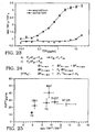
- a standard for each strain This is done by determining the affect (prion index) of a particular treatment on a known amount of a known strain. This can be plotted as shown in Figure 25.
- the strain of other samples can be readily determined -- standards are preferably calculated at a number of different concentrations for each strain.
- To determine the strain of a simple sample one calculates the concentration of protein in the sample and the affect of treatment on that concentration. The results are matched to the standard (as in Figure 25) to determine the strain.
- Example 18 is a specific example of such as taken in combination with Figure 25.
- the same concentration of the same strain will be effected in the same way by a given treatment. However, the same concentration of different strains will be effected differently by the same treatment making it possible to determine the strain.
- the assay of the invention can be applied to determining the presence of any protein which assumes two different conformational shapes, one of which is associated with the disease.
- the following is a non-limiting list of diseases with associated insoluble proteins which assume two or more different conformations.
- Alzheimer's Disease APP A ⁇ peptide, ⁇ 1-antichymotrypsin, tan, non-A ⁇ component Prion diseases, Creutzfeld Jakob disease, scrapie and bovine spongeform encephalopathy PrP Sc ALS SOD and neurofilament Pick's disease
- Pick body Parkinson's disease Lewy body Diabetes Type 1 Amylin Multiple myeloma-plasma cell dyscrasias IgGL-chain Familial amyloidotic polyneuropathy Transthyretin Medullary carcinoma of thyroid Procalcitonin Chronic renal failure ⁇ 2 -microglobulin Congestive heart failure Atrial natriuretic factor Senile cardiac and systemic amyloidosis Transthyretin Chronic inflammation Serum amyloid A Atherosclerosis ApoA1 Familial amyloidosis Gelsolin
- insoluble proteins listed above each include a number of variance or mutations which result in different strains which are all encompassed by the present.
- Known pathogenic mutations and polymorphisms in the PrP gene related to prion diseases are given below and the sequences of human, sheep and bovine are given in US 5,565,186, issued October 15, 1996.
- One aspect of the invention involves a two step process to diagnose Alzheimer's disease based on the presence of a constricted form of a protein ( ⁇ A4 amyloidosis) by quantitatively measuring ⁇ -sheet form of ⁇ A4 protein in sample material, e.g., in the brain or body fluids.
- sample material e.g., in the brain or body fluids.
- the sample is divided into two aliquots.
- the first aliquot is crosslinked to a solid plastic (long chain polymeric material) support in native conformation through a chemical activation step under the nondenaturing conditions.
- the second portion of the sample is first denatured and then crosslinked to the plastic support. Both portions of the sample material react in situ with the labeled antibodies that preferentially recognize soluble ⁇ A4 or denatured ⁇ A4 of the human or a given animal species.
- the amount of the antibody bound to denatured or native conformations of ⁇ A4 protein is recorded by the signal of the labeled secondary antibody.
- the excess of the signal obtained with the denatured sample compared to that expected change in the signal obtained with the native ⁇ -helical conformation of ⁇ A4 protein is the measure of the amount of ⁇ -sheet structured ⁇ A4 in the original sample.
- the formula developed for calculation of ⁇ A4 content is provided above in connection with the calculation of PrP Sc content.
- ⁇ A4 amyloidosis (Alzheimer's disease) is established by three procedures: (1) measurement of denatured sample alone and by detecting the increase in the total ⁇ A4 amount (concentration) in the examined sample above the background levels of soluble ⁇ A4 obtained from normal controls; (2) calculation of the ratio between denatured versus native signal for a given antibodies (protein index) - for example values higher than 2 for monoclonal antibody 6F3D and europium labeled secondary antibody; (3) evaluation of the change of the denatured sample signal over that expected change in the signal for ⁇ -helical conformation of ⁇ A4 as a measure of the amount of infectious ⁇ -sheet structured ⁇ A4 in the original sample.
- the formula developed for calculation of ⁇ A4 content is provided above.
- the particular strain of ⁇ A4 can also be determined using the same methodology described above to determine the strain of PrP Sc in a sample.
- the invention provides a direct diagnostic method for detecting the presence pathogenic forms of ⁇ A4 protein in pharmaceuticals, biopsy or autopsy tissue, brain, spinal cord, peripheral nerves, muscle, cerebrospinal fluid, blood and blood components, lymph nodes, and in animal- or human-derived cultures expressing or potentially expressing ⁇ A4 protein.
- the invention also makes it possible to follow the ⁇ -helix-to- ⁇ -sheet conformational transition of ⁇ A4 protein, or its fragments of synthetic or recombinant origin, and to provide a method to screen compounds for their ability to stabilize the normal soluble conformation of ⁇ A4 protein and thus prevent conversion into pathogenic insoluble and ⁇ -sheet-structured ⁇ A4 protein.
- Typical methods of sample denaturation include: (1) physical, such as hydrostatic pressure or temperature, (2) chemical, such as acidic or alkaline pH, chaotropic salts, or denaturing detergents, and (3) combination of above.
- Methods of chemical or affinity coupling of ⁇ A4 protein to a plastic support are described in available literature and may vary.
- Antibodies used in the diagnostic assay may be polyclonal, monoclonal or recombinant Fab and must be species specific with preferential binding to the soluble or denatured form of ⁇ A4 with preferably at least a 2-fold difference in reactivity between ⁇ -helical and ⁇ -sheet structured ⁇ A4, assuming the same amount of antigen.
- Methods of sample attachment to the plastic support may vary and may be covalent or non-covalent as described in available literature.
- the sensitivity of the assay described in the examples may be increased by using high-affinity antibodies, sandwich formate, immunoprecipitation, or differential centrifugation. However, only the antibodies with an affinity at least a 2 fold for denatured and compared to the native ⁇ -sheet conformation of ⁇ A4 of the same species shall be used for the diagnostic assay.
- Methods of antibody generation, purification, labeling and detection may vary. The antibody binding to different conformations of ⁇ A4 protein was measured by time-resolved, dissociation-enhanced fluorescence.
- the system of detection of ⁇ A4-bound IgG on solid support in situ or in solution may vary and may use direct or indirect immunological methods including direct radiolabels, fluorescence, luminescence, avidin-biotin amplification, or enzyme-linked assays with color or luminescent substrates.
- a pH of 7.4 was maintained throughout the run by the automated addition of 10% H 2 SO 4 or 24% NH 4 OH.
- the final volume was 10 L in which an OD 600 of ⁇ 100 was achieved after 36 h.
- the E. coli was harvested by centrifugation at 10,000 x g for 30 min and the resulting paste was stored at -20°C.
- E. coli paste was resuspended in 1 L of 25 mM Tris ⁇ HCl, pH 8.0, 5 mM EDTA (buffer A). This was centrifuged at 10,000 x g for 20 min, and the supernatant containing soluble periplasmic proteins was discarded. The pellet was resuspended in 1 L of buffer A, passed through a cell disrupter twice (Microfluidics International, model MF 110), and centrifuged at 30,000 x g for 1 h, after which the supernatant was discarded and the pellet was washed once in buffer A and centrifuged again at 30,000 x g for 1 h.
- the pellet could be stored at -20°C prior to further separation. It was subsequently solubilized in 8M Gdn ⁇ HCl/25 mM Tris-HCl, pH 8.0/100 mM DTT (buffer B) and centrifuged at 14,000 x g for 20 min to remove the remaining insoluble matter.
- Fractions enriched for the recombinant prion protein as identified by SDS-PAGE were pooled and further purified by reversed phase high performance liquid chromatography (RP-HPLC) employing a 25 mm x 25 cm C-4 column (Vydac); Buffer 1: H 2 O/0.1% TFA, Buffer 2: acetonitrile/0.09% TFA, flow rate 5 mL/min.
- RP-HPLC reversed phase high performance liquid chromatography
- Both proteins produced per Example 1 were used as a standards for the prion assay and to establish the sensitivity and linearity range of the diagnostic method.
- the purified Syrian hamster brain PrP c was used for the calibration of prion protein detection and correlated with results obtained on recombinant SHaPrP90-231 in ⁇ -helical, ⁇ -sheet, and denatured conformations.
- the PrP c protein was purified as described with some minor modifications [Pan, Stahl et al. (1992) Protein Sci 1 :1343-1352; Pan, Baldwin et al. (1993) Proc Natl Acad Sci USA 90 :10962-10966]. Protein content was determined by amino acid analysis. The purity of PrP c protein, as demonstrated on SDS PAGE followed by silver staining and Western, was ⁇ 95%.
- Standard Syrian hamster PrP Sc was purified from a standard pool of scrapie strain Sc237 infected hamster brains as described with only minor modifications [Turk, Teplow et al. (1988) Eur J Biochem 176 :21-30].
- the infectivity of this standard as determined by an incubation time assay on Syrian hamsters after intracerebral inoculation, was 10 7.3 ID 50 /ml and specific infectivity 10 8.2 ID 50 /mg of PrP Sc protein. However, the specific infectivity may vary from lot to lot ⁇ 10 0.5 ID 50 /mg.
- the protein content was determined by BCA assay using Bovine serum albumin as a standard. The preparation was considered homogeneous with one major band on SDS PAGE after silver staining and Western Blots.
- SHaPrP90-231 in ⁇ -helical, ⁇ -sheet and random coil conformations were covalently attached to glutaraldehyde activated polystyrene plates and incubated with serially diluted primary antibody.
- the amount of IgG reacting with each conformation of SHaPrP90-231 was determined either directly with Eu-labeled 3F4 IgG, or indirectly with europium labeled anti-rabbit or anti mouse antibody, according to usual protocols and the total signal was measured by time-resolved, dissociation-enhanced fluorescence.
- For the assay development were selected antibodies with the signal ratio of denatured versus ⁇ -sheet conformation of SHaPrP90-231 equal or higher than 4.
- Purified recombinant SHaPrP90-231 was diluted into 5% (w/v) brain homogenate obtained from PrP 0/0 mouse and containing no prion protein.
- the brain homogenate was made by three 30 sec bursts in PowerGen homogenizer equipped with plastic disposable probe in TBS, pH 7.4 containing protease inhibitors cocktail (1 mM PMSF, 2 ⁇ g/ml of Aprotinin, and 2 ⁇ g/ml of Leupeptin) and spun at 5°C for 5 min at 500 G in a desktop centrifuge.
- the resulting supernatant was diluted 1:1 in TBS with final 4% (w/v) Sarcosyl and homogenized again by three 30 sec bursts in a PowerGen homogenizer. Next, the homogenate was spiked with different dilutions of recombinant SHaPrP90-231 in ⁇ -helical or ⁇ -sheet conformations.
- the analyte PrP in different conformations is preincubated with europium labeled 3F4 IgG and then transferred to the polystyrene plate coated with recombinant ShaPrP90-231 in SDS-denatured state.
- the results for analyte SHaPrP90-231 in ⁇ -helical and denatured state ( Figure 2) indicate marked difference in both available binding sites and affinity of europium-labeled 3F4 IgG with different conformations of prion protein.
- each sample of dilution curve was divided into two aliquots: (1) untreated and designated native; (2) mixed with final 4M Gdn HCl and heated for 5 min at 100°C and designated denatured. Both samples were diluted 20-fold by H 2 O and aliquots loaded on polystyrene plate activated with glutaraldehyde. The plates, incubated overnight at 5°C, were blocked with TBS, pH 7.8, containing 0.5% BSA (w/v) and 6% Sorbitol (w/v). In the next step, they were washed three time with TBS, pH 7.8 containing 0.05% (v/v) of Tween® 20 and incubated with europium-labeled antibodies listed above. The plates were developed after an additional 7 washing steps in enhancement solution provided by the europium label supplier (Wallac Inc., Turku, Finland) and signal counted on DELFIA 1234 Fluorometer (Wallac Inc., Turku, Finland).
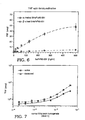
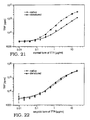
- results are expressed in two different forms: (1) as a ratio ( Figure 6) , where index ⁇ 1.8 for recombinant SHaPrP90-231 indicates that the protein was originally in all ⁇ -helix conformation and index >1.8 indicates presence of ⁇ -sheet conformation; (2) as a formula shown herein and exemplified in Example 11, where the excess increase of signal above that expected for ⁇ -helical conformation is proportionate to the amount of SHaPrP90-231 in ⁇ -sheet conformation.
- the input/output calibration for denatured ShaPrP90-231 in both ⁇ -helical and ⁇ -sheet conformations was linear within three orders of magnitude, and provide a high degree of confidence (Figure 5) for the assay. Also assayed was PrP c , serially diluted into PrP 0/0 mouse homogenate which provided results with high degree of confidence within linearity range of 3 orders of magnitude. Next, the assay was calibrated by purified infectious PrP Sc in the presence of 5% PrP 0/0 brain homogenate. The calibration with PrP Sc provided a linear response within similar range.
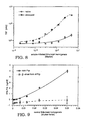
- the ratio between the signal of denatured versus native brain PrP c was for Eu-labeled monoclonal 3F4 IgG 2.2 ( Figure 7), for polyclonal N12 and P3 1.0.
- the calibration for PrP Sc gave ratio ⁇ 20 ( Figure 8) for 3F4 Eu-labeled IgG and >4 for N12 and P3 antibodies.
- all PrP c was in full range in ⁇ -helical conformation.
- the calculated amount of PrP Sc in infectious, ⁇ -sheet conformation was as anticipated close to the total amount of prion protein.
- This prion assay may be used: (1) in direct mode in brain, tissue samples, body fluids or pharmaceuticals after determination of normal control values; or (2) in indirect mode by detecting the elevation of total PrP in the brain of experimental animal intracerebrally inoculated with tested tissue, body fluid or pharmaceutical sample suspected of containing prions.
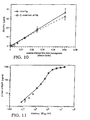
- the ratio between antibody affinity for denatured versus native Syrian hamster brain PrP c protein is in the broad concentration range for Eu-labeled 3F4 IgG between 0.8-2.2.
- the ratio in the scrapie infected brain is through the full linearity range above those values ( Figure 11).
- the "prion index" gives a relative indicator of the presence of infectious PrP Sc and therefore prions.
- This mode of prion assay is being used: (1) in direct mode in brain, tissue samples, body fluids or pharmaceuticals after determination of the index for normal controls; or (2) in indirect mode by detecting the elevated denatured/native prion protein index in the brain of experimental animals intracerebrally inoculated with tested tissue, body fluid or pharmaceutical sample suspected from containing prions.
- This mode of prion assay is used: (1) in direct mode in brain, tissue samples, body fluids or pharmaceuticals; or (2) in indirect mode, by calculating PrP Sc content in the brain of experimental animals intracerebrally inoculated with tested tissue, body fluid or pharmaceutical samples suspected for containing prions. After establishing a calibration curve between PrP Sc and prion titer, it is possible to estimate the titer directly from PrP Sc content.
- the samples are then divided into two aliquots: (1) untreated, designated native; (2) mixed with final 4M Gdn HCl and heated for 5 min at 100°C, designated denatured. Both samples are diluted 20-fold by H 2 O and aliquots loaded on polystyrene plate activated with glutaraldehyde. The plates, incubated overnight at 5°C, were blocked with TBS, pH 7.8, containing 0.5% BSA (w/v) and 6% Sorbitol (w/v). In the next step, they were washed three time with TBS, pH 7.8 containing 0.05% (v/v) of Tween® 20 and incubated with europium-labeled 3F4 IgG. The plates were developed after additional 7 washing steps in enhancement solution provided by the europium label supplier (Wallac Inc., Turku, Finland) and signal counted on DELFIA 1234 Fluorometer (Wallac Inc., Turku, Finland).
- the degree of conversion from ⁇ -helical to ⁇ -sheet conformation of PrP is calculated from the "prion index" or alternatively from the formulae provided herein. Some compounds which inhibit the conversion by apparently stabilizing the native-like conformation of prion protein may have therapeutic potential in vivo.
- Soluble forms of ⁇ A4 (1-40) and ⁇ A4 (1-42) were obtained from Bachem (Torrance, CA).
- One portion of the fresh lyophilized peptide was solubilized in PBS, pH 7.4, containing 20% (v/v) of HFIP (hexafluoroisopropanol; 1,1,1,3,3,3-hexafluoro-2-propanol) or 1% (w/v) SDS (sodium dodecylsulfate) at final protein concentration 50 ⁇ M; this protein was designated "soluble" and was stored at -80°C until use.
- HFIP hexafluoroisopropanol; 1,1,1,3,3,3-hexafluoro-2-propanol
- SDS sodium dodecylsulfate
- ⁇ A4 protein in ⁇ -helical, ⁇ -sheet and random coil conformations were covalently attached to the glutaraldehyde activated polystyrene plates and incubated with serially diluted primary antibody.
- the amount of IgG reacting with each conformation of ⁇ A4 was determined with europium labeled anti-rabbit or anti mouse antibody according usual protocols and the total signal was measured by time-resolved, dissociation-enhanced fluorescence.
- Antibodies with the signal ratio of denatured versus ⁇ -sheet conformation of ⁇ A4 equal or higher than 2 were selected to develop the assay.
- each sample of dilution curve of ⁇ -helical and ⁇ -sheet forms of ⁇ A4 protein was divided into two aliquots: (1) untreated (designated native); and (2) mixed with final 4M Gdn ⁇ HCl/1% Sarcosyl and heated for 5 min at 100°C (designated "denatured”). Both samples were diluted 20-fold by H 2 O and aliquots loaded on polystyrene plate activated with 0.2% glutaraldehyde for 2 hrs. The plates, incubated overnight at 5°C, were blocked with TBS, pH 7.8, containing 0.5% BSA (w/v) and 6% Sorbitol (w/v).
- the samples were then washed three time with TBS, pH 7.8 containing 0.05% (v/v) of Tween® 20 and incubated with primary antibodies against ⁇ A4 protein (6F3D, Research Diagnostics Inc., Flanders, NJ).
- the samples were washed and then developed with europium-labeled secondary antibodies against mouse IgG (Wallac Inc., Turku, Finland).
- the plates were developed after an additional 7 washing steps in enhancement solution (Wallac Inc., Turku, Finland) and signal counted on DELFIA 1234 Fluorometer (Wallac Inc., Turku, Finland).
- the results for analyte indicate marked difference in both available binding sites and affinity anti- ⁇ A4 IgG with different conformations of ⁇ A4 protein.
- the method could be carried out using a competitive assay, where the analyte ⁇ A4, in different conformations is preincubated with anti- ⁇ A4 IgG and then transferred to the polystyrene plate coated with synthetic ⁇ A4 protein in Gdn ⁇ HCl-denatured state.
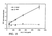
- results may be expressed in two different forms: (1) as a ratio ( Figure 15), where index ⁇ 1.0 for ⁇ A4 indicates that the protein was originally in all ⁇ -helix conformation and index >1.5 indicates presence of ⁇ -sheet conformation; (2) per the formula shown above where the excess increase of signal above that expected for ⁇ -helical conformation is proportionate to the amount of ⁇ A4 in ⁇ -sheet conformation.
- the ratio between antibody affinity for denatured versus native human ⁇ A4 protein is in the broad concentration range for 6F3D IgG between 0.8-1.0.
- the ration for ⁇ -sheet ⁇ A4 is through the full linearity range above those values ( Figure 15).
- the " ⁇ A4 amyloid index" gives a relative indicator of the presence of pathogenic insoluble ⁇ -sheet ⁇ A4.
- This mode of ⁇ A4 assay may be used in direct mode in brain, tissue samples, body fluids or pharmaceuticals after determination of the index for normal controls.
- the amount of pathogenic forms of ⁇ -sheet ⁇ A4 protein can be calculated in brain, tissue samples, body fluids or pharmaceuticals.
- the samples are then divided into two aliquots: (1) untreated, containing 1% Sarcosyl and designated “native”; and (2) mixed with final 4M Gdn HCl/1% Sarcosyl and heated for 5 min at 100°C, designated “denatured”. Both samples are diluted 20-fold by H 2 O and aliquots loaded on polystyrene plate activated with glutaraldehyde. The plates, incubated overnight at 5°C, were blocked with TBS, pH 7.8, containing 0.5% BSA (w/v) and 6% Sorbitol (w/v).
- the degree of conversion from ⁇ -helical to ⁇ -sheet conformation of ⁇ A4 is calculated from the "amyloid index" ( Figure 15) or alternatively from the formula shown above. Any compound which inhibits the conversion by stabilizing the ⁇ -helical conformation of ⁇ A4 protein may have therapeutic potential in vivo by preventing formation of mature amyloid.
- Soluble forms of TTR were obtained from Sigma Chemical Comp.
- One portion of the protein was solubilized in 100 mM KCl buffer, pH 7.4, at final protein concentration 0.5 mg/ml; this protein was designated "normal” and was stored at -80°C until use.
- the second portion of the protein was converted into amyloid as described [Lai, Colon et al. (1996) Biochemistry 35(20) :6470-82]. Briefly, the protein was resuspended in 100 mM KCl buffer, containing 50 mM sodium acetate, pH 4.4, at protein concentration 0.2 mg/ml, and incubated for 72 hrs at 37°C. This portion of the protein was designated "amyloid" and was stored until use at -80°C.
- each sample of protein taken from the dilution curve of normal and amyloid forms of TTR was divided into two aliquots: (1) untreated and designated “native”; (2) mixed with final 4M Gdn HCl/1% Sarcosyl and heated for 5 min at 100°C and designated “denatured.” Both samples were diluted 20-fold by H 2 O and aliquots loaded on polystyrene plate activated with 0.2% glutaraldehyde for 2 hrs. The plates, incubated overnight at 5°C, were blocked with TBS, pH 7.8, containing 0.5% BSA (w/v) and 6% Sorbitol (w/v).
- the ratio between antibody affinity for denatured versus native form of normal human TTR is in the broad concentration range for polyclonal antibody 1-3:3.
- the ratio for amyloid form of TTR is through the full linearity range between 0.7-1.0 ( Figure 18).
- the "TTR amyloid index” gives a relative indicator of the presence of pathogenic, insoluble and amyloid-forming TTR. This mode of TTR assay is being used in direct mode in brain, tissue samples, body fluids or pharmaceuticals after determination of the index for normal controls.
- the amount of amyloid form of TTR can be calculated in peripheral nerve, tissue samples; body fluids or pharmaceuticals.
- the lower than expected increase of signal for normal conformation is proportional to the amount of TTR in amyloid conformation.
- the aliquots of a 0.2 mg/ml solution of the normal form of synthetic TTR, or corresponding recombinant or synthetic peptides of the TTR are incubated in 100 mM KCl buffer, containing 50 mM of sodium acetate, pH 4.4 for 72 hrs at 37°C with 10 -3 - 10 -6 M concentrations of tested organic compounds.
- the samples are then divided into two aliquots: (1) untreated, containing 1% Sarcosyl and designated “native”; (2) mixed with final 4M Gdn HCl/1% Sarcosyl and heated for 5 min at 100°C, designated “denatured.” Both samples are diluted 20-fold with H 2 O and aliquots loaded on polystyrene plate activated with glutaraldehyde. The plates, incubated overnight at 5°C, were blocked with TBS, pH 7.8, containing 0.5% BSA (w/v) and 6% Sorbitol (w/v).
- the degree of conversion from normal to amyloid conformation of TTR is calculated from the "amyloid index" ( Figure 18) or alternatively from the formula ( Figure 19). Any compound which inhibits the conversion by stabilizing normal conformation of TTR may have therapeutic potential in vivo by preventing formation of mature amyloid.
- the plates were developed after an additional 7 washing steps in enhancement solution provided by the Europium label supplier (Wallac Inc., Turku, Finland) and signal counted on DELFIA 1234 Fluorometer (Wallac Inc., Turki, Finland).
- the PrP Sc content and "prion index" ratio of antibody binding to denatured:native PrP protein
- the PrP Sc content and "prion index” were calculated as described in Examples 11 and 8 and plotted in xy coordinates as shown in Figure 25. Other samples tested and resulting in the same calculations could be presumed to have the same strain of prion therein.
Landscapes
- Health & Medical Sciences (AREA)
- Life Sciences & Earth Sciences (AREA)
- Chemical & Material Sciences (AREA)
- Engineering & Computer Science (AREA)
- Biomedical Technology (AREA)
- Molecular Biology (AREA)
- Organic Chemistry (AREA)
- Immunology (AREA)
- Medicinal Chemistry (AREA)
- General Health & Medical Sciences (AREA)
- Biochemistry (AREA)
- Proteomics, Peptides & Aminoacids (AREA)
- Urology & Nephrology (AREA)
- Hematology (AREA)
- Neurology (AREA)
- Biophysics (AREA)
- Genetics & Genomics (AREA)
- Analytical Chemistry (AREA)
- Neurosurgery (AREA)
- Gastroenterology & Hepatology (AREA)
- Biotechnology (AREA)
- Cell Biology (AREA)
- Zoology (AREA)
- Toxicology (AREA)
- Microbiology (AREA)
- Pathology (AREA)
- General Physics & Mathematics (AREA)
- Food Science & Technology (AREA)
- Physics & Mathematics (AREA)
- Bioinformatics & Cheminformatics (AREA)
- Nuclear Medicine, Radiotherapy & Molecular Imaging (AREA)
- Psychiatry (AREA)
- Chemical Kinetics & Catalysis (AREA)
- General Chemical & Material Sciences (AREA)
- Public Health (AREA)
- Pharmacology & Pharmacy (AREA)
- Animal Behavior & Ethology (AREA)
- Hospice & Palliative Care (AREA)
- Veterinary Medicine (AREA)
- Peptides Or Proteins (AREA)
Applications Claiming Priority (3)
| Application Number | Priority Date | Filing Date | Title |
|---|---|---|---|
| US08/804,536 US5891641A (en) | 1997-02-21 | 1997-02-21 | Assay for disease related conformation of a protein |
| US804536 | 1997-02-21 | ||
| EP98906471A EP0970372B1 (fr) | 1997-02-21 | 1998-02-20 | Dosage pour la detection d'une structure proteique associee a une maladie |
Related Parent Applications (1)
| Application Number | Title | Priority Date | Filing Date |
|---|---|---|---|
| EP98906471A Division EP0970372B1 (fr) | 1997-02-21 | 1998-02-20 | Dosage pour la detection d'une structure proteique associee a une maladie |
Publications (2)
| Publication Number | Publication Date |
|---|---|
| EP1416281A2 true EP1416281A2 (fr) | 2004-05-06 |
| EP1416281A3 EP1416281A3 (fr) | 2004-05-19 |
Family
ID=25189206
Family Applications (2)
| Application Number | Title | Priority Date | Filing Date |
|---|---|---|---|
| EP04000945A Withdrawn EP1416281A3 (fr) | 1997-02-21 | 1998-02-20 | Essai pour détecter une conformation de protéine liée à une maladie |
| EP98906471A Expired - Lifetime EP0970372B1 (fr) | 1997-02-21 | 1998-02-20 | Dosage pour la detection d'une structure proteique associee a une maladie |
Family Applications After (1)
| Application Number | Title | Priority Date | Filing Date |
|---|---|---|---|
| EP98906471A Expired - Lifetime EP0970372B1 (fr) | 1997-02-21 | 1998-02-20 | Dosage pour la detection d'une structure proteique associee a une maladie |
Country Status (10)
| Country | Link |
|---|---|
| US (1) | US5891641A (fr) |
| EP (2) | EP1416281A3 (fr) |
| JP (1) | JP3774236B2 (fr) |
| AT (1) | ATE267394T1 (fr) |
| AU (1) | AU725844B2 (fr) |
| BR (1) | BR9807600A (fr) |
| CA (1) | CA2278577A1 (fr) |
| DE (2) | DE69823985T2 (fr) |
| ES (1) | ES2218807T3 (fr) |
| WO (1) | WO1998037411A1 (fr) |
Families Citing this family (51)
| Publication number | Priority date | Publication date | Assignee | Title |
|---|---|---|---|---|
| US6719988B2 (en) | 1997-02-21 | 2004-04-13 | The Regents Of The University Of California | Antiseptic compositions for inactivating prions |
| US6620629B1 (en) * | 1997-02-21 | 2003-09-16 | The Regents Of The University Of California | Method for detecting prions |
| US6221614B1 (en) * | 1997-02-21 | 2001-04-24 | The Regents Of The University Of California | Removal of prions from blood, plasma and other liquids |
| US6617119B2 (en) | 1997-02-21 | 2003-09-09 | The Regents Of The University Of California | Assay for specific strains of multiple disease related conformations of a protein |
| US6720355B2 (en) | 1997-02-21 | 2004-04-13 | The Regents Of The University Of California | Sodium dodecyl sulfate compositions for inactivating prions |
| KR20010041135A (ko) * | 1998-02-20 | 2001-05-15 | 더 리젠츠 오브 더 유니버시티 오브 캘리포니아 | 단백질의 다중의 질병 관련된 입체구조의 특이적인스트레인에 대한 분석 |
| US6127130A (en) | 1998-03-13 | 2000-10-03 | The United States Of America As Represented By The Secretary Of The Navy | Multiassay method for determining the concentrations of antigens and interferants |
| GB2371049B (en) * | 1998-11-04 | 2003-06-04 | Imp College Innovations Ltd | Methods of making antibodies against ß-forms of prion proteins and their uses |
| GB2362385B (en) * | 1998-11-04 | 2002-06-19 | Imp College Innovations Ltd | Non-aggregated ß-forms of prion proteins |
| GB2348203B (en) | 1998-11-04 | 2002-06-19 | Imp College Innovations Ltd | Solube beta-forms of prion proteins, methods of preparation and use |
| US6821738B2 (en) * | 1999-01-20 | 2004-11-23 | The Board Of Regents For Oklahoma State University | Broad spectrum bio-detection of nerve agents, organophosphates, and other chemical warfare agents |
| US6605445B1 (en) * | 1999-02-22 | 2003-08-12 | Bayer Corporation | Rapid method of determining clearance of prion protein |
| GB9904395D0 (en) * | 1999-02-25 | 1999-04-21 | Fluorescience Ltd | Protein assay |
| US6972198B2 (en) * | 1999-02-26 | 2005-12-06 | Cyclacel, Ltd. | Methods and compositions using protein binding partners |
| US6166187A (en) * | 1999-03-05 | 2000-12-26 | The Regents Of The University Of California | Method of concentrating prion proteins in blood samples |
| GB9908059D0 (en) | 1999-04-09 | 1999-06-02 | Imp College Innovations Ltd | Diagnosis and treatment of diseases |
| DE19918141A1 (de) * | 1999-04-21 | 2000-10-26 | Boehringer Ingelheim Vetmed | Verfahren zur Diagnose von übertragbaren Spongiformen Enzephalopathien |
| US7041807B1 (en) | 1999-06-23 | 2006-05-09 | Caprion Pharmaceuticals, Inc. | Antibodies to a YYX epitope of a mammalian prion protein |
| NZ516240A (en) * | 1999-06-23 | 2005-11-25 | Caprion Pharmaceuticals Inc | Prion protein peptides and uses thereof |
| US6537548B1 (en) * | 2000-07-27 | 2003-03-25 | The Regents Of The University Of California | Antibodies specific for ungulate PrP |
| EP1399110A4 (fr) * | 2001-05-01 | 2004-12-01 | Univ California | Anticorps empechant la propagation de prions et favorisant la clairance d'une infectiosite |
| EP1395833B1 (fr) | 2001-05-31 | 2013-02-27 | Adlyfe, Inc. | Procede de detection de proteines mal pliees |
| US20050026165A1 (en) * | 2001-05-31 | 2005-02-03 | Cindy Orser | Detection of conformationally altered proteins and prions |
| US20030050517A1 (en) * | 2001-07-31 | 2003-03-13 | Ahting Herbert C. | Process for producing glycerin |
| WO2003027631A2 (fr) * | 2001-09-20 | 2003-04-03 | The Regents Of The University Of California | Echantillon de muscle prepare pour un essai de detection de prions |
| US7803315B2 (en) * | 2001-10-05 | 2010-09-28 | American Sterilizer Company | Decontamination of surfaces contaminated with prion-infected material with gaseous oxidizing agents |
| DE10152677A1 (de) | 2001-10-19 | 2003-05-08 | Aventis Behring Gmbh | Antikörper zum spezifischen Nachweis von pathogenen Prionen humanen Ursprungs und damit durchgeführten Nachweisverfahren |
| US7045297B2 (en) * | 2001-11-14 | 2006-05-16 | Prion Developmental Laboratories, Inc. | Rapid prion-detection assay |
| US20030092090A1 (en) * | 2001-11-14 | 2003-05-15 | Kiamars Hajizadeh | Rapid prion-detection device, system, and test kit |
| US7939641B2 (en) * | 2002-04-09 | 2011-05-10 | The Scripps Research Institute | Motif-grafted hybrid polypeptides and uses thereof |
| US20040072236A1 (en) | 2002-09-27 | 2004-04-15 | Neil Cashman | PrPSc -interacting molecules and uses thereof |
| FR2849205B1 (fr) * | 2002-12-20 | 2005-02-11 | Afssa | Procede d'amplification de la detection de la prpsc et utilisation d'un ligand adjuvant macrocyclique pour une telle amplification |
| US20040192887A1 (en) * | 2003-03-25 | 2004-09-30 | Ralph Zahn | PH-dependent polypeptide aggregation and its use |
| US20060035242A1 (en) * | 2004-08-13 | 2006-02-16 | Michelitsch Melissa D | Prion-specific peptide reagents |
| US20050256425A1 (en) | 2004-05-11 | 2005-11-17 | Inpro Biotechnology, Inc. | Device, system and method for extracting and preparing brain tissue |
| US9488655B2 (en) * | 2004-07-14 | 2016-11-08 | The Regents Of The University Of California | Biomarkers for detection of early- and late-stage endometrial cancer |
| WO2012047930A2 (fr) | 2010-10-04 | 2012-04-12 | The Regents Of The University Of California | Compositions et procédés de traitement de cancers gynécologiques |
| EP2199801B1 (fr) * | 2004-07-14 | 2013-05-29 | The Regents of The University of California | Biomarqueurs pour la détection précoce du cancer des ovaires |
| US20080027025A1 (en) * | 2004-08-27 | 2008-01-31 | The Regents Of The University Of California | Protection From And Treatment Of Prion Protein Infection |
| WO2006076683A2 (fr) * | 2005-01-13 | 2006-07-20 | Novartis Vaccines And Diagnostics Inc. | Isolement et detection de prions pathogenes |
| US20090130774A1 (en) * | 2005-01-13 | 2009-05-21 | David Peretz | Elisa assays using prion-specific peptide reagents |
| US8372593B2 (en) * | 2005-02-15 | 2013-02-12 | Adlyfe, Inc. | Method for detecting misfolded proteins and prions |
| WO2006102099A2 (fr) * | 2005-03-18 | 2006-09-28 | The Regents Of The University Of California | Anticorps specifiques de la proteine prp humaine et bovine |
| JP4724816B2 (ja) * | 2005-09-06 | 2011-07-13 | シャープ株式会社 | タンパク質の測定方法 |
| US20070196863A1 (en) * | 2006-02-17 | 2007-08-23 | Hanson Technologies, Inc. | Prion protein detection |
| JP5097206B2 (ja) * | 2006-07-28 | 2012-12-12 | エイディーライフ インコーポレイティッド | 診断および治療のためのペプチドプローブ |
| EP2282753A1 (fr) * | 2008-04-30 | 2011-02-16 | Novartis AG | Essai de conformères pathogènes |
| US9241976B2 (en) | 2011-08-29 | 2016-01-26 | The Regents Of The University Of California | Use of HDL-related molecules to treat and prevent proinflammatory conditions |
| CN104781271B (zh) | 2012-08-20 | 2018-07-06 | 加利福尼亚大学董事会 | 具有生物可逆的基团的多核苷酸 |
| JP7178607B2 (ja) * | 2018-02-23 | 2022-11-28 | 学校法人麻布獣医学園 | アミロイドタンパク質を抽出する試薬 |
| US20240199857A1 (en) * | 2022-12-16 | 2024-06-20 | The Chinese University Of Hong Kong | Chaotropes-assisted deep immunostaining |
Family Cites Families (7)
| Publication number | Priority date | Publication date | Assignee | Title |
|---|---|---|---|---|
| US4806627A (en) * | 1987-05-29 | 1989-02-21 | Research Foundation Of Mental Hygiene Inc. | Hybrid cell lines producing monoclonal antibodies dircted against scrapie-associated fibril proteins |
| EP0613495B1 (fr) * | 1991-11-14 | 2005-02-02 | WEISSMANN, Charles | Animaux transgeniques non-humains exempts de prions |
| WO1993023432A1 (fr) * | 1992-05-15 | 1993-11-25 | New York University | Polypeptides de pion solubles et procedes de detection et de purification |
| US5565186A (en) * | 1994-05-13 | 1996-10-15 | The Regents Of The University Of California | Method of detecting prions in a sample and transgenic animal used for same |
| US5750361A (en) * | 1995-11-02 | 1998-05-12 | The Regents Of The University Of California | Formation and use of prion protein (PRP) complexes |
| WO1997043649A1 (fr) * | 1996-05-14 | 1997-11-20 | Winnacker Ernst Ludwig | CHAPERONS POUVANT SE LIER AUX PRIONS ET DISTINGUER LES ISOFORMES PrP?C ET PrPSC¿ |
| EP0861900A1 (fr) * | 1997-02-21 | 1998-09-02 | Erziehungsdirektion Of The Canton Zurich | Détection immunologique de prions |
-
1997
- 1997-02-21 US US08/804,536 patent/US5891641A/en not_active Expired - Fee Related
-
1998
- 1998-02-20 EP EP04000945A patent/EP1416281A3/fr not_active Withdrawn
- 1998-02-20 DE DE69823985T patent/DE69823985T2/de not_active Expired - Fee Related
- 1998-02-20 JP JP53606498A patent/JP3774236B2/ja not_active Expired - Fee Related
- 1998-02-20 DE DE0970372T patent/DE970372T1/de active Pending
- 1998-02-20 WO PCT/US1998/002992 patent/WO1998037411A1/fr not_active Ceased
- 1998-02-20 CA CA002278577A patent/CA2278577A1/fr not_active Abandoned
- 1998-02-20 ES ES98906471T patent/ES2218807T3/es not_active Expired - Lifetime
- 1998-02-20 BR BR9807600-0A patent/BR9807600A/pt not_active Application Discontinuation
- 1998-02-20 AU AU61688/98A patent/AU725844B2/en not_active Ceased
- 1998-02-20 EP EP98906471A patent/EP0970372B1/fr not_active Expired - Lifetime
- 1998-02-20 AT AT98906471T patent/ATE267394T1/de not_active IP Right Cessation
Also Published As
| Publication number | Publication date |
|---|---|
| WO1998037411A1 (fr) | 1998-08-27 |
| AU725844B2 (en) | 2000-10-19 |
| DE970372T1 (de) | 2001-10-25 |
| AU6168898A (en) | 1998-09-09 |
| DE69823985D1 (de) | 2004-06-24 |
| JP3774236B2 (ja) | 2006-05-10 |
| BR9807600A (pt) | 2000-02-22 |
| EP0970372B1 (fr) | 2004-05-19 |
| DE69823985T2 (de) | 2005-07-21 |
| JP2001516448A (ja) | 2001-09-25 |
| US5891641A (en) | 1999-04-06 |
| EP1416281A3 (fr) | 2004-05-19 |
| EP0970372A4 (fr) | 2002-04-10 |
| ES2218807T3 (es) | 2004-11-16 |
| CA2278577A1 (fr) | 1998-08-27 |
| EP0970372A1 (fr) | 2000-01-12 |
| ATE267394T1 (de) | 2004-06-15 |
Similar Documents
| Publication | Publication Date | Title |
|---|---|---|
| EP0970372B1 (fr) | Dosage pour la detection d'une structure proteique associee a une maladie | |
| US6406864B2 (en) | Assay for disease related conformation of a protein and isolating same | |
| EP1057022B1 (fr) | Dosage pour souches specifiques de conformations d'une proteine relatives a plusieurs maladies | |
| US20010001061A1 (en) | Assay for disease related conformation of a protein | |
| US6617119B2 (en) | Assay for specific strains of multiple disease related conformations of a protein | |
| AU768840B2 (en) | Prion protein standard and method of making same | |
| AU764888B2 (en) | Assay for disease related conformation of a protein | |
| MXPA99007739A (en) | Assay for disease related conformation of a protein | |
| MXPA00008126A (en) | Assay for specific strains of multiple disease related conformations of a protein | |
| Barry | Prusiner et al. | |
| MXPA01003626A (en) | Assay for disease related conformation of a protein | |
| AU2003271329A1 (en) | Assay for disease related conformation of a protein |
Legal Events
| Date | Code | Title | Description |
|---|---|---|---|
| PUAI | Public reference made under article 153(3) epc to a published international application that has entered the european phase |
Free format text: ORIGINAL CODE: 0009012 |
|
| PUAL | Search report despatched |
Free format text: ORIGINAL CODE: 0009013 |
|
| AC | Divisional application: reference to earlier application |
Ref document number: 0970372 Country of ref document: EP Kind code of ref document: P |
|
| AK | Designated contracting states |
Kind code of ref document: A2 Designated state(s): AT BE CH DE DK ES FI FR GB GR IE IT LI LU MC NL PT SE |
|
| AK | Designated contracting states |
Kind code of ref document: A3 Designated state(s): AT BE CH DE DK ES FI FR GB GR IE IT LI LU MC NL PT SE |
|
| 17P | Request for examination filed |
Effective date: 20041104 |
|
| AKX | Designation fees paid |
Designated state(s): AT BE CH DE DK ES FI FR GB GR IE IT LI LU MC NL PT SE |
|
| 17Q | First examination report despatched |
Effective date: 20070301 |
|
| STAA | Information on the status of an ep patent application or granted ep patent |
Free format text: STATUS: THE APPLICATION IS DEEMED TO BE WITHDRAWN |
|
| 18D | Application deemed to be withdrawn |
Effective date: 20070712 |