EP0861900A1 - Détection immunologique de prions - Google Patents
Détection immunologique de prions Download PDFInfo
- Publication number
- EP0861900A1 EP0861900A1 EP97102837A EP97102837A EP0861900A1 EP 0861900 A1 EP0861900 A1 EP 0861900A1 EP 97102837 A EP97102837 A EP 97102837A EP 97102837 A EP97102837 A EP 97102837A EP 0861900 A1 EP0861900 A1 EP 0861900A1
- Authority
- EP
- European Patent Office
- Prior art keywords
- prp
- monoclonal antibody
- prion
- antibodies
- prion protein
- Prior art date
- Legal status (The legal status is an assumption and is not a legal conclusion. Google has not performed a legal analysis and makes no representation as to the accuracy of the status listed.)
- Withdrawn
Links
Images



Classifications
-
- C—CHEMISTRY; METALLURGY
- C07—ORGANIC CHEMISTRY
- C07K—PEPTIDES
- C07K16/00—Immunoglobulins [IG], e.g. monoclonal or polyclonal antibodies
- C07K16/18—Immunoglobulins [IG], e.g. monoclonal or polyclonal antibodies against material from animals or humans
- C07K16/28—Immunoglobulins [IG], e.g. monoclonal or polyclonal antibodies against material from animals or humans against receptors, cell surface antigens or cell surface determinants
- C07K16/2872—Immunoglobulins [IG], e.g. monoclonal or polyclonal antibodies against material from animals or humans against receptors, cell surface antigens or cell surface determinants against prion molecules, e.g. CD230
-
- C—CHEMISTRY; METALLURGY
- C07—ORGANIC CHEMISTRY
- C07K—PEPTIDES
- C07K14/00—Peptides having more than 20 amino acids; Gastrins; Somatostatins; Melanotropins; Derivatives thereof
- C07K14/435—Peptides having more than 20 amino acids; Gastrins; Somatostatins; Melanotropins; Derivatives thereof from animals; from humans
- C07K14/46—Peptides having more than 20 amino acids; Gastrins; Somatostatins; Melanotropins; Derivatives thereof from animals; from humans from vertebrates
- C07K14/47—Peptides having more than 20 amino acids; Gastrins; Somatostatins; Melanotropins; Derivatives thereof from animals; from humans from vertebrates from mammals
-
- A—HUMAN NECESSITIES
- A61—MEDICAL OR VETERINARY SCIENCE; HYGIENE
- A61K—PREPARATIONS FOR MEDICAL, DENTAL OR TOILETRY PURPOSES
- A61K38/00—Medicinal preparations containing peptides
-
- C—CHEMISTRY; METALLURGY
- C07—ORGANIC CHEMISTRY
- C07K—PEPTIDES
- C07K2317/00—Immunoglobulins specific features
- C07K2317/30—Immunoglobulins specific features characterized by aspects of specificity or valency
- C07K2317/34—Identification of a linear epitope shorter than 20 amino acid residues or of a conformational epitope defined by amino acid residues
Definitions
- the present invention relates to monoclonal antibodies reacting with certain epitopes of recombinant bovine prion protein, native and denatured normal or disease-specific prion proteins in soluble or insoluble state, stable hybridoma cell lines producing these monoclonal antibodies, recombinant expression vectors for the expression of recombinant bovine prion protein, purified recombinant bovine prion protein, a test kit for the diagnosis of prion diseases, diagnostic methods for the immunological detection of prion diseases, pharmaceutical preparations for the prevention and therapy of prion diseases, a method for clearing biological material from infective prion proteins, and methods for the production of these materials.
- Prion diseases are transmissible neurodegenerative diseases of the central nervous system (for review see Prusiner, 1991). They can be transmitted, inherited or occur sporadically and are observed in animals (e.g. bovine spongiform encephalopathy [BSE] in cattle, scrapie in sheep) as well as in humans (Creutzfeldt-Jakob disease, Gerstmann-St Hurssler-Scheinker syndrome, Fatal Familial Insomnia, Kurv). Prion diseases have a characteristically long incubation period and, with the onset of clinical symptoms, lead to ataxia, dementia, psychiatric disturbances and sleeplessness before inevitable death occurs.
- BSE bovine spongiform encephalopathy
- Neuropathological changes include vacuolar degeneration of brain tissue, astrogliosis and amyloid plaque formation.
- a systemic immune response nor an obvious specific immune response like antibody production to PrP has been observed (Kasper et al., 1982; Garfin et al., 1978) however, some mild activation of immune cells in the brain was reported (Williams et al., 1995; Williams et al., 1994).
- the infectious agent appears to exist in a variety of strains, which cause distinct incubation times and histopathology (Bruce et al., 1994; Hecker et al., 1992). Transmission of prion diseases is possible between species and most easily within the same species (Prusiner, 1991).
- PrP Sc disease-specific protein
- PrP C an abnormal isoform of a host protein
- PrP Sc and PrP C have an apparent molecular weight of 33-35 kDa on SDS-polyacrylamide gels. They have the same amino acid sequence and are glycosylated at two asparagine residues (Oesch et al., 1985).
- PrP Sc is shortened to a characteristic 27-30 kDa fragment while PrP C is fully digested (Bolton et al., 1982; Oesch et al., 1985); this led to the conclusion that the disease-specific isoform PrP Sc is partially protease resistant while the normal host isoform PrP C is not.
- PrP C is attached to the cell surface by a glycosyl phosphatidylinositol (GPI) anchor while PrP Sc accumulates intracellularly within cytoplasmic vesicles (Stahl et al., 1987).
- GPI glycosyl phosphatidylinositol
- Another difference between PrP C and PrP Sc is reflected in their three-dimensional structure: PrP Sc has less alpha helical secondary structures and increased beta sheet content as compared to PrP C (Pan et al., 1993). So far, no chemical differences between the two isoforms have been observed (Stahl et al., 1993).
- PrP Sc and PrP C have the same amino acid sequence but a different folding. The misfolded prion protein is associated with infectivity and neurotoxicity.
- the infectious agent is inactivated by treatments which denature proteins while reagents destroying nucleic acids have no effect (Diener et al., 1982; Alper et al., 1978). In addition, no nucleic acid essential for the infectious particle has been identified to date ( see Riesner et al., 1993). This has lead to the hypothesis that PrP Sc itself might comprise the infectious particle (Griffith, 1967; Prusiner, 1982). According to this hypothesis, replication of infectivity is achieved by the replication of the pathogenic conformation. It is supposed that infectious PrP Sc molecules convert the normal host protein PrP C to the PrP Sc conformation (Cohen et al., 1994).
- PrP C The function of the normal host protein, PrP C .
- Mice devoid of PrP C are viable and show no obvious signs of neurological and physical impairment (Bueler et al., 1992).
- these mice are not susceptible to infection with prion diseases, underlining the central importance of PrP in the replication of infectivity and/or pathology of these diseases (Bueler et al., 1993; Prusiner et al., 1993).
- More subtle investigations of PrP knockout mice revealed impaired synaptic function (Collinge et al., 1994) and altered sleep regulation (Tobler et al., 1996). However, a molecular function of PrP C could not be deduced from these findings.
- BSE Prion diseases have gained public interest with the appearance of BSE in the early eighties in Great Britain (Hope et al., 1988); for review see (Wells and Wilesmith, 1995). The disease is supposed to have been transmitted by feeding prion-contaminated meat and bone meal to cattle. It is thought that BSE prions originated from scrapie-diseased sheep by crossing the species barrier from sheep to cattle. BSE has caused an epidemic of considerable importance for both, public health and cattle-dependent economy. Remarkably, no diagnostic method suitable for mass screening of infected tissues of cattle has been developed to date.
- PrP Sc can be detected in the CNS after half of the incubation time in experimentally infected laboratory animals (Jendroska et al., 1991; Hecker et al., 1992), it may serve as an early marker of infection. Hence, specific and sensitive detection of PrP Sc allows the identification of infected animals at a subclinical stage and will help to reduce possible human health risks.
- Oesch et al. (1994) have used a procedure that allows to quantitate the disease-specific isoform of PrP in hamsters.
- the procedure is based on an ELIFA (enzyme-linked immuno-filtration assay), and is adapted to the particularities of the prion protein, especially the poor solubility of the disease-specific isoform that has made application of conventional ELISA techniques difficult.
- This procedure (described in detail below) allows for testing of thousands of samples and is thus appropriate for routine screening of animals and humans for prion diseases.
- Tagliavini et al. (WO 93/23432) describe a method for detecting soluble prion polypeptides.
- the drawback of this method is that the inventors claim to detect prion polypeptides that are soluble in vivo, however, it is known in the art that the disease-associated prion protein PrP Sc is insoluble in vivo. State of the art is that insoluble PrP Sc has to be solubilized in vitro to be detected by immunological methods.
- Tagliavini et al. state page 3, line 31) "such truncated scrapie proteins have not been found to exist in vivo in substantially soluble form .
- the inventors give an example wherein they show soluble prion polypeptide fragments in the cerebrospinal fluid (CSF) of patients that do not suffer of the human prion disease CJD but of other unrelated diseses.
- CSF cerebrospinal fluid
- the inventors do not show in vivo soluble protease-resistant prion polypeptides which would prove their hypothesis about the existence of disease-specific prion polypetides in CSF.
- to show prion polypeptides in CSF they use an immunoblot (Western blot); this technique is not appropriate to detect naturally occuring soluble prion polypeptides, since the immunoblot technique requires solubilization of proteins in vitro prior to gel electrophoresis. This procedure would then solubilize even insoluble prion polypeptides that would be suspended in CSF.
- Wisniewski et al. (US 4806627; Kascsak et al., 1987) describe the monoclonal antibody 265K3F4 produced by hybridoma cell line ATCC HB 9222 directed against scrapie-associated fibril proteins.
- the drawback of this method is that by immunizing wild-type mice with PrP, due to self-tolerance, an antigenic reaction against many epitopes is suppressed.
- Wisniewski et al. immunized wilde-type mice with purified scrapie-associated fibrils (SAF) which are multimeric complexes consisting of PrP Sc purified by ultracentrifugation.
- SAF scrapie-associated fibrils
- the antigen has to be denatured either by formic acid or SDS to be detected. It is stated (Kascsak et al., 1987) that the 3F4 antibody binds to undenatured SAF 10-fold weaker than to formic acid-denatured SAF. However, the fact that they fail to show the data neither in their patent application nor in their paper suggests that the 3F4 antibody does not sufficiently detect undenatured SAF. This is in accordance with our findings: repeatedly, with the 3F4 antibody it was impossible to detect undenatured SAF of protease-digested brain homogenates from hamsters (Korth & Oesch, unpublished results).
- Krasemann et al. (1996) have produced monoclonal antibodies by means of immunizing PrP 0/0 -mice. After DNA-immunization by injecting the DNA coding for the human prion protein directly into a regenerating muscle, the mice were subsequently boosted with Semliki Forest Virus particles comprising recombinant human prion protein. The authors present hybridoma cell lines producing monoclonal antibodies that bind to the native and denatured normal human prion protein. The binding of these antibodies to the native or denatured disease-specific prion protein, however, is not demonstrated. Furtheron, the obtained antibodies bind to a peptide ELISA system, however an ELISA to normal or disease-specific prion protein is not shown.
- PrP knockout mice with highly purified recombinant PrP followed by fusion of splenocytes from these mice with myeloma cells resulted in hybridoma cell lines that secrete highly specific antibodies to both PrP isoforms (PrP C and PrP Sc ) in their native as well as denatured state.
- PrP C and PrP Sc highly specific immunological testing for prion disease was developed.
- the present invention concerns a monoclonal antibody or a fragment thereof capable of specifically binding to recombinant bovine prion protein, and native and denatured normal PrP c and disease-specific PrP Sc in an antigen-antibody complex.
- the present invention concerns further an antibody or a fragment thereof capable of specifically binding to the binding region (idiotype) of said antibody.
- the present invention concerns further a hybridoma cell line capable of producing a monoclonal antibody capable of specifically binding to recombinant bovine prior protein, and native and denatured normal PrP c and disease-specific PrP Sc in an antigen-antibody complex.
- the present invention concerns further a recombinant expression vector for the expression of recombinant bovine prior protein.
- the present invention concerns further a substantially purified recombinant bovine prion protein, which may be in reduced or oxidized form.
- the present invention concerns further a method for the production of an antibody as mentioned above, comprising culturing a hybridoma cell line as mentioned above and isolating the monoclonal antibody from the supernatant.
- the present invention concerns further a method for the production of a hybridoma cell line as mentioned above, comprising administering to PrP 0/0 mice (knockout mice without a functional PrP gene) an immunizing amount of recombinant prior protein as mentioned above, removing the spleen from the immunized mice, recovering splenocytes therefrom, fusing the latter with P3X63Ag8U.1 myeloma cells (ATCC CRL 1597), growing the cells in a selection medium, screening the cells with recombinant PrP and isolating the positive cells.
- the present invention concerns further a method for the production of an expression vector as mentioned above, comprising amplifying DNA from bovine genomic DNA coding for PrP by means of N- and C-terminal primers, and inserting the amplified DNA coding for PrP in the correct reading frame into an expression vector.
- the present invention concerns further a method for the production of recombinant bovine prion protein comprising culturing microorganisms or cell lines with an expression vector as mentioned above in an appropriate culture medium and isolating and purifying the recombinant protein.
- the present invention concerns further a test kit for the diagnosis of prion diseases.
- the present invention concerns further an immunological detection procedure for the diagnosis of infective disease specific prions.
- the present invention concerns further a pharmaceutical preparation for the therapy and prevention of prion diseases comprising a monoclonal antibody as mentioned above and pharmaceutical carrier.
- the present invention concerns further a method for the therapy or prevention of prion diseases comprising administering to a patient suffering from such disease or being likely to becoming a victim of this disease a therapeutical or preventive amount of a monoclonal antibody as mentioned above.
- the present invention concerns further a method for clearing biological material from infective prion proteins comprising treating said material with a monoclonal antibody as mentioned above.
- a monoclonal antibody according to the invention is intended to bind to, detect and qualitatively and quantitatively measure the presence of epitopes of prion proteins, whether they are in soluble or insoluble form in various tissue specimens, such as homogenates or sections of brain, spleen, tonsils, white blood cells, or others, and body fluids, such as blood, cerebrospinal fluid saliva, urine, or others.
- the present mABs bind to epitopes of amino acids in a row or to epitopes of amino acids on different loopes of the three-dimensional structure of native PrPs which are spatially close to each other.
- a particular group of the present antibodies binds only to native disease-specific PrP and not to native normal PrP.
- monoclonal antibody comprises also chimeric monoclonal antibodies having similar properties, which are derived from different animals, such as human/mouse chimeric antibodies or any other chimeric molecule comprising the antigen-binding part of the monoclonal antibody (idiotype) with other molecules such as antibody fragments of other monoclonal antibodies or enzymes.
- a fragment of a monoclonal antibody comprising the binding part of the monoclonal antibody (idiotype) is likewise capable of specifically binding the antigen and is termed Fab or (Fab') 2, depending on wether the monoclonal antibody is digested with papain or pepsin, respectively.
- a synthetic antibody or fragments thereof is designed according to the amino acids or substituted homologous amino acids composing the idiotype responsible for binding the antigen.
- Homologous amino acids are defined as exchanges within the following five groups: 1. Small aliphatic, nonpolar or slightly polar residues: alanine, serine, threonine, glycine, proline; 2. Polar, negatively charged residues and their amides: aspartic acid, asparagine, glutamic acid, glutamine; 3. Polar, positively charged residues: histidine, arginine, lysine; 4. Large aliphatic, nonpolar residues: methionine, leucine, isoleucine, valine, cysteine; 5. Large aromatic residues: phenylalanine, tyrosine, tryptophan.
- Prefered monoclonal antibodies are those named 6H4, 34C9, and 15B3 which are produced by hybridoma cell lines DSM ACC2295, DSM ACC2296 and DSM ACC2298, respectively.
- the antibodies and fragments thereof are essential tools for immunological detection procedures based on the binding of the prion protein to the presented monoclonal antibodies in an antigen-antibody complex.
- the monoclonal antibodies of the invention react with recombinant bovine PrP as well as native or denatured PrP C and PrP Sc whether they are in soluble or insoluble state.
- the monoclonal antibodies react furtheron with PrP from different species, for example humans, hamsters, pigs, sheep, cattle and mice.
- the present antibodies by forming an antigen-antibody complex between the presented monoclonal antibodies and the prior protein can be used to inhibit neurotoxic and infectious properties of the disease-specific prior protein.
- the invention concerns further anti-idiotype antibodies which are antibodies that bind with the binding region (idiotype) to the binding region of the original monoclonal antibody.
- the anti-idiotype antibody mimicks features of the original antigen, in this case features of PrP.
- Anti-idiotype antibodies are raised as polyclonal antibodies (serum) or monoclonal antibodies from animals immunized with the preferred antibodies according to the invention.
- Anti-idiotype antibodies are valuable tools in detecting and blocking interactions of the original antigen (PrP), particularly interactions with receptors, and can therefore be used in prevention and therapy of prion diseases.
- the hybridoma cell lines are The hybridoma cell lines.
- a stable hybridoma cell line according to the invention is capable of producing a monoclonal antibody as defined above over a prolonged time period of at least 6 months.
- Such cell lines are derived from the fusion of a spleen cell expressing the antibody derived from mice lacking a functional PrP gene, and a myeloma cell of mice providing survival of the fused cell line.
- Prefered hybridoma cell lines are DSM ACC2295, DSM ACC2296 and DSM ACC2298.
- the first two cell lines were deposited under the Budapest Treaty on February 06, 1997, at the Deutsche Sammlung von Mikroorganismen und Zellkulturen GmbH, Mascheroder Weg 1b, D-38124 Braunschweig, a recognized public depository for strains of microorganisms.
- the hybridome cell line producing mAB 15B3 was deposited February 13, 1997 under number DSM ACC2298 at the same depository.
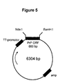
- An expression vector for the expression of the recombinant bovine prior protein is a DNA vector, e. g. based on the pET11a vector by Novagen, comprising essential sequences for expression in the respective host, e. g. a promoter, such as the T7-promotor, and the DNA coding for the bovine prior protein from codons 25 to 242 with an additional codon ATG at the 5 -end of the PrP-coding DNA, and sequence for selecting, eg. the ampicillin gene, multiplication, and termination.
- a promoter such as the T7-promotor
- a prefered expression vector is pbPrP3 as shown in Figure 5.
- bovine prion protein The recombinant bovine prion protein
- the present recombinant bovine prior protein comprises the amino acid sequence ID No: 2. It may be unglycosylated or glycosylated.
- the present recombinant bovine prior protein Pr P is purified to a homogeneity of >98%. It can be present in oxidized or reduced form. In the oxidized form the single -S-S-bridge is present whereas in the reduced form two SH groups are present instead.
- the amino acid sequence of the present bovine PrP is shown by ID NO: 2.
- the protein is glycosylated if expressed in a glycosylating eukaryotic cell line, such as Chinese Hamster cells, or unglycosylated if expressed in a procaryotic cell line, such as Escherichia coli. Mixtures of the oxidized and reduced form are also comprised.
- the oxidized form has the molecular weight of 23676.8 D, and the reduced form 236886.1 D as determined by electrospray mass spectroscopy.
- the present full length recombinant bovine prior protein is unique in terms of its homogeneity, since other groups in the art have reported of being unable to purify homogenous full length recombinant prion protein of other species (Mehlhorn et al., 1996; Riek et al., 1996).
- the reduced form of the recombinant PrP is particularly interesting since it has been reported to contain more ⁇ -sheet secondary structures than the oxidized form (Mehlhorn et al., 1996), hence mimicking structural features of PrP Sc : However, the reduced recombinant isoforms have been reported to be neither protease-resistant nor infectious (Mehlhorn et al., 1996).
- a native prion protein PrP is the prior protein polypeptide in a fully folded state, i.e. the three-dimensional structure is present. Only in the native, i.e. folded state, PrP exists in different isoforms (normal native vs. disease-specific native PrP).
- a denatured prion protein is the prion protein polypeptide in the unfolded state. Unfolding is usually achieved by the addition of chaotropic substances such as urea or guanidinium hydrochloride. In the denatured state, both PrP isoforms are irreversibly the same, even if they have been normal native or disease-specific native before.
- An antigen-antibody complex is a physical attachment of an antibody or fragment thereof with the corresponding antigen by intermolecular forces because the surfaces match in a unique way.
- the matching surface on the antibody is called idiotype and the surface on the antigen is called epitope.
- Suitable epitopes detectable by the present antibodies are for example linear amino acid sequences comprising about 3 to about 15 amino acids in a row or are completely three-dimensional ( patch ) in that very remote amino acid residues of the linear peptide backbone of the protein are, due to the unique folding, very close together in space to form an epitope.
- the present method for the production of an antibody according to the invention comprises culturing a hybridoma cell line as mentioned above and isolating the monoclonal antibody from the supernatant of the growth media.
- Culturing is carried out in flasks in HT- medium or in a cell culturing system called technomouse in serum-free, synthetic medium (Turbodoma medium, supplied by Messi, Zurich).
- technomouse hybridoma cells are cultured in a sterile chamber surrounded by a protein-impermeable membrane that is perfused by the respective medium in a constant flow rate (for example, turbomedium at 80 ml/h); antibodies are collected from the chamber with the help of a syringe at regular intervals.
- Isolation of monoclonal antibodies is carried out by extraction from the supernatant by conventional biochemical methods, e.g. by use of affinity columns with the corresponding immobilized antigen or by any other method used in the art, such as gel filtration or ion exchange chromatography.
- biochemical methods e.g. by use of affinity columns with the corresponding immobilized antigen or by any other method used in the art, such as gel filtration or ion exchange chromatography.
- Chimeric antibodies and fragments thereof can be produced by genetic engineering methods, e. g. by sequencing the antibody or the desired fragment thereof and constructing DNAs coding for the chimeric antibody or the fragment thereof which DNAs are inserted into an appropriate expression vector and expressed to produce the antibody or the fragment thereof in a procaryotic or eukaryotic cell line.
- a fragment binding to a PrP epitope can be combined with a human heavy chain to produce chimeric antibodies for use in humans as therapeutic or preventive agents against a prion disease.
- a fragment binding to a PrP epitope can also be combined with other enzymes, proteins or molecules to give rise to chimeric molecules combining the biological functions of these, for example for targeting an enzymatic activity to a place defined by the proximity of the PrP epitope.
- the present method for the production of a hybridoma cell line comprises administering to PrP 0/0 mice (knockout mice without a functional PrP gene) an immunizing amount of a recombinant pure prion protein, removing the spleen from the immunized mice, recovering splenocytes therefrom, fusing the latter with appropriate myeloma cells, growing the cells in a selection medium which does not support survival of the unfused cells, e. g.
- the peptide library used is commercially available from Jerini Biotools (Berlin, Germany). It consists of 104 spots with peptides of 13 amino acids, whereby the sequence of each peptide overlaps by 11 amino acids with the foregoing peptide.
- An immunizing amount of a recombinant bovine prion protein is from about 50 to 100 ⁇ g. It is administered dissolved in an appropriate solvent, e. g, PBS and Freund s adjuvant several times, e. g. three times subcutaneously followed by an intraperitoneal and an intravenous injection shortly prior to spleen removal.
- an appropriate solvent e. g, PBS and Freund s adjuvant several times, e. g. three times subcutaneously followed by an intraperitoneal and an intravenous injection shortly prior to spleen removal.
- PrP 0/0 mice were a gift from Prof. Weissmann of the University of Switzerland. They were obtained according to Büeler et al. (1992).
- Appropriate myeloma cells are for example P3X63Ag8U.1, deposited and available under ATCC CRL 1597.
- the method for the production of an expression vector comprises inserting a DNA coding for PrP in the correct reading frame into an expression vector.
- a DNA coding for PrP is shown by SEQ ID NO:1.
- This DNA can be obtained by amplifying DNA from bovine genomic DNA coding for PrP by means of the N- and C-terminal primers shown by SEQ ID NO: 3 and SEQ ID NO:4, respectively.
- Bovine genomic DNA is isolated from bovine kidney cells and supplied by Clonentech, U.S.A. Degenerate allelic forms of this DNA coding for the same PrP may be used.
- targeted mutations can be introduced into the PrP DNA to give rise to distince conformational isoforms of the translated gene product.
- the production of purified recombinant bovine PrP comprises culturing a cell line with an expression vector capable of expressing the bovine PrP in an appropriate culture medium, such as in the case of E. coli in Luria broth medium, isolating the prion protein from the inclusion bodies by lysing the cells e. g. with lysozym and Triton-X-100 in the case of E. coli, solubilizing the inclusion bodies with urea and and purifying the protein by convenional methods, e. g. by chromatography, for example on an anionic exchange sepharose column and a C4-reverse phase HPLC column.
- an appropriate culture medium such as in the case of E. coli in Luria broth medium
- isolating the prion protein from the inclusion bodies by lysing the cells e. g. with lysozym and Triton-X-100 in the case of E. coli, solubilizing the inclusion bodies with urea and and pur
- the oxidized form is obtained by treament with an oxidizing agent, e. g. with Cu 2 SO 4 , and the reduced form by treatment with a reducing agent, e. g. ⁇ -mercaptoethanol, according to conventional methods. They can be separated by reverse-phase high pressure liquid chromatography.
- an oxidizing agent e. g. with Cu 2 SO 4
- a reducing agent e. g. ⁇ -mercaptoethanol
- An immunological detection procedure for the detection of prion disease, especially BSE, whereby disease-specific PrP Sc protein in biological material of an animal or human is detected comprises treatment of a first probe of said material with a monoclonal antibody according to the invention and detecting the mixed PrP C /PrP Sc - antibody complex, treating a second probe of said material first with proteinase K and then with the monoclonal antibody according to the invention, detecting the PrP Sc -antibody complex and analyzing the results of both probes.
- a specific monoclonal antibody according to the invention is able to detect PrP Sc in a PrP Sc -antibody complex without prior protease-digestion of the tissue specimen to be examined.
- the biological material can be insoluble or soluble in buffer or body fluids. It can be derived from any part of the body, e. g. from the brain or tissue sections, in which case it is used in form of a homogenate, or any body fluid, e. g. cerebrospinal fluid, urine, saliva or blood.
- body fluids fluid-resident cells, e.g. white blood cells in the case of blood expressing PrP, can be purified and analyzed either in immunohistochemistry or as a homogenate.
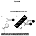
- the detection of the PrP Sc -antibody complex is carried out in particular by immunological procedures, such as Western blotting, ELIFA, and various ELISA techniques, such as capture ELISA.
- tissue sections, tissue homogenates or body fluids of prion-infected animals such as BSE-diseased cattle or humans having the CJD
- tissue sections, tissue homogenates or body fluids of prion-infected animals such as BSE-diseased cattle or humans having the CJD
- the protease-resistant, disease-specific isoform of the prion protein in its native form be it soluble or insoluble.
- Tissue homogenates and body fluids are for example such as from biopsy of brain, lymph nodes, spleens, tonsils, peripheral nerves, cerebrospinal fluids, urine, platelets or white blood cells.
- Particular immunological procedures comprise for example enzyme-linked immunofiltration assay (ELIFA), enzyme-linked immunoabsorbent assay (ELISA), Western blot assay, dot blot assay, immunodecoration and immunohistochemistry.
- ELIFA enzyme-linked immunofiltration assay
- ELISA enzyme-linked immunoabsorbent assay
- Western blot assay Western blot assay
- dot blot assay immunodecoration and immunohistochemistry.
- test kit for the diagnosis of prion diseases The test kit for the diagnosis of prion diseases
- the test kit for the diagnosis of prion diseases comprises devices and materials enabling the diagnosis of prion disease in biological materials, and is particularly suited for screening large amounts of samples for the presence of PrP Sc .
- a test kit comprises in particular one or more monoclonal antibodies according to the invention, purified bovine recombinant PrP protein as mentioned above, nitrocellulose sheets, microtiter plates, or microtiter plates coated with the monoclonal antibodies according to the invention, a secondary anti-mouse antibody that is coupled with an enzyme and its substrate or any other molecular compound for a detection reaction (e.g.
- a peroxidase-labeled anti-mouse IgG antibody, TMB or any other peroxidase substrate hydrogen peroxide
- proteinase K a blocking buffer
- homogenization buffer a homogenization buffer
- calibration curve a description of how to perform the test.
- Another test kit is designed in the dipstick format and is without need of radioactive tracers, enzymes or substrates and basically reduces the number of handling steps to one.
- the one-step procedure involves the capture of the disease-specific PrP Sc with one of the antibodies according to the invention which are immobilized on a test strip. Captured disease-specific PrP Sc is detected directly by a second antibody according to the invention, which is coupled to particular colloid particles.
- This specific detector complex results in the formation of coloured spots on the test strip which are visible in less than 30 minutes depending on the concentration of the test sample. The spots are a permanent record of the test result and, upon longer exposure even increase the sensitivity of the test without generating higher background.
- the pharmaceutical preparation for the therapy and prevention of prion diseases in a mammal, including humans comprises an effective amount of one or more antibodies fragments therof or chimeric antibodies as described, produced according to the invention, eventually purified according to conventional methods, and a conventional pharmaceutical carrier.
- An antibody obtained may be solubilized together with the carrier in an appropriate buffer, e. g. an aqueous physiological sodium chloride solution.
- This may be clarified by centrifugation and used in concentrated liquid form for injection, or completely dried if desired by any of the conventional methods, such as lyophilization, spray or freeze drying, in form of a dry powder, which can be pressed into tablets, filled into capsules, or applied as a dry powder in form of a nasal spray, whereby conventional production methods are applied, and conventional pharmaceutical carriers are optionally added.
- the monoclonal antibodies of the present invention bind to between species highly conserved regions in the PrP molecule that may have functional significance (Oesch et al., 1991). It is envisioned that blocking this binding site by the monoclonal antibodies, fragments thereof or chimeric antibodies as defined above will abolish biological effects of prions. Blocking of the infectivity of prions by occupying distinct sites on the disease-specific form of PrP is foreseen to represent a therapeutic strategy in treating prion diseases or a preventive strategy in preincubating suspected prion-infected tissue specimens with the present monoclonal antibodies. The normal form of PrP appears not to be of vital importance in the living animal because mice with a deleted PrP gene are viable (Bueler et al., 1992). Anti-PrP antibodies may therefore be used without side effects to neutralize prions in humans or animals.
- the present invention concerns further a method for the therapy or prevention of prion disease, such as a disease mediated by the neurotoxic effects of prion proteins or fragments of prion proteins, comprising administering to a patient suffering from such disease or being likely to becoming a victim of this disease a therapeutical or preventive amount of a monoclonal antibody, a fragment thereof or a chimeric antibody as described above.
- prion disease such as a disease mediated by the neurotoxic effects of prion proteins or fragments of prion proteins
- the method of protecting a mammal, including a human, against an infectious prion disease comprises administering one or a combinantion of the present antibodies, fragments thereof or chimeric antibodies or a pharmaceutical preparation comprising the antibodies produced by the present invention.
- the pharmaceutical preparation is preferably administered by injection, e. g. intrathecally (into the cerebrospinal fluid), into the blood with respective pharmaceutical agents or methods increasing the permeability of the blood-brain barrier or as a chimeric antibody, fused to, or containing additional signal sequences that allow passage through the blood-brain barrier (for review see Friden, 1994).
- An intranasal application of the monoclonal antibodies, fragments or chimeras thereof is also possible.
- the pharmaceutical preparations have to be administered according to the judgment of the physician in amounts depending on the concentration of the antibodies comprised thereby and the route of administration so that a protective or curative effect is obtained.
- the amounts and method of administration are to be selected further depending upon the age and weight of the patient, the nature and severity of the infection as well as the general condition of the patient. In general it is sufficient to administer the antibodies in amounts of about 1 to 100 mg per patient in a single or in repeated doses.
- the method for clearing biological material from prions comprises treating said material with one or several monoclonal antibodies according to the invention such that prions or prion proteins or fragments thereof become functionally inactivated in terms of their infectivity and/or neurotoxicity.
- the pharmaceutical preparations described above may be used for this purpose, whereby the pharmaceutical carrier may be replaced by a suitable other solvent in case the biological material is not intended to be used for transplantation.
- Example 1 Immunological diagnosis of prion diseases using tissue from infected animals
- PrP C and PrP Sc can be distinguished according to their different sensitivity to digestion with protease K.
- Undigested PrP C and PrP Sc have a molecular weight of 33-35 kDa.
- PrP C is readily fully digested while PrP Sc is partially resistant, i.e. the N-terminus of PrP Sc is removed leading to a shift in molecular weight from 33-35 kDa to 27-30 kD (Oesch et al., 1985).
- Proteinase K is therefore used to digest the tissue specimen to be examined.
- monoclonal antibody 15B3, detecting a conformational epitope specific for the disease-specific isoform PrP Sc may even be used without prior protease digestion.
- Example 1.1 Method for the preparation of tissue homogenates
- an homogenizer Omni, USA
- 10% homogenates were diluted 10 fold; one part of the homogenate was digested with proteinase K at 0, 10 or 100 ⁇ g/ml.
- the probe containing 0 mg/ml served as a control for the specificity of the protein
- bovine PrP Sc brain homogenates from BSE-infected and normal cattle were diluted tenfold in the presence of 40 or 80% ethanol /HEPES-sucrose buffer. Suspension of brain homogenates in ethanol was an important step and effectively stabilized the ⁇ -sheet structure of the PrP Sc isoform (Oesch et al., 1994), thereby increasing its protease resistance (Piesner et al., 1996).
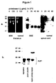
- Tissue specimens were homogenized as described in example 1.1., one part protease-digested, the other not (as described above), diluted to 10% and separated by SDS polyacrylamide gel electrophoresis (SDS-PAGE) on 12% gels (Sambrook et al., 1989). Gels were then electroblotted onto 0.45 ⁇ m nitrocellulose (NC) membranes, incubated with the respective monoclonal antibodies followed by a secondary anti-mouse IgG antibody coupled to peroxidase. Bound peroxidase activity was detected with a chemiluminescence kit (ECL, Amersham, USA).
- the present antibodies were furthermore able to detect PrP in various tissue extracts from humans, cattle, pigs, sheep, mice and hamsters ( Figures 1b and 2). Given that the epitopes of our antibodies are highly conserved (Oesch et al., 1991), we expect our antibodies to stain PrP from other species such as rat or deer as well.
- the membrane was removed from the ELIFA apparatus, placed in a plastic tray and then incubated on a rocking table sequentially for the indicated times with the following reagents (inbetween steps, the filters were always washed 3x with PBS): 5% BSA/TBST (30min); avidin (25 ⁇ g/ml, 30min); biotin (2 ⁇ g/ml; 30min); monoclonal antibodies 6H4 or 34C9 in TBST (2 h RT or 10 h o/n); secondary, biotinylated anti-mouse IgG (Vectastain, USA, dilution 1:5000; 1 h RT); and streptavidin coupled to peroxidase (Boehringer, Germany, dilution 1:25000; 15-60 min RT).
- monoclonal antibodies 6H4 or 34C9 were biotinylated eliminating the step with the biotinylated secondary antibody.
- Still another procedure involved coupling of mABs 6H4 or 34C9 directly to peroxidase according to the manufacturer (Pierce, USA). Amplification of peroxidase activity was achieved by the ELAST-kit according to the manufacturer (DuPont, USA).
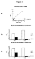
- the standard curve for the ELIFA was obtained by serial dilutions of ultra-pure and defined amounts of recombinant bovine PrP (see below).
- lyophilized recombinant PrP was suspended in an antigen-dilution buffer (1M guanidinium thiocyanate and 0.01% human serum albumin in PBS). This buffer allows maximum binding of recombinant PrP to the nitrocellulose membrane.
- the standard curve is essential, since it allows to control both the quality and the reliability of the ELIFA-procedure.
- the standard curve allows to exactly quantify bovine PrP C /PrP Sc amounts in given tissue specimens (Oesch et al., 1994) ( Figure 3a).
- Example 1.4 Conventional ELISA (Enzyme Linked Immunosorbent Assay)
- the antigen (present in a 10% homogenate as described in example 1.1.) was incubated for 2 h at RT in 96-well microtiterplates (Nunc, Denmark). Blocking was achieved with 5% BSA after antigen incubation. After washing, the plate was incubated with the biotinylated monoclonal antibody antibody 6H4 for 2 h at RT. Washing with H 2 O and PBS was performed before streptavidin-coupled peroxidase (Boehringer, Germany) was applied and peroxidase activity detected with the substrate according to the manufacturer (TMB/H 2 O 2 ; Kierkegaard & Perry). The reaction was stopped by the addition of 2 M H 3 PO 4 .
- the plate is read by an ELISA reader at 450 nm with a reference at 620 nm.
- peroxidase-coupled monoclonal antibody 6H4 was used instead of biotinylated antibody (see example 1.3.). Amplification of peroxidase activity was achieved by the ELAST-kit according to the manufacturer (DuPont, USA). Results of this immunological test are depicted in Figure 3c.
- monoclonal antibodies according to the invention were either covalently linked or coated to microtiterplates (Nunc, Denmark). Subsequently, the wells were blocked with 5% BSA for 30 min at RT and the procedure according to example 1.4. followed.
- the PCR product was digested with the restriction enzymes Nde I and BamH I (Boehringer, Germany).
- the appropriate DNA fragment of 650 bp was purified on a 1% agarose gel and ligated into the pET11a vector (Novagen) previously digested with Nde I and BamHI. Ligated products were transfected into E. coli strain DH5 ⁇ .
- the appropriate clone containing the PrP open reading frame in the pET11a vector was selected and subsequently termed pbPrP3 ( Figure 5).
- Plasmid pbPrP3 was transfected into E. coli strain BL21(DE3) (Novagen), that is capable of translating the plasmid into a protein.
- E. coli strain BL21(DE3) Novagen
- bovine PrP cells were stimulated with 1 mM IPTG according to standard techniques (Sambrook et al., 1989). Recombinant bovine PrP was purified from inclusion bodies.
- the pellet containing the bacteria was further processed for isolation of PrP from inclusion bodies as follows: the bacterial pellet from a 1 liter culture was resuspended in 100 ml of 2 mM EDTA, 50 mMTris-HCl pH 7.5 and lysed by the addition of lysozyme (final concentration 100 mg/ml) and Triton-X-100 (final conc. 1%) for 15 min at 37 0 C. Then, MgCl 2 (final conc. 15 mM) and DNAse I (final conc. 10 ⁇ g/ml) were added. The suspension was shaken at room temperature until the DNA was digested and the solution was clear (30 min).
- the solution was then centrifuged at 10.000 x g for 30 min at 4°C.
- the supernatant containing all the soluble proteins was discarded and the pellet, containing the inclusion bodies, was homogenized and resuspended in 1/10 th of the original culture volume containing 8M deionized urea, 10 mM MOPS pH 7.5.
- This suspension was then shaken at room temperature overnight and then centrifuged at 10.000 x g for 30 min at 4°C.
- the supernatant containing solubilized material from inclusion bodies was then further processed.
- CM sepharose column (Pharmacia) was equilibrated first with the elution buffer containing 8M deionized urea, 500 mM NaCl, 10mM MOPS pH 7.5, then with the washing buffer containing 8M deionized urea, 10mM MOPS pH 7.
- a CM sepharose column (50 ml bed volume) was loaded with 100 ml of the solution containing the solubilized proteins from inclusion bodies. The column was washed twice with 25 ml 50 mM NaCl, 8M urea, 10 mM MOPS and once with 100 mM NaCl, 8M urea, 10 mM MOPS.
- Bovine recombinant PrP was eluted with 500 mM NaCl, 8M urea, 10 mM MOPS. SDS-PAGE and silver staining showed that at this step only one protein of about 24 kD was present in the eluent, corresponding to the calculated molecular weight of 23,6 kD ( Figure 6a). This fraction was then further processed.
- Proteins eluted from the CM sepharose were subsequently either oxidized with 10 ⁇ M Cu 2 SO 4 or reduced with 2% ⁇ -mercaptoethanol for several hours before they were loaded on a C 4 -reverse phase HPLC column.
- the HPLC column was perfused with a 0-85% gradient of acetonitrile in 0.1% trifluoroacetic acid (TFA).
- TFA trifluoroacetic acid
- the oxidized or reduced bovine recombinant PrP eluted about 40 or 45% acetonitrile, respectively.
- the eluted fractions were lyophilized (Sambrook et al., 1989) and redissolved in distilled water.
- Electrospray mass spectroscopy revealed single peaks of 23676.8 and 23686.1 Dalton for oxidized and reduced recombinant bovinePrP, respectively, indicating a correct and uniform translation of the bovine PrP open reading frame in pbPrP3.
- Example 2.2 Immunization of animals and hybridoma production
- Oxidized or reduced bovine recombinant PrP or a mixture of both amounting to a total of 100 ⁇ g in a single dose (dissolved in PBS) were used to immunize PrP 0/0 mice, i.e. mice without a functional PrP gene (Bueler et al., 1992) that were kindly provided by Prof. C. Weissmann, University of Zurich.
- the reduced form of the recombinant PrP was particularly interesting since it has been reported to contain more ⁇ -sheet secondary structures than the oxidized form in a recombinant Syrian hamster PrP fragment (Mehlhorn et al., 1996), hence mimicking structural features of PrP Sc , however, the reduced recombinant isoforms have been reported to be neither protease-resistant nor infectious (Mehlhorn et al., 1996).
- mice received three subcutaneous injections (day 0 with Freund s complete adjuvans, days 21 and 42 with Freund s incomplete adjuvans) of the antigens in a constant 100 ⁇ g amount and in a volume of 100 ml.
- mice were boosted with the antigen intraperitoneally and the next day intravenously with adjuvant Pertussi Berna (Berna, Switzerland; extract of Bordetella pertussis bacteria).
- mice were anesthetized and decapitated. The spleen from immunized mice was removed, and splenocytes were recovered.
- Mouse myeloma cells (cell line P3X63Ag8U.1, ATCC CRL 1597; Scharff, 1978) were mixed to the splenocytes at a ratio of 1:5 and fused by the addition of 50% PEG (polyethylenglycol) for 8 min at RT according to standard techniques (Kennett, 1980). Cells were then washed and grown overnight. The next day, cells were suspended in selective medium (HAT) and plated in 96-well microtiterplates. The selective medium contains aminopterin that is toxic for those cells that have not been fused to splenocytes and thus eliminates uncontrolled cell growth of irrelevant cells (Kennett, 1980).
- HAT selective medium
- the selective medium contains aminopterin that is toxic for those cells that have not been fused to splenocytes and thus eliminates uncontrolled cell growth of irrelevant cells (Kennett, 1980).
- Example 2.3 Screening hybridomas for specific antibodies
- 96-well microtiter plates were coated with recombinant bovine PrP (0.25 ⁇ g/well) for 4 h at RT and then blocked with 5% BSA/H 2 0 for 1h at RT. After washing with H 2 0 and PBS, culture medium from wells containing hybridoma colonies was transfered to the mictrotiter plates (50 ⁇ l per well) and incubated overnight at 4 0 C. After washing with H 2 0 and PBS, bound antibodies were detected with a peroxidase-labeled anti- mouse IgG antibody (Cappell, Switzerland) followed by colorimetric detection with TMB/H 2 O 2 (Kierkegaard & Perry, USA) as described in example 1.4.
- 1% brain homogenates of normal and BSE-infected cattle were either left undigested or protease-digested for the BSE brain homogenate and blotted onto a nitrocellulose membrane as described by for the ELIFA procedure (see above) (Oesch et al., 1994). After blotting, the membrane was blocked with 5% low-fat milk in TBST, and incubated with the antibody-containing culture medium. Subsequently, the NC was incubated with a secondary, peroxidase-coupled anti-mouse IgG antibody and developed with a chemiluminescence kit (ECL, Amersham). This technique allowed for detection of hybridoma cell lines that produce antibodies against native PrP C and bovine PrP Sc or of conformation-sensitive antibodies that distinguish between PrP C and bovine PrP Sc (as described for mAB 15B3 in Figure 6b).
- Hybridoma cell lines were further selected on the capability of the produced antibodies to recognize PrP of brain homogenates and recombinant bovine PrP on Western blots. Brain homogenates of various tissues and various species were blotted as described in example 1.2..
- the prefered mAB 34C9 recognizes PrP in the brain homogenates of cattle, mice, pig, and humans ( Figure 2).
- a peptide library consisting of 104 peptides numbered 1 to 104, purchased from Jerini Biotools (Berlin, Germany) was used to map the epitopes that are recognized by the antibodies.
- the peptides are covalently linked to a cellulose membrane,have each a length of 13 amino acids and together cover the entire length of the bovine prion protein (total of 104). Each peptide overlaps by 11 amino acids with the next peptide. Binding of antibodies to those peptides can be visualized by the ECL system as described for Western blotting in Example 1.2.
- the monoclonal antibodies of the present invention bound to peptides comprised in the region of helix 1 in the three-dimensional model of the mouse recombinant C-terminal prion protein fragment described by Riek et al. (1996). It is hereby assumed that this mouse recombinant C-terminal prion protein fragment reflects the structure of native PrP C , has the same structure as full length PrP and that the structure will be similar in different species. Based on these assumptions the following statements are mAB 6H4 binds to the three library peptides Nos.
- mAB15B3 binds to the 7 library peptides Nos. 73 to 79 comprising amino acids 140-152 of the bovine PrP sequence SEQ ID NO: 1 ( Figure 7c), and additional "patches” situated on library peptides Nos. 33-34, 52 and 69.
- Monoclonal antibody 15B3 recognizes native bovine PrP Sc better than native bovine PrP C ( Figure 6b).
- 10% bovine brain homogenates of normal undigested and BSE-diseased protease-digested cattle were made as described in Example 1.1.
- the samples were diluted to a 0.5% homogenate with either PBS, 4M or 8M deionized urea/PBS buffer and incubated at 37 0 C for 1h.
- the samples were then blotted onto a nitrocellulose membrane with the qualitative, conformation-sensitive ELIFA protocol as described in example 3 above.
- the long stretch of peptides from 73 to 79 on the peptide library corresponds to the amino acids 140 to 152 of PrP (SEQ ID NO: 1) that partially comprises the ⁇ -sheet containing strand 2 in the three-dimensional PrP model of Riek et al. (1996).
- the few distant peptides that stain positive in the peptide library ( Figure 7) are all near this strand 2 in the Riek et al. model.
- mAB 15B3 binds to a ⁇ -sheet comprising epitope that is present in native specific PrP Sc , but not in native normal PrP C .
- Brain homogenates from BSE-infected cattle are obtained as described in Example 1.1.
- the exact amount of present bovine PrP Sc are measured with the help of the ELIFA technique or the ELISA technique as descibed in examples 1.3 to 1.5, respectively.
- Serial dilutions of this infected brain homogenate are aliquoted.
- To these serial dilutions are added the preferred mABs 6H4, 34C9 or 15B3, or a mixture thereof, in molar amounts exceeding the molar amounts of measured PrP Sc .
- the mix is incubated for 4h at RT and then 100 ⁇ l are injected intracerebrally into the animal.
- Transgenic mice overexpressing mouse PrP (tg35; Fischer et al., 1996) are used as an animal model for measuring the infectivity of bovine PrP Sc .
- hybridoma cell lines were deposited under the Budapest Treaty at the Deutsche Sammlung von Mikroorganismen und Zellkulturen GmbH, Mascheroder Weg 1b, D-38124 Braunschweig, as follows:
Landscapes
- Health & Medical Sciences (AREA)
- Chemical & Material Sciences (AREA)
- Organic Chemistry (AREA)
- Life Sciences & Earth Sciences (AREA)
- Medicinal Chemistry (AREA)
- Immunology (AREA)
- General Health & Medical Sciences (AREA)
- Genetics & Genomics (AREA)
- Biochemistry (AREA)
- Molecular Biology (AREA)
- Proteomics, Peptides & Aminoacids (AREA)
- Biophysics (AREA)
- Toxicology (AREA)
- Zoology (AREA)
- Gastroenterology & Hepatology (AREA)
- Peptides Or Proteins (AREA)
- Preparation Of Compounds By Using Micro-Organisms (AREA)
- Medicines Containing Antibodies Or Antigens For Use As Internal Diagnostic Agents (AREA)
- Micro-Organisms Or Cultivation Processes Thereof (AREA)
Priority Applications (11)
| Application Number | Priority Date | Filing Date | Title |
|---|---|---|---|
| EP97102837A EP0861900A1 (fr) | 1997-02-21 | 1997-02-21 | Détection immunologique de prions |
| AT98910699T ATE440868T1 (de) | 1997-02-21 | 1998-02-18 | Monoklonaler antikírper gegen ein prion protein und dessen verwendung zum nachweis von prionen |
| EP98910699A EP1007700B1 (fr) | 1997-02-21 | 1998-02-18 | Anticorps monoclonal contre une protéine prion et son utilisation pour la détection de prions |
| PCT/EP1998/000917 WO1998037210A1 (fr) | 1997-02-21 | 1998-02-18 | Detection immunologique de prions |
| US09/380,015 US6765088B1 (en) | 1997-02-21 | 1998-02-18 | Immunological detection of prions |
| NZ336656A NZ336656A (en) | 1997-02-21 | 1998-02-18 | Immunological detection of prions with antibodies and their use in the prevention and therapy of prion diseases |
| CA2280791A CA2280791C (fr) | 1997-02-21 | 1998-02-18 | Detection immunologique de prions |
| EP09010591.7A EP2172486B1 (fr) | 1997-02-21 | 1998-02-18 | Anticorps monoclonal contre un prion et son utilisation pour la détection de prions |
| DE69841102T DE69841102D1 (de) | 1997-02-21 | 1998-02-18 | Monoklonaler antikörper gegen ein prion protein und dessen verwendung zum nachweis von prionen |
| AU64986/98A AU742838B2 (en) | 1997-02-21 | 1998-02-18 | Immunological detection of prions |
| US10/867,589 US7259246B2 (en) | 1997-02-21 | 2004-06-15 | Immunological detection of prions |
Applications Claiming Priority (2)
| Application Number | Priority Date | Filing Date | Title |
|---|---|---|---|
| EP97102837A EP0861900A1 (fr) | 1997-02-21 | 1997-02-21 | Détection immunologique de prions |
| PCT/EP1998/000917 WO1998037210A1 (fr) | 1997-02-21 | 1998-02-18 | Detection immunologique de prions |
Publications (1)
| Publication Number | Publication Date |
|---|---|
| EP0861900A1 true EP0861900A1 (fr) | 1998-09-02 |
Family
ID=8226511
Family Applications (3)
| Application Number | Title | Priority Date | Filing Date |
|---|---|---|---|
| EP97102837A Withdrawn EP0861900A1 (fr) | 1997-02-21 | 1997-02-21 | Détection immunologique de prions |
| EP98910699A Expired - Lifetime EP1007700B1 (fr) | 1997-02-21 | 1998-02-18 | Anticorps monoclonal contre une protéine prion et son utilisation pour la détection de prions |
| EP09010591.7A Expired - Lifetime EP2172486B1 (fr) | 1997-02-21 | 1998-02-18 | Anticorps monoclonal contre un prion et son utilisation pour la détection de prions |
Family Applications After (2)
| Application Number | Title | Priority Date | Filing Date |
|---|---|---|---|
| EP98910699A Expired - Lifetime EP1007700B1 (fr) | 1997-02-21 | 1998-02-18 | Anticorps monoclonal contre une protéine prion et son utilisation pour la détection de prions |
| EP09010591.7A Expired - Lifetime EP2172486B1 (fr) | 1997-02-21 | 1998-02-18 | Anticorps monoclonal contre un prion et son utilisation pour la détection de prions |
Country Status (8)
| Country | Link |
|---|---|
| US (2) | US6765088B1 (fr) |
| EP (3) | EP0861900A1 (fr) |
| AT (1) | ATE440868T1 (fr) |
| AU (1) | AU742838B2 (fr) |
| CA (1) | CA2280791C (fr) |
| DE (1) | DE69841102D1 (fr) |
| NZ (1) | NZ336656A (fr) |
| WO (1) | WO1998037210A1 (fr) |
Cited By (41)
| Publication number | Priority date | Publication date | Assignee | Title |
|---|---|---|---|---|
| WO1999015651A1 (fr) * | 1997-09-20 | 1999-04-01 | Prionics Ag | Polypeptides synthetiques pour le diagnostic et le traitement de maladies a prion |
| WO2000029850A1 (fr) * | 1998-11-17 | 2000-05-25 | Wallac Oy | Immuno-essai de recherche d'encephalopathies spongiformes transmissibles chez les bovins |
| WO2000029849A1 (fr) * | 1998-11-17 | 2000-05-25 | Wallac Oy | Immuno-essai de recherche d'encephalopathies spongiformes transmissibles chez les mammiferes |
| WO2000043791A2 (fr) | 1999-01-25 | 2000-07-27 | Minerva Biotechnologies Corporation | Detection rapide et efficace d'une agregation de proteines aberrantes dans des maladies neurodegeneratives |
| GB2346971A (en) * | 1999-02-19 | 2000-08-23 | Audrey Ann Coop | Diagnostic test for infectious prion protein in the blood |
| WO2000078344A1 (fr) * | 1999-06-23 | 2000-12-28 | Caprion Pharmaceuticals, Inc. | Peptides proteiniques du prion et utilisations associees |
| WO2001023425A1 (fr) * | 1999-09-28 | 2001-04-05 | Universität Zürich | Facteurs ayant une activite de liaison au prion dans du serum ou du plasma et agents permettant de detecter l'encephalopathie spongiforme transmissible |
| WO2001035104A1 (fr) * | 1999-11-12 | 2001-05-17 | Commissariat A L'energie Atomique | Procede de diagnostic d'une esst provoquee par une souche d'atnc dans un echantillon biologique |
| GB2360089A (en) * | 2000-03-10 | 2001-09-12 | Univ Geneve | Diagnostic assay for transmisible spongiform encephalopathies |
| FR2808278A1 (fr) * | 2000-04-28 | 2001-11-02 | Inst Nat Sante Rech Med | Proteines a prions et leurs utilisations |
| GB2362385A (en) * | 1998-11-04 | 2001-11-21 | Imp College Innovations Ltd | Non-aggregated beta forms of prion proteins and antibodies against such proteins |
| EP0970372A4 (fr) * | 1997-02-21 | 2002-04-10 | Univ California | Dosage pour la detection d'une structure proteique associee a une maladie |
| WO2001044816A3 (fr) * | 1999-12-15 | 2002-05-30 | Univ California | Regulation conformationnelle et topologique des proteines |
| EP1213301A3 (fr) * | 2000-12-08 | 2002-06-19 | Fujirebio Inc. | Anticorps reconnaissant les prions anormaux, procédé permettant de produire lesdits anticorps et utilisation desdits anticorps dans des essais immunologiques |
| GB2371049A (en) * | 1998-11-04 | 2002-07-17 | Imp College Innovations Ltd | Methods of making antibodies against ß-forms of prion proteins |
| DE10120562A1 (de) * | 2001-04-26 | 2002-10-31 | Horst Messer | Verfahren zur Diagnose von übertragbaren spongiformen Enzephalopathien |
| WO2002057793A3 (fr) * | 2001-01-19 | 2003-03-06 | Baxter Int | Methode de detection de la proteine prp et trousses associees |
| WO2002057789A3 (fr) * | 2001-01-19 | 2003-03-13 | Enfer Technology Ltd | Test pour encephalopathies spongiformes transmissibles |
| US6534036B1 (en) | 1998-11-04 | 2003-03-18 | D. Gen Limited | Biological materials and methods useful in the diagnosis and treatment of diseases |
| US6617119B2 (en) | 1997-02-21 | 2003-09-09 | The Regents Of The University Of California | Assay for specific strains of multiple disease related conformations of a protein |
| US6620629B1 (en) | 1997-02-21 | 2003-09-16 | The Regents Of The University Of California | Method for detecting prions |
| WO2003000719A3 (fr) * | 2001-06-20 | 2003-12-11 | Univ Ramot | Produit antigenique manifestant plusieurs copies d'un epitope d'un polypeptide formant des depots implique dans des maladies a formation de plaques et procedes d'utilisation correspondants |
| EP1382971A1 (fr) * | 2002-07-17 | 2004-01-21 | Pepscan Systems B.V. | Detection de maladie a prion |
| WO2003029813A3 (fr) * | 2001-09-25 | 2004-02-26 | Prionics Ag | Bande de test permettant de deceler des proteines prions |
| WO2004003505A3 (fr) * | 2002-06-27 | 2004-03-18 | Univ Marlyland Baltimore | Procedes de detection et d'inactivation d'un prion |
| WO2004046728A1 (fr) * | 2002-11-18 | 2004-06-03 | Prionics Ag | Methode de detection de prpsc |
| EP1435521A1 (fr) * | 2002-12-19 | 2004-07-07 | Ortho-Clinical Diagnostics, Inc. | Détéction et diagnostic de l'encephalopathie spongiforme transmissible |
| EP1445615A1 (fr) * | 2003-02-10 | 2004-08-11 | Ortho-Clinical Diagnostics, Inc. | Procede pour discrimination des prions infectieux et non infectieux |
| EP1457500A1 (fr) * | 2003-03-14 | 2004-09-15 | University of Zurich | Proteinés solubles de prion hybrids et leurs utilisations en diagnostique, prévention et traítement de l'encephalopathie spongiforme transmissible |
| EP1470421A4 (fr) * | 2001-11-14 | 2005-03-16 | Prion Dev Lab Inc | Dosage biologique pour detecter rapidement la presence de prions |
| US6916419B2 (en) | 1997-02-21 | 2005-07-12 | The Regents Of The University Of California | Device for removal of prions from blood, plasma and other liquids |
| EP1119773B1 (fr) * | 1998-10-09 | 2005-07-20 | The Regents of the University of California | Essai de detection des proteines prion dans une conformation accociee a une maladie |
| EP1596199A1 (fr) * | 2004-05-14 | 2005-11-16 | Prionics AG | Méthode de détection des formes pathogènes des protéines prions. |
| US7041807B1 (en) | 1999-06-23 | 2006-05-09 | Caprion Pharmaceuticals, Inc. | Antibodies to a YYX epitope of a mammalian prion protein |
| WO2006079805A1 (fr) * | 2005-01-25 | 2006-08-03 | Cambridge Enterprise Limited | Dosage immunologique des maladies à prions |
| US7098317B1 (en) * | 2000-05-23 | 2006-08-29 | Blood Transfusion Center Of Slovenia | Antibodies capable to selectively detect prion PrPSc isoforms |
| US7368247B2 (en) | 2000-03-10 | 2008-05-06 | Universite De Geneve | Diagnostic assay for transmissible spongiform encephalopathies |
| US7399603B2 (en) | 2002-12-19 | 2008-07-15 | Ortho-Clinical Diagnostics, Inc. | Antibodies for discrimination of prions |
| US7435540B2 (en) | 2002-09-27 | 2008-10-14 | Idexx Corporation | PrPSc-selective peptides |
| EP2085404A1 (fr) | 2004-09-30 | 2009-08-05 | Ortho-Clinical Diagnostics, Inc. | Peptides pour la discrimination des prions |
| EP1158003B2 (fr) † | 2000-05-23 | 2012-12-26 | Blood Transfusion Centre of Slovenia | Anticorps pour la détection d'isoformes du prion PrP Sc |
Families Citing this family (44)
| Publication number | Priority date | Publication date | Assignee | Title |
|---|---|---|---|---|
| DE19918141A1 (de) * | 1999-04-21 | 2000-10-26 | Boehringer Ingelheim Vetmed | Verfahren zur Diagnose von übertragbaren Spongiformen Enzephalopathien |
| BR0012765A (pt) * | 1999-07-27 | 2002-04-02 | Imp College Innovations Ltd | Peptìdeo, métodos de fabricar um anticorpo, de fabricar um agente de ligação capaz de ligar-se a uma forma celular de uma proteìna de prion, de remover uma forma celular de uma proteìna de prion de uma amostra, e de usar um anticorpo e/ou agente de ligação, anticorpo, agente de ligação, uso de um anticorpo ou agente de ligação, kit, e , sequência de peptìdeo de uma forma celular de uma proteìna de prion |
| IL141950A0 (en) * | 2000-10-22 | 2002-03-10 | Hadasit Med Res Service | Diagnosis of prion diseases |
| US7303907B2 (en) | 2001-01-08 | 2007-12-04 | Health Protection Agency | Degradation and detection of TSE infectivity |
| DE10104480A1 (de) * | 2001-01-31 | 2002-08-14 | November Ag Molekulare Medizin | Verfahren, Verwendung und Kit zum Auffinden von Wirkstoffen gegen mit spezifischen Proteinen assoziierte Krankheiten |
| DE10106835A1 (de) * | 2001-02-14 | 2002-09-05 | Basf Lynx Bioscience Ag | Signaltransduktionsproteine 15B3, 15B3-1 und 15B3-2 und zugrundeliegende DNA-Sequenzen |
| DE10143647A1 (de) * | 2001-09-05 | 2003-03-27 | Prionics Ag Zuerich | DNA-Sequenzen codierend für variable Bereiche von Antikörpern, die an das Prion-Protein (PrP) binden sowie PrP-bindende Polypeptide |
| US20030092199A1 (en) * | 2001-11-14 | 2003-05-15 | Kiamars Hajizadeh | Prion-detection business methods |
| US20030092090A1 (en) * | 2001-11-14 | 2003-05-15 | Kiamars Hajizadeh | Rapid prion-detection device, system, and test kit |
| EP1312617A1 (fr) | 2001-11-14 | 2003-05-21 | ATOFINA Research | Polypropylène à résistance au choc élevée |
| GB0227886D0 (en) * | 2002-11-29 | 2003-01-08 | Medical Res Council | Prion inhibition |
| FR2849204B1 (fr) * | 2002-12-20 | 2005-02-11 | Afssa | Procede de detection de la prpsc utilisant un antibiotique d de la famille des aminoglycosides pour l'elimination et la detection de la prpsc dans des echantillons biologiques |
| US20060035242A1 (en) * | 2004-08-13 | 2006-02-16 | Michelitsch Melissa D | Prion-specific peptide reagents |
| ES2398394T3 (es) | 2003-08-13 | 2013-03-15 | Novartis Vaccines And Diagnostics, Inc. | Reactivos peptídicos específicos de prión |
| AU2004266324B8 (en) * | 2003-08-20 | 2011-04-28 | Promis Neurosciences Inc. | Epitope protection assay and method for detecting protein conformations |
| US20060199194A1 (en) * | 2003-11-10 | 2006-09-07 | Q-Rna, Inc. | Methods of detection using immuno-Q-Amp technology |
| US20050214805A1 (en) * | 2003-11-10 | 2005-09-29 | Q-Rna, Inc. | Methods of detection employing immuno-Q-Amp technology |
| US20090130774A1 (en) * | 2005-01-13 | 2009-05-21 | David Peretz | Elisa assays using prion-specific peptide reagents |
| WO2006076683A2 (fr) * | 2005-01-13 | 2006-07-20 | Novartis Vaccines And Diagnostics Inc. | Isolement et detection de prions pathogenes |
| WO2006076497A2 (fr) * | 2005-01-13 | 2006-07-20 | Novartis Vaccines And Diagnostics Inc. | Isolation de prions pathogeniques |
| WO2006102099A2 (fr) * | 2005-03-18 | 2006-09-28 | The Regents Of The University Of California | Anticorps specifiques de la proteine prp humaine et bovine |
| RU2281510C1 (ru) * | 2005-03-23 | 2006-08-10 | Общество С Ограниченной Ответственностью "Биохарт" | АНТИТЕЛА, СПЕЦИФИЧЕСКИ РЕАГИРУЮЩИЕ С ПРИОННЫМ ПРОТЕИНОМ PrP ИЛИ ЕГО ФРАГМЕНТОМ, И ИХ ПРИМЕНЕНИЕ |
| US20070015211A1 (en) * | 2005-06-22 | 2007-01-18 | Prosetta Corporation | Conformer-specific antibodies and method of use, thereof |
| WO2007030804A1 (fr) * | 2005-09-09 | 2007-03-15 | Novartis Ag | Reactifs peptoides specifiques des prions |
| US7794692B2 (en) * | 2005-12-02 | 2010-09-14 | Amorfix Life Sciences Ltd. | Methods and compositions for detecting amyotrophic lateral sclerosis |
| US7887803B2 (en) | 2005-12-02 | 2011-02-15 | Amorfix Life Sciences | Methods and compositions to treat misfolded-SOD1 mediated diseases |
| EP2514824A3 (fr) | 2006-03-03 | 2013-01-23 | Amorfix Life Sciences Ltd. | Procédés et compositions pour traiter et détecter des maladies à médiation par SOD1 à mauvais repliement |
| JP5164971B2 (ja) * | 2006-04-21 | 2013-03-21 | ピープルバイオ,アイ エヌ シー | 三次元的相互作用を用いてマルチマー形成ポリペプチドのモノマーからマルチマーを分別検出する方法 |
| US8492097B2 (en) * | 2006-04-24 | 2013-07-23 | Diagnostic Hybrids, Inc. | Compositions and methods for human metapneumovirus monoclonal antibodies |
| WO2008029965A1 (fr) * | 2006-09-08 | 2008-03-13 | Peoplebio, Inc. | Dosage de réaction simultané destiné à détecter de façon différentielle une forme multimère |
| US8013122B2 (en) | 2006-12-20 | 2011-09-06 | Kieu Hoang | Method of purifying apolipoprotein A-1 |
| JP2011520108A (ja) * | 2008-04-30 | 2011-07-14 | ノバルティス アーゲー | 病原性配座異性体についてのアッセイ |
| BRPI1011404B1 (pt) * | 2009-05-05 | 2022-05-03 | Amgen Inc | Polipeptídeos mutantes fgf21, polipeptídeo de fusão, multímero, composição farmacêutica, ácido nucleico isolado, vetor e célula hospedeira |
| US8828986B2 (en) | 2011-04-20 | 2014-09-09 | Prosetta Antiviral Inc. | Antiviral compounds |
| US8785434B2 (en) | 2010-04-30 | 2014-07-22 | Prosetta Antiviral Inc. | Antiviral compounds |
| WO2012125983A1 (fr) | 2011-03-17 | 2012-09-20 | Prosetta Antiviral Inc. | Traitements antiviraux |
| US20120301904A1 (en) | 2011-04-26 | 2012-11-29 | Prosetta Antiviral, Inc | Multiprotein assemblies |
| CN103044546B (zh) * | 2011-11-24 | 2015-09-30 | 上海转基因研究中心 | 一种朊蛋白抗体及其制备方法和应用 |
| CN102558349B (zh) * | 2012-01-12 | 2013-12-11 | 吉林大学 | 一种识别天然结构梅花鹿朊蛋白的单克隆抗体及制备方法 |
| CN102565410B (zh) * | 2012-01-12 | 2014-08-06 | 吉林大学 | 一种梅花鹿朊蛋白免疫学检测方法 |
| CA2891291A1 (fr) | 2012-11-12 | 2014-05-15 | Moontunes, Inc. | Systemes et procedes pour communiquer un evenement en direct a des utilisateurs a l'aide d'internet |
| FR2998373B1 (fr) | 2012-11-20 | 2015-01-09 | Franklab Sas | Procede de mesure de l'aptitude d'une formulation a inactiver un atnc. |
| WO2016149537A1 (fr) | 2015-03-17 | 2016-09-22 | The United States Of America, As Represented By The Secretary, Department Of Health And Human Services | Protéine de prion du campagnol roussâtre utilisée comme substrat à large spectre pour détection et discrimination de souches de prions basées sur une conversion induite par agitation en temps réel (rt-quic) |
| JP6716799B1 (ja) * | 2020-02-07 | 2020-07-01 | 三菱日立パワーシステムズ株式会社 | 温度計保護筒、温度計測器具及び蒸気配管構造物並びに温度計保護筒の設置方法 |
Citations (2)
| Publication number | Priority date | Publication date | Assignee | Title |
|---|---|---|---|---|
| WO1993011155A1 (fr) * | 1991-12-03 | 1993-06-10 | Proteus Molecular Design Limited | Fragments de proteines de prion |
| WO1993023432A1 (fr) * | 1992-05-15 | 1993-11-25 | New York University | Polypeptides de pion solubles et procedes de detection et de purification |
Family Cites Families (1)
| Publication number | Priority date | Publication date | Assignee | Title |
|---|---|---|---|---|
| EP1416280A3 (fr) * | 1995-09-14 | 2005-08-10 | The Regents of the University of California | Anticorps spécifique de PrPsc native |
-
1997
- 1997-02-21 EP EP97102837A patent/EP0861900A1/fr not_active Withdrawn
-
1998
- 1998-02-18 AT AT98910699T patent/ATE440868T1/de not_active IP Right Cessation
- 1998-02-18 US US09/380,015 patent/US6765088B1/en not_active Expired - Lifetime
- 1998-02-18 CA CA2280791A patent/CA2280791C/fr not_active Expired - Lifetime
- 1998-02-18 EP EP98910699A patent/EP1007700B1/fr not_active Expired - Lifetime
- 1998-02-18 EP EP09010591.7A patent/EP2172486B1/fr not_active Expired - Lifetime
- 1998-02-18 NZ NZ336656A patent/NZ336656A/en not_active IP Right Cessation
- 1998-02-18 AU AU64986/98A patent/AU742838B2/en not_active Expired
- 1998-02-18 DE DE69841102T patent/DE69841102D1/de not_active Expired - Lifetime
- 1998-02-18 WO PCT/EP1998/000917 patent/WO1998037210A1/fr not_active Ceased
-
2004
- 2004-06-15 US US10/867,589 patent/US7259246B2/en not_active Expired - Fee Related
Patent Citations (2)
| Publication number | Priority date | Publication date | Assignee | Title |
|---|---|---|---|---|
| WO1993011155A1 (fr) * | 1991-12-03 | 1993-06-10 | Proteus Molecular Design Limited | Fragments de proteines de prion |
| WO1993023432A1 (fr) * | 1992-05-15 | 1993-11-25 | New York University | Polypeptides de pion solubles et procedes de detection et de purification |
Non-Patent Citations (7)
| Title |
|---|
| A. PARAF: "Scrapie and bovine spongiform encephalopathy: immunological properties and diagnosis for food products.", LAIT, vol. 76, no. 6, November 1996 (1996-11-01), PARIS, pages 571 - 578, XP002036866 * |
| B. SCHREUDER ET AL.: "Preclinical test for prion diseases.", NATURE, vol. 381, no. 6583, 13 June 1996 (1996-06-13), LONDON, pages 563, XP000579899 * |
| C. KORTH ET AL.: "Characterization of native and recombinant prion protein isoforms by differential exposure of antibody epitopes.", SOCIETY FOR NEUROSCIENCE, ABSTRACTS, vol. 22, no. 1-3, 16 November 1996 (1996-11-16) - 21 November 1996 (1996-11-21), pages 2159, XP002036864 * |
| M. CANN ET AL.: "Antibody fragments to PrP generated using phage display technology.", JOURNAL OF CELLULAR BIOCHEMISTRY, SUPPLEMENT, vol. 18 part D, 1994, pages 198, XP002036868 * |
| R. KASCSAK ET AL.: "The role of antibodies to PrP in the diagnosis of transmissible spongiform encephalopathies.", DEVELOPMENTS IN BIOLOGICAL STANDARDIZATION, vol. 80, 1993, BASEL, pages 141 - 151, XP002036865 * |
| S. KRASEMANN ET AL.: "Induction of antibodies against human prion proteins (PrP) by DNA-mediated immunization of PrP0/0 mice.", JOURNAL OF IMMUNOLOGICAL METHODS, vol. 199, no. 2, 15 December 1996 (1996-12-15), AMSTERDAM, pages 109 - 118, XP002036869 * |
| S. PRUSINER ET AL.: "Immunologic and molecular biologic studies of prion proteins in bovine spongiform encephalopathy.", JOURNAL OF INFECTIOUS DISEASES, vol. 167, March 1993 (1993-03-01), pages 602 - 613, XP002036867 * |
Cited By (65)
| Publication number | Priority date | Publication date | Assignee | Title |
|---|---|---|---|---|
| US6620629B1 (en) | 1997-02-21 | 2003-09-16 | The Regents Of The University Of California | Method for detecting prions |
| EP1416281A3 (fr) * | 1997-02-21 | 2004-05-19 | The Regents of the University of California | Essai pour détecter une conformation de protéine liée à une maladie |
| EP0970372A4 (fr) * | 1997-02-21 | 2002-04-10 | Univ California | Dosage pour la detection d'une structure proteique associee a une maladie |
| US6617119B2 (en) | 1997-02-21 | 2003-09-09 | The Regents Of The University Of California | Assay for specific strains of multiple disease related conformations of a protein |
| US6916419B2 (en) | 1997-02-21 | 2005-07-12 | The Regents Of The University Of California | Device for removal of prions from blood, plasma and other liquids |
| WO1999015651A1 (fr) * | 1997-09-20 | 1999-04-01 | Prionics Ag | Polypeptides synthetiques pour le diagnostic et le traitement de maladies a prion |
| EP1119773B1 (fr) * | 1998-10-09 | 2005-07-20 | The Regents of the University of California | Essai de detection des proteines prion dans une conformation accociee a une maladie |
| GB2362385A (en) * | 1998-11-04 | 2001-11-21 | Imp College Innovations Ltd | Non-aggregated beta forms of prion proteins and antibodies against such proteins |
| GB2371049B (en) * | 1998-11-04 | 2003-06-04 | Imp College Innovations Ltd | Methods of making antibodies against ß-forms of prion proteins and their uses |
| US7875259B2 (en) | 1998-11-04 | 2011-01-25 | D-Gen Limited | Biological materials and methods useful in the diagnosis and treatment of diseases |
| US6534036B1 (en) | 1998-11-04 | 2003-03-18 | D. Gen Limited | Biological materials and methods useful in the diagnosis and treatment of diseases |
| GB2362385B (en) * | 1998-11-04 | 2002-06-19 | Imp College Innovations Ltd | Non-aggregated ß-forms of prion proteins |
| GB2371049A (en) * | 1998-11-04 | 2002-07-17 | Imp College Innovations Ltd | Methods of making antibodies against ß-forms of prion proteins |
| WO2000029849A1 (fr) * | 1998-11-17 | 2000-05-25 | Wallac Oy | Immuno-essai de recherche d'encephalopathies spongiformes transmissibles chez les mammiferes |
| WO2000029850A1 (fr) * | 1998-11-17 | 2000-05-25 | Wallac Oy | Immuno-essai de recherche d'encephalopathies spongiformes transmissibles chez les bovins |
| WO2000043791A3 (fr) * | 1999-01-25 | 2001-07-26 | Minerva Biotechnologies Corp | Detection rapide et efficace d'une agregation de proteines aberrantes dans des maladies neurodegeneratives |
| WO2000043791A2 (fr) | 1999-01-25 | 2000-07-27 | Minerva Biotechnologies Corporation | Detection rapide et efficace d'une agregation de proteines aberrantes dans des maladies neurodegeneratives |
| GB2346971B (en) * | 1999-02-19 | 2004-07-21 | Audrey Ann Coop | Blood test for infectious prion protein using antibodies raised to eye lens crystallin proteins |
| GB2346971A (en) * | 1999-02-19 | 2000-08-23 | Audrey Ann Coop | Diagnostic test for infectious prion protein in the blood |
| AU783484B2 (en) * | 1999-06-23 | 2005-11-03 | Thallion Pharmaceuticals Inc. | Prion protein peptides and uses thereof |
| US7041807B1 (en) | 1999-06-23 | 2006-05-09 | Caprion Pharmaceuticals, Inc. | Antibodies to a YYX epitope of a mammalian prion protein |
| WO2000078344A1 (fr) * | 1999-06-23 | 2000-12-28 | Caprion Pharmaceuticals, Inc. | Peptides proteiniques du prion et utilisations associees |
| WO2001023425A1 (fr) * | 1999-09-28 | 2001-04-05 | Universität Zürich | Facteurs ayant une activite de liaison au prion dans du serum ou du plasma et agents permettant de detecter l'encephalopathie spongiforme transmissible |
| US7097997B1 (en) | 1999-11-12 | 2006-08-29 | Commissariat A L'energie Atomique | Method for diagnosing a transmissible spongiform subacute encephalyopathy caused by an unconventional transmissible agent strain in a biological sample |
| CN100383529C (zh) * | 1999-11-12 | 2008-04-23 | 原子能委员会 | 诊断可传播性亚急性海绵状脑病的方法 |
| US7429463B2 (en) | 1999-11-12 | 2008-09-30 | Commissariat A L'energie Atomique | Method for diagnosing a transmissible spongiform subacute encephalopathy caused by an unconventional transmissible agent strain in a biological sample |
| JP4842478B2 (ja) * | 1999-11-12 | 2011-12-21 | コミッサリア ア レネルジ アトミック エ オー エネルジ アルターネイティブス | 生物学的試料における非通常伝達因子株によって引起こされる亜急性伝達性海綿状脳症の診断方法 |
| FR2801106A1 (fr) * | 1999-11-12 | 2001-05-18 | Commissariat Energie Atomique | Procede de diagnostic d'une esst provoquee par une souche d'atnc dans un echantillon biologique et son utilisation dans le diagnostic differentiel des differentes souches d'atnc |
| WO2001035104A1 (fr) * | 1999-11-12 | 2001-05-17 | Commissariat A L'energie Atomique | Procede de diagnostic d'une esst provoquee par une souche d'atnc dans un echantillon biologique |
| JP2003530541A (ja) * | 1999-11-12 | 2003-10-14 | コミッサリア ア レネルジ アトミック | 生物学的試料における非通常伝達因子株によって引起こされる亜急性伝達性海綿状脳症の診断方法 |
| US7041462B2 (en) | 1999-12-15 | 2006-05-09 | Regents Of The University Of California | Conformational and topological protein regulation |
| US6821742B2 (en) | 1999-12-15 | 2004-11-23 | Regents Of The University Of California | Conformational and topological protein regulation |
| WO2001044816A3 (fr) * | 1999-12-15 | 2002-05-30 | Univ California | Regulation conformationnelle et topologique des proteines |
| GB2360089A (en) * | 2000-03-10 | 2001-09-12 | Univ Geneve | Diagnostic assay for transmisible spongiform encephalopathies |
| US7368247B2 (en) | 2000-03-10 | 2008-05-06 | Universite De Geneve | Diagnostic assay for transmissible spongiform encephalopathies |
| FR2808278A1 (fr) * | 2000-04-28 | 2001-11-02 | Inst Nat Sante Rech Med | Proteines a prions et leurs utilisations |
| WO2001083747A3 (fr) * | 2000-04-28 | 2002-02-28 | Inst Nat Sante Rech Med | Proteines a prions et leurs utilisations |
| EP1158003B2 (fr) † | 2000-05-23 | 2012-12-26 | Blood Transfusion Centre of Slovenia | Anticorps pour la détection d'isoformes du prion PrP Sc |
| US7098317B1 (en) * | 2000-05-23 | 2006-08-29 | Blood Transfusion Center Of Slovenia | Antibodies capable to selectively detect prion PrPSc isoforms |
| EP1213301A3 (fr) * | 2000-12-08 | 2002-06-19 | Fujirebio Inc. | Anticorps reconnaissant les prions anormaux, procédé permettant de produire lesdits anticorps et utilisation desdits anticorps dans des essais immunologiques |
| WO2002057793A3 (fr) * | 2001-01-19 | 2003-03-06 | Baxter Int | Methode de detection de la proteine prp et trousses associees |
| AU2002226643B2 (en) * | 2001-01-19 | 2006-11-02 | Enfer Technology Limited | Test for transmissible spongiform encephalopathies |
| WO2002057789A3 (fr) * | 2001-01-19 | 2003-03-13 | Enfer Technology Ltd | Test pour encephalopathies spongiformes transmissibles |
| DE10120562C2 (de) * | 2001-04-26 | 2003-04-10 | Horst Messer | Verfahren zur Diagnose von übertragbaren spongiformen Enzephalopathien |
| DE10120562A1 (de) * | 2001-04-26 | 2002-10-31 | Horst Messer | Verfahren zur Diagnose von übertragbaren spongiformen Enzephalopathien |
| WO2003000719A3 (fr) * | 2001-06-20 | 2003-12-11 | Univ Ramot | Produit antigenique manifestant plusieurs copies d'un epitope d'un polypeptide formant des depots implique dans des maladies a formation de plaques et procedes d'utilisation correspondants |
| WO2003029813A3 (fr) * | 2001-09-25 | 2004-02-26 | Prionics Ag | Bande de test permettant de deceler des proteines prions |
| AU2002349311B2 (en) * | 2001-09-25 | 2007-11-08 | Prionics Ag | Test strips for the detection of prion proteins |
| EP1470421A4 (fr) * | 2001-11-14 | 2005-03-16 | Prion Dev Lab Inc | Dosage biologique pour detecter rapidement la presence de prions |
| US7045297B2 (en) | 2001-11-14 | 2006-05-16 | Prion Developmental Laboratories, Inc. | Rapid prion-detection assay |
| WO2004003505A3 (fr) * | 2002-06-27 | 2004-03-18 | Univ Marlyland Baltimore | Procedes de detection et d'inactivation d'un prion |
| WO2004007555A3 (fr) * | 2002-07-17 | 2004-04-01 | Pepscan System B V | Detection de la maladie a prion |
| EP1382971A1 (fr) * | 2002-07-17 | 2004-01-21 | Pepscan Systems B.V. | Detection de maladie a prion |
| US7435540B2 (en) | 2002-09-27 | 2008-10-14 | Idexx Corporation | PrPSc-selective peptides |
| WO2004046728A1 (fr) * | 2002-11-18 | 2004-06-03 | Prionics Ag | Methode de detection de prpsc |
| US7399603B2 (en) | 2002-12-19 | 2008-07-15 | Ortho-Clinical Diagnostics, Inc. | Antibodies for discrimination of prions |
| EP1435521A1 (fr) * | 2002-12-19 | 2004-07-07 | Ortho-Clinical Diagnostics, Inc. | Détéction et diagnostic de l'encephalopathie spongiforme transmissible |
| EP1445615A1 (fr) * | 2003-02-10 | 2004-08-11 | Ortho-Clinical Diagnostics, Inc. | Procede pour discrimination des prions infectieux et non infectieux |
| EP1457500A1 (fr) * | 2003-03-14 | 2004-09-15 | University of Zurich | Proteinés solubles de prion hybrids et leurs utilisations en diagnostique, prévention et traítement de l'encephalopathie spongiforme transmissible |
| WO2004081052A1 (fr) * | 2003-03-14 | 2004-09-23 | University Of Zuerich | Proteines prions hybrides solubles et leur utilisation en diagnostic, prevention et traitement d'encephalopathies spongiformes transmissibles |
| WO2005114214A1 (fr) * | 2004-05-14 | 2005-12-01 | Prionics Ag | Procede de detection de maladie associee a la proteine prion |
| AU2005246032B2 (en) * | 2004-05-14 | 2010-08-12 | Prionics Ag | Method for the detection of disease-related prion |
| EP1596199A1 (fr) * | 2004-05-14 | 2005-11-16 | Prionics AG | Méthode de détection des formes pathogènes des protéines prions. |
| EP2085404A1 (fr) | 2004-09-30 | 2009-08-05 | Ortho-Clinical Diagnostics, Inc. | Peptides pour la discrimination des prions |
| WO2006079805A1 (fr) * | 2005-01-25 | 2006-08-03 | Cambridge Enterprise Limited | Dosage immunologique des maladies à prions |
Also Published As
| Publication number | Publication date |
|---|---|
| EP2172486A2 (fr) | 2010-04-07 |
| CA2280791C (fr) | 2011-06-21 |
| NZ336656A (en) | 2002-05-31 |
| US7259246B2 (en) | 2007-08-21 |
| ATE440868T1 (de) | 2009-09-15 |
| DE69841102D1 (de) | 2009-10-08 |
| CA2280791A1 (fr) | 1998-08-27 |
| US6765088B1 (en) | 2004-07-20 |
| AU6498698A (en) | 1998-09-09 |
| WO1998037210A1 (fr) | 1998-08-27 |
| EP2172486B1 (fr) | 2014-11-05 |
| US20060025575A1 (en) | 2006-02-02 |
| EP1007700A1 (fr) | 2000-06-14 |
| EP1007700B1 (fr) | 2009-08-26 |
| AU742838B2 (en) | 2002-01-10 |
| EP2172486A3 (fr) | 2012-02-29 |
Similar Documents
| Publication | Publication Date | Title |
|---|---|---|
| US6765088B1 (en) | Immunological detection of prions | |
| US6290954B1 (en) | Antibodies specific for native PrPSc | |
| JP6300842B2 (ja) | ヒト抗タウ抗体 | |
| DK2450056T3 (en) | Prevention and treatment of synucleinopathic and amyloidogenic disease | |
| WO1996018900A1 (fr) | Prevention de l'agregation des proteines | |
| Rubenstein et al. | Immune surveillance and antigen conformation determines humoral immune response to the prion protein immunogen | |
| Ball et al. | Engineering the prion protein using chemical synthesis | |
| Yuan et al. | Accessibility of a critical prion protein region involved in strain recognition and its implications for the early detection of prions | |
| US7094553B2 (en) | Antibodies specific for ungulate PrP | |
| CA2479576A1 (fr) | Traitement de l'infection par l'encephalopathie spongiforme transmissible | |
| AU720533B2 (en) | Soluble proteins | |
| KR100829447B1 (ko) | 프리온 단백질의 진전병 동형체들을 선택적으로 검출할 수있는 항체 | |
| EP2762494A2 (fr) | Anticorps monoclonaux dirigés contre la protéine du prion de la tremblante (PrPSc) et leur utilisation | |
| US20240248100A1 (en) | Septapeptides associated with neurodegeneracy | |
| KR101633791B1 (ko) | 수크라아제-이소말타아제에 대한 항체의 제조 방법 | |
| Yuana et al. | Research Article Accessibility of a critical prion protein region involved in strain recognition and its implications for the early detection of prions | |
| MXPA99004171A (en) | Soluble proteins |
Legal Events
| Date | Code | Title | Description |
|---|---|---|---|
| PUAI | Public reference made under article 153(3) epc to a published international application that has entered the european phase |
Free format text: ORIGINAL CODE: 0009012 |
|
| AK | Designated contracting states |
Kind code of ref document: A1 Designated state(s): GB |
|
| AKX | Designation fees paid | ||
| RBV | Designated contracting states (corrected) | ||
| STAA | Information on the status of an ep patent application or granted ep patent |
Free format text: STATUS: THE APPLICATION IS DEEMED TO BE WITHDRAWN |
|
| 18D | Application deemed to be withdrawn |
Effective date: 19990304 |