EP1482841B1 - Nahtanker und adaptationsvorrichtung - Google Patents
Nahtanker und adaptationsvorrichtung Download PDFInfo
- Publication number
- EP1482841B1 EP1482841B1 EP02807095A EP02807095A EP1482841B1 EP 1482841 B1 EP1482841 B1 EP 1482841B1 EP 02807095 A EP02807095 A EP 02807095A EP 02807095 A EP02807095 A EP 02807095A EP 1482841 B1 EP1482841 B1 EP 1482841B1
- Authority
- EP
- European Patent Office
- Prior art keywords
- suture anchor
- suture
- anchor
- needle
- platform
- Prior art date
- Legal status (The legal status is an assumption and is not a legal conclusion. Google has not performed a legal analysis and makes no representation as to the accuracy of the status listed.)
- Expired - Lifetime
Links
Images













Classifications
-
- A—HUMAN NECESSITIES
- A61—MEDICAL OR VETERINARY SCIENCE; HYGIENE
- A61B—DIAGNOSIS; SURGERY; IDENTIFICATION
- A61B17/00—Surgical instruments, devices or methods
- A61B17/04—Surgical instruments, devices or methods for suturing wounds; Holders or packages for needles or suture materials
- A61B17/0401—Suture anchors, buttons or pledgets, i.e. means for attaching sutures to bone, cartilage or soft tissue; Instruments for applying or removing suture anchors
-
- A—HUMAN NECESSITIES
- A61—MEDICAL OR VETERINARY SCIENCE; HYGIENE
- A61B—DIAGNOSIS; SURGERY; IDENTIFICATION
- A61B17/00—Surgical instruments, devices or methods
- A61B17/04—Surgical instruments, devices or methods for suturing wounds; Holders or packages for needles or suture materials
- A61B17/0467—Instruments for cutting sutures
-
- A—HUMAN NECESSITIES
- A61—MEDICAL OR VETERINARY SCIENCE; HYGIENE
- A61B—DIAGNOSIS; SURGERY; IDENTIFICATION
- A61B17/00—Surgical instruments, devices or methods
- A61B2017/00743—Type of operation; Specification of treatment sites
- A61B2017/00805—Treatment of female stress urinary incontinence
-
- A—HUMAN NECESSITIES
- A61—MEDICAL OR VETERINARY SCIENCE; HYGIENE
- A61B—DIAGNOSIS; SURGERY; IDENTIFICATION
- A61B17/00—Surgical instruments, devices or methods
- A61B17/04—Surgical instruments, devices or methods for suturing wounds; Holders or packages for needles or suture materials
- A61B17/0401—Suture anchors, buttons or pledgets, i.e. means for attaching sutures to bone, cartilage or soft tissue; Instruments for applying or removing suture anchors
- A61B2017/0409—Instruments for applying suture anchors
-
- A—HUMAN NECESSITIES
- A61—MEDICAL OR VETERINARY SCIENCE; HYGIENE
- A61B—DIAGNOSIS; SURGERY; IDENTIFICATION
- A61B17/00—Surgical instruments, devices or methods
- A61B17/04—Surgical instruments, devices or methods for suturing wounds; Holders or packages for needles or suture materials
- A61B17/0401—Suture anchors, buttons or pledgets, i.e. means for attaching sutures to bone, cartilage or soft tissue; Instruments for applying or removing suture anchors
- A61B2017/0412—Suture anchors, buttons or pledgets, i.e. means for attaching sutures to bone, cartilage or soft tissue; Instruments for applying or removing suture anchors having anchoring barbs or pins extending outwardly from suture anchor body
-
- A—HUMAN NECESSITIES
- A61—MEDICAL OR VETERINARY SCIENCE; HYGIENE
- A61B—DIAGNOSIS; SURGERY; IDENTIFICATION
- A61B17/00—Surgical instruments, devices or methods
- A61B17/04—Surgical instruments, devices or methods for suturing wounds; Holders or packages for needles or suture materials
- A61B17/0401—Suture anchors, buttons or pledgets, i.e. means for attaching sutures to bone, cartilage or soft tissue; Instruments for applying or removing suture anchors
- A61B2017/0414—Suture anchors, buttons or pledgets, i.e. means for attaching sutures to bone, cartilage or soft tissue; Instruments for applying or removing suture anchors having a suture-receiving opening, e.g. lateral opening
-
- A—HUMAN NECESSITIES
- A61—MEDICAL OR VETERINARY SCIENCE; HYGIENE
- A61B—DIAGNOSIS; SURGERY; IDENTIFICATION
- A61B17/00—Surgical instruments, devices or methods
- A61B17/04—Surgical instruments, devices or methods for suturing wounds; Holders or packages for needles or suture materials
- A61B17/0401—Suture anchors, buttons or pledgets, i.e. means for attaching sutures to bone, cartilage or soft tissue; Instruments for applying or removing suture anchors
- A61B2017/0427—Suture anchors, buttons or pledgets, i.e. means for attaching sutures to bone, cartilage or soft tissue; Instruments for applying or removing suture anchors having anchoring barbs or pins extending outwardly from the anchor body
- A61B2017/0437—Suture anchors, buttons or pledgets, i.e. means for attaching sutures to bone, cartilage or soft tissue; Instruments for applying or removing suture anchors having anchoring barbs or pins extending outwardly from the anchor body the barbs being resilient or spring-like
-
- A—HUMAN NECESSITIES
- A61—MEDICAL OR VETERINARY SCIENCE; HYGIENE
- A61B—DIAGNOSIS; SURGERY; IDENTIFICATION
- A61B17/00—Surgical instruments, devices or methods
- A61B17/04—Surgical instruments, devices or methods for suturing wounds; Holders or packages for needles or suture materials
- A61B17/0401—Suture anchors, buttons or pledgets, i.e. means for attaching sutures to bone, cartilage or soft tissue; Instruments for applying or removing suture anchors
- A61B2017/0445—Suture anchors, buttons or pledgets, i.e. means for attaching sutures to bone, cartilage or soft tissue; Instruments for applying or removing suture anchors cannulated, e.g. with a longitudinal through-hole for passage of an instrument
-
- A—HUMAN NECESSITIES
- A61—MEDICAL OR VETERINARY SCIENCE; HYGIENE
- A61B—DIAGNOSIS; SURGERY; IDENTIFICATION
- A61B17/00—Surgical instruments, devices or methods
- A61B17/04—Surgical instruments, devices or methods for suturing wounds; Holders or packages for needles or suture materials
- A61B17/0401—Suture anchors, buttons or pledgets, i.e. means for attaching sutures to bone, cartilage or soft tissue; Instruments for applying or removing suture anchors
- A61B2017/0446—Means for attaching and blocking the suture in the suture anchor
-
- A—HUMAN NECESSITIES
- A61—MEDICAL OR VETERINARY SCIENCE; HYGIENE
- A61B—DIAGNOSIS; SURGERY; IDENTIFICATION
- A61B17/00—Surgical instruments, devices or methods
- A61B17/04—Surgical instruments, devices or methods for suturing wounds; Holders or packages for needles or suture materials
- A61B17/0401—Suture anchors, buttons or pledgets, i.e. means for attaching sutures to bone, cartilage or soft tissue; Instruments for applying or removing suture anchors
- A61B2017/0446—Means for attaching and blocking the suture in the suture anchor
- A61B2017/0448—Additional elements on or within the anchor
- A61B2017/045—Additional elements on or within the anchor snug fit within the anchor
-
- A—HUMAN NECESSITIES
- A61—MEDICAL OR VETERINARY SCIENCE; HYGIENE
- A61B—DIAGNOSIS; SURGERY; IDENTIFICATION
- A61B17/00—Surgical instruments, devices or methods
- A61B17/04—Surgical instruments, devices or methods for suturing wounds; Holders or packages for needles or suture materials
- A61B17/0401—Suture anchors, buttons or pledgets, i.e. means for attaching sutures to bone, cartilage or soft tissue; Instruments for applying or removing suture anchors
- A61B2017/0446—Means for attaching and blocking the suture in the suture anchor
- A61B2017/0458—Longitudinal through hole, e.g. suture blocked by a distal suture knot
-
- A—HUMAN NECESSITIES
- A61—MEDICAL OR VETERINARY SCIENCE; HYGIENE
- A61B—DIAGNOSIS; SURGERY; IDENTIFICATION
- A61B17/00—Surgical instruments, devices or methods
- A61B17/04—Surgical instruments, devices or methods for suturing wounds; Holders or packages for needles or suture materials
- A61B17/0401—Suture anchors, buttons or pledgets, i.e. means for attaching sutures to bone, cartilage or soft tissue; Instruments for applying or removing suture anchors
- A61B2017/0464—Suture anchors, buttons or pledgets, i.e. means for attaching sutures to bone, cartilage or soft tissue; Instruments for applying or removing suture anchors for soft tissue
-
- A—HUMAN NECESSITIES
- A61—MEDICAL OR VETERINARY SCIENCE; HYGIENE
- A61B—DIAGNOSIS; SURGERY; IDENTIFICATION
- A61B17/00—Surgical instruments, devices or methods
- A61B17/04—Surgical instruments, devices or methods for suturing wounds; Holders or packages for needles or suture materials
- A61B2017/0496—Surgical instruments, devices or methods for suturing wounds; Holders or packages for needles or suture materials for tensioning sutures
-
- A—HUMAN NECESSITIES
- A61—MEDICAL OR VETERINARY SCIENCE; HYGIENE
- A61B—DIAGNOSIS; SURGERY; IDENTIFICATION
- A61B90/00—Instruments, implements or accessories specially adapted for surgery or diagnosis and not covered by any of the groups A61B1/00 - A61B50/00, e.g. for luxation treatment or for protecting wound edges
- A61B90/06—Measuring instruments not otherwise provided for
- A61B2090/062—Measuring instruments not otherwise provided for penetration depth
Definitions
- This invention relates to suture anchors for delivering and fastening suture within tissue.
- Suture anchors have been developed for anchoring sutures in endoscopic or arthroscopic surgery through single sided access. Most prior art suture anchors are delivered from a lumen of a needle or a tubular device.
- Prior art include US patent 4,235,238 by H. Ogiu et al., issued on Nov. 25, 1980, US patent 4,741,330 by J. Hayhurst, issued on May 3, 1988, US patent 4,669,473 by W. Richards et al., issued on June 2, 1987, US patent 5,800,445 by K. Ratcliff et al., issued on September 1, 1998, US patent 5,041,129 by J. Hayhurst et al., issued on August 20, 1991, US patent 5,845,645 by P. Bonutti, issued on Dec.
- FIG. 1 depicts the prior art 235, which has completed the rotation within tissue.
- the suture 122 is looped near or at both ends of the anchor 235, as depicted in the prior art patents.
- the strands of suture 122 connected to the anchor 235 are widely spaced apart.
- the strands of suture 122 spread open, as indicated by the shaded area 236, opening or pushing out the tissue 130 along the path of anchor 235 entry.
- the widely spaced sutures 122 wedge open the tissue directly above the anchor 235.
- the pullout strength of the anchor 235 is likely to be low.
- the probable mode of failure is likely to be anchor 235 pullout, as depicted in Figure 2, rather than suture 122 brealcage. While the widely spaced suture 122 provides favorable leverage for rapid rotation, it appears to sacrifice the strength of tissue anchoring.
- WO 01/39671 (Smith & Nephew, Inc) discloses a wound closure device having first and second anchors connected by a flexible member whose length can be adjusted.
- This invention is capable of anchoring a suture in either partial- or full-tissue thickness fastening, without the cumbersome manipulations of the suture or delivery device as described in prior art.
- the suture anchor contains a platform designed to improve anchoring strength within tissue.
- a curved anchor made with elastic material contains a lumen for the needle.
- a fin protrudes from one side and a platform covers the opposite side of the anchor. The fin is on the concave side and at the proximal end, while the platform is on the convex side of the curved anchor.
- a suture passes through an opening in the platform, loops around the concave side of the anchor, and exits through another opening in the platform. As a result, both strands of the suture can be pulled from the convex side of the anchor.
- the suture anchor is resiliently straightened by a rigid needle inserted through the lumen of the anchor.
- the needle contains a widened portion or a step to prevent the anchor from sliding up the needle.
- the needle is used to deliver the anchor by puncturing into tissue. At a proper depth, the needle can then be withdrawn.
- the protruded fin is tapered for tissue insertion, but behaves as a tissue snagging barb, hooking onto the tissue and resisting pullout. As a result, the needle withdrawal strips the anchor off the needle, and at the same time deploys the anchor within the tissue at the proper depth.
- the anchor resumes the elastic curvature within the tissue after withdrawal of the rigid needle.
- the fin at the proximal end of the concave curvature is laterally pressed into the adjacent tissue, while the central portion of the convex curvature connecting to the suture is pushed in the opposite direction further away from the fin.
- curvature resumption within tissue increases the distance between the fin and the openings for the suture, as the fin is pressed laterally into the tissue.
- Multiple anchors can be linked by a suture and delivered in series into tissue. When the suture is pulled , the anchors draw close to each other to shorten or approximate the pierced tissue.
- REFERENCE NUMBER 100 Intervertebral disc 125 Suture knot 101 Urethra 126 Cortical bone 102 Urethropelvic ligament 127 Bladder 103 Stepped or smooth needle 128 Nucleus pulposus 104 Lumen of suture anchor 130 Soft tissue 109 Plunger 131 Lateral wall of urethra 111 Disc compressor 132 Rectum 112 Bladder neck 133 Platform of anchor 113 Mucosa 134 Fin of anchor 114 Vagina 138 Tendon or ligament 115 Pubic symphysis 144 Suture anchor 117 Urine 150 Lumen of urethra 118 Cancellous bone 151 Posterior wall of urethra 119 Annular contact surface 152 Anterior wall of urethra 122 Suture 153 Needle indentation
- disc compressor 111 (Fig. 47) suture lock 239 (Figs. 59,60), suture cutting device 250 (Figs. 63-69), and guide 185 (Fig. 86).
- a curved anchor 144 is made with elastic material containing a longitudinal lumen or passage 104, a fin 134 at or near the proximal end, and a relatively flat platform 133 on the convex side of the curvature with two openings 123 for a suture 122, as shown in Figure 3.
- Figure 4 depicts a relatively rigid trocar or needle 103 inserted through the lumen 104 to resiliently straighten the elastic anchor 144 .
- the needle 103 is marked with measuring units, visible under endoscope, to indicate depth of needle 103 penetration into tissue.
- the distal portion of the needle 103 is sized and configured to fit into the lumen 104 of the anchor 144. To prevent the anchor 144 from sliding up the needle 103 during tissue penetration, the cross-sectional diameter of the needle 103 is not uniform.
- a step 165 on the needle 103 blocks the anchor 144 from sliding upward, over the needle 103.
- Figure 5 depicts the proximal end of the resiliently straightened anchor 144 resting on the step 165 of the needle 103, with the fin 134 protruding over or above the step 165.
- the elastic suture anchor 144 has a curved position and a straightened position.
- Figure 6 depicts a side view of the curved anchor 144 straightened by the rigid stepped needle 103.
- the distal tips of the anchor 144, platform 133 and fin 134 are tapered and/or beveled to accommodate tissue penetration.
- the proximal end of the fin 134 is designed to resist anchor 144 pull out during withdrawal of the stepped needle 103.
- Figure 7 depicts the top view of the anchor 144 with an elliptical platform 133 tapered at both distal and proximal ends.
- the tapered distal end of the platform 133 is designed for tissue penetration spearheaded by the stepped needle 103.
- Figure 8 depicts the bottom view with tapered distal ends of the anchor 144 and the fin 134 for ease of tissue penetration.
- the suture 122 passes through the openings 123 on the platform 133 and loops under the straightened anchor 144 to distribute tension of the suture 122. Since the suture 122 is not tied to the anchor 144, the suture 122 can slide freely, even after the anchor 144 is fastened within tissue. A sliding suture 144 can be useful, sometimes essential in tissue reattachment or other surgical manipulations.
- the fin 134 serves as a reversed barb or a snag, favoring tissue penetration but resisting anchor 144 pullout.
- the anchor 144 is delivered by tissue piercing with the stepped needle 103, as shown in Figure 5.
- the depth of anchor 144 insertion is known by the measuring units on the stepped needle 103, as shown in Figures 4 and 5.
- the barb-like fin 134 catches, hooks or snags onto the surround tissue, allowing the anchor 144 to slide off the withdrawn stepped needle 103.
- the anchor 144 remains in the tissue with the suture 122 attached. In essence, the anchor 144 is delivered in the tissue simply by inserting and withdrawing the stepped needle 103.
- the delivered anchor 144 is designed to rotate and fasten within tissue. After withdrawal of the stepped needle 103, the anchor 144 resumes the curved configuration, laterally pressing the pointed proximal end of the fin 134 into the tissue.
- the distance, W, between the suture openings 123 and the proximal end of the fin 134, as shown in Figure 9, provides initial rotational torque, when tension is applied to the suture 122 by the surgeon.
- the tapered proximal end of the platform 133 is shaped for lateral tissue penetration when the anchor 144 is pulled by the suture 122.
- the curved arrow in Figure 9 indicates the rotational direction of the anchor 144 within the tissue from vertical to near horizontal, about 90°, as a direct response to suture 122 tension, shown as a straight arrow.
- the fin 134 guides, spearheads and/or prevents the anchor 144 from twisting during rotation or pivoting within tissue, repositioning the platform 133 from being parallel with the suture 122, as shown in Figure 5, to being near perpendicular with the suture 122 for maximum anchoring power.
- Anchor 144 rotation within the tissue may also be favored if L 1 is longer than L 2 , where L 1 is the distance between the proximal end of the anchor 144 to suture openings 123, and L 2 is the distance between the distal end of the anchor 144 to suture openings 123.
- L 1 is the distance between the proximal end of the anchor 144 to suture openings 123
- L 2 is the distance between the distal end of the anchor 144 to suture openings 123.
- the anchor 144 may over rotate, beyond 90°. As a result, the suture 122 would no longer be perpendicular to the platform 133, and the anchoring strength could possibly weaken.
- Partial thickness suturing is common in open surgery, and rotation of the curved anchor 144 within the tissue allows the surgeon to obtain partial thickness suturing in endoscopic, arthroscopic or laparoscopic procedures.
- the curved suture anchor 144 is designed for: (1) elastically straightening with the stepped needle 103, (2) tissue penetration with tapered distal portions, (3) dislodging with the barb-like fin 134, (4) curvature resumption following needle 103 withdrawal, (5) rotation within the tissue driven by suture 122 tension, and (6) anchoring strength with the large platform 133.
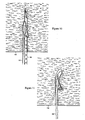
- Figure 10 depicts penetration of the stepped needle 103 loaded with the suture anchor 144 into soft tissue 130.
- a scale on the stepped needle 103 visible to the surgeon measures the depth of anchor 144 insertion.
- the fin 134 of the anchor 144 protrudes outwardly, catching the tissue 130 and preventing the anchor 144 from pulling out as the stepped needle 103 is withdrawn. In essence, withdrawal of the stepped needle 103 dislodges or strips off the anchor 144, allowing the suture anchor 144 to remain at or near the intended depth of insertion.
- Figure 11 depicts resumption of the curved configuration of the anchor 144 after withdrawal of the stepped needle 103. The curvature also provides compression on the fin 134, embedding the fin 134 laterally into tissue 130.
- Figure 12 depicts tension applied to the suture 122 to pull and rotate the anchor 144 from an insertion or vertical position to an anchoring or horizontal position.
- the initial lateral mobility is favored by (1) the curvature of the suture anchor 144, and (2) protrusion of the fin 134.
- Figure 13 depicts further tension applied to the suture 122, orienting the platform 133 to nearly perpendicular to the suture 122 under tension. With the large surface area of the platform 133 pressing against the tissue 130, the suture 122 is secured with good anchoring strength for surgical repair.
- the rotation of the anchor 144 within the tissue provides partial thickness suturing with endoscopic, arthroscopic or laparoscopic capability.
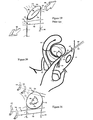
- Figure 15 shows a failed lumen 100 closure and hypermobility under stress with the urethropelvic ligaments 102 pulling the lateral walls 131 of the poorly supported urethra 101.
- Figure 16 shows the mid-sagittal view of Figure 15 during stress, with urethropelvic ligaments pulling perpendicularly above and below the plane of the page.
- Figure 16 also indicates that the section of poorly supported posterior wall 151 withdraws from mucosal 113 coaptation, leading to urine 117 leakage.
- FIG. 17 indicates the pre-surgical position of the vagina 114 with a dotted line, and that of the urethra 101 and bladder with dashed lines.
- Figure 17 also shows a large incision 157 required for repositioning and suturing both the vagina 114 and urethra 101 toward the abdominal wall.
- the post-surgical positions of the vagina 114 and backboardsupported urethra 101 are depicted with solid lines.
- the sutures 122 are knotted 125 to fascia or ligament on the abdominal wall.
- Figure 18 indicates a section of the backboard-supported posterior wall 151. This significantly invasive procedure provides the backboard support needed for lumen 150 closure during stress with concurrent pulling of the urethropelvic ligaments 102 to prevent urine leakage, as shown in Figure 19.
- the suture anchor 144 system can provide similar backboard support to the posterior wall 151 of the urethra 101.
- a catheter 154 is introduced through the urethra 101 into the bladder 127.
- the descended bladder 127 depicted in dotted lines, is lifted by the pressure against the wall of the vagina 144.
- the surgeon can also feel the catheter 154 within the urethra 101 to guide the needle/anchor 103/144 insertion lateral to the urethra 101, as shown in Figure 20, into the vaginal 114 wall.
- the fin 134 hooks onto the vaginal 114 tissue, stripping the anchor 144 off the withdrawing needle 103.
- the method of guiding the needle 103 with the surgeon's finger is currently being used with the Stamey needle, a prior art device, for repairing stress urinary incontinence.
- the needle/anchor 103/144 system does not require passing the suture 122 back and forth from the vagina 114 cavity to the abdominal wall.
- the suture 122 introduced by the Stamey needle is exposed within the vagina, which increases the risk of infection.
- the suture anchor 144 on the other hand, can be deployed within the vaginal 114 wall, as partial thickness suturing in open surgery.
- the suture anchor 144 can also be delivered and deployed in the vaginal 114 cavity, as full thickness suturing.
- Figure 21 depicts four suture anchors 144 fastened within the anterior vaginal 114 wall, providing backboard support to the posterior wall 151 of the urethra 101.
- the sutures 122 from the anchors 144 are knotted to fascia or ligament, similar to Figure 17, but requiring only a much smaller incision 157.
- the orientation of the anchor 144 within tissue can be significant.
- the anchors 144 deployed perpendicular to the urethra 101 as depicted in Figure 21, may provide a more firm backboard support than the anchors 144 deployed parallel to the urethra 101.
- the lumen 104 of the anchor 144 can be made non-round, elliptical for example, as shown in Figure 22, with the stepped needle 103 sized and configured to fit the lumen 104.
- Figure 23 shows an extended fin 134 sized and configured to fit into an indentation 153 on the stepped needle 103.
- an extended portion from the stepped needle 103 can fit into an indentation in the anchor 144 to prevent the anchor 144 from spinning on the stepped needle 103.
- Figure 24 depicts a patient with uterine 161 prolapse, a common problem in women.
- Uterine 161 prolapse is normally surgically treated with hysterectomy, removal of the uterus 161, either through vaginal or abdominal incision. The following procedure is ideally used in conjunction with the ligament-tightening procedure described in Figures 80 and 81.
- Figure 25 depicts lifting and repositioning of the uterus 161 with a uterine tool 163 containing a blunt distal end 171 , a shaft 172, a handle 159 and a lift 160.
- the stepped needle 103 with the suture anchor 144 is then inserted through a small incision 157, guided by an endoscope, into the repositioned uterus 161.
- the fin 134 hooks onto the uterine 161 tissue, dislodging the anchor 144 from the withdrawn needle 103.
- the needle 103 and anchor 144 insertion procedure is repeated, and the sutures 122 are knotted 125 on the fascia or a ligament on the abdominal wall, as shown in Figure 26, similar to the suture 122 tying for correcting urinary incontinence.
- Other supporting structures such as the round ligament and broad ligament of the uterus, may also be suitable for fastening the suture 122 to and supporting the repositioned uterus 161.
- the suture anchor 144 can also be used in orthopaedic repairs.
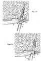
- Figure 27 depicts penetration of the stepped needle 103 and anchor 144 through a torn ligament 138 into freshly decorticated cancellous bone 118.
- the stepped needle 103 also contains a sleeve 220, freely sliding over the stepped needle 103.
- the position of the ligament 138 can be manipulated and maintained with grippers 221 on the distal end of the sleeve 220, as the stepped needle 103 is withdrawn.
- the fin 134 acts as a barb, hooking onto the cancellous bone 118, and stripping the anchor 144 off the withdrawing needle 103.
- Figure 28 depicts curvature resumption of the suture anchor 144 within the porous cancellous bone 118 after having slid off the withdrawn stepped needle 103.
- Figure 29 depicts tension applied to the suture 122, pulling on the curved anchor 144 and driving the fin 134 further laterally.
- the platform 133 of the anchor 144 provides a large surface area to press against the bone 118 and resist pull out.
- Figure 30 depicts another anchor 114 delivered by the stepped needle 103 through the torn ligament 138 into the cancellous bone 118.
- the stepped needle 103 is then withdrawn with the second anchor 114 also fastened within bone 118.
- Figure 31 depicts suture knot 125 tying to fasten the torn ligament 138 onto the bone.
- both the anchors 144 and sutures 122 can be made with biodegradable materials to prevent device migration with time.
- the anchoring strength of the suture anchor 144 can be further improved.
- the anchor 144 reaches full anchoring strength as the anchor 144 forms almost a T-configuration or is perpendicular with the suture 122, as shown in Figure 13.
- the elastic anchor 144 may curve further, or even fold into a V-configuration.
- the anchoring strength would greatly decrease.
- bend stops 155 can be added along both sides of the anchor 144 to increase rigidity and anchoring strength of the anchor 144.
- Figure 32 depicts the bend stop 155 with a gap or V-groove 156 beneath the platform 133.
- the gap 156 is closed to resist further bending of the anchor 144, as depicted in Figure 32.
- the elastic anchor 144 is resiliently straightened by the stepped needle 103, the gap 156 is opened, as shown in Figure 33.
- Figure 34 depicts the side view of the resiliently straightened anchor 144, showing the open gap 156 of the bend stop 155 beneath the platform 133.
- Figure 35 depicts the bottom or belly view of the resiliently straightened anchor 144, showing the bilateral bend stops 155 and open gaps 156.
- the bend stops 155 are designed and positioned to limit or resist excessive anchor 144 bending to maximize anchoring strength.
- a straight and rigid anchor 144 with the fin 134 can also rotate within tissue by utilizing the tension applied to the suture 122.
- the curvature of the anchor 144 increases the distance, W, to provide additional torque for lateral rotation.
- a rigid anchor 144 as shown in Figure 36, a larger and more protruded fin 134 may adequately provide torque for the anchor 144 rotation within the tissue.
- Figure 37 depicts the side view of the rigid anchor 144 showing a distance, W 1 , measured from the proximal tip of the fin 134 to the suture opening 123. The distance, W 1 , provides the initial rotational torque as tension is applied to the suture 122 by the surgeon.
- a rigid anchor 144 By elevating the suture openings 123 from a protrusion, a rigid anchor 144, shown in Figure 38 with side view in Figure 39, provides an even greater distance, W 2 , for greater initial rotational torque.
- the fin 134 can be made pointed or angled, as shown in Figures 36 to 39 to facilitate lateral tissue penetration and anchor 144 rotation. Rotation of the anchor 144 within tissue is also favored when L 1 > L 2 , where L 1 is the distance between the proximal tip of the fin 134 and the suture openings 123, and L 2 is the distance between the distal end of the anchor 144 and the suture openings 123.
- the tapered proximal ends as shown in Figures 36 and 38, also help to facilitate lateral insertion into tissue during anchors 144 rotation.
- Figure 40 depicts a suture attachment 164 without threading through the platform 133.
- the platform 133 may not be necessary.
- Figure 41 shows an anchor 144 with the fin 134 but without a platform.
- Figure 42 shows a curved anchor 144 without a fin. With a curvature built into the anchor 144, it may be sufficient to provide initial torque to rotate the anchor 144 within tissue when tension is applied to the suture 122.
- the suture anchor 144 may also be used for full thickness anchoring.
- Figure 43 depicts a curved suture anchor 144 with a platform 133 on the concave side of the curvature.
- the fin 134 is made blunt to avoid damage to adjacent tissue.
- the anchor 144 is loaded onto the stepped needle 103 with a sleeve 220 capable of sliding over the stepped needle 103, as shown in Figure 44.
- the sleeve 220 is similar to that shown in Figure 28 for holding and manipulating tissue.
- the sleeve 220 can also be used to push the anchor 144 off the stepped needle 103 and deploy the anchor 144 outside the tissue.
- the protruded fin 134 can provide an additional function, as a contact point for the sleeve 220.
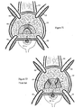
- Figure 45 depicts a cross section of a bulging L4-5 intervertebral disc 100 located between psoas major muscles 188.
- the stepped needle 103 carrying the anchor 144 as shown in Figure 44, is delivered through a small posteriolateral incision, into the bulging annulus and nucleus pulposus 128, as shown in Figure 45.
- the advancement of the stepped needle 103 stops as the distal tip of the stepped needle 103 exits the disc 100.
- the sliding sleeve 220 is used to push and expel the anchor 144 with the attached suture 122 out of the disc 100.
- the orientation of the anchor 144 can be corrected by advancing the distal tip of the sleeve 220 to manipulate the anchor 144 and pull on the suture 122 until the suture anchor 144 is properly positioned. Both the stepped needle 103 and sleeve 220 are withdrawn after proper deployment of the anchor 144.
- Figure 47 depicts a curved disc compressor 111 with two openings 123 for the suture 122 and a round or blunt annular compressing region 119.
- Figure 48 depicts knot 125 tying and bulge compression of the fastened disc compressor 111.
- the suture 122 is secured with full thickness anchoring by the anchor 144 and compressor 111.
- the bulge is compressed and fastened to alleviate pain from nerve impingement.
- Two suture anchors 144 with unique suture 122 arrangement between them can be loaded in series on a stepped needle 103 to be deployed within tissue. As the suture 122 is pulled by the surgeon, the anchors 144 draw close to each other, pulling in or approximating the inserted tissue.
- Figure 49 depicts portions of two anchors 144 connected by a suture 122 through holes 123A, 123B, 123C, 123D, 123E, 123F, 123G then 123H. Proximal ends of the suture 122 are threaded through a plunger 109. The holes 123B, 123C, 123F and 123G are angled to facilitate sliding of the suture 122 after anchor 144 rotation.
- the suture 122 between the holes 123D and 123E forms a stationary loop beneath the proximal anchor 144.
- the strands of suture 122 will slide from 123F to 123G and from 123C to 123B .
- the anchors 144 With the stationary loop beneath the proximal anchor 144, the anchors 144 will draw close to each other to approximate, compress or plicate (fold) the inserted tissue.
- the distal and proximal suture anchors 144 with the suture 122 form an approximating device 273 designed for minimally invasive use.
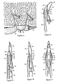
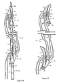
- Two resiliently straightened anchors 144 are loaded in series on a double-stepped 165 needle 103, as indicated in Figure 50. Similar to Figure 49, the suture 122 is threaded through holes 123A, 123B, 123C, 123D, 123E, 123F, 123G then 123H. For clarification, the suture 122 from holes 123A to 123D is white and from holes 123E to 123H is black. Both white and black sutures 122 are slack to clarify points of origin. The distal end of the proximal anchor 144 is tapered for lateral tissue penetration.
- the lumen 104 of the distal anchor 144 is smaller than the lumen 104 of the proximal anchor 144, each corresponding to the sizes of the distal and proximal steps 165 of the needle 103.
- the distance between the steps 165 can be pre-set or fixed to deliver the anchors 144.
- the needle 103 is withdrawn to deposit both anchors 144 with the connecting suture 122, as shown in Figure 51. Both anchors 144 resume their curved configuration. In vertical or insertion position, the angled suture holes 123B and 123G of the distal anchor 144 are designed to resist suture 122 sliding and to favor pivoting of the distal anchor 144, as shown in Figure 52. The rotation of the distal anchor 144 creates tension on the suture 122 connecting holes 123C to 123D and 123F to 123E , as shown in Figures 49 and 53.
- the tension of the sutures 122 lifts the proximal anchor 144 by the loop beneath holes 123D to 123E, as shown in Figures 53 and 49.
- the proximal anchor 144 also rotates, laterally pressing the pointed distal end into the tissue, with the fin 134 behaving like a rudder to direct rotation.
- the proximal anchor 144 can also be inserted by a sliding sleeve 220, rather than by the stationary second step 165 of the needle 103.
- Figure 54 shows a stepped needle 103 insertion to deliver the distal anchor 144 into the tissue 130. As the tissue 130 is snagged by the fin 134, partial withdrawal of the needle 103 deposits the distal anchor 144 within tissue 130, as indicated in Figure 55.
- the proximal anchor 144 is delivered by pushing the sleeve 220 and pulling the suture 122, as shown in Figure 56. Suture 122 pulling also initiates pivoting of the distal anchor 144.
- Figure 57 shows complete insertion of the proximal anchor 144 into the tissue 130. The needle 103 is then withdrawn to deposit the proximal anchor 144, as shown in Figure 58, to complete the installation of the approximating device 273.
- the approximating device 273 can be tightened and maintained under tension.
- a one-way suture lock 239 prevents backsliding during tying and allows further tightening of the suture 122 to fasten the approximating device 273.
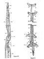
- Figure 59 depicts the composition of a suture lock 239 with a pair of sutures 122 passing through a hole 240 of a cone 266 into a loop 267 of an one-way grip 237 with individual grippers 241, then threaded through a passage 238 at the proximal end of the grip 237.
- the suture 122 passed through the loop 267 helps to direct the one-way grip 237 into the cone 266.
- the passage 238 of the grip 237 provides a foundation for suture knot 125 tying.
- the loop 267 and passage 238 also keep the pair of sutures 122 apart to obtain maximum locking strength within the cone 266.
- the cylindrical grippers 241 are arranged in angle, layers, sized and configured to fit within the cone 266. Each layer of the grippers 241 are tapered, narrow at the top and widened at the base, biased against backsliding of the suture 122 but allowing further suture 122 tightening.
- Figure 60 shows the lock 239 assembly with the pair of sutures 122 fastened between the cone 266 and biased grippers 241. The pair of sutures 122 is inserted into a plunger 109.
- the plunger 109 is bilaterally tapered at the distal end, as shown in Figure 60, for pushing against the proximal end of the one-way grip 237 without interfering with the pulling of the suture 122 to tighten the approximating device, as shown in Figure 61.
- slipknots 125 can be tied then delivered by a knot pusher 245 onto the proximal end of the one-way grip 237, as shown in Figure 62.
- a suture 122 cutting device 250 contains an inner tube 246 and outer tube 247.
- Figure 63 shows a channel open from the distal end of the inner tube 246 to a side window 248 of the suture cutter 250.
- Figure 64 shows the outer tube 247 also containing a side window 248.
- the inner tube 246 is tightly fitted inside the outer tube 247 with overlapping side windows 248, as shown in Figure 65, to form the suture cutter 250.
- the suture cutter 250 is a relatively thin tubular device.
- the excess suture 122 is threaded through the distal opening and out the overlapping side windows 248 of the inner tube 246 and outer tube 247, as shown in Figure 66.
- the cutter 250 can slide along the suture 122 into tissue through the entry punctured by needle 103 and anchors 144.
- Figure 67 shows a mid-longitudinal view of the suture cutter 250 with sharp edges 249 at the side windows 248. As the outer tube 247 slides against the inner tube 246 or vice versa, the sharp edges 249 behave like scissors, cutting the sutures 122 extending out of the side windows 248, as shown in Figure 68.
- Figure 69 shows a mid-longitudinal view of suture 122 cutting by sliding the outer 247 and inner tube 246 against each other.
- Figure 70 depicts suture 122 cutting with the device 250 after knot 125 tying. The cutter 250 is then withdrawn from tissue. As a result, all components are concealed within the tissue to complete the installation of the minimally invasive approximating device.
- the scarred tissue 268 of the external sphincter 251 can be revealed beneath adipose tissue 272 with retractors 196 opening a semi-circular incision between the vagina 114 and the rectum 132, as shown in Figure 71.
- the scarred sphincter 251 is cut, as shown in Figure 72.
- the scarred tissue 268 is overlapped, sutured and knotted 125 to tighten around the internal sphincter 252 beneath, as indicated in Figure 73.
- the tightness of the sphincteric repair is judged by the feel of the surgeon's finger. After surgical repair of the sphincter 251, painful defecation is inevitable. Infection is also common.
- Sphincter 251 repair can be minimally invasive using the approximating devices 273.
- radiopaque, echogenic or other tracing agents can be injected through a lumen 269, as shown in Figure 74, as the needle 103 advances into the body.
- the injected tracing agent is likely to diffuse quickly.
- diffusion of the tracing agent is limited, so it might be possible to indicate the shape of the tissue, an important criterion for verifying the target site for suture 122 anchoring.
- the muscular external sphincter 251 encircles the rectum 132 beneath the adipose tissue 272, as shown in Figures 71 and 75.
- the needle 103 is laterally inserted between the vagina 114 and rectum 132 to bridge both sides of the loose external sphincter 251.
- the needle 103 can be made with a slight curvature for puncturing through skin and adipose tissue 272, then into both sides of the loose sphincter 251.
- the anchors 144 can be inserted with the procedures similar to Figures 54 to 58, positioning the pair of anchors 144 into opposite sides of the loose sphincter 251.
- Figure 75 depicts tightening of the external sphincter 251 by pulling the suture 122 and pushing the plunger 109 against the proximal end of the suture lock 239 at the same time, as shown in Figure 61.
- the approximating device 273 restricts and narrows the circular external sphincter 251 by taking up the scarred 268 and loose tissue, as shown in Figure 75.
- the sutures 122 can then be knotted 125 and cut beneath the skin, as shown in Figures 62, 70 and 75.
- the suture 122, anchors 144 and lock 239 can be made with biodegradable materials.
- Oozing from the sphincteric 251 muscle traumatized by insertions of needles 103 and suture anchors 144 can initiate permanent tissue adhesion, holding and keeping the sphincter 251 in the approximated position even after degradation of the suture 122 and the anchors 144.
- the tips of most surgical needles are designed to cut as well as puncture into tissue.
- a tip without cutting edges similar to a sewing needle shown in Figure 76, is preferred.
- the tip with non-cutting edges is more likely to advance within a tissue with longitudinally oriented fibers, especially accompany with rotation during advancement.
- the slender tissue can be a tendon or a ligament with collagen bundles 270 formed lengthwise along the tissue.
- Figure 77 depicts the needle 103 with non-cutting edges being advanced along a ligament 138 using rotational motion to drill and split a path between collagen bundles 270.
- the needle 103 can also be made with flexible or shape memory material, such as nickel-titanium alloy, to conform within the tendon or ligament 138.
- flexible or shape memory material such as nickel-titanium alloy
- both the distal and proximal anchors 144 can then be individually delivered with sleeves 220.
- radiopaque, echogenic or other tracing agents can also be injected through a lumen 269, as shown in Figure 78.
- Uterine prolapse is commonly caused by sagging ligaments.
- the current treatment is hysterectomy.
- Figure 79 indicates a cross-sectional view of uterine 161 supports.
- the cardinal ligament 253 provides for lateral support, sacrouterine ligament 254 for posterior support and fascia 255 for anterior support to the uterus 161.
- the muscles and ligaments are relaxed.
- the uterus 161 is pulled down from the vagina 114 by a grasping device 259 to expose the cardinal 253 and sacrouterine 254 ligaments, as shown in Figure 80, with ovaries 256, fallopian tubes 258 and round ligaments 257 within the abdomen.
- the needle 103 is advanced along the ligament 253 or 254 to deliver the anchors 144, as shown in Figure 80.
- the sutures 122 are loaded with suture locks 239 and plungers 109.
- the approximating devices 273 are then individually tightened by advancing the plungers 109 against the suture locks 239, while the sutures 122 are being pulled to plicate and shorten the ligament 253 and/or 254, as shown in Figure 81.
- the ligament 253 and/or 254 is folded, crinkled or bunched together under the tension of the approximating devices 273.
- the cervix 271 and the entire uterus 161 are lifted by the shortened cardinal 253 and/or sacrouterine 254 ligaments.
- the shortened ligament can be permanently maintained to uphold the uterus 161.
- the ligament 253 and/or 254 are traumatized by insertions of needles 103 and anchors 144, oozing from the traumatized tissue can initiate tissue adhesion to hold and keep the ligament 253 and/or 254 in the plicated position even after degradation of the suture 122 and the anchors 144.
- the plicated ligament 253 and/or 254 also undergo tissue remodeling, including collagen crosslinking, which may also result in permanent shortening of the ligament 253 and/or 254.
- a modified procedure and a suture-gripping device are designed for fastening an anchor 144 within thin tissue.
- Figure 82 depicts partial insertion of the proximal anchor 144 of the approximating device 273 into a thin tissue 130.
- Figure 83 shows a prior art suture-gripping device 264, with jutted flaps 265 biting and resisting upward slippage of the suture 122.
- the suture-gripping device 264 loaded on the suture 122 is followed by the plunger 109, as indicated in Figure 84.
- the needle 103 and sleeve 220 are then withdrawn from tissue 130.
- the sutures 122 are pulled, and the plunger 109 is pushed against the suture gripping device 264 to draw the proximal anchor 144 into the tissue 130 and tighten the approximating device 273. Then, knots 125 are tied beneath the gripping device 264 to secure the sutures 122, as shown in Figure 85.
- the guide 185 contains a track 262 for the needle 103 to slide along, an extendible arm 260 to align with the needle 103, and a pointer 261 to indicate the target site.
- measuring units on the arm 260 indicate depth of needle 103 penetration.
- the traditional surgical treatment for urinary incontinence is to provide backboard support to the urethral posterior wall 151 by pulling the vagina 114 forward with sutures 122.
- the sutures 122 are then fastened onto the fascia or ligament in the abdominal wall, as indicated in Figures 17 and 18.
- the approximating device 273 can provide similar backboard support to the posterior wall 151 without any incision 157.
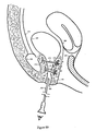
- Figure 87 depicts the vagina 114 is dilated with a retractor 196.
- the needle 103 is inserted through the anterior wall of the retracted vagina 114, lateral to the bladder neck 112, through the fascia 255 or ligament into adipose tissue 272 above the pubic symphysis 115.
- the distal anchor 144 is then deployed within the adipose tissue 272 and the proximal anchor 155 within the vaginal 114 wall with the suture-gripping device 264.
- the approximating device 273 is then tightened by pulling the suture 122 and pushing the plunger 109.
- the tightness of the plication can be seen through the urethra 101 with an endoscope 263.
- the suture 122 is then knotted 125 and cut, as shown in Figures 85 and 88.
- Figure 88 shows a minimally invasive approach to supporting the posterior-urethral wall 151 of the urethra 101 by pulling the vaginal 114 wall forward with approximating devices 273.
- trauma from insertion of needles 103 and anchors 144 can lead to tissue adhesion, providing permanent posterior wall 151 support even after degradation of the suture 122, anchor 144 and gripping device 264.
- the needle 103 can be inserted lateral to the bladder neck 112 or the urethra 101, into the retropubic space 274, area between the pubic symphysis 115 and bladder/urethra 127/101, to deliver the distal anchor 144.
- the proximal anchors 144 are deployed as mentioned within the vaginal 114 wall.
- the bladder neck 112 as well as the urethra 101 are sandwiched between the anterior 152 fascia and the vagina 114, as shown in Figure 89, to tighten the bladder neck 112 and treat sphincteric deficiency.
- the most difficult step in installing the approximating device 273 is probably the guidance of the needle 103 safely and accurately into tissue.
- multiple pairs of approximating devices 273 can be loaded or passed along the needle 103, as shown in Figure 90. With only a single needle 103 insertion, the approximating strength is greatly enhanced with multiple devices 273 installed, as shown in Figure 91.
- the dynamics of anchor 144 pivoting or rotation responding to suture 122 tension is especially significant within thin tissue 130. From observation within transparent gel wax, the initial movement of a crude prototype anchor 144 responding to suture 122 tension was in both pullout and lateral rotational directions. A similar result was obtained in meat. The suture 122 was not truly fastened until the prototype anchor 144 had rotated from the insertion position to fastening or perpendicular position. Before the fastened position was achieved, the suture 122 could be pulled with some resistance. The pivotal or rotational efficiency of the anchor 144 can probably be described by the pullout distance of the pulled suture 122. In an experiment using pork and the crude prototype anchor 144, the pullout distance was about one and half lengths of the prototype anchor 144 before the anchor 144 was secured. Within thin tissue, the anchor 144 would be pulled out before reaching the fastened position. With modifications to the crude prototype anchor 144, rotational efficiency can be significantly improved.
- the needle 103 can also contain an inner and outer sleeves 220.
- the sleeves 220 are stacked over each other, and both sleeves 220 capable of sliding over the needle 103, as shown in Figure 92.
- the lumen 104 of the distal anchor 144 fits over the distal portion of the needle 103, but too small to fit over the inner sleeve 220.
- the slightly larger lumen 104 of the proximal anchor 144 fits over the inner sleeve 220, but too small to fit over the outer sleeve 220.
- the inner sleeve 220 supports the distal anchor 144 and the outer sleeve 220 supports the proximal anchor 144, with both sleeves 220 and anchors 144 fit over the needle 103.
- the anchors 144 and sleeves 220 are punctured into tissue.
- the inner sleeve 220 is held stationary while the needle 103 is partially withdrawn to disengage and deploy the distal anchor 144.
- the outer sleeve 220 is held stationary while the needle 103 is fully withdrawn to deploy the proximal anchor 144.
- the fin 134 can extend beyond the length of the body 275 and be made pointed to spearhead and expedite the rotation of the suture anchor 144, as shown in Figure 93.
- the side view of the pointed and extended fin 134 is more evident in Figure 94.
- the sharpened fin 134 helps lateral penetration into tissue 130.
- Extension of the fin 134 lengthens L 1 favors and expedites lateral rotation of the anchor 144.
- L 1 is significantly lengthened, the suture holes 123 are still at or near the center of the platform 133 to prevent excessive rotation after reaching the fastened position.
- Anchor 144 rotation begins with lateral tissue 130 penetration of the fin 134, followed by the proximal end of the body 275, then the platform 134 of the anchor 144.
- the proximal portion of the platform 133 is tapered and curved toward the fin 134, as shown in Figures 93 and 94.
- the tapered proximal end of the anchor 144 is supported by a shape-matching step 165 on the needle 103, as shown in Figure 94.
- the shape-matching contact between the anchor 144 and the step 165 also helps to minimize spinning of the anchor 144 around the delivering needle 103.
- the curvature near the proximal end of the anchor 144 is more likely to have better rotational efficiency than the efficiency of the curvature situated near the distal end of the anchor 144.
- the suture anchor 144 can be made with polylactate, polyglycolic, poly-lactide-co-glycolide, polycaprolactone, trimethylene carbonate or combinations of these materials. Many of these degradable polymers are in US FDA approved products.
- degradable polymers such as polydioxanone, polyanhydride, trimethylene carbonate, poly-beta-hydroxybutyrate, polyhydroxyvalerate, poly-gama-ethyl-glutamate, poly-DTH-iminocarbonate, poly-bisphenol-A-iminocarbonate, poly-ortho-ester, polycyanoacrylate and polyphosphazene can also be used.
- Nickel-titanium alloy, spring-tempered stainless steel, titanium, stainless steel or other metallic material provides strength and durability.
- the anchor 144 can also be coated with biocompatible polymers, such as polyurethane, polytetrafluoroethylene, silicon, ultra high molecular weight polyethylene or other material.
- biocompatible polymers such as polyurethane, polytetrafluoroethylene, silicon, ultra high molecular weight polyethylene or other material.
- the anchor 144 can also be coated with lubricants, growth factors, nutrients, buffering agents, collagen, hydroxyapatite, analgesics, sealants, blood clotting agents, antibiotics, radiopaque or echogenic agents. All materials should be able to withstand sterilization by gamma, electron beam, autoclave, ETO, plasma or UV light to prevent infection.
- the stepped needle 103 and sleeve 220 can be made with stainless steel, titanium, nickel titanium other metal or alloy.
- the stepped needle 103 and sleeve 220 can be coated with lubricant, blood clotting, radiopaque or echogenic agents.
- the stepped needle 103 can be made curved to gain accessibility for the surgeon.
- the sleeve 220 can also be made with elastic material, such as nickel titanium, polypropylene, polyethylene or other flexible material.
- the stepped needle 103 and sleeve 220 can also be coated with lubricant, antibiotic, radiopaque or echogenic agents.
- the suture 122 can be permanent or biodegradable, braided or monofilament.
- the suture 122 can also be metallic for strength and durability.
- the anchor 144 is designed for partial thickness or full thickness suture 122 anchoring and is delivered with the stepped needle 103. Deployment of the anchor 144 can be as simple as inserting and withdrawing the stepped needle 103 in and from tissue. The sleeve 220 sliding over the stepped or a smooth needle 103 can be helpful in deploying the anchor 144 and manipulating tissue.
- the curvature of the anchor 144 promotes initial anchor 144 rotation within tissue when tension is applied to the suture 122.
- the fin 134 is designed to (1) dislodge the anchor 144, (2) enhance initial rotation of the anchor 144 , and (3) stabilize the anchor 144 during rotation.
- the platform 133 especially fortified with bend stops 155, is designed to increase the anchoring strength within tissue. When multiple anchors 144 are delivered in series into tissue, as the suture 122 is pulled, the anchors 144 draw close to each other to plicate or approximate the pierced tissue.
Landscapes
- Health & Medical Sciences (AREA)
- Surgery (AREA)
- Life Sciences & Earth Sciences (AREA)
- Medical Informatics (AREA)
- Animal Behavior & Ethology (AREA)
- Engineering & Computer Science (AREA)
- Biomedical Technology (AREA)
- Heart & Thoracic Surgery (AREA)
- Veterinary Medicine (AREA)
- Molecular Biology (AREA)
- Nuclear Medicine, Radiotherapy & Molecular Imaging (AREA)
- General Health & Medical Sciences (AREA)
- Public Health (AREA)
- Rheumatology (AREA)
- Surgical Instruments (AREA)
- Sealing Material Composition (AREA)
- Prostheses (AREA)
- Reinforcement Elements For Buildings (AREA)
Claims (42)
- Nahtmaterialanker, der mit einer Nahtmaterialanker-Zuführvorrichtung, die Nadel hat, ausbringbar ist, wobei der Nahtmaterialanker aufweist:dadurch gekennzeichnet, dass der Ankerkörper (144) aus einem elastischen Material ausgebildet ist und eine gerade gerichtete Stellung und eine gekrümmte Stellung hat, wobei der Nahtmaterialanker ferner aufweist:einen Nahtmaterialankerkörper (144), der eine Längsachse aufweist,eine Nahtmaterialöffnung (123), die dimensioniert und ausgestaltet ist, um ein Nahtmaterial (122) aufzunehmen, wobei die Nahtmaterialöffnung durch einen Teil des Nahtmaterialankers führt,wobei bei der Verwendung der Nahtmaterialkörper elastisch in die gerade gerichtete Stellung gerade gerichtet ist, wenn sich die Nadel der Nahtmaterialanker-Zuführvorrichtung in dem Durchgang befindet,einen Durchgang (104), der entlang der Längsachse durch den Nahtmaterialanker verläuft, wobei der Durchgang dimensioniert und ausgestaltet ist, damit die Nadel der Nahtmaterialanker-Zuführvorrichtung durch diesen verläuft, undeine Führungsplattform (133), die an einer Seite des Nahtmaterialankerkörpers (144) befestigt ist,
und wobei der Nahtmaterialankerkörper wieder seine gekrümmte Stellung annimmt, wenn die Nadel entfernt wird. - Nahtmaterialanker nach Anspruch 1, bei dem die Führungsplattform im Wesentlichen eben ist.
- Nahtmaterialanker nach Anspruch 1, bei dem die Nahtmaterialöffnung durch die Plattform verläuft.
- Nahtmaterialanker nach Anspruch 3, bei dem die Plattform einen Vorsprung aufweist und die Nahtmaterialöffnung durch den Vorsprung verläuft.
- Nahtmaterialanker nach Anspruch 3, der ferner eine zweite Nahtmaterialöffnung aufweist, die durch die Plattform verläuft, und wobei bevorzugt eine der Nahtmaterialöffnungen auf einer linken Seite der Plattform angeordnet ist und die andere Nahtmaterialöffnung auf einer rechten Seite der Plattform angeordnet ist.
- Nahtmaterialanker nach Anspruch 1, bei dem die Plattform auf einer konkaven Seite angeordnet ist, wenn sich der Nahtmaterialanker in der gekrümmten Stellung befindet.
- Nahtmaterialanker nach Anspruch 1, bei dem die Plattform auf einer konvexen Seite angeordnet ist, wenn sich der Nahtmaterialanker in der gekrümmten Stellung befindet.
- Nahtmaterialanker nach Anspruch 1, der ferner eine Vertiefung aufweist, die sich an einer unteren Fläche der Plattform befindet, und wobei die Vertiefung geschlossen ist, wenn sich der Nahtmaterialanker in der gekrümmten Stellung befindet, wobei die Vertiefung bevorzugt V-förmig ist.
- Nahtmaterialanker nach Anspruch 1, bei dem sich die Plattform nahe einem mittleren Bereich des Nahtmaterialankers befindet.
- Nahtmaterialanker nach Anspruch 1, bei dem sich die Plattform entlang zumindest eines Großteils einer Länge des Nahtmaterialankerkörpers erstreckt.
- Nahtmaterialanker nach Anspruch 1, bei dem sich die Plattform entlang ungefähr drei Vierteln einer Länge des Nahtmaterialankerkörpers erstreckt.
- Nahtmaterialanker nach Anspruch 1, bei dem eine Längsachse der Plattform im Wesentlichen parallel zu der Längsachse des Nahtmaterialankerkörpers verläuft.
- Nahtmaterialanker nach Anspruch 1, bei dem sich ein distales Ende oder ein proximales Ende der Plattform verjüngt oder bei dem sich die Plattform an sowohl dem proximalen als auch dem distalen Ende verjüngt.
- Nahtmaterialanker nach Anspruch 1, bei dem die Plattform im Wesentlichen tangential zu dem Nahtmaterialankerkörper befestigt ist.
- Nahtmaterialanker nach Anspruch 1, bei dem die Plattform länglich ist.
- Nahtmaterialanker nach Anspruch 1, bei dem sich ein proximales Ende der Plattform um ein proximales Ende des Nahtmaterialankerkörpers krümmt und dadurch das proximale Ende des Nahtmaterialankerkörpers abdeckt.
- Nahtmaterialanker, der mit einer Nahtmaterialanker-Zuführvorrichtung, die eine Nadel hat, ausbringbar ist, wobei der Nahtmaterialanker aufweist:dadurch gekennzeichnet, dass der Nahtmaterialankerkörper (144) aus einem elastischen Material ausgebildet ist und eine gerade gerichtete Stellung und eine gekrümmte Stellung aufweist, wobei der Nahtmaterialanker ferner einen Durchgang (104) aufweist, der entlang der Längsachse durch den Nahtmaterialanker verläuft, wobei der Durchgang dimensioniert und ausgestaltet ist, damit die Nadel der Nahtmaterialanker-Zuführvorrichtung durch diesen verläuft,einen Nahtmaterialankerkörper (144), der eine Längsachse aufweist,eine Nahtmaterialöffnung (123), die dimensioniert und ausgestaltet ist, um ein Nahtmaterial (122) aufzunehmen, wobei die Nahtmaterialöffnung durch einen Teil des Nahtmaterialankers führt, undeine Führungsrippe (134), die an einer Seite des Nahtmaterialankerkörpers befestigt ist,
wobei bei der Verwendung der Nahtmaterialankerkörper elastisch in die gerade gerichtete Stellung gerade gerichtet ist, wenn sich die Nadel der Nahtmaterialanker-Zuführvorrichtung in dem Durchgang befindet,
und wobei der Nahtmaterialankerkörper wieder seine gekrümmte Stellung annimmt, wenn die Nadel entfernt wird. - Nahtmaterialanker nach Anspruch 17, bei dem sich die Rippe im Wesentlichen senkrecht von dem Nahtmaterialankerkörper erstreckt.
- Nahtmaterialanker nach Anspruch 17, bei dem die Rippe in der Nähe eines proximalen Endes des Nahtmaterialankerkörpers angeordnet ist.
- Nahtmaterialanker nach Anspruch 17, bei dem sich die Rippe verjüngt, so dass ein proximales Ende der Rippe größer als ein distales Ende der Rippe ist.
- Nahtmaterialanker nach Anspruch 17, bei dem die Rippe auf einer konkaven Seite angeordnet ist, wenn sich der Nahtmaterialanker in der gekrümmten Stellung befindet.
- Nahtmaterialanker nach Anspruch 17, bei dem die Rippe auf einer konvexen Seite angeordnet ist, wenn sich der Nahtmaterialanker in der gekrümmten Stellung befindet.
- Nahtmaterialanker nach Anspruch 17, bei dem ein proximales Ende der Rippe abgewinkelt ist, bevorzugt, um zu einer sich verjüngenden distalen Stufe der Nadel zu passen.
- Nahtmaterialanker nach Anspruch 17, bei dem sich die Rippe von einem mittleren Bereich des Nahtmaterialankerkörpers nach außen erstreckt und bei dem die Nahtmaterialöffnung durch diese verläuft.
- Nahtmaterialanker nach Anspruch 17, bei dem die Nahtmaterialöffnung gegenüber der Rippe angeordnet ist.
- Nahtmaterialanker nach Anspruch 17, bei dem die Rippe eine spitze proximale Spitze hat, die sich über ein Ende des Nahtmaterialankerkörpers hinaus erstreckt.
- Nahtmaterialanker, der mit einer Nahtmaterialanker-Zuführvorrichtung, die eine Nadel hat, ausbringbar ist, wobei der Nahtmaterialanker aufweist:dadurch gekennzeichnet, dass die Rippe eine proximale Spitze aufweist, die sich über ein Ende des Nahtmaterialankerkörpers hinaus erstreckt, und dass der Nahtmaterialanker ferner aufweist:einen Nahtmaterialankerkörper (144), der eine Längsachse aufweist,eine Nahtmaterialöffnung (123), die dimensioniert und ausgestaltet ist, um ein Nahtmaterial (122) aufzunehmen, wobei die Nahtmaterialöffnung durch einen Teil des Nahtmaterialankers führt, undeine Führungsrippe (134), die an einer ersten Seite des Nahtmaterialankerkörpers befestigt ist,einen Durchgang (104), der entlang der Längsachse durch den Nahtmaterialanker verläuft, wobei der Durchgang dimensioniert und ausgestaltet ist, damit die Nadel der Nahtmaterialanker-Zuführvorrichtung durch diesen verläuft, undeine Führungsplattform (133), die an einer zweiten Seite des Nahtmaterialankerkörpers befestigt ist.
- Nahtmaterialanker nach Anspruch 27, bei dem sich die Führungsrippe im Wesentlichen senkrecht zu der Führungsplattform erstreckt.
- Nahtmaterialanker nach Anspruch 27, bei dem sich ein proximales Ende der Plattform um ein proximales Ende des Nahtmaterialankerkörpers krümmt und dadurch das proximale Ende des Nahtmaterialankerkörpers abdeckt.
- Nahtmaterialanker nach Anspruch 27, bei dem der Nahtmaterialanker aus einem elastischen Material ausgebildet ist.
- Nahtmaterialanker nach Anspruch 30, bei dem der Nahtmaterialanker eine gerade gerichtete Stellung und eine gekrümmte Stellung aufweist und bei dem der Nahtmaterialanker bei der Verwendung durch die Nadel in der gerade gerichteten Stellung gehalten werden kann, wenn sich die Nadel in dem Durchgang befindet, und bei dem der Nahtmaterialanker wieder seine gekrümmte Stellung annimmt, wenn die Nadel entfernt wird.
- Nahtmaterialanker nach Anspruch 1, 17 oder 27, bei dem der Nahtmaterialanker biologisch abbaubar ist.
- Nahtmaterialanker nach Anspruch 1, 17 oder 27, der ferner eine Beschichtung auf dem Nahtmaterialanker aufweist, wobei die Beschichtung bevorzugt aus der Gruppe von Beschichtungen ausgewählt ist, die aus Gleitmitteln, Wachstumsfaktoren, Nährstoffen, Puffermitteln, Kollagen, Hydroxylapatit, Analgetika, Dichtmitteln, Blutgerinnungsmitteln, Antibiotika, röntgensichtbaren Mitteln und echogenen Mitteln besteht, oder aus der Gruppe von Beschichtungen ausgewählt ist, die aus Polyurethan, Polytetrafluorethylen, Silizium und Polyethylen mit ultrahohem Molekulargewicht besteht.
- Nahtmaterialanker nach Anspruch 1, 17 oder 27, der ferner eine Beschichtung aus einem biokompatiblen Polymer aufweist.
- Nahtmaterialanker nach Anspruch 1, 17 oder 27, bei dem der Nahtmaterialanker aus einem Material ausgebildet ist, das aus der Gruppe von Materialien ausgewählt ist, die aus Polypropylen, Polyethylen, Polyether-etherketon, Acetalharz, Polysulfon, Polycarbonat, Polylactat, Polyglycol, Polylactid-Coglycolid, Polycaprolacton, Trimethylencarbonat, Polydioxanon, Polyanhydrid, Trimethylencarbonat, Poly-β-Hydroxybutyrat, Polyhydroxyvalerat, Poly-γ-ethylglutamat, Poly-DTH-iminocarbonat, Poly-Bisphenol-A-iminocarbonat, Poly-Orthoester, Polycyanoacrylat, Polyphosphazen, Nickel-Titan-Legierung, federgehärtetem Edelstahl, Titan, Edelstahl und metallischem Material besteht.
- Nahtmaterialanker nach Anspruch 1, 17 oder 27, der ferner einen zweiten Nahtmaterialanker und einen Nahtmaterial aufweist, das durch die Nahtmaterialöffnung und durch eine zweite Nahtmaterialöffnung gefädelt ist, die durch einen Körper des zweiten Nahtmaterialankers verläuft.
- Nahtmaterialanker-Zuführsystem, das aufweist:wobei bei der Verwendung die Nadel den Nahtmaterialanker in der gerade gerichteten Stellung hält, wenn sich die Nadel in dem Durchgang befindet.den Nahtmaterialanker (144) nach Anspruch 1, 17 oder 31,eine Nadel mit einer Stufe, die die Nadel in einen proximalen Teil und einen distalen Teil unterteilt, wobei der distale Teil dimensioniert und ausgestaltet ist, um in den Durchgang zu passen, wobei der proximale Teil einen größeren Durchmesser aufweist,
- Nahtmaterialanker-Zuführsystem nach Anspruch 37, bei dem ein proximales Ende des Nahtmaterialankerkörpers an der Stufe anliegt, wenn sich die Nadel in dem Durchgang befindet.
- Nahtmaterialanker-Zuführsystem nach Anspruch 37, bei dem ein scharfes distales Ende der Nadel von einem distalen Ende des Durchgangs vorsteht.
- Nahtmaterialanker-Zuführsystem nach Anspruch 37, das ferner eine zweite Stufe an der Nadel und einen zweiten Nahtmaterialanker aufweist, und wobei das Nahtmaterial bevorzugt durch beide der Nahtmaterialanker verläuft.
- Nahtmaterialanker-Zuführvorrichtung, die aufweist:eine Nadel,eine erste Umhüllung, die um die Nadel angeordnet ist,eine zweite Umhüllung, die um die Nadel und die erste Umhüllung angeordnet ist, undden Nahtmaterialanker (144) nach Anspruch 1, 17 oder 27.
- Nahtmaterialanker-Zuführvorrichtung nach Anspruch 41, bei der der Nahtmaterialanker einen ersten Durchgang hat, der durch diesen verläuft, wobei der erste Durchgang dimensioniert ist, um die Nadel aufzunehmen, und zu klein dimensioniert ist, um die erste Umhüllung aufzunehmen, und die ferner einen zweiten Nahtmaterialanker aufweist, der einen zweiten Durchgang hat, der durch diesen verläuft, wobei der zweite Durchgang dimensioniert ist, um die erste Umhüllung aufzunehmen, und zu klein dimensioniert ist, um die zweite Umhüllung aufzunehmen.
Applications Claiming Priority (3)
| Application Number | Priority Date | Filing Date | Title |
|---|---|---|---|
| US36494702P | 2002-03-14 | 2002-03-14 | |
| US364947P | 2002-03-14 | ||
| PCT/US2002/041399 WO2003077772A1 (en) | 2002-03-14 | 2002-12-24 | Suture anchor and approximating device |
Publications (2)
| Publication Number | Publication Date |
|---|---|
| EP1482841A1 EP1482841A1 (de) | 2004-12-08 |
| EP1482841B1 true EP1482841B1 (de) | 2005-12-07 |
Family
ID=28041990
Family Applications (1)
| Application Number | Title | Priority Date | Filing Date |
|---|---|---|---|
| EP02807095A Expired - Lifetime EP1482841B1 (de) | 2002-03-14 | 2002-12-24 | Nahtanker und adaptationsvorrichtung |
Country Status (8)
| Country | Link |
|---|---|
| US (4) | US7766939B2 (de) |
| EP (1) | EP1482841B1 (de) |
| AT (1) | ATE311817T1 (de) |
| AU (1) | AU2002367772A1 (de) |
| CA (1) | CA2477220C (de) |
| DE (1) | DE60207893T2 (de) |
| ES (1) | ES2254792T3 (de) |
| WO (1) | WO2003077772A1 (de) |
Cited By (2)
| Publication number | Priority date | Publication date | Assignee | Title |
|---|---|---|---|---|
| US9504461B2 (en) | 2005-05-20 | 2016-11-29 | Neotract, Inc. | Anchor delivery system |
| US10105132B2 (en) | 2005-05-20 | 2018-10-23 | Neotract, Inc. | Devices, systems and methods for treating benign prostatic hyperplasia and other conditions |
Families Citing this family (297)
| Publication number | Priority date | Publication date | Assignee | Title |
|---|---|---|---|---|
| US8795332B2 (en) | 2002-09-30 | 2014-08-05 | Ethicon, Inc. | Barbed sutures |
| US6241747B1 (en) | 1993-05-03 | 2001-06-05 | Quill Medical, Inc. | Barbed Bodily tissue connector |
| US5931855A (en) | 1997-05-21 | 1999-08-03 | Frank Hoffman | Surgical methods using one-way suture |
| US8323341B2 (en) | 2007-09-07 | 2012-12-04 | Intrinsic Therapeutics, Inc. | Impaction grafting for vertebral fusion |
| WO2004100841A1 (en) * | 1999-08-18 | 2004-11-25 | Intrinsic Therapeutics, Inc. | Devices and method for augmenting a vertebral disc nucleus |
| US7717961B2 (en) * | 1999-08-18 | 2010-05-18 | Intrinsic Therapeutics, Inc. | Apparatus delivery in an intervertebral disc |
| US20040024465A1 (en) * | 1999-08-18 | 2004-02-05 | Gregory Lambrecht | Devices and method for augmenting a vertebral disc |
| US7998213B2 (en) * | 1999-08-18 | 2011-08-16 | Intrinsic Therapeutics, Inc. | Intervertebral disc herniation repair |
| US7972337B2 (en) | 2005-12-28 | 2011-07-05 | Intrinsic Therapeutics, Inc. | Devices and methods for bone anchoring |
| US7553329B2 (en) * | 1999-08-18 | 2009-06-30 | Intrinsic Therapeutics, Inc. | Stabilized intervertebral disc barrier |
| JP4247519B2 (ja) | 1999-08-18 | 2009-04-02 | イントリンジック セラピューティックス インコーポレイテッド | 髄核オーグメンテーションおよび保定のための装置および方法 |
| US7094258B2 (en) * | 1999-08-18 | 2006-08-22 | Intrinsic Therapeutics, Inc. | Methods of reinforcing an annulus fibrosis |
| US7887551B2 (en) | 1999-12-02 | 2011-02-15 | Smith & Nephew, Inc. | Soft tissue attachment and repair |
| US7056331B2 (en) | 2001-06-29 | 2006-06-06 | Quill Medical, Inc. | Suture method |
| US6848152B2 (en) | 2001-08-31 | 2005-02-01 | Quill Medical, Inc. | Method of forming barbs on a suture and apparatus for performing same |
| EP1482841B1 (de) | 2002-03-14 | 2005-12-07 | Yeung, Jeffery E. | Nahtanker und adaptationsvorrichtung |
| US6773450B2 (en) | 2002-08-09 | 2004-08-10 | Quill Medical, Inc. | Suture anchor and method |
| US8100940B2 (en) | 2002-09-30 | 2012-01-24 | Quill Medical, Inc. | Barb configurations for barbed sutures |
| US20040088003A1 (en) * | 2002-09-30 | 2004-05-06 | Leung Jeffrey C. | Barbed suture in combination with surgical needle |
| US7624487B2 (en) | 2003-05-13 | 2009-12-01 | Quill Medical, Inc. | Apparatus and method for forming barbs on a suture |
| JP2007515988A (ja) * | 2003-06-20 | 2007-06-21 | イントリンジック セラピューティックス インコーポレイテッド | 椎間板の環状欠損からインプラントを送出する装置および方法 |
| US9861346B2 (en) | 2003-07-14 | 2018-01-09 | W. L. Gore & Associates, Inc. | Patent foramen ovale (PFO) closure device with linearly elongating petals |
| US7347863B2 (en) | 2004-05-07 | 2008-03-25 | Usgi Medical, Inc. | Apparatus and methods for manipulating and securing tissue |
| US20050187565A1 (en) * | 2004-02-20 | 2005-08-25 | Baker Steve G. | Tissue fixation devices and a transoral endoscopic gastroesophageal flap valve restoration device and assembly using same |
| US7632287B2 (en) * | 2004-02-20 | 2009-12-15 | Endogastric Solutions, Inc. | Tissue fixation devices and assemblies for deploying the same |
| US11272926B2 (en) * | 2004-02-20 | 2022-03-15 | Endogastric Solutions, Inc. | Tissue fixation devices and assemblies for deploying the same |
| US7608092B1 (en) | 2004-02-20 | 2009-10-27 | Biomet Sports Medicince, LLC | Method and apparatus for performing meniscus repair |
| US8444657B2 (en) | 2004-05-07 | 2013-05-21 | Usgi Medical, Inc. | Apparatus and methods for rapid deployment of tissue anchors |
| US8257394B2 (en) | 2004-05-07 | 2012-09-04 | Usgi Medical, Inc. | Apparatus and methods for positioning and securing anchors |
| DE102004022590A1 (de) * | 2004-05-07 | 2005-12-01 | Feussner, Hubertus, Prof.Dr.med. | Blindniet zur Adaption von biologischem Gewebe und Vorrichtung zum Setzen des selbigen, insbesondere durch den Instrumentenkanal eines Endoskops |
| CN101431947A (zh) | 2004-05-14 | 2009-05-13 | 奎尔医疗公司 | 缝合方法和器械 |
| AU2005295418A1 (en) * | 2004-10-18 | 2006-04-27 | Temple University Of The Commonwealth System Of Higher Education | Apparatus and method of endoscopic suturing |
| US8128658B2 (en) | 2004-11-05 | 2012-03-06 | Biomet Sports Medicine, Llc | Method and apparatus for coupling soft tissue to bone |
| US8137382B2 (en) | 2004-11-05 | 2012-03-20 | Biomet Sports Medicine, Llc | Method and apparatus for coupling anatomical features |
| US8840645B2 (en) | 2004-11-05 | 2014-09-23 | Biomet Sports Medicine, Llc | Method and apparatus for coupling soft tissue to a bone |
| US8298262B2 (en) | 2006-02-03 | 2012-10-30 | Biomet Sports Medicine, Llc | Method for tissue fixation |
| US8361113B2 (en) | 2006-02-03 | 2013-01-29 | Biomet Sports Medicine, Llc | Method and apparatus for coupling soft tissue to a bone |
| US7601165B2 (en) | 2006-09-29 | 2009-10-13 | Biomet Sports Medicine, Llc | Method and apparatus for forming a self-locking adjustable suture loop |
| US8118836B2 (en) | 2004-11-05 | 2012-02-21 | Biomet Sports Medicine, Llc | Method and apparatus for coupling soft tissue to a bone |
| US7905904B2 (en) | 2006-02-03 | 2011-03-15 | Biomet Sports Medicine, Llc | Soft tissue repair device and associated methods |
| US7909851B2 (en) | 2006-02-03 | 2011-03-22 | Biomet Sports Medicine, Llc | Soft tissue repair device and associated methods |
| US7857830B2 (en) | 2006-02-03 | 2010-12-28 | Biomet Sports Medicine, Llc | Soft tissue repair and conduit device |
| US8303604B2 (en) | 2004-11-05 | 2012-11-06 | Biomet Sports Medicine, Llc | Soft tissue repair device and method |
| US8088130B2 (en) | 2006-02-03 | 2012-01-03 | Biomet Sports Medicine, Llc | Method and apparatus for coupling soft tissue to a bone |
| US9801708B2 (en) | 2004-11-05 | 2017-10-31 | Biomet Sports Medicine, Llc | Method and apparatus for coupling soft tissue to a bone |
| US7749250B2 (en) | 2006-02-03 | 2010-07-06 | Biomet Sports Medicine, Llc | Soft tissue repair assembly and associated method |
| US20060189993A1 (en) | 2004-11-09 | 2006-08-24 | Arthrotek, Inc. | Soft tissue conduit device |
| US9017381B2 (en) | 2007-04-10 | 2015-04-28 | Biomet Sports Medicine, Llc | Adjustable knotless loops |
| US7608098B1 (en) | 2004-11-09 | 2009-10-27 | Biomet Sports Medicine, Llc | Bone fixation device |
| US8034090B2 (en) | 2004-11-09 | 2011-10-11 | Biomet Sports Medicine, Llc | Tissue fixation device |
| US8998949B2 (en) | 2004-11-09 | 2015-04-07 | Biomet Sports Medicine, Llc | Soft tissue conduit device |
| US7914539B2 (en) | 2004-11-09 | 2011-03-29 | Biomet Sports Medicine, Llc | Tissue fixation device |
| US20060106405A1 (en) * | 2004-11-16 | 2006-05-18 | Fann James I | Systems and methods for delivering fastener to opposed tissue structures |
| US8128640B2 (en) | 2005-02-07 | 2012-03-06 | Ivy Sports Medicine LLC | System and method for all-inside suture fixation for implant attachment and soft tissue repair |
| WO2006086275A2 (en) * | 2005-02-07 | 2006-08-17 | Regen Biologics, Inc. | System and method for all-inside suture fixation for implant attachment and soft tissue repair |
| DE102005021885A1 (de) * | 2005-05-04 | 2006-11-16 | Karl Storz Gmbh & Co. Kg | Vorrichtung zum Einbringen eines Ankerelements samt Faden in einen Knochen |
| US9549739B2 (en) | 2005-05-20 | 2017-01-24 | Neotract, Inc. | Devices, systems and methods for treating benign prostatic hyperplasia and other conditions |
| US8333776B2 (en) | 2005-05-20 | 2012-12-18 | Neotract, Inc. | Anchor delivery system |
| US8628542B2 (en) | 2005-05-20 | 2014-01-14 | Neotract, Inc. | Median lobe destruction apparatus and method |
| US8157815B2 (en) * | 2005-05-20 | 2012-04-17 | Neotract, Inc. | Integrated handle assembly for anchor delivery system |
| US8603106B2 (en) | 2005-05-20 | 2013-12-10 | Neotract, Inc. | Integrated handle assembly for anchor delivery system |
| US8834492B2 (en) | 2005-05-20 | 2014-09-16 | Neotract, Inc. | Continuous indentation lateral lobe apparatus and method |
| US9149266B2 (en) | 2005-05-20 | 2015-10-06 | Neotract, Inc. | Deforming anchor device |
| US7896891B2 (en) | 2005-05-20 | 2011-03-01 | Neotract, Inc. | Apparatus and method for manipulating or retracting tissue and anatomical structure |
| US7645286B2 (en) * | 2005-05-20 | 2010-01-12 | Neotract, Inc. | Devices, systems and methods for retracting, lifting, compressing, supporting or repositioning tissues or anatomical structures |
| US8945152B2 (en) | 2005-05-20 | 2015-02-03 | Neotract, Inc. | Multi-actuating trigger anchor delivery system |
| US10195014B2 (en) | 2005-05-20 | 2019-02-05 | Neotract, Inc. | Devices, systems and methods for treating benign prostatic hyperplasia and other conditions |
| US10925587B2 (en) | 2005-05-20 | 2021-02-23 | Neotract, Inc. | Anchor delivery system |
| US8394113B2 (en) | 2005-05-20 | 2013-03-12 | Neotract, Inc. | Coiled anchor device |
| US9364212B2 (en) | 2005-05-20 | 2016-06-14 | Neotract, Inc. | Suture anchoring devices and methods for use |
| US8425535B2 (en) | 2005-05-20 | 2013-04-23 | Neotract, Inc. | Multi-actuating trigger anchor delivery system |
| US8668705B2 (en) | 2005-05-20 | 2014-03-11 | Neotract, Inc. | Latching anchor device |
| US8491606B2 (en) | 2005-05-20 | 2013-07-23 | Neotract, Inc. | Median lobe retraction apparatus and method |
| US8529584B2 (en) | 2005-05-20 | 2013-09-10 | Neotract, Inc. | Median lobe band implant apparatus and method |
| US7909836B2 (en) * | 2005-05-20 | 2011-03-22 | Neotract, Inc. | Multi-actuating trigger anchor delivery system |
| US8298291B2 (en) * | 2005-05-26 | 2012-10-30 | Usgi Medical, Inc. | Methods and apparatus for securing and deploying tissue anchors |
| US9585651B2 (en) * | 2005-05-26 | 2017-03-07 | Usgi Medical, Inc. | Methods and apparatus for securing and deploying tissue anchors |
| US8951285B2 (en) * | 2005-07-05 | 2015-02-10 | Mitralign, Inc. | Tissue anchor, anchoring system and methods of using the same |
| US7815659B2 (en) * | 2005-11-15 | 2010-10-19 | Ethicon Endo-Surgery, Inc. | Suture anchor applicator |
| US11259792B2 (en) | 2006-02-03 | 2022-03-01 | Biomet Sports Medicine, Llc | Method and apparatus for coupling anatomical features |
| US8562647B2 (en) | 2006-09-29 | 2013-10-22 | Biomet Sports Medicine, Llc | Method and apparatus for securing soft tissue to bone |
| US10517587B2 (en) | 2006-02-03 | 2019-12-31 | Biomet Sports Medicine, Llc | Method and apparatus for forming a self-locking adjustable loop |
| US9271713B2 (en) | 2006-02-03 | 2016-03-01 | Biomet Sports Medicine, Llc | Method and apparatus for tensioning a suture |
| US8251998B2 (en) | 2006-08-16 | 2012-08-28 | Biomet Sports Medicine, Llc | Chondral defect repair |
| US8597327B2 (en) | 2006-02-03 | 2013-12-03 | Biomet Manufacturing, Llc | Method and apparatus for sternal closure |
| US11311287B2 (en) | 2006-02-03 | 2022-04-26 | Biomet Sports Medicine, Llc | Method for tissue fixation |
| US8652171B2 (en) | 2006-02-03 | 2014-02-18 | Biomet Sports Medicine, Llc | Method and apparatus for soft tissue fixation |
| US7959650B2 (en) | 2006-09-29 | 2011-06-14 | Biomet Sports Medicine, Llc | Adjustable knotless loops |
| US9149267B2 (en) | 2006-02-03 | 2015-10-06 | Biomet Sports Medicine, Llc | Method and apparatus for coupling soft tissue to a bone |
| US8506597B2 (en) | 2011-10-25 | 2013-08-13 | Biomet Sports Medicine, Llc | Method and apparatus for interosseous membrane reconstruction |
| US8562645B2 (en) | 2006-09-29 | 2013-10-22 | Biomet Sports Medicine, Llc | Method and apparatus for forming a self-locking adjustable loop |
| US8968364B2 (en) | 2006-02-03 | 2015-03-03 | Biomet Sports Medicine, Llc | Method and apparatus for fixation of an ACL graft |
| US8771352B2 (en) | 2011-05-17 | 2014-07-08 | Biomet Sports Medicine, Llc | Method and apparatus for tibial fixation of an ACL graft |
| US8801783B2 (en) | 2006-09-29 | 2014-08-12 | Biomet Sports Medicine, Llc | Prosthetic ligament system for knee joint |
| US9468433B2 (en) | 2006-02-03 | 2016-10-18 | Biomet Sports Medicine, Llc | Method and apparatus for forming a self-locking adjustable loop |
| US9078644B2 (en) | 2006-09-29 | 2015-07-14 | Biomet Sports Medicine, Llc | Fracture fixation device |
| US9538998B2 (en) | 2006-02-03 | 2017-01-10 | Biomet Sports Medicine, Llc | Method and apparatus for fracture fixation |
| US8652172B2 (en) | 2006-02-03 | 2014-02-18 | Biomet Sports Medicine, Llc | Flexible anchors for tissue fixation |
| US9408599B2 (en) | 2006-02-03 | 2016-08-09 | Biomet Sports Medicine, Llc | Method and apparatus for coupling soft tissue to a bone |
| US8574235B2 (en) | 2006-02-03 | 2013-11-05 | Biomet Sports Medicine, Llc | Method for trochanteric reattachment |
| US20070219558A1 (en) * | 2006-03-15 | 2007-09-20 | Allen Deutsch | Method and apparatus for arthroscopic surgery using suture anchors |
| US20070239208A1 (en) * | 2006-04-05 | 2007-10-11 | Crawford Bruce S | Surgical implantation device and method |
| US8070768B2 (en) * | 2006-04-19 | 2011-12-06 | Vibrynt, Inc. | Devices and methods for treatment of obesity |
| US20110172767A1 (en) * | 2006-04-19 | 2011-07-14 | Pankaj Rathi | Minimally invasive, direct delivery methods for implanting obesity treatment devices |
| US20090281376A1 (en) * | 2006-04-19 | 2009-11-12 | Acosta Pablo G | Devices, system and methods for minimally invasive abdominal surgical procedures |
| US8342183B2 (en) * | 2006-04-19 | 2013-01-01 | Vibrynt, Inc. | Devices and methods for treatment of obesity |
| US8187297B2 (en) * | 2006-04-19 | 2012-05-29 | Vibsynt, Inc. | Devices and methods for treatment of obesity |
| US20090287227A1 (en) * | 2006-04-19 | 2009-11-19 | Newell Matthew B | Minimally invasive ,methods for implanting obesity treatment devices |
| US8556925B2 (en) * | 2007-10-11 | 2013-10-15 | Vibrynt, Inc. | Devices and methods for treatment of obesity |
| US8585733B2 (en) | 2006-04-19 | 2013-11-19 | Vibrynt, Inc | Devices, tools and methods for performing minimally invasive abdominal surgical procedures |
| US8398668B2 (en) * | 2006-04-19 | 2013-03-19 | Vibrynt, Inc. | Devices and methods for treatment of obesity |
| US20090281386A1 (en) * | 2006-04-19 | 2009-11-12 | Acosta Pablo G | Devices, system and methods for minimally invasive abdominal surgical procedures |
| US7976554B2 (en) * | 2006-04-19 | 2011-07-12 | Vibrynt, Inc. | Devices, tools and methods for performing minimally invasive abdominal surgical procedures |
| AU2007257870B2 (en) | 2006-06-08 | 2013-08-15 | Boston Scientific Scimed, Inc. | Method and apparatus for levator distension repair |
| US9918826B2 (en) | 2006-09-29 | 2018-03-20 | Biomet Sports Medicine, Llc | Scaffold for spring ligament repair |
| US8672969B2 (en) | 2006-09-29 | 2014-03-18 | Biomet Sports Medicine, Llc | Fracture fixation device |
| US11259794B2 (en) | 2006-09-29 | 2022-03-01 | Biomet Sports Medicine, Llc | Method for implanting soft tissue |
| US8500818B2 (en) | 2006-09-29 | 2013-08-06 | Biomet Manufacturing, Llc | Knee prosthesis assembly with ligament link |
| EP2066272A2 (de) * | 2006-12-28 | 2009-06-10 | Vibrynt, Inc. | Vorrichtungen und verfahren zur behandlung von adipositas |
| US8920465B2 (en) * | 2007-01-16 | 2014-12-30 | Board Of Regents Of The University Of Texas System | Needle-electrode and tissue anchor system |
| US12502169B2 (en) | 2007-01-16 | 2025-12-23 | Biomet Sports Medicine, Llc | Soft tissue repair device and associated methods |
| US11660190B2 (en) | 2007-03-13 | 2023-05-30 | Edwards Lifesciences Corporation | Tissue anchors, systems and methods, and devices |
| AU2008236731B2 (en) | 2007-04-04 | 2013-09-19 | Ajay Rane | Kit for levator avulsion repair |
| WO2008124603A1 (en) | 2007-04-05 | 2008-10-16 | Nmt Medical, Inc. | Septal closure device with centering mechanism |
| US20080255612A1 (en) | 2007-04-13 | 2008-10-16 | Angiotech Pharmaceuticals, Inc. | Self-retaining systems for surgical procedures |
| US8133553B2 (en) | 2007-06-18 | 2012-03-13 | Zimmer, Inc. | Process for forming a ceramic layer |
| US8309521B2 (en) * | 2007-06-19 | 2012-11-13 | Zimmer, Inc. | Spacer with a coating thereon for use with an implant device |
| US9974636B2 (en) | 2007-06-29 | 2018-05-22 | Boston Scientific Scimed, Inc. | Surgical articles and methods for treating pelvic conditions |
| US8758366B2 (en) | 2007-07-09 | 2014-06-24 | Neotract, Inc. | Multi-actuating trigger anchor delivery system |
| JP5599707B2 (ja) | 2007-07-27 | 2014-10-01 | エーエムエス リサーチ コーポレイション | 骨盤底の処置ならびに関連する器具およびインプラント |
| US8465515B2 (en) * | 2007-08-29 | 2013-06-18 | Ethicon Endo-Surgery, Inc. | Tissue retractors |
| US20110196492A1 (en) * | 2007-09-07 | 2011-08-11 | Intrinsic Therapeutics, Inc. | Bone anchoring systems |
| EP2550978B1 (de) | 2007-09-27 | 2014-04-30 | Ethicon, LLC | Vorrichtung zum Scheiden von Widerhaken auf einer Naht |
| US8771314B2 (en) * | 2007-09-28 | 2014-07-08 | Ethicon, Inc. | Surgical anchor device |
| US8608049B2 (en) | 2007-10-10 | 2013-12-17 | Zimmer, Inc. | Method for bonding a tantalum structure to a cobalt-alloy substrate |
| US20090112259A1 (en) * | 2007-10-31 | 2009-04-30 | Angiotech Pharmaceuticals, Inc. | Recombinant expressed bioadsorbable polyhydroxyalkonate monofilament and multi-filaments self-retaining sutures |
| US20090143819A1 (en) * | 2007-10-31 | 2009-06-04 | D Agostino William L | Coatings for modifying monofilament and multi-filaments self-retaining sutures |
| US8430890B2 (en) * | 2007-11-02 | 2013-04-30 | Jeffrey Milsom | Method and apparatus for endoscopically treating rectal prolapse |
| WO2011130556A1 (en) | 2010-04-14 | 2011-10-20 | Cornell University | Method and apparatus for endoscopically treating rectal prolapse |
| GB0722192D0 (en) | 2007-11-12 | 2007-12-19 | Angiomed Gmbh & Co | Elongate medical device |
| US8517931B2 (en) * | 2007-11-26 | 2013-08-27 | Ethicon Endo-Surgery, Inc. | Tissue retractors |
| US8916077B1 (en) | 2007-12-19 | 2014-12-23 | Ethicon, Inc. | Self-retaining sutures with retainers formed from molten material |
| BRPI0820129B8 (pt) | 2007-12-19 | 2021-06-22 | Angiotech Pharm Inc | processo de formação de uma sutura autorretentora e sutura autorretentora |
| US8118834B1 (en) | 2007-12-20 | 2012-02-21 | Angiotech Pharmaceuticals, Inc. | Composite self-retaining sutures and method |
| WO2009097556A2 (en) | 2008-01-30 | 2009-08-06 | Angiotech Pharmaceuticals, Inc. | Appartaus and method for forming self-retaining sutures |
| US8615856B1 (en) | 2008-01-30 | 2013-12-31 | Ethicon, Inc. | Apparatus and method for forming self-retaining sutures |
| WO2009105663A2 (en) | 2008-02-21 | 2009-08-27 | Angiotech Pharmaceuticals, Inc. | Method and apparatus for elevating retainers on self-retaining sutures |
| US8216273B1 (en) | 2008-02-25 | 2012-07-10 | Ethicon, Inc. | Self-retainers with supporting structures on a suture |
| US8641732B1 (en) | 2008-02-26 | 2014-02-04 | Ethicon, Inc. | Self-retaining suture with variable dimension filament and method |
| EP2098172B1 (de) | 2008-03-04 | 2017-02-22 | Arthrex, Inc. | System zur Meniskusreparatur unter Verwendung eines Nahtimplantat-Cinchkonstrukts |
| US20130165967A1 (en) | 2008-03-07 | 2013-06-27 | W.L. Gore & Associates, Inc. | Heart occlusion devices |
| WO2009129251A2 (en) | 2008-04-15 | 2009-10-22 | Angiotech Pharmaceuticals, Inc. | Self-retaining sutures with bi-directional retainers or uni-directional retainers |
| US8708887B2 (en) | 2008-05-29 | 2014-04-29 | Ams Research Corporation | Minimally invasive levator avulsion repair |
| US8828029B2 (en) * | 2008-06-30 | 2014-09-09 | Arthrocare Corporation | Independent suture tensioning and snaring apparatus |
| US8556858B2 (en) * | 2008-07-09 | 2013-10-15 | Cook Medical Technologies Llc | Anchor assembly |
| US20100023025A1 (en) * | 2008-07-25 | 2010-01-28 | Zeiner Mark S | Reloadable laparoscopic fastener deploying device with disposable cartridge for use in a gastric volume reduction procedure |
| US20100023024A1 (en) * | 2008-07-25 | 2010-01-28 | Zeiner Mark S | Reloadable laparoscopic fastener deploying device with disposable cartridge for use in a gastric volume reduction procedure |
| US20100023026A1 (en) * | 2008-07-25 | 2010-01-28 | Zeiner Mark S | Reloadable laparoscopic fastener deploying device with disposable cartridge for use in a gastric volume reduction procedure |
| US20100023022A1 (en) * | 2008-07-25 | 2010-01-28 | Zeiner Mark S | Reloadable laparoscopic fastener deploying device with disposable cartridge use in a gastric volume reduction procedure |
| EP2339970B1 (de) | 2008-07-30 | 2018-11-14 | Neotract, Inc. | Ankerabgabesystem mit auswechselbarer kartusche |
| EP2344048B1 (de) | 2008-07-30 | 2016-09-07 | Neotract, Inc. | Geschlitzte verankerungsvorrichtung |
| KR101271960B1 (ko) * | 2008-08-18 | 2013-06-07 | 키옵티크 포토닉스 게엠베하 운트 콤파니 카게 | 대물렌즈 제조 방법 |
| US12245759B2 (en) | 2008-08-22 | 2025-03-11 | Biomet Sports Medicine, Llc | Method and apparatus for coupling soft tissue to bone |
| US12419632B2 (en) | 2008-08-22 | 2025-09-23 | Biomet Sports Medicine, Llc | Method and apparatus for coupling anatomical features |
| WO2010022729A1 (en) * | 2008-08-29 | 2010-03-04 | Coloplast A/S | High aspect ratio surgical anchor and method of use |
| US20100063351A1 (en) * | 2008-09-04 | 2010-03-11 | Coloplast A/S | System for surgical treatment of urinary incontinence and method of use |
| MX339174B (es) | 2008-11-03 | 2016-05-12 | Ethicon Llc | Longitud de sutura autorretenible y metodo y dispositivo para su uso. |
| US11219515B2 (en) | 2009-02-05 | 2022-01-11 | Coloplast A/S | Incontinence treatment device having a first anchor assembly and a second anchor assembly |
| US9717581B2 (en) * | 2009-02-05 | 2017-08-01 | Coloplast A/S | Incontinence treatment device including a system of anchors |
| US8430826B2 (en) * | 2009-03-04 | 2013-04-30 | Covidien Lp | Specimen retrieval apparatus |
| US8343227B2 (en) | 2009-05-28 | 2013-01-01 | Biomet Manufacturing Corp. | Knee prosthesis assembly with ligament link |
| US12096928B2 (en) | 2009-05-29 | 2024-09-24 | Biomet Sports Medicine, Llc | Method and apparatus for coupling soft tissue to a bone |
| US8956389B2 (en) | 2009-06-22 | 2015-02-17 | W. L. Gore & Associates, Inc. | Sealing device and delivery system |
| US12551209B2 (en) | 2009-06-22 | 2026-02-17 | Biomet Sports Medicine, Llc | Method and apparatus for coupling soft tissue to a bone |
| US9381006B2 (en) | 2009-06-22 | 2016-07-05 | W. L. Gore & Associates, Inc. | Sealing device and delivery system |
| US20120029556A1 (en) | 2009-06-22 | 2012-02-02 | Masters Steven J | Sealing device and delivery system |
| US12616462B2 (en) | 2009-09-30 | 2026-05-05 | Biomet Sports Medicine, Llc | Soft tissue repair device and method |
| US20110087249A1 (en) * | 2009-10-09 | 2011-04-14 | Tyco Healthcare Group Lp | Internal Tissue Anchors |
| US20110087067A1 (en) * | 2009-10-09 | 2011-04-14 | Tyco Healthcare Group Lp | Internal retractor systems |
| US9986893B2 (en) | 2009-12-15 | 2018-06-05 | Cornell University | Method and apparatus for manipulating the side wall of a body lumen or body cavity so as to provide increased visualization of the same and/or increased access to the same, and/or for stabilizing instruments relative to the same |
| US11986150B2 (en) | 2009-12-15 | 2024-05-21 | Lumendi Ltd. | Method and apparatus for manipulating the side wall of a body lumen or body cavity so as to provide increased visualization of the same and/or increased access to the same, and/or for stabilizing instruments relative to the same |
| US11877722B2 (en) * | 2009-12-15 | 2024-01-23 | Cornell University | Method and apparatus for manipulating the side wall of a body lumen or body cavity |
| US12121209B2 (en) | 2014-02-11 | 2024-10-22 | Cornell University | Method and apparatus for providing increased visualization and manipulation of a body side wall |
| WO2011090628A2 (en) | 2009-12-29 | 2011-07-28 | Angiotech Pharmaceuticals, Inc. | Bidirectional self-retaining sutures with laser-marked and/or non-laser marked indicia and methods |
| US8460319B2 (en) | 2010-01-11 | 2013-06-11 | Anulex Technologies, Inc. | Intervertebral disc annulus repair system and method |
| US9980708B2 (en) | 2010-01-20 | 2018-05-29 | Micro Interventional Devices, Inc. | Tissue closure device and method |
| EP2525723B1 (de) * | 2010-01-20 | 2017-09-20 | Micro Interventional Devices, Inc. | Gewebereparaturimplantat sowie vorrichtung zu seiner verabreichung |
| US10743854B2 (en) | 2010-01-20 | 2020-08-18 | Micro Interventional Devices, Inc. | Tissue closure device and method |
| US10959840B2 (en) | 2010-01-20 | 2021-03-30 | Micro Interventional Devices, Inc. | Systems and methods for affixing a prosthesis to tissue |
| US10058314B2 (en) | 2010-01-20 | 2018-08-28 | Micro Interventional Devices, Inc. | Tissue closure device and method |
| US20110218191A1 (en) * | 2010-03-03 | 2011-09-08 | Boehringer Ingelheim Vetmedica Gmbh | Use of meloxicam for the long term-treatment of kidney disorders in cats |
| US20110218387A1 (en) * | 2010-03-05 | 2011-09-08 | Neotract, Inc. | Anchors for use in medical applications |
| EP2552325A4 (de) | 2010-03-31 | 2015-05-27 | Siesta Medical Inc | Nahtdurchgangssysteme und -verfahren zur aufhängung und kompression von zungen- und anderem gewebe |
| KR101883143B1 (ko) | 2010-05-04 | 2018-07-31 | 에티컨, 엘엘씨 | 자가-유지형 봉합재를 생성하기 위한 레이저 커팅 시스템 및 방법 |
| US9044267B2 (en) | 2010-06-11 | 2015-06-02 | Entourage Medical Technologies, Inc. | System and method for transapical access and closure |
| AU2011265232B2 (en) | 2010-06-11 | 2015-01-22 | Ethicon Llc | Suture delivery tools for endoscopic and robot-assisted surgery and methods |
| US20120095504A1 (en) | 2010-09-20 | 2012-04-19 | Shanley John F | Method for providing surgical access |
| US9737289B2 (en) * | 2010-10-29 | 2017-08-22 | Vectec S.A. | Single use, disposable, tissue suspender device |
| CA2816326C (en) | 2010-11-03 | 2020-12-15 | Ethicon, Llc | Drug-eluting self-retaining sutures and methods relating thereto |
| MX342984B (es) | 2010-11-09 | 2016-10-19 | Ethicon Llc | Suturas de autorretencion de emergencia y envasado de estas. |
| US20120203249A1 (en) * | 2011-02-09 | 2012-08-09 | Allegheny-Singer Research Institute | Method and device for tendon repair |
| WO2012125617A1 (en) * | 2011-03-14 | 2012-09-20 | Reznik Alan M | Nonlinear self seating suture anchor for confined spaces |
| WO2012129534A2 (en) | 2011-03-23 | 2012-09-27 | Angiotech Pharmaceuticals, Inc. | Self-retaining variable loop sutures |
| US9161749B2 (en) | 2011-04-14 | 2015-10-20 | Neotract, Inc. | Method and apparatus for treating sexual dysfunction |
| US12329373B2 (en) | 2011-05-02 | 2025-06-17 | Biomet Sports Medicine, Llc | Method and apparatus for soft tissue fixation |
| US20130172931A1 (en) | 2011-06-06 | 2013-07-04 | Jeffrey M. Gross | Methods and devices for soft palate tissue elevation procedures |
| US9198648B2 (en) * | 2011-06-20 | 2015-12-01 | Ethicon, Inc. | Method and device for approximating tissue |
| EP2734148B1 (de) | 2011-07-22 | 2019-06-05 | Boston Scientific Scimed, Inc. | Beckenimplantatsystem |
| US9414903B2 (en) * | 2011-07-22 | 2016-08-16 | Astora Women's Health, Llc | Pelvic implant system and method |
| US9770232B2 (en) | 2011-08-12 | 2017-09-26 | W. L. Gore & Associates, Inc. | Heart occlusion devices |
| KR101123100B1 (ko) * | 2011-09-05 | 2012-03-16 | 주식회사 메타바이오메드 | 고강도 연조직 고정 도구 |
| US9421008B2 (en) * | 2011-09-23 | 2016-08-23 | Arthrex, Inc. | Soft suture-based anchors |
| US12582395B2 (en) | 2011-11-03 | 2026-03-24 | Biomet Sports Medicine, Llc | Method and apparatus for forming a self-locking adjustable loop |
| US9357991B2 (en) | 2011-11-03 | 2016-06-07 | Biomet Sports Medicine, Llc | Method and apparatus for stitching tendons |
| US9370350B2 (en) | 2011-11-10 | 2016-06-21 | Biomet Sports Medicine, Llc | Apparatus for coupling soft tissue to a bone |
| US9381013B2 (en) | 2011-11-10 | 2016-07-05 | Biomet Sports Medicine, Llc | Method for coupling soft tissue to a bone |
| US9357992B2 (en) | 2011-11-10 | 2016-06-07 | Biomet Sports Medicine, Llc | Method for coupling soft tissue to a bone |
| US9962149B2 (en) | 2011-11-14 | 2018-05-08 | Arthrocare Corporation | Tissue repair assembly |
| US9445803B2 (en) * | 2011-11-23 | 2016-09-20 | Howmedica Osteonics Corp. | Filamentary suture anchor |
| US9113867B2 (en) | 2011-12-15 | 2015-08-25 | Ethicon Endo-Surgery, Inc. | Devices and methods for endoluminal plication |
| US9173657B2 (en) | 2011-12-15 | 2015-11-03 | Ethicon Endo-Surgery, Inc. | Devices and methods for endoluminal plication |
| US9351722B2 (en) | 2011-12-23 | 2016-05-31 | Arthrex, Inc. | Drive system for tissue repair |
| US9259217B2 (en) | 2012-01-03 | 2016-02-16 | Biomet Manufacturing, Llc | Suture Button |
| US9314362B2 (en) | 2012-01-08 | 2016-04-19 | Vibrynt, Inc. | Methods, instruments and devices for extragastric reduction of stomach volume |
| US8382775B1 (en) | 2012-01-08 | 2013-02-26 | Vibrynt, Inc. | Methods, instruments and devices for extragastric reduction of stomach volume |
| CN103257053A (zh) * | 2012-02-15 | 2013-08-21 | 刘苗生 | 一种自变形多轴向穿刺器 |
| EP2793746B1 (de) | 2012-03-06 | 2019-02-27 | Phillip A. Williams, Md, Pc | Medizinische Vorrichtung zum Einsetzen einer Naht oder eines Ankers |
| US8801680B2 (en) * | 2012-03-14 | 2014-08-12 | Becton, Dickinson And Company | Angled retracting sheath for safety needle |
| US8992547B2 (en) | 2012-03-21 | 2015-03-31 | Ethicon Endo-Surgery, Inc. | Methods and devices for creating tissue plications |
| US10292801B2 (en) | 2012-03-29 | 2019-05-21 | Neotract, Inc. | System for delivering anchors for treating incontinence |
| US9192369B2 (en) | 2012-03-30 | 2015-11-24 | Depuy Mitek, Llc | Stacked plate suture anchor |
| US9855028B2 (en) * | 2012-04-06 | 2018-01-02 | Arthrocare Corporation | Multi-suture knotless anchor for attaching tissue to bone and related method |
| US9186241B2 (en) * | 2012-06-22 | 2015-11-17 | Peter Michael Sutherland Walker | Graft fixation device |
| US10130353B2 (en) | 2012-06-29 | 2018-11-20 | Neotract, Inc. | Flexible system for delivering an anchor |
| US9232939B2 (en) * | 2012-09-11 | 2016-01-12 | Biomet Sports Medicine, Llc | Flexible planar member for tissue fixation |
| US9517060B2 (en) | 2012-09-27 | 2016-12-13 | Ethicon, Inc. | Method and device for approximating tissue |
| US10828019B2 (en) | 2013-01-18 | 2020-11-10 | W.L. Gore & Associates, Inc. | Sealing device and delivery system |
| US9737294B2 (en) | 2013-01-28 | 2017-08-22 | Cartiva, Inc. | Method and system for orthopedic repair |
| WO2014117107A1 (en) | 2013-01-28 | 2014-07-31 | Cartiva, Inc. | Systems and methods for orthopedic repair |
| US9757119B2 (en) | 2013-03-08 | 2017-09-12 | Biomet Sports Medicine, Llc | Visual aid for identifying suture limbs arthroscopically |
| US10182806B2 (en) | 2013-03-12 | 2019-01-22 | Arthrocare Corporation | Tissue repair assembly |
| US9918827B2 (en) | 2013-03-14 | 2018-03-20 | Biomet Sports Medicine, Llc | Scaffold for spring ligament repair |
| WO2014176270A1 (en) | 2013-04-22 | 2014-10-30 | Pivot Medical, Inc. | Method and apparatus for attaching tissue to bone |
| US10070857B2 (en) | 2013-08-31 | 2018-09-11 | Mitralign, Inc. | Devices and methods for locating and implanting tissue anchors at mitral valve commissure |
| WO2015077421A1 (en) * | 2013-11-20 | 2015-05-28 | The Regents Of The University Of California | System and method for depositing a hook at a target within a soft tissue |
| US9962150B2 (en) | 2013-12-20 | 2018-05-08 | Arthrocare Corporation | Knotless all suture tissue repair |
| US10136886B2 (en) | 2013-12-20 | 2018-11-27 | Biomet Sports Medicine, Llc | Knotless soft tissue devices and techniques |
| JP6446271B2 (ja) * | 2014-01-08 | 2018-12-26 | クック・メディカル・テクノロジーズ・リミテッド・ライアビリティ・カンパニーCook Medical Technologies Llc | 弁周囲漏出を遮断するための装置 |
| US9980715B2 (en) | 2014-02-05 | 2018-05-29 | Trinity Orthopedics, Llc | Anchor devices and methods of use |
| US20150250470A1 (en) * | 2014-03-05 | 2015-09-10 | Coaxis Medical, Inc. | Suture anchoring system and delivery method |
| US9615822B2 (en) | 2014-05-30 | 2017-04-11 | Biomet Sports Medicine, Llc | Insertion tools and method for soft anchor |
| US9700291B2 (en) | 2014-06-03 | 2017-07-11 | Biomet Sports Medicine, Llc | Capsule retractor |
| US9808230B2 (en) | 2014-06-06 | 2017-11-07 | W. L. Gore & Associates, Inc. | Sealing device and delivery system |
| US10039543B2 (en) | 2014-08-22 | 2018-08-07 | Biomet Sports Medicine, Llc | Non-sliding soft anchor |
| US10085735B2 (en) * | 2014-10-29 | 2018-10-02 | Smith & Nephew, Inc. | Modular tissue repair kit and devices and method related thereto |
| BR112017008364A2 (pt) * | 2014-10-29 | 2017-12-19 | Smith & Nephew Inc | kit de reparo de tecido modular e dispositivos e métodos relacionados ao mesmo |
| US10022116B2 (en) | 2014-12-04 | 2018-07-17 | Coloplast A/S | Method of fixating an implantable material in a patient |
| EP3256054B8 (de) * | 2015-02-10 | 2025-09-24 | Lydus Medical Ltd. | Vorrichtung zur durchgängigen kopplung eines ersten und eines zweiten körperorgans |
| US9955980B2 (en) | 2015-02-24 | 2018-05-01 | Biomet Sports Medicine, Llc | Anatomic soft tissue repair |
| US9974534B2 (en) | 2015-03-31 | 2018-05-22 | Biomet Sports Medicine, Llc | Suture anchor with soft anchor of electrospun fibers |
| CA2938576A1 (en) * | 2015-08-12 | 2017-02-12 | Howmedica Osteonics Corp. | Methods for forming scaffolds |
| US10568617B2 (en) | 2016-04-20 | 2020-02-25 | Medos International Sarl | Meniscal repair devices, systems, and methods |
| US10932769B2 (en) | 2016-05-26 | 2021-03-02 | Ivy Sports Medicine, Llc | System and method for all-inside suture fixation for implant attachment and soft tissue repair |
| US11154291B2 (en) | 2016-08-03 | 2021-10-26 | Karl Storz Se & Co. Kg | Surgical medical suture needle with surgical suture and anchor system |
| WO2018165405A1 (en) | 2017-03-10 | 2018-09-13 | Applied Medical Research, Inc. | Spinal interbody cage comprising a bulk interbody cage, a top face, a bottom face, pillars, and slots |
| WO2018165400A1 (en) | 2017-03-10 | 2018-09-13 | Applied Medical Research, Inc. | Hard-tissue implant comprising a bulk implant, a face, pillars, slots, and at least one support member |
| JP7112418B2 (ja) * | 2017-03-16 | 2022-08-03 | カンヌフロウ インコーポレイテッド | 目的組織にシート状材料を固定するためのシステムおよび方法 |
| US12059148B2 (en) * | 2017-06-05 | 2024-08-13 | Conmed Corporation | Suture system and related methods for connecting and creating suspension between at least two bodies |
| WO2019126718A1 (en) | 2017-12-23 | 2019-06-27 | Neotract, Inc. | Expandable tissue engagement apparatus and method |
| EP3773348B1 (de) | 2018-04-10 | 2025-04-02 | Alps Holding Llc | Wirbelzwischenkörperkäfig mit ober- und unterseiten mit maschenstrukturen, säulen und schlitzen |
| US10576248B2 (en) | 2018-07-23 | 2020-03-03 | Crossbay Medical, Inc. | Apparatus and method for everting catheter for uterine access for biopsy and cytology |
| WO2020096827A1 (en) | 2018-11-07 | 2020-05-14 | Neotract, Inc. | System for delivery of a fiducial marker |
| CN109316217A (zh) * | 2018-11-20 | 2019-02-12 | 杭州医美医疗器械有限公司 | 医用弹力线结构 |
| EP3890623B1 (de) * | 2018-12-05 | 2026-04-01 | Paragon 28, Inc. | Weichgewebe-implantatsysteme |
| US11375994B2 (en) * | 2019-07-12 | 2022-07-05 | Abbot Cardiovascular Systems, Inc. | Methods, systems, and devices for positioning sutures for closing an opening in tissue |
| BR112021024233B1 (pt) | 2019-09-11 | 2022-08-30 | Gary A. Zwick, Trustee Of The Everest Trust Uta April 20, 2017 | Implante para fixar um tendão ou um ligamento a um tecido duro compreendendo um eixo, primeiros pilares para entrar em contato com um tecido duro, e segundos pilares para entrar em contato com um tendão ou um ligamento |
| WO2021050712A1 (en) * | 2019-09-11 | 2021-03-18 | Gary A. Zwick, Trustee Of The Everest Trust Uta April 20, 2017 | Implant for attaching a tendon or a ligament to a hard tissue |
| JP7450706B2 (ja) | 2019-10-09 | 2024-03-15 | クロスベイ メディカル インコーポレイテッド | 子宮腔におけるiud送達及び配置用の外転カテーテルのための装置及び方法 |
| ES2988389T3 (es) | 2020-04-29 | 2024-11-20 | Coloplast As | Un sistema de anclaje de tejido que incluye un dispositivo de fijación y una herramienta de entrega |
| USD980424S1 (en) | 2020-04-29 | 2023-03-07 | Coloplast A/S | Tissue anchor |
| WO2022031455A1 (en) | 2020-08-03 | 2022-02-10 | Teleflex Life Sciences Limited | Handle and cartridge system for medical interventions |
| WO2022077418A1 (zh) * | 2020-10-16 | 2022-04-21 | 迈磊医疗器材股份有限公司 | 腹腔镜手术的针头组及具有该针头组的打结装置 |
| US20220151603A1 (en) * | 2020-11-13 | 2022-05-19 | Siesta Medical, Inc. | Anchoring systems and methods |
| WO2022104262A1 (en) | 2020-11-16 | 2022-05-19 | Lumendi Ltd. | Methods and apparatus for inverting a hollow sleeve and thereafter reverting an inverted hollow sleeve |
| US12178424B2 (en) | 2021-02-04 | 2024-12-31 | Aurora Medical Technologies Corp | Suturing breakaway anchor strip |
| US11375991B1 (en) | 2021-04-08 | 2022-07-05 | Integrity Orthopaedics, Inc. | Tensionable and lockable micro suture anchors and anchor arrays for anatomical attachment of soft tissue to bone |
| US11375995B1 (en) | 2021-04-08 | 2022-07-05 | Integrity Orthopaedics, Inc. | Locking suture construct for tensioned suture to suture stitches in anchor arrays for attaching soft tissue to bone |
| US11375992B1 (en) | 2021-04-08 | 2022-07-05 | Integrity Orthopaedics, Inc. | Cartridge device for suture anchor and suture management during implantation of a micro suture anchor array |
| US11382612B1 (en) | 2021-04-08 | 2022-07-12 | Integrity Orthopaedics, Inc. | Method for creating a tensionable and lockable suture anchor array for anatomical attachment of tissue to bone |
| US12226307B2 (en) | 2021-04-08 | 2025-02-18 | Integrity Orthopaedics, Inc. | Individually lockable cinch loop micro suture anchor array for high density anatomical attachment of soft tissue to bone |
| US12396839B2 (en) | 2021-04-08 | 2025-08-26 | Integrity Orthopaedics, Inc. | Method and apparatus for creating a seam-like anatomical low creep attachment of soft tissue to bone |
| US12453545B2 (en) | 2021-04-08 | 2025-10-28 | Integrity Orthopaedics, Inc. | Tensionable and lockable soft suture anchors and anchor arrays for anatomical attachment of soft tissue to bone |
| US11382613B1 (en) | 2021-04-08 | 2022-07-12 | Integrity Orthopaedics, Inc. | Methods for transtendinous implantation of knotless micro suture anchors and anchor arrays |
| CN116041662B (zh) * | 2021-12-14 | 2024-12-06 | 常州威斯敦粘合材料有限责任公司 | 弹性体聚合物、光伏透明背板用外层透明自修复涂层 |
| US12064156B2 (en) | 2023-01-09 | 2024-08-20 | John F. Krumme | Dynamic compression fixation devices |
| US20240252161A1 (en) * | 2023-01-31 | 2024-08-01 | Patrick MASSEY | Expandable synthetic sleeve to affix tissue |
| CN116849786A (zh) * | 2023-08-14 | 2023-10-10 | 苏州竞捷医疗科技有限公司 | 半月板缝合装置、自恢复可弯缝合钉和半月板缝合系统 |
Family Cites Families (320)
| Publication number | Priority date | Publication date | Assignee | Title |
|---|---|---|---|---|
| US2734299A (en) | 1956-02-14 | Igudolph | ||
| US659422A (en) | 1900-06-12 | 1900-10-09 | George W Shidler | Surgical instrument. |
| US789467A (en) | 1903-06-12 | 1905-05-09 | Stillman A West | Method of tying knots. |
| US780392A (en) | 1903-09-14 | 1905-01-17 | Brown Straw Binder Company | Straw-bundle tie. |
| US2579192A (en) | 1950-08-15 | 1951-12-18 | George H Sciaroni | Suturing instrument |
| US2697624A (en) | 1951-11-03 | 1954-12-21 | John A Thomas | Portable knot tying device |
| US2646298A (en) | 1952-07-15 | 1953-07-21 | Joseph C Leary | Method of knot tying |
| US2825592A (en) | 1954-01-06 | 1958-03-04 | Semple James Mckenzie | Portable knot tying device for smooth filaments |
| US3326586A (en) | 1965-07-09 | 1967-06-20 | Robert M Frost | Snell knot tying tool |
| US3470834A (en) | 1968-03-08 | 1969-10-07 | Dennison Mfg Co | Fastener attaching device |
| US3521918A (en) | 1968-08-14 | 1970-07-28 | Richard L Hammond | Fishline knotting fixture and cutter |
| US3716058A (en) | 1970-07-17 | 1973-02-13 | Atlanta Res Inst | Barbed suture |
| US3713680A (en) | 1971-02-09 | 1973-01-30 | S Pagano | Knot typing device for barrel knots |
| US3756638A (en) | 1972-02-22 | 1973-09-04 | L Stockberger | Knot tyer |
| US3875648A (en) | 1973-04-04 | 1975-04-08 | Dennison Mfg Co | Fastener attachment apparatus and method |
| US3873140A (en) | 1973-10-15 | 1975-03-25 | Moodus Sports Products | Fish hook holder and knot tying device |
| US3931667A (en) | 1974-05-08 | 1976-01-13 | Dennison Manufacturing Company | Interlocking attachment device |
| US3976079A (en) | 1974-08-01 | 1976-08-24 | Samuels Peter B | Securing devices for sutures |
| US4006747A (en) | 1975-04-23 | 1977-02-08 | Ethicon, Inc. | Surgical method |
| US4235238A (en) | 1978-05-11 | 1980-11-25 | Olympus Optical Co., Ltd. | Apparatus for suturing coeliac tissues |
| US4210148A (en) | 1978-11-03 | 1980-07-01 | Stivala Oscar G | Retention suture system |
| DE2853289C2 (de) | 1978-12-09 | 1980-12-18 | B. Braun Melsungen Ag, 3508 Melsungen | Knopf für chirurgische Zwecke |
| US4409974A (en) | 1981-06-29 | 1983-10-18 | Freedland Jeffrey A | Bone-fixating surgical implant device |
| US4513746A (en) | 1981-10-09 | 1985-04-30 | United States Surgical Corp. | Instrument for applying plastic-like surgical fastening devices |
| US6656182B1 (en) | 1982-05-20 | 2003-12-02 | John O. Hayhurst | Tissue manipulation |
| US4741330A (en) | 1983-05-19 | 1988-05-03 | Hayhurst John O | Method and apparatus for anchoring and manipulating cartilage |
| US5417691A (en) | 1982-05-20 | 1995-05-23 | Hayhurst; John O. | Apparatus and method for manipulating and anchoring tissue |
| US4823794A (en) | 1982-07-12 | 1989-04-25 | Pierce William S | Surgical pledget |
| US5435805A (en) | 1992-08-12 | 1995-07-25 | Vidamed, Inc. | Medical probe device with optical viewing capability |
| US5370675A (en) | 1992-08-12 | 1994-12-06 | Vidamed, Inc. | Medical probe device and method |
| US4493323A (en) | 1982-12-13 | 1985-01-15 | University Of Iowa Research Foundation | Suturing device and method for using same |
| US4621640A (en) | 1984-01-09 | 1986-11-11 | Mulhollan James S | Mechanical needle carrier and method for its use |
| US4657461A (en) | 1984-10-26 | 1987-04-14 | Smith Gareth J | Anchoring bolt |
| US4750492A (en) | 1985-02-27 | 1988-06-14 | Richards Medical Company | Absorbable suture apparatus, method and installer |
| US4955913A (en) | 1985-03-28 | 1990-09-11 | Robinson Walter C | Surgical tie |
| US4669473A (en) | 1985-09-06 | 1987-06-02 | Acufex Microsurgical, Inc. | Surgical fastener |
| US4705040A (en) | 1985-11-18 | 1987-11-10 | Medi-Tech, Incorporated | Percutaneous fixation of hollow organs |
| US4738255A (en) | 1986-04-07 | 1988-04-19 | Biotron Labs, Inc. | Suture anchor system |
| US5123914A (en) | 1986-05-19 | 1992-06-23 | Cook Incorporated | Visceral anchor for visceral wall mobilization |
| US4762128A (en) | 1986-12-09 | 1988-08-09 | Advanced Surgical Intervention, Inc. | Method and apparatus for treating hypertrophy of the prostate gland |
| US4714281A (en) | 1987-01-02 | 1987-12-22 | Peck Richard M | Device and method for tying a twisted clinch knot |
| US4744364A (en) | 1987-02-17 | 1988-05-17 | Intravascular Surgical Instruments, Inc. | Device for sealing percutaneous puncture in a vessel |
| US4898156A (en) | 1987-05-18 | 1990-02-06 | Mitek Surgical Products, Inc. | Suture anchor |
| US4899743A (en) | 1987-12-15 | 1990-02-13 | Mitek Surgical Products, Inc. | Suture anchor installation tool |
| US4968315A (en) | 1987-12-15 | 1990-11-06 | Mitek Surgical Products, Inc. | Suture anchor and suture anchor installation tool |
| US4926860A (en) | 1988-02-05 | 1990-05-22 | Flexmedics Corporation | ARthroscopic instrumentation and method |
| US5053046A (en) | 1988-08-22 | 1991-10-01 | Woodrow W. Janese | Dural sealing needle and method of use |
| US4946468A (en) | 1989-06-06 | 1990-08-07 | Mitek Surgical Products, Inc. | Suture anchor and suture anchor installation tool |
| US5002550A (en) | 1989-06-06 | 1991-03-26 | Mitek Surgical Products, Inc. | Suture anchor installation tool |
| RU2062121C1 (ru) | 1989-10-13 | 1996-06-20 | Давыдов Владимир Александрович | Устройство для дилатации |
| US5919202A (en) | 1989-12-05 | 1999-07-06 | Yoon; Inbae | Surgical instrument with jaws and movable internal needle and method for use thereof |
| US5041129A (en) | 1990-07-02 | 1991-08-20 | Acufex Microsurgical, Inc. | Slotted suture anchor and method of anchoring a suture |
| US5269809A (en) | 1990-07-02 | 1993-12-14 | American Cyanamid Company | Locking mechanism for use with a slotted suture anchor |
| US5236445A (en) | 1990-07-02 | 1993-08-17 | American Cyanamid Company | Expandable bone anchor and method of anchoring a suture to a bone |
| US5203787A (en) | 1990-11-19 | 1993-04-20 | Biomet, Inc. | Suture retaining arrangement |
| US5129912B2 (en) | 1991-01-07 | 2000-01-11 | Urohealth Systems Inc | Device and method for applying suture |
| US5100421A (en) | 1991-02-05 | 1992-03-31 | Cyprus Endosurgical Tools, Inc. | Christoudias curved needle suture assembly |
| DE4106823C1 (de) | 1991-03-04 | 1992-06-25 | Liebscher Kunststofftechnik, 8032 Graefelfing, De | |
| US5405352A (en) | 1991-04-09 | 1995-04-11 | Weston; Peter V. | Suture knot, method for its formation and use, and knot forming apparatus |
| US5217470A (en) | 1991-04-29 | 1993-06-08 | Weston Peter V | Apparatuses and methods for formation and use of a slipknot as a surgical suture knot |
| US5258015A (en) | 1991-05-03 | 1993-11-02 | American Cyanamid Company | Locking filament caps |
| US5234454A (en) | 1991-08-05 | 1993-08-10 | Akron City Hospital | Percutaneous intragastric balloon catheter and method for controlling body weight therewith |
| US5861002A (en) | 1991-10-18 | 1999-01-19 | Desai; Ashvin H. | Endoscopic surgical instrument |
| US5411520A (en) | 1991-11-08 | 1995-05-02 | Kensey Nash Corporation | Hemostatic vessel puncture closure system utilizing a plug located within the puncture tract spaced from the vessel, and method of use |
| ATE163528T1 (de) | 1991-12-03 | 1998-03-15 | Boston Scient Ireland Ltd | Implantationsvorrichtung für einen knochenanker |
| US5217486A (en) | 1992-02-18 | 1993-06-08 | Mitek Surgical Products, Inc. | Suture anchor and installation tool |
| US5364408A (en) | 1992-09-04 | 1994-11-15 | Laurus Medical Corporation | Endoscopic suture system |
| US7060077B2 (en) | 1992-09-04 | 2006-06-13 | Boston Scientific Scimed, Inc. | Suturing instruments and methods of use |
| US6048351A (en) | 1992-09-04 | 2000-04-11 | Scimed Life Systems, Inc. | Transvaginal suturing system |
| US5578044A (en) | 1992-09-04 | 1996-11-26 | Laurus Medical Corporation | Endoscopic suture system |
| US5540704A (en) | 1992-09-04 | 1996-07-30 | Laurus Medical Corporation | Endoscopic suture system |
| US5368599A (en) | 1992-10-08 | 1994-11-29 | United States Surgical Corporation | Surgical fastening apparatus with suture array |
| IL103737A (en) | 1992-11-13 | 1997-02-18 | Technion Res & Dev Foundation | Stapler device particularly useful in medical suturing |
| US5370646A (en) | 1992-11-16 | 1994-12-06 | Reese; H. William | Bone clamp and installation tool |
| US5814073A (en) | 1996-12-13 | 1998-09-29 | Bonutti; Peter M. | Method and apparatus for positioning a suture anchor |
| US5380334A (en) * | 1993-02-17 | 1995-01-10 | Smith & Nephew Dyonics, Inc. | Soft tissue anchors and systems for implantation |
| US5735861A (en) | 1995-09-06 | 1998-04-07 | Wilson-Cook Medical, Inc. | Channel mounted activating mechanism for an endoscopic ligator |
| US5334200A (en) | 1993-03-02 | 1994-08-02 | Lanny L. Johnson | Suture knot making device and method for use |
| US5330488A (en) | 1993-03-23 | 1994-07-19 | Goldrath Milton H | Verres needle suturing kit |
| SE505436C2 (sv) | 1993-04-27 | 1997-08-25 | Ams Medinvent Sa | Prostatastent |
| US5845645A (en) | 1993-05-14 | 1998-12-08 | Bonutti; Peter M. | Method of anchoring a suture |
| US5403348A (en) | 1993-05-14 | 1995-04-04 | Bonutti; Peter M. | Suture anchor |
| US5464426A (en) * | 1993-05-14 | 1995-11-07 | Bonutti; Peter M. | Method of closing discontinuity in tissue |
| US5549630A (en) | 1993-05-14 | 1996-08-27 | Bonutti; Peter M. | Method and apparatus for anchoring a suture |
| US5472446A (en) | 1993-06-02 | 1995-12-05 | De La Torre; Roger A. | Surgical instrument for tying a knot in a length of suture at a remote location |
| US5716368A (en) | 1993-06-02 | 1998-02-10 | General Surgical Innovations, Inc. | Knotmaker with curved elongate member used in tying a ligature |
| US5505735A (en) | 1993-06-10 | 1996-04-09 | Mitek Surgical Products, Inc. | Surgical anchor and method for using the same |
| US5500000A (en) | 1993-07-01 | 1996-03-19 | United States Surgical Corporation | Soft tissue repair system and method |
| US5391182A (en) | 1993-08-03 | 1995-02-21 | Origin Medsystems, Inc. | Apparatus and method for closing puncture wounds |
| US5354271A (en) | 1993-08-05 | 1994-10-11 | Voda Jan K | Vascular sheath |
| US5507754A (en) | 1993-08-20 | 1996-04-16 | United States Surgical Corporation | Apparatus and method for applying and adjusting an anchoring device |
| CA2124651C (en) | 1993-08-20 | 2004-09-28 | David T. Green | Apparatus and method for applying and adjusting an anchoring device |
| US5899911A (en) | 1993-08-25 | 1999-05-04 | Inlet Medical, Inc. | Method of using needle-point suture passer to retract and reinforce ligaments |
| US5536273A (en) | 1993-12-09 | 1996-07-16 | Lehrer; Theodor | Apparatus and method of extracorporeally applying and locking laparoscopic suture and loop ligatures |
| AU1011595A (en) | 1994-01-13 | 1995-07-20 | Ethicon Inc. | Spiral surgical tack |
| US5690677A (en) | 1994-02-17 | 1997-11-25 | Arthrex, Inc. | Method for installing a suture anchor through a cannulated tissue-shifting guide |
| CA2141911C (en) | 1994-02-24 | 2002-04-23 | Jude S. Sauer | Surgical crimping device and method of use |
| US5545178A (en) | 1994-04-29 | 1996-08-13 | Kensey Nash Corporation | System for closing a percutaneous puncture formed by a trocar to prevent tissue at the puncture from herniating |
| US5470337A (en) | 1994-05-17 | 1995-11-28 | Moss; Gerald | Surgical fastener |
| US5630824A (en) | 1994-06-01 | 1997-05-20 | Innovasive Devices, Inc. | Suture attachment device |
| US5573540A (en) | 1994-07-18 | 1996-11-12 | Yoon; Inbae | Apparatus and method for suturing an opening in anatomical tissue |
| US5501690A (en) | 1994-09-02 | 1996-03-26 | Ethicon Endo-Surgery | Suturing device |
| US5545171A (en) | 1994-09-22 | 1996-08-13 | Vidamed, Inc. | Anastomosis catheter |
| US5531763A (en) | 1994-10-07 | 1996-07-02 | United States Surgical Corporation | Suture cinching apparatus |
| US5480406A (en) | 1994-10-07 | 1996-01-02 | United States Surgical Corporation | Method of employing surgical suturing apparatus to tie knots |
| US5554162A (en) | 1994-12-02 | 1996-09-10 | Delange; Gregory S. | Method and device for surgically joining luminal structures |
| IL111985A (en) | 1994-12-14 | 1999-04-11 | Medical Influence Technologies | Staple and thread assembly particularly for use in power-driven staplers for medical suturing |
| US5665109A (en) | 1994-12-29 | 1997-09-09 | Yoon; Inbae | Methods and apparatus for suturing tissue |
| US5550172A (en) * | 1995-02-07 | 1996-08-27 | Ethicon, Inc. | Utilization of biocompatible adhesive/sealant materials for securing surgical devices |
| RU2112571C1 (ru) | 1995-04-11 | 1998-06-10 | Педдер Валерий Викторович | Способ лечения хронического простатита и устройство для его осуществления (варианты) |
| US5593421A (en) | 1995-06-06 | 1997-01-14 | Bauer; William | Suture element delivery device and method |
| US6117161A (en) * | 1995-06-06 | 2000-09-12 | Li Medical Tecnologies, Inc. | Fastener and fastening method, particularly for fastening sutures to bone |
| US6086608A (en) | 1996-02-22 | 2000-07-11 | Smith & Nephew, Inc. | Suture collet |
| US5647836A (en) | 1995-09-28 | 1997-07-15 | Blake, Iii; Joseph W. | Method and means for treating female urinary incontinence |
| US5800445A (en) | 1995-10-20 | 1998-09-01 | United States Surgical Corporation | Tissue tagging device |
| US5690649A (en) | 1995-12-05 | 1997-11-25 | Li Medical Technologies, Inc. | Anchor and anchor installation tool and method |
| US5725556A (en) | 1995-12-15 | 1998-03-10 | M & R Medical, Inc. | Suture locking apparatus |
| US5626614A (en) | 1995-12-22 | 1997-05-06 | Applied Medical Resources Corporation | T-anchor suturing device and method for using same |
| US5707394A (en) | 1996-02-07 | 1998-01-13 | Bristol-Myers Squibb Company | Pre-loaded suture anchor with rigid extension |
| US5697950A (en) | 1996-02-07 | 1997-12-16 | Linvatec Corporation | Pre-loaded suture anchor |
| US6436107B1 (en) | 1996-02-20 | 2002-08-20 | Computer Motion, Inc. | Method and apparatus for performing minimally invasive surgical procedures |
| US5741276A (en) | 1996-03-28 | 1998-04-21 | Innovative Surgical Instruments | Apparatus for facilitating the performance of surgical procedures such as the placement of sutures, ligatures and the like |
| WO1997039689A1 (en) | 1996-04-19 | 1997-10-30 | Applied Medical Resources Corporation | Grasping clip applier |
| US5746753A (en) | 1996-05-13 | 1998-05-05 | Boston Scientific Corporation | Needle grasping apparatus |
| FR2750031B1 (fr) | 1996-06-19 | 1998-09-18 | Martin Jean Jacques | Dispositif d'ancrage osseux pour fil de suture et instrument permettant la mise en place de ce dispositif |
| US5782862A (en) | 1996-07-01 | 1998-07-21 | Bonutti; Peter M. | Suture anchor inserter assembly and method |
| US5752963A (en) | 1996-08-19 | 1998-05-19 | Bristol-Myers Squibb Company | Suture anchor driver |
| US5718717A (en) * | 1996-08-19 | 1998-02-17 | Bonutti; Peter M. | Suture anchor |
| US5810848A (en) | 1996-08-21 | 1998-09-22 | Hayhurst; John O. | Suturing system |
| US5830221A (en) | 1996-09-20 | 1998-11-03 | United States Surgical Corporation | Coil fastener applier |
| US5948001A (en) | 1996-10-03 | 1999-09-07 | United States Surgical Corporation | System for suture anchor placement |
| US5814072A (en) | 1996-11-15 | 1998-09-29 | Bonutti; Peter M. | Method and apparatus for use in anchoring a suture |
| US5948002A (en) | 1996-11-15 | 1999-09-07 | Bonutti; Peter M. | Apparatus and method for use in positioning a suture anchor |
| US5928252A (en) | 1997-01-21 | 1999-07-27 | Regen Biologics, Inc. | Device and method for driving a needle and meniscal repair |
| US5964732A (en) | 1997-02-07 | 1999-10-12 | Abbeymoor Medical, Inc. | Urethral apparatus with position indicator and methods of use thereof |
| US5954057A (en) | 1997-02-12 | 1999-09-21 | Li Medical Technologies, Inc. | Soft tissue suspension clip, clip assembly, emplacement tool and method |
| US5782864A (en) | 1997-04-03 | 1998-07-21 | Mitek Surgical Products, Inc. | Knotless suture system and method |
| US5846254A (en) | 1997-04-08 | 1998-12-08 | Ethicon Endo-Surgery, Inc. | Surgical instrument for forming a knot |
| US5964756A (en) | 1997-04-11 | 1999-10-12 | Vidamed, Inc. | Transurethral needle ablation device with replaceable stylet cartridge |
| US5919198A (en) | 1997-04-17 | 1999-07-06 | Ethicon Endo-Surgery, Inc. | Disposable cartridge with drivers |
| US6120539A (en) | 1997-05-01 | 2000-09-19 | C. R. Bard Inc. | Prosthetic repair fabric |
| US5908428A (en) | 1997-05-27 | 1999-06-01 | United States Surgical Corporation | Stitching devices for heart valve replacement surgery |
| US6071292A (en) | 1997-06-28 | 2000-06-06 | Transvascular, Inc. | Transluminal methods and devices for closing, forming attachments to, and/or forming anastomotic junctions in, luminal anatomical structures |
| FR2765732B1 (fr) | 1997-07-04 | 1999-08-27 | France Telecom | Antenne en helice variable |
| US5899921A (en) | 1997-07-25 | 1999-05-04 | Innovasive Devices, Inc. | Connector device and method for surgically joining and securing flexible tissue repair members |
| US6159234A (en) | 1997-08-01 | 2000-12-12 | Peter M. Bonutti | Method and apparatus for securing a suture |
| US6056722A (en) | 1997-09-18 | 2000-05-02 | Iowa-India Investments Company Limited Of Douglas | Delivery mechanism for balloons, drugs, stents and other physical/mechanical agents and methods of use |
| US5868762A (en) | 1997-09-25 | 1999-02-09 | Sub-Q, Inc. | Percutaneous hemostatic suturing device and method |
| US6551328B2 (en) | 1997-11-03 | 2003-04-22 | Symbiosis Corporation | Surgical instrument for invagination and fundoplication |
| US5954747A (en) | 1997-11-20 | 1999-09-21 | Clark; Ron | Meniscus repair anchor system |
| US6626919B1 (en) | 1997-12-29 | 2003-09-30 | Lee L. Swanstrom | Method and apparatus for attaching or locking an implant to an anatomic vessel or hollow organ wall |
| US6068648A (en) | 1998-01-26 | 2000-05-30 | Orthodyne, Inc. | Tissue anchoring system and method |
| US5921986A (en) | 1998-02-06 | 1999-07-13 | Bonutti; Peter M. | Bone suture |
| US6045551A (en) | 1998-02-06 | 2000-04-04 | Bonutti; Peter M. | Bone suture |
| US6280460B1 (en) | 1998-02-13 | 2001-08-28 | Heartport, Inc. | Devices and methods for performing vascular anastomosis |
| RU2128012C1 (ru) | 1998-03-11 | 1999-03-27 | Оспанов Орал Базарбаевич | Аппарат о.б.оспанова для компрессионных анастомозов |
| US5944739A (en) | 1998-03-12 | 1999-08-31 | Surgical Dynamics, Inc. | Suture anchor installation system |
| US6010514A (en) | 1998-03-17 | 2000-01-04 | Burney; Bryan T. | Suturing assembly and method of use |
| US5931844A (en) | 1998-03-31 | 1999-08-03 | Smith & Nephew, Inc. | Surgical drive tool |
| US5971447A (en) | 1998-04-15 | 1999-10-26 | Steck, Iii; William F | Fisherman's knot tying device |
| US6143006A (en) | 1998-04-18 | 2000-11-07 | Chan; Kwan-Ho | Apparatus and method for tying and tensioning knots |
| US6382214B1 (en) * | 1998-04-24 | 2002-05-07 | American Medical Systems, Inc. | Methods and apparatus for correction of urinary and gynecological pathologies including treatment of male incontinence and female cystocele |
| US6080167A (en) | 1998-04-28 | 2000-06-27 | Lyell; Mark S. | Anastomotic instrument |
| US6216703B1 (en) | 1998-05-08 | 2001-04-17 | Thermatrx, Inc. | Therapeutic prostatic thermotherapy |
| EP1079740B1 (de) | 1998-05-21 | 2007-08-29 | Christopher J. Walshe | System zum fixieren von gewebe |
| US6488691B1 (en) | 1998-06-24 | 2002-12-03 | Brendan J. Carroll | Laparascopic incision closure device |
| US6261302B1 (en) | 1998-06-26 | 2001-07-17 | Ethicon Endo-Surgery, Inc. | Applier for implantable surgical marker |
| WO2000009017A1 (en) | 1998-08-10 | 2000-02-24 | Coroneo, Inc. | Surgical suture and associated anchoring mechanism for tissue retraction |
| US6332889B1 (en) | 1998-08-27 | 2001-12-25 | Onux Medical, Inc. | Surgical suturing instrument and method of use |
| US6200329B1 (en) | 1998-08-31 | 2001-03-13 | Smith & Nephew, Inc. | Suture collet |
| US6093194A (en) | 1998-09-14 | 2000-07-25 | Endocare, Inc. | Insertion device for stents and methods for use |
| US6030393A (en) | 1998-09-15 | 2000-02-29 | Corlew; Earvin L. | Needle and procedure for relieving urinary incontinence |
| US6110183A (en) | 1998-12-22 | 2000-08-29 | Cook Incorporated | Suture anchor device |
| DE69931018T2 (de) | 1998-12-30 | 2006-11-23 | Ethicon, Inc. | Fadensicherungsgerät |
| AU5924099A (en) | 1998-12-31 | 2000-07-24 | Jeffrey E. Yeung | Tissue fastening devices and delivery means |
| US6053908A (en) | 1999-01-07 | 2000-04-25 | Design Standards Corporation | Ratchet assembly for surgical instrument |
| US6306158B1 (en) * | 1999-03-02 | 2001-10-23 | Edwin C. Bartlett | Suture anchor and associated method of implantation |
| WO2000051530A1 (en) | 1999-03-03 | 2000-09-08 | Smith & Nephew, Inc. | Methods, systems, and instruments for inserting prosthetic implants |
| US8137364B2 (en) | 2003-09-11 | 2012-03-20 | Abbott Laboratories | Articulating suturing device and method |
| US6228096B1 (en) | 1999-03-31 | 2001-05-08 | Sam R. Marchand | Instrument and method for manipulating an operating member coupled to suture material while maintaining tension on the suture material |
| EP2078498B1 (de) | 1999-04-09 | 2010-12-22 | Evalve, Inc. | Vorrichtung zur Herzklappenoperation |
| US6258124B1 (en) | 1999-05-10 | 2001-07-10 | C. R. Bard, Inc. | Prosthetic repair fabric |
| US6461355B2 (en) | 1999-05-27 | 2002-10-08 | Ams Research Corporation | Insulated electrode and method of assembly |
| US6368346B1 (en) | 1999-06-03 | 2002-04-09 | American Medical Systems, Inc. | Bioresorbable stent |
| US6821285B2 (en) | 1999-06-22 | 2004-11-23 | Ndo Surgical, Inc. | Tissue reconfiguration |
| US6663639B1 (en) | 1999-06-22 | 2003-12-16 | Ndo Surgical, Inc. | Methods and devices for tissue reconfiguration |
| US6835200B2 (en) | 1999-06-22 | 2004-12-28 | Ndo Surgical. Inc. | Method and devices for tissue reconfiguration |
| US6506196B1 (en) | 1999-06-22 | 2003-01-14 | Ndo Surgical, Inc. | Device and method for correction of a painful body defect |
| US6494888B1 (en) | 1999-06-22 | 2002-12-17 | Ndo Surgical, Inc. | Tissue reconfiguration |
| US7416554B2 (en) | 2002-12-11 | 2008-08-26 | Usgi Medical Inc | Apparatus and methods for forming and securing gastrointestinal tissue folds |
| US6206895B1 (en) | 1999-07-13 | 2001-03-27 | Scion Cardio-Vascular, Inc. | Suture with toggle and delivery system |
| US6592609B1 (en) * | 1999-08-09 | 2003-07-15 | Bonutti 2003 Trust-A | Method and apparatus for securing tissue |
| US6527794B1 (en) | 1999-08-10 | 2003-03-04 | Ethicon, Inc. | Self-locking suture anchor |
| US6290711B1 (en) | 1999-08-13 | 2001-09-18 | Innovasive Devices, Inc. | Connector device and method for surgically joining and securing flexible tissue repair members |
| US6425919B1 (en) * | 1999-08-18 | 2002-07-30 | Intrinsic Orthopedics, Inc. | Devices and methods of vertebral disc augmentation |
| EP1082941B1 (de) | 1999-09-10 | 2005-03-23 | Zimmer Technology, Inc. | Bindeelement zur Wundheilung |
| US6358258B1 (en) | 1999-09-14 | 2002-03-19 | Abbott Laboratories | Device and method for performing end-to-side anastomosis |
| US6641592B1 (en) | 1999-11-19 | 2003-11-04 | Lsi Solutions, Inc. | System for wound closure |
| US7887551B2 (en) * | 1999-12-02 | 2011-02-15 | Smith & Nephew, Inc. | Soft tissue attachment and repair |
| DE60044511D1 (de) * | 1999-12-02 | 2010-07-15 | Smith & Nephew Inc | Vorrichtung zum verschliessen einer wunde |
| US6635073B2 (en) | 2000-05-03 | 2003-10-21 | Peter M. Bonutti | Method of securing body tissue |
| US6527702B2 (en) | 2000-02-01 | 2003-03-04 | Abbeymoor Medical, Inc. | Urinary flow control device and method |
| US6706047B2 (en) | 2000-02-15 | 2004-03-16 | Eva Corporation | Suture support assembly |
| ES2440190T3 (es) | 2000-03-03 | 2014-01-28 | C. R. Bard, Inc. | Dispositivo endoscópico de aposición de tejido con múltiples puertos de succión |
| US6402750B1 (en) | 2000-04-04 | 2002-06-11 | Spinlabs, Llc | Devices and methods for the treatment of spinal disorders |
| US6908473B2 (en) | 2000-04-14 | 2005-06-21 | Jeffry B. Skiba | Tissue anchoring devices, biological vessel suspending devices and systems and methods utilizing same |
| US6663589B1 (en) | 2000-06-20 | 2003-12-16 | Haim Halevy | Catheter system |
| US7141038B2 (en) | 2000-08-07 | 2006-11-28 | Abbeymoor Medical, Inc. | Endourethral device and method |
| AU2001286704A1 (en) | 2000-08-24 | 2002-03-04 | Surgical Connections, Inc. | Surgical stabilizer devices and methods |
| US6530932B1 (en) | 2000-08-30 | 2003-03-11 | Ethicon Endo-Surgery, Inc. | Anastomosis device having improved tissue presentation |
| AU2001288573A1 (en) | 2000-08-31 | 2002-03-13 | Abbeymoor Medical, Inc. | Diagnostic urethral assembly and method |
| US6322112B1 (en) | 2000-09-14 | 2001-11-27 | Franklin R. Duncan | Knot tying methods and apparatus |
| US6533796B1 (en) | 2000-10-11 | 2003-03-18 | Lsi Solutions, Inc. | Loader for surgical suturing instrument |
| US6425900B1 (en) | 2000-10-19 | 2002-07-30 | Ethicon Endo-Surgery | Method for attaching hernia mesh |
| US6572626B1 (en) | 2000-10-19 | 2003-06-03 | Ethicon Endo-Surgery, Inc. | Surgical instrument having a fastener delivery mechanism |
| US6773438B1 (en) | 2000-10-19 | 2004-08-10 | Ethicon Endo-Surgery | Surgical instrument having a rotary lockout mechanism |
| US6551333B2 (en) | 2000-10-19 | 2003-04-22 | Ethicon Endo-Surgery, Inc. | Method for attaching hernia mesh |
| US7485124B2 (en) | 2000-10-19 | 2009-02-03 | Ethicon Endo-Surgery, Inc. | Surgical instrument having a fastener delivery mechanism |
| US6949064B2 (en) | 2000-10-20 | 2005-09-27 | Bard Brachytherapy, Inc. | Brachytherapy seed deployment system |
| ATE417550T1 (de) | 2000-11-02 | 2009-01-15 | Smith & Nephew Inc | Verschlussvorrichtung zum wiederherstellen von gewebe |
| WO2002041804A1 (en) | 2000-11-22 | 2002-05-30 | Impra, Inc., A Subsidiary Of C.R. Bard, Inc. | High density microwall expanded polytetrafluoroethylene tubular structure |
| US6821282B2 (en) | 2000-11-27 | 2004-11-23 | Scimed Life Systems, Inc. | Full thickness resection device control handle |
| US6398795B1 (en) | 2000-11-30 | 2002-06-04 | Scimed Life Systems, Inc. | Stapling and cutting in resectioning for full thickness resection devices |
| CA2435060C (en) | 2001-01-23 | 2009-11-24 | Abbeymoor Medical, Inc. | Endourethral device & method |
| US20020161382A1 (en) | 2001-03-29 | 2002-10-31 | Neisz Johann J. | Implant inserted without bone anchors |
| US20040030217A1 (en) | 2001-02-02 | 2004-02-12 | Yeung Jeffrey E | Urethral muscle controlled micro-invasive sphincteric closure device |
| US7083638B2 (en) | 2001-02-12 | 2006-08-01 | Arthrocare Corporation | Method and apparatus for attaching connective tissues to bone using a knotless suture anchoring device |
| US6770076B2 (en) | 2001-02-12 | 2004-08-03 | Opus Medical, Inc. | Method and apparatus for attaching connective tissues to bone using a knotless suture anchoring device |
| US6802846B2 (en) | 2001-02-12 | 2004-10-12 | Ams Research Corporation | Foreign body retrieval device and method |
| US6709493B2 (en) | 2001-03-26 | 2004-03-23 | Gore Enterprise Holdings, Inc. | Device for reducing the presence of moisture within an enclosure containing a heat source |
| US6926732B2 (en) | 2001-06-01 | 2005-08-09 | Ams Research Corporation | Stent delivery device and method |
| US6821291B2 (en) | 2001-06-01 | 2004-11-23 | Ams Research Corporation | Retrievable stent and method of use thereof |
| EP2308391B1 (de) | 2001-06-14 | 2016-08-31 | Endoevolution, Llc | Gerät zur Anbringung chirurgischer Nähte mit Fadenmanagement |
| CA2451150C (en) | 2001-06-22 | 2011-10-25 | Abbeymoor Medical, Inc. | Urethral profiling device & methodology |
| US7727248B2 (en) | 2001-06-25 | 2010-06-01 | Ethicon Endo-Surgery, Inc. | Surgical clip |
| WO2003005889A2 (en) | 2001-07-10 | 2003-01-23 | Ams Research Corporation | Surgical kit for treating prostate tissue |
| US6596013B2 (en) | 2001-09-20 | 2003-07-22 | Scimed Life Systems, Inc. | Method and apparatus for treating septal defects |
| US6767037B2 (en) | 2001-09-27 | 2004-07-27 | Depuy Mitek, Inc. | Sliding and locking surgical knot |
| US6773436B2 (en) | 2001-09-28 | 2004-08-10 | Depuy Mitek, Inc. | Absorbable bone anchor |
| EP1434530A2 (de) | 2001-10-12 | 2004-07-07 | AMS Research Corporation | Operationsinstrument und verfahren |
| CA2462960C (en) | 2001-10-18 | 2010-10-12 | Abbeymoor Medical, Inc. | Endourethral device & method |
| US20060167477A1 (en) | 2001-11-20 | 2006-07-27 | Arcia Rovil P | Device and method for performing end-to-side anastomosis |
| DE10159470B4 (de) | 2001-12-04 | 2012-12-13 | Ovesco Endoscopy Gmbh | Chirurgisches Instrument mit Ankervorrichtung am Kopfende |
| US6572653B1 (en) | 2001-12-07 | 2003-06-03 | Rush E. Simonson | Vertebral implant adapted for posterior insertion |
| US6790213B2 (en) | 2002-01-07 | 2004-09-14 | C.R. Bard, Inc. | Implantable prosthesis |
| US7402166B2 (en) | 2002-02-15 | 2008-07-22 | A&P Feigl Family Limited Partnership | Devices and methods for positioning sutures |
| RU2221501C2 (ru) | 2002-02-28 | 2004-01-20 | Оспанов Орал Базарбаевич | Способ лапароскопического выполнения компрессионного анастомоза |
| EP1482841B1 (de) | 2002-03-14 | 2005-12-07 | Yeung, Jeffery E. | Nahtanker und adaptationsvorrichtung |
| US6699263B2 (en) | 2002-04-05 | 2004-03-02 | Cook Incorporated | Sliding suture anchor |
| US6951565B2 (en) | 2002-04-24 | 2005-10-04 | Linvatec Biomaterials Ltd. | Device for inserting surgical implants |
| US7011688B2 (en) | 2002-05-10 | 2006-03-14 | C.R. Bard, Inc. | Prosthetic repair fabric |
| US6736854B2 (en) | 2002-05-10 | 2004-05-18 | C. R. Bard, Inc. | Prosthetic repair fabric with erosion resistant edge |
| US6736823B2 (en) | 2002-05-10 | 2004-05-18 | C.R. Bard, Inc. | Prosthetic repair fabric |
| ES2376660T3 (es) | 2002-05-17 | 2012-03-15 | Tyco Healthcare Group Lp | Sistema endoscópico de retracción de órganos y método de utilización del mismo. |
| CA2484761C (en) | 2002-05-24 | 2010-11-09 | Angiotech International Ag | Compositions and methods for coating medical implants |
| US7758637B2 (en) | 2003-02-06 | 2010-07-20 | Guided Delivery Systems, Inc. | Delivery devices and methods for heart valve repair |
| US9226825B2 (en) | 2002-06-13 | 2016-01-05 | Guided Delivery Systems, Inc. | Delivery devices and methods for heart valve repair |
| US7666193B2 (en) | 2002-06-13 | 2010-02-23 | Guided Delivery Sytems, Inc. | Delivery devices and methods for heart valve repair |
| US20050216078A1 (en) | 2002-06-13 | 2005-09-29 | Guided Delivery Systems, Inc. | Delivery devices and methods for heart valve repair |
| US7753922B2 (en) | 2003-09-04 | 2010-07-13 | Guided Delivery Systems, Inc. | Devices and methods for cardiac annulus stabilization and treatment |
| US20040243227A1 (en) | 2002-06-13 | 2004-12-02 | Guided Delivery Systems, Inc. | Delivery devices and methods for heart valve repair |
| US7753858B2 (en) | 2002-06-13 | 2010-07-13 | Guided Delivery Systems, Inc. | Delivery devices and methods for heart valve repair |
| US8287555B2 (en) | 2003-02-06 | 2012-10-16 | Guided Delivery Systems, Inc. | Devices and methods for heart valve repair |
| EP1530441B1 (de) | 2002-06-13 | 2017-08-02 | Ancora Heart, Inc. | Vorrichtung und verfahren zur herzklappenreparatur |
| US6972027B2 (en) * | 2002-06-26 | 2005-12-06 | Stryker Endoscopy | Soft tissue repair system |
| US6715804B2 (en) | 2002-07-05 | 2004-04-06 | Daniel James Beers | Fisherman's knot tier |
| US7094199B2 (en) | 2002-07-23 | 2006-08-22 | Sherwood Services Ag | Ivs obturator instrument and procedure |
| EA007116B1 (ru) | 2002-08-23 | 2006-06-30 | Квинно Сентр Пти Лтд. | Якорный фиксатор и его использование |
| US7329264B2 (en) | 2002-10-07 | 2008-02-12 | Arthrex, Inc. | Instrument handle for storing suture and needles |
| US6821283B2 (en) | 2002-10-16 | 2004-11-23 | Civco Medical Instruments, Inc. | Suturing system and method |
| US7105004B2 (en) | 2002-10-21 | 2006-09-12 | Start Llc | One-hand locking and releasing handheld medical instrument |
| US20090149871A9 (en) | 2002-11-01 | 2009-06-11 | Jonathan Kagan | Devices and methods for treating morbid obesity |
| US7090690B2 (en) | 2002-11-19 | 2006-08-15 | Arthrocare Corporation | Devices and methods for repairing soft tissue |
| US7340300B2 (en) | 2003-04-25 | 2008-03-04 | Medtronic, Inc. | Neurostimulation delivery during transurethral prostate treatment |
| WO2004103189A1 (en) | 2003-05-16 | 2004-12-02 | C.R. Bard, Inc. | Single intubation, multi-stitch endoscopic suturing system |
| JP4338440B2 (ja) | 2003-05-22 | 2009-10-07 | Hoya株式会社 | 内視鏡用磁気誘導手段誘導システム |
| US7320701B2 (en) | 2003-06-02 | 2008-01-22 | Linvatec Corporation | Push-in suture anchor, insertion tool, and method for inserting a push-in suture anchor |
| ATE416678T1 (de) | 2003-08-26 | 2008-12-15 | Zimmer Spine Inc | Zugangssysteme für die minimal invasive chirurgie |
| US7303108B2 (en) | 2003-09-29 | 2007-12-04 | Ethicon Endo-Surgery, Inc. | Surgical stapling instrument incorporating a multi-stroke firing mechanism with a flexible rack |
| US20050273138A1 (en) | 2003-12-19 | 2005-12-08 | Guided Delivery Systems, Inc. | Devices and methods for anchoring tissue |
| US20050154401A1 (en) | 2004-01-08 | 2005-07-14 | Scimed Life Systems, Inc. | Suturing device for implantable device |
| US7066944B2 (en) | 2004-03-11 | 2006-06-27 | Laufer Michael D | Surgical fastening system |
| US7255675B2 (en) | 2004-03-23 | 2007-08-14 | Michael Gertner | Devices and methods to treat a patient |
| US20060282081A1 (en) | 2004-04-16 | 2006-12-14 | Fanton Gary S | Apparatus and method for securing tissue to bone with a suture |
| US20050251175A1 (en) | 2004-05-07 | 2005-11-10 | Ethicon Endo-Surgery, Inc. | Anchors for use in attachment of bladder tissues to pelvic floor tissues following a prostatectomy |
| WO2005112784A2 (en) | 2004-05-14 | 2005-12-01 | Ethicon Endo-Surgery, Inc. | Devices and methods for locking and cutting a suture in a medical procedure |
| US7334822B1 (en) | 2004-05-20 | 2008-02-26 | Hines Jr Robert S | Bimini twist knot tying device |
| US7112186B2 (en) | 2004-05-26 | 2006-09-26 | Shah Tilak M | Gastro-occlusive device |
| US7232448B2 (en) | 2004-06-17 | 2007-06-19 | Ethicon, Inc. - Usa | Minimally invasive stitching device |
| US20060026750A1 (en) | 2004-08-03 | 2006-02-09 | Rodney Ballance | Superscrubber |
| US7322974B2 (en) | 2004-08-10 | 2008-01-29 | Medtronic, Inc. | TUNA device with integrated saline reservoir |
| US7261710B2 (en) | 2004-10-13 | 2007-08-28 | Medtronic, Inc. | Transurethral needle ablation system |
| US20060089646A1 (en) | 2004-10-26 | 2006-04-27 | Bonutti Peter M | Devices and methods for stabilizing tissue and implants |
| US9463012B2 (en) | 2004-10-26 | 2016-10-11 | P Tech, Llc | Apparatus for guiding and positioning an implant |
| US7608108B2 (en) | 2005-04-29 | 2009-10-27 | Jmea Corporation | Tissue repair system |
| US7645286B2 (en) | 2005-05-20 | 2010-01-12 | Neotract, Inc. | Devices, systems and methods for retracting, lifting, compressing, supporting or repositioning tissues or anatomical structures |
| US7896891B2 (en) | 2005-05-20 | 2011-03-01 | Neotract, Inc. | Apparatus and method for manipulating or retracting tissue and anatomical structure |
| US8157815B2 (en) | 2005-05-20 | 2012-04-17 | Neotract, Inc. | Integrated handle assembly for anchor delivery system |
| US7846179B2 (en) | 2005-09-01 | 2010-12-07 | Ovalis, Inc. | Suture-based systems and methods for treating septal defects |
| US7588583B2 (en) | 2005-09-14 | 2009-09-15 | Rhaphis Medical, Inc. | Suturing device, system and method |
| US20080065120A1 (en) | 2005-10-31 | 2008-03-13 | Zannis Anthony D | Surgical instrument, kit and method for creating mattress-type stitches |
| US7815659B2 (en) | 2005-11-15 | 2010-10-19 | Ethicon Endo-Surgery, Inc. | Suture anchor applicator |
| EP1996084A1 (de) | 2006-03-22 | 2008-12-03 | Sapphire Medical, Inc. | Nahtdurchgangsvorrichtungen und ihre verwendung |
| US7862582B2 (en) | 2006-05-02 | 2011-01-04 | Ethicon Endo-Surgery, Inc. | Suture management |
| WO2007137226A2 (en) | 2006-05-19 | 2007-11-29 | Ams Research Corporation | Method and articles for treatment of stress urinary incontinence |
| US8870916B2 (en) | 2006-07-07 | 2014-10-28 | USGI Medical, Inc | Low profile tissue anchors, tissue anchor systems, and methods for their delivery and use |
| US9788825B2 (en) | 2006-08-04 | 2017-10-17 | Depuy Mitek, Llc | Suture anchor with relief mechanism |
| US9750492B2 (en) | 2006-08-04 | 2017-09-05 | Depuy Mitek, Llc | Suture anchor system with tension relief mechanism |
| US20080058710A1 (en) | 2006-08-15 | 2008-03-06 | Wilk Peter J | Surgical kit, closure device, and associated method |
| AU2007303137B2 (en) | 2006-10-03 | 2014-04-10 | Sinclair Pharmaceuticals Limited | Minimally invasive tissue support |
| US7674275B2 (en) | 2006-10-05 | 2010-03-09 | Ethicon Endo-Surgery, Inc. | Suture anchor |
| US8702729B2 (en) | 2006-10-13 | 2014-04-22 | Boston Scientific Scimed, Inc. | Placing multiple sutures |
| US20080154378A1 (en) | 2006-12-22 | 2008-06-26 | Warsaw Orthopedic, Inc. | Bone implant having engineered surfaces |
| US8617185B2 (en) | 2007-02-13 | 2013-12-31 | P Tech, Llc. | Fixation device |
| US8128657B2 (en) | 2007-02-27 | 2012-03-06 | Olympus Medical Systems Corp. | Suture instrument |
| US7658311B2 (en) | 2007-06-22 | 2010-02-09 | Ethicon Endo-Surgery, Inc. | Surgical stapling instrument with a geared return mechanism |
| US8758366B2 (en) | 2007-07-09 | 2014-06-24 | Neotract, Inc. | Multi-actuating trigger anchor delivery system |
| US20100286106A1 (en) | 2008-03-10 | 2010-11-11 | Yigal Gat | Methods and apparatus for treating the prostate |
| US20100023026A1 (en) | 2008-07-25 | 2010-01-28 | Zeiner Mark S | Reloadable laparoscopic fastener deploying device with disposable cartridge for use in a gastric volume reduction procedure |
| US9833277B2 (en) | 2009-04-27 | 2017-12-05 | Nxthera, Inc. | Systems and methods for prostate treatment |
-
2002
- 2002-12-24 EP EP02807095A patent/EP1482841B1/de not_active Expired - Lifetime
- 2002-12-24 DE DE60207893T patent/DE60207893T2/de not_active Expired - Lifetime
- 2002-12-24 CA CA002477220A patent/CA2477220C/en not_active Expired - Lifetime
- 2002-12-24 ES ES02807095T patent/ES2254792T3/es not_active Expired - Lifetime
- 2002-12-24 AT AT02807095T patent/ATE311817T1/de not_active IP Right Cessation
- 2002-12-24 WO PCT/US2002/041399 patent/WO2003077772A1/en not_active Ceased
- 2002-12-24 AU AU2002367772A patent/AU2002367772A1/en not_active Abandoned
-
2004
- 2004-08-05 US US10/914,059 patent/US7766939B2/en not_active Expired - Fee Related
-
2010
- 2010-06-19 US US12/803,110 patent/US8454655B2/en not_active Expired - Fee Related
-
2013
- 2013-03-15 US US13/838,500 patent/US8777992B2/en not_active Expired - Fee Related
-
2014
- 2014-06-09 US US14/299,027 patent/US20140288600A1/en not_active Abandoned
Cited By (3)
| Publication number | Priority date | Publication date | Assignee | Title |
|---|---|---|---|---|
| US9504461B2 (en) | 2005-05-20 | 2016-11-29 | Neotract, Inc. | Anchor delivery system |
| US10105132B2 (en) | 2005-05-20 | 2018-10-23 | Neotract, Inc. | Devices, systems and methods for treating benign prostatic hyperplasia and other conditions |
| US10349932B2 (en) | 2013-03-15 | 2019-07-16 | Neotract, Inc. | Anchor delivery system |
Also Published As
| Publication number | Publication date |
|---|---|
| EP1482841A1 (de) | 2004-12-08 |
| US7766939B2 (en) | 2010-08-03 |
| DE60207893D1 (de) | 2006-01-12 |
| DE60207893T2 (de) | 2006-08-17 |
| CA2477220A1 (en) | 2003-09-25 |
| US20140288600A1 (en) | 2014-09-25 |
| US20060030884A1 (en) | 2006-02-09 |
| AU2002367772A1 (en) | 2003-09-29 |
| WO2003077772A1 (en) | 2003-09-25 |
| US20130261665A1 (en) | 2013-10-03 |
| ATE311817T1 (de) | 2005-12-15 |
| CA2477220C (en) | 2007-11-06 |
| US8777992B2 (en) | 2014-07-15 |
| ES2254792T3 (es) | 2006-06-16 |
| US20100324596A1 (en) | 2010-12-23 |
| US8454655B2 (en) | 2013-06-04 |
Similar Documents
| Publication | Publication Date | Title |
|---|---|---|
| CA2477220C (en) | Suture anchor and approximating device | |
| AU2022241600B2 (en) | Systems, devices and methods for closing an abdominal wall defect | |
| US6077216A (en) | Device for transvaginally suspending the bladder neck | |
| US6001104A (en) | Bone anchor implantation device | |
| JP5030797B2 (ja) | インプラント装着及び軟組織修復の完全に体内の縫合固着のためのシステム及び方法 | |
| US5611515A (en) | Bladder neck suspension procedure | |
| US11051924B2 (en) | System and method for treating prolapse and incontinence | |
| US20090023982A1 (en) | Apparatus and method for incision-free vaginal prolapse repair | |
| US20030055313A1 (en) | Surgical articles for placing an implant about a tubular tissue structure and methods | |
| US20110313454A1 (en) | Apparatus and methods for achilles tendon repair | |
| US12484892B2 (en) | Suture-based assembly for tissue repair, and associated devices and methods | |
| US20120316384A1 (en) | Method for treatment of pelvic organ prolapse conditions | |
| US20120316577A1 (en) | Anchor tip orientation device and method |
Legal Events
| Date | Code | Title | Description |
|---|---|---|---|
| PUAI | Public reference made under article 153(3) epc to a published international application that has entered the european phase |
Free format text: ORIGINAL CODE: 0009012 |
|
| 17P | Request for examination filed |
Effective date: 20040817 |
|
| AK | Designated contracting states |
Kind code of ref document: A1 Designated state(s): AT BE BG CH CY CZ DE DK EE ES FI FR GB GR IE IT LI LU MC NL PT SE SI SK TR |
|
| AX | Request for extension of the european patent |
Extension state: AL LT LV MK RO |
|
| 17Q | First examination report despatched |
Effective date: 20050126 |
|
| GRAP | Despatch of communication of intention to grant a patent |
Free format text: ORIGINAL CODE: EPIDOSNIGR1 |
|
| GRAS | Grant fee paid |
Free format text: ORIGINAL CODE: EPIDOSNIGR3 |
|
| GRAA | (expected) grant |
Free format text: ORIGINAL CODE: 0009210 |
|
| AK | Designated contracting states |
Kind code of ref document: B1 Designated state(s): AT BE BG CH CY CZ DE DK EE ES FI FR GB GR IE IT LI LU MC NL PT SE SI SK TR |
|
| PG25 | Lapsed in a contracting state [announced via postgrant information from national office to epo] |
Ref country code: SI Free format text: LAPSE BECAUSE OF FAILURE TO SUBMIT A TRANSLATION OF THE DESCRIPTION OR TO PAY THE FEE WITHIN THE PRESCRIBED TIME-LIMIT Effective date: 20051207 Ref country code: AT Free format text: LAPSE BECAUSE OF FAILURE TO SUBMIT A TRANSLATION OF THE DESCRIPTION OR TO PAY THE FEE WITHIN THE PRESCRIBED TIME-LIMIT Effective date: 20051207 Ref country code: NL Free format text: LAPSE BECAUSE OF FAILURE TO SUBMIT A TRANSLATION OF THE DESCRIPTION OR TO PAY THE FEE WITHIN THE PRESCRIBED TIME-LIMIT Effective date: 20051207 Ref country code: TR Free format text: LAPSE BECAUSE OF FAILURE TO SUBMIT A TRANSLATION OF THE DESCRIPTION OR TO PAY THE FEE WITHIN THE PRESCRIBED TIME-LIMIT Effective date: 20051207 Ref country code: EE Free format text: LAPSE BECAUSE OF FAILURE TO SUBMIT A TRANSLATION OF THE DESCRIPTION OR TO PAY THE FEE WITHIN THE PRESCRIBED TIME-LIMIT Effective date: 20051207 Ref country code: SK Free format text: LAPSE BECAUSE OF FAILURE TO SUBMIT A TRANSLATION OF THE DESCRIPTION OR TO PAY THE FEE WITHIN THE PRESCRIBED TIME-LIMIT Effective date: 20051207 Ref country code: FI Free format text: LAPSE BECAUSE OF FAILURE TO SUBMIT A TRANSLATION OF THE DESCRIPTION OR TO PAY THE FEE WITHIN THE PRESCRIBED TIME-LIMIT Effective date: 20051207 Ref country code: CZ Free format text: LAPSE BECAUSE OF FAILURE TO SUBMIT A TRANSLATION OF THE DESCRIPTION OR TO PAY THE FEE WITHIN THE PRESCRIBED TIME-LIMIT Effective date: 20051207 Ref country code: BE Free format text: LAPSE BECAUSE OF FAILURE TO SUBMIT A TRANSLATION OF THE DESCRIPTION OR TO PAY THE FEE WITHIN THE PRESCRIBED TIME-LIMIT Effective date: 20051207 |
|
| REG | Reference to a national code |
Ref country code: GB Ref legal event code: FG4D |
|
| REG | Reference to a national code |
Ref country code: CH Ref legal event code: EP |
|
| PG25 | Lapsed in a contracting state [announced via postgrant information from national office to epo] |
Ref country code: CY Free format text: LAPSE BECAUSE OF FAILURE TO SUBMIT A TRANSLATION OF THE DESCRIPTION OR TO PAY THE FEE WITHIN THE PRESCRIBED TIME-LIMIT Effective date: 20051224 |
|
| PG25 | Lapsed in a contracting state [announced via postgrant information from national office to epo] |
Ref country code: IE Free format text: LAPSE BECAUSE OF NON-PAYMENT OF DUE FEES Effective date: 20051229 |
|
| REG | Reference to a national code |
Ref country code: IE Ref legal event code: FG4D |
|
| PG25 | Lapsed in a contracting state [announced via postgrant information from national office to epo] |
Ref country code: MC Free format text: LAPSE BECAUSE OF NON-PAYMENT OF DUE FEES Effective date: 20051231 |
|
| REF | Corresponds to: |
Ref document number: 60207893 Country of ref document: DE Date of ref document: 20060112 Kind code of ref document: P |
|
| PG25 | Lapsed in a contracting state [announced via postgrant information from national office to epo] |
Ref country code: LU Free format text: LAPSE BECAUSE OF NON-PAYMENT OF DUE FEES Effective date: 20060207 |
|
| REG | Reference to a national code |
Ref country code: CH Ref legal event code: NV Representative=s name: TROESCH SCHEIDEGGER WERNER AG |
|
| PG25 | Lapsed in a contracting state [announced via postgrant information from national office to epo] |
Ref country code: DK Free format text: LAPSE BECAUSE OF FAILURE TO SUBMIT A TRANSLATION OF THE DESCRIPTION OR TO PAY THE FEE WITHIN THE PRESCRIBED TIME-LIMIT Effective date: 20060307 Ref country code: BG Free format text: LAPSE BECAUSE OF FAILURE TO SUBMIT A TRANSLATION OF THE DESCRIPTION OR TO PAY THE FEE WITHIN THE PRESCRIBED TIME-LIMIT Effective date: 20060307 Ref country code: GR Free format text: LAPSE BECAUSE OF FAILURE TO SUBMIT A TRANSLATION OF THE DESCRIPTION OR TO PAY THE FEE WITHIN THE PRESCRIBED TIME-LIMIT Effective date: 20060307 |
|
| REG | Reference to a national code |
Ref country code: SE Ref legal event code: TRGR |
|
| PG25 | Lapsed in a contracting state [announced via postgrant information from national office to epo] |
Ref country code: PT Free format text: LAPSE BECAUSE OF FAILURE TO SUBMIT A TRANSLATION OF THE DESCRIPTION OR TO PAY THE FEE WITHIN THE PRESCRIBED TIME-LIMIT Effective date: 20060508 |
|
| NLV1 | Nl: lapsed or annulled due to failure to fulfill the requirements of art. 29p and 29m of the patents act | ||
| REG | Reference to a national code |
Ref country code: ES Ref legal event code: FG2A Ref document number: 2254792 Country of ref document: ES Kind code of ref document: T3 |
|
| ET | Fr: translation filed | ||
| REG | Reference to a national code |
Ref country code: IE Ref legal event code: MM4A |
|
| PLBE | No opposition filed within time limit |
Free format text: ORIGINAL CODE: 0009261 |
|
| STAA | Information on the status of an ep patent application or granted ep patent |
Free format text: STATUS: NO OPPOSITION FILED WITHIN TIME LIMIT |
|
| 26N | No opposition filed |
Effective date: 20060908 |
|
| REG | Reference to a national code |
Ref country code: DE Ref legal event code: R081 Ref document number: 60207893 Country of ref document: DE Owner name: NEO TRACT, INC. ( N.D. GES. D. STAATES DELAWAR, US Free format text: FORMER OWNER: TERESA T. YEUNG,JEFFREY E. YEUNG, , US Effective date: 20110215 Ref country code: DE Ref legal event code: R081 Ref document number: 60207893 Country of ref document: DE Owner name: NEO TRACT, INC. ( N.D. GES. D. STAATES DELAWAR, US Free format text: FORMER OWNERS: YEUNG, TERESA T., SAN JOSE, CALIF., US; YEUNG, JEFFREY E., SAN JOSE, CALIF., US Effective date: 20110215 |
|
| REG | Reference to a national code |
Ref country code: GB Ref legal event code: 732E Free format text: REGISTERED BETWEEN 20110623 AND 20110629 |
|
| REG | Reference to a national code |
Ref country code: FR Ref legal event code: PLFP Year of fee payment: 14 |
|
| REG | Reference to a national code |
Ref country code: FR Ref legal event code: PLFP Year of fee payment: 15 |
|
| REG | Reference to a national code |
Ref country code: FR Ref legal event code: PLFP Year of fee payment: 16 |
|
| PGFP | Annual fee paid to national office [announced via postgrant information from national office to epo] |
Ref country code: CH Payment date: 20171221 Year of fee payment: 16 Ref country code: SE Payment date: 20171221 Year of fee payment: 16 |
|
| PGFP | Annual fee paid to national office [announced via postgrant information from national office to epo] |
Ref country code: ES Payment date: 20180124 Year of fee payment: 16 |
|
| PGFP | Annual fee paid to national office [announced via postgrant information from national office to epo] |
Ref country code: IT Payment date: 20171227 Year of fee payment: 16 |
|
| REG | Reference to a national code |
Ref country code: SE Ref legal event code: EUG |
|
| PG25 | Lapsed in a contracting state [announced via postgrant information from national office to epo] |
Ref country code: SE Free format text: LAPSE BECAUSE OF NON-PAYMENT OF DUE FEES Effective date: 20181225 |
|
| REG | Reference to a national code |
Ref country code: CH Ref legal event code: PL |
|
| PG25 | Lapsed in a contracting state [announced via postgrant information from national office to epo] |
Ref country code: IT Free format text: LAPSE BECAUSE OF NON-PAYMENT OF DUE FEES Effective date: 20181224 |
|
| PG25 | Lapsed in a contracting state [announced via postgrant information from national office to epo] |
Ref country code: CH Free format text: LAPSE BECAUSE OF NON-PAYMENT OF DUE FEES Effective date: 20181231 Ref country code: LI Free format text: LAPSE BECAUSE OF NON-PAYMENT OF DUE FEES Effective date: 20181231 |
|
| REG | Reference to a national code |
Ref country code: ES Ref legal event code: FD2A Effective date: 20200204 |
|
| PG25 | Lapsed in a contracting state [announced via postgrant information from national office to epo] |
Ref country code: ES Free format text: LAPSE BECAUSE OF NON-PAYMENT OF DUE FEES Effective date: 20181225 |
|
| PGFP | Annual fee paid to national office [announced via postgrant information from national office to epo] |
Ref country code: GB Payment date: 20201228 Year of fee payment: 19 Ref country code: FR Payment date: 20201227 Year of fee payment: 19 |
|
| PGFP | Annual fee paid to national office [announced via postgrant information from national office to epo] |
Ref country code: DE Payment date: 20201229 Year of fee payment: 19 |
|
| REG | Reference to a national code |
Ref country code: DE Ref legal event code: R119 Ref document number: 60207893 Country of ref document: DE |
|
| GBPC | Gb: european patent ceased through non-payment of renewal fee |
Effective date: 20211224 |
|
| PG25 | Lapsed in a contracting state [announced via postgrant information from national office to epo] |
Ref country code: GB Free format text: LAPSE BECAUSE OF NON-PAYMENT OF DUE FEES Effective date: 20211224 Ref country code: DE Free format text: LAPSE BECAUSE OF NON-PAYMENT OF DUE FEES Effective date: 20220701 |
|
| PG25 | Lapsed in a contracting state [announced via postgrant information from national office to epo] |
Ref country code: FR Free format text: LAPSE BECAUSE OF NON-PAYMENT OF DUE FEES Effective date: 20211231 |