EP1488240B1 - Detection de marqueurs nitres specifiques - Google Patents
Detection de marqueurs nitres specifiques Download PDFInfo
- Publication number
- EP1488240B1 EP1488240B1 EP03711987A EP03711987A EP1488240B1 EP 1488240 B1 EP1488240 B1 EP 1488240B1 EP 03711987 A EP03711987 A EP 03711987A EP 03711987 A EP03711987 A EP 03711987A EP 1488240 B1 EP1488240 B1 EP 1488240B1
- Authority
- EP
- European Patent Office
- Prior art keywords
- amino acid
- nitrated
- binding partner
- protein
- immunological binding
- Prior art date
- Legal status (The legal status is an assumption and is not a legal conclusion. Google has not performed a legal analysis and makes no representation as to the accuracy of the status listed.)
- Expired - Lifetime
Links
Images



Classifications
-
- G—PHYSICS
- G01—MEASURING; TESTING
- G01N—INVESTIGATING OR ANALYSING MATERIALS BY DETERMINING THEIR CHEMICAL OR PHYSICAL PROPERTIES
- G01N33/00—Investigating or analysing materials by specific methods not covered by groups G01N1/00 - G01N31/00
- G01N33/48—Biological material, e.g. blood, urine; Haemocytometers
- G01N33/50—Chemical analysis of biological material, e.g. blood, urine; Testing involving biospecific ligand binding methods; Immunological testing
- G01N33/68—Chemical analysis of biological material, e.g. blood, urine; Testing involving biospecific ligand binding methods; Immunological testing involving proteins, peptides or amino acids
- G01N33/6803—General methods of protein analysis not limited to specific proteins or families of proteins
-
- C—CHEMISTRY; METALLURGY
- C07—ORGANIC CHEMISTRY
- C07K—PEPTIDES
- C07K16/00—Immunoglobulins [IG], e.g. monoclonal or polyclonal antibodies
- C07K16/18—Immunoglobulins [IG], e.g. monoclonal or polyclonal antibodies against material from animals or humans
-
- G—PHYSICS
- G01—MEASURING; TESTING
- G01N—INVESTIGATING OR ANALYSING MATERIALS BY DETERMINING THEIR CHEMICAL OR PHYSICAL PROPERTIES
- G01N33/00—Investigating or analysing materials by specific methods not covered by groups G01N1/00 - G01N31/00
- G01N33/48—Biological material, e.g. blood, urine; Haemocytometers
- G01N33/50—Chemical analysis of biological material, e.g. blood, urine; Testing involving biospecific ligand binding methods; Immunological testing
- G01N33/53—Immunoassay; Biospecific binding assay; Materials therefor
- G01N33/564—Immunoassay; Biospecific binding assay; Materials therefor for pre-existing immune complex or autoimmune disease, i.e. systemic lupus erythematosus, rheumatoid arthritis, multiple sclerosis, rheumatoid factors or complement components C1-C9
-
- G—PHYSICS
- G01—MEASURING; TESTING
- G01N—INVESTIGATING OR ANALYSING MATERIALS BY DETERMINING THEIR CHEMICAL OR PHYSICAL PROPERTIES
- G01N33/00—Investigating or analysing materials by specific methods not covered by groups G01N1/00 - G01N31/00
- G01N33/48—Biological material, e.g. blood, urine; Haemocytometers
- G01N33/50—Chemical analysis of biological material, e.g. blood, urine; Testing involving biospecific ligand binding methods; Immunological testing
- G01N33/68—Chemical analysis of biological material, e.g. blood, urine; Testing involving biospecific ligand binding methods; Immunological testing involving proteins, peptides or amino acids
- G01N33/6887—Chemical analysis of biological material, e.g. blood, urine; Testing involving biospecific ligand binding methods; Immunological testing involving proteins, peptides or amino acids from muscle, cartilage or connective tissue
-
- G—PHYSICS
- G01—MEASURING; TESTING
- G01N—INVESTIGATING OR ANALYSING MATERIALS BY DETERMINING THEIR CHEMICAL OR PHYSICAL PROPERTIES
- G01N33/00—Investigating or analysing materials by specific methods not covered by groups G01N1/00 - G01N31/00
- G01N33/48—Biological material, e.g. blood, urine; Haemocytometers
- G01N33/50—Chemical analysis of biological material, e.g. blood, urine; Testing involving biospecific ligand binding methods; Immunological testing
- G01N33/68—Chemical analysis of biological material, e.g. blood, urine; Testing involving biospecific ligand binding methods; Immunological testing involving proteins, peptides or amino acids
- G01N33/6893—Chemical analysis of biological material, e.g. blood, urine; Testing involving biospecific ligand binding methods; Immunological testing involving proteins, peptides or amino acids related to diseases not provided for elsewhere
-
- G—PHYSICS
- G01—MEASURING; TESTING
- G01N—INVESTIGATING OR ANALYSING MATERIALS BY DETERMINING THEIR CHEMICAL OR PHYSICAL PROPERTIES
- G01N33/00—Investigating or analysing materials by specific methods not covered by groups G01N1/00 - G01N31/00
- G01N33/48—Biological material, e.g. blood, urine; Haemocytometers
- G01N33/50—Chemical analysis of biological material, e.g. blood, urine; Testing involving biospecific ligand binding methods; Immunological testing
- G01N33/68—Chemical analysis of biological material, e.g. blood, urine; Testing involving biospecific ligand binding methods; Immunological testing involving proteins, peptides or amino acids
- G01N33/6893—Chemical analysis of biological material, e.g. blood, urine; Testing involving biospecific ligand binding methods; Immunological testing involving proteins, peptides or amino acids related to diseases not provided for elsewhere
- G01N33/6896—Neurological disorders, e.g. Alzheimer's disease
-
- Y—GENERAL TAGGING OF NEW TECHNOLOGICAL DEVELOPMENTS; GENERAL TAGGING OF CROSS-SECTIONAL TECHNOLOGIES SPANNING OVER SEVERAL SECTIONS OF THE IPC; TECHNICAL SUBJECTS COVERED BY FORMER USPC CROSS-REFERENCE ART COLLECTIONS [XRACs] AND DIGESTS
- Y10—TECHNICAL SUBJECTS COVERED BY FORMER USPC
- Y10S—TECHNICAL SUBJECTS COVERED BY FORMER USPC CROSS-REFERENCE ART COLLECTIONS [XRACs] AND DIGESTS
- Y10S530/00—Chemistry: natural resins or derivatives; peptides or proteins; lignins or reaction products thereof
- Y10S530/827—Proteins from mammals or birds
- Y10S530/84—Bones; tendons; teeth; cartilage
-
- Y—GENERAL TAGGING OF NEW TECHNOLOGICAL DEVELOPMENTS; GENERAL TAGGING OF CROSS-SECTIONAL TECHNOLOGIES SPANNING OVER SEVERAL SECTIONS OF THE IPC; TECHNICAL SUBJECTS COVERED BY FORMER USPC CROSS-REFERENCE ART COLLECTIONS [XRACs] AND DIGESTS
- Y10—TECHNICAL SUBJECTS COVERED BY FORMER USPC
- Y10T—TECHNICAL SUBJECTS COVERED BY FORMER US CLASSIFICATION
- Y10T436/00—Chemistry: analytical and immunological testing
- Y10T436/17—Nitrogen containing
- Y10T436/173076—Nitrite or nitrate
Definitions
- the present invention relates to a method for performing a qualitative or quantitative assay which may be used for detecting quantifying or monitoring oxidative damage especially in relation to an inflammatory condition. Detection of nitrated specific proteins or fragments thereof versus equivalent non-nitrated proteins or fragments thereof, may serve as an index for oxidative damage in for example inflammatory bowel disease, systemic lupus erythematosus, arthritis, cancer, Parkinson's or Alzheimer's disease.
- ROS reactive oxygen species
- ROS ROS generating processes and systems known to modify proteins
- irradiation inflammation
- metal catalysed reactions such as Fe(II) or Cu(I) reduction
- various other oxidizing compounds or free radicals including nitric oxide (NO), peroxy-itrite, H 2 O 2 or hydroxyl, hydroperoxyl superoxide and lipid peroxyl radicals.
- NO nitric oxide synthetase
- cyclo-oxygenase cyclo-oxygenase and mono-amine oxidase B, whereof some are induced under inflammatory conditions.
- oxidative potential in the micro-environment of an organism is under tight control by a number of balancing systems including antioxidants, free radical scavengers, reductases, peroxidases, catalase, glutathione-S-transferase, superoxide dismutase and various metal-binding proteins.
- antioxidants free radical scavengers
- reductases reductases
- peroxidases peroxidases
- catalase catalase
- glutathione-S-transferase superoxide dismutase
- various metal-binding proteins various metal-binding proteins.
- Nitric oxide (NO) is produced in many tissues and regulates diverse functions, such as smooth muscle relaxation, non-specific defence against microorganisms, neurotransmitter and a possible modulator of the cartilage matrix.
- Nitric oxide synthetase (NOS) is responsible for the production of NO.
- NOS Nitric oxide synthetase
- iNOS activity is thought to account for the proinflammatory effects of NO, as seen in conditions such as inflammatory bowel diseases, spontaneous gut inflammation, cardiovascular inflammation and arthritic diseases (osteoarthritis (OA) or rheumatoid arthritis (RA)).
- OA osteoarthritis
- RA rheumatoid arthritis
- OA and RA NO is produced in large amounts by chondrocytes, macrophages and inflamed synovium.
- a high level of nitrite/ nitrate has been found in the synovial fluid, serum and urine of patients with OA and RA (Lotz 1999).
- elevated NO levels cannot be considered a specific marker for any given disease or condition, as several different processes and tissues can give rise to systemic elevated NO levels.
- D.C.Paik in Connective Tissue Research vol 42(2)p111(2001 ) discloses a method for assessing the level of collagen damage induced by nitric oxide/nitrite ion based on detecting the amount of nitrotyrosine in collagen by HPLC.
- WO01/84160A2 discloses a method for identifying oxidative modification of a protein by determination of nitrotyrosine by mass spectrometry.
- WO98/49558 A1 discloses a method for detecting endogenous antibodies against nitrated MAG produced by patients suffering of autoimmune disease.
- the present invention relates to methods for quantifying NO 2 -modified amino acids within the specific context of a protein or fragments thereof. Determination of such nitrated specific proteins or fragments thereof enables an assessment of oxidative damage and metabolic state of the given protein. This provides diagnostic assays, which associate metabolic changes and oxidative damage in specific diseases.
- a method for performing a qualitative or quantitative immunoassay for protein oxidation comprising detecting in a sample an amino acid sequence which is characteristic of a specific protein present in joint tissue and which contains one or more aromatic amino acid residues in nitrated form wherein the assay comprises, detecting binding of an immunological binding partner which is immunoreactive with said nitrated form of said aromatic amino acid residue.
- the method may comprise monitoring oxidative damage by detecting one or more nitrated aromatic residues in combination with an amino acid sequence specific to a protein or peptide, preferably specific to a certain tissue, in a biological sample.
- Preferred aromatic amino acid residues are nitrotyrosine and/ or nitrotryptophan.
- the method of the present invention can be applied for monitoring a pathological process involving an oxidative damage correlating with non-inflammatory diseases like arteriosclerosis, cancer, Alzheimer's disease, Parkinson's disease or inflammatory diseases like asthma, cardiovascular inflammation, diabetes, inflammatory bowel disease, psoriasis, systemic lupus erythematosus, arthritis.
- the method of the present invention will enable the monitoring of a catabolic process of a joint tissue by measuring a protein or peptide derived from the extra cellular matrix of cartilage, joint synovium or subchondral bone.
- a protein might be collagen types I, II, III, VI, IX or XI, aggrecan, biglycan, chondromodulin, cartilage link protein, cartilage oligomeric matrix protein, cartilage intermediate layer protein or a fragment thereof, wherein a nitrotyrosine and/ or nitrotryptophan residue is located.
- the detection performed in the method of the present invention may be carried out using an antibody, which specifically binds a nitrated epitope comprising at least one nitrated aromatic amino acid residue in conjunction with a specific amino acid sequence.
- the detection performed in the method of the present invention can also be carried out utilizing two antibodies, one which is specific for the nitrated aromatic amino acid residue in a context independent manner and one which recognizes the specific amino acid sequence.
- the detection methods of the present invention can be performed in a way that generate an index of oxidative damage/ inflammation, by using a second antibody, which specifically binds a non-nitrated amino acid sequence equivalent to the nitrated sequence bound by the antibody which recognizes the nitrated epitope described above, thereby generating a ratio between a specific nitrated and non-nitrated protein or peptide.
- the relative amounts of nitrated and non-nitrated protein or peptide may be differently expressed, for instance as a difference or as the ratio of the nitrated content to the total of nitrated and non-nitrated content.
- the index of oxidative damage/ inflammation generated by application of the present invention, to a sample from a mammal can be used to provide means for diagnosis or assessment of the severity of a disease involving oxidative damage, especially joint tissue diseases.
- a kit utilizing an antibody, which recognizes a nitrated epitope and a second antibody recognizing the similar non-nitrated epitope, together with suitable labels is provided.
- a supplement to such a kit is a peptide, which contains a succession of amino acids equivalent to the binding epitope for one of the mentioned antibodies, for competition assays.
- the kits of the present invention can be applied to samples like mammalian body fluids, extracts from cells or tissues or supernatants from cells or tissues cultured in vitro.
- the present invention especially relates to detection of nitrated collagen type II protein, wherein the nitrated amino acid is the tyrosine of one of the sequences His-Arg-Gly-Tyr-Pro-Gly-Leu-Asp-Gly or Leu-Gln-Tyr-Met-Arg-Ala.
- Synthetic nitrated peptides including these sequences may be used to raise polyclonal and/ or monoclonal antibodies, as well as cell lines producing such monoclonal antibodies.
- the present invention is based upon a new approach for development of diagnostic and prognostic assays for monitoring pathological conditions in of mammalian tissues, combining metabolic tissue specific markers with the state of an oxidative/ inflammatory condition.
- One of the key effects of oxidative damage is nitration of aromatic amino acid residues such as phenylalanine, tyrosine or tryptophan.
- the presence of one or more nitrated aromatic amino acid residues in or near a marker specific to a certain tissue or disease provides information about the metabolic state of a tissue from which a marker originates as well as the oxidative condition of the same tissue.
- immunological binding partner includes polyclonal, monoclonal or humanized antibodies, including Fc fragments, Fab fragments, chimeric antibodies or other antigen-specific antibody fragments.
- biochemical marker includes a protein, protein fragment, polypeptide, domain structure, peptide or otherwise proteolytically processed protein, representing changes within a specific tissue, which becomes detectable in relation to such changes.
- nitrated epitope includes a site within an antigen containing a nitrophenylalanine, nitrotyrosine or nitrotryptophan residue, where the epitope recognized by an antibody constitutes the nitrated residue and enough adjacent amino acid residues to gain protein specificity for the antibody.
- two independent epitopes means two sites within the same protein, polypeptide, or peptide recognized by two different antibodies which preferably can bind their respective epitopes simultaneously.
- one of the sites is or contains a single nitrated aromatic amino acid residue.
- non-nitrated aromatic amino acid residue means a phenylalanine, tyrosine or tryptophan residue situated in a protein or peptide, where the aromatic ring of the amino acid residue has been modified by covalent attachment (substitution) of a NO 2 -group.
- nitrophenylalanine means a phenylalanine residue situated in a protein or peptide, where the aromatic ring of the phenylalanine residue has been modified by covalent attachment of a NO 2 -group.
- nitrotryptophan means a tryptophan residue situated in a protein or peptide, where the aromatic ring of the tryptophan residue has been modified by covalent attachment of a NO 2 -group.
- nitrotyrosine means a tyrosine or phenylanaline residue situated in a protein or peptide, where the aromatic ring of the tyrosine has been modified by covalent attachment of a NO 2 -group or the aromatic ring of the phenylalanine residue has been modified by combined covalent attachment of an OH-group at position 4 and a NO 2 -group in one of the remaining positions.
- Tyr:NO 2 means nitrotyrosine.
- the preferred position of the nitro-group is adjacent to the hydroxyl-group, for example as shown in Formula I.
- a protein, peptide or fragment thereof containing a nitrated aromatic amino acid residue is detected by means of identification, in which the identification relies on detection of the nitrated aromatic residue, in combination with a specific amino acid sequence.
- the nitration of the aromatic amino acid has arisen as a result of oxidative damage, preferably connected to an inflammatory condition.
- the nitrated aromatic amino acids to be measured are nitrotyrosine and/ or nitrotryptophan residues.
- Most preferred are nitrotyrosine residues, which can either be generated through the nitration of a tyrosine residue, or through the combined hydroxylation and nitration of a phenylalanine residue.
- the degree of nitrated proteins can be used to assess the severity of this damage.
- this is assessed by generating an index of oxidative damage by measuring a nitrated residue in conjunction with its surrounding sequence of a protein, peptide or fragment thereof containing a nitrated aromatic amino acid residue versus the equivalent non-nitrated protein, peptide or fragment thereof, with the exception that a nitrotyrosine may be a phenylalanine in the non-nitrated peptide.
- Another embodiment of the present invention includes its application for in-vitro diagnosis or assessment of severity of a disease connected to an oxidative pathology and/ or inflammatory condition.
- diseases could be, but is not limited to, arteriosclerosis, Alzheimer's, asthma, Chrons disease, Parkinson's disease, cancer, cataractogenisis, diabetes, bronchopulmonary dysplasia, multiple sclerosis, muscular dystrophy, inflammatory bowel diseases, psoriasis, systemic lupus erythematosus, osteoarthritis or rheumatoid arthritis
- Inflammation of joint tissues is seen in conjunction with arthritic diseases such as RA and OA or as a result of an acute joint injury.
- This inflammatory condition is very often connected to a catabolic process within the tissue, leading to its gradual degradation.
- Such catabolic processes within a tissue releases proteins, peptides or fragments thereof to numerous body fluids, like the synovial fluid, blood, serum or urine.
- Such molecules can be utilized as biochemical marker(s) for joint tissue degradation/ catabolism. Protein or protein fragments derived from the cartilage matrix or subchondral bone, containing at least one tyrosine, phenylalanine or tryptophan residue are more likely to become nitrated upon the inflammatory increase in NO production as seen in OA and RA.
- Measuring nitrated versus non-nitrated catabolic markers enables the correlation between degradation and inflammation. This principle can also be applied to markers for gut inflammatory diseases or other diseases where oxidative NO 2 -modifications occur, such as in arteriosclerosis, cancer, Alzheimer's or Parkinson's disease.
- the nitrated aromatic residues have arisen as a result of an inflammatory condition in a joint tissue is situated in a protein, peptide or fragment thereof derived from the extra cellular matrix or cartilage, joint synovium or subchondral bone, which can be measured in order to monitor the catabolic process in the tissue.
- the protein or fragment thereof, which is monitored constitutes a marker of cartilage degradation associated with an inflammatory joint disease.
- the nitrated form versus the non-nitrated form of such a cartilage matrix derived marker is measured generating an index of inflammation.
- Proteins or fragments thereof, which can be nitrated and act as markers of interest include, but are not limited to, collagen types I, II, III, VI, IX or XI, aggrecan, cartilage link protein, cartilage oligomeric matrix protein, cartilage intermediate layer protein.
- a preferred marker protein is collagen type II.
- Collagen type II contains two tyrosine's, which can be nitrated upon oxidative damage.
- the first sequence HRGYPGLDG His-Arg-Gly-Tyr-Pro-Gly-Leu-Asp-Gly
- the second sequence LQYMRA Leu-Gln-Tyr-Met-Arg-Ala
- the amino acids sequences including the tyrosine residues are specific for type II collagen and can be employed as specific biochemical markers of catabolic processes in the cartilage tissue.
- a preferred embodiment is the detection of the nitrated type II collagen marker versus the equivalent non-nitrated type II collagen marker providing an index of the oxidative damage and/ or the inflammatory condition of the cartilage tissue in associated with the metabolic condition of the same tissue.
- the principle of this method also applies to other tissue specific markers originating from oxidative damaged tissues undergoing metabolic changes at the same time.
- the detection of a nitrated aromatic amino acid residue in combination with a sequence located within a specific protein, peptide or fragment thereof can be performed in numerous ways, such as, but not limited to, HPLC, mass spectroscopy, iso-electric focusing, sequencing or immunoassays.
- One preferred method of detection is the use of an immunoassay, utilizing an antibody, which specifically binds at least one nitrated aromatic amino acid residue in conjunction with the surrounding amino acid sequence of a specific protein (nitrated epitope).
- Assay forms in which such an antibody can be applied include, but not limited to, ELISA, microarray, RIA, FACS, Western blotting, immunoaffinity chromatography, and immunohistochemistry.
- Another method of detection is a sandwich immunoassay, utilizing an antibody, which specifically recognizes a nitrated aromatic amino acid residue independent of the surrounding sequence and a second antibody, which recognizes a specific amino acid sequence located within the same protein, peptide or a fragment thereof as the nitrated amino acid residue.
- the second antibody can very well be a polyclonal antibody with specificity towards a specific protein e.g. collagen type II, where the actual epitope has not been identified.
- the most preferred method for monitoring a pathological process involving oxidative damage and/ or an inflammatory condition in association with metabolic changes utilizes the generation of an index as described above. More specifically such an index is generated by contacting an antibody, which specifically binds a nitrated epitope, with a biological sample. Upon reaction with the first antibody, the antibody-peptide conjugates can be detected by different affinity/ visualization or isolation methods. If appropriate, a second antibody, which specifically binds an equivalent non-nitosylated amino acid sequence as the first antibody, is contacted with an aliquot of the same biological sample as the first antibody, or the remnants (supernatant) resulting from an isolation of the first antibody-peptide conjugate.
- the ratio between nitrated and non-nitrated peptide and/ or protein in the biological sample is determined.
- Methods of determination are well known in the art, for example ELISA, microarray, RIA, FACS, immunoaffinity chromatography, and immunohistochemistry. If applying other methods than chromatography, it is important that the two antibodies are labelled in a manner that enables differentiation between them. This could be different fluoresce (e.g. red, green, yellow), enzymatic label vs. radioactive label and so forth.
- An index of oxidative damage and/ or inflammation in association with the metabolic condition of the tissue from where the specific protein originates can be provided by evaluating ratios from a patient in relation to ratios from healthy individuals.
- the biological sample is first contacted with an enzyme having the ability to selectively cleave unwound (non-helical) collagens without cleaving the His-Arg-Gly-Tyr-Pro-Gly-Leu-Asp-Gly and/ or the Leu-Gln-Tyr-Met-Arg-Ala epitope.
- Such enzymes could be, but is not limited to, trypsin or chymotrypsin, which are unable to cleave wound (native) collagen within the ⁇ -helix.
- the fragments of unwound collagen are then extracted from the biological sample to produce an extract of unwound collagen fragments. This extract can then be assayed as mentioned in the above.
- Antibodies with the properties described above are raised against a peptide constituting a nitrated epitope.
- the peptide is used as an antigen for immunisation.
- the peptide is emulsified in an adjuvant medium, preferably incomplete Freund's adjuvant and injected subcutaneously or into the peritoneal cavity of a mammalian host, preferably a rodent most preferred rabbits, even more preferred BalbC mice.
- an adjuvant medium preferably incomplete Freund's adjuvant and injected subcutaneously or into the peritoneal cavity of a mammalian host, preferably a rodent most preferred rabbits, even more preferred BalbC mice.
- an adjuvant medium preferably incomplete Freund's adjuvant and injected subcutaneously or into the peritoneal cavity of a mammalian host, preferably a rodent most preferred rabbits, even more preferred BalbC mice.
- To enhance immunogenic properties of the antigenic peptide it can be coupled to a carrier
- Useful carriers are proteins such as keyhole limpet hemocyanin (KLH), edestin, thyrogloboloin, albumins, such as bovine or human serum albumin (BSA or HSA), tetanus toxoid, and cholera toxoid, polyaminoacids, such as poly-(D-lysine-D-glutamic acid).
- KLH keyhole limpet hemocyanin
- edestin thyrogloboloin
- albumins such as bovine or human serum albumin (BSA or HSA)
- BSA or HSA bovine or human serum albumin
- tetanus toxoid tetanus toxoid
- cholera toxoid polyaminoacids, such as poly-(D-lysine-D-glutamic acid).
- Booster injections may be given at regular intervals until an immune response is obtained, the last injection may be given intravenously
- Antisera will be screened for their ability to bind the desired epitope and their amount of cross reactivity to the non-nitrated epitope. Antisera from the most promising hosts may be used in their crude form or purified.
- Monoclonal antibodies may be prepared from the immunised mice with the highest antibody titre, by fusing lymphocytes isolated from the spleen of these mice with a myeloma cell line. The generated hybridoma clones are screened for their ability to produce antibodies, which recognize the desired epitope. Cell lines can be established for production and purification of monoclonal antibodies.
- the synthetic peptide for antibody and cell line generation as described above is (X aa ) m -His-Arg-Gly-Tyr:NO 2 -Pro-Gly-(X aa ) n , wherein X aa denote any amino acid or derivatives thereof and m and n are independent integers e.g. from 0 to 10
- the synthetic peptide for antibody and cell line generation as described above is (X aa ) m -His-Arg-Gly-Tyr:NO 2 -Pro-Gly-Leu-Asp-Gly-(X aa ) n , wherein X aa denote any amino acid or derivatives thereof and m and n are independent integers e.g. from 0 to 10.
- the synthetic peptides for antibody and cell line generation as described above has the form (X aa ) m -Leu-Gln-Tyr:NO 2 -Met-Arg-Ala-(X aa ) n , wherein X aa denote any amino acid or derivatives thereof and m and n are independent integers e.g. from 0 to 10.
- the second antibodies utilized in the present invention are generated using the same or similar techniques as for the preparation of the nitrosyl binding antibodies.
- One embodiment of the present invention constitutes the development of a diagnostic kit according to claims 16-19 for use in detection and monitoring of oxidative damage and/ or an inflammatory condition.
- This includes an antibody recognizing a nitrated epitope, preferably utilizing an antibody of the present invention, either alone or in combination with a second antibody with specificity towards the equivalent non-nitrated sequence, enabling a simultaneous assessment of tissue metabolism and oxidative damage.
- the kit can be applied on mammalian body fluids or extracts of cells or tissues, preferably derived from humans.
- a peptide of 6 to 20 amino acids in which a succession of amino acids is equivalent to the binding epitope for one of said antibodies, might be supplied either in a labelled or non labelled form.
- the antibodies may be labelled by joining them, either covalently or non-covalently, with a reporter molecule.
- Suitable reporter molecules or labels which may be used for ease of detection, include radioisotopes, enzymes, fluorescent, chemiluminescent, or chromogenic agents as well as substrates, cofactors, inhibitors, magnetic particles, and the like.
- One of the non-labelled antibodies or a peptide of the kit might be immobilised, preferably on a solid surface like a micro-titter plate, possibly by conjugation to a suitable protein carrier like BSA.
- the first antibody in the kit described above recognize the nitrosylated collagen type II sequences previously described, and the second antibody recognizes the equivalent non-nitrated sequence.
- Example 1 Collagen type II immunoassay
- a sequence of nine amino acids (His-Arg-Gly-Tyr:NO 2 -Pro-Gly-Leu-Asp-Gly) derived from the triple helical region of type II collagen [( ⁇ 1) II] and a second sequence of six amino acids Leu-Gln-Tyr:NO 2 -Met-Arg-Ala derived from the C-telopeptide of type II collagen were synthesized using standard Fmoc solid-phase peptide synthesis (HBTU/HOBt protocol) (Chan,W.C. and White, P.D., 2000).
- amino acids sequence was conjugated to thyroglobulin by a carbodiimide procedure (Soinila et al 1992).
- Rabbits were injected intraperitoneally with 1 ml of the conjugate emulsified in complete Freund's adjuvant. The conjugate and the adjuvant were mixed in equal volumes. Injections were repeated four times every month with a similar amount of conjugate in incomplete Freund's adjuvant. Ten days after the last injection, the rabbits were sacrificed for the final bleeding. Blood were collected and centrifuged for 10 minutes at 1500 x g at 4°C. The supernatants were stored at -20°C.
- a competitive immunoassay was developed to quantify breakdown products of nitrated type II collagen containing following sequence His-Arg-Gly-Tyr:NO 2 -Pro-Gly-Leu-Asp-Gly.
- Synthetic His-Arg-Gly-Tyr:NO 2 -Pro-Gly-Leu-Asp-Gly peptides were biotinylated and incubated at 1.25 ng/ml on streptavidie coated plates (Nunc, Denmark) for 1 hour at room temperature.
- Fifty ⁇ l of calibrators (to generate a standard curve) or unknown samples, diluted in Ultroser G (Gibco) were added to separate wells.
- the coloration was read with a microplate reader (Labsystem iEMS Reader MF, Finland) at 450 nm and corrected for absorbance at 620 nm.
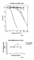
- a standard curve was constructed on a log-linear graph by plotting the B/Bo of 6 calibrators (10 to 0.01 nM) ( figure 1 ).
- the concentration of HIS-ARG-GLY-TYR:NO2-PRO-GLY-LEU-ASP-GLY containing peptides in the samples, were determined by interpolation on the calibration curve.
- the detection limit of the assay described in example 1 is calculated as the mean (M) Bo value of 21 determinations of standard A minus 3 times the standard derivation (SD) of Bo (M A -3*SD A ). For Coll2-1:NO2 D37 the detection limit was 25 pM.
- the antisera produced were tested for their specificity for His-Arg-Gly-Tyr:N02-Pro-Gly-Leu-Asp-Gly, by use of the immunoassay described in example 1.
- Native type II collagen, type I collagen, nitrated collagen type I, nitrated BSA and BSA were not able to compete with the coated His-Arg-Gly-Tyr:NO2-Pro-Gly-Leu-Asp-Gly peptide in the applied concentrations, whereas the antiserum showed weak affinity to the non-Nitrated His-Arg-Gly-Tyr-Pro-Gly-Leu-Asp-Gly sequence and nitrated collagen type II and strong affinity to the His-Arg-Gly-Tyr:NO2-Pro-Gly-Leu-Asp-Gly sequence. A lack of binding affinity has also been demonstrated with L-nitrotyrosine.
- Example 3 Coll2-1(NO2) levels are elevated in animal models of inflammatory arthritis
- CIA Lewis-rat Collagen Induced Arthritis
- joint inflammation is induced by immunization with collagen type II which provoke a severe inflammatory response to joint cartilage apparent as paw joint swelling and subsequent destruction of joints when histological analysis is performed.
- Arthritis was monitored by macroscopic scoring of swelling and redness of the paws.
- Cartilage and bone erosion was monitored by quantifying urinary levels of CartiLaps and serum levels of RatLaps, two biochemical markers of cartilage and bone resorption. These assays were performed as specified by the manufacturer (Nordic Bioscience Diagnostics, Herlev, Denmark).
- the study cohort comprised 21 female Lewis rats which are ovarectomized (OVX). At baseline, the weight was determined and the animals were anesthesised for the OVX procedure. After induction of general anesthesia with Hypnorm-Dormicum (1 part Hypnorm® + 1 part Dormicum® + 2 part sterile de-ionised water, dose 0.15-0.2 ml/100 g body weight), a standard OVX was performed. The weight of the animals was determined on a weekly basis throughout the study period. For measurements of bone and cartilage markers urine samples were obtained by either placing the animals in a metabolic cage for 30 - 60 min and awaiting spontaneous urination or by gently rubbing the belly of the animal, i.e.
- the rats were immunized with collagen type II 1 week after OVX to induce inflammatory arthritis. Each rat was immunized with 150 ⁇ g bovine collagen type II emulsified in Incomplete Freunds Adjuvant. The paws of the rats were scored daily from day 11 by visual observation of the paws. Each paw is scored on a scale 0-4 and the score for the four paws are added.
- the combined arthritis score reached a score of 10 for a given animal or if the animal developed very severe arthritis in one paw or had CIA for more that 16 days, the animal was sacrified for ethical considerations, as more severe arthritis is associated with significant pain and discomfort. The remainder of the animals were maintained for 27 days after OVX before being terminated.
- Coll2-1 (N02) level in the serum and the presence of external signs of arthritis had a very good correlation ( Fig 3 ).
- the levels of Coll2-1(NO2) were significantly elevated in the rats which developed inflammatory arthritis (sick) whereas animals which did not develop disease showed significantly lower levels of Col2-1(NO2).
- the level of collagen fragments increased before the appearance of severe arthritis symptoms implying that Coll2-1(NO2) is a predictive biomarker for inflammation induced cartilage erosion.
- Example 4 Human Articular Cartilage Explants Produce coll2-1 (NO2) in vitro.
- Articular cartilage explants are commonly used as an in vitro system to assess cartilage metabolism. Articular cartilage was obtained from adult human patients undergoing joint replacement surgery and the cartilage was excised either as cylindrical plugs (5-30 mg) or as slices (20-30 mg). The explants were cultured in 96-well plates in 200 mL serum free DMEM medium, in the presence of recombinant human IL-1 ⁇ 5 ng/mL (Sigma, St. Louis, USA), Oncostatin M 50 ng/mL (Sigma, St. Louis, USA) and human plasminogen 10 ⁇ g/mL (Sigma, St. Louis, USA). Plasminogen is a physiological MMP activator that induces collagen type II degradation.
- MMP activator APMA aminophenyl mercuric acetate, SIGMA, St Louis, USA
- Example 5 Physiological levels of Coll 2-1(NO2) in healthy men and women.
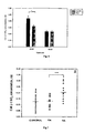
- Coll 2-1 (NO2) serum levels did not vary significantly in the investigated age interval (20 - 65 years)( Fig. 5 ).
- the comparison of peptide levels by sex showed that up to 45 years of age, Coll 2-1 (N02) concentration was higher in women than in men but the difference was only significant for the subclasses 20-25 and 26-31 years old (figure X+3).
- Coll 2-1 (NO2) level was higher in pre-menopausal women than in postmenopausal women.
- Coll2-1(NO2) level is higher in young individuals (pre-menopausal) than in older individuals ( Fig. 6 ).
- the level of these fragments is identical for both men and women in the range of 56-65 years old whereas pre-menopausal women showed significantly higher levels of the marker compared to men and postmenopausal women.
- Estrogen up-regulates production of inducible nitric oxide synthetase (iNOS) and thus levels of nitric oxide, which in turn is responsible for generation of nitro tyrosine modified proteins as quantified in the Co12-1(NO2) assay.
- iNOS inducible nitric oxide synthetase
- Example 6 RA patients have more elevated Coll2-1(NO2) levels than OA patients
- Coll 2-1 (NO2) concentration were also measured in serum samples of 14 patients with early RA. At the sampling time, these patients had not received any medication, and all had a C-reactive protein level above 5 mg/L.
- Example 7 Development of an assay specific for a nitrosylated collagen type II epitope derived from the C-telopeptide region (Coll2-2(NO2) ELISA)
- the coating buffer was 0.08 M NaHCO 3 pH 9.6.
- the saturation buffer was composed of 1.5 mM KH 2 PO 4 8 mM Na 2 HPO 4 , 2 mM KCl, 138 mM NaCl, 5 g/L bovine serum albumin (BSA), 53 g/L lactose monohydrate pH 7.2.
- the washing buffer was a solution of 25 mM Tris, 50 mM NaCl pH 7.3. The standard curve and the dilution of samples, when it was necessary, were realized in 50 mM Tris, 138 mM NaCl, 7 g/L BSA, 1ml/L Tween 20 pH 8.0.
- the dilutions of the antisera (1/80 000) and of the second antibody 1/5000) were done in Na 2 HPO 4 10 mM, KH 2 PO 4 1.5 mM, KCl 2 mM, NaCl 150 mM, EDTA 25 mM, BSA 1 % Tween 0.1% pH 7.4.
- Rabbits were injected intra-peritoneally with 1 ml of the conjugated peptides (0.5 mg/ml) emulsified in complete Freund's adjuvant. The conjugate and the adjuvant were mixed in equal volumes. Injections were repeated four times every month using the same peptide concentration that those of the first injection in incomplete Freund's adjuvant. Ten days after the last injection, the rabbits were sacrificed. Blood was collected and centrifuged for 10 min at 2500 rpm at 4°C. The supernatant was kept and stored at -20°C. At each bleeding, antisera were screened by titration experiment for the presence of anti- GGGLQY(N02)MRA antibody. The antisera with the highest titers were selected for the following experiments.
- D33 antibody is highly specific for nitrosylated Coll2-2 fragment and does not react with the same fragment non-nitrosylated Coll2-2 or with any other fragment slightly similar in the sequence (Coll2-1) or completely different sequences (BSA NO2).
- BSA NO2 completely different sequences
- the figured showed also that the chosen sequence of Coll2-2 against which D33 was raised cannot be recognized when it is still “included” in the full length protein (Coll II (NO2)).
- the nitrosylated sequence is recognized only in the form of free fragments ( Fig. 8 ).
- intra-assay CV In order to address the question of correct and reproducible results measured in this assay intra-assay CV was evaluated by measurement of the same urine samples in different emplacement in the same plate. Table 1: Evaluation of intra-assay CV (%) of Coll2-2 N02) assay. Coll 2-2 (NO2) concentration CV intra-assay (%) (Mean ⁇ SD, nM) Urine 1 1.56 ⁇ 0.15 9.1 Urine 2 2.86 ⁇ 0.26 9.1 The intra-assay showed that the samples could be measured in a good reproducible manner (Good standard variation of samples mean concentration of Coll2-2(NO2) fragments). The CV on samples measurement for both samples used here were below 10% witnessing also of the assay precision.
Landscapes
- Health & Medical Sciences (AREA)
- Life Sciences & Earth Sciences (AREA)
- Engineering & Computer Science (AREA)
- Chemical & Material Sciences (AREA)
- Immunology (AREA)
- Biomedical Technology (AREA)
- Molecular Biology (AREA)
- Hematology (AREA)
- Urology & Nephrology (AREA)
- Medicinal Chemistry (AREA)
- Physics & Mathematics (AREA)
- General Health & Medical Sciences (AREA)
- Biochemistry (AREA)
- Analytical Chemistry (AREA)
- Microbiology (AREA)
- Biotechnology (AREA)
- Food Science & Technology (AREA)
- Pathology (AREA)
- Proteomics, Peptides & Aminoacids (AREA)
- Cell Biology (AREA)
- General Physics & Mathematics (AREA)
- Organic Chemistry (AREA)
- Biophysics (AREA)
- Bioinformatics & Cheminformatics (AREA)
- Genetics & Genomics (AREA)
- Bioinformatics & Computational Biology (AREA)
- Rehabilitation Therapy (AREA)
- Rheumatology (AREA)
- Neurology (AREA)
- Neurosurgery (AREA)
- Peptides Or Proteins (AREA)
- Investigating Or Analysing Biological Materials (AREA)
- Measuring Or Testing Involving Enzymes Or Micro-Organisms (AREA)
- Investigating Or Analyzing Non-Biological Materials By The Use Of Chemical Means (AREA)
- Agricultural Chemicals And Associated Chemicals (AREA)
- Dental Preparations (AREA)
Claims (19)
- Un procédé pour effectuer un immunoessai qualitatif ou quantitatif pour l'oxydation de protéines, comprenant la détection dans un échantillon d'une séquence d'acides aminés qui est caractéristique d'une protéine spécifique présente dans le tissu articulaire et qui contient un ou plusieurs résidus d'acides aminés aromatiques sous forme nitrée, dans lequel l'essai comprend la détection de la liaison d'un partenaire de liaison immunologique qui est immunoréactif avec ladite forme nitrée dudit résidu d'acide aminé aromatique.
- Un procédé selon la revendication 1, dans lequel ledit partenaire de liaison immunologique est spécifiquement réactif avec ladite forme nitrée dudit résidu d'acide aminé aromatique dans le contexte de ladite séquence d'acides aminés qui est caractéristique d'une protéine spécifique.
- Un procédé selon la revendication 1, dans lequel ledit partenaire de liaison immunologique est réactif avec ladite forme nitrée dudit résidu d'acide aminé aromatique d'une manière indépendante du contexte et ledit essai comprend la détection de la liaison à la fois dudit partenaire de liaison immunologique et d'un second partenaire de liaison immunologique contres des séquences d'acides aminés nitrées dans un format de sandwich, dans lequel ledit second partenaire de liaison immunologique a une spécificité de liaison pour une séquence d'acides aminés qui est caractéristique de ladite protéine spécifique.
- Un procédé selon l'une des revendications précédentes, dans lequel ladite protéine est le collagène de type I, II, III, VI, IX ou XI, l'aggrecane, une protéine de liaison du cartilage, une protéine oligomèrique du cartilage ou une protéine de la couche intermédiaire du cartilage.
- Un procédé selon l'une des revendications précédentes, dans lequel ledit ou lesdits résidus d'acides aminés aromatiques nitrés est/sont la nitrotyrosine ou le nitrotryptophane.
- Un procédé selon la revendication 5, dans lequel la séquence d'acides aminés qui est détectée comprend la séquence HRGYPGLDG dans laquelle le résidu d'acide aminé Y est la tyrosine nitrée ou est compris à l'intérieur de ladite séquence et comprend ladite tyrosine nitrée.
- Un procédé selon la revendication 5, dans lequel la séquence d'acides aminés qui est détectée comprend la séquence LQYMRA dans laquelle le résidu d'acide aminé Y est la tyrosine nitrée ou est compris à l'intérieur de ladite séquence et comprend ladite tyrosine nitrée.
- Un partenaire de liaison immunologique spécifiquement réactif avec la forme nitrée d'un résidu d'acide aminé aromatique dans le contexte d'une séquence d'acides aminés qui est caractéristique d'une protéine spécifique ayant une spécificité de liaison pour un épitope contenu dans la séquence d'acides aminés HRGY:NO2PGLDG, lequel épitope contient le résidu d'acide aminé Y:NO2 et est caractéristique du collagène de type II.
- Un partenaire de liaison immunologique selon la revendication 8, lequel est un anticorps dirigé contre un peptide ayant la séquence (Xaa)mHRGY:NO2PGLDG(Xaa)n, où X désigne n'importe quel acide aminé ou un dérivé de celui-ci et m et n sont des nombres entiers indépendants de 1 à 10.
- Un partenaire de liaison immunologique spécifiquement réactif avec la forme nitrée d'un résidu d'acide aminé aromatique dans le contexte d'une séquence d'acides aminés qui est caractéristique d'une protéine spécifique, ayant une spécificité de liaison pour un épitope contenu dans la séquence d'acides aminés LQY:NO2MRA, lequel épitope contient le résidu d'acide aminé Y:NO2 et est caractéristique du collagène de type II.
- Un partenaire de liaison immunologique selon la revendication 10, lequel est un anticorps dirigé contre un peptide ayant la séquence (Xaa)mLQY:NO2MRA(Xaa)n, où X désigne n'importe quel acide aminé ou un dérivé de celui-ci et m et n sont des nombres entiers indépendants de 1 à 10.
- Un partenaire de liaison immunologique selon l'une des revendications 8 à 11, lequel est un anticorps monoclonal.
- Une lignée cellulaire produisant un anticorps monoclonal selon la revendication 12.
- Un procédé pour l'étude de l'existence ou de l'étendue d'un dommage oxydatif comprenant la mesure dans un échantillon biologique des quantités relatives de formes nitrée et non nitrée d'une séquence d'acides aminés qui est caractéristique d'une protéine spécifique provenant de la matrice du cartilage et qui contient un ou plusieurs résidus d'acides aminés aromatiques aptes à être nitrés, en mettant en contact un échantillon biologique provenant d'un individu ou une partie d'un tel échantillon avec un premier partenaire de liaison immunologique qui se lie à ladite forme nitrée de ladite séquence d'acides aminés et en déterminant quantitativement ladite liaison ; en mettant en contact l'échantillon biologique ou une partie de celui-ci avec un second partenaire de liaison immunologique qui se lie à ladite forme non nitrée de ladite séquence d'acides aminés et en déterminant quantitativement ladite liaison ; en déterminant un rapport entre lesdites déterminations pour fournir un rapport des quantités relatives desdites formes nitrée et non nitrée de ladite séquence présentes dans l'échantillon ; et en comparant la valeur mesurée avec des valeurs caractéristiques d'individus en bonne santé ou d'individus ayant une pathologie connue.
- Un procédé selon la revendication 14, dans lequel ledit premier partenaire de liaison immunologique est l'une des revendications 8 à 12.
- Un kit destiné à être utilisé dans la mise en oeuvre d'un procédé selon l'une des revendications 1 à 7 ou 14 et 15 et comprenant : un partenaire de liaison immunologique qui se lie de façon spécifique à la forme nitrée d'un résidu d'acide aminé aromatique dans le contexte d'une séquence d'acides aminés qui est caractéristique d'une protéine spécifique présente dans le tissu articulaire ; et des moyens pour détecter la liaison dudit partenaire de liaison avec ladite forme nitrée dudit résidu d'acide aminé aromatique.
- Un kit selon la revendication 16, dans lequel le partenaire de liaison immunologique est l'une des revendications 8 à 12.
- Un kit selon la revendication 16 ou la revendication 17, comprenant un partenaire de liaison immunologique qui est réactif avec ladite séquence d'acides aminés sous forme non nitrée.
- Un kit selon l'une des revendications 16 à 18, comprenant un peptide réactif avec ledit partenaire de liaison immunologique.
Applications Claiming Priority (5)
| Application Number | Priority Date | Filing Date | Title |
|---|---|---|---|
| US36392502P | 2002-03-13 | 2002-03-13 | |
| DK200200381 | 2002-03-13 | ||
| DKPA200200381 | 2002-03-13 | ||
| US363925P | 2002-03-13 | ||
| PCT/EP2003/002559 WO2003076946A2 (fr) | 2002-03-13 | 2003-03-12 | Detection de marqueurs nitres specifiques |
Publications (2)
| Publication Number | Publication Date |
|---|---|
| EP1488240A2 EP1488240A2 (fr) | 2004-12-22 |
| EP1488240B1 true EP1488240B1 (fr) | 2009-10-14 |
Family
ID=27806430
Family Applications (1)
| Application Number | Title | Priority Date | Filing Date |
|---|---|---|---|
| EP03711987A Expired - Lifetime EP1488240B1 (fr) | 2002-03-13 | 2003-03-12 | Detection de marqueurs nitres specifiques |
Country Status (8)
| Country | Link |
|---|---|
| US (4) | US7393649B2 (fr) |
| EP (1) | EP1488240B1 (fr) |
| AT (1) | ATE445842T1 (fr) |
| AU (1) | AU2003218743A1 (fr) |
| DE (1) | DE60329663D1 (fr) |
| DK (1) | DK1488240T3 (fr) |
| ES (1) | ES2335095T3 (fr) |
| WO (1) | WO2003076946A2 (fr) |
Families Citing this family (8)
| Publication number | Priority date | Publication date | Assignee | Title |
|---|---|---|---|---|
| ES2335095T3 (es) | 2002-03-13 | 2010-03-22 | Universite De Liege | Deteccion de marcadores nitrados especificos. |
| US8852879B2 (en) * | 2004-04-29 | 2014-10-07 | The Children's Hospital Of Philadelphia | Materials and methods for the detection of nitrated fibrinogen |
| US20050244905A1 (en) * | 2004-04-29 | 2005-11-03 | Harry Ischiropoulos | Nitrated fibrinogen as a marker for coronary artery disease and rapid blood clot formation |
| FR2908413B1 (fr) * | 2006-11-15 | 2009-03-06 | Synarc Soc Par Actions Simplif | Marqueur specifique d'une degradation oxydative de tissus contenant du collagene de type iii,moyens et methodes,kits de diagnostic de suivi ou de pronostic des pathologies ciblees par ce marqueur. |
| FR2926369A1 (fr) * | 2008-01-10 | 2009-07-17 | Univ Joseph Fourier | Utilisation d'une proteine ou d'une sequence peptidique nitree pour la mise en oeuvre d'une methode de diagnostic |
| US10175250B2 (en) * | 2012-06-11 | 2019-01-08 | Gabriela Venturini Da Silva | Nitrated cardiac troponin I as a biomarker of cardiac ischemia |
| CA2832215A1 (fr) | 2013-07-18 | 2015-01-18 | Northern Sydney Local Health District | Biomarqueurs de contrainte d'oxydation |
| JP2021155362A (ja) * | 2020-03-27 | 2021-10-07 | 株式会社ナリス化粧品 | ペルオキシナイトライト活性阻害方法および、ペルオキシナイトライト活性阻害剤のスクリーニング方法 |
Citations (2)
| Publication number | Priority date | Publication date | Assignee | Title |
|---|---|---|---|---|
| WO1998049558A1 (fr) * | 1997-04-28 | 1998-11-05 | Institut National De La Sante Et De La Recherche Medicale (Inserm) | Methodes pour la detection de pathologies auto-immunes |
| WO2001084160A2 (fr) * | 2000-05-02 | 2001-11-08 | Mitokor | Determination proteomique des modifications de nitrotyrosine proteique par spectrometrie de masse |
Family Cites Families (7)
| Publication number | Priority date | Publication date | Assignee | Title |
|---|---|---|---|---|
| US5702909A (en) * | 1987-11-06 | 1997-12-30 | Washington Research Foundation | Methods of detecting collagen type II degradation in vivo |
| US6132976A (en) * | 1992-12-04 | 2000-10-17 | Shriners Hospitals For Children | Immunoassays for the measurement of collagen denaturation and cleavage in cartilage |
| WO1996004311A1 (fr) * | 1994-08-03 | 1996-02-15 | University Of Alabama At Birmingham Research Foundation | Anticorps monoclonal dirige contre la nitrotyrosine, procedes de diagnostic et procedes therapeutiques |
| FR2757864B1 (fr) * | 1996-12-30 | 1999-03-19 | Centre Nat Rech Scient | Anticorps reconnaissant specifiquement une proteine nitrosylee, leur procede de preparation, leur utilisation therapeutique et diagnostique |
| GB9928052D0 (en) | 1999-11-26 | 2000-01-26 | Osteometer Biotech As | Assay of isomerised and/or optically inverted proteins and protein fragments |
| US6812212B2 (en) * | 2000-07-28 | 2004-11-02 | Hyesook Kim | Assessment of oxidant stress in vitro and in vivo |
| ES2335095T3 (es) * | 2002-03-13 | 2010-03-22 | Universite De Liege | Deteccion de marcadores nitrados especificos. |
-
2003
- 2003-03-12 ES ES03711987T patent/ES2335095T3/es not_active Expired - Lifetime
- 2003-03-12 DE DE60329663T patent/DE60329663D1/de not_active Expired - Lifetime
- 2003-03-12 AT AT03711987T patent/ATE445842T1/de not_active IP Right Cessation
- 2003-03-12 DK DK03711987.2T patent/DK1488240T3/da active
- 2003-03-12 US US10/507,479 patent/US7393649B2/en not_active Expired - Fee Related
- 2003-03-12 WO PCT/EP2003/002559 patent/WO2003076946A2/fr not_active Ceased
- 2003-03-12 EP EP03711987A patent/EP1488240B1/fr not_active Expired - Lifetime
- 2003-03-12 AU AU2003218743A patent/AU2003218743A1/en not_active Abandoned
-
2008
- 2008-06-27 US US12/216,063 patent/US7915001B2/en not_active Expired - Fee Related
-
2011
- 2011-01-20 US US13/010,357 patent/US20110223684A1/en not_active Abandoned
- 2011-08-30 US US13/221,014 patent/US8329874B2/en not_active Expired - Fee Related
Patent Citations (2)
| Publication number | Priority date | Publication date | Assignee | Title |
|---|---|---|---|---|
| WO1998049558A1 (fr) * | 1997-04-28 | 1998-11-05 | Institut National De La Sante Et De La Recherche Medicale (Inserm) | Methodes pour la detection de pathologies auto-immunes |
| WO2001084160A2 (fr) * | 2000-05-02 | 2001-11-08 | Mitokor | Determination proteomique des modifications de nitrotyrosine proteique par spectrometrie de masse |
Also Published As
| Publication number | Publication date |
|---|---|
| US20120010389A1 (en) | 2012-01-12 |
| US8329874B2 (en) | 2012-12-11 |
| US7915001B2 (en) | 2011-03-29 |
| ES2335095T3 (es) | 2010-03-22 |
| ATE445842T1 (de) | 2009-10-15 |
| DK1488240T3 (da) | 2010-03-01 |
| AU2003218743A1 (en) | 2003-09-22 |
| US20110223684A1 (en) | 2011-09-15 |
| AU2003218743A8 (en) | 2003-09-22 |
| US20090203040A1 (en) | 2009-08-13 |
| EP1488240A2 (fr) | 2004-12-22 |
| WO2003076946A3 (fr) | 2004-03-25 |
| US7393649B2 (en) | 2008-07-01 |
| WO2003076946A2 (fr) | 2003-09-18 |
| US20050176081A1 (en) | 2005-08-11 |
| DE60329663D1 (de) | 2009-11-26 |
Similar Documents
| Publication | Publication Date | Title |
|---|---|---|
| US8329874B2 (en) | Detection of specific nitrated markers | |
| JP6095702B2 (ja) | 線維症バイオマーカアッセイ | |
| JP3423720B2 (ja) | 体液中のコラーゲン断片を測定する方法、該方法を実施するためのテストキット及び手段、並びにコラーゲンの代謝に関連する疾患の存在を診断するために該方法を使用する方法・用途 | |
| Deberg et al. | New serum biochemical markers (Coll 2-1 and Coll 2-1 NO2) for studying oxidative-related type II collagen network degradation in patients with osteoarthritis and rheumatoid arthritis | |
| JPH11502622A (ja) | 体液中のタンパク質断片の検定方法 | |
| Srivastava et al. | A urine midmolecule osteocalcin assay shows higher discriminatory power than a serum midmolecule osteocalcin assay during short-term alendronate treatment of osteoporotic patients | |
| JPH0780913B2 (ja) | 完全無傷なプラコラーゲンペプチド(▲iii▼型)およびプロコラーゲン(▲iii▼型)の選択的な免疫学的測定法 | |
| JPH10507266A (ja) | 体液中のコラーゲンの断片化パターンの評価とコラーゲンの代謝に関連する疾患の診断 | |
| CN101466732A (zh) | Iv型胶原样免疫反应性多肽 | |
| US7728112B2 (en) | Method for monitoring collagen type II degradation in cartilage | |
| EP1766411B1 (fr) | Methode de determination d'un processus de degradation tissulaire par detection de neoepitopes comp | |
| EP2086996B1 (fr) | Marqueur specifique d'une degradation oxydative de tissus contenant du collagene de type iii, moyens, methodes et kits de diagnostic, de suivi ou de pronostic des pathologies ciblees par ce marqueur | |
| JPH0785085B2 (ja) | フイブロネクチン測定試薬 | |
| JP2002518676A (ja) | 軟骨吸収アッセイ | |
| JPH03108665A (ja) | 生物学的液体中の4型コラーゲンペプチドの免疫学的な測定法及び測定キット | |
| KR20100060208A (ko) | 페난트렌 오염 검출을 위한 마커 및 키트 |
Legal Events
| Date | Code | Title | Description |
|---|---|---|---|
| PUAI | Public reference made under article 153(3) epc to a published international application that has entered the european phase |
Free format text: ORIGINAL CODE: 0009012 |
|
| 17P | Request for examination filed |
Effective date: 20041013 |
|
| AK | Designated contracting states |
Kind code of ref document: A2 Designated state(s): AT BE BG CH CY CZ DE DK EE ES FI FR GB GR HU IE IT LI LU MC NL PT RO SE SI SK TR |
|
| AX | Request for extension of the european patent |
Extension state: AL LT LV MK |
|
| RAP1 | Party data changed (applicant data changed or rights of an application transferred) |
Owner name: UNIVERSITE DE LIEGE |
|
| 17Q | First examination report despatched |
Effective date: 20061130 |
|
| GRAP | Despatch of communication of intention to grant a patent |
Free format text: ORIGINAL CODE: EPIDOSNIGR1 |
|
| GRAS | Grant fee paid |
Free format text: ORIGINAL CODE: EPIDOSNIGR3 |
|
| GRAA | (expected) grant |
Free format text: ORIGINAL CODE: 0009210 |
|
| AK | Designated contracting states |
Kind code of ref document: B1 Designated state(s): AT BE BG CH CY CZ DE DK EE ES FI FR GB GR HU IE IT LI LU MC NL PT RO SE SI SK TR |
|
| REG | Reference to a national code |
Ref country code: GB Ref legal event code: FG4D |
|
| REG | Reference to a national code |
Ref country code: CH Ref legal event code: EP |
|
| REG | Reference to a national code |
Ref country code: IE Ref legal event code: FG4D |
|
| REF | Corresponds to: |
Ref document number: 60329663 Country of ref document: DE Date of ref document: 20091126 Kind code of ref document: P |
|
| REG | Reference to a national code |
Ref country code: SE Ref legal event code: TRGR |
|
| REG | Reference to a national code |
Ref country code: DK Ref legal event code: T3 |
|
| REG | Reference to a national code |
Ref country code: ES Ref legal event code: FG2A Ref document number: 2335095 Country of ref document: ES Kind code of ref document: T3 |
|
| NLV1 | Nl: lapsed or annulled due to failure to fulfill the requirements of art. 29p and 29m of the patents act | ||
| PG25 | Lapsed in a contracting state [announced via postgrant information from national office to epo] |
Ref country code: FI Free format text: LAPSE BECAUSE OF FAILURE TO SUBMIT A TRANSLATION OF THE DESCRIPTION OR TO PAY THE FEE WITHIN THE PRESCRIBED TIME-LIMIT Effective date: 20091014 Ref country code: PT Free format text: LAPSE BECAUSE OF FAILURE TO SUBMIT A TRANSLATION OF THE DESCRIPTION OR TO PAY THE FEE WITHIN THE PRESCRIBED TIME-LIMIT Effective date: 20100215 |
|
| PG25 | Lapsed in a contracting state [announced via postgrant information from national office to epo] |
Ref country code: SI Free format text: LAPSE BECAUSE OF FAILURE TO SUBMIT A TRANSLATION OF THE DESCRIPTION OR TO PAY THE FEE WITHIN THE PRESCRIBED TIME-LIMIT Effective date: 20091014 |
|
| PG25 | Lapsed in a contracting state [announced via postgrant information from national office to epo] |
Ref country code: AT Free format text: LAPSE BECAUSE OF FAILURE TO SUBMIT A TRANSLATION OF THE DESCRIPTION OR TO PAY THE FEE WITHIN THE PRESCRIBED TIME-LIMIT Effective date: 20091014 |
|
| PG25 | Lapsed in a contracting state [announced via postgrant information from national office to epo] |
Ref country code: EE Free format text: LAPSE BECAUSE OF FAILURE TO SUBMIT A TRANSLATION OF THE DESCRIPTION OR TO PAY THE FEE WITHIN THE PRESCRIBED TIME-LIMIT Effective date: 20091014 Ref country code: BG Free format text: LAPSE BECAUSE OF FAILURE TO SUBMIT A TRANSLATION OF THE DESCRIPTION OR TO PAY THE FEE WITHIN THE PRESCRIBED TIME-LIMIT Effective date: 20100114 Ref country code: RO Free format text: LAPSE BECAUSE OF FAILURE TO SUBMIT A TRANSLATION OF THE DESCRIPTION OR TO PAY THE FEE WITHIN THE PRESCRIBED TIME-LIMIT Effective date: 20091014 |
|
| PLBE | No opposition filed within time limit |
Free format text: ORIGINAL CODE: 0009261 |
|
| STAA | Information on the status of an ep patent application or granted ep patent |
Free format text: STATUS: NO OPPOSITION FILED WITHIN TIME LIMIT |
|
| PG25 | Lapsed in a contracting state [announced via postgrant information from national office to epo] |
Ref country code: SK Free format text: LAPSE BECAUSE OF FAILURE TO SUBMIT A TRANSLATION OF THE DESCRIPTION OR TO PAY THE FEE WITHIN THE PRESCRIBED TIME-LIMIT Effective date: 20091014 Ref country code: CZ Free format text: LAPSE BECAUSE OF FAILURE TO SUBMIT A TRANSLATION OF THE DESCRIPTION OR TO PAY THE FEE WITHIN THE PRESCRIBED TIME-LIMIT Effective date: 20091014 |
|
| 26N | No opposition filed |
Effective date: 20100715 |
|
| PG25 | Lapsed in a contracting state [announced via postgrant information from national office to epo] |
Ref country code: MC Free format text: LAPSE BECAUSE OF NON-PAYMENT OF DUE FEES Effective date: 20100331 Ref country code: GR Free format text: LAPSE BECAUSE OF FAILURE TO SUBMIT A TRANSLATION OF THE DESCRIPTION OR TO PAY THE FEE WITHIN THE PRESCRIBED TIME-LIMIT Effective date: 20100115 |
|
| REG | Reference to a national code |
Ref country code: CH Ref legal event code: PL |
|
| PG25 | Lapsed in a contracting state [announced via postgrant information from national office to epo] |
Ref country code: IE Free format text: LAPSE BECAUSE OF NON-PAYMENT OF DUE FEES Effective date: 20100312 |
|
| PG25 | Lapsed in a contracting state [announced via postgrant information from national office to epo] |
Ref country code: LI Free format text: LAPSE BECAUSE OF NON-PAYMENT OF DUE FEES Effective date: 20100331 Ref country code: CH Free format text: LAPSE BECAUSE OF NON-PAYMENT OF DUE FEES Effective date: 20100331 |
|
| PG25 | Lapsed in a contracting state [announced via postgrant information from national office to epo] |
Ref country code: CY Free format text: LAPSE BECAUSE OF FAILURE TO SUBMIT A TRANSLATION OF THE DESCRIPTION OR TO PAY THE FEE WITHIN THE PRESCRIBED TIME-LIMIT Effective date: 20091014 |
|
| PG25 | Lapsed in a contracting state [announced via postgrant information from national office to epo] |
Ref country code: LU Free format text: LAPSE BECAUSE OF NON-PAYMENT OF DUE FEES Effective date: 20100312 Ref country code: HU Free format text: LAPSE BECAUSE OF FAILURE TO SUBMIT A TRANSLATION OF THE DESCRIPTION OR TO PAY THE FEE WITHIN THE PRESCRIBED TIME-LIMIT Effective date: 20100415 Ref country code: NL Free format text: LAPSE BECAUSE OF FAILURE TO SUBMIT A TRANSLATION OF THE DESCRIPTION OR TO PAY THE FEE WITHIN THE PRESCRIBED TIME-LIMIT Effective date: 20091014 |
|
| PG25 | Lapsed in a contracting state [announced via postgrant information from national office to epo] |
Ref country code: TR Free format text: LAPSE BECAUSE OF FAILURE TO SUBMIT A TRANSLATION OF THE DESCRIPTION OR TO PAY THE FEE WITHIN THE PRESCRIBED TIME-LIMIT Effective date: 20091014 |
|
| REG | Reference to a national code |
Ref country code: FR Ref legal event code: PLFP Year of fee payment: 14 |
|
| REG | Reference to a national code |
Ref country code: FR Ref legal event code: PLFP Year of fee payment: 15 |
|
| REG | Reference to a national code |
Ref country code: FR Ref legal event code: PLFP Year of fee payment: 16 |
|
| PGFP | Annual fee paid to national office [announced via postgrant information from national office to epo] |
Ref country code: GB Payment date: 20190320 Year of fee payment: 17 Ref country code: FR Payment date: 20190322 Year of fee payment: 17 Ref country code: DE Payment date: 20190321 Year of fee payment: 17 Ref country code: IT Payment date: 20190325 Year of fee payment: 17 |
|
| PGFP | Annual fee paid to national office [announced via postgrant information from national office to epo] |
Ref country code: BE Payment date: 20190320 Year of fee payment: 17 Ref country code: DK Payment date: 20190322 Year of fee payment: 17 Ref country code: SE Payment date: 20190320 Year of fee payment: 17 |
|
| PGFP | Annual fee paid to national office [announced via postgrant information from national office to epo] |
Ref country code: ES Payment date: 20190418 Year of fee payment: 17 |
|
| REG | Reference to a national code |
Ref country code: DE Ref legal event code: R119 Ref document number: 60329663 Country of ref document: DE |
|
| REG | Reference to a national code |
Ref country code: DK Ref legal event code: EBP Effective date: 20200331 |
|
| REG | Reference to a national code |
Ref country code: BE Ref legal event code: MM Effective date: 20200331 |
|
| PG25 | Lapsed in a contracting state [announced via postgrant information from national office to epo] |
Ref country code: FR Free format text: LAPSE BECAUSE OF NON-PAYMENT OF DUE FEES Effective date: 20200331 Ref country code: DE Free format text: LAPSE BECAUSE OF NON-PAYMENT OF DUE FEES Effective date: 20201001 Ref country code: SE Free format text: LAPSE BECAUSE OF NON-PAYMENT OF DUE FEES Effective date: 20200313 |
|
| PG25 | Lapsed in a contracting state [announced via postgrant information from national office to epo] |
Ref country code: BE Free format text: LAPSE BECAUSE OF NON-PAYMENT OF DUE FEES Effective date: 20200331 |
|
| GBPC | Gb: european patent ceased through non-payment of renewal fee |
Effective date: 20200312 |
|
| PG25 | Lapsed in a contracting state [announced via postgrant information from national office to epo] |
Ref country code: GB Free format text: LAPSE BECAUSE OF NON-PAYMENT OF DUE FEES Effective date: 20200312 Ref country code: DK Free format text: LAPSE BECAUSE OF NON-PAYMENT OF DUE FEES Effective date: 20200331 |
|
| REG | Reference to a national code |
Ref country code: ES Ref legal event code: FD2A Effective date: 20210728 |
|
| PG25 | Lapsed in a contracting state [announced via postgrant information from national office to epo] |
Ref country code: IT Free format text: LAPSE BECAUSE OF NON-PAYMENT OF DUE FEES Effective date: 20200312 |
|
| PG25 | Lapsed in a contracting state [announced via postgrant information from national office to epo] |
Ref country code: ES Free format text: LAPSE BECAUSE OF NON-PAYMENT OF DUE FEES Effective date: 20200313 |