EP1493034B1 - Verfahren zur diagnose von entzündungserkrankungen und infektionen mittels bestimmung von lasp-1/ lap-1 - Google Patents
Verfahren zur diagnose von entzündungserkrankungen und infektionen mittels bestimmung von lasp-1/ lap-1 Download PDFInfo
- Publication number
- EP1493034B1 EP1493034B1 EP03722485A EP03722485A EP1493034B1 EP 1493034 B1 EP1493034 B1 EP 1493034B1 EP 03722485 A EP03722485 A EP 03722485A EP 03722485 A EP03722485 A EP 03722485A EP 1493034 B1 EP1493034 B1 EP 1493034B1
- Authority
- EP
- European Patent Office
- Prior art keywords
- lasp
- sepsis
- protein
- immunoreactivity
- determination
- Prior art date
- Legal status (The legal status is an assumption and is not a legal conclusion. Google has not performed a legal analysis and makes no representation as to the accuracy of the status listed.)
- Expired - Lifetime
Links
- 102100035118 LIM and SH3 domain protein 1 Human genes 0.000 title claims abstract description 158
- 238000000034 method Methods 0.000 title claims abstract description 38
- 208000015181 infectious disease Diseases 0.000 title claims abstract description 27
- 208000027866 inflammatory disease Diseases 0.000 title abstract description 7
- 101710162163 LIM and SH3 domain protein 1 Proteins 0.000 claims abstract description 156
- 206010040047 Sepsis Diseases 0.000 claims abstract description 87
- 230000009885 systemic effect Effects 0.000 claims abstract description 20
- 238000003745 diagnosis Methods 0.000 claims abstract description 12
- 238000004393 prognosis Methods 0.000 claims abstract description 3
- 108090000623 proteins and genes Proteins 0.000 claims description 71
- 102000004169 proteins and genes Human genes 0.000 claims description 66
- 108090000765 processed proteins & peptides Proteins 0.000 claims description 46
- 150000001413 amino acids Chemical class 0.000 claims description 40
- 102100027421 Heat shock cognate 71 kDa protein Human genes 0.000 claims description 30
- 108090000922 TNF receptor-associated factor 3 Proteins 0.000 claims description 30
- 239000012634 fragment Substances 0.000 claims description 27
- 108010048233 Procalcitonin Proteins 0.000 claims description 25
- CWCXERYKLSEGEZ-KDKHKZEGSA-N procalcitonin Chemical compound C([C@@H](C(=O)N1CCC[C@H]1C(=O)N[C@@H](CCC(N)=O)C(=O)N[C@H](C(=O)N[C@@H](C)C(=O)N[C@@H]([C@@H](C)CC)C(=O)NCC(=O)N[C@@H](C(C)C)C(=O)NCC(=O)N[C@@H](C)C(=O)N1[C@@H](CCC1)C(=O)NCC(O)=O)[C@@H](C)O)NC(=O)[C@@H](NC(=O)[C@H](CC=1NC=NC=1)NC(=O)[C@H](CC=1C=CC=CC=1)NC(=O)[C@H](CCCCN)NC(=O)[C@H](CC(N)=O)NC(=O)[C@H](CC=1C=CC=CC=1)NC(=O)[C@H](CC(O)=O)NC(=O)[C@H](CCC(N)=O)NC(=O)[C@@H](NC(=O)[C@H](CC=1C=CC(O)=CC=1)NC(=O)[C@@H](NC(=O)CNC(=O)[C@H](CC(C)C)NC(=O)[C@H](CCSC)NC(=O)[C@H]1NC(=O)[C@H]([C@@H](C)O)NC(=O)[C@H](CO)NC(=O)[C@H](CC(C)C)NC(=O)[C@H](CC(N)=O)NC(=O)CNC(=O)[C@@H](N)CSSC1)[C@@H](C)O)[C@@H](C)O)[C@@H](C)O)C1=CC=CC=C1 CWCXERYKLSEGEZ-KDKHKZEGSA-N 0.000 claims description 25
- 102000004196 processed proteins & peptides Human genes 0.000 claims description 22
- 238000001514 detection method Methods 0.000 claims description 18
- 210000002966 serum Anatomy 0.000 claims description 16
- 208000024827 Alzheimer disease Diseases 0.000 claims description 15
- 238000003556 assay Methods 0.000 claims description 12
- 238000003018 immunoassay Methods 0.000 claims description 11
- 230000003053 immunization Effects 0.000 claims description 10
- 238000002649 immunization Methods 0.000 claims description 10
- 239000003153 chemical reaction reagent Substances 0.000 claims description 5
- 238000011156 evaluation Methods 0.000 claims description 5
- 108010074051 C-Reactive Protein Proteins 0.000 claims description 4
- 102100032752 C-reactive protein Human genes 0.000 claims description 4
- 102000007434 glycine N-acyltransferase Human genes 0.000 claims description 4
- 108020005567 glycine N-acyltransferase Proteins 0.000 claims description 4
- 102000014914 Carrier Proteins Human genes 0.000 claims description 3
- 238000005516 engineering process Methods 0.000 claims description 3
- 108010078791 Carrier Proteins Proteins 0.000 claims description 2
- 102100022905 Keratin, type II cytoskeletal 1 Human genes 0.000 claims description 2
- 101710194922 Keratin, type II cytoskeletal 1 Proteins 0.000 claims description 2
- 102000011782 Keratins Human genes 0.000 claims description 2
- 108010076876 Keratins Proteins 0.000 claims description 2
- 101000859568 Methanobrevibacter smithii (strain ATCC 35061 / DSM 861 / OCM 144 / PS) Carbamoyl-phosphate synthase Proteins 0.000 claims description 2
- 238000004590 computer program Methods 0.000 claims description 2
- 238000000338 in vitro Methods 0.000 claims description 2
- 238000012544 monitoring process Methods 0.000 claims description 2
- 238000002560 therapeutic procedure Methods 0.000 claims description 2
- 206010061216 Infarction Diseases 0.000 claims 2
- 230000000747 cardiac effect Effects 0.000 claims 2
- 230000007574 infarction Effects 0.000 claims 2
- 239000013060 biological fluid Substances 0.000 abstract description 3
- 230000000694 effects Effects 0.000 abstract 1
- 235000018102 proteins Nutrition 0.000 description 63
- 230000004054 inflammatory process Effects 0.000 description 39
- 206010061218 Inflammation Diseases 0.000 description 31
- 241000282414 Homo sapiens Species 0.000 description 23
- 239000000047 product Substances 0.000 description 19
- 235000001014 amino acid Nutrition 0.000 description 18
- 239000000499 gel Substances 0.000 description 16
- 239000000126 substance Substances 0.000 description 15
- 210000004556 brain Anatomy 0.000 description 14
- 241001465754 Metazoa Species 0.000 description 12
- 230000027455 binding Effects 0.000 description 12
- 208000010125 myocardial infarction Diseases 0.000 description 11
- FAPWRFPIFSIZLT-UHFFFAOYSA-M Sodium chloride Chemical compound [Na+].[Cl-] FAPWRFPIFSIZLT-UHFFFAOYSA-M 0.000 description 10
- 210000005013 brain tissue Anatomy 0.000 description 10
- 210000004027 cell Anatomy 0.000 description 10
- 201000010099 disease Diseases 0.000 description 10
- 208000037265 diseases, disorders, signs and symptoms Diseases 0.000 description 10
- 239000002158 endotoxin Substances 0.000 description 10
- 238000001502 gel electrophoresis Methods 0.000 description 10
- 230000002757 inflammatory effect Effects 0.000 description 10
- 241000282520 Papio Species 0.000 description 9
- 210000002381 plasma Anatomy 0.000 description 9
- 210000001519 tissue Anatomy 0.000 description 9
- 108020004635 Complementary DNA Proteins 0.000 description 8
- 241000288906 Primates Species 0.000 description 8
- 239000000090 biomarker Substances 0.000 description 8
- 239000000700 radioactive tracer Substances 0.000 description 8
- 230000001225 therapeutic effect Effects 0.000 description 8
- 102000000395 SH3 domains Human genes 0.000 description 7
- 108050008861 SH3 domains Proteins 0.000 description 7
- 238000000539 two dimensional gel electrophoresis Methods 0.000 description 7
- 241001494479 Pecora Species 0.000 description 6
- 229920006008 lipopolysaccharide Polymers 0.000 description 6
- 238000004949 mass spectrometry Methods 0.000 description 6
- 238000005259 measurement Methods 0.000 description 6
- 230000026731 phosphorylation Effects 0.000 description 6
- 238000006366 phosphorylation reaction Methods 0.000 description 6
- 239000000243 solution Substances 0.000 description 6
- 102000002151 Microfilament Proteins Human genes 0.000 description 5
- 108010040897 Microfilament Proteins Proteins 0.000 description 5
- 241001504519 Papio ursinus Species 0.000 description 5
- 108010089430 Phosphoproteins Proteins 0.000 description 5
- 102000007982 Phosphoproteins Human genes 0.000 description 5
- PXIPVTKHYLBLMZ-UHFFFAOYSA-N Sodium azide Chemical compound [Na+].[N-]=[N+]=[N-] PXIPVTKHYLBLMZ-UHFFFAOYSA-N 0.000 description 5
- 238000004458 analytical method Methods 0.000 description 5
- 238000013459 approach Methods 0.000 description 5
- 239000003795 chemical substances by application Substances 0.000 description 5
- 238000001962 electrophoresis Methods 0.000 description 5
- 230000014509 gene expression Effects 0.000 description 5
- 230000002458 infectious effect Effects 0.000 description 5
- 238000002372 labelling Methods 0.000 description 5
- 239000003550 marker Substances 0.000 description 5
- 210000000056 organ Anatomy 0.000 description 5
- 239000011780 sodium chloride Substances 0.000 description 5
- 238000012360 testing method Methods 0.000 description 5
- IJGRMHOSHXDMSA-UHFFFAOYSA-N Atomic nitrogen Chemical compound N#N IJGRMHOSHXDMSA-UHFFFAOYSA-N 0.000 description 4
- 102000004127 Cytokines Human genes 0.000 description 4
- 108090000695 Cytokines Proteins 0.000 description 4
- 206010040070 Septic Shock Diseases 0.000 description 4
- 238000006243 chemical reaction Methods 0.000 description 4
- 230000001086 cytosolic effect Effects 0.000 description 4
- 239000003814 drug Substances 0.000 description 4
- 229940079593 drug Drugs 0.000 description 4
- 208000014674 injury Diseases 0.000 description 4
- 238000011809 primate model Methods 0.000 description 4
- 238000011160 research Methods 0.000 description 4
- 230000036303 septic shock Effects 0.000 description 4
- 239000001488 sodium phosphate Substances 0.000 description 4
- 229910000162 sodium phosphate Inorganic materials 0.000 description 4
- 239000007790 solid phase Substances 0.000 description 4
- 238000010186 staining Methods 0.000 description 4
- RYFMWSXOAZQYPI-UHFFFAOYSA-K trisodium phosphate Chemical compound [Na+].[Na+].[Na+].[O-]P([O-])([O-])=O RYFMWSXOAZQYPI-UHFFFAOYSA-K 0.000 description 4
- 108010062271 Acute-Phase Proteins Proteins 0.000 description 3
- 102000011767 Acute-Phase Proteins Human genes 0.000 description 3
- 108091003079 Bovine Serum Albumin Proteins 0.000 description 3
- 108700024394 Exon Proteins 0.000 description 3
- PEDCQBHIVMGVHV-UHFFFAOYSA-N Glycerine Chemical compound OCC(O)CO PEDCQBHIVMGVHV-UHFFFAOYSA-N 0.000 description 3
- 239000004793 Polystyrene Substances 0.000 description 3
- 108010026552 Proteome Proteins 0.000 description 3
- BMQYVXCPAOLZOK-UHFFFAOYSA-N Trihydroxypropylpterisin Natural products OCC(O)C(O)C1=CN=C2NC(N)=NC(=O)C2=N1 BMQYVXCPAOLZOK-UHFFFAOYSA-N 0.000 description 3
- 102000004142 Trypsin Human genes 0.000 description 3
- 108090000631 Trypsin Proteins 0.000 description 3
- 239000012491 analyte Substances 0.000 description 3
- 238000010171 animal model Methods 0.000 description 3
- 239000012131 assay buffer Substances 0.000 description 3
- 239000012148 binding buffer Substances 0.000 description 3
- 230000015572 biosynthetic process Effects 0.000 description 3
- 229940098773 bovine serum albumin Drugs 0.000 description 3
- 239000000872 buffer Substances 0.000 description 3
- 230000004087 circulation Effects 0.000 description 3
- KRKNYBCHXYNGOX-UHFFFAOYSA-N citric acid Chemical compound OC(=O)CC(O)(C(O)=O)CC(O)=O KRKNYBCHXYNGOX-UHFFFAOYSA-N 0.000 description 3
- 239000002299 complementary DNA Substances 0.000 description 3
- 239000000284 extract Substances 0.000 description 3
- 230000013595 glycosylation Effects 0.000 description 3
- 238000006206 glycosylation reaction Methods 0.000 description 3
- 238000002347 injection Methods 0.000 description 3
- 239000007924 injection Substances 0.000 description 3
- 238000011835 investigation Methods 0.000 description 3
- BMQYVXCPAOLZOK-XINAWCOVSA-N neopterin Chemical compound OC[C@@H](O)[C@@H](O)C1=CN=C2NC(N)=NC(=O)C2=N1 BMQYVXCPAOLZOK-XINAWCOVSA-N 0.000 description 3
- 230000001575 pathological effect Effects 0.000 description 3
- 229920002223 polystyrene Polymers 0.000 description 3
- 230000004044 response Effects 0.000 description 3
- 239000003053 toxin Substances 0.000 description 3
- 231100000765 toxin Toxicity 0.000 description 3
- 230000008733 trauma Effects 0.000 description 3
- 239000012588 trypsin Substances 0.000 description 3
- 102000007469 Actins Human genes 0.000 description 2
- 108010085238 Actins Proteins 0.000 description 2
- 208000002109 Argyria Diseases 0.000 description 2
- 208000023275 Autoimmune disease Diseases 0.000 description 2
- 208000035143 Bacterial infection Diseases 0.000 description 2
- 206010006187 Breast cancer Diseases 0.000 description 2
- 208000026310 Breast neoplasm Diseases 0.000 description 2
- 102000055006 Calcitonin Human genes 0.000 description 2
- 108060001064 Calcitonin Proteins 0.000 description 2
- 108091060211 Expressed sequence tag Proteins 0.000 description 2
- DHMQDGOQFOQNFH-UHFFFAOYSA-N Glycine Chemical compound NCC(O)=O DHMQDGOQFOQNFH-UHFFFAOYSA-N 0.000 description 2
- 241000282412 Homo Species 0.000 description 2
- 206010028980 Neoplasm Diseases 0.000 description 2
- 229920001213 Polysorbate 20 Polymers 0.000 description 2
- 241000293869 Salmonella enterica subsp. enterica serovar Typhimurium Species 0.000 description 2
- 206010051379 Systemic Inflammatory Response Syndrome Diseases 0.000 description 2
- 239000007983 Tris buffer Substances 0.000 description 2
- XSQUKJJJFZCRTK-UHFFFAOYSA-N Urea Chemical compound NC(N)=O XSQUKJJJFZCRTK-UHFFFAOYSA-N 0.000 description 2
- YVNQAIFQFWTPLQ-UHFFFAOYSA-O [4-[[4-(4-ethoxyanilino)phenyl]-[4-[ethyl-[(3-sulfophenyl)methyl]amino]-2-methylphenyl]methylidene]-3-methylcyclohexa-2,5-dien-1-ylidene]-ethyl-[(3-sulfophenyl)methyl]azanium Chemical compound C1=CC(OCC)=CC=C1NC1=CC=C(C(=C2C(=CC(C=C2)=[N+](CC)CC=2C=C(C=CC=2)S(O)(=O)=O)C)C=2C(=CC(=CC=2)N(CC)CC=2C=C(C=CC=2)S(O)(=O)=O)C)C=C1 YVNQAIFQFWTPLQ-UHFFFAOYSA-O 0.000 description 2
- 208000022362 bacterial infectious disease Diseases 0.000 description 2
- 210000004369 blood Anatomy 0.000 description 2
- 239000008280 blood Substances 0.000 description 2
- 230000017531 blood circulation Effects 0.000 description 2
- BBBFJLBPOGFECG-VJVYQDLKSA-N calcitonin Chemical compound N([C@H](C(=O)N[C@@H](CC(C)C)C(=O)NCC(=O)N[C@@H](CCCCN)C(=O)N[C@@H](CC(C)C)C(=O)N[C@@H](CO)C(=O)N[C@@H](CCC(N)=O)C(=O)N[C@@H](CCC(O)=O)C(=O)N[C@@H](CC(C)C)C(=O)N[C@@H](CC=1NC=NC=1)C(=O)N[C@@H](CCCCN)C(=O)N[C@@H](CC(C)C)C(=O)N[C@@H](CCC(N)=O)C(=O)N[C@@H]([C@@H](C)O)C(=O)N[C@@H](CC=1C=CC(O)=CC=1)C(=O)N1[C@@H](CCC1)C(=O)N[C@@H](CCCNC(N)=N)C(=O)N[C@@H]([C@@H](C)O)C(=O)N[C@@H](CC(N)=O)C(=O)N[C@@H]([C@@H](C)O)C(=O)NCC(=O)N[C@@H](CO)C(=O)NCC(=O)N[C@@H]([C@@H](C)O)C(=O)N1[C@@H](CCC1)C(N)=O)C(C)C)C(=O)[C@@H]1CSSC[C@H](N)C(=O)N[C@@H](CO)C(=O)N[C@@H](CC(N)=O)C(=O)N[C@@H](CC(C)C)C(=O)N[C@@H](CO)C(=O)N[C@@H]([C@@H](C)O)C(=O)N1 BBBFJLBPOGFECG-VJVYQDLKSA-N 0.000 description 2
- 229960004015 calcitonin Drugs 0.000 description 2
- 201000011510 cancer Diseases 0.000 description 2
- 238000005119 centrifugation Methods 0.000 description 2
- 230000008859 change Effects 0.000 description 2
- 210000000349 chromosome Anatomy 0.000 description 2
- 230000001684 chronic effect Effects 0.000 description 2
- 230000021615 conjugation Effects 0.000 description 2
- 230000000875 corresponding effect Effects 0.000 description 2
- 230000008878 coupling Effects 0.000 description 2
- 238000010168 coupling process Methods 0.000 description 2
- 238000005859 coupling reaction Methods 0.000 description 2
- 230000006378 damage Effects 0.000 description 2
- 230000001419 dependent effect Effects 0.000 description 2
- 238000011161 development Methods 0.000 description 2
- 230000018109 developmental process Effects 0.000 description 2
- 238000003748 differential diagnosis Methods 0.000 description 2
- 239000003480 eluent Substances 0.000 description 2
- 239000012149 elution buffer Substances 0.000 description 2
- 230000002496 gastric effect Effects 0.000 description 2
- 238000002523 gelfiltration Methods 0.000 description 2
- 230000009931 harmful effect Effects 0.000 description 2
- 230000028993 immune response Effects 0.000 description 2
- 230000000977 initiatory effect Effects 0.000 description 2
- 108010045069 keyhole-limpet hemocyanin Proteins 0.000 description 2
- 238000001819 mass spectrum Methods 0.000 description 2
- 239000000463 material Substances 0.000 description 2
- 108020004999 messenger RNA Proteins 0.000 description 2
- 239000000203 mixture Substances 0.000 description 2
- 229910052757 nitrogen Inorganic materials 0.000 description 2
- 239000002245 particle Substances 0.000 description 2
- 239000000256 polyoxyethylene sorbitan monolaurate Substances 0.000 description 2
- 235000010486 polyoxyethylene sorbitan monolaurate Nutrition 0.000 description 2
- 239000002243 precursor Substances 0.000 description 2
- 238000012545 processing Methods 0.000 description 2
- 238000004088 simulation Methods 0.000 description 2
- 239000000758 substrate Substances 0.000 description 2
- 102000013498 tau Proteins Human genes 0.000 description 2
- 108010026424 tau Proteins Proteins 0.000 description 2
- 210000001685 thyroid gland Anatomy 0.000 description 2
- LWIHDJKSTIGBAC-UHFFFAOYSA-K tripotassium phosphate Chemical compound [K+].[K+].[K+].[O-]P([O-])([O-])=O LWIHDJKSTIGBAC-UHFFFAOYSA-K 0.000 description 2
- LENZDBCJOHFCAS-UHFFFAOYSA-N tris Chemical compound OCC(N)(CO)CO LENZDBCJOHFCAS-UHFFFAOYSA-N 0.000 description 2
- 238000005406 washing Methods 0.000 description 2
- LLXVXPPXELIDGQ-UHFFFAOYSA-N (2,5-dioxopyrrolidin-1-yl) 3-(2,5-dioxopyrrol-1-yl)benzoate Chemical compound C=1C=CC(N2C(C=CC2=O)=O)=CC=1C(=O)ON1C(=O)CCC1=O LLXVXPPXELIDGQ-UHFFFAOYSA-N 0.000 description 1
- MZOFCQQQCNRIBI-VMXHOPILSA-N (3s)-4-[[(2s)-1-[[(2s)-1-[[(1s)-1-carboxy-2-hydroxyethyl]amino]-4-methyl-1-oxopentan-2-yl]amino]-5-(diaminomethylideneamino)-1-oxopentan-2-yl]amino]-3-[[2-[[(2s)-2,6-diaminohexanoyl]amino]acetyl]amino]-4-oxobutanoic acid Chemical compound OC[C@@H](C(O)=O)NC(=O)[C@H](CC(C)C)NC(=O)[C@H](CCCN=C(N)N)NC(=O)[C@H](CC(O)=O)NC(=O)CNC(=O)[C@@H](N)CCCCN MZOFCQQQCNRIBI-VMXHOPILSA-N 0.000 description 1
- FWMNVWWHGCHHJJ-SKKKGAJSSA-N 4-amino-1-[(2r)-6-amino-2-[[(2r)-2-[[(2r)-2-[[(2r)-2-amino-3-phenylpropanoyl]amino]-3-phenylpropanoyl]amino]-4-methylpentanoyl]amino]hexanoyl]piperidine-4-carboxylic acid Chemical compound C([C@H](C(=O)N[C@H](CC(C)C)C(=O)N[C@H](CCCCN)C(=O)N1CCC(N)(CC1)C(O)=O)NC(=O)[C@H](N)CC=1C=CC=CC=1)C1=CC=CC=C1 FWMNVWWHGCHHJJ-SKKKGAJSSA-N 0.000 description 1
- 208000030090 Acute Disease Diseases 0.000 description 1
- 206010002091 Anaesthesia Diseases 0.000 description 1
- 206010003694 Atrophy Diseases 0.000 description 1
- 241000894006 Bacteria Species 0.000 description 1
- 241000283690 Bos taurus Species 0.000 description 1
- 208000014644 Brain disease Diseases 0.000 description 1
- 125000001433 C-terminal amino-acid group Chemical group 0.000 description 1
- 208000017667 Chronic Disease Diseases 0.000 description 1
- 239000004971 Cross linker Substances 0.000 description 1
- FBPFZTCFMRRESA-JGWLITMVSA-N D-glucitol Chemical compound OC[C@H](O)[C@@H](O)[C@H](O)[C@H](O)CO FBPFZTCFMRRESA-JGWLITMVSA-N 0.000 description 1
- 108020004414 DNA Proteins 0.000 description 1
- SNRUBQQJIBEYMU-UHFFFAOYSA-N Dodecane Natural products CCCCCCCCCCCC SNRUBQQJIBEYMU-UHFFFAOYSA-N 0.000 description 1
- KCXVZYZYPLLWCC-UHFFFAOYSA-N EDTA Chemical compound OC(=O)CN(CC(O)=O)CCN(CC(O)=O)CC(O)=O KCXVZYZYPLLWCC-UHFFFAOYSA-N 0.000 description 1
- 102000004190 Enzymes Human genes 0.000 description 1
- 108090000790 Enzymes Proteins 0.000 description 1
- 241000233866 Fungi Species 0.000 description 1
- 239000004471 Glycine Substances 0.000 description 1
- 101001023330 Homo sapiens LIM and SH3 domain protein 1 Proteins 0.000 description 1
- 101001024703 Homo sapiens Nck-associated protein 5 Proteins 0.000 description 1
- 101001059454 Homo sapiens Serine/threonine-protein kinase MARK2 Proteins 0.000 description 1
- 108090000144 Human Proteins Proteins 0.000 description 1
- 102000003839 Human Proteins Human genes 0.000 description 1
- 102100034343 Integrase Human genes 0.000 description 1
- 102000000589 Interleukin-1 Human genes 0.000 description 1
- 108010002352 Interleukin-1 Proteins 0.000 description 1
- 102000003814 Interleukin-10 Human genes 0.000 description 1
- 108090000174 Interleukin-10 Proteins 0.000 description 1
- 101000690484 Leptodactylus fallax Aggression-stimulating peptide Proteins 0.000 description 1
- 108010048043 Macrophage Migration-Inhibitory Factors Proteins 0.000 description 1
- 102100037791 Macrophage migration inhibitory factor Human genes 0.000 description 1
- 208000009525 Myocarditis Diseases 0.000 description 1
- 102100036946 Nck-associated protein 5 Human genes 0.000 description 1
- 208000036110 Neuroinflammatory disease Diseases 0.000 description 1
- 108091007960 PI3Ks Proteins 0.000 description 1
- 102000003993 Phosphatidylinositol 3-kinases Human genes 0.000 description 1
- 108090000430 Phosphatidylinositol 3-kinases Proteins 0.000 description 1
- 108091000080 Phosphotransferase Proteins 0.000 description 1
- ONIBWKKTOPOVIA-UHFFFAOYSA-N Proline Natural products OC(=O)C1CCCN1 ONIBWKKTOPOVIA-UHFFFAOYSA-N 0.000 description 1
- 101800004937 Protein C Proteins 0.000 description 1
- 102000017975 Protein C Human genes 0.000 description 1
- 108010029485 Protein Isoforms Proteins 0.000 description 1
- 102000001708 Protein Isoforms Human genes 0.000 description 1
- 108010076504 Protein Sorting Signals Proteins 0.000 description 1
- 102000004022 Protein-Tyrosine Kinases Human genes 0.000 description 1
- 108090000412 Protein-Tyrosine Kinases Proteins 0.000 description 1
- 108010092799 RNA-directed DNA polymerase Proteins 0.000 description 1
- 101800001700 Saposin-D Proteins 0.000 description 1
- 102100028904 Serine/threonine-protein kinase MARK2 Human genes 0.000 description 1
- 108010071390 Serum Albumin Proteins 0.000 description 1
- 102000007562 Serum Albumin Human genes 0.000 description 1
- 108010017842 Telomerase Proteins 0.000 description 1
- 108060008682 Tumor Necrosis Factor Proteins 0.000 description 1
- 102000000852 Tumor Necrosis Factor-alpha Human genes 0.000 description 1
- 241000700605 Viruses Species 0.000 description 1
- 208000027418 Wounds and injury Diseases 0.000 description 1
- 238000002835 absorbance Methods 0.000 description 1
- 230000003213 activating effect Effects 0.000 description 1
- 239000002671 adjuvant Substances 0.000 description 1
- 238000001261 affinity purification Methods 0.000 description 1
- 230000032683 aging Effects 0.000 description 1
- 239000013566 allergen Substances 0.000 description 1
- 230000003321 amplification Effects 0.000 description 1
- 230000037005 anaesthesia Effects 0.000 description 1
- 238000012443 analytical study Methods 0.000 description 1
- 230000002788 anti-peptide Effects 0.000 description 1
- 238000002820 assay format Methods 0.000 description 1
- 230000037444 atrophy Effects 0.000 description 1
- 244000052616 bacterial pathogen Species 0.000 description 1
- 230000009286 beneficial effect Effects 0.000 description 1
- 230000008901 benefit Effects 0.000 description 1
- 108091008324 binding proteins Proteins 0.000 description 1
- 230000029918 bioluminescence Effects 0.000 description 1
- 238000005415 bioluminescence Methods 0.000 description 1
- 238000009640 blood culture Methods 0.000 description 1
- 210000001124 body fluid Anatomy 0.000 description 1
- 239000010839 body fluid Substances 0.000 description 1
- 230000037396 body weight Effects 0.000 description 1
- 210000004899 c-terminal region Anatomy 0.000 description 1
- 239000004202 carbamide Substances 0.000 description 1
- 230000003197 catalytic effect Effects 0.000 description 1
- 210000004046 cell surface extension Anatomy 0.000 description 1
- 230000030570 cellular localization Effects 0.000 description 1
- 230000002490 cerebral effect Effects 0.000 description 1
- 238000012512 characterization method Methods 0.000 description 1
- 239000007795 chemical reaction product Substances 0.000 description 1
- 230000000052 comparative effect Effects 0.000 description 1
- 230000000295 complement effect Effects 0.000 description 1
- 239000000470 constituent Substances 0.000 description 1
- 238000001816 cooling Methods 0.000 description 1
- NKLPQNGYXWVELD-UHFFFAOYSA-M coomassie brilliant blue Chemical compound [Na+].C1=CC(OCC)=CC=C1NC1=CC=C(C(=C2C=CC(C=C2)=[N+](CC)CC=2C=C(C=CC=2)S([O-])(=O)=O)C=2C=CC(=CC=2)N(CC)CC=2C=C(C=CC=2)S([O-])(=O)=O)C=C1 NKLPQNGYXWVELD-UHFFFAOYSA-M 0.000 description 1
- 230000002596 correlated effect Effects 0.000 description 1
- 230000001054 cortical effect Effects 0.000 description 1
- 230000009260 cross reactivity Effects 0.000 description 1
- 125000000151 cysteine group Chemical group N[C@@H](CS)C(=O)* 0.000 description 1
- 210000004292 cytoskeleton Anatomy 0.000 description 1
- 230000008260 defense mechanism Effects 0.000 description 1
- 230000003111 delayed effect Effects 0.000 description 1
- 238000013461 design Methods 0.000 description 1
- 239000003599 detergent Substances 0.000 description 1
- 238000002405 diagnostic procedure Methods 0.000 description 1
- 230000029087 digestion Effects 0.000 description 1
- 238000010790 dilution Methods 0.000 description 1
- 239000012895 dilution Substances 0.000 description 1
- BNIILDVGGAEEIG-UHFFFAOYSA-L disodium hydrogen phosphate Chemical compound [Na+].[Na+].OP([O-])([O-])=O BNIILDVGGAEEIG-UHFFFAOYSA-L 0.000 description 1
- 229940088598 enzyme Drugs 0.000 description 1
- 210000002919 epithelial cell Anatomy 0.000 description 1
- 238000002474 experimental method Methods 0.000 description 1
- 238000000605 extraction Methods 0.000 description 1
- 235000013312 flour Nutrition 0.000 description 1
- 238000007710 freezing Methods 0.000 description 1
- 230000008014 freezing Effects 0.000 description 1
- 238000010230 functional analysis Methods 0.000 description 1
- 230000007274 generation of a signal involved in cell-cell signaling Effects 0.000 description 1
- 230000002068 genetic effect Effects 0.000 description 1
- PCHJSUWPFVWCPO-UHFFFAOYSA-N gold Chemical group [Au] PCHJSUWPFVWCPO-UHFFFAOYSA-N 0.000 description 1
- 238000004128 high performance liquid chromatography Methods 0.000 description 1
- 230000003054 hormonal effect Effects 0.000 description 1
- 229940088597 hormone Drugs 0.000 description 1
- 239000005556 hormone Substances 0.000 description 1
- 102000048713 human LASP1 Human genes 0.000 description 1
- 230000001900 immune effect Effects 0.000 description 1
- 230000016784 immunoglobulin production Effects 0.000 description 1
- 230000006872 improvement Effects 0.000 description 1
- 238000011534 incubation Methods 0.000 description 1
- 230000006698 induction Effects 0.000 description 1
- 230000028709 inflammatory response Effects 0.000 description 1
- 229940076144 interleukin-10 Drugs 0.000 description 1
- 238000001990 intravenous administration Methods 0.000 description 1
- 230000001788 irregular Effects 0.000 description 1
- 238000001155 isoelectric focusing Methods 0.000 description 1
- 210000000265 leukocyte Anatomy 0.000 description 1
- 239000007788 liquid Substances 0.000 description 1
- 239000007791 liquid phase Substances 0.000 description 1
- 210000004185 liver Anatomy 0.000 description 1
- 230000004807 localization Effects 0.000 description 1
- 239000006249 magnetic particle Substances 0.000 description 1
- 230000014759 maintenance of location Effects 0.000 description 1
- 230000003211 malignant effect Effects 0.000 description 1
- 238000004519 manufacturing process Methods 0.000 description 1
- 238000001840 matrix-assisted laser desorption--ionisation time-of-flight mass spectrometry Methods 0.000 description 1
- 244000005700 microbiome Species 0.000 description 1
- 230000009456 molecular mechanism Effects 0.000 description 1
- 239000004570 mortar (masonry) Substances 0.000 description 1
- 230000004899 motility Effects 0.000 description 1
- 210000003205 muscle Anatomy 0.000 description 1
- 230000002107 myocardial effect Effects 0.000 description 1
- 210000001640 nerve ending Anatomy 0.000 description 1
- 210000004498 neuroglial cell Anatomy 0.000 description 1
- 230000003959 neuroinflammation Effects 0.000 description 1
- 238000006386 neutralization reaction Methods 0.000 description 1
- 238000003199 nucleic acid amplification method Methods 0.000 description 1
- 239000002773 nucleotide Substances 0.000 description 1
- 125000003729 nucleotide group Chemical group 0.000 description 1
- 230000002018 overexpression Effects 0.000 description 1
- 238000004091 panning Methods 0.000 description 1
- 238000010647 peptide synthesis reaction Methods 0.000 description 1
- 239000012071 phase Substances 0.000 description 1
- 102000020233 phosphotransferase Human genes 0.000 description 1
- 230000035479 physiological effects, processes and functions Effects 0.000 description 1
- 230000035790 physiological processes and functions Effects 0.000 description 1
- 230000006461 physiological response Effects 0.000 description 1
- 229920002401 polyacrylamide Polymers 0.000 description 1
- 229920000136 polysorbate Polymers 0.000 description 1
- 229910052573 porcelain Inorganic materials 0.000 description 1
- 229910000160 potassium phosphate Inorganic materials 0.000 description 1
- 235000011009 potassium phosphates Nutrition 0.000 description 1
- 229940124606 potential therapeutic agent Drugs 0.000 description 1
- 238000002360 preparation method Methods 0.000 description 1
- 230000008569 process Effects 0.000 description 1
- 230000002250 progressing effect Effects 0.000 description 1
- 210000002307 prostate Anatomy 0.000 description 1
- 229960000856 protein c Drugs 0.000 description 1
- 238000000746 purification Methods 0.000 description 1
- 238000010791 quenching Methods 0.000 description 1
- 230000000171 quenching effect Effects 0.000 description 1
- 239000011541 reaction mixture Substances 0.000 description 1
- 238000011084 recovery Methods 0.000 description 1
- 230000011514 reflex Effects 0.000 description 1
- 238000010839 reverse transcription Methods 0.000 description 1
- 238000004007 reversed phase HPLC Methods 0.000 description 1
- 238000012552 review Methods 0.000 description 1
- 230000028327 secretion Effects 0.000 description 1
- 230000035945 sensitivity Effects 0.000 description 1
- 230000019491 signal transduction Effects 0.000 description 1
- 229910052708 sodium Inorganic materials 0.000 description 1
- 239000011734 sodium Substances 0.000 description 1
- 238000001228 spectrum Methods 0.000 description 1
- 210000001324 spliceosome Anatomy 0.000 description 1
- 102000009076 src-Family Kinases Human genes 0.000 description 1
- 108010087686 src-Family Kinases Proteins 0.000 description 1
- 239000010421 standard material Substances 0.000 description 1
- 238000010561 standard procedure Methods 0.000 description 1
- 239000006228 supernatant Substances 0.000 description 1
- 239000013589 supplement Substances 0.000 description 1
- 208000024891 symptom Diseases 0.000 description 1
- 230000033772 system development Effects 0.000 description 1
- 230000005026 transcription initiation Effects 0.000 description 1
- 238000013519 translation Methods 0.000 description 1
- 230000001960 triggered effect Effects 0.000 description 1
- 230000002227 vasoactive effect Effects 0.000 description 1
- 238000012800 visualization Methods 0.000 description 1
- 239000003643 water by type Substances 0.000 description 1
- 230000003442 weekly effect Effects 0.000 description 1
- 238000010626 work up procedure Methods 0.000 description 1
- 230000004572 zinc-binding Effects 0.000 description 1
Images




Classifications
-
- G—PHYSICS
- G01—MEASURING; TESTING
- G01N—INVESTIGATING OR ANALYSING MATERIALS BY DETERMINING THEIR CHEMICAL OR PHYSICAL PROPERTIES
- G01N33/00—Investigating or analysing materials by specific methods not covered by groups G01N1/00 - G01N31/00
- G01N33/48—Biological material, e.g. blood, urine; Haemocytometers
- G01N33/50—Chemical analysis of biological material, e.g. blood, urine; Testing involving biospecific ligand binding methods; Immunological testing
- G01N33/53—Immunoassay; Biospecific binding assay; Materials therefor
- G01N33/566—Immunoassay; Biospecific binding assay; Materials therefor using specific carrier or receptor proteins as ligand binding reagents where possible specific carrier or receptor proteins are classified with their target compounds
-
- A—HUMAN NECESSITIES
- A61—MEDICAL OR VETERINARY SCIENCE; HYGIENE
- A61P—SPECIFIC THERAPEUTIC ACTIVITY OF CHEMICAL COMPOUNDS OR MEDICINAL PREPARATIONS
- A61P25/00—Drugs for disorders of the nervous system
- A61P25/28—Drugs for disorders of the nervous system for treating neurodegenerative disorders of the central nervous system, e.g. nootropic agents, cognition enhancers, drugs for treating Alzheimer's disease or other forms of dementia
-
- A—HUMAN NECESSITIES
- A61—MEDICAL OR VETERINARY SCIENCE; HYGIENE
- A61P—SPECIFIC THERAPEUTIC ACTIVITY OF CHEMICAL COMPOUNDS OR MEDICINAL PREPARATIONS
- A61P29/00—Non-central analgesic, antipyretic or antiinflammatory agents, e.g. antirheumatic agents; Non-steroidal antiinflammatory drugs [NSAID]
-
- A—HUMAN NECESSITIES
- A61—MEDICAL OR VETERINARY SCIENCE; HYGIENE
- A61P—SPECIFIC THERAPEUTIC ACTIVITY OF CHEMICAL COMPOUNDS OR MEDICINAL PREPARATIONS
- A61P31/00—Antiinfectives, i.e. antibiotics, antiseptics, chemotherapeutics
-
- A—HUMAN NECESSITIES
- A61—MEDICAL OR VETERINARY SCIENCE; HYGIENE
- A61P—SPECIFIC THERAPEUTIC ACTIVITY OF CHEMICAL COMPOUNDS OR MEDICINAL PREPARATIONS
- A61P9/00—Drugs for disorders of the cardiovascular system
-
- G—PHYSICS
- G01—MEASURING; TESTING
- G01N—INVESTIGATING OR ANALYSING MATERIALS BY DETERMINING THEIR CHEMICAL OR PHYSICAL PROPERTIES
- G01N33/00—Investigating or analysing materials by specific methods not covered by groups G01N1/00 - G01N31/00
- G01N33/48—Biological material, e.g. blood, urine; Haemocytometers
- G01N33/50—Chemical analysis of biological material, e.g. blood, urine; Testing involving biospecific ligand binding methods; Immunological testing
- G01N33/68—Chemical analysis of biological material, e.g. blood, urine; Testing involving biospecific ligand binding methods; Immunological testing involving proteins, peptides or amino acids
- G01N33/6893—Chemical analysis of biological material, e.g. blood, urine; Testing involving biospecific ligand binding methods; Immunological testing involving proteins, peptides or amino acids related to diseases not provided for elsewhere
-
- G—PHYSICS
- G01—MEASURING; TESTING
- G01N—INVESTIGATING OR ANALYSING MATERIALS BY DETERMINING THEIR CHEMICAL OR PHYSICAL PROPERTIES
- G01N33/00—Investigating or analysing materials by specific methods not covered by groups G01N1/00 - G01N31/00
- G01N33/48—Biological material, e.g. blood, urine; Haemocytometers
- G01N33/50—Chemical analysis of biological material, e.g. blood, urine; Testing involving biospecific ligand binding methods; Immunological testing
- G01N33/68—Chemical analysis of biological material, e.g. blood, urine; Testing involving biospecific ligand binding methods; Immunological testing involving proteins, peptides or amino acids
- G01N33/6893—Chemical analysis of biological material, e.g. blood, urine; Testing involving biospecific ligand binding methods; Immunological testing involving proteins, peptides or amino acids related to diseases not provided for elsewhere
- G01N33/6896—Neurological disorders, e.g. Alzheimer's disease
-
- G—PHYSICS
- G01—MEASURING; TESTING
- G01N—INVESTIGATING OR ANALYSING MATERIALS BY DETERMINING THEIR CHEMICAL OR PHYSICAL PROPERTIES
- G01N2800/00—Detection or diagnosis of diseases
- G01N2800/26—Infectious diseases, e.g. generalised sepsis
Definitions
- the present invention relates to novel uses of the protein LASP-1 and possibly closely related proteins which have the same sequence as LASP-1 at least with respect to their first 200 amino acids and thus a comparable immunoreactivity, as well as fragments thereof, for the medical diagnosis of sepsis and sepsis-like systemic infections as well as heart attacks and Alzheimer's disease according to claim 1. It is based on the first detection of high levels of the protein LASP-1 in the brain tissue of primates, in which experimentally by toxin administration sepsis or systemic inflammation was caused, as well the subsequent detection of a greatly increased LASP-1 immunoreactivity in the circulation of patients with sepsis and Alzheimer's patients, with an increased corresponding immunoreactivity was also found in patients with myocardial infarction.
- the present invention has its origin in intensive research by Applicant in connection with further improvements in the diagnosis and therapy of inflammation and infection.
- inflammations are generally certain physiological responses of an organism to various external agents such. Injuries, burns, allergens, infections by microorganisms such as bacteria and fungi and viruses, on foreign tissues causing rejection reactions, or on certain inflammatory endogenous conditions of the body, e.g. in autoimmune diseases and cancer. Inflammations can appear as harmless, localized reactions of the body, but are also typical features of many serious chronic and acute diseases of individual tissues, organs, organ and tissue parts.
- the Alzheimer's disease As a disease that is accompanied by typical symptoms of inflammation, namely the brain, in the context of the present patent application, the Alzheimer's disease is considered, which is characterized by an inexplicably progressing cerebral cortical atrophy usually occurring in the 5th to 6th decade of life and due to their frequency has a great economic importance.
- procalcitonin is a prohormone whose serum concentrations reach very high levels under conditions of systemic inflammation of infectious etiology (sepsis), while it is virtually undetectable in healthy individuals. High levels of procalcitonin are also achieved at a relatively early stage of sepsis, so the determination of procalcitonin is also useful for the early detection of sepsis and the early distinction of infectious sepsis from severe inflammation based on other causes.
- procalcitonin as a sepsis marker is the subject of the publication M.Assicot, et al., "High serum proconcitonin concentrations in patients with sepsis and infection", The Lancet, vol.341, No.8844, 1993, 515-518 ; and the patents DE 42 27 454 C2 respectively. EP 0 656 121 B1 respectively. US 5,639,617 , Reference is made expressly to the above-mentioned patents and early references cited in said publication to supplement the present specification. In recent years, the number of publications on procalcitonin has risen sharply.
- the present invention is based on the fact that in primates and humans in sepsis in the cytoplasmic brain tissue, the highly overexpressed protein LASP-1, or a protein which is identical to this within the first 200 amino acids, can be detected in a soluble form, namely in contrast to healthy or untreated, where it is not or only found in concentrations at the analytical detection limit, and also unlike other body tissues of septic primate experimental animals, what this protein for the inflammatory diagnosis and infection or sepsis diagnosis with brain involvement seems appropriate.
- the invention is further based on the finding that a marked increase in LASP-1 immunoreactivity is also measurable in the circulation of human sepsis patients, but also of Alzheimer's patients, which underpins the value of findings based on primate brain extracts or greatly increased.
- Claims 1 to 11 further define the resulting novel methods according to the present invention, and preferred embodiments thereof.
- the starting point of the invention was the finding that after experimental induction of artificial sepsis in baboons by endotoxin administration (LPS from Salmonella typhimurium) and 2D gel electrophoretic workup of brain tissue of the treated animals as one of only in the
- the protein was found to be substantially identical to the known protein LASP-1, at least over the portion of amino acids 1 to 200, and the protein was only observed in brain tissue at a significantly increased concentration.
- the identification of the product found in the protein spot is described in more detail in the experimental part, and the described identification is to be regarded, according to accepted interpretive principles, as a reliable identification of a product having an amino acid sequence of the protein LASP-1.
- LASP-1 The calculated molecular weight of the peptide chain of LASP-1 (see SEQ ID NO: 1, 261 amino acids) is 29 717 daltons.
- LASP-1 is physiologically present in post-translationally processed form (glycosylated and phosphorylated).
- the product later named LASP-1 was originally identified as a 40 kDa cAMP-dependent phosphoprotein ("pp40") in gastric wall cells (cf. CS Chew et al., Journal of Cell Science 113, 2035-2045 (2000 ); CS Chew et al., Am.J.Physiol. 275 (Cell Physiol. 44): C56-C67, (1998 )).
- LASP-1 The amino acid sequence of the phosphoprotein designated LASP-1 was originally determined to be that of the putative expression product of the cDNA of a gene "MLN 50" in the region q11-q21.3 of chromosome 17 overexpressed in human breast cancer cell lines ( C.Tomasetto et al., FEBS Letters 373 (1995): 245-249 ; C. Tomasetto et al., Genomics 28: 367-376 (1995 ); I.Bièche et al., Cancer Res. 56: 3886-3890 (1996 )).
- LASP-1 is derived from the fact that in this protein for the first time a so-called LIM domain (a Zn-binding domain known from various proteins; A.
- LASP-1 mRNA is ubiquitously detectable in normal cells (eg, prostate, liver, muscle, brain, various cell types) and is overexpressed in a certain percentage of breast cancers. It has been discovered that LASP-1 is an actin-binding protein believed to be among the cytoskeleton-associated proteins and is important for cell shape and motility and possibly signal transduction. It is also detectable in nerve endings. LASP-1 is difficult to solubilize by detergents. In vitro, LASP-1 has proved to be a substrate that can be phosphorylated excellently with the cAMP-dependent serine / threonine kinase PKA. LASP-1 has different expression patterns in different epithelial cell types (cf.
- LASP-1 a largely coincident protein in overexpressed form in the brain tissue in sepsis is of high scientific, diagnostic and possibly also therapeutic interest.
- LASP-1 is a kinase substrate and phosphoprotein gives it some resemblance to the so-called tau proteins, which play a role in Alzheimer's disease, especially for diagnostic purposes (cf. GA Jicha et al., Journal of Neurochemistry, 69, 2087-2095 (1997 ) and the literature cited therein; such as EP 673 418 ; EP 737 208 ; EP 772 634 ; EP 610 330 ; and EP 618 968 ).
- Alzheimer's disease can be regarded as a brain-specific inflammatory process, without making any statement as to the cause or course of the disease ( Lih-Fen Lue et al., GLIA 35: 72-79, 2001 ; Michael Hüll et al., DDT Vol.4, No.6: 275-282; (June 1999 ); F. Licastro et al., Journal of Neuroimmunology 103 (2000), 97-102 ; Neuroinflammation Working Group: Haruhiko Akiyama et al., Neurobiology of Aging 21 (2000) 383-421 ; Michael Hull et al., Exp.Opin.Invest.Drugs (2000) 9 (4): 671-683 ; M.
- the international patent application WO0185986 describes a method for identifying modulators of binding of LASP-1 to one of the subunits of a phosphatidylinositol-3-kinase isoform (PI3K ⁇ ) expressed primarily in leukocytes. With these modulators damage by excessive inflammatory reactions (including sepsis) should be avoided.
- PI3K ⁇ phosphatidylinositol-3-kinase isoform
- LASP-1 comparatively high LASP-1 concentrations in the brain tissue of primates in which artificial sepsis was induced by toxin administration, with simultaneous impossibility, LASP-1 in otherwise identically treated samples from control animals, or in other tissue samples of the septic animals, and the subsequent immunodiagnostic detection in serum or plasma samples from human patients is highly significant. Since the occurrence was seen only in the treated animals, already relatively short time after the sepsis release by toxin administration, it is possible that fact to provide a promising diagnostic method for the diagnosis of sepsis or sepsis-like systemic infections by determination of LASP 1-immunoreactivity use. Of particular interest is the demonstrated suitability of LASP-1 as a diagnostic marker and prognostic marker for Alzheimer's disease.
- LASP-1 may be by any suitable detection method, but the determination in a serum or plasma sample of a patient by immunodiagnostic means (using an immunoassay) using suitable selective antibodies appears to be most advantageous from a practical point of view.
- LASP-1 fragments or suitable partial sequences thereof can also be used according to known methods of the state of the art for generating specific polyclonal or monoclonal antibodies which are used as adjuvants for the diagnostic determination of LASP-1 in body fluids of a patient and / or are also suitable as potential therapeutic agents.
- Certain currently preferred polyclonal affinity purified anti-LASP-1 antibodies are described in more detail in the experimental section below.
- the generation of suitable monoclonal antibodies against known partial peptide sequences belongs today to the general state of the art and does not need to be particularly described. Furthermore, expressly mention should be made of antibody production using techniques of direct genetic immunization with a corresponding DNA.
- LASP-1 product with a molar mass of about 36 kDa was found in the gel described in the presently described work in the gel of 2D electrophoresis, which has a sufficient solubility for the described determination, it could not be ruled out that the LASP found. 1 product, a different from the actual, described in the literature LASP-1 expression product, eg the product of an alternative splicing, and / or a certain type of glycosylation and / or phosphorylation, which gives it the observed solubility and due to which it differs significantly from the 40 kDa product isolated from the gastric wall.
- LASP-1 or possibly LASP-1 fragments or partially sequence-identical products such as LAP-1 can be used as immunoreactivity or specific marker peptides (biomarkers) for the diagnostic detection and follow-up of sepsis, sepsis-type systemic infections, myocardial infarction and inflammation of the brain, such as Alzheimer's disease.
- biomarkers specific marker peptides
- the determination of LASP-1 can be carried out for the differential diagnosis and detection as well as for the development of a prognosis, the assessment of the severity and the course evaluation of inflammations and infections.
- LASP-1 determines the content of LASP-1 and concludes from the detected presence and / or amount of LASP-1 on the presence of sepsis, myocardial infarction or Alzheimer's disease and the result obtained with the severity of sepsis, myocardial infarction and Alzheimer's disease, and possibly assess the treatment options and / or the treatment outlook.
- LASP-1 in the context of a multiparameter determination, in which at least one further inflammatory or infection parameter is determined and in which a measurement result is obtained in the form of a set of at least two measured variables, which are used for the fine diagnosis of inflammation or Infection is evaluated.
- Such further parameters of inflammation or infection are to be regarded as those selected from the group of the parameters which are known in some cases or disclosed in the applicant's prior or parallel patent applications, which consists of procalcitonin, CA 125, CA 19-9, S100B, S100A- Proteins, soluble cytokeratin fragments, in particular CYFRA 21, TPS and / or soluble cytokeratin-1 fragments (sCY1F), the peptides inflammin and CHP, peptide prohormones, glycine N-acyltransferase (GNAT), carbamoyl phosphate synthetase 1 (CPS 1) and their fragments, and the C-reactive protein (CRP) or fragments of all the proteins mentioned.
- the method is carried out as a heterogeneous sandwich immunoassay in which one of the antibodies is attached to any solid phase, for example the walls of coated test tubes (eg polystyrene, coated tubes, CT) or to microtiter plates, for example made of polystyrene. or immobilized on particles, for example magnetic particles, while the other antibody carries a residue which constitutes a directly detectable label or allows selective linkage with a label and serves to detect the sandwich structures formed.
- a delayed or subsequent immobilization using suitable solid phases is also possible.
- all labeling techniques which can be used in assays of the type described can be used, for which labels with radioisotopes, enzymes, fluorescence, chemiluminescence or bioluminescence labels and directly optically detectable color labels, such as, for example, gold atoms and dye particles, as used in particular for so-called point of-care (POC) or rapid tests are used.
- the two antibodies can also have parts of a detection system of the type described below in connection with homogeneous assays.
- the inventive method can also be configured as a homogeneous method in which the sandwich complexes formed from the two antibodies and the LASP-1 to be detected remain suspended in the liquid phase.
- Such techniques can be designed in particular as fluorescence amplification or fluorescence quenching detection methods.
- a particularly preferred such method relates to the use of paired detection reagents, as described, for example, in US Pat U.S. Patent 4,822,733 . EP-B1-180492 or EP-B1-539 477 and the prior art cited therein.
- samples of the individual frozen tissues (1 g) were added under nitrogen cooling with 1.5 ml of buffer A (50 mM Tris / HCl, pH 7.1, 100 mM KCl, 20% glycerol) and pulverized to a flour in a porcelain mortar ( see. J.Klose, "Fractionated Extraction of Total Tissue Proteins from Mouse and Human for 2-D Electrophoresis", in: Methods in Molecular Biology, Vol.112: 2-D Proteome Analysis Protocols, Humana Press Inc., Totowa, NJ ). After a subsequent 1 hour centrifugation at 100,000 g and + 4 ° C, the supernatant obtained was recovered and stored at -80 ° C until further processing.
- buffer A 50 mM Tris / HCl, pH 7.1, 100 mM KCl, 20% glycerol
- brain extract containing 100 ⁇ g of protein was assayed for 9M urea, 70 mM DTT, 2% Ampholyte pH 2-4 and then separated by analytical 2D gel electrophoresis, as in J. Klose, et al., "Two-dimensional electrophoresis of proteins: An updated protocol and implications for a functional analysis of the genome", Electrophoresis 1995, 16, 1034-1059 ; is described.
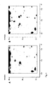
- Fig. 1 Figure 3 shows a comparison of the 2D electrophoresis gels for a control (A) and a treated animal (B) sample, where the additional protein spot in (B) corresponds to a new protein whose position is indicated by an arrow and a circle.
- the new specific proteins identified in the protein spot patterns of the analytical 2D gel electrophoresis were then subsequently prepared by preparative 2D gel electrophoresis using 350 ⁇ g protein (see again, (10).)
- the staining was carried out using Coomassie Brilliant Blue G250 (cf. see. V.Neuhoff, et al., "Improved staining of proteins in polyacrylamide gels including isoelectric focusing gels with clear background at nanogram sensitivity using Coomassie Brilliant Blue G-250 and R-250", Electrophoresis 1988, 9, 255-262 ).
- the protein spots preselected for further analysis were excised from the gel using the method described in A.
- FIGS. 1 (A) and 1 (B) is shown in brain tissue extracts from baboons that have been administered an LPS injection, including a spot of a protein for which a molecular weight of about 36 ⁇ 3 kDa was estimated from gel electrophoresis data as compared to markers of known molecular weight, while from the relative position of the protein from the first dimension, an isoelectric point of about 6.6-7.0 was determined.
- the mass 1202.714 corresponds to the sequence QQSELQSQVR (SEQ ID NO: 2).
- the theoretical molecular weight of this fragment is 1201.604. In the amino acid sequence of LASP-1, this sequence is found at positions 76-95.
- Mass 1254,615 corresponds to the sequence ACFHCETCK (SEQ ID NO: 3). The theoretical molecular weight of this fragment is 1253.498. In the amino acid sequence of LASP-1, this sequence is found at positions 28-36.
- the mass 1291,745 corresponds to the sequence KPYCNAHYPK (SEQ ID NO: 4).
- the theoretical molecular weight of this fragment is 1290,617.
- this sequence is found at positions 50-59.
- Mass 1360,813 corresponds to the sequence VNCLDKFWHK (SEQ ID NO: 5). The theoretical molecular weight of this fragment is 1359.675. In the amino acid sequence of LASP-1, this sequence is found at positions 18-27.
- the mass 1418.898 corresponds to the sequence GFSVVADTPELQR (SEQ ID NO: 6).
- the theoretical molecular weight of this fragment is 1417.719.
- this sequence is found at positions 97-109.
- Mass 1443,959 corresponds to the sequence LKQQSELQSQVR (SEQ ID NO: 7). The theoretical molecular weight of this fragment is 1442.783. In the amino acid sequence of LASP-1, this sequence is found at positions 74-85.
- the mass 1489.826 corresponds to the sequence MGPSGGEGMEPERR (SEQ ID NO: 8).
- the theoretical molecular weight of this fragment is 1488.644.
- this sequence is found at positions 131-144.
- Mass 1551,943 was assigned to the sequence TGDTGMLPANYVEAI (SEQ ID NO: 9).
- the theoretical molecular weight of this fragment is 1550.728.
- amino acid sequence of LASP-1 such a sequence is found at positions 247-261.
- the mass 1604.040 corresponds to the sequence GKGFSVVADTPELQR (SEQ ID NO: 10).
- the theoretical molecular weight of this fragment is 1602.836.
- this sequence is found at positions 95-109.
- Mass 1608.960 corresponds to the sequence QSFTMVADTPENLR (SEQ ID NO: 11). The theoretical molecular weight of this fragment is 1607.771. In the amino acid sequence of LASP-1, this sequence is found at positions 60-73.
- LASP-1 appears to show irregular behavior in gel-electrophoretic molecular mass determinations, so that the deviations of the molecular weights found are not necessarily in contradiction to the substance found as LASP-1 with the amino acid sequence according to SEQ ID NO: 1 to identify.
- antisera the starting from the fourth month after the immunization, which prepared peptide-specific antibodies.
- the peptides PKE18, PQQ14 and PVK19 were first coupled to SulfoLink gel (see working instructions "SulfoLink Kit” from PIERCE, Rockford, IL, USA). In each case, 5 mg of peptide per 5 ml of gel were offered for coupling.
- protein-free protein content of the wash eluate ⁇ 0.02 A280 nm
- Elution buffer was added to the washed columns and 1 ml fractions were collected.
- the protein content of each fraction was determined by the BCA method (see working instructions from PIERCE, Rockford, IL, USA).
- Fractions with protein concentrations> 0.8 mg / ml were pooled, neutralized with PBS buffer and thus diluted to protein concentrations of about 1 mg / ml.
- the yields were: 45 mg anti-PKE18 antibody, 59 mg anti-PQQ14 antibody and 14 mg anti-PVK19 antibody.
- the chemiluminescent labeling of the antibodies 100 ⁇ l of the anti-PVK19 antibody (1 mg / ml) or 125 ⁇ l of the anti-PQQ14 antibody (0.8 mg / ml) were mixed with 10 ⁇ l each of MA70-acridinium-NHS ester (1 mg / ml; Company HOECHST Behring) and incubated for 15 minutes at room temperature. Then, in each case 400 .mu.l 1 M glycine was added and incubated for a further 10 minutes.
- the labeling mixtures were then rebuffered via NAP-5 gel filtration columns (Pharmacia) in 1 ml of eluent A (50 mM potassium phosphate, 100 mM NaCl, pH 7.4) according to the instructions and freed from low molecular weight constituents.
- eluent A 50 mM potassium phosphate, 100 mM NaCl, pH 7.4
- Gel filtration HPLCs were carried out to separate last residues of labels not bound to antibodies (column: Waters Protein Pak SW300).
- the samples were applied and chromatographed with eluent A at a flow rate of 1 ml / min.
- the wavelengths 280 nm and 368 nm were measured with a continuous flow photometer.
- the absorbance ratio 368 nm / 280 nm as a measure of the degree of labeling of the antibodies was 0.10 for both antibodies at the peak.
- the monomeric antibody-containing fractions (retention time 8-10 min) were collected and collected in 3 ml of 100 mM sodium phosphate, 150 mM NaCl, 5% Bovine Serum Albumin, 0.1% sodium azide, pH 7.4.
- Irradiated 5 ml polystyrene tubes were coated with anti-PQQ14 antibody or anti-PKE18 antibody as follows: The antibodies were diluted in 50 mM Tris, 100 mM NaCl, pH 7.8 to a concentration of 6.6 ⁇ g / ml. 300 ⁇ l of this solution were pipetted into each tube. The tubes were incubated at 22 ° C for 20 hours. The solution was sucked off. Then, each tube was filled with 4.2 ml of 10 mM sodium phosphate, 2% Karion FP, 0.3% Bovine Serum Albumin, pH 6.5. After 20 hours, the solution was filtered with suction. Finally, the tubes were dried in a vacuum dryer.
- the standard material used was a chemically synthesized artificial peptide PKK54 containing pos. 121-137, 147-159, 170-187 of LASP-1 (SEQ ID NO: 15). This peptide was serially diluted in normal horse serum (SIGMA). Concentrations according to the weight of peptide were ascribed to the standards thus prepared.
- Samples were EDTA plasmas or apparently healthy individuals, patients with sepsis, patients with Alzheimer's disease and patients with myocardial infarction.
- test tubes 50 ⁇ l of standard or sample and 150 ⁇ l of assay buffer were pipetted into the test tubes. It was incubated for two hours at 22 ° C with shaking. Then it was washed 4 times with 1 ml of washing solution (0.1% Tween 20) per tube. Then 200 ⁇ l assay buffer containing 0.5 million RLU of the MA70-labeled tracer antibody was pipetted into each tube. It was incubated for two hours at 22 ° C with shaking.
- the other two epitopes are located outside protein binding domains (actin binding sites, LIM, SH3).
- a direct influence on antibody binding to these epitopes by any bound proteins is therefore not expected.
- Whether the LASP-1 immunoreactivity in the serum is actually free, has not yet been proven by the present analyzes and may be determined by further investigations.
- LASP-1 and LAP-1 appear to result from differential splicing of the primary transcript of the same gene:
- the sequences of the cDNAs of LASP-1 and LAP-1 (Accession # BC007560 and BC012460) were compared to the sequence of the human genome.
- An associated gene was located on chromosome 17, near 17q21, region 39,023K-39,076K. By sequence comparison, an exon-intron structure of the gene could be derived.
- the cDNAs for LASP-1 and LAP-1 result from the splicing of eight. Exons. Exons 2-5 are not different for both cDNAs. However, exons 6, 7 and 8 are not identical in both cDNAs, but overlap.
- Exon 1 differs in both cDNAs in terms of length at the 5'-terminus. It remains unclear whether the different lengths of exon 1 of both cDNAs result from different transcription initiation, or from different terminations in the reverse transcription of the mRNAs.
- the translated range is sufficient for both cDNAs from the 3 'region to the 5' region of exon 7:
- the amino acid sequences of both translation products are identical up to the amino acid proline (Item 200), but then completely different (see SEQ ID NO: 1 in comparison with SEQ ID NO: 16).
- LASP-1 comprises a total of 261 amino acids, LAP-1 includes 323 amino acids.
- the SH3 domain starts at position 202. Exactly this SH3 range is not present in LAP-1 but is replaced by another range of unknown function. The lack of the SH3 domain in LAP-1 led to the naming (in "LASP-1" S stands for SH3 domain).
- LASP-1 and LAP-1 have ESTs (expressed sequence tags) in the relevant database GenBank "Human EST entries” (e.g., BU553748 for LASP-1 and BG281978 for LAP-1). This clearly shows that the above-described entries BC007560 and BC012460 are not singular observations or even artifacts, but rather indicate that in fact both splicing variants are expressed. However, under what conditions the gene is expressed in LASP-1 or LAP-1 is unknown.
- LASP-1 Due to the extensive sequence identity with LASP-1 should also be considered a determination and / or co-determination of LAP-1 as a provision of the present invention.
- the reagents are chosen such that the determination can distinguish between LASP-1 and LAP-1, e.g. by selecting in a sandwich assay the binding partners (solid phase bound or labeled) such that at least one of them specifically recognizes an epitope in the C-terminal part above the position 200 of LAP-1 or LASP-1, especially above the position 200 of LAP-1 , one can check whether the LASP-1 immunoreactivity detected in a biological fluid is wholly or partially due to LAP-1, and LAP-1 can also be found alone next to LASP-1, or next to the total amount of LASP-1 and LAP-1, thereby further elucidating the clinical relevance of a presence of LAP-1 for sepsis and / or Alzheimer's disease.
- the binding partners solid phase bound or labeled
- LASP-1 immunoreactivity The role of the highly elevated LASP-1 immunoreactivity in blood circulation observed in pathological conditions is unclear. However, the fact that an elevation is associated with a pathological condition suggests that LASP-1 immunoreactivity may be involved in the molecular mechanism of the disease or in the body's own attempt to control the disease, possibly even playing a key role in it. It is therefore promising to carry out further investigations based on the findings in the present application with a therapeutic aim. Depending on whether the LASP-1 immunoreactivity is more harmful or useful, there is a therapeutic benefit from either an exogenously induced decrease or an increase in LASP-1 immunoreactivity.
- LASP-1 and / or LAP-1 concentrations detected for the first time allow their use as new biomarkers for sepsis, Alzheimer's and also in connection with myocardial infarction.
- the first proven disease-accompanying occurrence of LASP-1 in the circulation of sepsis, Alzheimer's and myocardial infarction patients also opens up new interesting research approaches for the treatment of the relevant diseases.
Landscapes
- Health & Medical Sciences (AREA)
- Life Sciences & Earth Sciences (AREA)
- Engineering & Computer Science (AREA)
- Biomedical Technology (AREA)
- Chemical & Material Sciences (AREA)
- Immunology (AREA)
- Urology & Nephrology (AREA)
- Molecular Biology (AREA)
- Hematology (AREA)
- Medicinal Chemistry (AREA)
- General Health & Medical Sciences (AREA)
- Biotechnology (AREA)
- Biochemistry (AREA)
- Analytical Chemistry (AREA)
- Physics & Mathematics (AREA)
- Microbiology (AREA)
- General Physics & Mathematics (AREA)
- Food Science & Technology (AREA)
- Cell Biology (AREA)
- Pathology (AREA)
- Pharmacology & Pharmacy (AREA)
- General Chemical & Material Sciences (AREA)
- Proteomics, Peptides & Aminoacids (AREA)
- Neurology (AREA)
- Chemical Kinetics & Catalysis (AREA)
- Neurosurgery (AREA)
- Veterinary Medicine (AREA)
- Public Health (AREA)
- Animal Behavior & Ethology (AREA)
- Organic Chemistry (AREA)
- Nuclear Medicine, Radiotherapy & Molecular Imaging (AREA)
- Bioinformatics & Cheminformatics (AREA)
- Rheumatology (AREA)
- Heart & Thoracic Surgery (AREA)
- Cardiology (AREA)
- Oncology (AREA)
- Pain & Pain Management (AREA)
- Communicable Diseases (AREA)
- Hospice & Palliative Care (AREA)
- Psychiatry (AREA)
Description
- Die vorliegende Erfindung betrifft neue Verwendungen des Proteins LASP-1 sowie ggf. eng verwandter Proteine, die wenigstens hinsichtlich ihrer ersten 200 Aminosäuren die gleiche Sequenz wie LASP-1 und damit eine vergleichbare Immunreaktivität aufweisen, sowie von Fragmenten davon, für die medizinischen Diagnostik von Sepsis und sepsisähnlichen systemischen Infektionen sowie von Herzinfarkten und der Alzheimer Krankheit gemäß Anspruch 1. Sie beruht auf dem erstmaligen Nachweis stark erhöhter Konzentrationen des Proteins LASP-1 im Gehirngewebe von Primaten, bei denen experimentell durch Toxinverabreichunge eine Sepsis bzw. systemische Entzündung hervorgerufen wurde, sowie auf dem anschließenden Nachweis einer stark erhöhten LASP-1 Immunreaktivität in der Zirkulation von Patienten mit Sepsis und von Alzheimer-Patienten, wobei eine erhöhte entsprechende Immunreaktivität auch bei Patienten mit Herzinfarkt festgestellt wurde.
- Wenn in der nachfolgenden Beschreibung von LASP-1 gesprochen wird, soll dieser Begriff nicht nur das bekannte Protein LASP-1 mit der in SEQ ID NO:1 angegebenen konkreten Aminosäuresequenz bezeichnen, sondern ggf. auch verwandte Proteine, die innerhalb der ersten 200 Aminosäuren mit diesem LASP-1 sequenzidentisch sind, z.B. das Protein LAP-1, zu dem sich am Ende des experimentellen Teils dieser Anmeldung noch erläuternde Ausführungen finden. Diese Definition gilt für alle Bezugnahmen auf LASP-1, es sei denn, es ergibt sich aus dem konkreten Zusammenhang für den Fachmann etwas anderes. Wenn von LASP-1 gesprochen wird, meint dieser Begriff außerdem auch Fragmente mit einer LASP-1-Immunreaktivität, wobei alle genannten Produkte sowohl frei als auch ggf. an Bindeproteine gebunden und/oder in einer posttranslational modifizierten Form, z.B. in glycosylierter und/oder phosphorylierter Form, vorliegen können.
- Die vorliegende Erfindung hat ihren Ursprung in intensiven Forschungsarbeiten der Anmelderin im Zusammenhang mit weiteren Verbesserungen der Diagnose und Therapie von Entzündungen und Infektionen.
- Als Entzündungen (Inflammationen) werden ganz allgemein bestimmte physiologische Reaktionen eines Organismus auf verschiedenartige äußere Einwirkungen wie z.B. Verletzungen, Verbrennungen, Allergene, Infektionen durch Mikroorganismen wie Bakterien und Pilze und Viren, auf Fremdgewebe, die Abstoßungsreaktionen auslösen, oder auf bestimmte entzündungsauslösende endogene Zustände des Körpers, z.B. bei Autoimmunerkrankungen und Krebs, bezeichnet. Entzündungen können als harmlose, lokal begrenzte Reaktionen des Körpers auftreten, sind jedoch auch typische Merkmale zahlreicher ernster chronischer und akuter Erkrankungen von einzelnen Geweben, Organen, Organ- und Gewebsteilen.
- Lokale Entzündungen sind dabei meist Teil der gesunden Immunreaktion des Körpers auf schädliche Einwirkungen, und damit Teil des lebenserhaltenden Abwehrmechanismus des Organismus. Wenn Entzündungen jedoch Teil einer fehlgeleiteten Reaktion des Körpers auf bestimmte endogene Vorgänge wie z.B. bei Autoimmunerkrankungen sind und/oder chronischer Natur sind, oder wenn sie systemische Ausmaße erreichen, wie beim systemischen Inflammationssyndrom (Systemic Inflammatory Response Syndrome, SIRS) oder bei einer auf infektiöse Ursachen zurückzuführenden schweren Sepsis, geraten die für Entzündungsreaktionen typischen physiologischen Vorgänge außer Kontrolle und werden zum eigentlichen, häufig lebensbedrohlichen Krankheitsgeschehen. Als Erkrankung, die von typischen Symptomen einer Entzündung, und zwar des Gehirns, begleitet wird, wird im Zusammenhang der vorliegenden Patentanmeldung auch die Alzheimer Krankheit angesehen, die durch eine in der Regel im 5. bis 6. Lebensjahrzehnt auftretende, unaufhaltsam fortschreitende Großhirnrindenatrophie charakterisiert ist und aufgrund ihrer Häufigkeit eine große volkswirtschaftliche Bedeutung aufweist.
- Es ist heute bekannt, dass die Entstehung und der Verlauf von entzündlichen Prozessen von einer beträchtlichen Anzahl von Substanzen, die überwiegend proteinischer bzw. peptidischer Natur sind, gesteuert werden bzw. von einem mehr oder weniger zeitlich begrenzten Auftreten bestimmter Biomoleküle begleitet sind. Zu den an Entzündungsreaktionen beteiligten endogenen Substanzen gehören insbesondere solche, die zu den Cytokinen, Mediatoren, vasoaktiven Substanzen, Akutphasenproteinen und/oder hormonellen Regulatoren gezählt werden können. Die Entzündungsreaktion stellt eine komplexe physiologische Reaktion dar, an der sowohl das Entzündungsgeschehen aktivierende endogene Substanzen (z.B. TNF-α, Interleukin-1) als auch desaktivierende Substanzen (z.B. Interleukin-10) beteiligt sind.
- Bei systemischen Entzündungen wie im Falle einer Sepsis bzw. des septischen Schocks weiten sich die entzündungsspezifischen Reaktionskaskaden unkontrolliert auf den gesamten Körper aus und werden dabei, im Sinne einer überschießenden Immunantwort, lebensbedrohlich. Zu den gegenwärtigen Kenntnissen über das Auftreten und die möglichen Rolle einzelner Gruppen endogener entzündungsspezifischer Substanzen wird beispielsweise verwiesen auf A.Beishuizen, et al., "Endogenous Mediators in Sepsis and Septic Shock", Advances in Clinical Chemistry, Vol.33, 1999, 55-131; und C.Gabay, et al., "Acute Phase Proteins and Other Systemic Responses to Inflammation", The New England Journal of Medicine, Vol.340, No.6, 1999, 448-454. Da sich das Verständnis von Sepsis und verwandten systemischen entzündlichen Erkrankungen, und damit auch die anerkannten Definitionen, in den letzten Jahren gewandelt haben, wird außerdem verwiesen auf K.Reinhart, et al., "Sepsis und septischer Schock", in: Intensivmedizin, Georg Thieme Verlag, Stuttgart.New York, 2001, 756-760; wo eine moderne Definition des Sepsis-Begriffes gegeben wird. Im Rahmen der vorliegenden Anmeldung werden die Begriffe Sepsis bzw. entzündliche Erkrankungen in Anlehnung an die Definitionen verwendet, wie sie den genannten drei Literaturstellen entnommen werden können.
- Während wenigstens im europäischen Raum die durch eine positive Blutkultur nachweisbare systemische bakterielle Infektion lange den Sepsisbegriff prägte, wird die Sepsis heute in erster Linie als systemische Entzündung verstanden, die infektiöse Ursachen hat, als Krankheitsgeschehen jedoch große Ähnlichkeiten mit systemischen Entzündungen aufweist, die durch andere Ursachen ausgelöst werden. Dem genannten Wandel des Sepsis-Verständnisses entsprechen Veränderungen der diagnostischen Ansätze. So wurde der direkte Nachweis bakterieller Erreger durch komplexe Überwachungen physiologischer Parameter und in jüngerer Zeit insbesondere auch durch den Nachweis bestimmter am Sepsisgeschehen bzw. am Entzündungsgeschehen beteiligter endogener Substanzen, d.h. spezifischer "Biomarker", ersetzt bzw. ergänzt.
- Von der großen Zahl von Mediatoren und Akutphasenproteinen, von denen man weiß oder vermutet, dass sie an einem Entzündungsgeschehen beteiligt sind, eignen sich dabei für diagnostische Zwecke insbesondere solche, deren Auftreten sehr spezifisch für entzündliche Erkrankungen bzw. bestimmte Phasen oder Ausprägungen entzündlicher Erkrankungen ist, deren Konzentrationen sich drastisch und diagnostisch signifikant verändern und die außerdem die für Routinebestimmungen erforderlichen Stabilitäten aufweisen und hohe Konzentrationswerte erreichen. Für diagnostische Zwecke steht dabei die zuverlässige Korrelation von Krankheitsgeschehen (Entzündung, Sepsis) mit dem jeweiligen Biomarker im Vordergrund (gute Spezifität und Selektivität), ohne dass dessen Rolle in der komplexen Kaskade der am Entzündungsgeschehen beteiligten endogenen Substanzen bekannt sein muss.
- Eine bekannte, als Sepsis-Biomarker besonders geeignete endogene Substanz ist Procalcitonin. Procalcitonin ist ein Prohormon, dessen Serum-Konzentrationen unter den Bedingungen einer systemischen Entzündung infektiöser Ätiologie (Sepsis) sehr hohe Werte erreichen, während es bei Gesunden so gut wie nicht nachweisbar ist. Hohe Werte an Procalcitonin werden außerdem in einem relativ frühen Stadium einer Sepsis erreicht, so dass sich die Bestimmung von Procalcitonin auch zur Früherkennung einer Sepsis und zur frühen Unterscheidung einer infektiös bedingten Sepsis von schweren Entzündungen, die auf anderen Ursachen beruhen, eignet. Die Bestimmung von Procalcitonin als Sepsismarker ist Gegenstand der Veröffentlichung M.Assicot, et al., "High serum procalcitonin concentrations in patients with sepsis and infection", The Lancet, vol.341, No.8844, 1993, 515-518; und der Patente
DE 42 27 454 C2 bzw.EP 0 656 121 B1 bzw.US 5,639,617 . Auf die genannten Patente und in der genannten Veröffentlichung angeführten frühen Literaturstellen wird zur Ergänzung der vorliegenden Beschreibung ausdrücklich Bezug genommen. In den letzten Jahren ist die Zahl der Veröffentlichungen zum Thema Procalcitonin stark angestiegen. Stellvertretend für zusammenfassende Veröffentlichungen aus jüngerer Zeit wird daher noch verwiesen auf W.Karzai et al., "Procalcitonin - A New Indicator of the Systemic Response to Severe infection", Infection, Vol. 25, 1997, 329-334; und M.Oczenski et al., "Procalcitonin: a new parameter for the diagnosis of bacterial infection in the peri-operative period", European Journal of Anaesthesiology 1998, 15, 202-209; sowie ferner H.Redl, et al., "Procalcitonin release patterns in a baboon model of trauma and sepsis: Relationship to cytokines and neopterin", Crit Care Med 2000, Vol.28. No.11, 3659-3663; und H.Redl, et al., "Non-Human Primate Models of Sepsis", in: Sepsis 1998; 2:243-253; und die darin zitierten weiteren Literaturstellen. - Die Verfügbarkeit des Sepsismarkers Procalcitonin hat der Sepsisforschung starke Impulse gegeben, und es werden gegenwärtig intensive Anstrengungen unternommen, weitere Biomarker zu finden, die die Procalcitonin-Bestimmung ergänzen können und/oder zusätzliche Informationen für Zwecke der Feindiagnostik bzw. Differentialdiagnostik zu liefern vermögen. Erschwert wird die Suche nach potentiellen neuen Sepsis-Biomarkern allerdings dadurch, dass häufig noch sehr wenig oder nichts über die genaue Funktion bzw. über die genauen Gründe für das Auftreten bestimmter endogener Substanzen, die am Entzündungs- oder Infektionsgeschehen beteiligt sind, bekannt ist.
- Die Ergebnisse der experimentellen Überprüfung eines fruchtbaren rein hypothetischen Ansatzes zur Ermittlung weiterer potentieller Sepsismarker finden sich in
DE 198 47 690 A1 bzw.WO 00/22439 - In der vorliegenden Anmeldung werden Ergebnisse eines anderen, rein experimentellen Ansatzes für die Suche nach weiteren entzündungs- bzw. sepsisspezifischen Biomolekülen berichtet. Auch diese experimentellen Untersuchungen nehmen ihren Ausgang bei der Bestimmung von Procalcitonin im Zusammenhang mit systemischen entzündlichen Reaktionen infektiöser Ätiologie. So war sehr früh beobachtet worden, dass bei Sepsis das Procalcitonin offensichtlich nicht auf die gleiche Weise gebildet wird, wie dann, wenn es Vorläufer für das Hormon Calcitonin ist. So wurden hohe Procalcitoninspiegel auch bei Patienten beobachtet, denen die Schilddrüse entfernt worden war. Deshalb kann die Schilddrüse nicht dasjenige Organ sein, in dem Procalcitonin bei Sepsis gebildet bzw. ausgeschüttet wird. In den Veröffentlichungen H.Redl, et al., "Procalcitonin release patterns in a baboon model of trauma and sepsis: Relationship to cytokines and neopterin", Crit Care Med 2000, Vol.28. No.11, 3659-3663; und H.Redl, et al., "Non-Human Primate Models of Sepsis", Sepsis 1998; 2:243-253; werden die Ergebnissen von experimentellen Untersuchungen berichtet, die der Klärung der Bildung von Procalcitonin bei Sepsis dienen sollten. In den genannten Arbeiten wird durch Endotoxinverabreichung an Primaten (Paviane) eine künstliche Sepsis erzeugt, und es wird bestimmt, bei welchen experimentell erzeugten Zuständen die höchsten Procalcitoninkonzentrationen im Blut erreicht werden. Eine Weiterentwicklung des in den genannten Arbeiten beschriebene Versuchstiermodells dient im Rahmen der vorliegenden Anmeldung dazu, neue endogene entzündungs- und/- oder sepsisspezifische Biomarker von peptidischer bzw. proteinischer Natur zu ermitteln, deren Auftreten für bestimmte Formen von Entzündungen und/oder Sepsis charakteristisch ist und die daher eine spezifische, ggf. organspezifische Sepsis- bzw. Entzündungsdiagnose ermöglichen. Das Primatenmodell wurde dabei aufgrund der sehr großen Ähnlichkeit der Physiologie von Primaten und Menschen und der hohen Kreuzreaktivität mit vielen therapeutischen und diagnostischen humanen Reagenzien gewählt.
- Da die bei Entzündungen gebildeten endogenen Substanzen Teil der komplexen Reaktionskaskade des Körpers sind, sind derartige Substanzen außerdem nicht nur von diagnostischem Interesse, sondern es wird gegenwärtig auch mit erheblichem Aufwand versucht, durch Beeinflussung der Entstehung und/- oder der Konzentration einzelner derartiger Substanzen therapeutisch in das Entzündungsgeschehen einzugreifen, um die zum Beispiel bei Sepsis beobachtete systemische Ausbreitung der Entzündung möglichst frühzeitig zu stoppen. In diesem Sinne sind nachweislich am Entzündungsgeschehen beteiligte endogene Substanzen auch als potentielle therapeutische Targets anzusehen. Versuche, ansetzend an bestimmten Mediatoren des Entzündungsgeschehens dieses positiv therapeutisch zu beeinflussen, sind beschrieben beispielsweise in E.A.Panacek, "Anti-TNF strategies", Journal für Anästhesie und Intensivbehandlung; Nr.2, 2001, 4-5; T.Calandra, et al., "Protection from septic shock by neutralization of macrophage migration inhibitory factor", Nature Medicine, Vol.6, No.2, 2000, 164-170; oder K.Garber, "Protein C may be sepsis solution", Nature Biotechnology, Vol.18, 2000, 917-918. Angesichts der bisherigen eher enttäuschenden therapeutischen Ansätze besteht ein hohes Interesse daran, weitere möglichst entzündungs- bzw. sepsisspezifische endogene Biomoleküle zu identifizieren, die auch als therapeutische Targets neue Erfolgsaussichten für die Entzündungsbekämpfung eröffnen.
- Die vorliegende Erfindung beruht darauf, dass sich in Primaten und Menschen bei Sepsis im cytoplasmatischen Gehirngewebe das stark überexprimierte Protein LASP-1, bzw. ein Protein, das mit diesem innerhalb der ersten 200 Aminosäuren identisch ist, in einer löslichen Form nachweisen läßt, und zwar im Unterschied zu Gesunden bzw. Unbehandelten, bei denen es nicht oder nur in Konzentrationen an der analytischen Nachweisgrenze gefunden wird, und auch im Unterschied zu anderen Körpergeweben septischer Primaten-Versuchstiere, was dieses Protein für die Entzündungsdiagnostik und Infektions- bzw. Sepsisdiagnostik mit Gehirnbeteiligung geeignet erscheinen läßt.
- Die Erfindung beruht ferner darauf, dass festgestellt wurde, dass auch in der Zirkulation von menschlichen Sepsispatienten, jedoch auch von Alzheimer-Patienten, eine deutlich erhöhte LASP-1 Immunreaktivität messbar ist, was den Wert der auf der Basis von Gehirnextrakten von Primaten gewonnenen Erkenntnisse untermauert bzw. stark erhöht.
- Die Verwendungen in der Diagnostik, die sich aufgrund des erstmals nachgewiesenen gehirnspezifischen Auftretens von LASP-1 bei der experimentellen Simulation von Sepsis, sowie aus dem Nachweis einer deutlich erhöhten LASP-1 Immunreaktivität in der Serum-oder Plasmaproben ergeben, werden nachfolgend noch näher beschrieben.
- Die Ansprüche 1 bis 11 definieren die sich ergebenden neuen Verfahren gemäß der vorliegenden Erfindung, und bevorzugter Ausführungsformen davon, näher.
- Wie nachfolgend im experimentellen Teil noch näher ausgeführt wird, war Ausgangspunkt der Erfindung die Feststellung, dass nach experimenteller Auslösung einer künstlichen Sepsis in Pavianen durch Endotoxinverabreichung (LPS aus Salmonella Typhimurium) und 2D-gelektrophoretischer Aufarbeitung von Gehirngewebe der behandelten Tiere als eines der nur bei den behandelten Tieren identifizierbaren Produkte ein Protein gefunden wurde, das weitgehend, und zwar wenigstens über den Abschnitt der Aminosäuren 1 bis 200, mit dem bekannten Protein LASP-1 identisch war, wobei das Protein außerdem nur im Gehirngewebe in erheblich erhöhter Konzentration beobachtet wurde. Zu seiner Identifizierung wurde ein neuer Proteinspot, der nur im Gehirngewebe der behandelten Tiere gefunden wurde und gemäß Gelektrophorese ein Molekulargewicht von etwa 36 ± 3 kDA und einen isoelektrischen Punkt von ca. 6,6-7,0 aufwies, aus dem Elektrophoresegel isoliert und durch Trypsinverdauung in Bruchstücke zerlegt, die auf an sich bekannte Weise massenspektrometrisch analysiert wurden. Durch Vergleich mit dem bekannten Massenspektrum des trypsinbehandelten Proteins LASP-1 wurde das Protein des isolierten Spots als eines identifiziert, das wenigstens über weite Abschnitte mit dem Protein LASP-1 identisch war.
- Die Identifizierung des in dem Proteinspot gefundenen Produkts wird im experimentellen Teil näher beschrieben, wobei die beschriebene Identifizierung nach anerkannten Interpretationsgrundsätzen als sichere Identifizierung eines Produkts mit einer Aminosäuresequenz des Proteins LASP-1 anzusehen ist.
- Das rechnerisch ermittelte Molekulargewicht der Peptidkette von LASP-1 (vgl. SEQ ID NO:1; 261 Aminosäuren) beträgt 29 717 Dalton. LASP-1 liegt jedoch physiologisch in posttranslational prozessierter Form (glykosyliert und phosphoryliert) vor. So wurde das später als LASP-1 bezeichnete Produkt ursprünglich als ein 40 kDA cAMP-abhängiges Phosphoprotein ("pp40") in Magenwandzellen identifiziert (vgl. C.S. Chew et al., Journal of Cell Science 113, 2035-2045 (2000); C.S. Chew et al., Am.J.Physiol. 275 (Cell Physiol. 44): C56-C67, (1998)).
- Die Aminosäuresequenz des als LASP-1 bezeichneten Phosphoproteins wurde ursprünglich als die des putativen Expressionsprodukts der cDNA eines in humanen Brustkrebs-Zellinien überexprimierten Gens "MLN 50" aus dem Bereich q11-q21.3 des Chromosoms 17 ermittelt (C.Tomasetto at al., FEBS Letters 373 (1995): 245-249; C.Tomasetto et al., Genomics 28: 367-376 (1995); I.Bièche et al., Cancer Res. 56: 3886-3890 (1996)). Der Name LASP-1 geht darauf zurück, dass in diesem Protein erstmals eine sog. LIM-Domäne (eine aus verschiedenen Proteinen bekannte Zn-bindende Domäne; vgl. A.Hammarström et al., Biochemistry 1996, 35, 12723-12732) mit einer aus der rezeptorunabhängigen sog. src-Kinase, einer Tyrosin-Kinase, bekannten Domäne (SH3) kombiniert ist (LIM and SH3 Protein); vgl. auch Brian K. Kay et al., FASEB J. 14, 231-241 (2000).
- LASP-1-mRNA ist ubiquitär in normalen Zellen nachweisbar (z.B. Prostata, Leber, Muskel, Gehirn, verschiedenen ZellTypen) und wird in einem gewissen prozentualen Anteil von Brustkrebsfällen überexprimiert. Es wurde festgestellt, dass LASP-1 ein Actin-bindendes Protein ist, für das angenommen wird, dass es zu den Cytoskelett-assoziierten Proteinen zu zählen ist und für die Zellform und -beweglichkeit sowie möglicherweise die Signalübertragung von Bedeutung ist. Es ist auch in Nervenenden nachweisbar. LASP-1 wird durch Detergentien nur schwer solubilisiert. In vitro hat sich LASP-1 als Substrat erwiesen, dass sich hervorragend mit der cAMP-abhängigen Serin/Threonin-Kinase PKA phosphorylieren läßt. LASP-1 weist in unterschiedlichen Epithel-Zelltypen unterschiedliche Expressionsmuster auf (vgl. C.S. Chew et al., Journal of Cell Science 113, 2035-2045 (2000); C.S. Chew et al., Am.J.Physiol. 275 (Cell Physiol. 44): C56-C67, (1998)).
- Das Auftreten von LASP-1 bzw. eines weitgehend damit übereinstimmenden Proteins in überexprimierter Form im Gehirngewebe bei Sepsis ist von hohem wissenschaftlichen, diagnostischen und möglicherweise auch therapeutischen Interesse. Die Tatsache, dass LASP-1 ein Kinase-Substrat und Phosphoprotein ist, verleiht ihm eine gewisse Ähnlichkeit mit den sogenannen tau-Proteinen, die bei der Alzheimer Krankheit eine Rolle insbesondere für diagnostische Zwecke spielen (vgl. z.B. G.A.Jicha et al., Journal of Neurochemistry,69., 2087-2095 (1997) und die darin zitierte Literatur; sowie
EP 673 418 EP 737 208 EP 772 634 EP 610 330 EP 618 968 - Es ist nunmehr gut bekannt, dass die Alzheimer Krankheit als gehirnspezifischer Entzündungsprozess angesehen werden kann, ohne dass damit eine Aussage bezüglich Krankheitsursache oder Krankheitsfolge gemacht werden soll (Lih-Fen Lue et al., GLIA 35:72-79, 2001; Michael Hüll et al., DDT Vol.4, No.6: 275-282; (June 1999); F.Licastro et al., Journal of Neuroimmunologie 103 (2000), 97-102; Neuroinflammation Working Group: Haruhiko Akiyama et al., Neurobiology of Aging 21 (2000) 383-421; Michael Hüll et al., Exp.Opin.Invest.Drugs (2000) 9(4):671-683; M.Hüll et al., Eur Arch Psychiatry Clin Neurosci (1996) 246:124-128; J.Rhodin et al., Annals New York Academy of Sciences, 199x, 345-352). Die Tatsache, dass das Phosphoprotein LASP-1 bzw. ein damit innerhalb der ersten 200 Aminosäuren übereinstimmendes Protein unter experimentellen Bedingungen, die eine Sepsis bzw. ein sepsisähnliches systemisches Entzündungsgeschehen simulieren, gehirnspezifisch in überexprimierter Form nachweisbar ist, machte es sehr wahrscheinlich, dass auch bei der Alzheimer Krankheit eine Überexpression von Lasp-1 bzw. einer bestimmten Expressionsform von LASP-1 beobachtbar ist und die Bestimmung von LASP-1 daher auch für die Alzheimer-Diagnostik Bedeutung erlangen wird. Anschließend durchgeführte immundiagnostische Bestimmungen einer Immunreaktivität in Seren von Sepsis- und Alzheimer-Patienten, die der Sequenz der ersten 200 Aminosäuren von LASP-1 zugeordnet werden konnte, bestätigten diese Vermutung experimentell in überzeugender, eindrücklicher Weise.
- Die internationale Patentanmeldung
WO0185986 - Es war aufgrund der bisher zugänglichen Befunde nicht zu erwarten, das die physiologischen Konzentrationen von LASP-1 in einer Serum- oder Plasmaprobe als Folge einer Sepsis und/oder gehirnspezifischen Entzündung wie Alzheimer signifikant und nachweisbar verändert sind und daher eine Bestimmung der LASP-1-Konzentrationen in solchen Proben im Rahmen eines Sepsisgeschehens oder einer entzündlichen Gehirnerkrankung diagnostisch von Interesse sein könnte.
- Der erfindungsgemäße Nachweis von vergleichsweise hohen LASP-1-Konzentrationen im Gehirngewebe von Primaten, bei denen durch Toxinverabreichung eine künstliche Sepsis ausgelöst wurde, bei gleichzeitiger Unmöglichkeit, LASP-1 in ansonsten völlig gleich behandelten Proben von Kontrolltieren, oder in anderen Gewebeproben der septischen Tiere, zu erkennen, und der nachfolgende immundiagnostische Nachweise in Serum- oder Plasmaproben von menschlichen Patienten, ist hoch signifikant. Da das Auftreten nur bei den behandelten Tieren zu beobachten war, und zwar bereits relativ kurze Zeit nach der Sepsisauslösung durch Toxinverabreichung, ist es möglich, diese Tatsache zur Schaffung eines vielversprechenden diagnostischen Verfahrens zur Diagnose von Sepsis bzw. sepsisähnlichen, systemischen Infektionen durch Bestimmung von LASP 1-Immunreaktivität zu nutzen. Von besonderem Interesse ist außerdem die nachgewiesene Eignung von LASP-1 als diagnostischer Marker und Prognosemarker für die Alzheimer Krankheit.
- Die Bestimmung von LASP-1 kann dabei nach irgendeinem beliebigen geeigneten Nachweisverfahren erfolgen, wobei jedoch die Bestimmung in einer Serum- oder Plasmaprobe , eines Patienten auf immundiagnostischem Wege (mittels eines Immunoassays) unter Verwendung geeigneter selektiver Antikörper unter praktischen Gesichtspunkten am vorteilhaftesten erscheint.
- Aufgrund der Tatsache, dass bei einer experimentellen Sepsisauslösung im Gehirn von Primaten sowie anschließend auch in einer Serum- oder Plasmaprobe von menschlichen Patienten erstmals ein erhöhtes Auftreten von LASP-1 bzw. eines damit innerhalb der ersten 200 Aminosäuren identischen Proteins nachgewiesen werden konnte, wird somit die Möglichkeit geschaffen, die LASP-1 Immunreaktivität insbesondere für diagnostische Zwecke im Zusammenhang mit Sepsis und sepsisähnlichen systemischen Infektionen, sowie der Alzheimer Krankheit und Herzinfarkten, zu nutzen. Dafür können LASP-1 oder geeignete Teilpeptide davon oder Epitope oder Epitopkombinationen ggf. auch nach Verfahren, die inzwischen zum Stand der Technik gehören, synthetisch oder gentechnologisch gezielt hergestellt werden. LASP-1-Teilpeptide, ggf. in markierter Form, können auch als Kalibratoren, Tracer und Kompetitoren für bestimmte Assayformate für immundiagnostische Bestimmungen benötigt und dafür wie erläutert hergestellt werden. Bestimmte derzeit bevorzugte konkrete Ausführungsformen werden im nachfolgenden experimentellen Teil noch näher beschrieben.
- Ferner können LASP-1-Fragmente bzw. geeignete Teilsequenzen davon nach bekannten Verfahren des modernen Standes der Technik auch zur Erzeugung spezifischer polyklonaler oder auch monoklonaler Antikörper verwendet werden, die als Hilfsmittel für die diagnostische Bestimmung von LASP-1 in Körperflüssigkeiten eines Patienten und/oder auch als potentielle therapeutische Mittel geeignet sind. Bestimmte derzeit bevorzugte konkrete polyklonale affinitätsgereinigte anti-LASP-1 Antikörper werden im nachfolgenden experimentellen Teil noch näher beschrieben. Die Erzeugung geeigneter monoklonaler Antikörper gegen bekannte Peptid-Teilsequenzen gehört heute zum allgemeinen Stand der Technik und muss nicht besonders beschrieben werden. Ferner ist auch ausdrücklich die Antikörpererzeugung unter Anwendung von Techniken der direkten genetischen Immunisierung mit einer entsprechenden DNA zu erwähnen. Es liegt somit im Rahmen der vorliegenden Erfindung, zur Immunisierung z.B. eine cDNA von LASP-1 oder LASP-1-Fragmenten zu verwenden, da es sich in der Vergangenheit gezeigt hat, dass bei der Anwendung derartiger Immunisierungstechniken das Spektrum der gewinnbaren Antikörper erweitert werden kann. Es können aber auch bereits bekannte Antikörper gegen LASP-1 verwendet werden (vgl. z.B. V.Schreiber et al., Molecular Medicine 4: 675-687, 1998; C.S. Chew et al., Am.J.Physiol. 275 (Cell Physiol. 44): C56-C67, (1998)).
- Da im Rahmen der vorliegend beschriebenen Arbeiten im Gel des 2D-Elektrophorese ein LASP-1-Produkt mit einer molaren Massen von ca. 36 kDa gefunden wurde, das eine für die beschriebene Bestimmung ausreichende Löslichkeit aufweist, war nicht auszuschließen, dass das gefundene LASP-1-Produkt ein vom eigentlichen, in der Literatur beschriebenen LASP-1 abweichendes Expressionsprodukt, z.B. das Produkt eines alternativen Splicings, und/oder eine bestimmte Art der Glykosylierung und/oder Phosphorylierung aufweist, die ihm die beobachtete Löslichkeit verleiht und aufgrund derer es sich von dem aus der Magenwand isolierten 40 kDa-Produkt signifikant unterscheidet. Es wurde daher von Anfang der Untersuchungen an als im Rahmen der vorliegenden Erfindung liegend angesehen, die Bestimmung von LASP-1 mit Hilfe eines spezifischen Assays durchzuführen, bei dem spezifische Teilsequenzen und/oder ggf. auch Glykosylierungs- und/oder Phosphorylierungsmuster erkannt werden. Die Herstellung von Antikörpern, insbesondere monoklonalen Antikörpern, die spezifische Glykosylierungen und/oder Phosphorylierungen erkennen, ist grundsätzlich bekannt und z.B. im Zusammenhang mit der Bestimmung bestimmter Phosphorylierungsformen der sogenannten tau-Proteine im Rahmen der Alzheimer-Diagnostik beschrieben.
- Bei der immunologischen Bestimmung von LASP-1 kann dabei grundsätzlich so vorgegangen werden, wie das z.B. für die selektive Procalcitoninbestimmung beschrieben ist in P.P.Ghillani, et al., "Monoclonal antipeptide antibodies as tools to dissect closely related gene products", The Journal of Immunology, vol. 141, No.9, 1988, 3156-3163; und P.P.Ghillani, et al., "Identification and Measurement of Calcitonin Precursors in Serum of Patients with Malignant Diseases", Cancer research, vol.49, No.23, 1989, 6845-6851. Variationen der beschriebenen Techniken und/oder weitere Immunisierungstechniken kann der Fachmann einschlägigen Standardwerken und Veröffentlichungen entnehmen und sinngemäß anwenden.
- LASP-1 oder ggf. LASP-1-Fragmente oder partiell sequenzidentische Produkte wie LAP-1 können aufgrund der vorliegenden Ergebnisse als Immunreaktivität bzw. spezifische Markerpeptide (Biomarker) zum diagnostischen Nachweis und zur Verlaufskontrolle von Sepsis, systemischen Infektionen vom Sepsistyp, Herzinfarkten und Entzündungen des Gehirns wie der Alzheimer-Krankheit dienen. Wie die Bestimmung von Procalcitonin kann dabei die Bestimmung von LASP-1 zur differentialdiagnostischen Früherkennung und zur Erkennung sowie für die Erstellung einer Verlaufsprognose, zur Beurteilung des Schweregrads und zur therapiebegleitenden Verlaufsbeurteilung von Entzündungen und Infektionen erfolgen, wobei man bei einem derartigen Verfahren in einer Serum- oder Plasma-Probe eines Patienten den Gehalt von LASP-1 bestimmt und aus der festgestellten Anwesenheit und/oder Menge von LASP-1 auf das Vorliegen einer Sepsis, Herzinfarktes oder der Alzheimer Krankheit schließt und das erhaltene Ergebnis mit dem Schweregrad der Sepsis, des Herzinfarktes und der Alzheimer Krankheit korreliert und ggf. die Behandlungsmöglichkeiten und/oder die Behandlungsaussichten abschätzt.
- Besonders interessant erscheint eine Bestimmung von LASP-1 im Rahmen einer Multiparameter-Bestimmung, bei der gleichzeitig mindestens ein weiterer Entzündungs- oder Infektionsparameter bestimmt wird und bei der ein Messergebnis in Form eines Satzes von mindestens zwei Messgrößen gewonnen wird, der zur Feindiagnostik der Entzündung oder Infektion ausgewertet wird. Als derartige weitere Entzündungs- oder Infektionsparameter sind solche anzusehen, die aus der Gruppe der teilweise bekannten oder in den älteren bzw. parallelen Patentanmeldungen der Anmelderin offenbarten Parameter ausgewählt sind, die besteht aus Procalcitonin, CA 125, CA 19-9, S100B, S100A-Proteinen, löslichen Cytokeratin-Fragmenten, insbesondere CYFRA 21, TPS und/oder löslichen Cytokeratin-1-Fragmenten (sCY1F), den Peptiden Inflammin und CHP, Peptid-Prohormonen, der Glycin-N-Acyltransferase (GNAT), der Carbamoylphosphat Synthetase 1 (CPS 1) und deren Fragmenten und dem C-reaktiven Protein (CRP) oder Fragmenten aller genannten Proteine. Es ist vorteilhaft, die Multiparameter-Bestimmung als Simultanbestimmung mittels einer Chiptechnologie-Messvorrichtung oder einer immunchromatographischen Messvorrichtung durchzufüren, bei der die Auswertung des mit der Messvorrichtung gewonnenen komplexen Messergebnisses mit Hilfe eines Computerprogramms erfolgt.
- Bei einer bevorzugten Ausführungsform wird das Verfahren als heterogener Sandwich-Immunoassay durchgeführt, bei dem einer der Antikörper an eine beliebige Festphase, beispielsweise die Wände beschichteter Teströhrchen (z.B. aus Polystyrol; "Coated Tubes"; CT) oder an Mikrotiterplatten, zum Beispiel aus Polystyrol, oder an Partikel, beispielsweise Magnetpartikel immobilisiert ist, während der andere Antikörper einen Rest trägt, der ein direkt nachweisbares Label darstellt oder eine selektive Verknüpfung mit einem Label ermöglicht und der Detektion der gebildeten Sandwich-Strukturen dient. Auch eine zeitlich verzögerte bzw. nachträgliche Immobilisierung unter Verwendung geeigneter Festphasen ist möglich.
- Grundsätzlich können alle in Assays der beschriebenen Art verwendbaren Markierungstechniken angewandt werden, zu denen Markierungen mit Radioisotopen, Enzymen, Fluoreszenz-, Chemolumineszenz- oder Biolumineszenz-Labeln und direkt optisch detektierbaren Farbmarkierungen, wie beispielsweise Goldatomen und Farbstoffteilchen, wie sie insbesondere für sog. Point-of-Care (POC) oder Schnelltests verwendet werden, gehören. Die beiden Antikörper können auch im Falle heterogener Sandwich-Immunoassays Teile eines Nachweissystems der nachfolgend im Zusammenhang mit homogenen Assays beschriebenen Art aufweisen.
- Es liegt somit im Rahmen der vorliegenden Erfindung, das erfindungsgemäße Verfahren auch als Schnelltest auszugestalten.
- Das erfindungsgemäße Verfahren kann ferner als homogenes Verfahren ausgestaltet werden, bei dem die aus den beiden Antikörpern und dem nachzuweisenden LASP-1 gebildeten Sandwichkomplexe in der flüssigen Phase suspendiert bleiben. In einem solchen Fall ist es bevorzugt, beide Antikörper mit Teilen eines Nachweissystems zu markieren, das dann, wenn beide Antikörper in einen einzigen Sandwich integriert werden, eine Signalerzeugung oder Signalauslösung ermöglicht. Derartige Techniken sind insbesondere als Fluoreszenzverstärkungs- oder Fluoreszenzlöschungs-Nachweisverfahren ausgestaltbar. Ein besonderes bevorzugtes derartiges Verfahren betrifft die Verwendung von paarweise einzusetzenden Nachweisreagenzien, wie sie beispielsweise beschrieben sind in
US-A-4 822 733 ,EP-B1-180 492 EP-B1-539 477 - Nachfolgend wird die Auffindung und Identifizierung von LASP-1 und dessen Bestimmung in biologischen Flüssigkeiten (Seren bzw. Plasmen) von Patienten mit verschiedenen Krankheiten in näheren Einzelheiten geschildert. Die Figuren zeigen:
- Fig. 1
- Ansichten von 2D-Elektrophoresegelen, die einen Vergleich der Spotmuster der cytoplasmatischen Gehirnproteine eines gesunden Pavians (A) mit denen der Gehirnproteine eines Pavians 5h nach einer durch LPS-Verabreichung induzierten Sepsis (B) ermöglichen. Der Pfeil zeigt die Position des erfindungsgemäßen sepsisspezifischen Produkts (LASP-1) an, das in Darstellung (B) durch einen Kreis hervorgehoben ist;
- Fig. 2
- das Massenspektrum des aus dem Gel der 2D-Gelelek- trophorese isolierten, trypsinverdauten Produkts.
- Fig. 3
- eine vergleichende Darstellung der Ergebnisse der Messung der LASP-1 (1-200) Immunreaktivität unter Verwendung von Immunoassays mit verschiedenen Paaren von drei Antikörpern, die in festphasenge- bundener bzw. markierter Form eingesetzt wurden und die Aminosäuresequenzen 121-137, 147-159 bzw. 170-187 von LASP-1 erkannten. Es ist zu erkennen, dass mit allen Paaren weitgehend identische Ergeb- nisse erhalten wurden.
- Fig. 4
- die Ergebnisse einer immundiagnostischen Bestim- mung einer LASP-1 (1-200) Immunreaktivität in Seren von 294 gesunden Kontrollpersonen und von 96 Sepsispatienten.
- Fig. 5
- die Ergebnisse einer immundiagnostischen Bestim- mung einer LASP-1 (1-200) Immunreaktivität in Seren von 294 gesunden Kontrollpersonen und von 80 Alzheimer-Patienten.
- Fig. 6
- die Ergebnisse einer immundiagnostischen Bestim- mung einer LASP-1 (1-200) Immunreaktivität in Seren von 294 gesunden Kontrollpersonen und von 20 Herzinfarkt-Patienten.
- In Anlehnung an die mit Pavianen durchgeführten Versuche zur Stimulierung der Procalcitonin-Ausschüttung durch Endotoxininjektionen (vgl.H.Redl, et al., "Procalcitonin release patterns in a baboon model of trauma and sepsis: Relationship to cytokines and neopterin", Crit Care Med 2000, Vol.28. No.11, 3659-3663; H.Redl, et al., "Non-Human Primate Models of Sepsis", in: Sepsis 1998; 2:243-253) wurden Pavianen (männlich, ca. 2 Jahre alt, 27 bis 29 kg schwer) jeweils 100 µg LPS (Lipopolysaccharid aus Salmonella Typhimurium, Bezugsquelle: Sigma) pro kg Körpergewicht intravenös verabreicht. 5 bis 5,5 h nach der Injektion wurden die Tiere durch intravenöse Verabreichung von 10 ml Doletal getötet. Innerhalb von 60 min nach ihrem Exitus wurden sämtliche Organe/Gewebe präpariert und durch Einfrieren in flüssigem Stickstoff stabilisiert.
- Bei der weiteren Verarbeitung wurden Proben der einzelnen tiefgefrorenen Gewebe (1g) unter Stickstoffkühlung mit 1,5 ml Puffer A (50mM Tris/HCl, pH 7,1, 100mM KCl, 20% Glycerol) versetzt und in einem Porzellanmörser zu einem Mehl pulverisiert (vgl. J.Klose, "Fractionated Extraction of Total Tissue Proteins from Mouse and Human for 2-D Electrophoresis", in: Methods in Molecular Biology, Vol.112: 2-D Proteome Analysis Protocols, Humana Press Inc., Totowa, NJ). Nach einer anschließenden 1-stündigen Zentrifugation bei 100.000 g und +4°C wurde der erhaltene Überstand gewonnen und bis zur weiteren Verarbeitung bei -80°C gelagert.
- Cytoplasmatische Gehrinzellproteinextrakte von einerseits gesunden Pavianen (Kontrolle) und andererseits Pavianen, denen LPS injiziert worden war, wurden im Rahmen einer Proteomanalyse verwendet. Bei der einleitenden analytischen 2D-Gelelektrophorese wurde Gehirnextrakt, 100 µg Protein enthaltend, auf 9M Harnstoff, 70 mM DTT, 2% Ampholyt pH 2-4 eingestellt und dann mittels analytischer 2D-Gelelektrophorese aufgetrennt, wie in J.Klose, et al., "Two-dimensional electrophoresis of proteins: An updated protocol and implications for a functional analysis of the genome", Electrophoresis 1995, 16, 1034-1059; beschrieben ist. Die Sichtbarmachung der Proteine im 2D-Elektrophoresegel erfolgte mittels Silverstaining (vgl. J.Heukeshoven, et al., "Improved silver staining procedure for fast staining in Phast-System Development Unit. I. Staining of sodium dodecyl gels", Electrophoresis 1988, 9, 28-32).
- Zur Auswertung wurden die Proteinspotmuster der Proben behandelter Tieren mit den Proteinspotmustern verglichen, die aus Gehirngewebeproben unbehandelter Tiere resultierten. Ferner wurden die Proteinspotmuster aus dem Gehirngewebe behandelter Tiere auch mit denen anderer Gewebe derselben behandelten Tiere verglichen (die Ergebnisse sind nicht im Einzelnen gezeigt). Substanzen, die bei keiner Kontrollprobe, aber bei allen behandelten Tieren zusätzlich auftraten, wurden für weitere analytische Untersuchungen selektiert.
Fig. 1 zeigt einen Vergleich der 2D-Elektrophoresegele für eine Kontrollprobe (A) und eine Probe eines behandelten Tieres (B), wobei der zusätzliche Proteinspot in (B) einem neuen Protein entspricht, dessen Position durch einen Pfeil und einen Kreis hervorgehoben ist. - Die im Proteinspotmuster der analytischen 2D-Gelelektrophorese identifizierten neuen spezifischen Proteine wurden dann anschließend mittels präparativer 2D-Gelelektrophorese unter Einsatz von 350 µg Protein präpariert (vgl. wiederum (10). Bei der präparativen 2D-Gelelektrophorese erfolgte die Färbung mittels Coomassie Brilliant Blue G250 (vgl. V.Neuhoff, et al., "Improved staining of proteins in polyacrylamide gels including isoelectric focusing gels with clear background at nanogram sensitivity using Coomassie Brilliant Blue G-250 and R-250", Electrophoresis 1988, 9, 255-262). Die für die weitere Analyse vorselektierten Proteinspots wurden aus dem Gel ausgeschnitten, unter Anwendung der Methode, die in A.Otto, et al., "Identification of human myocardial proteins separated by two-dimensional electrophoresis using an effective sample preparation for mass spectrometry", Electrophoresis 1996, 17, 1643-1650; beschrieben ist, trypsinverdaut und anschließend massenspektroskopisch analysiert und zwar unter Anwendung massenspektrometrischer Untersuchungen, wie sie z.B. in G.Neubauer, et al., "Mass spectrometry and EST-database searching allows characterization of the multi-protein spliceosome complex", in: nature genetics vol. 20, 1998, 46-50; J.Lingner, et al., "Reverse Transcriptase Motifs in the Catalytic Subunit of Telomerase", in: Science, Vol.276, 1997, 561-567; M.Mann, et al., "Use of mass spectrometry-derived data to annotate nucleotide and protein sequence databases", in: TRENDS in Biochemical Sciences, Vol.26, 1, 2001, 54-61; beschrieben und diskutiert werden. Die trypsinverdauten Proben wurden einer MALDI-TOF Massenspektrometrie unterzogen.
- Wie in den
Figuren 1(A) und 1(B) gezeigt ist, findet sich in Gehirngewebeextrakten von Pavianen, denen eine LPS-Injektion verabreicht worden war, u.a. ein Spot eines Proteins, für das aufgrund der Gelektrophoresedaten im Vergleich mit Markersubstanzen mit bekanntem Molekulargewicht ein Molekulargewicht von ca. 36 ± 3 kDa abgeschätzt wurde, während aus der relativen Position des Proteins aus der ersten Dimension ein isoelektrischer Punkt von ca. 6,6-7,0 ermittelt wurde. - Bei der Massenanalyse des tryptischen Verdaus des neuen Proteinspots wurde für 21 Bruchstücke unter Rückgriff auf Datenbankinformationen der Versuch einer Zuordnung zu Peptiden vorgenommen. Im Ergebnis entsprachen zwölf der erhaltenen Zuordnungen Bruchstücken der Proteinkette des Phosphoproteins LASP-1 (SEQ ID NO:1). Die sog. "Sequence Coverage" betrug 38%, was nach den anerkannten fachüblichen Kriterien als eindeutige Identifizierung des in dem Spot enthaltenen Proteins als Proteinkette von LASP-1 anzusehen ist.
- So konnten z.B. die folgenden Massen Bruchstücken der LASP-1-Proteinkette (SEQ ID NO.1) zugeordnet werden: 1202,714; 1254,615; 1291,745; 1360,813; 1418,898; 1443,959; 1489,826; 1551,943; 1604,040; 1608,960; und zwar war die Zuordnung gemäß MS-Fit 3.3.1. Protein Prospector 3.4.1., Database NCBInr.7.25.2002 wie folgt:
- Die Masse 1202,714 (MH+) entspricht der Sequenz QQSELQSQVR (SEQ ID NO:2). Das theoretische Molekulargewicht dieses Fragments beträgt 1201,604. In der Aminosäuresequenz von LASP-1 findet sich diese Sequenz an den Positionen 76-95 wieder.
- Die Masse 1254,615 entspricht der Sequenz ACFHCETCK (SEQ ID NO:3). Das theoretische Molekulargewicht dieses Fragments beträgt 1253,498. In der Aminosäuresequenz von LASP-1 findet sich diese Sequenz an den Positionen 28-36 wieder.
- Die Masse 1291,745 entspricht der Sequenz KPYCNAHYPK (SEQ ID NO:4). Das theoretische Molekulargewicht dieses Fragments beträgt 1290.617. In der Aminosäuresequenz von LASP-1 findet sich diese Sequenz an den Positionen 50-59 wieder.
- Die Masse 1360,813 entspricht der Sequenz VNCLDKFWHK (SEQ ID NO:5). Das theoretische Molekulargewicht dieses Fragments beträgt 1359,675. In der Aminosäuresequenz von LASP-1 findet sich diese Sequenz an den Positionen 18-27 wieder.
- Die Masse 1418,898 entspricht der Sequenz GFSVVADTPELQR (SEQ ID NO:6). Das theoretische Molekulargewicht dieses Fragments beträgt 1417,719. In der Aminosäuresequenz von LASP-1 findet sich diese Sequenz an den Positionen 97-109 wieder.
- Die Masse 1443,959 entspricht der Sequenz LKQQSELQSQVR (SEQ ID NO:7). Das theoretische Molekulargewicht dieses Fragments beträgt 1442,783. In der Aminosäuresequenz von LASP-1 findet sich diese Sequenz an den Positionen 74-85 wieder.
- Die Masse 1489,826 entspricht der Sequenz MGPSGGEGMEPERR (SEQ ID NO:8). Das theoretische Molekulargewicht dieses Fragments beträgt 1488,644. In der Aminosäuresequenz von LASP-1 findet sich diese Sequenz an den Positionen 131-144 wieder.
- Die Masse 1551,943 wurde der Sequenz TGDTGMLPANYVEAI (SEQ ID NO:9) zugeordnet. Das theoretische Molekulargewicht dieses Fragments beträgt 1550,728. In der Aminosäuresequenz von LASP-1 findet sich eine solche Sequenz an den Positionen 247-261 wieder.
- Die Masse 1604,040 entspricht der Sequenz GKGFSVVADTPELQR (SEQ ID NO:10). Das theoretische Molekulargewicht dieses Fragments beträgt 1602,836. In der Aminosäuresequenz von LASP-1 findet sich diese Sequenz an den Positionen 95-109 wieder.
- Die Masse 1608,960 entspricht der Sequenz QSFTMVADTPENLR (SEQ ID NO:11). Das theoretische Molekulargewicht dieses Fragments beträgt 1607,761. In der Aminosäuresequenz von LASP-1 findet sich diese Sequenz an den Positionen 60-73 wieder.
- Neun der zehn beschriebenen identifizierten Fragmente finden sich im Abschnitt der Aminosäuren 1-200 von LASP-1 wieder, d.h. demjenigen Protein-Abschnitt, der vor der sog. SH3-Domäne liegt, die bei Aminosäure 202 beginnt. Eine solche Übereinstimmung gilt nach anerkannten Interpretationsgrundsätzen als sichere Identifizierung des Produkts aus dem Proteinspot als ein wenigstens im Bereich der Aminosäuren 1-200 mit LASP-1 übereinstimmenden Protein. Das Molekularge-wicht von unphosphoryliertem LASP-1 (261 Aminosäuren; vgl. SEQ ID NO:1) beträgt jedoch nur 29 717 Dalton. Aus der Literatur ist es jedoch bekannt, dass LASP-1 bei gelektrophoretischen Molmasse-Bestimmungen ein irreguläres Verhalten zu zeigen scheint, so dass die gefundenen Abweichungen der Molmassen nicht unbedingt im Widerspruch dazu stehen, die gefundene Substanz als LASP-1 mit der Aminosäuresequenz gemäß SEQ ID NO:1 zu identifizieren.
- Es ist jedoch ebensowenig völlig auszuschließen, dass sich das nachgewiesene Produkt von dem genannten LASP-1 unterscheidet, z.B. das Produkt eines alternativen Splicings ist. Es wurden daher zur Auslotung der Möglichkeit, dass es sich um ein anderes verwandtes Expressionsprodukt gehandelt haben könnte, eine Folgeuntersuchungen vorgenommen, die die Möglichkeit einer Zuordnung des gefundenen Proteinspots zu einem nahe verwandten längeren Protein ausloten sollte. Diese unter Heranziehung von in cDNA-Datenbanken implizit vorhandenen Informationen durchgeführten Anschlußuntersuchungen führten zur Auffindung eines mit LASP-1 verwandten längeren Proteins, das das Produkt eines alternativen Splicings sein dürfte und anstelle der SH3-Domäne einen alternativen C-terminalen Aminosäurerest von 123 Aminosäuren aufweist. Das Protein weist die gleichen Aminosäuren 1-200 wie LASP-1 auf, so dass eine mit LASP-1 vergleichbare Immunreaktivität erwartet werden könnte. Dem genannten verwandten Protein wurde die Bezeichnung LAP-1 gegeben. Zu seiner Identifizierung finden sich nähere Angaben im nachfolgenden Abschnitt 5.
- Abgeleitet von der bekannten Aminosäuresequenz des humanen LASP-1 wurden drei Bereiche ausgewählt (Pos. 121-137: Peptidbereich 1; Pos. 147-159: Peptidbereich 2; Pos. 170-187: Peptidbereich 3;). Jeweils ergänzt um einen N-terminalen Cystein-Rest wurden die Bereiche nach Standardverfahren als lösliche Peptide chemisch synthetisiert, gereinigt, mittels Massenspektrometrie und Reversed Phase HPLC qualitätskontrolliert und in Aliquots lyophilisiert (Firma JERINI AG, Berlin, Deutschland). Die Aminosäuresequenzen der synthetischen Peptide lauten.
Peptid PKE18 CKYHEEFEKSRMGPSGGE (SEQ ID NO:13) Peptid PQQ14 CQDGSSYRRPLEQQ (SEQ ID NO:14) Peptid PVK19 CVYQQPQQQPVAQSYGGYK (SEQ ID NO:17) - Auerdem wurde als Standardpeptide folgendes Peptid synthetisiert, das sich aus den Positionen 121-137, 147-159, 170-187 des LASP-1 gemäß SEQ ID NO:1 zusammensetzte:
Peptid PKK54 
- Für die Peptidauswahl waren folgende Überlegungen ausschlaggebend:
- Das 261 Aminosäuren umfassende Protein LASP-1 enthält zwei potentielle Protein-Bindungsdomänen, die LIM-Domäne am N-Terminus (Pos. 5-56) , und die SH3-Domäne am C-Terminus (Pos. 202-261) (vgl. Tomasetto et al., a.a.O. 1995). Ob und welche Proteinbindungen LASP-1 über diese Domänen eingeht, ist bis heute unbekannt. Als weitere strukturelle Merkmale von LASP-1 sind zwei mögliche Actin-Bindungsdomänen beschrieben: Diese liegen direkt aufeinander folgend im Bereich der Pos. 61-133 (Schreiber et al., a.a.O. 1998), und es ist gezeigt worden, daß LASP-1 tatsächlich Actin binden kann (Schreiber et al., a.a.O., 1998). Die Bindung an Actin wird durch Phosphorylierung an den Positionen 99 und 146 beeinflußt (Chew et al, a.a.O., 2002).
- Zur Entwicklung von Immunoassays für LASP-1 und zur Prüfung der Frage, ob LASP-1 auch außerzellulär, d.h. als Immunreaktivität in normalen und pathologischen humanen Serum-/Plasmaproben nachzuweisen ist, orientierten sich die Erfinder an den oben beschriebenen bekannten Informationen zur Struktur von LASP-1: Es wurden drei Peptide zur Immunisierung ausgewählt, die nicht im Bereich der LIM- und SH3-Domänen liegen, da diese Domänen in LASP-1 möglicherweise durch gebundenes Protein für Antikörper nicht zugänglich sein könnten. Desweiteren enthalten die drei ausgewählten Peptide nicht die beschriebenen Phosphorylierungsstellen. Für eines der Peptide wurde in Kauf genommen, daß es teilweise in einen möglichen Actin-Bindungsbereich fällt.
- Mittels MBS (m-Maleimidobenzoyl-N-hydroxysuccinimid Ester) wurden die Peptide PKE18, PQQ14 und PVK19 jeweils an das Trägerprotein KLH (Keyhole limpet hemocyanin) konjugiert (s. Arbeitsanleitung "NHS-Esters-Maleimide Crosslinkers" Firma PIERCE, Rockford, IL, USA). Mit diesen Konjugaten wurden Schafe nach folgendem Schema immunisiert: Jedes Schaf erhielt initial 100 µg Konjugat (Massenangabe bezogen auf den Peptid-Anteil des Konjugats) und anschließend 4-wöchentlich je 50 µg Konjugat (Massenangabe bezogen auf den Peptid-Anteil des Konjugats). Beginnend mit dem vierten Monat nach Beginn der Immunisierung wurden 4-wöchentlich je Schaf 700 ml Blut abgenommen und daraus durch Zentrifugation Antiserum gewonnen. Konjugationen, Immunisierungen und Gewinnung von Antiseren wurden von der Firma MicroPharm, Carmarthenshire, UK, durchgeführt.
- In einem 1-Schritt Verfahren wurden aus den Antiseren, die beginnend mit dem vierten Monat nach der Immuniserung gewonnen worden waren, die Peptid-spezifischen Antikörper präpariert.
- Dazu wurden zunächst die Peptide Peptide PKE18, PQQ14 und PVK19 an SulfoLink Gel gekoppelt (s. Arbeitsanleitung "SulfoLink Kit" Firma PIERCE, Rockford, IL, USA). Dabei wurden zur Kopplung je 5 mg Peptid pro 5 ml Gel angeboten.
- Die Affinitätsreinigung von Peptid-spezifischen Antikörpern aus Schaf Antiseren gegen beide Peptide wurde wie folgt durchgeführt:
- Die Peptid-Säulen wurden zunächst drei mal im Wechsel mit je 10 ml Elutionspuffer (50 mM Citronensäure, pH 2.2) und Bindungspuffer (100 mM Natriumphosphat, 0.1% Tween, pH 6.8) gewaschen. 50 ml der Antiseren wurden über 0,2 µm filtriert und mit dem vorhandenen Säulenmaterial versetzt. Dazu wurde das Gel quantitativ mit 10 ml Bindungspuffer aus der Säule gespült. Die Inkubation erfolgte über Nacht bei Raumtemperatur unter Schwenken. Die Ansätze wurden quantitativ in Leersäulen (NAP 25, Pharmacia, entleert) überführt . Die Durchläufe wurden verworfen. Anschließend wurde mit 250 ml Bindungspuffer proteinfrei (Proteingehalt des Wascheluats < 0,02 A280 nm) gewaschen. Auf die gewaschene Säulen wurde Elutionspuffer gegeben, und es wurden Fraktionen ä 1 ml gesammelt. Von jeder Fraktion wurde der Proteingehalt mittels der BCA-Methode (s. Arbeitsanleitung Firma PIERCE, Rockford, IL, USA) bestimmt. Fraktionen mit Proteinkonzentrationen > 0.8 mg/ml wurden gepoolt, mit PBS Puffer neutralisiert und damit auf Proteinkonzentrationen von etwa 1 mg/ml verdünnt. Die Ausbeuten betrugen: 45 mg anti-PKE18 Antikörper, 59 mg anti-PQQ14 Antikörper und 14 mg anti-PVK19 Antikörper.
- Zur Chemilumineszenzmarkierung der Antikörper wurden 100 µl des anti-PVK19 Antikörpers (1 mg/ml) bzw. 125 µl des anti-PQQ14 Antikörpers (0.8 mg/ml) mit je 10 µl MA70-Akridinium-NHS-Ester (1 mg/ml; Firma HOECHST Behring) versetzt und 15 Minuten bei Raumtemperatur inkubiert. Dann wurden je 400 µl 1 M Glycin zugesetzt und weitere 10 Minuten inkubiert. Anschließend wurden die Markierungsansätze über NAP-5 Gelfiltrationssäulen (Pharmacia) in je 1 ml Laufmittel A (50 mM Kaliumphosphat, 100 mM NaCl, pH 7.4) nach Arbeitsanleitung umgepuffert und dabei von niedermolekularen Bestandteilen befreit. Zur Abtrennung letzter Reste nicht an Antikörper gebundenen Labels wurden Gelfiltrations-HPLCs durchgeführt (Säule: Waters Protein Pak SW300). Die Proben wurden aufgetragen und bei einer Flußrate von 1 ml/min mit Laufmittel A chromatographiert. Mit einem Duchflußphotometer wurden die Wellenlängen 280 nm und 368 nm gemessen. Das Absorptionsverhältnis 368 nm/280 nm als Maß für den Markierungsgrad der Antikörper betrug für beide Antikörper am Peak 0.10. Die monomeren Antikörper enthaltenden Fraktionen (Retentionszeit 8-10 min) wurden gesammelt und in 3 ml 100 mM Natriumphosphat, 150 mM NaCl, 5% Bovines Serum Albumin, 0.1% Natrium Azid, pH 7.4, gesammelt.
- Bestrahlte 5 ml Polystyrolröhrchen (Firma Greiner) wurden mit anti-PQQ14 Antikörper bzw. anti-PKE18 Antikörper wie folgt beschichtet: Die Antikörper wurden in 50 mM Tris, 100 mM NaCl, pH 7,8 zu einer Konzentration von 6.6 µg/ml verdünnt. In jedes Röhrchen wurden 300 µl dieser Lösung pipettiert. Die Röhrchen würden 20 Stunden bei 22°C inkubiert. Die Lösung wurde abgesaugt. Dann wurde jedes Röhrchen mit 4.2 ml 10 mM Natriumphosphat, 2% Karion FP, 0.3% Bovines Serum Albumin, pH 6.5 befüllt. Nach 20 Stunden wurde die Lösung abgesaugt. Schließlich wurden die Röhrchen in einem Vakuumtrockner getrocknet.
- Es wurde ein Assaypuffer folgender Zusammensetzung hergestellt:
- 100 mM Natriumphosphat, 150 mM NaCl, 5% Bovines Serum Albumin, 0.1% unspez. Schaf IgG, 0.1% Natrium Azid, pH 7.4
- Als Standardmaterial diente ein chemisch synthetisiertes künstliches Peptid PKK54, das die Pos. 121-137, 147-159, 170-187 von LASP-1 (SEQ ID NO:15) enthält. Dieses Peptid wurde seriell in Pferdenormalserum (Firma SIGMA) verdünnt. Den so hergestellten Standards wurden Konzentrationen gemäß der Einwaage an Peptid zugeschrieben.
- Meßproben waren EDTA-Plasmen oder Seren von augenscheinlich Gesunden, von Patienten mit Sepsis, von Patienten mit der Alzheimer'schen Krankheit und Patienten mit Herzinfarkt.
- Es wurden drei Typen von Sandwichassays durchgeführt. Folgende Antikörperkombinationen kamen dabei zum Einsatz:
- Röhrchen: anti-PKE18 / Tracer: anti-PVK19
- Röhrchen: anti-PQQ14 / Tracer: anti-PKE18
- Röhrchen: anti-PQQ14 / Tracer: anti-PVK19
- Alle Assays wurden wie folgt durchgeführt:
- In die Teströhrchen wurden je 50 µl Standard bzw. Probe sowie 150 µl Assaypuffer pipettiert. Es wurde zwei Stunden bei 22°C unter Schütteln inkubiert. Dann wurde 4 x mit je 1 ml Waschlösung (0.1% Tween 20) pro Röhrchen gewaschen. Dann wurde in jedes Röhrchen 200 µl Assaypuffer, enthaltend 0.5 Millionen RLU des MA70-markierten Tracer-Antikörpers, pipettiert. Es wurde zwei Stunden bei 22°C unter Schütteln inkubiert. Dann wurde 4 x mit je 1 ml Waschlösung (0.1% Tween 20) pro Röhrchen gewaschen, abtropfen gelassen und die am Röhrchen gebundene Chemilumineszenz in einem Luminometer (Firma BERTHOLD, LB952T; Basisreagenzien BRAHMS AG) vermessen.
- Unter Verwendung der Software MultiCalc (Spline Fit) wurde die Konzentration an LASP-1 Immunreaktivität abgelesen.
- Überraschenderweise zeigte sich, daß mit allen drei oben beschriebenen Sandwich-Assays in Seren von Sepsispatienten eine deutliche und vergleichbare LASP-1 Immunreaktivität nachweisbar war (vgl.
Figur 3 ). Diese Immunreaktivität lag deutlich höher als bei Kontrollproben von Gesunden. Das nachgewiesene Vorkommen von LASP-1 Immunreaktivität in der Blutzirkulation war vor dem Hintergrund des bekannten Wissens zu LASP-1 überraschend, da LASP-1 keine strukturellen Merkmale (wie eine Signalsequenz) enthält, die eine extrazelluläre Lokalisation des Moleküls vorhersagbar machen würden. Auch der Stand der Technik gibt keinerlei Hinweis auf eine solche Lokalisationsmöglichkeit. Vielmehr ist eine intrazelluläre Lokalisation in Zellmembran-Extensionen beschrieben (Schreiber et al., a.a.O., 1998). - Mit einem der Assays (Röhrchen: anti-PQQ14 / Tracer: anti-PVK19) wurden umfangreichere Vermessungen durchgeführt. Zur Standardisierung dienten dabei Verdünnungen von Standardpeptid PKK54 in Pferdeserum. Dabei bestätigte sich die deutliche Erhöhung von LASP-1 Immunreaktivität bei einer größeren Fallzahl von Seren von Patienten mit Sepsis (
Figur 4 ). Eine gegenüber gesunden Kontrollen ebenfalls erhöhte Immunreaktivität wurde auch für Patienten mit der Alzheimer'schen Krankheit (Figur 5 ) und, etwas weniger ausgeprägt, Patienten mit Herzinfarkt (Figur 6 ) nachgewiesen. - Die Signalintensitäten für die einzelnen Serumproben - im Sandwichimmunoassay etwa proportional zur Analytkonzentration - korrelierten zwischen drei verschiedenen Assays (
Figur 3 ). Diese Korrelation zeigt an, daß alle drei Epitope für die Bindung der verwendeten Antikörper bei allen vermessen Proben in ähnlicher Weise zugänglich sind. Für einen Analyten, der nicht an andere Proteine gebunden ist, wäre dies zu erwarten und nicht überraschend. Für LASP-1 allerdings sind mehrere Möglichkeiten beschrieben, Bindungen mit Proteinen einzugehen (a.a.O.). Eines der Epitope (Pos. 121-137) überlappt mit einer Actin-Bindungsstelle. Offenbar ist also diese Bindungsstelle in dem im Serum meßbaren Analyten mit LASP-1 Immunreaktivität nicht besetzt. Die beiden anderen Epitope (Pos. 147-159 und Pos. 170-187) liegen außerhalb von Proteinbindungsdomänen (Actin-Bindungsstellen, LIM, SH3). Ein direkter Einfluß auf Antikörperbindung an diese Epitope durch etwaig gebundene Proteine ist daher nicht zu erwarten. Jedoch ist nicht auszuschließen, daß es im Fall einer Proteinbindung zu einer indirekten sterischen Behinderung der Antikörperbindung kommen könnte. Offenbar ist dies aber nicht der Fall. Ob die LASP-1 Immunreaktivität im Serum tatsächlich frei vorliegt, ist durch die vorliegenden Analysen noch nicht bewiesen und ist ggf. durch weitere Untersuchungen festzustellen. - Ein Vergleich der Aminosäuresequenzen der drei zur Erzeugung der anti-LASP-1 Antikörper verwendeten Peptide PKE18 (Pos. 121-137 in LASP-1; SEQ ID NO:13), PQQ14 (Pos. 147-159 in LASP-1; SEQ ID NO:14) und PVK19 (Pos. 170-187 in LASP-1; SEQ ID NO:17) mit den in der Datenbank GenBank eingetragenen Sequenzen aller bekannter humaner Proteine bzw. aus cDNAs abgeleiteten Proteine zeigte, daß neben LASP-1 (z.B. Accession Nr. BC012460) ein zweites Protein existiert, das diese Sequenzen enthält (Accession Nr. BC007560). Dieses wird im folgenden als LAP-1 bezeichnet. Ihm kann die Sequenz gemäß SEQ ID NO:16 zugeordnet werden. Die mit den oben beschriebenen Immunoassays gemessene Immunreaktivität könnte also sowohl von LASP-1 (SEQ ID NO:1) selbst als auch von LAP-1 (SEQ ID NO:16) herrühren.
- Der Anmelderin ist kein Stand der Technik zu LAP-1 bekannt geworden, der über Einträge zur Primärstruktur in der Datenbank hinausgeht. Um nähere Information zur Struktur und Genese von LASP-1 im Vergleich zu LAP-1 zu erhalten, wurden daher von den Erfindern selbst weitergehende Analysen durchgeführt, deren Ergebnisse im folgenden zusammen gefaßt werden:
- LASP-1 und LAP-1 resultieren offenbar aus einem unterschiedlichen Splicing des Primärtranskripts ein und desselben Gens: Die Sequenzen der cDNAs von LASP-1 und LAP-1 (Accession Nr. BC007560 und BC012460) wurden mit der Sequenz des Humangenoms verglichen. Dabei wurde ein zugehöriges Gen auf Chromosom 17, nahe 17q21, Region 39,023K-39,076K lokalisiert. Durch den Sequenzvergleich ließ sich eine Exon-Intron-Struktur des Gens ableiten. Die cDNAs für LASP-1 und LAP-1 resultieren aus dem Splicing von acht.Exons. Die Exons 2-5 unterscheiden sich für beide cDNAs nicht. Die Exons 6, 7 und 8 sind jedoch bei beiden cDNAs nicht identisch, überlappen sich aber. Exon 1 unterscheidet sich bei beiden cDNAs bezüglich der Länge am 5'-Terminus. Unklar bleibt, ob die unterschiedlichen Längen von Exon 1 beider cDNAs aus unterschiedlichen Transkriptionsinitiationen, oder aber aus unterschiedlichen Abbrüchen bei der reversen Transkription der mRNAs resultieren. Der translatierte Bereich reicht für beide cDNAs vom 3'-Bereich bis in den 5'-Bereich des Exons 7: Die Aminosäuresequenzen beider Translationsprodukte sind bis zur Aminosäure Prolin (Pos. 200) identisch, danach aber völlig unterschiedlich (vgl. SEQ ID NO:1 im Vergleich mit SEQ ID NO:16). LASP-1 umfaßt insgesamt 261 Aminosäuren, LAP-1 umfaßt 323 Aminosäuren. In LASP-1 beginnt die SH3 Domäne bei Position 202. Genau dieser SH3-Bereich ist in LAP-1 nicht vorhanden, sondern durch einen anderen Bereich unbekannter Funktion ersetzt. Das Fehlen der SH3 Domäne in LAP-1 führte zur Namensgebung (in "LASP-1" steht S für SH3 Domäne).
- Sowohl zu LASP-1 als auch zu LAP-1 sind ESTs (expressed sequence tags) in der einschlägigen Datenbank GenBank "Human EST entries" vorhanden (z.B. BU553748 für LASP-1 und BG281978 für LAP-1). Das zeigt eindeutig, daß die oben beschriebenen Einträge BC007560 und BC012460 keine singulären Beobachtungen oder gar Artefakte sind, sondern weist vielmehr darauf hin, daß tatsächlich beide Splicing-Varianten exprimiert werden. Unter welchen Bedingungen das Gen jedoch in LASP-1 oder LAP-1 exprimiert wird, ist unbekannt.
- Aufgrund der weitgehenden Sequenzidentität mit LASP-1 soll auch eine Bestimmung und/oder Mitbestimmung von LAP-1 als Bestimmung nach der vorliegenden Erfindung angesehen werden.
- Wählt man für ein Bestimmungsverfahren die Reagenzien so aus, dass die Bestimmung zwischen LASP-1 und LAP-1 unterscheiden kann, z.B. indem man in einem Sandwichassay die Bindungspartner (festphasengebunden oder markiert) so wählt, dass wenigstens einer davon spezifisch ein Epitop im C-terminalen Teil oberhalb der Position 200 von LAP-1 oder LASP-1 erkennt, insbesondere oberhalb der Position 200 von LAP-1, kann man prüfen, ob die in einer biologischen Flüssigkeit nachgewiesene LASP-1 Immunreaktivität ganz oder teilweise auf LAP-1 zurück zu führen ist, und man kann LAP-1 auch allein neben LASP-1, oder neben der Gesamtmenge von LASP-1 und LAP-1, bestimmen und dadurch die klinische Relevanz einer Anwesenheit von LAP-1 für Sepsis und/oder die Alzheimer Krankheit genauer ausloten.
- Welche Funktion die in pathologischen Bedingungen beobachtete stark erhöhte LASP-1 Immunreaktivität in der Blutzirkulation hat, ist offen. Schon der Umstand aber, daß eine Erhöhung mit einem pathologischen Zustand assoziiert ist, legt die Annahme nahe, daß die LASP-1 Immunreaktivität am molekularen Mechanismus der Krankheit oder aber dem körpereigenen Versuch der Bekämpfung der Krankheit beteiligt ist, möglicherweise dabei sogar eine Schlüsselrolle spielt. Es ist daher vielversprechend, weitere, auf den Erkenntnissen in der vorliegenden Anmeldung aufbauende Untersuchungen mit therapeutischer Zielrichtung durchzuführen. Je nachdem ob die LASP-1 Immunreaktivität eher schädlich oder nützlich ist, ergibt sich ein therapeutischer Nutzen entweder durch eine exogen herbeigeführte Minderung oder aber Erhöhung der LASP-1 Immunreaktivität.
- Die erstmals festgestellten erhöhten LASP-1- bzw. ggf. LAP-1-Konzentrationen erlauben deren Verwendung als neue Biomarker für Sepsis, Alzheimer und auch im Zusammenhang mit Herzinfarkt. Das erstmal bewiesene krankheitsbegleitende Auftreten von LASP-1 in der Zirkulation von Sepsis-, Alzheimer- und Herzinfarkt-Patienten eröffnet ferner neue interessante Forschungsansätze zur Therapie der einschlägigen Erkrankungen.
-
- <110> B.R.A.H.M.S Aktiengesellschaft
- <120> Verfahren zur Diagnose von Entzündungserkrankungen und Infektionen unter Bestimmung von LASP-1 Immunreaktivität
- <130> 3677PCT AS
- <140>
<141> - <150>
02008840.7
<151> 2002-04-19 - <160> 17
- <170> PatentIn Ver. 2.1
- <210> 1
<211> 261
<212> PRT
<213> Homo sapiens - <400> 1


- <210> 2
<211> 10
<212> PRT
<213> Homo sapiens - <400> 2

- <210> 3
<211> 9
<212> PRT
<213> Homo sapiens - <400> 3

- <210> 4
<211> 10
<212> PRT
<213> Homo sapiens - <400> 4

- <210> 5
<211> 10
<212> PRT
<213> Homo sapiens - <400> 5

- <210> 6
<211> 13
<212> PRT
<213> Homo sapiens - <400> 6

- <210> 7
<211> 12
<212> PRT
<213> Homo sapiens - <400> 7

- <210> 8
<211> 14
<212> PRT
<213> Homo sapiens - <400> 8

- <210> 9
<211> 19
<212> PRT
<213> Artificial Sequence - <220>
<223> Description of Artificial Sequence: Synthetic Peptide - <400> 9

- <210> 10
<211> 15
<212> PRT
<213> Homo sapiens - <400> 10

- <210> 11
<211> 15
<212> PRT
<213> Homo sapiens - <400> 11

- <210> 12
<211> 14
<212> PRT
<213> Homo sapiens - <400> 12

- <210> 13
<211> 18
<212> PRT
<213> Artificial Sequence - <220>
<223> Description of Artificial Sequence: Synthetic Peptide - <400> 13

- <210> 14
<211> 14
<212> PRT
<213> Artificial Sequence - <220>
<223> Description of Artificial Sequence: Synthetic Peptide - <400> 14

- <210> 15
<211> 54
<212> PRT
<213> Artificial Sequence - <220>
<223> Description of Artificial Sequence: Synthetic Peptide - <400> 15

- <210> 16
<211> 323
<212> PRT
<213> Homo sapiens - <400> 16


- <210> 17
<211> 19
<212> PRT
<213> Artificial Sequence - <220>
<223> Description of Artificial Sequence: Synthetic Peptide - <400> 17

Claims (11)
- Diagnostisches in vitro Verfahren zur Früherkennung und Erkennung, für die Verlaufsprognose und die Beurteilung des Schweregrads und zur therapiebegleitenden Verlaufsbeurteilung von sepsisähnlichen systemischen Infektionen und Sepsis, Herzinfarkt und der Alzheimer Krankheit, bei dem man in einer Serum- oder Plasmaprobe eines Patienten die Anwesenheit und/oder Menge des Proteins LASP-1 (SEQ ID NO:1) oder eines Proteins, das mit diesem wenigstens im Bereich der ersten 200 Aminosäuren sequenzidentisch ist und eine dem genannten Bereich entsprechende Immunreaktivität aufweist, in freier und/oder proteingebundener Form oder posttranslational modifizierter Form bestimmt und aus der Anwesenheit und/oder Menge der bestimmten LASP-1 Immunreaktivität Schlüsse hinsichtlich des Vorliegens, des zu erwartenden Verlaufs, des Schweregrads oder des Erfolgs einer Therapie der sepsisähnlichen systemischen Infektion oder Sepsis, des Herzinfarkts oder der Alzheimer Krankheit zieht.
- Verfahren nach Anspruch 1, dadurch gekennzeichnet, dass man bei der Bestimmung die Immunreaktivität eines Proteins, oder eines Fragments davon, bestimmt oder mitbestimmt, das als LAP-1 bezeichnet wird und die Aminosäuresequenz gemäß SEQ ID NO:16 aufweist.
- Verfahren nach einem der Ansprüche 1 oder 2, dadurch gekennzeichnet, dass man bei der Bestimmung eine Immunreaktivität bestimmt, die den Positionen 147 bis 187 von LASP-1 (SEQ ID NO:1) zugeordnet werden kann.
- Verfahren nach einem der Ansprüche 1 bis 3, dadurch gekennzeichnet, dass man die Immunreaktivität einer posttranslational gebildeten löslichen Form von LASP-1 und/oder LAP-1 bestimmt.
- Verfahren nach einem der Ansprüche 1 bis 4, dadurch gekennzeichnet, dass es ein immundiagnostisches Bestimmungsverfahren (Immunoassay) vom Sandwichtyp ist.
- Verfahren nach einem der Ansprüche 1 bis 5, dadurch gekennzeichnet, dass es ein immundiagnostisches Bestimmungsverfahren unter Einsatz von Antikörper-Reagenzien ist, die durch Immunisierung mit einem an ein Trägerprotein konjugierten Peptid erzeugt wurden, das ausgewählt ist aus Peptiden mit den Aminosäuresequenzen gemäß SEQ ID NO:13, SEQ ID NO:14 oder SEQ ID NO:17.
- Verfahren nach einem der Ansprüche 1 bis 6, dadurch gekennzeichnet, dass es ein Verfahren zur Sepsisdiagnose ist und im Rahmen einer Multiparameter-Bestimmung neben der LASP-1-Immunreaktivität wenigstens ein weiterer Parameter bestimmt wird, der ausgewählt ist aus der Gruppe, die besteht aus Procalcitonin, CA 125, CA 19-9, S100B, S100A-Proteinen, löslichen Cytokeratin-Fragmenten, insbesondere CYFRA 21, TPS und/oder löslichen Cytokeratin-1-Fragmenten (sCY1F), den Peptiden Inflammin und CHP, Peptid-Prohormonen, der Glycin-N-Acyltransferase (GNAT), der Carbamoylphosphat Synthetase 1 (CPS 1) und dem C-reaktiven Protein (CRP) oder Fragmenten davon.
- Verfahren nach Anspruch 7, dadurch gekennzeichnet, dass die Multiparameter-Bestimmung als Simultanbestimmung mittels einer Chiptechnologie-Messvorrichtung oder einer immunchromatographischen Messvorrichtung erfolgt.
- Verfahren nach Anspruch 8, dadurch gekennzeichnet, dass die Auswertung des mit der Messvorrichtung gewonnenen komplexen Messergebnisses mit Hilfe eines Computerprogramms erfolgt.
- Verfahren nach einem der vorausgehenden Ansprüche, dadurch gekennzeichnet, dass man eine Bestimmung von LASP-1 und LAP-1 so durchführt, dass man beide gemeinsam bestimmt.
- Verfahren nach einem der vorausgehenden Ansprüche, dadurch gekennzeichnet, daß man die Bestimmung von LASP-1 und/oder LAP-1 so durchführt, dass man Meßwerte gewinnt, die für die Anwesenheit und/oder Mengen nur von LASP-1 oder nur von LAP-1 kennzeichnend sind.
Priority Applications (1)
| Application Number | Priority Date | Filing Date | Title |
|---|---|---|---|
| EP03722485A EP1493034B1 (de) | 2002-04-19 | 2003-04-15 | Verfahren zur diagnose von entzündungserkrankungen und infektionen mittels bestimmung von lasp-1/ lap-1 |
Applications Claiming Priority (4)
| Application Number | Priority Date | Filing Date | Title |
|---|---|---|---|
| EP02008840A EP1355158A1 (de) | 2002-04-19 | 2002-04-19 | Verfahren zur Diagnose von Entzündungserkrankungen und Infektionen unter Bestimmung des Phosphoproteins LASP-1 als Inflammationsmarker |
| EP02008840 | 2002-04-19 | ||
| PCT/EP2003/003940 WO2003089934A2 (de) | 2002-04-19 | 2003-04-15 | Verfahren zur diagnose von entzündungserkrankungen und infektionen mittels bestimmung von lasp-1/lap-1 |
| EP03722485A EP1493034B1 (de) | 2002-04-19 | 2003-04-15 | Verfahren zur diagnose von entzündungserkrankungen und infektionen mittels bestimmung von lasp-1/ lap-1 |
Publications (2)
| Publication Number | Publication Date |
|---|---|
| EP1493034A2 EP1493034A2 (de) | 2005-01-05 |
| EP1493034B1 true EP1493034B1 (de) | 2011-01-05 |
Family
ID=28459504
Family Applications (2)
| Application Number | Title | Priority Date | Filing Date |
|---|---|---|---|
| EP02008840A Withdrawn EP1355158A1 (de) | 2002-04-19 | 2002-04-19 | Verfahren zur Diagnose von Entzündungserkrankungen und Infektionen unter Bestimmung des Phosphoproteins LASP-1 als Inflammationsmarker |
| EP03722485A Expired - Lifetime EP1493034B1 (de) | 2002-04-19 | 2003-04-15 | Verfahren zur diagnose von entzündungserkrankungen und infektionen mittels bestimmung von lasp-1/ lap-1 |
Family Applications Before (1)
| Application Number | Title | Priority Date | Filing Date |
|---|---|---|---|
| EP02008840A Withdrawn EP1355158A1 (de) | 2002-04-19 | 2002-04-19 | Verfahren zur Diagnose von Entzündungserkrankungen und Infektionen unter Bestimmung des Phosphoproteins LASP-1 als Inflammationsmarker |
Country Status (7)
| Country | Link |
|---|---|
| US (2) | US7405049B2 (de) |
| EP (2) | EP1355158A1 (de) |
| JP (1) | JP4515099B2 (de) |
| AT (1) | ATE494556T1 (de) |
| DE (1) | DE50313393D1 (de) |
| ES (1) | ES2356625T3 (de) |
| WO (1) | WO2003089934A2 (de) |
Families Citing this family (11)
| Publication number | Priority date | Publication date | Assignee | Title |
|---|---|---|---|---|
| EP1318402B1 (de) * | 2001-12-04 | 2005-04-20 | B.R.A.H.M.S Aktiengesellschaft | Verfahren zur Diagnose von Sepsis unter Bestimmung von CA 125 |
| ATE282831T1 (de) * | 2001-12-04 | 2004-12-15 | Brahms Ag | Verfahren zur diagnose von sepsis unter bestimmung löslicher cytokeratinfragmente |
| EP1355159A1 (de) * | 2002-04-19 | 2003-10-22 | B.R.A.H.M.S Aktiengesellschaft | Verwendungen von Fragmenten der Carbamoylphosphat Synthetase 1 (CPS 1) für die Diagnose von Entzündungserkrankungen und Sepsis |
| MXPA05005071A (es) | 2002-11-12 | 2005-07-22 | Becton Dickinson Co | Diagnostico de la sepsis o sirs usando perfiles de biomarcadores. |
| BR0316232A (pt) | 2002-11-12 | 2005-10-04 | Becton Dickinson Co | Métodos para determinar o estado de sepsia, para prognosticar o começo de sepsia e para diagnosticar a sìndrome de resposta inflamatória sistêmica em um indivìduo |
| WO2006005589A2 (en) * | 2004-07-12 | 2006-01-19 | Geneprot Inc. | Polypeptide species useful for the treatment of neurological disorders |
| KR20080006617A (ko) | 2005-04-15 | 2008-01-16 | 백톤 디킨슨 앤드 컴퍼니 | 패혈증의 진단 |
| EP1780287A1 (de) * | 2005-10-26 | 2007-05-02 | B.R.A.H.M.S. Aktiengesellschaft | In vitro Verfahren zur Diagnose von neurodegenerativen Erkrankungen |
| DE102007009751A1 (de) | 2007-02-28 | 2008-09-04 | B.R.A.H.M.S Aktiengesellschaft | Verfahren zur selektiven Bestimmung von Procalcitonin 1-116 für diagnostische Zwecke sowie Antikörper und Kits zur Durchführung eines solchen Verfahrens |
| US8669113B2 (en) | 2008-04-03 | 2014-03-11 | Becton, Dickinson And Company | Advanced detection of sepsis |
| WO2017011819A1 (en) * | 2015-07-15 | 2017-01-19 | Northeastern University | Microdroplet based bioassay platform |
Family Cites Families (27)
| Publication number | Priority date | Publication date | Assignee | Title |
|---|---|---|---|---|
| US4822733A (en) * | 1985-05-28 | 1989-04-18 | Amoco Corporation | Lifetime-resolved assay procedures |
| FR2664699B1 (fr) | 1990-07-13 | 1995-08-18 | Cis Bio Int | Procede d'amplification du signal d'emission d'un compose luminescent. |
| AU9164591A (en) * | 1990-12-14 | 1992-07-08 | New York University | Functional interactions between glial s-100b and central nervous system serotonergic neurons |
| DE4227454C1 (de) * | 1992-08-19 | 1994-02-03 | Henning Berlin Gmbh | Verfahren zur Früherkennung, zur Erkennung des Schweregrads sowie zur therapiebegleitenden Verlaufsbeurteilung einer Sepsis sowie Mittel zur Durchführung des Verfahrens |
| DE69434304T2 (de) * | 1993-03-26 | 2005-12-29 | Merkus, Franciscus Wilhelmus H.M. | Pharmazeutische Zusammensetzungen zur intranasalen Verabreichung von Dihydroergotamin |
| JPH11510393A (ja) * | 1995-08-09 | 1999-09-14 | インスティチュート ナショナル ドゥ ラ セント エト ドゥ ラ リサーチェ メディカル | 白血病マーカーとしておよび乳癌の予後において有用な単離された核酸分子 |
| AU5328598A (en) * | 1996-12-18 | 1998-07-15 | Medical Research Council | Lysophosphatidic acid acyltransferase gene and its use |
| DE19847690A1 (de) * | 1998-10-15 | 2000-04-20 | Brahms Diagnostica Gmbh | Verfahren und Substanzen für die Diagnose und Therapie von Sepsis und sepsisähnlichen systemischen Infektionen |
| DE19915485A1 (de) * | 1999-04-07 | 2000-10-19 | Hugo A Katus | Therapie der Herzinsuffizienz |
| AU7508100A (en) * | 1999-10-04 | 2001-05-10 | Jens Andersen | Human seizure related proteins |
| US6727073B1 (en) * | 1999-11-19 | 2004-04-27 | Binax, Inc. | Method for detecting enteric disease |
| WO2002000857A2 (en) * | 2000-06-15 | 2002-01-03 | Millennium Pharmaceuticals, Inc. | 53320, a human acyltransferase and uses therefor |
| IT1318504B1 (it) * | 2000-05-08 | 2003-08-27 | Talent Srl | Metodo e apparecchiatura per la diagnosi precoce di tumori vescicalisu campioni di urina. |
| WO2001085986A2 (en) * | 2000-05-10 | 2001-11-15 | Icos Corporation | Phosphatidyl inositol 3-kinase delta binding partner |
| AU2001280959A1 (en) * | 2000-08-01 | 2002-02-13 | Carepoint Diagnostics, Inc. | Analysis of biological samples for platelet activation or coagulation activationmarkers using microparticules |
| US6743619B1 (en) * | 2001-01-30 | 2004-06-01 | Nuvelo | Nucleic acids and polypeptides |
| DE10130985B4 (de) * | 2001-06-27 | 2004-03-18 | B.R.A.H.M.S Ag | Verfahren zur Diagnose von Sepsis und schweren Infektionen unter Bestimmung löslicher Cytokeratin-1-Fragmente |
| US20040044181A1 (en) * | 2001-08-31 | 2004-03-04 | Tang Y. Tom | Novel nucleic acids and polypeptides |
| EP1318402B1 (de) * | 2001-12-04 | 2005-04-20 | B.R.A.H.M.S Aktiengesellschaft | Verfahren zur Diagnose von Sepsis unter Bestimmung von CA 125 |
| ATE272215T1 (de) * | 2001-12-04 | 2004-08-15 | Brahms Ag | Verfahren zur diagnose von sepsis unter bestimmung von ca 19-9 |
| ATE309542T1 (de) * | 2001-12-04 | 2005-11-15 | Brahms Ag | Verwendung der glycin-n-acyl-transferase (gnat) für die diagnose von entzündungserkrankungen und sepsis |
| ATE282831T1 (de) * | 2001-12-04 | 2004-12-15 | Brahms Ag | Verfahren zur diagnose von sepsis unter bestimmung löslicher cytokeratinfragmente |
| DE50103030D1 (de) * | 2001-12-04 | 2004-09-02 | Brahms Ag | Verfahren zur Diagnose von Sepsis unter Bestimmung von S100B |
| EP1318407B1 (de) * | 2001-12-07 | 2004-09-29 | B.R.A.H.M.S Aktiengesellschaft | Verwendungen der Aldose-1-Epimerase (Mutarotase) für die Diagnose von Entzündungserkrankungen und Sepsis |
| EP1355159A1 (de) * | 2002-04-19 | 2003-10-22 | B.R.A.H.M.S Aktiengesellschaft | Verwendungen von Fragmenten der Carbamoylphosphat Synthetase 1 (CPS 1) für die Diagnose von Entzündungserkrankungen und Sepsis |
| EP1369693A1 (de) * | 2002-06-04 | 2003-12-10 | B.R.A.H.M.S Aktiengesellschaft | Verfahren zur Sepsisdiagnose und zur Kontrolle von Spenderblut durch Bestimmung von anti-Asialo-Gangliosid-Antikörpern |
| ATE335763T1 (de) * | 2002-11-20 | 2006-09-15 | Brahms Ag | Sandwich-immunoassay zur bestimmung von proanp- teilpeptiden |
-
2002
- 2002-04-19 EP EP02008840A patent/EP1355158A1/de not_active Withdrawn
-
2003
- 2003-04-15 EP EP03722485A patent/EP1493034B1/de not_active Expired - Lifetime
- 2003-04-15 US US10/511,758 patent/US7405049B2/en not_active Expired - Fee Related
- 2003-04-15 AT AT03722485T patent/ATE494556T1/de active
- 2003-04-15 JP JP2003586616A patent/JP4515099B2/ja not_active Expired - Fee Related
- 2003-04-15 ES ES03722485T patent/ES2356625T3/es not_active Expired - Lifetime
- 2003-04-15 WO PCT/EP2003/003940 patent/WO2003089934A2/de not_active Ceased
- 2003-04-15 DE DE50313393T patent/DE50313393D1/de not_active Expired - Lifetime
-
2008
- 2008-03-03 US US12/041,370 patent/US7763435B2/en not_active Expired - Fee Related
Non-Patent Citations (1)
| Title |
|---|
| LICASTRO F ET AL: "Increased plasma levels of interleukin-1, interleukin-6 and alpha-1-antichymotrypsin in patients with Alzheimer's disease: Peripheral inflammation or signals from the brain?", JOURNAL OF NEUROIMMUNOLOGY, vol. 103, no. 1, 1 February 2000 (2000-02-01), pages 97 - 102, XP003001710, DOI: doi:10.1016/S0165-5728(99)00226-X * |
Also Published As
| Publication number | Publication date |
|---|---|
| ATE494556T1 (de) | 2011-01-15 |
| US20060029990A1 (en) | 2006-02-09 |
| JP4515099B2 (ja) | 2010-07-28 |
| DE50313393D1 (de) | 2011-02-17 |
| WO2003089934A2 (de) | 2003-10-30 |
| US7763435B2 (en) | 2010-07-27 |
| EP1493034A2 (de) | 2005-01-05 |
| ES2356625T3 (es) | 2011-04-11 |
| US7405049B2 (en) | 2008-07-29 |
| WO2003089934A3 (de) | 2004-04-01 |
| US20080248509A1 (en) | 2008-10-09 |
| JP2005523452A (ja) | 2005-08-04 |
| EP1355158A1 (de) | 2003-10-22 |
Similar Documents
| Publication | Publication Date | Title |
|---|---|---|
| EP1488209B1 (de) | Bestimmung eines midregionalen proadrenomedullin-teilpeptids in einer biologischen flüssigkeit zu diagnostischen zwecken sowie immunoassays für die durchführung einer solchen bestimmung | |
| DE3587667T2 (de) | Verfahren und Reagenzien zur Bestimmung der Proteinalterung. | |
| DE69521608T2 (de) | Verwendung von humanem neutrophil-lipocalin(hnl) als diagnostischer marker und anti-hnl-antikörperzubereitung | |
| US7763435B2 (en) | Method for diagnosis of alzheimer's disease with determination of LASP-1 immunoreactivity | |
| EP1497662B1 (de) | Verwendungen der carbamoylphosphat synthetase 1 (cps 1) und ihrer fragmente für die diagnose von sepsis | |
| EP1318407B1 (de) | Verwendungen der Aldose-1-Epimerase (Mutarotase) für die Diagnose von Entzündungserkrankungen und Sepsis | |
| EP1562984B1 (de) | Sandwich-immunoassay zur bestimmung von proanp-teilpeptiden | |
| DE10130985B4 (de) | Verfahren zur Diagnose von Sepsis und schweren Infektionen unter Bestimmung löslicher Cytokeratin-1-Fragmente | |
| EP1564558B1 (de) | Verfahren zur Bestimmung der Bildung von Endothelinen zu Zwecken der medizinischen Diagnostik, sowie Antikörper und Kits für die Durchführung eines solchen Verfahrens | |
| WO2003048782A1 (de) | Verfahren zur diagnose von sepsis unter bestimmung löslicher cytokeratinfragmente | |
| DE10082504B4 (de) | Verfahren, Immunoassay und Kit zur Vorhersage von cardiovasculären und /oder endothelialen Erkrankungen | |
| DE69733957T2 (de) | Verfahren zur bestimmung der gegenwart des gehirnspezifischen proteins s-100 beta | |
| EP1318406B1 (de) | Verwendung der Glycin-N-Acyl-Transferase (GNAT) für die Diagnose von Entzündungserkrankungen und Sepsis | |
| EP1856538B1 (de) | Bestimmung von kurzkettiger srl-alkoholdehydrogenase (dhrs4) als biomarker für entzündungen und infektionen | |
| DE10131922A1 (de) | Verwendung des Calcineurin B Homologen Proteins (CHP) für die Diagnose und Therapie von Entzündungserkrankungen und Sepsis | |
| EP1792186B1 (de) | Verwendungen der carbamoylphosphat sythetase 1 (cps 1) als humoraler biomarker für die diagnose von tumorerkrankungen | |
| DE102004047968A1 (de) | Bestimmung von Gastrokine 1 (GKN1) als Biomarker für Entzündungen und Infektionen | |
| DE3879684T2 (de) | Von protein ps2 abgeleitete peptide. | |
| WO2000019207A2 (de) | Verfahren zur bestimmung von atrialem natriuretischem peptid (anp) |
Legal Events
| Date | Code | Title | Description |
|---|---|---|---|
| PUAI | Public reference made under article 153(3) epc to a published international application that has entered the european phase |
Free format text: ORIGINAL CODE: 0009012 |
|
| 17P | Request for examination filed |
Effective date: 20041102 |
|
| AK | Designated contracting states |
Kind code of ref document: A2 Designated state(s): AT BE BG CH CY CZ DE DK EE ES FI FR GB GR HU IE IT LI LU MC NL PT RO SE SI SK TR |
|
| 17Q | First examination report despatched |
Effective date: 20080214 |
|
| GRAP | Despatch of communication of intention to grant a patent |
Free format text: ORIGINAL CODE: EPIDOSNIGR1 |
|
| RAP1 | Party data changed (applicant data changed or rights of an application transferred) |
Owner name: B.R.A.H.M.S GMBH |
|
| GRAS | Grant fee paid |
Free format text: ORIGINAL CODE: EPIDOSNIGR3 |
|
| GRAA | (expected) grant |
Free format text: ORIGINAL CODE: 0009210 |
|
| AK | Designated contracting states |
Kind code of ref document: B1 Designated state(s): AT BE BG CH CY CZ DE DK EE ES FI FR GB GR HU IE IT LI LU MC NL PT RO SE SI SK TR |
|
| REG | Reference to a national code |
Ref country code: GB Ref legal event code: FG4D Free format text: NOT ENGLISH |
|
| REG | Reference to a national code |
Ref country code: CH Ref legal event code: EP |
|
| REG | Reference to a national code |
Ref country code: IE Ref legal event code: FG4D Free format text: LANGUAGE OF EP DOCUMENT: GERMAN |
|
| REF | Corresponds to: |
Ref document number: 50313393 Country of ref document: DE Date of ref document: 20110217 Kind code of ref document: P |
|
| REG | Reference to a national code |
Ref country code: DE Ref legal event code: R096 Ref document number: 50313393 Country of ref document: DE Effective date: 20110217 |
|
| REG | Reference to a national code |
Ref country code: ES Ref legal event code: FG2A Ref document number: 2356625 Country of ref document: ES Kind code of ref document: T3 Effective date: 20110411 |
|
| REG | Reference to a national code |
Ref country code: NL Ref legal event code: VDEP Effective date: 20110105 |
|
| PG25 | Lapsed in a contracting state [announced via postgrant information from national office to epo] |
Ref country code: SI Free format text: LAPSE BECAUSE OF FAILURE TO SUBMIT A TRANSLATION OF THE DESCRIPTION OR TO PAY THE FEE WITHIN THE PRESCRIBED TIME-LIMIT Effective date: 20110105 |
|
| PG25 | Lapsed in a contracting state [announced via postgrant information from national office to epo] |
Ref country code: SE Free format text: LAPSE BECAUSE OF FAILURE TO SUBMIT A TRANSLATION OF THE DESCRIPTION OR TO PAY THE FEE WITHIN THE PRESCRIBED TIME-LIMIT Effective date: 20110105 Ref country code: PT Free format text: LAPSE BECAUSE OF FAILURE TO SUBMIT A TRANSLATION OF THE DESCRIPTION OR TO PAY THE FEE WITHIN THE PRESCRIBED TIME-LIMIT Effective date: 20110505 Ref country code: GR Free format text: LAPSE BECAUSE OF FAILURE TO SUBMIT A TRANSLATION OF THE DESCRIPTION OR TO PAY THE FEE WITHIN THE PRESCRIBED TIME-LIMIT Effective date: 20110406 |
|
| REG | Reference to a national code |
Ref country code: IE Ref legal event code: FD4D |
|
| PG25 | Lapsed in a contracting state [announced via postgrant information from national office to epo] |
Ref country code: NL Free format text: LAPSE BECAUSE OF FAILURE TO SUBMIT A TRANSLATION OF THE DESCRIPTION OR TO PAY THE FEE WITHIN THE PRESCRIBED TIME-LIMIT Effective date: 20110105 Ref country code: FI Free format text: LAPSE BECAUSE OF FAILURE TO SUBMIT A TRANSLATION OF THE DESCRIPTION OR TO PAY THE FEE WITHIN THE PRESCRIBED TIME-LIMIT Effective date: 20110105 Ref country code: CY Free format text: LAPSE BECAUSE OF FAILURE TO SUBMIT A TRANSLATION OF THE DESCRIPTION OR TO PAY THE FEE WITHIN THE PRESCRIBED TIME-LIMIT Effective date: 20110105 Ref country code: BG Free format text: LAPSE BECAUSE OF FAILURE TO SUBMIT A TRANSLATION OF THE DESCRIPTION OR TO PAY THE FEE WITHIN THE PRESCRIBED TIME-LIMIT Effective date: 20110405 |
|
| BERE | Be: lapsed |
Owner name: B.R.A.H.M.S G.M.B.H. Effective date: 20110430 |
|
| PG25 | Lapsed in a contracting state [announced via postgrant information from national office to epo] |
Ref country code: IE Free format text: LAPSE BECAUSE OF FAILURE TO SUBMIT A TRANSLATION OF THE DESCRIPTION OR TO PAY THE FEE WITHIN THE PRESCRIBED TIME-LIMIT Effective date: 20110105 Ref country code: EE Free format text: LAPSE BECAUSE OF FAILURE TO SUBMIT A TRANSLATION OF THE DESCRIPTION OR TO PAY THE FEE WITHIN THE PRESCRIBED TIME-LIMIT Effective date: 20110105 Ref country code: DK Free format text: LAPSE BECAUSE OF FAILURE TO SUBMIT A TRANSLATION OF THE DESCRIPTION OR TO PAY THE FEE WITHIN THE PRESCRIBED TIME-LIMIT Effective date: 20110105 |
|
| PLBE | No opposition filed within time limit |
Free format text: ORIGINAL CODE: 0009261 |
|
| STAA | Information on the status of an ep patent application or granted ep patent |
Free format text: STATUS: NO OPPOSITION FILED WITHIN TIME LIMIT |
|
| PG25 | Lapsed in a contracting state [announced via postgrant information from national office to epo] |
Ref country code: CZ Free format text: LAPSE BECAUSE OF FAILURE TO SUBMIT A TRANSLATION OF THE DESCRIPTION OR TO PAY THE FEE WITHIN THE PRESCRIBED TIME-LIMIT Effective date: 20110105 Ref country code: RO Free format text: LAPSE BECAUSE OF FAILURE TO SUBMIT A TRANSLATION OF THE DESCRIPTION OR TO PAY THE FEE WITHIN THE PRESCRIBED TIME-LIMIT Effective date: 20110105 Ref country code: MC Free format text: LAPSE BECAUSE OF NON-PAYMENT OF DUE FEES Effective date: 20110430 Ref country code: SK Free format text: LAPSE BECAUSE OF FAILURE TO SUBMIT A TRANSLATION OF THE DESCRIPTION OR TO PAY THE FEE WITHIN THE PRESCRIBED TIME-LIMIT Effective date: 20110105 |
|
| REG | Reference to a national code |
Ref country code: CH Ref legal event code: PL |
|
| 26N | No opposition filed |
Effective date: 20111006 |
|
| GBPC | Gb: european patent ceased through non-payment of renewal fee |
Effective date: 20110415 |
|
| REG | Reference to a national code |
Ref country code: FR Ref legal event code: ST Effective date: 20111230 |
|
| PG25 | Lapsed in a contracting state [announced via postgrant information from national office to epo] |
Ref country code: CH Free format text: LAPSE BECAUSE OF NON-PAYMENT OF DUE FEES Effective date: 20110430 Ref country code: DE Free format text: LAPSE BECAUSE OF NON-PAYMENT OF DUE FEES Effective date: 20111101 Ref country code: BE Free format text: LAPSE BECAUSE OF NON-PAYMENT OF DUE FEES Effective date: 20110430 Ref country code: LI Free format text: LAPSE BECAUSE OF NON-PAYMENT OF DUE FEES Effective date: 20110430 Ref country code: FR Free format text: LAPSE BECAUSE OF NON-PAYMENT OF DUE FEES Effective date: 20110502 |
|
| REG | Reference to a national code |
Ref country code: DE Ref legal event code: R097 Ref document number: 50313393 Country of ref document: DE Effective date: 20111006 |
|
| REG | Reference to a national code |
Ref country code: DE Ref legal event code: R119 Ref document number: 50313393 Country of ref document: DE Effective date: 20111101 |
|
| PG25 | Lapsed in a contracting state [announced via postgrant information from national office to epo] |
Ref country code: GB Free format text: LAPSE BECAUSE OF NON-PAYMENT OF DUE FEES Effective date: 20110415 Ref country code: IT Free format text: LAPSE BECAUSE OF NON-PAYMENT OF DUE FEES Effective date: 20110415 |
|
| REG | Reference to a national code |
Ref country code: AT Ref legal event code: MM01 Ref document number: 494556 Country of ref document: AT Kind code of ref document: T Effective date: 20110415 |
|
| PG25 | Lapsed in a contracting state [announced via postgrant information from national office to epo] |
Ref country code: AT Free format text: LAPSE BECAUSE OF NON-PAYMENT OF DUE FEES Effective date: 20110415 |
|
| PG25 | Lapsed in a contracting state [announced via postgrant information from national office to epo] |
Ref country code: LU Free format text: LAPSE BECAUSE OF NON-PAYMENT OF DUE FEES Effective date: 20110415 |
|
| REG | Reference to a national code |
Ref country code: ES Ref legal event code: FD2A Effective date: 20130606 |
|
| PG25 | Lapsed in a contracting state [announced via postgrant information from national office to epo] |
Ref country code: ES Free format text: LAPSE BECAUSE OF NON-PAYMENT OF DUE FEES Effective date: 20110416 |
|
| PG25 | Lapsed in a contracting state [announced via postgrant information from national office to epo] |
Ref country code: TR Free format text: LAPSE BECAUSE OF FAILURE TO SUBMIT A TRANSLATION OF THE DESCRIPTION OR TO PAY THE FEE WITHIN THE PRESCRIBED TIME-LIMIT Effective date: 20110105 |
|
| PG25 | Lapsed in a contracting state [announced via postgrant information from national office to epo] |
Ref country code: HU Free format text: LAPSE BECAUSE OF FAILURE TO SUBMIT A TRANSLATION OF THE DESCRIPTION OR TO PAY THE FEE WITHIN THE PRESCRIBED TIME-LIMIT Effective date: 20110105 |