EP1512971A1 - Procédé de détérmination qualitative et/ou quantitative de polyethyléne glycols dans des liquides biologiques - Google Patents
Procédé de détérmination qualitative et/ou quantitative de polyethyléne glycols dans des liquides biologiques Download PDFInfo
- Publication number
- EP1512971A1 EP1512971A1 EP03077769A EP03077769A EP1512971A1 EP 1512971 A1 EP1512971 A1 EP 1512971A1 EP 03077769 A EP03077769 A EP 03077769A EP 03077769 A EP03077769 A EP 03077769A EP 1512971 A1 EP1512971 A1 EP 1512971A1
- Authority
- EP
- European Patent Office
- Prior art keywords
- peg
- molecular weight
- iodine
- urine
- preferred
- Prior art date
- Legal status (The legal status is an assumption and is not a legal conclusion. Google has not performed a legal analysis and makes no representation as to the accuracy of the status listed.)
- Withdrawn
Links
- 229920001223 polyethylene glycol Polymers 0.000 title claims abstract description 259
- 238000000034 method Methods 0.000 title claims abstract description 63
- 239000013060 biological fluid Substances 0.000 title claims abstract description 28
- 238000001514 detection method Methods 0.000 title claims abstract description 15
- 239000002202 Polyethylene glycol Substances 0.000 claims abstract description 189
- 210000002700 urine Anatomy 0.000 claims abstract description 96
- 239000003153 chemical reaction reagent Substances 0.000 claims abstract description 71
- 229910001516 alkali metal iodide Inorganic materials 0.000 claims abstract description 31
- 239000002253 acid Substances 0.000 claims abstract description 26
- PNDPGZBMCMUPRI-UHFFFAOYSA-N iodine Chemical compound II PNDPGZBMCMUPRI-UHFFFAOYSA-N 0.000 claims abstract description 17
- 230000035699 permeability Effects 0.000 claims abstract description 14
- 210000004347 intestinal mucosa Anatomy 0.000 claims abstract description 13
- 210000000936 intestine Anatomy 0.000 claims abstract description 12
- 230000004962 physiological condition Effects 0.000 claims abstract description 7
- 230000001788 irregular Effects 0.000 claims abstract description 3
- 229910052740 iodine Inorganic materials 0.000 claims description 63
- 239000011630 iodine Substances 0.000 claims description 63
- QTBSBXVTEAMEQO-UHFFFAOYSA-N Acetic acid Chemical compound CC(O)=O QTBSBXVTEAMEQO-UHFFFAOYSA-N 0.000 claims description 60
- ZCYVEMRRCGMTRW-UHFFFAOYSA-N 7553-56-2 Chemical compound [I] ZCYVEMRRCGMTRW-UHFFFAOYSA-N 0.000 claims description 56
- NLKNQRATVPKPDG-UHFFFAOYSA-M potassium iodide Chemical compound [K+].[I-] NLKNQRATVPKPDG-UHFFFAOYSA-M 0.000 claims description 42
- 239000011541 reaction mixture Substances 0.000 claims description 29
- 230000003287 optical effect Effects 0.000 claims description 28
- 238000005259 measurement Methods 0.000 claims description 22
- VEXZGXHMUGYJMC-UHFFFAOYSA-N Hydrochloric acid Chemical compound Cl VEXZGXHMUGYJMC-UHFFFAOYSA-N 0.000 claims description 12
- 230000008859 change Effects 0.000 claims description 10
- 239000011347 resin Substances 0.000 claims description 10
- 229920005989 resin Polymers 0.000 claims description 10
- 239000003456 ion exchange resin Substances 0.000 claims description 9
- 229920003303 ion-exchange polymer Polymers 0.000 claims description 9
- 238000006243 chemical reaction Methods 0.000 claims description 7
- KGBXLFKZBHKPEV-UHFFFAOYSA-N boric acid Chemical compound OB(O)O KGBXLFKZBHKPEV-UHFFFAOYSA-N 0.000 claims description 6
- 239000004327 boric acid Substances 0.000 claims description 6
- NWUYHJFMYQTDRP-UHFFFAOYSA-N 1,2-bis(ethenyl)benzene;1-ethenyl-2-ethylbenzene;styrene Chemical compound C=CC1=CC=CC=C1.CCC1=CC=CC=C1C=C.C=CC1=CC=CC=C1C=C NWUYHJFMYQTDRP-UHFFFAOYSA-N 0.000 claims description 5
- 239000004705 High-molecular-weight polyethylene Substances 0.000 claims description 3
- 238000000424 optical density measurement Methods 0.000 claims description 3
- 210000002381 plasma Anatomy 0.000 claims description 3
- 229910052788 barium Inorganic materials 0.000 claims description 2
- DSAJWYNOEDNPEQ-UHFFFAOYSA-N barium atom Chemical compound [Ba] DSAJWYNOEDNPEQ-UHFFFAOYSA-N 0.000 claims description 2
- 210000001175 cerebrospinal fluid Anatomy 0.000 claims description 2
- 210000003722 extracellular fluid Anatomy 0.000 claims description 2
- 150000002500 ions Chemical group 0.000 claims description 2
- 210000002751 lymph Anatomy 0.000 claims description 2
- 238000002203 pretreatment Methods 0.000 claims description 2
- 239000000523 sample Substances 0.000 description 37
- 238000002156 mixing Methods 0.000 description 21
- 239000012472 biological sample Substances 0.000 description 16
- 230000003870 intestinal permeability Effects 0.000 description 16
- 239000000203 mixture Substances 0.000 description 16
- 238000011088 calibration curve Methods 0.000 description 15
- 229920002565 Polyethylene Glycol 400 Polymers 0.000 description 14
- 238000010790 dilution Methods 0.000 description 13
- 239000012895 dilution Substances 0.000 description 13
- XLYOFNOQVPJJNP-UHFFFAOYSA-N water Substances O XLYOFNOQVPJJNP-UHFFFAOYSA-N 0.000 description 13
- 238000004458 analytical method Methods 0.000 description 11
- 239000000243 solution Substances 0.000 description 11
- 238000003556 assay Methods 0.000 description 9
- 238000004737 colorimetric analysis Methods 0.000 description 8
- 150000002496 iodine Chemical class 0.000 description 7
- 239000007864 aqueous solution Substances 0.000 description 6
- MTHSVFCYNBDYFN-UHFFFAOYSA-N diethylene glycol Chemical compound OCCOCCO MTHSVFCYNBDYFN-UHFFFAOYSA-N 0.000 description 6
- 239000000090 biomarker Substances 0.000 description 5
- 150000001875 compounds Chemical class 0.000 description 5
- 230000000968 intestinal effect Effects 0.000 description 5
- 239000000376 reactant Substances 0.000 description 5
- 239000003729 cation exchange resin Substances 0.000 description 4
- 239000003814 drug Substances 0.000 description 4
- 229940079593 drug Drugs 0.000 description 4
- 229920002521 macromolecule Polymers 0.000 description 4
- 238000011158 quantitative evaluation Methods 0.000 description 4
- 239000007787 solid Substances 0.000 description 4
- 238000012360 testing method Methods 0.000 description 4
- DKNPRRRKHAEUMW-UHFFFAOYSA-N Iodine aqueous Chemical compound [K+].I[I-]I DKNPRRRKHAEUMW-UHFFFAOYSA-N 0.000 description 3
- 238000005349 anion exchange Methods 0.000 description 3
- 229910001422 barium ion Inorganic materials 0.000 description 3
- 238000002405 diagnostic procedure Methods 0.000 description 3
- 238000009792 diffusion process Methods 0.000 description 3
- 239000007903 gelatin capsule Substances 0.000 description 3
- 208000028774 intestinal disease Diseases 0.000 description 3
- 230000004048 modification Effects 0.000 description 3
- 238000012986 modification Methods 0.000 description 3
- 238000001556 precipitation Methods 0.000 description 3
- 108090000623 proteins and genes Proteins 0.000 description 3
- 102000004169 proteins and genes Human genes 0.000 description 3
- 238000011002 quantification Methods 0.000 description 3
- 239000000126 substance Substances 0.000 description 3
- CSCPPACGZOOCGX-UHFFFAOYSA-N Acetone Chemical compound CC(C)=O CSCPPACGZOOCGX-UHFFFAOYSA-N 0.000 description 2
- 208000011231 Crohn disease Diseases 0.000 description 2
- 206010061958 Food Intolerance Diseases 0.000 description 2
- 206010020751 Hypersensitivity Diseases 0.000 description 2
- 206010061218 Inflammation Diseases 0.000 description 2
- 229920000604 Polyethylene Glycol 200 Polymers 0.000 description 2
- 239000004721 Polyphenylene oxide Substances 0.000 description 2
- 208000004880 Polyuria Diseases 0.000 description 2
- 241000700605 Viruses Species 0.000 description 2
- 230000007815 allergy Effects 0.000 description 2
- 125000000129 anionic group Chemical group 0.000 description 2
- 239000000427 antigen Substances 0.000 description 2
- 102000036639 antigens Human genes 0.000 description 2
- 108091007433 antigens Proteins 0.000 description 2
- 230000004888 barrier function Effects 0.000 description 2
- 125000002091 cationic group Chemical group 0.000 description 2
- 229920001429 chelating resin Polymers 0.000 description 2
- 238000010668 complexation reaction Methods 0.000 description 2
- 239000002537 cosmetic Substances 0.000 description 2
- 238000011161 development Methods 0.000 description 2
- 208000037265 diseases, disorders, signs and symptoms Diseases 0.000 description 2
- 208000035475 disorder Diseases 0.000 description 2
- 230000035619 diuresis Effects 0.000 description 2
- 230000000694 effects Effects 0.000 description 2
- 238000011156 evaluation Methods 0.000 description 2
- 235000013305 food Nutrition 0.000 description 2
- 230000002496 gastric effect Effects 0.000 description 2
- 210000001035 gastrointestinal tract Anatomy 0.000 description 2
- 229920001519 homopolymer Polymers 0.000 description 2
- 208000015181 infectious disease Diseases 0.000 description 2
- 230000004054 inflammatory process Effects 0.000 description 2
- 239000007788 liquid Substances 0.000 description 2
- 229920000570 polyether Polymers 0.000 description 2
- 238000000746 purification Methods 0.000 description 2
- 238000010257 thawing Methods 0.000 description 2
- 230000000699 topical effect Effects 0.000 description 2
- QGZKDVFQNNGYKY-UHFFFAOYSA-O Ammonium Chemical compound [NH4+] QGZKDVFQNNGYKY-UHFFFAOYSA-O 0.000 description 1
- 241000894006 Bacteria Species 0.000 description 1
- LSNNMFCWUKXFEE-UHFFFAOYSA-M Bisulfite Chemical compound OS([O-])=O LSNNMFCWUKXFEE-UHFFFAOYSA-M 0.000 description 1
- 208000015943 Coeliac disease Diseases 0.000 description 1
- 206010009900 Colitis ulcerative Diseases 0.000 description 1
- GUBGYTABKSRVRQ-CUHNMECISA-N D-Cellobiose Chemical compound O[C@@H]1[C@@H](O)[C@H](O)[C@@H](CO)O[C@H]1O[C@@H]1[C@@H](CO)OC(O)[C@H](O)[C@H]1O GUBGYTABKSRVRQ-CUHNMECISA-N 0.000 description 1
- SHZGCJCMOBCMKK-UHFFFAOYSA-N D-mannomethylose Natural products CC1OC(O)C(O)C(O)C1O SHZGCJCMOBCMKK-UHFFFAOYSA-N 0.000 description 1
- 208000005577 Gastroenteritis Diseases 0.000 description 1
- SHZGCJCMOBCMKK-JFNONXLTSA-N L-rhamnopyranose Chemical compound C[C@@H]1OC(O)[C@H](O)[C@H](O)[C@H]1O SHZGCJCMOBCMKK-JFNONXLTSA-N 0.000 description 1
- PNNNRSAQSRJVSB-UHFFFAOYSA-N L-rhamnose Natural products CC(O)C(O)C(O)C(O)C=O PNNNRSAQSRJVSB-UHFFFAOYSA-N 0.000 description 1
- 241001465754 Metazoa Species 0.000 description 1
- 239000004698 Polyethylene Substances 0.000 description 1
- 102000029797 Prion Human genes 0.000 description 1
- 108091000054 Prion Proteins 0.000 description 1
- MUPFEKGTMRGPLJ-RMMQSMQOSA-N Raffinose Natural products O(C[C@H]1[C@@H](O)[C@H](O)[C@@H](O)[C@@H](O[C@@]2(CO)[C@H](O)[C@@H](O)[C@@H](CO)O2)O1)[C@@H]1[C@H](O)[C@@H](O)[C@@H](O)[C@@H](CO)O1 MUPFEKGTMRGPLJ-RMMQSMQOSA-N 0.000 description 1
- 206010038540 Renal tubular necrosis Diseases 0.000 description 1
- PMZURENOXWZQFD-UHFFFAOYSA-L Sodium Sulfate Chemical compound [Na+].[Na+].[O-]S([O-])(=O)=O PMZURENOXWZQFD-UHFFFAOYSA-L 0.000 description 1
- QAOWNCQODCNURD-UHFFFAOYSA-N Sulfuric acid Chemical compound OS(O)(=O)=O QAOWNCQODCNURD-UHFFFAOYSA-N 0.000 description 1
- MUPFEKGTMRGPLJ-UHFFFAOYSA-N UNPD196149 Natural products OC1C(O)C(CO)OC1(CO)OC1C(O)C(O)C(O)C(COC2C(C(O)C(O)C(CO)O2)O)O1 MUPFEKGTMRGPLJ-UHFFFAOYSA-N 0.000 description 1
- 201000006704 Ulcerative Colitis Diseases 0.000 description 1
- 230000002159 abnormal effect Effects 0.000 description 1
- 238000010521 absorption reaction Methods 0.000 description 1
- 239000000654 additive Substances 0.000 description 1
- 229910052783 alkali metal Inorganic materials 0.000 description 1
- 150000001340 alkali metals Chemical class 0.000 description 1
- 230000004075 alteration Effects 0.000 description 1
- 239000003957 anion exchange resin Substances 0.000 description 1
- 230000002547 anomalous effect Effects 0.000 description 1
- 210000004369 blood Anatomy 0.000 description 1
- 239000008280 blood Substances 0.000 description 1
- 210000000170 cell membrane Anatomy 0.000 description 1
- 239000000084 colloidal system Substances 0.000 description 1
- 239000012141 concentrate Substances 0.000 description 1
- 238000010908 decantation Methods 0.000 description 1
- 238000000502 dialysis Methods 0.000 description 1
- 231100000673 dose–response relationship Toxicity 0.000 description 1
- 239000003995 emulsifying agent Substances 0.000 description 1
- 239000000839 emulsion Substances 0.000 description 1
- 230000007613 environmental effect Effects 0.000 description 1
- 239000012530 fluid Substances 0.000 description 1
- 229940056944 golytely Drugs 0.000 description 1
- 230000036571 hydration Effects 0.000 description 1
- 238000006703 hydration reaction Methods 0.000 description 1
- XMBWDFGMSWQBCA-UHFFFAOYSA-N hydrogen iodide Chemical compound I XMBWDFGMSWQBCA-UHFFFAOYSA-N 0.000 description 1
- 230000006872 improvement Effects 0.000 description 1
- 229910001410 inorganic ion Inorganic materials 0.000 description 1
- 230000003993 interaction Effects 0.000 description 1
- 210000005027 intestinal barrier Anatomy 0.000 description 1
- 230000007358 intestinal barrier function Effects 0.000 description 1
- 238000009263 intestinal permeability test Methods 0.000 description 1
- 210000003734 kidney Anatomy 0.000 description 1
- JCQLYHFGKNRPGE-FCVZTGTOSA-N lactulose Chemical compound OC[C@H]1O[C@](O)(CO)[C@@H](O)[C@@H]1O[C@H]1[C@H](O)[C@@H](O)[C@@H](O)[C@@H](CO)O1 JCQLYHFGKNRPGE-FCVZTGTOSA-N 0.000 description 1
- 229960000511 lactulose Drugs 0.000 description 1
- PFCRQPBOOFTZGQ-UHFFFAOYSA-N lactulose keto form Natural products OCC(=O)C(O)C(C(O)CO)OC1OC(CO)C(O)C(O)C1O PFCRQPBOOFTZGQ-UHFFFAOYSA-N 0.000 description 1
- 239000003550 marker Substances 0.000 description 1
- 239000000463 material Substances 0.000 description 1
- 239000002547 new drug Substances 0.000 description 1
- 231100000252 nontoxic Toxicity 0.000 description 1
- 230000003000 nontoxic effect Effects 0.000 description 1
- 231100000956 nontoxicity Toxicity 0.000 description 1
- 108020004707 nucleic acids Proteins 0.000 description 1
- 102000039446 nucleic acids Human genes 0.000 description 1
- 150000007523 nucleic acids Chemical class 0.000 description 1
- 239000002674 ointment Substances 0.000 description 1
- 210000000056 organ Anatomy 0.000 description 1
- 230000008506 pathogenesis Effects 0.000 description 1
- 230000001575 pathological effect Effects 0.000 description 1
- 230000010412 perfusion Effects 0.000 description 1
- 230000000144 pharmacologic effect Effects 0.000 description 1
- NBIIXXVUZAFLBC-UHFFFAOYSA-K phosphate Chemical compound [O-]P([O-])([O-])=O NBIIXXVUZAFLBC-UHFFFAOYSA-K 0.000 description 1
- -1 polyethylene Polymers 0.000 description 1
- 229920000573 polyethylene Polymers 0.000 description 1
- 229920000642 polymer Polymers 0.000 description 1
- 239000013641 positive control Substances 0.000 description 1
- FJWLWIRHZOHPIY-UHFFFAOYSA-N potassium;hydroiodide Chemical compound [K].I FJWLWIRHZOHPIY-UHFFFAOYSA-N 0.000 description 1
- 238000002360 preparation method Methods 0.000 description 1
- 230000008569 process Effects 0.000 description 1
- 108090000765 processed proteins & peptides Proteins 0.000 description 1
- 102000004196 processed proteins & peptides Human genes 0.000 description 1
- 230000006920 protein precipitation Effects 0.000 description 1
- MUPFEKGTMRGPLJ-ZQSKZDJDSA-N raffinose Chemical compound O[C@H]1[C@H](O)[C@@H](CO)O[C@@]1(CO)O[C@@H]1[C@H](O)[C@@H](O)[C@H](O)[C@@H](CO[C@@H]2[C@@H]([C@@H](O)[C@@H](O)[C@@H](CO)O2)O)O1 MUPFEKGTMRGPLJ-ZQSKZDJDSA-N 0.000 description 1
- 238000011084 recovery Methods 0.000 description 1
- 230000035945 sensitivity Effects 0.000 description 1
- 238000004611 spectroscopical analysis Methods 0.000 description 1
- 229920001059 synthetic polymer Polymers 0.000 description 1
- 230000009885 systemic effect Effects 0.000 description 1
- 238000011287 therapeutic dose Methods 0.000 description 1
- 210000001519 tissue Anatomy 0.000 description 1
- 231100000041 toxicology testing Toxicity 0.000 description 1
Images


Classifications
-
- G—PHYSICS
- G01—MEASURING; TESTING
- G01N—INVESTIGATING OR ANALYSING MATERIALS BY DETERMINING THEIR CHEMICAL OR PHYSICAL PROPERTIES
- G01N33/00—Investigating or analysing materials by specific methods not covered by groups G01N1/00 - G01N31/00
- G01N33/48—Biological material, e.g. blood, urine; Haemocytometers
- G01N33/50—Chemical analysis of biological material, e.g. blood, urine; Testing involving biospecific ligand binding methods; Immunological testing
- G01N33/52—Use of compounds or compositions for colorimetric, spectrophotometric or fluorometric investigation, e.g. use of reagent paper and including single- and multilayer analytical elements
-
- G—PHYSICS
- G01—MEASURING; TESTING
- G01N—INVESTIGATING OR ANALYSING MATERIALS BY DETERMINING THEIR CHEMICAL OR PHYSICAL PROPERTIES
- G01N31/00—Investigating or analysing non-biological materials by the use of the chemical methods specified in the subgroup; Apparatus specially adapted for such methods
- G01N31/22—Investigating or analysing non-biological materials by the use of the chemical methods specified in the subgroup; Apparatus specially adapted for such methods using chemical indicators
Definitions
- the present invention relates to a method for qualitative and/or semi-quantitative detection and/or quantitative detection of polyethylene glycols in biological fluids, a method for assessing the permeability of the intestine by detecting polyethylene glycols in biological fluids and a kit.
- the subject matter encompasses a new method to measure the concentration of synthetic biocompatible macromolecules in biological fluids, particularly in order to evaluate the permeability of the intestine to macromolecules.
- Polyethylene glycols are widely used as synthetic polymers in different fields, such as in the industry of food, agriculture, cosmetic, pharmaceutics and phytosanitary compounds as well as in medical applications.
- the invention disclosed below is related to this class of polymers and the measurement of their concentrations in biological fluids.
- one of the main applications to measure polyethylene glycol concentration in biological fluids is the analysis of intestinal permeability, especially to macromolecules as explained below.
- One essential function of the intestine is to work as a barrier in order to control the passage of molecules between intestinal lumen and blood compartments. Increase of this permeability does not only allow an anomalous passage of low molecular weight substances, but also, in certain circumstances, does permit antigens like proteins and viruses to cross the intestinal mucosa in high amounts. This passage may have pathological consequences like infections, inflammations, allergies and food intolerances.
- commercially available permeability tests use only low molecular weight markers, such as lactulose, rhamnose, raffinose, cellobiose, or 51 Cr-EDTA. However, these tests give only information on the intestinal permeability changes for low molecular weight products.
- Polyethylene glycols (PEG, chemical abstract n° 25322-68-3) with a molecular weight in a range from 400 to 10,000 have also been reported as intestinal marker in the literature and some of them have been used for preliminary clinical tests.
- Polyethylene glycol (PEG) is defined as polyethylene glycol homopolymers having the generic formula H-[-O-CH 2 -CH 2 -] n -OH or compounds containing one or several PEG sequences.
- Hyden S. The recovery of polyethylene glycol after passage through the digestive tract, Ann. R. Agric. Coll. Sweden 22, 411-424 (1955).
- the method of Hyden requires to take a lot of precautions, due to the instability of the PEG-Ba 2+ -trichloroacetic acid emulsion.
- Malawer and Powell [Malawer, S.J., Powell D.W.; An improved turbidimetric analysis of polyethylene glycol utilizing an emulsifier, Gastroenterology, 44, 250-256 (1967)], Buxton et al.
- the object of the present invention is to provide a rapid method for differentially detecting and/or measuring of polyethylene glycols (PEG) in biological fluids, wherein in a given sample high molecular weight (h.m.w.) and low molecular weight (I.m.w.) PEG is present, and to provide a method for assessing the permeability of the intestine.
- PEG polyethylene glycols
- the biological fluid is pre-treated by ion exchange resins before the reagent is added. This purification allows a more accurate measurement of PEG, due to the removal of factors which might influence the reaction.
- the detection of polyethylene glycols is performed under conditions where essentially no barium ions are present in the reagent.
- the mixing of the iodine reagent with the biological fluid induces a colour change of the solution.
- the optical density of the solution is related to the PEG concentration. Therefore, in a preferred embodiment of the method, after the addition of the reagent the presence of polyethylene glycol is determined by detecting a colour change. Preferably, this detection is performed by optical density measurement. For this measurement a wavelength between 500 and 700 nm is preferably used, most preferred a wavelength at 550 nm.
- Polyethylene glycols (PEGs) to be detected by the method according to the present invention are defined as polyethylene glycol homopolymers having the generic formula: H-[-O-CH 2 -CH 2 -] n -OH or compounds containing one or several PEG sequences. Preferably these compounds are covalently linked to one or several PEG sequences.
- the reagent consists mainly of iodine, alkali metal iodide and an acid in an aqueous solution.
- concentration of the acid is set between 10mM and 5M.
- acid preferably acetic acid, boric acid or hydrochloric acid may be used, in particular acetic acid.
- alkali metal iodide preferably potassium iodide may be used.
- the method is suitable for determining polyethylene glycol concentrations, in a large range of molecular weights. But the method has also been adapted in order to determine the concentrations of both a low molecular weight PEG (I.m.w. PEG) and a high molecular weight PEG (h.m.w. PEG) present simultaneously in a biological fluid, i.e. an aqueous sample.
- a biological fluid i.e. an aqueous sample.
- the method according to the present application can preferably be used for the analysis of biological fluids selected from the following group: urine, blood plasma, interstitial fluid, lymph, cerebrospinal fluid.
- biological fluids selected from the following group: urine, blood plasma, interstitial fluid, lymph, cerebrospinal fluid.
- the final concentration in the reaction mixture of iodine for the measurement of PEG concentration is adjusted between 0.1 and 20 mM, preferably between 1 and 15 mM.
- the object is also solved by a method for assessing the permeability of the intestine wherein two PEGs of different molecular weight, low molecular weight PEG (I.m.w. PEG) and high molecular weight PEG (h.m.w. PEG) are used as markers, wherein the I.m.w. PEG is able to cross the intestinal mucosa under physiological conditions and the h.m.w. PEG is essentially not able to cross the intestinal mucosa under physiological conditions, but is able to cross the intestinal mucosa under non-physiological, irregular conditions; and wherein h.m.w. and I.m.w. PEG concentrations are measured in a sample of urine from an individual after oral administration of h.m.w.
- PEG and I.m.w. PEG wherein the differential index of h.m.w. PEG and I.m.w. PEG in the urine is calculated after determining the concentrations of h.m.w. PEG and I.m.w. PEG by using a reagent for quantitative detection of polyethylene glycol (PEG) in biological fluids, the reagent containing iodine, alkali metal iodide and an acid.
- PEG polyethylene glycol
- the method for assessing the permeability of the intestine comprises the following steps:
- a urine sample with known h.m.w. and/or I.m.w. PEG concentration is prepared and treated in the same way as the biological sample as reference.
- the method further comprises after the pretreatment of urine sample, a step for determining the h.m.w. or I.m.w.concentration by using a calibration curve obtained with the pretreated urine.
- additional steps allow an immediate evaluation of an unknown amount of PEG added to the pretreated urine.
- the concentrations of h.mw. PEG and I.m.w. PEG in the urine sample are calculated from the optical density measurements, and then the ratio of h.m.w. PEG concentration on I.m.w. PEG concentration is determined as differential index.
- this differential index indicates the permeability of the intestine.
- the h.m.w. PEG and the I.m.w. PEG used in the biological sample, for example urine for receiving the calibration curve should have the same grade as the PEG administered to the patient.
- the urine sample of step a) is divided in at least two portions before or after pre-treatment by ion exchange resins and the portions obtained are separately used in step b) and c).
- the invention provides a method for the differential analysis of polyethylene glycols of different molecular weights in biological fluids.
- the inventive method is simple, sensitive, and rapid, allowing it to be carried out in a very short time and using non toxic environmental friendly reagents.
- This method also provides an intestinal permeability diagnostic methodology, as these molecules can pass through the gastro-intestinal tract and the kidney barrier. The information acquired from this diagnostic tool is of importance for the identification and the following of several intestinal diseases or other disorders, actually known or unknown, but related to a modification of intestinal permeability.
- the method also provides a simple tool to investigate the efficiency of galenic forms designed to improve the bio-availability of drugs, particularly those concerned with the administration of macromolecular drugs, such as peptides, proteins, nucleic acids, and the likes.
- the method according to the present invention is suitable for assessing the permeability of the intestinal barrier adopting two PEG of different molecular weight as biomarkers.
- PEG of low molecular weight PEG (I.m.w.)
- PEG of high molecular weight PEG (h.m.w.)
- PEG (h.m.w.) PEG of high molecular weight
- the determination of the differential index of a h.m.w. PEG concentration on a I.m.w. PEG concentration in an urine sample allows to take into account of possible physiological variations such as gastro-intestinal transit time, diuresis and so on.
- the present invention is the description of an analytical technique which allows the measurement of PEG concentration in biological fluids.
- the invention is particularly of interest because the assay as designed is rapid, sensitive, non expensive, specific and applicable to a wide range of molecular weight of PEGs (between 200 and 200,000, preferably between 400 and at least 25,000). Due to the selectivity of the assay, it can be performed directly on urine or any other biological liquid as previously defined. Due to the simplicity of the test and the non-toxicity of the reagents required to perform it, the assay could be performed for example by a patient himself, in order to get at least a semi-quantitative evaluation of his intestinal permeability by the time.
- this new diagnostic tool could find applications in clinics, in particular in order to better understand the origin, development, follow-up of the following clinical disorders: infections (due to prions, viruses, bacteria), allergies, food intolerance, intestinal disorders or diseases (Crohn disease, celiac disease, gastro-enteritis). Also other inflammation processes which could occur in systemic organs or tissues as a consequence of the altered intestinal passage of antigens may be studied. Furthermore by taking into account the simplicity and low cost of the assay, it could also be used by people in order to evaluate possible intestinal permeability changes during some stresses such as sport activities.
- PEG assay may, however, not be limited only to the intestinal permeability test as reported above.
- PEGs are extensively used as additives for different applications, i.e. food, drugs, cosmetic, ointments.
- PEGs are frequently used as non-absorbable markers to study water movements in human and animal transport studies (Jacobson, E.D., Bondy, D.C., Broitman, S.A. et al.; Validity of polyethylene glycol in estimating intestinal water volume, Gastroenterology, 44, 761-767 (1963).
- PEGs have been used in order to concentrate by dialysis or to purify proteins using respectively their hydration and complexation properties.
- the reagent for the determination of h.m.w. PEG is used, which is prepared by the following steps:
- the reagent for the determination of I.m.w. PEG is prepared by the following steps:
- the concentration of the acid in the reagent used for method is between 10mM and 5M.
- the acid may be selected from the following group: acetic acid, boric acid or hydrochloric acid, preferably acetic acid.
- the final concentration of iodine in the reaction mixture is adjusted to 0.1 - 5 mM, preferably to 1 - 5 mM, more preferred to 1 - 3.0 mM, even further preferred to 1.6 - 2.0 mM, more preferred to 1.8 - 2.0 mM, and most preferred to about 1.8 mM.
- the final concentration of iodine in the reaction mixture is adjusted to higher than 5 mM, in particular it is adjusted to 5 - 20 mM, more preferred to 5 - 15 mM, even more preferred to 8 - 13 mM, and most preferred to 8.33 -12.5 mM.
- the relation of the concentration of iodine : alkali metal iodide in the reagent is in the range of 1 : 1 to 1 : 10, preferably in the range of 1 : 2 to 1 : 7, more preferred 1 : 3 to 1 : 6, more preferred 1 : 4 to 1 : 5, and most preferred about 1 : 4,6.
- alkali metal iodide used shall be preferably potassium iodide.
- the optical density is measured using a wavelength between 500 and 700 nm. Particularly the optical density is measured at 550 nm.
- the pretreatment of urine may be carried out by simple mixing of urine with ion exchange resins.
- ions exchange resins may be anionic, cationic or a mixture of them.
- CE cation exchange resin
- AE anion exchange resin
- the volume ratio urine:CE:AE is in the range 0.1:1:1 to 10:1:1 ; preferably in the range 0.4:1.1. to 5:1:1 and most preferred 1:1:1.
- After mixture urine and resins may be shaked during 2 to 50 minutes, preferably 15 minutes.
- the h.m.w. PEG detected by the inventive method has a molecular weight in the range of 1,000 to 200,000, preferably between 4,000 and 25,000 and more preferred between 6,000 and 20,000.
- the I.m.w. PEG used in this method has a mean molecular weight below 1,000, more preferred a mean molecular weight in the range of 300 to 500, and most preferred a mean molecular weight of about 400.
- polyethylene glycols especially a h.m.w and a I.m.w. PEG having these molecular weights, the status of the permeability of the intestine can be properly assessed.
- I.m.w. PEG which is normally absorbed by a simple passive diffusion process into the intestinal mucosa, is used in the assessment of the intestinal permeability in order to avoid influences of gastro-intestinal transit time, diuresis and so on. Therefore the determination of the differential index of h.m.w. PEG concentration on I.m.w. PEG concentration in an urine sample allows to take into account of these possible physiological variations.
- the final concentration of iodine in the reaction mixture may be varied depending on the exact molecular weight of the h.m.w. or I.m.w. PEG used in the assay.
- the iodine in the reaction mixture is adjusted to a final concentration of X [mM] depending on the molecular weight Y of the PEG according to the following table:
- a I.m.w. PEG preferably with a molecular weight of about 400 and a h.m.w. PEG with a molecular weight from 4,000 to 25,000 will be chosen.
- h.m.w. PEGs with 6,000, 10,000 and 20,000, respectively have been used in the examples described below.
- the final concentration of iodine will preferably be in the range of between 5 and 15 mM, more preferred between 8 and 13.
- the final concentration of iodine in the reaction mixture is adjusted to 0.1 - 5 mM, preferably to 1 - 5 mM, more preferred to 1 - 3.0 mM, even more preferred to 1.8 - 2.0 mM, and most preferred to about 1.8 mM.
- h.m.w. PEG of a lower range for example 6,000 PEG
- the person skilled in the art will choose a final concentration of iodine which is not too close to 5 mM in order to avoid a colour reaction also with PEG 400.
- a lower final concentration of 1.8 mM may be used in order to adjust the iodine concentration in that way that essentially no colour reaction with PEG 400 will occur.
- the object is also solved by a kit for qualitative and/or semi-quantitative and/or quantitative detection of polyethylene glycols in biological fluids containing iodine, alkali metal iodide and an acid.
- the alkali metal is potassium iodide.
- the acid is either acetic acid, boric acid or hydrochloric acid, preferably acetic acid.
- the kit contains iodine, alkali metal iodide and an acid.
- the acid is selected from the following group: acetic acid, boric acid or hydrochloric acid may be contained in the kit, preferably acetic acid.
- alkali metal iodide preferably potassium iodide is contained.
- the kit contains PEG for use as positive control.
- the kit contains a h.m.w. PEG and/or a I.m.w. PEG.
- the h.m.w. PEG has a mean molecular weight in the range of 1,000 to 200,000, preferably between 4,000 and 25,000 and more preferred between 6,000 and 20,000.
- the I.m.w. PEG used has a mean molecular weight below 1,000, more preferred a mean molecular weight in the range of 300 to 500, and most preferred a mean molecular weight of about 400.
- the most preferred way to conduct the diagnostic test of the intestinal permeability assessment is performed in the following manner.
- the molecular weight of the PEG(s) could be in the range of 400 to 20,000.
- This PEG(s) could be taken either under the form of a solution, or under a dry form where PEG(s) is (are) incorporated in a galenic form, such as a gelatin capsule.
- the urine of the patient will be collected for 6 up to 24 hours after the PEG oral administration. Urine can be collected in any adequate bottle, provided that the recipient can be adequately closed and chemically cleaned. Without any restriction to other possibilities, one adequate material for the collecting bottle is polyethylene.
- urine is kept in a fridge, for example at 4°C. If PEG concentration measurement does not occur immediately after collection, collected urine is conserved in a freezer, for example at -20°C. After thawing, urine sample may be centrifuged.
- One volume of the collected urine is pretreated by one volume of a cation exchange resin (Amberlite 200 from Fluka) and one volume of an anion exchange resin (Amberlite IRA-900Cl from Supelco).
- the mixture of urine with resins is shaked for 15 minutes.
- Urine is then separated from resin by decantation and is called pretreated urine.
- a determined volume of an adequate reagent containing iodine and a given acid is added so that the final iodide concentration becomes 1.8mM for h.m.w. and 12.5 mM for I.m.w.PEG.
- the addition of this iodine reagent is performed in such a way to assure an intimate and rapid mixing with the biological sample.
- the combination of the sample with the iodine reagent is called the reaction mixture.
- the iodine reagent is prepared according to the following steps:
- the mixing of the potassium iodine with PEG present in the biological sample induces a colour change of the solution whose optical density is related to the PEG concentration.
- the presence of a given PEG in urine can thus be visualized directly by the practitioner or eventually by the patient. But preferentially a precise quantification of PEG concentration in urine is desirable.
- a colorimetric analysis of the reaction mixture with reference to a calibration curve will allow to determine the PEG concentration in a range between 1 and at least 100 ⁇ g/ml of urine. In case of higher PEG concentration, a dilution of the urine sample is recommended.
- the optical density is generally determined between 500 and 700 nm, preferentially at 550 nm.
- Example 1 Colorimetric calibration curve of PEG 6,000 in urine in the presence of fixed concentrations of PEG 400.
- the iodine reagent has been prepared according to the following steps:
- the final concentration of iodine in the reaction mixture in this example is 1.8 mM.
- the mixing of the iodine reagent with PEG present in the biological sample induces a colour change of the solution with optical density is related to the polyether concentration.
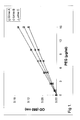
- a colorimetric analysis of the reaction mixture allows to establish the calibration curve (eventually determined a given time after the mixing) which will allow to determine the PEG 6,000 concentration in a range between 1 and at least 30 ⁇ g/ml of urine. In case of higher PEG concentration, a dilution of the urine sample is recommended.
- Figure 1 corresponds to typical calibration curves for PEG 6,000 obtained in these specific conditions. It shows the measurement of PEG 6,000 concentration in three different urine samples. The optical density was measured at 550 nm.
- the measurement of PEG is very simple and based on a change of colour of the reaction mixture, which can be detected visually. It can be estimated by reference to a colour scale calibration curve.
- Example 2 Colorimetric calibration curve of PEG 20,000 in urine
- the iodine reagent has been prepared according to example 1.
- the final concentration of iodine in the reaction mixture in this example is 1.8 mM.
- the mixing of the iodine reagent with PEG present in the biological sample induces a colour change of the solution whose optical density is related to the polyether concentration.
- a colorimetric analysis of the reaction mixture allows to establish the calibration curve (eventually determined a given time after the mixing) which will allow to determine the PEG 20,000 concentration in a range between 1 and at least 30 ⁇ g/ml or urine. In case of a higher PEG concentration, a dilution of the urine sample is recommended.
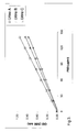
- Figure 2 corresponds to typical calibration curves obtained in these specific conditions.
- the figure shows the measurement of PEG 20,000 in three different urine samples.
- Example 3 Colorimetric calibration curve of PEG 400 in urine in the presence of fixed concentrations of PEG 6,000.
- iodine reagent is added (0.8 ml). The addition of this iodine reagent is performed in such a way to assure an intimate and rapid mixing with the biological sample. As so prepared, the combination of the sample with the iodine reagent is called the reaction mixture.
- the iodine reagent has been prepared according to the following steps:
- the final concentration of iodine in the reaction mixture in this example is 12.5 mM.
- the mixing of the iodine reagent with PEG present in the biological sample induces a colour change of the solution whose optical density is related to the PEG concentration.
- a colorimetric analysis of the reaction mixture allows to establish the calibration curve (eventually determined a given time after the mixing) which will allow to determine the PEG 400 concentration in a range between 20 and at 140 ⁇ g/ml or urine. In case of higher PEG concentration, a dilution of the urine sample is recommend.
- Figure 3 corresponds to typical calibration curves obtained for PEG 400 in the presence of fixed concentrations of PEG 6,000.
- Example 5 Semi-quantitative evaluation of the intestinal permeability measurement carried out by the practitioner or eventually by the patient
- one of the most preferred ways to conduct the diagnostic test should be performed in the following manner:
- a given dose of the low molecular weight PEG (between 0.1 and 50 g, but preferably between 1 and 10 g of PEG 200 -1000) and a high molecular weight PEG (between 0.1 and 50 g, but preferably between 1 and 10 g of PEG 6,000 - 20,000), could be taken by the patient either under the form of a solution or under a dry form where PEGs are incorporated in a galenic form, such as a gelatin capsule.
- the urine will be collected by the patient for 6 up to 24 hours after the PEG oral administration. During collection, urine is kept in a fridge, for example at 4°C.
- the urine will be submitted to a pretreatment, and mixed with a determined volume of an iodine reagent.
- the iodine reagent made commercially available, will have the composition given in example 1.
- the range of molecular weight of PEG taken orally by the patient a significant modification of the colour intensity of the mixture appearing just after mixing will attest an alteration in intestinal permeability.
- the extent of colour intensity variation will be appreciated by the practitoner or eventually thepatient with the help of a given reference colour scale providing a semi-quantitative evaluation of the intestinal permeability modification.
- Example 6 Quantitative evaluation of the intestinal permeability measurement carried out in a clinical laboratory
- a given dose of the low molecular weight PEG (between 0.1 and 50 g, but preferably between 1 and 10 g of PEG 200 -1000) and a high molecular weight PEG (between 0.1 and 50 g, but preferably between 1 and 10 g of PEG 6,000 - 20,000), could be taken by the patient either under the form of a solution or under a dry form where PEGs are incorporated in a galenic form, such as a gelatin capsule.
- the urine will be collected by the patient for 6 up to 24 hours after the PEG oral administration. During collection, urine is kept in a fridge, for example at 4°C. If PEG concentration measurement does not occur immediately after collection, collected urine is conserved in a freezer, for example at -20°C. After thawing, urine sample may be centrifuged.
- the amount of both high and low molecular weight PEG recovered in urine could be quantified following the sequential steps described below:
- iodine reagent To a given volume (0.8 ml) of a pretreated urine sample, 0.2 ml of iodine reagent is added. The addition of this reagent is performed in such a way to assure an intimate and rapid mixing with the biological sample. As so prepared, the combination of the sample with the iodine reagent is called the reaction mixture.
- the iodine reagent has been prepared according to the composition given in example 1.
- the mixing of the iodine reagent with PEG present in the biological sample induces a colour change of the solution whose optical density is related to the PEG concentration.
- a colorimetric analysis of the reaction mixture with reference to a calibration curve will allow to determine the PEG concentration in a range between 1 and at least 30 ⁇ g/ml of urine. In case of higher PEG concentration, a dilution of the urine sample is recommended.
- the optical density is generally determined between 500 and 700 nm, preferentially at 550 nm. In this specific concentration of iodine present in the reagent mixture, the presence of I.m.w. PEG 400 in the urine sample does not significantly interfere with analysis of the high molecular weight PEG.
- iodine reagent (0.8 ml) is added.
- the addition of this iodine reagent is performed in such a way to assure an intimate and rapid mixing with the biological sample.
- the combination of the sample with the iodine reagent is called the reaction mixture.
- the iodine reagent has been prepared according to example 3.
- the mixing of the iodine reagent with PEG present in the biological sample induces a colour change of the solution whose optical density is related to the PEG concentration.
- a colorimetric analysis of the reaction mixture allows to establish the calibration curve (eventually prepared by using urineand determined a given time after the mixing) which will allow to determine the PEG 400 concentration in a range between 25 and 150 ⁇ g/ml of urine. In case of higher PEG concentration, a dilution of the urine sample is recommended.
- the optical density is generally determined between 500 and 700 nm, preferentially at 550 nm.
Landscapes
- Life Sciences & Earth Sciences (AREA)
- Health & Medical Sciences (AREA)
- Immunology (AREA)
- Molecular Biology (AREA)
- Hematology (AREA)
- Engineering & Computer Science (AREA)
- Chemical & Material Sciences (AREA)
- Urology & Nephrology (AREA)
- Physics & Mathematics (AREA)
- Pathology (AREA)
- General Physics & Mathematics (AREA)
- Biomedical Technology (AREA)
- General Health & Medical Sciences (AREA)
- Biochemistry (AREA)
- Analytical Chemistry (AREA)
- Microbiology (AREA)
- Medicinal Chemistry (AREA)
- Food Science & Technology (AREA)
- Biotechnology (AREA)
- Cell Biology (AREA)
- Biophysics (AREA)
- Investigating Or Analysing Biological Materials (AREA)
- Investigating Or Analyzing Non-Biological Materials By The Use Of Chemical Means (AREA)
Priority Applications (4)
| Application Number | Priority Date | Filing Date | Title |
|---|---|---|---|
| EP03077769A EP1512971A1 (fr) | 2003-09-04 | 2003-09-04 | Procédé de détérmination qualitative et/ou quantitative de polyethyléne glycols dans des liquides biologiques |
| PCT/EP2004/051970 WO2005024423A2 (fr) | 2003-09-04 | 2004-08-31 | Procede de detection qualitative et/ou quantitative de glycols polyethylene dans des fluides biologiques |
| EP04766652A EP1664771A2 (fr) | 2003-09-04 | 2004-08-31 | Procede de detection qualitative et/ou quantitative de glycols polyethylene dans des fluides biologiques |
| US10/570,784 US20070196927A1 (en) | 2003-09-04 | 2004-08-31 | Method For Qualitative And/Or Quantitative Detection Of Polyethylene Glycols In Biological Fluids |
Applications Claiming Priority (1)
| Application Number | Priority Date | Filing Date | Title |
|---|---|---|---|
| EP03077769A EP1512971A1 (fr) | 2003-09-04 | 2003-09-04 | Procédé de détérmination qualitative et/ou quantitative de polyethyléne glycols dans des liquides biologiques |
Publications (1)
| Publication Number | Publication Date |
|---|---|
| EP1512971A1 true EP1512971A1 (fr) | 2005-03-09 |
Family
ID=34130241
Family Applications (1)
| Application Number | Title | Priority Date | Filing Date |
|---|---|---|---|
| EP03077769A Withdrawn EP1512971A1 (fr) | 2003-09-04 | 2003-09-04 | Procédé de détérmination qualitative et/ou quantitative de polyethyléne glycols dans des liquides biologiques |
Country Status (1)
| Country | Link |
|---|---|
| EP (1) | EP1512971A1 (fr) |
Citations (2)
| Publication number | Priority date | Publication date | Assignee | Title |
|---|---|---|---|---|
| US3753972A (en) * | 1970-04-28 | 1973-08-21 | Research Corp | Fractionation of agar |
| SU1381401A1 (ru) * | 1986-02-25 | 1988-03-15 | Институт цитологии и генетики СО АН СССР | Способ определени полиэтиленоксида в водных растворах |
-
2003
- 2003-09-04 EP EP03077769A patent/EP1512971A1/fr not_active Withdrawn
Patent Citations (2)
| Publication number | Priority date | Publication date | Assignee | Title |
|---|---|---|---|---|
| US3753972A (en) * | 1970-04-28 | 1973-08-21 | Research Corp | Fractionation of agar |
| SU1381401A1 (ru) * | 1986-02-25 | 1988-03-15 | Институт цитологии и генетики СО АН СССР | Способ определени полиэтиленоксида в водных растворах |
Non-Patent Citations (5)
| Title |
|---|
| CHILDS C.E.: "The Determination of Polyethylene Glycol in Gamma Globulin Solutions", MICROCHEMICAL JOURNAL, vol. 20, 1975, pages 190 - 192, XP009027279 * |
| DATABASE WPI Section Ch Week 198837, Derwent World Patents Index; Class A25, Page 3, AN 1988-263362, XP002273163, BOGDANOVA L.A., ET AL.: "METHOD OF DETERMINING POLYETHYLENEOXIDE IN AQUEOUS SOLUTIONS" * |
| ELSENHANS B ET AL: "DIFFERENTIAL CHANGES IN THE URINARY EXCRETION OF TWO ORALLY ADMINISTERED POLYETHYLENE GLYCOL MARKERS PEG-900 AND PEG-4000 IN RATS AFTER FEEDING VARIOUS CARBOHYDRATE GELLING AGENTS", JOURNAL OF NUTRITION, vol. 119, no. 3, 1989, pages 380 - 387, XP009035402, ISSN: 0022-3166 * |
| GHANDEHARI HAMIDREZA ET AL: "Size-dependent permeability of hydrophilic probes across rabbit colonic epithelium", JOURNAL OF PHARMACOLOGY AND EXPERIMENTAL THERAPEUTICS, vol. 280, no. 2, 1997, pages 747 - 753, XP002293441, ISSN: 0022-3565 * |
| HASTEWELL JOHN ET AL: "Enhancement of human calcitonin absorption across the rat colon in vivo", INTERNATIONAL JOURNAL OF PHARMACEUTICS (AMSTERDAM), vol. 101, no. 1-2, 1994, pages 115 - 120, XP002293442, ISSN: 0378-5173 * |
Similar Documents
| Publication | Publication Date | Title |
|---|---|---|
| de Swart et al. | Second international round robin for the quantification of serum non-transferrin-bound iron and labile plasma iron in patients with iron-overload disorders | |
| JP6518629B2 (ja) | 血液検査キット及び血液分析方法 | |
| EP3321677B1 (fr) | Kit de test sanguin et procédé d'analyse l'utilisant | |
| Koenig et al. | Usefulness of whole blood, plasma, peritoneal fluid, and peritoneal fluid supernatant glucose concentrations obtained by a veterinary point-of-care glucometer to identify septic peritonitis in dogs with peritoneal effusion | |
| Yılmaz et al. | Comparison of different methods for measurement of electrolytes in patients admitted to the intensive care unit | |
| Howanitz et al. | Practices for identifying and rejecting hemolyzed specimens are highly variable in clinical laboratories | |
| CN102854314A (zh) | 一种胶乳免疫比浊法进行幽门螺杆菌抗体检测的试剂盒 | |
| Alberti et al. | The pH of chemistry assays plays an important role in monoclonal immunoglobulin interferences | |
| Bürki et al. | Using the hemolysis index of Abbott’s Alinity c for the measurement of plasma free hemoglobin in ECMO patients | |
| Burmester et al. | Evaluation of a rapid method for the determination of plasma fibrinogen | |
| Lewińska et al. | From the bottom of an old jar: A fluorometric method for the determination of creatinine in human serum | |
| Simeoli et al. | Utilization of volumetric absorptive microsampling and dried plasma spot for quantification of anti-fungal triazole agents in pediatric patients by using liquid chromatography-tandem mass spectrometry | |
| WO2017006962A1 (fr) | Kit d'examen sanguin, et procédé d'analyse sanguine | |
| CN104049089B (zh) | 一种检测纤维蛋白原的方法及试剂盒 | |
| Preeti et al. | Estimation of serum creatinine by routine Jaffé’s method and by dry chemistry in icteric and hemolytic serum samples | |
| Pagliaro et al. | A non-invasive strategy for haemoglobin screening of blood donors | |
| US20070196927A1 (en) | Method For Qualitative And/Or Quantitative Detection Of Polyethylene Glycols In Biological Fluids | |
| EP0097472A1 (fr) | Méthode pour la détermination du calcium dans une prise liquide | |
| ES2332354T3 (es) | Deteccion de litio en muestras biologicas liquidas y los reactivos para dicho fin. | |
| Pinkerton et al. | A colorimetric determination for 1, 1-dimethylhydrazine (UDMH) in air, blood and water | |
| EP1512971A1 (fr) | Procédé de détérmination qualitative et/ou quantitative de polyethyléne glycols dans des liquides biologiques | |
| Dimeski et al. | Paraprotein interference with turbidimetric gentamicin assay | |
| Ranjan et al. | A comparative quality evaluation of point-of-care methodology for testing hemoglobin in blood donors by two different technologies | |
| CN104849224A (zh) | 一种补体c3检测试剂盒及其制备 | |
| Krambeck et al. | Analysis of mixed stones is prone to error: a study with US laboratories using micro CT for verification of sample content |
Legal Events
| Date | Code | Title | Description |
|---|---|---|---|
| PUAI | Public reference made under article 153(3) epc to a published international application that has entered the european phase |
Free format text: ORIGINAL CODE: 0009012 |
|
| AK | Designated contracting states |
Kind code of ref document: A1 Designated state(s): AT BE BG CH CY CZ DE DK EE ES FI FR GB GR HU IE IT LI LU MC NL PT RO SE SI SK TR |
|
| AX | Request for extension of the european patent |
Extension state: AL LT LV MK |
|
| AKX | Designation fees paid | ||
| REG | Reference to a national code |
Ref country code: DE Ref legal event code: 8566 |
|
| STAA | Information on the status of an ep patent application or granted ep patent |
Free format text: STATUS: THE APPLICATION IS DEEMED TO BE WITHDRAWN |
|
| 18D | Application deemed to be withdrawn |
Effective date: 20050910 |