EP1539929B1 - Techniques permettant de restaurer le repertoire immunologique de patients qui presente des defauts immunologiques lies a l'auto-immunite et a un organe ou a une transplantation de cellules souche hematopoietiques multipotentes - Google Patents
Techniques permettant de restaurer le repertoire immunologique de patients qui presente des defauts immunologiques lies a l'auto-immunite et a un organe ou a une transplantation de cellules souche hematopoietiques multipotentes Download PDFInfo
- Publication number
- EP1539929B1 EP1539929B1 EP03761997.0A EP03761997A EP1539929B1 EP 1539929 B1 EP1539929 B1 EP 1539929B1 EP 03761997 A EP03761997 A EP 03761997A EP 1539929 B1 EP1539929 B1 EP 1539929B1
- Authority
- EP
- European Patent Office
- Prior art keywords
- cells
- cell
- population
- antibody
- antigen
- Prior art date
- Legal status (The legal status is an assumption and is not a legal conclusion. Google has not performed a legal analysis and makes no representation as to the accuracy of the status listed.)
- Expired - Lifetime
Links
Images




Classifications
-
- C—CHEMISTRY; METALLURGY
- C12—BIOCHEMISTRY; BEER; SPIRITS; WINE; VINEGAR; MICROBIOLOGY; ENZYMOLOGY; MUTATION OR GENETIC ENGINEERING
- C12N—MICROORGANISMS OR ENZYMES; COMPOSITIONS THEREOF; PROPAGATING, PRESERVING, OR MAINTAINING MICROORGANISMS; MUTATION OR GENETIC ENGINEERING; CULTURE MEDIA
- C12N5/00—Undifferentiated human, animal or plant cells, e.g. cell lines; Tissues; Cultivation or maintenance thereof; Culture media therefor
- C12N5/0081—Purging biological preparations of unwanted cells
- C12N5/0087—Purging against subsets of blood cells, e.g. purging alloreactive T cells
-
- A—HUMAN NECESSITIES
- A61—MEDICAL OR VETERINARY SCIENCE; HYGIENE
- A61K—PREPARATIONS FOR MEDICAL, DENTAL OR TOILETRY PURPOSES
- A61K40/00—Cellular immunotherapy
- A61K40/10—Cellular immunotherapy characterised by the cell type used
- A61K40/11—T-cells, e.g. tumour infiltrating lymphocytes [TIL] or regulatory T [Treg] cells; Lymphokine-activated killer [LAK] cells
-
- A—HUMAN NECESSITIES
- A61—MEDICAL OR VETERINARY SCIENCE; HYGIENE
- A61K—PREPARATIONS FOR MEDICAL, DENTAL OR TOILETRY PURPOSES
- A61K40/00—Cellular immunotherapy
- A61K40/20—Cellular immunotherapy characterised by the effect or the function of the cells
- A61K40/22—Immunosuppressive or immunotolerising
-
- A—HUMAN NECESSITIES
- A61—MEDICAL OR VETERINARY SCIENCE; HYGIENE
- A61K—PREPARATIONS FOR MEDICAL, DENTAL OR TOILETRY PURPOSES
- A61K40/00—Cellular immunotherapy
- A61K40/40—Cellular immunotherapy characterised by antigens that are targeted or presented by cells of the immune system
- A61K40/41—Vertebrate antigens
- A61K40/416—Antigens related to auto-immune diseases; Preparations to induce self-tolerance
-
- A—HUMAN NECESSITIES
- A61—MEDICAL OR VETERINARY SCIENCE; HYGIENE
- A61K—PREPARATIONS FOR MEDICAL, DENTAL OR TOILETRY PURPOSES
- A61K40/00—Cellular immunotherapy
- A61K40/40—Cellular immunotherapy characterised by antigens that are targeted or presented by cells of the immune system
- A61K40/41—Vertebrate antigens
- A61K40/418—Antigens related to induction of tolerance to non-self
-
- A—HUMAN NECESSITIES
- A61—MEDICAL OR VETERINARY SCIENCE; HYGIENE
- A61K—PREPARATIONS FOR MEDICAL, DENTAL OR TOILETRY PURPOSES
- A61K40/00—Cellular immunotherapy
- A61K40/40—Cellular immunotherapy characterised by antigens that are targeted or presented by cells of the immune system
- A61K40/41—Vertebrate antigens
- A61K40/42—Cancer antigens
-
- A—HUMAN NECESSITIES
- A61—MEDICAL OR VETERINARY SCIENCE; HYGIENE
- A61K—PREPARATIONS FOR MEDICAL, DENTAL OR TOILETRY PURPOSES
- A61K40/00—Cellular immunotherapy
- A61K40/40—Cellular immunotherapy characterised by antigens that are targeted or presented by cells of the immune system
- A61K40/46—Viral antigens
-
- A—HUMAN NECESSITIES
- A61—MEDICAL OR VETERINARY SCIENCE; HYGIENE
- A61P—SPECIFIC THERAPEUTIC ACTIVITY OF CHEMICAL COMPOUNDS OR MEDICINAL PREPARATIONS
- A61P1/00—Drugs for disorders of the alimentary tract or the digestive system
- A61P1/04—Drugs for disorders of the alimentary tract or the digestive system for ulcers, gastritis or reflux esophagitis, e.g. antacids, inhibitors of acid secretion, mucosal protectants
-
- A—HUMAN NECESSITIES
- A61—MEDICAL OR VETERINARY SCIENCE; HYGIENE
- A61P—SPECIFIC THERAPEUTIC ACTIVITY OF CHEMICAL COMPOUNDS OR MEDICINAL PREPARATIONS
- A61P1/00—Drugs for disorders of the alimentary tract or the digestive system
- A61P1/16—Drugs for disorders of the alimentary tract or the digestive system for liver or gallbladder disorders, e.g. hepatoprotective agents, cholagogues, litholytics
-
- A—HUMAN NECESSITIES
- A61—MEDICAL OR VETERINARY SCIENCE; HYGIENE
- A61P—SPECIFIC THERAPEUTIC ACTIVITY OF CHEMICAL COMPOUNDS OR MEDICINAL PREPARATIONS
- A61P11/00—Drugs for disorders of the respiratory system
-
- A—HUMAN NECESSITIES
- A61—MEDICAL OR VETERINARY SCIENCE; HYGIENE
- A61P—SPECIFIC THERAPEUTIC ACTIVITY OF CHEMICAL COMPOUNDS OR MEDICINAL PREPARATIONS
- A61P11/00—Drugs for disorders of the respiratory system
- A61P11/02—Nasal agents, e.g. decongestants
-
- A—HUMAN NECESSITIES
- A61—MEDICAL OR VETERINARY SCIENCE; HYGIENE
- A61P—SPECIFIC THERAPEUTIC ACTIVITY OF CHEMICAL COMPOUNDS OR MEDICINAL PREPARATIONS
- A61P11/00—Drugs for disorders of the respiratory system
- A61P11/06—Antiasthmatics
-
- A—HUMAN NECESSITIES
- A61—MEDICAL OR VETERINARY SCIENCE; HYGIENE
- A61P—SPECIFIC THERAPEUTIC ACTIVITY OF CHEMICAL COMPOUNDS OR MEDICINAL PREPARATIONS
- A61P13/00—Drugs for disorders of the urinary system
- A61P13/12—Drugs for disorders of the urinary system of the kidneys
-
- A—HUMAN NECESSITIES
- A61—MEDICAL OR VETERINARY SCIENCE; HYGIENE
- A61P—SPECIFIC THERAPEUTIC ACTIVITY OF CHEMICAL COMPOUNDS OR MEDICINAL PREPARATIONS
- A61P15/00—Drugs for genital or sexual disorders; Contraceptives
-
- A—HUMAN NECESSITIES
- A61—MEDICAL OR VETERINARY SCIENCE; HYGIENE
- A61P—SPECIFIC THERAPEUTIC ACTIVITY OF CHEMICAL COMPOUNDS OR MEDICINAL PREPARATIONS
- A61P17/00—Drugs for dermatological disorders
-
- A—HUMAN NECESSITIES
- A61—MEDICAL OR VETERINARY SCIENCE; HYGIENE
- A61P—SPECIFIC THERAPEUTIC ACTIVITY OF CHEMICAL COMPOUNDS OR MEDICINAL PREPARATIONS
- A61P17/00—Drugs for dermatological disorders
- A61P17/02—Drugs for dermatological disorders for treating wounds, ulcers, burns, scars, keloids, or the like
-
- A—HUMAN NECESSITIES
- A61—MEDICAL OR VETERINARY SCIENCE; HYGIENE
- A61P—SPECIFIC THERAPEUTIC ACTIVITY OF CHEMICAL COMPOUNDS OR MEDICINAL PREPARATIONS
- A61P17/00—Drugs for dermatological disorders
- A61P17/06—Antipsoriatics
-
- A—HUMAN NECESSITIES
- A61—MEDICAL OR VETERINARY SCIENCE; HYGIENE
- A61P—SPECIFIC THERAPEUTIC ACTIVITY OF CHEMICAL COMPOUNDS OR MEDICINAL PREPARATIONS
- A61P17/00—Drugs for dermatological disorders
- A61P17/14—Drugs for dermatological disorders for baldness or alopecia
-
- A—HUMAN NECESSITIES
- A61—MEDICAL OR VETERINARY SCIENCE; HYGIENE
- A61P—SPECIFIC THERAPEUTIC ACTIVITY OF CHEMICAL COMPOUNDS OR MEDICINAL PREPARATIONS
- A61P19/00—Drugs for skeletal disorders
- A61P19/02—Drugs for skeletal disorders for joint disorders, e.g. arthritis, arthrosis
-
- A—HUMAN NECESSITIES
- A61—MEDICAL OR VETERINARY SCIENCE; HYGIENE
- A61P—SPECIFIC THERAPEUTIC ACTIVITY OF CHEMICAL COMPOUNDS OR MEDICINAL PREPARATIONS
- A61P21/00—Drugs for disorders of the muscular or neuromuscular system
-
- A—HUMAN NECESSITIES
- A61—MEDICAL OR VETERINARY SCIENCE; HYGIENE
- A61P—SPECIFIC THERAPEUTIC ACTIVITY OF CHEMICAL COMPOUNDS OR MEDICINAL PREPARATIONS
- A61P21/00—Drugs for disorders of the muscular or neuromuscular system
- A61P21/04—Drugs for disorders of the muscular or neuromuscular system for myasthenia gravis
-
- A—HUMAN NECESSITIES
- A61—MEDICAL OR VETERINARY SCIENCE; HYGIENE
- A61P—SPECIFIC THERAPEUTIC ACTIVITY OF CHEMICAL COMPOUNDS OR MEDICINAL PREPARATIONS
- A61P25/00—Drugs for disorders of the nervous system
-
- A—HUMAN NECESSITIES
- A61—MEDICAL OR VETERINARY SCIENCE; HYGIENE
- A61P—SPECIFIC THERAPEUTIC ACTIVITY OF CHEMICAL COMPOUNDS OR MEDICINAL PREPARATIONS
- A61P27/00—Drugs for disorders of the senses
- A61P27/02—Ophthalmic agents
-
- A—HUMAN NECESSITIES
- A61—MEDICAL OR VETERINARY SCIENCE; HYGIENE
- A61P—SPECIFIC THERAPEUTIC ACTIVITY OF CHEMICAL COMPOUNDS OR MEDICINAL PREPARATIONS
- A61P29/00—Non-central analgesic, antipyretic or antiinflammatory agents, e.g. antirheumatic agents; Non-steroidal antiinflammatory drugs [NSAID]
-
- A—HUMAN NECESSITIES
- A61—MEDICAL OR VETERINARY SCIENCE; HYGIENE
- A61P—SPECIFIC THERAPEUTIC ACTIVITY OF CHEMICAL COMPOUNDS OR MEDICINAL PREPARATIONS
- A61P29/00—Non-central analgesic, antipyretic or antiinflammatory agents, e.g. antirheumatic agents; Non-steroidal antiinflammatory drugs [NSAID]
- A61P29/02—Non-central analgesic, antipyretic or antiinflammatory agents, e.g. antirheumatic agents; Non-steroidal antiinflammatory drugs [NSAID] without antiinflammatory effect
-
- A—HUMAN NECESSITIES
- A61—MEDICAL OR VETERINARY SCIENCE; HYGIENE
- A61P—SPECIFIC THERAPEUTIC ACTIVITY OF CHEMICAL COMPOUNDS OR MEDICINAL PREPARATIONS
- A61P3/00—Drugs for disorders of the metabolism
- A61P3/02—Nutrients, e.g. vitamins, minerals
-
- A—HUMAN NECESSITIES
- A61—MEDICAL OR VETERINARY SCIENCE; HYGIENE
- A61P—SPECIFIC THERAPEUTIC ACTIVITY OF CHEMICAL COMPOUNDS OR MEDICINAL PREPARATIONS
- A61P3/00—Drugs for disorders of the metabolism
- A61P3/08—Drugs for disorders of the metabolism for glucose homeostasis
- A61P3/10—Drugs for disorders of the metabolism for glucose homeostasis for hyperglycaemia, e.g. antidiabetics
-
- A—HUMAN NECESSITIES
- A61—MEDICAL OR VETERINARY SCIENCE; HYGIENE
- A61P—SPECIFIC THERAPEUTIC ACTIVITY OF CHEMICAL COMPOUNDS OR MEDICINAL PREPARATIONS
- A61P35/00—Antineoplastic agents
- A61P35/02—Antineoplastic agents specific for leukemia
-
- A—HUMAN NECESSITIES
- A61—MEDICAL OR VETERINARY SCIENCE; HYGIENE
- A61P—SPECIFIC THERAPEUTIC ACTIVITY OF CHEMICAL COMPOUNDS OR MEDICINAL PREPARATIONS
- A61P37/00—Drugs for immunological or allergic disorders
- A61P37/02—Immunomodulators
-
- A—HUMAN NECESSITIES
- A61—MEDICAL OR VETERINARY SCIENCE; HYGIENE
- A61P—SPECIFIC THERAPEUTIC ACTIVITY OF CHEMICAL COMPOUNDS OR MEDICINAL PREPARATIONS
- A61P37/00—Drugs for immunological or allergic disorders
- A61P37/02—Immunomodulators
- A61P37/04—Immunostimulants
-
- A—HUMAN NECESSITIES
- A61—MEDICAL OR VETERINARY SCIENCE; HYGIENE
- A61P—SPECIFIC THERAPEUTIC ACTIVITY OF CHEMICAL COMPOUNDS OR MEDICINAL PREPARATIONS
- A61P37/00—Drugs for immunological or allergic disorders
- A61P37/02—Immunomodulators
- A61P37/06—Immunosuppressants, e.g. drugs for graft rejection
-
- A—HUMAN NECESSITIES
- A61—MEDICAL OR VETERINARY SCIENCE; HYGIENE
- A61P—SPECIFIC THERAPEUTIC ACTIVITY OF CHEMICAL COMPOUNDS OR MEDICINAL PREPARATIONS
- A61P37/00—Drugs for immunological or allergic disorders
- A61P37/08—Antiallergic agents
-
- A—HUMAN NECESSITIES
- A61—MEDICAL OR VETERINARY SCIENCE; HYGIENE
- A61P—SPECIFIC THERAPEUTIC ACTIVITY OF CHEMICAL COMPOUNDS OR MEDICINAL PREPARATIONS
- A61P43/00—Drugs for specific purposes, not provided for in groups A61P1/00-A61P41/00
-
- A—HUMAN NECESSITIES
- A61—MEDICAL OR VETERINARY SCIENCE; HYGIENE
- A61P—SPECIFIC THERAPEUTIC ACTIVITY OF CHEMICAL COMPOUNDS OR MEDICINAL PREPARATIONS
- A61P5/00—Drugs for disorders of the endocrine system
- A61P5/14—Drugs for disorders of the endocrine system of the thyroid hormones, e.g. T3, T4
-
- A—HUMAN NECESSITIES
- A61—MEDICAL OR VETERINARY SCIENCE; HYGIENE
- A61P—SPECIFIC THERAPEUTIC ACTIVITY OF CHEMICAL COMPOUNDS OR MEDICINAL PREPARATIONS
- A61P5/00—Drugs for disorders of the endocrine system
- A61P5/14—Drugs for disorders of the endocrine system of the thyroid hormones, e.g. T3, T4
- A61P5/16—Drugs for disorders of the endocrine system of the thyroid hormones, e.g. T3, T4 for decreasing, blocking or antagonising the activity of the thyroid hormones
-
- A—HUMAN NECESSITIES
- A61—MEDICAL OR VETERINARY SCIENCE; HYGIENE
- A61P—SPECIFIC THERAPEUTIC ACTIVITY OF CHEMICAL COMPOUNDS OR MEDICINAL PREPARATIONS
- A61P5/00—Drugs for disorders of the endocrine system
- A61P5/38—Drugs for disorders of the endocrine system of the suprarenal hormones
- A61P5/40—Mineralocorticosteroids, e.g. aldosterone; Drugs increasing or potentiating the activity of mineralocorticosteroids
-
- A—HUMAN NECESSITIES
- A61—MEDICAL OR VETERINARY SCIENCE; HYGIENE
- A61P—SPECIFIC THERAPEUTIC ACTIVITY OF CHEMICAL COMPOUNDS OR MEDICINAL PREPARATIONS
- A61P7/00—Drugs for disorders of the blood or the extracellular fluid
-
- A—HUMAN NECESSITIES
- A61—MEDICAL OR VETERINARY SCIENCE; HYGIENE
- A61P—SPECIFIC THERAPEUTIC ACTIVITY OF CHEMICAL COMPOUNDS OR MEDICINAL PREPARATIONS
- A61P7/00—Drugs for disorders of the blood or the extracellular fluid
- A61P7/06—Antianaemics
-
- A—HUMAN NECESSITIES
- A61—MEDICAL OR VETERINARY SCIENCE; HYGIENE
- A61P—SPECIFIC THERAPEUTIC ACTIVITY OF CHEMICAL COMPOUNDS OR MEDICINAL PREPARATIONS
- A61P9/00—Drugs for disorders of the cardiovascular system
-
- A—HUMAN NECESSITIES
- A61—MEDICAL OR VETERINARY SCIENCE; HYGIENE
- A61P—SPECIFIC THERAPEUTIC ACTIVITY OF CHEMICAL COMPOUNDS OR MEDICINAL PREPARATIONS
- A61P9/00—Drugs for disorders of the cardiovascular system
- A61P9/04—Inotropic agents, i.e. stimulants of cardiac contraction; Drugs for heart failure
-
- C—CHEMISTRY; METALLURGY
- C12—BIOCHEMISTRY; BEER; SPIRITS; WINE; VINEGAR; MICROBIOLOGY; ENZYMOLOGY; MUTATION OR GENETIC ENGINEERING
- C12N—MICROORGANISMS OR ENZYMES; COMPOSITIONS THEREOF; PROPAGATING, PRESERVING, OR MAINTAINING MICROORGANISMS; MUTATION OR GENETIC ENGINEERING; CULTURE MEDIA
- C12N5/00—Undifferentiated human, animal or plant cells, e.g. cell lines; Tissues; Cultivation or maintenance thereof; Culture media therefor
- C12N5/06—Animal cells or tissues; Human cells or tissues
- C12N5/0602—Vertebrate cells
- C12N5/0634—Cells from the blood or the immune system
- C12N5/0636—T lymphocytes
-
- Y—GENERAL TAGGING OF NEW TECHNOLOGICAL DEVELOPMENTS; GENERAL TAGGING OF CROSS-SECTIONAL TECHNOLOGIES SPANNING OVER SEVERAL SECTIONS OF THE IPC; TECHNICAL SUBJECTS COVERED BY FORMER USPC CROSS-REFERENCE ART COLLECTIONS [XRACs] AND DIGESTS
- Y02—TECHNOLOGIES OR APPLICATIONS FOR MITIGATION OR ADAPTATION AGAINST CLIMATE CHANGE
- Y02A—TECHNOLOGIES FOR ADAPTATION TO CLIMATE CHANGE
- Y02A50/00—TECHNOLOGIES FOR ADAPTATION TO CLIMATE CHANGE in human health protection, e.g. against extreme weather
- Y02A50/30—Against vector-borne diseases, e.g. mosquito-borne, fly-borne, tick-borne or waterborne diseases whose impact is exacerbated by climate change
Definitions
- the present invention relates generally to methods to eliminate undesired (e.g. autoreactive, alloreactive, pathogenic) subpopulations of T cells from a mixed population ofT cells.
- undesired e.g. autoreactive, alloreactive, pathogenic
- TCR T cell antigen receptor
- APC antigen-presenting cells
- T lymphocytes typically require a second co-stimulatory signal.
- Co-stimulation is typically necessary for a T helper cell to produce sufficient cytokine levels that induce clonal expansion. Bretscher, Immunol. Today 13: 74, 1992 ; June et al., Immunol. Today 15:321, 1994 .
- the major co-stimulatory signal occurs when a member of the B7 family ligands (CD80 (B7.1) or CD86 (B7.2)) on an activated antigen-presenting cell (APC) binds to CD28 on a T cell.
- T cell compositions useful in immunotherapy have the potential to generate a variety of T cell compositions useful in immunotherapy.
- Successful immunotherapy can be aided by increasing the reactivity and quantity of T cells by efficient stimulation.
- successful immunotherapy can be aided by the elimination of unwanted autoreactive or alloreactive cells.
- IL-2 interleukin-2
- IL-2 has been used together with an anti-CD3 antibody to stimulate T cell proliferation, predominantly expanding the CD8 + subpopulation of T cells.
- Both the APC signals directed towards the TCR/CD3 complex and CD28 on the surface of T cells are thought to be required for optimal T cell activation, expansion, and long-term survival of the T cells upon re-infusion.
- the requirement for MHC-matched APCs as accessory cells presents a significant problem for long-term culture systems because APCs are relatively short-lived. Therefore, in a long-term culture system, APCs must be continually obtained from a source and replenished.
- the necessity for a renewable supply of accessory cells is problematic for treatment of immunodeficiencies in which accessory cells are affected.
- the cells may contaminate the entire T cell population during long-term culture.
- a co-stimulatory signal may be delivered to a T cell population, for example, by exposing the cells to a CD3 ligand and a CD28 ligand attached to a solid phase surface, such as a bead. See C. June, et al. (U.S. Patent No. 5,858,358 ); C. June et al. WO 99/953823 . While these methods are capable of achieving therapeutically useful T cell populations, increased robustness and ease ofT cell preparation remain less than ideal.
- Shibuya et al discloses an ex-vivo method of simulating T cells to overcome tumour-related immunosupression.
- Lum et al Journal of Immunotherapy, Lippincott, Williams & Wilkins, Hagerstown, MD, US, Vol 24, No. 5, 2001, P408-419 discloses a similar process to that of Shibuya et al, where the aim is to activate anti-tumour T cells by exposing them to immobilized anti-CD3 and anti-CD28 antibodies.
- the methods currently available in the art have not focused on short-term expansion of T cells or obtaining a more robust population of T cells and the beneficial results thereof. None of these methods has described using such or similar methods to eliminate an undesired clonal or oligoclonal T cell population from a T cell population nor the beneficial results thereof. Moreover, the methods previously available tend to further skew the clonality of the T cell population rather than eliminate undesired reactive clones from a T cell population, and restore a normal immune repertoire. For maximum in vivo effectiveness, theoretically, an ex vivo , or in vivo-generated, activated T cell population should be in a state that can maximally orchestrate an immune response to cancer, infectious disease, or other disease states.
- the activated T cell populations should be in a state to reconstitute a normal T cell repertoire with a reduced presence or entirely without the presence of autoreactive or potentially pathogenic alloreactive T cells.
- patients with autoimmune diseases are treated with long-term immunosuppression to inhibit the autoreactive T cells that cause disease.
- the immunosuppressive agents are stopped, disease recurs often concomitant with reappearance of disease causing T cells that re-emerge in these patients.
- the major problem in hematopoietic stem cell transplantation is graft-versus-host disease (GVHD), which is caused by alloreactive T cells present in the infused hematopoietic stem cell preparation.
- graft rejection mediated by alloreactive host T cells is the major problem, usually overcome by long-term immunosuppression of the transplant recipient.
- the present invention provides methods to generate an increased number of more highly activated and more pure T cells that have surface receptor and cytokine production characteristics that appear more healthy and natural than other expansion methods and further provides for the diminution or elimination of undesired autoreactive or alloreactive populations of T cells.
- the present invention is useful in providing populations of T cells for use in the setting of autoimmune diseases, hematopoietic stem cell, and organ transplantation, as well as other settings where reconstitution of an ablated, abrogated, or otherwise dysfunctional T cell immune system is desired.
- CD28 expression is correlated with a lack of CD40 ligand expression rendering these CD4 + T cells incapable of promoting B cell differentiation and immunoglobulin secretion.
- Aging-related accumulation of CD4 + CD28 - T cells results in an immune compartment that is skewed towards autoreactive responses and away from the generation of high-affinity B cell responses against exogenous antigens.
- One aspect of the present invention provides for an ex vivo method for eliminating at least a substantial portion of a clonal T cell population from a mixed population of T cells from an individual, comprising, exposing a population of cells wherein at least a portion thereof comprises T cells to one or more pro-apoptotic or growth inhibiting compositions wherein said exposure induces apoptosis or growth inhibition in at least a substantial portion of at least one clonal T cell population present in the mixed population of T cells; thereby eliminating at least a substantial portion of said clonal T cell from the mixed population, characterized in that the pro-apoptopic or growth inhibiting composition comprises an autoantigen.
- the method further comprises expanding the mixed population of T cells, by exposing the remaining mixed population of T cells to the pro-apoptotic composition, wherein said exposure induces proliferation in the mixed population of T cells.
- the pro-apoptotic composition comprises anti-CD3 and anti-CD28 antibodies co-immobilized on a bead.
- the pro-apoptotic composition used to eliminate at least a substantial portion of said clonal T cell population from the mixed population of T cells is the same composition used to expand the remaining mixed population ofT cells.
- the method further comprises expanding the remaining population of cells. In another embodiment, the method further comprises expanding the remaining population of cells by exposing the remaining population of cells to a surface wherein the surface has attached thereto one or more agents that ligate a cell surface moiety of at least a portion of the remaining T cells and stimulates said remaining T cells.
- the surface has attached thereto a first agent that ligates a first T cell surface moiety of a T cell, and the same or a second surface has attached thereto a second agent that ligates a second moiety of said T cell, wherein said ligation by the first and second agent induces proliferation of said T cell.
- the agent attached to the surface is an antibody or an antibody fragment.
- the first agent is an antibody or a fragment thereof
- the second agent is an antibody or a fragment thereof
- the first and the second agents are different antibodies.
- the first agent is an anti-CD3 antibody, an anti-CD2 antibody, or an antibody fragment of an anti-CD3 or anti-CD2 antibody.
- the second agent is an anti-CD28 antibody or antibody fragment thereof.
- the first agent is an anti-CD3 antibody and the second agent is an anti-CD28 antibody.
- the cells are exposed to the surfaces of the present invention for a time sufficient to increase polyclonality.
- this increase in polyclonality comprises a shift from mono to oligoclonality or to polyclonality of the T cell population as measured by a V ⁇ , V ⁇ , V ⁇ or V ⁇ spectratype profile of at least one V ⁇ , V ⁇ , V ⁇ or V ⁇ family gene.
- Illustrative pro-apoptotic compositions of the present invention include but are not limited to anti-CD3 antibody, anti-CD2 antibody, anti-CD28 antibody, anti-CD20 antibody, target antigen, MHC-peptide tetramers, Fas ligand, anti-Fas antibody, IL-2, IL-4, TRAIL, rolipram, doxorubicin, chlorambucil, fludarabine, cyclophosphamide, azathioprine, methotrexate, cyclosporine, mycophenolate, FK506, inhibitors of bcl-2, topoisomerase inhibitor, interleukin-1 ⁇ converting enzyme (ICE)-binding agents, Shigella IpaB protein, staurosporine, ultraviolet irradiation, gamma irradiation, tumor necrosis factor, target antigens nucleic acid molecules, proteins or peptides, and non-protein or non-polynucleotide compounds.
- the pro-apoptotic compositions comprises an autoantigen.
- autoantigens of the present invention include but are not limited to, myelin basic protein (MBP), MBP 84-102, MBP 143-168, pancreatic islet cell antigens, collagen, CLIP-170, thyroid antigens, nucleic acid, acetylcholine receptor, S Antigen, and type II collagen.
- the present invention may be useful in methods for treating autoimmune disease in a patient comprising administering to a patient the populations of T cells generated by the present invention, for example where the patient has been treated with a chemotherapeutic agent prior to administering the population of T cells.
- chemotherapeutic agents include but are not limited to campath, anti-CD3 antibodies, cytoxin, fludarabine, cyclosporine, FK506, mycophenolic acid, steroids, FR901228, and irradiation.
- the patient is treated with a T cell ablative therapy prior to administration of the populations of T cells generated by the present invention.
- One aspect of the present invention is an ex vivo method for eliminating at least a substantial portion of a clonal T cell population from a population of T cells, comprising, providing a population of cells wherein at least a portion thereof comprises T cells; exposing the population of cells to one or more agents that sensitize at least a portion of the T cells to further activation or stimulation, exposing the population of cells to a surface wherein the surface has attached thereto one or more agents that ligate a cell surface moiety of at least a portion of the sensitized T cells and stimulates said sensitized T cells, wherein the exposure of said sensitized T cells to said surface is for a time sufficient to induce apoptosis of said sensitized T cells; thereby eliminating said sensitized T cells from the population, characterized in that the composition that sensitizes comprises an autoantigen.
- the method further comprises exposing said population of cells to said surface for a time sufficient to stimulate at least a portion of the remaining T cells and wherein said at least a portion of the remaining cells proliferates.
- the method provides that said surface has attached thereto a first agent that ligates a first T cell surface moiety of a T cell; and the same or a second surface has attached thereto a second agent that ligates a second moiety of said T cell, wherein said ligation by the first and second agent induces proliferation of said T cell.
- at least one agent is an antibody or an antibody fragment.
- the first agent is an antibody or a fragment thereof
- the second agent is an antibody or a fragment thereof.
- the first and the second agents are different antibodies.
- the first agent is an anti-CD3 antibody, an anti-CD2 antibody, or an antibody fragment of an anti-CD3 or anti-CD2 antibody.
- the second agent is an anti-CD28 antibody or antibody fragment thereof.
- the first agent is an anti-CD3 antibody and the second agent is an anti-CD28 antibody.
- cells are exposed to said surface for a time sufficient to increase polyclonality
- the increase in polyclonality comprises a shift from mono to oligoclonality or to polyclonality of the T cell population as measured by a V ⁇ , V ⁇ , V ⁇ or V ⁇ spectratype profile of at least one V ⁇ , V ⁇ , V ⁇ or V ⁇ family gene.
- the patient requires a hematopoietic stem cell transplant.
- the composition that sensitizes recipient PBMCs that have been treated such that they are unable to continue dividing and the population of cells comprises donor T cells.
- the present invention may be useful in methods for reducing the risk of, or the severity of, an adverse GVHD effect in a patient who is undergoing a hematopoietic stem cell transplant, comprising administering to said patient the population of T cells produced according to the methods described herein.
- the patients to receive the cells require an organ transplant.
- the composition that sensitizes comprises irradiated donor cells and the population of cells comprises recipient T cells. These cells may be administered to a patient receiving an organ transplant, to reduce the risk of organ rejection. For example, the organ transplant patient is treated with a T cell ablative therapy prior to administration of the population of T cells.
- the composition that sensitizes comprises an autoantigen.
- autoantigens of the present invention include but are not limited to myelin basic protein (MBP), MBP 84-102, MBP 143-168, pancreatic islet cell antigens, S Antigen, and type II collagen.
- MBP myelin basic protein
- a patient with an autoimmune disease may be treated by administration of a population of T cells generated according to this method.
- the patient may be treated with a T cell ablative therapy prior to administering the population of T cells.
- the purity of the CD3+/CD28 + T cells may be at least 90% pure. In further embodiments, the purity of the CD3 + /CD28 + T cells is 91%, 92%, 93%, 94%, 95%, 96%, 97%, or 98% pure. The purity of the CD3 + /CD28 + T cells may be at least 99% pure. The purity of the CD3 + /CD28 + T cells may be at least 99.9% pure. The population of CD3 + /CD28 + T cells may comprise less than 9%, 8%, 7%, 6%, 5%, 4%, 3%, 2%, 1%, 0.5% or 0.1% contaminating CD28 - T cells.
- the CD3 + surface molecule is stimulated using anti-CD3 antibodies and the CD28 + surface molecule is stimulated using anti-CD28 antibodies.
- a substantially pure population of CD3 + CD28 + T cells, including CD4 + CD3 + CD28 + T cells, and CD8 + CD3 + CD28 + T cells can be used in the treatment of people suffering from autoimmune diseases such as, rheumatoid arthritis, multiple sclerosis, insulin dependent diabetes, Addison's disease, celiac disease, chronic fatigue syndrome, inflammatory bowel disease, ulcerativecolitis, Crohn's disease, Fibromyalgia, systemic lupus erythematosus, psoriasis, Sjogren's syndrome, hyperthyroidism/Graves disease, hypothyroidism/Hashimoto's disease, Insulin-dependent diabetes (type 1), Myasthenia Gravis, endometriosis, scleroderma, pernicious anemia, Goodpasture syndrome, Wegener's disease, glomerulonephritis, aplastic anemia, paroxysmal nocturnal hemoglobinuria, myelodysplastic syndrome,
- biocompatible refers to the property of being predominantly non-toxic to living cells.
- stimulation refers to a primary response induced by ligation of a cell surface moiety.
- such stimulation entails the ligation of a receptor and a subsequent signal transduction event.
- stimulation refers to the ligation of a T cell surface moiety that in one embodiment subsequently induces a signal transduction event, such as binding the TCR/CD3 complex.
- the stimulation event may activate a cell and up- or down-regulate expression of cell surface molecules such as receptors or adhesion molecules, or up- or down-regulate secretion of a molecule, such as downregulation of Tumor Growth Factor beta (TGF- ⁇ ).
- TGF- ⁇ Tumor Growth Factor beta
- ligation of cell surface moieties may result in the reorganization of cytoskeletal structures, or in the coalescing of cell surface moieties, each of which could serve to enhance, modify, or alter subsequent cell responses.
- activation refers to the state of a cell following sufficient cell surface moiety ligation to induce a measurable morphological, phenotypic, and/or functional change.
- T cells such activation may be the state of a T cell that has been sufficiently stimulated to induce cellular proliferation.
- Activation of a T cell may also induce cytokine production and/or secretion, and up- or down-regulation of expression of cell surface molecules such as receptors or adhesion molecules, or up- or down-regulation of secretion of certain molecules, and performance of regulatory or cytolytic effector functions.
- this term infers either up- or down-regulation of a particular physico-chemical process.
- target cell refers to any cell that is intended to be stimulated by cell surface moiety ligation.
- an “antibody”, as used herein, includes both polyclonal and monoclonal antibodies (mAb); primatized (e.g., humanized); murine; mouse-human; mouse-primate; and chimeric; and may be an intact molecule, a fragment thereof (such as scFv, Fv, Fd, Fab, Fab' and F(ab)' 2 fragments), or multimers or aggregates of intact molecules and/or fragments; and may occur in nature or be produced, e.g., by immunization, synthesis or genetic engineering; an "antibody fragment,” as used herein, refers to fragments, derived from or related to an antibody, which bind antigen and which in some embodiments may be derivatized to exhibit structural features that facilitate clearance and uptake, e.g., by the incorporation of galactose residues. This includes, e.g., F(ab), F(ab)' 2 , scFv, light chain variable region (V L ), heavy chain
- protein includes proteins, glycoproteins and other cell-derived modified proteins, polypeptides and peptides; and may be an intact molecule, a fragment thereof, or multimers or aggregates of intact molecules and/or fragments; and may occur in nature or be produced, e.g ., by synthesis (including chemical and/or enzymatic) or genetic engineering.
- agent refers to a molecule that binds to a defined population of cells.
- the agent may bind any cell surface moiety, such as a receptor, an antigenic determinant, or other binding site present on the target cell population.
- the agent may be a protein, peptide, antibody and antibody fragments thereof, fusion proteins, synthetic molecule, an organic molecule ( e.g ., a small molecule), or the like.
- antibodies are used as a prototypical example of such an agent.
- cell surface moiety may refer to a cell surface receptor, an antigenic determinant, or any other binding site present on a target cell population.
- agent that binds a cell surface moiety and "cell surface moiety”, as used herein, should be viewed as a complementary/anti-complementary set of molecules that demonstrate specific binding, generally of relatively high affinity.
- a “co-stimulatory signal”, as used herein, refers to a signal, which in combination with a primary signal, such as TCR/CD3 ligation, leads to T cell proliferation and/or activation.
- Separatation includes any means of substantially purifying one component from another (e.g ., by filtration, affinity, buoyant density, or magnetic attraction).
- a "surface”, as used herein, refers to any surface capable of having an agent attached thereto and includes, without limitation, metals, glass, plastics, copolymers, colloids, lipids, cell surfaces, and the like. Essentially any surface that is capable of retaining an agent bound or attached thereto.
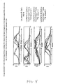
- a population of T cells is considered monoclonal (or mono-specific) when the V ⁇ , V ⁇ , V ⁇ , and/or V ⁇ spectratype profile for a given TCR V ⁇ , V ⁇ , V ⁇ , and/or V ⁇ family has a single predominant peak.
- Spectratype analysis distinguishes rearranged variable genes of a particular size, not sequence.
- a single peak could represent a population of T cells expressing any one of a limited number of rearranged TCR variable genes (V ⁇ , V ⁇ , V ⁇ , or V ⁇ ) comprising any one of the 4 potential nucleotides (adenine (a), guanine (g), cytosine (c), or thymine (t)) or a combination of the 4 nucleotides at the junctional region.
- V ⁇ , V ⁇ , V ⁇ , or V ⁇ comprising any one of the 4 potential nucleotides (adenine (a), guanine (g), cytosine (c), or thymine (t)) or a combination of the 4 nucleotides at the junctional region.
- Oleclonality in the context of a population of T cells, refers to a population of T cells that has multiple, but narrow antigen specificity. This can be defined by spectratype analysis (a measure of the TCR V ⁇ , V ⁇ , V ⁇ , or V ⁇ ) chain hypervariable region repertoire). A population of T cells is considered oligoclonal when the V ⁇ spectratype profile for a given TCR V ⁇ , V ⁇ , V ⁇ , or V ⁇ family has between about 2 and about 4 predominant peaks. This can also be defined by generation and characterization of antigen-specific clones to an antigen of interest.
- Polyclonality in the context of a population of T cells, refers to a population of T cells that has multiple and broad antigen specificity. This can be by spectratype analysis (a measure of the TCR V ⁇ , V ⁇ , V ⁇ , or V ⁇ chain hypervariable region repertoire). A population of T cells is considered polyclonal when the V ⁇ spectratype profile for a given TCR V ⁇ , V ⁇ , V ⁇ , or V ⁇ family has multiple peaks, typically 5 or more predominant peaks and in most cases with Gaussian distribution. Polyclonality can also be defined by generation and characterization of antigen-specific clones to an antigen of interest.
- Restoring or increasing the polyclonality refers to a shift from a monoclonal profile to an oligoclonal profile or to a polyclonal profile, or from an oligoclonal profile to a polyclonal profile, in expressed TCR V ⁇ , V ⁇ , V ⁇ , and/or V ⁇ genes in a population of T cells, as measured by spectratype analysis or by similar analysis such as flow cytometry or sequence analysis.
- the shift from a monoclonal V ⁇ , V ⁇ , V ⁇ , and/or V ⁇ expression profile in a population of T cells to an oligoclonal profile or to a polyclonal profile is generally seen in at least one TCR V ⁇ , V ⁇ , V ⁇ , and/or V ⁇ family.
- this shift is observed in 2, 3, 4, or 5 V ⁇ families. In certain embodiments of the present invention, a shift is observed in 6, 7, 8, 9, or 10 V ⁇ families. In a further embodiment of the present invention, a shift is observed in from 11, 12, 13, or 14 V ⁇ families. In a further embodiment of the present invention, a shift is observed in from 15 to 20 V ⁇ families. In a further embodiment of the present invention, a shift is observed in 20 to 24 V ⁇ families. In another embodiment, a shift is seen in all V ⁇ families.
- the functional significance of restoring or increasing the polyclonality of a population of T cells is that the immune potential, or the ability to respond to a full breadth of antigens, of the population of T cells is restored or increased.
- some T cells within a population may not have their TCRs engaged by the methods set forth herein (e.g ., T cells with downregulated TCR expression). However, by being in close proximity to T cells activated by the methods described herein, and the factors secreted by them, these T cells may in turn upregulate their TCR expression thereby resulting in a further increase in the polyclonality of the population of T cells. Restoration or increase in polyclonality can also be measured by determining the breadth of response to a particular antigen of interest, for example by measuring the number of different epitopes recognized by antigen-specific cells. This can be carried out using standard techniques for generating and cloning antigen-specific T cells in vitro.
- clonal T cell population refers to a T cell population that has a given range of specificities against a given target antigen. This can be measured by any number of assays known in the art, for example by generating and measuring the breadth of specificities ( i.e . number of different specificities) of antigen-specific clones in a given population.
- a clonal T cell population can also be defined by having either monoclonal or oligoclonal specificity as defined by spectratype analysis (a measure of the TCR V ⁇ , V ⁇ , V ⁇ , or V ⁇ chain hypervariable region repertoire).
- animal or "mammal” as used herein, encompasses all mammals, including humans.
- the animal of the present invention is a human subject.
- exposing refers to bringing into the state or condition of immediate proximity or direct contact.
- proliferation means to grow or multiply by producing new cells.
- Immuno response or responsiveness refers to activation of cells of the immune system, including but not limited to, T cells, such that a particular effector function(s) of a particular cell is induced. Effector functions may include, but are not limited to, proliferation, secretion of cytokines, secretion of antibodies, expression of regulatory and/or adhesion molecules, and the ability to induce cytolysis.
- Stimulating an immune response refers to any stimulation such that activation and induction of effector functions of cells of the immune system are achieved.
- Immuno response dysfunction refers to the inappropriate activation and/or proliferation, or lack thereof, of cells of the immune system, and/or the inappropriate secretion, or lack thereof, of cytokines, and/or the inappropriate or inadequate induction of other effector functions of cells of the immune system, such as expression of regulatory, adhesion, and/or homing receptors, and the induction of cytolysis.
- Particles may include a colloidal particle, a microsphere, nanoparticle, a bead, or the like.
- a surface may be any surface capable of having a ligand bound thereto or integrated into, including cell surfaces (for example K562 cells), and that is biocompatible, that is, substantially non-toxic to the target cells to be stimulated.
- commercially available surfaces such as beads or other particles
- beads or other particles are useful (e.g ., Miltenyi Particles, Miltenyi Biotec, Germany; Sepharose beads, Pharmacia Fine Chemicals, Sweden; DYNABEADSTM, Dynal Inc., New York; PURABEADSTM, Prometic Biosciences, magnetic beads from Immunicon, Huntingdon Valley, PA, microspheres from Bangs Laboratories, Inc., Fishers, IN).
- Paramagnetic particles refer to particles, as defined above, that localize in response to a magnetic field.
- a “pro-apoptotic composition” “apoptotic compositions” or “inducer of apoptosis”, as used herein refers to any composition or stimulus that increases the apoptotic activity of a cell either when administered alone or in conjunction with other pro-apoptotic compositions.
- the pro-apoptotic compositions used in the methods of the present invention preferably induce apoptosis in activated T cells, NKT, NK or B-cells.
- a pro-apoptotic composition of the present invention will induce apoptosis without further activation/stimulation.
- compositions or stimuli include, but are not limited to, deprivation of a growth factor, oxidizing conditions, heat stress, serum starvation, phorbol myristate acetate (PMA) and ionomycin, superantigens ( e.g .
- SEA SEB, and the like
- various antibodies such as anti-CD2, anti-CD3, anti-CD28, anti-CD20, anti-Fas antibody, or any combination thereof, MHC-peptide tetramers or dimers, Fas ligand, IL-2, IL-4, TRAIL, rolipram, doxorubicin, chlorambucil, fludarabine, corticosteroids, glucocorticoids, cyclosporine, cyclophosphamide, FK506, azathioprine, methotrexate, mycophenolate, annexin, caspases, inhibitors of bcl-2, topoisomerase inhibitors, interleukin-1 ⁇ converting enzyme (ICE)-binding agents, Shigella IpaB protein, staurosporine, ultraviolet irradiation, gamma irradiation, radiation, tumor necrosis factor, various histone deacetylase inhibitors, and others well known in the art.
- the pro-apoptotic compositions comprises a surface, such as a magnetic bead, having attached thereto one or more agents that binds a cell surface moiety.
- the agent can be any agent as described herein.
- the surface has attached thereto at least anti-CD3 antibodies.
- the surface has attached thereto anti-CD3 and anti-CD28 antibodies.
- a stimulator of apoptosis can be a polypeptide that is capable of increasing or inducing the apoptotic activity of a cell.
- the pro-apoptotic composition comprises activated T cells, such as XCELLERATED T cellsTM (such as those described in U.S. Patent Application No. 10/133,236 ), in particular for inducing apoptosis in populations of B-cells.
- activated T cells such as XCELLERATED T cellsTM (such as those described in U.S. Patent Application No. 10/133,236 ), in particular for inducing apoptosis in populations of B-cells.
- Other illustrative pro-apoptotic compositions include, but are not limited to, irradiated cells (e.g . donor or recipient (allogeneic) cells), target antigens (e.g .
- autoimmune target antigens for example, in multiple sclerosis, the target antigen identified as myelin basic protein (MBP) MBP 84-102, or MBP 143-168; pancreatic islet cell antigens; in uveitis, the S Antigen; or in rheumatoid arthritis, type II or other types of collagen; in Grave's disease, thyroid receptor; in Myasthena gravis, acetylcholine receptor), cytoplasmic linker protein-170 (CLIP-170), nucleic acid molecules, proteins or peptides, and non-protein or non-polynucleotide compounds.
- MBP myelin basic protein
- pancreatic islet cell antigens in uveitis, the S Antigen; or in rheumatoid arthritis, type II or other types of collagen
- Grave's disease thyroid receptor
- Myasthena gravis acetylcholine receptor
- cytoplasmic linker protein-170 CLIP
- composition that sensitizes cells to further activation or stimulation or "sensitizing composition” as used herein is any composition which sensitizes cells to subsequent activation/stimulation. Upon subsequent activation/stimulation, sensitized cells undergo apoptosis. Sensitizing compositions of the present invention also sensitize cells to the effects of pro-apoptotic compositions.
- Illustrative compositions that sensitize cells to further activation, stimulation, or the effects of pro-apoptotic compositions include cells that have been treated such that they are unable to continue dividing, for example by irradiation, (e.g . donor or recipient (allogeneic) cells), superantigens (e.g .
- target antigens e.g . defined autoimmune target antigens for example, in multiple sclerosis, the target antigen identified as myelin basic protein (MBP) MBP 84-102, or MBP 143-168; pancreatic islet cell antigens; in uveitis, the S Antigen; or in rheumatoid arthritis, type II or other types of collagen; in Grave's disease, thyroid receptor; in Myasthena gravis, acetylcholine receptor, nucleic acid molecules, proteins or peptides, and non-protein or non-polynucleotide compounds), protein, glycoprotein, peptides, antibody/antigen complexes, cell lysate, non-soluble cell debris, apoptotic bodies, necrotic cells, whole cells from a cell line that have been treated such that they are unable to continue dividing, natural or synthetic complex carbohydrates, lipoproteins, transformed cells or cell line, transfected cells or
- target antigens e.g
- Apoptosis for purposes of the present invention, is defined as programmed cell death.
- Apoptosis is a programmed cell death which is a widespread phenomenon that plays a crucial role in the myriad of physiological and pathological processes.
- Apoptosis occurs in embryogenesis, metamorphosis, endocrine-dependent tissue atrophy, normal tissue turnover, and death of immune thymocytes (induced through their antigen-receptor complex or by glucocorticoids) ( Itoh et al., Cell 66:233, 1991 ).
- T cells that recognize self-antigens are destroyed through the apoptotic process, whereas others are positively selected.
- T cells recognizing certain self epitopes e.g ., inefficiently processed and presented antigenic determinants of a given self protein
- Necrosis is an accidental cell death which is the cell's response to a variety of harmful conditions and toxic substances.
- Apoptosis morphologically distinct from necrosis, is a spontaneous form of cell death that occurs in many different tissues under various conditions. Apoptosis occurs in two stages.
- the cell undergoes nuclear and cytoplasmic condensation, and may eventually break into a number of membrane-bound fragments containing structurally intact apoptotic bodies, which are phagocytosed by neighboring cells and rapidly degraded. Alternatively, cells entering the apoptotic pathway may be phagocytosed prior to degeneration into membrane bound bodies.
- Apoptosis is observed in many different tissues, healthy and neoplastic, adult and embryonic. Death occurs spontaneously, or is induced by physiological or noxious agents. Apoptosis is a basic physiological process that plays a major role in the regulation of cell populations.
- Apoptosis can be determined by methods such as, for example, DNA ladder, electron or light microscopy, flow cytometry, and different commercially available kits for the determination of apoptosis.
- a "growth inhibiting composition” is any substance that inhibits growth in cells, or otherwise renders cells dysfunctional and unable to divide either when administered alone or in conjunction with other compositions of the present invention.
- the growth-inhibiting compositions used in the methods of the present invention preferably inhibit growth in activated T cells, NKT, NK or B-cells.
- Illustrative examples of such compositions or stimuli include, but are not limited to, but are not limited to, deprivation of a growth factor, oxidizing conditions, heat stress, serum starvation, phorbol myristate acetate (PMA) and ionomycin, superantigens (e.g.
- SEA SEB, and the like
- various antibodies such as anti-CD2, anti-CD3, anti-CD28, anti-CD20, anti-Fas antibody, or any combination thereof, MHC-peptide tetramers or dimers, Fas ligand, IL-2, IL-4, TRAIL, rolipram, doxorubicin, chlorambucil, fludarabine, corticosteroids, glucocorticoids, cyclosporine, cyclophosphamide, FK506, azathioprine, methotrexate, mycophenolate, annexin, caspases, inhibitors of bcl-2, topoisomerase inhibitors, interleultin-1 ⁇ converting enzyme (ICE)-binding agents, Shigella IpaB protein, staurosporine, ultraviolet irradiation, gamma irradiation, radiation, tumor necrosis factor, various histone deacetylase inhibitors, and others well known in the art.
- the growth inhibiting compositions comprises a surface, such as a magnetic bead, having attached thereto one or more agents that binds a cell surface moiety.
- the agent can be any agent as described herein.
- the surface has attached thereto at least anti-CD3 antibodies.
- the surface has attached thereto anti-CD3 and anti-CD28 antibodies.
- a growth inhibiting composition can comprise a polypeptide that is capable of inhibiting growth of a cell.
- polypeptides include those peptides such as Bax, Bad, Bcl-xS, Bak, Bik, and active caspases.
- illustrative growth inhibiting compositions include, but are not limited to, irradiated cells (e.g. donor or recipient (allogeneic) cells), target antigens (e.g . defined autoimmune target antigens for example, in multiple sclerosis, the target antigen identified as myelin basic protein (MBP) MBP 84-102, or MBP 143-168; pancreatic islet cell antigens; in uveitis, the S Antigen; or in rheumatoid arthritis, type II or other types of collagen; in Grave's disease, thyroid receptor; in Myasthena gravis, acetylcholine receptor), cytoplasmic linker protein-170 (CLIP-170), nucleic acid molecules, proteins or peptides, and non-protein or non-polynucleotide compounds.
- irradiated cells e.g. donor or recipient (allogeneic) cells
- target antigens e.g . defined autoimmune target antigen
- a "substantially pure" population of CD3 + /CD28 + T cells is a population of cells that is comprised of at least about 90% CD3 + /CD28 + T cells.
- a "substantially pure" population of CD3+/CD28+ T cells is a population of cells that is comprised of at least about 91%, 92%, 93%, 94%, 95%, 96%, 97%, or 98% CD3 + /CD28 + T cells, preferably at least about 99%, and even more preferably about 99.9% or more.
- cells to be exposed to the pro-apoptotic or growth inhibiting compositions and/or sensitizing compositions are from the circulating blood of an individual and are obtained from one or more units of blood or from an apheresis or leukapheresis.
- the apheresis product typically contains lymphocytes, including T cells, monocytes, granulocytes, B cells, other nucleated white blood cells, red blood cells, and platelets.
- lymphocytes including T cells, monocytes, granulocytes, B cells, other nucleated white blood cells, red blood cells, and platelets.
- a source of T cells Prior to exposure to a sensitizing composition and subsequent activation and/or stimulation, a source of T cells is obtained from a subject.
- the term "subject" is intended to include living organisms in which an immune response can be elicited ( e.g ., mammals).
- T cells can be obtained from a number of sources, including peripheral blood mononuclear cells, bone marrow, thymus, tissue biopsy, tumor, lymph node tissue, gut associated lymphoid tissue, mucosa associated lymphoid tissue, spleen tissue, or any other lymphoid tissue, and tumors.
- T cells can be obtained from T cell lines and from autologous or allogeneic sources.
- T cells may also be obtained from a xenogeneic source, for example, from mouse, rat, non-human primate, and pig.
- T cells can be obtained from a unit of blood collected from a subject using any number of techniques known to the skilled artisan, such as ficoll separation.
- cells from the circulating blood of an individual are obtained by apheresis or leukapheresis.
- the apheresis product typically contains lymphocytes, including T cells, monocytes, granulocytes, B cells, other nucleated white blood cells, red blood cells, and platelets.
- the cells collected by apheresis may be washed to remove the plasma fraction and to place the cells in an appropriate buffer or media for subsequent processing steps.
- the cells are washed with phosphate buffered saline (PBS).
- the wash solution lacks calcium and may lack magnesium or may lack many if not all divalent cations.
- a washing step may be accomplished by methods known to those in the art, such as by using a semi-automated "flow-through” centrifuge (for example, the Cobe 2991 cell processor, Baxter) according to the manufacturer's instructions.
- the cells may be resuspended in a variety of biocompatible buffers, such as, for example, calcium (Ca)-free, magnesium (Mg)-free PBS.
- the undesirable components of the apheresis sample may be removed and the cells directly resuspended in culture media.
- T cells are isolated from peripheral blood lymphocytes by lysing or removing the red blood cells and depleting the monocytes, for example, by centrifugation through a PERCOLLTM gradient.
- a specific subpopulation of T cells such as CD28 + , CD4 + , CD8 + , CD45RA + , and CD45RO + T cells, can be further isolated by positive or negative selection techniques.
- T cells are isolated by incubation with anti-CD3/anti-CD28 (i.e ., 3x28)-conjugated beads, such as DYNABEADS® M-450 CD3/CD28 T, for a time period sufficient for positive selection of the desired T cells.
- the time period is about 30 minutes. In a further embodiment, the time period ranges from 30 minutes to 36 hours or longer and all integer values there between. In a further embodiment, the time period is at least 1, 2, 3, 4, 5, or 6 hours. In yet another preferred embodiment, the time period is 10 to 24 hours. In one preferred embodiment, the incubation time period is 24 hours. For isolation of T cells from patients with leukemia, use of longer incubation times, such as 24 hours, can increase cell yield. Longer incubation times may be used to isolate T cells in any situation where there are few T cells as compared to other cell types, such in isolating tumor infiltrating lymphocytes (TIL) from tumor tissue or from immunocompromised individuals.
- TIL tumor infiltrating lymphocytes
- CD3 + CD28 + T cells can be positively selected using CD3/CD28 conjugated magnetic beads (e.g ., DYNABEADS® M-450 CD3/CD28 T cell Expander).
- enrichment of a T cell population by negative selection can be accomplished with a combination of antibodies directed to surface markers unique to the negatively selected cells.
- a preferred method is cell sorting and/or selection via negative magnetic immunoadherence or flow cytometry that uses a cocktail of monoclonal antibodies directed to cell surface markers present on the cells negatively selected.
- a monoclonal antibody cocktail typically includes antibodies to CD14, CD20, CD1 lb, CD16, HLA-DR, and CD8.
- the present invention may enable the provision of a T cell population or composition that has been depleted or enriched for populations of cells expressing a variety of markers, such as CD62L, CD45RA or CD45RO, cytokines (e.g . IL-2, IFN- ⁇ , IL-4, IL-10), cytokine receptors ( e.g . CD25), perforin, adhesion molecules (e.g . VLA-1, VLA-2, VLA-4, LPAM-1, LFA-1), and/or homing molecules (e.g . L-Selectin), prior to sensitization, stimulation and expansion.
- markers such as CD62L, CD45RA or CD45RO, cytokines (e.g . IL-2, IFN- ⁇ , IL-4, IL-10), cytokine receptors ( e.g . CD25), perforin, adhesion molecules (e.g . VLA-1, VLA-2, VLA-4, LPAM-1, LFA-1), and
- Cells expressing any of these markets are depleted or positively selected by antibodies or other ligands/binding agents directed to the marker.
- One of ordinary skill in the art would readily be able to identify a variety of particular methodologies for depleting or positively selecting for a sample of cells expressing a desired marker.
- Monocyte populations may be depleted from blood preparations prior to ex vivo expansion by a variety of methodologies, including anti-CD 14 coated beads or columns, or utilization of the phagocytotic activity of these cells to facilitate removal or through adherence to plastic.
- the invention uses paramagnetic particles of a size sufficient to be engulfed by phagocytotic monocytes.
- the paramagnetic particles are commercially available beads, for example, those produced by Dynal AS under the trade name DYNABEADSTM. Exemplary DYNABEADSTM in this regard are M-280, M-450, and M-500.
- non-specific cells are removed by coating the paramagnetic particles with "irrelevant" proteins (e.g ., serum proteins or antibodies).
- Irrelevant proteins and antibodies include those proteins and antibodies or fragments thereof that do not specifically target the T cells to be expanded.
- the irrelevant beads include beads coated with sheep anti-mouse antibodies, goat anti-mouse antibodies, and human serum albumin.
- Such depletion of monocytes is performed by preincubating PBMC that have been isolated from whole blood using Ficoll, or apheresed peripheral blood with one or more varieties of irrelevant or non-antibody coupled paramagnetic particles at any amount that allows for removal of monocytes (approximately a 20:1 bead:cell ratio)for about 30 minutes to 2 hours at 22 to 37 degrees C, followed by magnetic removal of cells which have attached to or engulfed the paramagnetic particles. Preincubation can also be done at temperatures as low as 3-4 degrees C. Such separation can be performed using standard methods available in the art.
- any magnetic separation methodology may be used including a variety of which are commercially available, (e.g ., DYNAL ® Magnetic Particle Concentrator (DYNAL MPC ® )).
- DYNAL MPC ® Magnetic Particle Concentrator
- Assurance of requisite depletion can be monitored by a variety of methodologies known to those of ordinary skill in the art, including flow cytometric analysis of CD14 positive cells, before and after said depletion.
- T cells for exposure to pro-apoptotic and/or sensitizing compositions and subsequent stimulation may also be frozen after the washing step, which does not require the monocyte-removal step.
- the freeze and subsequent thaw step provides a more uniform product by removing granulocytes and to some extent monocytes in the cell population.
- the cells may be suspended in a freezing solution.
- one method involves using PBS containing a final concentration of 10% DMSO and 4% human serum albumin, or other suitable cell freezing media, the cells then are frozen to -80°C at a rate of 1° per minute and stored in the vapor phase of a liquid nitrogen storage tank.
- the present invention provides for methods to eliminate at least a portion of undesired clonal populations of T cells from a population of immune cells.
- Undesired populations of cells can be eliminated or reduced by a statistically significant amount directly through the exposure of said cells to a pro-apoptotic composition. Exposure to the pro-apoptotic composition can take place in vivo or in vitro . Without being bound by theory, the previously activated cells are thought to be more sensitive to apoptotic compositions than naive or unactivated cells. Therefore, exposure to apoptotic compositions either in vivo or in vitro , using doses and conditions that induce apoptosis, will selectively kill highly activated cells such as unwanted autoreactive cells in a patient.
- the present invention provides methods for the elimination of at least a substantial portion of any unwanted subpopulation of clonal T cells from a mixed population of immune cells.
- a substantial portion means at least 70% of the unwanted subpopulation of cells.
- a substantial portion means 75%, 80%, 85%, 90%, 95%, 96%, 97%, 98%, 99% and higher of the unwanted subpopulation of cells.
- Elimination of cells can be measured using any number of techniques known in the art, including but not limited to flow cytometric analysis using a variety of antibodies and/or peptide-MHC tetramers and functional assays such as proliferation and chromium release assays.
- Pro-apoptotic compositions or inducers of apoptosis refers to any composition or stimulus that increases the apoptotic activity of a cell either when administered alone or in conjunction with other pro-apoptotic compositions.
- the pro-apoptotic compositions used in the methods of the present invention preferably induce apoptosis in activated T cells, NKT cells, NK cells, and B cells.
- the amount and conditions under which the pro-apoptotic compositions induce desired apoptosis may vary and can be determined by the skilled artisan using routine optimization.
- a pro-apoptotic composition of the present invention will induce apoptosis without further activation/stimulation.
- agents or stimuli include, but are not limited to, deprivation of a growth factor, oxidizing conditions, heat stress, freeze-thaw stress, serum starvation, various antibodies, such as anti-CD2, anti-CD3, anti-CD28, anti-CD20, or anti-Fas antibody; MHC-peptide tetramers; Fas ligand, TRAIL, FR901228 (as described in U.S. Patent No.
- ICE interleukin-1 ⁇ converting enzyme
- the pro-apoptotic composition comprises a surface, such as a magnetic bead, having attached thereto one or more agents that binds a cell surface moiety.
- the agent can be any agent as described herein.
- the surface has attached thereto at least anti-CD3 antibodies.
- the surface has attached thereto anti-CD3 and anti-CD28 antibodies.
- a stimulator of apoptosis can be a polypeptide that is capable of increasing or inducing the apoptotic activity of a cell.
- Such polypeptides include those that directly regulate the apoptotic pathway such as Bax, Bad, Bcl-xL, Bak, Bik, and active caspases as well as those that indirectly regulate the pathway.
- Other illustrative pro-apoptotic compositions include, but are not limited to, irradiated cells (e.g . donor or recipient (allo) cells), target antigens (e.g .
- autoimmune target antigens such as myelin basic protein (MBP), pancreatic islet cell antigens, cytoplasmic linker protein-170 (CLIP-170), Sjogren's syndrome antigen A (SS-A/Ro), Sjogren's syndrome antigen B (SS-B/La), Sjogren's lupus antigen (SL), scleroderma antigen 70 (Scl-70)) nucleic acid molecules, proteins or peptides, and non-protein or non-polynucleotide compounds.
- MBP myelin basic protein
- CLIP-170 cytoplasmic linker protein-170
- Sjogren's syndrome antigen A SS-A/Ro
- Sjogren's syndrome antigen B SS-B/La
- Sjogren's lupus antigen SL
- scleroderma antigen 70 Scl-70
- any pro-apoptotic composition used in the methods of the present invention would need to be routinely carried out over a range of doses to determine: 1) the behavior of these substances; and 2) safety and identification of any untoward effects 3) optimal doses for effective induction of apoptosis in cells to be eliminated.
- the particular pro-apoptotic compositions employed in the methods described herein would require individual routine optimization.
- the population of remaining cells that has been cleared of unwanted reactive subpopulations of cells can then be administered to the patient without further stimulation/activation or expansion.
- cells are exposed to pro-apoptotic compositions multiple times either alone or in combination with other pro-apoptotic compositions.
- the cells remaining in the population following exposure to a pro-apoptotic compositions of the present invention are activated/stimulated and expanded in vitro as described below in the sections entitled "Stimulation/Activation of Cell Populations" and "Expansion of Cell Populations”.
- the pro-apoptotic composition and the composition used to activate/stimulate and expand are the same composition.
- a surface having attached thereto an agent, as described herein is used as a pro-apoptotic composition and further, used to active/stimulate and expand a mixed population.
- certain clonal cells in the population are induced to undergo apoptosis while others are stumulated/activated and proliferate in response to the composition.
- the cells remaining following exposure to one or more pro-apoptotic compositions are further stimulated/activated and expanded in vivo .
- In vivo stimulation and expansion of the cells of the present invention can be carried out using any number of cytokines, such as IL-2 and IL-4 or other agents described herein that simulate cells.
- the cells remaining following exposure to one or more pro-apoptotic compositions are further stimulated/activated and expanded in vitro using the surfaces and agents bound thereto as described below in the sections entitled "Stimulation/Activation of Cell Populations" and "Expansion of T cell Populations".
- the stimulation and activation of the remaining cells that have not undergone apoptosis using the surfaces of the present invention can increase polyclonality of said remaining population of T cells as measured by the breadth of the response of the population to a given antigen.
- Restoration or increase in polyclonality can be measured by determining the breadth of response to a particular antigen of interest, for example by measuring the number of different epitopes recognized by antigen-specific cells. This can be carried out using standard techniques for generating and cloning antigen-specific T cells in vitro.
- Polyclonality of the T cell compositions of the present invention are as described in U.S. Patent Application No. 60/375,733 .
- Spectratype analysis is a method for measuring TCR V ⁇ , V ⁇ , V ⁇ , or V ⁇ gene usage by a pool of T cells and levels of nucleotide insertion during the recombination process in T cell development (as described in U.S. Patent No. 5,837,447 ).
- Spectratype analysis can be used to measure the breadth or narrowness of the T cell immune response potential. Additionally, spectratype analysis can be used to determine if specific undesired clonal populations of T cells have been removed from a mixed population of T cells.
- V, D, and J gene segments to combine together randomly introduces a large element of combinatorial diversity into the TCR repertoire.
- the precise point at which V, D, and J segments join can vary, giving rise to local amino acid diversity at the junction.
- the exact nucleotide position of joining can differ by as much as 10 residues resulting in deletion of nucleotides from the ends of the V, D, and J gene segments, thereby producing codon changes at the junctions of these segments.
- Diversity is further increased during the rearrangement process when additional nucleotides not encoded by either gene segment are added at the junction between the joined gene segments. (The variability created by this process is called "N-region diversity.") ( Janeway, Travers, Walport. Immunobiology. Fourth Ed., 98 and 150. Elsevier Science Ltd/Garland Publishing. 1999 ).
- the level of diversity for the T cell repertoire can be measured, in part, by evaluating which TCR V ⁇ , V ⁇ , V ⁇ , or V ⁇ chains are being employed by individual T cells within a pool of circulating T cells, and by the number of random nucleotides inserted next to the V ⁇ gene at the V-D-J or V-J gene junctions.
- the circulating T cell pool contains T cells expressing the full range of TCR V ⁇ , V ⁇ , V ⁇ , or V ⁇ chains and when those individual V region chains are derived from gene recombination events which utilize the broadest array of inserted nucleotides, the T cell arm of the immune system will have its greatest potential for recognizing the universe of potential antigens.
- TCR V region chains expressed by the circulating pool of T cells When the range of TCR V region chains expressed by the circulating pool of T cells is limited or reduced, and when expressed TCRs utilize chains encoded by recombined genes with limited nucleotide insertions, the breadth of the immune response potential is correspondingly reduced. The consequences of this are a reduced ability to respond to the wide variety of antigens leading to increased risks of infection and cancer.
- Illustrative assays to measure apoptosis comprise DNA ladder, electron or light microscopy, flow cytometry, and different commercially available kits for the determination of apoptosis.
- cells are observed for morphological changes, such as chromatin condensation, cell shrinkage, increased granularity and other indicia of apoptosis known to those of skill in the art. Chromatin condensation can be detected by standard methods, such as light microscopy of stained cell preparations.
- the cells are exposed to a growth inhibiting composition as described herein.
- the growth inhibiting compositions of the present invention inhibit growth in at least a substantial portion of at least one clonal population of T cells such that when a mixed population of cells is activated/stimulated and expanded as described herein, the growth inhibited cells do not expand and are eventually out-competed by the mixed population of cells. The end result of this being the effective elimination of the growth inhibited cells from the mixed population of cells.
- At least a substantial portion of an undesired population of cells can be eliminated by first sensitizing the cells to further stimulation/activation and then further simulating or activating them by exposure to a surface of the present invention.
- This additional stimulation/activation induces apoptosis in the sensitized cells, leading to their elimination from the population.
- the sensitizing compositions also sensitize cells to the effects of pro-apoptotic compositions described above.
- the present invention provides for methods to eliminate at least a substantial portion of an undesired clonal population of T cells from a population of immune cells by exposure to one or more compositions that sensitize the undesired populations of cells to further stimulation/action or to the effects of a pro-compositions comprising populations of cells that no longer contain undesired cells, and uses thereof.
- a population of T cells is exposed to a composition or compositions that sensitize to further activation or stimulation, at least a portion of cells, e.g. previously highly activated T cells.
- the sensitized cells comprise undesired autoreactive T cells.
- the sensitized cells comprise alloreactive cells present in donor hematopoietic stem cell.
- the sensitized cells comprise alloreactive cells from a potential organ transplant recipient.
- the sensitizing composition comprises irradiated cells.
- the irradiated cells are from a hematopoietic stem cell transplant recipient and the cells to be sensitized are from the hematopoietic stem cell transplant donor.
- the sensitizing composition comprises irradiated cells from an organ donor and the cells to be sensitized are cells from the organ recipient.
- the cells to be sensitized are cells from an organ recipient post-transplant. Cells are typically irradiated with gamma rays in the range of about 3000 to 3600 rads, more preferably at about 3300 rads.
- lymphoblastoid or tumor cell lines are typically irradiated with gamma rays in the range of about 6000 to 10,000 rads, more preferably at about 8000 rads. Cells may also be treated by other means such as with chemical agents (e.g., etiposide, mitomycin, and the like).
- chemical agents e.g., etiposide, mitomycin, and the like.
- Sensitizing compositions include any composition or combination of compositions that sensitizes T cells, to subsequent stimulation such that subsequent stimulation or activation induces apoptosis. Sensitizing compositions of the present invention also include any composition or combination of compositions that sensitizes T cells, to subsequent exposure to a pro-apoptotic composition. Sensitizing compositions include but are not limited to antibodies such as anti-CD2, anti-CD3, anti-FAS; MHC-peptide dimers or tetramers, cytokines such as IL-2, TRAIL, compounds such as rolipram, doxorubicin, chlorambucil and fludarabine. Sensitizing compositions also include FAS-ligand and the natural ligands for CD2 and CD3.
- Sensitizing compositions also include inhibitors of bc1-2, such as those described in U.S. Patent No. 6,277,844 , topoisomerase inhibitors, such as etoposide, CPT-11 and topotecan, and others, such as described in U.S. Patent No. 5,834,012 .
- Other illustrative sensitizing compositions include interleukin-113 converting enzyme (ICE)-binding agents that induce apoptosis, such as Shigella IpaB protein described in U.S. Patent No. 5,972,899 , or compounds described in U.S. Patent Nos. 6,350,741 , 6,294,546 and 6,329,365 .
- ICE interleukin-113 converting enzyme
- Sensitizing compositions comprise autoantigens.
- Autoantigens may be defined autoimmune target antigens e.g. defined autoimmune target antigens for example, in multiple sclerosis, the target antigen identified as myelin basic protein (MBP) MBP 84-102, or MBP 143-168; pancreatic islet cell antigens; in uveitis, the S Antigen; or in rheumatoid arthritis, type II or other types of collagen; in SLE, cytoplasmic linker protein-170 (CLIP-170); Sjogren's syndrome antigen A (SS-A/Ro), Sjogren's syndrome antigen B (SS-B/La), Sjogren's lupus antigen (SL); scleroderma antigen 70 (Scl-70); in Grave's disease, thyroid receptor; in Myasthena gravis, acetylcholine receptor, nucleic acid molecules, proteins or peptides, and non-protein or non-poly
- Autoantigens also comprise peptide mixtures eluted from MHC molecules known to be associated with autoimmunity, for example, HLA-DQ and -DR molecules that confer susceptibility to several common autoimmune diseases, such as type 1 diabetes, rheumatoid arthritis and multiple sclerosis, or HLA-B27 molecules known to confer susceptibility to reactive arthritis and ankylosing spondylitis.
- Autoantigens may also be synthesized peptides predicted to bind to MHC molecules associated with autoimmune diseases.
- Sensitizing compositions may be used for selectively eliminating at least a substantial portion of a population of T cells expressing a specific V ⁇ , V ⁇ , V ⁇ or V ⁇ gene.
- a specific V ⁇ , V ⁇ , V ⁇ or V ⁇ gene For example, antibodies specific for a particular V ⁇ , V ⁇ , V ⁇ or V ⁇ gene can be used to specifically sensitize the T cells according to the methods of the present invention.
- T cells expressing a particular V ⁇ , V ⁇ , V ⁇ or V ⁇ gene of interest can be negatively selected, thereby eliminating at least a substantial portion of them from a population.
- one or more sensitizing compositions are used simultaneously and for times sufficient to induce the desired sensitization.
- immunization i.e. in vivo sensitization
- an agent that sensitizes cells to further stimulation/activation or exposure to a pro-apoptotic composition is then followed by a waiting period during which the agent activates the subset of cells bearing reactive receptors, such as T cells bearing reactive TCRs, causing them to express cytokine receptors, such as the IL-2 receptor.
- reactive receptors such as T cells bearing reactive TCRs
- cytokine receptors such as the IL-2 receptor
- the waiting period can be as short as about 12 hours or as long as about 72 hours, and in the case of various disease states, due to retarded immune responsiveness, this period may be as long as 120 hours, becoming increasingly optimal toward the upper end of this range.
- IL-2 or other appropriate cytokines, such as IL-4, are administered to induce apoptosis in the activated cells as described above.
- Administration of IL-2 to humans has been well-studied in cancer patients, and various doses have been evaluated ( Loize, M.T., L.W. Frana, S.O. Sharrow, R.J. Robb and S.A. Rosenberg, In vivo administration of purified human interleukin 2.
- I. Half-life and immunologic effects of the Jurkat cell line-derived interleukin 2. J. Immunol. 134:157-166 (1985 ). Lotze, J.T., Y.L. Malory, S.E. Ettinghausen, A.A. Rayner, S.O.
- IL-2 should be given I.V., either as frequent bolus doses or as a continuous infusion. Doses that have been previously established range between about 300 to about 3000 units/kg/hour continuous infusion, or from 104 to 106 units/kg I.V. bolus.
- the population of cells may be exposed to one or more sensitizing compositions in vitro.
- tests on any sensitizing composition used in the methods would need to be routinely carried out over a range of doses to determine: 1) the pharmacokinetic behavior of these substances; and 2) safety and identification of any untoward effects 3) optimal doses for effective induction of apoptosis in cells to be eliminated.
- the particular sensitizing compositions employed in the methods described herein would require individual routine optimization.
- a population of immune cells comprising sensitized cells as described above is further activated or stimulated to induce apoptosis as described below in the section entitled "Stimulation/Activation of Cell Populations", thereby eliminating the sensitized T cells, from the mixed population of cells.
- the desired cells that remain e.g ., those cells that are not sensitized to undergo apoptosis, are activated and stimulated to expand, thereby resulting in a population of activated cells from which at least a substantial portion of unwanted subpopulations of T have been eliminated.
- stimulation/activation as described herein may be carried out on cells remaining following exposure of a mixed population of cells directly to pro-apoptotic compositions.
- the subsequent stimulation and activation provided by the present invention restores polyclonality to the population of T cells with respect to expressed TCR genes as indicated by spectratype analysis.
- sensitized cells are stimulated/activated as described below multiple times with or without additional sensitizing composition, as many times as is necessary to eliminate at least a substantial portion of the undesired cells.
- the present invention provides for methods to stimulate cells a second or more times in the presence of antigen (i.e ., sensitizing composition) after the initial round of stimulation/activation.
- the present application provides for methods to stimulate cells from the hematopoietic stem cell donor a second or more times, or as many times as necessary to eliminate at least a substantial portion of the undesired cells, in the presence of irradiated cells from a hematopoietic stem cell transplant recipient.
- the present application provides for methods to stimulate cells from the organ recipient a second or more times, or as many times as is necessary to eliminate at least a substantial portion of undesired cells, if necessary in the presence of irradiated cells from the organ donor.
- the methods are carried out on cells from a patient ( e.g .
- graft versus leukemic cell effect may be desired.
- the cells are sensitized and then exposed to a pro-apoptotic composition, thereby eliminating at least a substantial portion of cells that have become sensitized to the effects of the pro-apoptotic composition.
- the cells remaining in the population can then be further stimulated/activated and expanded as described below.
- the cells are exposed to a growth inhibiting composition as described herein.
- the growth inhibiting compositions inhibit growth in at least a substantial portion of at least one clonal population of T cells such that when a mixed population of cells is activated/stimulated and expanded as described herein, the growth inhibited cells do not expand and are eventually out-competed by the mixed population of cells. The end result of this being the effective elimination of the growth inhibited cells from the mixed population of cells.
- a pure population of CD3 + CD28 + T cells can be generated by magnetic concentration, selection, and stimulating the mixed population of T cells with a composition capable of stimulating both CD3 and CD28 molecules on the surface of a T cell. Selection and stimulation of both the CD3 and CD28 molecules on the surface of a cell results in the activation and proliferation of this subset of cells. Conversely, under conditions described herein, exposure of a CD3 + CD28 T cell to a composition capable of selecting and stimulating both CD3 and CD28 surface molecules would be insufficient to induce both activation and expansion of this population of T cells.
- TCR Triggering of the TCR by either a specific antigen or by a molecule capable of stimulating the CD3 surface molecule, for example an anti-CD3 antibody, is considered insufficient to induce expansion and lymphokine secretion unless supplemented by co-stimulatory signals, i.e ., the specific stimulation of the CD28 molecule. In fact, in the absence of co-stimulation, these T cells may acquire a state of non-responsiveness or energy.
- a population of substantially pure CD3 + CD28 + T cells contains less than 10% CD3 + CD28- T cells.
- a population of substantially pure CD3 + CD28 + T cells contains less than 9%, 8%, 7%, 6%, 5%, 4%, 3%, 2%,1%, 0.5%, or 0.1% CD3 + CD28 - T cells
- the methods of the present application e.g ., the stimulation and selection of a mixed population ofT cells using a composition capable of triggering CD3 and simulating CD28, would result in the generation of a substantially pure population of CD3 + CD28 + T cells
- This population of substantially pure CD3 + CD28 + T cells can be used to treat acute or chronic GVHD.
- a population of substantially pure CD3 + CD28 + T cells can be used to treat autoimmune diseases, such as rheumatoid arthritis, multiple sclerosis, insulin dependent diabetes, Addison's disease, celiac disease, chronic fatigue syndrome, colitis, Crohn's disease, fibromyalgia, lupus, psoriasis, Sjogren's syndrome, hyperthyroidism/Graves disease, hypothyroidism/Hashimoto's disease, insulin-dependent diabetes (type 1), Myasthenia Gravis, endometriosis, scleroderma, pernicious anemia, Goodpasture syndrome, Wegener's disease and rheumatic fever.
- autoimmune diseases such as rheumatoid arthritis, multiple sclerosis, insulin dependent diabetes, Addison's disease, celiac disease, chronic fatigue syndrome, colitis, Crohn's disease,
- the cells can be used to treat autoimmunity associated with large granular lymphocyte leukemia (LGL).
- LGL large granular lymphocyte leukemia
- a mixed population of immune cells could be removed from a donor and these cells stimulated with a composition capable of stimulating CD3 and CD28 molecules. While not wanting to be bound by theory, it is postulated that this stimulation results in the specific activation and expansion of CD3 + CD28 + T cells, and result in the anergy of T cells that lack the expression of the co-stimulatory molecule, CD28. Once the pure population of CD3 + CD28 + T cells has been generated, these cells can then be infused for the treatment of an autoimmune disease, LGL, or GVHD.
- the stimulated and activated T cells are generated by cell surface moiety ligation that induces activation.
- the stimulated and activated T cells are generated by activating a population of T cells solely via engagement of the TCR, for example using anti-CD antibodies or natural ligands for the TCR.
- the stimulated an activated T cells are generated by activating a population of T cells and stimulating an accessory molecule on the surface of the T cells with a ligand which binds the accessory molecule, as described for example, in US patent application numbers 08/253,694 , 08/435,816 , 08/592,711 , 09/183,055 , 09/350,202 , and 09/252,150 , and patent numbers 6,352,694 , 5,858,358 and 5,883,223 .
- sensitized cells In the context of sensitized cells described above, activating said sensitized population of T cells and stimulating an accessory molecule on the surface of said sensitized T cells with a ligand which binds the accessory molecule induces apoptosis and subsequent elimination of the cells.
- T cell activation of cells may be accomplished by cell surface moiety ligation, such as stimulating the T cell receptor (TCR)/CD3 complex or the CD2 surface protein.
- TCR T cell receptor
- a number of anti-human CD3 monoclonal antibodies are commercially available, exemplary are, clone BC3 (XR-CD3; Fred Hutchinson Cancer Research Center, Seattle, WA), OKT3, prepared from hybridoma cells obtained from the American Type Culture Collection, and monoclonal antibody G19-4.
- stimulatory forms of anti-CD2 antibodies are known and available. Stimulation through CD2 with anti-CD2 antibodies is typically accomplished using a combination of at least two different anti-CD2 antibodies.
- Stimulatory combinations of anti-CD2 antibodies that have been described include the following: the T11.3 antibody in combination with the T11.1 or T11.2 antibody ( Meuer et al., Cell 36:897-906, 1984 ), and the 9.6 antibody (which recognizes the same epitope as T11.1) in combination with the 9-1 antibody ( Yang et al., J. Immunol. 137:1097-1100, 1986 ).
- Other antibodies that bind to the same epitopes as any of the above described antibodies can also be used.
- Additional antibodies, or combinations of antibodies can be prepared and identified by standard techniques.
- Stimulation may also be achieved through contact with superantigens (e.g ., Staphylococcus enterotoxin A (SEA), Staphylococcus enterotoxin B (SEB), Toxic Shock Syndrome Toxin 1 (TSST-1)), endotoxin, or through a variety of mitogens, including but not limited to, phytohemagglutinin (PHA), phorbol myristate acetate (PMA) and ionomycin, lipopolysaccharide (LPS), T cell mitogen, and IL-2.
- superantigens e.g ., Staphylococcus enterotoxin A (SEA), Staphylococcus enterotoxin B (SEB), Toxic Shock Syndrome Toxin 1 (TSST-1)
- endotoxin e.g ., Staphylococcus enterotoxin A (SEA), Staphylococcus enterotoxin B (SEB), Toxic Shock Syndrome
- a co-stimulatory or accessory molecule on the surface of the T cells such as CD28
- a ligand that binds the accessory molecule can be used to stimulate T cells.
- any agent including an anti-CD28 antibody or fragment thereof capable of cross-linking the CD28 molecule, or a natural ligand for CD28 can be used to stimulate T cells.
- Exemplary anti-CD28 antibodies or fragments thereof useful in the context of the present application include monoclonal antibody 9.3 (IgG2 a ) (Bristol-Myers Squibb, Princeton, NJ), monoclonal antibody KOLT-2 (IgG1), 15E8 (IgG1), 248.23.2 (IgM), clone B-T3 (XR-CD28; Diaclone, Besançon, France) and EX5.3D10 (IgG2 a ) (ATCC HB11373).
- Exemplary natural ligands include the B7 family of proteins, such as B7-1 (CD80) and B7-2 (CD86) ( Freedman et al., J. Immunol.
- accessory molecules on the surface of the T cells that can be stimulated with a ligand that binds the accessory molecule include, but are not limited to, CD54, LFA-1, ICOS, and CD40.
- binding homologues of a natural ligand can also be used in accordance with the present application.
- Other agents may include natural and synthetic ligands. Agents may include, but are not limited to, other antibodies or fragments thereof, growth factor, cytokine, chemokine, soluble receptor, steroid, hormone, mitogen, such as PHA, or other superantigens.
- the subsequent stimulation and activation of the remaining cells that have not undergone apoptosis or have not been sensitized to undergo apoptosis restores polyclonality to said remaining population of T cells with respect to expressed TCR genes as indicated by spectratype analysis.
- the present application provides for expansion of the population of cells that remains following exposure of the population to a pro-apoptotic compositions or a sensitizing composition and any subsequent induction of apoptosis in undesired subpopulations of cells, preferably autoreactive or undesired alloreactive T cells.
- the remaining T cells may be stimulated by a single agent.
- remaining T cells are stimulated with two or more agents, one that induces a primary signal and additional agents that induce one or more co-stimulatory signals.
- Ligands useful for stimulating a single signal or stimulating a primary signal and an accessory molecule that stimulates a second signal may be used in soluble form, attached to the surface of a cell, or immobilized on a surface as described herein.
- a ligand or agent that is attached to a surface serves as a "surrogate" antigen presenting cell (APC).
- both primary and secondary agents are co-immobilized on a surface.
- the molecule providing the primary activation signal, such as a CD3 ligand, and the co-stimulatory molecule, such as a CD28 ligand are coupled to the same surface, for example, a particle. Further, as noted earlier, one, two, or more stimulatory molecules may be used on the same or differing surfaces.
- the cell population may be stimulated as described herein, such as by contact with an anti-CD3 antibody or an anti-CD2 antibody immobilized on a surface, or by contact with a protein kinase C activator (e.g ., bryostatin) in conjunction with a calcium ionophore.
- a protein kinase C activator e.g ., bryostatin
- a ligand that binds the accessory molecule is used.
- a population of CD4 + cells can be contacted with an anti-CD3 antibody and an anti-CD28 antibody, under conditions appropriate for stimulating proliferation of the T cells.
- a population of cells can be contacted with PMA and ionomycin.
- an anti-CD3 antibody and the anti-CD28 antibody B-T3, XR-CD28 (Diaclone, Besançon, France) can be used as can other methods commonly known in the art ( Berg et al., Transplant Proc. 30(8):3975-3977, 1998 ; Haanen et al., J. Exp. Med. 190(9):1319-1328, 1999 ; Garland et al., J. Immunol Meth. 227(1-2):53-63, 1999 ).
- the primary stimulatory signal and the co-stimulatory signal for the T cell may be provided by different protocols.
- the agents providing each signal may be in solution or coupled to a surface. When coupled to a surface, the agents may be coupled to the same surface ( i.e ., in "cis” formation) or to separate surfaces ( i.e ., in "trans” formation).
- one agent may be coupled to a surface and the other agent in solution.
- the agent providing the co-stimulatory signal is bound to a cell surface and the agent providing the primary activation signal is in solution or coupled to a surface. In certain embodiments, both agents can be in solution.
- the agents may be in soluble form, and then cross-linked to a surface, such as a cell expressing Fc or Sc receptors or an antibody or other binding agent which will bind to the agents.
- a surface such as a cell expressing Fc or Sc receptors or an antibody or other binding agent which will bind to the agents.
- the two agents are immobilized on a spherical or semi-spherical surface, the prototypic examples being beads or cells, either on the same bead, i.e ., "cis,” or to separate beads, i.e., "trans.”
- the agent providing the primary activation signal is an anti-CD3 antibody and the agent providing the co-stimulatory signal is an anti-CD28 antibody; and both agents are co-immobilized to the same bead in equivalent molecular amounts.
- a 1:1 ratio of each antibody bound to the beads for T cell expansion and T cell growth is used.
- a ratio of anti CD3:anti-CD28 antibodies (CD3:CD28) bound to the beads is used such that an increase in T cell expansion is observed as compared to the expansion observed using a ratio of 1:1.
- an increase of from about 0.5 to about 3 fold is observed as compared to the expansion observed using a ratio of 1:1.
- the ratio of anti-CD3 :anti-CD28 (CD3 :CD28) antibody bound to the beads ranges from about 100:1 1 to 1:100 and all integer values there between.
- the ratio of CD3:CD28 is at least about 95:1, 90:1, 85:1, 80:1, 75:1, 70:1, 65:1, 60:1, 55:1, 50:1, 45:1, 40:1, 35:1, 30:1, 25:1, 20:1, 15:1, 10:1, 9:1, 8:1, 7:1, 6:1, 5:1, 4:1, 3:1, 2:1, or 1:1.
- more anti-CD28 antibody is bound to the particles than anti-CD3 antibody, i.e. the ratio of CD3:CD28 is less than one.
- the ratio of anti-CD28 antibody to anti CD3 antibody bound to the beads is greater than 2:1. In one particular embodiment, a 1:200 CD3:CD28 ratio of antibody bound to beads is used.
- a 1:150 CD3:CD28 ratio of antibody bound to beads is used. In one particular embodiment, a 1:100 CD3:CD28 ratio of antibody bound to beads is used. In another embodiment, a 1:75 CD3:CD28 ratio of antibody bound to beads is used. In a further embodiment, a 1:50 CD3:CD28 ratio of antibody bound to beads is used. In another embodiment, a 1:45 CD3:CD28 ratio of antibody bound to beads is used. In another embodiment, a 1:40 CD3:CD28 ratio of antibody bound to beads is used. In another embodiment, a 1:35 CD3:CD28 ratio of antibody bound to beads is used. In another embodiment, a 1:30 CD3:CD28 ratio of antibody bound to beads is used.
- a 1:25 CD3:CD28 ratio of antibody bound to beads is used. In another embodiment, a 1:20 CD3:CD28 ratio of antibody bound to beads is used. In another embodiment, a 1:15 CD3:CD28 ratio of antibody bound to beads is used. In one preferred embodiment, a 1:10 CD3:CD28 ratio of antibody bound to beads is used. In another embodiment, a 1:5 CD3:CD28 ratio of antibody bound to beads is used. In another embodiment, a 1:4 CD3:CD28 ratio of antibody bound to beads is used. In another embodiment, a 1:3 CD3:CD28 ratio of antibody bound to the beads is used. In yet another embodiment, a 3:1 CD3:CD28 ratio of antibody bound to the beads is used.
- Ratios of particles to cells from 1:500 to 500:1 and any integer values in between may be used to stimulate T cells or other target cells.
- the ratio of particle to cells may depend on particle size relative to the target cell. For example, small sized beads could only bind a few cells, while larger beads could bind many.
- the ratio of cells to particles ranges from 1:100 to 100:1 1 and any integer values in-between and in further embodiments the ratio comprises 1:50 to 50:1 and any integer values in between.
- the ratio of cells to particles ranges from 1:9 to 9:1 and any integer values in between, can also be used to stimulate T cells.
- the ratio of anti-CD3-and anti-CD28-coupled particles to T cells that result in T cell stimulation can vary as noted above, however certain preferred values include at least 1:150, 1:125, 1:100, 1:75, 1:50, 1:40, 1:30, 1:20, 1:10, 1:9, 1:8, 1:7, 1:6, 1:5, 1:4, 1:3, 1:2.5, 1:2, 1:1, 2:1, 3:1, 4:1, 5:1, 6:1, 7:1, 8:1, 9:1, 10:1, and 15:1, with one preferred ratio being at least 1:1 particles per T cell.
- the preferred ratio of particles to cells is 1:5 or 1:10.
- a ratio of particles to cells of 1:1 or less is used.
- the ratio of particles to cells can be varied depending on the day of stimulation.
- the ratio of particles to cells is from 1:1 to 10:1 1 on the first day and additional particles are added to the cells every day or every other day thereafter for up to 10 days, at final ratios of from 1:1 to 1:10 (based on cell counts on the day of addition).
- the ratio of particles to cells is at least about 1:2.5 on the first day and additional particles are added to the cells on day 5 at about 1:10, 1:25, 1:50 or 1:100, on day 7 at 1:10, 1:25, 1:50, or 1:100 and on day 9 at 1:10, 1:25, 1:50, or 1:100.
- the ratio of particles to cells is 1:1 on the first day of stimulation and adjusted to 1:5 on the third and fifth days of stimulation. In another embodiment, particles are added on a daily or every other day basis to a final ratio of 1:1 on the first day, and 1:5 on the third and fifth days of stimulation. In another embodiment, the ratio of particles to cells is 2:1 on the first day of stimulation and adjusted to 1:10 on the third and fifth days of stimulation. In another embodiment, particles are added on a daily or every other day basis to a final ratio of 1:1 on the first day, and 1:10 on the third and fifth days of stimulation.
- ratios will vary depending on particle size and on cell size and type.
- bead:cell ratios can be varied to selectively expand or delete antigen-specific (memory) T cells.
- the particular bead:cell ratio used selectively deletes antigen-specific T cells.
- high bead:cell ratios such as about 5:1, 10:1, 15:1, 20:1, 25:1, 30:1, 35:1, 40:1, 45:1, 50:1, and higher, induce deletion of antigen-specific T cells. Without being bound by theory, it is thought that the antigen-specific T cells are sensitized to further stimulation.
- Stimulation with high bead:cell ratios provides a high concentration of stimulating antibody, leading to over-stimulation of antigen-specific T cells, causing them to die, either by apoptosis or other mechanisms.
- the bead compositions described herein are functioning as a pro-apoptotic composition.
- the same composition used as a pro-apoptotic composition e.g ., a surface having attached thereto an agent that stimulates a cell surface moiety, such as the bead compositions described herein
- the same composition used as a pro-apoptotic composition is used to expand the remaining mixed population of cells for use in any variety of immunotherapeutic settings as described herein.
- compositions and methods described herein can be used to expand specific populations of T cells, or to delete specific populations of T cells, for use in any variety of immunotherapeutic settings described herein.
- a resting T cell has a mean diameter of about 6.8 microns, and upon initial activation and stimulation, in the presence of the stimulating ligand, the T cell mean diameter will increase to over 12 microns by day 4 and begin to decrease by about day 6.
- the T cells When the mean T cell diameter decreases to approximately 8 microns, the T cells may be reactivated and re-stimulated to induce further proliferation of the T cells.
- the rate of T cell proliferation and time for T cell re-stimulation can be monitored by assaying for the presence of cell surface molecules, such as, CD154, CD54, CD25, CD137, CD134, which are induced on activated T cells.
- T cells For inducing long-term stimulation of a population of CD4 + and/or CD8 + T cells, it may be necessary to reactivate and re-stimulate the T cells with a stimulatory agent such as an anti-CD3 antibody and an anti-CD28 antibody (e.g. B-T3, XR-CD28 (Diaclone, Besançon, France)) several times to produce a population of CD4 + or CD8 + cells increased in number from about 10 to about 1,000-fold the original T cell population.
- a stimulatory agent such as an anti-CD3 antibody and an anti-CD28 antibody (e.g. B-T3, XR-CD28 (Diaclone, Besançon, France)) several times to produce a population of CD4 + or CD8 + cells increased in number from about 10 to about 1,000-fold the original T cell population.
- a stimulatory agent such as an anti-CD3 antibody and an anti-CD28 antibody (e.g. B-T3, XR-CD28 (Diaclon
- T cells expanded by the method of the present application secrete substantial levels of cytokines (e.g ., IL-2, IFN- ⁇ , IL-4, GM-CSF and TNF- ⁇ ) into the culture supernatants.
- cytokines e.g ., IL-2, IFN- ⁇ , IL-4, GM-CSF and TNF- ⁇
- CD4 + T cells expanded by use of anti-CD3 and anti-CD28 co-stimulation secrete high levels of GM-CSF and TNF- ⁇ into the culture medium.
- These cytokines can be purified from the culture supernatants or the supernatants can be used directly for maintaining cells in culture.
- the T cells expanded by the method of the present application together with the culture supernatant and cytokines can be administered to support the growth of cells in vivo.
- T cell stimulation is performed, for example with anti-CD3 and anti-CD28 antibodies co-immobilized on beads (3x28 beads), for a period of time sufficient for the cells to return to a quiescent state (low or no proliferation) (approximately 8-14 days after initial stimulation).
- the stimulation signal is then removed from the cells and the cells are washed and infused back into the patient.
- the cells at the end of the stimulation phase are rendered "super-inducible" by the methods of the present application, as demonstrated by their ability to respond to antigens and the ability of these cells to demonstrate a memory-like phenotype, as is evidence by the examples.
- the activated T cells demonstrate a robust response characterized by unique phenotypic properties, such as sustained CD154 expression, increased cytokine production, etc.
- the cells such as T cells are combined with agent-coated or conjugated beads, the beads and the cells are subsequently separated, and then the cells are cultured.
- the agent-coated or conjugated beads and cells prior to culture, are not separated but are cultured together.
- the beads and cells are first concentrated by application of a force, resulting in cell surface moiety ligation, thereby inducing cell stimulation and/or polarization of the activation signal.
- the cell surface moieties may be ligated by allowing paramagnetic beads to which anti-CD3 and anti-CD28 antibodies are attached (3x28 beads) to contact the T cells prepared.
- the cells for example, 10 4 to 10 9 T cells
- beads for example, DYNABEADS® M-450 CD3/CD28 T paramagnetic beads at a ratio of 1:1
- a buffer preferably PBS (without divalent cations such as, calcium and magnesium).
- the target cell may be very rare in the sample and comprise only 0.01% of the sample or the entire sample ( i.e.
- any cell number is within the context of the present application.
- it may be desirable to significantly decrease the volume in which particles and cells are mixed together i.e. , increase the concentration of cells), to ensure maximum contact of cells and particles.
- a concentration of about 2 billion cells/ml is used.
- greater than 100 million cells/ml is used.
- a concentration of cells of 10, 15, 20, 25, 30, 35, 40, 45, or 50 million cells/ml is used.
- a concentration of cells from 75, 80, 85, 90, 95, or 100 million cells/ml is used.
- concentrations of 125 or 150 million cells/ml can be used.
- Using high concentrations can result in increased cell yield, cell activation, and cell expansion. Further, use of high cell concentrations allows more efficient capture of cells that may weakly express target antigens of interest, such as CD28-negative T cells. Such populations of cells may have therapeutic value and would be desirable to obtain. For example, using high concentration of cells allows more efficient selection of CD8+ T cells that normally have weaker CD28 expression.
- the concentration of cells used is about 5 X 10 6 /ml. In other embodiments, the concentration used can be from about 1 X 10 5 /ml to about 1 X 10 6 /ml, and any integer value in between.
- the buffer that the cells are suspended in may be any that is appropriate for the particular cell type.
- the buffer may contain other components, e.g. 1-5% serum, necessary to maintain cell integrity during the process.
- the cells and beads may be combined in cell culture media.
- the cells and beads may be mixed, for example, by rotation, agitation or any means for mixing, for a period of time ranging from one minute to several hours.
- the container of beads and cells is then concentrated by a force, such as placing in a magnetic field. Media and unbound cells are removed and the cells attached to the beads or other surface are washed, for example, by pumping via a peristaltic pump, and then resuspended in media appropriate for cell culture.
- the mixture may be cultured for 30 minutes to several hours (about 3 hours) to about 14 days or any hourly or minute integer value in between. In another embodiment, the mixture may be cultured for 21 days. In one embodiment of the invention the beads and the T cells are cultured together for about eight days. In another embodiment, the beads and T cells are cultured together for 2-3 days. As described above, several cycles of stimulation may also be desired such that culture time of T cells can be 60 days or more. Conditions appropriate for T cell culture include an appropriate media (e.g.
- Media can include RPMI 1640, AIM-V, DMEM, MEM, ⁇ -MEM, F-12, X-Vivo 15, and X-Vivo 20, with added amino acids and vitamins, either serum-free or supplemented with an appropriate amount of serum (or plasma) or a defined set of hormones, and/or an amount of cytokine(s) sufficient for the growth and expansion of T cells.
- Antibiotics e.g ., penicillin and streptomycin
- the target cells are maintained under conditions necessary to support growth, for example, an appropriate temperature (e.g ., 37° C) and atmosphere (e.g., air plus 5% C0 2 ).
- bead:cell ratios can be tailored to obtain a desired T cell phenotype. In one particular embodiment, bead:cell ratios can be vaired to selectively expand or delete antigen-specific (memory) T cells. In one embodiment, the particular bead:cell ratio used selectively deletes antigen-specific T cells. In a further embodiment, the particular bead:cell ratio used selectively expands antigen-specific T cells. The skilled artisan would readily appreciate that any ratio can be used as long as the desired expansion or deletion of antigen-specific T cells occurs. Therefore, the compositions and methods described herein can be used to expand specific populations of T cells, or to delete specific populations of T cells, for use in any variety of immunotherapeutic settings described herein.
- the time of exposure to stimulatory agents such as anti-CD3/anti-CD28 (i.e ., 3x28)-coated beads may be modified or tailored in such a way to obtain a desired T cell phenotype.
- a desired population of T cells can be selected using any number of selection techniques, prior to stimulation.
- CD4 + T cells which express important immune-regulatory molecules, such as GM-CSF, CD40L, and IL-2, for example.
- GM-CSF GM-CSF
- CD40L CD40L
- IL-2 important immune-regulatory molecules
- a method, such as that described herein, which preserves or enhances the CD4:CD8 ratio could be of significant benefit.
- Increased numbers of CD4 + T cells can increase the amount of cell-expressed CD40L introduced into patients, potentially improving target cell visibility (improved APC function). Similar effects can be seen by increasing the number of infused cells expressing GM-CSF, or IL-2, all of which are expressed predominantly by CD4 + T cells.
- the XCELLERATE TM approaches described herein can also be utilized, by for example, pre-selecting for CD8 + cells prior to stimulation and/or culture. Such situations may exist where increased levels of IFN- ⁇ or increased cytolysis of a target cell is preferred.
- T cells are isolated by incubation with 3x28 beads, such as DYNABEADS ® M-450, for a time period sufficient for positive selection of the desired T cells.
- the time period is about 30 minutes.
- the time period is at least 1, 2, 3, 4, 5, or 6 hours.
- the time period is 10 to 24 hours or more.
- the incubation time period is 24 hours. For isolation of T cells from cancer patients, use of longer incubation times, such as 24 hours, can increase cell yield.
- stimulation and/or expansion times may be 10 weeks or less, 8 weeks or less, four weeks or less, 2 weeks or less, 10 days or less, or 8 days or less (four weeks or less includes all time ranges from 4 weeks down to 1 day (24 hours) or any value between these numbers).
- stimulation and expansion may be carried out for 6 days or less, 4 days or less, 2 days or less, and in other embodiments for as little as 24 or less hours, and preferably 4-6 hours or less (these ranges include any integer values in between).
- the population of T cells may not increase in number as dramatically, but the population will provide more robust and healthy activated T cells that can continue to proliferate in vivo and more closely resemble the natural effector T cell pool.
- T cell help is often the limiting factor in antibody responses to protein antigens
- the ability to selectively expand or selectively infuse a CD4 + rich population of T cells into a subject is extremely beneficial. Further benefits of such enriched populations are readily apparent in that activated helper T cells that recognize antigens presented by B lymphocytes deliver two types of stimuli, physical contact and cytokine production, that result in the proliferation and differentiation of B cells.
- one of ordinary skill in the art understands removal of the stimulation signal from the cells is dependent upon the type of surface used. For example, if paramagnetic beads are used, then magnetic separation is the feasible option. Separation techniques are described in detail by paramagnetic bead manufacturers' instructions (for example, DYNAL Inc., Oslo, Norway). Furthermore, filtration may be used if the surface is a bead large enough to be separated from the cells. In addition, a variety of transfusion filters are commercially available, including 20 micron and 80 micron transfusion filters (Baxter). Accordingly, so long as the beads are larger than the mesh size of the filter, such filtration is highly efficient. In a related embodiment, the beads may pass through the filter, but cells may remain, thus allowing separation. In one particular embodiment, the biocompatible surface used degrades ( i.e . is biodegradable) in culture during the exposure period.
- antibodies used in the methods described herein can be readily obtained from public sources, such as the American Type Culture Collection (ATCC), antibodies to T cell accessory molecules and the CD3 complex can be produced by standard techniques. Methodologies for generating antibodies for use in the methods of the invention are well-known in the art and are discussed in further detail herein.
- ATCC American Type Culture Collection
- the methods of the present invention preferably use ligands bound to a surface.
- the surface may be any surface capable of having a ligand bound thereto or integrated into and that is biocompatible, that is, substantially non-toxic to the target cells to be stimulated.
- the biocompatible surface may be biodegradable or non-biodegradable.
- the surface may be natural or synthetic, and a synthetic surface may be a polymer.
- the surface may comprise collagen, purified proteins, purified peptides, polysaccharides, glycosaminoglycans, extracellular matrix compositions, liposomes, or cell surfaces.
- a polysaccharide may include for example, cellulose, agarose, dextran, chitosan, hyaluronic acid, or alginate.
- polymers may include polyesters, polyethers, polyanhydrides, polyalkylcyanoacryllates, polyacrylamides, polyorthoesters, polyphosphazenes, polyvinylacetates, block copolymers, polypropylene, polytetrafluorethylene (PTFE), or polyurethanes.
- the polymer may be lactic acid or a copolymer.
- a copolymer may comprise lactic acid and glycolic acid (PLGA).
- Non-biodegradable surfaces may include polymers, such as poly(dimethylsiloxane) and poly(ethylene-vinyl acetate).
- Biocompatible surfaces include for example, glass (e.g ., bioglass), collagen, chitin, metal, hydroxyapatite, aluminate, bioceramic materials, hyaluronic acid polymers, alginate, acrylic ester polymers, lactic acid polymer, glycolic acid polymer, lactic acid/glycolic acid polymer, purified proteins, purified peptides, or extracellular matrix compositions.
- Other polymers comprising a surface may include glass, silica, silicon, hydroxyapatite, hydrogels, collagen, acrolein, polyacrylamide, polypropylene, polystyrene, nylon, or any number of plastics or synthetic organic polymers, or the like.
- the surface may comprise a biological structure, such as a liposome or cell surface.
- the surface may be in the form of a lipid, a plate, bag, pellet, fiber, mesh, or particle.
- a particle may include, a colloidal particle, a microsphere, nanoparticle, a bead, or the like.
- commercially available surfaces such as beads or other particles, are useful (e.g ., Miltenyi Particles, Miltenyi Biotec, Germany; Sepharose beads, Pharmacia Fine Chemicals, Sweden; DYNABEADSTM, Dynal Inc., New York; PURABEADSTM, Prometic Biosciences).
- the bead may be of any size that effectuates target cell stimulation.
- beads are preferably from about 5 nanometers to about 500 ⁇ m in size. Accordingly, the choice of bead size depends on the particular use the bead will serve. For example, if the bead is used for monocyte depletion, a small size is chosen to facilitate monocyte ingestion (e.g ., 1.0 ⁇ m and 4.5 ⁇ m in diameter or any size that may be engulfed, such as nanometer sizes); however, when separation of beads by filtration is desired, bead sizes of no less than 50 ⁇ m are typically used.
- the beads when using paramagnetic beads, the beads typically range in size from about 2.8 ⁇ m to about 500 ⁇ m and more preferably from about 2.8 ⁇ m to about 50 ⁇ m.
- super-paramagnetic nanoparticles which can be as small as about 10 -5 nm. Accordingly, as is readily apparent from the discussion above, virtually any particle size may be utilized.
- An agent may be attached, incorporated into, coupled to, or integrated into a surface by a variety of methods known and available in the art.
- the agent may be a natural ligand, a protein ligand, or a synthetic ligand.
- the attachment may be covalent or noncovalent, electrostatic, or hydrophobic and may be accomplished by a variety of attachment means, including for example, chemical, mechanical, enzymatic, electrostatic, or other means whereby a ligand is capable of stimulating the cells.
- the attachment of the agent may be direct or indirect ( e.g . tethered).
- the antibody to a ligand first may be attached to a surface (direct attachment), or avidin or streptavidin, or a second antibody that binds the first, may be attached to the surface for binding to a biotinylated ligand (indirect attachment).
- the attachment may be via genetic expression of the agent using any number of technologies known in the art, such as transfection or transduction, etc of an expression vector comprising the coding region of the agent of interest.
- the antibody to the ligand may be attached to the surface via an anti-idiotype antibody. Another example includes using protein A or protein G, or other non-specific antibody binding molecules, attached to surfaces to bind an antibody.
- the ligand may be attached to the surface by chemical means, such as cross-linking to the surface, using commercially available cross-linking reagents (Pierce, Rockford, IL) or other means.
- the ligands are covalently bound to the surface.
- commercially available tosyl-activated DYNABEADSTM or DYNABEADSTM with epoxy-surface reactive groups are incubated with the polypeptide ligand of interest according to the manufacturer's instructions. Briefly, such conditions typically involve incubation in a phosphate buffer from pH 4 to pH 9.5 at temperatures ranging from 4 to 37 degrees C.
- the agent such as certain ligands may be of singular origin or multiple origins and may be antibodies or fragments thereof while in another aspect, when utilizing T cells, the co-stimulatory ligand is a B7 molecule (e.g ., B7-1, B7-2). These ligands are coupled to the surface by any of the different attachment means discussed above.
- the B7 molecule to be coupled to the surface may be isolated from a cell expressing the co-stimulatory molecule, or obtained using standard recombinant DNA technology and expression systems that allow for production and isolation of the co-stimulatory molecule(s) as described herein.
- Fragments, mutants, or variants of a B7 molecule that retain the capability to trigger a co-stimulatory signal in T cells when coupled to the surface of a cell can also be used.
- any ligand useful in the activation and induction of proliferation of a subset of T cells may also be immobilized on beads or culture vessel surfaces or any surface.
- covalent binding of the ligand to the surface is one preferred methodology, adsorption or capture by a secondary monoclonal antibody may also be used.
- the amount of a particular ligand attached to a surface may be readily determined by flow cytometric analysis if the surface is that of beads or determined by enzyme-linked immunosorbant assay (ELISA) if the surface is a tissue culture dish, mesh, fibers, bags, for example.
- ELISA enzyme-linked immunosorbant assay
- the stimulatory form of a B7 molecule or an anti-CD28 antibody or fragment thereof is attached to the same solid phase surface as the agent that stimulates the TCR/CD3 complex, such as an anti-CD3 antibody.
- the agent that stimulates the TCR/CD3 complex such as an anti-CD3 antibody.
- anti-CD3 antibodies other antibodies that bind to receptors that mimic antigen signals may be used.
- the beads or other surfaces may be coated with combinations of anti-CD2 antibodies and a B7 molecule and in particular anti-CD3 antibodies and anti-CD28 antibodies.
- the agents When coupled to a surface, the agents may be coupled to the same surface (i.e ., in “cis” formation) or to separate surfaces ( i.e ., in "trans” formation). Alternatively, one agent may be coupled to a surface and the other agent in solution. In one embodiment, the agent providing the co-stimulatory signal is bound to a cell surface and the agent providing the primary activation signal is in solution or coupled to a surface.
- the two agents are immobilized on beads, either on the same bead, i.e., "cis," or to separate beads, i.e ., "trans.”
- the agent providing the primary activation signal is an anti-CD3 antibody and the agent providing the co-stimulatory signal is an anti-CD28 antibody; and both agents are co-immobilized to the same bead in equivalent molecular amounts.
- a 1:1 ratio of each antibody bound to the beads for CD4 + T cell expansion and T cell growth is used.
- a ratio of anti CD3:CD28 antibodies bound to the beads is used such that an increase in T cell expansion is observed as compared to the expansion observed using a ratio of 1:1.
- an increase of from about .5 to about 3 fold is observed as compared to the expansion observed using a ratio of 1:1.
- the ratio of CD3:CD28 antibody bound to the beads ranges from 100:1 to 1:100 and all integer values there between.
- more anti-CD28 antibody is bound to the particles than anti-CD3 antibody, i.e . the ratio of CD3:CD28 is less than one.
- the ratio of anti CD28 antibody to anti CD3 antibody bound to the beads is greater than 2:1.
- a 1:200 CD3:CD28 ratio of antibody bound to beads is used.
- a 1:150 CD3:CD28 ratio of antibody bound to beads is used.
- a 1:100 CD3:CD28 ratio of antibody bound to beads is used.
- a 1:75 CD3:CD28 ratio of antibody bound to beads is used.
- a 1:50 CD3:CD28 ratio of antibody bound to beads is used.
- a 1:45 CD3:CD28 ratio of antibody bound to beads is used.
- a 1:40 CD3:CD28 ratio of antibody bound to beads is used.
- a 1:35 CD3:CD28 ratio of antibody bound to beads is used.
- a 1:30 CD3:CD28 ratio of antibody bound to beads is used.
- a 1:25 CD3:CD28 ratio of antibody bound to beads is used.
- a 1:20 CD3:CD28 ratio of antibody bound to beads is used.
- a 1:15 CD3:CD28 ratio of antibody bound to beads is used.
- a 1:10 CD3:CD28 ratio of antibody bound to beads is used.
- a 1:5 CD3:CD28 ratio of antibody bound to beads is used.
- a 1:4 CD3:CD28 ratio of antibody bound to beads is used.
- a 1:3 CD3:CD28 ratio of antibody bound to the beads is used.
- a 3:1 CD3:CD28 ratio of antibody bound to the beads is used.
- Agents contemplated include protein ligands, natural ligands, and synthetic ligands. Agents that can bind to cell surface moieties, and under certain conditions, cause ligation and aggregation that leads to signaling include, but are not limited to, lectins (for example, phyotohaemagluttinin (PHA), lentil lectins, concanavalin A), antibodies, antibody fragments, peptides, polypeptides, glycopeptides, receptors, B cell receptor and T cell receptor ligands, MHC-peptide dimers or tetramers, extracellular matrix components, steroids, hormones (for example, growth hormone, corticosteroids, prostaglandins, tetra-iodo thyronine), bacterial moieties (such as lipopolysaccharides), mitogens, superantigens and their derivatives, growth factors, cytokines, adhesion molecules (such as, L-selectin, LFA-3, CD54
- useful agents when it is desirous to stimulate T cells, useful agents include ligand that are capable of binding the CD3/TCR complex, CD2, and/or CD28 and initiating activation or proliferation, respectively.
- ligand includes those proteins that are the "natural" ligand for the cell surface protein, such as a B7 molecule for CD28, as well as artificial ligands such as antibodies directed to the cell surface protein.
- Such antibodies and fragments thereof may be produced in accordance with conventional techniques, such as hybridoma methods and recombinant DNA and protein expression techniques.
- Useful antibodies and fragments may be derived from any species, including humans, or may be formed as chimeric proteins, which employ sequences from more than one species.
- Antibodies may be produced as genetically engineered immunoglobulins (Ig) or Ig fragments designed to have desirable properties.
- Ig immunoglobulins
- antibodies may include a recombinant IgG that is a chimeric fusion protein having at least one variable (V) region domain from a first mammalian species and at least one constant region domain from a second distinct mammalian species. Most commonly, a chimeric antibody has murine variable region sequences and human constant region sequences.
- Such a murine/human chimeric immunoglobulin may be "humanized” by grafting the complementarity determining regions (CDRs), which confer binding specificity for an antigen, derived from a murine antibody into human-derived V region framework regions and human-derived constant regions.
- CDRs complementarity determining regions
- Antibodies containing CDRs of different specificities can also be combined to generate multi-specific (bi or tri-specific, etc.) antibodies. Fragments of these molecules may be generated by proteolytic digestion, or optionally, by proteolytic digestion followed by mild reduction of disulfide bonds and alkylation, or by recombinant genetic engineering techniques.
- Antibodies are defined to be "immunospecific" if they specifically bind the antigen with an affinity constant, K a , of greater than or equal to about 10 4 M -1 , preferably of greater than or equal to about 10 5 M -1 , more preferably of greater than or equal to about 10 6 M -1 , and still more preferably of greater than or equal to about 10 7 M -1 .
- affinity constant K a
- Affinities of binding partners or antibodies can be readily determined using conventional techniques, for example, those described by Scatchard et al. (Ann. N.Y. Acad. Sci. USA 51:660, 1949 ) or by surface plasmon resonance (BIAcore, Biosensor, Piscataway, NJ) See, e.g., Wolff et al., Cancer Res., 53:2560-2565, 1993 ).
- Antibodies may generally be prepared by any of a variety of techniques known to those having ordinary skill in the art ( See, e.g ., Harlow et al., Antibodies: A Laboratory Manual, 1988, Cold Spring Harbor Laboratory ). In one such technique, an animal is immunized with the ligand as antigen to generate polyclonal antisera. Suitable animals include rabbits, sheep, goats, pigs, cattle, and may include smaller mammalian species, such as, mice, rats, and hamsters. Antibodies of the present invention may also be generated as described in U.S. Patent Nos: 6,150,584 , 6,130,364 , 6,114,598 , 5,833,985 , 6,071,517 , 5,756,096 , 5,736,137 , and 5,837,243 .
- An immunogen may be comprised of cells expressing the ligand, purified or partially purified ligand polypeptides or variants or fragments thereof, or ligand peptides.
- Ligand peptides may be generated by proteolytic cleavage or may be chemically synthesized.
- Peptides for immunization may be selected by analyzing the primary, secondary, or tertiary structure of the ligand according to methods know to those skilled in the art in order to determine amino acid sequences more likely to generate an antigenic response in a host animal ( See, e.g., Novotny, Mol. Immunol. 28:201-207, 1991 ; Berzoksky, Science 229:932-40, 1985 ).
- Preparation of the immunogen may include covalent coupling of the ligand polypeptide or variant or fragment thereof, or peptide to another immunogenic protein, such as, keyhole limpet hemocyanin or bovine serum albumin.
- the peptide, polypeptide, or cells may be emulsified in an adjuvant ( See Harlow et al., Antibodies: A Laboratory Manual, 1988 Cold Spring Harbor Laboratory ).
- animals receive one or more booster immunizations according to a preferable schedule for the animal species.
- the immune response may be monitored by periodically bleeding the animal, separating the sera, and analyzing the sera in an immunoassay, such as an Ouchterlony assay, to assess the specific antibody titer.
- the animals may be bled periodically to accumulate the polyclonal antisera.
- Polyclonal antibodies that bind specifically to the ligand polypeptide or peptide may then be purified from such antisera, for example, by affinity chromatography using protein A or using the ligand polypeptide or peptide coupled to a suitable solid support.
- Monoclonal antibodies that specifically bind ligand polypeptides or fragments or variants thereof may be prepared, for example, using the technique of Kohler and Milstein (Nature, 256:495-497, 1975 ; Eur. J. Immunol. 6:511-519, 1976 ) and improvements thereto.
- Hybridomas which are immortal eucaryotic cell lines, may be generated that produce antibodies having the desired specificity to a ligand polypeptide or variant or fragment thereof.
- Lymphoid cells most commonly, spleen cells, obtained from an immunized animal may be immortalized by fusion with a drug-sensitized myeloma cell fusion partner, preferably one that is syngeneic with the immunized animal.
- the spleen cells and myeloma cells may be combined for a few minutes with a membrane fusion-promoting agent, such as polyethylene glycol or a nonionic detergent, and then plated at low density on a selective medium that supports the growth of hybridoma cells, but not myeloma cells.
- a preferred selection media is HAT (hypoxanthine, aminopterin, thymidine).
- Hybridomas producing antibody with high affinity and specificity for the ligand antigen are preferred.
- Hybridomas that produce monoclonal antibodies that specifically bind to a ligand polypeptide or variant or fragment thereof are contemplated by the present invention.
- Monoclonal antibodies may be isolated from the supernatants of hybridoma cultures.
- An alternative method for production of a murine monoclonal antibody is to inject the hybridoma cells into the peritoneal cavity of a syngeneic mouse. The mouse produces ascites fluid containing the monoclonal antibody. Contaminants may be removed from the antibody by conventional techniques, such as chromatography, gel filtration, precipitation, or extraction.
- Human monoclonal antibodies may be generated by any number of techniques. Methods include but are not limited to, Epstein Barr Virus (EBV) transformation of human peripheral blood cells ( see , U. S. Patent No. 4,464,456 ), in vitro immunization of human B cells ( see, e.g ., Boerner et al., J. Immunol. 147:86-95, 1991 ), fusion of spleen cells from immunized transgenic mice carrying human immunoglobulin genes and fusion of spleen cells from immunized transgenic mice carrying immunoglobulin genes inserted by yeast artificial chromosome (YAC) (see, e.g., U. S. Patent No.
- EBV Epstein Barr Virus
- YAC yeast artificial chromosome
- Chimeric antibodies and humanized antibodies for use in the present invention may be generated.
- a chimeric antibody has at least one constant region domain derived from a first mammalian species and at least one variable region domain derived from a second distinct mammalian species ( See, e.g., Morrison et al., Proc. Natl. Acad. Sci. USA, 81:6851-55, 1984 ).
- a chimeric antibody may be constructed by cloning the polynucleotide sequences that encode at least one variable region domain derived from a non-human monoclonal antibody, such as the variable region derived from a murine, rat, or hamster monoclonal antibody, into a vector containing sequences that encode at least one human constant region.
- a non-human monoclonal antibody such as the variable region derived from a murine, rat, or hamster monoclonal antibody
- the human constant region chosen may depend upon the effector functions desired for the particular antibody.
- Another method known in the art for generating chimeric antibodies is homologous recombination ( U.S. Patent No. 5,482,856 ).
- the vectors will be transfected into eukaryotic cells for stable expression of the chimeric antibody.
- a non-human/human chimeric antibody may be further genetically engineered to create a "humanized" antibody.
- Such an antibody has a plurality of CDRs derived from an immunoglobulin of a non-human mammalian species, at least one human variable framework region, and at least one human immunoglobulin constant region. Humanization may yield an antibody that has decreased binding affinity when compared with the non-human monoclonal antibody or the chimeric antibody.
- Those having skill in the art therefore, use one or more strategies to design humanized antibodies.
- antigen-binding fragments of antibodies may be preferred.
- Such fragments include Fab fragments or F(ab') 2 fragments, which may be prepared by proteolytic digestion with papain or pepsin, respectively.
- the antigen binding fragments may be separated from the Fc fragments by affinity chromatography, for example, using immobilized protein A or immobilized ligand polypeptide or a variant or a fragment thereof.
- An alternative method to generate Fab fragments includes mild reduction of F(ab')2 fragments followed by alkylation (See, e.g., Weir, Handbook of Experimental Immunology, 1986, Blackwell Scientific, Bost on).
- Non-human, human, or humanized heavy chain and light chain variable regions of any of the above described Ig molecules may be constructed as single chain Fv (sFv) fragments (single chain antibodies). See, e.g., Bird et al., Science 242:423-426, 1988 ; Huston et al., Proc. Natl. Acad Sci. USA 85:5879-5883, 1988 .
- Multifunctional fusion proteins may be generated by linking polynucleotide sequences encoding an sFv in-frame with polynucleotide sequences encoding various effector proteins. These methods are known in the art, and are disclosed, for example, in EP-B1-0318554 , U.S. Patent No. 5,132,405 , U.S. Patent No. 5,091,513 , and U.S. Patent No. 5,476,786 .
- phage display See, e.g., Winter et al., Annul. Rev. Immunol. 12:433-55,1994 ; Burton et al., Adv. Immunol. 57:191-280, 1994 ).
- Human or murine immunoglobulin variable region gene combinatorial libraries may be created in phage vectors that can be screened to select Ig fragments (Fab, Fv, sFv, or multimers thereof) that bind specifically to a ligand polypeptide or variant or fragment thereof ( See, e.g., U.S. Patent No.
- compositions and methodologies described herein can be used to eliminate at least a portion of undesired clonal populations of T cells from a population of immune cells.
- the methods of the present invention are also used to selectively expand a population of cells that have been deleted for undesired clonal populations for use in the treatment of immune defects associated with hematopoietic stem cell transplantation (including allotransplantation and autotransplantation from sources that include blood, cord blood, and bone marrow), organ transplantation (e.g ., acute or chronic GVHD), and autoimmune diseases, including autoimmune disease caused by cancers such as large granular lymphocyte (LGL) leukemia, chronic lymphocytic leukemia (CLL) or by common variable immunodeficiency.
- LGL large granular lymphocyte
- CLL chronic lymphocytic leukemia
- a population of cells in the case of T cells, that express TCRs that are polyclonal with respect to antigen reactivity, but essentially homogeneous with respect to either CD4 + or CD8 + , can be produced that have been cleared of any undesired subpopulations of cells, such as autoreactive cells or alloreactive cells.
- the method allows for the expansion of the resulting population of T -cells in numbers sufficient to reconstitute an individual's total CD4 + or CD8 + T cell population (the population of lymphocytes in an individual is approximately 5 X 10 11 cells).
- the resulting cell population can also be genetically transduced using a variety of techniques known to the skilled artisan and used for immunotherapy.
- graft-versus-host disease graft-versus-host disease
- the present invention may be used to remove alloreative T cells and to expand the remaining T cell population for infusion into the patient.
- the cell compositions of the present invention can be used alone or in conjunction with other therapies.
- T cell compositions created by the present invention may be used in the context of any autoimmune disease.
- autoimmune diseases include, but are not limited to, systemic lupus erythematosus
- SLE multiple sclerosis
- MS multiple sclerosis
- rheumatoid arthritis progressive systemic sclerosis
- Sjogren's syndrome multiple sclerosis
- polymyositis dermatomyositis, uveitis, arthritis
- Type I insulin-dependent diabetes Hashimoto's thyroiditis
- Grave's thyroiditis myasthenia gravis
- autoimmune myocarditis vasculitis
- aplastic anemia autoimmune hemolytic anemia
- myelodysplastic syndrome Evan's syndrome
- stiff person syndrome atopic dermatitis
- psoriasis Behchet's syndrome, Crohn's disease, biliary cirrhosis
- inflammatory bowel disease ulcerative colitis
- Goodpasture's syndrome Wegener's granulomatosis
- paroxysmal nocturnal hemaglobinuria myelodysplastic syndrome
- allergic disorders such as hay fever, extrinsic asthma, or insect bite
- T cell compositions generated by the present invention may include the treatment and/or prophylaxis of: inflammatory and hyperproliferative skin diseases and cutaneous manifestations of immunologically mediated illnesses, such as, seborrhoeis dermatitis, angioedemas, erythemas, acne, and Alopecia areata; various eye diseases (autoimmune and otherwise); allergic reactions, such as pollen allergies, reversible obstructive airway disease, which includes condition such as asthma (for example, bronchial asthma, allergic asthma, intrinsic asthma, extrinsic asthma and dust asthma), particularly chronic or inveterate asthma (for example, late asthma and airway hyper-responsiveness), bronchitis, allergic rhinitis, and the like; inflammation of mucous and blood vessels.
- inflammatory and hyperproliferative skin diseases and cutaneous manifestations of immunologically mediated illnesses such as, seborrhoeis dermatitis, angioedemas, erythemas, acne, and Alopecia areata
- the T cell compositions generated by the present invention may be used in the treatment of immune defects associated with organ transplantation, e.g ., host versus graft disease. Treatment of immune defects associated with any organ transplantation is contemplated herein.
- the methods and cells generated by the present invention can be used in the treatment of immune defects associated with kidney, heart, lung, and liver transplantation.
- the cells generated by the present application are administered to a patient following treatment with an agent such as chemotherapy, radiation, immunosuppressive agents, such as cyclosporine, azathioprine, methotrexate, mycophenolate, and FK506, antibodies, or other immunoablative agents such as CAMPATH, anti-CD3 antibodies, cyclophosphamide, fludarabine, cyclosporine, FK506, rapamycin, mycophenolic acid, steroids, FR901228 , and irradiation.
- agents such as chemotherapy, radiation, immunosuppressive agents, such as cyclosporine, azathioprine, methotrexate, mycophenolate, and FK506, antibodies, or other immunoablative agents such as CAMPATH, anti-CD3 antibodies, cyclophosphamide, fludarabine, cyclosporine, FK506, rapamycin, mycophenolic acid, steroids, FR901228 , and irradiation.
- These drugs inhibit
- the cell compositions are administered to a patient with autoimmune disease following T cell ablative therapy using either chemotherapy agents such as, fludarabine, external-beam radiation therapy (CRT), cyclophosphamide, or antibodies such as OKT3 or CAMPATH.
- chemotherapy agents such as, fludarabine, external-beam radiation therapy (CRT), cyclophosphamide, or antibodies such as OKT3 or CAMPATH.
- the cell compositions generated by the present application are administered to a patient with autoimmune disease following B-cell ablative therapy such as agents that react with CD20, e.g . Rituxan.
- the dosage of the above treatments to be administered to a patient will vary with the precise nature of the condition being treated and the recipient of the treatment.
- the scaling of dosages for human administration can be performed according to art-accepted practices.
- the dose for CAMPATH for example, will generally be in the range 1 to about 100 mg for an adult patient, usually administered daily for a period between 1 and 30 days.
- the preferred daily dose is 1 to 10 mg per day although in some instances larger doses of up to 40 mg per day may be used (described in U.S. Patent No. 6,120,766 ).
- compositions generated by the present application can be used in conjunction with other therapies available in the art for treatment of autoimmune disease.
- T cells can be stimulated and expanded as described herein induce or enhance responsiveness in an individual who is immunocompromised as a result of treatment associated with hematopoietic stem cell transplantation.
- the present application provides methods for reducing the risk of, or the severity of, an adverse GVHD effect in a patient who is undergoing a hematopoietic stem cell transplant, comprising administering to the patient a population of T cells of the present application.
- at least a substantial portion of alloreactive cells present in the donor hematopoietic stem cells are eliminated by the methods of the present application.
- the T cell compositions generated by the present application are administered to a patient undergoing a hematopoietic stem cell transplantation following treatment with chemotherapy agents.
- at least a substantial portion of alloreactive cells from the donor marrow are eliminated in vitro using the methods of the present application then further stimulated and expanded and then administered to the patient
- at least a substantial portion of alloreactive cells from the donor marrow are eliminated in vitro using the methods of the present application then administered to the patient and expanded in vivo.
- compositions generated by the present application can be used in conjunction with other therapies available in the art for use in hematopoietic stem cell transplantation, such as administration of G-CSF, IL-2, IL-11, IL-7, IL-12, and antiviral treatments.
- T cells can be stimulated and expanded as described herein to induce or enhance responsiveness in an individual who is immunocompromised as a result of treatment associated with organ transplantation, including but not limited to, kidney, heart, lung, and liver transplantation.
- organ transplantation including but not limited to, kidney, heart, lung, and liver transplantation.
- at least a substantial portion of alloreactive cells present in the recipient are eliminated by the methods of the present application.
- the present application provides methods for reducing the risk of, or the severity of, organ rejection.
- the T cell compositions generated by the present application are administered to a patient undergoing an organ transplant following treatment with chemotherapy agents.
- compositions generated by the present application can be used in conjunction with other therapies available known in the art for use in organ transplantation.
- Another embodiment provides a method for selectively expanding a population of TH1 cells from a population of CD4 + T cells.
- CD4 + T cells are co-stimulated with an anti-CD28 antibody, such as the monoclonal antibody 9.3, inducing secretion of Till-specific cytokines, including IFN- ⁇ , resulting in enrichment of T H1 cells over T H2 cells.
- the present application further provides a method for selectively expanding a population of TH2 cells from a population of CD4 + T cells.
- CD4 + T cells are co-stimulated with an anti-CD28 antibody, such as the monoclonal antibody B-T3, XR-CD28, inducing secretion of T H2 -specific cytokines, resulting in enrichment of T H2 cells over T H1 cells (see for example, Fowler, et al. Blood 1994 Nov 15;84(10):3540-9 ; Cohen, et al., Ciba Found Symp 1994;187:179-93 ).
- an anti-CD28 antibody such as the monoclonal antibody B-T3, XR-CD28
- Regulatory T cells can be generated using art-recognized techniques as described for example, in Woo, et aL, J Immunol. 2002 May 1;168(9):4272-6 ; Shevach, E.M., Annu. Rev. Immunol. 2000, 18:423 ; Stephens, et al., Eur. J. Immunol. 2001, 31:1247 ; Salomon, et al, Immunity 2000, 12:431 ; and Sakaguchi, et al., Immunol. Rev. 2001,182:18 .
- the present application further provides a method for selectively expanding a population of T cells expressing a specific V ⁇ , V ⁇ , V ⁇ or V ⁇ gene.
- T cells expressing a particular V ⁇ , V ⁇ , V ⁇ or V ⁇ gene are positively or negatively selected and then further expanded/stimulated according to the methods of the present application.
- stimulated and expanded T cells expressing a particular V ⁇ , V ⁇ , V ⁇ or V ⁇ gene of interest can be positively or negatively selected and further stimulated and expanded.
- blood is drawn into a stand-alone disposable device directly from the patient that contains a sensitizing composition and or two or more immobilized antibodies (e.g., anti-CD3 and anti-CD28) or other components to 69 stimulate receptors required for T cell activation prior to the cells being administered to the subject (e.g ., immobilized on plastic surfaces or upon separable microparticles).
- the disposable device may comprise a container (e.g ., a plastic bag, or flask) with appropriate tubing connections suitable for combining/docking with syringes and sterile docking devices.
- This device will contain a solid surface for immobilization of T cell activation components (e.g ., anti-CD3 and anti-CD28 antibodies); these may be the surfaces of the container itself or an insert and will typically be a flat surface, an etched flat surface, an irregular surface, a porous pad, fiber, clinically acceptable/safe ferro-fluid, beads, etc.).
- T cell activation components e.g ., anti-CD3 and anti-CD28 antibodies
- these may be the surfaces of the container itself or an insert and will typically be a flat surface, an etched flat surface, an irregular surface, a porous pad, fiber, clinically acceptable/safe ferro-fluid, beads, etc.
- the subject can remain connected to the device, or the device can be separable from the patient. Further, the device may be utilized at room temperature or incubated at physiologic temperature using a portable incubator.
- devices and methods for collecting and processing blood and blood products are well known, one of skill in the art would readily recognize that given the teachings provided herein, that a variety of devices that fulfill the needs set forth above may be readily designed or existing devices modified. Accordingly, as such devices and methods are not limited by the specific embodiments set forth herein, but would include any device or methodology capable of maintaining sterility and which maintains blood in a fluid form in which complement activation is reduced and wherein components necessary for T cell activation (e.g ., anti-CD3 and anti-CD28 antibodies or ligands thereto) may be immobilized or separated from the blood or blood product prior to administration to the subject.
- components necessary for T cell activation e.g ., anti-CD3 and anti-CD28 antibodies or ligands thereto
- the methods and devices could be used to provide rapid activation of T cells from cryopreserved whole blood, peripheral blood mononuclear cells, other cyropreserved blood-derived cells, or cryopreserved T cell lines upon thaw and prior to subject administration.
- the methods and devices can be used to boost the activity of a previously ex vivo expanded T cell product or T cell line prior to administration to the subject, thus providing a highly activated T cell product.
- the methods and devices above may be utilized for autologous or allogeneic cell therapy simultaneously with the subject and donor.
- compositions generated by the present application are administered to a patient in conjunction with a composition that enhances T cells in vivo, for example, IL,-2, IL-4, IL-7, IL-10, IL-12, and IL-15.
- a composition that enhances T cells in vivo for example, IL,-2, IL-4, IL-7, IL-10, IL-12, and IL-15.
- T cells expanded by the present application have a relatively long half-life in the body, these cells could act as perfect vehicles for gene therapy, by carrying a desired nucleic acid sequence of interest and potentially homing to sites of cancer, disease, or infection.
- the cells expanded by the present application may be delivered to a patient in combination with a vaccine, one or more cytokines, one or more therapeutic antibodies, etc.
- a vaccine one or more cytokines, one or more therapeutic antibodies, etc.
- Virtually any therapy that would benefit by a more robust T cell population is within the context of the methods of use described herein.
- IDDM In NOD mice, a model of human IDDM, therapeutic strategies that target T cells have been successful in preventing IDDM ( Makin et al., Exp. Anim., 29:1, 1980 ). These include neonatal thymectomy, administration of cyclosporine, and infusion of anti-pan T cell, anti-CD4, or anti-CD25 (IL-2R) monoclonal antibodies (mAbs) ( Tarui et al., Insulitis and Type I DiabetesLessons from the NOD Mouse, Academic Press, Tokyo, p.143,1986 ).
- mAbs monoclonal antibodies
- EAE model multiple sclerosis
- rheumatoid arthritis graft-versus-host disease
- graft-versus-host disease transplantation models for studying graft rejection using skin graft, heart transplant, islet of Langerhans transplants, large and small intestine transplants, and the like
- asthma models systemic lupus erythematosus (systemic autoimmunity—lpr or NZBx NZWF1 models), and the like.
- systemic lupus erythematosus systemic autoimmunity—lpr or NZBx NZWF1 models
- Collagen-induced arthritis is a T cell dependent animal model of rheumatoid arthritis (RA) ( D. E. Trentham et al., "Autoimmunity to Type II Collagen: An Experimental Model of Arthritis," J. Exp. Med.146: 857-868 (1977 )).
- RA rheumatoid arthritis
- susceptible rats develop polyarthritis with histologic changes of pannus formation and bone/cartilage erosion.
- humoral and cellular responses to CII occur in CIA as well as RA ( E.
- CIA is a useful animal model of RA that serves as an in vivo system for the investigation of potentially new therapeutic interventions as described in the present application.
- T cell populations of the present application may be administered either alone, or as a pharmaceutical composition in combination with diluents and/or with other components such as IL-2 or other cytokines or cell populations.
- pharmaceutical compositions may comprise a target cell population as described herein, in combination with one or more pharmaceutically or physiologically acceptable carriers, diluents or excipients.
- compositions may comprise buffers such as neutral buffered saline, phosphate buffered saline and the like; carbohydrates such as glucose, mannose, sucrose or dextrans, mannitol; proteins; polypeptides or amino acids such as glycine; antioxidants; chelating agents such as EDTA or glutathione; adjuvants ( e.g ., aluminum hydroxide); and preservatives.
- buffers such as neutral buffered saline, phosphate buffered saline and the like
- carbohydrates such as glucose, mannose, sucrose or dextrans, mannitol
- proteins such as glucose, mannose, sucrose or dextrans, mannitol
- proteins such as glucose, mannose, sucrose or dextrans, mannitol
- proteins such as glucose, mannose, sucrose or dextrans, mannitol
- proteins such as glucose, mannose, sucrose or dextrans, mannitol
- proteins such as glucose,
- compositions may be administered in a manner appropriate to the disease to be treated (or prevented).
- the quantity and frequency of administration will be determined by such factors as the condition of the patient, and the type and severity of the patient's disease, although appropriate dosages may be determined by clinical trials.
- an immunologically effective amount or "therapeutic amount” is indicated
- the precise amount of the compositions of the present invention to be administered can be determined by a physician with consideration of individual differences in age, weight, disease severity and condition of the patient and any other factors relevant to treatment of the patient
- a pharmaceutical composition comprising the subject T cells, may be administered at a dosage of 10 4 to 10 7 APC/kg body weight, preferably 10 5 to 10 6 APC/kg body weight, including all integer values within those ranges.
- Cell compositions may also be administered multiple times at these dosages.
- the cells can be administered by using infusion techniques that are commonly known in immunotherapy (see, e.g ., Rosenberg et al., New Eng. J. "of Med. 319:1676, 1988 ).
- the optimal dosage and treatment regime for a particular patient can readily be determined by one skilled in the art of medicine by monitoring the patient for signs of disease and adjusting the treatment accordingly.
- T cells are administered approximately at 1 X 10 9 to 2 X 10 11 cells to the patient.
- X 10 9 See, e.g. , U.S. Pat. No. 5,057,423 .
- lower numbers of cells in the range of 10 6 /kilogram (10 6 10 11 per patient) may be administered.
- T or B cells are administered at 1 X10 5 , 1 X 10 6 , 1 X 10 7 , 1 X 10 8 , 5 X 10 8 , 1 X 10 9 , 5 X 10 9 , 1 X 10 10 , 5 X 10 10 , 1 X 10 11 , 5 X 10 11 , or 1 X 10 12 cells to the subject.
- T or B cell compositions may be administered multiple times at dosages within these ranges.
- the T or B cells may be autologous or heterologous (allogeneic or xenogeneic) to the patient undergoing therapy.
- the treatment may also include administration of mitogens (e.g ., PHA) or lymphokines, cytokines, and/or chemokines (e.g ., GM-CSF, IL-4, IL-13, Flt3-L, RANTES, MIP 1 a, etc.) as described herein to enhance restoration of the immune response.
- mitogens e.g ., PHA
- lymphokines e.g ., IL-4, IL-13, Flt3-L, RANTES, MIP 1 a, etc.
- chemokines e.g GM-CSF, IL-4, IL-13, Flt3-L, RANTES, MIP 1 a, etc.
- compositions generated by the present invention may be useful in methods for preventing, inhibiting,or reducing the severity of autoimmune disease in an animal, which comprise administering to an animal an effective amount of the subject activated polyclonal T cells that have been cleared of undesired subpopulations of autoreactive T cells.
- the T cell compositions can be administered in conjunction with Tcell ablative therapy and/or other therapies for the treatment of autoimmune diseases.
- compositions generated by the present invention may be useful in providing methods for preventing, inhibiting, or reducing the severity of graft-versus-host disease in an animal requiring a hematopoietic stem cell transplant, which comprise administering to an animal an effective amount of the subject donor bone marrow that has been cleared of undesired subpopulations of alloreactive T cells.
- the compositions can be administered in conjunction with other therapies for the treatment of immune defects associated with hematopoietic stem cell transplantation.
- compositions generated by the present invention may be useful in providing methods for preventing, inhibiting, or reducing the severity of host-versus-graft disease or graft rejection in an animal requiring an organ transplant, which comprise administering to an animal an effective amount of the subject T cell compositions that has been cleared of undesired subpopulations of alloreactive T cells.
- the compositions can be administered in conjunction with other therapies for the treatment of immune defects associated with organ transplantation.
- compositions may be carried out in any convenient manner, including by aerosol inhalation, injection, ingestion, transfusion, implantation or transplantation.
- the compositions may be administered to a patient subcutaneously, intradermally, intramuscularly, by intravenous (i.v.) injection, intratumorally, or intraperitoneally.
- the T cell compositions are administered by i.v. injection.
- the compositions of activated T cells may be injected directly into a tumor or lymph node.
- the pharmaceutical composition can be delivered in a controlled release system.
- a pump may be used (see Langer, 1990, Science 249:1527-1533 ; Sefton 1987, CRC Crit. Ref. Biomed. Eng. 14:201 ; Buchwald et al., 1980; Surgery 88:507 ; Saudek et al., 1989, N. Engl. J. Med. 321:574 ).
- Polymeric materials can be used (see Medical Applications of Controlled Release, 1974, Langer and Wise (eds.), CRC Pres., Boca Raton, F la.; Controlled Drug Bioavailability, Drug Product Design and Performance, 1984, Smolen and Ball (eds.), Wiley, New York ; Ranger and Peppas, 1983; J. Macromol. Sci. Rev. Macromol. Chem. 23:61 ; see also Levy et al., 1985, Science 228:190 ; During et al., 1989, Ann. Neurol. 25:351 ; Howard et al., 1989, J. Neurosurg. 71:105 ).
- a controlled release system can be placed in proximity of the therapeutic target, thus requiring only a fraction of the systemic dose (see, e.g ., Medical Applications of Controlled Release, 1984, Langer and Wise (eds.), CRC Pres., Boca Raton, Fla., vol. 2, pp. 115-138 ).
- the T cell and/or sensitizing composition compositions may also be administered using any number of matrices.
- Matrices have been utilized for a number of years within the context of tissue engineering (see, e.g ., Principles of Tissue Engineering (Lanza, Langer, and Chick (eds.)), 1997 . Matrices may be used within the novel context of acting as an artificial lymphoid organ to support, maintain, or modulate the immune system, typically through modulation of T cells. Accordingly, one can utilize those matrix compositions and formulations which have demonstrated utility in tissue engineering.
- the type of matrix that may be used in the compositions, devices and methods is virtually limitless and may include both biological and synthetic matrices.
- the compositions and devices set forth by U.S. Patent Nos: 5,980,889 ; 5,913,998 ; 5,902,745 ; 5,843,069 ; 5,787,900 ; or 5,626,561 are utilized.
- Matrices comprise features commonly associated with being biocompatible when administered to a mammalian host. Matrices may be formed from both natural and synthetic materials.
- the matrices may be non-biodegradable in instances where it is desirable to leave permanent structure or removable strictures in the body of an animal, such as an implant; or biodegradable.
- the matrices may take the form of sponges, implants, tubes, telfa pads, fibers, hollow fibers, lyophilized components, gels, powder, porous compositions, liposomes, cells, or nanoparticles.
- matrices can be designed to allow for sustained release of seeded cells or produced cytokine or other active agent.
- the matrix may be flexible and elastic, and may be described as a semisolid scaffold that is permeable to substances such as inorganic salts, aqueous fluids and dissolved gaseous agents including oxygen.
- a matrix is used herein as an example of a biocompatible substance. Wherever the term matrix or matrices appears these terms should be read to include devices and other substances which allow for cellular retention or cellular traversal, are biocompatible, and are capable of allowing traversal of macromolecules either directly through the substance such that the substance itself is a semi-permeable membrane or used in conjunction with a particular semi-permeable substance.
- This example describes the elimination of antigen-specific T cells from a mixed population of cells by restimulation with anti CD3/CD28 XCELLERATE TM beads (3X28 beads).
- the generation of XCELLERATED T cells TM using the processes described herein is essentially as described in U.S. Patent Application No. 10/133,236 .
- Human PBMC were screened for HLA-A2 CMVpp65 positivity by flow cytometry using HLA-A2 tetramers loaded with CMVpp65 peptide (HLA-A2-CMVpp65). Approximately 3% of the CD3+CD8+ T cells in the donor selected expressed TCR specific for HLA-A2-CMVpp65 ( Figure 1 ).
- PBMC from the donor (donor 2) and control donor (donor 1) were activated with CMV antigen coated onto paramagnetic beads and by day 10 of culture, many cells were shown by flow cytometric analysis to be CD25 (IL-2R) positive, and all of the HLA-A2 CMVpp65+ T cells expressed high levels of CD25, indicating activation ( Figure 2 , right panel).
- This example describes an illustrative assay for measuring apoptosis.
- DNA Fragmentation Assay Cells are lysed in 50 ⁇ l of lysis buffer (10 mM EDTA, 50 mM Tris pH 8,0.5% sodium dodecyl sulfate, 0.5 mg/ml proteinase K). RNAse A (0.5 mg/ml) is added and lysates are incubated for 1 hr. at 37°C. Two phenol extraction (equal volumes) are performed, followed by one chloroform extraction. DNA is precipitated with two volumes of ice-cold ethanol and incubated at -80°C for 1 hr. DNA is pelleted by centrifugation at 14,000 rpm for 10 minutes at 4°C.
- Pellets are air-dried for 30 minutes, resuspended in 50 ⁇ l of Tris-EDTA pH 8. DNA is electrophoresed in a 1.8% agarose gel in 1 X TBE running buffer (0.05 M Tris base, 0.05 M boric acid, 1 mM disodium EDTA), according to the methods of Preston, et al., Cancer Res., 1994, 54, 4214-4223 .
- This example describes the deletion of leukemic B-cells in B-CLL patient samples by co-culture with XCELLERATED T cells TM .
- XCELLERATED T cells TM generated essentially as described in U.S. Patent Application No. 10/133,236 , were co-cultured with unmanipulated autologous leukemic cells from B-CLL patients. Cell surface markers for CD54, CD80, CD95 (FAS) and CD86, and Annexin/PI (apoptosis) were measured at 24 and 48 hours by flow cytometry. XCELLERATED T cells TM were shown to drive up expression of CD95 (FAS) on leukemic B cells ( Figure 4 ).
- XCELLERATED T cells TM upregulate important effector molecules on leukemic B cells, induce functional FAS on leukemic B-cells, and can drive leukemic B-cells into apoptosis pathways.
- Leukemic B cells were virtually undetectable by the end of the XCELLERATE TM process. Therefore, XCELLERATED T cells TM can be used as a sensitizing composition or a pro-apoptotic composition for the elimination of leukemic B-cells from a mixed population of cells.
- the bead:cell ratio can have a profound effect on expansion of different populations of T cells.
- a high bead:cell ratio (3:1 - 10:1) tends to induce death in antigen-specific T cells while a lower bead:cell ratio (1:1-1:10) leads to expansion of antigen-specific T cells.
- the data described below show that lower bead:cell ratios lead to improved cell expansion in polyclonal cell populations as well.
- this example shows that lower bead:cell ratios improve overall cell expansion.
- this example demonstrates that at high bead:cell ratios, the beads described herein can be used as pro-apoptotic compositions.
- XCELLERATED T cells TM are manufactured from a peripheral blood mononuclear cell (PBMC) apheresis product. After collection from the patient at the clinical site, the PBMC apheresis are washed and cryopreserved. Cells were then thawed, and placed in culture @37°C/5% CO 2 for 1 hour to allow monocytes and other adherent cells to bind to the culture plate. Non-adherent cells were transferred to new culture plates for stimulation as follows.
- PBMC peripheral blood mononuclear cell
- a volume containing a total of 5 x 10 8 CD3 + T cells is taken and set-up with 1.5 x 10 9 DYNABEADS® M-450 CD3/CD28 T to initiate the XCELLERATETM process (approx. 3:1 beads to T cells).
- the mixture of cells and DYNABEADS ® M-450 CD3/CD28 T are then incubated at 37°C, 5% CO 2 for approximately 8 days to generate XCELLERATED T cells TM for a first infusion.
- the remaining monocyte-depleted PBMC are cryopreserved until a second or further cell product expansion (approximately 21 days later) at which time they are thawed, washed and then a volume containing a total of 5 x 10 8 CD3 + T cells is taken and set-up with 1.5 x 10 9 DYNABEADS ® M-450 CD3/CD28 T to initiate the XCELLERATE TM Process for a second infusion.
- the CD3 + T cells activate and expand.
- the anti-CD3 mAb used is BC3 (XR-CD3; Fred Hutchinson Cancer Research Center, Seattle, WA), and the anti-CD28 mAb (B-T3, XR-CD28) is obtained from Diaclone, Besançon, France.
- mice containing cells for which adherent cells had been removed then have beads added at bead:T cell ratios as shown in Table 1.
- the beads used in this Example comprised the DYNABEADS® M-450 CD3/CD28 T with a 1:1 CD3:CD28 antibody ratio bound on the beads.
- Table 1 Varying Bead:Cell Ratios can Selectively Expand or Delete Memory CD8 T cells Bead:Cell Ratio Fold Increase Polyclonal T cells CMV Antigen-Specific T cells 10:1 149 0 5:1 294 0 3:1 346 1.4 1:1 562 20.6 1:5 113 53 1:10 79 45.8
- This example describes the deletion of allo-reactive T cells following restimulation with CD3/CD28 XCELLERATE TM beads.
- PBMC peripheral blood mononuclear cells
- allogeneic PBMC- or JY-stimulated PBMC were then cultured with CD3/CD28 beads using the XCELLERATE TM process essentially as described in U.S. Patent Application No. 10/350,305 , with and without 30 minute positive selection with CD3/CD28 beads.
- the cells were then restimulated with either allogeneic PBMC or JY allogeneic antigen and CD25 up-regulation was measured.
Landscapes
- Health & Medical Sciences (AREA)
- Life Sciences & Earth Sciences (AREA)
- General Health & Medical Sciences (AREA)
- Engineering & Computer Science (AREA)
- Veterinary Medicine (AREA)
- Public Health (AREA)
- Animal Behavior & Ethology (AREA)
- Organic Chemistry (AREA)
- Bioinformatics & Cheminformatics (AREA)
- Chemical & Material Sciences (AREA)
- Nuclear Medicine, Radiotherapy & Molecular Imaging (AREA)
- Pharmacology & Pharmacy (AREA)
- Medicinal Chemistry (AREA)
- General Chemical & Material Sciences (AREA)
- Chemical Kinetics & Catalysis (AREA)
- Immunology (AREA)
- Epidemiology (AREA)
- Biomedical Technology (AREA)
- Hematology (AREA)
- Diabetes (AREA)
- Wood Science & Technology (AREA)
- Biotechnology (AREA)
- Genetics & Genomics (AREA)
- Zoology (AREA)
- Pulmonology (AREA)
- Endocrinology (AREA)
- Microbiology (AREA)
- Biochemistry (AREA)
- Cell Biology (AREA)
- General Engineering & Computer Science (AREA)
- Dermatology (AREA)
- Orthopedic Medicine & Surgery (AREA)
- Rheumatology (AREA)
- Cardiology (AREA)
- Neurology (AREA)
- Physical Education & Sports Medicine (AREA)
- Pain & Pain Management (AREA)
- Obesity (AREA)
- Heart & Thoracic Surgery (AREA)
- Molecular Biology (AREA)
Claims (20)
- Procédé ex vivo destiné à éliminer au moins une partie importante d'une sous-population de lymphocytes T clonaux d'une population mixte de lymphocytes T chez un individu, comprenant l'exposition d'une population de cellules, dont au moins une partie de celle-ci comprend des lymphocytes T, à une ou plusieurs compositions pro-apoptotiques ou inhibitrices de la croissance, ladite exposition induisant une apoptose ou une inhibition de la croissance chez au moins une partie importante d'au moins une population de lymphocytes T clonaux présents dans la population mixte de lymphocytes T ; éliminant ainsi au moins une partie importante de ladite population de lymphocytes T clonaux de la population mixte de lymphocytes T ;
caractérisé en ce que la composition pro-apoptotique ou inhibitrice de la croissance comporte un auto-antigène. - Procédé de la revendication 1, comprenant en outre l'augmentation de la population mixte restante de lymphocytes T.
- Procédé ex vivo destiné à éliminer au moins une partie importante d'une sous-population de lymphocytes T clonaux d'une population mixte de lymphocytes T, comprenant :a. l'exposition d'une population de cellules dont au moins une partie comprend des lymphocytes T à une ou plusieurs compositions sensibilisant au moins une partie des lymphocytes T pour mieux les activer ou les stimuler,b. l'exposition de la population des cellules à une surface, ladite surface ayant, attachés à celle-ci, un ou plusieurs agents qui ligaturent une fraction de surface cellulaire d'au moins une partie des lymphocytes T sensibilisés et stimulant lesdits lymphocytes T sensibilisés, ladite exposition desdits lymphocytes T sensibilisés à ladite surface étant d'une durée de temps suffisante pour induire une apoptose desdits lymphocytes T sensibilisés ;éliminant ainsi les lymphocytes T sensibilisés de la population ;
caractérisé en ce que la composition sensibilisante comprend un auto-antigène. - Procédé de la revendication 3 dans lequel l'étape (b) comprend en outre l'exposition de ladite population de cellules à ladite surface pendant une durée de temps suffisante pour stimuler au moins une partie des cellules restantes et dans lequel au moins une partie des cellules restantes prolifère.
- Procédé de la revendication 2 ou de la revendication 4 dans lequel la population mixte restante de cellules est augmentée en exposant ladite population à une surface, ladite surface ayant, attachés à celle-ci, un ou plusieurs agents qui ligaturent une fraction de surface cellulaire d'au moins une partie des lymphocytes T restants et stimulant lesdits lymphocytes T restants.
- Procédé de la revendication 5 dans lequel ladite surface, a, attaché à celle-ci, un premier agent qui ligature une première fraction de surface cellulaire d'un lymphocyte T, et la même ou une seconde surface, a, attaché à celle-ci, un second agent qui ligature une seconde fraction dudit lymphocyte T, ladite ligature par le premier et par le second agent induisant une prolifération dudit lymphocyte T.
- Procédé de l'une quelconque des revendications précédentes dans lequel l'auto-antigène est choisi dans le groupe qui consiste en la protéine basique de la myéline (PBM), la PBM 84-102, la PBM 143-168, les antigènes cellulaires des îlots pancréatiques, le collagène, les antigènes thyroïdiens, le Scl-70, l'acide nucléique, le récepteur de l'acétylcholine, l'antigène S et le collagène de type II.
- Procédé de l'une quelconque des revendications précédentes dans lequel la composition pro-apoptotique comprend des cellules allogéniques ou xénogéniques.
- Procédé de l'une quelconque des revendications précédentes dans lequel l'auto-antigène comprend une ou plusieurs compositions choisies dans le groupe qui consiste en des antigènes cibles, des molécules de l'acide nucléique, des protéines ou des peptides et des composés non protéiques ou non polynucléotidiques.
- Procédé de la revendication 2 ou de la revendication 4 dans lequel l'exposition desdites cellules à ladite surface est d'une durée suffisante pour augmenter la polyclonalité.
- Procédé de la revendication 10 dans lequel l'augmentation polyclonale comprend un passage de la monoclonalité à l'oligoclonalité ou à la polyclonalité de la population de lymphocytes T mesuré par un profil de type spectral Vβ, Vά, Vγ ou Vδ d'au moins un gène de la famille Vβ, Vά, Vγ ou Vδ.
- Procédé de l'une des revendications 3 à 11 dans lequel au moins un agent est un anticorps ou un fragment de celui-ci se liant à un antigène.
- Procédé de la revendication 6 dans lequel le premier agent est un anticorps ou un fragment de celui-ci se liant à un antigène, et le second agent est un anticorps ou un fragment de celui-ci se liant à un antigène.
- Procédé de la revendication 13 dans lequel le premier et le second agent sont des anticorps différents.
- Procédé de la revendication 13 ou de la revendication 14 dans lequel le premier agent est un anticorps anti-CD3, un anticorps anti-CD2 ou un fragment se liant à un antigène d'un anticorps anti-CD3 ou d'un anticorps anti-CD2.
- Procédé de l'une des revendications 13 à 15 dans lequel le second agent est un anticorps anti-CD28 ou un fragment de celui-ci se liant à un antigène.
- Procédé de l'une des revendications 13 à 16 dans lequel le premier agent est un anticorps anti-CD3 et le second agent est un anticorps anti-CD28.
- Procédé de l'une quelconque des revendications précédentes, comprenant en outre la formulation de la population de lymphocytes T ainsi générée à des fins thérapeutiques.
- Procédé de la revendication 3 dans lequel la composition sensibilisante comprend des CMSP réceptrices ayant été traitées de telle sorte qu'elles sont incapables de continuer de se diviser et la population de lymphocytes T comprend des lymphocytes T donneurs.
- Procédé de la revendication 3 dans lequel la composition sensibilisante comprend des cellules donneuses ayant été traitées de telle sorte qu'elles sont incapables de continuer de se diviser et la population de lymphocytes T comprend des lymphocytes T récepteurs.
Applications Claiming Priority (7)
| Application Number | Priority Date | Filing Date | Title |
|---|---|---|---|
| US39304202P | 2002-06-28 | 2002-06-28 | |
| US393042P | 2002-06-28 | ||
| US43121202P | 2002-12-04 | 2002-12-04 | |
| US431212P | 2002-12-04 | ||
| US44200103P | 2003-01-22 | 2003-01-22 | |
| US442001P | 2003-01-22 | ||
| PCT/US2003/019842 WO2004003142A2 (fr) | 2002-06-28 | 2003-06-24 | Compositions et techniques permettant de restaurer le repertoire immunologique de patients qui presente des defauts immunologiques lies a l'auto-immunite et a un organe ou a une transplantation de cellules souche hematopoietiques multipotentes |
Publications (3)
| Publication Number | Publication Date |
|---|---|
| EP1539929A2 EP1539929A2 (fr) | 2005-06-15 |
| EP1539929A4 EP1539929A4 (fr) | 2006-10-04 |
| EP1539929B1 true EP1539929B1 (fr) | 2013-04-10 |
Family
ID=30003841
Family Applications (1)
| Application Number | Title | Priority Date | Filing Date |
|---|---|---|---|
| EP03761997.0A Expired - Lifetime EP1539929B1 (fr) | 2002-06-28 | 2003-06-24 | Techniques permettant de restaurer le repertoire immunologique de patients qui presente des defauts immunologiques lies a l'auto-immunite et a un organe ou a une transplantation de cellules souche hematopoietiques multipotentes |
Country Status (9)
| Country | Link |
|---|---|
| US (1) | US20040151704A1 (fr) |
| EP (1) | EP1539929B1 (fr) |
| JP (1) | JP2006506987A (fr) |
| AU (1) | AU2003253684A1 (fr) |
| CA (1) | CA2490401A1 (fr) |
| DK (1) | DK1539929T3 (fr) |
| ES (1) | ES2422193T3 (fr) |
| TW (1) | TW200403340A (fr) |
| WO (1) | WO2004003142A2 (fr) |
Cited By (15)
| Publication number | Priority date | Publication date | Assignee | Title |
|---|---|---|---|---|
| US11220670B2 (en) | 2016-11-17 | 2022-01-11 | Iovance Biotherapeutics, Inc. | Remnant tumor infiltrating lymphocytes and methods of preparing and using the same |
| US11357841B2 (en) | 2017-01-06 | 2022-06-14 | Iovance Biotherapeutics, Inc. | Expansion of tumor infiltrating lymphocytes with potassium channel agonists and therapeutic uses thereof |
| US11401506B2 (en) | 2014-04-10 | 2022-08-02 | H. Lee Moffitt Cancer Center And Research Institute, Inc. | Enhanced expansion of tumor-infiltrating lymphocytes for adoptive cell therapy |
| US11667890B2 (en) | 2016-10-31 | 2023-06-06 | Iovance Biotherapeutics, Inc. | Engineered artificial antigen presenting cells for tumor infiltrating lymphocyte expansion |
| US11939596B2 (en) | 2017-03-29 | 2024-03-26 | Iovance Biotherapeutics, Inc. | Processes for production of tumor infiltrating lymphocytes and uses of same in immunotherapy |
| US11981921B2 (en) | 2022-04-15 | 2024-05-14 | Iovance Biotherapeutics, Inc. | TIL expansion processes using specific cytokine combinations and/or AKTi treatment |
| US11998568B2 (en) | 2017-03-29 | 2024-06-04 | Iovance Biotherapeutics, Inc. | Processes for production of tumor infiltrating lymphocytes and uses of same in immunotherapy |
| US12104172B2 (en) | 2018-01-08 | 2024-10-01 | Iovance Biotherapeutics, Inc. | Processes for generating TIL products enriched for tumor antigen-specific t-cells |
| US12188048B2 (en) | 2016-10-26 | 2025-01-07 | Iovance Biotherapeutics, Inc. | Restimulation of cryopreserved tumor infiltrating lymphocytes |
| US12226522B2 (en) | 2018-11-05 | 2025-02-18 | lovance Biotherapeutics, Inc. | Processes for production of tumor infiltrating lymphocytes and uses of the same in immunotherapy |
| US12473532B2 (en) | 2018-05-10 | 2025-11-18 | Iovance Biotherapeutics, Inc. | Processes for production of tumor infiltrating lymphocytes and uses of same in immunotherapy |
| US12553029B2 (en) | 2020-10-06 | 2026-02-17 | Iovance Biotherapeutics, Inc. | Treatment of NSCLC patients with tumor infiltrating lymphocyte therapies |
| US12553031B2 (en) | 2021-03-25 | 2026-02-17 | Iovance Biotherapeutics, Inc. | Methods and compositions for T-cell coculture potency assays and use with cell therapy products |
| US12611427B2 (en) | 2018-11-05 | 2026-04-28 | Iovance Biotherapeutics, Inc. | Treatment of NSCLC patients refractory for anti-PD-1 antibody |
| US12629356B2 (en) | 2022-06-10 | 2026-05-19 | Iovance Biotherapeutics, Inc. | Expansion of tumor infiltrating lymphocytes with potassium channel agonists and therapeutic uses thereof |
Families Citing this family (25)
| Publication number | Priority date | Publication date | Assignee | Title |
|---|---|---|---|---|
| US7572631B2 (en) | 2000-02-24 | 2009-08-11 | Invitrogen Corporation | Activation and expansion of T cells |
| US20050226857A1 (en) * | 2001-06-01 | 2005-10-13 | Xcyte Therapies, Inc. | T cell therapy for the treatment of cachexia and chronic diseases |
| US20050084967A1 (en) * | 2002-06-28 | 2005-04-21 | Xcyte Therapies, Inc. | Compositions and methods for eliminating undesired subpopulations of T cells in patients with immunological defects related to autoimmunity and organ or hematopoietic stem cell transplantation |
| US20110142887A1 (en) * | 2009-12-15 | 2011-06-16 | Immunovative Therapies Ltd. | Methods and compositions for liquidation of tumors |
| US20060228352A1 (en) * | 2005-02-24 | 2006-10-12 | Schoenberger Stephen P | TRAIL and methods of modulating T cell activity and adaptive immune responses using TRAIL |
| WO2006133398A2 (fr) * | 2005-06-08 | 2006-12-14 | Invitrogen Corporation | Lymphocytes t donneurs favorisant la prise de greffe |
| US9726673B2 (en) | 2005-11-23 | 2017-08-08 | Genentech, Inc. | Methods and compositions related to B cell assays |
| AU2007266565A1 (en) | 2006-05-31 | 2007-12-06 | Styx Llc | Methods of selecting stem cells and uses thereof |
| WO2008010225A2 (fr) * | 2006-07-18 | 2008-01-24 | Medical Research Fund At The Tel Aviv Sourasky Medical Center | Renforcement de l'activité des lymphocytes t régulateurs à l'aide de statines |
| CN101490244A (zh) * | 2006-07-20 | 2009-07-22 | 理查德·伯特 | 利用有丝分裂灭活的干细胞修复损伤组织的方法 |
| WO2009111778A2 (fr) * | 2008-03-07 | 2009-09-11 | University Of Pittsburgh-Of The Commonwealth System Of Higher Education | Ganglions lymphatiques comme site de régénération |
| US8754890B2 (en) * | 2009-07-31 | 2014-06-17 | Oracle International Corporation | Graphical interface with data presence indicators |
| AU2013235513A1 (en) | 2012-03-19 | 2014-10-02 | Richard Burt | Methods and compositions for regenerating and repairing damaged tissue using nonviable irradiated or lyophilized pluripotent stem cells |
| JP2016523102A (ja) * | 2013-06-24 | 2016-08-08 | アントフロゲネシス コーポレーション | T細胞を増殖させるための方法 |
| JP6692834B2 (ja) | 2015-02-09 | 2020-05-13 | スリングショット バイオサイエンシーズ, インコーポレイテッド | 調整可能な光学特性を有するヒドロゲル粒子およびその使用の方法 |
| TWI579722B (zh) * | 2015-02-17 | 2017-04-21 | 陳沛隆 | 藥物不良反應風險評估方法及其裝置 |
| CN107614458B (zh) | 2015-05-01 | 2021-05-11 | 百进生物科技公司 | 稳定的纳米磁性颗粒分散体 |
| JP6752231B2 (ja) | 2015-06-01 | 2020-09-09 | カリフォルニア インスティテュート オブ テクノロジー | 抗原を用いて特異的集団に関してt細胞をスクリーニングするための組成物および方法 |
| US10813955B2 (en) | 2015-09-29 | 2020-10-27 | Genani Corporation | Methods for treating age-related organ or tissue dysfunction through heterochronic transbiosis using nonviable pluripotent stem cells |
| US20190127697A1 (en) * | 2016-04-30 | 2019-05-02 | BioLegend, Inc. | Compositions and methods for performing magnetibuoyant separations |
| US10294454B2 (en) | 2016-08-24 | 2019-05-21 | General Electric Company | Methods and kits for cell activation |
| US12258613B2 (en) | 2017-03-08 | 2025-03-25 | California Institute Of Technology | Pairing antigen specificity of a T cell with T cell receptor sequences |
| EP3601359A4 (fr) * | 2017-03-20 | 2021-02-17 | QT Holdings Corp | Procédés et compositions pour la modulation de cellules immunitaires |
| DE102017115732A1 (de) | 2017-07-13 | 2019-01-17 | GS Elektromedizinische Geräte G. Stemple GmbH | Vorrichtung zur kardiopulmonalen Massage und/oder Reanimation |
| CA3110018A1 (fr) * | 2018-08-22 | 2020-02-27 | Cellect Biotherapeutics Ltd. | Modulation de cellules sensibles a l'apoptose |
Family Cites Families (107)
| Publication number | Priority date | Publication date | Assignee | Title |
|---|---|---|---|---|
| US175272A (en) * | 1876-03-28 | Improvement in wrenches and bit-stocks | ||
| US190323A (en) * | 1877-05-01 | Improvement in wet-ore separators | ||
| US118659A (en) * | 1871-08-29 | Improvement in pump-regulators | ||
| US91079A (en) * | 1869-06-08 | Improvement in device for operating throttle-valves | ||
| US23377A (en) * | 1859-03-29 | leavitt | ||
| US28879A (en) * | 1860-06-26 | Improvement in seeding-machines | ||
| US119571A (en) * | 1871-10-03 | Improvement in stove-pipe thimbles | ||
| US176378A (en) * | 1876-04-18 | Improvement in collars for cooking-stoves | ||
| US113328A (en) * | 1871-04-04 | Improvement in water-proof cellars | ||
| US37860A (en) * | 1863-03-10 | faulder | ||
| US90362A (en) * | 1869-05-25 | Improvement in horse-rakes | ||
| US39650A (en) * | 1863-08-25 | Improvement in churns | ||
| US34513A (en) * | 1862-02-25 | Improvement in machines for stacking hay | ||
| US194395A (en) * | 1877-08-21 | Improvement in sleeping-cars | ||
| US219463A (en) * | 1879-09-09 | Improvement in band-cutter and feeder for thrashers | ||
| US165531A (en) * | 1875-07-13 | Improvement in eraser-holders | ||
| US6409A (en) * | 1849-05-01 | Bight or left habtd lock | ||
| US177554A (en) * | 1876-05-16 | Improvement in bush-cutting implements | ||
| US180050A (en) * | 1876-07-18 | Improvement in bench-planes | ||
| US176850A (en) * | 1876-05-02 | Improvement in lamp-burners | ||
| US164331A (en) * | 1875-06-08 | Improvement iisj the iviamufacture of trowels | ||
| US146396A (en) * | 1874-01-13 | Improvement in stud and button fastenings | ||
| US34517A (en) * | 1862-02-25 | Improved slide-valve for steam-engines | ||
| US161433A (en) * | 1875-03-30 | Improvement in coal-hods | ||
| US4041A (en) * | 1845-05-10 | Improvement in retting hemp | ||
| US175242A (en) * | 1876-03-28 | Improvement in postage and revenue stamps | ||
| US182730A (en) * | 1876-09-26 | Improvement in meat-cutting machines | ||
| US180808A (en) * | 1876-08-08 | Improvement in loom-harness | ||
| US39569A (en) * | 1863-08-18 | Improvement in stockings | ||
| US31253A (en) * | 1861-01-29 | Machine | ||
| US37845A (en) * | 1863-03-03 | Improved perforations in lamp-burners | ||
| US113341A (en) * | 1924-04-28 | 1871-04-04 | Samuel W Pope | Improvement in shovel-plows |
| FR901228A (fr) | 1943-01-16 | 1945-07-20 | Deutsche Edelstahlwerke Ag | Système d'aimant à entrefer annulaire |
| US5106746A (en) * | 1985-05-22 | 1992-04-21 | E. I. Du Pont De Nemours And Company | Process for the in vitro immunization of human splenocytes against tumor associated antigens |
| US5081029A (en) * | 1985-09-25 | 1992-01-14 | Oncogen | Methods of adoptive immunotherapy for treatment of aids |
| WO1988000970A2 (fr) * | 1986-08-08 | 1988-02-11 | Regents Of The University Of Minnesota | Procede de culture de leucocytes |
| US5132405A (en) | 1987-05-21 | 1992-07-21 | Creative Biomolecules, Inc. | Biosynthetic antibody binding sites |
| AU612370B2 (en) | 1987-05-21 | 1991-07-11 | Micromet Ag | Targeted multifunctional proteins |
| US5091513A (en) | 1987-05-21 | 1992-02-25 | Creative Biomolecules, Inc. | Biosynthetic antibody binding sites |
| US5057423A (en) | 1987-12-18 | 1991-10-15 | University Of Pittsburgh | Method for the preparation of pure LAK-active lymphocytes |
| US6010902A (en) * | 1988-04-04 | 2000-01-04 | Bristol-Meyers Squibb Company | Antibody heteroconjugates and bispecific antibodies for use in regulation of lymphocyte activity |
| US5223409A (en) | 1988-09-02 | 1993-06-29 | Protein Engineering Corp. | Directed evolution of novel binding proteins |
| US5858358A (en) | 1992-04-07 | 1999-01-12 | The United States Of America As Represented By The Secretary Of The Navy | Methods for selectively stimulating proliferation of T cells |
| US5223426A (en) * | 1988-12-15 | 1993-06-29 | T Cell Sciences, Inc. | Monoclonal antibodies reactive with defined regions of the t-cell antigen receptor |
| US5766947A (en) * | 1988-12-14 | 1998-06-16 | Astra Ab | Monoclonal antibodies reactive with an epitope of a Vβ3.1 variable region of a T cell receptor |
| US5763266A (en) * | 1989-06-15 | 1998-06-09 | The Regents Of The University Of Michigan | Methods, compositions and devices for maintaining and growing human stem and/or hematopoietics cells |
| DE3923279A1 (de) * | 1989-07-14 | 1990-01-18 | Will W Prof Dr Minuth | Minusheets ist ein neues produkt, um zellen in beliebigen behaeltnissen in hochdifferenzierter form auf einer moeglichst natuerlichen unterlage zu kultivieren |
| US5728388A (en) * | 1989-10-03 | 1998-03-17 | Terman; David S. | Method of cancer treatment |
| US6126945A (en) * | 1989-10-03 | 2000-10-03 | Pharmacia Ab | Tumor killing effects of enterotoxins, superantigens, and related compounds |
| DE69032484D1 (de) * | 1989-10-27 | 1998-08-20 | Arch Dev Corp | Zusammensetzungen und deren verwendung zur förderung der immunopotentiation |
| US5635354A (en) * | 1991-01-09 | 1997-06-03 | Institut National De La Sante Et De La Recherche Medicale (Inserm) | Method for describing the repertoires of antibodies (Ab) and of T-cell receptors (TcR) of an individual's immune system |
| US6197298B1 (en) * | 1991-04-19 | 2001-03-06 | Tanox, Inc. | Modified binding molecules specific for T lymphocytes and their use as in vivo immune modulators in animals |
| US6129916A (en) * | 1991-04-19 | 2000-10-10 | Tanox, Inc. | Method of Increasing activation on proliferation of T cells using antibody-microbead conjugates |
| US5872222A (en) * | 1991-04-19 | 1999-02-16 | Tanox Biosystems, Inc. | Conjugates of polymers and antibodies specific for T lymphocytes, and their use as adjuvants |
| CA2065658A1 (fr) * | 1991-04-19 | 1992-10-20 | Tse-Wen Chang | Conjugues de liposomes ou microbilles et anticorps specifiques pour les lymphocytes t et leur utilisation in vivo en tant que modulateurs de l'immunite |
| US6083503A (en) * | 1991-08-28 | 2000-07-04 | The United States Of America As Represented By The Department Of Health And Human Services | Interleukin-2 stimulated T lymphocyte cell death for the treatment of autoimmune diseases, allergic responses, and graft rejection |
| GB9120508D0 (en) * | 1991-09-26 | 1991-11-06 | Nycomed As | Diagnostic agents |
| GB9125768D0 (en) | 1991-12-04 | 1992-02-05 | Hale Geoffrey | Therapeutic method |
| US5776966A (en) * | 1992-05-27 | 1998-07-07 | University Of British Columbia | Selective cell inactivation in blood |
| CA2140878C (fr) * | 1992-08-10 | 2007-12-04 | Michael J. Lenardo | Methode d'elimination des lymphocytes t faisant appel a l'interleukine 4 |
| US5869337A (en) * | 1993-02-12 | 1999-02-09 | President And Fellows Of Harvard College | Regulated transcription of targeted genes and other biological events |
| US6140120A (en) * | 1993-02-12 | 2000-10-31 | Board Of Trustees Of Leland Stanford Jr. University | Regulated transcription of targeted genes and other biological events |
| EP1978095A1 (fr) * | 1993-02-12 | 2008-10-08 | The Board Of Trustees Of The Leland Stanford Junior University | Transcription régulée de gènes ciblés et autres évènements biologiques |
| DE4410136A1 (de) * | 1993-03-31 | 1994-10-06 | Boehringer Mannheim Gmbh | Tumorizide T-Lymphozyten |
| KR960701988A (ko) * | 1993-04-20 | 1996-03-28 | 윌리엄 에스. 로빈슨 | 세포내 감염원에 감염된 개체의 치료방법 및 치료물질(methods and materials for treatment of individuals infected with intracellular in-fectious agents) |
| US7211259B1 (en) * | 1993-05-07 | 2007-05-01 | Immunex Corporation | 4-1BB polypeptides and DNA encoding 4-1BB polypeptides |
| US5942607A (en) * | 1993-07-26 | 1999-08-24 | Dana-Farber Cancer Institute | B7-2: a CTLA4/CD28 ligand |
| AU678843B2 (en) | 1993-08-10 | 1997-06-12 | W.L. Gore & Associates, Inc. | Cell encapsulating device |
| US5759546A (en) * | 1994-02-04 | 1998-06-02 | Weinberg; Andrew D. | Treatment of CD4 T-cell mediated conditions |
| IL112969A (en) * | 1994-03-17 | 2001-05-20 | Baxter Int | Pharmaceutical compositions for the treatment of cancer comprising allogenic lymphocytes or their combination with a t-cell activator |
| US6719972B1 (en) * | 1994-06-03 | 2004-04-13 | Repligen Corporation | Methods of inhibiting T cell proliferation or IL-2 accumulation with CTLA4- specific antibodies |
| DE4421391C1 (de) * | 1994-06-18 | 1995-11-30 | Gsf Forschungszentrum Umwelt | Verwendung von Antikörpern gegen T-Zellen zur verlängerten Immunsuppression |
| US5827642A (en) * | 1994-08-31 | 1998-10-27 | Fred Hutchinson Cancer Research Center | Rapid expansion method ("REM") for in vitro propagation of T lymphocytes |
| FR2729570A1 (fr) * | 1995-01-24 | 1996-07-26 | Idm Immuno Designed Molecules | Procede de preparation de macrophages actives, trousses et compositions pour la mise en oeuvre de ce procede |
| US5677139A (en) * | 1995-04-21 | 1997-10-14 | President And Fellows Of Harvard College | In vitro differentiation of CD34+ progenitor cells into T lymphocytes |
| US5834012A (en) | 1995-05-03 | 1998-11-10 | Roman Perez-Soler | Lipid complexed topoisomerase I inhibitors |
| CA2190628C (fr) | 1995-06-07 | 2000-05-30 | Mark D. Butler | Appareil implantable pouvant contenir un dispositif therapeutique; methode pour y installer ou en retirer le dispositif |
| US5626561A (en) | 1995-06-07 | 1997-05-06 | Gore Hybrid Technologies, Inc. | Implantable containment apparatus for a therapeutical device and method for loading and reloading the device therein |
| US6096532A (en) * | 1995-06-07 | 2000-08-01 | Aastrom Biosciences, Inc. | Processor apparatus for use in a system for maintaining and growing biological cells |
| GB9518611D0 (en) * | 1995-09-12 | 1995-11-15 | Ludwig Inst Cancer Res | Anti-apoptotic factors |
| WO1997010807A1 (fr) | 1995-09-22 | 1997-03-27 | Gore Hybrid Technologies, Inc. | Dispositif ameliore d'encapsulation de cellules |
| US5972899A (en) | 1996-01-25 | 1999-10-26 | New York University | Apoptosis induced by Shigella IpaB |
| US5869270A (en) * | 1996-01-31 | 1999-02-09 | Sunol Molecular Corporation | Single chain MHC complexes and uses thereof |
| CA2250394A1 (fr) * | 1996-03-30 | 1997-10-09 | Boehringer Mannheim Gmbh | Procede de production de cellules t activees marquees specifiques de tumeurs et leur utilisation pour le traitement de tumeurs |
| US5962319A (en) * | 1997-05-19 | 1999-10-05 | Bml, Inc. | Human-Th1-specific protein, gene encoding the protein, transformants, recombinant vectors, and antibodies related to the gene |
| IL129138A0 (en) * | 1996-10-11 | 2000-02-17 | Bristol Myers Squibb Co | Methods and compositions for immunomodulation |
| US6465251B1 (en) * | 1996-11-13 | 2002-10-15 | Dana-Farber Cancer Institute, Inc. | Method of promoting b-cell proliferation and activation with CD40 ligand and cyclosporin |
| JP4094066B2 (ja) | 1996-12-06 | 2008-06-04 | バーテックス ファーマシューティカルズ インコーポレイテッド | インターロイキン−1β変換酵素のインヒビター |
| JP4274584B2 (ja) | 1996-12-06 | 2009-06-10 | バーテックス ファーマシューティカルズ インコーポレイテッド | インターロイキン−1β変換酵素のインヒビター |
| US6340461B1 (en) * | 1996-12-17 | 2002-01-22 | David Stephen Terman | Superantigen based methods and compositions for treatment of diseases |
| US5910403A (en) * | 1997-05-15 | 1999-06-08 | The Regents Of University Of California | Methods for measuring cellular proliferation and destruction rates in vitro and in vivo |
| US6689605B1 (en) * | 1997-05-22 | 2004-02-10 | Uab Research Foundation | Controlling immune response to specific antigens |
| US6232445B1 (en) * | 1997-10-29 | 2001-05-15 | Sunol Molecular Corporation | Soluble MHC complexes and methods of use thereof |
| CA2321093A1 (fr) * | 1998-02-20 | 1999-08-26 | The Rockefeller University | Presentation des antigenes aux lymphocytes t au moyen de cellules d'apoptose |
| US6284879B1 (en) * | 1998-04-16 | 2001-09-04 | The General Hospital Corporation | Transport associated protein splice variants |
| US6094471A (en) | 1998-04-22 | 2000-07-25 | Smithsonian Astrophysical Observatory | X-ray diagnostic system |
| WO1999055843A2 (fr) * | 1998-04-24 | 1999-11-04 | Hemosol Inc. | Developpement ex vivo efficace de lymphocytes t a cd4?+ et cd8+¿ a partir de sujets infectes par le vih |
| JP4601166B2 (ja) * | 1998-05-11 | 2010-12-22 | ミルテニィ バイオテック ゲーエムベーハー | 抗原特異的t細胞の直接的選択方法 |
| CA2244554A1 (fr) * | 1998-07-30 | 2000-01-30 | Vasogen Inc. | Inhibition d'une greffe par rapport a la maladie d'un hote |
| US6277844B1 (en) | 1998-09-14 | 2001-08-21 | Sydney Spector | Compound for selective treatment of malignant cells by inhibiting cell cycle progression, decreasing Bcl2, and increasing apoptosis |
| US6294546B1 (en) | 1999-08-30 | 2001-09-25 | The Broad Of Trustees Of The Leland Stanford Junior University | Uses of diterpenoid triepoxides as an anti-proliferative agent |
| US6406699B1 (en) * | 1999-10-05 | 2002-06-18 | Gary W. Wood | Composition and method of cancer antigen immunotherapy |
| US20030119185A1 (en) * | 2000-02-24 | 2003-06-26 | Xcyte Therapies, Inc. | Activation and expansion of cells |
| BR0108545A (pt) * | 2000-02-24 | 2004-06-29 | Xcyte Therapies Inc | Estimulação e concentração simultâneas das células |
| EP1241249A1 (fr) * | 2001-03-12 | 2002-09-18 | Gerold Schuler | Lymphocytes T CD4+CD25+ régulateurs provenant du sang humain |
| CN1541113A (zh) * | 2001-04-27 | 2004-10-27 | �Ƹ��� | 利用激活的t细胞使抗原提呈细胞成熟 |
| US20040241153A1 (en) * | 2001-08-31 | 2004-12-02 | Fowler Daniel H. | Methods of generating human cd4+ th1 cells |
-
2003
- 2003-06-24 US US10/603,577 patent/US20040151704A1/en not_active Abandoned
- 2003-06-24 DK DK03761997.0T patent/DK1539929T3/da active
- 2003-06-24 ES ES03761997T patent/ES2422193T3/es not_active Expired - Lifetime
- 2003-06-24 EP EP03761997.0A patent/EP1539929B1/fr not_active Expired - Lifetime
- 2003-06-24 AU AU2003253684A patent/AU2003253684A1/en not_active Abandoned
- 2003-06-24 WO PCT/US2003/019842 patent/WO2004003142A2/fr not_active Ceased
- 2003-06-24 JP JP2004549825A patent/JP2006506987A/ja active Pending
- 2003-06-24 CA CA002490401A patent/CA2490401A1/fr not_active Abandoned
- 2003-06-27 TW TW092117630A patent/TW200403340A/zh unknown
Cited By (28)
| Publication number | Priority date | Publication date | Assignee | Title |
|---|---|---|---|---|
| US11401506B2 (en) | 2014-04-10 | 2022-08-02 | H. Lee Moffitt Cancer Center And Research Institute, Inc. | Enhanced expansion of tumor-infiltrating lymphocytes for adoptive cell therapy |
| US12188048B2 (en) | 2016-10-26 | 2025-01-07 | Iovance Biotherapeutics, Inc. | Restimulation of cryopreserved tumor infiltrating lymphocytes |
| US11667890B2 (en) | 2016-10-31 | 2023-06-06 | Iovance Biotherapeutics, Inc. | Engineered artificial antigen presenting cells for tumor infiltrating lymphocyte expansion |
| US11293009B2 (en) | 2016-11-17 | 2022-04-05 | Iovance Biotherapeutics, Inc. | Remnant tumor infiltrating lymphocytes and methods of preparing and using the same |
| US11401507B2 (en) | 2016-11-17 | 2022-08-02 | Iovance Biotherapeutics, Inc. | Remnant tumor infiltrating lymphocytes and methods of preparing and using the same |
| US11220670B2 (en) | 2016-11-17 | 2022-01-11 | Iovance Biotherapeutics, Inc. | Remnant tumor infiltrating lymphocytes and methods of preparing and using the same |
| US12233075B2 (en) | 2016-11-17 | 2025-02-25 | Iovance Biotherapeutics, Inc. | Remnant tumor infiltrating lymphocytes and methods of preparing and using the same |
| US11357841B2 (en) | 2017-01-06 | 2022-06-14 | Iovance Biotherapeutics, Inc. | Expansion of tumor infiltrating lymphocytes with potassium channel agonists and therapeutic uses thereof |
| US12194061B2 (en) | 2017-03-29 | 2025-01-14 | Iovance Biotherapeutics, Inc. | Processes for production of tumor infiltrating lymphocytes and uses of same in immunotherapy |
| US12622927B2 (en) | 2017-03-29 | 2026-05-12 | Iovance Biotherapeutics, Inc. | Processes for production of tumor infiltrating lymphocytes and uses of same in immunotherapy |
| US12121541B2 (en) | 2017-03-29 | 2024-10-22 | Iovance Biotherapeutics, Inc. | Processes for production of tumor infiltrating lymphocytes and uses of same in immunotherapy |
| US11998568B2 (en) | 2017-03-29 | 2024-06-04 | Iovance Biotherapeutics, Inc. | Processes for production of tumor infiltrating lymphocytes and uses of same in immunotherapy |
| US12611428B2 (en) | 2017-03-29 | 2026-04-28 | Iovance Biotherapeutics, Inc. | Processes for production of tumor infiltrating lymphocytes and uses of same in immunotherapy |
| US12570959B2 (en) | 2017-03-29 | 2026-03-10 | Iovance Biotherapeutics, Inc. | Processes for production of tumor infiltrating lymphocytes and uses of same in immunotherapy |
| US12226434B2 (en) | 2017-03-29 | 2025-02-18 | Iovance Biotherapeutics, Inc. | Processes for production of tumor infiltrating lymphocytes and uses of same in immunotherapy |
| US11939596B2 (en) | 2017-03-29 | 2024-03-26 | Iovance Biotherapeutics, Inc. | Processes for production of tumor infiltrating lymphocytes and uses of same in immunotherapy |
| US12485145B2 (en) | 2017-03-29 | 2025-12-02 | Iovance Biotherapeutics, Inc. | Processes for production of tumor infiltrating lymphocytes and uses of same in immunotherapy |
| US12104172B2 (en) | 2018-01-08 | 2024-10-01 | Iovance Biotherapeutics, Inc. | Processes for generating TIL products enriched for tumor antigen-specific t-cells |
| US12473532B2 (en) | 2018-05-10 | 2025-11-18 | Iovance Biotherapeutics, Inc. | Processes for production of tumor infiltrating lymphocytes and uses of same in immunotherapy |
| US12453697B2 (en) | 2018-11-05 | 2025-10-28 | Iovance Biotherapeutics, Inc. | Processes for production of tumor infiltrating lymphocytes and uses of the same in immunotherapy |
| US12280140B2 (en) | 2018-11-05 | 2025-04-22 | Iovance Biotherapeutics, Inc. | Processes for production of tumor infiltrating lymphocytes and uses of the same in immunotherapy |
| US12611427B2 (en) | 2018-11-05 | 2026-04-28 | Iovance Biotherapeutics, Inc. | Treatment of NSCLC patients refractory for anti-PD-1 antibody |
| US12226522B2 (en) | 2018-11-05 | 2025-02-18 | lovance Biotherapeutics, Inc. | Processes for production of tumor infiltrating lymphocytes and uses of the same in immunotherapy |
| US12553029B2 (en) | 2020-10-06 | 2026-02-17 | Iovance Biotherapeutics, Inc. | Treatment of NSCLC patients with tumor infiltrating lymphocyte therapies |
| US12553031B2 (en) | 2021-03-25 | 2026-02-17 | Iovance Biotherapeutics, Inc. | Methods and compositions for T-cell coculture potency assays and use with cell therapy products |
| US12570961B2 (en) | 2021-03-25 | 2026-03-10 | Iovance Biotherapeutics, Inc. | Methods and compositions for T-cell coculture potency assays and use with cell therapy products |
| US11981921B2 (en) | 2022-04-15 | 2024-05-14 | Iovance Biotherapeutics, Inc. | TIL expansion processes using specific cytokine combinations and/or AKTi treatment |
| US12629356B2 (en) | 2022-06-10 | 2026-05-19 | Iovance Biotherapeutics, Inc. | Expansion of tumor infiltrating lymphocytes with potassium channel agonists and therapeutic uses thereof |
Also Published As
| Publication number | Publication date |
|---|---|
| JP2006506987A (ja) | 2006-03-02 |
| EP1539929A4 (fr) | 2006-10-04 |
| EP1539929A2 (fr) | 2005-06-15 |
| TW200403340A (en) | 2004-03-01 |
| DK1539929T3 (da) | 2013-07-15 |
| AU2003253684A1 (en) | 2004-01-19 |
| ES2422193T3 (es) | 2013-09-09 |
| CA2490401A1 (fr) | 2004-01-08 |
| US20040151704A1 (en) | 2004-08-05 |
| WO2004003142A3 (fr) | 2004-11-25 |
| WO2004003142A2 (fr) | 2004-01-08 |
Similar Documents
| Publication | Publication Date | Title |
|---|---|---|
| US20200071663A1 (en) | Methods for eliminating at least a substantial portion of a clonal antigen-specific memory t cell subpopulation | |
| US20040151704A1 (en) | Compositions and methods for restoring immune repertoire in patients with immunological defects related to autoimmunity and organ or hematopoietic stem cell transplantation | |
| US20230302052A1 (en) | Methods for treating chronic lymphocytic leukemia (cll) | |
| US20100092436A1 (en) | Compositions and methods for restoring immune responsiveness in patients with immunological defects | |
| US7572631B2 (en) | Activation and expansion of T cells | |
| EP1594958B1 (fr) | Activation et multiplication de cellules t | |
| US7541184B2 (en) | Activation and expansion of cells | |
| US20060286089A1 (en) | Compositions and methods for the treatment of burns and sepsis | |
| US20040175373A1 (en) | Compositions and methods for eliminating undesired subpopulations of T cells in patients with immunological defects related to autoimmunity and organ or hematopoietic stem cell transplantation | |
| WO2006133398A2 (fr) | Lymphocytes t donneurs favorisant la prise de greffe | |
| CN100379852C (zh) | 用于在免疫缺陷患者体内恢复免疫响应的组合物和方法 | |
| KR20050023347A (ko) | 자가면역 및 기관 또는 조혈 줄기 세포 이식과 관련된면역학적 결함을 갖는 환자에서 면역 레퍼토리를 복구하는방법 및 이를 위한 조성물 |
Legal Events
| Date | Code | Title | Description |
|---|---|---|---|
| PUAI | Public reference made under article 153(3) epc to a published international application that has entered the european phase |
Free format text: ORIGINAL CODE: 0009012 |
|
| 17P | Request for examination filed |
Effective date: 20050128 |
|
| AK | Designated contracting states |
Kind code of ref document: A2 Designated state(s): AT BE BG CH CY CZ DE DK EE ES FI FR GB GR HU IE IT LI LU MC NL PT RO SE SI SK TR |
|
| AX | Request for extension of the european patent |
Extension state: AL LT LV MK |
|
| DAX | Request for extension of the european patent (deleted) | ||
| A4 | Supplementary search report drawn up and despatched |
Effective date: 20060906 |
|
| 17Q | First examination report despatched |
Effective date: 20070118 |
|
| RAP1 | Party data changed (applicant data changed or rights of an application transferred) |
Owner name: INVITROGEN CORPORATION |
|
| RAP1 | Party data changed (applicant data changed or rights of an application transferred) |
Owner name: LIFE TECHNOLOGIES CORPORATION |
|
| GRAP | Despatch of communication of intention to grant a patent |
Free format text: ORIGINAL CODE: EPIDOSNIGR1 |
|
| GRAS | Grant fee paid |
Free format text: ORIGINAL CODE: EPIDOSNIGR3 |
|
| GRAA | (expected) grant |
Free format text: ORIGINAL CODE: 0009210 |
|
| AK | Designated contracting states |
Kind code of ref document: B1 Designated state(s): AT BE BG CH CY CZ DE DK EE ES FI FR GB GR HU IE IT LI LU MC NL PT RO SE SI SK TR |
|
| REG | Reference to a national code |
Ref country code: GB Ref legal event code: FG4D |
|
| REG | Reference to a national code |
Ref country code: CH Ref legal event code: EP Ref country code: AT Ref legal event code: REF Ref document number: 606031 Country of ref document: AT Kind code of ref document: T Effective date: 20130415 |
|
| REG | Reference to a national code |
Ref country code: IE Ref legal event code: FG4D |
|
| REG | Reference to a national code |
Ref country code: DE Ref legal event code: R096 Ref document number: 60343747 Country of ref document: DE Effective date: 20130606 |
|
| REG | Reference to a national code |
Ref country code: CH Ref legal event code: NV Representative=s name: REUTELER AND CIE S.A., CH Ref country code: DK Ref legal event code: T3 |
|
| REG | Reference to a national code |
Ref country code: SE Ref legal event code: TRGR |
|
| PG25 | Lapsed in a contracting state [announced via postgrant information from national office to epo] |
Ref country code: SI Free format text: LAPSE BECAUSE OF FAILURE TO SUBMIT A TRANSLATION OF THE DESCRIPTION OR TO PAY THE FEE WITHIN THE PRESCRIBED TIME-LIMIT Effective date: 20130410 |
|
| REG | Reference to a national code |
Ref country code: NL Ref legal event code: T3 |
|
| REG | Reference to a national code |
Ref country code: ES Ref legal event code: FG2A Ref document number: 2422193 Country of ref document: ES Kind code of ref document: T3 Effective date: 20130909 |
|
| REG | Reference to a national code |
Ref country code: AT Ref legal event code: MK05 Ref document number: 606031 Country of ref document: AT Kind code of ref document: T Effective date: 20130410 |
|
| PG25 | Lapsed in a contracting state [announced via postgrant information from national office to epo] |
Ref country code: GR Free format text: LAPSE BECAUSE OF FAILURE TO SUBMIT A TRANSLATION OF THE DESCRIPTION OR TO PAY THE FEE WITHIN THE PRESCRIBED TIME-LIMIT Effective date: 20130711 Ref country code: AT Free format text: LAPSE BECAUSE OF FAILURE TO SUBMIT A TRANSLATION OF THE DESCRIPTION OR TO PAY THE FEE WITHIN THE PRESCRIBED TIME-LIMIT Effective date: 20130410 Ref country code: PT Free format text: LAPSE BECAUSE OF FAILURE TO SUBMIT A TRANSLATION OF THE DESCRIPTION OR TO PAY THE FEE WITHIN THE PRESCRIBED TIME-LIMIT Effective date: 20130812 Ref country code: FI Free format text: LAPSE BECAUSE OF FAILURE TO SUBMIT A TRANSLATION OF THE DESCRIPTION OR TO PAY THE FEE WITHIN THE PRESCRIBED TIME-LIMIT Effective date: 20130410 |
|
| PG25 | Lapsed in a contracting state [announced via postgrant information from national office to epo] |
Ref country code: CY Free format text: LAPSE BECAUSE OF FAILURE TO SUBMIT A TRANSLATION OF THE DESCRIPTION OR TO PAY THE FEE WITHIN THE PRESCRIBED TIME-LIMIT Effective date: 20130410 Ref country code: BG Free format text: LAPSE BECAUSE OF FAILURE TO SUBMIT A TRANSLATION OF THE DESCRIPTION OR TO PAY THE FEE WITHIN THE PRESCRIBED TIME-LIMIT Effective date: 20130710 |
|
| PG25 | Lapsed in a contracting state [announced via postgrant information from national office to epo] |
Ref country code: CZ Free format text: LAPSE BECAUSE OF FAILURE TO SUBMIT A TRANSLATION OF THE DESCRIPTION OR TO PAY THE FEE WITHIN THE PRESCRIBED TIME-LIMIT Effective date: 20130410 Ref country code: SK Free format text: LAPSE BECAUSE OF FAILURE TO SUBMIT A TRANSLATION OF THE DESCRIPTION OR TO PAY THE FEE WITHIN THE PRESCRIBED TIME-LIMIT Effective date: 20130410 Ref country code: MC Free format text: LAPSE BECAUSE OF FAILURE TO SUBMIT A TRANSLATION OF THE DESCRIPTION OR TO PAY THE FEE WITHIN THE PRESCRIBED TIME-LIMIT Effective date: 20130410 Ref country code: EE Free format text: LAPSE BECAUSE OF FAILURE TO SUBMIT A TRANSLATION OF THE DESCRIPTION OR TO PAY THE FEE WITHIN THE PRESCRIBED TIME-LIMIT Effective date: 20130410 |
|
| PLBE | No opposition filed within time limit |
Free format text: ORIGINAL CODE: 0009261 |
|
| STAA | Information on the status of an ep patent application or granted ep patent |
Free format text: STATUS: NO OPPOSITION FILED WITHIN TIME LIMIT |
|
| PG25 | Lapsed in a contracting state [announced via postgrant information from national office to epo] |
Ref country code: IT Free format text: LAPSE BECAUSE OF FAILURE TO SUBMIT A TRANSLATION OF THE DESCRIPTION OR TO PAY THE FEE WITHIN THE PRESCRIBED TIME-LIMIT Effective date: 20130410 Ref country code: RO Free format text: LAPSE BECAUSE OF FAILURE TO SUBMIT A TRANSLATION OF THE DESCRIPTION OR TO PAY THE FEE WITHIN THE PRESCRIBED TIME-LIMIT Effective date: 20130410 |
|
| 26N | No opposition filed |
Effective date: 20140113 |
|
| REG | Reference to a national code |
Ref country code: IE Ref legal event code: MM4A |
|
| REG | Reference to a national code |
Ref country code: DE Ref legal event code: R097 Ref document number: 60343747 Country of ref document: DE Effective date: 20140113 |
|
| PG25 | Lapsed in a contracting state [announced via postgrant information from national office to epo] |
Ref country code: IE Free format text: LAPSE BECAUSE OF NON-PAYMENT OF DUE FEES Effective date: 20130624 |
|
| PG25 | Lapsed in a contracting state [announced via postgrant information from national office to epo] |
Ref country code: TR Free format text: LAPSE BECAUSE OF FAILURE TO SUBMIT A TRANSLATION OF THE DESCRIPTION OR TO PAY THE FEE WITHIN THE PRESCRIBED TIME-LIMIT Effective date: 20130410 |
|
| PG25 | Lapsed in a contracting state [announced via postgrant information from national office to epo] |
Ref country code: LU Free format text: LAPSE BECAUSE OF NON-PAYMENT OF DUE FEES Effective date: 20130624 Ref country code: HU Free format text: LAPSE BECAUSE OF FAILURE TO SUBMIT A TRANSLATION OF THE DESCRIPTION OR TO PAY THE FEE WITHIN THE PRESCRIBED TIME-LIMIT; INVALID AB INITIO Effective date: 20030624 |
|
| REG | Reference to a national code |
Ref country code: FR Ref legal event code: PLFP Year of fee payment: 14 |
|
| REG | Reference to a national code |
Ref country code: FR Ref legal event code: PLFP Year of fee payment: 15 |
|
| REG | Reference to a national code |
Ref country code: FR Ref legal event code: PLFP Year of fee payment: 16 |
|
| PGFP | Annual fee paid to national office [announced via postgrant information from national office to epo] |
Ref country code: SE Payment date: 20220627 Year of fee payment: 20 Ref country code: NL Payment date: 20220620 Year of fee payment: 20 Ref country code: GB Payment date: 20220628 Year of fee payment: 20 Ref country code: DK Payment date: 20220623 Year of fee payment: 20 Ref country code: DE Payment date: 20220616 Year of fee payment: 20 |
|
| PGFP | Annual fee paid to national office [announced via postgrant information from national office to epo] |
Ref country code: BE Payment date: 20220629 Year of fee payment: 20 |
|
| PGFP | Annual fee paid to national office [announced via postgrant information from national office to epo] |
Ref country code: FR Payment date: 20220629 Year of fee payment: 20 |
|
| PGFP | Annual fee paid to national office [announced via postgrant information from national office to epo] |
Ref country code: ES Payment date: 20220713 Year of fee payment: 20 |
|
| PGFP | Annual fee paid to national office [announced via postgrant information from national office to epo] |
Ref country code: CH Payment date: 20220622 Year of fee payment: 20 |
|
| REG | Reference to a national code |
Ref country code: DE Ref legal event code: R071 Ref document number: 60343747 Country of ref document: DE |
|
| REG | Reference to a national code |
Ref country code: DK Ref legal event code: EUP Expiry date: 20230624 |
|
| REG | Reference to a national code |
Ref country code: NL Ref legal event code: MK Effective date: 20230623 |
|
| REG | Reference to a national code |
Ref country code: CH Ref legal event code: PL Ref country code: ES Ref legal event code: FD2A Effective date: 20230630 |
|
| REG | Reference to a national code |
Ref country code: BE Ref legal event code: MK Effective date: 20230624 |
|
| REG | Reference to a national code |
Ref country code: GB Ref legal event code: PE20 Expiry date: 20230623 |
|
| PG25 | Lapsed in a contracting state [announced via postgrant information from national office to epo] |
Ref country code: ES Free format text: LAPSE BECAUSE OF EXPIRATION OF PROTECTION Effective date: 20230625 |
|
| REG | Reference to a national code |
Ref country code: SE Ref legal event code: EUG |
|
| PG25 | Lapsed in a contracting state [announced via postgrant information from national office to epo] |
Ref country code: GB Free format text: LAPSE BECAUSE OF EXPIRATION OF PROTECTION Effective date: 20230623 |