EP1549679B1 - Proteine de fusion comprenant un domaine se liant au recepteur de l'angiopoietine et un domaine de multimerisation - Google Patents
Proteine de fusion comprenant un domaine se liant au recepteur de l'angiopoietine et un domaine de multimerisation Download PDFInfo
- Publication number
- EP1549679B1 EP1549679B1 EP03760110A EP03760110A EP1549679B1 EP 1549679 B1 EP1549679 B1 EP 1549679B1 EP 03760110 A EP03760110 A EP 03760110A EP 03760110 A EP03760110 A EP 03760110A EP 1549679 B1 EP1549679 B1 EP 1549679B1
- Authority
- EP
- European Patent Office
- Prior art keywords
- ang1
- cells
- receptor
- comp
- tie2
- Prior art date
- Legal status (The legal status is an assumption and is not a legal conclusion. Google has not performed a legal analysis and makes no representation as to the accuracy of the status listed.)
- Expired - Lifetime
Links
- 102000005962 receptors Human genes 0.000 title claims description 77
- 108020003175 receptors Proteins 0.000 title claims description 77
- 230000027455 binding Effects 0.000 title claims description 49
- 238000009739 binding Methods 0.000 title claims description 47
- 108010009906 Angiopoietins Proteins 0.000 title description 5
- 102000009840 Angiopoietins Human genes 0.000 title description 5
- 102000037865 fusion proteins Human genes 0.000 title description 2
- 108020001507 fusion proteins Proteins 0.000 title description 2
- 230000000694 effects Effects 0.000 claims abstract description 32
- 210000004027 cell Anatomy 0.000 claims description 143
- 108090000623 proteins and genes Proteins 0.000 claims description 85
- 239000003446 ligand Substances 0.000 claims description 77
- 238000000034 method Methods 0.000 claims description 57
- 150000007523 nucleic acids Chemical class 0.000 claims description 57
- 210000002889 endothelial cell Anatomy 0.000 claims description 51
- 102000039446 nucleic acids Human genes 0.000 claims description 40
- 108020004707 nucleic acids Proteins 0.000 claims description 40
- 101710176668 Cartilage oligomeric matrix protein Proteins 0.000 claims description 30
- 108010090091 TIE-2 Receptor Proteins 0.000 claims description 28
- 102000012753 TIE-2 Receptor Human genes 0.000 claims description 28
- 239000013598 vector Substances 0.000 claims description 25
- 101100107610 Arabidopsis thaliana ABCF4 gene Proteins 0.000 claims description 19
- 101100068078 Saccharomyces cerevisiae (strain ATCC 204508 / S288c) GCN4 gene Proteins 0.000 claims description 19
- 238000000338 in vitro Methods 0.000 claims description 17
- 230000003511 endothelial effect Effects 0.000 claims description 16
- 239000013604 expression vector Substances 0.000 claims description 16
- 210000003958 hematopoietic stem cell Anatomy 0.000 claims description 15
- 239000000539 dimer Substances 0.000 claims description 14
- 239000013638 trimer Substances 0.000 claims description 14
- 230000006378 damage Effects 0.000 claims description 13
- 230000004663 cell proliferation Effects 0.000 claims description 12
- 230000005855 radiation Effects 0.000 claims description 12
- 241000124008 Mammalia Species 0.000 claims description 11
- 108010072582 Matrilin Proteins Proteins 0.000 claims description 11
- 238000004519 manufacturing process Methods 0.000 claims description 11
- 108010048154 Angiopoietin-1 Proteins 0.000 claims description 8
- 230000003247 decreasing effect Effects 0.000 claims description 6
- 239000003814 drug Substances 0.000 claims description 6
- 230000002401 inhibitory effect Effects 0.000 claims description 6
- 230000001737 promoting effect Effects 0.000 claims description 5
- 230000010261 cell growth Effects 0.000 claims description 4
- 230000007423 decrease Effects 0.000 claims description 4
- 230000005764 inhibitory process Effects 0.000 claims description 4
- 102000050000 TIE-1 Receptor Human genes 0.000 claims description 2
- 108010090089 TIE-1 Receptor Proteins 0.000 claims description 2
- 230000035407 negative regulation of cell proliferation Effects 0.000 claims description 2
- 102000055007 Cartilage Oligomeric Matrix Human genes 0.000 claims 4
- 102000055008 Matrilin Proteins Human genes 0.000 claims 4
- 102000009088 Angiopoietin-1 Human genes 0.000 claims 3
- 230000004927 fusion Effects 0.000 abstract description 45
- 230000003389 potentiating effect Effects 0.000 abstract description 6
- 102100034594 Angiopoietin-1 Human genes 0.000 description 177
- 101000924552 Homo sapiens Angiopoietin-1 Proteins 0.000 description 173
- 101100481410 Mus musculus Tek gene Proteins 0.000 description 80
- 101100481408 Danio rerio tie2 gene Proteins 0.000 description 79
- 108090000765 processed proteins & peptides Proteins 0.000 description 76
- 102000004196 processed proteins & peptides Human genes 0.000 description 71
- 229920001184 polypeptide Polymers 0.000 description 66
- 235000018102 proteins Nutrition 0.000 description 59
- 102000004169 proteins and genes Human genes 0.000 description 59
- 229940024606 amino acid Drugs 0.000 description 54
- 235000001014 amino acid Nutrition 0.000 description 51
- 150000001413 amino acids Chemical class 0.000 description 50
- 108020004414 DNA Proteins 0.000 description 36
- 238000011282 treatment Methods 0.000 description 32
- 241000282414 Homo sapiens Species 0.000 description 27
- 102100027473 Cartilage oligomeric matrix protein Human genes 0.000 description 26
- 102000007056 Recombinant Fusion Proteins Human genes 0.000 description 26
- 108010008281 Recombinant Fusion Proteins Proteins 0.000 description 26
- 239000000203 mixture Substances 0.000 description 26
- 230000004083 survival effect Effects 0.000 description 26
- 150000001875 compounds Chemical class 0.000 description 24
- 230000026731 phosphorylation Effects 0.000 description 24
- 238000006366 phosphorylation reaction Methods 0.000 description 24
- 102100033668 Cartilage matrix protein Human genes 0.000 description 22
- 208000037265 diseases, disorders, signs and symptoms Diseases 0.000 description 22
- 230000014509 gene expression Effects 0.000 description 19
- 230000015572 biosynthetic process Effects 0.000 description 18
- 230000001225 therapeutic effect Effects 0.000 description 18
- 210000001519 tissue Anatomy 0.000 description 18
- PEDCQBHIVMGVHV-UHFFFAOYSA-N Glycerine Chemical compound OCC(O)CO PEDCQBHIVMGVHV-UHFFFAOYSA-N 0.000 description 17
- 241000699670 Mus sp. Species 0.000 description 17
- 230000033115 angiogenesis Effects 0.000 description 17
- 238000001727 in vivo Methods 0.000 description 17
- 239000000872 buffer Substances 0.000 description 16
- 101001018382 Homo sapiens Cartilage matrix protein Proteins 0.000 description 15
- 230000004071 biological effect Effects 0.000 description 15
- 201000010099 disease Diseases 0.000 description 15
- 238000000746 purification Methods 0.000 description 15
- 238000003556 assay Methods 0.000 description 14
- 239000003795 chemical substances by application Substances 0.000 description 14
- 230000001965 increasing effect Effects 0.000 description 14
- 238000013508 migration Methods 0.000 description 14
- 101100323232 Mus musculus Ang3 gene Proteins 0.000 description 13
- 230000006907 apoptotic process Effects 0.000 description 13
- 238000001415 gene therapy Methods 0.000 description 13
- 210000004379 membrane Anatomy 0.000 description 13
- 239000012528 membrane Substances 0.000 description 13
- 230000005012 migration Effects 0.000 description 13
- 125000003275 alpha amino acid group Chemical group 0.000 description 12
- 238000004458 analytical method Methods 0.000 description 12
- -1 and in particular Proteins 0.000 description 12
- 238000002347 injection Methods 0.000 description 12
- 239000007924 injection Substances 0.000 description 12
- 241001465754 Metazoa Species 0.000 description 11
- FAPWRFPIFSIZLT-UHFFFAOYSA-M Sodium chloride Chemical compound [Na+].[Cl-] FAPWRFPIFSIZLT-UHFFFAOYSA-M 0.000 description 11
- 239000000556 agonist Substances 0.000 description 11
- 239000000463 material Substances 0.000 description 11
- XUJNEKJLAYXESH-REOHCLBHSA-N L-Cysteine Chemical compound SC[C@H](N)C(O)=O XUJNEKJLAYXESH-REOHCLBHSA-N 0.000 description 10
- 229940012952 fibrinogen Drugs 0.000 description 10
- 239000008188 pellet Substances 0.000 description 10
- 108010073929 Vascular Endothelial Growth Factor A Proteins 0.000 description 9
- 102000005789 Vascular Endothelial Growth Factors Human genes 0.000 description 9
- 108010019530 Vascular Endothelial Growth Factors Proteins 0.000 description 9
- 230000004913 activation Effects 0.000 description 9
- 239000012634 fragment Substances 0.000 description 9
- 230000006870 function Effects 0.000 description 9
- 239000002502 liposome Substances 0.000 description 9
- 238000002415 sodium dodecyl sulfate polyacrylamide gel electrophoresis Methods 0.000 description 9
- 238000010186 staining Methods 0.000 description 9
- 210000000130 stem cell Anatomy 0.000 description 9
- 108091026890 Coding region Proteins 0.000 description 8
- DHMQDGOQFOQNFH-UHFFFAOYSA-N Glycine Chemical compound NCC(O)=O DHMQDGOQFOQNFH-UHFFFAOYSA-N 0.000 description 8
- 206010028980 Neoplasm Diseases 0.000 description 8
- 108010076504 Protein Sorting Signals Proteins 0.000 description 8
- 102100026115 S-adenosylmethionine synthase isoform type-1 Human genes 0.000 description 8
- 230000000692 anti-sense effect Effects 0.000 description 8
- 210000004087 cornea Anatomy 0.000 description 8
- 238000002474 experimental method Methods 0.000 description 8
- 239000000499 gel Substances 0.000 description 8
- 210000004185 liver Anatomy 0.000 description 8
- 238000002360 preparation method Methods 0.000 description 8
- 230000002829 reductive effect Effects 0.000 description 8
- 239000000243 solution Substances 0.000 description 8
- 102100034608 Angiopoietin-2 Human genes 0.000 description 7
- 102000008946 Fibrinogen Human genes 0.000 description 7
- 108010049003 Fibrinogen Proteins 0.000 description 7
- 108091028043 Nucleic acid sequence Proteins 0.000 description 7
- 239000005557 antagonist Substances 0.000 description 7
- 230000001640 apoptogenic effect Effects 0.000 description 7
- 238000013459 approach Methods 0.000 description 7
- 208000035475 disorder Diseases 0.000 description 7
- 230000003394 haemopoietic effect Effects 0.000 description 7
- 230000002209 hydrophobic effect Effects 0.000 description 7
- 230000000968 intestinal effect Effects 0.000 description 7
- 210000004072 lung Anatomy 0.000 description 7
- 230000001404 mediated effect Effects 0.000 description 7
- 239000000047 product Substances 0.000 description 7
- XJMOSONTPMZWPB-UHFFFAOYSA-M propidium iodide Chemical compound [I-].[I-].C12=CC(N)=CC=C2C2=CC=C(N)C=C2[N+](CCC[N+](C)(CC)CC)=C1C1=CC=CC=C1 XJMOSONTPMZWPB-UHFFFAOYSA-M 0.000 description 7
- 239000006228 supernatant Substances 0.000 description 7
- MTCFGRXMJLQNBG-REOHCLBHSA-N (2S)-2-Amino-3-hydroxypropansäure Chemical compound OC[C@H](N)C(O)=O MTCFGRXMJLQNBG-REOHCLBHSA-N 0.000 description 6
- 102000004190 Enzymes Human genes 0.000 description 6
- 108090000790 Enzymes Proteins 0.000 description 6
- 102000003974 Fibroblast growth factor 2 Human genes 0.000 description 6
- 108090000379 Fibroblast growth factor 2 Proteins 0.000 description 6
- WZUVPPKBWHMQCE-UHFFFAOYSA-N Haematoxylin Chemical compound C12=CC(O)=C(O)C=C2CC2(O)C1C1=CC=C(O)C(O)=C1OC2 WZUVPPKBWHMQCE-UHFFFAOYSA-N 0.000 description 6
- 101000924533 Homo sapiens Angiopoietin-2 Proteins 0.000 description 6
- ROHFNLRQFUQHCH-YFKPBYRVSA-N L-leucine Chemical compound CC(C)C[C@H](N)C(O)=O ROHFNLRQFUQHCH-YFKPBYRVSA-N 0.000 description 6
- 239000004480 active ingredient Substances 0.000 description 6
- 239000013543 active substance Substances 0.000 description 6
- 210000001367 artery Anatomy 0.000 description 6
- 239000011324 bead Substances 0.000 description 6
- 230000012292 cell migration Effects 0.000 description 6
- 230000008859 change Effects 0.000 description 6
- 239000002299 complementary DNA Substances 0.000 description 6
- 239000003937 drug carrier Substances 0.000 description 6
- 229940088598 enzyme Drugs 0.000 description 6
- 239000007943 implant Substances 0.000 description 6
- 230000003993 interaction Effects 0.000 description 6
- 235000005772 leucine Nutrition 0.000 description 6
- 108020001756 ligand binding domains Proteins 0.000 description 6
- 239000003550 marker Substances 0.000 description 6
- 229920001223 polyethylene glycol Polymers 0.000 description 6
- 125000002924 primary amino group Chemical group [H]N([H])* 0.000 description 6
- 238000000159 protein binding assay Methods 0.000 description 6
- 239000011780 sodium chloride Substances 0.000 description 6
- 238000012546 transfer Methods 0.000 description 6
- 230000003612 virological effect Effects 0.000 description 6
- BWGNESOTFCXPMA-UHFFFAOYSA-N Dihydrogen disulfide Chemical compound SS BWGNESOTFCXPMA-UHFFFAOYSA-N 0.000 description 5
- ROHFNLRQFUQHCH-UHFFFAOYSA-N Leucine Natural products CC(C)CC(N)C(O)=O ROHFNLRQFUQHCH-UHFFFAOYSA-N 0.000 description 5
- 241000699666 Mus <mouse, genus> Species 0.000 description 5
- 239000000020 Nitrocellulose Substances 0.000 description 5
- 239000002202 Polyethylene glycol Substances 0.000 description 5
- 230000001154 acute effect Effects 0.000 description 5
- 201000011510 cancer Diseases 0.000 description 5
- 238000010276 construction Methods 0.000 description 5
- 239000006185 dispersion Substances 0.000 description 5
- 238000010494 dissociation reaction Methods 0.000 description 5
- 230000005593 dissociations Effects 0.000 description 5
- 238000009472 formulation Methods 0.000 description 5
- 210000001035 gastrointestinal tract Anatomy 0.000 description 5
- 230000011132 hemopoiesis Effects 0.000 description 5
- 239000004615 ingredient Substances 0.000 description 5
- 238000001990 intravenous administration Methods 0.000 description 5
- 238000011068 loading method Methods 0.000 description 5
- 210000004400 mucous membrane Anatomy 0.000 description 5
- 229920001220 nitrocellulos Polymers 0.000 description 5
- 239000002243 precursor Substances 0.000 description 5
- 230000035755 proliferation Effects 0.000 description 5
- 108091008146 restriction endonucleases Proteins 0.000 description 5
- 230000001177 retroviral effect Effects 0.000 description 5
- 239000000523 sample Substances 0.000 description 5
- 230000028327 secretion Effects 0.000 description 5
- 238000001890 transfection Methods 0.000 description 5
- 230000002792 vascular Effects 0.000 description 5
- XLYOFNOQVPJJNP-UHFFFAOYSA-N water Chemical compound O XLYOFNOQVPJJNP-UHFFFAOYSA-N 0.000 description 5
- 229920000936 Agarose Polymers 0.000 description 4
- 239000006144 Dulbecco’s modified Eagle's medium Substances 0.000 description 4
- 108010010803 Gelatin Proteins 0.000 description 4
- 239000004471 Glycine Substances 0.000 description 4
- 101710154606 Hemagglutinin Proteins 0.000 description 4
- 101001056901 Homo sapiens Delta(14)-sterol reductase TM7SF2 Proteins 0.000 description 4
- MHAJPDPJQMAIIY-UHFFFAOYSA-N Hydrogen peroxide Chemical compound OO MHAJPDPJQMAIIY-UHFFFAOYSA-N 0.000 description 4
- 101000805163 Mus musculus Docking protein 2 Proteins 0.000 description 4
- 108700020796 Oncogene Proteins 0.000 description 4
- 101710093908 Outer capsid protein VP4 Proteins 0.000 description 4
- 101710135467 Outer capsid protein sigma-1 Proteins 0.000 description 4
- 108010069381 Platelet Endothelial Cell Adhesion Molecule-1 Proteins 0.000 description 4
- 102100024616 Platelet endothelial cell adhesion molecule Human genes 0.000 description 4
- 101710176177 Protein A56 Proteins 0.000 description 4
- 108020004511 Recombinant DNA Proteins 0.000 description 4
- 240000004808 Saccharomyces cerevisiae Species 0.000 description 4
- 102000002938 Thrombospondin Human genes 0.000 description 4
- 108060008245 Thrombospondin Proteins 0.000 description 4
- 239000007983 Tris buffer Substances 0.000 description 4
- 208000027418 Wounds and injury Diseases 0.000 description 4
- 230000009471 action Effects 0.000 description 4
- 238000000137 annealing Methods 0.000 description 4
- 210000004369 blood Anatomy 0.000 description 4
- 239000008280 blood Substances 0.000 description 4
- 210000004204 blood vessel Anatomy 0.000 description 4
- 239000002775 capsule Substances 0.000 description 4
- 239000013592 cell lysate Substances 0.000 description 4
- 230000001413 cellular effect Effects 0.000 description 4
- 108700010039 chimeric receptor Proteins 0.000 description 4
- 230000004087 circulation Effects 0.000 description 4
- 238000000576 coating method Methods 0.000 description 4
- 201000000159 corneal neovascularization Diseases 0.000 description 4
- 230000004069 differentiation Effects 0.000 description 4
- 239000008273 gelatin Substances 0.000 description 4
- 229920000159 gelatin Polymers 0.000 description 4
- 235000019322 gelatine Nutrition 0.000 description 4
- 235000011852 gelatine desserts Nutrition 0.000 description 4
- 239000003102 growth factor Substances 0.000 description 4
- 239000000185 hemagglutinin Substances 0.000 description 4
- 238000002513 implantation Methods 0.000 description 4
- 238000003780 insertion Methods 0.000 description 4
- 230000037431 insertion Effects 0.000 description 4
- 238000010253 intravenous injection Methods 0.000 description 4
- 239000007788 liquid Substances 0.000 description 4
- 108010082117 matrigel Proteins 0.000 description 4
- 230000004048 modification Effects 0.000 description 4
- 238000012986 modification Methods 0.000 description 4
- 210000000056 organ Anatomy 0.000 description 4
- 238000003566 phosphorylation assay Methods 0.000 description 4
- 108091033319 polynucleotide Proteins 0.000 description 4
- 102000040430 polynucleotide Human genes 0.000 description 4
- 239000002157 polynucleotide Substances 0.000 description 4
- 230000003248 secreting effect Effects 0.000 description 4
- 239000007787 solid Substances 0.000 description 4
- 235000000346 sugar Nutrition 0.000 description 4
- 239000003826 tablet Substances 0.000 description 4
- 238000004627 transmission electron microscopy Methods 0.000 description 4
- LENZDBCJOHFCAS-UHFFFAOYSA-N tris Chemical compound OCC(N)(CO)CO LENZDBCJOHFCAS-UHFFFAOYSA-N 0.000 description 4
- 241000701161 unidentified adenovirus Species 0.000 description 4
- 239000013603 viral vector Substances 0.000 description 4
- 102000007698 Alcohol dehydrogenase Human genes 0.000 description 3
- 108010021809 Alcohol dehydrogenase Proteins 0.000 description 3
- 239000004475 Arginine Substances 0.000 description 3
- DCXYFEDJOCDNAF-UHFFFAOYSA-N Asparagine Natural products OC(=O)C(N)CC(N)=O DCXYFEDJOCDNAF-UHFFFAOYSA-N 0.000 description 3
- CURLTUGMZLYLDI-UHFFFAOYSA-N Carbon dioxide Chemical compound O=C=O CURLTUGMZLYLDI-UHFFFAOYSA-N 0.000 description 3
- 238000000018 DNA microarray Methods 0.000 description 3
- 230000004568 DNA-binding Effects 0.000 description 3
- 241000702421 Dependoparvovirus Species 0.000 description 3
- 102100037830 Docking protein 2 Human genes 0.000 description 3
- 101800003838 Epidermal growth factor Proteins 0.000 description 3
- LFQSCWFLJHTTHZ-UHFFFAOYSA-N Ethanol Chemical compound CCO LFQSCWFLJHTTHZ-UHFFFAOYSA-N 0.000 description 3
- XZWYTXMRWQJBGX-VXBMVYAYSA-N FLAG peptide Chemical compound NCCCC[C@@H](C(O)=O)NC(=O)[C@H](CC(O)=O)NC(=O)[C@H](CC(O)=O)NC(=O)[C@H](CC(O)=O)NC(=O)[C@H](CC(O)=O)NC(=O)[C@H](CCCCN)NC(=O)[C@@H](NC(=O)[C@@H](N)CC(O)=O)CC1=CC=C(O)C=C1 XZWYTXMRWQJBGX-VXBMVYAYSA-N 0.000 description 3
- WQZGKKKJIJFFOK-GASJEMHNSA-N Glucose Natural products OC[C@H]1OC(O)[C@H](O)[C@@H](O)[C@@H]1O WQZGKKKJIJFFOK-GASJEMHNSA-N 0.000 description 3
- UESJMAMHDLEHGM-NHCYSSNCSA-N Gly-Ile-Leu Chemical compound NCC(=O)N[C@@H]([C@@H](C)CC)C(=O)N[C@@H](CC(C)C)C(O)=O UESJMAMHDLEHGM-NHCYSSNCSA-N 0.000 description 3
- BCCRXDTUTZHDEU-VKHMYHEASA-N Gly-Ser Chemical compound NCC(=O)N[C@@H](CO)C(O)=O BCCRXDTUTZHDEU-VKHMYHEASA-N 0.000 description 3
- 108060003393 Granulin Proteins 0.000 description 3
- 101000805166 Homo sapiens Docking protein 2 Proteins 0.000 description 3
- 108010001336 Horseradish Peroxidase Proteins 0.000 description 3
- 108060003951 Immunoglobulin Proteins 0.000 description 3
- ODKSFYDXXFIFQN-BYPYZUCNSA-P L-argininium(2+) Chemical compound NC(=[NH2+])NCCC[C@H]([NH3+])C(O)=O ODKSFYDXXFIFQN-BYPYZUCNSA-P 0.000 description 3
- DCXYFEDJOCDNAF-REOHCLBHSA-N L-asparagine Chemical compound OC(=O)[C@@H](N)CC(N)=O DCXYFEDJOCDNAF-REOHCLBHSA-N 0.000 description 3
- ZDXPYRJPNDTMRX-VKHMYHEASA-N L-glutamine Chemical compound OC(=O)[C@@H](N)CCC(N)=O ZDXPYRJPNDTMRX-VKHMYHEASA-N 0.000 description 3
- KDXKERNSBIXSRK-YFKPBYRVSA-N L-lysine Chemical compound NCCCC[C@H](N)C(O)=O KDXKERNSBIXSRK-YFKPBYRVSA-N 0.000 description 3
- OUYCCCASQSFEME-QMMMGPOBSA-N L-tyrosine Chemical compound OC(=O)[C@@H](N)CC1=CC=C(O)C=C1 OUYCCCASQSFEME-QMMMGPOBSA-N 0.000 description 3
- 108090001090 Lectins Proteins 0.000 description 3
- 102000004856 Lectins Human genes 0.000 description 3
- KDXKERNSBIXSRK-UHFFFAOYSA-N Lysine Natural products NCCCCC(N)C(O)=O KDXKERNSBIXSRK-UHFFFAOYSA-N 0.000 description 3
- 239000004472 Lysine Substances 0.000 description 3
- OKKJLVBELUTLKV-UHFFFAOYSA-N Methanol Chemical compound OC OKKJLVBELUTLKV-UHFFFAOYSA-N 0.000 description 3
- 101100216078 Mus musculus Ang4 gene Proteins 0.000 description 3
- 230000004988 N-glycosylation Effects 0.000 description 3
- 206010061309 Neoplasm progression Diseases 0.000 description 3
- 108700026244 Open Reading Frames Proteins 0.000 description 3
- 108091000080 Phosphotransferase Proteins 0.000 description 3
- 102100033237 Pro-epidermal growth factor Human genes 0.000 description 3
- DNIAPMSPPWPWGF-UHFFFAOYSA-N Propylene glycol Chemical compound CC(O)CO DNIAPMSPPWPWGF-UHFFFAOYSA-N 0.000 description 3
- CZMRCDWAGMRECN-UGDNZRGBSA-N Sucrose Chemical compound O[C@H]1[C@H](O)[C@@H](CO)O[C@@]1(CO)O[C@@H]1[C@H](O)[C@@H](O)[C@H](O)[C@@H](CO)O1 CZMRCDWAGMRECN-UGDNZRGBSA-N 0.000 description 3
- 229930006000 Sucrose Natural products 0.000 description 3
- 235000010724 Wisteria floribunda Nutrition 0.000 description 3
- 238000010521 absorption reaction Methods 0.000 description 3
- 239000002253 acid Substances 0.000 description 3
- 239000011149 active material Substances 0.000 description 3
- 125000000539 amino acid group Chemical group 0.000 description 3
- 239000003242 anti bacterial agent Substances 0.000 description 3
- ODKSFYDXXFIFQN-UHFFFAOYSA-N arginine Natural products OC(=O)C(N)CCCNC(N)=N ODKSFYDXXFIFQN-UHFFFAOYSA-N 0.000 description 3
- 235000009697 arginine Nutrition 0.000 description 3
- 235000009582 asparagine Nutrition 0.000 description 3
- 229960001230 asparagine Drugs 0.000 description 3
- 230000001580 bacterial effect Effects 0.000 description 3
- 230000009286 beneficial effect Effects 0.000 description 3
- 230000008901 benefit Effects 0.000 description 3
- 239000012472 biological sample Substances 0.000 description 3
- 210000000601 blood cell Anatomy 0.000 description 3
- 230000017531 blood circulation Effects 0.000 description 3
- 210000001124 body fluid Anatomy 0.000 description 3
- 239000010839 body fluid Substances 0.000 description 3
- 210000004899 c-terminal region Anatomy 0.000 description 3
- 210000004323 caveolae Anatomy 0.000 description 3
- 230000005779 cell damage Effects 0.000 description 3
- 208000037887 cell injury Diseases 0.000 description 3
- 239000013599 cloning vector Substances 0.000 description 3
- 235000018417 cysteine Nutrition 0.000 description 3
- 238000012217 deletion Methods 0.000 description 3
- 230000037430 deletion Effects 0.000 description 3
- 238000001514 detection method Methods 0.000 description 3
- 238000010586 diagram Methods 0.000 description 3
- 239000002612 dispersion medium Substances 0.000 description 3
- 230000002708 enhancing effect Effects 0.000 description 3
- 229940116977 epidermal growth factor Drugs 0.000 description 3
- ZDXPYRJPNDTMRX-UHFFFAOYSA-N glutamine Natural products OC(=O)C(N)CCC(N)=O ZDXPYRJPNDTMRX-UHFFFAOYSA-N 0.000 description 3
- 235000004554 glutamine Nutrition 0.000 description 3
- 238000003384 imaging method Methods 0.000 description 3
- RAXXELZNTBOGNW-UHFFFAOYSA-N imidazole Natural products C1=CNC=N1 RAXXELZNTBOGNW-UHFFFAOYSA-N 0.000 description 3
- 102000018358 immunoglobulin Human genes 0.000 description 3
- 230000001976 improved effect Effects 0.000 description 3
- 238000007912 intraperitoneal administration Methods 0.000 description 3
- 238000007913 intrathecal administration Methods 0.000 description 3
- 238000007914 intraventricular administration Methods 0.000 description 3
- XEEYBQQBJWHFJM-UHFFFAOYSA-N iron Substances [Fe] XEEYBQQBJWHFJM-UHFFFAOYSA-N 0.000 description 3
- 239000002523 lectin Substances 0.000 description 3
- 210000001232 limbus corneae Anatomy 0.000 description 3
- 210000004698 lymphocyte Anatomy 0.000 description 3
- 235000018977 lysine Nutrition 0.000 description 3
- 210000004962 mammalian cell Anatomy 0.000 description 3
- 244000005700 microbiome Species 0.000 description 3
- 239000003094 microcapsule Substances 0.000 description 3
- 239000011859 microparticle Substances 0.000 description 3
- 230000007935 neutral effect Effects 0.000 description 3
- 238000006384 oligomerization reaction Methods 0.000 description 3
- 239000012188 paraffin wax Substances 0.000 description 3
- 230000037361 pathway Effects 0.000 description 3
- 239000008194 pharmaceutical composition Substances 0.000 description 3
- 239000000546 pharmaceutical excipient Substances 0.000 description 3
- 102000020233 phosphotransferase Human genes 0.000 description 3
- 229920000642 polymer Polymers 0.000 description 3
- 239000000843 powder Substances 0.000 description 3
- 239000003531 protein hydrolysate Substances 0.000 description 3
- 238000001959 radiotherapy Methods 0.000 description 3
- 102000027426 receptor tyrosine kinases Human genes 0.000 description 3
- 108091008598 receptor tyrosine kinases Proteins 0.000 description 3
- 230000004044 response Effects 0.000 description 3
- 230000019491 signal transduction Effects 0.000 description 3
- 230000011664 signaling Effects 0.000 description 3
- 239000002904 solvent Substances 0.000 description 3
- 238000003860 storage Methods 0.000 description 3
- 238000007920 subcutaneous administration Methods 0.000 description 3
- 239000000758 substrate Substances 0.000 description 3
- 239000005720 sucrose Substances 0.000 description 3
- 108091006106 transcriptional activators Proteins 0.000 description 3
- 230000005751 tumor progression Effects 0.000 description 3
- 235000002374 tyrosine Nutrition 0.000 description 3
- OUYCCCASQSFEME-UHFFFAOYSA-N tyrosine Natural products OC(=O)C(N)CC1=CC=C(O)C=C1 OUYCCCASQSFEME-UHFFFAOYSA-N 0.000 description 3
- VBEQCZHXXJYVRD-GACYYNSASA-N uroanthelone Chemical compound C([C@@H](C(=O)N[C@H](C(=O)N[C@@H](CS)C(=O)N[C@@H](CC(N)=O)C(=O)N[C@@H](CS)C(=O)N[C@H](C(=O)N[C@@H]([C@@H](C)CC)C(=O)NCC(=O)N[C@@H](CC=1C=CC(O)=CC=1)C(=O)N[C@@H](CO)C(=O)NCC(=O)N[C@@H](CC(O)=O)C(=O)N[C@@H](CCCNC(N)=N)C(=O)N[C@@H](CS)C(=O)N[C@@H](CCC(N)=O)C(=O)N[C@@H]([C@@H](C)O)C(=O)N[C@@H](CCCNC(N)=N)C(=O)N[C@@H](CC(O)=O)C(=O)N[C@@H](CC(C)C)C(=O)N[C@@H](CCCNC(N)=N)C(=O)N[C@@H](CC=1C2=CC=CC=C2NC=1)C(=O)N[C@@H](CC=1C2=CC=CC=C2NC=1)C(=O)N[C@@H](CCC(O)=O)C(=O)N[C@@H](CC(C)C)C(=O)N[C@@H](CCCNC(N)=N)C(O)=O)C(C)C)[C@@H](C)O)NC(=O)[C@H](CO)NC(=O)[C@H](CC(O)=O)NC(=O)[C@H](CC(C)C)NC(=O)[C@H](CO)NC(=O)[C@H](CCC(O)=O)NC(=O)[C@@H](NC(=O)[C@H](CC=1NC=NC=1)NC(=O)[C@H](CCSC)NC(=O)[C@H](CS)NC(=O)[C@@H](NC(=O)CNC(=O)CNC(=O)[C@H](CC(N)=O)NC(=O)[C@H](CC(C)C)NC(=O)[C@H](CS)NC(=O)[C@H](CC=1C=CC(O)=CC=1)NC(=O)CNC(=O)[C@H](CC(O)=O)NC(=O)[C@H](CC=1C=CC(O)=CC=1)NC(=O)[C@H](CO)NC(=O)[C@H](CO)NC(=O)[C@H]1N(CCC1)C(=O)[C@H](CS)NC(=O)CNC(=O)[C@H]1N(CCC1)C(=O)[C@H](CC=1C=CC(O)=CC=1)NC(=O)[C@H](CO)NC(=O)[C@@H](N)CC(N)=O)C(C)C)[C@@H](C)CC)C1=CC=C(O)C=C1 VBEQCZHXXJYVRD-GACYYNSASA-N 0.000 description 3
- 230000008728 vascular permeability Effects 0.000 description 3
- 239000003981 vehicle Substances 0.000 description 3
- YBJHBAHKTGYVGT-ZKWXMUAHSA-N (+)-Biotin Chemical compound N1C(=O)N[C@@H]2[C@H](CCCCC(=O)O)SC[C@@H]21 YBJHBAHKTGYVGT-ZKWXMUAHSA-N 0.000 description 2
- 208000030507 AIDS Diseases 0.000 description 2
- 108020004774 Alkaline Phosphatase Proteins 0.000 description 2
- 102000002260 Alkaline Phosphatase Human genes 0.000 description 2
- 102100022014 Angiopoietin-1 receptor Human genes 0.000 description 2
- 108090000672 Annexin A5 Proteins 0.000 description 2
- 102000004121 Annexin A5 Human genes 0.000 description 2
- CIWBSHSKHKDKBQ-JLAZNSOCSA-N Ascorbic acid Chemical compound OC[C@H](O)[C@H]1OC(=O)C(O)=C1O CIWBSHSKHKDKBQ-JLAZNSOCSA-N 0.000 description 2
- 241000894006 Bacteria Species 0.000 description 2
- 241000283690 Bos taurus Species 0.000 description 2
- 241000167854 Bourreria succulenta Species 0.000 description 2
- 108020004635 Complementary DNA Proteins 0.000 description 2
- 229920002261 Corn starch Polymers 0.000 description 2
- FBPFZTCFMRRESA-KVTDHHQDSA-N D-Mannitol Chemical compound OC[C@@H](O)[C@@H](O)[C@H](O)[C@H](O)CO FBPFZTCFMRRESA-KVTDHHQDSA-N 0.000 description 2
- 238000007900 DNA-DNA hybridization Methods 0.000 description 2
- RTZKZFJDLAIYFH-UHFFFAOYSA-N Diethyl ether Chemical compound CCOCC RTZKZFJDLAIYFH-UHFFFAOYSA-N 0.000 description 2
- 108010020195 FLAG peptide Proteins 0.000 description 2
- 238000011771 FVB mouse Methods 0.000 description 2
- 238000000729 Fisher's exact test Methods 0.000 description 2
- 102000016621 Focal Adhesion Protein-Tyrosine Kinases Human genes 0.000 description 2
- 108010067715 Focal Adhesion Protein-Tyrosine Kinases Proteins 0.000 description 2
- WSFSSNUMVMOOMR-UHFFFAOYSA-N Formaldehyde Chemical compound O=C WSFSSNUMVMOOMR-UHFFFAOYSA-N 0.000 description 2
- 241000233866 Fungi Species 0.000 description 2
- 108010015776 Glucose oxidase Proteins 0.000 description 2
- 239000004366 Glucose oxidase Substances 0.000 description 2
- WHUUTDBJXJRKMK-UHFFFAOYSA-N Glutamic acid Natural products OC(=O)C(N)CCC(O)=O WHUUTDBJXJRKMK-UHFFFAOYSA-N 0.000 description 2
- SXRSQZLOMIGNAQ-UHFFFAOYSA-N Glutaraldehyde Chemical compound O=CCCCC=O SXRSQZLOMIGNAQ-UHFFFAOYSA-N 0.000 description 2
- 102000003886 Glycoproteins Human genes 0.000 description 2
- 108090000288 Glycoproteins Proteins 0.000 description 2
- 101000753291 Homo sapiens Angiopoietin-1 receptor Proteins 0.000 description 2
- 102000004195 Isomerases Human genes 0.000 description 2
- 108090000769 Isomerases Proteins 0.000 description 2
- ONIBWKKTOPOVIA-BYPYZUCNSA-N L-Proline Chemical compound OC(=O)[C@@H]1CCCN1 ONIBWKKTOPOVIA-BYPYZUCNSA-N 0.000 description 2
- QNAYBMKLOCPYGJ-REOHCLBHSA-N L-alanine Chemical compound C[C@H](N)C(O)=O QNAYBMKLOCPYGJ-REOHCLBHSA-N 0.000 description 2
- CKLJMWTZIZZHCS-REOHCLBHSA-N L-aspartic acid Chemical compound OC(=O)[C@@H](N)CC(O)=O CKLJMWTZIZZHCS-REOHCLBHSA-N 0.000 description 2
- WHUUTDBJXJRKMK-VKHMYHEASA-N L-glutamic acid Chemical compound OC(=O)[C@@H](N)CCC(O)=O WHUUTDBJXJRKMK-VKHMYHEASA-N 0.000 description 2
- HNDVDQJCIGZPNO-YFKPBYRVSA-N L-histidine Chemical compound OC(=O)[C@@H](N)CC1=CN=CN1 HNDVDQJCIGZPNO-YFKPBYRVSA-N 0.000 description 2
- AGPKZVBTJJNPAG-WHFBIAKZSA-N L-isoleucine Chemical compound CC[C@H](C)[C@H](N)C(O)=O AGPKZVBTJJNPAG-WHFBIAKZSA-N 0.000 description 2
- FFEARJCKVFRZRR-BYPYZUCNSA-N L-methionine Chemical compound CSCC[C@H](N)C(O)=O FFEARJCKVFRZRR-BYPYZUCNSA-N 0.000 description 2
- COLNVLDHVKWLRT-QMMMGPOBSA-N L-phenylalanine Chemical compound OC(=O)[C@@H](N)CC1=CC=CC=C1 COLNVLDHVKWLRT-QMMMGPOBSA-N 0.000 description 2
- AYFVYJQAPQTCCC-GBXIJSLDSA-N L-threonine Chemical compound C[C@@H](O)[C@H](N)C(O)=O AYFVYJQAPQTCCC-GBXIJSLDSA-N 0.000 description 2
- QIVBCDIJIAJPQS-VIFPVBQESA-N L-tryptophane Chemical compound C1=CC=C2C(C[C@H](N)C(O)=O)=CNC2=C1 QIVBCDIJIAJPQS-VIFPVBQESA-N 0.000 description 2
- KZSNJWFQEVHDMF-BYPYZUCNSA-N L-valine Chemical compound CC(C)[C@H](N)C(O)=O KZSNJWFQEVHDMF-BYPYZUCNSA-N 0.000 description 2
- 229930195725 Mannitol Natural products 0.000 description 2
- 241000699660 Mus musculus Species 0.000 description 2
- 206010030113 Oedema Diseases 0.000 description 2
- 102000004316 Oxidoreductases Human genes 0.000 description 2
- 108090000854 Oxidoreductases Proteins 0.000 description 2
- ISWSIDIOOBJBQZ-UHFFFAOYSA-N Phenol Chemical compound OC1=CC=CC=C1 ISWSIDIOOBJBQZ-UHFFFAOYSA-N 0.000 description 2
- ONIBWKKTOPOVIA-UHFFFAOYSA-N Proline Natural products OC(=O)C1CCCN1 ONIBWKKTOPOVIA-UHFFFAOYSA-N 0.000 description 2
- KCLANYCVBBTKTO-UHFFFAOYSA-N Proparacaine Chemical compound CCCOC1=CC=C(C(=O)OCCN(CC)CC)C=C1N KCLANYCVBBTKTO-UHFFFAOYSA-N 0.000 description 2
- 102000004022 Protein-Tyrosine Kinases Human genes 0.000 description 2
- 108090000412 Protein-Tyrosine Kinases Proteins 0.000 description 2
- 108010017324 STAT3 Transcription Factor Proteins 0.000 description 2
- 108010029477 STAT5 Transcription Factor Proteins 0.000 description 2
- 206010040047 Sepsis Diseases 0.000 description 2
- MTCFGRXMJLQNBG-UHFFFAOYSA-N Serine Natural products OCC(N)C(O)=O MTCFGRXMJLQNBG-UHFFFAOYSA-N 0.000 description 2
- 102100024040 Signal transducer and activator of transcription 3 Human genes 0.000 description 2
- 102100024481 Signal transducer and activator of transcription 5A Human genes 0.000 description 2
- AYFVYJQAPQTCCC-UHFFFAOYSA-N Threonine Natural products CC(O)C(N)C(O)=O AYFVYJQAPQTCCC-UHFFFAOYSA-N 0.000 description 2
- 239000004473 Threonine Substances 0.000 description 2
- 102000006601 Thymidine Kinase Human genes 0.000 description 2
- 108020004440 Thymidine kinase Proteins 0.000 description 2
- 108091023040 Transcription factor Proteins 0.000 description 2
- 102000040945 Transcription factor Human genes 0.000 description 2
- QIVBCDIJIAJPQS-UHFFFAOYSA-N Tryptophan Natural products C1=CC=C2C(CC(N)C(O)=O)=CNC2=C1 QIVBCDIJIAJPQS-UHFFFAOYSA-N 0.000 description 2
- KZSNJWFQEVHDMF-UHFFFAOYSA-N Valine Natural products CC(C)C(N)C(O)=O KZSNJWFQEVHDMF-UHFFFAOYSA-N 0.000 description 2
- 206010052428 Wound Diseases 0.000 description 2
- 230000002378 acidificating effect Effects 0.000 description 2
- 239000012190 activator Substances 0.000 description 2
- 239000002671 adjuvant Substances 0.000 description 2
- 239000011543 agarose gel Substances 0.000 description 2
- 235000004279 alanine Nutrition 0.000 description 2
- 230000004075 alteration Effects 0.000 description 2
- 239000002870 angiogenesis inducing agent Substances 0.000 description 2
- 230000002491 angiogenic effect Effects 0.000 description 2
- 230000006427 angiogenic response Effects 0.000 description 2
- 230000000844 anti-bacterial effect Effects 0.000 description 2
- 229940121375 antifungal agent Drugs 0.000 description 2
- 239000003429 antifungal agent Substances 0.000 description 2
- 238000003782 apoptosis assay Methods 0.000 description 2
- 239000007864 aqueous solution Substances 0.000 description 2
- 235000003704 aspartic acid Nutrition 0.000 description 2
- 239000012298 atmosphere Substances 0.000 description 2
- WQZGKKKJIJFFOK-VFUOTHLCSA-N beta-D-glucose Chemical compound OC[C@H]1O[C@@H](O)[C@H](O)[C@@H](O)[C@@H]1O WQZGKKKJIJFFOK-VFUOTHLCSA-N 0.000 description 2
- OQFSQFPPLPISGP-UHFFFAOYSA-N beta-carboxyaspartic acid Natural products OC(=O)C(N)C(C(O)=O)C(O)=O OQFSQFPPLPISGP-UHFFFAOYSA-N 0.000 description 2
- 230000008512 biological response Effects 0.000 description 2
- 210000001185 bone marrow Anatomy 0.000 description 2
- 210000004556 brain Anatomy 0.000 description 2
- 239000001506 calcium phosphate Substances 0.000 description 2
- 210000000845 cartilage Anatomy 0.000 description 2
- 238000004113 cell culture Methods 0.000 description 2
- 210000003169 central nervous system Anatomy 0.000 description 2
- 238000006243 chemical reaction Methods 0.000 description 2
- 235000019693 cherries Nutrition 0.000 description 2
- OSASVXMJTNOKOY-UHFFFAOYSA-N chlorobutanol Chemical compound CC(C)(O)C(Cl)(Cl)Cl OSASVXMJTNOKOY-UHFFFAOYSA-N 0.000 description 2
- 239000011248 coating agent Substances 0.000 description 2
- 238000013270 controlled release Methods 0.000 description 2
- 239000008120 corn starch Substances 0.000 description 2
- XUJNEKJLAYXESH-UHFFFAOYSA-N cysteine Natural products SCC(N)C(O)=O XUJNEKJLAYXESH-UHFFFAOYSA-N 0.000 description 2
- 230000034994 death Effects 0.000 description 2
- 230000002950 deficient Effects 0.000 description 2
- 239000003405 delayed action preparation Substances 0.000 description 2
- 238000011161 development Methods 0.000 description 2
- 230000018109 developmental process Effects 0.000 description 2
- MUCZHBLJLSDCSD-UHFFFAOYSA-N diisopropyl fluorophosphate Chemical compound CC(C)OP(F)(=O)OC(C)C MUCZHBLJLSDCSD-UHFFFAOYSA-N 0.000 description 2
- 239000003085 diluting agent Substances 0.000 description 2
- 238000009826 distribution Methods 0.000 description 2
- 238000004520 electroporation Methods 0.000 description 2
- 238000010828 elution Methods 0.000 description 2
- 238000005538 encapsulation Methods 0.000 description 2
- 238000005516 engineering process Methods 0.000 description 2
- 239000003623 enhancer Substances 0.000 description 2
- 239000002532 enzyme inhibitor Substances 0.000 description 2
- 239000011536 extraction buffer Substances 0.000 description 2
- 239000012530 fluid Substances 0.000 description 2
- GNBHRKFJIUUOQI-UHFFFAOYSA-N fluorescein Chemical compound O1C(=O)C2=CC=CC=C2C21C1=CC=C(O)C=C1OC1=CC(O)=CC=C21 GNBHRKFJIUUOQI-UHFFFAOYSA-N 0.000 description 2
- 235000003599 food sweetener Nutrition 0.000 description 2
- 238000001476 gene delivery Methods 0.000 description 2
- 239000008103 glucose Substances 0.000 description 2
- 229940116332 glucose oxidase Drugs 0.000 description 2
- 235000019420 glucose oxidase Nutrition 0.000 description 2
- 235000013922 glutamic acid Nutrition 0.000 description 2
- 239000004220 glutamic acid Substances 0.000 description 2
- 230000013595 glycosylation Effects 0.000 description 2
- 238000006206 glycosylation reaction Methods 0.000 description 2
- 230000012010 growth Effects 0.000 description 2
- 239000001963 growth medium Substances 0.000 description 2
- 210000002216 heart Anatomy 0.000 description 2
- HNDVDQJCIGZPNO-UHFFFAOYSA-N histidine Natural products OC(=O)C(N)CC1=CN=CN1 HNDVDQJCIGZPNO-UHFFFAOYSA-N 0.000 description 2
- 230000006801 homologous recombination Effects 0.000 description 2
- 238000002744 homologous recombination Methods 0.000 description 2
- 238000011532 immunohistochemical staining Methods 0.000 description 2
- 238000001114 immunoprecipitation Methods 0.000 description 2
- 230000001771 impaired effect Effects 0.000 description 2
- 238000011065 in-situ storage Methods 0.000 description 2
- 230000006698 induction Effects 0.000 description 2
- 230000001939 inductive effect Effects 0.000 description 2
- 208000015181 infectious disease Diseases 0.000 description 2
- 238000001802 infusion Methods 0.000 description 2
- 208000014674 injury Diseases 0.000 description 2
- 238000007918 intramuscular administration Methods 0.000 description 2
- 230000000302 ischemic effect Effects 0.000 description 2
- AGPKZVBTJJNPAG-UHFFFAOYSA-N isoleucine Natural products CCC(C)C(N)C(O)=O AGPKZVBTJJNPAG-UHFFFAOYSA-N 0.000 description 2
- 229960000310 isoleucine Drugs 0.000 description 2
- 239000007951 isotonicity adjuster Substances 0.000 description 2
- 230000003902 lesion Effects 0.000 description 2
- 230000004807 localization Effects 0.000 description 2
- HQKMJHAJHXVSDF-UHFFFAOYSA-L magnesium stearate Chemical compound [Mg+2].CCCCCCCCCCCCCCCCCC([O-])=O.CCCCCCCCCCCCCCCCCC([O-])=O HQKMJHAJHXVSDF-UHFFFAOYSA-L 0.000 description 2
- 239000000594 mannitol Substances 0.000 description 2
- 235000010355 mannitol Nutrition 0.000 description 2
- 238000005259 measurement Methods 0.000 description 2
- 239000002609 medium Substances 0.000 description 2
- 230000034217 membrane fusion Effects 0.000 description 2
- 238000001883 metal evaporation Methods 0.000 description 2
- 229930182817 methionine Natural products 0.000 description 2
- DWCZIOOZPIDHAB-UHFFFAOYSA-L methyl green Chemical compound [Cl-].[Cl-].C1=CC(N(C)C)=CC=C1C(C=1C=CC(=CC=1)[N+](C)(C)C)=C1C=CC(=[N+](C)C)C=C1 DWCZIOOZPIDHAB-UHFFFAOYSA-L 0.000 description 2
- 238000000520 microinjection Methods 0.000 description 2
- 238000010232 migration assay Methods 0.000 description 2
- 238000013365 molecular weight analysis method Methods 0.000 description 2
- 210000001616 monocyte Anatomy 0.000 description 2
- 239000000178 monomer Substances 0.000 description 2
- 230000035772 mutation Effects 0.000 description 2
- 239000002736 nonionic surfactant Substances 0.000 description 2
- 231100000252 nontoxic Toxicity 0.000 description 2
- 230000003000 nontoxic effect Effects 0.000 description 2
- 125000003729 nucleotide group Chemical group 0.000 description 2
- 210000004940 nucleus Anatomy 0.000 description 2
- 230000008520 organization Effects 0.000 description 2
- 238000012856 packing Methods 0.000 description 2
- WEXRUCMBJFQVBZ-UHFFFAOYSA-N pentobarbital Chemical compound CCCC(C)C1(CC)C(=O)NC(=O)NC1=O WEXRUCMBJFQVBZ-UHFFFAOYSA-N 0.000 description 2
- COLNVLDHVKWLRT-UHFFFAOYSA-N phenylalanine Natural products OC(=O)C(N)CC1=CC=CC=C1 COLNVLDHVKWLRT-UHFFFAOYSA-N 0.000 description 2
- 108010079892 phosphoglycerol kinase Proteins 0.000 description 2
- 239000006187 pill Substances 0.000 description 2
- 239000003755 preservative agent Substances 0.000 description 2
- 230000002265 prevention Effects 0.000 description 2
- 230000002062 proliferating effect Effects 0.000 description 2
- 230000006337 proteolytic cleavage Effects 0.000 description 2
- 230000009396 radiation induced apoptosis Effects 0.000 description 2
- 230000006798 recombination Effects 0.000 description 2
- 230000009467 reduction Effects 0.000 description 2
- 230000001105 regulatory effect Effects 0.000 description 2
- 230000000241 respiratory effect Effects 0.000 description 2
- PYWVYCXTNDRMGF-UHFFFAOYSA-N rhodamine B Chemical compound [Cl-].C=12C=CC(=[N+](CC)CC)C=C2OC2=CC(N(CC)CC)=CC=C2C=1C1=CC=CC=C1C(O)=O PYWVYCXTNDRMGF-UHFFFAOYSA-N 0.000 description 2
- 238000012216 screening Methods 0.000 description 2
- 238000012163 sequencing technique Methods 0.000 description 2
- 235000004400 serine Nutrition 0.000 description 2
- 239000012679 serum free medium Substances 0.000 description 2
- 210000000952 spleen Anatomy 0.000 description 2
- 238000005507 spraying Methods 0.000 description 2
- 238000006467 substitution reaction Methods 0.000 description 2
- 239000000829 suppository Substances 0.000 description 2
- 238000001356 surgical procedure Methods 0.000 description 2
- 239000003765 sweetening agent Substances 0.000 description 2
- 239000006188 syrup Substances 0.000 description 2
- 235000020357 syrup Nutrition 0.000 description 2
- 230000009885 systemic effect Effects 0.000 description 2
- 230000008685 targeting Effects 0.000 description 2
- 230000002123 temporal effect Effects 0.000 description 2
- 238000012360 testing method Methods 0.000 description 2
- 229940124597 therapeutic agent Drugs 0.000 description 2
- 235000008521 threonine Nutrition 0.000 description 2
- 239000003053 toxin Substances 0.000 description 2
- 231100000765 toxin Toxicity 0.000 description 2
- 108700012359 toxins Proteins 0.000 description 2
- 238000013518 transcription Methods 0.000 description 2
- 230000035897 transcription Effects 0.000 description 2
- 238000011830 transgenic mouse model Methods 0.000 description 2
- 238000013519 translation Methods 0.000 description 2
- 230000005747 tumor angiogenesis Effects 0.000 description 2
- 239000004474 valine Substances 0.000 description 2
- 230000004862 vasculogenesis Effects 0.000 description 2
- 230000007998 vessel formation Effects 0.000 description 2
- 239000001993 wax Substances 0.000 description 2
- 238000001262 western blot Methods 0.000 description 2
- 230000029663 wound healing Effects 0.000 description 2
- FFJCNSLCJOQHKM-CLFAGFIQSA-N (z)-1-[(z)-octadec-9-enoxy]octadec-9-ene Chemical compound CCCCCCCC\C=C/CCCCCCCCOCCCCCCCC\C=C/CCCCCCCC FFJCNSLCJOQHKM-CLFAGFIQSA-N 0.000 description 1
- 150000005207 1,3-dihydroxybenzenes Chemical class 0.000 description 1
- IIZPXYDJLKNOIY-JXPKJXOSSA-N 1-palmitoyl-2-arachidonoyl-sn-glycero-3-phosphocholine Chemical compound CCCCCCCCCCCCCCCC(=O)OC[C@H](COP([O-])(=O)OCC[N+](C)(C)C)OC(=O)CCC\C=C/C\C=C/C\C=C/C\C=C/CCCCC IIZPXYDJLKNOIY-JXPKJXOSSA-N 0.000 description 1
- QKNYBSVHEMOAJP-UHFFFAOYSA-N 2-amino-2-(hydroxymethyl)propane-1,3-diol;hydron;chloride Chemical compound Cl.OCC(N)(CO)CO QKNYBSVHEMOAJP-UHFFFAOYSA-N 0.000 description 1
- BZTDTCNHAFUJOG-UHFFFAOYSA-N 6-carboxyfluorescein Chemical compound C12=CC=C(O)C=C2OC2=CC(O)=CC=C2C11OC(=O)C2=CC=C(C(=O)O)C=C21 BZTDTCNHAFUJOG-UHFFFAOYSA-N 0.000 description 1
- 102100031126 6-phosphogluconolactonase Human genes 0.000 description 1
- 108010029731 6-phosphogluconolactonase Proteins 0.000 description 1
- ZCYVEMRRCGMTRW-UHFFFAOYSA-N 7553-56-2 Chemical compound [I] ZCYVEMRRCGMTRW-UHFFFAOYSA-N 0.000 description 1
- 108010000239 Aequorin Proteins 0.000 description 1
- 201000010000 Agranulocytosis Diseases 0.000 description 1
- 230000007730 Akt signaling Effects 0.000 description 1
- 108010088751 Albumins Proteins 0.000 description 1
- GUBGYTABKSRVRQ-XLOQQCSPSA-N Alpha-Lactose Chemical compound O[C@@H]1[C@@H](O)[C@@H](O)[C@@H](CO)O[C@H]1O[C@@H]1[C@@H](CO)O[C@H](O)[C@H](O)[C@H]1O GUBGYTABKSRVRQ-XLOQQCSPSA-N 0.000 description 1
- 102000052866 Amino Acyl-tRNA Synthetases Human genes 0.000 description 1
- 108700028939 Amino Acyl-tRNA Synthetases Proteins 0.000 description 1
- 101000961187 Anacardium occidentale 11S globulin seed storage protein Ana o 2.0101 Proteins 0.000 description 1
- 206010002091 Anaesthesia Diseases 0.000 description 1
- 108010048036 Angiopoietin-2 Proteins 0.000 description 1
- 108020000948 Antisense Oligonucleotides Proteins 0.000 description 1
- 206010002961 Aplasia Diseases 0.000 description 1
- 108010039627 Aprotinin Proteins 0.000 description 1
- 206010003445 Ascites Diseases 0.000 description 1
- 108010024976 Asparaginase Proteins 0.000 description 1
- 102000015790 Asparaginase Human genes 0.000 description 1
- 241000416162 Astragalus gummifer Species 0.000 description 1
- 210000002237 B-cell of pancreatic islet Anatomy 0.000 description 1
- 102100026189 Beta-galactosidase Human genes 0.000 description 1
- 206010065553 Bone marrow failure Diseases 0.000 description 1
- 238000009010 Bradford assay Methods 0.000 description 1
- 210000003771 C cell Anatomy 0.000 description 1
- UXVMQQNJUSDDNG-UHFFFAOYSA-L Calcium chloride Chemical compound [Cl-].[Cl-].[Ca+2] UXVMQQNJUSDDNG-UHFFFAOYSA-L 0.000 description 1
- 108010045403 Calcium-Binding Proteins Proteins 0.000 description 1
- 102000005701 Calcium-Binding Proteins Human genes 0.000 description 1
- 241000282472 Canis lupus familiaris Species 0.000 description 1
- OKTJSMMVPCPJKN-UHFFFAOYSA-N Carbon Chemical compound [C] OKTJSMMVPCPJKN-UHFFFAOYSA-N 0.000 description 1
- 102000000844 Cell Surface Receptors Human genes 0.000 description 1
- 108010001857 Cell Surface Receptors Proteins 0.000 description 1
- 108010049048 Cholera Toxin Proteins 0.000 description 1
- 102000009016 Cholera Toxin Human genes 0.000 description 1
- KRKNYBCHXYNGOX-UHFFFAOYSA-K Citrate Chemical compound [O-]C(=O)CC(O)(CC([O-])=O)C([O-])=O KRKNYBCHXYNGOX-UHFFFAOYSA-K 0.000 description 1
- 102100023755 Coiled-coil domain-containing protein 192 Human genes 0.000 description 1
- 102000029816 Collagenase Human genes 0.000 description 1
- 108060005980 Collagenase Proteins 0.000 description 1
- 238000011537 Coomassie blue staining Methods 0.000 description 1
- 102000004127 Cytokines Human genes 0.000 description 1
- 108090000695 Cytokines Proteins 0.000 description 1
- FBPFZTCFMRRESA-FSIIMWSLSA-N D-Glucitol Natural products OC[C@H](O)[C@H](O)[C@@H](O)[C@H](O)CO FBPFZTCFMRRESA-FSIIMWSLSA-N 0.000 description 1
- IGXWBGJHJZYPQS-SSDOTTSWSA-N D-Luciferin Chemical compound OC(=O)[C@H]1CSC(C=2SC3=CC=C(O)C=C3N=2)=N1 IGXWBGJHJZYPQS-SSDOTTSWSA-N 0.000 description 1
- FBPFZTCFMRRESA-JGWLITMVSA-N D-glucitol Chemical compound OC[C@H](O)[C@@H](O)[C@H](O)[C@H](O)CO FBPFZTCFMRRESA-JGWLITMVSA-N 0.000 description 1
- WQZGKKKJIJFFOK-QTVWNMPRSA-N D-mannopyranose Chemical compound OC[C@H]1OC(O)[C@@H](O)[C@@H](O)[C@@H]1O WQZGKKKJIJFFOK-QTVWNMPRSA-N 0.000 description 1
- 102000053602 DNA Human genes 0.000 description 1
- 102100033215 DNA nucleotidylexotransferase Human genes 0.000 description 1
- 108010008286 DNA nucleotidylexotransferase Proteins 0.000 description 1
- CYCGRDQQIOGCKX-UHFFFAOYSA-N Dehydro-luciferin Natural products OC(=O)C1=CSC(C=2SC3=CC(O)=CC=C3N=2)=N1 CYCGRDQQIOGCKX-UHFFFAOYSA-N 0.000 description 1
- AHCYMLUZIRLXAA-SHYZEUOFSA-N Deoxyuridine 5'-triphosphate Chemical compound O1[C@H](COP(O)(=O)OP(O)(=O)OP(O)(O)=O)[C@@H](O)C[C@@H]1N1C(=O)NC(=O)C=C1 AHCYMLUZIRLXAA-SHYZEUOFSA-N 0.000 description 1
- YZCKVEUIGOORGS-OUBTZVSYSA-N Deuterium Chemical compound [2H] YZCKVEUIGOORGS-OUBTZVSYSA-N 0.000 description 1
- 229920001353 Dextrin Polymers 0.000 description 1
- 239000004375 Dextrin Substances 0.000 description 1
- 206010012689 Diabetic retinopathy Diseases 0.000 description 1
- 235000019739 Dicalciumphosphate Nutrition 0.000 description 1
- 108090000204 Dipeptidase 1 Proteins 0.000 description 1
- 108010053187 Diphtheria Toxin Proteins 0.000 description 1
- 102000016607 Diphtheria Toxin Human genes 0.000 description 1
- 206010061818 Disease progression Diseases 0.000 description 1
- 101100015729 Drosophila melanogaster drk gene Proteins 0.000 description 1
- KCXVZYZYPLLWCC-UHFFFAOYSA-N EDTA Chemical compound OC(=O)CN(CC(O)=O)CCN(CC(O)=O)CC(O)=O KCXVZYZYPLLWCC-UHFFFAOYSA-N 0.000 description 1
- 102000001301 EGF receptor Human genes 0.000 description 1
- 108060006698 EGF receptor Proteins 0.000 description 1
- 241000283086 Equidae Species 0.000 description 1
- 102000003951 Erythropoietin Human genes 0.000 description 1
- 108090000394 Erythropoietin Proteins 0.000 description 1
- 241000588724 Escherichia coli Species 0.000 description 1
- 108090000371 Esterases Proteins 0.000 description 1
- 241000282326 Felis catus Species 0.000 description 1
- 102000009123 Fibrin Human genes 0.000 description 1
- 108010073385 Fibrin Proteins 0.000 description 1
- BWGVNKXGVNDBDI-UHFFFAOYSA-N Fibrin monomer Chemical compound CNC(=O)CNC(=O)CN BWGVNKXGVNDBDI-UHFFFAOYSA-N 0.000 description 1
- BJGNCJDXODQBOB-UHFFFAOYSA-N Fivefly Luciferin Natural products OC(=O)C1CSC(C=2SC3=CC(O)=CC=C3N=2)=N1 BJGNCJDXODQBOB-UHFFFAOYSA-N 0.000 description 1
- 101150101862 GRB14 gene Proteins 0.000 description 1
- 101150052409 GRB7 gene Proteins 0.000 description 1
- 229910052688 Gadolinium Inorganic materials 0.000 description 1
- 108010001515 Galectin 4 Proteins 0.000 description 1
- 102100039556 Galectin-4 Human genes 0.000 description 1
- 241000287828 Gallus gallus Species 0.000 description 1
- 108700028146 Genetic Enhancer Elements Proteins 0.000 description 1
- 108700039691 Genetic Promoter Regions Proteins 0.000 description 1
- 108010073178 Glucan 1,4-alpha-Glucosidase Proteins 0.000 description 1
- 102100022624 Glucoamylase Human genes 0.000 description 1
- 108010018962 Glucosephosphate Dehydrogenase Proteins 0.000 description 1
- 108010017080 Granulocyte Colony-Stimulating Factor Proteins 0.000 description 1
- 102000004269 Granulocyte Colony-Stimulating Factor Human genes 0.000 description 1
- 206010018687 Granulocytopenia Diseases 0.000 description 1
- 108091005904 Hemoglobin subunit beta Proteins 0.000 description 1
- 208000009889 Herpes Simplex Diseases 0.000 description 1
- 241000238631 Hexapoda Species 0.000 description 1
- 108010093488 His-His-His-His-His-His Proteins 0.000 description 1
- 241000282412 Homo Species 0.000 description 1
- 101000732676 Homo sapiens Angiopoietin-4 Proteins 0.000 description 1
- 101000978235 Homo sapiens Coiled-coil domain-containing protein 192 Proteins 0.000 description 1
- 101000617830 Homo sapiens Sterol O-acyltransferase 1 Proteins 0.000 description 1
- 101000955962 Homo sapiens Vacuolar protein sorting-associated protein 51 homolog Proteins 0.000 description 1
- 108090000144 Human Proteins Proteins 0.000 description 1
- 102000003839 Human Proteins Human genes 0.000 description 1
- XQFRJNBWHJMXHO-RRKCRQDMSA-N IDUR Chemical compound C1[C@H](O)[C@@H](CO)O[C@H]1N1C(=O)NC(=O)C(I)=C1 XQFRJNBWHJMXHO-RRKCRQDMSA-N 0.000 description 1
- DGAQECJNVWCQMB-PUAWFVPOSA-M Ilexoside XXIX Chemical compound C[C@@H]1CC[C@@]2(CC[C@@]3(C(=CC[C@H]4[C@]3(CC[C@@H]5[C@@]4(CC[C@@H](C5(C)C)OS(=O)(=O)[O-])C)C)[C@@H]2[C@]1(C)O)C)C(=O)O[C@H]6[C@@H]([C@H]([C@@H]([C@H](O6)CO)O)O)O.[Na+] DGAQECJNVWCQMB-PUAWFVPOSA-M 0.000 description 1
- 108700005091 Immunoglobulin Genes Proteins 0.000 description 1
- 206010061218 Inflammation Diseases 0.000 description 1
- 108090001061 Insulin Proteins 0.000 description 1
- 102000015696 Interleukins Human genes 0.000 description 1
- 108010063738 Interleukins Proteins 0.000 description 1
- 102000012411 Intermediate Filament Proteins Human genes 0.000 description 1
- 108010061998 Intermediate Filament Proteins Proteins 0.000 description 1
- 241000235058 Komagataella pastoris Species 0.000 description 1
- 150000007649 L alpha amino acids Chemical class 0.000 description 1
- AHLPHDHHMVZTML-BYPYZUCNSA-N L-Ornithine Chemical compound NCCC[C@H](N)C(O)=O AHLPHDHHMVZTML-BYPYZUCNSA-N 0.000 description 1
- FFFHZYDWPBMWHY-VKHMYHEASA-N L-homocysteine Chemical compound OC(=O)[C@@H](N)CCS FFFHZYDWPBMWHY-VKHMYHEASA-N 0.000 description 1
- LRQKBLKVPFOOQJ-YFKPBYRVSA-N L-norleucine Chemical compound CCCC[C@H]([NH3+])C([O-])=O LRQKBLKVPFOOQJ-YFKPBYRVSA-N 0.000 description 1
- GUBGYTABKSRVRQ-QKKXKWKRSA-N Lactose Natural products OC[C@H]1O[C@@H](O[C@H]2[C@H](O)[C@@H](O)C(O)O[C@@H]2CO)[C@H](O)[C@@H](O)[C@H]1O GUBGYTABKSRVRQ-QKKXKWKRSA-N 0.000 description 1
- 108010085895 Laminin Proteins 0.000 description 1
- 235000010643 Leucaena leucocephala Nutrition 0.000 description 1
- 240000007472 Leucaena leucocephala Species 0.000 description 1
- 108060001084 Luciferase Proteins 0.000 description 1
- 239000005089 Luciferase Substances 0.000 description 1
- DDWFXDSYGUXRAY-UHFFFAOYSA-N Luciferin Natural products CCc1c(C)c(CC2NC(=O)C(=C2C=C)C)[nH]c1Cc3[nH]c4C(=C5/NC(CC(=O)O)C(C)C5CC(=O)O)CC(=O)c4c3C DDWFXDSYGUXRAY-UHFFFAOYSA-N 0.000 description 1
- 102000013460 Malate Dehydrogenase Human genes 0.000 description 1
- 108010026217 Malate Dehydrogenase Proteins 0.000 description 1
- 244000246386 Mentha pulegium Species 0.000 description 1
- 235000016257 Mentha pulegium Nutrition 0.000 description 1
- 235000004357 Mentha x piperita Nutrition 0.000 description 1
- 108090000157 Metallothionein Proteins 0.000 description 1
- 241000713333 Mouse mammary tumor virus Species 0.000 description 1
- 241001529936 Murinae Species 0.000 description 1
- 101000780064 Mus musculus Angiogenin-2 Proteins 0.000 description 1
- 101000732675 Mus musculus Angiopoietin-4 Proteins 0.000 description 1
- 101100434911 Mus musculus Angpt1 gene Proteins 0.000 description 1
- 241000238367 Mya arenaria Species 0.000 description 1
- 101710135898 Myc proto-oncogene protein Proteins 0.000 description 1
- 102100038895 Myc proto-oncogene protein Human genes 0.000 description 1
- 101710107068 Myelin basic protein Proteins 0.000 description 1
- 108060008487 Myosin Proteins 0.000 description 1
- 102000003505 Myosin Human genes 0.000 description 1
- 238000005481 NMR spectroscopy Methods 0.000 description 1
- 108091061960 Naked DNA Proteins 0.000 description 1
- 206010029113 Neovascularisation Diseases 0.000 description 1
- 108010025020 Nerve Growth Factor Proteins 0.000 description 1
- 102000007072 Nerve Growth Factors Human genes 0.000 description 1
- AHLPHDHHMVZTML-UHFFFAOYSA-N Orn-delta-NH2 Natural products NCCCC(N)C(O)=O AHLPHDHHMVZTML-UHFFFAOYSA-N 0.000 description 1
- UTJLXEIPEHZYQJ-UHFFFAOYSA-N Ornithine Natural products OC(=O)C(C)CCCN UTJLXEIPEHZYQJ-UHFFFAOYSA-N 0.000 description 1
- 241000283973 Oryctolagus cuniculus Species 0.000 description 1
- 101150044441 PECAM1 gene Proteins 0.000 description 1
- 229910019142 PO4 Inorganic materials 0.000 description 1
- 101150038791 Pak1 gene Proteins 0.000 description 1
- 108010067372 Pancreatic elastase Proteins 0.000 description 1
- 101710126321 Pancreatic trypsin inhibitor Proteins 0.000 description 1
- 229930040373 Paraformaldehyde Natural products 0.000 description 1
- 241001494479 Pecora Species 0.000 description 1
- 102000003992 Peroxidases Human genes 0.000 description 1
- 108010053210 Phycocyanin Proteins 0.000 description 1
- 108010004729 Phycoerythrin Proteins 0.000 description 1
- 229920003171 Poly (ethylene oxide) Polymers 0.000 description 1
- 239000004952 Polyamide Substances 0.000 description 1
- 208000008601 Polycythemia Diseases 0.000 description 1
- 239000004698 Polyethylene Substances 0.000 description 1
- 108010039918 Polylysine Proteins 0.000 description 1
- 241000589516 Pseudomonas Species 0.000 description 1
- 101000725512 Rattus norvegicus Cartilage oligomeric matrix protein Proteins 0.000 description 1
- 102000006382 Ribonucleases Human genes 0.000 description 1
- 108010083644 Ribonucleases Proteins 0.000 description 1
- 108010039491 Ricin Proteins 0.000 description 1
- 241000714474 Rous sarcoma virus Species 0.000 description 1
- 101710186227 S-adenosylmethionine synthase Proteins 0.000 description 1
- 102000014400 SH2 domains Human genes 0.000 description 1
- 108050003452 SH2 domains Proteins 0.000 description 1
- 108091058545 Secretory proteins Proteins 0.000 description 1
- 102000040739 Secretory proteins Human genes 0.000 description 1
- 238000012300 Sequence Analysis Methods 0.000 description 1
- 108010071390 Serum Albumin Proteins 0.000 description 1
- 102000007562 Serum Albumin Human genes 0.000 description 1
- 229920001800 Shellac Polymers 0.000 description 1
- 241000700584 Simplexvirus Species 0.000 description 1
- 241000256251 Spodoptera frugiperda Species 0.000 description 1
- 102100021993 Sterol O-acyltransferase 1 Human genes 0.000 description 1
- 101000697584 Streptomyces lavendulae Streptothricin acetyltransferase Proteins 0.000 description 1
- 241000282887 Suidae Species 0.000 description 1
- NINIDFKCEFEMDL-UHFFFAOYSA-N Sulfur Chemical compound [S] NINIDFKCEFEMDL-UHFFFAOYSA-N 0.000 description 1
- 239000005864 Sulphur Substances 0.000 description 1
- 210000001744 T-lymphocyte Anatomy 0.000 description 1
- 102000005450 TIE receptors Human genes 0.000 description 1
- 108010006830 TIE receptors Proteins 0.000 description 1
- GKLVYJBZJHMRIY-OUBTZVSYSA-N Technetium-99 Chemical compound [99Tc] GKLVYJBZJHMRIY-OUBTZVSYSA-N 0.000 description 1
- 208000005485 Thrombocytosis Diseases 0.000 description 1
- 102000036693 Thrombopoietin Human genes 0.000 description 1
- 108010041111 Thrombopoietin Proteins 0.000 description 1
- 229920001615 Tragacanth Polymers 0.000 description 1
- 101710150448 Transcriptional regulator Myc Proteins 0.000 description 1
- YZCKVEUIGOORGS-NJFSPNSNSA-N Tritium Chemical compound [3H] YZCKVEUIGOORGS-NJFSPNSNSA-N 0.000 description 1
- 102000005937 Tropomyosin Human genes 0.000 description 1
- 108010030743 Tropomyosin Proteins 0.000 description 1
- 102100033019 Tyrosine-protein phosphatase non-receptor type 11 Human genes 0.000 description 1
- 101710116241 Tyrosine-protein phosphatase non-receptor type 11 Proteins 0.000 description 1
- 108010046334 Urease Proteins 0.000 description 1
- 108091008605 VEGF receptors Proteins 0.000 description 1
- 102000009484 Vascular Endothelial Growth Factor Receptors Human genes 0.000 description 1
- 241000251539 Vertebrata <Metazoa> Species 0.000 description 1
- 108010067390 Viral Proteins Proteins 0.000 description 1
- 208000036142 Viral infection Diseases 0.000 description 1
- 108010006886 Vitrogen Proteins 0.000 description 1
- UVJQIYZYQQKIAC-UHFFFAOYSA-N [amino(ethylsulfanyl)methylidene]-[4-(trifluoromethyl)phenyl]azanium;chloride Chemical compound Cl.CCSC(N)=NC1=CC=C(C(F)(F)F)C=C1 UVJQIYZYQQKIAC-UHFFFAOYSA-N 0.000 description 1
- 230000003187 abdominal effect Effects 0.000 description 1
- 230000005856 abnormality Effects 0.000 description 1
- 239000003070 absorption delaying agent Substances 0.000 description 1
- OIPILFWXSMYKGL-UHFFFAOYSA-N acetylcholine Chemical compound CC(=O)OCC[N+](C)(C)C OIPILFWXSMYKGL-UHFFFAOYSA-N 0.000 description 1
- 229960004373 acetylcholine Drugs 0.000 description 1
- 238000005903 acid hydrolysis reaction Methods 0.000 description 1
- 238000003916 acid precipitation Methods 0.000 description 1
- 150000007513 acids Chemical class 0.000 description 1
- DZBUGLKDJFMEHC-UHFFFAOYSA-N acridine Chemical class C1=CC=CC2=CC3=CC=CC=C3N=C21 DZBUGLKDJFMEHC-UHFFFAOYSA-N 0.000 description 1
- 230000000996 additive effect Effects 0.000 description 1
- 230000001464 adherent effect Effects 0.000 description 1
- 238000001042 affinity chromatography Methods 0.000 description 1
- 230000004931 aggregating effect Effects 0.000 description 1
- 229940087458 alcaine Drugs 0.000 description 1
- 239000000783 alginic acid Substances 0.000 description 1
- 235000010443 alginic acid Nutrition 0.000 description 1
- 229920000615 alginic acid Polymers 0.000 description 1
- 229960001126 alginic acid Drugs 0.000 description 1
- 150000004781 alginic acids Chemical class 0.000 description 1
- 108010004469 allophycocyanin Proteins 0.000 description 1
- 108010050122 alpha 1-Antitrypsin Proteins 0.000 description 1
- 108010026331 alpha-Fetoproteins Proteins 0.000 description 1
- DIZPMCHEQGEION-UHFFFAOYSA-H aluminium sulfate (anhydrous) Chemical compound [Al+3].[Al+3].[O-]S([O-])(=O)=O.[O-]S([O-])(=O)=O.[O-]S([O-])(=O)=O DIZPMCHEQGEION-UHFFFAOYSA-H 0.000 description 1
- 230000003321 amplification Effects 0.000 description 1
- 230000037005 anaesthesia Effects 0.000 description 1
- 230000019552 anatomical structure morphogenesis Effects 0.000 description 1
- 208000007502 anemia Diseases 0.000 description 1
- 238000002399 angioplasty Methods 0.000 description 1
- 238000010171 animal model Methods 0.000 description 1
- 229940088710 antibiotic agent Drugs 0.000 description 1
- 239000000427 antigen Substances 0.000 description 1
- 108091007433 antigens Proteins 0.000 description 1
- 102000036639 antigens Human genes 0.000 description 1
- 239000003963 antioxidant agent Substances 0.000 description 1
- 235000006708 antioxidants Nutrition 0.000 description 1
- 239000000074 antisense oligonucleotide Substances 0.000 description 1
- 238000012230 antisense oligonucleotides Methods 0.000 description 1
- 125000003118 aryl group Chemical group 0.000 description 1
- 235000010323 ascorbic acid Nutrition 0.000 description 1
- 229960005070 ascorbic acid Drugs 0.000 description 1
- 239000011668 ascorbic acid Substances 0.000 description 1
- 229960003272 asparaginase Drugs 0.000 description 1
- DCXYFEDJOCDNAF-UHFFFAOYSA-M asparaginate Chemical compound [O-]C(=O)C(N)CC(N)=O DCXYFEDJOCDNAF-UHFFFAOYSA-M 0.000 description 1
- 230000002238 attenuated effect Effects 0.000 description 1
- 230000003190 augmentative effect Effects 0.000 description 1
- 210000003719 b-lymphocyte Anatomy 0.000 description 1
- 239000013602 bacteriophage vector Substances 0.000 description 1
- 210000002469 basement membrane Anatomy 0.000 description 1
- 108010005774 beta-Galactosidase Proteins 0.000 description 1
- 102000006635 beta-lactamase Human genes 0.000 description 1
- 239000011230 binding agent Substances 0.000 description 1
- 238000010256 biochemical assay Methods 0.000 description 1
- 230000033228 biological regulation Effects 0.000 description 1
- 229960000074 biopharmaceutical Drugs 0.000 description 1
- 238000001574 biopsy Methods 0.000 description 1
- 238000001815 biotherapy Methods 0.000 description 1
- 229960002685 biotin Drugs 0.000 description 1
- 235000020958 biotin Nutrition 0.000 description 1
- 239000011616 biotin Substances 0.000 description 1
- 230000000903 blocking effect Effects 0.000 description 1
- 230000037396 body weight Effects 0.000 description 1
- 238000006664 bond formation reaction Methods 0.000 description 1
- 210000000481 breast Anatomy 0.000 description 1
- 210000003123 bronchiole Anatomy 0.000 description 1
- 239000006189 buccal tablet Substances 0.000 description 1
- 239000008366 buffered solution Substances 0.000 description 1
- 239000001110 calcium chloride Substances 0.000 description 1
- 229910001628 calcium chloride Inorganic materials 0.000 description 1
- 229910000389 calcium phosphate Inorganic materials 0.000 description 1
- 235000011010 calcium phosphates Nutrition 0.000 description 1
- 150000001720 carbohydrates Chemical class 0.000 description 1
- 235000014633 carbohydrates Nutrition 0.000 description 1
- 229910052799 carbon Inorganic materials 0.000 description 1
- 235000011089 carbon dioxide Nutrition 0.000 description 1
- 239000000969 carrier Substances 0.000 description 1
- 230000007910 cell fusion Effects 0.000 description 1
- 230000009766 cell sprouting Effects 0.000 description 1
- 239000002458 cell surface marker Substances 0.000 description 1
- 108091092356 cellular DNA Proteins 0.000 description 1
- 230000036755 cellular response Effects 0.000 description 1
- 238000012512 characterization method Methods 0.000 description 1
- 239000002738 chelating agent Substances 0.000 description 1
- 238000002512 chemotherapy Methods 0.000 description 1
- 239000007958 cherry flavor Substances 0.000 description 1
- 210000004978 chinese hamster ovary cell Anatomy 0.000 description 1
- 229960004926 chlorobutanol Drugs 0.000 description 1
- 238000004587 chromatography analysis Methods 0.000 description 1
- 210000000349 chromosome Anatomy 0.000 description 1
- 238000011281 clinical therapy Methods 0.000 description 1
- 238000010367 cloning Methods 0.000 description 1
- 229960002424 collagenase Drugs 0.000 description 1
- 239000003086 colorant Substances 0.000 description 1
- 238000004440 column chromatography Methods 0.000 description 1
- 230000000295 complement effect Effects 0.000 description 1
- 238000013329 compounding Methods 0.000 description 1
- 230000001276 controlling effect Effects 0.000 description 1
- 238000010168 coupling process Methods 0.000 description 1
- 210000001100 crypt cell Anatomy 0.000 description 1
- 239000013078 crystal Substances 0.000 description 1
- 210000004748 cultured cell Anatomy 0.000 description 1
- 150000001945 cysteines Chemical class 0.000 description 1
- 210000004292 cytoskeleton Anatomy 0.000 description 1
- 238000005695 dehalogenation reaction Methods 0.000 description 1
- 210000001787 dendrite Anatomy 0.000 description 1
- 230000001419 dependent effect Effects 0.000 description 1
- 238000001212 derivatisation Methods 0.000 description 1
- 229910052805 deuterium Inorganic materials 0.000 description 1
- 235000019425 dextrin Nutrition 0.000 description 1
- 206010012601 diabetes mellitus Diseases 0.000 description 1
- 238000003745 diagnosis Methods 0.000 description 1
- 238000000502 dialysis Methods 0.000 description 1
- NEFBYIFKOOEVPA-UHFFFAOYSA-K dicalcium phosphate Chemical compound [Ca+2].[Ca+2].[O-]P([O-])([O-])=O NEFBYIFKOOEVPA-UHFFFAOYSA-K 0.000 description 1
- 229910000390 dicalcium phosphate Inorganic materials 0.000 description 1
- 229940038472 dicalcium phosphate Drugs 0.000 description 1
- 235000005911 diet Nutrition 0.000 description 1
- 230000037213 diet Effects 0.000 description 1
- 230000029087 digestion Effects 0.000 description 1
- UGMCXQCYOVCMTB-UHFFFAOYSA-K dihydroxy(stearato)aluminium Chemical compound CCCCCCCCCCCCCCCCCC(=O)O[Al](O)O UGMCXQCYOVCMTB-UHFFFAOYSA-K 0.000 description 1
- 238000006471 dimerization reaction Methods 0.000 description 1
- 150000002016 disaccharides Chemical class 0.000 description 1
- 230000005750 disease progression Effects 0.000 description 1
- 239000002552 dosage form Substances 0.000 description 1
- 231100000673 dose–response relationship Toxicity 0.000 description 1
- 239000000975 dye Substances 0.000 description 1
- 238000000635 electron micrograph Methods 0.000 description 1
- 238000001493 electron microscopy Methods 0.000 description 1
- 239000012149 elution buffer Substances 0.000 description 1
- 231100001129 embryonic lethality Toxicity 0.000 description 1
- 239000000839 emulsion Substances 0.000 description 1
- 230000012202 endocytosis Effects 0.000 description 1
- 210000001163 endosome Anatomy 0.000 description 1
- 230000009764 endothelial cell sprouting Effects 0.000 description 1
- 230000008753 endothelial function Effects 0.000 description 1
- 210000003038 endothelium Anatomy 0.000 description 1
- 210000003989 endothelium vascular Anatomy 0.000 description 1
- 230000002255 enzymatic effect Effects 0.000 description 1
- YQGOJNYOYNNSMM-UHFFFAOYSA-N eosin Chemical compound [Na+].OC(=O)C1=CC=CC=C1C1=C2C=C(Br)C(=O)C(Br)=C2OC2=C(Br)C(O)=C(Br)C=C21 YQGOJNYOYNNSMM-UHFFFAOYSA-N 0.000 description 1
- 210000003979 eosinophil Anatomy 0.000 description 1
- 210000002919 epithelial cell Anatomy 0.000 description 1
- 210000003743 erythrocyte Anatomy 0.000 description 1
- 229940059659 erythromycin ophthalmic ointment Drugs 0.000 description 1
- 229940105423 erythropoietin Drugs 0.000 description 1
- BEFDCLMNVWHSGT-UHFFFAOYSA-N ethenylcyclopentane Chemical compound C=CC1CCCC1 BEFDCLMNVWHSGT-UHFFFAOYSA-N 0.000 description 1
- 210000001723 extracellular space Anatomy 0.000 description 1
- 230000002349 favourable effect Effects 0.000 description 1
- 210000004700 fetal blood Anatomy 0.000 description 1
- 230000001605 fetal effect Effects 0.000 description 1
- 239000000835 fiber Substances 0.000 description 1
- 229950003499 fibrin Drugs 0.000 description 1
- 210000002950 fibroblast Anatomy 0.000 description 1
- 239000000796 flavoring agent Substances 0.000 description 1
- 238000007667 floating Methods 0.000 description 1
- ZFKJVJIDPQDDFY-UHFFFAOYSA-N fluorescamine Chemical compound C12=CC=CC=C2C(=O)OC1(C1=O)OC=C1C1=CC=CC=C1 ZFKJVJIDPQDDFY-UHFFFAOYSA-N 0.000 description 1
- MHMNJMPURVTYEJ-UHFFFAOYSA-N fluorescein-5-isothiocyanate Chemical compound O1C(=O)C2=CC(N=C=S)=CC=C2C21C1=CC=C(O)C=C1OC1=CC(O)=CC=C21 MHMNJMPURVTYEJ-UHFFFAOYSA-N 0.000 description 1
- 238000000799 fluorescence microscopy Methods 0.000 description 1
- 239000007850 fluorescent dye Substances 0.000 description 1
- 229960005051 fluostigmine Drugs 0.000 description 1
- 235000013305 food Nutrition 0.000 description 1
- 235000013355 food flavoring agent Nutrition 0.000 description 1
- 238000004108 freeze drying Methods 0.000 description 1
- 230000005714 functional activity Effects 0.000 description 1
- 125000000524 functional group Chemical group 0.000 description 1
- 230000000799 fusogenic effect Effects 0.000 description 1
- 239000007903 gelatin capsule Substances 0.000 description 1
- 238000002523 gelfiltration Methods 0.000 description 1
- 230000002068 genetic effect Effects 0.000 description 1
- 239000011521 glass Substances 0.000 description 1
- 108020004445 glyceraldehyde-3-phosphate dehydrogenase Proteins 0.000 description 1
- 102000006602 glyceraldehyde-3-phosphate dehydrogenase Human genes 0.000 description 1
- 230000001456 gonadotroph Effects 0.000 description 1
- 210000003714 granulocyte Anatomy 0.000 description 1
- 101150098203 grb2 gene Proteins 0.000 description 1
- PJJJBBJSCAKJQF-UHFFFAOYSA-N guanidinium chloride Chemical compound [Cl-].NC(N)=[NH2+] PJJJBBJSCAKJQF-UHFFFAOYSA-N 0.000 description 1
- 229940093915 gynecological organic acid Drugs 0.000 description 1
- 230000036541 health Effects 0.000 description 1
- 229910001385 heavy metal Inorganic materials 0.000 description 1
- 238000007490 hematoxylin and eosin (H&E) staining Methods 0.000 description 1
- 230000002607 hemopoietic effect Effects 0.000 description 1
- 210000003494 hepatocyte Anatomy 0.000 description 1
- 210000003630 histaminocyte Anatomy 0.000 description 1
- 229940088597 hormone Drugs 0.000 description 1
- 239000005556 hormone Substances 0.000 description 1
- 235000001050 hortel pimenta Nutrition 0.000 description 1
- 238000009396 hybridization Methods 0.000 description 1
- GPRLSGONYQIRFK-UHFFFAOYSA-N hydron Chemical compound [H+] GPRLSGONYQIRFK-UHFFFAOYSA-N 0.000 description 1
- 125000001165 hydrophobic group Chemical group 0.000 description 1
- 238000004191 hydrophobic interaction chromatography Methods 0.000 description 1
- 210000003016 hypothalamus Anatomy 0.000 description 1
- 229940072221 immunoglobulins Drugs 0.000 description 1
- 238000002991 immunohistochemical analysis Methods 0.000 description 1
- 238000003364 immunohistochemistry Methods 0.000 description 1
- 238000012744 immunostaining Methods 0.000 description 1
- 238000011503 in vivo imaging Methods 0.000 description 1
- 230000002779 inactivation Effects 0.000 description 1
- 210000003000 inclusion body Anatomy 0.000 description 1
- 238000011534 incubation Methods 0.000 description 1
- 229910052738 indium Inorganic materials 0.000 description 1
- APFVFJFRJDLVQX-UHFFFAOYSA-N indium atom Chemical compound [In] APFVFJFRJDLVQX-UHFFFAOYSA-N 0.000 description 1
- 239000000411 inducer Substances 0.000 description 1
- 230000004054 inflammatory process Effects 0.000 description 1
- 239000003112 inhibitor Substances 0.000 description 1
- 230000000977 initiatory effect Effects 0.000 description 1
- 239000007972 injectable composition Substances 0.000 description 1
- 230000010354 integration Effects 0.000 description 1
- 229940047122 interleukins Drugs 0.000 description 1
- 210000003963 intermediate filament Anatomy 0.000 description 1
- 210000002490 intestinal epithelial cell Anatomy 0.000 description 1
- 210000004347 intestinal mucosa Anatomy 0.000 description 1
- 238000001361 intraarterial administration Methods 0.000 description 1
- 230000003834 intracellular effect Effects 0.000 description 1
- 239000007928 intraperitoneal injection Substances 0.000 description 1
- 229910052740 iodine Inorganic materials 0.000 description 1
- 239000011630 iodine Substances 0.000 description 1
- 238000004255 ion exchange chromatography Methods 0.000 description 1
- 229910052742 iron Inorganic materials 0.000 description 1
- 230000001788 irregular Effects 0.000 description 1
- 208000028867 ischemia Diseases 0.000 description 1
- QWTDNUCVQCZILF-UHFFFAOYSA-N isopentane Chemical compound CCC(C)C QWTDNUCVQCZILF-UHFFFAOYSA-N 0.000 description 1
- 150000002540 isothiocyanates Chemical class 0.000 description 1
- 230000000155 isotopic effect Effects 0.000 description 1
- 238000005304 joining Methods 0.000 description 1
- 210000002510 keratinocyte Anatomy 0.000 description 1
- 210000003734 kidney Anatomy 0.000 description 1
- 230000002147 killing effect Effects 0.000 description 1
- 238000002372 labelling Methods 0.000 description 1
- 239000008101 lactose Substances 0.000 description 1
- 239000010410 layer Substances 0.000 description 1
- 239000000787 lecithin Substances 0.000 description 1
- 229940067606 lecithin Drugs 0.000 description 1
- 235000010445 lecithin Nutrition 0.000 description 1
- 150000002614 leucines Chemical class 0.000 description 1
- 208000032839 leukemia Diseases 0.000 description 1
- 201000002364 leukopenia Diseases 0.000 description 1
- 231100001022 leukopenia Toxicity 0.000 description 1
- 230000000670 limiting effect Effects 0.000 description 1
- XMGQYMWWDOXHJM-UHFFFAOYSA-N limonene Chemical compound CC(=C)C1CCC(C)=CC1 XMGQYMWWDOXHJM-UHFFFAOYSA-N 0.000 description 1
- 150000002632 lipids Chemical class 0.000 description 1
- 238000001638 lipofection Methods 0.000 description 1
- 210000003141 lower extremity Anatomy 0.000 description 1
- 239000000314 lubricant Substances 0.000 description 1
- 210000002751 lymph Anatomy 0.000 description 1
- 230000006674 lysosomal degradation Effects 0.000 description 1
- 210000002540 macrophage Anatomy 0.000 description 1
- 235000019359 magnesium stearate Nutrition 0.000 description 1
- 238000012423 maintenance Methods 0.000 description 1
- 238000013227 male C57BL/6J mice Methods 0.000 description 1
- 210000001161 mammalian embryo Anatomy 0.000 description 1
- 229910052748 manganese Inorganic materials 0.000 description 1
- 239000011159 matrix material Substances 0.000 description 1
- 230000035800 maturation Effects 0.000 description 1
- 230000007246 mechanism Effects 0.000 description 1
- 210000003593 megakaryocyte Anatomy 0.000 description 1
- 125000002496 methyl group Chemical group [H]C([H])([H])* 0.000 description 1
- 235000010270 methyl p-hydroxybenzoate Nutrition 0.000 description 1
- OSWPMRLSEDHDFF-UHFFFAOYSA-N methyl salicylate Chemical compound COC(=O)C1=CC=CC=C1O OSWPMRLSEDHDFF-UHFFFAOYSA-N 0.000 description 1
- 239000010445 mica Substances 0.000 description 1
- 229910052618 mica group Inorganic materials 0.000 description 1
- 238000000386 microscopy Methods 0.000 description 1
- 210000004925 microvascular endothelial cell Anatomy 0.000 description 1
- 238000010369 molecular cloning Methods 0.000 description 1
- 150000002772 monosaccharides Chemical class 0.000 description 1
- 210000002200 mouth mucosa Anatomy 0.000 description 1
- 210000003205 muscle Anatomy 0.000 description 1
- 210000000663 muscle cell Anatomy 0.000 description 1
- 210000000066 myeloid cell Anatomy 0.000 description 1
- 230000002071 myeloproliferative effect Effects 0.000 description 1
- 210000004165 myocardium Anatomy 0.000 description 1
- 108010065781 myosin light chain 2 Proteins 0.000 description 1
- 239000006199 nebulizer Substances 0.000 description 1
- 210000000653 nervous system Anatomy 0.000 description 1
- 210000000440 neutrophil Anatomy 0.000 description 1
- 238000003199 nucleic acid amplification method Methods 0.000 description 1
- 239000002773 nucleotide Substances 0.000 description 1
- 239000003921 oil Substances 0.000 description 1
- 235000019198 oils Nutrition 0.000 description 1
- 210000004248 oligodendroglia Anatomy 0.000 description 1
- 238000001543 one-way ANOVA Methods 0.000 description 1
- 239000007968 orange flavor Substances 0.000 description 1
- 150000007524 organic acids Chemical class 0.000 description 1
- 235000005985 organic acids Nutrition 0.000 description 1
- 229960003104 ornithine Drugs 0.000 description 1
- 238000004806 packaging method and process Methods 0.000 description 1
- 229920002866 paraformaldehyde Polymers 0.000 description 1
- 238000007911 parenteral administration Methods 0.000 description 1
- 230000036961 partial effect Effects 0.000 description 1
- 239000002245 particle Substances 0.000 description 1
- 229960001412 pentobarbital Drugs 0.000 description 1
- KHIWWQKSHDUIBK-UHFFFAOYSA-N periodic acid Chemical compound OI(=O)(=O)=O KHIWWQKSHDUIBK-UHFFFAOYSA-N 0.000 description 1
- 210000005259 peripheral blood Anatomy 0.000 description 1
- 239000011886 peripheral blood Substances 0.000 description 1
- 108040007629 peroxidase activity proteins Proteins 0.000 description 1
- 235000020030 perry Nutrition 0.000 description 1
- 230000002085 persistent effect Effects 0.000 description 1
- 230000000144 pharmacologic effect Effects 0.000 description 1
- 229960003742 phenol Drugs 0.000 description 1
- RXNXLAHQOVLMIE-UHFFFAOYSA-N phenyl 10-methylacridin-10-ium-9-carboxylate Chemical compound C12=CC=CC=C2[N+](C)=C2C=CC=CC2=C1C(=O)OC1=CC=CC=C1 RXNXLAHQOVLMIE-UHFFFAOYSA-N 0.000 description 1
- NBIIXXVUZAFLBC-UHFFFAOYSA-K phosphate Chemical compound [O-]P([O-])([O-])=O NBIIXXVUZAFLBC-UHFFFAOYSA-K 0.000 description 1
- 239000010452 phosphate Substances 0.000 description 1
- 239000008363 phosphate buffer Substances 0.000 description 1
- 150000003905 phosphatidylinositols Chemical class 0.000 description 1
- ZWLUXSQADUDCSB-UHFFFAOYSA-N phthalaldehyde Chemical compound O=CC1=CC=CC=C1C=O ZWLUXSQADUDCSB-UHFFFAOYSA-N 0.000 description 1
- 230000035790 physiological processes and functions Effects 0.000 description 1
- 210000002381 plasma Anatomy 0.000 description 1
- BASFCYQUMIYNBI-UHFFFAOYSA-N platinum Chemical compound [Pt] BASFCYQUMIYNBI-UHFFFAOYSA-N 0.000 description 1
- 229920001983 poloxamer Polymers 0.000 description 1
- 229920002647 polyamide Polymers 0.000 description 1
- 229920000573 polyethylene Polymers 0.000 description 1
- 229920002338 polyhydroxyethylmethacrylate Polymers 0.000 description 1
- 229920000656 polylysine Polymers 0.000 description 1
- 229920005862 polyol Polymers 0.000 description 1
- 150000003077 polyols Chemical class 0.000 description 1
- 229920000136 polysorbate Polymers 0.000 description 1
- 239000001267 polyvinylpyrrolidone Substances 0.000 description 1
- 229920000036 polyvinylpyrrolidone Polymers 0.000 description 1
- 235000013855 polyvinylpyrrolidone Nutrition 0.000 description 1
- 230000004481 post-translational protein modification Effects 0.000 description 1
- 230000002980 postoperative effect Effects 0.000 description 1
- OXCMYAYHXIHQOA-UHFFFAOYSA-N potassium;[2-butyl-5-chloro-3-[[4-[2-(1,2,4-triaza-3-azanidacyclopenta-1,4-dien-5-yl)phenyl]phenyl]methyl]imidazol-4-yl]methanol Chemical compound [K+].CCCCC1=NC(Cl)=C(CO)N1CC1=CC=C(C=2C(=CC=CC=2)C2=N[N-]N=N2)C=C1 OXCMYAYHXIHQOA-UHFFFAOYSA-N 0.000 description 1
- 229920001592 potato starch Polymers 0.000 description 1
- 239000002244 precipitate Substances 0.000 description 1
- 238000001556 precipitation Methods 0.000 description 1
- 230000002335 preservative effect Effects 0.000 description 1
- 230000009465 prokaryotic expression Effects 0.000 description 1
- 230000002035 prolonged effect Effects 0.000 description 1
- 229960003981 proparacaine Drugs 0.000 description 1
- 230000000069 prophylactic effect Effects 0.000 description 1
- 235000010232 propyl p-hydroxybenzoate Nutrition 0.000 description 1
- 210000001147 pulmonary artery Anatomy 0.000 description 1
- 230000002685 pulmonary effect Effects 0.000 description 1
- 238000011002 quantification Methods 0.000 description 1
- 230000002285 radioactive effect Effects 0.000 description 1
- 239000000376 reactant Substances 0.000 description 1
- 239000002464 receptor antagonist Substances 0.000 description 1
- 229940044551 receptor antagonist Drugs 0.000 description 1
- 230000010837 receptor-mediated endocytosis Effects 0.000 description 1
- 238000003259 recombinant expression Methods 0.000 description 1
- 238000005215 recombination Methods 0.000 description 1
- 230000009703 regulation of cell differentiation Effects 0.000 description 1
- 230000012760 regulation of cell migration Effects 0.000 description 1
- 230000025053 regulation of cell proliferation Effects 0.000 description 1
- 230000022532 regulation of transcription, DNA-dependent Effects 0.000 description 1
- 239000003488 releasing hormone Substances 0.000 description 1
- 230000000717 retained effect Effects 0.000 description 1
- 238000004366 reverse phase liquid chromatography Methods 0.000 description 1
- 230000002441 reversible effect Effects 0.000 description 1
- 238000012552 review Methods 0.000 description 1
- CVHZOJJKTDOEJC-UHFFFAOYSA-N saccharin Chemical compound C1=CC=C2C(=O)NS(=O)(=O)C2=C1 CVHZOJJKTDOEJC-UHFFFAOYSA-N 0.000 description 1
- 229940081974 saccharin Drugs 0.000 description 1
- 235000019204 saccharin Nutrition 0.000 description 1
- 239000000901 saccharin and its Na,K and Ca salt Substances 0.000 description 1
- 210000003079 salivary gland Anatomy 0.000 description 1
- 150000003839 salts Chemical class 0.000 description 1
- 239000012723 sample buffer Substances 0.000 description 1
- 229920006395 saturated elastomer Polymers 0.000 description 1
- 238000006748 scratching Methods 0.000 description 1
- 230000002393 scratching effect Effects 0.000 description 1
- 210000000582 semen Anatomy 0.000 description 1
- 210000002966 serum Anatomy 0.000 description 1
- 239000004208 shellac Substances 0.000 description 1
- ZLGIYFNHBLSMPS-ATJNOEHPSA-N shellac Chemical compound OCCCCCC(O)C(O)CCCCCCCC(O)=O.C1C23[C@H](C(O)=O)CCC2[C@](C)(CO)[C@@H]1C(C(O)=O)=C[C@@H]3O ZLGIYFNHBLSMPS-ATJNOEHPSA-N 0.000 description 1
- 229940113147 shellac Drugs 0.000 description 1
- 235000013874 shellac Nutrition 0.000 description 1
- 102000035025 signaling receptors Human genes 0.000 description 1
- 108091005475 signaling receptors Proteins 0.000 description 1
- 239000002356 single layer Substances 0.000 description 1
- 210000002027 skeletal muscle Anatomy 0.000 description 1
- 210000000813 small intestine Anatomy 0.000 description 1
- 239000011734 sodium Substances 0.000 description 1
- 229910052708 sodium Inorganic materials 0.000 description 1
- 238000007921 solubility assay Methods 0.000 description 1
- 235000010199 sorbic acid Nutrition 0.000 description 1
- 229940075582 sorbic acid Drugs 0.000 description 1
- 239000004334 sorbic acid Substances 0.000 description 1
- 239000000600 sorbitol Substances 0.000 description 1
- 241000894007 species Species 0.000 description 1
- 239000007921 spray Substances 0.000 description 1
- 230000006641 stabilisation Effects 0.000 description 1
- 238000011105 stabilization Methods 0.000 description 1
- 239000003381 stabilizer Substances 0.000 description 1
- 238000010561 standard procedure Methods 0.000 description 1
- 239000008223 sterile water Substances 0.000 description 1
- 230000001954 sterilising effect Effects 0.000 description 1
- 238000004659 sterilization and disinfection Methods 0.000 description 1
- 230000000638 stimulation Effects 0.000 description 1
- 210000002784 stomach Anatomy 0.000 description 1
- 238000012916 structural analysis Methods 0.000 description 1
- 239000000126 substance Substances 0.000 description 1
- 150000005846 sugar alcohols Chemical class 0.000 description 1
- 150000008163 sugars Chemical class 0.000 description 1
- 239000000725 suspension Substances 0.000 description 1
- 238000013268 sustained release Methods 0.000 description 1
- 239000012730 sustained-release form Substances 0.000 description 1
- 208000024891 symptom Diseases 0.000 description 1
- 208000011580 syndromic disease Diseases 0.000 description 1
- 210000001179 synovial fluid Anatomy 0.000 description 1
- 238000003786 synthesis reaction Methods 0.000 description 1
- 230000002381 testicular Effects 0.000 description 1
- 238000002560 therapeutic procedure Methods 0.000 description 1
- 230000004797 therapeutic response Effects 0.000 description 1
- 206010043554 thrombocytopenia Diseases 0.000 description 1
- 210000001541 thymus gland Anatomy 0.000 description 1
- 230000000699 topical effect Effects 0.000 description 1
- 230000002103 transcriptional effect Effects 0.000 description 1
- 238000010361 transduction Methods 0.000 description 1
- 230000026683 transduction Effects 0.000 description 1
- 238000003151 transfection method Methods 0.000 description 1
- 239000012096 transfection reagent Substances 0.000 description 1
- 230000009466 transformation Effects 0.000 description 1
- 230000009261 transgenic effect Effects 0.000 description 1
- 230000010474 transient expression Effects 0.000 description 1
- 102000035160 transmembrane proteins Human genes 0.000 description 1
- 108091005703 transmembrane proteins Proteins 0.000 description 1
- 229940108519 trasylol Drugs 0.000 description 1
- QORWJWZARLRLPR-UHFFFAOYSA-H tricalcium bis(phosphate) Chemical compound [Ca+2].[Ca+2].[Ca+2].[O-]P([O-])([O-])=O.[O-]P([O-])([O-])=O QORWJWZARLRLPR-UHFFFAOYSA-H 0.000 description 1
- 229910052722 tritium Inorganic materials 0.000 description 1
- 230000004614 tumor growth Effects 0.000 description 1
- 210000003954 umbilical cord Anatomy 0.000 description 1
- 210000003606 umbilical vein Anatomy 0.000 description 1
- 241000701447 unidentified baculovirus Species 0.000 description 1
- 210000002700 urine Anatomy 0.000 description 1
- 238000001291 vacuum drying Methods 0.000 description 1
- 230000006459 vascular development Effects 0.000 description 1
- 208000019553 vascular disease Diseases 0.000 description 1
- 210000003556 vascular endothelial cell Anatomy 0.000 description 1
- 235000015112 vegetable and seed oil Nutrition 0.000 description 1
- 239000008158 vegetable oil Substances 0.000 description 1
- 230000009385 viral infection Effects 0.000 description 1
- 235000012431 wafers Nutrition 0.000 description 1
- 239000009637 wintergreen oil Substances 0.000 description 1
- 210000005253 yeast cell Anatomy 0.000 description 1
Images


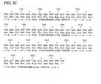
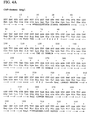

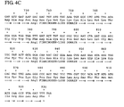
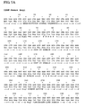
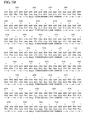


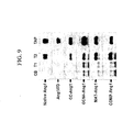


Classifications
-
- C—CHEMISTRY; METALLURGY
- C07—ORGANIC CHEMISTRY
- C07K—PEPTIDES
- C07K19/00—Hybrid peptides, i.e. peptides covalently bound to nucleic acids, or non-covalently bound protein-protein complexes
-
- C—CHEMISTRY; METALLURGY
- C07—ORGANIC CHEMISTRY
- C07K—PEPTIDES
- C07K14/00—Peptides having more than 20 amino acids; Gastrins; Somatostatins; Melanotropins; Derivatives thereof
- C07K14/435—Peptides having more than 20 amino acids; Gastrins; Somatostatins; Melanotropins; Derivatives thereof from animals; from humans
- C07K14/475—Growth factors; Growth regulators
- C07K14/515—Angiogenesic factors; Angiogenin
-
- A—HUMAN NECESSITIES
- A61—MEDICAL OR VETERINARY SCIENCE; HYGIENE
- A61K—PREPARATIONS FOR MEDICAL, DENTAL OR TOILETRY PURPOSES
- A61K38/00—Medicinal preparations containing peptides
- A61K38/16—Peptides having more than 20 amino acids; Gastrins; Somatostatins; Melanotropins; Derivatives thereof
- A61K38/17—Peptides having more than 20 amino acids; Gastrins; Somatostatins; Melanotropins; Derivatives thereof from animals; from humans
-
- A—HUMAN NECESSITIES
- A61—MEDICAL OR VETERINARY SCIENCE; HYGIENE
- A61P—SPECIFIC THERAPEUTIC ACTIVITY OF CHEMICAL COMPOUNDS OR MEDICINAL PREPARATIONS
- A61P35/00—Antineoplastic agents
-
- A—HUMAN NECESSITIES
- A61—MEDICAL OR VETERINARY SCIENCE; HYGIENE
- A61P—SPECIFIC THERAPEUTIC ACTIVITY OF CHEMICAL COMPOUNDS OR MEDICINAL PREPARATIONS
- A61P39/00—General protective or antinoxious agents
-
- C—CHEMISTRY; METALLURGY
- C07—ORGANIC CHEMISTRY
- C07K—PEPTIDES
- C07K14/00—Peptides having more than 20 amino acids; Gastrins; Somatostatins; Melanotropins; Derivatives thereof
- C07K14/435—Peptides having more than 20 amino acids; Gastrins; Somatostatins; Melanotropins; Derivatives thereof from animals; from humans
-
- C—CHEMISTRY; METALLURGY
- C07—ORGANIC CHEMISTRY
- C07K—PEPTIDES
- C07K14/00—Peptides having more than 20 amino acids; Gastrins; Somatostatins; Melanotropins; Derivatives thereof
- C07K14/435—Peptides having more than 20 amino acids; Gastrins; Somatostatins; Melanotropins; Derivatives thereof from animals; from humans
- C07K14/705—Receptors; Cell surface antigens; Cell surface determinants
-
- A—HUMAN NECESSITIES
- A61—MEDICAL OR VETERINARY SCIENCE; HYGIENE
- A61K—PREPARATIONS FOR MEDICAL, DENTAL OR TOILETRY PURPOSES
- A61K38/00—Medicinal preparations containing peptides
-
- C—CHEMISTRY; METALLURGY
- C07—ORGANIC CHEMISTRY
- C07K—PEPTIDES
- C07K2319/00—Fusion polypeptide
Definitions
- the present invention provides for a novel method of producing novel chimeric molecules that have enhanced biological activity, and easier production and purification processes as compared with molecules in their native form.
- the invention also provides for nucleic acids useful for producing biologically active chimeric polypeptides, and the fusion polypeptides themselves.
- Transmembrane protein kinases serve as signaling receptors for a variety of polypeptide ligands, eliciting such diverse responses as cell survival, proliferation and differentiation from many cell types and tissues ( van der Geer et al., 1994, Annu Rev Cell Biol. 10:251-337 ).
- Receptor tyrosine kinases have the ability to interact with different ligands and bring about various cellular responses.
- RTK Receptor tyrosine kinases
- One type of RTK is tyrosine kinase with immunoglobulin and epidermal growth factor receptor homology domains (Tie), Tie2 ( Dumont et al., 1993, Oncogene 8:1293-1301 ; Mustonen and Alitalo, 1995, J Cell Biol.
- Tie2 is expressed predominantly on endothelial cells, hematopoietic cells, or their embryonic precursors, and it is required for normal vascular development ( Sato et al., 1995, Nature 376:70-74 ). Functional disruption of Tie2 in transgenic mice results in embryonic lethality by day E9.5 to 10.5, with effects on the microvasculature resulting in reduced numbers of endothelial cells, and abnormalities of vascular morphogenesis and hematopoiesis ( Sato et al., 1995, Nature 376:70-74 ). Thus, Tie2 is critical for angiogenesis and hematopoiesis during development
- Ang1 is the ligand for Tie2 ( Davis et al., 1996, Cell 87:1161-1169 ; WO9611269 ) ( Figure 1 ).
- Ang1 contains 498 amino acids, including an amino-terminal secretory signal sequence ( Figure 1 ).
- Human and mouse Ang1 are 97.6% identical.
- the amino-terminal region, consisting of residues 100-280 is weakly related to myosin and its relatives, in the regions of these proteins where they are known to possess coiled-coil quaternary structure ( Figure 1 ).
- the second region, consisting of residues 280-498 has strong similarity to the carboxy-terminal domain of fibrinogen ( Figure 1 ).
- Ang1 is a multimer, held together by coiled-coil structures and disulfide crosslinks.
- Recombinant Ang1 is a 70-kDa (reduced condition) secreted glycoprotein that binds to the Tie2 receptor with a Kd of approximately 3.7 nM, and induces tyrosine phosphorylation of Tie2 in endothelial cells ( Davis et al., 1996, Cell 87:1161-1169 ).
- Ang1 protein consists of an amino-tenninal secretory signal sequence (residues 1-19), a unique domain (residues 20-158), a coiled-coil oligomerization domain (residues 159-254), a short linker region (residues 255-283), and a carboxy-terminal fibrinogen-like domain (residues 284-498).
- Angiopoietin-1 is a specific and critical growth factor for blood vessel formation ( Davis et al., 1996, Cell 87:1161-1169 ; Yancopoulos et al., 2000, Nature 407:242-248 ). Recent studies indicate that Ang1 could be used for preventing vascular leakages, therapeutic vasculogenesis, and therapeutic endothelial cell survival ( Thurston et al., 2000, Nat. Med. 6:460-463 ; Chae et al., 2000, Arterioscler. Thromb. Vasc. Biol. 20:2573-2578 ; Kwak et al., 2000, Circulation 101:2317-2324 ). However, Ang1 protein is not easily available, and generation of recombinant Ang1 is extremely difficult with current techniques. Multimerization of the coiled coil domains during production of the ligand hampered purification.
- Ang2 angiopoietin-2
- Ang1 angiopoietin-2
- Ang2 contains 496 amino acids and has a secretory signal sequence.
- Human and mouse Ang2 are 85% identical and approximately 60% identical to Ang1.
- Ang2 has an amino-terminal coiled-coil domain and a carboxy-terminal fibrinogen-like domain.
- Ang1 and Ang2 have similar binding affinities for Tie2.
- Ang2 acts as an antagonist of Tie2 through inhibition of Ang1-induced phosphorylation of Tie2 ( Maisonpierre et al., 1997, Science 277:55-60 ).
- Mouse angiopoietin-3 (Ang3) and human angiopoietin-4 (Ang4) were identified through low stringency hybridization screening with Ang1 and Ang2 cDNAs ( Valenzuela et al., 1999, Proc Natl Acad Sci. 96:1904-1909 ).
- Ang3 and Ang4 are probably interspecies orthologs.
- Ang4 phosphorylates Tie2, while Ang3 inhibits Angl-induced phosphorylation of Tie2 ( Valenzuela et al., 1999, Proc Natl Acad Sci. 96:1904-1909 ).
- Multimeric form of Ang1 phosphorylates Tie2.
- phosphorylated Tie2 interacts cytoplasmically with Grb2, Grb7, Grb14, the protein tyrosine phosphatase Shp2, and the p85 subunit of phosphatidylinositol 3'-kinase (PI 3'-kinase) via their SH2 domains ( Jones et al., 1999, J Biol Chem. 274:30896-30905 ).
- Association between p85 and Tie2 results in PI 3'-kinase activation and subsequent induction of the serine-threonine kinase Akt. ( Kontos et al., 1998, Mol Cell Biol.
- Ang1 induces endothelial cell survival through this PI 3'-kinase/Akt signaling pathway ( Kim et al., 2000, Circ Res. 86:24-29 ). In addition, Ang1 induces endothelial cell sprouting through the activation of PI 3'-kinase and focal adhesion kinase ( Kim et al., 2000, Circ Res. 86:952-959 ).
- Tie2 PI 3'-kinase, Akt, and focal adhesion kinase are crucial elements in the signal transduction pathway leading to survival and migration in endothelial cells.
- Phosphorylated Tie2 also interacts with Dok-R/Dok-2, leading to activation of Dok-R/Dok-2 (Jones and Dumont, 1998, 17:1097-1108).
- Phosphorylated Dok-R interacts with rasGAP, Nck, and Crk (Jones and Dumont, 1998, 17:1097-1108). These signaling molecules may be involved in cell migration and proliferation, organization of the cytoskeleton, and regulation of Ras signaling. Recently, it was shown that Dok-R/Dok-2 is responsible for recruiting Nck and p21-activating kinase (Pak/Pak1) to the activated receptor ( Master et al., 2001, EMBO J. 20:5919-5928 ).
- HSCs hematopoietic stem cells
- HSCs produce Ang1, suggesting that HSCs can promote the migration of endothelial cells and establish a hematopoietic environment ( Takakura et al., 2000, Cell 102:199-209 ).
- Ang1* is a recombinant version of Ang1 that is easier to produce and purify ( Maisonpierre et al., 1997, Science 277:55-60 ; ( PCT WO 98/05779 )).
- Ang1* contains a modified amino-terminus and mutated Cys 265 .
- the biological activity of recombinant Ang1 and Ang1* is similar. However, some of the same problems are encountered in producing Ang1* because the size of Ang1* is too large for efficient recombinant generation.
- U. S. Patent No. 6,455,035 discloses a method of decreasing or inhibiting vascular permeability in a mammal by administering to the mammal a Tie-2 receptor activator. Moreover, USP '035 also discloses that the coiled coil domain of Ang1 was deleted so that the multimerization of the coiled coil domains would not hamper ligand purification.
- WO 00/37462 discloses a method of enhancing the biological activity of Ang1 through deletion of coiled-coil domain and insertion with Fc-portion of immunoglobulin for making a tetramer form of chimeric fusion Ang1 (Ang1-1FD-Fc-FD).
- Ang1-1FD-Fc-FD is equivalent to Ang1* in its ability to stimulate phosphorylation of the Tie2 receptor.
- the claimed invention overcomes the above-mentioned problems, and provides for novel, biologically active, soluble forms of chimeric polypeptide ligands that bind to specific receptors on cells, including growth factors belonging to angiopoietin family.
- polypeptide ligands are useful in promoting or inhibiting a differential function and/or influencing the phenotype, such as growth, survival, contractility, migration, and/or proliferation, of receptor-bearing cells such as endothelial cells, hematopoietic stem cells, and endothelial precursor cells.
- the invention also provides for nucleic acids encoding such polypeptide ligands, and both prokaryotic and eukaryotic expression systems for producing such polypeptide ligands.
- soluble forms of the polypeptide ligands described herein may be used to promote or inhibit biological responses in receptor-expressing cells.
- the present invention is directed to a coiled coil chimeric molecule comprising a coiled-coil domain linked to a receptor binding domain of a ligand which forms a biologically active multimer, and wherein the chimeric molecule in its non-multimerio form is not biologically active.
- the coiled coil domain includes those belonging to a protein belonging to matrix protein family, transcription factor family, growth factor family or secretory protein family.
- the coiled coil domain may be of a matrix protein, in particular, cartilage matrix protein or cartilage oligomeric matrix protein.
- the receptor binding domain may bind to a variety of receptors, and in particular, Tie2 or Tie1 receptor.
- the receptor binding domain may be a fibrinogen-like domain of angiopoietin-1.
- the coiled coil chimeric molecule may be linked directly or indirectly to an extracellular domain of the receptor.
- the present invention is also directed to an isolated nucleic acid encoding a coiled coil chimeric molecule, which is discussed above. Further, the invention is directed to an expression vector that includes this nucleic acid. A host cell comprising the vector is also included within the purview of the invention.
- the present invention also includes a soluble biologically active multimer comprising the coiled coil chimeric molecule discussed above.
- the multimer may be a homomer or a heteromer. If the multimer is a heteromer, then it is preferred that the coiled coil domain be heterogeneous with respect to at least one specie.
- the multimer may be a dimer, trimer, tetramer, pentamer, hexamer, hoptamer, octamer, nanomer or decamer.
- the multimer may comprise coiled coil domains, which form parallel or anti-parallel structure.
- the present invention is also directed to a method of promoting cell growth comprising contacting a coiled coil chimeric molecule comprising a coiled-coil domain linked to a receptor binding domain of a ligand as described above, to a population of cells that express receptors that are specific for the ligand, wherein said ligand is an agonist for said receptor, and wherein a multimeric form of the coiled coil chimeric molecule interacts with the receptor, which results in cell growth promotion.
- the cells may be endothelial cells, hematopoietic cells or other cells that express each specific receptor.
- the present invention is directed to a method of promoting cell proliferation comprising contacting a coiled coil chimeric molecule comprising a coiled-coil domain linked to a receptor binding domain of a ligand as described above, to a population of cells that express receptors that are specific for the ligand, wherein said ligand is an agonist for said receptor, and wherein a multimeric form of the coiled coil chimeric molecule interacts with the receptor, which results in cell proliferation.
- the cells may be endothelial cells, hematopoietic cells or other cells that express each specific receptor.
- the present invention is directed to a method of decreasing or inhibiting cell proliferation comprising contacting a coiled coil chimeric molecule comprising a coiled-coil domain linked to a ligand binding domain of a receptor as described above, to a population of cells that express ligands that are specific for the receptor, wherein a multimeric form of the coiled coil chimeric molecule interacts with the ligand, which results in decrease or inhibition of cell proliferation.
- the cells may be endothelial cells, hematopoietic cells or other cells that express each specific receptor.
- the present invention is directed to a method of decreasing or inhibiting ligand activity comprising contacting a coiled coil chimeric molecule comprising a coiled-coil domain linked to a ligand binding domain of a receptor as described above, to a sample comprising a ligand that is specific for the receptor, wherein a multimeric form of the coiled coil chimeric molecule binds the ligand, which results in a decrease or inhibition of ligand activity.
- the present invention is directed to a method of making a chimeric molecule comprising: (A) recombinantly combining a nucleic acid encoding a coiled-coil domain with a nucleic acid encoding either a receptor binding region of a ligand or a ligand binding region of a receptor to produce a chimeric gene construct; and (B) expressing the gene construct in a host cell to produce the chimeric molecule.
- the invention is directed to a use of the invention molecule for the manufacture of a medicament for protecting endothelial cells against radiation damage to a mammal that has been exposed to irradiation, comprising administering a therapeutically effective amount of the coiled coil chimeric molecule described above to the mammal in need thereof.
- cartilage matrix protein CMP, matrilin-1, and MAT are used interchangeably.
- FIGURE 1 shows a schematic structure of angiopoietin-1.
- Amino acids 1-19 are the secretory signal sequence (S)
- amino acids 158-255 are the coiled-coil oligomeric domain (CCOD)
- amino acids 226-283 are the linker (L)
- amino acids 284-498 are the fibrinogen-like domain (FLD).
- cysteines (C) at amino acids 41, 54, 265, 315, 435, 437, 439, and 452.
- FIGURES 2A-2C show nucleic acid sequence (SEQ ID NO:1) and deduced amino acid sequence (SEQ ID NO:2) of Ang1/FD (fibrinogen-like domain of Ang1).
- FIGURES 3A-3C show nucleic acid sequence (SEQ ID NO:3) and deduced amino acid sequence (SEQ ID NO:4) of GCN4/CC-Angl/FD (coiled-coil domain of GCN4-Ang1/FD).
- FIGURES 4A-4C show nucleic acid sequence (SEQ ID NO:5) and deduced amino acid sequence (SEQ ID NO:6) of CMP/CC-Angl/FD (coiled-coil domain of CMP-Ang1/FD).
- FIGURES 5A-5C show nucleic acid sequence (SEQ ID NO:7) and deduced amino acid sequence (SEQ ID NO:8) of COMP/CC-Angl/FD (coiled-coil domain of COMP-Ang1/FD).
- FIGURE 6 shows a schematic diagram for generation of gene constructs for multimeric chimeric-Ang1.
- M stands for multiple cloning site.
- FIGURE 7 shows a schematic diagram of native Ang1, GCN4-Angl/FD, MAT-Ang1/FD, and COMP-Ang1/FD.
- FIGURES 8A-8B show that Ang1 variants produce lower-order oligomers.
- FIGURE 8A shows molecular weight analysis of recombinant native Ang1, Ang1/FD, CC-Ang1, GCN4-Angl MAT/CC-Ang1, and COMP/CC-Ang1 under reducing (R) and non-reducing (NR) conditions. Numbers and bars indicate molecular sizes (kDa). 100 ng of each recombinant protein (reduced and non-reduced) is separated by 4-15% sodium dodecyl sulfate-polyacrylamide gel electrophoresis (SDS-PAGE), and electro-blotted to nitrocellulose membranes.
- SDS-PAGE sodium dodecyl sulfate-polyacrylamide gel electrophoresis
- FIGURE 8B shows glycerol sprayed/low-angle rotary metal-shadowed specimens that were imaged by TEM.
- Native Ang1 reveals variable-sized multimers of the basic 'mushroom-shaped' trimeric structure.
- CC-Angl reveals trimers
- GCN4-Ang1 reveals dimers
- MAT-Ang1 reveals mostly tetramer
- COMP-Ang1 reveals mostly pentamers.
- FIGURE 9 shows in vitro binding assay between recombinant native Ang1, Ang1/FD, CC-Ang1, GCN4-Angl MAT/CC-Ang1, and COMP/CC-Angl and soluble Tie1-Fc (sTie1-Fc, T1) and soluble Tie2-Fc (sTie2-Fc, T2).
- soluble Tie1-Fc sTie1-Fc, T1
- Tie2-Fc soluble Tie2-Fc
- sTie1-Fc or sTie2-Fc Twenty nanograms of each protein was incubated with 100 ng of sTie1-Fc or sTie2-Fc for 4 hr, and then, protein-A conjugated agarose beads were added. The precipitate was separated by 10% SDS-PAGE, and electro-blotted on to nitrocellulose membranes. The nitrocellulose membranes were Western blotted with anti-FLAG M1 antibody, washed, and incubated with horseradish peroxidase-conjugated secondary antibody. Signals were visualized by chemiluminescent detection according to the manufacturer's protocol (Amersham Pharmacia Biotech) and chemiluminescence scanner (LAS-1000, Fuji Film, Tokyo).
- FIGURES 10A - 10D show in vitro binding assay between recombinant native Ang1, GCN4-Ang1/FD, MAT-Angl/FD, or COMP-Ang1/FD and immobilized soluble Tie2-Fc using Biacore assay.
- Each recombinant protein was passed over a Biacore sensor chip that had extracellular domain of Tie2-Fc and Fc protein covalently coupled to it, followed by dissembling in absence of the recombinant proteins. Concentrations of the recombinant proteins are shown in each panel. Specificity of binding was assessed by substracting Fc protein binding value from Tie2-Fc binding value during the measurement. Binding of the sensor chip is provided in resonance units (RUs).
- RUs resonance units
- FIGURE 11 shows BIAcore analysis of native Ang1 (NA), GCN4-Ang1 (G), MAT-Ang1 (M) or COMP-Ang1 (C) with sTie2-Fc immobilized on a chip.
- Typical sensogram showing the association and dissociation profile of 60 nM of recombinant protein.
- An equal amount of VEGF was used as a control.
- FIGURES 12A-12B show comparison of native Ang1 (N), GCN4-Ang1 (G), MAT-Ang1 (M) or COMP-Ang1 (C) in a Tie2 phosphorylation assay using human umbilical venous endothelial cells (HUVECs). Serum-starved HUVECs were treated with 200 ng/ml of native Ang1 or 100 ng/ml of Ang1 variants for 10 min (A), or the indicated times (B) and the phosphorylations of Tie2 were measured. Fold: Densitometric analyses are presented as the relative ratio of phospho-Tie2 to Tie2.
- the relative ratio of phospho-Tie2 (pTie2) to Tie2 at time 0 is arbitrarily presented as 1. Numbers represent the mean ⁇ S.D. from 3 experiments. *, P ⁇ 0.05 versus CB. #, P ⁇ 0.05 versus NA.
- the cells were harvested in extraction buffer, and 0.5 mg of protein was used for immunoprecipitation. Tie2 proteins in the samples were immunoprecipitated with anti-Tie2 antibody and collected. Immunoprecipitated samples were Western blotted with anti-phospho-tyrosine antibody (upper panel), and the membrane was re-blotted with anti-Tie2 antibody to verify equal loading of protein in each lane (lower panel). Results were similar in three independent experiments.
- FIGURES 13A-13C show comparison of native Ang1, GCN4-Angl/FD, MAT-Angl/FD, or COMP-Ang1/FD in Akt (Ser473) phosphorylation assay in HUVECs.
- upper band indicates that the Western blots were probed with anti-phospho-Akt (Ser473) antibody.
- Lower band shows the blot that was reprobed with anti-Akt antibody to verify equal loading of protein in each lane.
- HUVECs were incubated with control buffer (CB), and 200 ng of native Ang1 (N), GCN4-Angl/FD (G), MAT-Ang1/FD (M), or COMP-Ang1/FD (C) for 15 min.
- HUVECs were incubated with control buffer (CB) and indicated amount of COMP-Ang1/FD.
- C HUVECs were incubated with 200 ng of COMP-Ang1/FD for indicated times. After treatment, cell lysates were harvested. Each lane contains 50 ⁇ g of total protein from the cell lysates. Results were similar in three independent experiments. Fold: Densitometric analyses are presented as the relative ratio of phospho-Akt (Ser472) to Akt. The relative ratio measured for control buffer was arbitrarily set at 1. Numbers represent the mean ⁇ S.D. from three experiments.
- FIGURES 14A-14T show a comparison between native Ang1, GCN4-Angl, MAT-Ang1, and COMP-Ang1 in survival, migration, tube formation and sprouting activities of primary cultured endothelial cells. Representative photographs are shown. Native Ang1 and MAT-Ang1-induced notable increase in survival, migration, tube formation and sprouting activities. Notably, COMP-Ang1-induced survival, migration, tube formation and sprouting activities were greater than native Ang1- and MAT-Ang1- induced survival, migration, tube formation and sprouting activities. However, GCN4-Ang1 did not appear to exhibit any notable change in survival, migration, tube formation or sprouting activities.
- FIGURES 15A-15D show quantification of biological activities.
- CB control buffer
- NA native Ang1
- G GCN4-Angl
- M MAT-Ang1
- C COMP-Ang1.
- * P ⁇ 0.05 versus CB.
- # P ⁇ 0.05 versus NA (200 ng/ml).
- FIGURES 16A-16D show that COMP-Ang1 alone produces angiogenesis in the mouse corneal micropocket assay.
- A Macroscopic photographs of mouse corneas. Corneal angiogenesis was not observed with control buffer (CB) or with native Ang1. COMP-Ang1 induced corneal angiogenesis extending from the limbus across the cornea.
- B left panels show in situ BS-1 lectin fluorescent staining of corneal limbus vessels 6 days after pellet implantation. COMP-Ang1 increased the diameter of the limbar artery, while CB and native Ang1 induced no changes.
- B right panels show hematoxylin and eosin staining confirms that COMP-Ang1 induced angiogenesis with significant vessel formation. Arrows indicate blood vessels containing red blood cells.
- FIGURES 17A-17F show acute injection of COMP-Ang1 induces Tie2 phosphorylation in lung.
- A shows distribution of Tie2 in several organs including brain (B), heart (H), lung (Lu), liver (Li), kidney (K), small intestine (I), spleen (S), and ear (E) of adult mice. Protein lysates from each organ were immunoprecipitated and immunoblotted with anti-Tie2 antibody (upper panel). Each protein lysate was also immunoblotted with anti-actin antibody to verify equal amounts of total protein loaded (lower panel).
- B-D show in vivo phosphorylation of Tie2 stimulated by i.v.
- E-F show immunohistochemical staining of Tie2 counterstained with methyl green ( E ) and PECAM-1 counterstained with Meyer's hematoxylin ( F ). Most Tie2 staining corresponds to PECAM staining.
- White arrows indicate large blood vessels with positive staining. Gray arrows indicate bronchioles with negative staining.
- FIGURES 18A-18I show that COMP-Ang1 protects against radiation-induced microvascular apoptosis and death.
- A-G show mice which were given whole-body irradiation with 15 Gy and sacrificed after 4 hrs. While small intestinal villi and crypts from control mice have almost no apoptosis ( A, E ), villi and crypts from irradiated mice reveal extensive apoptosis (arrows) in lamina intestinal cells ( B , F ). I.V. injection of COMP-Ang1 markedly reduced radiation-induced apoptosis in lamina limba cells but not in cells of crypts ( C, G ).
- Co-staining of PECAM-1 (gray-pastel colors; arrow heads) and TUNEL (dark brown; arrows) allows for counting the number of apoptotic endothelial cells among the lamina propria cells ( D ).
- H shows frequency histograms of percent of apoptotic endothelial cells in the lamina propria of approximately 1,000 villi from each group. Small intestinal specimens were obtained 4 hrs after 15 Gy irradiation with or without i.v. injection of COMP-Ang1. Data represent mean scores from 5 experiments.
- a method of using coiled-coil domains for "multimerizing" ligands which enhances the biological activity of such ligands that, absent such multimerization, would have lower levels of biological activity.
- This method may be used to multimerize receptor binding domains from any ligand that has improved affinity and/or increased activity (i.e. signaling ability) when they were multimerized as compared to the non-multimerized form of the ligand.
- the present invention also provides for methods of using coiled-coil domains for "multimerizing" soluble receptors, which functions to make otherwise inactive soluble receptors biologically active, or which enhances the biological and binding activity of receptors that, absent such multimerization, would have lower levels of biological and binding activity.
- This method may be used to multimerize ligand binding domains using any receptor, which has improved affinity and/or increased activity (i.e. binding) when they were multimerized as compared to the native form of the soluble receptor.
- polypeptide As used herein, “about” or “substantially” generally provides a leeway from being limited to an exact number. For example, as used in the context of the length of a polypeptide sequence, “about” or “substantially” indicates that the polypeptide is not to be limited to the recited number of amino acids. A few amino acids add to or subtracted from the N-terminus or C-terminus may be included so long as the functional activity such as its binding activity is present.
- administration in combination with one or more further therapeutic agents includes simultaneous (concurrent) and consecutive administration in any order.
- agonist refers to a ligand that binds to a receptor, which activates the receptor and stimulates physiologic activity.
- Ang1 is considered to be an agonist of Tie2 receptor.
- amino acid and “amino acids” refer to all naturally occurring L- ⁇ -amino acids. This definition is meant to include norleucine, ornithine, and homocysteine.
- amino acid sequence variant refers to molecules with some differences in their amino acid sequences as compared to a reference (e.g. native sequence) polypeptide.
- the amino acid alterations may be substitutions, insertions, deletions or any desired combinations of such changes in a native amino acid sequence.
- Substitutional variants are those that have at least one amino acid residue in a native sequence removed and a different amino acid inserted in its place at the same position.
- the substitutions may be single, where only one amino acid in the molecule has been substituted, or they may be multiple, where two or more amino acids have been substituted in the same molecule.
- Substitutes for an amino acid within the sequence may be selected from other members of the class to which the amino acid belongs.
- the nonpolar (hydrophobic) amino acids include alanine, leucine, isoleucine, valine, proline, phenylalanine, tryptophan and methionine.
- the polar neutral amino acids include glycine, serine, threonine, cysteine, tyrosine, asparagine and glutamine.
- the positively charged (basic) amino acids include arginine, lysine and histidine.
- the negatively charged (acidic) amino acids include aspartic acid and glutamic acid.
- proteins or fragments or derivatives thereof which exhibit the same or similar biological activity and derivatives which are differentially modified during or after translation, e.g., by glycosylation, proteolytic cleavage, linkage to an antibody molecule or other cellular ligand, and so on.
- Insertional variants are those with one or more amino acids inserted immediately adjacent to an amino acid at a particular position in a native amino acid sequence. Immediately adjacent to an amino acid means connected to either the ⁇ -carboxy or ⁇ -amino functional group of the amino acid.
- Deletional variants are those with one or more amino acids in the native amino acid sequence removed. Ordinarily, deletional variants will have one or two amino acids deleted in a particular region of the molecule.
- antagonist refers to a ligand that tends to nullify the action of another ligand, as a ligand that binds to a cell receptor without eliciting a biological response.
- biologically active refers to the ability of a molecule to specifically bind to and signal through a native receptor, e.g., a native Tie2 receptor, or to block the ability of a native Tie receptor (e.g., Tie2) to participate in signal transduction.
- the (native and variant) ligands of the present invention include agonists and antagonists of a native receptor, e.g. Tie2 receptor.
- Preferred biological activities of the ligands of the present invention include the ability to induce or inhibit vascularization. The ability to induce vascularization will be useful for the treatment of biological conditions and disease, where vascularization is desirable. On the other hand, the ability to inhibit or block vascularization may, for example, be useful in preventing or attenuating cell proliferation and tumor growth.
- Preferred biological activities of the ligands of the present invention include the ability to inhibit vascular permeability.
- the ability to inhibit vascular permeability will be useful for treatment of medical conditions and diseases such as diabetic retinopathy, edema, and ascites.
- Preferred biological activities of the ligands of the present invention include the ability to maintain endothelial cell integrity (including preventing apoptosis). The ability to maintain endothelial cell integrity will be useful for treatment of medical conditions and diseases such as mannitol treatment, irradiation, and sepsis.
- the biological activity of the chimeric receptor includes its ability to inhibit or competitively inhibit the ligand's activity by binding to its ligand.
- cell proliferation may be inhibited if the ligand is an agonist for cell proliferation.
- administration of chimeric receptor may act as an enhancer of cell proliferation if the ligand is an antagonist for cell proliferation.
- chimeric ligand and chimeric receptor be labeled with a detectable label, such as radioisotope, fluorescent tag, enzymatic tag, or a chemiluminescent tag to determine ligand-receptor binding interaction.
- a detectable label such as radioisotope, fluorescent tag, enzymatic tag, or a chemiluminescent tag to determine ligand-receptor binding interaction.
- assay systems employing the chimeric molecule is also contemplated.
- carriers include pharmaceutically acceptable carrier, excipients, or stabilizers which are nontoxic to the cell or mammal being exposed thereto at the dosages and concentrations employed.
- pharmaceutically acceptable carrier is an aqueous pH buffered solution.
- Examples of pharmaceutically acceptable carriers include without limitation buffers such as phosphate, citrate, and other organic acids; antioxidants including ascorbic acid; low molecular weight (less than about 10 residues) polypeptide; proteins, such as serum albumin, gelatin, or immunoglobulins; hydroplilic polymers such as polyvinylpyrrolidone; amino acids such as glycine, glutamine, asparagine, arginine or lysine; monosaccharides, disaccharides, and other carbohydrates including glucose, mannose, or dextrins; chelating agents such as EDTA; sugar alcohols such as mannitol or sorbitol; salt-forming counterions such as sodium; and/or nonionic surfactants such as TWEEN ® , polyethylene glycol (PEG), and PLURONICS ® .
- buffers such as phosphate, citrate, and other organic acids
- antioxidants including ascorbic acid
- chimeric ligand refers to the combination of coiled coil domain and a receptor binding domain or a ligand binding domain.
- the resultant chimeric polypeptide is capable of forming biologically active multimers, which are soluble.
- the coiled coil domain may be derived from any source, including any animal or mammalian protein, and in particular any human protein, and further includes those that are synthetically made.
- the coiled coil domain and the ligand or receptor constructs may be from the same or different source.
- the chimeric construct comprises the coiled coil domain and a receptor binding domain of a ligand or a ligand binding domain of a receptor, and further may include other components that may be included so long their inclusion does not interfere with the formation of a biologically active multimer that has improved solubility, ease of recombinant production of the chimeric polypeptide and substantially similar or greater potency as the native ligand or native soluble receptor.
- FLAG sequence may be included for ease of purification, provided its inclusion does not interfere with the function of the chimeric molecule.
- the FLAG sequence also may be removed if a humanized construct is desired.
- an effective amount is an amount sufficient to effect beneficial or desired clinical or biochemical results.
- An effective amount can be administered one or more times.
- An effective amount of an inhibitor compound is an amount that is sufficient to palliate, ameliorate, stabilize, reverse, allow or delay the progression of the disease state.
- fragments or “functional derivatives” refers to biologically active amino acid sequence variants and fragments of the native ligands or receptors of the present invention, as well as covalent modifications, including derivatives obtained by reaction with organic derivatizing agents, post-translational modifications, derivatives with nonproteinaceous polymers, and immunoadhesins.
- host cell includes an individual cell or cell culture which can be or has been a recipient of a vector of this invention.
- Host cells include progeny of a single host cell, and the progeny may not necessarily be completely identical (in morphology or in total DNA complement) to the original parent cell due to natural, accidental, or deliberate mutation and/or change.
- ligand refers to any molecule or agent, or compound that specifically binds covalently or transiently to a molecule such as a polypeptide.
- ligand may include antibody.
- ligand may refer to a molecule sought to be bound by another molecule with high affinity, such as in a ligand trap.
- ligand binding domain refers to the portion of the receptor that binds to the ligands and includes the minimal portion of the receptor that is necessary to bind its ligand.
- linked refers to direct or indirect connection between the multimerizing domain and the ligand or receptor. Both a direct fusion between these two domains or indirect fusion as by the domains being separated by a linker or an intervening domain or element are contemplated, so long as the activity of the chimeric fusion is present.
- mammal for purposes of treatment refers to any animal classified as a mammal, including humans, domestic and farm animals, and zoo, sports, or pet animals, such as dogs, cats, cattle, horses, sheep, pigs, and so on.
- the mammal is human.
- multimer or “multimeric” refers to the joining of the multimerizing agent such as the coiled coil domain to each other to form a dimer, trimer, tetramer, pentamer, hexamer, heptamer, octamer, nanomer, decamer and so on, which may be in a parallel or anti-parallel form, through intramolecular or intermolecular bonds.
- pharmaceutically acceptable carrier and/or diluent includes any and all solvents, dispersion media, coatings antibacterial and antifungal agents, isotonic and absorption delaying agents and the like.
- the use of such media and agents for pharmaceutical active substances is well known in the art. Except insofar as any conventional media or agent is incompatible with the active ingredient, use thereof in the therapeutic compositions is contemplated. Supplementary active ingredients can also be incorporated into the compositions.
- Dosage unit form refers to physically discrete units suited as unitary dosages for the mammalian subjects to be treated; each unit containing a predetermined quantity of active material calculated to produce the desired therapeutic effect in association with the required pharmaceutical carrier.
- the specification for the dosage unit forms of the invention are dictated by and directly dependent on (a) the unique characteristics of the active material and the particular therapeutic effect to be achieved, and (b) the limitations inherent in the art of compounding such an active material for the treatment of disease in living subjects having a diseased condition in which bodily health is impaired.
- the principal active ingredient is compounded for convenient and effective administration in effective amounts with a suitable pharmaceutically acceptable carrier in dosage unit form.
- a unit dosage form can, for example, contain the principal active compound in amounts ranging from 0.5 ⁇ g to about 2000 mg. Expressed in proportions, the active compound is generally present in from about 0.5 ⁇ g/ml of carrier. In the case of compositions containing supplementary active ingredients, the dosages are determined by reference to the usual dose and manner of administration of the said ingredients.
- receptor binding domain refers to the portion of the ligand that binds to the receptor and includes the minimal portion of the ligand that is necessary to bind its receptor.
- the present invention is based on the discovery that a multimerizing agent, such as a coiled coil domain, which was previously perceived as a source of hindrance for isolating recombinant proteins containing them, has been found to provide advantageous features of easy recombinant protein expression and purification, greater solubility and greater or substantially equal potency compared with the native protein containing the coiled coil domain.
- sample or “biological sample” is referred to in its broadest sense, and includes any biological sample obtained from an individual, body fluid, cell line, tissue culture, or other source which may contain a chimeric Angl binding factor, depending on the type of assay that is to be performed.
- biological samples include body fluids, such as semen, lymph, sera, plasma, urine, synovial fluid, spinal fluid and so on. Methods for obtaining tissue biopsies and body fluids from mammals are well known in the art.
- the term “specifically binds” refers to a non-random binding reaction between two molecules, for example between an antibody molecule immunoreaoting with an antigen, or a non-antibody ligand reacting with another polypeptide, such as chimeric Ang1 specifically binding with Tie2.
- subject is a vertebrate, preferably a mammal, more preferably a human.
- treatment is an approach for obtaining beneficial or desired clinical results.
- beneficial or desired clinical results include, but are not limited to, alleviation of symptoms, diminishment of extent of disease, stabilized (i.c., not worsening) state of disease, delay or slowing of disease progression, amelioration or palliation of the disease state, and remission (whether partial or total), whether detectable or undetectable.
- Treatment can also mean prolonging survival as compared to expected survival if not receiving treatment.
- Treatment refers to both therapeutic treatment and prophylactic or preventative measures. Those in need of treatment include those already with the disorder as well as those in which the disorder is to be prevented.
- “Palliating" a disease means that the extent and/or undesirable clinical manifestations of a disease state are lessened and/or the time course of the progression is slowed or lengthened, as compared to a situation without treatment.
- a polynucleotide vector of this invention may be in any of several forms, including, but not limited to, RNA, DNA, RNA encapsulated in a retroviral coat, DNA encapsulated in an adenovirus coat, DNA packaged in another viral or viral-like form (such as herpes simplex, and adeno-associated virus (AAV)), DNA encapsulated in liposomes, DNA complexed with polylysine, complexed with synthetic polycationic molecules, complexed with compounds such as polyethylene glycol (PEG) to immunologically "mask” the molecule and/or increase half-life, or conjugated to a non-viral protein.
- RNA Ribonucleic acid
- the polynucleotide is DNA.
- DNA includes not only bases A, T, C, and G, but also includes any of their analogs or modified forms of these bases, such as methylated nucleotides, internucleotide modifications such as uncharged linkages and thioates, use of sugar analogs, and modified and/or alternative backbone structures, such as polyamides.
- the present invention includes a multimers forming domain.
- coiled coil domain is exemplified.
- the coiled coil domain may be any amino acid sequence that forms a coiled coil structure. While the exemplified coiled coil domains herein are those cloned from a variety of proteins, it is understood that various mutations and derivatization are encompassed by the invention, so long as the resultant coiled coil domain is recognized by a person of skill in the art as a coiled coil structure and the coiled coil domain containing chimera is capable of forming a multimer, is easily soluble, and is able to provide similar or greater potency with respect to the native ligand or receptor.
- the multimerizing domain and the binding domain may be from the same protein, or they may be from different proteins.
- Ang1 coiled coil domain may be linked to its own fibrinogen-like domain in a more efficiently manner.
- a cartilage oligomeric matrix protein could be linked to the Ang1 fibrinogen-like domain.
- Ang1 portion referred to is the fibrinogen domain of Ang1.
- MAT-Ang1 is also sometimes referred to as CMP-Angl, CMP-Angl/FD, or CMP/CC-Angl/FD in the figures.
- COMP-Ang1 produced the most potent effect on Tie2 and Akt phosphorylation.
- COMP-Angl could be the most biologically active among the variants because of its rapid association and dissociation rate.
- COMP-Angl may induce Tie2 clustering in endothelial caveolae more efficiently than the other recombinant Angl proteins.
- Our data indicate that Tie2 molecules are localized in endothelial caveolae.
- an engineered Ang1 protein with an oligomeric structure better suited to clustering Tie2 in caveolae may better facilitate Tie2 multimerization and phosphorylation.
- Ang1 failed to stimulate an angiogenic response when administered alone.
- Ang1 augmented postnatal angiogenesis.
- native Ang1 failed to stimulate an angiogenic response.
- COMP-Angl alone stimulates angiogenesis.
- COMP-Angl produced an increase in the luminal diameter of the basal limbus. This role is consistent with increased frequency of enlarged vessel diameters in Ang1-overexpressing transgenic mice ( Suri, C. et al. 1998, Science 282: 468-471 ; Thurston, G. et al. 1999, Science 286:2511-2514 ).
- COMP-Angl can produce more effective blood flow by increasing the diameter of arterial lumens.
- Angl can counteract VEGF-induced side effects such as edema and inflammation, while having an additive effect on angiogenesis ( Yamak, H. J. et al. 2000, Circulation 101:2317-2324 ; Thurston, G. et al. 1999, Science 286:2511-2514 ; Kim, I. et al. 2001, Circ. Res. 89:477-479 ).
- COMP-Angl protein delivery may be useful for accurate and safe therapeutic angiogenesis.
- Integrity of the vascular endothelium in response to physical, biochemical, and immune-mediated damage is important to maintaining endothelial function and preventing vascular diseases ( Cines, D.B. et al. 1998, Blood 91:3527-3561 ).
- extensive apoptosis of microvascular endothelial cells of the lamina intestinal is the primary lesion initiating intestinal radiation damage ( Paris, F. et al. 2001, Science 293:293-297 ).
- GI tract damage during abdominal radiotherapy limits the dose that can be used during cancer treatment. Radiation damage to vascular endothelial cells can be prevented by VEGF ( Okunieff, P. et al. 1998, Radiat. Res.
- VEGF receptors are expressed in endothelial cells that are actively involved in vasculogenesis and angiogenesis such as tumor progression
- bFGF receptors are expressed in both endothelial and non-endothelial cells including cancer cells ( Paris, F. et al. 2001, Science 293:293-29 ; Veikkola, T. & Alitalo, K. 1999, Seminars in Cancer Biol. 9:211-220 ). Therefore, administration of VEGF and bFGF for protection of endothelial damage may help tumor progression.
- Tie2 is selectively expressed in active form in the endothelial cells of normal adult vessels ( Wong, A.L. et al. 1997, Circ. Res. 81:567-574 ).
- the angiopoietin/Tie2 system in normal adult blood vessels may be important in maintaining the integrity of non-proliferating endothelial cells ( Kim, I. et al. 2001, Cardiovas. Res. 49:872-881 ).
- Our immunohistochemical staining indicates that Tie2 is selectively expressed in most endothelial cells of normal adult vessels and capillaries.
- Tie2 is effectively activated by acute administration of COMP-Angl.
- Ang1 may increase tumor angiogenesis during radiation therapy.
- the role of Ang1 in tumor angiogenesis is still controversial. It has even been suggested that Angl may suppress tumor progression through 'stabilization' of tumor vessels ( Tian, S. et al. 2002, Br. J. Cancer 286:645-651 ; Hawighorst, T. et al. 2002, Am. J. Pathol. 160:1381-1392 ). Therefore, using Ang1 for protection against radiation-induced endothelial cell damage could be ideal.
- COMP-Angl treatment strongly protects against extensive radiation-induced extensive endothelial apoptosis in villi, but has no observed effect on non-endothelial cells.
- COMP-Angl treatment prolongs survival periods, perhaps as a result of decreasing damage to the GI tract. Optimizing the dosage and route of administration of COMP-Angl could further improve endothelial cell survival following radiation-induced endothelial cell damage.
- COMP-Angl is superior to native Ang1 in several ways including efficiency of generation, potency, Tie2 activation in vivo , angiogenesis, and protection against endothelial injury in vivo . It seems likely that it can be further applied to the prevention of vascular leakage, protection against sepsis-induced endothelial cell injury, enhancement of re-endothelialization after angioplasty, and in vitro amplification of Tie2 positive endothelial precursor stem cells.
- ⁇ -helical coiled coil is probably the most widespread subunit oligomerization motif found in proteins. Accordingly, coiled coils fulfill a variety of different functions. In several families of transcriptional activators, for example, short leucine zippers play an important role in positioning the DNA-binding regions on the DNA ( Ellenberger et al., 1992, Cell 71:1223-1237 ). Coiled coils are also used to form oligomers of intermediate filament proteins. Coiled-coil proteins furthemore appear to play an important role in both vesicle and viral membrane fusion ( Skehel and Wiley, 1998, Cell 95:871-874 ).
- hydrophobic sequences embedded in the membranes to be fused, are located at the same end of the rod-shaped complex composed of a bundle of long ⁇ -helices. This molecular arrangement is believed to cause close membrane apposition as the complexes are assembled for membrane fusion.
- the coiled coil is often used to control oligomerization. It is found in many types of proteins, including transcription factors such as, but not limited to GCN4, viral fusion peptides, SNARE complexes and certain tRNA synthetases, among others. Very long coiled coils are found in proteins such as tropomyosin, intermediate filaments and spindle-pole-body components.
- Coiled coils involve a number of ⁇ -helices that are supercoiled around each other in a highly organized manner that associate in a parallel or an antiparallel orientation. Although dimers and trimers are the most common.
- the helices may be from the same or from different proteins.
- the coiled-coil is formed by component helices coming together to bury their hydrophobic seams. As the hydrophobic seams twist around each helix, so the helices also twist to coil around each other, burying the hydrophobic seams and forming a supercoil. It is the characteristic interdigitation of side chains between neighbouring helices, known as knobs-into-holes packing, that defines the structure as a coiled coil.
- the helices do not have to run in the same direction for this type of interaction to occur, although parallel conformation is more common. Antiparallel conformation is very rare in trimers and unknown in pentamers, but more common in intramolecular dimers, where the two helices are often connected by a short loop.
- the heterotrimeric coiled-coil protein laminin plays an important role in the formation of basement membranes.
- Other examples are the thrombospondins and cartilage oligomeric matrix protein (COMP) in which three (thrombospondins 1 and 2) or five (thrombospondins 3, 4 and COMP) chains are connected.
- the molecules have a flower bouquet-like appearance, and the reason for their oligomeric structure is probably the multivalent interaction of the C-terminal domains with cellular receptors.
- the yeast transcriptional activator GCN4 is 1 of over 30 identified eukaryotic proteins containing the basic region leucine zipper (bZIP) DNA-binding motif ( Ellenberger et al., 1992, Cell 71:1223-1237 ).
- the bZIP dimer is a pair of continuous alpha helices that form a parallel coiled-coil over their carboxy-teminal 34 residues and gradually diverge toward their amino termini to pass through the major groove of the DNA binding site.
- the coiled-coil dimerization interface is oriented almost perpendicular to the DNA axis, giving the complex the appearance of the letter T.
- bZIP contains a 4-3 heptad repeat of hydrophobic and nonpolar residues that pack together in a parallel alpha-helical coiled-coil ( Ellenberger et al., 1992, Cell 71:1223-1237 ).
- the stability of the dimer results from the side-by-side packing of leucines and nonpolar residues in positions a and d of the heptad repeat, as well as a limited number of intra- and interhelical salt bridges, shown in a crystal structure of the GCN4 leucine zipper peptide ( Ellenberger et al., 1992,Cell 71:1223-1237 ).
- CMP Cartilage Matrix Protein
- CMP (matrilin-1) was isolated from bovine tracheal cartilage as a homotrimer of subunits of M r 52,000 ( Paulsson and Heineg ⁇ rd, 1981, Biochem J. 197:367-375 ), where each subunit consists of a vWFA1 module, a single EGF domain, a vWFA2 module and a coiled coil domain spanning five heptads ( Kiss et al., 1989, J. Biol. Chem. 264:8126-8134 ; Hauser and Paulsson, 1994, J. Biol. Chem. 269:25747-25753 ).
- Electron microscopy of purified CMP showed a bouquet-like trimer structure in which each subunit forms an ellipsoid emerging from a common point corresponding to the coiled coil ( Hauser and Paulsson, 1994, J. Biol. Chem. 269:25747-25753 ).
- the coiled coil domain in matrilin-1 has been extensively studied.
- the trimeric structure is retained after complete reduction of interchain disulfide bonds under non-denaturing conditions ( Hauser and Paulsson, 1994, J. Biol. Chem. 269:25747-25753 ).
- COMP non-collagenous glycoprotein
- the protein is a 524 kDa homopentamer of five subunits which consists of an N-terminal heptad repeat region (cc) followed by four epidermal growth factor (EGF)-like domains (EF), seven calcium-binding domains (T3) and a C-terminal globular domain (TC).
- EGF epidermal growth factor
- T3 calcium-binding domains
- TC C-terminal globular domain
- COMP belongs to the family of thrombospondins.
- the present invention also provides for a nucleic acid encoding a fusion polypeptide wherein the fusion polypeptide comprises a first subunit comprising at least one copy of the receptor binding domain of a ligand, the first subunit being fused to the C-terminal end of a multimerizing component.
- the present invention provides for a nucleic acid encoding a fusion polypeptide wherein the fusion polypeptide comprises a first subunit comprising at least one copy of the receptor binding domain of a ligands, the first subunit being fused to the N-terminal end of a multimerizing component
- the multimerizing component may be the coiled coil domain.
- an expression vector comprising a nucloic acid molecule of the invention as described herein, wherein the nucleic acid molecule is operatively linked to an expression control sequence.
- a host-vector system for the production of a fusion polypeptide which comprises the expression vector of the invention which has been introduced into a host cell suitable for expression of the fusion polypeptide.
- the suitable host cell may be a bacterial cell such as E . coli , a yeast cell, such as Pichia pastoris , an insect cell, such as Spodoptera frugiperda , or a mammalian cell, such as a COS or CHO cell.
- the present invention also contemplates methods of producing the fusion polypeptides of the invention by growing cells of the host-vector system described herein, under conditions permitting production of the fusion polypeptide and recovering the fusion polypeptide so produced.
- the fusion polypeptides useful for practicing the present invention may be prepared by expression in a prokaryotic or eukaryotic expression system.
- the recombinant gene may be expressed and the polypeptide purified utilizing any number of methods.
- the gene may be subcloned into a bacterial expression vector, such as for example, but not by way of limitation, pZErO.
- the fusion polypeptides may be purified by any technique which allows for the subsequent formation of a stable, biologically active protein.
- the factors may be recovered from cells either as soluble proteins or as inclusion bodies, from which they may be extracted quantitatively by 8M guanidinium hydrochloride and dialysis.
- any number of purification methods may be used, including but not limited to conventional ion exchange chromatography, affinity chromatography, different sugar chromatography, hydrophobic interaction chromatography, reverse phase chromatography or gel filtration.
- fusion polypeptide includes functionally equivalent molecules in which amino acid residues are substituted for residues within the sequence resulting in a silent or conservative change.
- one or more amino acid residues within the sequence can be substituted by another amino acid of a similar polarity which acts as a functional equivalent, resulting in a silent or conservative alteration.
- Substitutes for an amino acid within the sequence may be selected from other members of the class to which the amino acid belongs.
- the nonpolar (hydrophobic) amino acids include alanine, leucine, isoleucine, valine, proline, phenylalanine, tryptophan and methionine.
- the polar neutral amino acids include glycine, serine, threonine, cysteine, tyrosine, asparagine and glutamine.
- the positively charged (basic) amino acids include arginine, lysine and histidine.
- the negatively charged (acidic) amino acids include aspartic acid and glutamic acid.
- proteins or fragments or derivatives thereof which exhibit the same or similar biological activity and derivatives which are differentially modified during or after translation, e.g., by glycosylation, proteolytic cleavage, linkage to an antibody molecule or other cellular ligand, etc.
- Cells that express the fusion polypeptides of the invention are genetically engineered to produce them by, for example, transfection, transduction, electropration, or microinjection techniques.
- the present invention contemplates use of the fusion polypeptides described herein in tagged form.
- any of the methods known to one skilled in the art for the insertion of DNA fragments into a vector may be used to construct expression vectors encoding the fusion polypeptides of the invention using appropriate transcriptional/translational control signals and protein coding sequences. These methods may include in vitro recombinant DNA and synthetic techniques and in vivo recombinations (genetic recombination). Expression of nucleic acid sequence encoding the fusion polypeptides of the invention may be regulated by a second nucleic acid sequence so that the fusion polypeptide is expressed in a host transformed with the recombinant DNA molecule. For example, expression of the fusion polypeptides described herein may be controlled by any promoter/enhancer element known in the art.
- Promoters which may be used to control expression of the fusion polypeptide include, but are not limited to the long terminal repeat as described in Squinto et al., (1991, Cell 65:1-20 ); the SV40 early promoter region ( Bemoist and Chambon, 1981, Nature 290:304-310 ), the CMV promoter, the M-MuLV 5' terminal repeat the promoter contained in the 3'long terminal repeat of Rous sarcoma virus ( Yamamoto, et al., 1980, Cell 22:787-797 ), the herpes thymidine kinase promoter ( Wagner et al., 1981, Proc. Natl. Acad. Sci. U.S.A.
- the regulatory sequences of the metallothionein gene ( Brinster et al., 1982, Nature 296:39-42 ); prokaryotic expression vectors such as the ⁇ -lactamase promoter ( Villa-Kamaroff, et al., 1978, Proc. Natl. Acad. Sci. U.S.A. 75:3727-3731 ), or the tac promoter ( DeBoer, et al., 1983, Proc. Natl. Acad: Sci. U.S.A.
- mice mammary tumor virus control region which is active in testicular, breast, lymphoid and mast cells
- Leder et al., 1986, Cell 45:485-495 albumin gene control region which is active in liver
- albumin gene control region which is active in liver
- alpha-fetoprotein gene control region which is active in liver
- Krumlauf et al., 1985, Mol. Cell. Biol. 5:1639-1648 Hammer et al., 1987, Science 235:53-58
- alpha 1-antitrypsin gene control region which is active in the liver
- beta-globin gene control region which is active in myeloid cells ( Mogram et al., 1985, Nature 315:338-340 ; Kollias et al., 1986, Cell 46:89-94 ); myelin basic protein gene control region which is active in oligodendrocyte cells in the brain ( Readhead et al., 1987, Cell 48:703-712 ); myosin light chain-2 gene control region which is active in skeletal muscle ( Shani, 1985, Nature 314:283-286 ), and gonadotropic releasing hormone gene control region which is active in the hypothalamus ( Mason et al., 1986, Science 234:1372-1378 ).
- expression vectors capable of being replicated in a bacterial or eukaryotic host comprising nucleic acids encoding a fusion polypeptide as described herein, and in particular modified angiopoietin, are used to transfect the host and thereby direct expression of such nucleic acid to produce fusion polypeptides which may then be recovered in biologically active form.
- a biologically active form includes a form capable of binding to the relevant receptor and causing a differentiated function and/or influencing the phenotype of the cell expressing the receptor. Such biologically active forms would, for example, induce phosphorylation of the tyrosine kinase domain of Tie2 receptor, or stimulation of synthesis of cellular DNA.
- Expression vectors containing the nucleic acid inserts can be identified by without limitation, at least three general approaches: (a) DNA-DNA hybridization, (b) presence or absence of "marker" gene functions, and (c) expression of inserted sequences.
- the presence of foreign nucleic acids inserted in an expression vector can be detected by DNA-DNA hybridization using probes comprising sequences that are homologous to an inserted nucleic acid sequences.
- the recombinant vector/host system can be identified and selected based upon the presence or absence of certain "marker" gene functions (e.g., thymidine kinase activity, resistance to antibiotics, transformation phenotype, occlusion body formation in baculovirus, etc.) caused by the insertion of foreign nucleic acid sequences in the vector. For example, if an efl nucleic acid sequence is inserted within the marker gene sequence of the vector, recombinants containing the insert can be identified by the absence of the marker gene function.
- recombinant expression vectors can be identified by assaying the foreign nucleic acid product expressed by the recombinant constructs.
- Such assays can be based, for example, on the physical or functional properties of the nucleic acid product of interest, for example, by binding of a ligand to a receptor or portion thereof which may be tagged with, for example, a detectable antibody or portion thereof or binding to antibodies produced against the protein of interest or a portion thereof.
- the fusion polypeptide in particular modified angiopoietin of the present invention, may be expressed in the host cells transiently, constitutively or permanently.
- the invention contemplates the development of a fusion polypeptide as a therapeutic agent for the treatment of patients suffering from disorders involving cells, tissues or organs which express the Tie2 receptor.
- a fusion polypeptide as a therapeutic agent for the treatment of patients suffering from disorders involving cells, tissues or organs which express the Tie2 receptor.
- Such molecules may be used in a method of treatment of the human or animal body, or in a method of diagnosis.
- Tie2 receptor has been identified in association with endothelial cells and, blocking of agonists of the receptor such as Ang-1 has been shown to prevent vascularization
- Tie2 agonist fusion polypeptides may be useful for the induction of vascularization in diseases or disorders where such vascularization is indicated. Such diseases or disorders would include wound healing, ischemia and diabetes.
- the ligands may be tested in animal models and used therapeutically as described for other agents, such as vascular endothelial growth factor (VEGF), which is another endothelial cell-specific angiogenic factor.
- VEGF vascular endothelial growth factor
- U.S. Patent No. 5,332,671 describe in vitro and in vivo studies that may be used to demonstrate the effect of an angiogenic factor in enhancing blood flow to ischemic myocardium, enhancing wound healing, and in other therapeutic settings wherein neoangiogenesis is desired. See also European Patent Application 0 550 296 A2 ; Banai, et al., Circulation 89:2183-2189 (1994 ); Unger, et al. Am.J.Physiol. 266:H1588-H1595 (1994 ); and Lazarous, et al. Circulation 91:145-153 (1995 ).
- the agonist fusion polypeptides may be used alone or in combination with one or more additional pharmaceutically active compounds such as, for example, VEGF or basic fibroblast growth factor (bFGF).
- Tie2 receptorbodies As described in Example 9 in WO 96/31598 , have been shown to prevent or attenuate vascularization in certain situations and in certain amounts.
- Tie2 antagonist fusion polypeptides would also be useful for those purposes. These antagonists may be used alone or in combination with other compositions, such as anti-VEGF antibodies, that have been shown to be useful in treating conditions in which the therapeutic intent is to block angiogenesis.
- the Tie2 agonist fusion polypeptides described herein may be used as hematopoietic factors.
- a variety of hematopoietic factors and their receptors are involved in the proliferation and/or differentiation and/or migration of the various cells types contained within blood. Because Tie2 receptors are expressed in early hematopoietic cells, the Tie2 ligands are expected to play a comparable role in the proliferation or differentiation or migration of these cells.
- Tie2 agonist fusion polypeptide compositions may be prepared, assayed, examined in in vitro and in vivo biological systems and used therapeutically as described in any of the following: U.S. Patent No. 4,810,643 ; Lee, et al., Proc.
- the fusion polypeptides may be used to diagnose or treat conditions in which normal hematopoiesis is suppressed, including, but not limited to anemia, thrombocytopenia, leukopenia and granulocytopenia.
- the fusion polypeptides may be used to stimulate differentiation of blood cell precursors in situations where a patient has a disease, such as acquired immune deficiency syndrome (AIDS), which is associated with reduction in normal blood cell levels, or in clinical settings in which enhancement of hematopoietic populations is desired, such as in conjunction, with bone marrow transplant, or in the treatment of aplasia or myelosuppression caused by radiation, chemical treatment or chemotherapy.
- AIDS acquired immune deficiency syndrome
- the fusion polypeptides of the present invention may be used alone, or in combination with other pharmaceutically active agents such as, for example, cytokines, neurotrophins, interleukins, etc.
- the fusion polypeptides may be used in conjunction with any of a number of factors which are known to induce stem. cell or other hematopoietic precursor proliferation, or factors acting on later cells in the hematopoietic pathway, including, but not limited to, hemopoietic maturation factor, thrombopoietin, stem cell factor, erythropoietin, G-CSF, CM-CSF and so on.
- Tie2 receptor antagonist fusion polypeptides are used to diagnose or treat patients in which the desired result is inhibition of a hematopoietic pathway, such as for the treatment of myeloproliferative or other proliferative disorders of blood forming organs such as thrombocythemias, polycythemias and leukemias.
- treatment may comprise use of a therapeutically effective amount of the fusion polypeptides as described herein.
- compositions for use according to the invention includes the fusion polypeptides described above in a pharmacologically acceptable liquid, solid or semi-solid carrier, linked to a carrier or targeting molecule (e.g., antibody, hormone, growth factor, etc.) and/or incorporated into liposomes, microcapsules, and controlled release preparation prior to administration in vivo .
- a carrier or targeting molecule e.g., antibody, hormone, growth factor, etc.
- the pharmaceutical composition may comprise a fusion polypeptide in an aqueous solution, such as sterile water, saline, phosphate buffer or dextrose solution.
- the active agents may be comprised in a solid (e.g. wax) or semi-solid (e.g. gelatinous) formulation that may be implanted into a patient in need of such treatment.
- the administration route may be any mode of administration known in the art, including but not limited to intravenously, intrathecally, subcutaneously, intrauterinely, by injection into involved tissue, intraarterially, intranasally, orally, or via an implanted device.
- Administration may result in the distribution of the active agent of the invention throughout the body or in a localized area.
- intravenous or intrathecal administration of agent may be desirable.
- an implant containing active agent may be placed in or near the lesioned area.
- Suitable implants include, but are not limited to, gelfoam, wax, spray, or microparticle-based implants.
- compositions comprising the fusion polypeptides described herein, in a pharmacologically acceptable vehicle.
- the compositions may be administered systemically or locally. Any appropriate mode of administration known in the art may be used, including, but not limited to, intravenous, intrathecal, intraarterial, intranasal, oral, subcutaneous, intraperitoneal, or by local injection or surgical implant. Sustained release formulations are also provided for.
- nucleic acids comprising sequences encoding the chimeric Ang1 polypeptide are administered to prevent vascular leakage, and for therapeutic vasoulogenesis, by way of gene therapy.
- Gene therapy refers to therapy performed by the administration to a subject of an expressed or expressible nucleic acid.
- the nucleic acids produce their encoded protein that mediates a therapeutic effect.
- nucleic acid sequences may encode a chimeric-Ang1 or Tie2 polypeptide, in which the nucleic acid sequences are part of expression vectors that express the polypeptides in a suitable host.
- such nucleic acid sequences have promoters operably linked to the polypeptide coding region, said promoter being inducible or constitutive, and, optionally, tissue-specific.
- nucleic acid molecules are used in which the polypeptide coding sequences and any other desired sequences are flanked by regions that promote homologous recombination at a desired site in the genome, thus providing for intrachromosomal expression of the antibody encoding nucleic acids ( Koller and Smithies, Proc. Natl. Acad. Sci. USA 86:8932-8935 (1989 ); Zijlstra et al., Nature 342:435-438 (1989 ).
- Delivery of the nucleic acids into a patient may be either direct, in which case the patient is directly exposed to the nucleic acid or nucleic acid- carrying vectors, or indirect, in which case, cells are first transformed with the nucleic acids in vitro , then transplanted into the patient. These two approaches are known, respectively, as in vivo or ex vivo gene therapy.
- the nucleic acid sequences are directly administered in vivo , where it is expressed to produce the encoded product.
- This can be accomplished by any of numerous methods known in the art, e.g., by constructing them as part of an appropriate nucleic acid expression vector and administering it so that they become intracellular, e.g., by infection using defective or attenuated retrovirals or other viral vectors, or by direct injection of naked DNA, or coating with lipids or cell-surface receptors or transfecting agents, encapsulation in liposomes, microparticles, or microcapsules, or by administering them in linkage to a peptide which is known to enter the nucleus, by administering it in linkage to a ligand subject to receptor-mediated endocytosis (see, e.g., Wu and Wu, J.
- nucleic acid-ligand complexes can be formed in which the ligand comprises a fusogenic viral peptide to disrupt endosomes, allowing the nucleic acid to avoid lysosomal degradation.
- the nucleic acid can be targeted in vivo for cell specific uptake and expression, by targeting a specific receptor.
- the nucleic acid can be introduced intracellularly and incorporated within host cell DNA for expression, by homologous recombination ( Koller and Smithies, Proc. Natl. Acad. Sci. USA 86:8932-8935 (1989 ); Zijlstra et al., Nature 342:435-438 (1989 )).
- viral vectors that contain nucleic acid sequences encoding the polypeptide are used.
- the nucleic acid sequences encoding the polypeptide to be used in gene therapy are cloned into one or more vectors, which facilitates delivery of the gene into a patient.
- Retroviral vectors, adenoviral vectors and adeno-associated viruses are examples of viral vectors that may be used. Retroviral vectors contain the components necessary for the correct packaging of the viral genome and integration into the host cell DNA.
- Adenoviruses are especially attractive vehicles for delivering genes to respiratory epithelia because they naturally infect respiratory epithelia where they cause a mild disease.
- Other targets for adenovirus-based delivery systems are liver, the central nervous system, endothelial cells, and muscle.
- Adenoviruses have the advantage of being capable of infecting non-dividing cells.
- adeno-associated virus AAV has also been proposed for use in gene therapy.
- Another approach to gene therapy involves transferring a gene to cells in tissue culture by such methods as electroporation, lipofection, calcium phosphate mediated transfection, or viral infection.
- the method of transfer includes the transfer of a selectable marker to the cells. The cells are then placed under selection to isolate those cells that have taken up and are expressing the transferred gene. Those cells are then delivered to a patient.
- the nucleic acid is introduced into a cell prior to administration in vivo of the resulting recombinant cell.
- introduction can be carried out by any method known in the art, including but not limited to transfection, electroporation, microinjection, infection with a viral or bacteriophage vector containing the nucleic acid sequences, cell fusion, chromosome-mediated gene transfer, microcell-mediated gene transfer, spheroplast fusion and so on.
- Numerous techniques are known in the art for the introduction of foreign genes into cells and may be used in accordance with the present invention, provided that the necessary developmental and physiological functions of the recipient cells are not disrupted.
- the technique should provide for the stable transfer of the nucleic acid to the cell, so that the nucleic acid is expressible by the cell and preferably heritable and expressible by its cell progeny.
- Cells into which a nucleic acid can be introduced for purposes of gene therapy encompass any desired, available cell type, and include but are not limited to epithelial cells, endothelial cells, keratinocytes, fibroblasts, muscle cells, hepatocytes; blood cells such as T-lymphocytes, B-lymphocytes, monocytes, macrophages, neutrophils, eosinophils, megakaryocytes, granulocytes; various stem or progenitor cells, in particular hematopoietic stem or progenitor cells, e.g., as obtained from bone marrow, umbilical cord blood, peripheral blood, fetal liver, and so on.
- the cell used for gene therapy is autologous to the patient.
- nucleic acid sequences encoding the polypeptide are introduced into the cells such that they are expressible by the cells or their progeny, and the recombinant cells are then administered in vivo for therapeutic effect.
- stem or progenitor cells are used. Any stem and/or progenitor cells which can be isolated and maintained in vitro can potentially be used.
- the nucleic acid to be introduced for purposes of gene therapy comprises an inducible promoter operably linked to the coding region, such that expression of the nucleic acid is controllable by controlling the presence or absence of the appropriate inducer of transcription.
- the formulation of therapeutic compounds is generally known in the art and reference can conveniently be made to Remington's Pharmaceutical Sciences, 17th ed., Mack Publishing Co., Easton, Pa., USA. For example, from about 0.05 ⁇ g to about 20 mg per kilogram of body weight per day may be administered. Dosage regime may be adjusted to provide the optimum therapeutic response. For example, several divided doses may be administered daily or the dose may be proportionally reduced as indicated by the exigencies of the therapeutic situation.
- the active compound may be administered in a convenient manner such as by the oral, intravenous (where water soluble), intramuscular, subcutaneous, intra nasal, intradermal or suppository routes or implanting (eg using slow release molecules by the intraperitoneal route or by using cells e.g.
- the peptide may be required to be coated in a material to protect it from the action of enzymes, acids and other natural conditions which may inactivate said ingredients.
- the low lipophilicity of the peptides will allow them to be destroyed in the gastrointestinal tract by enzymes capable of cleaving peptide bonds and in the stomach by acid hydrolysis.
- they will be coated by, or administered with, a material to prevent its inactivation.
- peptides may be administered in an adjuvant, co-admilistered with enzyme inhibitors or in liposomes.
- Adjuvants contemplated herein include resorcinols, non-ionic surfactants such as polyoxyethylene oleyl ether and n-hexadecyl polyethylene ether.
- Enzyme inhibitors include pancreatic trypsin inhibitor, diisopropylfluorophosphate (DEP) and trasylol.
- Liposomes include water-in-oil-in-water CGF emulsions as well as conventional liposomes.
- the active compounds may also be administered parenterally or intraperitoneally.
- Dispersions can also be prepared in glycerol liquid polyethylene glycols, and mixtures thereof and in oils. Under ordinary conditions of storage and use, these preparations contain a preservative to prevent the growth of microorganisms.
- the pharmaceutical forms suitable for injectable use include sterile aqueous solutions (where water soluble) or dispersions and sterile powders for the extemporaneous preparation of sterile injectable solutions or dispersion.
- the form must be sterile and must be fluid to the extent that easy syringability exists. It must be stable under the conditions of manufacture and storage and must be preserved against the contaminating action of microorganisms such as bacteria and fungi.
- the carrier can be a solvent or dispersion medium containing, for example, water, ethanol, polyol (for example, glycerol, propylene glycol and liquid polyethylene glycol, and the like), suitable mixtures thereof, and vegetable oils.
- the proper fluidity can be maintained, for example, by the use of a coating such as lecithin, by the maintenance of the required particle size in the case of dispersion and by the use of superfactants.
- the prevention of the action of microorganisms can be brought about by various antibacterial and antifungal agents, for example, chlorobutanol, phenol, sorbic acid, theomersal and the like. In many cases, it will be preferable to include isotonic agents, for example, sugars or sodium chloride.
- Prolonged absorption of the injectable compositions can be brought about by the use in the composition of agents delaying absorption, for example, aluminium monostearate and gelatin.
- Sterile injectable solutions are prepared by incorporating the active compounds in the required amount in the appropriate solvent with various other ingredients enumerated above, as required, followed by filtered sterilization.
- dispersions are prepared by incorporating the various sterile active ingredient into a sterile vehicle which contains the basic dispersion medium and the required other ingredients from those enumerated above.
- the preferred methods of preparation are vacuum drying and the freeze-drying technique which yield a powder of the active ingredient plus any additional desired ingredient from a previously sterile-filtered solution thereof.
- the active compound may be orally administered, for example, with an insert diluent or with an assimilable edible carrier, or it may be enclosed in hard or soft shell gelatin capsule, or it may be compressed into tablets, or it may be incorporated directly with the food of the diet.
- the active compound may be incorporated with excipients and used in the form of ingestible tablets, buccal tablets, troches, capsules, elixirs, suspensions, syrups, wafers, and the like. Such compositions and preparations should contain at least 1% by weight of active compound.
- compositions and preparations may, of course, be varied and may conveniently be between about 5 to about 80% of the weight of the unit.
- the amount of active compound in such therapeutically useful compositions is such that a suitable dosage will be obtained.
- Preferred compositions or preparations according to the present invention are prepared so that an oral dosage unit form contains between about 0.1 ⁇ g and 2000 mg of active compound.
- the tablets, pills, capsules and the like may also contain the following: A binder such as gum tragacanth, acacia, corn starch or gelatin; excipients such as dicalcium phosphate; a disintegrating agent such as corn starch, potato starch, alginic acid and the like; a lubricant such as magnesium stearate; and a sweetening agent such as sucrose, lactose or saccharin may be added or a flavoring agent such as peppermint, oil of wintergreen, or cherry flavoring.
- a binder such as gum tragacanth, acacia, corn starch or gelatin
- excipients such as dicalcium phosphate
- a disintegrating agent such as corn starch, potato starch, alginic acid and the like
- a lubricant such as magnesium stearate
- a sweetening agent such as sucrose, lactose or saccharin may be added or a flavoring agent such as peppermint, oil of winter
- any material used in preparing any dosage unit form should be pharmaceutically pure and substantially non-toxic in the amounts employed.
- the active compound may be incorporated into sustained-release preparations and formulations.
- Various delivery systems are known and can be used to administer a compound e.g., encapsulation in liposomes, microparticles, microcapsules, recombinant cells capable of expressing the compound, recoptor-mediated endocytosis, construction of a nucleic acid as part of a retroviral or other vector, etc.
- Methods of introduction include but are not limited to intradermal, intramuscular, intraperitoneal, intravenous, subcutaneous, intranasal, epidural, and oral routes.
- the compounds or compositions may be administered by any convenient route, for example by infusion or bolus injection, by absorption through epithelial or mucocutaneous linings (e.g., oral mucosa, rectal and intestinal mucosa, etc.) and may be administered together with other biologically active agents. Administration can be systemic or local. In addition, it may bo desirable to introduce the pharmaceutical compounds or compositions of the invention into the central nervous system by any suitable route, including intraventricular and intrathecal injection; intraventricular injection may be facilitated by an intraventricular catheter, for example, attached to a reservoir, such as an Ommaya reservoir. Pulmonary administration can also be employed, e.g., by use of an inhaler or nebulizer, and formulation with an aerosolizing agent.
- epithelial or mucocutaneous linings e.g., oral mucosa, rectal and intestinal mucosa, etc.
- Administration can be systemic or local.
- a protein including an antibody or a peptide of the invention
- care must be taken to use materials to which the protein does not absorb.
- the compound or composition can be delivered in a vesicle, in particular a liposome.
- the compound or composition can be delivered in a controlled release system.
- a pump may be used.
- polymeric materials can be used.
- a controlled release system can be placed in proximity of the therapeutic target, thus requiring only a fraction of the systemic dose.
- Suitable enzyme labels include, for example, those from the oxidase group, which catalyze the production of hydrogen peroxide by reacting with substrate.
- Glucose oxidase is particularly preferred as it has good stability and its substrate (glucose) is readily available.
- Activity of an oxidase label may be assayed by measuring the concentration of hydrogen peroxide formed by the enzyme-labeled antibody/substrate reaction.
- radioisotopes such as iodine ( 125 I, 121 I), carbon ( 14 C), sulphur ( 35 S), tritium ( 3 H), indium ( 112 In), and technetium ( 99m Tc), and fluorescent labels, such as fluorescein and rhodamine, and biotin.
- suitable labels for the chimeric-Ang1, Tie2 or chimeric Ang1/Tie2 complex-specific antibodies of the present invention are provided below.
- suitable enzyme labels include malate dehydrogenase, ⁇ -5-steroid isomerase, yeast-alcohol dehydrogenase, ⁇ -glycerol phosphate dehydrogenase, triose phosphate isomerase, peroxidase, alkaline phosphatase, asparaginase, glucose oxidase, ⁇ -galactosidase, ribonuclease, urease, catalyse, glucose-6-phosphate dehydrogenase, glucoamylase, and acetylcholine esterase.
- radioisotopic labels examples include 3 H, 111 In, 125 I, 131 I, 32 P, 35 S, 14 C, 51 Cr, 57 To, 58 Co, 59 Fe, 75 Se, 152 Eu, 90 Y, 67 Cu, 217 Ci, 211 At, 212 Pb, 47 Sc, 109 Pd, etc.
- 111 In is preferred isotope where in vivo imaging is used since its avoids the problem of dehalogenation of the 125 I or 131 I-labeled polypeptide by the liver.
- this radionucleotide has a more favorable gamma emission energy for imaging.
- 111 In coupled to monoclonal antibodies with 1-(P-isothiocyanatobenzyl)-DPTA has shown little uptake in non-tumors tissues, particularly the liver, and therefore enhances specificity of tumor localization.
- non-radioactive isotopic labels examples include 157 Gd, 55 Mn, 162 Dy, 52 Tr , and 56 Fe.
- fluorescent labels examples include an 152 Eu label, a fluorescein label, an isothiocyanate label, a rhodamine label, a phycoerythrin label, a phycocyanin label, an allophycocyanin label, an o-phthaldehyde label, and a fluorescamine label.
- Suitable toxin labels include, Pseudomonas toxin, diphtheria toxin, ricin, and cholera toxin.
- chemiluminescent labels include a luminal label, an isoluminal label, an aromatic acridinium ester label, an imidazole label, an acridinium salt label, an oxalate ester label, a luciferin label, a luciferase label, and an aequorin label.
- nuclear magnetic resonance contrasting agents examples include heavy metal nuclei such as Gd, Mn, and iron. Deuterium may also be used. Other contrasting agents also exist for EPIT, PET or other imaging mechanisms, which are known to persons of skill in the art.
- Coupling techniques include the glutaraldehyde method, the periodate method, the dimaleimide method, the m-maleimidobonzyl-N-hydroxy-succinimide ester method.
- polypeptides of the present invention may be used to detect chimeric-Ang1, Tie2 or chimeric Ang1/Tie2 complex using biochip and biosensor technology.
- Biochip and biosensors may comprise the polypeptides of the present invention to detect antibodies, which specifically recognize chimeric Ang1/Tie2 complex.
- Bio chip and biosensors may also comprise antibodies which specifically recognize the polypeptides of the present invention to detect "chimeric Ang1/Tie2 complex.
- Ang1 that contains only fibrinogen-like domain (amino acids 284-498) (Angl/FD) was generated.
- Recombinant Ang1/FD was a ⁇ 34 kDa secreted monomer, while recombinantly produced native Ang1 was a multimer (data not shown).
- the monomeric Ang1/FD did not bind to soluble Tie2, nor did it phosphorylate Tie2.
- native Ang1 bound to Tie2 and also phosphorylated it. Thus, it appeared that both the amino terminus and multimerization of Ang1 could be necessary for Tie2 binding and activation.
- the full cDNA of human Ang1 was amplified from human adult heart cDNA library (Clontech) by PCR for 30 cycles at an annealing temperature of 52°C using sense (5'-GTGC GGATTC ACAATGACAGTTTTC-3' (SEQ ID NO:9), including BamHI restriction enzyme site; original 5'-GTGC GGCAGT ACAATGACAGTTTTC-3' (SEQ ID NO:10)) and antisense primers (5'-GCTTTCA GATATC TAAAGGTCGAAT-3' (SEQ ID NO:11), including EcoRV restriction enzyme site; original 5'-GCTTTCA AAAATC TAAAGGTCGAAT-3' (SEQ ID NO:12)).
- the amplified DNA was cloned into the pCR-Blunt vector (Invitrogen) and sequence determined.
- human Ang1 cDNA was re-subcloned into a CMV promoter-driven mammalian cell expression vector, pcDNA3.1/Myc-His (Invitrogen), which has a DNA fragment (63 bp) encoding c-myc and a 6X His tag at the 3'-terminus of the coding region as an open reading frame (CMV-Ang1-M-H).
- PCR was performed on CMV-Ang1-M-H for 25 cycles at an annealing temperature of 60°C using sense primer (5'-CAGAAAAGCTTGGGAGAAGATAT-3' (SEQ ID NO:13)) and antisense primer (5'-TAGAAGGCACAGTCGAGGCTGA-3' (SEQ ID NO:14)).
- sense primer 5'-CAGAAAAGCTTGGGAGAAGATAT-3' (SEQ ID NO:13)
- antisense primer 5'-TAGAAGGCACAGTCGAGGCTGA-3' (SEQ ID NO:14)
- the PCR products were subcloned into cloning vector pCR2.1 (Invitrogen) and sequenced. The insert was cut with HindIII and PmeI, then subcloned into HindIII and EcoRV sites in pFLAG-CMV1 (Sigma). This vector was named 'native Angl'.
- Ang1 the amino-terminal portion of Ang1 (261 amino-acid) was replaced with the short coiled-coil domain of yeast transcriptional activator GCN4, cartilage matrix protein (CMP; matrilin or MAT), and cartilage oligomeric matrix protein (COMP).
- the coiled-coil domain of GCN4 is 31-amino acids and forms a parallel dimer.
- the coiled-coil domain of CMP is 43-amino acids and formes a parallel trimer.
- the coiled-coil domain of COMP is 45-amino acids and forms a parallel pentamer.
- Dr. Richard A. Kammerer (Department of Structural Biology, University of Basel) provided cDNA encoding coiled-coil domain of yeast GCN4, chicken MAT, and rat COMP.
- PCR primers for coiled-coil domain of GCN4, CMP (MAT) and COMP including BglII and BamH1 restriction enzyme sites were designed.
- GCN4 BglII Sense primer: 5'-c agatct taatgaaacagctggaagacaa-3' (SEQ ID NO:15).
- Antisense primer 5'-tt ggatcc ttcaccaaccagttttttcagac-3' (SEQ ID NO:16).
- Antisense primer 5'- aa ggatcc gatgattttgttttccagcgc-3' (SEQ ID NO:18).
- PCR was performed for 30 cycles at an annealing temperature of 52°C.
- the PCR products were subcloned into cloning vector pZErO-2 (Invitrogen) and sequenced. It was named 'pZErO-2CCD'.
- Sense and antisense nucleotides were annealed.
- the annealed reactants were ligated into the HindIII and XhoI sites of the mammalian cell expression vector, pCDNA3.1 (Invitrogen).
- This vector was named 'pCDNA-Signal-FLAG', which was incubated with BglII and BamHI.
- the released PCR fragment was inserted into BamH1 digested pCDNA-Signal-FLAG.
- This vector was named 'pCDNA-Signal-FLAG-CC'.
- PCR primers for the linker and fibrinogen domain of Ang1 including BamH1 and Xho1 restriction enzyme sites were designed.
- A1LF Xho1 Antisense primer: 5'-tt ctcgag tcaaaaatctaaaggtcgaatcatc-3' (SEQ ID NO:24).
- PCR was performed for 30 cycles at an annealing temperature of 52°C.
- the PCR products were subcloned into cloning vector pZErO-2 (In Vitrogen) and sequenced. It was named 'pZErO-2A1LF', which was incubated with BamHI and Xho1.
- the released PCR fragment was inserted into BamH1 and XhoI digested pCDNA-Signal-FLAG-CC.
- This vector was named 'pCDNA-Signal-FLAG-CC-A1LF'.
- Figure 6 shows a schematic diagram for generating gene constructs for the multimeric chimeric-Ang1.
- sequence-verified by standard techniques using an ABI PRISM 3100 Genetic Analyzer (Applied Biosystems , Foster City, CA) and Taq Dideoxy Terminator Cycle Sequencing Kit using BigDye Terminator Cycle Sequencing version 2.0 (Applied Biosystems, Inc., Foster City, CA), ABI 373A DNA sequencer and subcloned into the mammalian expression vector pcDNA3.1 (Invitrogen, Inc.).
- bridging sequences described infra were introduced to provide convenient restriction sites and to give flexibility to the junctions between the domains, but there is no indication that there is a critical nature to these bridging sequences, though varying the length of the linker in some of these constructs led to some variation in the amount of protein produced.
- Native Ang1 consists of a preprotrypsin leader sequence (Met-Ser-Ala-Leu-Leu-Ile-Leu-Ala-Leu-Val-Gly-Ala-Ala-Ala (SEQ ID NO:25)) at its amino terminus to allow for secretion (bases 1-42), a FLAG tag sequence (Asp-Tyr-Lys-Asp-Asp-Asp-Asp-Lys (SEQ ID NO:26), bases 43-66), a bridging amino acid Leu (bases 67-69), the coding sequence of Ang1 (bases 70-1470), another bridging sequence consisting of the amino acids Asp-Ile-Gln-His-Ser-Gly-Gly-Arg-Ser-Ser-Leu-Glu-Gly-Pro-Arg-Phe (SEQ ID NO:27) (bases 1471-1518), and the Myc epitope (Glu-Gln-Ly
- GCN4/CC-Ang1/FD consists of a hemagglutinin signal sequence at its amino terminus to allow for secretion (bases 1-48 of SEQ ID NO:3 ( Figure 3A ), a FLAG tag sequence (bases 49-72 of SEQ ID NO:3 ( Figure 3A )), a short bridging sequence consisting of the amino acids Gly-Ile-Leu of SEQ ID NO:3 ( Figure 3A )), the coding sequence of GCN4 coiled-coil domain (bases 82-174 of SEQ ID NO:3 ( Figure 3A )), another bridging sequence of the amino acids Gly-Ser (bases 175-180 of SEQ ID NO:3 ( Figure 3A )), and the coding sequence for the linker region of Ang1 (bases 181-249 of SEQ ID NO:3 ( Figure 3A )) followed by fibrinogen domain of Ang1 (FD) (bases 250-897 of SEQ ID NO:3 ( Figures 3A-3C )).
- CMP/CC-Ang1/FD consists of a hemagglutinin signal sequence at its amino terminus to allow for secretion (bases 1-48 of SEQ ID NO:5 ( Figure 4A )), a FLAG tag sequence (bases 49-72 of SEQ ID NO:5 ( Figure 4A )), a short bridging sequence consisting of the amino acids Gly-Ile-Leu (bases 73-81 of SEQ ID NO:5 ( Figure 4A )), the coding sequence of MAT coiled-coil domain (bases 82-210 of SEQ ID NO:5 ( Figure 4A )), another bridging sequence consisting of the amino acids Gly-Ser (bases 211-216 of SEQ ID NO:5 ( Figure 4A )), and the coding sequence for the linker region of Ang1 (bases 217-285 of SEQ ID NO:5 ( Figure 4A )) followed by fibrinogen domain of Ang1 (FD) (bases 286-933 of SEQ ID NO:5 (
- COMP/CC-Angl/FD consists of a hemagglutinin signal sequence at its amino terminus to allow for secretion (bases 1-48 of SEQ ID NO:7 ( Figure 5A )), a FLAG tag sequence (bases 49-72 of SEQ ID NO:7 ( Figure 5A )), a short bridging sequence consisting of the amino acids Gly-Ile-Leu (bases 73-81 of SEQ ID NO:7 ( Figure 5A )), the coding sequence of COMP coiled-coil domain (bases 82-221 of SEQ ID NO:7 ( Figure 5A )), another bridging sequence consisting of amino acids Gly-Ser (bases 222-227 of SEQ ID NO:7 ( Figure 5A )), and the coding sequence for the linker region of Ang1 (bases 228-296 of SEQ ID NO:7 ( Figure 5A )) followed by fibrinogen domain of Ang1 (FD) (bases 250-949 of SEQ ID NO:7 ( Figures 5A
- EXAMPLE 8 CHARACTERIZATION OF CHIMERIC-ANG1 PROTEIN - MOLECULAR WEIGHT ANALYSIS
- the predicted molecular weights for native Ang1, Angl/FD, CC-Ang1, GCN4-Ang1, MAT-Ang1 and COMP-Ang1 under reduced state were determined using the Swiss-PROT Program.
- Predicted weights of FLAG-tagged native Ang1, GCN4-Angl, MAT-Ang1 and COMP-Ang1 are 58810.82 Da, 32044.28 Da, 33330.88 Da and 33522.96 Da, respectively.
- There are five N-linked glycosylation sites in native Ang1 while there is one predicted N-linked glycosylation site in each of GCN4-Angl, MAT-Ang1 and COMP-Ang1. These predicted N-linked glycosylation sites could potentially increase the molecular weight by approximately 2500 Da/site.
- the predicted molecular weights for native Ang1, GCN4-Ang1, MAT-Ang1 and COMP-Ang1 under reducing conditions are approximately, 71.3 kDa, 34.5 kDa, 35.8 kDa, and 36.0 kDa, respectively.
- Subsequent SDS PAGE analyses ( Figure 8 ) of COS cell-derived protein described infra confirmed these approximate molecular weights.
- Molecular weights of native Ang1 ( ⁇ 75 kDa), GCN4-Angl ( ⁇ 40 kDa), MAT-Ang1 ( ⁇ 43 kDa), and COMP-Ang1 ( ⁇ 44 kDa) were determined under reducing conditions.
- COS-7 cells American Type Culture Collection, Manassas, VA
- Effectene a liposome transfection method according to manufacturer's instructions (Qiagen, Inc.). Briefly, COS-7 cells were grown on gelatin-coated 100 mm dishes with Dulbecco's modified Eagle's medium (DMEM) with 10% FBS at 37°C in 5% CO 2 atmosphere. About 40-50% confluent dishes of COS-7 cells were used for transfection. Effectene transfection reagents were mixed with DNA-Enhancer mixture.
- DMEM Dulbecco's modified Eagle's medium
- transfection complexes were added onto the cells and the cells were incubated with DMEM with 5% FBS at 37°C in 5% CO 2 atmosphere. The supernatant was harvested from transfected cells after 48-60 hr.
- anti-FLAG M1 antibody-agarose affinity gel column chromatography (Sigma-Aldrich, Inc.). Briefly, anti-FLAG M1 antibody-agarose gel was washed with 0.1 M glycine/HCl (pH 3.5) and equilibrated with 1X TBS buffer (50 mM Tris, 150 mM NaCl, pH 7.4). The supernatant containing each recombinant protein was passed through a column filled with anti-FLAG M1 antibody-agarose gel. After triple passage of the supernatant, the column was washed with 1X TBS buffer containing 1 mM CaCl 2 .
- the recombinant protein bound to the M1 gel was eluted with elution buffer containing 1X TBS and FLAG peptide (Sigma-Aldrich, Inc.).
- the relatively easy purification of the recombinant proteins provides a distinct advantage over purifying the parent protein, angiopoietin-1, which requires a highly extensive and labor-intensive purification scheme.
- After purification of COS-7 supernatants, recombinant proteins were quantitated using conventional Bradford method with DU 800 spectrophotometer (Beckman, Inc.) and confirmed with Coomassie blue staining of SDS-PAGE gel. These analyses showed that 600-700 ⁇ g of each recombinant protein/liter of COS-7 cell supernatant was obtained, which represents moderate level of expression.
- the recombinant proteins were efficiently secreted from COS-7 cells with more than 98% of the total proteins present in the culture medium. All recombinant proteins bound with high efficiently (>95%) to their respective purification matrices. Approximately 40-50% of the recombinant native Ang1 was recovered from the purification matrix, while approximately 80% of the GCN4-, MAT-, and COMP-Ang1 variants were recovered. A solubility assay with acid precipitation indicated that GCN4-Ang1, MAT-Ang1, and COMP-Ang1 have high solubility, but native Ang1 has a low solubility (Table 1). Furthermore, GCN4-Angl, MAT-Ang1, and COMP-Ang1 are very stable and do not aggregate over time, whereas native Ang1 progressively lost its activity and aggregated over time (Table 1).
- COS-7 cell supernatant yielded approximately 1 mg each of purified native Ang1, GCN4-Ang1, MAT-Ang1 and COMP-Ang1 that were used in the studies described infra to further characterize the protein.
- Binding analysis of GCN4-Angl, MAT-Ang1, and COMP-Ang1 to soluble Tie2-Fc receptor was performed using in vitro binding assay and Biacore assay. In-vitro standard binding assays revealed that GCN4-Ang1, MAT-Ang1, and COMP-Ang1 bind to soluble Tie2 receptor ( Figure 9 ).
- the samples were further separated by 10% SDS-PAGE, and electro-blotted on to nitrocellulose membranes, and Western blotted with anti-FLAG M1 antibody to detect the bound recombinant chimeric-Ang1, and further washed and incubated with horseradish peroxidase-conjugated secondary antibody.
- Signal was visualized by chemiluminescent detection according to the manufacturer's protocol (Amersham Pharmacia Biotech) using chemilummescence scanner (LAS-1000, Fuji Film, Tokyo). See Figure 9 . Approximately 75%, 20%, 95% or 100% of native Ang1, GCN4-Angl, MAT-Ang1, and COMP-Ang1, respectively, bound to sTie2-Fc. In contrast, none of them bound to sTie1-Fc.
- BIAcore a standard Biacore analysis was performed by BIA2000 (BIAcore, Inc.). Briefly, 600 ng of Tie-2-Fc receptor protein (R&D Inc), which is a fusion protein composed of the ectodomain of Tie-2 receptor and Fc domain of human IgG1, was immobilized on a Biacore chip (Sensor Chip CM5). As a control, 600 ng of Fc protein, which had only the Fc domain of human IgG1, was also immobilized on the same chip. The binding affinity was obtained by subtracting the response value using Fc protein from the values obtained using the Tie-2-Fc protein.
- the recombinant proteins including native Ang1, GCN4-Angl, MAT-Ang1 and COMP-Ang1 were passed over the chip to allow binding between Tie-2 receptor ectodomain and the recombinant proteins.
- the binding step was followed by a dissociation step that allowed the bound proteins to dissociate from the Tie-2 receptor ectodomain.
- GCN4-Ang1 was completely dissociated from the immobilized Tie-2 receptor ectodomain within 5 minutes, while MAT-Ang1, COMP-Ang1, and native Ang1 was not as easily dissociated, implying that there is a strong interaction between MAT-Ang1, COMP-Ang1, native Ang1 and Tie-2 receptor ectodomain.
- association phase of native Ang1 was slower than GCN4-Angl/FD, 1MAT-Ang1/FD, COMP-Angl/FD, implying that modification of coiled-coil domain of the native Ang1 increased its association constant to Tie2 receptor, and that high affinity of the native Ang1 came from its slow dissociation.
- Human umbilical vein endothelial cells were prepared from human umbilical cords by collagenase digestion and maintained as previously described ( Kim et al., 2000 Circ. Res. 86: 24-29 ). The primary cultured cells used for the biochemical assays were between passages 2 and 3. Primary cultured HUVECs were incubated with control buffer, and 200 ng of native Ang1, and 100 ng of GCN4-Ang1, MAT-Ang1, or COMP-Ang1 for 10 min. The cells were harvested in extraction buffer, and 0.5 mg of protein was used for immunoprecipitation. Tie2 proteins in the sample were immunoprecipitated with anti-Tie2 antibody and collected.
- Immunoprecipitated samples were Western blotted with anti-phospho-tyrosine antibody ( Figure 12A , upper band), and the membrane was re-blotted with anti-Tie2 antibody ( Figure 12A , lower band) to verify equal loading of protein in each lane.
- the assay revealed that COMP-Ang1-induced Tie2 phosphorylation ( ⁇ 8.8 fold) is much higher than phosphorylation induced by native Ang1 ( ⁇ 3.6 fold) or MAT-Ang1 ( ⁇ 4.0 fold). However, GCN4-Angl did not change Tie2 phosphorylation ( Figure 12 ).
- HUVECs were incubated with control buffer, and 200 ng of native Ang1, GCN4-Angl, MAT-Ang1 or COMP-Ang1 for 15 min. Alternatively, HUVECs were incubated with control buffer and different amounts of COMP-Ang1. HUVECs were also incubated with 200 ng of COMP-Ang1 for different times. After treatment, cell lysates were harvested. Each lane contained 50 ⁇ g of total protein from the cell lysates. Western blots were probed with anti-phospho-Akt (Ser473) antibody. The blot was reprobed with anti-Akt antibody ( Figure 13A-13C , lower bands) to verify equal loading of protein in each lane.
- HUVECs were plated onto gelatinized 24-well plates (7 x 10 4 cells per well) in M-199 containing 20% FBS and incubated for 12 hr. The wells were extensively washed with PBS, and the medium was changed to serum-free M-199 containing control buffer, 200 ng/ml of native Ang1, GCN4-Angl, MAT-Ang1, or COMP-Ang1, and incubated for 30 hr. Floating apoptotic cells were collected with 2 washes in PBS. Adherent cells were collected by trypsinization. All cells were stained with Annexin V FLUOS staining kit (Roche Molecular Biochemicals, Mannheim, Germany) for 15 min at 20°C.
- HUVECs were allowed to migrate for 10 hr and were rinsed with PBS, followed by fixing with absolute methanol and staining with Giemsa. Migration was quantitated by counting the number of cells that moved beyond the reference line. All experiments were performed in triplicate. The results were: Native Ang1 (about 1.86 fold increase of cell migration), GCN4-Ang1 (about 1.20 fold increase of cell migration), MAT-Ang1 (about 2.06 fold increase of cell migration) and COMP-Ang1 (about 2.58 fold increase of cell migration). See Figures 14F-14J . See also Table 2.
- Matrigel (Sigma-Aldrich Inc.) was thawed overnight at 4 °C and mixed to homogeneity using cooled pipette tips. Matrigel was added to the 24-well tissue culture plate (250 ⁇ l/well) at 4°C. The 24-well plate was brought to a 37°C cell culture incubator and incubated for 1 hr to allow the Matrigel to solidify. HUVECs were trypsinized, counted, resuspended in serum free M-199 medium, and added on Matrigel (lx 10 5 cells/well) in the presence of control buffer, and 200 ng/ml of native Ang1, GCN4-Angl, MAT-Ang1 or COMP-Ang1.
- PPAECs porcine pulmonary artery endothelial cells
- a sucrose aluminum sulfate pellet (0.4 x 0.4 x 0.2 mm; Sigma-Aldrich) was coated with hydron polymer (Sigma-Aldrich) containing one of the following: control buffer (0.05 mol/L Tris-HCl, pH 7.5, 150 mmol/L NaCl, 0.05% Chaps); native Ang1 (600 ng/pellet); or COMP-Ang1 (300 ng/pellet).
- the pellet was positioned 0.5 mm from the corneal limbus, and erythromycin ophthalmic ointment was applied to each operated eye.
- mice On postoperative day 6 after pellet implantation, the corneas of all mice were examined by a Coolpix995 digital Camera (Nikon, Japan) attached to a SZ4045TRPT stereomicroscope (Olympus, Japan) and photographed. Vessel length and the arc of corneal circumference occupied by angiogenesis (circumferential angiogenesis, in degrees) were measured. After these measurements were completed, mice received an i.v. injection of 500 ⁇ g of the endothelial cell-specific marker BS-1 lectin conjugated to FITC (Vector Laboratories, Burlingame, California). After 30 minutes, the animals were killed. The eyes were enucleated and fixed in 1% paraformaldehyde. After fixation, the corneas were placed on glass slides and examined by an AxioCAM CCD camera attached to an Axioplan2 Imaging fluorescence microscopy (Carl Zeiss, Germany).
- Fig. 16A corneal angiogenesis was evaluated using a stereomicroscope.
- the pellet containing control buffer or native Ang1 alone did not induce significant angiogenesis.
- the pellet containing COMP-Ang1 induced corneal angiogenesis extending from the limbus across the cornea (0.96 ⁇ 0.22 mm in length) ( Fig. 16A-16D ).
- COMP-Ang1 shows a potent angiogenic effect in vivo .
- EXAMPLE 19 Acute injection of COMP-Ang1 induces Tie2 phosphorylation in lung.
- EXAMPLE 20 - COMP-Ang1 protects against radiation-induced apoptosis and death.
- GI gastrointestinal
- Oxidative damage to the gastrointestinal (GI) tract limits the use of radiation in cancer treatment.
- Microvascular endothelial apoptosis in the intestinal villi is the primary lesion caused by high doses of radiation ( Paris, F. et al. 2001, Science 293:293-297 ). Therefore, we examined the effect of intravenous (i.v.) treatment with COMP-Ang1 on microvascular endothelial survival in a murine whole-body irradiation model.
- mice were injected iv with 100 ⁇ g COMP-Ang1 in 33- ⁇ g doses delivered 30 min before, 30 min after, and 90 min after irradiation.
- 100 ⁇ g BSA was injected at the same time. Survival was monitored every 12 hr after irradiation. Actuarial survival was calculated by the product limit Kaplan-Meier method ( Kaplan, E. & Meier, P. 1958, J. Am. Statist. Assoc. 53:457-816 ), and P values were evaluated by two-tailed Fisher's exact test.
- mice Upon killing of mice, the tissues were frozen in OCT in methyl-butane (Sigma-Aldrich) on dry ice or were fixed in 10% neutral buffered formalin and embedded in paraffin. Frozen tissue blocks were sectioned at 10 ⁇ m and the sections were incubated with anti-Tie2 antibody (C-20, Santa Cruz Biotechnology, Santa Cruz, CA) or anti-PECAM-1 antibody (MEC13.3, BD PharMingen, San Diego, California) at 4°C overnight. Signals were visualized with the Cell and Tissue Staining Kit (R&D Systems). Sections were counterstained with methyl green or Meyer's hematoxylin.
- Paraffin tissue blocks in corneas were sectioned at 5 ⁇ m and stained with hematoxylin and eosin. Paraffin tissue blocks in irradiated tissues were sectioned 8 ⁇ m thick and were co-stained with PECAM-1 and TUNEL method (Intergen, Norcross, Georgia) to detect endothelial apoptotic cells.
- the microcapillary endothelial cells and apoptotic cells in the small intestinal villi and lung endothelium were viewed, counted, and photographed with an Axioskope2 plus microscope (Carl Zeiss, Germany) equipped with color CCD camera (ProgResC14, Jenoptik, Germany) and monitor. Two independent, blinded investigators counted apoptotic endothelial cells in approximately 1,000 villi from 5 different animals (approximately 200 villi per mouse) for each group. Inter-investigator variation was ⁇ 5%.
- Terminal deoxynucleotidyl transferase-mediated dUTP nick-end labeling TUNEL
- IV injection of COMP-Ang1 into FVB mice immediately before and after irradiation with 15 Gy dramatically reduced apoptotic endothelial damage, but did not affect apoptosis of intestinal epithelial cells and crypt cells ( Fig. 18A-18H ).
- COMP-Ang1 provides strong protection against irradiation damage specifically to endothelial cells. Accordingly, the actual survival of 15 Gy-irradiated mice increased from a mean of 144 ⁇ 7 hrs (median 144 hrs) in control mice to 190 ⁇ 8 hrs (median 192 hrs) in mice treated with i.v. COMP-Ang1 ( Fig. 18I ).
- i.v. treatment with COMP-Ang1 could be very effective for suppressing side effects of radiation therapy in the GI tract by protecting against micro vascular endothelial apoptosis in intestinal villi.
Landscapes
- Health & Medical Sciences (AREA)
- Chemical & Material Sciences (AREA)
- Life Sciences & Earth Sciences (AREA)
- Organic Chemistry (AREA)
- General Health & Medical Sciences (AREA)
- Medicinal Chemistry (AREA)
- Proteomics, Peptides & Aminoacids (AREA)
- Biochemistry (AREA)
- Zoology (AREA)
- Toxicology (AREA)
- Molecular Biology (AREA)
- Genetics & Genomics (AREA)
- Biophysics (AREA)
- Gastroenterology & Hepatology (AREA)
- Veterinary Medicine (AREA)
- Animal Behavior & Ethology (AREA)
- Pharmacology & Pharmacy (AREA)
- Public Health (AREA)
- Immunology (AREA)
- Chemical Kinetics & Catalysis (AREA)
- General Chemical & Material Sciences (AREA)
- Nuclear Medicine, Radiotherapy & Molecular Imaging (AREA)
- Vascular Medicine (AREA)
- Cell Biology (AREA)
- Engineering & Computer Science (AREA)
- Bioinformatics & Cheminformatics (AREA)
- Epidemiology (AREA)
- Peptides Or Proteins (AREA)
- Micro-Organisms Or Cultivation Processes Thereof (AREA)
- Medicines That Contain Protein Lipid Enzymes And Other Medicines (AREA)
- Preparation Of Compounds By Using Micro-Organisms (AREA)
Claims (22)
- Molécule chimérique à superhélice comprenant un domaine de superhélice d'une protéine de matrice de cartilage (CMP), d'une protéine de matrice oligomère de cartilage (COMP) ou GCN4 liée à un domaine de liaison à un récepteur d'angiopoïétine-1 (Ang-1).
- Molécule chimérique à superhélice selon la revendication 1, dans laquelle le domaine de liaison à un récepteur se lie au récepteur Tie2 ou Tie1.
- Molécule chimérique à superhélice selon la revendication 1, dans laquelle le domaine de liaison à un récepteur de l'angiopoïétine-1 est un domaine de type fibrinogènc.
- Molécule chimérique à superhélice selon la revendication 1, dans laquelle le domaine de superhélice est lié à un domaine extracellulaire du récepteur.
- Acide nucléique isolé codant la molécule chimérique à superhélice de la revendication 1.
- Vecteur d'expression comprenant l'acide nucléique de la revendication 5.
- Cellule hôte isolée comprenant le vecteur de la revendication 6.
- Multimère biologiquement actif et soluble comprenant la molécule chimérique à superhélice selon la revendication 1, 2 ou 3.
- Multimère selon la revendication 8, qui est un homomère.
- Multimère selon la revendication 8, qui est un hétéromère.
- Multimère selon la revendication 10, dans lequel le domaine de superhélice est hétérogène.
- Multimère selon la revendication 8, qui est un dimère, trimère, tétramère, pentamère, hexambre, heptamère, octamère, nanomère ou décamère.
- Multimère selon la revendication 8, dans lequel ledit domaine de superhélice forme une structure parallèle ou antiparallèle.
- Procédé in vitro pour favoriser la croissance cellulaire comprenant la mise en contact de la molécule chimérique à superhélice de la revendication 1, avec une population de cellules qui expriment des récepteurs Tie2, ce qui permet de favoriser la croissance cellulaire.
- Procédé selon la revendication 14, dans lequel lesdites cellules sont des cellules endothéliales, des cellules hématopoïétiques ou d'autres cellules qui expriment chaque récepteur spécifique.
- Procédé in vitro pour favoriser la prolifération cellulaire comprenant la mise en contact de la molécule chimérique à superhélice de la revendication 1, avec une population de cellules qui expriment des récepteurs Tie2, ce qui conduit à une prolifération cellulaire.
- Procédé selon la revendication 16, dans lequel lesdites cellules sont des cellules endothéliales, des cellules hématopoïétiques ou d'autres cellules qui expriment chaque récepteur spécifique.
- Procédé in vitro pour diminuer ou inhiber la prolifération cellulaire comprenant la mise en contact de la molécule chimérique à superhélice de la revendication 1, avec une population de cellules qui expriment des ligands qui sont spécifiques du récepteur, où une forme multimère de la molécule chimérique à superhélice interagit avec le ligand, ce qui conduit à une diminution ou une inhibition de la prolifération cellulaire.
- Procédé selon la revendication 18, dans lequel lesdites cellules sont des cellules endothéliales, des cellules hématopoïétiques ou d'autres cellules qui expriment chaque récepteur spécifique.
- Procédé in vitro pour diminuer ou inhiber l'activité d'un ligand comprenant la mise en contact de la molécule chimérique à superhélice de la revendication 1, avec un échanlillon comprenant un ligand qui est spécifique du récepteur, où une forme multimère de la molécule chimérique à superhélice se lie au ligand, ce qui conduit à une diminution ou une inhibition de l'activité de ligand.
- Procédé de préparation d'une molécule chimérique comprenant les étapes consistant à :(A) combiner de façon recombinante un acide nucléique codant un domaine de superhélice de la protéine de matrice de cartilage (CMP), de la protéine de matrice oligomère de cartilage (COMP) ou de GCN4 avec un acide nucléique codant une région de liaison à un récepteur d'angiopoïétine-1 (Ang-1), pour produire une construction génique chimérique ; et(B) exprimer la construction génique dans une cellule hôte pour produire la molécule chimérique.
- Utilisation de la molécule selon la revendication 1, pour la fabrication d'un médicament destiné à protéger des cellules endothéliales vis-à-vis d'un endomnagement par rayonnement chez un mammifère qui a été exposé à une irradiation.
Applications Claiming Priority (5)
| Application Number | Priority Date | Filing Date | Title |
|---|---|---|---|
| US273180 | 1981-06-12 | ||
| US38254102P | 2002-05-21 | 2002-05-21 | |
| US382541P | 2002-05-21 | ||
| US10/273,180 US7081443B2 (en) | 2002-05-21 | 2002-10-18 | Chimeric comp-ang1 molecule |
| PCT/IB2003/003814 WO2003106501A1 (fr) | 2002-05-21 | 2003-05-21 | Proteine de fusion comprenant un domaine se liant au recepteur de l'angiopoietine et un domaine de multimerisation |
Publications (3)
| Publication Number | Publication Date |
|---|---|
| EP1549679A1 EP1549679A1 (fr) | 2005-07-06 |
| EP1549679A4 EP1549679A4 (fr) | 2006-06-21 |
| EP1549679B1 true EP1549679B1 (fr) | 2009-07-29 |
Family
ID=29552950
Family Applications (1)
| Application Number | Title | Priority Date | Filing Date |
|---|---|---|---|
| EP03760110A Expired - Lifetime EP1549679B1 (fr) | 2002-05-21 | 2003-05-21 | Proteine de fusion comprenant un domaine se liant au recepteur de l'angiopoietine et un domaine de multimerisation |
Country Status (10)
| Country | Link |
|---|---|
| US (3) | US7081443B2 (fr) |
| EP (1) | EP1549679B1 (fr) |
| JP (1) | JP4384597B2 (fr) |
| KR (1) | KR100611620B1 (fr) |
| AT (1) | ATE437893T1 (fr) |
| AU (1) | AU2003256001A1 (fr) |
| CA (1) | CA2486649C (fr) |
| DE (1) | DE60328615D1 (fr) |
| DK (1) | DK1549679T3 (fr) |
| WO (1) | WO2003106501A1 (fr) |
Families Citing this family (46)
| Publication number | Priority date | Publication date | Assignee | Title |
|---|---|---|---|---|
| US7081443B2 (en) * | 2002-05-21 | 2006-07-25 | Korea Advanced Institutes Of Science And Technology (Kaist) | Chimeric comp-ang1 molecule |
| KR20060017585A (ko) * | 2003-04-04 | 2006-02-24 | 유니베르시떼 드 로잔느 | 암 치료용 펩타바디 |
| US20060024825A1 (en) * | 2004-07-26 | 2006-02-02 | Medipost Co. Ltd. | Method of promoting expansion of stem cells |
| EP1819355A4 (fr) * | 2004-09-28 | 2009-08-12 | Genexel Sein Inc | Methodes d'utilisation d'une molecule superhelice chimere |
| US8346346B1 (en) | 2005-01-24 | 2013-01-01 | The Board Of Trustees Of The Leland Stanford Junior University | Optical analysis system and approach therefor |
| WO2007110001A2 (fr) * | 2006-03-29 | 2007-10-04 | Centro De Ingenieria Genetica Y Biotecnologia | Molécules chimériques fusionnées à la matriline-1, à usage thérapeutique |
| ES2612258T3 (es) | 2006-10-27 | 2017-05-16 | Sunnybrook Health Sciences Center | Agonistas multiméricos de Tie 2 y usos de los mismos en la estimulación de la angiogénesis |
| JPWO2008093827A1 (ja) * | 2007-02-01 | 2010-05-20 | 財団法人大阪産業振興機構 | 中枢神経障害治療剤及び中枢神経障害の治療方法 |
| US10259860B2 (en) * | 2007-02-27 | 2019-04-16 | Aprogen Inc. | Fusion proteins binding to VEGF and angiopoietin |
| JP2010532662A (ja) * | 2007-07-06 | 2010-10-14 | イントラダイム コーポレイション | 癌および他の血管新生関連疾患の治療のための方法および組成物 |
| US20090162359A1 (en) | 2007-12-21 | 2009-06-25 | Christian Klein | Bivalent, bispecific antibodies |
| US9266967B2 (en) | 2007-12-21 | 2016-02-23 | Hoffmann-La Roche, Inc. | Bivalent, bispecific antibodies |
| US8268314B2 (en) | 2008-10-08 | 2012-09-18 | Hoffmann-La Roche Inc. | Bispecific anti-VEGF/anti-ANG-2 antibodies |
| CA2756244A1 (fr) | 2009-04-02 | 2010-10-07 | Roche Glycart Ag | Anticorps multispecifiques renfermant des anticorps de longueur entiere et des fragments fab a chaine unique |
| SG175077A1 (en) | 2009-04-07 | 2011-11-28 | Roche Glycart Ag | Trivalent, bispecific antibodies |
| US9676845B2 (en) | 2009-06-16 | 2017-06-13 | Hoffmann-La Roche, Inc. | Bispecific antigen binding proteins |
| EP2478013B1 (fr) | 2009-09-16 | 2018-10-24 | F.Hoffmann-La Roche Ag | Complexes protéiques contenant une super-hélice et/ou une attache et leurs utilisations |
| TW201138821A (en) | 2010-03-26 | 2011-11-16 | Roche Glycart Ag | Bispecific antibodies |
| AR080794A1 (es) | 2010-03-26 | 2012-05-09 | Hoffmann La Roche | Anticuerpos bivalentes biespecificos anti- vegf/ anti-ang-2 |
| CN103068846B9 (zh) | 2010-08-24 | 2016-09-28 | 弗·哈夫曼-拉罗切有限公司 | 包含二硫键稳定性Fv片段的双特异性抗体 |
| WO2012048009A2 (fr) * | 2010-10-05 | 2012-04-12 | Massachusetts Institute Of Technology | Hydrogels nanostructurés s'associant physiquement destinés à des biomatériaux injectables, adaptés et résistants |
| SG191153A1 (en) | 2010-12-23 | 2013-07-31 | Hoffmann La Roche | Polypeptide-polynucleotide-complex and its use in targeted effector moiety delivery |
| AR085404A1 (es) | 2011-02-28 | 2013-09-25 | Hoffmann La Roche | Proteinas de union a antigeno |
| MX342034B (es) | 2011-02-28 | 2016-09-12 | Hoffmann La Roche | Proteinas monovalentes que se unen a antigenos. |
| CA2861124A1 (fr) | 2012-02-10 | 2013-08-15 | Genentech, Inc. | Anticorps et autres heteromultimeres monocatenaires |
| EP2662447B1 (fr) * | 2012-05-09 | 2020-02-05 | Fraunhofer-Gesellschaft zur Förderung der angewandten Forschung e.V. | Corps à forisomes artificiels avec protéines de fusion SEO-F, cellules de levures ou végétales avec vecteurs codants ces protéines |
| KR20150030744A (ko) | 2012-06-27 | 2015-03-20 | 에프. 호프만-라 로슈 아게 | 표적에 특이적으로 결합하는 하나 이상의 결합 단위를 포함하는 항체 Fc-영역 접합체의 제조 방법 및 그의 용도 |
| RU2639287C2 (ru) | 2012-06-27 | 2017-12-20 | Ф. Хоффманн-Ля Рош Аг | Способ отбора и получения высокоселективных и мультиспецифичных нацеливающих групп с заданными свойствами, включающих по меньшей мере две различные связывающие группировки, и их применения |
| SG11201408538PA (en) | 2012-07-13 | 2015-02-27 | Roche Glycart Ag | Bispecific anti-vegf/anti-ang-2 antibodies and their use in the treatment of ocular vascular diseases |
| KR101407730B1 (ko) | 2012-11-23 | 2014-06-12 | 인하대학교 산학협력단 | 안지오포이에틴-4 단백질을 유효성분으로 포함하는 발기부전의 예방 또는 치료용 조성물 |
| CA2909080C (fr) | 2013-04-11 | 2022-07-26 | Sunnybrook Research Institute | Methodes, utilisations et compositions d'agonistes de tie2 |
| US9828422B2 (en) | 2013-07-29 | 2017-11-28 | Samsung Electronics Co., Ltd. | Anti-Ang2 antibody |
| KR102196450B1 (ko) | 2013-09-17 | 2020-12-30 | 삼성전자주식회사 | Tie2와 결합을 유도하는 항 Ang2 항체를 포함하는 항암제 |
| BR112016006929A2 (pt) | 2013-10-11 | 2017-09-19 | Hoffmann La Roche | Anticorpo, ácido nucleico, vetor de expressão, célula hospedeira, métodos de preparação de anticorpo, de tratamento de pacientes e de geração de um anticorpo, composição farmacêutica e uso do anticorpo |
| EP3227332B1 (fr) | 2014-12-03 | 2019-11-06 | F.Hoffmann-La Roche Ag | Anticorps multi-spécifiques |
| GB201504840D0 (en) | 2015-03-23 | 2015-05-06 | Ucl Business Plc | Chimeric antigen receptor |
| ES2867249T3 (es) | 2015-11-06 | 2021-10-20 | Regeneron Pharma | Angiopoyetinas para usar en la promoción de la coagulación sanguínea y en el tratamiento de trastornos de la coagulación sanguínea |
| JP6957039B2 (ja) * | 2016-09-06 | 2021-11-02 | 学校法人慶應義塾 | 融合タンパク質、構造体、捕集剤、捕集する方法、dna、及びベクター |
| KR20260047644A (ko) | 2017-03-30 | 2026-04-08 | 더 유니버서티 어브 퀸슬랜드 | 키메라 분자 및 그의 용도 |
| CN107033249B (zh) * | 2017-05-16 | 2020-07-14 | 成都开乐药业有限公司 | sTie2融合蛋白、其载体及含有sTie2融合蛋白的药物组合物 |
| KR102005712B1 (ko) * | 2018-01-15 | 2019-07-31 | 전남대학교산학협력단 | 다중화 요소 및 가용성 조절 기술의 접목을 통한 재조합 단백질 생산방법 및 이의 용도 |
| MY195550A (en) | 2018-10-29 | 2023-01-31 | Hoffmann La Roche | Antibody Formulation |
| JP2023515825A (ja) * | 2020-02-28 | 2023-04-14 | ノースウェスタン ユニバーシティ | 血管疾患を治療するためのアンジオポエチン模倣物およびtie2アゴニストとしての、c4結合タンパク質c末端セグメントとアンジオポエチン-1フィブリノゲン様ドメインとの間のキメラ融合 |
| JP2023515827A (ja) * | 2020-02-28 | 2023-04-14 | ノースウェスタン ユニバーシティ | 房水流出を高め、眼圧を減少させる方法 |
| GB202108761D0 (en) * | 2021-06-18 | 2021-08-04 | Ikarovec Ltd | Ocular disorders |
| KR20250039300A (ko) * | 2023-09-11 | 2025-03-20 | 서울대학교병원 | 안지오포이에틴-1 융합 단백질 및 이의 용도 |
Family Cites Families (17)
| Publication number | Priority date | Publication date | Assignee | Title |
|---|---|---|---|---|
| US4810643A (en) | 1985-08-23 | 1989-03-07 | Kirin- Amgen Inc. | Production of pluripotent granulocyte colony-stimulating factor |
| US5332671A (en) | 1989-05-12 | 1994-07-26 | Genetech, Inc. | Production of vascular endothelial cell growth factor and DNA encoding same |
| JP2657113B2 (ja) | 1989-10-16 | 1997-09-24 | アムジエン・インコーポレーテツド | 幹細胞因子 |
| EP0550296A3 (en) | 1991-11-28 | 1993-12-29 | Terumo Corp | Vascular endothelial cells growth factor |
| US5922572A (en) | 1994-01-25 | 1999-07-13 | Human Genome Sciences, Inc. | Polynucleotides encoding haemopoietic maturation factor |
| US5814464A (en) | 1994-10-07 | 1998-09-29 | Regeneron Pharma | Nucleic acids encoding TIE-2 ligand-2 |
| EP0799243A4 (fr) | 1994-11-28 | 1998-08-19 | Wistar Inst | PROTEINES p53 A DOMAINES DE TETRAMERISATION MODIFIES |
| JP4122056B2 (ja) | 1995-04-06 | 2008-07-23 | リジエネロン ファーマシューティカルズ,インコーポレイテッド | Tie−2リガンド、その作製方法および使用方法 |
| US6265564B1 (en) | 1996-08-02 | 2001-07-24 | Regeneron Pharmaceuticals, Inc. | Expressed ligand-vascular intercellular signalling molecule |
| IL143558A0 (en) | 1998-12-23 | 2002-04-21 | Regeneron Pharma | Method of enhancing the biological activity of ligands |
| MXPA01007602A (es) * | 1999-02-01 | 2003-06-24 | Beth Israel Hospital | Comp/tsp-1, comp/tsp-2 y otras proteinas quimericas.. |
| US6455035B1 (en) | 1999-03-26 | 2002-09-24 | Regeneron Pharmaceuticals, Inc. | Angiopoietins and methods of use thereof |
| WO2001047951A2 (fr) | 1999-12-23 | 2001-07-05 | An-Go-Gen Inc. | Analogues de l'angiopoietine |
| WO2002002746A2 (fr) | 2000-06-29 | 2002-01-10 | University Of Connecticut | Piege a interaction fonctionnelle d'heterodimerisation induit par une superhelice |
| US20040115180A1 (en) | 2001-03-06 | 2004-06-17 | Mustapha Abdelouahed | Carrier chimeric proteins targeted carrier chimeric proteins and preparation thereof |
| WO2003048185A2 (fr) | 2001-11-30 | 2003-06-12 | Genvec, Inc. | Facteurs associes a l'angiopoietine |
| US7081443B2 (en) * | 2002-05-21 | 2006-07-25 | Korea Advanced Institutes Of Science And Technology (Kaist) | Chimeric comp-ang1 molecule |
-
2002
- 2002-10-18 US US10/273,180 patent/US7081443B2/en not_active Expired - Lifetime
-
2003
- 2003-05-21 EP EP03760110A patent/EP1549679B1/fr not_active Expired - Lifetime
- 2003-05-21 KR KR1020047018883A patent/KR100611620B1/ko not_active Expired - Lifetime
- 2003-05-21 WO PCT/IB2003/003814 patent/WO2003106501A1/fr not_active Ceased
- 2003-05-21 AU AU2003256001A patent/AU2003256001A1/en not_active Abandoned
- 2003-05-21 CA CA2486649A patent/CA2486649C/fr not_active Expired - Fee Related
- 2003-05-21 DK DK03760110T patent/DK1549679T3/da active
- 2003-05-21 DE DE60328615T patent/DE60328615D1/de not_active Expired - Lifetime
- 2003-05-21 AT AT03760110T patent/ATE437893T1/de not_active IP Right Cessation
- 2003-05-21 JP JP2004513331A patent/JP4384597B2/ja not_active Expired - Fee Related
-
2005
- 2005-11-21 US US11/284,465 patent/US7309586B2/en not_active Expired - Lifetime
-
2007
- 2007-12-13 US US11/956,291 patent/US7691366B2/en not_active Expired - Lifetime
Also Published As
| Publication number | Publication date |
|---|---|
| CA2486649C (fr) | 2014-08-05 |
| WO2003106501A1 (fr) | 2003-12-24 |
| EP1549679A4 (fr) | 2006-06-21 |
| JP2006504405A (ja) | 2006-02-09 |
| US7309586B2 (en) | 2007-12-18 |
| US7081443B2 (en) | 2006-07-25 |
| US20030220476A1 (en) | 2003-11-27 |
| DK1549679T3 (da) | 2009-10-26 |
| US7691366B2 (en) | 2010-04-06 |
| KR20050004198A (ko) | 2005-01-12 |
| US20060074230A1 (en) | 2006-04-06 |
| US20080160574A1 (en) | 2008-07-03 |
| EP1549679A1 (fr) | 2005-07-06 |
| ATE437893T1 (de) | 2009-08-15 |
| DE60328615D1 (de) | 2009-09-10 |
| AU2003256001A1 (en) | 2003-12-31 |
| CA2486649A1 (fr) | 2003-12-24 |
| KR100611620B1 (ko) | 2006-09-01 |
| JP4384597B2 (ja) | 2009-12-16 |
Similar Documents
| Publication | Publication Date | Title |
|---|---|---|
| EP1549679B1 (fr) | Proteine de fusion comprenant un domaine se liant au recepteur de l'angiopoietine et un domaine de multimerisation | |
| EP2126092B1 (fr) | Protéines de fusion se liant aux facteurs de croissance | |
| JP6018648B2 (ja) | キメラのコイルドコイル(二重コイル)分子を使用する方法 | |
| US20150183864A1 (en) | Fusion protein capable of binding vegf-a and tnf-alpha |
Legal Events
| Date | Code | Title | Description |
|---|---|---|---|
| PUAI | Public reference made under article 153(3) epc to a published international application that has entered the european phase |
Free format text: ORIGINAL CODE: 0009012 |
|
| 17P | Request for examination filed |
Effective date: 20041220 |
|
| AK | Designated contracting states |
Kind code of ref document: A1 Designated state(s): AT BE BG CH CY CZ DE DK EE ES FI FR GB GR HU IE IT LI LU MC NL PT RO SE SI SK TR |
|
| AX | Request for extension of the european patent |
Extension state: AL LT LV MK |
|
| DAX | Request for extension of the european patent (deleted) | ||
| A4 | Supplementary search report drawn up and despatched |
Effective date: 20060522 |
|
| 17Q | First examination report despatched |
Effective date: 20060911 |
|
| RAP1 | Party data changed (applicant data changed or rights of an application transferred) |
Owner name: GENEXEL-SEIN, INC. |
|
| RIN1 | Information on inventor provided before grant (corrected) |
Inventor name: GENEXEL-SEIN, INC. |
|
| GRAP | Despatch of communication of intention to grant a patent |
Free format text: ORIGINAL CODE: EPIDOSNIGR1 |
|
| RAP1 | Party data changed (applicant data changed or rights of an application transferred) |
Owner name: GENEXEL-SEIN, INC. |
|
| RIN1 | Information on inventor provided before grant (corrected) |
Inventor name: KOH, GOU YOUNGBIOMEDICAL RESEARCH CENTER |
|
| GRAS | Grant fee paid |
Free format text: ORIGINAL CODE: EPIDOSNIGR3 |
|
| GRAA | (expected) grant |
Free format text: ORIGINAL CODE: 0009210 |
|
| AK | Designated contracting states |
Kind code of ref document: B1 Designated state(s): AT BE BG CH CY CZ DE DK EE ES FI FR GB GR HU IE IT LI LU MC NL PT RO SE SI SK TR |
|
| REG | Reference to a national code |
Ref country code: GB Ref legal event code: FG4D |
|
| REG | Reference to a national code |
Ref country code: CH Ref legal event code: EP |
|
| REG | Reference to a national code |
Ref country code: IE Ref legal event code: FG4D |
|
| REF | Corresponds to: |
Ref document number: 60328615 Country of ref document: DE Date of ref document: 20090910 Kind code of ref document: P |
|
| REG | Reference to a national code |
Ref country code: DK Ref legal event code: T3 |
|
| REG | Reference to a national code |
Ref country code: SE Ref legal event code: TRGR |
|
| PG25 | Lapsed in a contracting state [announced via postgrant information from national office to epo] |
Ref country code: AT Free format text: LAPSE BECAUSE OF FAILURE TO SUBMIT A TRANSLATION OF THE DESCRIPTION OR TO PAY THE FEE WITHIN THE PRESCRIBED TIME-LIMIT Effective date: 20090729 Ref country code: FI Free format text: LAPSE BECAUSE OF FAILURE TO SUBMIT A TRANSLATION OF THE DESCRIPTION OR TO PAY THE FEE WITHIN THE PRESCRIBED TIME-LIMIT Effective date: 20090729 Ref country code: ES Free format text: LAPSE BECAUSE OF FAILURE TO SUBMIT A TRANSLATION OF THE DESCRIPTION OR TO PAY THE FEE WITHIN THE PRESCRIBED TIME-LIMIT Effective date: 20091109 |
|
| PG25 | Lapsed in a contracting state [announced via postgrant information from national office to epo] |
Ref country code: SI Free format text: LAPSE BECAUSE OF FAILURE TO SUBMIT A TRANSLATION OF THE DESCRIPTION OR TO PAY THE FEE WITHIN THE PRESCRIBED TIME-LIMIT Effective date: 20090729 |
|
| PG25 | Lapsed in a contracting state [announced via postgrant information from national office to epo] |
Ref country code: BG Free format text: LAPSE BECAUSE OF FAILURE TO SUBMIT A TRANSLATION OF THE DESCRIPTION OR TO PAY THE FEE WITHIN THE PRESCRIBED TIME-LIMIT Effective date: 20091029 Ref country code: PT Free format text: LAPSE BECAUSE OF FAILURE TO SUBMIT A TRANSLATION OF THE DESCRIPTION OR TO PAY THE FEE WITHIN THE PRESCRIBED TIME-LIMIT Effective date: 20091129 |
|
| PG25 | Lapsed in a contracting state [announced via postgrant information from national office to epo] |
Ref country code: CZ Free format text: LAPSE BECAUSE OF FAILURE TO SUBMIT A TRANSLATION OF THE DESCRIPTION OR TO PAY THE FEE WITHIN THE PRESCRIBED TIME-LIMIT Effective date: 20090729 Ref country code: EE Free format text: LAPSE BECAUSE OF FAILURE TO SUBMIT A TRANSLATION OF THE DESCRIPTION OR TO PAY THE FEE WITHIN THE PRESCRIBED TIME-LIMIT Effective date: 20090729 Ref country code: RO Free format text: LAPSE BECAUSE OF FAILURE TO SUBMIT A TRANSLATION OF THE DESCRIPTION OR TO PAY THE FEE WITHIN THE PRESCRIBED TIME-LIMIT Effective date: 20090729 |
|
| PG25 | Lapsed in a contracting state [announced via postgrant information from national office to epo] |
Ref country code: SK Free format text: LAPSE BECAUSE OF FAILURE TO SUBMIT A TRANSLATION OF THE DESCRIPTION OR TO PAY THE FEE WITHIN THE PRESCRIBED TIME-LIMIT Effective date: 20090729 Ref country code: BE Free format text: LAPSE BECAUSE OF FAILURE TO SUBMIT A TRANSLATION OF THE DESCRIPTION OR TO PAY THE FEE WITHIN THE PRESCRIBED TIME-LIMIT Effective date: 20090729 |
|
| PLBE | No opposition filed within time limit |
Free format text: ORIGINAL CODE: 0009261 |
|
| STAA | Information on the status of an ep patent application or granted ep patent |
Free format text: STATUS: NO OPPOSITION FILED WITHIN TIME LIMIT |
|
| REG | Reference to a national code |
Ref country code: CH Ref legal event code: NV Representative=s name: KELLER & PARTNER PATENTANWAELTE AG WINTERTHUR |
|
| 26N | No opposition filed |
Effective date: 20100503 |
|
| PG25 | Lapsed in a contracting state [announced via postgrant information from national office to epo] |
Ref country code: GR Free format text: LAPSE BECAUSE OF FAILURE TO SUBMIT A TRANSLATION OF THE DESCRIPTION OR TO PAY THE FEE WITHIN THE PRESCRIBED TIME-LIMIT Effective date: 20091030 |
|
| PG25 | Lapsed in a contracting state [announced via postgrant information from national office to epo] |
Ref country code: MC Free format text: LAPSE BECAUSE OF NON-PAYMENT OF DUE FEES Effective date: 20100531 |
|
| PG25 | Lapsed in a contracting state [announced via postgrant information from national office to epo] |
Ref country code: IT Free format text: LAPSE BECAUSE OF FAILURE TO SUBMIT A TRANSLATION OF THE DESCRIPTION OR TO PAY THE FEE WITHIN THE PRESCRIBED TIME-LIMIT Effective date: 20090729 |
|
| PG25 | Lapsed in a contracting state [announced via postgrant information from national office to epo] |
Ref country code: IE Free format text: LAPSE BECAUSE OF NON-PAYMENT OF DUE FEES Effective date: 20100521 |
|
| PG25 | Lapsed in a contracting state [announced via postgrant information from national office to epo] |
Ref country code: CY Free format text: LAPSE BECAUSE OF FAILURE TO SUBMIT A TRANSLATION OF THE DESCRIPTION OR TO PAY THE FEE WITHIN THE PRESCRIBED TIME-LIMIT Effective date: 20090729 |
|
| PG25 | Lapsed in a contracting state [announced via postgrant information from national office to epo] |
Ref country code: LU Free format text: LAPSE BECAUSE OF NON-PAYMENT OF DUE FEES Effective date: 20100521 Ref country code: HU Free format text: LAPSE BECAUSE OF FAILURE TO SUBMIT A TRANSLATION OF THE DESCRIPTION OR TO PAY THE FEE WITHIN THE PRESCRIBED TIME-LIMIT Effective date: 20100130 |
|
| PG25 | Lapsed in a contracting state [announced via postgrant information from national office to epo] |
Ref country code: TR Free format text: LAPSE BECAUSE OF FAILURE TO SUBMIT A TRANSLATION OF THE DESCRIPTION OR TO PAY THE FEE WITHIN THE PRESCRIBED TIME-LIMIT Effective date: 20090729 |
|
| REG | Reference to a national code |
Ref country code: CH Ref legal event code: PCAR Free format text: NEW ADDRESS: EIGERSTRASSE 2 POSTFACH, 3000 BERN 14 (CH) |
|
| REG | Reference to a national code |
Ref country code: FR Ref legal event code: PLFP Year of fee payment: 14 |
|
| REG | Reference to a national code |
Ref country code: FR Ref legal event code: PLFP Year of fee payment: 15 |
|
| REG | Reference to a national code |
Ref country code: FR Ref legal event code: PLFP Year of fee payment: 16 |
|
| PGFP | Annual fee paid to national office [announced via postgrant information from national office to epo] |
Ref country code: NL Payment date: 20190521 Year of fee payment: 17 |
|
| PGFP | Annual fee paid to national office [announced via postgrant information from national office to epo] |
Ref country code: DK Payment date: 20190626 Year of fee payment: 17 |
|
| PGFP | Annual fee paid to national office [announced via postgrant information from national office to epo] |
Ref country code: SE Payment date: 20190509 Year of fee payment: 17 Ref country code: FR Payment date: 20190510 Year of fee payment: 17 |
|
| PGFP | Annual fee paid to national office [announced via postgrant information from national office to epo] |
Ref country code: CH Payment date: 20190523 Year of fee payment: 17 |
|
| PGFP | Annual fee paid to national office [announced via postgrant information from national office to epo] |
Ref country code: DE Payment date: 20190730 Year of fee payment: 17 |
|
| PGFP | Annual fee paid to national office [announced via postgrant information from national office to epo] |
Ref country code: GB Payment date: 20190710 Year of fee payment: 17 |
|
| REG | Reference to a national code |
Ref country code: CH Ref legal event code: PFA Owner name: GENEXEL-SEIN, INC., KR Free format text: FORMER OWNER: GENEXEL-SEIN, INC., KR |
|
| REG | Reference to a national code |
Ref country code: DE Ref legal event code: R119 Ref document number: 60328615 Country of ref document: DE |
|
| REG | Reference to a national code |
Ref country code: DK Ref legal event code: EBP Effective date: 20200531 |
|
| REG | Reference to a national code |
Ref country code: NL Ref legal event code: MM Effective date: 20200601 |
|
| PG25 | Lapsed in a contracting state [announced via postgrant information from national office to epo] |
Ref country code: CH Free format text: LAPSE BECAUSE OF NON-PAYMENT OF DUE FEES Effective date: 20200531 Ref country code: LI Free format text: LAPSE BECAUSE OF NON-PAYMENT OF DUE FEES Effective date: 20200531 Ref country code: SE Free format text: LAPSE BECAUSE OF NON-PAYMENT OF DUE FEES Effective date: 20200522 |
|
| PG25 | Lapsed in a contracting state [announced via postgrant information from national office to epo] |
Ref country code: NL Free format text: LAPSE BECAUSE OF NON-PAYMENT OF DUE FEES Effective date: 20200601 |
|
| GBPC | Gb: european patent ceased through non-payment of renewal fee |
Effective date: 20200521 |
|
| PG25 | Lapsed in a contracting state [announced via postgrant information from national office to epo] |
Ref country code: GB Free format text: LAPSE BECAUSE OF NON-PAYMENT OF DUE FEES Effective date: 20200521 Ref country code: FR Free format text: LAPSE BECAUSE OF NON-PAYMENT OF DUE FEES Effective date: 20200531 Ref country code: DK Free format text: LAPSE BECAUSE OF NON-PAYMENT OF DUE FEES Effective date: 20200531 |
|
| PG25 | Lapsed in a contracting state [announced via postgrant information from national office to epo] |
Ref country code: DE Free format text: LAPSE BECAUSE OF NON-PAYMENT OF DUE FEES Effective date: 20201201 |