EP1615564B1 - Procede et dispositif de surveillance respiratoire - Google Patents
Procede et dispositif de surveillance respiratoire Download PDFInfo
- Publication number
- EP1615564B1 EP1615564B1 EP03778559.9A EP03778559A EP1615564B1 EP 1615564 B1 EP1615564 B1 EP 1615564B1 EP 03778559 A EP03778559 A EP 03778559A EP 1615564 B1 EP1615564 B1 EP 1615564B1
- Authority
- EP
- European Patent Office
- Prior art keywords
- patient
- diaphragm
- lung
- array
- acoustic impedance
- Prior art date
- Legal status (The legal status is an assumption and is not a legal conclusion. Google has not performed a legal analysis and makes no representation as to the accuracy of the status listed.)
- Expired - Lifetime
Links
- 230000029058 respiratory gaseous exchange Effects 0.000 title claims description 15
- 238000012544 monitoring process Methods 0.000 title claims description 13
- 238000012806 monitoring device Methods 0.000 title 1
- 210000004072 lung Anatomy 0.000 claims description 33
- 238000002604 ultrasonography Methods 0.000 claims description 26
- 238000000034 method Methods 0.000 claims description 19
- 230000005855 radiation Effects 0.000 claims description 19
- 206010028980 Neoplasm Diseases 0.000 claims description 9
- 210000001015 abdomen Anatomy 0.000 claims description 9
- 238000001959 radiotherapy Methods 0.000 claims description 6
- 238000005259 measurement Methods 0.000 claims description 2
- 238000002847 impedance measurement Methods 0.000 claims 1
- 238000002595 magnetic resonance imaging Methods 0.000 description 6
- 238000001574 biopsy Methods 0.000 description 5
- 238000002591 computed tomography Methods 0.000 description 5
- 230000003902 lesion Effects 0.000 description 4
- 230000000241 respiratory effect Effects 0.000 description 4
- 238000006073 displacement reaction Methods 0.000 description 3
- 210000000056 organ Anatomy 0.000 description 3
- 238000011282 treatment Methods 0.000 description 2
- 230000000007 visual effect Effects 0.000 description 2
- 206010042618 Surgical procedure repeated Diseases 0.000 description 1
- 230000003187 abdominal effect Effects 0.000 description 1
- 239000000853 adhesive Substances 0.000 description 1
- 230000001070 adhesive effect Effects 0.000 description 1
- 238000003491 array Methods 0.000 description 1
- 238000007630 basic procedure Methods 0.000 description 1
- 230000008878 coupling Effects 0.000 description 1
- 238000010168 coupling process Methods 0.000 description 1
- 238000005859 coupling reaction Methods 0.000 description 1
- 238000003384 imaging method Methods 0.000 description 1
- 238000003780 insertion Methods 0.000 description 1
- 230000037431 insertion Effects 0.000 description 1
- 230000003601 intercostal effect Effects 0.000 description 1
- 230000005865 ionizing radiation Effects 0.000 description 1
- 239000000523 sample Substances 0.000 description 1
- 238000001356 surgical procedure Methods 0.000 description 1
- 230000001960 triggered effect Effects 0.000 description 1
Images
Classifications
-
- A—HUMAN NECESSITIES
- A61—MEDICAL OR VETERINARY SCIENCE; HYGIENE
- A61B—DIAGNOSIS; SURGERY; IDENTIFICATION
- A61B5/00—Measuring for diagnostic purposes; Identification of persons
- A61B5/103—Measuring devices for testing the shape, pattern, colour, size or movement of the body or parts thereof, for diagnostic purposes
- A61B5/11—Measuring movement of the entire body or parts thereof, e.g. head or hand tremor or mobility of a limb
- A61B5/113—Measuring movement of the entire body or parts thereof, e.g. head or hand tremor or mobility of a limb occurring during breathing
-
- A—HUMAN NECESSITIES
- A61—MEDICAL OR VETERINARY SCIENCE; HYGIENE
- A61B—DIAGNOSIS; SURGERY; IDENTIFICATION
- A61B5/00—Measuring for diagnostic purposes; Identification of persons
- A61B5/08—Measuring devices for evaluating the respiratory organs
-
- A—HUMAN NECESSITIES
- A61—MEDICAL OR VETERINARY SCIENCE; HYGIENE
- A61B—DIAGNOSIS; SURGERY; IDENTIFICATION
- A61B8/00—Diagnosis using ultrasonic, sonic or infrasonic waves
- A61B8/08—Clinical applications
- A61B8/0858—Clinical applications involving measuring tissue layers, e.g. skin, interfaces
-
- A—HUMAN NECESSITIES
- A61—MEDICAL OR VETERINARY SCIENCE; HYGIENE
- A61B—DIAGNOSIS; SURGERY; IDENTIFICATION
- A61B5/00—Measuring for diagnostic purposes; Identification of persons
- A61B5/72—Signal processing specially adapted for physiological signals or for diagnostic purposes
- A61B5/7271—Specific aspects of physiological measurement analysis
- A61B5/7285—Specific aspects of physiological measurement analysis for synchronizing or triggering a physiological measurement or image acquisition with a physiological event or waveform, e.g. an ECG signal
-
- A—HUMAN NECESSITIES
- A61—MEDICAL OR VETERINARY SCIENCE; HYGIENE
- A61B—DIAGNOSIS; SURGERY; IDENTIFICATION
- A61B6/00—Apparatus or devices for radiation diagnosis; Apparatus or devices for radiation diagnosis combined with radiation therapy equipment
- A61B6/54—Control of apparatus or devices for radiation diagnosis
- A61B6/541—Control of apparatus or devices for radiation diagnosis involving acquisition triggered by a physiological signal
-
- A—HUMAN NECESSITIES
- A61—MEDICAL OR VETERINARY SCIENCE; HYGIENE
- A61N—ELECTROTHERAPY; MAGNETOTHERAPY; RADIATION THERAPY; ULTRASOUND THERAPY
- A61N5/00—Radiation therapy
- A61N5/10—X-ray therapy; Gamma-ray therapy; Particle-irradiation therapy
- A61N5/1048—Monitoring, verifying, controlling systems and methods
- A61N5/1064—Monitoring, verifying, controlling systems and methods for adjusting radiation treatment in response to monitoring
Definitions
- the present invention relates to a non-invasive method and apparatus for monitoring the degree of inspiration of a medical or surgical patient's lungs.
- pre-operative CT images may be generated. These are then used to determine how the biopsy needle should be inserted in order to reach its target. After the needle has been inserted, fresh CT images may be generated in order to check the position of the needle. If necessary, this can then be corrected and the procedure repeated in an iterative manner.
- the patient is asked to hold his breath when the CT scan takes place. He is then asked to hold his breath at the same level of inspiration during puncture.
- the images may not accurately reflect the position of the target within the patient.
- an objective means of monitoring the level of inspiration could improve the puncture accuracy because the operator would have better control over the movement of the target along the longitudinal body axis (the z-direction).
- US 6,110,112 discloses a medical guide apparatus for breath-coordinated puncturing of the body.
- a reference point is located which will be visible in both an ultrasound scan and in an MRI or CT scan.
- the arrangement is such that the location of the reference point will vary with respiration in the same manner as the target point.
- the target (which shows up in only the MRI or CT scan) can then be located by calculating its position relative to the reference point. The practitioner must calculate the relative displacements in the x, y and z-axes of the target point from the reference point.
- the apparatus of US 6,110,112 consists of an ultrasound applicator with a puncture instrument attached thereto.
- the relative displacement between the ultrasound applicator and the puncture instrument is variable.
- the practitioner therefore sets the relative displacement of the puncture instrument from the ultrasound applicator such that the puncture instrument is directed at the target.
- the practitioner locates the reference point with the ultrasound applicator and performs the puncture.
- This method requires that the reference point used moves in exactly the same way as the target area throughout the respiration cycle. It also requires a cumbersome apparatus to be mounted above the patient after the CT or MRI scan has been performed.
- WO 02/041776 discloses a system using ultrasound to track the motion of an organ, such as a diaphragm.
- a method of determining the degree of lung inspiration in a patient comprising the step of non-invasively detecting the position of the patient's diaphragm by means of an array of at least two ultrasound transducer elements on the patient extending in the direction of the longitudinal (z) axis of the patient over the lung sinus, wherein: each individual transducer element detects an ultrasound beam which is reflected from tissue adjacent thereto and provides an output signal; and comprising the step of measuring the strength of each output signal to obtain a value for the acoustic impedance of said tissue adjacent to the transducer element; and determining the position of the diaphragm based upon a comparison of the measured acoustic impedance values from each transducer.
- the invention is based on the recognition that the diaphragm provides an effective and measurable reference point that may be used to define lung inspiration.
- the invention may thus be used to assist in image-guided procedures as discussed above.
- the diaphragm position may therefore be used to define the degree of lung inspiration when an image of the patient is generated and the method may further comprise the step of reproducing that degree of lung inspiration in order to perform a medical or surgical procedure on the patient based on that image.
- diaphragm position may first be determined and then, whilst the patient holds his breath, images may be generated. Subsequently, the degree of lung inspiration may be substantially reproduced by the patient inhaling until the previously determined diaphragm position is achieved. The desired procedure is then carried out whilst the patient holds his breath.
- Ultrasound does not subject the patient to ionizing radiation and is convenient to use. Although ultrasound cannot image certain body structures it provides a very effective way to determine the position of the diaphragm.
- a conventional hand-held ultrasound probe could be used to scan the patient's abdomen. However, this would still require a reference point to define the diaphragm's position and it would be difficult to monitor movement of the diaphragm.
- the invention makes use of the fact that air has a very high acoustic impedance compared to non-aerated tissue (tissue not containing air).
- tissue not containing air tissue not containing air.
- the lung sinus opens up.
- the acoustic impedance of the lung sinus is thus increased.
- transducer elements located adjacent to aerated tissue will provide very distinct outputs compared to those adjacent to non-aerated tissue. In this way the invention allows accurate real time monitoring of the position of the diaphragm.
- the transducer elements are adapted to extend over the lung sinus, they may extend from the upper abdomen of a patient to the lower chest, the upper abdomen and the lower chest meeting at the lung sinus and being divided from one another by the diaphragm.
- the invention also extends to an apparatus for performing such a method and therefore, viewed from another aspect, the invention provides an apparatus for monitoring the position of a patient's diaphragm comprising: an array of at least two ultrasound transducer elements for placing on the patient in the direction of the longitudinal (z) axis of the patient to extend over the lung sinus, wherein each individual transducer element is arranged to detect an ultrasound beam which is reflected from tissue adjacent thereto and to provide an output signal; wherein the apparatus is arranged to measure the strength of each output signal to obtain a value for the acoustic impedance of said tissue adjacent to the transducer element, and to determine the position of the diaphragm based upon a comparison of the measured acoustic impedance values
- the array preferably comprises low cost disposable transducer elements.
- the array is preferably fixed to the patient's skin in a lower intercostal space using an adhesive.
- a coupling medium (ultrasound gel) should preferably be applied to the patient's skin under the transducer elements.
- the transducer array may indicate its own proper placement by giving feedback to the user.
- the array of ultrasound transducer elements is placed over the patient's lower chest and/or upper abdomen and is moved to a desired location over the lung sinus using feedback from the ultrasound transducer elements.
- the array of transducer elements is made long enough to extend across the full extent of movement of a patient's diaphragm, i.e. to cover a patient's full breathing range.
- the array of transducer elements may be shorter than the full breathing range of a patient, so as for example to cover 50% of a patient's full breathing range.
- a single transducer element could be used rather than an array but this would require cooperation from a patient to hold their breath while the diaphragm was located and the transducer element positioned thereon.
- the transducer array of the present invention need only be a one dimensional array in the direction of the longitudinal (z) axis of the patient, a two dimensional array or a plurality of two dimensional arrays could be used if required to more accurately measure the exact two or three dimensional shape and location of an aerated cavity inside a patient.
- the transducer array will start monitoring the movement of the lung sinus; each transducer element will detect a change in impedance as the patient breathes.
- the output from the transducer array can be registered for later use or constantly fed to a monitor or device.
- the outputs from the transducer elements may be regarded as essentially digital-in simple terms the output from each may be regarded as either indicating air (above the diaphragm) or indicating tissue (on or below the diaphragm). These outputs may then be fed to a processor that determines the position of the "step" from one to the other. This will correspond to diaphragm position. An output may then be made on a display, for example as a numerical value on an arbitrary scale.
- the measured acoustic impedance from each transducer element is used as an input to the processor. Impedance may then be processed as a function that varies with the z-direction.
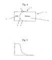
- the impedance values may, for example, be plotted on a visual display and a curve fitted to them. The diaphragm position will correspond to the point of greatest gradient. This can either be determined visually by an operator or calculated and presented as a position-value.
- the apparatus may be coupled to a source of radiation and be arranged to trigger that source to emit radiation when the diaphragm is at a specific position.
- the radiation need only be directed at the patient when it is directly focused on its target.
- the direction of the radiation beam may be controlled to follow the movement of the target based on the determined position of the diaphragm.
- the location of the tumor is measured at several different levels of inspiration and the path of the tumor throughout a normal breathing cycle is calculated as a function of diaphragm position.
- the radiation source is mounted on a tracking device that is controlled by the control unit to direct the radiation towards the calculated position of the tumor based on the current measurement of diaphragm position.
- the inventors have recognized that when monitoring the respiratory status of seriously ill patients, e.g. in the intensive care unit (ICU) it would be useful to measure the position of the diaphragm by means of the present invention. This would give important information about the quality of respiration that is not available with the techniques currently used in the ICU.
- ICU intensive care unit
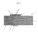
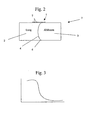
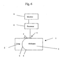
- Fig. 1 shows a respiration monitoring apparatus in accordance with the present invention.
- a patient 1 having a lung 2 and an abdomen 3 being separated therefrom by a diaphragm 4 is fitted with an ultrasound transducer array 5 positioned approximately centrally over the lung sinus 6 prior to being given a CT or MRI scan.
- the transducer array 5 consists of a plurality of individual transducer elements 7 that span at least a part of the region of diaphragm movement i.e. from a relaxed state to a contracted state.
- the transducer array is adapted to span the entire region of diaphragm movement. In alternative embodiments however, it can be adapted to span only 75%, 50% or 25% of the region of diaphragm movement respectively.
- the array can be adapted to span from 1% to 15% or from 1% to 20% or from 1% to 25% of the region of diaphragm movement respectively.
- Each individual transducer element 7 emits an ultrasound pulse and then detects its echo in the known manner. Because air has a much higher acoustic impedance than tissue, the reflection of the ultrasound beam is much more pronounced when the lung is insonated. Thus, the strength of signal received will be much higher when the pulse is reflected from an aerated space than when the pulse is reflected by tissue.
- the strength of the receiving signal i.e. the degree to which the transducers are receiving from air and from tissue, it is possible to determine to a high degree of accuracy the position of the patient's diaphragm.
- the measured acoustic impedance from each transducer element is used as an input to a processor 8..Impedance may then be processed as a function that varies in the z-direction. The impedance values are then plotted on a visual display 9 and a curve fitted to them. The diaphragm position will correspond to the point of greatest gradient. This is calculated by the processor 8, highlighted on the display 9 and presented as a numerical position-value, also on the display 9..
- a CT or an MRI scan is performed on the patient to determine the precise location of the target (e.g. a lesion to be punctured).
- the patient is required to hold his breath so that a clear image is produced with the lungs in one position. While the scan is being performed and while the patient is holding his or her breath, the exact position of the diaphragm is presented on the display and the position-value is noted.
- the image from the scan is used to calculate the depth and angle at which a needle must be inserted for the lesion to be punctured.
- the patient is asked to inhale until the display indicates that the diaphragm is in the same position as it was when the scan was performed. If the patient inhales too much and the transducer array indicates that the level of inspiration is greater than that held during the scan, the operator can instruct the patient to exhale a little. If necessary, the patient can relax and inhale again until the operator is happy with the position of the diaphragm.
- the operator can be sure that the lesion is at the same position within the patient as it is shown in the CT or MR image while he or she performs the puncture.
- the location of the needle may, however, still be checked by means of a further scan.
- the apparatus of the present invention can also be used to improve radiotherapy treatments by reducing the area that needs to be irradiated.
- the basic procedure described above is employed, however, the embodiment is modified to provide a control output from the processor for controlling a source of radiation.
- a radiation source is aimed at that location. This is connected to the control output such that the radiation source only emits when triggered to do so by the output signal from the processor.
- the patient is allowed to breathe continuously throughout the radiation treatment.
- the processor uses the outputs from the transducer array to continuously monitor the position of the diaphragm.
- the processor sends a signal to trigger the radiation source to irradiate the target area of the patient.
- the area of the patient that needs to be irradiated can be significantly reduced because the location of the target can be determined to a much greater accuracy.
Landscapes
- Health & Medical Sciences (AREA)
- Life Sciences & Earth Sciences (AREA)
- Molecular Biology (AREA)
- Surgery (AREA)
- Physics & Mathematics (AREA)
- Veterinary Medicine (AREA)
- Biophysics (AREA)
- Pathology (AREA)
- Engineering & Computer Science (AREA)
- Biomedical Technology (AREA)
- Heart & Thoracic Surgery (AREA)
- Animal Behavior & Ethology (AREA)
- Public Health (AREA)
- General Health & Medical Sciences (AREA)
- Medical Informatics (AREA)
- Physiology (AREA)
- Oral & Maxillofacial Surgery (AREA)
- Dentistry (AREA)
- Nuclear Medicine, Radiotherapy & Molecular Imaging (AREA)
- Radiology & Medical Imaging (AREA)
- Pulmonology (AREA)
- Ultra Sonic Daignosis Equipment (AREA)
- Apparatus For Radiation Diagnosis (AREA)
- Magnetic Resonance Imaging Apparatus (AREA)
Claims (9)
- Procédé de détermination du degré d'inspiration pulmonaire chez un patient (1), comprenant l'étape de détection en mode non invasif de la position du diaphragme (4) du patient au moyen d'un réseau d'au moins deux éléments transducteurs ultrasonores (7) sur le patient s'étendant dans la direction de l'axe longitudinal (z) du patient sur le sinus pulmonaire (6), dans lequel chaque élément transducteur individuel détecte un faisceau ultrasonore qui est réfléchi par le tissu qui lui est adjacent et délivre un signal de sortie ;
et comprenant l'étape de mesure de l'intensité de chaque signal de sortie pour obtenir une valeur pour l'impédance acoustique dudit tissu adjacent à l'élément transducteur ; et de détermination de la position du diaphragme sur la base d'une comparaison des valeurs d'impédance acoustique mesurées issues de chaque transducteur. - Procédé selon la revendication 1, dans lequel la position du diaphragme est utilisée comme point de référence pour définir le degré d'inspiration pulmonaire lorsqu'une image du patient (1) est générée.
- Procédé selon la revendication 2, dans lequel la position du diaphragme est tout d'abord déterminée tandis que le patient (1) retient sa respiration et que des images sont générées simultanément avec cette situation, le degré d'inspiration pulmonaire étant ensuite reproduit par le patient inhalant ou inhalant et exhalant jusqu'à ce que la position du diaphragme précédemment déterminée soit obtenue.
- Procédé selon l'une quelconque des revendications précédentes, dans lequel le réseau d'éléments transducteurs ultrasonores (7) est placé sur la poitrine inférieure et/ou l'abdomen supérieur du patient et est déplacé dans une position souhaitée sur le sinus pulmonaire (6) en utilisant une rétroaction des éléments transducteurs ultrasonores.
- Appareil de surveillance de la position du diaphragme (4) d'un patient, comprenant un réseau d'au moins deux éléments transducteurs ultrasonores (7) à placer sur le patient (1) dans la direction de l'axe longitudinal (z) du patient pour s'étendre sur le sinus pulmonaire (6), dans lequel chaque élément transducteur individuel est aménagé pour détecter un faisceau ultrasonore qui est réfléchi par un tissu qui lui est adjacent et pour délivrer un signal de sortie ;
dans lequel l'appareil est aménagé pour mesurer l'intensité de chaque signal de sortie pour obtenir une valeur pour l'impédance acoustique dudit tissu adjacent à l'élément transducteur et pour déterminer la position du diaphragme sur la base d'une comparaison des valeurs d'impédance acoustique mesurées. - Appareil selon la revendication 5, dans lequel le réseau d'éléments transducteurs (7) est un réseau unidimensionnel.
- Appareil selon l'une quelconque des revendications 5 ou 6, dans lequel l'impédance acoustique mesurée issue de chaque élément transducteur (7) est utilisée comme entrée dans un processeur (8) et les mesures d'impédance acoustique sont traitées pour fournir une fonction qui varie avec le mouvement du diaphragme (4) dans la direction z.
- Appareil de radiothérapie comprenant une source de rayonnement, une unité de commande et un appareil de surveillance de la position du diaphragme selon les revendications 5, 6 ou 7, dans lequel la source est montée sur un dispositif de suivi et l'unité de commande est aménagée de manière à commander la source pour diriger le rayonnement vers la position calculée d'une tumeur sur la base de la mesure courante de la position du diaphragme.
- Procédé de surveillance respiratoire par surveillance du mouvement du diaphragme d'un patient en utilisant le procédé ou l'appareil selon l'une quelconque des revendications 1 à 7.
Applications Claiming Priority (2)
| Application Number | Priority Date | Filing Date | Title |
|---|---|---|---|
| GB0228189A GB2396012B (en) | 2002-12-03 | 2002-12-03 | Respiration monitor |
| PCT/GB2003/005211 WO2004049951A1 (fr) | 2002-12-03 | 2003-12-03 | Procede et dispositif de surveillance respiratoire |
Publications (3)
| Publication Number | Publication Date |
|---|---|
| EP1615564A1 EP1615564A1 (fr) | 2006-01-18 |
| EP1615564B1 true EP1615564B1 (fr) | 2014-07-23 |
| EP1615564B8 EP1615564B8 (fr) | 2014-10-08 |
Family
ID=9948991
Family Applications (1)
| Application Number | Title | Priority Date | Filing Date |
|---|---|---|---|
| EP03778559.9A Expired - Lifetime EP1615564B8 (fr) | 2002-12-03 | 2003-12-03 | Procede et dispositif de surveillance respiratoire |
Country Status (4)
| Country | Link |
|---|---|
| EP (1) | EP1615564B8 (fr) |
| AU (1) | AU2003285561A1 (fr) |
| GB (1) | GB2396012B (fr) |
| WO (1) | WO2004049951A1 (fr) |
Families Citing this family (11)
| Publication number | Priority date | Publication date | Assignee | Title |
|---|---|---|---|---|
| US9211106B2 (en) | 2010-04-29 | 2015-12-15 | Neorad As | Coupling an ultrasound probe to the skin |
| GB2479930B (en) | 2010-04-29 | 2017-12-06 | Respinor As | Coupling an ultrasound probe to the skin |
| GB201519985D0 (en) * | 2015-11-12 | 2015-12-30 | Respinor As | Ultrasonic method and apparatus for respiration monitoring |
| WO2017198787A1 (fr) | 2016-05-19 | 2017-11-23 | Pmd Device Solutions Limited | Appareil et procédé de détection de respiration dysfonctionnelle |
| US11583249B2 (en) * | 2017-09-08 | 2023-02-21 | Biosense Webster (Israel) Ltd. | Method and apparatus for performing non-fluoroscopic transseptal procedure |
| US12144604B2 (en) | 2018-12-11 | 2024-11-19 | Respinor As | Systems and methods for motion compensation in ultrasonic respiration monitoring |
| CN113301845B (zh) * | 2018-12-11 | 2024-08-20 | 雷斯皮诺尔公共有限责任公司 | 用于超声呼吸监测中的运动补偿的系统和方法 |
| HRP20240182T1 (hr) | 2020-03-26 | 2024-04-26 | Vanderbilt University | Ljudska monoklonska protutijela za teški akutni respiratorni sindrom koronavirusa 2 (sars-cov-2) |
| WO2022073242A1 (fr) | 2020-10-10 | 2022-04-14 | 深圳迈瑞生物医疗电子股份有限公司 | Procédé d'imagerie ultrasonore en combinaison avec un signal physiologique et un dispositif électronique |
| TW202342510A (zh) | 2022-02-18 | 2023-11-01 | 英商Rq生物科技有限公司 | 抗體 |
| CN116784824B (zh) * | 2022-03-16 | 2025-12-02 | 深圳惟德精准医疗科技有限公司 | 呼吸监测方法、装置、电子设备及存储介质 |
Citations (1)
| Publication number | Priority date | Publication date | Assignee | Title |
|---|---|---|---|---|
| US4431007A (en) * | 1981-02-04 | 1984-02-14 | General Electric Company | Referenced real-time ultrasonic image display |
Family Cites Families (6)
| Publication number | Priority date | Publication date | Assignee | Title |
|---|---|---|---|---|
| US4915103A (en) * | 1987-12-23 | 1990-04-10 | N. Visveshwara, M.D., Inc. | Ventilation synchronizer |
| US5363844A (en) * | 1993-08-13 | 1994-11-15 | Mayo Foundation For Medical Education And Research | Breath-hold monitor for MR imaging |
| US6118847A (en) * | 1998-01-15 | 2000-09-12 | Siemens Medical Systems, Inc. | System and method for gated radiotherapy based on physiological inputs |
| DE19946092A1 (de) * | 1999-09-25 | 2001-03-29 | Philips Corp Intellectual Pty | Verfahren und Vorrichtung zur Ermittlung eines 3D-Bilddatensatzes eines sich periodisch bewegenden Körperorgans |
| ES2193030T3 (es) * | 2000-11-22 | 2003-11-01 | Brainlab Ag | Metodo para determinar el volumen de los pulmones. |
| WO2002041776A1 (fr) * | 2000-11-24 | 2002-05-30 | Feinberg David A | Ultrasons a l'interieur d'appareils irm permettant de guider des sequences d'impulsion irm |
-
2002
- 2002-12-03 GB GB0228189A patent/GB2396012B/en not_active Expired - Fee Related
-
2003
- 2003-12-03 EP EP03778559.9A patent/EP1615564B8/fr not_active Expired - Lifetime
- 2003-12-03 WO PCT/GB2003/005211 patent/WO2004049951A1/fr not_active Ceased
- 2003-12-03 AU AU2003285561A patent/AU2003285561A1/en not_active Abandoned
Patent Citations (1)
| Publication number | Priority date | Publication date | Assignee | Title |
|---|---|---|---|---|
| US4431007A (en) * | 1981-02-04 | 1984-02-14 | General Electric Company | Referenced real-time ultrasonic image display |
Non-Patent Citations (1)
| Title |
|---|
| ETLIK O. ET AL: "Demonstrating the effect of theophylline treatment on diaphragmatic movement in chronic obstructive pulmonary disease patients by MR-fluoroscopy", EUROPEAN JOURNAL OF RADIOLOGY, vol. 51, no. 2, August 2004 (2004-08-01), pages 150 - 154 * |
Also Published As
| Publication number | Publication date |
|---|---|
| AU2003285561A1 (en) | 2004-06-23 |
| GB2396012B (en) | 2006-03-15 |
| EP1615564A1 (fr) | 2006-01-18 |
| WO2004049951A1 (fr) | 2004-06-17 |
| GB2396012A (en) | 2004-06-09 |
| EP1615564B8 (fr) | 2014-10-08 |
| GB0228189D0 (en) | 2003-01-08 |
Similar Documents
| Publication | Publication Date | Title |
|---|---|---|
| US8814793B2 (en) | Respiration monitor | |
| JP3784840B2 (ja) | 画像案内式胸部治療法およびその装置 | |
| KR100750279B1 (ko) | 치료시 호흡 및 환자 움직임을 보상하기 위한 장치 및 방법 | |
| AU743966B2 (en) | Intrabody measurement | |
| JP4448194B2 (ja) | 医療用機器の診断及び取扱いならびに映像システム | |
| EP2628456B1 (fr) | Dispositif de thérapie d'onde de choc avec suivi de cible | |
| JP5255964B2 (ja) | 手術支援装置 | |
| US20080171930A1 (en) | Method and apparatus for positioning an instrument in a predetermined region within a patient's body | |
| JP2008535560A (ja) | 身体ボリュームにおける誘導介入的医療デバイスのための3次元イメージング | |
| JP2013517039A (ja) | 撮像装置 | |
| JPH11313821A (ja) | 医用タ―ゲット装置 | |
| JP2002306473A (ja) | カテーテルの位置を決定する方法及び超音波撮像システム | |
| JP2010213773A (ja) | 呼吸指示装置 | |
| EP0900048B1 (fr) | Dispositif de therapie du thorax guidee par image | |
| EP1615564B1 (fr) | Procede et dispositif de surveillance respiratoire | |
| CN116669633A (zh) | 多平面运动管理系统 | |
| EP3079584B1 (fr) | Procédé et système de surveillance respiratoire durant des interventions guidées par image | |
| CN116784824B (zh) | 呼吸监测方法、装置、电子设备及存储介质 | |
| KR20190082161A (ko) | 피부 특성에 따라 에너지를 조사하는 시술장치 | |
| AU773122B2 (en) | Image-guided thoracic therapy and apparatus therefor |
Legal Events
| Date | Code | Title | Description |
|---|---|---|---|
| PUAI | Public reference made under article 153(3) epc to a published international application that has entered the european phase |
Free format text: ORIGINAL CODE: 0009012 |
|
| 17P | Request for examination filed |
Effective date: 20050624 |
|
| AK | Designated contracting states |
Kind code of ref document: A1 Designated state(s): AT BE BG CH CY CZ DE DK EE ES FI FR GB GR HU IE IT LI LU MC NL PT RO SE SI SK TR |
|
| AX | Request for extension of the european patent |
Extension state: AL LT LV MK |
|
| DAX | Request for extension of the european patent (deleted) | ||
| 17Q | First examination report despatched |
Effective date: 20061031 |
|
| APBK | Appeal reference recorded |
Free format text: ORIGINAL CODE: EPIDOSNREFNE |
|
| APBN | Date of receipt of notice of appeal recorded |
Free format text: ORIGINAL CODE: EPIDOSNNOA2E |
|
| RAP1 | Party data changed (applicant data changed or rights of an application transferred) |
Owner name: NEORAD A/S |
|
| APBR | Date of receipt of statement of grounds of appeal recorded |
Free format text: ORIGINAL CODE: EPIDOSNNOA3E |
|
| APAF | Appeal reference modified |
Free format text: ORIGINAL CODE: EPIDOSCREFNE |
|
| APAQ | Information on invitation to file observation in appeal modified |
Free format text: ORIGINAL CODE: EPIDOSCOBA2E |
|
| APBX | Invitation to file observations in appeal sent |
Free format text: ORIGINAL CODE: EPIDOSNOBA2E |
|
| APBZ | Receipt of observations in appeal recorded |
Free format text: ORIGINAL CODE: EPIDOSNOBA4E |
|
| APBT | Appeal procedure closed |
Free format text: ORIGINAL CODE: EPIDOSNNOA9E |
|
| GRAP | Despatch of communication of intention to grant a patent |
Free format text: ORIGINAL CODE: EPIDOSNIGR1 |
|
| INTG | Intention to grant announced |
Effective date: 20130612 |
|
| GRAS | Grant fee paid |
Free format text: ORIGINAL CODE: EPIDOSNIGR3 |
|
| GRAA | (expected) grant |
Free format text: ORIGINAL CODE: 0009210 |
|
| AK | Designated contracting states |
Kind code of ref document: B1 Designated state(s): AT BE BG CH CY CZ DE DK EE ES FI FR GB GR HU IE IT LI LU MC NL PT RO SE SI SK TR |
|
| REG | Reference to a national code |
Ref country code: GB Ref legal event code: FG4D |
|
| REG | Reference to a national code |
Ref country code: DE Ref legal event code: R081 Ref document number: 60346531 Country of ref document: DE Owner name: RESPINOR AS, NO Free format text: FORMER OWNER: NEORAD A/S, OSLO, NO |
|
| REG | Reference to a national code |
Ref country code: CH Ref legal event code: EP |
|
| REG | Reference to a national code |
Ref country code: IE Ref legal event code: FG4D |
|
| REG | Reference to a national code |
Ref country code: AT Ref legal event code: REF Ref document number: 678389 Country of ref document: AT Kind code of ref document: T Effective date: 20140815 |
|
| REG | Reference to a national code |
Ref country code: DE Ref legal event code: R096 Ref document number: 60346531 Country of ref document: DE Effective date: 20140904 |
|
| RAP2 | Party data changed (patent owner data changed or rights of a patent transferred) |
Owner name: NEORAD AS |
|
| REG | Reference to a national code |
Ref country code: DE Ref legal event code: R081 Ref document number: 60346531 Country of ref document: DE Owner name: RESPINOR AS, NO Free format text: FORMER OWNER: NEORAD A/S, OSLO, NO |
|
| REG | Reference to a national code |
Ref country code: AT Ref legal event code: MK05 Ref document number: 678389 Country of ref document: AT Kind code of ref document: T Effective date: 20140723 |
|
| REG | Reference to a national code |
Ref country code: NL Ref legal event code: VDEP Effective date: 20140723 |
|
| PG25 | Lapsed in a contracting state [announced via postgrant information from national office to epo] |
Ref country code: FI Free format text: LAPSE BECAUSE OF FAILURE TO SUBMIT A TRANSLATION OF THE DESCRIPTION OR TO PAY THE FEE WITHIN THE PRESCRIBED TIME-LIMIT Effective date: 20140723 Ref country code: GR Free format text: LAPSE BECAUSE OF FAILURE TO SUBMIT A TRANSLATION OF THE DESCRIPTION OR TO PAY THE FEE WITHIN THE PRESCRIBED TIME-LIMIT Effective date: 20141024 Ref country code: BG Free format text: LAPSE BECAUSE OF FAILURE TO SUBMIT A TRANSLATION OF THE DESCRIPTION OR TO PAY THE FEE WITHIN THE PRESCRIBED TIME-LIMIT Effective date: 20141023 Ref country code: SE Free format text: LAPSE BECAUSE OF FAILURE TO SUBMIT A TRANSLATION OF THE DESCRIPTION OR TO PAY THE FEE WITHIN THE PRESCRIBED TIME-LIMIT Effective date: 20140723 Ref country code: ES Free format text: LAPSE BECAUSE OF FAILURE TO SUBMIT A TRANSLATION OF THE DESCRIPTION OR TO PAY THE FEE WITHIN THE PRESCRIBED TIME-LIMIT Effective date: 20140723 Ref country code: PT Free format text: LAPSE BECAUSE OF FAILURE TO SUBMIT A TRANSLATION OF THE DESCRIPTION OR TO PAY THE FEE WITHIN THE PRESCRIBED TIME-LIMIT Effective date: 20141124 |
|
| PG25 | Lapsed in a contracting state [announced via postgrant information from national office to epo] |
Ref country code: AT Free format text: LAPSE BECAUSE OF FAILURE TO SUBMIT A TRANSLATION OF THE DESCRIPTION OR TO PAY THE FEE WITHIN THE PRESCRIBED TIME-LIMIT Effective date: 20140723 Ref country code: NL Free format text: LAPSE BECAUSE OF FAILURE TO SUBMIT A TRANSLATION OF THE DESCRIPTION OR TO PAY THE FEE WITHIN THE PRESCRIBED TIME-LIMIT Effective date: 20140723 Ref country code: CY Free format text: LAPSE BECAUSE OF FAILURE TO SUBMIT A TRANSLATION OF THE DESCRIPTION OR TO PAY THE FEE WITHIN THE PRESCRIBED TIME-LIMIT Effective date: 20140723 |
|
| REG | Reference to a national code |
Ref country code: DE Ref legal event code: R097 Ref document number: 60346531 Country of ref document: DE |
|
| PG25 | Lapsed in a contracting state [announced via postgrant information from national office to epo] |
Ref country code: DK Free format text: LAPSE BECAUSE OF FAILURE TO SUBMIT A TRANSLATION OF THE DESCRIPTION OR TO PAY THE FEE WITHIN THE PRESCRIBED TIME-LIMIT Effective date: 20140723 Ref country code: EE Free format text: LAPSE BECAUSE OF FAILURE TO SUBMIT A TRANSLATION OF THE DESCRIPTION OR TO PAY THE FEE WITHIN THE PRESCRIBED TIME-LIMIT Effective date: 20140723 Ref country code: CZ Free format text: LAPSE BECAUSE OF FAILURE TO SUBMIT A TRANSLATION OF THE DESCRIPTION OR TO PAY THE FEE WITHIN THE PRESCRIBED TIME-LIMIT Effective date: 20140723 Ref country code: RO Free format text: LAPSE BECAUSE OF FAILURE TO SUBMIT A TRANSLATION OF THE DESCRIPTION OR TO PAY THE FEE WITHIN THE PRESCRIBED TIME-LIMIT Effective date: 20140723 Ref country code: SK Free format text: LAPSE BECAUSE OF FAILURE TO SUBMIT A TRANSLATION OF THE DESCRIPTION OR TO PAY THE FEE WITHIN THE PRESCRIBED TIME-LIMIT Effective date: 20140723 Ref country code: IT Free format text: LAPSE BECAUSE OF FAILURE TO SUBMIT A TRANSLATION OF THE DESCRIPTION OR TO PAY THE FEE WITHIN THE PRESCRIBED TIME-LIMIT Effective date: 20140723 |
|
| PLBE | No opposition filed within time limit |
Free format text: ORIGINAL CODE: 0009261 |
|
| STAA | Information on the status of an ep patent application or granted ep patent |
Free format text: STATUS: NO OPPOSITION FILED WITHIN TIME LIMIT |
|
| PG25 | Lapsed in a contracting state [announced via postgrant information from national office to epo] |
Ref country code: BE Free format text: LAPSE BECAUSE OF NON-PAYMENT OF DUE FEES Effective date: 20141231 |
|
| 26N | No opposition filed |
Effective date: 20150424 |
|
| PG25 | Lapsed in a contracting state [announced via postgrant information from national office to epo] |
Ref country code: LU Free format text: LAPSE BECAUSE OF FAILURE TO SUBMIT A TRANSLATION OF THE DESCRIPTION OR TO PAY THE FEE WITHIN THE PRESCRIBED TIME-LIMIT Effective date: 20141203 |
|
| REG | Reference to a national code |
Ref country code: IE Ref legal event code: MM4A |
|
| PG25 | Lapsed in a contracting state [announced via postgrant information from national office to epo] |
Ref country code: IE Free format text: LAPSE BECAUSE OF NON-PAYMENT OF DUE FEES Effective date: 20141203 |
|
| PG25 | Lapsed in a contracting state [announced via postgrant information from national office to epo] |
Ref country code: SI Free format text: LAPSE BECAUSE OF FAILURE TO SUBMIT A TRANSLATION OF THE DESCRIPTION OR TO PAY THE FEE WITHIN THE PRESCRIBED TIME-LIMIT Effective date: 20140723 |
|
| REG | Reference to a national code |
Ref country code: FR Ref legal event code: PLFP Year of fee payment: 13 |
|
| PG25 | Lapsed in a contracting state [announced via postgrant information from national office to epo] |
Ref country code: MC Free format text: LAPSE BECAUSE OF FAILURE TO SUBMIT A TRANSLATION OF THE DESCRIPTION OR TO PAY THE FEE WITHIN THE PRESCRIBED TIME-LIMIT Effective date: 20140723 |
|
| PG25 | Lapsed in a contracting state [announced via postgrant information from national office to epo] |
Ref country code: HU Free format text: LAPSE BECAUSE OF FAILURE TO SUBMIT A TRANSLATION OF THE DESCRIPTION OR TO PAY THE FEE WITHIN THE PRESCRIBED TIME-LIMIT; INVALID AB INITIO Effective date: 20031203 Ref country code: TR Free format text: LAPSE BECAUSE OF FAILURE TO SUBMIT A TRANSLATION OF THE DESCRIPTION OR TO PAY THE FEE WITHIN THE PRESCRIBED TIME-LIMIT Effective date: 20140723 |
|
| REG | Reference to a national code |
Ref country code: DE Ref legal event code: R081 Ref document number: 60346531 Country of ref document: DE Owner name: RESPINOR AS, NO Free format text: FORMER OWNER: NEORAD AS, OSLO, NO Ref country code: DE Ref legal event code: R082 Ref document number: 60346531 Country of ref document: DE Representative=s name: DEHNS, GB |
|
| REG | Reference to a national code |
Ref country code: CH Ref legal event code: NV Representative=s name: GEVERS SA, CH Ref country code: CH Ref legal event code: PUE Owner name: RESPINOIR AS, NO Free format text: FORMER OWNER: NEORAD AS, NO |
|
| REG | Reference to a national code |
Ref country code: FR Ref legal event code: PLFP Year of fee payment: 14 |
|
| REG | Reference to a national code |
Ref country code: FR Ref legal event code: CA Effective date: 20170607 Ref country code: FR Ref legal event code: TP Owner name: RESPINOR A/S, NO Effective date: 20170607 |
|
| REG | Reference to a national code |
Ref country code: FR Ref legal event code: PLFP Year of fee payment: 15 |
|
| REG | Reference to a national code |
Ref country code: CH Ref legal event code: NV Representative=s name: BOVARD SA NEUCHATEL CONSEILS EN PROPRIETE INTE, CH |
|
| REG | Reference to a national code |
Ref country code: DE Ref legal event code: R082 Ref document number: 60346531 Country of ref document: DE |
|
| PGFP | Annual fee paid to national office [announced via postgrant information from national office to epo] |
Ref country code: GB Payment date: 20221212 Year of fee payment: 20 Ref country code: FR Payment date: 20221214 Year of fee payment: 20 Ref country code: DE Payment date: 20221219 Year of fee payment: 20 |
|
| PGFP | Annual fee paid to national office [announced via postgrant information from national office to epo] |
Ref country code: CH Payment date: 20221215 Year of fee payment: 20 |
|
| REG | Reference to a national code |
Ref country code: DE Ref legal event code: R071 Ref document number: 60346531 Country of ref document: DE |
|
| REG | Reference to a national code |
Ref country code: CH Ref legal event code: PL |
|
| REG | Reference to a national code |
Ref country code: GB Ref legal event code: PE20 Expiry date: 20231202 |
|
| PG25 | Lapsed in a contracting state [announced via postgrant information from national office to epo] |
Ref country code: GB Free format text: LAPSE BECAUSE OF EXPIRATION OF PROTECTION Effective date: 20231202 |
|
| PG25 | Lapsed in a contracting state [announced via postgrant information from national office to epo] |
Ref country code: GB Free format text: LAPSE BECAUSE OF EXPIRATION OF PROTECTION Effective date: 20231202 |