EP1685861A2 - Revêtement pour un stent pour la libération de médicaments - Google Patents
Revêtement pour un stent pour la libération de médicaments Download PDFInfo
- Publication number
- EP1685861A2 EP1685861A2 EP06001891A EP06001891A EP1685861A2 EP 1685861 A2 EP1685861 A2 EP 1685861A2 EP 06001891 A EP06001891 A EP 06001891A EP 06001891 A EP06001891 A EP 06001891A EP 1685861 A2 EP1685861 A2 EP 1685861A2
- Authority
- EP
- European Patent Office
- Prior art keywords
- stent
- coating
- carbon
- columnar structures
- substrate
- Prior art date
- Legal status (The legal status is an assumption and is not a legal conclusion. Google has not performed a legal analysis and makes no representation as to the accuracy of the status listed.)
- Withdrawn
Links
Images





Classifications
-
- A—HUMAN NECESSITIES
- A61—MEDICAL OR VETERINARY SCIENCE; HYGIENE
- A61L—METHODS OR APPARATUS FOR STERILISING MATERIALS OR OBJECTS IN GENERAL; DISINFECTION, STERILISATION OR DEODORISATION OF AIR; CHEMICAL ASPECTS OF BANDAGES, DRESSINGS, ABSORBENT PADS OR SURGICAL ARTICLES; MATERIALS FOR BANDAGES, DRESSINGS, ABSORBENT PADS OR SURGICAL ARTICLES
- A61L31/00—Materials for other surgical articles, e.g. stents, stent-grafts, shunts, surgical drapes, guide wires, materials for adhesion prevention, occluding devices, surgical gloves, tissue fixation devices
- A61L31/08—Materials for coatings
- A61L31/082—Inorganic materials
- A61L31/084—Carbon; Graphite
-
- A—HUMAN NECESSITIES
- A61—MEDICAL OR VETERINARY SCIENCE; HYGIENE
- A61F—FILTERS IMPLANTABLE INTO BLOOD VESSELS; PROSTHESES; DEVICES PROVIDING PATENCY TO, OR PREVENTING COLLAPSING OF, TUBULAR STRUCTURES OF THE BODY, e.g. STENTS; ORTHOPAEDIC, NURSING OR CONTRACEPTIVE DEVICES; FOMENTATION; TREATMENT OR PROTECTION OF EYES OR EARS; BANDAGES, DRESSINGS OR ABSORBENT PADS; FIRST-AID KITS
- A61F2/00—Filters implantable into blood vessels; Prostheses, i.e. artificial substitutes or replacements for parts of the body; Appliances for connecting them with the body; Devices providing patency to, or preventing collapsing of, tubular structures of the body, e.g. stents
- A61F2/82—Devices providing patency to, or preventing collapsing of, tubular structures of the body, e.g. stents
- A61F2/86—Stents in a form characterised by the wire-like elements; Stents in the form characterised by a net-like or mesh-like structure
- A61F2/90—Stents in a form characterised by the wire-like elements; Stents in the form characterised by a net-like or mesh-like structure characterised by a net-like or mesh-like structure
- A61F2/91—Stents in a form characterised by the wire-like elements; Stents in the form characterised by a net-like or mesh-like structure characterised by a net-like or mesh-like structure made from perforated sheets or tubes, e.g. perforated by laser cuts or etched holes
-
- A—HUMAN NECESSITIES
- A61—MEDICAL OR VETERINARY SCIENCE; HYGIENE
- A61L—METHODS OR APPARATUS FOR STERILISING MATERIALS OR OBJECTS IN GENERAL; DISINFECTION, STERILISATION OR DEODORISATION OF AIR; CHEMICAL ASPECTS OF BANDAGES, DRESSINGS, ABSORBENT PADS OR SURGICAL ARTICLES; MATERIALS FOR BANDAGES, DRESSINGS, ABSORBENT PADS OR SURGICAL ARTICLES
- A61L31/00—Materials for other surgical articles, e.g. stents, stent-grafts, shunts, surgical drapes, guide wires, materials for adhesion prevention, occluding devices, surgical gloves, tissue fixation devices
- A61L31/14—Materials characterised by their function or physical properties, e.g. injectable or lubricating compositions, shape-memory materials, surface modified materials
- A61L31/146—Porous materials, e.g. foams or sponges
-
- A—HUMAN NECESSITIES
- A61—MEDICAL OR VETERINARY SCIENCE; HYGIENE
- A61L—METHODS OR APPARATUS FOR STERILISING MATERIALS OR OBJECTS IN GENERAL; DISINFECTION, STERILISATION OR DEODORISATION OF AIR; CHEMICAL ASPECTS OF BANDAGES, DRESSINGS, ABSORBENT PADS OR SURGICAL ARTICLES; MATERIALS FOR BANDAGES, DRESSINGS, ABSORBENT PADS OR SURGICAL ARTICLES
- A61L31/00—Materials for other surgical articles, e.g. stents, stent-grafts, shunts, surgical drapes, guide wires, materials for adhesion prevention, occluding devices, surgical gloves, tissue fixation devices
- A61L31/14—Materials characterised by their function or physical properties, e.g. injectable or lubricating compositions, shape-memory materials, surface modified materials
- A61L31/16—Biologically active materials, e.g. therapeutic substances
-
- A—HUMAN NECESSITIES
- A61—MEDICAL OR VETERINARY SCIENCE; HYGIENE
- A61F—FILTERS IMPLANTABLE INTO BLOOD VESSELS; PROSTHESES; DEVICES PROVIDING PATENCY TO, OR PREVENTING COLLAPSING OF, TUBULAR STRUCTURES OF THE BODY, e.g. STENTS; ORTHOPAEDIC, NURSING OR CONTRACEPTIVE DEVICES; FOMENTATION; TREATMENT OR PROTECTION OF EYES OR EARS; BANDAGES, DRESSINGS OR ABSORBENT PADS; FIRST-AID KITS
- A61F2250/00—Special features of prostheses classified in groups A61F2/00 - A61F2/26 or A61F2/82 or A61F9/00 or A61F11/00 or subgroups thereof
- A61F2250/0058—Additional features; Implant or prostheses properties not otherwise provided for
- A61F2250/0067—Means for introducing or releasing pharmaceutical products into the body
-
- A—HUMAN NECESSITIES
- A61—MEDICAL OR VETERINARY SCIENCE; HYGIENE
- A61F—FILTERS IMPLANTABLE INTO BLOOD VESSELS; PROSTHESES; DEVICES PROVIDING PATENCY TO, OR PREVENTING COLLAPSING OF, TUBULAR STRUCTURES OF THE BODY, e.g. STENTS; ORTHOPAEDIC, NURSING OR CONTRACEPTIVE DEVICES; FOMENTATION; TREATMENT OR PROTECTION OF EYES OR EARS; BANDAGES, DRESSINGS OR ABSORBENT PADS; FIRST-AID KITS
- A61F2250/00—Special features of prostheses classified in groups A61F2/00 - A61F2/26 or A61F2/82 or A61F9/00 or A61F11/00 or subgroups thereof
- A61F2250/0058—Additional features; Implant or prostheses properties not otherwise provided for
- A61F2250/0067—Means for introducing or releasing pharmaceutical products into the body
- A61F2250/0068—Means for introducing or releasing pharmaceutical products into the body the pharmaceutical product being in a reservoir
-
- A—HUMAN NECESSITIES
- A61—MEDICAL OR VETERINARY SCIENCE; HYGIENE
- A61L—METHODS OR APPARATUS FOR STERILISING MATERIALS OR OBJECTS IN GENERAL; DISINFECTION, STERILISATION OR DEODORISATION OF AIR; CHEMICAL ASPECTS OF BANDAGES, DRESSINGS, ABSORBENT PADS OR SURGICAL ARTICLES; MATERIALS FOR BANDAGES, DRESSINGS, ABSORBENT PADS OR SURGICAL ARTICLES
- A61L2300/00—Biologically active materials used in bandages, wound dressings, absorbent pads or medical devices
- A61L2300/10—Biologically active materials used in bandages, wound dressings, absorbent pads or medical devices containing or releasing inorganic materials
- A61L2300/108—Elemental carbon, e.g. charcoal
-
- Y—GENERAL TAGGING OF NEW TECHNOLOGICAL DEVELOPMENTS; GENERAL TAGGING OF CROSS-SECTIONAL TECHNOLOGIES SPANNING OVER SEVERAL SECTIONS OF THE IPC; TECHNICAL SUBJECTS COVERED BY FORMER USPC CROSS-REFERENCE ART COLLECTIONS [XRACs] AND DIGESTS
- Y10—TECHNICAL SUBJECTS COVERED BY FORMER USPC
- Y10T—TECHNICAL SUBJECTS COVERED BY FORMER US CLASSIFICATION
- Y10T29/00—Metal working
- Y10T29/49—Method of mechanical manufacture
- Y10T29/49826—Assembling or joining
- Y10T29/49885—Assembling or joining with coating before or during assembling
Definitions
- This invention relates to stents provided with coatings for eluting medication to prevent or lessen the severity of restenosis.
- stent coatings In order to minimize the response of surrounding tissue to the trauma of stent insertion and expansion, stent coatings must be biocompatible. A further requirement is that a stent coating must adhere to a substrate undergoing plastic deformation. This occurs during insertion and expansion of the stent into the vasculature system. Plastic deformation involves grain rotation and elongation, and intersection of slip planes with the substrate surface. The result is that on a scale below the grain size of the substrate, deformation is highly non-uniform, with some areas undergoing little or no deformation and others extreme deformation with associated increase in surface roughness and irregularity. Therefore, coating adhesion must be preserved through the deformation process.
- Passive coatings rely on biocompatible materials to minimize the body's response to placement of the stent into the vasculature.
- Generally recognized "passive" coating materials include carbon, iridium oxide, titanium, and the like, as disclosed in U.S. Patent No. 5,824,056 to Rosenberg.
- U.S. Patent No. 5,649,951 to Davidson discloses coatings of zirconium oxide or zirconium nitride.
- Drug eluting or "active" coatings have proven more effective for the prevention of restenosis.
- Such stents generally comprise a surface polymer containing a therapeutic drug for timed release.
- a second coating may be added to extend the period of effectiveness by limiting the rate of drug diffusion from the first, drug-containing coating.
- This second coating may be a polymer, or a sputtered coating as described in U.S. Patent No. to 6,716,444 to Castro et al.
- polymeric drug eluting coatings suffer from a number of disadvantages.
- they can have poor adhesion to the stent, especially while undergoing plastic deformation during insertion and expansion of the stent into the vasculature.
- some polymers actually contribute to restenosis.
- that part of the coating facing the inside of the vasculature lumen loses its medication content to the bloodstream with little beneficial effect.
- U.S. Patent No. 6,805,898 to Wu et al. attempted to overcome adhesion problems by introducing roughness to the vasculature-facing portion of the stent while leaving the blood-facing side in a polished condition for better hemocompatibility.
- Surface roughness was increased by means of grit blasting, sputtering, and the like. Not only did augmenting surface roughness improve adhesion between the polymer and the stent, it also allowed for a thicker polymer coating to be applied. However, the final stent configuration still had eluting polymer in contact with body tissue, allowing biocompatibility issues to persist.
- U.S. Patent Application Pub. No. 2004/01 72124 to Vallana et al. optimized the coating configuration by limiting the drug-eluting material to only that portion of the stent surface in contact with the vasculature. This was done by confining the drug eluting polymer to outward facing channels which were micro-machined into the stent mesh elements. All other stent surfaces were coated with hemocompatible carbon. Thus, the use of a biocompatible-problematic carrier polymer was minimized, but not eliminated.
- U.S. Patent No. 6,820,676 to Palmaz shows that, independent of the stent's surface composition, the surface texture of the stent or coating has an effect on the ability of proteins to adsorb into the stent surface, ultimately allowing thrombosis formation. It was shown that the surface texture can be controlled by grain size and other means to prevent protein adsorption and subsequent thrombosis.
- the drug-eluting outer layer of a stent consists of a porous sputtered metal or ceramic coating rather than a conventionally deposited polymer. This is done by placing the stent over a close-fitting mandrel and rotating the assembly in a sputter flux. The result is a coating that is evenly distributed over the outward-facing side of the stent's wire mesh while preventing the sputtered coating from reaching the inward facing side where a smooth hemocompatible surface is required. The stent is then removed from its mandrel, exposing all surfaces, and finally coated with a layer of carbon such as amorphous carbon or diamond-like carbon. The carbonaceous coating enhances biocompatibility without preventing elution of the therapeutic drug. The result is a stent that is adapted to both the hemodynamic and the immune response requirements of its vascular environment.
- Fig. 1 is a cross-sectional view of a stent 12 supporting a blood vessel 10 according to the present invention.
- Fig. 2 is a cross-sectional view of a wire 14 comprising the stent 12 shown in Fig. 1.
- Fig. 2A is an enlarged cross-sectional view of the indicated portion of the stent wire 14 shown in Fig. 2 comprising a biocompatible porous columnar coating 16 supported on the stent wire 14 with a thin carbonaceous material 18 providing a cap on each of the columns as well as covering the inside-facing surface 14B of the wire.
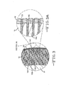
- Fig. 3 is a cross-sectional view of the stent portion shown in Fig. 2A, but with the capillary spaces between the columnar coating 16 Infused with a medication compound 20.
- Fig. 3A is an enlarged cross-sectional view of the indicated portion of Fig. 3.
- Fig. 4 is a cross-sectional view showing the interface between the stent and blood vessel 10 after deployment of the stent 12.
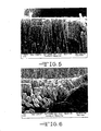
- Fig. 5 is an SEM photograph of a fracture cross-section of a porous columnar titanium nitride coating with porous carbon caps.
- Fig. 6 is a SEM photograph showing sputtered columnar aluminum nitride adhering to a substrate that has been subjected to plastic deformation.
- Fig. 7A is a schematic view of an unstrained stent wire 14 in a zero stress state.
- Fig. 7B is a schematic view of the stent wire 14 shown in Fig. 7A having been strained within its elastic limit and depicting the resulting tension and compression stress forces therein.
- Fig. 7C is a schematic view of the stent wire 14 shown in Fig. 7B being elastically strained and provided with a columnar coating 16 that is an unstrained state.
- Fig. 7D is a schematic view of the stent wire 14 shown in Fig. 7C in a relaxed, unstrained state and depicting the resulting tension and compression stress forces in the columnar coating 16.
- Fig. 7E is a schematic view of the stent wire 14 shown in Fig. 7C having been expanded past its elastic limit and depicting the resulting tension and compression stress forces in both the wire and the columnar coating 16.
- Fig. 8A is a cross-sectional view of a polymer 26 used as a reinforcing material between individual columns 16.
- Fig. 8B is a cross-sectional view of the stent shown in Fig. 8A provided with a diffusion limiting polymeric coating 28.
- Fig. 1 shows a cross-section of a blood vessel 10 with a stent 12 inserted and expanded therein.
- the medication eluting coating is limited to that portion of the stent in contact with tissue, which is exemplified by the blood vessel 10.
- the stent 12 is comprised of a plurality of wires 14 forming an elongated hollow tube and disposed so as to be capable of circumferential expansion.
- stent materials include platinum, Nitinol, and even medical grade 316L stainless steel containing about 16% nickel.
- the wires 14 provide for an elongated, expandable hollow tube that can, in a preferred embodiment, increase in diameter when the ends of the hollow tube are moved closer relative to each other and decrease in diameter when the ends are moved apart.
- a design objective is to have as little length change as possible when the stent is expanded.
- the stent 12 is positioned in the vasculature of a patient during or after a procedure, such as an angioplasty, atherectomy, or other interventional therapy, and then expanded to an appropriate size (i.e., approximately the same diameter as the vessel 10 in the region where placed), thus supporting that vascular region.
- a procedure such as an angioplasty, atherectomy, or other interventional therapy
- an appropriate size i.e., approximately the same diameter as the vessel 10 in the region where placed
- the stent 12 When in its expanded configuration, the stent 12 provides support to the vascular walls thereby preventing constriction of the vascular region in which it is located and maintaining the vascular lumen open. This is often referred to as maintaining vascular patency.
- Fig. 2 represents a cross-section of a wire 14 comprising the vascular stent 12.
- the stent wire 14 has a roughly circular cross-section comprising an outside-facing surface 14A and an inside-facing surface 14B.
- the outslde-facing surface 14A of the stent wire faces the blood vessel wall and serves as a substrate provided with a coating 16 of columnar material to a thickness of about 0.1 ⁇ m to about 20 ⁇ m.
- Sputtering causes the columnar material to first be physically absorbed with some implantation into the wire material. This is due to the kinetic energy generated by the sputtering process prior to the column growing to its desired length.
- sputtering is a preferred method for depositing the columnar coating 16
- other suitable thin film deposition method can be used. These Include chemical vapor deposition, pulsed laser deposition, evaporation including reactive evaporation, and thermal spray methods.
- wire 14 is shown having a circular cross-section, that is not necessary.
- Other embodiments of the stent 12 comprise wires 14 having triangular, square, rectangular, hexagonal, and the like cross-sections.
- each column of the coating 16 comprises an intermediate portion 16A extending to a base 16B adhered to the inside-facing surface 14B of the wire 14 and a tip 16C.
- Each column is of a relatively consistent cross-section along its length extending to the base 16B and tip 16C. That way, the columns are discrete members that only adhere to the wire substrate at their base 16B, but do not join to an immediately adjacent column.
- Titanium nitride is a preferred material for the columnar coating 16, although other useful materials include, but are not limited to, boron, aluminum, calcium, gold, hafnium, iridium, molybdenum, niobium, platinum, rhenium, ruthenium, silicon, silver, tantalum, titanium, tungsten, yttrium, and zirconium, and carbides, oxides, nitrides, oxynitrides, carbonitrides thereof.
- the inside-facing surface 14B of the wire 14 as well as each columnar tip 16C is coated with a carbonaceous material 18, such as amorphous carbon or diamond-like carbon.
- a carbonaceous material 18 such as amorphous carbon or diamond-like carbon.
- the carbon 18 assumes the morphology of a "cap” adhered to each tip 16C of the porous columnar coating 16 supported on the outside-facing surface 14A of the stent wire 14.
- the carbon caps 18, which are also preferably provided by a sputtering process, are at a thickness of about 0.05 ⁇ m to about 2.0 ⁇ m. That Is, the porosity of the drug-eluting columnar coating 16 is maintained.
- the carbon 18 that coats the bare metal inside-facing surface 14B of the stent wire 14 forms a smooth continuous pore-free layer suitable for contact with blood.
- the capillary spaces between the columns of the coating 16 and the carbon cap 18 are infused with medication 20 to inhibit restenosis.
- This can be done by various methods well known to those skilled in the art including spraying the stent with a medication solution, dipping the stent into a medication solution, immersing the stent in a medication solution under vacuum conditions and centrifuging the medication solution into the porosity.
- Fig. 4 shows the interface between the treated stent wire 14 and the blood vessel 10 after deployment of the stent 12 therein.
- Medication 20 residing in the capillary spaces of the columnar coating 16 is directed into the vessel 10 supported by the stent with the vessel tissue only contacting the biocompatible carbonaceous caps 18.
- Fig. 5 is a SEM photograph of a fracture cross-section of a porous columnar coating illustrating the volume of empty space therein and the internal surface roughness of the capillaries.
- the porous columnar coating consists of titanium nitride, which is widely used as a permanent implantable coating for bioelectrodes.
- the carbon cap on each individual titanium nitride column comprising the outer 200 nm to 300 nm of the coating. Deposition of the carbon layer was done with the mandrel removed from the stent mesh.
- the mesh was fixtured to expose all surfaces of the stent to sputter flux.
- the stent was rotated In the sputter flux during deposition, which was done with DC sputtering of a carbon target in argon process gas. Typical conditions are 7 mTorr, 250 Watts, no bias.
- Typical conditions are 7 mTorr, 250 Watts, no bias.
- Fig. 6 illustrates adhesion of a porous columnar coating of aluminum nitride even after extensive plastic deformation of the substrate.
- Reactive DC sputtering was used.
- the process gas was pure nitrogen at a pressure of about 5.3 mTorr.
- Power was set at 250 W on a 3 inch diameter planar target with no bias. Deposition time was 4 hours.
- a further aspect of the invention relates to controlling the stress state of each column comprising the coating 16 supported on the stent wires 14. Fixturing the stent 12 on a mandrel (not shown) subjected to a sputter flux provides for coating the outside-facing surface 14A thereof with the columnar coating 16 while protecting the inside-facing surface 14B of the stent wire 14. Increasing the degree of expansion over the mandrel to higher levels, within the elastic limit of the stent wire 14, and sputtering in that expanded state, lessens the overall stress on the columnar coating 16 when the stent 12 is finally Inserted and expanded in the blood vessel 10.
- Fig. 7A shows an unstrained stent wire 14.
- the wire has a generally elongate U-shape comprising spaced apart struts 14C and 14D joined together by a union portion 14E.
- Datum points 22 and 24 are indicated adjacent to the terminus of the respective struts 14C, 1 4D.
- the struts comprise a continuous structure such as a mesh and have no "terminus".
- the stresses set up in the union portion 14E include both tension forces (+ ⁇ e ) and compression forces (- ⁇ e ) within the elastic limits of the wire.
- the goal is to stress the union portion 14E of the wire 14 within its elastic limits so that the tension and compression strains create an opposite elastic pre-strain in the coating when the stent is removed from the mandrel.
- the struts 14C, 14D remain relatively unstressed.
- Fig. 7C shows the stent wire 14 in the same stressed state illustrated in Fig. 7B, but after the sputtered columnar coating 16 is applied.
- the columnar coating 16 is in a zero stress state.
- Fig. 7D when the stent is removed from the mandrel, it elastically springs back with the distance between the datum points 22, 24 being at or near to their original spacing shown in Fig. 7A.
- the columnar coating 16 is now in a stressed state opposite to that shown for the substrate in Fig. 7C.
- the columnar coating 16 on the outside-facing surface 14A is in a tension state within the elastic limits of the wire coating material (+ ⁇ e ) while the columnar coating on the inside-facing surface 14B is in a compression state (- ⁇ e ).
- Fig. 7E the wire 14 undergoes plastic deformation during the stent's expansion and placement in the vasculature. This is depicted by the opposing directions of the respective vector arrows 22A, 24A.
- the stress in the coating 16 is the stress due to deformation of the wire surface at the union portion 1 4E minus the coating pre-stress, as shown in Fig. 7E. Therefore, the final tension (- ⁇ f ) and compression (+ ⁇ f ) forces in the coating 16 are somewhat less than they would have been had the columnar coating been provided on the stent wire in a completely relaxed state in comparison to the actual stressed state the union portion 14E was In during the deposition process. The difference is the amount of elastic deformation in the union portion 14E of the stent wire 14 while the coating was being deposited (Fig. 7C).
- the elastic limit of the stent wire 14 can be determined by placing the stent over increasingly larger diameter mandrels, until the spring back upon removal does not return the stent to its original dimension.
- the film pre-stress can be achieved by using a nickel titanium shape memory alloy which can be made to assume the partially expanded configuration by heating in the sputter chamber.
- Fig. 8A Another aspect of the invention is shown in Fig. 8A.
- This embodiment relates to the use of a polymer 26 that is provided with a medication and infused into the porous columnar coating 16 to improve biocompatibility while increasing coating strength and adhesion.
- Suitable polymers include (but are not limited to) polyurethane, silicone, polyesters, polycarbonate, polyethylene, polyvinyl chloride, polypropylene methylacrylate, para-xylylene.
- a polymer 28 can also be used to moderate and control the diffusion of the medication from the capillaries of the porous coating 16 into the surrounding tissue.
- the polymeric coating 28 is added to the porous layer after it is infused with the therapeutic medication.
- the process coats all surfaces of the stent, it allows selection from a wider range of substrate materials, including those which improved radiopacity characteristics. This is an important consideration for locating the stent correctly during placement in the vasculature.
Landscapes
- Health & Medical Sciences (AREA)
- Life Sciences & Earth Sciences (AREA)
- Animal Behavior & Ethology (AREA)
- Veterinary Medicine (AREA)
- Heart & Thoracic Surgery (AREA)
- Public Health (AREA)
- Vascular Medicine (AREA)
- General Health & Medical Sciences (AREA)
- Engineering & Computer Science (AREA)
- Biomedical Technology (AREA)
- Surgery (AREA)
- Epidemiology (AREA)
- Chemical & Material Sciences (AREA)
- Physics & Mathematics (AREA)
- Medicinal Chemistry (AREA)
- Molecular Biology (AREA)
- Dispersion Chemistry (AREA)
- Inorganic Chemistry (AREA)
- Optics & Photonics (AREA)
- Cardiology (AREA)
- Oral & Maxillofacial Surgery (AREA)
- Transplantation (AREA)
- Materials For Medical Uses (AREA)
- Media Introduction/Drainage Providing Device (AREA)
- Prostheses (AREA)
Applications Claiming Priority (1)
| Application Number | Priority Date | Filing Date | Title |
|---|---|---|---|
| US64813305P | 2005-01-28 | 2005-01-28 |
Publications (2)
| Publication Number | Publication Date |
|---|---|
| EP1685861A2 true EP1685861A2 (fr) | 2006-08-02 |
| EP1685861A3 EP1685861A3 (fr) | 2006-09-27 |
Family
ID=36440925
Family Applications (1)
| Application Number | Title | Priority Date | Filing Date |
|---|---|---|---|
| EP06001891A Withdrawn EP1685861A3 (fr) | 2005-01-28 | 2006-01-30 | Revêtement pour un stent pour la libération de médicaments |
Country Status (2)
| Country | Link |
|---|---|
| US (4) | US8057543B2 (fr) |
| EP (1) | EP1685861A3 (fr) |
Cited By (9)
| Publication number | Priority date | Publication date | Assignee | Title |
|---|---|---|---|---|
| GB2451060A (en) * | 2007-07-11 | 2009-01-21 | Anthony Walter Anson | Dual coatings applied to medical devices |
| WO2009012099A3 (fr) * | 2007-07-13 | 2010-02-25 | Boston Scientific Limited | Endoprothèses à mémoire de forme renforcées de bore |
| WO2009012101A3 (fr) * | 2007-07-13 | 2010-02-25 | Boston Scientific Limited | Endoprothèses contenant des phases intermétalliques de borure |
| US7833266B2 (en) | 2007-11-28 | 2010-11-16 | Boston Scientific Scimed, Inc. | Bifurcated stent with drug wells for specific ostial, carina, and side branch treatment |
| US7951193B2 (en) | 2008-07-23 | 2011-05-31 | Boston Scientific Scimed, Inc. | Drug-eluting stent |
| US8048151B2 (en) | 2005-01-28 | 2011-11-01 | Greatbatch Ltd. | Stent coating for eluting medication |
| WO2015010643A1 (fr) * | 2013-07-26 | 2015-01-29 | 宁夏东方钽业股份有限公司 | Procédé de préparation d'un matériau poreux pour implant médical |
| CN110947030A (zh) * | 2019-11-29 | 2020-04-03 | 中国科学院深圳先进技术研究院 | 一种抗菌多级次金刚石复合材料及其制备方法和应用 |
| CN112516391A (zh) * | 2020-12-21 | 2021-03-19 | 辽宁垠艺生物科技股份有限公司 | 一种带膜支架的载药方法 |
Families Citing this family (124)
| Publication number | Priority date | Publication date | Assignee | Title |
|---|---|---|---|---|
| US7713297B2 (en) | 1998-04-11 | 2010-05-11 | Boston Scientific Scimed, Inc. | Drug-releasing stent with ceramic-containing layer |
| EP1132058A1 (fr) * | 2000-03-06 | 2001-09-12 | Advanced Laser Applications Holding S.A. | Prothèse intravasculaire |
| US6764505B1 (en) | 2001-04-12 | 2004-07-20 | Advanced Cardiovascular Systems, Inc. | Variable surface area stent |
| AU2002345328A1 (en) | 2001-06-27 | 2003-03-03 | Remon Medical Technologies Ltd. | Method and device for electrochemical formation of therapeutic species in vivo |
| US7169178B1 (en) * | 2002-11-12 | 2007-01-30 | Advanced Cardiovascular Systems, Inc. | Stent with drug coating |
| US20070106374A1 (en) * | 2004-01-22 | 2007-05-10 | Isoflux, Inc. | Radiopaque coating for biomedical devices |
| US20050266040A1 (en) * | 2004-05-28 | 2005-12-01 | Brent Gerberding | Medical devices composed of porous metallic materials for delivering biologically active materials |
| US8894824B2 (en) * | 2004-06-28 | 2014-11-25 | Isoflux, Inc. | Porous coatings for biomedical implants |
| US8840660B2 (en) | 2006-01-05 | 2014-09-23 | Boston Scientific Scimed, Inc. | Bioerodible endoprostheses and methods of making the same |
| US8089029B2 (en) | 2006-02-01 | 2012-01-03 | Boston Scientific Scimed, Inc. | Bioabsorbable metal medical device and method of manufacture |
| US20080299337A1 (en) * | 2006-02-09 | 2008-12-04 | Isoflux, Inc. | Method for the formation of surfaces on the inside of medical devices |
| US20070184257A1 (en) * | 2006-02-09 | 2007-08-09 | Isoflux, Inc. | Formation of nanoscale surfaces for the atttachment of biological materials |
| US20070224235A1 (en) | 2006-03-24 | 2007-09-27 | Barron Tenney | Medical devices having nanoporous coatings for controlled therapeutic agent delivery |
| US8187620B2 (en) | 2006-03-27 | 2012-05-29 | Boston Scientific Scimed, Inc. | Medical devices comprising a porous metal oxide or metal material and a polymer coating for delivering therapeutic agents |
| US8048150B2 (en) | 2006-04-12 | 2011-11-01 | Boston Scientific Scimed, Inc. | Endoprosthesis having a fiber meshwork disposed thereon |
| US8815275B2 (en) | 2006-06-28 | 2014-08-26 | Boston Scientific Scimed, Inc. | Coatings for medical devices comprising a therapeutic agent and a metallic material |
| EP2032091A2 (fr) | 2006-06-29 | 2009-03-11 | Boston Scientific Limited | Dispositifs médicaux avec revêtement sélectif |
| JP2009545407A (ja) | 2006-08-02 | 2009-12-24 | ボストン サイエンティフィック サイムド,インコーポレイテッド | 三次元分解制御を備えたエンドプロテーゼ |
| JP2010503469A (ja) | 2006-09-14 | 2010-02-04 | ボストン サイエンティフィック リミテッド | 薬物溶出性皮膜を有する医療デバイス |
| JP2010503494A (ja) | 2006-09-15 | 2010-02-04 | ボストン サイエンティフィック リミテッド | 生分解性内部人工器官およびその製造方法 |
| WO2008034031A2 (fr) | 2006-09-15 | 2008-03-20 | Boston Scientific Limited | Endoprothèses biodégradables et procédés de fabrication |
| EP2210625B8 (fr) | 2006-09-15 | 2012-02-29 | Boston Scientific Scimed, Inc. | Endoprothèse bio-érodable dotée de couches inorganiques biostables |
| US8052744B2 (en) | 2006-09-15 | 2011-11-08 | Boston Scientific Scimed, Inc. | Medical devices and methods of making the same |
| JP2010503482A (ja) | 2006-09-18 | 2010-02-04 | ボストン サイエンティフィック リミテッド | 内部人工器官 |
| US20080069858A1 (en) * | 2006-09-20 | 2008-03-20 | Boston Scientific Scimed, Inc. | Medical devices having biodegradable polymeric regions with overlying hard, thin layers |
| US20080090097A1 (en) * | 2006-10-11 | 2008-04-17 | The Penn State Research Foundation | Chemically and physically tailored structured thin film assemblies for corrosion prevention or promotion |
| US7981150B2 (en) | 2006-11-09 | 2011-07-19 | Boston Scientific Scimed, Inc. | Endoprosthesis with coatings |
| EP2125065B1 (fr) | 2006-12-28 | 2010-11-17 | Boston Scientific Limited | Endoprothèses bio-érodables et procédés de fabrication de celles-ci |
| US8070797B2 (en) | 2007-03-01 | 2011-12-06 | Boston Scientific Scimed, Inc. | Medical device with a porous surface for delivery of a therapeutic agent |
| US8431149B2 (en) | 2007-03-01 | 2013-04-30 | Boston Scientific Scimed, Inc. | Coated medical devices for abluminal drug delivery |
| US8067054B2 (en) | 2007-04-05 | 2011-11-29 | Boston Scientific Scimed, Inc. | Stents with ceramic drug reservoir layer and methods of making and using the same |
| US7976915B2 (en) | 2007-05-23 | 2011-07-12 | Boston Scientific Scimed, Inc. | Endoprosthesis with select ceramic morphology |
| US7942926B2 (en) | 2007-07-11 | 2011-05-17 | Boston Scientific Scimed, Inc. | Endoprosthesis coating |
| US8002823B2 (en) | 2007-07-11 | 2011-08-23 | Boston Scientific Scimed, Inc. | Endoprosthesis coating |
| US9284409B2 (en) | 2007-07-19 | 2016-03-15 | Boston Scientific Scimed, Inc. | Endoprosthesis having a non-fouling surface |
| US8815273B2 (en) | 2007-07-27 | 2014-08-26 | Boston Scientific Scimed, Inc. | Drug eluting medical devices having porous layers |
| US7931683B2 (en) | 2007-07-27 | 2011-04-26 | Boston Scientific Scimed, Inc. | Articles having ceramic coated surfaces |
| WO2009018340A2 (fr) | 2007-07-31 | 2009-02-05 | Boston Scientific Scimed, Inc. | Revêtement de dispositif médical par placage au laser |
| JP2010535541A (ja) | 2007-08-03 | 2010-11-25 | ボストン サイエンティフィック リミテッド | 広い表面積を有する医療器具用のコーティング |
| US8052745B2 (en) | 2007-09-13 | 2011-11-08 | Boston Scientific Scimed, Inc. | Endoprosthesis |
| US8689728B2 (en) * | 2007-10-05 | 2014-04-08 | Menendez Adolfo | Apparatus for holding a medical device during coating |
| US20090093870A1 (en) * | 2007-10-05 | 2009-04-09 | Bacoustics, Llc | Method for Holding a Medical Device During Coating |
| EP2617440B1 (fr) * | 2007-10-29 | 2017-01-11 | Zimmer, Inc. | Implants médicaux et procédés de délivrance d'agents biologiquement actifs |
| US8216632B2 (en) | 2007-11-02 | 2012-07-10 | Boston Scientific Scimed, Inc. | Endoprosthesis coating |
| US7938855B2 (en) | 2007-11-02 | 2011-05-10 | Boston Scientific Scimed, Inc. | Deformable underlayer for stent |
| US8029554B2 (en) | 2007-11-02 | 2011-10-04 | Boston Scientific Scimed, Inc. | Stent with embedded material |
| US8128677B2 (en) | 2007-12-12 | 2012-03-06 | Intact Vascular LLC | Device and method for tacking plaque to a blood vessel wall |
| US9375327B2 (en) | 2007-12-12 | 2016-06-28 | Intact Vascular, Inc. | Endovascular implant |
| US10022250B2 (en) | 2007-12-12 | 2018-07-17 | Intact Vascular, Inc. | Deployment device for placement of multiple intraluminal surgical staples |
| US7896911B2 (en) * | 2007-12-12 | 2011-03-01 | Innovasc Llc | Device and method for tacking plaque to blood vessel wall |
| US10166127B2 (en) | 2007-12-12 | 2019-01-01 | Intact Vascular, Inc. | Endoluminal device and method |
| US20110230954A1 (en) * | 2009-06-11 | 2011-09-22 | Peter Schneider | Stent device having focal elevating elements for minimal surface area contact with lumen walls |
| US9603730B2 (en) | 2007-12-12 | 2017-03-28 | Intact Vascular, Inc. | Endoluminal device and method |
| JP5581311B2 (ja) | 2008-04-22 | 2014-08-27 | ボストン サイエンティフィック サイムド,インコーポレイテッド | 無機材料のコーティングを有する医療デバイス及びその製造方法 |
| WO2009132176A2 (fr) | 2008-04-24 | 2009-10-29 | Boston Scientific Scimed, Inc. | Dispositifs médicaux comportant des couches de particules inorganiques |
| US7998192B2 (en) | 2008-05-09 | 2011-08-16 | Boston Scientific Scimed, Inc. | Endoprostheses |
| US8236046B2 (en) | 2008-06-10 | 2012-08-07 | Boston Scientific Scimed, Inc. | Bioerodible endoprosthesis |
| WO2009155328A2 (fr) | 2008-06-18 | 2009-12-23 | Boston Scientific Scimed, Inc. | Revêtement d'endoprothèse |
| US9402707B2 (en) | 2008-07-22 | 2016-08-02 | Neuravi Limited | Clot capture systems and associated methods |
| US7985252B2 (en) | 2008-07-30 | 2011-07-26 | Boston Scientific Scimed, Inc. | Bioerodible endoprosthesis |
| US8382824B2 (en) | 2008-10-03 | 2013-02-26 | Boston Scientific Scimed, Inc. | Medical implant having NANO-crystal grains with barrier layers of metal nitrides or fluorides |
| US8231980B2 (en) | 2008-12-03 | 2012-07-31 | Boston Scientific Scimed, Inc. | Medical implants including iridium oxide |
| US8789366B2 (en) * | 2008-12-10 | 2014-07-29 | Raytheon Company | Shape memory stored energy assemblies and methods for using the same |
| US8764286B2 (en) * | 2008-12-10 | 2014-07-01 | Raytheon Company | Shape memory thermal sensors |
| US20110234362A1 (en) | 2008-12-10 | 2011-09-29 | Raytheon Company | Shape memory circuit breakers |
| KR101087088B1 (ko) | 2008-12-29 | 2011-11-25 | 한국과학기술연구원 | 나노 구조 패턴을 갖는 약물 방출용 스텐트의 제조방법 및 이로부터 제조된 약물 방출용 스텐트 |
| WO2010101901A2 (fr) | 2009-03-02 | 2010-09-10 | Boston Scientific Scimed, Inc. | Implants médicaux à tamponnage spontané |
| US8071156B2 (en) | 2009-03-04 | 2011-12-06 | Boston Scientific Scimed, Inc. | Endoprostheses |
| US8287937B2 (en) | 2009-04-24 | 2012-10-16 | Boston Scientific Scimed, Inc. | Endoprosthese |
| US8678046B2 (en) * | 2009-09-20 | 2014-03-25 | Medtronic Vascular, Inc. | Apparatus and methods for loading a drug eluting medical device |
| EP2338534A2 (fr) * | 2009-12-21 | 2011-06-29 | Biotronik VI Patent AG | Implant médical, procédé de revêtement et procédé d'implantation |
| WO2011119573A1 (fr) | 2010-03-23 | 2011-09-29 | Boston Scientific Scimed, Inc. | Endoprothèses en métal bioérodable traitées en surface |
| EP2629684B1 (fr) | 2010-10-22 | 2018-07-25 | Neuravi Limited | Système de mise en prise et de retrait de caillot |
| US12076037B2 (en) | 2011-03-09 | 2024-09-03 | Neuravi Limited | Systems and methods to restore perfusion to a vessel |
| US11259824B2 (en) | 2011-03-09 | 2022-03-01 | Neuravi Limited | Clot retrieval device for removing occlusive clot from a blood vessel |
| US9301769B2 (en) | 2011-03-09 | 2016-04-05 | Neuravi Limited | Clot retrieval device for removing clot from a blood vessel |
| US10285831B2 (en) | 2011-06-03 | 2019-05-14 | Intact Vascular, Inc. | Endovascular implant |
| CN107157632B (zh) | 2012-01-25 | 2021-05-25 | 因特脉管有限公司 | 腔内装置和方法 |
| US9642635B2 (en) | 2013-03-13 | 2017-05-09 | Neuravi Limited | Clot removal device |
| SI2967611T1 (sl) | 2013-03-14 | 2019-04-30 | Neuravi Limited | Naprava za odstranjevanje akutnih blokad iz krvnih žil |
| US9433429B2 (en) | 2013-03-14 | 2016-09-06 | Neuravi Limited | Clot retrieval devices |
| PL2967610T3 (pl) | 2013-03-14 | 2019-07-31 | Neuravi Limited | Urządzenie do wydobywania skrzepu do usuwania ocyluzyjnego skrzepu z naczynia krwionośnego |
| US10285720B2 (en) | 2014-03-11 | 2019-05-14 | Neuravi Limited | Clot retrieval system for removing occlusive clot from a blood vessel |
| US10441301B2 (en) | 2014-06-13 | 2019-10-15 | Neuravi Limited | Devices and methods for removal of acute blockages from blood vessels |
| US10265086B2 (en) | 2014-06-30 | 2019-04-23 | Neuravi Limited | System for removing a clot from a blood vessel |
| US10617435B2 (en) | 2014-11-26 | 2020-04-14 | Neuravi Limited | Clot retrieval device for removing clot from a blood vessel |
| EP3682821B1 (fr) | 2014-11-26 | 2022-05-11 | Neuravi Limited | Dispositif de récupération de caillot pour éliminer un caillot d'occlusion d'un vaisseau sanguin |
| US11253278B2 (en) | 2014-11-26 | 2022-02-22 | Neuravi Limited | Clot retrieval system for removing occlusive clot from a blood vessel |
| US9433520B2 (en) | 2015-01-29 | 2016-09-06 | Intact Vascular, Inc. | Delivery device and method of delivery |
| US9375336B1 (en) | 2015-01-29 | 2016-06-28 | Intact Vascular, Inc. | Delivery device and method of delivery |
| US10993824B2 (en) | 2016-01-01 | 2021-05-04 | Intact Vascular, Inc. | Delivery device and method of delivery |
| KR102457315B1 (ko) | 2016-08-17 | 2022-10-24 | 뉴라비 리미티드 | 혈관으로부터 폐색 혈전을 제거하기 위한 혈전 회수 시스템 |
| KR102494176B1 (ko) | 2016-09-06 | 2023-02-02 | 뉴라비 리미티드 | 혈관으로부터 폐색 혈전을 제거하기 위한 혈전 회수 장치 |
| CN106943627B (zh) * | 2017-02-15 | 2020-10-27 | 北京华钽生物科技开发有限公司 | 高生物相容性纤维 |
| US11660218B2 (en) | 2017-07-26 | 2023-05-30 | Intact Vascular, Inc. | Delivery device and method of delivery |
| US10842498B2 (en) | 2018-09-13 | 2020-11-24 | Neuravi Limited | Systems and methods of restoring perfusion to a vessel |
| US11406416B2 (en) | 2018-10-02 | 2022-08-09 | Neuravi Limited | Joint assembly for vasculature obstruction capture device |
| ES2910600T3 (es) | 2019-03-04 | 2022-05-12 | Neuravi Ltd | Catéter de recuperación de coágulos accionado |
| US11529495B2 (en) | 2019-09-11 | 2022-12-20 | Neuravi Limited | Expandable mouth catheter |
| US11712231B2 (en) | 2019-10-29 | 2023-08-01 | Neuravi Limited | Proximal locking assembly design for dual stent mechanical thrombectomy device |
| US11779364B2 (en) | 2019-11-27 | 2023-10-10 | Neuravi Limited | Actuated expandable mouth thrombectomy catheter |
| US12539130B2 (en) | 2019-11-27 | 2026-02-03 | Neuravi Limited | Aspiration catheter, systems, and methods thereof |
| US11839725B2 (en) | 2019-11-27 | 2023-12-12 | Neuravi Limited | Clot retrieval device with outer sheath and inner catheter |
| US11517340B2 (en) | 2019-12-03 | 2022-12-06 | Neuravi Limited | Stentriever devices for removing an occlusive clot from a vessel and methods thereof |
| US11944327B2 (en) | 2020-03-05 | 2024-04-02 | Neuravi Limited | Expandable mouth aspirating clot retrieval catheter |
| US11633198B2 (en) | 2020-03-05 | 2023-04-25 | Neuravi Limited | Catheter proximal joint |
| US11883043B2 (en) | 2020-03-31 | 2024-01-30 | DePuy Synthes Products, Inc. | Catheter funnel extension |
| US11759217B2 (en) | 2020-04-07 | 2023-09-19 | Neuravi Limited | Catheter tubular support |
| US11717308B2 (en) | 2020-04-17 | 2023-08-08 | Neuravi Limited | Clot retrieval device for removing heterogeneous clots from a blood vessel |
| US11871946B2 (en) | 2020-04-17 | 2024-01-16 | Neuravi Limited | Clot retrieval device for removing clot from a blood vessel |
| US11730501B2 (en) | 2020-04-17 | 2023-08-22 | Neuravi Limited | Floating clot retrieval device for removing clots from a blood vessel |
| US11737771B2 (en) | 2020-06-18 | 2023-08-29 | Neuravi Limited | Dual channel thrombectomy device |
| US11937836B2 (en) | 2020-06-22 | 2024-03-26 | Neuravi Limited | Clot retrieval system with expandable clot engaging framework |
| US11439418B2 (en) | 2020-06-23 | 2022-09-13 | Neuravi Limited | Clot retrieval device for removing clot from a blood vessel |
| US11395669B2 (en) | 2020-06-23 | 2022-07-26 | Neuravi Limited | Clot retrieval device with flexible collapsible frame |
| CN111632205B (zh) * | 2020-06-24 | 2023-04-14 | 北京华钽生物科技开发有限公司 | 一种应用于植入医疗器械的医用涂层及其制备方法 |
| US11864781B2 (en) | 2020-09-23 | 2024-01-09 | Neuravi Limited | Rotating frame thrombectomy device |
| US11937837B2 (en) | 2020-12-29 | 2024-03-26 | Neuravi Limited | Fibrin rich / soft clot mechanical thrombectomy device |
| US12029442B2 (en) | 2021-01-14 | 2024-07-09 | Neuravi Limited | Systems and methods for a dual elongated member clot retrieval apparatus |
| US11872354B2 (en) | 2021-02-24 | 2024-01-16 | Neuravi Limited | Flexible catheter shaft frame with seam |
| US12064130B2 (en) | 2021-03-18 | 2024-08-20 | Neuravi Limited | Vascular obstruction retrieval device having sliding cages pinch mechanism |
| US11974764B2 (en) | 2021-06-04 | 2024-05-07 | Neuravi Limited | Self-orienting rotating stentriever pinching cells |
| US11937839B2 (en) | 2021-09-28 | 2024-03-26 | Neuravi Limited | Catheter with electrically actuated expandable mouth |
| US12011186B2 (en) | 2021-10-28 | 2024-06-18 | Neuravi Limited | Bevel tip expandable mouth catheter with reinforcing ring |
Family Cites Families (42)
| Publication number | Priority date | Publication date | Assignee | Title |
|---|---|---|---|---|
| GB1500701A (en) * | 1974-01-24 | 1978-02-08 | Atomic Energy Authority Uk | Vapour deposition apparatus |
| US4795458A (en) | 1987-07-02 | 1989-01-03 | Regan Barrie F | Stent for use following balloon angioplasty |
| US5611347A (en) * | 1989-07-25 | 1997-03-18 | Smith & Nephew, Inc. | Zirconium oxide and zirconium nitride coated percutaneous devices |
| US5222971A (en) * | 1990-10-09 | 1993-06-29 | Scimed Life Systems, Inc. | Temporary stent and methods for use and manufacture |
| US5607463A (en) * | 1993-03-30 | 1997-03-04 | Medtronic, Inc. | Intravascular medical device |
| US5765418A (en) * | 1994-05-16 | 1998-06-16 | Medtronic, Inc. | Method for making an implantable medical device from a refractory metal |
| US7611533B2 (en) * | 1995-06-07 | 2009-11-03 | Cook Incorporated | Coated implantable medical device |
| US5609629A (en) * | 1995-06-07 | 1997-03-11 | Med Institute, Inc. | Coated implantable medical device |
| US6174329B1 (en) * | 1996-08-22 | 2001-01-16 | Advanced Cardiovascular Systems, Inc. | Protective coating for a stent with intermediate radiopaque coating |
| US6387121B1 (en) * | 1996-10-21 | 2002-05-14 | Inflow Dynamics Inc. | Vascular and endoluminal stents with improved coatings |
| US5741327A (en) * | 1997-05-06 | 1998-04-21 | Global Therapeutics, Inc. | Surgical stent featuring radiopaque markers |
| KR20010040354A (ko) | 1998-01-19 | 2001-05-15 | 메드퀘스트 프러덕츠 인코퍼레이티드 | 전도성, 비정질 비점착성 코팅 제공방법 및 장치 |
| US7208011B2 (en) * | 2001-08-20 | 2007-04-24 | Conor Medsystems, Inc. | Implantable medical device with drug filled holes |
| US6379383B1 (en) * | 1999-11-19 | 2002-04-30 | Advanced Bio Prosthetic Surfaces, Ltd. | Endoluminal device exhibiting improved endothelialization and method of manufacture thereof |
| US6908624B2 (en) * | 1999-12-23 | 2005-06-21 | Advanced Cardiovascular Systems, Inc. | Coating for implantable devices and a method of forming the same |
| AU2001231099A1 (en) * | 2000-01-24 | 2001-07-31 | Smart Therapeutics, Inc. | Thin-film shape memory alloy device and method |
| JP4560964B2 (ja) * | 2000-02-25 | 2010-10-13 | 住友電気工業株式会社 | 非晶質炭素被覆部材 |
| DE10026485A1 (de) * | 2000-05-29 | 2001-12-13 | Udo Heinrich Grabowy | System mit einem Trägersubstrat mit einer Ti/P- bzw. AI/P-Beschichtung |
| US7402173B2 (en) * | 2000-09-18 | 2008-07-22 | Boston Scientific Scimed, Inc. | Metal stent with surface layer of noble metal oxide and method of fabrication |
| US6805898B1 (en) * | 2000-09-28 | 2004-10-19 | Advanced Cardiovascular Systems, Inc. | Surface features of an implantable medical device |
| US6716444B1 (en) * | 2000-09-28 | 2004-04-06 | Advanced Cardiovascular Systems, Inc. | Barriers for polymer-coated implantable medical devices and methods for making the same |
| US6471980B2 (en) * | 2000-12-22 | 2002-10-29 | Avantec Vascular Corporation | Intravascular delivery of mycophenolic acid |
| US6764505B1 (en) * | 2001-04-12 | 2004-07-20 | Advanced Cardiovascular Systems, Inc. | Variable surface area stent |
| CA2440394C (fr) * | 2001-05-07 | 2011-10-04 | Cornell Research Foundation, Inc. | Copolymeres biodegradables lies a un segment presentant plusieurs groupes fonctionnels |
| DE60120955T3 (de) * | 2001-07-20 | 2015-06-25 | Cid S.P.A. | Stent |
| US20030077310A1 (en) * | 2001-10-22 | 2003-04-24 | Chandrashekhar Pathak | Stent coatings containing HMG-CoA reductase inhibitors |
| US7371258B2 (en) * | 2001-10-26 | 2008-05-13 | St. Jude Medical, Inc. | Valved prosthesis with porous substrate |
| US20030088307A1 (en) * | 2001-11-05 | 2003-05-08 | Shulze John E. | Potent coatings for stents |
| US6939376B2 (en) * | 2001-11-05 | 2005-09-06 | Sun Biomedical, Ltd. | Drug-delivery endovascular stent and method for treating restenosis |
| BRPI0315649B1 (pt) * | 2002-10-25 | 2016-06-14 | Essentia Biosystems Inc | método para aumentar o teor de elastina na pele |
| WO2004043292A2 (fr) * | 2002-11-13 | 2004-05-27 | Setagon, Inc. | Dispositifs medicaux comportant des couches poreuses et leurs procedes de fabrication |
| ATE442181T1 (de) | 2003-03-03 | 2009-09-15 | Greatbatch Ltd | Beschichtungen mit geringer polarisation für implantierbare elektroden |
| US20050107870A1 (en) * | 2003-04-08 | 2005-05-19 | Xingwu Wang | Medical device with multiple coating layers |
| US20050038498A1 (en) * | 2003-04-17 | 2005-02-17 | Nanosys, Inc. | Medical device applications of nanostructured surfaces |
| US20040215313A1 (en) * | 2003-04-22 | 2004-10-28 | Peiwen Cheng | Stent with sandwich type coating |
| MXPA05011231A (es) | 2003-05-28 | 2006-09-14 | Soheil Asgari | Implante con superficies de carbono funcionaliza |
| US7435256B2 (en) * | 2003-11-06 | 2008-10-14 | Boston Scientific Scimed, Inc. | Method and apparatus for controlled delivery of active substance |
| US20050187607A1 (en) * | 2004-02-20 | 2005-08-25 | Akhtar Adil J. | Drug delivery device |
| US20050187608A1 (en) * | 2004-02-24 | 2005-08-25 | O'hara Michael D. | Radioprotective compound coating for medical devices |
| US7481835B1 (en) * | 2004-10-29 | 2009-01-27 | Advanced Cardiovascular Systems, Inc. | Encapsulated covered stent |
| US20060127443A1 (en) * | 2004-12-09 | 2006-06-15 | Helmus Michael N | Medical devices having vapor deposited nanoporous coatings for controlled therapeutic agent delivery |
| US8057543B2 (en) | 2005-01-28 | 2011-11-15 | Greatbatch Ltd. | Stent coating for eluting medication |
-
2006
- 2006-01-27 US US11/307,226 patent/US8057543B2/en not_active Expired - Fee Related
- 2006-01-30 EP EP06001891A patent/EP1685861A3/fr not_active Withdrawn
-
2009
- 2009-10-22 US US12/603,973 patent/US8048151B2/en not_active Expired - Fee Related
- 2009-12-23 US US12/646,096 patent/US8066764B2/en not_active Expired - Fee Related
-
2010
- 2010-04-07 US US12/755,777 patent/US8367151B2/en not_active Expired - Fee Related
Cited By (15)
| Publication number | Priority date | Publication date | Assignee | Title |
|---|---|---|---|---|
| US8367151B2 (en) | 2005-01-28 | 2013-02-05 | Greatbatch Ltd. | Stent coating for eluting medication |
| US8048151B2 (en) | 2005-01-28 | 2011-11-01 | Greatbatch Ltd. | Stent coating for eluting medication |
| US8066764B2 (en) | 2005-01-28 | 2011-11-29 | Greatbatch Ltd. | Stent coating for eluting medication |
| GB2451060B (en) * | 2007-07-11 | 2013-01-09 | Diameter Ltd | Dual coatings applied to medical devices |
| GB2451060A (en) * | 2007-07-11 | 2009-01-21 | Anthony Walter Anson | Dual coatings applied to medical devices |
| WO2009012099A3 (fr) * | 2007-07-13 | 2010-02-25 | Boston Scientific Limited | Endoprothèses à mémoire de forme renforcées de bore |
| WO2009012101A3 (fr) * | 2007-07-13 | 2010-02-25 | Boston Scientific Limited | Endoprothèses contenant des phases intermétalliques de borure |
| US7833266B2 (en) | 2007-11-28 | 2010-11-16 | Boston Scientific Scimed, Inc. | Bifurcated stent with drug wells for specific ostial, carina, and side branch treatment |
| US7951193B2 (en) | 2008-07-23 | 2011-05-31 | Boston Scientific Scimed, Inc. | Drug-eluting stent |
| WO2015010643A1 (fr) * | 2013-07-26 | 2015-01-29 | 宁夏东方钽业股份有限公司 | Procédé de préparation d'un matériau poreux pour implant médical |
| CN104338184A (zh) * | 2013-07-26 | 2015-02-11 | 宁夏东方钽业股份有限公司 | 一种医用植入多孔材料及其制备方法 |
| CN104338184B (zh) * | 2013-07-26 | 2017-10-27 | 宁夏东方钽业股份有限公司 | 一种医用植入多孔材料及其制备方法 |
| CN110947030A (zh) * | 2019-11-29 | 2020-04-03 | 中国科学院深圳先进技术研究院 | 一种抗菌多级次金刚石复合材料及其制备方法和应用 |
| CN110947030B (zh) * | 2019-11-29 | 2021-11-16 | 中国科学院深圳先进技术研究院 | 一种抗菌多级次金刚石复合材料及其制备方法和应用 |
| CN112516391A (zh) * | 2020-12-21 | 2021-03-19 | 辽宁垠艺生物科技股份有限公司 | 一种带膜支架的载药方法 |
Also Published As
| Publication number | Publication date |
|---|---|
| US20060200231A1 (en) | 2006-09-07 |
| US20100198335A1 (en) | 2010-08-05 |
| US8066764B2 (en) | 2011-11-29 |
| EP1685861A3 (fr) | 2006-09-27 |
| US8367151B2 (en) | 2013-02-05 |
| US20100042204A1 (en) | 2010-02-18 |
| US20100100172A1 (en) | 2010-04-22 |
| US8048151B2 (en) | 2011-11-01 |
| US8057543B2 (en) | 2011-11-15 |
Similar Documents
| Publication | Publication Date | Title |
|---|---|---|
| US8066764B2 (en) | Stent coating for eluting medication | |
| US6383215B1 (en) | Method and intravascular stent for reducing complications after implantation of an intravascular stent | |
| US6245104B1 (en) | Method of fabricating a biocompatible stent | |
| CA2497602C (fr) | Endoprothese pourvue d'une surface rugueuse et sa production | |
| EP2298249B1 (fr) | Greffes implantables métalliques autoportantes | |
| EP1711126B1 (fr) | Dispositif d'administration de medicaments | |
| EP1620047B1 (fr) | Greffons metalliques implantables et leur procede de fabrication | |
| ES2295995T3 (es) | Procedimiento de recubrimiento de endoprotesis para la liberacion de farmacos. | |
| US8257433B2 (en) | Coated implantable medical device | |
| EP1652550A1 (fr) | Stent a mettre en place in vivo | |
| US20090018644A1 (en) | Boron-Enhanced Shape Memory Endoprostheses | |
| US20090274737A1 (en) | Implant comprising a surface of reduced thrombogenicity | |
| JP2008509742A (ja) | ナノ多孔質層を備える医療用デバイスおよびそれを作製するための方法 | |
| CA2574972C (fr) | Dispositifs medicaux metalliques de liberation de medicaments et leur procede de fabrication | |
| US20150359620A1 (en) | Stent | |
| JP2009513206A (ja) | コートされた血管内器具の製造方法 | |
| EP2863963A1 (fr) | Prothèse implantable à entretoises creuses et revêtement de passivation, et son procédé de fabrication | |
| JP2005328893A (ja) | ストラット及びステント | |
| JP4460332B2 (ja) | ステント | |
| JP2001061970A (ja) | 脈管内カバードステント | |
| MX2007000912A (en) | Metallic drug-releasing medical devices and method of making same | |
| MXPA97007888A (en) | Implante de estenosis ("stent") coated for the release of farma |
Legal Events
| Date | Code | Title | Description |
|---|---|---|---|
| PUAI | Public reference made under article 153(3) epc to a published international application that has entered the european phase |
Free format text: ORIGINAL CODE: 0009012 |
|
| AK | Designated contracting states |
Kind code of ref document: A2 Designated state(s): AT BE BG CH CY CZ DE DK EE ES FI FR GB GR HU IE IS IT LI LT LU LV MC NL PL PT RO SE SI SK TR |
|
| AX | Request for extension of the european patent |
Extension state: AL BA HR MK YU |
|
| PUAL | Search report despatched |
Free format text: ORIGINAL CODE: 0009013 |
|
| AK | Designated contracting states |
Kind code of ref document: A3 Designated state(s): AT BE BG CH CY CZ DE DK EE ES FI FR GB GR HU IE IS IT LI LT LU LV MC NL PL PT RO SE SI SK TR |
|
| AX | Request for extension of the european patent |
Extension state: AL BA HR MK YU |
|
| 17P | Request for examination filed |
Effective date: 20070326 |
|
| AKX | Designation fees paid |
Designated state(s): DE FR IT SE |
|
| 17Q | First examination report despatched |
Effective date: 20070601 |
|
| STAA | Information on the status of an ep patent application or granted ep patent |
Free format text: STATUS: THE APPLICATION HAS BEEN WITHDRAWN |
|
| 18W | Application withdrawn |
Effective date: 20071009 |