EP1688037A2 - Hepatoblasten und Verfahren zu ihrer Isolierung - Google Patents
Hepatoblasten und Verfahren zu ihrer Isolierung Download PDFInfo
- Publication number
- EP1688037A2 EP1688037A2 EP06003392A EP06003392A EP1688037A2 EP 1688037 A2 EP1688037 A2 EP 1688037A2 EP 06003392 A EP06003392 A EP 06003392A EP 06003392 A EP06003392 A EP 06003392A EP 1688037 A2 EP1688037 A2 EP 1688037A2
- Authority
- EP
- European Patent Office
- Prior art keywords
- cells
- hepatoblasts
- liver
- suspension
- isolated
- Prior art date
- Legal status (The legal status is an assumption and is not a legal conclusion. Google has not performed a legal analysis and makes no representation as to the accuracy of the status listed.)
- Ceased
Links
Images









Classifications
-
- G—PHYSICS
- G01—MEASURING; TESTING
- G01N—INVESTIGATING OR ANALYSING MATERIALS BY DETERMINING THEIR CHEMICAL OR PHYSICAL PROPERTIES
- G01N33/00—Investigating or analysing materials by specific methods not covered by groups G01N1/00 - G01N31/00
- G01N33/48—Biological material, e.g. blood, urine; Haemocytometers
- G01N33/50—Chemical analysis of biological material, e.g. blood, urine; Testing involving biospecific ligand binding methods; Immunological testing
- G01N33/5005—Chemical analysis of biological material, e.g. blood, urine; Testing involving biospecific ligand binding methods; Immunological testing involving human or animal cells
- G01N33/5008—Chemical analysis of biological material, e.g. blood, urine; Testing involving biospecific ligand binding methods; Immunological testing involving human or animal cells for testing or evaluating the effect of chemical or biological compounds, e.g. drugs, cosmetics
- G01N33/5044—Chemical analysis of biological material, e.g. blood, urine; Testing involving biospecific ligand binding methods; Immunological testing involving human or animal cells for testing or evaluating the effect of chemical or biological compounds, e.g. drugs, cosmetics involving specific cell types
- G01N33/5067—Liver cells
-
- A—HUMAN NECESSITIES
- A61—MEDICAL OR VETERINARY SCIENCE; HYGIENE
- A61P—SPECIFIC THERAPEUTIC ACTIVITY OF CHEMICAL COMPOUNDS OR MEDICINAL PREPARATIONS
- A61P1/00—Drugs for disorders of the alimentary tract or the digestive system
- A61P1/16—Drugs for disorders of the alimentary tract or the digestive system for liver or gallbladder disorders, e.g. hepatoprotective agents, cholagogues, litholytics
-
- C—CHEMISTRY; METALLURGY
- C12—BIOCHEMISTRY; BEER; SPIRITS; WINE; VINEGAR; MICROBIOLOGY; ENZYMOLOGY; MUTATION OR GENETIC ENGINEERING
- C12N—MICROORGANISMS OR ENZYMES; COMPOSITIONS THEREOF; PROPAGATING, PRESERVING, OR MAINTAINING MICROORGANISMS; MUTATION OR GENETIC ENGINEERING; CULTURE MEDIA
- C12N5/00—Undifferentiated human, animal or plant cells, e.g. cell lines; Tissues; Cultivation or maintenance thereof; Culture media therefor
- C12N5/06—Animal cells or tissues; Human cells or tissues
- C12N5/0602—Vertebrate cells
- C12N5/067—Hepatocytes
- C12N5/0672—Stem cells; Progenitor cells; Precursor cells; Oval cells
-
- G—PHYSICS
- G01—MEASURING; TESTING
- G01N—INVESTIGATING OR ANALYSING MATERIALS BY DETERMINING THEIR CHEMICAL OR PHYSICAL PROPERTIES
- G01N33/00—Investigating or analysing materials by specific methods not covered by groups G01N1/00 - G01N31/00
- G01N33/48—Biological material, e.g. blood, urine; Haemocytometers
- G01N33/50—Chemical analysis of biological material, e.g. blood, urine; Testing involving biospecific ligand binding methods; Immunological testing
- G01N33/5005—Chemical analysis of biological material, e.g. blood, urine; Testing involving biospecific ligand binding methods; Immunological testing involving human or animal cells
- G01N33/5008—Chemical analysis of biological material, e.g. blood, urine; Testing involving biospecific ligand binding methods; Immunological testing involving human or animal cells for testing or evaluating the effect of chemical or biological compounds, e.g. drugs, cosmetics
- G01N33/5014—Chemical analysis of biological material, e.g. blood, urine; Testing involving biospecific ligand binding methods; Immunological testing involving human or animal cells for testing or evaluating the effect of chemical or biological compounds, e.g. drugs, cosmetics for testing toxicity
- G01N33/5017—Chemical analysis of biological material, e.g. blood, urine; Testing involving biospecific ligand binding methods; Immunological testing involving human or animal cells for testing or evaluating the effect of chemical or biological compounds, e.g. drugs, cosmetics for testing toxicity for testing neoplastic activity
-
- G—PHYSICS
- G01—MEASURING; TESTING
- G01N—INVESTIGATING OR ANALYSING MATERIALS BY DETERMINING THEIR CHEMICAL OR PHYSICAL PROPERTIES
- G01N33/00—Investigating or analysing materials by specific methods not covered by groups G01N1/00 - G01N31/00
- G01N33/48—Biological material, e.g. blood, urine; Haemocytometers
- G01N33/50—Chemical analysis of biological material, e.g. blood, urine; Testing involving biospecific ligand binding methods; Immunological testing
- G01N33/53—Immunoassay; Biospecific binding assay; Materials therefor
- G01N33/575—Immunoassay; Biospecific binding assay; Materials therefor for cancer
- G01N33/57525—Immunoassay; Biospecific binding assay; Materials therefor for cancer of the liver or pancreas
-
- A—HUMAN NECESSITIES
- A61—MEDICAL OR VETERINARY SCIENCE; HYGIENE
- A61K—PREPARATIONS FOR MEDICAL, DENTAL OR TOILETRY PURPOSES
- A61K35/00—Medicinal preparations containing materials or reaction products thereof with undetermined constitution
- A61K35/12—Materials from mammals; Compositions comprising non-specified tissues or cells; Compositions comprising non-embryonic stem cells; Genetically modified cells
-
- C—CHEMISTRY; METALLURGY
- C12—BIOCHEMISTRY; BEER; SPIRITS; WINE; VINEGAR; MICROBIOLOGY; ENZYMOLOGY; MUTATION OR GENETIC ENGINEERING
- C12N—MICROORGANISMS OR ENZYMES; COMPOSITIONS THEREOF; PROPAGATING, PRESERVING, OR MAINTAINING MICROORGANISMS; MUTATION OR GENETIC ENGINEERING; CULTURE MEDIA
- C12N2510/00—Genetically modified cells
Definitions
- the isolated hepatoblasts of the invention comprise liver stem cells (pluripotent precursors) and committed progenitors (precursors with only one fate) for either hepatocytes or bile duct cells.
- the isolated hepatoblasts of the invention may be used to treat liver dysfunction and for artificial livers, gene therapy, drug testing and vaccine production.
- the isolated hepatoblasts of the invention may be used for research, therapeutic and commercial purposes which require the use of populations of functional liver cells.
- the hepatoblasts of the invention generate daughter cells that can mature through the liver lineage and offer the entire range of liver functions, many of which are lineage-position specific. Further, the hepatoblasts of the invention have a greater capacity for proliferation and long-term viability than do mature liver cells. As a result, the hepatoblasts of the invention are better for research, therapeutic and commercial uses than mature liver cells.
- stem cells and early progenitors have long been known to exist in rapidly proliferating adult tissues such as bone marrow, gut and epidermis, but have only recently been thought to exist in quiescent tissues such as adult liver, an organ characterized by a long cellular life span.
- the ability of stem cells to self-replicate and produce daughter cells with multiple fates distinguishes them from committed progenitors.
- committed progenitors produce daughter cells with only one fate in terms of cell type, and these cells undergo a gradual maturation process wherein differentiated functions appear in a lineage-position-dependent process.
- the structural and functional units of the hepatic parenchyma is the acinus, which is organized like a wheel around two distinct vascular beds.
- the parenchyma which comprises the "spokes" of the wheel, consists of plates of cells lined on both sides by the fenestrated sinusoidal endothelium. Blood flows from the portal venules and hepatic arterioles at the portal triads, through sinusoids which align plates of parenchyma, to the terminal hepatic venules, the central vein.
- Hepatocytes display marked morphologic, biochemical and functional heterogeneity based on their acinar location (see Gebhardt, Pharmac. Ther., Vol. 53, pp. 275-354 (1990)).
- periportal parenchymal cells are small in size, midacinar cells are intermediate in size and pericentral cells are largest in size.
- tissue-specific gene expression is acinar-position-dependent leading to the hypothesis that the expression of genes is maturation-dependent (see Sigal et al., Amer. J. Physiol., Vol. 263, pp. G139-G148 (1993)).
- the liver is a stem cell and lineage system which has several parallels to the gut, skin and hemopoietic systems (see Sigal et al., Amer. J. Physiol., Vol. 263, pp. G139-G14B (1993); Sigal et al. In Extracellular Matrix, Zern and Reed, eds, Marcel Dekker, NY., pp. 507-537 (1993); and Brill et al., Liver Biology and Pathobiology, Arias et al., 3d eds, Raven Press, NY (1994 in press)). As such, it is expected that there are progenitor cell populations in the livers of all or most ages of animals.
- liver transplantation would clarify why researches have been unable to grow adult , mature liver cells in culture for more than a few rounds of division, have observed only a few divisions of mature, adult liver cells when injected in vivo into liver or into ectopic sites, and have had limited success in establishing artificial livers with adult liver cells.
- These impasses are of considerable concern in the use of isolated liver cells for liver transplantation, artificial livers, gene therapy and other therapeutic and commercial uses.
- hepatic progenitor cells hepatoblasts
- hepatoblasts hepatic progenitor cells
- the inventors have identified markers and developed a method for isolating hepatoblasts from the livers of animals at any age.
- the methods of the invention have been developed using embryonic and neonatal livers from rats, however, the method of the invention offers a systematic approach to isolating hepatoblasts from any age from any species.
- the methods of the invention have been developed with embryonic livers in which there are significant numbers of pluripotent liver cells (liver stem cells) and committed progenitors (cells with a single fate to become either hepatocytes or bile duct cells).
- the onset of differentiation of rat parenchymal cells of the liver occurs by the tenth day of gestation.
- parenchymal cells epipithelial or epitheloid cells
- parenchymal cells are morphologically homogeneous and consist of small cells with scant cytoplasm and, therefore, high nuclear to cytoplasmic ratios, with undifferentiated, pale, nuclei and a few intercellular adhesions.
- liver parenchymal cells at this stage are considered to be bipotent for bile duct cells and hepatocytes. Although they express, usually weakly, some liver-specific functions known to be activated very early in development, such as albumin and ⁇ -fetoprotein (AFP), they do not express adult-specific markers such as glycogen, urea-cycle enzymes or major urinary protein (MUP). Only a few islands of fetal cells are positive for BDS 7 , a bile duct cell-specific marker, and none are positive for HES 6 , a hepatocyte-specific marker (see Germain et al., Cancer Research, Vol. 48, pp. 4909-4918 (1988)).
- the hepatoblasts with scant cytoplasm and often ovoid-shaped nuclei comprise several cell populations including pluripotent liver stem cells and committed progenitors, each having only one fate for either bile duct cells or hepatocytes.
- hepatoblasts By the fifteenth day of gestation, hepatoblasts increasingly are comprised of the committed progenitors that differentiate along either the bile duct or the hepatocytic lineage. Their maturation is denoted by changes in morphology (increasing size, increasing numbers of cytoplasmic organelles and vacuoles, heterogeneous nuclear morphologies and an increase in pigmented granules), which can be distinguished readily by flow cytometric parameters. "Forward scatter” measures cell size. “Side scatter” measures cellular complexity or granularity, which is affected by the numbers of cellular organelles. Autofluorescence is dependent upon lipofuscins and other pigments that increase with maturation.
- liver-specific genes With maturation, it is necessary to study the hepatoblasts directly.
- the study of hepatoblasts is hindered by the difficulty in isolating them since they always constitute a small portion, less than 10%, of the cell types within the liver in embryonic, neonatal, and adult life.
- the liver In the embryo, the liver is the site for both hepatopoiesis (formation of liver cells) and hemopoiesis (formation of blood cells). Hempoietic cells migrate from the yolk sac into the liver during the twelfth day of gestation.
- hemopoiesis particularly erythropoiesis
- hemopoietic cells comprising 50% or more of the liver mass.
- the majority of the liver cells are either hemopoietic cells or mature liver cells (hepatocytes or bile duct cells).
- a transient decrease in parenchymal functions at day eighteen of gestation is due not to a decrease in hepatic cells or in their expression of these genes, but occurs because it is the peak of erythropoiesis, when most of the liver consists of erythroid cells.
- Hemopoiesis in the liver declines rapidly after birth as it transfers to the bone marrow, the site of hemopoiesis in the adult. Nevertheless, isolation of hepatoblasts in adult liver remains problematic, since they comprise a very small percentage of hepatic cells.
- hepatoblasts can generate all developmental stages of liver cells and, therefore, offer the entire range of liver-specific functions encoded by genes activated and expressed in early to late stages of differentiation, have much greater growth potential than mature liver cells, have greater proliferative potential and offer cells with greater ability for transfection with appropriate genes (i.e., greater capacity for gene therapy), it is desirable to isolate hepatoblasts (as opposed to mature liver cells).
- This invention relates to isolated hepatoblasts and to methods of isolating hepatoblasts utilizing panning techniques and flow cytometry (fluorescence activated cell sorting) on cell suspensions of liver cells.
- Dissociated liver cells are panned and fluorescence activated cell sorted utilizing antibodies so as to greatly reduce the numbers of contaminating cell types, such as hemopoietic cells in embryonic liver or mature liver cells in adults.
- the cells that do not adhere to the panning dishes are negatively sorted using multiple antibodies to the cnritaminant cell types which leads to a cell population highly enriched for immature hepatic cell types, and then segregated into distinct subcategories of immature hepatic cell types by multiparametric fluorescence activated cell sorting.
- This invention is further directed to the use of isolated hepatoblasts for the treatment of liver dysfunction and for the production of artificial livers.
- This invention relates to isolated hepatoblasts and to methods of isolating hepatoblasts from dissociated liver cells utilizing panning techniques and fluorescence activated cell sorting.
- the isolated hepatoblasts of the invention can be used to treat liver dysfunction, to produce artificial livers, in the study of liver functions, in gene therapy, in drug testing and in vaccine production.
- Livers are dissociated by enzymatic digestion, avoiding enzymes such as pronase that adversely affect hepatoblasts, and then kept in solutions which are chilled and which contain chelating agents such as EGTA, which results in cells that can be sustained as single cells.
- Dissociated liver cells are then panned with antibodies to greatly reduce the numbers of contaminating cell types (hemopoietic cells, including red blood cells, endothelial cells and other mesenchymal cells in embryonic and neonatal liver, and mature liver cells, hepatocytes, bile duct cells, endothelial cells and other mesenchymal cells in adult liver). Panning alone, although rapid, is inefficient and does not yield very pure cell populations.
- the contaminant cell types reduced through panning protocols are erythroid, myeloid and other hemopoietic cell types and endothelia (mesenchymal cell types).
- the panning steps lead to a cell population enriched for immature hepatic cell types.
- the contaminant cell types are mature hepatocytes, bile duct cells, endothelia and some hemopoietic cell populations.
- Panned cells are also sorted for multiple markers that distinguish distinct subcategories of hepatic precursor cell populations.
- the markers identified are (a) the extent of granularity as measured by side scatter on fluorescence activated cell sorting, wherein more immature cell populations are more agranular, and increasing granularity correlates with increasing maturity; (b) the extent of autofluorescence, wherein increasing autofluorescence correlates with increasing maturity; and/or (c) the expression of a hepatic cell marker (such as the oval cell marker OC.3, which is detected by monoclonal.antibody 374.3).
- a hepatic cell marker such as the oval cell marker OC.3, which is detected by monoclonal.antibody 374.3.
- Liver cells which do not express hemopoietic or endothelial cell antigens recognized by monoclonal antibodies OX-43 and/or OX-44 (which recognize myeloid cells and endothelia) and which do not express antigens recognized by a monoclonal antibody to an erythroid antigen comprise the hepatoblasts of the invention.
- the hepatoblasts of the invention include three categories of immature liver cells:
- Fischer 344 rats with known durations of pregnancy were obtained from Harlan Sprague Dawley, Inc. (Indianapolis, IN) and maintained in the animal facility of the Albert Einstein College of Medicine, Bronx, NY on a standard rat chow diet with 12 hour light cycles.
- the first day of gestation is defined as day 0.
- Use of animals was in accordance with the NIH Policy on the care and use of laboratory animals and was approved by the Animal Care and Use Committee of the Albert Einstein College of Medicine.
- the livers were moved to a 50 ml conical centrifuge tube by pipette, gently triturated 6 to 8 times to partially disaggregate the tissue and then centrifuged at 400 g for 5 minutes at 4°C. All subsequent centrifugation steps were performed at the same settings. The supernatant was removed and the pellet of cells and tissue was resuspended in 50 ml 0.6% Collagenase D (Boehringer Mannheim, Indianapolis, IN) in HBSS containing 1 mM CaCl 2 , gently triturated and then stirred at 37°C for 15 minutes in an Erlenmeyer flask.
- Collagenase D Boehringer Mannheim, Indianapolis, IN
- the dispersed cells were pooled, suspended in HBSS containing 1 mM EGTA and filtered through a 46 ⁇ m tissue collector (Bellco Glass, Inc., Vineland, NY). The cell suspension was centrifuged and the cells were resuspended in HBSS supplemented with MEM amino acids, MEM vitamins, MEM non-essential amino acids, insulin (10 ⁇ g/ml), iron-saturated transferrin (10 ⁇ g/ml), free fatty acids (7.6 mEq/L, as described by Chessebeuf et al., 1984, Nu-Chek-Prep, Elysian, MN), trace elements, albumin (0.1%, fraction V, fatty acid free, Miles Inc., Kankakee, IL), myo-inositol (0.5 mM) and gentamicin (10 ⁇ g/ml, Gibco BRL, Grand Island, NY) (HBSS-MEM). Cell number and viability were determined by hemacyto
- panning dishes were prepared according to the procedure of Wysocki and Sato (1978) using a rabbit anti-rat RBC IgG (Rockland Inc., Gilbertsville, PA). Antibodies (0.5 mg/dish) diluted in 9 ml of 0.05 M Tris pH 9.5 were poured on 100 mm 2 bacteriological polystyrene petri dishes (Falcon, Lincoln Park, NJ). The dishes were swirled to evenly coat the surface and incubated at room temperature for 40 minutes. The coated dishes were washed four times with PBS and once with HBSS containing 0.1% BSA prior to use.
- OX-43 mouse monoclonal antibody (1/200-15 ⁇ g/ml, MCA 276, Bioproducts for Science, Indianapolis, IN) and monoclonal antibody 374.3 (1/500-1/750, a gift of R. Faris and D. Hixon, Brown University, Buffalo, RI) simultaneously at 4°C for 40 minutes.
- OX-43 recognizes an antigen on endothelial cells, a subpopulation of macrophages and erythroid cells (see Barclay, Immunology, Vol. 42, pp. 593-600 (1981) and Robinson et al., Immunology, Vol. 57, pp.
- Second antibodies were PE-conjugated anti-mouse IgG, heavy chain specific (Southern Biotechnology Inc., AL) and FITC-conjugated anti-mouse IgM, heavy chain specific (Sigma Chemical Co., St. Louis, MO). Negative controls included cells without label and cells labeled with mouse isotype controls.
- Fluorescence Activated Cell Sorting was performed with a Becton Dickinson FACSTAR plus (San Jose, CA) using a 4W argon laser with 60 mW of power and a 100 ⁇ m nozzle. Fluorescent emission at 488 run excitation was collected after passing through a 530/30 nm band pass filter for FITC and 585/42 nm for . PE. Fluorescence measurements were performed using logarithmic amplification on biparametric plots of FL1 (FITC) vs FL2 (PE). Cells were considered positive when fluorescence was greater than 95% of the negative control cells.
- FACSTAR plus parameters were FSC gain 8 and SSC gain 8. These settings allowed all cells to be visualized on scale.
- HBSS was utilized as sheath fluid. For analysis, a minimum of 10,000 events were measured. List mode data were acquired and analyzed using LysisII software. Dead cells were gated out using propidium iodide fluorescence histograms on unlabeled cells.
- Kankakee, IL IL
- trace elements CuSO 4 •5H 2 O (0.0000025 mg/l), FeSO 4 •7H 2 O (0.8 mg/l), MnSO 4 •7H 2 O (0.0000024 mg/l), (NH 4 ) 6 Mo 7 O 24 •H 2 O (0.0012 mg/l), NiCl 2 •6H 2 O (0.000012 mg/l), NH 4 VO 3 (0.000058 mg/l), H 2 SeO 3 (0.00039 mg/l); Hepes (31 mM) and Gentamicin (10 ⁇ g/ml; Gibco BRL, Grand Island. NY). Reagents were supplied by Sigma Chemical Company, 5t. Louis, MO, unless otherwise specified. The trace element mix was a gift from Dr. I. Lemishka, Princeton University, NJ.
- Second antibodies included species specific Rhodamine conjugated antibodies corresponding to the primary antibodies.
- Negative controls consisted of cells stained with mouse or rabbit IgG or mouse isotype controls. Freshly isolated adult hepatocytes were used as positive controls for albumin staining.
- GTT Gamma-glutamyltranspeptidase
- the cDNA clones complementary to specific mRNAs were radioactively labeled by primer extension with 32P dCTP as described by Feinberg et al., Anal. Biochem., Vol. 137, pp. 266-267 (1984).
- the cDNAs used in hybridization were rat albumin (a gift of Dr. Zern, Jefferson University, Philadelphia, PA), and mouse ⁇ -fetoprotein, (Dr. Tighlman, Princeton, NJ), GGT (obtained from Dr. M. Manson, MRC Medical Research Council, Surrey, UK) and PG19. Autoradiograms were scanned with a Quantimat densitometer (Model 920; Manufacturer's Cambridge Instrument). The data for each of the genes was normalized to that for the common gene 18S (J. Darnell, Rockefeller University, New York, NY).
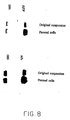
- Sorted cells from both OX-43 positive and negative gates were assayed for expression of liver specific genes by Western blot .analysis and by indirect immunofluorescence. As shown in Figure 2, panel A, there was a minimal amount of albumin in the OX-43-positive cell fraction, detected by Western blotting, as compared with the OX-43-negative cells. No AFP positive cells could be shown by indirect immunofluorescence on cytospins of sorted OX-43-positive cells, as opposed to 30% of OX-43-negative cells expressing the fetal liver marker (see Figure 2, panels B and C). It was concluded that at day 14 of gestation, all fetal liver parenchymal cells are OX-43-negative. Therefore, in order to achieve "cleaner" gates, OX-43-positive and negative cells were separated on a SSC versus FL2 plot and studied separately.
- R3 cells About 2-3% of R3 cells (less than 0.2% of the total ungated cell suspension) were intensely stained for albumin and AFP. They also expressed GGT and CK 19, markers of the bile duct lineage. However the majority of the cells appeared to be small, blast-like cells, and did not express liver specific genes but expressed classical hemopoietic markers such as Thy-1 and serglycin (see Table 1 and Figures 3 and 4). Most of the liver parenchymal cells were found in the R4 gate (see Table 1 and Figure 3). The vast majority of the cells expressed albumin, AFP and GGT, all markers of fetal liver parenchyma. No hemopoietic or fat storing cell markers were detected in that gate.
- the cell population designated R5 is a heterogeneous one (see Figure 4), comprising mainly two cell types: (1) cells that morphologically appear to be normoblasts; and (2) simple small cells that did not express parenchymal liver genes. The ratio between these two cell types varied somewhat and was dependent on the efficiency of the RBC panning.
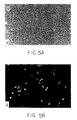
- R4 cells Sorted cells from all 5 populations were cultured for 4 days to determine in vitro fates. When plated at high density under the conditions described, R4 cells yielded clusters of epithelial cells surrounded by very few scattered stromal cells (see Figure 5A and Table 2 below). TABLE 2 Characterization of R4 cells after 4 days in culture Marker Epithelial Cells Stromal Cells Albumin + neg AFP ⁇ neg GGT ++ neg CK 19 +(30%) neg 258.26 neg neg neg neg neg IGF IIr + (perinuclear staining) + (perinuclear staining)
- Fisher 344 rats with known durations of pregnancy were obtained from Harlan Sprague Dawley, Inc. (Indianapolis, IN) and maintained in the animal facility of the Albert Einstein College of Medicine, Bronx, NY on a standard rat chow diet with 12 hour light cycles. By convention, the first day of gestation is defined as day O. Use of animals was in accordance with the NIH Policy on the care and use of laboratory animals and was approved by the Animal Care and use Committee of the Albert Einstein College of Medicine.
- the cell pellet was resuspended in HBSS containing 0.2 mM EGTA and 0.5% BSA (HBSS-EGTA-0.5% BSA), and the cell number was estimated with a Coulter Counter (Coulter Electronics, Inc., Hialeah, FL). Cell viability was assessed by exclusion of 0.04% trypan blue, and an aliquot of the suspension was centrifuged in a tared microfuge tube at 450g for 5 minutes.
- the enriched hepatoblasts were then incubated simultaneously at 4°C for 40 minutes with mouse monoclonal antibody OX-43 (15 ⁇ g/ml, MCA276, Serotec, Indianapolis, IN) and monoclonal antibody OX-44 (18 ⁇ g/ml, MCA371, Serotec, Indianapolis, IN).
- OX-43 recognizes an antigen on macrophages, endothelial cells and red blood cells
- OX-44 recognizes the membrane-glycoprotein CD53 that is present on all rat myeloid cells as well as peripheral lymphoid cells, and is related to the human leukocyte antigen CD37.
- cells were panned at 4°C for 10 minutes in a dish coated with the goat anti-mouse whole IgG antibody, and non-adherent cells were removed as described above.
- cytospins of the various cell suspensions were fixed with either ice-cold ethanol or alcohol, acetone and carbowax 1540 (Fix-Rite, Richard-Allan Medical Industries, Richland, MI).
- the fixed cells were immunostained by indirect immunofluorescence or the biotin/streptavidin method using ⁇ -galactosidase (BioGenex, San Ramon, CA) with rabbit anti-rat albumin IgG (USB Corp., Cleveland, OH) or rabbit anti-mouse AFP antiserum (ICN rmmuttoHiological.s, Lisle, IL) as primary antibodies.
- Negative controls consisted of cells stained with the primary antibodies omitted. Positive controls for albumin staining were done with freshly isolated adult hepatocytes.
- RNA samples were resolved by electrophoresis through 1% agarose formaldehyde gels in 3-(N-morpholino)-propanesulfonic acid buffer, then transferred to Gene Screen (New England Nuclear, Boston, MA), which was prehybridized, and then hybridized with the appropriate probes.
- the cDNA clones complementary to specific mRNAs were radioactively labeled by primer extension with 32 P dCTP.
- the cDNAs used were rat albumin, mouse AFP and mouse 18S (J. Darnell, Rockefeller University, NY). Autoradiograms were scanned with a Quantimat densitometer (Model 920; Manufacturer's Cambridge Instxument). The data for each of the genes was normalized to that for the common gene 18S.
- Flow cytometric analysis was performed on a Becton-Dickinson FACScan (San Jose, CA) with a 15mW air-cooled argon laser.
- Cell sorting was performed with a Becton Dickinson FACSTAR plus (San Jose, CA) using a 4W argon laser with 60mW of power and 100 ⁇ m nozzle.
- fluorescent emission at 488 nm excitation was collected after passing through a 530/30 nm band pass filter for FITC. Fluorescence measurements were performed using logarithmic amplification. Cells were considered positive when fluorescence was greater than 95% of the negative control cells.
- the detector value was E-1 for forward scatter (FSC) with mid-range amplification.
- FSC forward scatter
- SSC side scatter
- Equivalent FACSTAR plus parameters were FSC gain 4 and SSC gain 8. These settings allowed all cells to be visualized on scale.
- FSC and SSC gating were performed using linear amplification, dividing both parameters into 256 arbitrary units (A.U.). For analysis, at least 10,000 events were measured.
- List mode data were acquired and analyzed using LysisII software. Cells before and after sorting were maintained at 4°C and in HBSS supplemented with insulin, transferrin, free fatty acids, trace elements, albumin, and gentamicin as detailed for supplements added to the HDM.
- Isolated cells were labeled with a combination of OX-43 and OX-44 (mouse IgGs) and monoclonal antibody 374.3 (mouse IgM, Hixson and Faris, Brown University, Buffalo, RI) followed by FITC-conjugated goat anti-mouse IGG (heavy chain specific, So Biotech, Birmingham, AL) and PE-conjugated goat anti-mouse IgM (heavy chain specific, So Biotech, Birmingham, AL).
- Twenty-four well plates were coated with type IV collagen extracted from EHS tumors. Panned cells at densities between 12,500 and 25,000 cells per cm 2 were plated per well and allowed to attach for four to five hours after which the medium with the non-adhering cells were gently removed and replaced by fresh medium. Cells were cultured at 37°C in a fully humidified atmosphere containing 5% CO 2 and were observed daily for 5 to 16 days. A complete medium change was performed every 48 hours.
- cells were fixed with ice-cold ethanol and stained in situ by immunochemistry or by immunofluorescence for albumin and AFP.
- the weight of the liver at the 15th day of gestation was 9.1 ⁇ 1.3 mg. Collagenase treatment digested the liver completely, and only minimal particulate matter was excluded by the tissue sieve.
- the number of cells obtained at this step was 1.07 x 10 7 /liver, and the weight of the dissociated cells was 8.6 ⁇ 1.1 mg/liver, 95% of the whole organ weight.
- the suspension consisted almost entirely of isolated single cells with occasional small aggregates that increased in size and number in the absence of EGTA and at temperatures greater than 4°C. Viability by trypan blue exclusion was greater than 90%.
- phase contrast microscopy demonstrated that the adherent cells exhibited an erythroid morphology. Only rare cells were positive for albumin by immunochemistry.
- the non-adherent cells constituted 29 ⁇ 5% and 16 ⁇ 4%, respectively, of the cell number of the freshly dispersed fetal liver (original suspension).
- Panning proved successful for liver tissue at all fetal and early neonatal ages, although the variation in hemopoietic constituents with developmental age resulted in differing degrees of enrichment (data not shown).
- the efficiency of the RBC panning procedure varied with the antibody lot. With antibodies of poor efficiency for direct panning, however, indirect immunoadherence was successful for the cells labeled in suspension followed by panning with anti-rabbit IgG coated petri dishes.
- fetal liver cells When examined by flow cytometry, fetal liver cells constituted a heterogeneous population with respect to FSC, a measure of cell size, and SSC, a measure of cytoplasmic complexity. Cytologically, there was a broad range in cell size (5 to 15 ⁇ by Coulter Counter, data not shown), but cell size was not found to be useful in separating hemopoietic from parenchymal precursors. Rather, the populations were best segregated using SSC. The definition of granular versus agranular cells was made based on a linear scale for side scatter using biparametric plots of fluorescence versus side scatter. Based on the population profiles, 50 A.U. usually demarcated the agranular from the granular cells.
- the fetal liver cells could be isolated into three populations: agranular cells (the R1 population), which were positive for the endothelial and/or myeloid markers (OX43/OX44), and agranular (R2) and granular (R3) cells negative for the OX43/OX44 markers (see Figure 9).
- the demarcation between positive and negative was higher for the granular than the agranular populations due to greater autofluorescence of the granular cells.
- Analysis of the sorted FACS populations demonstrated that less than 1% and 3.0 ⁇ 0.7% of the cells in the R1 and R2 populations, respectively, were positive for AFP.
- the FACS pattern for OX-43/OX-44 was similar for all gestational ages except for a subtle increase in the R1 (and concomitant decrease in the R3 population) with increasing gestational age due to increasing hepatic erythropoiesis (data not shown).
- the inventors have developed methods, incorporating panning technologies and multiparametric FAC sorting, which isolate cell populations highly enriched for liver parenchymal cell precursors.
- the methods of this invention have been found by the inventors to be applicable to the isolation of hepatic precursor cells from liver from gestational age day 13 through the early neonatal period.
- the liver dispersion procedure described yields a population of predominantly single cells with greater than 90% viability, and at gestation day 15, 95% of the whole organ weight is recovered.
- the panning procedures remove up to 84% of the total cell number, and simultaneously enrich the hepatoblast population by 5-fold.
- the tendency of the cells to aggregate is prevented by maintaining the cells at 4°C and by removing calcium with EGTA, interfering with CAM-mediated aggregation.
- the advantage of maintaining the cells as a single cell suspension is two-fold. First, measurement of parenchymal specific functions can be determined on a cellular basis, overcoming the physiologically irrelevant changes in hemopoietic cell population. Second, procedures such as FACS which demand a single cell suspension can be easily performed.
- OX43/44 - cell population which were predominantly AFP + , approximately 30% of the cells were OC.3 + and probably represented bile duct cell precursors, whereas the OC.3 - cells were probable hepatocyte precursors. However, a small percentage of agranular, OX43/44 - cells were AFP + .
- liver stem cell field In comparison to the hemopoietic field, the liver stem cell field is still in its infancy. However, the ability to isolate specific populations by FACS sorting using these parameters with subsequent in vitro and in vivo fate studies will greatly aid in identifying the liver stem cell. Furthermore, this technology is applicable to the study of all aspects of liver stem cell biology including the biliary epithelium, carcinogenesis, regeneration, aging and tissue-specific gene expression.
Landscapes
- Health & Medical Sciences (AREA)
- Life Sciences & Earth Sciences (AREA)
- Engineering & Computer Science (AREA)
- Biomedical Technology (AREA)
- Chemical & Material Sciences (AREA)
- Immunology (AREA)
- Hematology (AREA)
- Molecular Biology (AREA)
- Urology & Nephrology (AREA)
- Biotechnology (AREA)
- Cell Biology (AREA)
- Bioinformatics & Cheminformatics (AREA)
- General Health & Medical Sciences (AREA)
- Biochemistry (AREA)
- Microbiology (AREA)
- Medicinal Chemistry (AREA)
- Food Science & Technology (AREA)
- Analytical Chemistry (AREA)
- General Physics & Mathematics (AREA)
- Pathology (AREA)
- Toxicology (AREA)
- Physics & Mathematics (AREA)
- Gastroenterology & Hepatology (AREA)
- Organic Chemistry (AREA)
- Tropical Medicine & Parasitology (AREA)
- Genetics & Genomics (AREA)
- Wood Science & Technology (AREA)
- Zoology (AREA)
- Pharmacology & Pharmacy (AREA)
- Public Health (AREA)
- Chemical Kinetics & Catalysis (AREA)
- General Chemical & Material Sciences (AREA)
- Nuclear Medicine, Radiotherapy & Molecular Imaging (AREA)
- General Engineering & Computer Science (AREA)
- Animal Behavior & Ethology (AREA)
- Developmental Biology & Embryology (AREA)
- Veterinary Medicine (AREA)
- Micro-Organisms Or Cultivation Processes Thereof (AREA)
- Steroid Compounds (AREA)
- Medicines Containing Material From Animals Or Micro-Organisms (AREA)
Applications Claiming Priority (2)
| Application Number | Priority Date | Filing Date | Title |
|---|---|---|---|
| US15593993A | 1993-11-19 | 1993-11-19 | |
| EP95903543A EP0729297B1 (de) | 1993-11-19 | 1994-11-16 | Hepatoblasten und verfahren zu ihrer isolierung |
Related Parent Applications (2)
| Application Number | Title | Priority Date | Filing Date |
|---|---|---|---|
| EP95903543A Division EP0729297B1 (de) | 1993-11-19 | 1994-11-16 | Hepatoblasten und verfahren zu ihrer isolierung |
| EP95903543.7 Division | 1995-05-26 |
Publications (2)
| Publication Number | Publication Date |
|---|---|
| EP1688037A2 true EP1688037A2 (de) | 2006-08-09 |
| EP1688037A3 EP1688037A3 (de) | 2006-12-13 |
Family
ID=22557387
Family Applications (2)
| Application Number | Title | Priority Date | Filing Date |
|---|---|---|---|
| EP95903543A Expired - Lifetime EP0729297B1 (de) | 1993-11-19 | 1994-11-16 | Hepatoblasten und verfahren zu ihrer isolierung |
| EP06003392A Ceased EP1688037A3 (de) | 1993-11-19 | 1994-11-16 | Hepatoblasten und Verfahren zu ihrer Isolierung |
Family Applications Before (1)
| Application Number | Title | Priority Date | Filing Date |
|---|---|---|---|
| EP95903543A Expired - Lifetime EP0729297B1 (de) | 1993-11-19 | 1994-11-16 | Hepatoblasten und verfahren zu ihrer isolierung |
Country Status (10)
| Country | Link |
|---|---|
| EP (2) | EP0729297B1 (de) |
| JP (2) | JPH09505206A (de) |
| AT (1) | ATE318075T1 (de) |
| AU (1) | AU1256395A (de) |
| CA (1) | CA2177043C (de) |
| DE (1) | DE69434637T2 (de) |
| ES (1) | ES2260759T3 (de) |
| IL (1) | IL111693A (de) |
| WO (1) | WO1995013697A1 (de) |
| ZA (1) | ZA949180B (de) |
Families Citing this family (12)
| Publication number | Priority date | Publication date | Assignee | Title |
|---|---|---|---|---|
| EP0953633A1 (de) * | 1998-04-28 | 1999-11-03 | Livercell L.L.C. | Zellkulturverfahren und Medium zur Herstellung von vermehrungsfähige, normale, differenzierte Humane Leberzellen |
| AU779706B2 (en) | 1999-01-19 | 2005-02-10 | University Of North Carolina At Chapel Hill, The | Human liver progenitors |
| US7456017B2 (en) | 1999-10-01 | 2008-11-25 | University Of North Carolina At Chapel Hill | Processes for clonal growth of hepatic progenitor cells |
| US20020187133A1 (en) | 1999-10-01 | 2002-12-12 | Hiroshi Kubota | Methods of isolating bipotent hepatic progenitor cells |
| WO2001053462A1 (en) * | 2000-01-19 | 2001-07-26 | University Of North Carolina At Chapel Hill | Liver tissue source |
| CN1461341A (zh) * | 2000-10-03 | 2003-12-10 | 北卡罗来纳大学 | 肝祖先细胞克隆生长的方法 |
| MXPA03008882A (es) | 2001-03-27 | 2003-12-08 | Vertex Pharma | Composiciones y metodos utiles para la infeccion por hcv. |
| ATE371018T1 (de) | 2001-06-22 | 2007-09-15 | Stemcells Inc | Le-zellen (liver engrafting cells), assays und verwendungen davon |
| HUE032238T2 (en) * | 2002-03-15 | 2017-09-28 | Univ North Carolina Chapel Hill | Primitive and proximal liver stem cells |
| JP7386539B2 (ja) * | 2018-08-03 | 2023-11-27 | 株式会社細胞応用技術研究所 | 培養上清製剤の製造方法 |
| CN112912087A (zh) * | 2018-09-14 | 2021-06-04 | 卢卡科学株式会社 | 线粒体向淋巴器官中的移植以及用于该移植的组合物 |
| CN113046304B (zh) * | 2021-04-09 | 2023-05-16 | 四川大学华西医院 | 一种线粒体的分离方法 |
Family Cites Families (3)
| Publication number | Priority date | Publication date | Assignee | Title |
|---|---|---|---|---|
| US5087570A (en) * | 1988-05-10 | 1992-02-11 | Weissman Irving L | Homogeneous mammalian hematopoietic stem cell composition |
| WO1993003142A1 (en) * | 1991-08-07 | 1993-02-18 | Albert Einstein College Of Medicine | Proliferation of hepatocyte precursors |
| ES2157225T3 (es) * | 1992-10-09 | 2001-08-16 | Advanced Tissue Sciences Inc | Celulas hepaticas de reserva. |
-
1994
- 1994-11-16 ES ES95903543T patent/ES2260759T3/es not_active Expired - Lifetime
- 1994-11-16 DE DE69434637T patent/DE69434637T2/de not_active Expired - Lifetime
- 1994-11-16 WO PCT/US1994/013216 patent/WO1995013697A1/en not_active Ceased
- 1994-11-16 CA CA2177043A patent/CA2177043C/en not_active Expired - Fee Related
- 1994-11-16 EP EP95903543A patent/EP0729297B1/de not_active Expired - Lifetime
- 1994-11-16 AT AT95903543T patent/ATE318075T1/de not_active IP Right Cessation
- 1994-11-16 JP JP7514579A patent/JPH09505206A/ja active Pending
- 1994-11-16 AU AU12563/95A patent/AU1256395A/en not_active Abandoned
- 1994-11-16 EP EP06003392A patent/EP1688037A3/de not_active Ceased
- 1994-11-18 ZA ZA949180A patent/ZA949180B/xx unknown
- 1994-11-18 IL IL11169394A patent/IL111693A/en not_active IP Right Cessation
-
2004
- 2004-06-24 JP JP2004186968A patent/JP2004339227A/ja active Pending
Also Published As
| Publication number | Publication date |
|---|---|
| DE69434637T2 (de) | 2006-09-21 |
| EP0729297B1 (de) | 2006-02-22 |
| JPH09505206A (ja) | 1997-05-27 |
| ES2260759T3 (es) | 2006-11-01 |
| DE69434637D1 (de) | 2006-04-27 |
| IL111693A0 (en) | 1995-01-24 |
| EP1688037A3 (de) | 2006-12-13 |
| EP0729297A1 (de) | 1996-09-04 |
| IL111693A (en) | 2001-01-28 |
| CA2177043C (en) | 2011-08-09 |
| ZA949180B (en) | 1995-07-26 |
| WO1995013697A1 (en) | 1995-05-26 |
| JP2004339227A (ja) | 2004-12-02 |
| ATE318075T1 (de) | 2006-03-15 |
| EP0729297A4 (de) | 2004-05-06 |
| AU1256395A (en) | 1995-06-06 |
| CA2177043A1 (en) | 1995-05-26 |
Similar Documents
| Publication | Publication Date | Title |
|---|---|---|
| US7109028B2 (en) | Methods of isolating hepatic progenitors | |
| KR101119878B1 (ko) | 프리미티브 간 줄기세포 및 프록시멀 간 줄기세포 | |
| Sigal et al. | Evidence for a terminal differentiation process in the rat liver | |
| Sigal et al. | Characterization and enrichment of fetal rat hepatoblasts by immunoadsorption (“panning”) and fluorescence‐activated cell sorting | |
| HK1046930B (zh) | 人肝脏祖先 | |
| CA2688309C (en) | Hepatic stellate cell precursors and methods of isolating same | |
| EP1034255A1 (de) | Auf eine zellinie beschränkte gliale vorläuferzellen aus dem zentralen nervensystem | |
| EP0729297B1 (de) | Hepatoblasten und verfahren zu ihrer isolierung | |
| JP2004527257A (ja) | Hla/mhc遺伝子産物マーカーで免疫標識することによって幹細胞を単離するための方法 | |
| HK1095246A (en) | Hepatoblasts and method of isolating same | |
| IL139319A (en) | Isolated hepatocyte precursors, cell cultures containing the same, and compositions containing said cell cultures | |
| HOLST | RONALD FARIS’zyxwvutsrqpo | |
| US20040259246A1 (en) | Liver cell progenitor and use for treatment of liver diseases | |
| HK1225070A1 (en) | Primitive and proximal hepatic stem cells | |
| HK1225070A (en) | Primitive and proximal hepatic stem cells | |
| HK1168124A (en) | Hepatic stellate cell precursors | |
| HK1176641B (en) | Human liver progenitors | |
| HK1168124B (en) | Hepatic stellate cell precursors |
Legal Events
| Date | Code | Title | Description |
|---|---|---|---|
| PUAI | Public reference made under article 153(3) epc to a published international application that has entered the european phase |
Free format text: ORIGINAL CODE: 0009012 |
|
| AC | Divisional application: reference to earlier application |
Ref document number: 0729297 Country of ref document: EP Kind code of ref document: P |
|
| AK | Designated contracting states |
Kind code of ref document: A2 Designated state(s): AT BE CH DE DK ES FR GB GR IE IT LI LU MC NL PT SE |
|
| PUAL | Search report despatched |
Free format text: ORIGINAL CODE: 0009013 |
|
| AK | Designated contracting states |
Kind code of ref document: A3 Designated state(s): AT BE CH DE DK ES FR GB GR IE IT LI LU MC NL PT SE |
|
| RIC1 | Information provided on ipc code assigned before grant |
Ipc: G01N 33/533 20060101AFI20061109BHEP Ipc: G01N 33/543 20060101ALI20061109BHEP Ipc: A01N 1/02 20060101ALI20061109BHEP Ipc: G01N 33/577 20060101ALI20061109BHEP Ipc: G01N 33/536 20060101ALI20061109BHEP Ipc: G01N 33/50 20060101ALI20061109BHEP Ipc: G01N 33/574 20060101ALI20061109BHEP Ipc: G01N 33/538 20060101ALI20061109BHEP |
|
| REG | Reference to a national code |
Ref country code: HK Ref legal event code: DE Ref document number: 1095246 Country of ref document: HK |
|
| 17P | Request for examination filed |
Effective date: 20070613 |
|
| AKX | Designation fees paid |
Designated state(s): AT BE CH DE DK ES FR GB GR IE IT LI LU MC NL PT SE |
|
| 17Q | First examination report despatched |
Effective date: 20080819 |
|
| STAA | Information on the status of an ep patent application or granted ep patent |
Free format text: STATUS: THE APPLICATION HAS BEEN REFUSED |
|
| 18R | Application refused |
Effective date: 20091007 |
|
| REG | Reference to a national code |
Ref country code: HK Ref legal event code: WD Ref document number: 1095246 Country of ref document: HK |