EP1700128B1 - Verfahren zur serologischen diagnose und bestimmung des immunisierungszustands unter einschluss verschiedener kontrollen - Google Patents
Verfahren zur serologischen diagnose und bestimmung des immunisierungszustands unter einschluss verschiedener kontrollen Download PDFInfo
- Publication number
- EP1700128B1 EP1700128B1 EP04817587A EP04817587A EP1700128B1 EP 1700128 B1 EP1700128 B1 EP 1700128B1 EP 04817587 A EP04817587 A EP 04817587A EP 04817587 A EP04817587 A EP 04817587A EP 1700128 B1 EP1700128 B1 EP 1700128B1
- Authority
- EP
- European Patent Office
- Prior art keywords
- antigen
- antigens
- control
- detection
- microbial
- Prior art date
- Legal status (The legal status is an assumption and is not a legal conclusion. Google has not performed a legal analysis and makes no representation as to the accuracy of the status listed.)
- Expired - Lifetime
Links
- 238000000034 method Methods 0.000 title claims abstract description 69
- 230000000405 serological effect Effects 0.000 title claims abstract description 21
- 238000003745 diagnosis Methods 0.000 title claims abstract description 14
- 238000002649 immunization Methods 0.000 title abstract description 7
- 239000000427 antigen Substances 0.000 claims abstract description 325
- 102000036639 antigens Human genes 0.000 claims abstract description 325
- 108091007433 antigens Proteins 0.000 claims abstract description 325
- 238000001514 detection method Methods 0.000 claims abstract description 143
- 210000002966 serum Anatomy 0.000 claims abstract description 107
- 230000000813 microbial effect Effects 0.000 claims abstract description 92
- 239000007787 solid Substances 0.000 claims abstract description 71
- 229960005486 vaccine Drugs 0.000 claims abstract description 64
- 230000003460 anti-nuclear Effects 0.000 claims abstract description 34
- 238000001179 sorption measurement Methods 0.000 claims abstract description 21
- 230000009257 reactivity Effects 0.000 claims abstract description 14
- 238000002405 diagnostic procedure Methods 0.000 claims abstract description 7
- 239000000126 substance Substances 0.000 claims description 142
- 108060003951 Immunoglobulin Proteins 0.000 claims description 72
- 102000018358 immunoglobulin Human genes 0.000 claims description 72
- 238000000151 deposition Methods 0.000 claims description 48
- 238000006243 chemical reaction Methods 0.000 claims description 44
- 210000004027 cell Anatomy 0.000 claims description 43
- 241000894007 species Species 0.000 claims description 35
- 241000894006 Bacteria Species 0.000 claims description 33
- 102000004169 proteins and genes Human genes 0.000 claims description 33
- 108090000623 proteins and genes Proteins 0.000 claims description 33
- 241000283707 Capra Species 0.000 claims description 27
- 239000000725 suspension Substances 0.000 claims description 26
- LFQSCWFLJHTTHZ-UHFFFAOYSA-N Ethanol Chemical compound CCO LFQSCWFLJHTTHZ-UHFFFAOYSA-N 0.000 claims description 25
- 229940072221 immunoglobulins Drugs 0.000 claims description 23
- 239000003795 chemical substances by application Substances 0.000 claims description 22
- 238000003556 assay Methods 0.000 claims description 21
- 241000700605 Viruses Species 0.000 claims description 20
- 241000191967 Staphylococcus aureus Species 0.000 claims description 19
- 239000007850 fluorescent dye Substances 0.000 claims description 18
- 238000002372 labelling Methods 0.000 claims description 18
- 239000010755 BS 2869 Class G Substances 0.000 claims description 17
- OKKJLVBELUTLKV-UHFFFAOYSA-N Methanol Chemical compound OC OKKJLVBELUTLKV-UHFFFAOYSA-N 0.000 claims description 15
- 239000011521 glass Substances 0.000 claims description 15
- 241001465754 Metazoa Species 0.000 claims description 14
- 201000005505 Measles Diseases 0.000 claims description 13
- 239000002245 particle Substances 0.000 claims description 13
- 201000005404 rubella Diseases 0.000 claims description 13
- 238000011002 quantification Methods 0.000 claims description 12
- 239000000872 buffer Substances 0.000 claims description 10
- 238000001215 fluorescent labelling Methods 0.000 claims description 10
- 238000005259 measurement Methods 0.000 claims description 9
- 244000052769 pathogen Species 0.000 claims description 9
- 241000233866 Fungi Species 0.000 claims description 8
- 208000005647 Mumps Diseases 0.000 claims description 8
- 230000027455 binding Effects 0.000 claims description 8
- 239000000975 dye Substances 0.000 claims description 8
- 230000005284 excitation Effects 0.000 claims description 8
- 230000008105 immune reaction Effects 0.000 claims description 8
- 208000010805 mumps infectious disease Diseases 0.000 claims description 8
- 244000045947 parasite Species 0.000 claims description 8
- 230000001717 pathogenic effect Effects 0.000 claims description 8
- CSCPPACGZOOCGX-UHFFFAOYSA-N Acetone Chemical compound CC(C)=O CSCPPACGZOOCGX-UHFFFAOYSA-N 0.000 claims description 7
- 241000287828 Gallus gallus Species 0.000 claims description 7
- 108010033040 Histones Proteins 0.000 claims description 7
- 206010043376 Tetanus Diseases 0.000 claims description 7
- 230000001580 bacterial effect Effects 0.000 claims description 7
- 210000004369 blood Anatomy 0.000 claims description 7
- 239000008280 blood Substances 0.000 claims description 7
- 206010013023 diphtheria Diseases 0.000 claims description 7
- 238000010828 elution Methods 0.000 claims description 7
- 239000000203 mixture Substances 0.000 claims description 7
- 230000003612 virological effect Effects 0.000 claims description 7
- 238000005406 washing Methods 0.000 claims description 7
- 241000606660 Bartonella Species 0.000 claims description 6
- 238000009007 Diagnostic Kit Methods 0.000 claims description 6
- 239000011230 binding agent Substances 0.000 claims description 6
- 241001445332 Coxiella <snail> Species 0.000 claims description 5
- 230000001681 protective effect Effects 0.000 claims description 5
- 206010014665 endocarditis Diseases 0.000 claims description 4
- 230000003834 intracellular effect Effects 0.000 claims description 4
- 239000004033 plastic Substances 0.000 claims description 4
- 229920003023 plastic Polymers 0.000 claims description 4
- 241000589968 Borrelia Species 0.000 claims description 3
- 108091003079 Bovine Serum Albumin Proteins 0.000 claims description 3
- 241000606161 Chlamydia Species 0.000 claims description 3
- 102000002322 Egg Proteins Human genes 0.000 claims description 3
- 108010000912 Egg Proteins Proteins 0.000 claims description 3
- 241000589902 Leptospira Species 0.000 claims description 3
- 208000016604 Lyme disease Diseases 0.000 claims description 3
- 241000204031 Mycoplasma Species 0.000 claims description 3
- 241000606701 Rickettsia Species 0.000 claims description 3
- 241000589886 Treponema Species 0.000 claims description 3
- 241000203807 Tropheryma Species 0.000 claims description 3
- 229940098773 bovine serum albumin Drugs 0.000 claims description 3
- 235000013345 egg yolk Nutrition 0.000 claims description 3
- 210000002969 egg yolk Anatomy 0.000 claims description 3
- 210000002950 fibroblast Anatomy 0.000 claims description 3
- 208000005252 hepatitis A Diseases 0.000 claims description 3
- 208000002672 hepatitis B Diseases 0.000 claims description 3
- 201000006082 Chickenpox Diseases 0.000 claims description 2
- 241000605314 Ehrlichia Species 0.000 claims description 2
- 208000000474 Poliomyelitis Diseases 0.000 claims description 2
- 241000191940 Staphylococcus Species 0.000 claims description 2
- 206010046980 Varicella Diseases 0.000 claims description 2
- 208000003152 Yellow Fever Diseases 0.000 claims description 2
- 230000008878 coupling Effects 0.000 claims description 2
- 238000010168 coupling process Methods 0.000 claims description 2
- 238000005859 coupling reaction Methods 0.000 claims description 2
- 238000004132 cross linking Methods 0.000 claims description 2
- 201000010099 disease Diseases 0.000 claims description 2
- 208000037265 diseases, disorders, signs and symptoms Diseases 0.000 claims description 2
- 229920000159 gelatin Polymers 0.000 claims description 2
- 235000019322 gelatine Nutrition 0.000 claims description 2
- 238000000338 in vitro Methods 0.000 claims description 2
- 244000005700 microbiome Species 0.000 claims description 2
- 238000010186 staining Methods 0.000 claims description 2
- 238000012800 visualization Methods 0.000 claims description 2
- 241000588832 Bordetella pertussis Species 0.000 claims 1
- 239000001828 Gelatine Substances 0.000 claims 1
- 208000004006 Tick-borne encephalitis Diseases 0.000 claims 1
- 239000006285 cell suspension Substances 0.000 claims 1
- 230000002538 fungal effect Effects 0.000 claims 1
- 230000003071 parasitic effect Effects 0.000 claims 1
- 238000010166 immunofluorescence Methods 0.000 abstract description 15
- 230000009897 systematic effect Effects 0.000 abstract description 6
- 229940043517 specific immunoglobulins Drugs 0.000 abstract description 3
- 238000012360 testing method Methods 0.000 description 45
- 230000008021 deposition Effects 0.000 description 43
- 239000000523 sample Substances 0.000 description 39
- 238000009589 serological test Methods 0.000 description 15
- 238000011534 incubation Methods 0.000 description 13
- 241000606678 Coxiella burnetii Species 0.000 description 12
- 239000003068 molecular probe Substances 0.000 description 10
- 208000015181 infectious disease Diseases 0.000 description 9
- 238000010790 dilution Methods 0.000 description 8
- 239000012895 dilution Substances 0.000 description 8
- 230000002458 infectious effect Effects 0.000 description 7
- 238000005119 centrifugation Methods 0.000 description 6
- 229940082150 encore Drugs 0.000 description 6
- 230000002255 enzymatic effect Effects 0.000 description 6
- 239000003550 marker Substances 0.000 description 6
- 208000035473 Communicable disease Diseases 0.000 description 5
- 241001494479 Pecora Species 0.000 description 5
- 238000013019 agitation Methods 0.000 description 5
- 239000003153 chemical reaction reagent Substances 0.000 description 5
- 239000003086 colorant Substances 0.000 description 5
- 229960003983 diphtheria toxoid Drugs 0.000 description 5
- 230000003053 immunization Effects 0.000 description 5
- 238000012544 monitoring process Methods 0.000 description 5
- 229960000814 tetanus toxoid Drugs 0.000 description 5
- 238000002255 vaccination Methods 0.000 description 5
- 241000283690 Bos taurus Species 0.000 description 4
- 102000004190 Enzymes Human genes 0.000 description 4
- 108090000790 Enzymes Proteins 0.000 description 4
- 239000003570 air Substances 0.000 description 4
- 230000002096 anti-tetanic effect Effects 0.000 description 4
- 230000008901 benefit Effects 0.000 description 4
- GNBHRKFJIUUOQI-UHFFFAOYSA-N fluorescein Chemical compound O1C(=O)C2=CC=CC=C2C21C1=CC=C(O)C=C1OC1=CC(O)=CC=C21 GNBHRKFJIUUOQI-UHFFFAOYSA-N 0.000 description 4
- 239000012634 fragment Substances 0.000 description 4
- 239000002609 medium Substances 0.000 description 4
- 239000013642 negative control Substances 0.000 description 4
- 239000012071 phase Substances 0.000 description 4
- 239000013641 positive control Substances 0.000 description 4
- 238000002360 preparation method Methods 0.000 description 4
- 239000000243 solution Substances 0.000 description 4
- 241000606070 Bartonella elizabethae Species 0.000 description 3
- 241000606108 Bartonella quintana Species 0.000 description 3
- 238000002965 ELISA Methods 0.000 description 3
- 241000589242 Legionella pneumophila Species 0.000 description 3
- 201000005702 Pertussis Diseases 0.000 description 3
- 239000002253 acid Substances 0.000 description 3
- 229940092526 bartonella elizabethae Drugs 0.000 description 3
- 229940092523 bartonella quintana Drugs 0.000 description 3
- 238000004140 cleaning Methods 0.000 description 3
- 230000001186 cumulative effect Effects 0.000 description 3
- BFMYDTVEBKDAKJ-UHFFFAOYSA-L disodium;(2',7'-dibromo-3',6'-dioxido-3-oxospiro[2-benzofuran-1,9'-xanthene]-4'-yl)mercury;hydrate Chemical compound O.[Na+].[Na+].O1C(=O)C2=CC=CC=C2C21C1=CC(Br)=C([O-])C([Hg])=C1OC1=C2C=C(Br)C([O-])=C1 BFMYDTVEBKDAKJ-UHFFFAOYSA-L 0.000 description 3
- 229940027941 immunoglobulin g Drugs 0.000 description 3
- 229940115932 legionella pneumophila Drugs 0.000 description 3
- 238000004519 manufacturing process Methods 0.000 description 3
- 230000003287 optical effect Effects 0.000 description 3
- 230000005855 radiation Effects 0.000 description 3
- 238000004062 sedimentation Methods 0.000 description 3
- 238000012795 verification Methods 0.000 description 3
- 241000271566 Aves Species 0.000 description 2
- 241001518086 Bartonella henselae Species 0.000 description 2
- 241001464955 Bartonella vinsonii Species 0.000 description 2
- SXRSQZLOMIGNAQ-UHFFFAOYSA-N Glutaraldehyde Chemical compound O=CCCCC=O SXRSQZLOMIGNAQ-UHFFFAOYSA-N 0.000 description 2
- 102000006947 Histones Human genes 0.000 description 2
- 241000282412 Homo Species 0.000 description 2
- DGAQECJNVWCQMB-PUAWFVPOSA-M Ilexoside XXIX Chemical compound C[C@@H]1CC[C@@]2(CC[C@@]3(C(=CC[C@H]4[C@]3(CC[C@@H]5[C@@]4(CC[C@@H](C5(C)C)OS(=O)(=O)[O-])C)C)[C@@H]2[C@]1(C)O)C)C(=O)O[C@H]6[C@@H]([C@H]([C@@H]([C@H](O6)CO)O)O)O.[Na+] DGAQECJNVWCQMB-PUAWFVPOSA-M 0.000 description 2
- 241000124008 Mammalia Species 0.000 description 2
- 240000004808 Saccharomyces cerevisiae Species 0.000 description 2
- PXIPVTKHYLBLMZ-UHFFFAOYSA-N Sodium azide Chemical compound [Na+].[N-]=[N+]=[N-] PXIPVTKHYLBLMZ-UHFFFAOYSA-N 0.000 description 2
- 230000004520 agglutination Effects 0.000 description 2
- 238000004458 analytical method Methods 0.000 description 2
- 229940092524 bartonella henselae Drugs 0.000 description 2
- 229940092522 bartonella vinsonii Drugs 0.000 description 2
- 239000003364 biologic glue Substances 0.000 description 2
- 230000015572 biosynthetic process Effects 0.000 description 2
- 239000006161 blood agar Substances 0.000 description 2
- 238000010586 diagram Methods 0.000 description 2
- 235000013601 eggs Nutrition 0.000 description 2
- 239000012149 elution buffer Substances 0.000 description 2
- 210000003527 eukaryotic cell Anatomy 0.000 description 2
- 238000011156 evaluation Methods 0.000 description 2
- 238000001914 filtration Methods 0.000 description 2
- 239000012530 fluid Substances 0.000 description 2
- 238000000799 fluorescence microscopy Methods 0.000 description 2
- 239000000499 gel Substances 0.000 description 2
- 239000001963 growth medium Substances 0.000 description 2
- 238000010438 heat treatment Methods 0.000 description 2
- 230000001900 immune effect Effects 0.000 description 2
- 238000010348 incorporation Methods 0.000 description 2
- 239000002054 inoculum Substances 0.000 description 2
- 238000009830 intercalation Methods 0.000 description 2
- 238000002493 microarray Methods 0.000 description 2
- 238000005497 microtitration Methods 0.000 description 2
- 230000007170 pathology Effects 0.000 description 2
- 230000008569 process Effects 0.000 description 2
- 230000002285 radioactive effect Effects 0.000 description 2
- 238000007430 reference method Methods 0.000 description 2
- 238000011160 research Methods 0.000 description 2
- 229910052708 sodium Inorganic materials 0.000 description 2
- 239000011734 sodium Substances 0.000 description 2
- 238000000527 sonication Methods 0.000 description 2
- 238000004448 titration Methods 0.000 description 2
- 238000012546 transfer Methods 0.000 description 2
- 108010088751 Albumins Proteins 0.000 description 1
- 102000009027 Albumins Human genes 0.000 description 1
- 241000282472 Canis lupus familiaris Species 0.000 description 1
- 241000700199 Cavia porcellus Species 0.000 description 1
- VEXZGXHMUGYJMC-UHFFFAOYSA-M Chloride anion Chemical compound [Cl-] VEXZGXHMUGYJMC-UHFFFAOYSA-M 0.000 description 1
- 241000699800 Cricetinae Species 0.000 description 1
- 241000283086 Equidae Species 0.000 description 1
- 241000282326 Felis catus Species 0.000 description 1
- 241000272496 Galliformes Species 0.000 description 1
- 108010010803 Gelatin Proteins 0.000 description 1
- 102000018071 Immunoglobulin Fc Fragments Human genes 0.000 description 1
- 108010091135 Immunoglobulin Fc Fragments Proteins 0.000 description 1
- 101100406879 Neurospora crassa (strain ATCC 24698 / 74-OR23-1A / CBS 708.71 / DSM 1257 / FGSC 987) par-2 gene Proteins 0.000 description 1
- 108010089610 Nuclear Proteins Proteins 0.000 description 1
- 102000007999 Nuclear Proteins Human genes 0.000 description 1
- 241000283973 Oryctolagus cuniculus Species 0.000 description 1
- 239000004698 Polyethylene Substances 0.000 description 1
- 229920001213 Polysorbate 20 Polymers 0.000 description 1
- 239000004793 Polystyrene Substances 0.000 description 1
- 206010037660 Pyrexia Diseases 0.000 description 1
- 206010037688 Q fever Diseases 0.000 description 1
- 241000700159 Rattus Species 0.000 description 1
- 241000194017 Streptococcus Species 0.000 description 1
- 241001080024 Telles Species 0.000 description 1
- 238000007605 air drying Methods 0.000 description 1
- 150000001298 alcohols Chemical class 0.000 description 1
- 239000012080 ambient air Substances 0.000 description 1
- 230000000890 antigenic effect Effects 0.000 description 1
- 239000008346 aqueous phase Substances 0.000 description 1
- 239000011324 bead Substances 0.000 description 1
- 210000000601 blood cell Anatomy 0.000 description 1
- 238000010241 blood sampling Methods 0.000 description 1
- 238000009534 blood test Methods 0.000 description 1
- 244000309466 calf Species 0.000 description 1
- 238000012412 chemical coupling Methods 0.000 description 1
- 239000007795 chemical reaction product Substances 0.000 description 1
- 210000000349 chromosome Anatomy 0.000 description 1
- 230000000536 complexating effect Effects 0.000 description 1
- 238000010668 complexation reaction Methods 0.000 description 1
- 239000000470 constituent Substances 0.000 description 1
- 239000007822 coupling agent Substances 0.000 description 1
- WQJONRMBVKFKOB-UHFFFAOYSA-N cyanatosulfanyl cyanate Chemical compound N#COSOC#N WQJONRMBVKFKOB-UHFFFAOYSA-N 0.000 description 1
- 230000007547 defect Effects 0.000 description 1
- 230000001419 dependent effect Effects 0.000 description 1
- 238000011161 development Methods 0.000 description 1
- 238000012631 diagnostic technique Methods 0.000 description 1
- 238000006073 displacement reaction Methods 0.000 description 1
- 238000004090 dissolution Methods 0.000 description 1
- 238000001035 drying Methods 0.000 description 1
- 210000000624 ear auricle Anatomy 0.000 description 1
- 230000008030 elimination Effects 0.000 description 1
- 238000003379 elimination reaction Methods 0.000 description 1
- 206010014599 encephalitis Diseases 0.000 description 1
- 238000005516 engineering process Methods 0.000 description 1
- 238000002474 experimental method Methods 0.000 description 1
- 239000012467 final product Substances 0.000 description 1
- MHMNJMPURVTYEJ-UHFFFAOYSA-N fluorescein-5-isothiocyanate Chemical compound O1C(=O)C2=CC(N=C=S)=CC=C2C21C1=CC=C(O)C=C1OC1=CC(O)=CC=C21 MHMNJMPURVTYEJ-UHFFFAOYSA-N 0.000 description 1
- 238000001943 fluorescence-activated cell sorting Methods 0.000 description 1
- 230000008014 freezing Effects 0.000 description 1
- 238000007710 freezing Methods 0.000 description 1
- 239000008273 gelatin Substances 0.000 description 1
- 235000011852 gelatine desserts Nutrition 0.000 description 1
- 210000005260 human cell Anatomy 0.000 description 1
- 238000005286 illumination Methods 0.000 description 1
- 230000028993 immune response Effects 0.000 description 1
- 230000001939 inductive effect Effects 0.000 description 1
- 201000007119 infective endocarditis Diseases 0.000 description 1
- 230000002687 intercalation Effects 0.000 description 1
- 150000002540 isothiocyanates Chemical class 0.000 description 1
- 238000009533 lab test Methods 0.000 description 1
- 239000004816 latex Substances 0.000 description 1
- 229920000126 latex Polymers 0.000 description 1
- 239000007788 liquid Substances 0.000 description 1
- 210000004962 mammalian cell Anatomy 0.000 description 1
- 239000000463 material Substances 0.000 description 1
- 238000012986 modification Methods 0.000 description 1
- 230000004048 modification Effects 0.000 description 1
- LWGJTAZLEJHCPA-UHFFFAOYSA-N n-(2-chloroethyl)-n-nitrosomorpholine-4-carboxamide Chemical compound ClCCN(N=O)C(=O)N1CCOCC1 LWGJTAZLEJHCPA-UHFFFAOYSA-N 0.000 description 1
- 230000009871 nonspecific binding Effects 0.000 description 1
- 210000004940 nucleus Anatomy 0.000 description 1
- 231100000255 pathogenic effect Toxicity 0.000 description 1
- 102000013415 peroxidase activity proteins Human genes 0.000 description 1
- 108040007629 peroxidase activity proteins Proteins 0.000 description 1
- -1 polyethylene Polymers 0.000 description 1
- 229920000573 polyethylene Polymers 0.000 description 1
- 229920000642 polymer Polymers 0.000 description 1
- 239000000256 polyoxyethylene sorbitan monolaurate Substances 0.000 description 1
- 235000010486 polyoxyethylene sorbitan monolaurate Nutrition 0.000 description 1
- 229920001184 polypeptide Polymers 0.000 description 1
- 229920002223 polystyrene Polymers 0.000 description 1
- 244000144977 poultry Species 0.000 description 1
- 238000001556 precipitation Methods 0.000 description 1
- 238000004321 preservation Methods 0.000 description 1
- 102000004196 processed proteins & peptides Human genes 0.000 description 1
- 108090000765 processed proteins & peptides Proteins 0.000 description 1
- 238000012545 processing Methods 0.000 description 1
- 239000000047 product Substances 0.000 description 1
- 238000000163 radioactive labelling Methods 0.000 description 1
- 238000005070 sampling Methods 0.000 description 1
- 238000012216 screening Methods 0.000 description 1
- 238000000926 separation method Methods 0.000 description 1
- 239000007790 solid phase Substances 0.000 description 1
- 230000006641 stabilisation Effects 0.000 description 1
- 238000011105 stabilization Methods 0.000 description 1
- 230000000087 stabilizing effect Effects 0.000 description 1
- 230000004936 stimulating effect Effects 0.000 description 1
- 238000003756 stirring Methods 0.000 description 1
- 239000000758 substrate Substances 0.000 description 1
- 239000013589 supplement Substances 0.000 description 1
- 239000004094 surface-active agent Substances 0.000 description 1
- 230000014599 transmission of virus Effects 0.000 description 1
- 125000001493 tyrosinyl group Chemical group [H]OC1=C([H])C([H])=C(C([H])=C1[H])C([H])([H])C([H])(N([H])[H])C(*)=O 0.000 description 1
Images

Classifications
-
- G—PHYSICS
- G01—MEASURING; TESTING
- G01N—INVESTIGATING OR ANALYSING MATERIALS BY DETERMINING THEIR CHEMICAL OR PHYSICAL PROPERTIES
- G01N33/00—Investigating or analysing materials by specific methods not covered by groups G01N1/00 - G01N31/00
- G01N33/48—Biological material, e.g. blood, urine; Haemocytometers
- G01N33/50—Chemical analysis of biological material, e.g. blood, urine; Testing involving biospecific ligand binding methods; Immunological testing
- G01N33/53—Immunoassay; Biospecific binding assay; Materials therefor
- G01N33/569—Immunoassay; Biospecific binding assay; Materials therefor for microorganisms, e.g. protozoa, bacteria, viruses
- G01N33/56911—Bacteria
-
- G—PHYSICS
- G01—MEASURING; TESTING
- G01N—INVESTIGATING OR ANALYSING MATERIALS BY DETERMINING THEIR CHEMICAL OR PHYSICAL PROPERTIES
- G01N33/00—Investigating or analysing materials by specific methods not covered by groups G01N1/00 - G01N31/00
- G01N33/48—Biological material, e.g. blood, urine; Haemocytometers
- G01N33/50—Chemical analysis of biological material, e.g. blood, urine; Testing involving biospecific ligand binding methods; Immunological testing
- G01N33/53—Immunoassay; Biospecific binding assay; Materials therefor
- G01N33/543—Immunoassay; Biospecific binding assay; Materials therefor with an insoluble carrier for immobilising immunochemicals
- G01N33/54366—Apparatus specially adapted for solid-phase testing
-
- G—PHYSICS
- G01—MEASURING; TESTING
- G01N—INVESTIGATING OR ANALYSING MATERIALS BY DETERMINING THEIR CHEMICAL OR PHYSICAL PROPERTIES
- G01N33/00—Investigating or analysing materials by specific methods not covered by groups G01N1/00 - G01N31/00
- G01N33/48—Biological material, e.g. blood, urine; Haemocytometers
- G01N33/50—Chemical analysis of biological material, e.g. blood, urine; Testing involving biospecific ligand binding methods; Immunological testing
- G01N33/53—Immunoassay; Biospecific binding assay; Materials therefor
- G01N33/569—Immunoassay; Biospecific binding assay; Materials therefor for microorganisms, e.g. protozoa, bacteria, viruses
- G01N33/56911—Bacteria
- G01N33/56938—Staphylococcus
-
- G—PHYSICS
- G01—MEASURING; TESTING
- G01N—INVESTIGATING OR ANALYSING MATERIALS BY DETERMINING THEIR CHEMICAL OR PHYSICAL PROPERTIES
- G01N33/00—Investigating or analysing materials by specific methods not covered by groups G01N1/00 - G01N31/00
- G01N33/48—Biological material, e.g. blood, urine; Haemocytometers
- G01N33/50—Chemical analysis of biological material, e.g. blood, urine; Testing involving biospecific ligand binding methods; Immunological testing
- G01N33/53—Immunoassay; Biospecific binding assay; Materials therefor
- G01N33/569—Immunoassay; Biospecific binding assay; Materials therefor for microorganisms, e.g. protozoa, bacteria, viruses
- G01N33/56983—Viruses
Definitions
- the present invention relates to a method and a kit for diagnosing human and animal infectious diseases. More particularly, it relates to a method for diagnosing human and animal infectious diseases which is based on the research in the patient's serum of antibodies specific for the infectious microbial agent, namely a bacterium, a virus, a parasite or a responsible fungus. of a pathology (referred to below as "microbe").
- the present invention also relates to a method and a diagnostic kit for the serological determination of the immunization status of individuals.
- This method of diagnosis is essential for the diagnosis of microbe infections, sampling, culture and / or difficult identification, in particular strict intracellular viruses and bacteria and facultative intracellular bacteria of the genera Rickettsia, Coxiella, Bartonella, Tropheryma , Ehrichia, Chlamydia, Mycoplasma, Treponema, Borrelia, and Leptospira for example, for which antigens consisting of whole microbes or fractions or fragments of microbes comprising one or more antigens are used as microbial antigens.
- microbes or their fraction remain visible as individualizable particles by microscopic observation using an optical microscope or an electron microscope, for example.
- the microbial serology consists in detecting in the patient's serum an antigen-antibody reaction in which the antigen is represented by all or fraction of the infectious microbial agent to be detected and the antibody is represented by specific human or animal immunoglobulins.
- said infectious microbial agent present in the patient's serum. It can be quantified by successively testing a series of increasing dilutions of reason 2 or patient's serum ratio from a first 1: 16 or 1: 50 dilution of the patient's serum.
- the present invention relates more particularly to detection and assay methods in which the presence is detected and, preferably, the amount of class M (IgM) and G (IgG) immunoglobulins specific for a microbial antigen characteristic of a microbe.
- IgM class M
- IgG G immunoglobulins specific for a microbial antigen characteristic of a microbe.
- the specificity of the infectious antigen / serum antibody reaction conditions the specificity and positive predictive value of the serological test based on this reaction and any binding of a non-specific antibody to the microbial antigen limits the specificity and the predictive value of the serological test. .
- the presence in the test serum, rheumatoid factors or anti-nuclear antibodies, is a source of non-specific binding of antibodies to the microbial antigen as explained below.
- rheumatoid factors are class M immunoglobulins recognizing the IgG Fc fragment of different species including human IgG (IgG anti IgG).
- rheumatoid factors in the serum of a patient is responsible for false positive results when microbial antigen-specific IgM is detected during serological tests for the indirect diagnosis of infectious diseases.
- these rheumatoid factors bind to the IgG specific for the microbial antigen contained in the serum of the patient, and therefore appear false-positive when detecting specific IgMs in the serological reaction using the microbial antigen in detection tests. agglutination.
- Anti-nuclear antibodies are indeed IgG antibodies directed nonspecifically against the set formed by DNA and nuclear proteins of the chromosome of eukaryotic cells and microbes, called histones. These anti-nuclear antibodies therefore bind nonspecifically on any eukaryotic cell including fungi and parasites and on any microbe, bacterium, DNA virus, parasite or fungus.
- This phenomenon leads to a non-specific positive reaction during serological tests using the detection of IgG specific to a bacterium, a DNA virus, a fungus or a whole parasite or comprising DNA / histone complexes as a microbial antigen. using anti-IgG detection antibodies in a serum containing anti-nuclear antibodies.
- US 2003/0017616 discloses solid phases to which antigen or antibody detection reagents are attached in a human serum sample and non-specific reaction control reagents.
- a first object of the present invention is to provide a reliable immunodetection test for microbial antigens, by a method and tools that are simple to implement and to perform for a routine laboratory application as part of the performance of the tests. series, in the case where the tested serum could contain rheumatoid factors or anti-nuclear antibodies, and a method that makes it possible to overcome the implementation of positive and / or negative control sera samples.
- Another object of the present invention is to provide a solid support preparation method allowing robotic deposition and non-covalent attachment by physical adsorption on the support of control antigens of the presence of rheumatoid factor and anti-nuclear antibody and microbial antigen, consisting of proteins or particulate antigens as explained above, as well as automated reading of the absence control of rheumatoid factor and anti-nuclear antibodies and the result of the immunological reaction in a patient serum test involving a microbial serology reaction of IgM or specific IgG by adsorption with the microbial antigen particulate or corpuscular deposited on a solid support.
- no commercially available serological test systematically includes the control of the introduction and the reactive value (or immunological reactivity) of the anti-IgM or anti-IgG detection secondary antibodies used, and another original purpose of the present invention. is to provide a test including this reactivity control.
- the sample comprises rheumatoid factors (IgG anti IgG)
- said first antigen consists of an IgG of the particular human patient's species
- this makes it possible to check the presence and the reactivity of a said first detection substance specifically recognizing serum IgGs. of the patient's species.
- human fibroblast cells especially HL60 cells.
- the sample to be tested comprises anti-nuclear antibodies (which are IgG, especially human), they can react with said second antigen (Ag 2 ) and be detected by said first detection substance (anti-IgG Ac 2 ) 2 ) since this is a substance reacting with IgG of the patient's species, in particular human, and form the complex (S-Ag 2 -IgG anti-Nucl-Ac 2 anti IgG * 2 ).
- the detection of a reaction between said second detection substance and said second solid-supported antigen (Ag 2 ) necessarily signifies that a complex is formed with anti-nuclear antibodies (Ab anti nucl) according to: -Ag 2 -IgG anti-IgM IgM-IgM- 1 IgM anti-IgM * 1 ), and therefore that the sample includes both rheumatoid factor and anti-nuclear antibodies.
- the detection of a reaction of the said first labeling substance with the said microbial antigen is evidence of the presence of IgG specific for the said microbial antigen and for the formation of a complex (S-Agmic IgG antiAgmic-Ac 2 anti IgG * 2 ) and not a false positive complex resulting from the reaction of the anti-nuclear antibodies with the microbial antigen according to the complex (S-Agmic-Ab anti nucl 2- anti-IgG * 2 ).
- the reactivity of said first detection substance introduced into said test serum sample in the absence of rheumatoid factor and anti-nuclear antibodies in said sample is also verified. Indeed, if said first detection substance is reactive in the absence of rheumatoid factor, the following complex (S-IgG 1 -Ac 2 anti IgG * 2 ) must be detected on said first antigen (IgG 1 ). In this case, the absence of reaction of said first detection substance with said second antigen is evidence of the absence of anti-nuclear antibodies.
- a robotic deposition of a solution of IgG and IgM of the patient's species, in particular human, ⁇ -specific is carried out.
- ⁇ -specific specific to the gamma chain of immunoglobulins of the patient's species, in particular human
- the present invention thus allows the systematic detections of rheumatoid factors, anti-nuclear antibodies, and the systematic control of the reactivity of the anti-immunoglobulin detection antibodies in a serum used for a serological diagnosis by indirect immunofluorescence, after the robotic deposition on a support solid immunoglobulins of the patient's species, including human, class G and M and nucleated cells of the patient's species.
- the protein A of Staphylococcus aureus (Staphylococcus aureus) has an affinity for serums of animal origin, in particular the horse, the cattle, the pig, the rabbit, the guinea pig, the mouse ; to a lesser degree hamster, rat and sheep.
- Staphylococcus aureus has an affinity for serums of animal origin, in particular the horse, the cattle, the pig, the rabbit, the guinea pig, the mouse ; to a lesser degree hamster, rat and sheep.
- chick and goat serum do not react with protein A.
- Protein A is a 42 kDa polypeptide, and is a wall component of Staphylococcus aureus strains , similar but different proteins are characterized at the same time. surface of bacteria of the genus Streptococcus [ Langone JJ Adv. Immunol. 1982, 32: 157-251 ]. This property of the protein A of Staphylococcus aureus has already been used in a serological test in humans for the serological diagnosis of infectious endocarditis [ Rolain JM, Lecam C, Raoult D. Simplified serological diagnosis of endocarditis due to Coxiella burnetii and Bartonella. Clin. Diag. Lab. Immunol. 2003; 10: 1147-8 ].
- the present invention therefore comprises the introduction of an introduction control of the serum to be tested during bacterial serology reactions.
- protein A reacts with animal and human immunoglobulins nonspecifically, even in the case of significant infectious pathology, it is possible to use this protein A as a positive control of the introduction of a serum of the species of the patient, in particular human, in the sample to be tested.
- said tested sample does indeed contain a serum of the patient's species by detecting whether immunoglobulins of the patient's species react with a fourth control antigen containing the protein A of a bacterium.
- Staphylococcus aureus preferably said fourth antigen being a whole Staphylococcus bacterium, by contacting said sample with a solid support to which is attached a said fourth control antigen, in the presence of said first detection substance which is a substance reacting with a immunoglobulin of the patient's species and not reacting with said fourth control antigen, preferably an anti-immunoglobulin antibody of the patient's species not reacting with said fourth control antigen, monitoring the presence of a serum being positive if said fourth antigen reacts with said sample of serum and said first detection substance.
- said fourth antigen is a whole Staphylococcus aureus bacterium comprising protein A. It is more particularly possible to use Staphylococcus aureus bacteria deposited in public collections such as the bacteria deposited with ATCC under No. 29213 and at the CNCM of the Institut Pasteur (France) under the number 65.8T as described in the publication mentioned above [ Rolain JM, Lecam C, Raoult D. Simplified serological diagnosis of endocarditis due to Coxiella burnetii and Bartonella. Clin. Diag. Lab. Immunol. 2003; 10: 1147-8 ].
- any bacterial strain identified as Staphylococcus aureus can be used as said fourth control antigen.
- said second detection substance is an animal immunoglobulin, preferably a goat or chicken immunoglobulin.
- a single solid support placed in contact with, where appropriate, simultaneously said first and second detection substances comprising a first and a second marking element, the second marking element emitting a signal different from the first marking element, from preferably, said first and second detection substances comprising a first and a second antibody reacting only with said class G and class M immunoglobulin of the class G patient species;
- the two said first and second detection substances are goat or chicken immunoglobulins, respectively anti-IgG and anti-IgM.
- the said detection substance (s) is (or are) an antibody (or antibodies) conjugated to a labeling element comprising a fluorescent substance (s) (s) (in particular, it is verified whether a reaction product complex between said fourth antigen and said third detection substance is detected by fluorescence.
- the dose of said immunoglobulin of the species of the patient specific for said microbial antigen is detected and, where appropriate, quantified in the sample to be tested, and the immunological reactions between said control antigens and said detection substances, by automated reading with the aid of a reading apparatus suitable for said marking elements, preferably an apparatus for reading a fluorescent signal of a fluorescent substance corresponding to the elements of the marking said detection substances.
- fluorescent labeling means that the antibody has been rendered fluorescent by coupling or complexing with a suitable fluorescent agent such as fluorescein iso (thio) cyanate or any other substance that emits detectable radiation after its illumination, each substance being characterized by the wavelength at which it must be illuminated (excitation wavelength) and the wavelength of the radiation it emits (emission wavelength).
- a suitable fluorescent agent such as fluorescein iso (thio) cyanate or any other substance that emits detectable radiation after its illumination, each substance being characterized by the wavelength at which it must be illuminated (excitation wavelength) and the wavelength of the radiation it emits (emission wavelength).
- radioactive labeling means that the antibody carries a radioactive isotope which makes it possible to dose it by counting the radioactivity associated with it, the isotope being able to be carried on either an element of the structure of the antibody, for example the constituent tyrosine residues, either on a suitable radical which has been fixed to it.
- enzyme labeling means that the specific antibody is coupled or complexed to an enzyme which, when associated with the use of appropriate reagents, allows quantitative measurement of that specific antibody.
- the fluorescence associated with the test sample is read directly on a suitable apparatus capable of detecting the radiation at the emission wavelength and quantifying it.
- control antigens and microbial antigens are fixed on a solid support by physical adsorption.
- said microbial antigen is a particulate or corpuscular antigen as described above, consisting of inactivated whole microbe or microbe fraction.
- control antigens and microbials that make it possible to accommodate a deposition by simple physical adsorption, and more particularly when said microbial antigens are corpuscular antigens.
- said microbial antigen is selected from microorganisms comprising a bacterium, a virus, a parasite or a fungus.
- said microbial antigen is an intracellular bacterium, and in particular, said microbial antigen is selected from bacteria of the genus Rickettsia, Coxiella, Bartonella, Tropheryma, Ehrlichia, Chlamydia, Mycoplasma, Treponema, Borrelia, and Leptospira, this list not being not exhaustive.
- said microbial antigen is a bacterium responsible for endocarditis.
- said antigen is a microbial antigen is a viral antigen, including a virus selected from HIV, CMV or Epstein-Barr, this list is not exhaustive.
- said microbial antigen is a vaccine antigen and said immunoglobulin specific for said vaccine agent to be detected is a class G immunoglobulin.
- the method then makes it possible to determine the immunization status of an individual, which is particularly useful for assessing the need for a vaccine booster.
- the determination in the serum of a person to be vaccinated, the presence or even the level of specific protective antibodies are a key element in deciding the administration of the vaccine or vaccine booster. Indeed, if the serum of the person contains antibodies specific for the pathogen to a protective concentration, there is no benefit but on the contrary risk taking to administer a vaccine booster to this person.
- Another object of the present invention is to provide a technique for determining the immunization status of a person, which is simple, fast and inexpensive, used especially by any laboratory to simultaneously know by analyzing the same sample of serum to be tested. , the concentration of antibodies against a plurality of currently available vaccines, and this on a relatively small sample volume and therefore compatible for samples in children, including the infant. More particularly, the diagnostic technique must be able to be automatable and reliably reproducible, both in its implementation and in the preparation of the solid supports used.
- the quantification is carried out by comparing the signal emitted by the antigen-antibody-detection substance detection complex of the test serum with a reference curve obtained by calibration using control sera containing a known concentration of antibodies to detect.
- vaccine antigen is intended to mean an antigen capable of stimulating an immune response of the patient inducing the production of protective serum antibodies, ie an antibody that binds to the pathogen in such a way that the organism is located. thus protected against the pathogenic effects of said agent.
- the same first detection substance is used for the detection of the different antibodies specific for the different vaccine antigens.
- a said first detection substance which is an anti-IgG immunoglobulin, preferably a goat or chicken immunoglobulin.
- said vaccine antigens are antigens of pathogens chosen from the viruses of mumps, rubella, measles, chickenpox, poliomyelitis, yellow fever, tick encephalitis. , hepatitis A, hepatitis B, and Pertussis pertussis pertussis bacteria, tetanus and diphtheria.
- the concentration of said specific antibodies reaches a threshold from which said specific antibody has a protective action protecting against the disease determined by the pathogen.
- the methods according to the invention are more particularly advantageous when at least one said microbial or vaccinal antigen is a corpuscular microbial antigen consisting of inactivated whole microbe or microbe fraction, in particular a vaccine antigen in the form of a viral suspension of whole live virus deactivated. .
- particulate or particle antigen can be fixed on the solid support simply by removal and physical adsorption or physicochemical bonding with the support by a solid support preparation method according to the invention. invention as will be explained below.
- particulate or corpuscular antigens have the advantage of being particularly visible after deposition on the solid support and, in particular, during the detection reading of possible immunological reactions.
- antigens consisting of inactivated whole microbes or fractions or fragments of microbes comprising one or more antigens are advantageously used as microbial or vaccine antigens. It is indeed possible to mechanically break up the microbe by mechanical agitation or sonication for example or to fragment the microbe by an enzymatic process to obtain a fraction retaining the antigens that support the serological reaction, object of the present invention.
- the fractions thus obtained are separated or further purified from the other components of the microbe and its culture medium by a suitable method, for example by centrifugation or by filtration. This is called an antigenic fraction or purified antigen.
- the entire microbe or any fraction of the microbe is hereinafter referred to as "antigen particulate or corpuscular "in that the entire microbe or a fraction thereof can not be dissolved by dissolution, but only in suspension in a suitable fluid.
- microbes or their fraction thus deposited are remarkably clearly visible as individualized particles by microscopic observation using an optical microscope or an electron microscope, for example.
- the microbial serology consists in detecting in the patient's serum an antigen-antibody reaction in which the antigen is represented by all or fraction of the infectious microbial agent to be detected and the antibody is represented by specific human or animal immunoglobulins.
- said infectious microbial agent present in the patient's serum. It can be quantified by successively testing a series of increasing dilutions of reason 2 or reason of the patient's serum from a first 1/16 dilution or 1/50 of the patient's serum.
- kits according to the invention comprises the same said solid support on which are fixed by physical adsorption at least one said particle antigen and said control antigens.
- a glass or plastic slide, a titration tube or a well of a microtitre plate is used as a solid support.
- plastic and more particularly, as a solid support, it is possible to use any device suitable for handling cell and bacterial suspensions and in particular tubes, glass or polymer slides, "jewelry" tubes or rigid polyethylene microtiter plates , polystyrene, chloride.
- the serum by venipuncture by needle or by capillary blood sampling, in particular from the earlobe, the pulp. finger, heel, coupled to a collection on a filter paper disc or preferably directly in a buffer allowing the elution of serum.
- the collection of said sample in a bottle containing a known volume of a buffer of elution presents as advantages its speed (the collection, elution, and dilution being done in one minute).
- the fact that the serum is diluted to a known concentration, in particular from 1/20 to 1/100, is a manipulation security with respect to the risk of accidents of exposure to the blood such as transmission of viruses and other blood pathogens for the sampler.
- the present invention allows the determination of the vaccination status of the person against several simultaneously against several vaccine antigens, on a single sample of serum taken by capillary puncture, in a period of time less than 2 hours.
- a determined volume of whole blood is collected by means of a capillary tube in a bottle containing a determined volume of a buffer permitting the elution of the serum, the serum then being preferably diluted to a determined concentration, of preferably from 1/100 to 1/20, and a determined volume of serum thus diluted on the different zones of deposition of said control antigens and vaccine antigens on said solid support.
- blood test or "capillary puncture”
- elution buffer an elution buffer
- the kit comprises a bottle having a determined volume of an elution buffer for the collection of a determined volume of a serum sample to be tested.
- the present invention also relates to a method for preparing a solid support of a diagnostic kit according to one of claims 24 to 26, on which are fixed at least one said microbial antigen, preferably corpuscular, a so-called first , a said second, and preferably a said third and a fourth said control antigens allowing detection by automated reading using a said first and, where appropriate, second detection substance, characterized in that the said microbial antigens, preferably corpuscular antigens and control antigens are robotically deposited with a deposition robot preferably comprising a syringe, said antigens preferably being associated with a dye, more preferably a fluorescent dye in the form of a suspension at a concentration allowing their visualization after deposition with the aid of said dye, and thus making it possible to verify the said antigens on said solid support.
- a said microbial antigen and, if appropriate, a second control antigen and, if appropriate, a fourth control antigen, in the form of suspension of non-confluent cell corpuscles, whole virus, are deposited robotically. or whole bacteria or cell fractions or bacteria.
- the inventors have developed, after numerous unsuccessful attempts, robotized deposition conditions of microbial antigens or corpuscular vaccines (inactivated whole microbe or inactivated whole microbe fraction) suspended in a deposition medium. Indeed, currently, solid deposit, by depositing robots, only homogeneous solutions of one or more molecules. To do this, the concentration of these corpuscular antigens is calibrated before deposition by counting the inactivated microbial particles by "fluorescence activated cell sorting" (FACS-scanned) and then robotically deposited on a solid support.
- FACS-scanned fluorescence activated cell sorting
- the development of these calibrated and robotic deposits involves the determination by tests of the optimal concentration for each of the antigens tested, the sub-optimal concentrations giving undetectable deposits, the supra-optimal concentrations causing sedimentation of the corpuscular antigens of strong density and micrometric dimensions such as whole or fractionated bacteria being deposited and thus a significant variation in the amount of antigen deposited.
- the corpuscular antigen deposits containing microbial DNA are calibrated by application of a preferably fluorescent dye, in particular a molecule such as AMCA which binds non-specifically to the proteins contained in the antigen or an intercalating molecule that nonspecifically binds to the DNA by intercalation in the double helix.
- This latter method is preferably used to label the cells used as a control on the slides.
- the excitation and emission wavelengths are advantageously chosen as a function of those used by the fluorochrome that labels the immunoglobulin detectors.
- This fluorescent, nonspecific tagging of the antigens can be performed before they are deposited by the deposition robot or after it.
- the fluorescent marker is advantageously chosen for its stability in the light of day.
- control antigens in the form of suspension of cells are calibrated at a concentration of 10 7 to 10 9 cells / ml, and said microbial control agents or antigens in the form of suspensions of bacteria or bacterial fractions at a concentration of 10 7 to 10 9 particles / ml and suspensions whole virus at a concentration of 10 9 to 10 10 particles / ml.
- control antigens and microbials or corpuscular vaccines are deposited in admixture with a protein binder, stabilizing the binding on said solid support.
- said protein binder is chosen from the complex organic mixture formed by egg yolk, gelatin, bovine serum albumin or non-human polyclonal IgG, preferably goat.
- These protein binders function as a biological glue of said antigen on the solid support
- bovine serum albumin can be used at a final concentration (volume for volume) of 1 to 5%, an egg yolk suspension at a final concentration of 1 to 10% and said goat IgG at a final concentration of 25%. at 75%.
- the inventors have noticed in various tests that some antigens bound by bovine egg or albumin were washed during the washing steps but that the IgG human immunoglobulins introduced as a control remained invariably fixed on the glass slide. .
- the inventors therefore use IgG from another species than humans, so as not to interfere in serological tests and the authors thus determined the remarkable properties of goat IgG as a biological glue for fixing particulate antigens, including particulate or corpuscular vaccine antigens.
- said corpuscular vaccine antigen is deposited on said solid support consisting of a glass slide, mixed with a goat polyclonal IgG immunoglobulin.
- the method for preparing a solid support according to the invention is characterized in that the said solid support is washed beforehand with a solution of an ethanol / acetone mixture, preferably at 50/50, then the binding of said antigens is deposited and stabilized by physical adsorption on said solid support by treatment with alcohol, preferably methanol or ethanol, which alcohol is then removed and, preferably, the fixation of the said antigens by staining, in particular by nonspecific fluorescent labeling of the proteins or of the DNA as explained above.
- alcohol preferably methanol or ethanol
- This prewash solution makes it possible to clean the support of any trace of detection substance or other residual and, in particular, to eliminate any fluorescence, while preserving the faculty of physical adsorption of the support with respect to the said antigens subsequently deposited.
- the alcohol stabilization treatment stabilizes the physical adsorption binding of both proteins, such as IgG, and particulate antigens.
- the figure 1 represents a slide stained with a fluorescent dye, making it possible to visualize all the 8 deposited antigens, HL60 cells, Staphylococcus aureus, Coxiella burnetii, Legionella pneumophila, Bartonella henselae, Bartonella quintana, Barionella vinsonii subsp. berkoffii, and Bartonella elizabethae in Example 3.
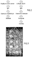
- the figure 2 is a diagram showing the plane of antigen deposition on a slide used in Example 4.
- the figure 3 is the image of the slide of Example 4, after incubation with four serum, obtained at 350 nanometers (non-specific fluorescent staining with non-specific fluorescent labeling of proteins with AMCA and DNA with dye HOESCHT 332-42 according to example 4.
- Example 1 Deposition of said second and fourth antigens on solid support.
- a cell counter Mericrocytes® BPC / Yeast, BioDETECT AS, Olso, Norway
- Staphylococcus aureus (ATCC accession No. 29213) was cultured on 5% sheep blood agar and then harvested in sterile PBS buffer containing 0.1% sodium azide. The inoculum was measured using the same cell counter and calibrated at 10 9 bacteria / mL which is the optimum concentration given the constraints of absence of sedimentation during deposition and a deposit in sufficient quantity of Staphylococcal particles then stored by freezing at -80 ° C.
- HL60 cells and Staphylococcus aureus bacteria were deposited at a concentration of 10 9 CFU / mL determined by FACS-scan (Microcytes®) glass slides (Reference LLR2-45, CML, Nemours, France). Cells and bacteria were deposited on the solid support using a deposition robot (Arrayer 427 TM, Affymetrix, MWG Biotech SA, Courtaboeuf, France). The deposits were air-dried for 30 minutes in the "spotter” chamber, then fixed in a 100% methanol bath for 10 minutes and then dried again in the open air.
- the figure 1 illustrates the robotic deposit of 6 columns of 6 deposits each of Staphylococcus aureus.
- Class G ⁇ -specific human immunoglobulins (Serotec, Cergy Saint-Christophe, France) diluted to a concentration of 5 mg / ml
- class- ⁇ -specific immunoglobulins IgM (Serotec) diluted to a concentration of 10 mg / mL to obtain well homogeneous spots
- a deposition robot model 427, Arrayer, Affymetrix, Inc., CA
- a solid support consisting of glass Reference LLR2-45, CML, Nemours, France.
- the deposits were made by transfer of the antigen suspension of a well of microtitration plate containing 25 ⁇ L of suspension, in a volume of 1 mL deposited, at 25 ° C. and 55% humidity in the chamber of the patient. deposit robot. These conditions were controlled by a thermo-hygrometer. The deposits of a size of 200 microns were air-dried for 30 minutes at 37.degree. C. in the enclosure of the deposition robot, then fixed in a 100% ethanol bath for 10 minutes, and then dried at room temperature. again in the open air. The efficiency of the robotic deposition and then the ethanol fixation after the baths required by the indirect immunofluorescence reaction was verified by the indirect immunofluorescence technique.
- an indirect immunofluorescence reaction was performed using a goat immunoglobulin (reference A-11013, Molecular Probes, Eugene, USA) as a secondary anti-human IgM antibody, coupled to Alexa 488 which is a fluorescent molecule excited at 488 nanometers and emitting at 540 nanometers and in using the same Axa 594-coupled immunoglobulin (A-21216, Molecular Probes, Eugene, USA) which is a fluorescent molecule excited at 594 nanometers and emitting at 640 nanometers in a second experiment.
- a goat immunoglobulin reference A-11013, Molecular Probes, Eugene, USA
- Alexa 488 which is a fluorescent molecule excited at 488 nanometers and emitting at 540 nanometers
- Axa 594-coupled immunoglobulin A-21216, Molecular Probes, Eugene, USA
- the reaction was read by fluorescence microscopy and showed fluorescent detection in all sera containing rheumatoid factors in the form of a bright spot for each of the 3 dilutions tested for Ig 1 deposits and none at all. fluorescence in sera not containing these factors.
- This example also illustrates that it is possible to perform robotic deposition of IgG and human IgM on a solid support under conditions compatible with performing an indirect immunofluorescence reaction for the detection of rheumatoid factors. Also, this example illustrates that it is possible to perform the detection of rheumatoid factors by an indirect immunofluorescence technique.
- Example 3 A serological diagnosis of Coxiella burnetii infection was made according to the method described in the present invention
- cells HL 60 (as said second antigen), Staphylococcus aureus (as said fourth antigen), human IgG (as said first antigen) and Coxiella burnetii
- Staphylococcus aureus (as said fourth antigen)
- human IgG (as said first antigen)
- Coxiella burnetii Nine Mile, Phase II (ATCC No. VR 616), Legionella pneumophila, Bartonella benselae, Bartonella quintana, Bartonella vinsonii subsp. berkoffii, and Bartonella elizabethae (as said microbial antigens).
- Staphylococcus aureus was cultured on 5% sheep blood agar then harvested in sterile PBS buffer containing 0.1% sodium acid. The inoculum was measured using the same cell counter and calibrated at 10 9 bacteria / mL and then frozen at -80 ° C.
- Class G human immunoglobulins (IgG) (Serotec, Cergy Saint-Christophe, France) were diluted extemporaneously at a concentration of 5 mg / ml.
- the bacterium Coxiella burnetii Nine Mile phase II was cultured on cell support and purified according to the usual protocols and quantified by the Microcyte® counter in order to standardize its concentration at 10 9 bacteria / mL which is the optimum concentration given the constraints of absence of sedimentation during deposition and deposition in sufficient quantity of the bacterial particles.
- This antigen was stored at -80 ° C in PBS buffer, 0.1% sodium acid.
- the solid support consisted of a glass slide previously cleaned with a mixture of 50% ethanol and 50% acetone and then dried in ambient air.
- the deposits were air-dried for 30 minutes in the enclosure of the deposition robot, then fixed in a 100% methanol bath for 10 minutes, and then dried again in the open air.
- the deposits were made by transfer of the antigen suspension of a well of microtitration plate containing 25 ⁇ L of suspension, in a volume of 1 mL deposited, at 25 ° C. and 55% humidity in the chamber of the patient. deposit robot. These conditions were controlled by a thermo-hygrometer. Deposits of a 200 ⁇ m dimension were air-dried for 30 minutes at 37 ° C. in the enclosure of the deposition robot, then fixed in a 100% methanol bath for 10 minutes, and then dried again in the open air .
- the figure 1 represents a slide stained by the fluorescent dye Hoescht 333-42 which is excited at 350 nanometers and emits at 460 nanometers (Molecular Probes), to visualize the set of 8 antigens deposited, HL60 cells, Staphylococcus aureus, Coxiella burnetii, Legionella pneumophila, Bartonella henselae, Bartonella quintana, Bartonella vinsonii subsp. berkoffii, and Bartonella elizabethae. These different antigens were deposited under the same conditions as Coxiella, described above.
- Example 4 Determination of the vaccination status of persons.
- a glass slide has been developed for the determination of the vaccination status of persons with a total of 8 antigen deposits with 3 control deposits and 5 vaccine antigen deposits on the same slide, divided into 2 columns.
- the methods of depositing and using the 3 control deposits comprising Staphylococcus aureus, IgG and HL-60 cells were set out in Examples 1 and 2 above.
Landscapes
- Health & Medical Sciences (AREA)
- Life Sciences & Earth Sciences (AREA)
- Immunology (AREA)
- Engineering & Computer Science (AREA)
- Molecular Biology (AREA)
- Biomedical Technology (AREA)
- Chemical & Material Sciences (AREA)
- Hematology (AREA)
- Urology & Nephrology (AREA)
- Physics & Mathematics (AREA)
- General Physics & Mathematics (AREA)
- Cell Biology (AREA)
- Food Science & Technology (AREA)
- Medicinal Chemistry (AREA)
- Biotechnology (AREA)
- Analytical Chemistry (AREA)
- Biochemistry (AREA)
- General Health & Medical Sciences (AREA)
- Microbiology (AREA)
- Pathology (AREA)
- Virology (AREA)
- Tropical Medicine & Parasitology (AREA)
- Medicines Containing Antibodies Or Antigens For Use As Internal Diagnostic Agents (AREA)
- Measuring Or Testing Involving Enzymes Or Micro-Organisms (AREA)
- Investigating Or Analysing Biological Materials (AREA)
- Apparatus Associated With Microorganisms And Enzymes (AREA)
- Testing Of Engines (AREA)
- Investigating Or Analysing Materials By Optical Means (AREA)
- Other Investigation Or Analysis Of Materials By Electrical Means (AREA)
Claims (34)
- Verfahren zur serologischen In-vitro-Diagnostik eines mikrobiellen Agens mittels Immunnachweis, bei dem das Vorliegen von Patienten-Immunglobulinen der beiden Klassen M und G oder nur der Klasse G, die für ein für das mikrobielle Agens charakteristisches mikrobielles Antigen spezifisch sind, in einer zu untersuchenden Serumprobe des Patienten nachgewiesen wird und vorzugsweise deren Menge bestimmt wird, und zwar mittels Nachweis und Quantifizierung eines immunologischen Reaktionskomplexes zwischen dem nachzuweisenden mikrobiellen Antigen und einem spezifischen Immunglobulin der Klasse G für die quantitative IgG-Bestimmung oder der Klasse M für die quantitative IgM-Bestimmung, mit Hilfe einer ersten Nachweissubstanz bestehend aus einem lediglich mit einem Immunglobulin der Spezies des Patienten der Klasse G reagierenden Antikörper, für die quantitative IgG-Bestimmung, oder einer zweiten Nachweissubstanz bestehend aus einem nur mit einem Immunglobulin der Spezies des Patienten der Klasse M reagierenden Antikörper, für die quantitative IgM-Bestimmung, dadurch gekennzeichnet, daß1/- die folgenden Schritte durchgeführt werden, bei denen:• die zu untersuchende Serumprobe, anschließend die erste und die zweite Nachweissubstanz oder nur die erste Nachweissubstanz mit wenigstens einem festen Träger in Kontakt gebracht werden, auf dem die folgenden Antigene fixiert worden sind:- ein erstes Kontrollantigen, das von einem unspezifischen Immunglobulin der Klasse G der Spezies des Patienten gebildet ist, und- ein zweites Kontrollantigen, das DNA/Histon-Komplexe umfaßt, sowie- gegebenenfalls ein drittes Kontrollantigen, das von einem unspezifischen Immunglobulin der Klasse M der Spezies des Patienten gebildet ist, wobei das Vorliegen des dritten Kontrollantigens im Fall einer quantitativen IgM-Bestimmung erforderlich ist, und- wenigstens ein mikrobielles Antigen, und• eine Reihe von Kontrollen durchgeführt wird, die folgendes umfassen:a- die Kontrolle der Reaktivität der ersten Nachweissubstanz durch Überprüfen, ob das erste Kontrollantigen mit der ersten Nachweissubstanz reagiert, und - im Fall der quantitativen IgM-Bestimmung - die Kontrolle des Vorliegens von Rheumafaktoren in der Serumprobe durch Überprüfen, ob das erste Kontrollantigen mit der Serumprobe und der zweiten Nachweissubstanz reagiert,b- die Kontrolle des Vorliegens antinuklearer Antikörper in der zu untersuchenden Serumprobe, durch Überprüfen, ob das zweite Kontrollantigen mit der Serumprobe und der ersten Nachweissubstanz reagiert,c- im Fall der quantitativen IgM-Bestimmung, die Kontrolle der Reaktivität der zweiten Nachweissubstanz durch Überprüfen, ob das dritte Kontrollantigen mit der zweiten Nachweissubstanz reagiert, undd- die Kontrolle des Vorliegens eines Serums der Spezies des Patienten in der zu untersuchenden Probe, und2/- das Ergebnis einer Reaktion zwischen dem mikrobiellen Antigen, der Serumprobe und einer Nachweissubstanz nur berücksichtigt wird, wenn die Kontrolle des Vorliegens eines Serums der Spezies des Patienten positiv ist und wenn die folgenden kumulativen Bedingungen gemeinsam vorliegen:a- das erste Kontrollantigen reagiert mit der ersten Nachweissubstanz,b- das zweite Kontrollantigen reagiert nicht, undc- gegebenenfalls, im Fall der quantitativen IgM-Bestimmung, reagiert das dritte Kontrollantigen mit der zweiten Nachweissubstanz.
- Verfahren nach Anspruch 1, dadurch gekennzeichnet, daß ein einziger fester Träger verwendet wird, der gegebenenfalls gleichzeitig mit der ersten und der zweiten Nachweissubstanz, die ein erstes bzw. ein zweites Markierungselement enthalten, in Kontakt gebracht wird, wobei das zweite Markierungselement ein anderes Signal als das erste Markierungselement aussendet, wobei die erste und die zweite Nachweissubtanz einen ersten bzw. einen zweiten Antikörper enthalten, der nur mit einem Immunglobulin der Spezies des Patienten der Klasse G bzw. der Klasse M reagiert.
- Verfahren nach Anspruch 1 oder 2, dadurch gekennzeichnet, daß kontrolliert wird, ob die untersuchte Probe tatsächlich ein Serum der Spezies des Patienten enthält, indem nachgewiesen wird, ob Immunglobuline der Spezies des Patienten mit einem vierten Kontrollantigen, welches das Protein A eines Staphylococcus aureus-Bakteriums enthält, reagieren, wobei vorzugsweise das vierte Antigen ein vollständiges Staphylococcus-Bakterium ist, und zwar dadurch, daß die Probe in Anwesenheit der ersten Nachweissubstanz, die ein Anti-Immunglobulin-Antikörper der Spezies des Patienten ist, der nicht mit dem vierten Kontrollantigen reagiert, mit einem festen Träger, auf dem ein viertes Kontrollantigen fixiert ist, in Kontakt gebracht wird, wobei die Kontrolle des Vorliegens eines Serums positiv ist, wenn das vierte Antigen mit der Serumprobe und der ersten Nachweissubstanz reagiert.
- Verfahren nach einem der Ansprüche 1 bis 3, dadurch gekennzeichnet, daß die erste Nachweissubstanz ein tierisches Immunglobulin, vorzugsweise ein Immunglobulin von der Ziege oder vom Huhn ist.
- Verfahren nach einem der Ansprüche 1 bis 4, dadurch gekennzeichnet, daß gegebenenfalls beide Nachweissubtanzen, also die erste und die zweite, Immunglobuline von der Ziege oder vom Huhn, Anti-IgG bzw. Anti-IgM sind.
- Verfahren nach einem der Ansprüche 1 bis 5, dadurch gekennzeichnet, daß das zweite Kontrollantigen aus Zellen humaner Fibroblasten besteht.
- Verfahren nach einem der Ansprüche 1 bis 6, dadurch gekennzeichnet, daß die Kontrollantigene und mikrobiellen Antigene durch physikalische Adsorption auf dem festen Träger fixiert sind.
- Verfahren nach einem der Ansprüche 1 bis 7, dadurch gekennzeichnet, daß das mikrobielle Antigen ein korpuskuläres Antigen ist, das aus einer inaktivierten vollständigen Mikrobe oder einer Mikrobenfraktion besteht.
- Serologisches Diagnostikverfahren nach einem der Ansprüche 1 bis 8, dadurch gekennzeichnet, daß das mikrobielle Agens aus den Mikroorganismen umfassend ein Bakterium, ein Virus, einen Parasiten oder einen Pilz ausgewählt ist.
- Serologisches Diagnostikverfahren nach Anspruch 9, dadurch gekennzeichnet, daß das mikrobielle Antigen ein intrazelluläres Bakterium oder ein Virus ist.
- Serologisches Diagnostikverfahren nach Anspruch 9 oder 10, dadurch gekennzeichnet, daß das mikrobielle Antigen aus den Bakterien der Gattung Rickettsia, Coxiella, Bartonella, Tropheryma, Ehrlichia, Chlamydia, Mycoplasma, Treponema, Borrelia und Leptospira ausgewählt ist.
- Serologisches Diagnostikverfahren nach Anspruch 11, dadurch gekennzeichnet, daß das mikrobielle Antigen ein für eine Endokarditis verantwortliches Bakterium ist.
- Serologisches Diagnostikverfahren nach Anspruch 10, dadurch gekennzeichnet, daß das mikrobielle Antigen ein virales Antigen ist, welches aus den Viren H.I.V., C.M.V., dem Epstein-Barr-Virus, Masernvirus, Rubella-Virus, Hepatitis-A-und Hepatitis-B-Virus ausgewählt ist.
- Verfahren nach einem der Ansprüche 1 bis 13, dadurch gekennzeichnet, daß:- die Menge der Immunglobuline von Patienten der beiden Klassen M und G, die für ein mikrobielles Antigen spezifisch sind, nachgewiesen und bestimmt wird,- wenigstens ein mikrobielles Antigen sowie das erste, das zweite und das dritte und das vierte Kontrollantigen auf einem gleichen festen Träger fixiert werden, und- für den Nachweis der unterschiedlichen mikrobiellen Antigene die gleichen ersten und zweiten Nachweissubstanzen mit unterschiedlichen Markierungselementen verwendet werden, wobei die erste und die zweite Nachweissubstanz tierische Immunglobuline sind, die nicht mit dem vierten Antigen reagieren.
- Verfahren nach Anspruch 1 bis 14, dadurch gekennzeichnet, daß als fester Träger ein Objektträger aus Glas oder Kunststoff, ein Titrationsröhrchen oder ein Well einer Kunststoff-Mikrotiterplatte verwendet wird.
- Verfahren nach einem der Ansprüche 1 bis 15, dadurch gekennzeichnet, daß durch automatisiertes Lesen mit Hilfe eines Gerätes zum Lesen eines fluoreszierenden Signals einer den Markierungselementen der Nachweissubstanzen entsprechenden fluoreszierenden Substanz das für das mikrobielle Antigen spezifische Immunglobulin der Spezies des Patienten in der zu untersuchenden Probe sowie die immunologischen Reaktionen zwischen den Kontrollantigenen und den Nachweissubstanzen nachgewiesen werden und gegebenenfalls die Menge des genannten Immunglobulins in der zu untersuchenden Probe bestimmt wird.
- Verfahren nach einem der Ansprüche 1 bis 16, dadurch gekennzeichnet, daß das mikrobielle Antigen ein Impfantigen ist und das für das nachzuweisende Impfantigen spezifische Immunglobulin ein Immunglobulin der Klasse G ist.
- Verfahren nach Anspruch 17, dadurch gekennzeichnet, daß der Impfstatus eines Individuums mittels Nachweis, vorzugsweise Quantifizierung der IgG-Serumantikörper, die für Impfantigene einer Vielzahl pathogener Agenzien von der Art Bakterien, Viren, Pilzen oder Parasiten spezifisch sind, dadurch bestimmt wird, daß der Nachweis, und vorzugsweise die Quantifizierung, eines Komplexes immunologischer Reaktionen zwischen jedem Impfantigen und jeweils jedem für das Impfantigen spezifischen, eventuell in einer zu untersuchenden Probe eines humanen Serums vorliegenden Antikörper durchgeführt wird, umfassend:1- das Inkontaktbringen einer gleichen zu untersuchenden Serumprobe, anschließend wenigstens einer ersten Nachweissubstanz, die mit wenigstens einem spezifischen Antikörper reagiert und die nicht mit irgendeinem der Impfantigene reagiert, mit einem gleichen festen Träger, auf dem eine Vielzahl von Impfantigenen, die einer Vielzahl pathogener Agenzien entsprechen, sowie das erste, das zweite und vorzugsweise das vierte Kontrollantigen fixiert sind, und2- wenigstens eine Kontrolle der Reaktivität der ersten Nachweissubstanz mit Hilfe eines ersten Kontrollantigens sowie eine Kontrolle des Vorliegens antinuklearer Antikörper mit Hilfe wenigstens eines zweiten Kontrollantigens und die Kontrolle des Vorliegens eines humanen Serums in der zu untersuchenden Probe.
- Verfahren nach Anspruch 18, dadurch gekennzeichnet, daß spezifische Antikörper des Typs IgG-Immunglobuline nachgewiesen werden und eine erste Nachweissubstanz eingesetzt wird, die ein Anti-IgG-Immunglobulin ist, das von einem Immunglobulin von der Ziege oder vom Huhn gebildet ist.
- Verfahren nach einem der Ansprüche 18 oder 19, dadurch gekennzeichnet, daß die Impfantigene Antigene der pathogenen Agenzien ausgewählt unter den folgenden sind, nämlich dem Mumpsvirus, Rubella-Virus, Masernvirus, Varizella-Virus, Poliomyelitis-Virus, Gelbfieber-Virus, Meningo-Enzephalitis-Virus, Hepatitis-A-Virus, Hepatitis-B-Virus und den Keuchhusten-Bakterien Bordetella pertussis, den Tetanus-und Diphterie-Bakterien.
- Verfahren nach einem der Ansprüche 18 bis 20, dadurch gekennzeichnet, daß ermittelt wird, ob die Konzentration des spezifischen Antikörpers eine Schwelle erreicht, ab welcher der spezifische Antikörper eine Schutzwirkung aufweist, die gegen die durch das Pathogen bestimmte Krankheit schützt.
- Verfahren nach einem der Ansprüche 18 bis 21, dadurch gekennzeichnet, daß eine bestimmte Gesamtblutmenge mit Hilfe einer Kapillarröhre in einem Kolben gesammelt wird, der eine bestimmte Menge eines Puffers enthält, der das Eluieren des Serums ermöglicht, wobei das Serum dann auf eine bestimmte Konzentration, vorzugsweise zwischen 1/100 und 1/20 verdünnt wird.
- Verfahren nach einem der Ansprüche 18 bis 22, dadurch gekennzeichnet, daß für jeden Nachweis und gegebenenfalls jede Quantifizierung eines Impfantigens die folgenden Messungen durchgeführt werden:1- eine erste Messung eines ersten Wertes, der für die Menge eines dritten Markierungselements repräsentativ ist, wobei der erste Wert der Wert der Stärke eines durch das dritte fluoreszierende Markierungselement ausgesandten Signals ist, wobei das dritte Markierungselement auf nicht spezifische Weise an jedes Protein im Auftragsbereich des Impfantigens bindet, und2- eine zweite Messung eines zweiten Wertes, der für die Menge eines ein anderes Signal als das dritte Markierungselement aussendenden ersten Markierungselements repräsentativ ist, wobei der zweite Wert der Wert der Stärke des Signals ist, das durch dieses erste fluoreszierende Markierungselement bei einer anderen Anregungswellenlänge als der des dritten fluorszierenden Markierungselements ausgesendet wird, wobei das erste Markierungselement das Element des Markierungselements der ersten Nachweissubstanz des Impfantigens im Auftragsbereich des Antigens ist, und3- es wird das Verhältnis zwischen dem ersten und dem zweiten Wert berechnet, und4- es wird der Wert des Verhältnisses mit dem eines mit einer Sammlung positiver und negativer Referenzseren erhaltenen Referenzverhältnisses verglichen, wodurch es mittels Vergleich möglich ist, entsprechend dem Wert des Verhältnisses zwischen dem ersten und dem zweiten Wert die Notwendigkeit oder die Nichtnotwendigkeit des Impfens der Person für das Impfantigen zu bestimmen.
- Diagnose-Kit, das für die Durchführung eines Verfahrens nach einem der Ansprüche 1 bis 23 nützlich ist, bei dem die quantitative IgG-Bestimmung durchgeführt wird, dadurch gekennzeichnet, daß es folgendes umfaßt:a- wenigstens einen festen Träger auf dem wenigstens folgendes fixiert ist:- ein mikrobielles Antigen, wie es in einem der Ansprüche 1 und 8 bis 13 sowie 17, 18 und 20 definiert ist, und- ein erstes sowie ein zweites Kontrollantigen, wie sie in einem der Ansprüche 1 und 6 definiert sind, und- vorzugsweise ein viertes Kontrollantigen, wie es in Anspruch 3 definiert ist, undb- eine erste Nachweissubstanz, wie sie in einem der Ansprüche 1, 2, 4, 5 und 14 definiert ist.
- Kit nach Anspruch 24, das für die Durchführung eines Verfahrens nach einem der Ansprüche 1 bis 23 nützlich ist, bei dem ferner die quantitative IgM-Bestimmung durchgeführt wird, dadurch gekennzeichnet, daß es außerdem folgendes umfaßt:a- einen festen Träger, auf dem ferner ein drittes Kontrollantigen, wie es in Anspruch 1 definiert ist, fixiert ist, sowieb- eine zweite Nachweissubstanz, wie sie in einem der Ansprüche 1, 2, 5 und 14 definiert ist.
- Kit nach Anspruch 24 oder 25, dadurch gekennzeichnet, daß es einen gleichen festen Träger umfaßt, auf dem mittels physikalischer Adsorption wenigstens ein korpuskuläres Antigen und die Kontrollantigene fixiert sind.
- Verfahren zum Herstellen eines festen Trägers eines Diagonse-Kits nach einem der Ansprüche 24 bis 26, auf dem wenigstens ein mikrobielles Antigen sowie ein erstes und ein zweites Kontrollantigen sowie vorzugsweise ein drittes und ein viertes Kontrollantigen fixiert sind, so daß ein Nachweis durch automatisiertes Lesen mit Hilfe einer ersten und gegebenenfalls zweiten Nachweissubstanz ermöglicht wird, dadurch gekennzeichnet, daß die mikrobiellen, vorzugsweise korpuskulären Antigene sowie Kontrollantigene mittels eines vorzugsweise eine Spritze umfassenden Auftragsroboters aufgetragen werden, wobei die korpuskulären Antigene vorzugsweise einem Farbstoff zugeordnet sind, weiterhin vorzugsweise einem fluoreszierenden Farbstoff in Form einer Suspension in einer Konzentration, die deren Visualisierung nach dem Auftragen mit Hilfe des Farbstoffs ermöglicht, wodurch es möglich ist, die Fixierung der Antigene auf dem festen Träger zu überprüfen.
- Verfahren nach Anspruch 27, dadurch gekennzeichnet, daß ein mikrobielles Antigen und gegebenenfalls ein zweites Kontrollantigen sowie gegebenenfalls ein viertes Kontrollantigen, in Form einer Suspension von Korpuskeln nicht konfluenter Zellen, ganzen Viren oder ganzen Bakterien oder Zell- oder Bakterienfraktionen mittels Roboter aufgetragen wird.
- Verfahren nach Anspruch 28, dadurch gekennzeichnet, daß die Kontrollantigene in Form einer Zellsuspension auf eine Konzentration von 107 bis 109 Zellen/ml eingestellt werden, die Kontrollantigene oder mikrobiellen Antigene in Form von Suspensionen von Bakterien oder Bakterienfraktionen auf eine Konzentration von 107 bis 109 Partikel/ml eingestellt werden und die Suspensionen von ganzen Viren auf eine Konzentration von 109 bis 1010 Partikel/ml eingestellt werden.
- Verfahren nach einem der Ansprüche 28 oder 29, dadurch gekennzeichnet, daß die Kontrollantigene und korpuskulären mikrobiellen Antigene in Mischung mit einem Proteinbindemittel, das die Fixierung auf dem festen Träger stabilisiert, aufgetragen werden.
- Verfahren nach Anspruch 30, dadurch gekennzeichnet, daß das Proteinbindemittel aus Eigelb, Gelatine, Rinderserumalbumin oder einem polyklonalen nicht humanen IgG, vorzugsweise von der Ziege ausgewählt ist.
- Verfahren nach Anspruch 31, dadurch gekennzeichnet, daß das korpuskuläre mikrobielle Antigen auf dem von einem gläsernen Objektträger gebildeten festen Träger in Mischung mit einem Immunglobulin von der Art polyklonales IgG von der Ziege aufgetragen wird.
- Verfahren nach einem der Ansprüche 27 bis 32, dadurch gekennzeichnet, daß ein vorheriges Waschen des festen Trägers mit einer Lösung aus einem Ethanol/Aceton-Gemisch, vorzugsweise in einem Verhältnis 50/50, durchgeführt wird, dann aufgetragen wird und die Fixierung der Antigene mittels physikalischer Adsorption auf dem festen Träger durch eine Behandlung mit Alkohol, vorzugsweise Methanol oder Ethanol stabilisiert wird, Alkohohl, der anschließend entfernt wird, und weiterhin vorzugsweise die Fixierung der Antigene mittels Anfärben, vorzugsweise durch eine unspezifische fluoreszierende Markierung der Proteine oder der DNA überprüft wird.
- Verfahren nach einem der Ansprüche 27 bis 32, dadurch gekennzeichnet, daß die Fixierung mittels physikalischer Adsorption der Kontrollantigene und mikrobiellen Antigene durch eine Vernetzungsbehandlung, vorzugsweise eine chemische Behandlung mit einem bifunktionellen Agens zur kovalenten Kopplung komplettiert wird.
Applications Claiming Priority (3)
| Application Number | Priority Date | Filing Date | Title |
|---|---|---|---|
| FR0315327A FR2864624A1 (fr) | 2003-12-24 | 2003-12-24 | Methode de diagnostic serologique des maladies infectieuses par immunodetection d'antigene microbien comprenant le controle de reaction faussement positive ou negative |
| FR0408600A FR2874093B1 (fr) | 2004-08-03 | 2004-08-03 | Methode et trousse de determination du statut vaccinal de personnes |
| PCT/FR2004/003364 WO2005064340A2 (fr) | 2003-12-24 | 2004-12-23 | Methode de diagnostic serologique et determination de statut vaccinal comprenant differents controles |
Publications (2)
| Publication Number | Publication Date |
|---|---|
| EP1700128A2 EP1700128A2 (de) | 2006-09-13 |
| EP1700128B1 true EP1700128B1 (de) | 2009-08-12 |
Family
ID=34740714
Family Applications (1)
| Application Number | Title | Priority Date | Filing Date |
|---|---|---|---|
| EP04817587A Expired - Lifetime EP1700128B1 (de) | 2003-12-24 | 2004-12-23 | Verfahren zur serologischen diagnose und bestimmung des immunisierungszustands unter einschluss verschiedener kontrollen |
Country Status (6)
| Country | Link |
|---|---|
| US (1) | US20070160978A1 (de) |
| EP (1) | EP1700128B1 (de) |
| AT (1) | ATE439594T1 (de) |
| BR (1) | BRPI0418126A (de) |
| DE (1) | DE602004022588D1 (de) |
| WO (1) | WO2005064340A2 (de) |
Families Citing this family (8)
| Publication number | Priority date | Publication date | Assignee | Title |
|---|---|---|---|---|
| DE102006027516B4 (de) * | 2006-06-09 | 2021-10-07 | Euroimmun Medizinische Labordiagnostika Ag | Verfahren zur Optimierung der automatischen Fluoreszenzmustererkennung in der Immundiagnostik |
| WO2008065474A1 (en) * | 2006-11-29 | 2008-06-05 | Laboratorio Celsius S.A. | Method for analyzing image data relating to agglutination assays |
| FR2918459B1 (fr) * | 2007-07-04 | 2009-12-04 | Inodiag | Methode de diagnostic serologique multiplexe in vitro des infections a spirochetes |
| RU2472158C1 (ru) * | 2011-11-30 | 2013-01-10 | Федеральное государственное Бюджетное учреждение "Государственный научный центр дерматовенерологии и косметологии Министерства здравоохранения и социального развития Российской Федерации (ФГБУ "ГНЦДК" Минздравсоцразвития России) | Состав расширенной квалификационной панели контрольных материалов для осуществления внешнего и внутрилабораторного контроля качества серологической диагностики сифилитической инфекции |
| KR101341994B1 (ko) | 2012-02-20 | 2013-12-16 | 대한민국 | 간접면역형광항체법에 의한 레지오넬라증 진단용 항원슬라이드의 제조방법 및 이를 이용한 마이크로 다가 항원슬라이드 |
| CN102692502B (zh) * | 2012-04-26 | 2015-02-04 | 北京北方生物技术研究所 | 一种快速检测甲型肝炎病毒IgM抗体的试剂盒及其制备方法 |
| CN113567660A (zh) * | 2021-07-22 | 2021-10-29 | 中南大学湘雅二医院 | 一种检测系统性红斑狼疮患者自身抗体的方法 |
| CN120275649B (zh) * | 2025-06-11 | 2025-08-26 | 上海交通大学医学院附属仁济医院 | 一种适用于制备检测抗核抗体荧光免疫层析试纸条的Hep-2细胞提取物制备工艺及试纸条的制备方法 |
Family Cites Families (5)
| Publication number | Priority date | Publication date | Assignee | Title |
|---|---|---|---|---|
| DD301364A7 (de) * | 1990-04-19 | 1992-12-10 | Berlin Buch Klinikum | Verfahren zur Bestimmung von antinukleären Antikörpern |
| US5478753A (en) * | 1993-06-29 | 1995-12-26 | Pb Diagnostic Systems, Inc. | Positive calibrator/control composition for an IgM serology assay and an IgM serology assay |
| DE19731465A1 (de) * | 1997-07-22 | 1999-01-28 | Boehringer Mannheim Gmbh | Verwendung von Kontrollflächen zur Detektion von Störproben in einem Nachweisverfahren |
| DE69938701D1 (de) * | 1998-04-14 | 2008-06-26 | Otsuka Pharma Co Ltd | Verfahren und vorrichtung zum nachweis von antikörpern |
| RU2213972C1 (ru) * | 2002-09-19 | 2003-10-10 | Научно-исследовательский институт вакцин и сывороток им. И.И. Мечникова РАМН | Способ определения состояния гуморального иммунитета к условно-патогенным бактериям одновременным выявлением igg антител |
-
2004
- 2004-12-23 BR BRPI0418126-3A patent/BRPI0418126A/pt not_active IP Right Cessation
- 2004-12-23 AT AT04817587T patent/ATE439594T1/de not_active IP Right Cessation
- 2004-12-23 WO PCT/FR2004/003364 patent/WO2005064340A2/fr not_active Ceased
- 2004-12-23 DE DE602004022588T patent/DE602004022588D1/de not_active Expired - Lifetime
- 2004-12-23 EP EP04817587A patent/EP1700128B1/de not_active Expired - Lifetime
- 2004-12-23 US US10/583,771 patent/US20070160978A1/en not_active Abandoned
Also Published As
| Publication number | Publication date |
|---|---|
| DE602004022588D1 (de) | 2009-09-24 |
| ATE439594T1 (de) | 2009-08-15 |
| EP1700128A2 (de) | 2006-09-13 |
| BRPI0418126A (pt) | 2007-04-10 |
| WO2005064340A3 (fr) | 2005-11-24 |
| WO2005064340A2 (fr) | 2005-07-14 |
| US20070160978A1 (en) | 2007-07-12 |
Similar Documents
| Publication | Publication Date | Title |
|---|---|---|
| Hosseini et al. | Enzyme-linked immunosorbent assay (ELISA): from A to Z | |
| US4133639A (en) | Test article including a covalently attached diagnostic reagent and method | |
| CN1325919C (zh) | 过敏原微阵列分析 | |
| James et al. | Detection of endotoxin using an evanescent wave fiber-optic biosensor | |
| FR2573872A1 (fr) | Procede et systeme d'appareillage pour dosage immunologique en phase solide | |
| DK174032B1 (da) | Sæt samt fremgangsmåde til immunometrisk dosering, der kan anvendes på hele celler | |
| EP1700128B1 (de) | Verfahren zur serologischen diagnose und bestimmung des immunisierungszustands unter einschluss verschiedener kontrollen | |
| CA2621485C (en) | Method for diagnosing infections utilizing surface antigens obtained without disrupting cells | |
| US8535951B2 (en) | Reagent for detection of analyte and process thereof | |
| WO2005064340B1 (fr) | Methode de diagnostic serologique et determination de statut vaccinal comprenant differents controles | |
| Manoharan et al. | Rapid serological profiling by an immunocomb-based dot-enzyme-linked immunosorbent test for three major poultry diseases | |
| EP1774336B1 (de) | Verfahren zum nachweis des immunisierungszustands einer person | |
| EP2167968A1 (de) | Verfahren zur serologischen multiplex-diagnose von spirochäten-infektionen in vitro | |
| WO1990007118A2 (fr) | Anticorps polymerises, diriges contre des immunoglobulines - leur utilisation dans des tests de diagnostic | |
| EP1518124B1 (de) | Verfahren zur serologischen diagnose | |
| FR2864624A1 (fr) | Methode de diagnostic serologique des maladies infectieuses par immunodetection d'antigene microbien comprenant le controle de reaction faussement positive ou negative | |
| JP6955261B2 (ja) | マイクロアレイ、マイクロアレイの製造方法、検査方法、及び検査キット | |
| US20050130223A1 (en) | Method, system and kit for detecting an analyte in a sample | |
| RU2769578C1 (ru) | Способ получения флуоресцирующего иммуноглобулинового диагностикума для выявления возбудителей риккетсиозов и коксиеллезов, флуоресцирующий иммуноглобулиновый диагностикум и его применение | |
| JPS5994068A (ja) | 血中膵ラ氏島細胞膜抗体のロゼット形成法による検出法 | |
| EP1478767B1 (de) | Serologisches in-vitro-verfahren zur diagnose von infektiöser endokarditis | |
| GB2034030A (en) | Analytical Test Article and Method | |
| US20060199175A1 (en) | Novel field testing method and device for detection of equine protozoal myeloencephalitis | |
| FR2857097A1 (fr) | Procede d'obtention d'un systeme d'etalonnage unique applique au dosage multiparametrique d'anticorps preferentiellement d'au moins les anticorps anti-gliadine et anti-transglutaminase. | |
| EP3399312A1 (de) | Erkennungsverfahren einer zellulären immunantwort |
Legal Events
| Date | Code | Title | Description |
|---|---|---|---|
| PUAI | Public reference made under article 153(3) epc to a published international application that has entered the european phase |
Free format text: ORIGINAL CODE: 0009012 |
|
| 17P | Request for examination filed |
Effective date: 20060602 |
|
| AK | Designated contracting states |
Kind code of ref document: A2 Designated state(s): AT BE BG CH CY CZ DE DK EE ES FI FR GB GR HU IE IS IT LI LT LU MC NL PL PT RO SE SI SK TR |
|
| DAX | Request for extension of the european patent (deleted) | ||
| 17Q | First examination report despatched |
Effective date: 20070917 |
|
| GRAP | Despatch of communication of intention to grant a patent |
Free format text: ORIGINAL CODE: EPIDOSNIGR1 |
|
| GRAS | Grant fee paid |
Free format text: ORIGINAL CODE: EPIDOSNIGR3 |
|
| GRAA | (expected) grant |
Free format text: ORIGINAL CODE: 0009210 |
|
| AK | Designated contracting states |
Kind code of ref document: B1 Designated state(s): AT BE BG CH CY CZ DE DK EE ES FI FR GB GR HU IE IS IT LI LT LU MC NL PL PT RO SE SI SK TR |
|
| REG | Reference to a national code |
Ref country code: GB Ref legal event code: FG4D Free format text: NOT ENGLISH |
|
| REG | Reference to a national code |
Ref country code: CH Ref legal event code: EP |
|
| REG | Reference to a national code |
Ref country code: IE Ref legal event code: FG4D |
|
| REF | Corresponds to: |
Ref document number: 602004022588 Country of ref document: DE Date of ref document: 20090924 Kind code of ref document: P |
|
| RAP2 | Party data changed (patent owner data changed or rights of a patent transferred) |
Owner name: INODIAG |
|
| NLT2 | Nl: modifications (of names), taken from the european patent patent bulletin |
Owner name: INODIAG Effective date: 20091014 |
|
| LTIE | Lt: invalidation of european patent or patent extension |
Effective date: 20090812 |
|
| PG25 | Lapsed in a contracting state [announced via postgrant information from national office to epo] |
Ref country code: FI Free format text: LAPSE BECAUSE OF FAILURE TO SUBMIT A TRANSLATION OF THE DESCRIPTION OR TO PAY THE FEE WITHIN THE PRESCRIBED TIME-LIMIT Effective date: 20090812 Ref country code: ES Free format text: LAPSE BECAUSE OF FAILURE TO SUBMIT A TRANSLATION OF THE DESCRIPTION OR TO PAY THE FEE WITHIN THE PRESCRIBED TIME-LIMIT Effective date: 20091123 Ref country code: LT Free format text: LAPSE BECAUSE OF FAILURE TO SUBMIT A TRANSLATION OF THE DESCRIPTION OR TO PAY THE FEE WITHIN THE PRESCRIBED TIME-LIMIT Effective date: 20090812 Ref country code: IS Free format text: LAPSE BECAUSE OF FAILURE TO SUBMIT A TRANSLATION OF THE DESCRIPTION OR TO PAY THE FEE WITHIN THE PRESCRIBED TIME-LIMIT Effective date: 20091212 Ref country code: AT Free format text: LAPSE BECAUSE OF FAILURE TO SUBMIT A TRANSLATION OF THE DESCRIPTION OR TO PAY THE FEE WITHIN THE PRESCRIBED TIME-LIMIT Effective date: 20090812 Ref country code: SE Free format text: LAPSE BECAUSE OF FAILURE TO SUBMIT A TRANSLATION OF THE DESCRIPTION OR TO PAY THE FEE WITHIN THE PRESCRIBED TIME-LIMIT Effective date: 20090812 |
|
| NLV1 | Nl: lapsed or annulled due to failure to fulfill the requirements of art. 29p and 29m of the patents act | ||
| PG25 | Lapsed in a contracting state [announced via postgrant information from national office to epo] |
Ref country code: SI Free format text: LAPSE BECAUSE OF FAILURE TO SUBMIT A TRANSLATION OF THE DESCRIPTION OR TO PAY THE FEE WITHIN THE PRESCRIBED TIME-LIMIT Effective date: 20090812 Ref country code: NL Free format text: LAPSE BECAUSE OF FAILURE TO SUBMIT A TRANSLATION OF THE DESCRIPTION OR TO PAY THE FEE WITHIN THE PRESCRIBED TIME-LIMIT Effective date: 20090812 Ref country code: PL Free format text: LAPSE BECAUSE OF FAILURE TO SUBMIT A TRANSLATION OF THE DESCRIPTION OR TO PAY THE FEE WITHIN THE PRESCRIBED TIME-LIMIT Effective date: 20090812 |
|
| REG | Reference to a national code |
Ref country code: IE Ref legal event code: FD4D |
|
| PG25 | Lapsed in a contracting state [announced via postgrant information from national office to epo] |
Ref country code: BG Free format text: LAPSE BECAUSE OF FAILURE TO SUBMIT A TRANSLATION OF THE DESCRIPTION OR TO PAY THE FEE WITHIN THE PRESCRIBED TIME-LIMIT Effective date: 20091112 Ref country code: PT Free format text: LAPSE BECAUSE OF FAILURE TO SUBMIT A TRANSLATION OF THE DESCRIPTION OR TO PAY THE FEE WITHIN THE PRESCRIBED TIME-LIMIT Effective date: 20091212 |
|
| PG25 | Lapsed in a contracting state [announced via postgrant information from national office to epo] |
Ref country code: CZ Free format text: LAPSE BECAUSE OF FAILURE TO SUBMIT A TRANSLATION OF THE DESCRIPTION OR TO PAY THE FEE WITHIN THE PRESCRIBED TIME-LIMIT Effective date: 20090812 Ref country code: EE Free format text: LAPSE BECAUSE OF FAILURE TO SUBMIT A TRANSLATION OF THE DESCRIPTION OR TO PAY THE FEE WITHIN THE PRESCRIBED TIME-LIMIT Effective date: 20090812 Ref country code: DK Free format text: LAPSE BECAUSE OF FAILURE TO SUBMIT A TRANSLATION OF THE DESCRIPTION OR TO PAY THE FEE WITHIN THE PRESCRIBED TIME-LIMIT Effective date: 20090812 Ref country code: IE Free format text: LAPSE BECAUSE OF FAILURE TO SUBMIT A TRANSLATION OF THE DESCRIPTION OR TO PAY THE FEE WITHIN THE PRESCRIBED TIME-LIMIT Effective date: 20090812 Ref country code: RO Free format text: LAPSE BECAUSE OF FAILURE TO SUBMIT A TRANSLATION OF THE DESCRIPTION OR TO PAY THE FEE WITHIN THE PRESCRIBED TIME-LIMIT Effective date: 20090812 |
|
| PG25 | Lapsed in a contracting state [announced via postgrant information from national office to epo] |
Ref country code: SK Free format text: LAPSE BECAUSE OF FAILURE TO SUBMIT A TRANSLATION OF THE DESCRIPTION OR TO PAY THE FEE WITHIN THE PRESCRIBED TIME-LIMIT Effective date: 20090812 |
|
| PLBE | No opposition filed within time limit |
Free format text: ORIGINAL CODE: 0009261 |
|
| STAA | Information on the status of an ep patent application or granted ep patent |
Free format text: STATUS: NO OPPOSITION FILED WITHIN TIME LIMIT |
|
| BERE | Be: lapsed |
Owner name: INODIAG Effective date: 20091231 |
|
| 26N | No opposition filed |
Effective date: 20100517 |
|
| PG25 | Lapsed in a contracting state [announced via postgrant information from national office to epo] |
Ref country code: MC Free format text: LAPSE BECAUSE OF NON-PAYMENT OF DUE FEES Effective date: 20100701 |
|
| REG | Reference to a national code |
Ref country code: CH Ref legal event code: PL |
|
| PG25 | Lapsed in a contracting state [announced via postgrant information from national office to epo] |
Ref country code: LI Free format text: LAPSE BECAUSE OF NON-PAYMENT OF DUE FEES Effective date: 20091231 Ref country code: GR Free format text: LAPSE BECAUSE OF FAILURE TO SUBMIT A TRANSLATION OF THE DESCRIPTION OR TO PAY THE FEE WITHIN THE PRESCRIBED TIME-LIMIT Effective date: 20091113 Ref country code: BE Free format text: LAPSE BECAUSE OF NON-PAYMENT OF DUE FEES Effective date: 20091231 Ref country code: CH Free format text: LAPSE BECAUSE OF NON-PAYMENT OF DUE FEES Effective date: 20091231 |
|
| PG25 | Lapsed in a contracting state [announced via postgrant information from national office to epo] |
Ref country code: LU Free format text: LAPSE BECAUSE OF NON-PAYMENT OF DUE FEES Effective date: 20091223 |
|
| PG25 | Lapsed in a contracting state [announced via postgrant information from national office to epo] |
Ref country code: HU Free format text: LAPSE BECAUSE OF FAILURE TO SUBMIT A TRANSLATION OF THE DESCRIPTION OR TO PAY THE FEE WITHIN THE PRESCRIBED TIME-LIMIT Effective date: 20100213 |
|
| PG25 | Lapsed in a contracting state [announced via postgrant information from national office to epo] |
Ref country code: TR Free format text: LAPSE BECAUSE OF FAILURE TO SUBMIT A TRANSLATION OF THE DESCRIPTION OR TO PAY THE FEE WITHIN THE PRESCRIBED TIME-LIMIT Effective date: 20090812 |
|
| PG25 | Lapsed in a contracting state [announced via postgrant information from national office to epo] |
Ref country code: CY Free format text: LAPSE BECAUSE OF FAILURE TO SUBMIT A TRANSLATION OF THE DESCRIPTION OR TO PAY THE FEE WITHIN THE PRESCRIBED TIME-LIMIT Effective date: 20090812 |
|
| PGFP | Annual fee paid to national office [announced via postgrant information from national office to epo] |
Ref country code: IT Payment date: 20121220 Year of fee payment: 9 |
|
| PGFP | Annual fee paid to national office [announced via postgrant information from national office to epo] |
Ref country code: FR Payment date: 20130107 Year of fee payment: 9 |
|
| PGFP | Annual fee paid to national office [announced via postgrant information from national office to epo] |
Ref country code: GB Payment date: 20130121 Year of fee payment: 9 Ref country code: DE Payment date: 20130110 Year of fee payment: 9 |
|
| REG | Reference to a national code |
Ref country code: DE Ref legal event code: R119 Ref document number: 602004022588 Country of ref document: DE |
|
| GBPC | Gb: european patent ceased through non-payment of renewal fee |
Effective date: 20131223 |
|
| REG | Reference to a national code |
Ref country code: FR Ref legal event code: ST Effective date: 20140829 |
|
| REG | Reference to a national code |
Ref country code: DE Ref legal event code: R119 Ref document number: 602004022588 Country of ref document: DE Effective date: 20140701 |
|
| PG25 | Lapsed in a contracting state [announced via postgrant information from national office to epo] |
Ref country code: DE Free format text: LAPSE BECAUSE OF NON-PAYMENT OF DUE FEES Effective date: 20140701 |
|
| PG25 | Lapsed in a contracting state [announced via postgrant information from national office to epo] |
Ref country code: FR Free format text: LAPSE BECAUSE OF NON-PAYMENT OF DUE FEES Effective date: 20131231 Ref country code: GB Free format text: LAPSE BECAUSE OF NON-PAYMENT OF DUE FEES Effective date: 20131223 |
|
| PG25 | Lapsed in a contracting state [announced via postgrant information from national office to epo] |
Ref country code: IT Free format text: LAPSE BECAUSE OF NON-PAYMENT OF DUE FEES Effective date: 20131231 |
|
| PG25 | Lapsed in a contracting state [announced via postgrant information from national office to epo] |
Ref country code: IT Free format text: LAPSE BECAUSE OF NON-PAYMENT OF DUE FEES Effective date: 20131223 |