EP1702929A2 - Utilisation thérapeutique de la protéine SMR 1 et dérivés actifs de celle-ci - Google Patents
Utilisation thérapeutique de la protéine SMR 1 et dérivés actifs de celle-ci Download PDFInfo
- Publication number
- EP1702929A2 EP1702929A2 EP06009596A EP06009596A EP1702929A2 EP 1702929 A2 EP1702929 A2 EP 1702929A2 EP 06009596 A EP06009596 A EP 06009596A EP 06009596 A EP06009596 A EP 06009596A EP 1702929 A2 EP1702929 A2 EP 1702929A2
- Authority
- EP
- European Patent Office
- Prior art keywords
- peptide
- xqhnpr
- pentapeptide
- smr1
- qhnpr
- Prior art date
- Legal status (The legal status is an assumption and is not a legal conclusion. Google has not performed a legal analysis and makes no representation as to the accuracy of the status listed.)
- Withdrawn
Links
- 230000001225 therapeutic effect Effects 0.000 title claims abstract description 44
- 108090000623 proteins and genes Proteins 0.000 title description 34
- 102000004169 proteins and genes Human genes 0.000 title description 30
- 108090000765 processed proteins & peptides Proteins 0.000 claims abstract description 233
- 210000001519 tissue Anatomy 0.000 claims abstract description 115
- 238000000034 method Methods 0.000 claims abstract description 90
- 229910052500 inorganic mineral Inorganic materials 0.000 claims abstract description 55
- 239000011707 mineral Substances 0.000 claims abstract description 55
- 210000000056 organ Anatomy 0.000 claims abstract description 55
- 210000000988 bone and bone Anatomy 0.000 claims abstract description 53
- 239000003446 ligand Substances 0.000 claims abstract description 48
- 210000003734 kidney Anatomy 0.000 claims abstract description 47
- 239000000203 mixture Substances 0.000 claims abstract description 30
- 230000000762 glandular Effects 0.000 claims abstract description 21
- 230000035800 maturation Effects 0.000 claims abstract description 20
- 208000037265 diseases, disorders, signs and symptoms Diseases 0.000 claims abstract description 17
- 239000005557 antagonist Substances 0.000 claims abstract description 15
- 210000000496 pancreas Anatomy 0.000 claims abstract description 15
- 239000000556 agonist Substances 0.000 claims abstract description 14
- 230000004071 biological effect Effects 0.000 claims abstract description 13
- 239000011159 matrix material Substances 0.000 claims abstract description 13
- 210000001156 gastric mucosa Anatomy 0.000 claims abstract description 12
- 201000010099 disease Diseases 0.000 claims abstract description 6
- 210000004027 cell Anatomy 0.000 claims description 62
- 102000004196 processed proteins & peptides Human genes 0.000 claims description 56
- 230000027455 binding Effects 0.000 claims description 38
- 150000002500 ions Chemical class 0.000 claims description 32
- 239000011575 calcium Substances 0.000 claims description 31
- 230000000694 effects Effects 0.000 claims description 30
- 230000002285 radioactive effect Effects 0.000 claims description 29
- 150000001413 amino acids Chemical class 0.000 claims description 28
- 229910052791 calcium Inorganic materials 0.000 claims description 27
- OYPRJOBELJOOCE-UHFFFAOYSA-N Calcium Chemical group [Ca] OYPRJOBELJOOCE-UHFFFAOYSA-N 0.000 claims description 26
- 238000004113 cell culture Methods 0.000 claims description 26
- 101710082215 SMR1 protein Proteins 0.000 claims description 25
- 239000000523 sample Substances 0.000 claims description 24
- 239000013598 vector Substances 0.000 claims description 24
- 238000002360 preparation method Methods 0.000 claims description 19
- 239000002207 metabolite Substances 0.000 claims description 18
- 230000033228 biological regulation Effects 0.000 claims description 15
- 238000012216 screening Methods 0.000 claims description 15
- 230000007547 defect Effects 0.000 claims description 13
- 230000002503 metabolic effect Effects 0.000 claims description 13
- 102000055006 Calcitonin Human genes 0.000 claims description 12
- 108060001064 Calcitonin Proteins 0.000 claims description 12
- BBBFJLBPOGFECG-VJVYQDLKSA-N calcitonin Chemical group N([C@H](C(=O)N[C@@H](CC(C)C)C(=O)NCC(=O)N[C@@H](CCCCN)C(=O)N[C@@H](CC(C)C)C(=O)N[C@@H](CO)C(=O)N[C@@H](CCC(N)=O)C(=O)N[C@@H](CCC(O)=O)C(=O)N[C@@H](CC(C)C)C(=O)N[C@@H](CC=1NC=NC=1)C(=O)N[C@@H](CCCCN)C(=O)N[C@@H](CC(C)C)C(=O)N[C@@H](CCC(N)=O)C(=O)N[C@@H]([C@@H](C)O)C(=O)N[C@@H](CC=1C=CC(O)=CC=1)C(=O)N1[C@@H](CCC1)C(=O)N[C@@H](CCCNC(N)=N)C(=O)N[C@@H]([C@@H](C)O)C(=O)N[C@@H](CC(N)=O)C(=O)N[C@@H]([C@@H](C)O)C(=O)NCC(=O)N[C@@H](CO)C(=O)NCC(=O)N[C@@H]([C@@H](C)O)C(=O)N1[C@@H](CCC1)C(N)=O)C(C)C)C(=O)[C@@H]1CSSC[C@H](N)C(=O)N[C@@H](CO)C(=O)N[C@@H](CC(N)=O)C(=O)N[C@@H](CC(C)C)C(=O)N[C@@H](CO)C(=O)N[C@@H]([C@@H](C)O)C(=O)N1 BBBFJLBPOGFECG-VJVYQDLKSA-N 0.000 claims description 12
- 229960004015 calcitonin Drugs 0.000 claims description 12
- 239000003814 drug Substances 0.000 claims description 12
- 239000000199 parathyroid hormone Substances 0.000 claims description 12
- 229920000642 polymer Polymers 0.000 claims description 12
- 102000003982 Parathyroid hormone Human genes 0.000 claims description 11
- 108090000445 Parathyroid hormone Proteins 0.000 claims description 11
- 102000030621 adenylate cyclase Human genes 0.000 claims description 11
- 108060000200 adenylate cyclase Proteins 0.000 claims description 11
- 239000012472 biological sample Substances 0.000 claims description 11
- 229960001319 parathyroid hormone Drugs 0.000 claims description 11
- 229920001184 polypeptide Polymers 0.000 claims description 11
- 210000002966 serum Anatomy 0.000 claims description 11
- 241000124008 Mammalia Species 0.000 claims description 10
- 208000035475 disorder Diseases 0.000 claims description 10
- 238000004519 manufacturing process Methods 0.000 claims description 10
- 102000008186 Collagen Human genes 0.000 claims description 9
- 108010035532 Collagen Proteins 0.000 claims description 9
- 229920001436 collagen Polymers 0.000 claims description 9
- 239000002356 single layer Substances 0.000 claims description 9
- GMRQFYUYWCNGIN-ZVUFCXRFSA-N 1,25-dihydroxy vitamin D3 Chemical group C1([C@@H]2CC[C@@H]([C@]2(CCC1)C)[C@@H](CCCC(C)(C)O)C)=CC=C1C[C@@H](O)C[C@H](O)C1=C GMRQFYUYWCNGIN-ZVUFCXRFSA-N 0.000 claims description 8
- DHCLVCXQIBBOPH-UHFFFAOYSA-N Glycerol 2-phosphate Chemical compound OCC(CO)OP(O)(O)=O DHCLVCXQIBBOPH-UHFFFAOYSA-N 0.000 claims description 8
- 150000007523 nucleic acids Chemical class 0.000 claims description 8
- 230000009870 specific binding Effects 0.000 claims description 8
- 239000001963 growth medium Substances 0.000 claims description 7
- 108020004707 nucleic acids Proteins 0.000 claims description 7
- 102000039446 nucleic acids Human genes 0.000 claims description 7
- 230000004913 activation Effects 0.000 claims description 6
- 210000003527 eukaryotic cell Anatomy 0.000 claims description 6
- 239000008188 pellet Substances 0.000 claims description 6
- 102000002260 Alkaline Phosphatase Human genes 0.000 claims description 5
- 108020004774 Alkaline Phosphatase Proteins 0.000 claims description 5
- 102000004961 Furin Human genes 0.000 claims description 5
- 108090001126 Furin Proteins 0.000 claims description 5
- 208000001132 Osteoporosis Diseases 0.000 claims description 5
- 210000001124 body fluid Anatomy 0.000 claims description 5
- 239000010839 body fluid Substances 0.000 claims description 5
- 239000004475 Arginine Substances 0.000 claims description 4
- KDXKERNSBIXSRK-UHFFFAOYSA-N Lysine Natural products NCCCCC(N)C(O)=O KDXKERNSBIXSRK-UHFFFAOYSA-N 0.000 claims description 4
- 239000004472 Lysine Substances 0.000 claims description 4
- ODKSFYDXXFIFQN-UHFFFAOYSA-N arginine Natural products OC(=O)C(N)CCCNC(N)=N ODKSFYDXXFIFQN-UHFFFAOYSA-N 0.000 claims description 4
- 239000012620 biological material Substances 0.000 claims description 4
- 238000012258 culturing Methods 0.000 claims description 4
- ZDXPYRJPNDTMRX-UHFFFAOYSA-N glutamine Natural products OC(=O)C(N)CCC(N)=O ZDXPYRJPNDTMRX-UHFFFAOYSA-N 0.000 claims description 4
- 238000003306 harvesting Methods 0.000 claims description 4
- 238000000527 sonication Methods 0.000 claims description 4
- 230000006820 DNA synthesis Effects 0.000 claims description 3
- 239000002299 complementary DNA Substances 0.000 claims description 3
- 238000012377 drug delivery Methods 0.000 claims description 3
- ODHCTXKNWHHXJC-VKHMYHEASA-N 5-oxo-L-proline Chemical group OC(=O)[C@@H]1CCC(=O)N1 ODHCTXKNWHHXJC-VKHMYHEASA-N 0.000 claims description 2
- 108010066717 Q beta Replicase Proteins 0.000 claims description 2
- 239000013592 cell lysate Substances 0.000 claims description 2
- 125000004435 hydrogen atom Chemical group [H]* 0.000 claims description 2
- 125000002887 hydroxy group Chemical group [H]O* 0.000 claims description 2
- 239000006193 liquid solution Substances 0.000 claims description 2
- 230000002934 lysing effect Effects 0.000 claims description 2
- 230000003204 osmotic effect Effects 0.000 claims description 2
- 239000000843 powder Substances 0.000 claims description 2
- 238000009738 saturating Methods 0.000 claims description 2
- 230000035939 shock Effects 0.000 claims description 2
- 230000002194 synthesizing effect Effects 0.000 claims description 2
- 101100533725 Mus musculus Smr3a gene Proteins 0.000 claims 3
- 101100149716 Rattus norvegicus Vcsa1 gene Proteins 0.000 claims 3
- 101150037481 SMR1 gene Proteins 0.000 claims 3
- 101100286750 Saccharomyces cerevisiae (strain ATCC 204508 / S288c) ILV2 gene Proteins 0.000 claims 3
- 230000005892 protein maturation Effects 0.000 claims 2
- 239000000599 controlled substance Substances 0.000 claims 1
- 230000010245 tubular reabsorption Effects 0.000 claims 1
- 230000008569 process Effects 0.000 abstract description 17
- 241001465754 Metazoa Species 0.000 abstract description 9
- 210000003298 dental enamel Anatomy 0.000 abstract description 3
- 210000001035 gastrointestinal tract Anatomy 0.000 abstract description 3
- 238000002560 therapeutic procedure Methods 0.000 abstract description 3
- 108090000244 Rat Proteins Proteins 0.000 abstract description 2
- 241000700159 Rattus Species 0.000 description 65
- 238000001727 in vivo Methods 0.000 description 34
- 235000018102 proteins Nutrition 0.000 description 29
- 210000004379 membrane Anatomy 0.000 description 28
- 239000012528 membrane Substances 0.000 description 28
- 108020003175 receptors Proteins 0.000 description 28
- 102000005962 receptors Human genes 0.000 description 28
- 235000001014 amino acid Nutrition 0.000 description 26
- 238000003556 assay Methods 0.000 description 25
- 238000009826 distribution Methods 0.000 description 25
- 229940024606 amino acid Drugs 0.000 description 24
- 238000002347 injection Methods 0.000 description 24
- 239000007924 injection Substances 0.000 description 24
- 210000002381 plasma Anatomy 0.000 description 23
- 210000004369 blood Anatomy 0.000 description 22
- 239000008280 blood Substances 0.000 description 22
- 210000001913 submandibular gland Anatomy 0.000 description 20
- 230000028327 secretion Effects 0.000 description 17
- WEVYAHXRMPXWCK-UHFFFAOYSA-N Acetonitrile Chemical compound CC#N WEVYAHXRMPXWCK-UHFFFAOYSA-N 0.000 description 15
- 238000002474 experimental method Methods 0.000 description 14
- 238000002372 labelling Methods 0.000 description 13
- 239000000047 product Substances 0.000 description 13
- 210000002784 stomach Anatomy 0.000 description 13
- 210000000936 intestine Anatomy 0.000 description 12
- 210000004877 mucosa Anatomy 0.000 description 12
- 230000009885 systemic effect Effects 0.000 description 12
- BQCADISMDOOEFD-UHFFFAOYSA-N Silver Chemical compound [Ag] BQCADISMDOOEFD-UHFFFAOYSA-N 0.000 description 11
- 239000000872 buffer Substances 0.000 description 11
- 239000011347 resin Substances 0.000 description 11
- 229920005989 resin Polymers 0.000 description 11
- 229910052709 silver Inorganic materials 0.000 description 11
- 239000004332 silver Substances 0.000 description 11
- 238000000376 autoradiography Methods 0.000 description 10
- 238000004587 chromatography analysis Methods 0.000 description 10
- 239000000284 extract Substances 0.000 description 10
- 238000004128 high performance liquid chromatography Methods 0.000 description 10
- 229940088597 hormone Drugs 0.000 description 10
- 239000005556 hormone Substances 0.000 description 10
- 230000004066 metabolic change Effects 0.000 description 10
- 238000011002 quantification Methods 0.000 description 10
- OKKJLVBELUTLKV-UHFFFAOYSA-N Methanol Chemical compound OC OKKJLVBELUTLKV-UHFFFAOYSA-N 0.000 description 9
- 230000009102 absorption Effects 0.000 description 9
- 238000010521 absorption reaction Methods 0.000 description 9
- -1 aluminum ions Chemical class 0.000 description 9
- 239000012148 binding buffer Substances 0.000 description 9
- 150000001875 compounds Chemical class 0.000 description 9
- 210000003722 extracellular fluid Anatomy 0.000 description 9
- 210000004283 incisor Anatomy 0.000 description 9
- 230000004044 response Effects 0.000 description 9
- 210000005239 tubule Anatomy 0.000 description 9
- JKMHFZQWWAIEOD-UHFFFAOYSA-N 2-[4-(2-hydroxyethyl)piperazin-1-yl]ethanesulfonic acid Chemical compound OCC[NH+]1CCN(CCS([O-])(=O)=O)CC1 JKMHFZQWWAIEOD-UHFFFAOYSA-N 0.000 description 8
- KCXVZYZYPLLWCC-UHFFFAOYSA-N EDTA Chemical compound OC(=O)CN(CC(O)=O)CCN(CC(O)=O)CC(O)=O KCXVZYZYPLLWCC-UHFFFAOYSA-N 0.000 description 8
- 239000007995 HEPES buffer Substances 0.000 description 8
- FAPWRFPIFSIZLT-UHFFFAOYSA-M Sodium chloride Chemical compound [Na+].[Cl-] FAPWRFPIFSIZLT-UHFFFAOYSA-M 0.000 description 8
- 230000035508 accumulation Effects 0.000 description 8
- 238000009825 accumulation Methods 0.000 description 8
- 239000003795 chemical substances by application Substances 0.000 description 8
- QWTDNUCVQCZILF-UHFFFAOYSA-N isopentane Chemical compound CCC(C)C QWTDNUCVQCZILF-UHFFFAOYSA-N 0.000 description 8
- 239000010410 layer Substances 0.000 description 8
- 239000007788 liquid Substances 0.000 description 8
- NBIIXXVUZAFLBC-UHFFFAOYSA-K phosphate Chemical compound [O-]P([O-])([O-])=O NBIIXXVUZAFLBC-UHFFFAOYSA-K 0.000 description 8
- 239000002243 precursor Substances 0.000 description 8
- 238000000746 purification Methods 0.000 description 8
- XLYOFNOQVPJJNP-UHFFFAOYSA-N water Chemical compound O XLYOFNOQVPJJNP-UHFFFAOYSA-N 0.000 description 8
- FYYHWMGAXLPEAU-UHFFFAOYSA-N Magnesium Chemical compound [Mg] FYYHWMGAXLPEAU-UHFFFAOYSA-N 0.000 description 7
- 150000001412 amines Chemical group 0.000 description 7
- 230000015572 biosynthetic process Effects 0.000 description 7
- 230000037396 body weight Effects 0.000 description 7
- 230000001413 cellular effect Effects 0.000 description 7
- 238000013270 controlled release Methods 0.000 description 7
- 238000002405 diagnostic procedure Methods 0.000 description 7
- 238000000605 extraction Methods 0.000 description 7
- 210000004185 liver Anatomy 0.000 description 7
- 239000011777 magnesium Substances 0.000 description 7
- 229910052749 magnesium Inorganic materials 0.000 description 7
- 230000001575 pathological effect Effects 0.000 description 7
- 239000012071 phase Substances 0.000 description 7
- 238000005406 washing Methods 0.000 description 7
- IAZDPXIOMUYVGZ-UHFFFAOYSA-N Dimethylsulphoxide Chemical compound CS(C)=O IAZDPXIOMUYVGZ-UHFFFAOYSA-N 0.000 description 6
- VEXZGXHMUGYJMC-UHFFFAOYSA-N Hydrochloric acid Chemical compound Cl VEXZGXHMUGYJMC-UHFFFAOYSA-N 0.000 description 6
- 108010052285 Membrane Proteins Proteins 0.000 description 6
- 229910019142 PO4 Inorganic materials 0.000 description 6
- 239000012505 Superdex™ Substances 0.000 description 6
- 239000007983 Tris buffer Substances 0.000 description 6
- 125000003275 alpha amino acid group Chemical group 0.000 description 6
- 238000004458 analytical method Methods 0.000 description 6
- 239000003599 detergent Substances 0.000 description 6
- 230000008030 elimination Effects 0.000 description 6
- 238000003379 elimination reaction Methods 0.000 description 6
- 239000008103 glucose Substances 0.000 description 6
- 230000003054 hormonal effect Effects 0.000 description 6
- 230000004060 metabolic process Effects 0.000 description 6
- WEXRUCMBJFQVBZ-UHFFFAOYSA-N pentobarbital Chemical compound CCCC(C)C1(CC)C(=O)NC(=O)NC1=O WEXRUCMBJFQVBZ-UHFFFAOYSA-N 0.000 description 6
- 239000010452 phosphate Substances 0.000 description 6
- 230000001105 regulatory effect Effects 0.000 description 6
- 238000000926 separation method Methods 0.000 description 6
- 239000011734 sodium Substances 0.000 description 6
- 210000000515 tooth Anatomy 0.000 description 6
- LENZDBCJOHFCAS-UHFFFAOYSA-N tris Chemical compound OCC(N)(CO)CO LENZDBCJOHFCAS-UHFFFAOYSA-N 0.000 description 6
- 210000002700 urine Anatomy 0.000 description 6
- WQZGKKKJIJFFOK-GASJEMHNSA-N Glucose Natural products OC[C@H]1OC(O)[C@H](O)[C@@H](O)[C@@H]1O WQZGKKKJIJFFOK-GASJEMHNSA-N 0.000 description 5
- 208000037147 Hypercalcaemia Diseases 0.000 description 5
- QPCDCPDFJACHGM-UHFFFAOYSA-N N,N-bis{2-[bis(carboxymethyl)amino]ethyl}glycine Chemical compound OC(=O)CN(CC(O)=O)CCN(CC(=O)O)CCN(CC(O)=O)CC(O)=O QPCDCPDFJACHGM-UHFFFAOYSA-N 0.000 description 5
- OAICVXFJPJFONN-UHFFFAOYSA-N Phosphorus Chemical compound [P] OAICVXFJPJFONN-UHFFFAOYSA-N 0.000 description 5
- 239000003098 androgen Substances 0.000 description 5
- 230000010072 bone remodeling Effects 0.000 description 5
- 238000006243 chemical reaction Methods 0.000 description 5
- 230000004087 circulation Effects 0.000 description 5
- 210000000172 cytosol Anatomy 0.000 description 5
- 238000006073 displacement reaction Methods 0.000 description 5
- 229940088598 enzyme Drugs 0.000 description 5
- 230000002496 gastric effect Effects 0.000 description 5
- 230000000148 hypercalcaemia Effects 0.000 description 5
- 208000030915 hypercalcemia disease Diseases 0.000 description 5
- 230000001965 increasing effect Effects 0.000 description 5
- 238000011534 incubation Methods 0.000 description 5
- 230000000968 intestinal effect Effects 0.000 description 5
- 238000010253 intravenous injection Methods 0.000 description 5
- 238000013507 mapping Methods 0.000 description 5
- 239000002609 medium Substances 0.000 description 5
- 239000002245 particle Substances 0.000 description 5
- 239000000137 peptide hydrolase inhibitor Substances 0.000 description 5
- 239000002953 phosphate buffered saline Substances 0.000 description 5
- 239000011574 phosphorus Substances 0.000 description 5
- 229910052698 phosphorus Inorganic materials 0.000 description 5
- 238000000159 protein binding assay Methods 0.000 description 5
- 230000009103 reabsorption Effects 0.000 description 5
- 238000004366 reverse phase liquid chromatography Methods 0.000 description 5
- 230000003248 secreting effect Effects 0.000 description 5
- 230000000638 stimulation Effects 0.000 description 5
- 230000002459 sustained effect Effects 0.000 description 5
- 238000003786 synthesis reaction Methods 0.000 description 5
- CIWBSHSKHKDKBQ-JLAZNSOCSA-N Ascorbic acid Chemical compound OC[C@H](O)[C@H]1OC(=O)C(O)=C1O CIWBSHSKHKDKBQ-JLAZNSOCSA-N 0.000 description 4
- IJGRMHOSHXDMSA-UHFFFAOYSA-N Atomic nitrogen Chemical compound N#N IJGRMHOSHXDMSA-UHFFFAOYSA-N 0.000 description 4
- 108091003079 Bovine Serum Albumin Proteins 0.000 description 4
- 125000001433 C-terminal amino-acid group Chemical group 0.000 description 4
- BHPQYMZQTOCNFJ-UHFFFAOYSA-N Calcium cation Chemical compound [Ca+2] BHPQYMZQTOCNFJ-UHFFFAOYSA-N 0.000 description 4
- RTZKZFJDLAIYFH-UHFFFAOYSA-N Diethyl ether Chemical compound CCOCC RTZKZFJDLAIYFH-UHFFFAOYSA-N 0.000 description 4
- 239000006144 Dulbecco’s modified Eagle's medium Substances 0.000 description 4
- 102000004190 Enzymes Human genes 0.000 description 4
- 108090000790 Enzymes Proteins 0.000 description 4
- WZUVPPKBWHMQCE-UHFFFAOYSA-N Haematoxylin Chemical compound C12=CC(O)=C(O)C=C2CC2(O)C1C1=CC=C(O)C(O)=C1OC2 WZUVPPKBWHMQCE-UHFFFAOYSA-N 0.000 description 4
- ZDXPYRJPNDTMRX-VKHMYHEASA-N L-glutamine Chemical compound OC(=O)[C@@H](N)CCC(N)=O ZDXPYRJPNDTMRX-VKHMYHEASA-N 0.000 description 4
- PXHVJJICTQNCMI-UHFFFAOYSA-N Nickel Chemical compound [Ni] PXHVJJICTQNCMI-UHFFFAOYSA-N 0.000 description 4
- DTQVDTLACAAQTR-UHFFFAOYSA-N Trifluoroacetic acid Chemical compound OC(=O)C(F)(F)F DTQVDTLACAAQTR-UHFFFAOYSA-N 0.000 description 4
- 239000002253 acid Substances 0.000 description 4
- 230000009471 action Effects 0.000 description 4
- 230000001800 adrenalinergic effect Effects 0.000 description 4
- 230000030570 cellular localization Effects 0.000 description 4
- 230000004700 cellular uptake Effects 0.000 description 4
- 238000005119 centrifugation Methods 0.000 description 4
- 230000001086 cytosolic effect Effects 0.000 description 4
- 238000001514 detection method Methods 0.000 description 4
- 238000010790 dilution Methods 0.000 description 4
- 239000012895 dilution Substances 0.000 description 4
- AFABGHUZZDYHJO-UHFFFAOYSA-N dimethyl butane Natural products CCCC(C)C AFABGHUZZDYHJO-UHFFFAOYSA-N 0.000 description 4
- DEFVIWRASFVYLL-UHFFFAOYSA-N ethylene glycol bis(2-aminoethyl)tetraacetic acid Chemical compound OC(=O)CN(CC(O)=O)CCOCCOCCN(CC(O)=O)CC(O)=O DEFVIWRASFVYLL-UHFFFAOYSA-N 0.000 description 4
- 239000012530 fluid Substances 0.000 description 4
- 230000006870 function Effects 0.000 description 4
- 238000010353 genetic engineering Methods 0.000 description 4
- 230000004079 mineral homeostasis Effects 0.000 description 4
- 239000002808 molecular sieve Substances 0.000 description 4
- 210000003205 muscle Anatomy 0.000 description 4
- 230000009871 nonspecific binding Effects 0.000 description 4
- 239000000813 peptide hormone Substances 0.000 description 4
- BDERNNFJNOPAEC-UHFFFAOYSA-N propan-1-ol Chemical compound CCCO BDERNNFJNOPAEC-UHFFFAOYSA-N 0.000 description 4
- 210000002307 prostate Anatomy 0.000 description 4
- 210000000512 proximal kidney tubule Anatomy 0.000 description 4
- 238000003127 radioimmunoassay Methods 0.000 description 4
- 230000002441 reversible effect Effects 0.000 description 4
- 210000003296 saliva Anatomy 0.000 description 4
- 230000000580 secretagogue effect Effects 0.000 description 4
- URGAHOPLAPQHLN-UHFFFAOYSA-N sodium aluminosilicate Chemical compound [Na+].[Al+3].[O-][Si]([O-])=O.[O-][Si]([O-])=O URGAHOPLAPQHLN-UHFFFAOYSA-N 0.000 description 4
- 210000004872 soft tissue Anatomy 0.000 description 4
- 238000006467 substitution reaction Methods 0.000 description 4
- 230000032258 transport Effects 0.000 description 4
- QTBSBXVTEAMEQO-UHFFFAOYSA-N Acetic acid Chemical compound CC(O)=O QTBSBXVTEAMEQO-UHFFFAOYSA-N 0.000 description 3
- USFZMSVCRYTOJT-UHFFFAOYSA-N Ammonium acetate Chemical compound N.CC(O)=O USFZMSVCRYTOJT-UHFFFAOYSA-N 0.000 description 3
- 239000005695 Ammonium acetate Substances 0.000 description 3
- 108010039627 Aprotinin Proteins 0.000 description 3
- 208000006386 Bone Resorption Diseases 0.000 description 3
- CURLTUGMZLYLDI-UHFFFAOYSA-N Carbon dioxide Chemical compound O=C=O CURLTUGMZLYLDI-UHFFFAOYSA-N 0.000 description 3
- 229920002307 Dextran Polymers 0.000 description 3
- WSFSSNUMVMOOMR-UHFFFAOYSA-N Formaldehyde Chemical compound O=C WSFSSNUMVMOOMR-UHFFFAOYSA-N 0.000 description 3
- PEDCQBHIVMGVHV-UHFFFAOYSA-N Glycerine Chemical compound OCC(O)CO PEDCQBHIVMGVHV-UHFFFAOYSA-N 0.000 description 3
- DGAQECJNVWCQMB-PUAWFVPOSA-M Ilexoside XXIX Chemical compound C[C@@H]1CC[C@@]2(CC[C@@]3(C(=CC[C@H]4[C@]3(CC[C@@H]5[C@@]4(CC[C@@H](C5(C)C)OS(=O)(=O)[O-])C)C)[C@@H]2[C@]1(C)O)C)C(=O)O[C@H]6[C@@H]([C@H]([C@@H]([C@H](O6)CO)O)O)O.[Na+] DGAQECJNVWCQMB-PUAWFVPOSA-M 0.000 description 3
- ODKSFYDXXFIFQN-BYPYZUCNSA-P L-argininium(2+) Chemical compound NC(=[NH2+])NCCC[C@H]([NH3+])C(O)=O ODKSFYDXXFIFQN-BYPYZUCNSA-P 0.000 description 3
- GDBQQVLCIARPGH-UHFFFAOYSA-N Leupeptin Natural products CC(C)CC(NC(C)=O)C(=O)NC(CC(C)C)C(=O)NC(C=O)CCCN=C(N)N GDBQQVLCIARPGH-UHFFFAOYSA-N 0.000 description 3
- 241000283973 Oryctolagus cuniculus Species 0.000 description 3
- 208000006735 Periostitis Diseases 0.000 description 3
- 230000037328 acute stress Effects 0.000 description 3
- 238000001042 affinity chromatography Methods 0.000 description 3
- 235000019257 ammonium acetate Nutrition 0.000 description 3
- 229940043376 ammonium acetate Drugs 0.000 description 3
- 238000013459 approach Methods 0.000 description 3
- 230000033558 biomineral tissue development Effects 0.000 description 3
- 230000024279 bone resorption Effects 0.000 description 3
- 210000004556 brain Anatomy 0.000 description 3
- 229910001424 calcium ion Inorganic materials 0.000 description 3
- 235000011089 carbon dioxide Nutrition 0.000 description 3
- 230000000747 cardiac effect Effects 0.000 description 3
- 230000015556 catabolic process Effects 0.000 description 3
- 238000006731 degradation reaction Methods 0.000 description 3
- 230000003111 delayed effect Effects 0.000 description 3
- 230000001419 dependent effect Effects 0.000 description 3
- 210000002919 epithelial cell Anatomy 0.000 description 3
- 230000029142 excretion Effects 0.000 description 3
- 239000012894 fetal calf serum Substances 0.000 description 3
- 238000004108 freeze drying Methods 0.000 description 3
- 239000011521 glass Substances 0.000 description 3
- 150000004676 glycans Chemical class 0.000 description 3
- 238000010191 image analysis Methods 0.000 description 3
- 238000000338 in vitro Methods 0.000 description 3
- 230000003993 interaction Effects 0.000 description 3
- 210000004731 jugular vein Anatomy 0.000 description 3
- GDBQQVLCIARPGH-ULQDDVLXSA-N leupeptin Chemical compound CC(C)C[C@H](NC(C)=O)C(=O)N[C@@H](CC(C)C)C(=O)N[C@H](C=O)CCCN=C(N)N GDBQQVLCIARPGH-ULQDDVLXSA-N 0.000 description 3
- 108010052968 leupeptin Proteins 0.000 description 3
- 239000007791 liquid phase Substances 0.000 description 3
- 238000012423 maintenance Methods 0.000 description 3
- 230000014759 maintenance of location Effects 0.000 description 3
- 239000000463 material Substances 0.000 description 3
- 238000005259 measurement Methods 0.000 description 3
- 238000003808 methanol extraction Methods 0.000 description 3
- 239000000178 monomer Substances 0.000 description 3
- 210000004409 osteocyte Anatomy 0.000 description 3
- 210000005009 osteogenic cell Anatomy 0.000 description 3
- 239000012188 paraffin wax Substances 0.000 description 3
- 229960001412 pentobarbital Drugs 0.000 description 3
- 210000003460 periosteum Anatomy 0.000 description 3
- 230000036470 plasma concentration Effects 0.000 description 3
- 229920003023 plastic Polymers 0.000 description 3
- 239000004033 plastic Substances 0.000 description 3
- 229920001282 polysaccharide Polymers 0.000 description 3
- 239000005017 polysaccharide Substances 0.000 description 3
- 125000006239 protecting group Chemical group 0.000 description 3
- 238000000163 radioactive labelling Methods 0.000 description 3
- 230000002829 reductive effect Effects 0.000 description 3
- 238000004007 reversed phase HPLC Methods 0.000 description 3
- 210000003079 salivary gland Anatomy 0.000 description 3
- 229910052708 sodium Inorganic materials 0.000 description 3
- 239000011780 sodium chloride Substances 0.000 description 3
- 238000005063 solubilization Methods 0.000 description 3
- 230000007928 solubilization Effects 0.000 description 3
- 241000894007 species Species 0.000 description 3
- 150000003431 steroids Chemical class 0.000 description 3
- 230000001300 stimulation of adenylate cyclase Effects 0.000 description 3
- 230000035882 stress Effects 0.000 description 3
- 239000000126 substance Substances 0.000 description 3
- 239000006228 supernatant Substances 0.000 description 3
- 238000012800 visualization Methods 0.000 description 3
- YBJHBAHKTGYVGT-ZKWXMUAHSA-N (+)-Biotin Chemical compound N1C(=O)N[C@@H]2[C@H](CCCCC(=O)O)SC[C@@H]21 YBJHBAHKTGYVGT-ZKWXMUAHSA-N 0.000 description 2
- IXPNQXFRVYWDDI-UHFFFAOYSA-N 1-methyl-2,4-dioxo-1,3-diazinane-5-carboximidamide Chemical compound CN1CC(C(N)=N)C(=O)NC1=O IXPNQXFRVYWDDI-UHFFFAOYSA-N 0.000 description 2
- WRDABNWSWOHGMS-UHFFFAOYSA-N AEBSF hydrochloride Chemical compound Cl.NCCC1=CC=C(S(F)(=O)=O)C=C1 WRDABNWSWOHGMS-UHFFFAOYSA-N 0.000 description 2
- 108010001478 Bacitracin Proteins 0.000 description 2
- 241000894006 Bacteria Species 0.000 description 2
- VGGGPCQERPFHOB-MCIONIFRSA-N Bestatin Chemical compound CC(C)C[C@H](C(O)=O)NC(=O)[C@@H](O)[C@H](N)CC1=CC=CC=C1 VGGGPCQERPFHOB-MCIONIFRSA-N 0.000 description 2
- VGGGPCQERPFHOB-UHFFFAOYSA-N Bestatin Natural products CC(C)CC(C(O)=O)NC(=O)C(O)C(N)CC1=CC=CC=C1 VGGGPCQERPFHOB-UHFFFAOYSA-N 0.000 description 2
- UXVMQQNJUSDDNG-UHFFFAOYSA-L Calcium chloride Chemical compound [Cl-].[Cl-].[Ca+2] UXVMQQNJUSDDNG-UHFFFAOYSA-L 0.000 description 2
- 108020004705 Codon Proteins 0.000 description 2
- 108700010070 Codon Usage Proteins 0.000 description 2
- 102000029816 Collagenase Human genes 0.000 description 2
- 108060005980 Collagenase Proteins 0.000 description 2
- 241000196324 Embryophyta Species 0.000 description 2
- KRHYYFGTRYWZRS-UHFFFAOYSA-N Fluorane Chemical compound F KRHYYFGTRYWZRS-UHFFFAOYSA-N 0.000 description 2
- 108010010803 Gelatin Proteins 0.000 description 2
- SXRSQZLOMIGNAQ-UHFFFAOYSA-N Glutaraldehyde Chemical compound O=CCCCC=O SXRSQZLOMIGNAQ-UHFFFAOYSA-N 0.000 description 2
- DHMQDGOQFOQNFH-UHFFFAOYSA-N Glycine Chemical compound NCC(O)=O DHMQDGOQFOQNFH-UHFFFAOYSA-N 0.000 description 2
- 241000238631 Hexapoda Species 0.000 description 2
- 201000002980 Hyperparathyroidism Diseases 0.000 description 2
- 208000013038 Hypocalcemia Diseases 0.000 description 2
- 208000000038 Hypoparathyroidism Diseases 0.000 description 2
- 208000015924 Lithiasis Diseases 0.000 description 2
- CSNNHWWHGAXBCP-UHFFFAOYSA-L Magnesium sulfate Chemical compound [Mg+2].[O-][S+2]([O-])([O-])[O-] CSNNHWWHGAXBCP-UHFFFAOYSA-L 0.000 description 2
- DRBBFCLWYRJSJZ-UHFFFAOYSA-N N-phosphocreatine Chemical compound OC(=O)CN(C)C(=N)NP(O)(O)=O DRBBFCLWYRJSJZ-UHFFFAOYSA-N 0.000 description 2
- 206010029148 Nephrolithiasis Diseases 0.000 description 2
- 206010031240 Osteodystrophy Diseases 0.000 description 2
- 108010058846 Ovalbumin Proteins 0.000 description 2
- 206010033645 Pancreatitis Diseases 0.000 description 2
- 229930040373 Paraformaldehyde Natural products 0.000 description 2
- 102000057297 Pepsin A Human genes 0.000 description 2
- 108090000284 Pepsin A Proteins 0.000 description 2
- ZLMJMSJWJFRBEC-UHFFFAOYSA-N Potassium Chemical compound [K] ZLMJMSJWJFRBEC-UHFFFAOYSA-N 0.000 description 2
- ONIBWKKTOPOVIA-UHFFFAOYSA-N Proline Natural products OC(=O)C1CCCN1 ONIBWKKTOPOVIA-UHFFFAOYSA-N 0.000 description 2
- 102000001253 Protein Kinase Human genes 0.000 description 2
- PXIPVTKHYLBLMZ-UHFFFAOYSA-N Sodium azide Chemical compound [Na+].[N-]=[N+]=[N-] PXIPVTKHYLBLMZ-UHFFFAOYSA-N 0.000 description 2
- YZCKVEUIGOORGS-NJFSPNSNSA-N Tritium Chemical compound [3H] YZCKVEUIGOORGS-NJFSPNSNSA-N 0.000 description 2
- 229920004890 Triton X-100 Polymers 0.000 description 2
- 239000013504 Triton X-100 Substances 0.000 description 2
- XPIVOYOQXKNYHA-RGDJUOJXSA-N [(2r,3s,4s,5r,6s)-3,4,5-trihydroxy-6-methoxyoxan-2-yl]methyl n-heptylcarbamate Chemical compound CCCCCCCNC(=O)OC[C@H]1O[C@H](OC)[C@H](O)[C@@H](O)[C@@H]1O XPIVOYOQXKNYHA-RGDJUOJXSA-N 0.000 description 2
- ODHCTXKNWHHXJC-UHFFFAOYSA-N acide pyroglutamique Natural products OC(=O)C1CCC(=O)N1 ODHCTXKNWHHXJC-UHFFFAOYSA-N 0.000 description 2
- UCTWMZQNUQWSLP-UHFFFAOYSA-N adrenaline Chemical compound CNCC(O)C1=CC=C(O)C(O)=C1 UCTWMZQNUQWSLP-UHFFFAOYSA-N 0.000 description 2
- 229910052782 aluminium Inorganic materials 0.000 description 2
- 229940030486 androgens Drugs 0.000 description 2
- 230000002337 anti-port Effects 0.000 description 2
- 229960004405 aprotinin Drugs 0.000 description 2
- 235000010323 ascorbic acid Nutrition 0.000 description 2
- 229960005070 ascorbic acid Drugs 0.000 description 2
- 239000011668 ascorbic acid Substances 0.000 description 2
- 229960003071 bacitracin Drugs 0.000 description 2
- 229930184125 bacitracin Natural products 0.000 description 2
- CLKOFPXJLQSYAH-ABRJDSQDSA-N bacitracin A Chemical compound C1SC([C@@H](N)[C@@H](C)CC)=N[C@@H]1C(=O)N[C@@H](CC(C)C)C(=O)N[C@H](CCC(O)=O)C(=O)N[C@@H]([C@@H](C)CC)C(=O)N[C@@H]1C(=O)N[C@H](CCCN)C(=O)N[C@@H]([C@@H](C)CC)C(=O)N[C@H](CC=2C=CC=CC=2)C(=O)N[C@@H](CC=2N=CNC=2)C(=O)N[C@H](CC(O)=O)C(=O)N[C@@H](CC(N)=O)C(=O)NCCCC1 CLKOFPXJLQSYAH-ABRJDSQDSA-N 0.000 description 2
- 230000003542 behavioural effect Effects 0.000 description 2
- 238000001574 biopsy Methods 0.000 description 2
- 210000002805 bone matrix Anatomy 0.000 description 2
- OZVBMTJYIDMWIL-AYFBDAFISA-N bromocriptine Chemical compound C1=CC(C=2[C@H](N(C)C[C@@H](C=2)C(=O)N[C@]2(C(=O)N3[C@H](C(N4CCC[C@H]4[C@]3(O)O2)=O)CC(C)C)C(C)C)C2)=C3C2=C(Br)NC3=C1 OZVBMTJYIDMWIL-AYFBDAFISA-N 0.000 description 2
- 229960002802 bromocriptine Drugs 0.000 description 2
- 210000004899 c-terminal region Anatomy 0.000 description 2
- 239000001110 calcium chloride Substances 0.000 description 2
- 229910001628 calcium chloride Inorganic materials 0.000 description 2
- 150000001720 carbohydrates Chemical class 0.000 description 2
- 125000003178 carboxy group Chemical group [H]OC(*)=O 0.000 description 2
- 238000005277 cation exchange chromatography Methods 0.000 description 2
- 125000002091 cationic group Chemical group 0.000 description 2
- 210000000170 cell membrane Anatomy 0.000 description 2
- 239000006285 cell suspension Substances 0.000 description 2
- 210000004691 chief cell of stomach Anatomy 0.000 description 2
- 238000013375 chromatographic separation Methods 0.000 description 2
- 230000002860 competitive effect Effects 0.000 description 2
- 230000001276 controlling effect Effects 0.000 description 2
- 238000004132 cross linking Methods 0.000 description 2
- 239000000287 crude extract Substances 0.000 description 2
- 230000003297 denaturating effect Effects 0.000 description 2
- 210000004268 dentin Anatomy 0.000 description 2
- 238000009792 diffusion process Methods 0.000 description 2
- XPPKVPWEQAFLFU-UHFFFAOYSA-J diphosphate(4-) Chemical compound [O-]P([O-])(=O)OP([O-])([O-])=O XPPKVPWEQAFLFU-UHFFFAOYSA-J 0.000 description 2
- 235000011180 diphosphates Nutrition 0.000 description 2
- 239000012153 distilled water Substances 0.000 description 2
- 229940079593 drug Drugs 0.000 description 2
- 238000010828 elution Methods 0.000 description 2
- 239000000839 emulsion Substances 0.000 description 2
- 238000001952 enzyme assay Methods 0.000 description 2
- 239000000262 estrogen Substances 0.000 description 2
- 239000013604 expression vector Substances 0.000 description 2
- 210000003414 extremity Anatomy 0.000 description 2
- 238000005194 fractionation Methods 0.000 description 2
- 239000000499 gel Substances 0.000 description 2
- 239000008273 gelatin Substances 0.000 description 2
- 229920000159 gelatin Polymers 0.000 description 2
- 235000019322 gelatine Nutrition 0.000 description 2
- 235000011852 gelatine desserts Nutrition 0.000 description 2
- 230000024924 glomerular filtration Effects 0.000 description 2
- 210000002149 gonad Anatomy 0.000 description 2
- 230000012010 growth Effects 0.000 description 2
- BCQZXOMGPXTTIC-UHFFFAOYSA-N halothane Chemical compound FC(F)(F)C(Cl)Br BCQZXOMGPXTTIC-UHFFFAOYSA-N 0.000 description 2
- 238000009396 hybridization Methods 0.000 description 2
- 229910052588 hydroxylapatite Inorganic materials 0.000 description 2
- 230000000705 hypocalcaemia Effects 0.000 description 2
- 230000001976 improved effect Effects 0.000 description 2
- ZPNFWUPYTFPOJU-LPYSRVMUSA-N iniprol Chemical compound C([C@H]1C(=O)NCC(=O)NCC(=O)N[C@H]2CSSC[C@H]3C(=O)N[C@@H](CCCCN)C(=O)N[C@@H](C)C(=O)N[C@@H](CCCNC(N)=N)C(=O)N[C@H](C(N[C@H](C(=O)N[C@@H](CCCNC(N)=N)C(=O)N[C@@H](CC=4C=CC(O)=CC=4)C(=O)N[C@@H](CC=4C=CC=CC=4)C(=O)N[C@@H](CC=4C=CC(O)=CC=4)C(=O)N[C@@H](CC(N)=O)C(=O)N[C@@H](C)C(=O)N[C@@H](CCCCN)C(=O)N[C@@H](C)C(=O)NCC(=O)N[C@@H](CC(C)C)C(=O)N[C@@H](CSSC[C@H](NC(=O)[C@H](CC(O)=O)NC(=O)[C@H](CCC(O)=O)NC(=O)[C@H](C)NC(=O)[C@H](CO)NC(=O)[C@H](CCCCN)NC(=O)[C@H](CC=4C=CC=CC=4)NC(=O)[C@H](CC(N)=O)NC(=O)[C@H](CC(N)=O)NC(=O)[C@H](CCCNC(N)=N)NC(=O)[C@H](CCCCN)NC(=O)[C@H](C)NC(=O)[C@H](CCCNC(N)=N)NC2=O)C(=O)N[C@@H](CCSC)C(=O)N[C@@H](CCCNC(N)=N)C(=O)N[C@@H]([C@@H](C)O)C(=O)N[C@@H](CSSC[C@H](NC(=O)[C@H](CC=2C=CC=CC=2)NC(=O)[C@H](CC(O)=O)NC(=O)[C@H]2N(CCC2)C(=O)[C@@H](N)CCCNC(N)=N)C(=O)N[C@@H](CC(C)C)C(=O)N[C@@H](CCC(O)=O)C(=O)N2[C@@H](CCC2)C(=O)N2[C@@H](CCC2)C(=O)N[C@@H](CC=2C=CC(O)=CC=2)C(=O)N[C@@H]([C@@H](C)O)C(=O)NCC(=O)N2[C@@H](CCC2)C(=O)N3)C(=O)NCC(=O)NCC(=O)N[C@@H](C)C(O)=O)C(=O)N[C@@H](CCC(N)=O)C(=O)N[C@H](C(=O)N[C@@H](CC=2C=CC=CC=2)C(=O)N[C@H](C(=O)N1)C(C)C)[C@@H](C)O)[C@@H](C)CC)=O)[C@@H](C)CC)C1=CC=C(O)C=C1 ZPNFWUPYTFPOJU-LPYSRVMUSA-N 0.000 description 2
- NOESYZHRGYRDHS-UHFFFAOYSA-N insulin Chemical compound N1C(=O)C(NC(=O)C(CCC(N)=O)NC(=O)C(CCC(O)=O)NC(=O)C(C(C)C)NC(=O)C(NC(=O)CN)C(C)CC)CSSCC(C(NC(CO)C(=O)NC(CC(C)C)C(=O)NC(CC=2C=CC(O)=CC=2)C(=O)NC(CCC(N)=O)C(=O)NC(CC(C)C)C(=O)NC(CCC(O)=O)C(=O)NC(CC(N)=O)C(=O)NC(CC=2C=CC(O)=CC=2)C(=O)NC(CSSCC(NC(=O)C(C(C)C)NC(=O)C(CC(C)C)NC(=O)C(CC=2C=CC(O)=CC=2)NC(=O)C(CC(C)C)NC(=O)C(C)NC(=O)C(CCC(O)=O)NC(=O)C(C(C)C)NC(=O)C(CC(C)C)NC(=O)C(CC=2NC=NC=2)NC(=O)C(CO)NC(=O)CNC2=O)C(=O)NCC(=O)NC(CCC(O)=O)C(=O)NC(CCCNC(N)=N)C(=O)NCC(=O)NC(CC=3C=CC=CC=3)C(=O)NC(CC=3C=CC=CC=3)C(=O)NC(CC=3C=CC(O)=CC=3)C(=O)NC(C(C)O)C(=O)N3C(CCC3)C(=O)NC(CCCCN)C(=O)NC(C)C(O)=O)C(=O)NC(CC(N)=O)C(O)=O)=O)NC(=O)C(C(C)CC)NC(=O)C(CO)NC(=O)C(C(C)O)NC(=O)C1CSSCC2NC(=O)C(CC(C)C)NC(=O)C(NC(=O)C(CCC(N)=O)NC(=O)C(CC(N)=O)NC(=O)C(NC(=O)C(N)CC=1C=CC=CC=1)C(C)C)CC1=CN=CN1 NOESYZHRGYRDHS-UHFFFAOYSA-N 0.000 description 2
- 230000031891 intestinal absorption Effects 0.000 description 2
- 230000003834 intracellular effect Effects 0.000 description 2
- 238000001990 intravenous administration Methods 0.000 description 2
- 230000037427 ion transport Effects 0.000 description 2
- 238000002955 isolation Methods 0.000 description 2
- 239000002502 liposome Substances 0.000 description 2
- 230000004807 localization Effects 0.000 description 2
- 230000007246 mechanism Effects 0.000 description 2
- 229910052751 metal Inorganic materials 0.000 description 2
- 239000002184 metal Substances 0.000 description 2
- 239000011325 microbead Substances 0.000 description 2
- 239000004005 microsphere Substances 0.000 description 2
- 229910052759 nickel Inorganic materials 0.000 description 2
- 229910052757 nitrogen Inorganic materials 0.000 description 2
- 239000002773 nucleotide Substances 0.000 description 2
- 125000003729 nucleotide group Chemical group 0.000 description 2
- 230000003287 optical effect Effects 0.000 description 2
- 210000002997 osteoclast Anatomy 0.000 description 2
- 230000002188 osteogenic effect Effects 0.000 description 2
- 229920002866 paraformaldehyde Polymers 0.000 description 2
- 230000037361 pathway Effects 0.000 description 2
- XYJRXVWERLGGKC-UHFFFAOYSA-D pentacalcium;hydroxide;triphosphate Chemical compound [OH-].[Ca+2].[Ca+2].[Ca+2].[Ca+2].[Ca+2].[O-]P([O-])([O-])=O.[O-]P([O-])([O-])=O.[O-]P([O-])([O-])=O XYJRXVWERLGGKC-UHFFFAOYSA-D 0.000 description 2
- 229940111202 pepsin Drugs 0.000 description 2
- 229950000964 pepstatin Drugs 0.000 description 2
- 108010091212 pepstatin Proteins 0.000 description 2
- FAXGPCHRFPCXOO-LXTPJMTPSA-N pepstatin A Chemical compound OC(=O)C[C@H](O)[C@H](CC(C)C)NC(=O)[C@H](C)NC(=O)C[C@H](O)[C@H](CC(C)C)NC(=O)[C@H](C(C)C)NC(=O)[C@H](C(C)C)NC(=O)CC(C)C FAXGPCHRFPCXOO-LXTPJMTPSA-N 0.000 description 2
- 230000002093 peripheral effect Effects 0.000 description 2
- 230000000144 pharmacologic effect Effects 0.000 description 2
- SONNWYBIRXJNDC-VIFPVBQESA-N phenylephrine Chemical compound CNC[C@H](O)C1=CC=CC(O)=C1 SONNWYBIRXJNDC-VIFPVBQESA-N 0.000 description 2
- 229960001802 phenylephrine Drugs 0.000 description 2
- YBYRMVIVWMBXKQ-UHFFFAOYSA-N phenylmethanesulfonyl fluoride Chemical compound FS(=O)(=O)CC1=CC=CC=C1 YBYRMVIVWMBXKQ-UHFFFAOYSA-N 0.000 description 2
- 150000003018 phosphorus compounds Chemical class 0.000 description 2
- 230000004962 physiological condition Effects 0.000 description 2
- 108091033319 polynucleotide Proteins 0.000 description 2
- 102000040430 polynucleotide Human genes 0.000 description 2
- 239000002157 polynucleotide Substances 0.000 description 2
- 239000011591 potassium Substances 0.000 description 2
- 229910052700 potassium Inorganic materials 0.000 description 2
- 108060006633 protein kinase Proteins 0.000 description 2
- 230000005180 public health Effects 0.000 description 2
- 239000000700 radioactive tracer Substances 0.000 description 2
- 230000009467 reduction Effects 0.000 description 2
- 210000005084 renal tissue Anatomy 0.000 description 2
- 238000003567 signal transduction assay Methods 0.000 description 2
- SQGYOTSLMSWVJD-UHFFFAOYSA-N silver(1+) nitrate Chemical compound [Ag+].[O-]N(=O)=O SQGYOTSLMSWVJD-UHFFFAOYSA-N 0.000 description 2
- 210000001154 skull base Anatomy 0.000 description 2
- 239000000661 sodium alginate Substances 0.000 description 2
- 235000010413 sodium alginate Nutrition 0.000 description 2
- 229940005550 sodium alginate Drugs 0.000 description 2
- 239000007790 solid phase Substances 0.000 description 2
- 239000011537 solubilization buffer Substances 0.000 description 2
- 230000003381 solubilizing effect Effects 0.000 description 2
- 239000000243 solution Substances 0.000 description 2
- 210000000278 spinal cord Anatomy 0.000 description 2
- 210000000952 spleen Anatomy 0.000 description 2
- 208000011580 syndromic disease Diseases 0.000 description 2
- 210000001541 thymus gland Anatomy 0.000 description 2
- 210000002303 tibia Anatomy 0.000 description 2
- 229950003937 tolonium Drugs 0.000 description 2
- HNONEKILPDHFOL-UHFFFAOYSA-M tolonium chloride Chemical compound [Cl-].C1=C(C)C(N)=CC2=[S+]C3=CC(N(C)C)=CC=C3N=C21 HNONEKILPDHFOL-UHFFFAOYSA-M 0.000 description 2
- 229940066528 trichloroacetate Drugs 0.000 description 2
- YNJBWRMUSHSURL-UHFFFAOYSA-N trichloroacetic acid Chemical compound OC(=O)C(Cl)(Cl)Cl YNJBWRMUSHSURL-UHFFFAOYSA-N 0.000 description 2
- 229910052722 tritium Inorganic materials 0.000 description 2
- 230000007306 turnover Effects 0.000 description 2
- 229950009811 ubenimex Drugs 0.000 description 2
- 241000701447 unidentified baculovirus Species 0.000 description 2
- 210000003932 urinary bladder Anatomy 0.000 description 2
- DGVVWUTYPXICAM-UHFFFAOYSA-N β‐Mercaptoethanol Chemical compound OCCS DGVVWUTYPXICAM-UHFFFAOYSA-N 0.000 description 2
- JWZZKOKVBUJMES-UHFFFAOYSA-N (+-)-Isoprenaline Chemical compound CC(C)NCC(O)C1=CC=C(O)C(O)=C1 JWZZKOKVBUJMES-UHFFFAOYSA-N 0.000 description 1
- PMHUSCHKTSTQEP-UHFFFAOYSA-N (4-carbamimidoylphenyl)methanesulfonyl fluoride Chemical compound NC(=N)C1=CC=C(CS(F)(=O)=O)C=C1 PMHUSCHKTSTQEP-UHFFFAOYSA-N 0.000 description 1
- UCTWMZQNUQWSLP-VIFPVBQESA-N (R)-adrenaline Chemical compound CNC[C@H](O)C1=CC=C(O)C(O)=C1 UCTWMZQNUQWSLP-VIFPVBQESA-N 0.000 description 1
- 229930182837 (R)-adrenaline Natural products 0.000 description 1
- QKNYBSVHEMOAJP-UHFFFAOYSA-N 2-amino-2-(hydroxymethyl)propane-1,3-diol;hydron;chloride Chemical compound Cl.OCC(N)(CO)CO QKNYBSVHEMOAJP-UHFFFAOYSA-N 0.000 description 1
- APIXJSLKIYYUKG-UHFFFAOYSA-N 3 Isobutyl 1 methylxanthine Chemical compound O=C1N(C)C(=O)N(CC(C)C)C2=C1N=CN2 APIXJSLKIYYUKG-UHFFFAOYSA-N 0.000 description 1
- MZCACYLMMMXSKC-UHFFFAOYSA-N 32 amino acid peptide Chemical compound C=1C=CC=CC=1CC(C(=O)NC(CC=1C2=CC=CC=C2NC=1)C(=O)NC(C(C)C)C(=O)NC(CCCCN)C(=O)NC(C(C)C)C(=O)NC(CCC(N)=O)C(=O)NC(CCCNC(N)=N)C(=O)NCC(O)=O)NC(=O)C(CCC(N)=O)NC(=O)C(CC(N)=O)NC(=O)C(CCCNC(N)=N)NC(=O)C(CCCNC(N)=N)NC(=O)C(CCSC)NC(=O)C(CCCCN)NC(=O)C(CC=1C2=CC=CC=C2NC=1)NC(=O)C(CCCCN)NC(=O)C(CCCCN)NC(=O)C(CC(C)C)NC(=O)C(CC(C)C)NC(=O)C(CCCCN)NC(=O)C(CC=1C2=CC=CC=C2NC=1)NC(=O)C(CC(C)C)NC(=O)C(CC(O)=O)NC(=O)C(CO)NC(=O)C(NC(=O)C(NC(=O)C(CCC(O)=O)NC(=O)C(CCC(N)=O)NC(=O)C(CO)NC(=O)C(CC(C)C)NC(=O)C1N(CCC1)C(=O)C1NCCC1)C(C)O)CC1=CC=CC=C1 MZCACYLMMMXSKC-UHFFFAOYSA-N 0.000 description 1
- BTJIUGUIPKRLHP-UHFFFAOYSA-N 4-nitrophenol Chemical compound OC1=CC=C([N+]([O-])=O)C=C1 BTJIUGUIPKRLHP-UHFFFAOYSA-N 0.000 description 1
- 108091006112 ATPases Proteins 0.000 description 1
- 102000012440 Acetylcholinesterase Human genes 0.000 description 1
- 108010022752 Acetylcholinesterase Proteins 0.000 description 1
- HRPVXLWXLXDGHG-UHFFFAOYSA-N Acrylamide Chemical compound NC(=O)C=C HRPVXLWXLXDGHG-UHFFFAOYSA-N 0.000 description 1
- 241000251468 Actinopterygii Species 0.000 description 1
- 208000003200 Adenoma Diseases 0.000 description 1
- 206010001233 Adenoma benign Diseases 0.000 description 1
- 102000057290 Adenosine Triphosphatases Human genes 0.000 description 1
- 108010024878 Adenovirus E1A Proteins Proteins 0.000 description 1
- 102000002281 Adenylate kinase Human genes 0.000 description 1
- 108020000543 Adenylate kinase Proteins 0.000 description 1
- DCXYFEDJOCDNAF-UHFFFAOYSA-N Asparagine Natural products OC(=O)C(N)CC(N)=O DCXYFEDJOCDNAF-UHFFFAOYSA-N 0.000 description 1
- 241000972773 Aulopiformes Species 0.000 description 1
- BVKZGUZCCUSVTD-UHFFFAOYSA-M Bicarbonate Chemical compound OC([O-])=O BVKZGUZCCUSVTD-UHFFFAOYSA-M 0.000 description 1
- 208000020084 Bone disease Diseases 0.000 description 1
- 206010065687 Bone loss Diseases 0.000 description 1
- 238000009010 Bradford assay Methods 0.000 description 1
- 210000003771 C cell Anatomy 0.000 description 1
- 208000004434 Calcinosis Diseases 0.000 description 1
- 229920002134 Carboxymethyl cellulose Polymers 0.000 description 1
- 102000005600 Cathepsins Human genes 0.000 description 1
- 108010084457 Cathepsins Proteins 0.000 description 1
- 229920001661 Chitosan Polymers 0.000 description 1
- 208000013725 Chronic Kidney Disease-Mineral and Bone disease Diseases 0.000 description 1
- RYGMFSIKBFXOCR-UHFFFAOYSA-N Copper Chemical compound [Cu] RYGMFSIKBFXOCR-UHFFFAOYSA-N 0.000 description 1
- 229920002261 Corn starch Polymers 0.000 description 1
- 101800000414 Corticotropin Proteins 0.000 description 1
- 102000004420 Creatine Kinase Human genes 0.000 description 1
- 108010042126 Creatine kinase Proteins 0.000 description 1
- XUIIKFGFIJCVMT-GFCCVEGCSA-N D-thyroxine Chemical compound IC1=CC(C[C@@H](N)C(O)=O)=CC(I)=C1OC1=CC(I)=C(O)C(I)=C1 XUIIKFGFIJCVMT-GFCCVEGCSA-N 0.000 description 1
- 108020004414 DNA Proteins 0.000 description 1
- QOSSAOTZNIDXMA-UHFFFAOYSA-N Dicylcohexylcarbodiimide Chemical compound C1CCCCC1N=C=NC1CCCCC1 QOSSAOTZNIDXMA-UHFFFAOYSA-N 0.000 description 1
- SHIBSTMRCDJXLN-UHFFFAOYSA-N Digoxigenin Natural products C1CC(C2C(C3(C)CCC(O)CC3CC2)CC2O)(O)C2(C)C1C1=CC(=O)OC1 SHIBSTMRCDJXLN-UHFFFAOYSA-N 0.000 description 1
- 102000010911 Enzyme Precursors Human genes 0.000 description 1
- 108010062466 Enzyme Precursors Proteins 0.000 description 1
- LFQSCWFLJHTTHZ-UHFFFAOYSA-N Ethanol Chemical class CCO LFQSCWFLJHTTHZ-UHFFFAOYSA-N 0.000 description 1
- 241000282326 Felis catus Species 0.000 description 1
- VZCYOOQTPOCHFL-OWOJBTEDSA-N Fumaric acid Chemical compound OC(=O)\C=C\C(O)=O VZCYOOQTPOCHFL-OWOJBTEDSA-N 0.000 description 1
- 102000004862 Gastrin releasing peptide Human genes 0.000 description 1
- 108090001053 Gastrin releasing peptide Proteins 0.000 description 1
- 102400000321 Glucagon Human genes 0.000 description 1
- 108060003199 Glucagon Proteins 0.000 description 1
- WHUUTDBJXJRKMK-UHFFFAOYSA-N Glutamic acid Natural products OC(=O)C(N)CCC(O)=O WHUUTDBJXJRKMK-UHFFFAOYSA-N 0.000 description 1
- 239000004471 Glycine Substances 0.000 description 1
- 206010018691 Granuloma Diseases 0.000 description 1
- 108010051696 Growth Hormone Proteins 0.000 description 1
- 108010078321 Guanylate Cyclase Proteins 0.000 description 1
- 102000014469 Guanylate cyclase Human genes 0.000 description 1
- 101000975003 Homo sapiens Kallistatin Proteins 0.000 description 1
- 101001077723 Homo sapiens Serine protease inhibitor Kazal-type 6 Proteins 0.000 description 1
- 206010020584 Hypercalcaemia of malignancy Diseases 0.000 description 1
- 206010020707 Hyperparathyroidism primary Diseases 0.000 description 1
- 206010021027 Hypomagnesaemia Diseases 0.000 description 1
- 206010061218 Inflammation Diseases 0.000 description 1
- 108090001061 Insulin Proteins 0.000 description 1
- 102000004877 Insulin Human genes 0.000 description 1
- 102000001399 Kallikrein Human genes 0.000 description 1
- 108060005987 Kallikrein Proteins 0.000 description 1
- 229940122920 Kallikrein inhibitor Drugs 0.000 description 1
- 102100023012 Kallistatin Human genes 0.000 description 1
- DCXYFEDJOCDNAF-REOHCLBHSA-N L-asparagine Chemical compound OC(=O)[C@@H](N)CC(N)=O DCXYFEDJOCDNAF-REOHCLBHSA-N 0.000 description 1
- CKLJMWTZIZZHCS-REOHCLBHSA-N L-aspartic acid Chemical compound OC(=O)[C@@H](N)CC(O)=O CKLJMWTZIZZHCS-REOHCLBHSA-N 0.000 description 1
- WHUUTDBJXJRKMK-VKHMYHEASA-N L-glutamic acid Chemical compound OC(=O)[C@@H](N)CCC(O)=O WHUUTDBJXJRKMK-VKHMYHEASA-N 0.000 description 1
- AGPKZVBTJJNPAG-WHFBIAKZSA-N L-isoleucine Chemical compound CC[C@H](C)[C@H](N)C(O)=O AGPKZVBTJJNPAG-WHFBIAKZSA-N 0.000 description 1
- ROHFNLRQFUQHCH-YFKPBYRVSA-N L-leucine Chemical compound CC(C)C[C@H](N)C(O)=O ROHFNLRQFUQHCH-YFKPBYRVSA-N 0.000 description 1
- XNSAINXGIQZQOO-UHFFFAOYSA-N L-pyroglutamyl-L-histidyl-L-proline amide Natural products NC(=O)C1CCCN1C(=O)C(NC(=O)C1NC(=O)CC1)CC1=CN=CN1 XNSAINXGIQZQOO-UHFFFAOYSA-N 0.000 description 1
- KZSNJWFQEVHDMF-BYPYZUCNSA-N L-valine Chemical compound CC(C)[C@H](N)C(O)=O KZSNJWFQEVHDMF-BYPYZUCNSA-N 0.000 description 1
- 108090001090 Lectins Proteins 0.000 description 1
- 102000004856 Lectins Human genes 0.000 description 1
- ROHFNLRQFUQHCH-UHFFFAOYSA-N Leucine Natural products CC(C)CC(N)C(O)=O ROHFNLRQFUQHCH-UHFFFAOYSA-N 0.000 description 1
- 102000003820 Lipoxygenases Human genes 0.000 description 1
- 108090000128 Lipoxygenases Proteins 0.000 description 1
- 102000010909 Monoamine Oxidase Human genes 0.000 description 1
- 108010062431 Monoamine oxidase Proteins 0.000 description 1
- 108700005084 Multigene Family Proteins 0.000 description 1
- 102000016943 Muramidase Human genes 0.000 description 1
- 108010014251 Muramidase Proteins 0.000 description 1
- 102400000569 Myeloperoxidase Human genes 0.000 description 1
- 108090000235 Myeloperoxidases Proteins 0.000 description 1
- 108010062010 N-Acetylmuramoyl-L-alanine Amidase Proteins 0.000 description 1
- GHAZCVNUKKZTLG-UHFFFAOYSA-N N-ethyl-succinimide Natural products CCN1C(=O)CCC1=O GHAZCVNUKKZTLG-UHFFFAOYSA-N 0.000 description 1
- HDFGOPSGAURCEO-UHFFFAOYSA-N N-ethylmaleimide Chemical compound CCN1C(=O)C=CC1=O HDFGOPSGAURCEO-UHFFFAOYSA-N 0.000 description 1
- 229910003251 Na K Inorganic materials 0.000 description 1
- 206010028980 Neoplasm Diseases 0.000 description 1
- 108091028043 Nucleic acid sequence Proteins 0.000 description 1
- CTQNGGLPUBDAKN-UHFFFAOYSA-N O-Xylene Chemical compound CC1=CC=CC=C1C CTQNGGLPUBDAKN-UHFFFAOYSA-N 0.000 description 1
- 108700020796 Oncogene Proteins 0.000 description 1
- 102000043276 Oncogene Human genes 0.000 description 1
- 208000010191 Osteitis Deformans Diseases 0.000 description 1
- 102000016979 Other receptors Human genes 0.000 description 1
- 238000012408 PCR amplification Methods 0.000 description 1
- 208000027868 Paget disease Diseases 0.000 description 1
- 208000035467 Pancreatic insufficiency Diseases 0.000 description 1
- 208000013612 Parathyroid disease Diseases 0.000 description 1
- 101800005164 Peptide V Proteins 0.000 description 1
- 102000004861 Phosphoric Diester Hydrolases Human genes 0.000 description 1
- 108090001050 Phosphoric Diester Hydrolases Proteins 0.000 description 1
- 108091000080 Phosphotransferase Proteins 0.000 description 1
- 241000276498 Pollachius virens Species 0.000 description 1
- 201000000981 Primary Hyperparathyroidism Diseases 0.000 description 1
- HMNSRTLZAJHSIK-YUMQZZPRSA-N Pro-Arg Chemical compound NC(=N)NCCC[C@@H](C(O)=O)NC(=O)[C@@H]1CCCN1 HMNSRTLZAJHSIK-YUMQZZPRSA-N 0.000 description 1
- 102100027467 Pro-opiomelanocortin Human genes 0.000 description 1
- 102000004005 Prostaglandin-endoperoxide synthases Human genes 0.000 description 1
- 108090000459 Prostaglandin-endoperoxide synthases Proteins 0.000 description 1
- 108010009736 Protein Hydrolysates Proteins 0.000 description 1
- ODHCTXKNWHHXJC-GSVOUGTGSA-N Pyroglutamic acid Natural products OC(=O)[C@H]1CCC(=O)N1 ODHCTXKNWHHXJC-GSVOUGTGSA-N 0.000 description 1
- 241000700157 Rattus norvegicus Species 0.000 description 1
- 102000007056 Recombinant Fusion Proteins Human genes 0.000 description 1
- 108010008281 Recombinant Fusion Proteins Proteins 0.000 description 1
- 108010086019 Secretin Proteins 0.000 description 1
- 102100037505 Secretin Human genes 0.000 description 1
- 108010071390 Serum Albumin Proteins 0.000 description 1
- 102000007562 Serum Albumin Human genes 0.000 description 1
- VYPSYNLAJGMNEJ-UHFFFAOYSA-N Silicium dioxide Chemical compound O=[Si]=O VYPSYNLAJGMNEJ-UHFFFAOYSA-N 0.000 description 1
- 102100038803 Somatotropin Human genes 0.000 description 1
- 241000256251 Spodoptera frugiperda Species 0.000 description 1
- 229920002472 Starch Polymers 0.000 description 1
- 229930006000 Sucrose Natural products 0.000 description 1
- CZMRCDWAGMRECN-UGDNZRGBSA-N Sucrose Chemical compound O[C@H]1[C@H](O)[C@@H](CO)O[C@@]1(CO)O[C@@H]1[C@H](O)[C@@H](O)[C@H](O)[C@@H](CO)O1 CZMRCDWAGMRECN-UGDNZRGBSA-N 0.000 description 1
- QAOWNCQODCNURD-UHFFFAOYSA-L Sulfate Chemical compound [O-]S([O-])(=O)=O QAOWNCQODCNURD-UHFFFAOYSA-L 0.000 description 1
- 102000003141 Tachykinin Human genes 0.000 description 1
- 239000004098 Tetracycline Substances 0.000 description 1
- 241000473945 Theria <moth genus> Species 0.000 description 1
- 239000000627 Thyrotropin-Releasing Hormone Substances 0.000 description 1
- 102400000336 Thyrotropin-releasing hormone Human genes 0.000 description 1
- 101800004623 Thyrotropin-releasing hormone Proteins 0.000 description 1
- 102000004142 Trypsin Human genes 0.000 description 1
- 229940122618 Trypsin inhibitor Drugs 0.000 description 1
- 101710162629 Trypsin inhibitor Proteins 0.000 description 1
- 108091000117 Tyrosine 3-Monooxygenase Proteins 0.000 description 1
- 102000048218 Tyrosine 3-monooxygenases Human genes 0.000 description 1
- 241000700618 Vaccinia virus Species 0.000 description 1
- KZSNJWFQEVHDMF-UHFFFAOYSA-N Valine Natural products CC(C)C(N)C(O)=O KZSNJWFQEVHDMF-UHFFFAOYSA-N 0.000 description 1
- 102000055135 Vasoactive Intestinal Peptide Human genes 0.000 description 1
- 108010003205 Vasoactive Intestinal Peptide Proteins 0.000 description 1
- 229930003316 Vitamin D Natural products 0.000 description 1
- QYSXJUFSXHHAJI-XFEUOLMDSA-N Vitamin D3 Natural products C1(/[C@@H]2CC[C@@H]([C@]2(CCC1)C)[C@H](C)CCCC(C)C)=C/C=C1\C[C@@H](O)CCC1=C QYSXJUFSXHHAJI-XFEUOLMDSA-N 0.000 description 1
- 108010093894 Xanthine oxidase Proteins 0.000 description 1
- 102100033220 Xanthine oxidase Human genes 0.000 description 1
- 230000002159 abnormal effect Effects 0.000 description 1
- 229940022698 acetylcholinesterase Drugs 0.000 description 1
- 230000002378 acidificating effect Effects 0.000 description 1
- 230000001154 acute effect Effects 0.000 description 1
- 239000002671 adjuvant Substances 0.000 description 1
- 238000007605 air drying Methods 0.000 description 1
- 125000003172 aldehyde group Chemical group 0.000 description 1
- 150000001299 aldehydes Chemical class 0.000 description 1
- XAGFODPZIPBFFR-UHFFFAOYSA-N aluminium Chemical compound [Al] XAGFODPZIPBFFR-UHFFFAOYSA-N 0.000 description 1
- 125000000539 amino acid group Chemical group 0.000 description 1
- 125000003277 amino group Chemical group 0.000 description 1
- 230000003321 amplification Effects 0.000 description 1
- 239000012491 analyte Substances 0.000 description 1
- 210000003484 anatomy Anatomy 0.000 description 1
- 239000000427 antigen Substances 0.000 description 1
- 108091007433 antigens Proteins 0.000 description 1
- 102000036639 antigens Human genes 0.000 description 1
- 239000008346 aqueous phase Substances 0.000 description 1
- 125000000637 arginyl group Chemical group N[C@@H](CCCNC(N)=N)C(=O)* 0.000 description 1
- 230000001174 ascending effect Effects 0.000 description 1
- 229960001230 asparagine Drugs 0.000 description 1
- 235000009582 asparagine Nutrition 0.000 description 1
- 235000003704 aspartic acid Nutrition 0.000 description 1
- 125000004429 atom Chemical group 0.000 description 1
- 238000001479 atomic absorption spectroscopy Methods 0.000 description 1
- 238000000211 autoradiogram Methods 0.000 description 1
- 238000009412 basement excavation Methods 0.000 description 1
- 239000011324 bead Substances 0.000 description 1
- WQZGKKKJIJFFOK-VFUOTHLCSA-N beta-D-glucose Chemical compound OC[C@H]1O[C@@H](O)[C@H](O)[C@@H](O)[C@@H]1O WQZGKKKJIJFFOK-VFUOTHLCSA-N 0.000 description 1
- NYMXYZMHOZAPHQ-SEPHDYHBSA-N beta-aminopropionitrile hemifumarate Chemical compound NCCC#N.NCCC#N.OC(=O)\C=C\C(O)=O NYMXYZMHOZAPHQ-SEPHDYHBSA-N 0.000 description 1
- OQFSQFPPLPISGP-UHFFFAOYSA-N beta-carboxyaspartic acid Natural products OC(=O)C(N)C(C(O)=O)C(O)=O OQFSQFPPLPISGP-UHFFFAOYSA-N 0.000 description 1
- 230000002457 bidirectional effect Effects 0.000 description 1
- 239000000227 bioadhesive Substances 0.000 description 1
- 230000003851 biochemical process Effects 0.000 description 1
- 229920002988 biodegradable polymer Polymers 0.000 description 1
- 239000004621 biodegradable polymer Substances 0.000 description 1
- 229960002685 biotin Drugs 0.000 description 1
- 235000020958 biotin Nutrition 0.000 description 1
- 239000011616 biotin Substances 0.000 description 1
- 230000008499 blood brain barrier function Effects 0.000 description 1
- 230000017531 blood circulation Effects 0.000 description 1
- 238000010241 blood sampling Methods 0.000 description 1
- 230000036770 blood supply Effects 0.000 description 1
- 210000001218 blood-brain barrier Anatomy 0.000 description 1
- 238000009835 boiling Methods 0.000 description 1
- 210000002449 bone cell Anatomy 0.000 description 1
- 230000010478 bone regeneration Effects 0.000 description 1
- 230000008416 bone turnover Effects 0.000 description 1
- 229910021538 borax Inorganic materials 0.000 description 1
- 229940098773 bovine serum albumin Drugs 0.000 description 1
- 239000006227 byproduct Substances 0.000 description 1
- 235000020964 calcitriol Nutrition 0.000 description 1
- 239000011612 calcitriol Substances 0.000 description 1
- GMRQFYUYWCNGIN-NKMMMXOESA-N calcitriol Chemical compound C1(/[C@@H]2CC[C@@H]([C@]2(CCC1)C)[C@@H](CCCC(C)(C)O)C)=C\C=C1\C[C@@H](O)C[C@H](O)C1=C GMRQFYUYWCNGIN-NKMMMXOESA-N 0.000 description 1
- 229960005084 calcitriol Drugs 0.000 description 1
- 150000001669 calcium Chemical class 0.000 description 1
- 239000001506 calcium phosphate Substances 0.000 description 1
- 229910000389 calcium phosphate Inorganic materials 0.000 description 1
- 235000011010 calcium phosphates Nutrition 0.000 description 1
- 159000000007 calcium salts Chemical class 0.000 description 1
- 201000011510 cancer Diseases 0.000 description 1
- 239000001768 carboxy methyl cellulose Substances 0.000 description 1
- 235000010948 carboxy methyl cellulose Nutrition 0.000 description 1
- 150000001732 carboxylic acid derivatives Chemical class 0.000 description 1
- 125000002057 carboxymethyl group Chemical group [H]OC(=O)C([H])([H])[*] 0.000 description 1
- 239000008112 carboxymethyl-cellulose Substances 0.000 description 1
- 238000012754 cardiac puncture Methods 0.000 description 1
- 210000000845 cartilage Anatomy 0.000 description 1
- 238000005341 cation exchange Methods 0.000 description 1
- 150000001768 cations Chemical class 0.000 description 1
- 230000011748 cell maturation Effects 0.000 description 1
- 239000013553 cell monolayer Substances 0.000 description 1
- 230000036978 cell physiology Effects 0.000 description 1
- 230000006567 cellular energy metabolism Effects 0.000 description 1
- 229920003086 cellulose ether Polymers 0.000 description 1
- 230000008859 change Effects 0.000 description 1
- 238000012512 characterization method Methods 0.000 description 1
- 239000002738 chelating agent Substances 0.000 description 1
- 210000000038 chest Anatomy 0.000 description 1
- RNFNDJAIBTYOQL-UHFFFAOYSA-N chloral hydrate Chemical compound OC(O)C(Cl)(Cl)Cl RNFNDJAIBTYOQL-UHFFFAOYSA-N 0.000 description 1
- 229960002327 chloral hydrate Drugs 0.000 description 1
- VDQQXEISLMTGAB-UHFFFAOYSA-N chloramine T Chemical compound [Na+].CC1=CC=C(S(=O)(=O)[N-]Cl)C=C1 VDQQXEISLMTGAB-UHFFFAOYSA-N 0.000 description 1
- 210000000349 chromosome Anatomy 0.000 description 1
- 230000037326 chronic stress Effects 0.000 description 1
- 238000003776 cleavage reaction Methods 0.000 description 1
- 238000012761 co-transfection Methods 0.000 description 1
- 239000011248 coating agent Substances 0.000 description 1
- 238000000576 coating method Methods 0.000 description 1
- 230000037319 collagen production Effects 0.000 description 1
- 229960002424 collagenase Drugs 0.000 description 1
- 230000009137 competitive binding Effects 0.000 description 1
- 230000001143 conditioned effect Effects 0.000 description 1
- 238000012790 confirmation Methods 0.000 description 1
- 230000001268 conjugating effect Effects 0.000 description 1
- 238000007796 conventional method Methods 0.000 description 1
- 229910052802 copper Inorganic materials 0.000 description 1
- 239000010949 copper Substances 0.000 description 1
- 239000007799 cork Substances 0.000 description 1
- 239000008120 corn starch Substances 0.000 description 1
- IDLFZVILOHSSID-OVLDLUHVSA-N corticotropin Chemical compound C([C@@H](C(=O)N[C@@H](CO)C(=O)N[C@@H](CCSC)C(=O)N[C@@H](CCC(O)=O)C(=O)N[C@@H](CC=1NC=NC=1)C(=O)N[C@@H](CC=1C=CC=CC=1)C(=O)N[C@@H](CCCNC(N)=N)C(=O)N[C@@H](CC=1C2=CC=CC=C2NC=1)C(=O)NCC(=O)N[C@@H](CCCCN)C(=O)N1[C@@H](CCC1)C(=O)N[C@@H](C(C)C)C(=O)NCC(=O)N[C@@H](CCCCN)C(=O)N[C@@H](CCCCN)C(=O)N[C@@H](CCCNC(N)=N)C(=O)N[C@@H](CCCNC(N)=N)C(=O)N1[C@@H](CCC1)C(=O)N[C@@H](C(C)C)C(=O)N[C@@H](CCCCN)C(=O)N[C@@H](C(C)C)C(=O)N[C@@H](CC=1C=CC(O)=CC=1)C(=O)N1[C@@H](CCC1)C(=O)N[C@@H](CC(N)=O)C(=O)NCC(=O)N[C@@H](C)C(=O)N[C@@H](CCC(O)=O)C(=O)N[C@@H](CC(O)=O)C(=O)N[C@@H](CCC(O)=O)C(=O)N[C@@H](CO)C(=O)N[C@@H](C)C(=O)N[C@@H](CCC(O)=O)C(=O)N[C@@H](C)C(=O)N[C@@H](CC=1C=CC=CC=1)C(=O)N1[C@@H](CCC1)C(=O)N[C@@H](CC(C)C)C(=O)N[C@@H](CCC(O)=O)C(=O)N[C@@H](CC=1C=CC=CC=1)C(O)=O)NC(=O)[C@@H](N)CO)C1=CC=C(O)C=C1 IDLFZVILOHSSID-OVLDLUHVSA-N 0.000 description 1
- 229960000258 corticotropin Drugs 0.000 description 1
- 230000008878 coupling Effects 0.000 description 1
- 238000010168 coupling process Methods 0.000 description 1
- 238000005859 coupling reaction Methods 0.000 description 1
- 230000009260 cross reactivity Effects 0.000 description 1
- 239000002577 cryoprotective agent Substances 0.000 description 1
- 239000013078 crystal Substances 0.000 description 1
- 210000004748 cultured cell Anatomy 0.000 description 1
- 230000034994 death Effects 0.000 description 1
- 230000003247 decreasing effect Effects 0.000 description 1
- 230000002950 deficient Effects 0.000 description 1
- 239000007857 degradation product Substances 0.000 description 1
- 238000005115 demineralization Methods 0.000 description 1
- 230000002328 demineralizing effect Effects 0.000 description 1
- 230000008021 deposition Effects 0.000 description 1
- 238000013461 design Methods 0.000 description 1
- 238000011161 development Methods 0.000 description 1
- 230000018109 developmental process Effects 0.000 description 1
- 238000000502 dialysis Methods 0.000 description 1
- 235000005911 diet Nutrition 0.000 description 1
- 230000000378 dietary effect Effects 0.000 description 1
- 230000004069 differentiation Effects 0.000 description 1
- 230000001079 digestive effect Effects 0.000 description 1
- 102000038379 digestive enzymes Human genes 0.000 description 1
- 108091007734 digestive enzymes Proteins 0.000 description 1
- QONQRTHLHBTMGP-UHFFFAOYSA-N digitoxigenin Natural products CC12CCC(C3(CCC(O)CC3CC3)C)C3C11OC1CC2C1=CC(=O)OC1 QONQRTHLHBTMGP-UHFFFAOYSA-N 0.000 description 1
- SHIBSTMRCDJXLN-KCZCNTNESA-N digoxigenin Chemical compound C1([C@@H]2[C@@]3([C@@](CC2)(O)[C@H]2[C@@H]([C@@]4(C)CC[C@H](O)C[C@H]4CC2)C[C@H]3O)C)=CC(=O)OC1 SHIBSTMRCDJXLN-KCZCNTNESA-N 0.000 description 1
- LOKCTEFSRHRXRJ-UHFFFAOYSA-I dipotassium trisodium dihydrogen phosphate hydrogen phosphate dichloride Chemical compound P(=O)(O)(O)[O-].[K+].P(=O)(O)([O-])[O-].[Na+].[Na+].[Cl-].[K+].[Cl-].[Na+] LOKCTEFSRHRXRJ-UHFFFAOYSA-I 0.000 description 1
- 229940042399 direct acting antivirals protease inhibitors Drugs 0.000 description 1
- 238000010494 dissociation reaction Methods 0.000 description 1
- 230000005593 dissociations Effects 0.000 description 1
- 238000001035 drying Methods 0.000 description 1
- 210000001198 duodenum Anatomy 0.000 description 1
- 239000012636 effector Substances 0.000 description 1
- 230000005684 electric field Effects 0.000 description 1
- 239000003792 electrolyte Substances 0.000 description 1
- 238000001962 electrophoresis Methods 0.000 description 1
- 230000003241 endoproteolytic effect Effects 0.000 description 1
- 230000002708 enhancing effect Effects 0.000 description 1
- 230000006353 environmental stress Effects 0.000 description 1
- 229960005139 epinephrine Drugs 0.000 description 1
- 210000000981 epithelium Anatomy 0.000 description 1
- 210000003743 erythrocyte Anatomy 0.000 description 1
- 150000002170 ethers Chemical class 0.000 description 1
- 238000011156 evaluation Methods 0.000 description 1
- 238000001704 evaporation Methods 0.000 description 1
- 230000008020 evaporation Effects 0.000 description 1
- 230000005284 excitation Effects 0.000 description 1
- 210000002907 exocrine cell Anatomy 0.000 description 1
- 210000001723 extracellular space Anatomy 0.000 description 1
- 201000000080 familial hypocalciuric hypercalcemia Diseases 0.000 description 1
- 239000000706 filtrate Substances 0.000 description 1
- 235000019688 fish Nutrition 0.000 description 1
- 239000000834 fixative Substances 0.000 description 1
- GNBHRKFJIUUOQI-UHFFFAOYSA-N fluorescein Chemical compound O1C(=O)C2=CC=CC=C2C21C1=CC=C(O)C=C1OC1=CC(O)=CC=C21 GNBHRKFJIUUOQI-UHFFFAOYSA-N 0.000 description 1
- 239000007850 fluorescent dye Substances 0.000 description 1
- 238000001215 fluorescent labelling Methods 0.000 description 1
- 230000004907 flux Effects 0.000 description 1
- 235000013305 food Nutrition 0.000 description 1
- 238000009472 formulation Methods 0.000 description 1
- 238000013467 fragmentation Methods 0.000 description 1
- 238000006062 fragmentation reaction Methods 0.000 description 1
- 239000012520 frozen sample Substances 0.000 description 1
- 239000003517 fume Substances 0.000 description 1
- 210000002618 gastric chief cell Anatomy 0.000 description 1
- 210000003736 gastrointestinal content Anatomy 0.000 description 1
- 238000002523 gelfiltration Methods 0.000 description 1
- 230000002068 genetic effect Effects 0.000 description 1
- 210000004907 gland Anatomy 0.000 description 1
- 230000001434 glomerular Effects 0.000 description 1
- MASNOZXLGMXCHN-ZLPAWPGGSA-N glucagon Chemical compound C([C@@H](C(=O)N[C@H](C(=O)N[C@@H](CCC(N)=O)C(=O)N[C@@H](CC=1C2=CC=CC=C2NC=1)C(=O)N[C@@H](CC(C)C)C(=O)N[C@@H](CCSC)C(=O)N[C@@H](CC(N)=O)C(=O)N[C@@H]([C@@H](C)O)C(O)=O)C(C)C)NC(=O)[C@H](CC(O)=O)NC(=O)[C@H](CCC(N)=O)NC(=O)[C@H](C)NC(=O)[C@H](CCCNC(N)=N)NC(=O)[C@H](CCCNC(N)=N)NC(=O)[C@H](CO)NC(=O)[C@H](CC(O)=O)NC(=O)[C@H](CC(C)C)NC(=O)[C@H](CC=1C=CC(O)=CC=1)NC(=O)[C@H](CCCCN)NC(=O)[C@H](CO)NC(=O)[C@H](CC=1C=CC(O)=CC=1)NC(=O)[C@H](CC(O)=O)NC(=O)[C@H](CO)NC(=O)[C@@H](NC(=O)[C@H](CC=1C=CC=CC=1)NC(=O)[C@@H](NC(=O)CNC(=O)[C@H](CCC(N)=O)NC(=O)[C@H](CO)NC(=O)[C@@H](N)CC=1NC=NC=1)[C@@H](C)O)[C@@H](C)O)C1=CC=CC=C1 MASNOZXLGMXCHN-ZLPAWPGGSA-N 0.000 description 1
- 229960004666 glucagon Drugs 0.000 description 1
- 239000003862 glucocorticoid Substances 0.000 description 1
- 235000013922 glutamic acid Nutrition 0.000 description 1
- 239000004220 glutamic acid Substances 0.000 description 1
- 230000002710 gonadal effect Effects 0.000 description 1
- 239000000122 growth hormone Substances 0.000 description 1
- 210000004349 growth plate Anatomy 0.000 description 1
- 230000003394 haemopoietic effect Effects 0.000 description 1
- 229960003132 halothane Drugs 0.000 description 1
- 230000009931 harmful effect Effects 0.000 description 1
- 210000003128 head Anatomy 0.000 description 1
- 230000002440 hepatic effect Effects 0.000 description 1
- 230000003284 homeostatic effect Effects 0.000 description 1
- 208000008750 humoral hypercalcemia of malignancy Diseases 0.000 description 1
- 235000003642 hunger Nutrition 0.000 description 1
- 210000004408 hybridoma Anatomy 0.000 description 1
- 235000011167 hydrochloric acid Nutrition 0.000 description 1
- 230000002209 hydrophobic effect Effects 0.000 description 1
- 239000001866 hydroxypropyl methyl cellulose Substances 0.000 description 1
- 229920003088 hydroxypropyl methyl cellulose Polymers 0.000 description 1
- 235000010979 hydroxypropyl methyl cellulose Nutrition 0.000 description 1
- UFVKGYZPFZQRLF-UHFFFAOYSA-N hydroxypropyl methyl cellulose Chemical compound OC1C(O)C(OC)OC(CO)C1OC1C(O)C(O)C(OC2C(C(O)C(OC3C(C(O)C(O)C(CO)O3)O)C(CO)O2)O)C(CO)O1 UFVKGYZPFZQRLF-UHFFFAOYSA-N 0.000 description 1
- 201000005991 hyperphosphatemia Diseases 0.000 description 1
- 239000000960 hypophysis hormone Substances 0.000 description 1
- 230000036039 immunity Effects 0.000 description 1
- 238000002649 immunization Methods 0.000 description 1
- 230000003053 immunization Effects 0.000 description 1
- 230000001771 impaired effect Effects 0.000 description 1
- 238000011065 in-situ storage Methods 0.000 description 1
- 230000001939 inductive effect Effects 0.000 description 1
- 230000004054 inflammatory process Effects 0.000 description 1
- 238000001802 infusion Methods 0.000 description 1
- 239000003112 inhibitor Substances 0.000 description 1
- 230000000977 initiatory effect Effects 0.000 description 1
- 229910052816 inorganic phosphate Inorganic materials 0.000 description 1
- 239000002198 insoluble material Substances 0.000 description 1
- 229940125396 insulin Drugs 0.000 description 1
- 230000010039 intracellular degradation Effects 0.000 description 1
- 238000007912 intraperitoneal administration Methods 0.000 description 1
- VBUWHHLIZKOSMS-RIWXPGAOSA-N invicorp Chemical compound C([C@@H](C(=O)N[C@@H](CC(C)C)C(=O)N[C@@H](CC(N)=O)C(=O)N[C@@H](CO)C(=O)N[C@@H]([C@@H](C)CC)C(=O)N[C@@H](CC(C)C)C(=O)N[C@@H](CC(N)=O)C(O)=O)NC(=O)[C@H](CCCCN)NC(=O)[C@H](CCCCN)NC(=O)[C@@H](NC(=O)[C@H](C)NC(=O)[C@H](CCSC)NC(=O)[C@H](CCC(N)=O)NC(=O)[C@H](CCCCN)NC(=O)[C@H](CCCNC(N)=N)NC(=O)[C@H](CC(C)C)NC(=O)[C@H](CCCNC(N)=N)NC(=O)[C@@H](NC(=O)[C@H](CC=1C=CC(O)=CC=1)NC(=O)[C@H](CC(N)=O)NC(=O)[C@H](CC(O)=O)NC(=O)[C@@H](NC(=O)[C@H](CC=1C=CC=CC=1)NC(=O)[C@@H](NC(=O)[C@H](C)NC(=O)[C@H](CC(O)=O)NC(=O)[C@H](CO)NC(=O)[C@@H](N)CC=1NC=NC=1)C(C)C)[C@@H](C)O)[C@@H](C)O)C(C)C)C1=CC=C(O)C=C1 VBUWHHLIZKOSMS-RIWXPGAOSA-N 0.000 description 1
- XMBWDFGMSWQBCA-RNFDNDRNSA-M iodine-131(1-) Chemical compound [131I-] XMBWDFGMSWQBCA-RNFDNDRNSA-M 0.000 description 1
- XEEYBQQBJWHFJM-UHFFFAOYSA-N iron Substances [Fe] XEEYBQQBJWHFJM-UHFFFAOYSA-N 0.000 description 1
- 229910052742 iron Inorganic materials 0.000 description 1
- 210000004153 islets of langerhan Anatomy 0.000 description 1
- 238000001155 isoelectric focusing Methods 0.000 description 1
- 229960000310 isoleucine Drugs 0.000 description 1
- AGPKZVBTJJNPAG-UHFFFAOYSA-N isoleucine Natural products CCC(C)C(N)C(O)=O AGPKZVBTJJNPAG-UHFFFAOYSA-N 0.000 description 1
- 229940039009 isoproterenol Drugs 0.000 description 1
- 210000003292 kidney cell Anatomy 0.000 description 1
- 230000003907 kidney function Effects 0.000 description 1
- 210000000244 kidney pelvis Anatomy 0.000 description 1
- ICAKDTKJOYSXGC-UHFFFAOYSA-K lanthanum(iii) chloride Chemical compound Cl[La](Cl)Cl ICAKDTKJOYSXGC-UHFFFAOYSA-K 0.000 description 1
- 239000002523 lectin Substances 0.000 description 1
- 239000002960 lipid emulsion Substances 0.000 description 1
- 150000002632 lipids Chemical class 0.000 description 1
- 238000004811 liquid chromatography Methods 0.000 description 1
- 238000005567 liquid scintillation counting Methods 0.000 description 1
- 230000033001 locomotion Effects 0.000 description 1
- 230000005923 long-lasting effect Effects 0.000 description 1
- 210000004072 lung Anatomy 0.000 description 1
- 239000004325 lysozyme Substances 0.000 description 1
- 229960000274 lysozyme Drugs 0.000 description 1
- 235000010335 lysozyme Nutrition 0.000 description 1
- 229910052943 magnesium sulfate Inorganic materials 0.000 description 1
- 230000036210 malignancy Effects 0.000 description 1
- 210000004962 mammalian cell Anatomy 0.000 description 1
- 208000027202 mammary Paget disease Diseases 0.000 description 1
- 239000003550 marker Substances 0.000 description 1
- 230000001404 mediated effect Effects 0.000 description 1
- 238000002483 medication Methods 0.000 description 1
- 230000008018 melting Effects 0.000 description 1
- 238000002844 melting Methods 0.000 description 1
- 102000006240 membrane receptors Human genes 0.000 description 1
- 108020004084 membrane receptors Proteins 0.000 description 1
- 108020004999 messenger RNA Proteins 0.000 description 1
- 208000030159 metabolic disease Diseases 0.000 description 1
- 229910021645 metal ion Inorganic materials 0.000 description 1
- 239000000693 micelle Substances 0.000 description 1
- 239000011859 microparticle Substances 0.000 description 1
- 230000005012 migration Effects 0.000 description 1
- 238000013508 migration Methods 0.000 description 1
- 239000002366 mineral element Substances 0.000 description 1
- 238000012986 modification Methods 0.000 description 1
- 230000004048 modification Effects 0.000 description 1
- 239000012120 mounting media Substances 0.000 description 1
- 238000013059 nephrectomy Methods 0.000 description 1
- 210000000885 nephron Anatomy 0.000 description 1
- 210000005036 nerve Anatomy 0.000 description 1
- 238000006386 neutralization reaction Methods 0.000 description 1
- 238000003199 nucleic acid amplification method Methods 0.000 description 1
- 229920001542 oligosaccharide Polymers 0.000 description 1
- 150000002482 oligosaccharides Chemical class 0.000 description 1
- 238000005457 optimization Methods 0.000 description 1
- 229940126701 oral medication Drugs 0.000 description 1
- 239000003960 organic solvent Substances 0.000 description 1
- 230000008520 organization Effects 0.000 description 1
- 230000011164 ossification Effects 0.000 description 1
- 210000000963 osteoblast Anatomy 0.000 description 1
- 230000001599 osteoclastic effect Effects 0.000 description 1
- 229940092253 ovalbumin Drugs 0.000 description 1
- 230000036407 pain Effects 0.000 description 1
- 239000004025 pancreas hormone Substances 0.000 description 1
- 210000004923 pancreatic tissue Anatomy 0.000 description 1
- 230000000849 parathyroid Effects 0.000 description 1
- 230000036961 partial effect Effects 0.000 description 1
- 230000007170 pathology Effects 0.000 description 1
- 229960003330 pentetic acid Drugs 0.000 description 1
- 108010011903 peptide receptors Proteins 0.000 description 1
- 102000014187 peptide receptors Human genes 0.000 description 1
- 238000010647 peptide synthesis reaction Methods 0.000 description 1
- 230000010412 perfusion Effects 0.000 description 1
- 150000003014 phosphoric acid esters Chemical class 0.000 description 1
- 102000020233 phosphotransferase Human genes 0.000 description 1
- 230000008560 physiological behavior Effects 0.000 description 1
- 230000001817 pituitary effect Effects 0.000 description 1
- 239000013612 plasmid Substances 0.000 description 1
- 229920000747 poly(lactic acid) Polymers 0.000 description 1
- 229920001467 poly(styrenesulfonates) Polymers 0.000 description 1
- 239000004626 polylactic acid Substances 0.000 description 1
- 238000001955 polymer synthesis method Methods 0.000 description 1
- 239000001267 polyvinylpyrrolidone Substances 0.000 description 1
- 235000013855 polyvinylpyrrolidone Nutrition 0.000 description 1
- 229920000036 polyvinylpyrrolidone Polymers 0.000 description 1
- 239000002244 precipitate Substances 0.000 description 1
- 238000001556 precipitation Methods 0.000 description 1
- 230000002265 prevention Effects 0.000 description 1
- 150000003141 primary amines Chemical class 0.000 description 1
- 238000012545 processing Methods 0.000 description 1
- 108010004914 prolylarginine Proteins 0.000 description 1
- 230000000644 propagated effect Effects 0.000 description 1
- 210000005267 prostate cell Anatomy 0.000 description 1
- 238000001742 protein purification Methods 0.000 description 1
- 230000017854 proteolysis Effects 0.000 description 1
- 230000002797 proteolythic effect Effects 0.000 description 1
- XNSAINXGIQZQOO-SRVKXCTJSA-N protirelin Chemical compound NC(=O)[C@@H]1CCCN1C(=O)[C@@H](NC(=O)[C@H]1NC(=O)CC1)CC1=CN=CN1 XNSAINXGIQZQOO-SRVKXCTJSA-N 0.000 description 1
- 239000012264 purified product Substances 0.000 description 1
- 125000001453 quaternary ammonium group Chemical group 0.000 description 1
- 230000008707 rearrangement Effects 0.000 description 1
- 238000011084 recovery Methods 0.000 description 1
- 230000026749 regulation of bone remodeling Effects 0.000 description 1
- 238000007634 remodeling Methods 0.000 description 1
- 230000008060 renal absorption Effects 0.000 description 1
- 201000006409 renal osteodystrophy Diseases 0.000 description 1
- 238000009877 rendering Methods 0.000 description 1
- 230000008439 repair process Effects 0.000 description 1
- 230000003938 response to stress Effects 0.000 description 1
- 230000004043 responsiveness Effects 0.000 description 1
- 230000000717 retained effect Effects 0.000 description 1
- 235000019515 salmon Nutrition 0.000 description 1
- 238000005070 sampling Methods 0.000 description 1
- 230000007017 scission Effects 0.000 description 1
- 238000007789 sealing Methods 0.000 description 1
- 150000003335 secondary amines Chemical class 0.000 description 1
- 229960002101 secretin Drugs 0.000 description 1
- OWMZNFCDEHGFEP-NFBCVYDUSA-N secretin human Chemical compound C([C@@H](C(=O)N[C@H](C(=O)N[C@@H](CO)C(=O)N[C@@H](CCC(O)=O)C(=O)N[C@@H](CC(C)C)C(=O)N[C@@H](CO)C(=O)N[C@@H](CCCNC(N)=N)C(=O)N[C@@H](CC(C)C)C(=O)N[C@@H](CCCNC(N)=N)C(=O)N[C@@H](CCC(O)=O)C(=O)NCC(=O)N[C@@H](C)C(=O)N[C@@H](CCCNC(N)=N)C(=O)N[C@@H](CC(C)C)C(=O)N[C@@H](CCC(N)=O)C(=O)N[C@@H](CCCNC(N)=N)C(=O)N[C@@H](CC(C)C)C(=O)N[C@@H](CC(C)C)C(=O)N[C@@H](CCC(N)=O)C(=O)NCC(=O)N[C@@H](CC(C)C)C(=O)N[C@@H](C(C)C)C(N)=O)[C@@H](C)O)NC(=O)[C@@H](NC(=O)CNC(=O)[C@H](CC(O)=O)NC(=O)[C@H](CO)NC(=O)[C@@H](N)CC=1NC=NC=1)[C@@H](C)O)C1=CC=CC=C1 OWMZNFCDEHGFEP-NFBCVYDUSA-N 0.000 description 1
- 230000009919 sequestration Effects 0.000 description 1
- UQDJGEHQDNVPGU-UHFFFAOYSA-N serine phosphoethanolamine Chemical compound [NH3+]CCOP([O-])(=O)OCC([NH3+])C([O-])=O UQDJGEHQDNVPGU-UHFFFAOYSA-N 0.000 description 1
- 229920000260 silastic Polymers 0.000 description 1
- 239000000741 silica gel Substances 0.000 description 1
- 229910002027 silica gel Inorganic materials 0.000 description 1
- 229910001961 silver nitrate Inorganic materials 0.000 description 1
- 238000001542 size-exclusion chromatography Methods 0.000 description 1
- 235000010339 sodium tetraborate Nutrition 0.000 description 1
- 238000010532 solid phase synthesis reaction Methods 0.000 description 1
- 239000002904 solvent Substances 0.000 description 1
- 238000010186 staining Methods 0.000 description 1
- 238000010561 standard procedure Methods 0.000 description 1
- 239000008107 starch Substances 0.000 description 1
- 235000019698 starch Nutrition 0.000 description 1
- 230000037351 starvation Effects 0.000 description 1
- 230000004960 subcellular localization Effects 0.000 description 1
- 239000005720 sucrose Substances 0.000 description 1
- 239000010414 supernatant solution Substances 0.000 description 1
- 239000013589 supplement Substances 0.000 description 1
- 230000008093 supporting effect Effects 0.000 description 1
- 230000004083 survival effect Effects 0.000 description 1
- 208000024891 symptom Diseases 0.000 description 1
- 238000007910 systemic administration Methods 0.000 description 1
- 229940037128 systemic glucocorticoids Drugs 0.000 description 1
- 108060008037 tachykinin Proteins 0.000 description 1
- 230000008685 targeting Effects 0.000 description 1
- 229910052713 technetium Inorganic materials 0.000 description 1
- GKLVYJBZJHMRIY-UHFFFAOYSA-N technetium atom Chemical compound [Tc] GKLVYJBZJHMRIY-UHFFFAOYSA-N 0.000 description 1
- 208000001608 teratocarcinoma Diseases 0.000 description 1
- 150000003512 tertiary amines Chemical class 0.000 description 1
- 238000012360 testing method Methods 0.000 description 1
- 229960002180 tetracycline Drugs 0.000 description 1
- 229930101283 tetracycline Natural products 0.000 description 1
- 235000019364 tetracycline Nutrition 0.000 description 1
- 150000003522 tetracyclines Chemical class 0.000 description 1
- 230000035922 thirst Effects 0.000 description 1
- 210000000115 thoracic cavity Anatomy 0.000 description 1
- 230000001971 thyroidal effect Effects 0.000 description 1
- 229940034199 thyrotropin-releasing hormone Drugs 0.000 description 1
- 229940034208 thyroxine Drugs 0.000 description 1
- XUIIKFGFIJCVMT-UHFFFAOYSA-N thyroxine-binding globulin Natural products IC1=CC(CC([NH3+])C([O-])=O)=CC(I)=C1OC1=CC(I)=C(O)C(I)=C1 XUIIKFGFIJCVMT-UHFFFAOYSA-N 0.000 description 1
- 230000003868 tissue accumulation Effects 0.000 description 1
- VZCYOOQTPOCHFL-UHFFFAOYSA-N trans-butenedioic acid Natural products OC(=O)C=CC(O)=O VZCYOOQTPOCHFL-UHFFFAOYSA-N 0.000 description 1
- 238000010361 transduction Methods 0.000 description 1
- 230000026683 transduction Effects 0.000 description 1
- 238000001890 transfection Methods 0.000 description 1
- 238000012546 transfer Methods 0.000 description 1
- 229940108519 trasylol Drugs 0.000 description 1
- QORWJWZARLRLPR-UHFFFAOYSA-H tricalcium bis(phosphate) Chemical compound [Ca+2].[Ca+2].[Ca+2].[O-]P([O-])([O-])=O.[O-]P([O-])([O-])=O QORWJWZARLRLPR-UHFFFAOYSA-H 0.000 description 1
- BSVBQGMMJUBVOD-UHFFFAOYSA-N trisodium borate Chemical compound [Na+].[Na+].[Na+].[O-]B([O-])[O-] BSVBQGMMJUBVOD-UHFFFAOYSA-N 0.000 description 1
- 239000012588 trypsin Substances 0.000 description 1
- 239000004474 valine Substances 0.000 description 1
- 229910052720 vanadium Inorganic materials 0.000 description 1
- 239000003981 vehicle Substances 0.000 description 1
- 230000000007 visual effect Effects 0.000 description 1
- 235000019166 vitamin D Nutrition 0.000 description 1
- 239000011710 vitamin D Substances 0.000 description 1
- 150000003710 vitamin D derivatives Chemical class 0.000 description 1
- 229940046008 vitamin d Drugs 0.000 description 1
- 239000003643 water by type Substances 0.000 description 1
- 239000008096 xylene Substances 0.000 description 1
Images


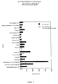


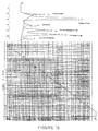


Classifications
-
- C—CHEMISTRY; METALLURGY
- C07—ORGANIC CHEMISTRY
- C07K—PEPTIDES
- C07K14/00—Peptides having more than 20 amino acids; Gastrins; Somatostatins; Melanotropins; Derivatives thereof
- C07K14/435—Peptides having more than 20 amino acids; Gastrins; Somatostatins; Melanotropins; Derivatives thereof from animals; from humans
- C07K14/475—Growth factors; Growth regulators
-
- A—HUMAN NECESSITIES
- A61—MEDICAL OR VETERINARY SCIENCE; HYGIENE
- A61P—SPECIFIC THERAPEUTIC ACTIVITY OF CHEMICAL COMPOUNDS OR MEDICINAL PREPARATIONS
- A61P3/00—Drugs for disorders of the metabolism
-
- A—HUMAN NECESSITIES
- A61—MEDICAL OR VETERINARY SCIENCE; HYGIENE
- A61K—PREPARATIONS FOR MEDICAL, DENTAL OR TOILETRY PURPOSES
- A61K38/00—Medicinal preparations containing peptides
-
- Y—GENERAL TAGGING OF NEW TECHNOLOGICAL DEVELOPMENTS; GENERAL TAGGING OF CROSS-SECTIONAL TECHNOLOGIES SPANNING OVER SEVERAL SECTIONS OF THE IPC; TECHNICAL SUBJECTS COVERED BY FORMER USPC CROSS-REFERENCE ART COLLECTIONS [XRACs] AND DIGESTS
- Y10—TECHNICAL SUBJECTS COVERED BY FORMER USPC
- Y10S—TECHNICAL SUBJECTS COVERED BY FORMER USPC CROSS-REFERENCE ART COLLECTIONS [XRACs] AND DIGESTS
- Y10S436/00—Chemistry: analytical and immunological testing
- Y10S436/815—Test for named compound or class of compounds
-
- Y—GENERAL TAGGING OF NEW TECHNOLOGICAL DEVELOPMENTS; GENERAL TAGGING OF CROSS-SECTIONAL TECHNOLOGIES SPANNING OVER SEVERAL SECTIONS OF THE IPC; TECHNICAL SUBJECTS COVERED BY FORMER USPC CROSS-REFERENCE ART COLLECTIONS [XRACs] AND DIGESTS
- Y10—TECHNICAL SUBJECTS COVERED BY FORMER USPC
- Y10S—TECHNICAL SUBJECTS COVERED BY FORMER USPC CROSS-REFERENCE ART COLLECTIONS [XRACs] AND DIGESTS
- Y10S530/00—Chemistry: natural resins or derivatives; peptides or proteins; lignins or reaction products thereof
- Y10S530/827—Proteins from mammals or birds
- Y10S530/854—Glands
Definitions
- the present invention pertains to the therapeutic use of a peptide molecule derived from maturation products of SMR1 (Submandibular rat protein 1).
- the intracellular or systemic hydro-mineral imbalance of the body of a mammal, and more specifically of the human body is the cause of multiple pathologies affecting the metabolism and the physiological behavior of diverse organs and tissues, such as bone, kidney, parathyroid, pancreas, intestine, the glandular mucosa of the stomach or the prostate as well as salivary glands.
- the maintenance of the transmembrane potassium/sodium and magnesium/calcium ratios is critically important in the control of cell excitation and the regulation of many aspects of intracellular metabolism.
- the most active tissues such as nerve, liver and muscle have a higher ratio of potassium/sodium and magnesium/calcium than inactive tissues such as skin and erythrocytes.
- the most active tissues have a higher phosphorus content than inactive tissues, in keeping with the role of phosphate esters in cellular energy metabolism.
- An adult human contains approximately 1,000 g of calcium (Krane et al., 1970). Some 99% of this calcium is in the skeleton in the form of hydroxyapatite, and 1 % is contained in the extracellular fluids and soft tissues. About 1% of the skeletal content of calcium is freely exchangeable with the extracellular fluids. Although small as a percentage of skeletal content, this exchangeable pool is approximately equal to the total content of calcium in the extracellular fluids and soft tissues, and serves as an important buffer or storehouse of calcium. Thus, calcium plays two predominant physiological roles in the organism. In bone, calcium salts provide the structural integrity of the skeleton.
- mineral ion balance refers to the state of mineral homeostasis in the organism vis-à-vis the environment. In zero balance, mineral intake and accretion exactly match mineral losses; in positive balance, mineral intake and accretion exceed mineral losses, and in negative balance, mineral losses exceed mineral intake and accretion. Under normal circumstances net calcium absorption provides a surplus of calcium that considerably exceeds systemic requirements.
- the extracellular pool of orthophosphate (about 550 mg in human) is in dynamic equilibrium with phosphorus entry and exit via the intestine, bone, kidney and soft tissues. In zero balance, fractional net phosphorus absorption is about two-thirds of phosphorus intake. This amount represents a vast excess over systemic requirements and is quantitatively excreted into the urine.
- the extracellular pool of magnesium (about 250 mg in human) is in bidirectional equilibrium with magnesium fluxes across the intestine, bone, kidney and soft tissues.
- the magnesium derived from the net intestinal absorption (about 100 mg/day in human) represents a systemic surplus and is quantitatively excreted.
- Two organs are mainly involved in the absorption and excretion of the different mineral ions of the body: 1) Hormonal and/or intrinsic mechanisms of mineral ion absorption in the intestine provide the body with a mineral supply that exceeds systemic mineral needs by a considerable measure; 2) the renal tubule plays the dominant quantitative role in maintaining normal mineral homeostasis.
- the 1,25-dihydroxyvitamin D (also named calcitriol) is the only recognized hormonal stimulus of active intestinal calcium absorption that occurs principally in the duodenum and the jujenum (Lemann Jr J., 1993). As a consequence, reduced net intestinal calcium absorption occurs when either dietary calcium intake is limited, when serum 1,25-dihydroxyvitamin D concentrations are low or when the intestine is unresponsive to this hormone. In contrast, increased intestinal calcium absorption occurs when serum 1,25-dihydroxyvitamin D concentrations are high. Thus defects in the regulation of the 1,25-dihydroxyvitamin D concentration in the serum can cause major disorders reducing or enhancing intestinal calcium absorption and lead to a pathological state. The 1,25-dihydroxyvitamin D also influences the body intake of phosphate.
- a second endogenous factor involved in the mineral ion balance is the parathyroid hormone (PTH).
- Parathyroid hormone regulates the level of calcium, and phosphate in blood by modulating the activity of specific cells in bone and kidney. These actions serve to: 1) stimulate reabsorption of calcium and phosphate from bone; 2) stimulate reabsorption of calcium and inhibit reabsorption of phosphate from glomerular filtrate; and 3) stimulate the renal synthesis of 1,25-dihydroxyvitamin D thereby increasing intestinal absorption of calcium and phosphate.
- Calcitonin is a 32-amino-acid peptide that is secreted primarily by thyroidal C-cells (Deftos, 1993). Its main biological effect is to inhibit osteoclastic bone resorption. This property has led to CT's use for disorders characterized by increased bone resorption, such as Paget's disease, osteoporosis and for the hypercalcemia of malignancy.
- the secretion of CT is regulated acutely by blood calcium and chronically by gender and perhaps age. Calcitonin is metabolized by the kidney and the liver. The amino acid sequence of CT is widely conserved through the evolution, from fish to mammals.
- Defects in the mineral ion balance are the cause of multiple disorders affecting either the bone, the kidney, the intestine, the pancreas, the dental tissues (enamel and ivory), or the stomach mucosa.
- a mineral ion imbalance affects the bone remodeling capacity causing disorders such as osteoporosis or affects the bone resorption capacity such as in the hyper-parathyroidism disease.
- the bone remodeling system has been characterized in numerous publications in the recent past (Parfitt, 1986). Bone remodeling occurs on trabecular and Haversian bone surfaces. The first step is activation of osteoclast precursors to form osteoclasts that then begin to excavate a cavity on a surface. After removal of bone tissues (about 0.05 mm 3 ), the site remains quiescent for a short time, following which activation of osteoblast precursors occurs at the site and the excavation is refilled.
- This process serves several functions, among them the removal of aged, microdamaged bone tissue and rearrangement of the bone architecture to meet the needs of mechanical support.
- bone loss, abnormal accumulation of microdamage, or errors in geometry can come about only through defects in this system, for example a defect in the mineral ion balance.
- Osteoporosis is a major public health concern and there is consequently a great need for new therapeutical molecules that will be able to regulate the mineral ion balance in the body and, if possible, more efficient and more selective (target specific) than the molecules presently used in therapeutic, such as oestrogen and calcitonin.
- Other bone absorption or resorption diseases may be caused by defects in the renal or gastrointestinal mineral ion metabolism, such as renal osteodystrophy or even caused by a pancreatic insufficiency.
- Hyperparathyroism is a hypercalcemia occurring in association with elevated levels of parathyroid hormone, often caused by a benign, solitary adenoma.
- Hypercalcemia may be the result of diverse other disorders such as familial hyperparathyroid syndromes (Szabo J. et al., 1993), familial hypocalciuric hypercalcemia (Marx S.J., 1993), hypercalcemia due to malignancy states (Stewart A.F., 1993; Mundy G.R., 1993) or due to granuloma-forming disorders (Adams, J.S., 1993).
- hypocalcemia which is encountered in diseases associated with a low serum albumin concentration or also with idiopathic hypoparathyroidism. Hypocalcemia itself is sometimes due to hypomagnesemia or hyperphosphatemia, or to an impaired secretion of the parathyroid hormone (Shane E., 1993) or also to vitamin D disorders (Insogna K.L., 1993).
- the dental tissues may also be affected in the case of a defect in the mineralization and formation of the dental ivory or enamel.
- Pancreas is also an organ very sensitive to a defect in mineral ion equilibrium, which is able to cause an inflammation named pancreatitis.
- Even the submandibular gland may be affected, causing a pathological state of lithiasis which is associated with calcium deposits.
- the kidney may also be affected, resulting in the development of nephrolithiasis.
- SMR1 submandibular rat 1 protein
- the gene encoding SMR1 belongs to a new multigene family, the VCS family, which has been localized to chromosome 14, bands p21-p22 (Courty et al., 1996; Rosinsky-Chupin et al., 1995) and for which human gene counterpart has been characterized.
- the gene has a similar organization to a number of hormone precursor genes (Rosinsky-Chupin et al., 1990).
- SMR1 mRNA is expressed in a highly tissue- , age- and sex-specific manner in the acinar cells of the male rat submaxillary gland (SMG) and in the prostate (Rosinsky-Chupin et al., 1993).
- SMR1 is selectively processed at pairs of basic amino acid sites in a tissue- and sex-specific manner to give rise to mature peptide products, in a manner similar to the maturation pathway of peptide-hormone precursors (Rougeot et al., 1994). Generally, this selective proteolytic fragmentation has been shown to be critical for the generation and the regulation of biologically active peptides (Lindberg et al., 1991; Steiner et al., 1992). The biosynthesis of the peptides generated from SMR1 or from its human counterpart by cleavage at pairs of arginine residues e.g.
- the undecapeptide: VRGPRRQHNPR; the hexapeptide: RQHNPR; and the pentapeptide: QHNPR is subject to distinct regulatory pathways depending on 1) the organ: SMG and prostate, 2) the developmental stage: from 6 weeks postnatal, 3) the sex: predominantly in the male, and 4) gonad hormones: the androgens. Furthermore, in vivo , the mature peptides which accumulate in the male rat SMG, are exported into the extracellular space in response to a specific external stimulus and, in this way are transported within the salivary and blood fluids (Rougeot et al., 1993).
- the inventors have now discovered that the maturation products of the SMR1 protein, specifically a peptide of structural formula XQHNPR recognize specific target sites in organs that are deeply involved in the mineral ion concentration. This discovery has led the inventors to assign to the SMR1 pentapeptide, hexapeptide or undecapeptide an active role in the regulation of the metal ion concentrations in the body fluids and tissues, and thus a therapeutic role of these peptides in all the metabolic disorders related to a mineral ion imbalance.
- one object of the present invention is the use of the above-described therapeutic peptides for treating bone, teeth, renal, kidney, intestine, pancreas, stomach mucosa or parathyroid disorders caused principally by a mineral ion imbalance in the body fluids or tissues.
- the therapeutic peptides according to the present invention are used for preventing or treating diseases like hyper- or hypo-parathyroidism, osteoporosis, pancreatitis, submandibular gland lithiasis, nephrolithiasis or osteodystrophy.
- Such biologically active derivatives of the therapeutic peptides according to the invention are peptides that are structurally and chemically related to the XQHNPR, such as peptides having the same amino acid sequence as the initial peptides but containing one or several modified amino acids that are able to confer a better in vivo stability to the therapeutically active molecule and which possess the same biological activity as the endogenous peptide or which behave as an antagonist molecule of the endogenous peptide.
- homologous peptide a peptide containing one or several amino acid substitutions in the XQHNPR sequence.
- the amino acid substitution consists in the replacement of one or several -consecutive or non-consecutive- amino acids by "equivalent” amino acids.
- the expression "equivalent” amino acid is used herein to name any amino acid that may substituted for one of the amino acids belonging to the initial peptide structure without modifying the hydrophilicity properties and the biological target of the initial peptide structure.
- the peptides containing one or several "equivalent” amino acids retain their specificity and affinity properties to the biological targets of the XQHNPR peptide.
- the "equivalent" amino acids are those which allow the generation or the obtention of a polypeptide or peptide with a modified sequence as regards to XQHNPR, said modified polypeptide or peptide being able to act as an agonist or an antagonist molecule of the XQHNPR peptide.
- amino acids may be determined by their structural homology with the initial amino acids to be replaced and by their biological activity on the target cells of the XQHNPR peptide.
- modified amino acid according to the present invention is also meant the replacement of a residue in the L-form by a residue in the D form or the replacement of the glutamine (Q) residue by a Pyro-glutamic acid compound.
- the synthesis of peptides containing at least one residue in the D-form is, for example, described by Koch et al. in 1977.
- the peptides used according to the present invention may be prepared in a conventional manner by peptide synthesis in liquid or solid phase by successive couplings of the different amino acid residues to be incorporated (from the N-terminal end to the C-terminal end in liquid phase, or from the C-terminal end to the N-terminal end in solid phase) wherein the N-terminal ends and the reactive side chains are previously blocked by conventional groups.
- a highly porous resin polymer is used, on which the first C-terminal amino acid of the chain is fixed.
- This amino acid is fixed to the resin by means of its carboxyl groups and its amine function is protected, for example, by the t-butyloxycarbonyl group.
- the protective group is removed from the amine function by washing the resin with an acid. If the protective group for the amine function is the t-butyloxycarbonyl group, it may be eliminated by treating the resin with trifluoroacetic acid.
- the second amino acid which supplies the second residue of the desired sequence is then coupled to the deprotected amine function of the first C-terminal amino acid fixed to the chain.
- the carboxyl function of this second amino acid is activated, for example, using dicyclohexylcarbodiimide, and the amine function is protected, for example, using t-butyloxycarbonyl.
- the first part of the desired peptide chain is obtained, which comprises two amino acids, the terminal amine function of which is protected.
- the amine function is deprotected and the third residue can then be fixed, under similar conditions, to those used in the addition of the second C-terminal amino acid.
- amino acids which are to form the peptide chain are fixed, one after another, to the amine group, which is previously deprotected each time, to the portion of the peptide chain already formed, which is attached to the resin.
- the protecting groups are eliminated from the various amino acids which constitute the peptide chain and the peptide is detached from the resin, for example using hydrofluoric acid.
- the peptides thus synthesized may also be a polymer of the XQHNPR peptide, that contains 2 to 20 monomer units of the amino acid sequence XQHNPR, preferably 4 to 15 monomer units and more preferably 5 to 10 monomer units.
- the polymers may be obtained by the technique of Merrifield or any other conventional peptide polymer synthesis method well known by a person skilled in the art.
- the peptides thus obtained may be purified, for example by high performance liquid chromatography, such as reverse phase and/or cationic exchange HPLC, as described by Rougeot et al. in 1994.
- the peptides used in the therapeutic method according to the present invention may also be obtained using genetic engineering methods.
- the nucleic acid sequence of the cDNA encoding the complete 146 amino acid SMR1 protein has been described in the PCT Patent Application No. WO 90/03891 (Rougeon et al. )
- WO 90/03891 Rosugeon et al.
- a person skilled in the art will refer to the general literature to determine which appropriate codons may be used to synthetize the desired peptide.
- nucleotide sequence For the genetic expression of the XQHNPR peptide, the following nucleotide sequence may be used, based on the general codon usage in mammals: Peptide V R G P R R Q H N P R DNAGTC AGA GGC CCA AGA AGA CAA CAT AAT CCT AGA or a nucleotide sequence hybridizing with the above sequence under stringent conditions and which encodes a peptide having qualitatively the same mineral ion regulation activity than the QHNPR peptide.
- the hybridization step is performed at 65°C in the presence of 6 x SSC, 5 x Denhardt solution, 0.5% SDS and 100 ⁇ g/ml salmon sperm DNA.
- the washing steps consist of:
- the expression of the polynucleotide that encodes the XQHNPR may be optimized, according to the organism in which the sequence has to be expressed and the specific codon usage of this organism (mammal, plant, bacterium etc.) For bacteria and plants, respectively, the general codon usages may be found in European Patent Application No. EP-0359472 (Mycogen ).
- the present invention also embraces the production by genetic engineering techniques of the SMR1 protein or one of its maturation products.
- the SMR1 protein (which can be considered as the precursor of the different maturation products) is processed by furin.
- Furin is a subtilisin-like convertase involved in postranslation endoproteolytic processing of various pro-hormones.
- furin may be advantageously used in combination with a precursor of the XQHNPR peptide in order to obtain the corresponding maturation product.
- a method for producing the SMR1 protein or one of its maturation products such as a XQHNPR peptide of the invention, or also a peptide containing "equivalent" amino acids as described above comprises the steps of:
- a suitable vector for the expression of the SMR1 protein and the convertase protein above-defined is baculovirus vector that can be propagated in insect cells and in insect cell lines.
- a specific suitable host vector system is the pVL1392/1393 baculovirus transfer vector (Pharmingen) that is used to transfect the SF9 cell line (ATCC N°CRL 1711) which is derived from Spodoptera frugiperda .
- Another suitable vector for performing the above-described process is a vaccinia virus vector.
- BSC-40 or LoVo are used for the transfection and culture steps.
- the purification of the recombinant protein may be realized by passage onto a Nickel or Copper affinity chromatography column.
- the Nickel chromatography column may contain the Ni-NTA resin (Porath et al., 1975).
- the PCR amplification reaction was described by Saiki et al. in 1985;
- the SDA technique was described by Walker et al. in 1992 and was improved by Spargo et al. in 1996;
- the TAS amplification reaction was described by Kwoh et al. in 1989;
- the 3SR technique was described by Guatelli et al. in 1990;
- the NASBA technique was described by Kievitis et al. in 1991;
- the LCR reaction was described by Landergen in 1991 and improved by Barany et al. in 1991;
- the RCR technique was described by Segev in 1992;
- the CPR technique was described by Duck et al. in 1990.
- the peptides produced by genetic engineering methods according to the invention may be characterized by binding onto an immunoaffinity chromatography column on which polyclonal or monoclonal antibodies directed to XQHNPR have previously been immobilized.
- the peptide of therapeutic value contained in the therapeutic compositions according to the present invention is purified by HPLC as described by Rougeot et al. in 1994.
- the reason to prefer this kind of peptide or protein purification is the lack of side products found in the elution samples.
- the antibodies may be prepared from hybridomas according to the technique described by Kohler and Milstein in 1975.
- the polyclonal antibodies may be prepared by immunization of a mammal, especially a mouse or a rabbit, with a peptide according to the invention that is combined with an adjuvant of immunity, and then by purifying the specific antibodies contained in the serum of the immunized animal onto an affinity chromatography column on which has previously been immobilized the peptide that has been used as the antigen.
- a technique for preparing and using an immunoaffinity chromatography column was described, for example, by Bird et al. in 1984.
- a preferred embodiment for preparing antibodies raised against the SMR1 protein or its maturation products is described hereafter, using the QHNPR pentapeptide as an example.
- the QHNPR peptide is conjugated to egg albumin (Calbiochem) using the benzidine-bis-diazoted procedure described by Gregory et al. in 1967, the ratio of peptide residues to one molecule of ovalbumin being 5:1.
- Rabbits are injected at time 0 with 1 mg of the conjugated peptide.
- Two months after the primary injection animals are injected with 0.5 mg of the conjugated peptide and a third injection of 0.5 mg of the same peptide is performed between two and four months after the second injection.
- Antiserum is harvested between two and four weeks following the third conjugated peptide injection and optionally purified onto an affinity chromatography column as previously described.
- the injection is an intradermally multi-points injection; generally ten points of injection are performed.
- biologically active derivatives of the therapeutic peptides of the invention consist in molecules that are either or both structurally or chemically unrelated to the endogenous peptides but bind to the same specific targets and have an agonist or an antagonist activity.
- the biologically active derivatives of the therapeutic peptides used in the therapeutic method according to the present invention are endowed with a longer in vivo half-life than their natural endogenous counterparts.
- the biologically active derivative of the XQHNPR peptide may be a protein, a peptide, a hormone, an antibody or a synthetic compound which is either a peptide or a non peptidic molecule, such as any compound that can be synthesized by the conventional methods of organic chemistry.
- Selection of the biologically active derivatives of the XQHNPR peptide of the invention is performed both in assessing the binding of a candidate ligand molecule to the known target cells or organs of the XQHNPR peptide, specifically the QHNPR pentapeptide, and in determining the metabolic changes induced by this candidate molecule on its target, such as the synthesis of the primary or secondary messenger metabolites as a result of a transduction signal via the protein kinases or adenylate cyclase and the activation of a protein of the G family.
- the binding of the candidate molecule to primary culture cells, established cell lines or fresh tissue samples (cryosections or slices) is performed as described hereafter.
- Binding assays of the candidate molecule are generally performed at a cold temperature (4°C). However, variations of the standard procedure described below involving incubation at 37°C are useful to assess the internalization and fate of the pentapeptide or one biologically active derivative candidate ligand molecule upon binding at the cell surface (Wakefield, 1987). In order to facilitate the reading of the hereinafter described protocol, QHNPR pentapeptide is used instead of a biologically active derivative candidate molecule.
- the candidate ligand molecule may be radioactively labeled ( 32 P, 35 S, 3 H, 125 I etc..) or nonradioactively labeled (biotin, digoxigenin, fluorescein etc..)
- the materials used to perform the above binding process are:
- the radio-iodinated label of QHNPR is realized as follows:
- the peptide QHNPR is labeled according to the method of Greenwood et al., 1963, using 1mCi Na 125 I (Amersham), 0.8 ⁇ g (1 nmol) pentapeptide and 15 ⁇ g chloramin T (Fluka) in 50 ⁇ l sodium borate, pH 8.
- the mixture is chromatographed by GF05 gel filtration (Pharmacia-LKB) to isolate the 125 I-Labelled pentapeptide and is subsequently chromatographed on reverse-phase porapak QTM (Waters-Millipore) to elute the mono-iodinated-labelled pentapeptide.
- the specific radioactivity corresponding to one atom of radioiodine/peptide molecule, is estimated as 1500 Ci/mmol.
- the assay by itself, comprises the following steps:
- step a) By about one hour incubation according to step a) above, it is intended a time period of incubation that is sufficient for the QHNPR or labeled QHNPR to bind to its specific target sites on the cell, taking into account that the assay is performed at a temperature of 4°C, thus at a temperature for which the membrane fluidity is low. Consequently, the appropriate incubation time period is comprised between 1 and 12 hours (overnight).
- receptor binding methods may also be used in order to select biologically active derivatives of the XQHNPR peptide of the invention such as those described by Whitcomb et al. in 1993, Epelbaum et al. in 1993, Ricci et al. in 1993, Loring et al. in 1993, Walsh et al. in 1995, Roberts et al. in 1995 or also Wu et al. in 1996 the binding assays described in the aforementioned articles being herein incorporated by reference.
- a candidate ligand molecule of interest for the purpose of the present invention that has been positively selected according to the previously described binding assay and in order to determine if this selected competitive ligand molecule acts as an agonist or as an antagonist molecule of the XQHNPR peptide of the invention
- different metabolic assays may be performed.
- an adenylate cyclase assay that is performed either on primary cultures of target cells or on cell lines or on target tissue homogenates.
- tissue homogenates or cultured cells (1-100 ⁇ g protein) are incubated in 50 mM of Tris/HEPES buffer, pH 7.5, containing 1 mM Mg SO 4 , 1 mM EGTA, 1 mM 3-isobutyl-1-methylxanthine, 0.1% (wt/vol) BSA, 1 mM ATP, 25 mM creatine phosphate, 260 U/ml creatine kinase (E.C. 2.7.3.2), and 6.5 U/ml myokinase (E.C. 2.7.4.3) in a final vol of 100 ⁇ l for 10 min at 37°C. The reaction is stopped by addition of 100 ⁇ l 0.5% (wt/vol) SDS and boiling for 2 min. cAMP is extracted with Dowex 50WX-8 and analyzed by RIA (Stangi et al., 1993).
- the quantity of cAMP produced in response to the presence of the candidate ligand molecule (10 -10 -10 -5 M. range) is assessed as described above.
- a stimulation of adenylate cyclase activity which is equivalent, when used alone, or greater, when associated with suboptimal concentration of QHNPR, than the stimulation of adenylate cyclase activity induced by XQHNPR alone, and more specifically QHNPR alone, is considered as positive and the candidate ligand molecule is thus classified as an agonist of the XQHNPR peptide of the invention.
- the quantity of cAMP produced in the presence of both XQHNPR, specifically QHNPR, and the candidate ligand molecule is assessed. If the presence of the candidate ligand molecule is able to abolish or block the stimulation of adenylate cyclase activity induced by XQHNPR, specifically, QHNPR, then the candidate ligand molecule is classified among the antagonist compounds of the peptide according to the invention.
- quantification of the adenylate cyclase activity represents only one embodiment of the screening methods according to the present invention.
- a person skilled in the art would have understood that diverse metabolic changes of the cell physiology may be visualized in order to assess the agonist or antagonist activity of the candidate ligand molecule to be assayed.
- Other metabolic change quantification assays are also encompassed by the present invention, such as changes in the kinases activity, in the GTP-G proteins dependent activity, in the production of cell- or tissue-specific metabolites such as collagenase by osteogenic cell types.
- the present invention also pertains to a process for screening ligand molecules that possess an agonist biological activity on the target receptor of the XQHNPR pentapeptide, comprising the steps of:
- Another object of the present invention comprises a process for screening ligand molecules that possess an antagonist biological activity on the target receptor of the RQHNPR pentapeptide, comprising the steps of:
- another metabolic assay in order to assess the agonist or the antagonist activity of the candidate ligand molecule comprises incubating the ligand candidate in the presence of an osteogenic primary cell culture or cell line and determining, either or both quantitatively and qualitatively, the collagen produced in response to the in vitro stimulation (with 10 -10 - 10 -5 M range concentration of the molecule to be tested).
- the binding assay and the metabolic assay may be performed on primary cell cultures, on established cell lines or on fresh tissue slices, tissue cryosections or tissue homogenates.
- a preferred cell line that is used in the screening methods according to the present invention is an osteogenic clonal mesodermal cell line from mouse, named C1, which is derived from mouse teratocarcinoma. It expresses the SV40 oncogenes under the control of the adenovirus E1a promoter (Chentoufi et al., 1993; Kellermann et al., 1990; Poliard et al., 1993; Poliard et al., 1995).
- clonal cell lines consist in various clonal cell lines derived from the same mouse strain from which the clonal cell line C1 above has been initially derived, more specifically, clonal cell lines derived from the epithelial cell of renal proximal tubules.
- Confluent osteogenic cells specifically the C1 cell line described above, are labeled for 24 h with 50 ⁇ Ci/ml of 4.5 [ 3 H]proline (32 Ci/mmol; Commissariat à I'Energie Atomique, Saclay, France) in DME supplemented with 1 % FBS, 100 ⁇ g/ml ascorbic acid, and 50 ⁇ g/ml b-aminopropionitrile fumarate (Sigma Chemical Co., Saint Louis, Missouri, USA).
- the cells are collected in 50 mM Tris-HCl (pH 7.4) containing 150 mM NaCl, 25 mM EDTA, 10 mM N-ethylmaleimide, and 2 mM PMSF, and pooled with the culture medium. After sonication and addition of trichloroacetate (TCA) (10%), the insoluble residue is collected by centrifugation.
- Tris-HCl pH 7.4
- TCA trichloroacetate
- Collagen types are determined by PAGE: pellets resuspended in 0.5 M acetic acid are digested with 100 ⁇ g/ml pepsin fumarate (Sigma Chemical Co., Saint Louis, Missouri, USA), pH 2.0 for 4 h at 4°C. The pepsin digest brought to pH 8.0 is dialyzed and lyophilized. Samples are then analyzed by SDS-PAGE. Delayed reduction with 2-mercaptoethanol is used in some samples. Labeled proteins are visualized by fluorography or autoradiography and quantified by a beta radioimager device.
- the non-radioactive beta-glycerophosphate may be replaced by a radioactive molecule.
- the phosphorous compound conventionally used as bone localizing agent is an inorganic phosphate or pyrophosphate.
- a radioactive molecule that can be used in the above assay is tetrasodium 32 P pyrophosphate (NEX 019, New England Nuclear) or 99 technetium that binds to the phosphorous compound.
- Another object of the present invention comprises a method for screening ligand molecules that possess an agonist or an antagonist biological activity on the target receptor of the XQHNPR pentapeptide, comprising the steps of:
- the eukaryotic cell of the above method may be either a cell that has been transfected or transformed with a nucleic acid coding for collagen, or a eukaryotic cell that is able to synthesize collagen in a constitutive or in an inducible manner.
- a preferred cell is a mammalian cell that is able to naturally synthesize collagen, such as the C1 cell line.
- Enzyme assays that may be performed comprise acetylcholinesterase ( Ellman G.L. et al., 1961, Biochem. Pharmacol., 7:88 ), cathepsin ( Barret A.J. et al., 1981, Methods Enzymol., 80:535 ), ATPase, cyclooxygenase ( Mitchell J.A. et al., 1993, Proc. Natl. Acad. Sci., 90:11693 ), guanylate cyclase ( Wolin M.S.
- Ion transport assays comprise Ca pump ( Jean T. et al., 1986, J. Biol. Chem., 261:16414 ), Ca channel ( Galizzi J.P. et al., 1987, J. Biol.
- Signal transduction assays that may be also used comprise Ca release ( Grynkievicz G. et al., 1985, J. Biol. Chem., 260:3440 ) or PI turnover ( White T.E. et al., 1993, Br. J. Pharmacol., 108:196 ).
- Ca release Grynkievicz G. et al., 1985, J. Biol. Chem., 260:3440
- PI turnover White T.E. et al., 1993, Br. J. Pharmacol., 108:196 .
- An object of the present invention is also a method for screening ligand molecules that possess an agonist or an antagonist biological activity on the target receptor of the XQHNPR pentapeptide, comprising the steps of:
- the metabolic change referred to in the above screening method is interchangeably a change in the production of any of the metabolites that are assayed according to the above-refrenced methods. Consequently, the time that is sufficient for the metabolic change to take place will depend on the assayed metabolite and is indicated, for a particular metabolite, in each article reference that has been given hereinbefore.
- tissue slices, tissue cryosections or tissue homogenates are used in the performing of the binding or metabolic assays in order to select the ligand molecules of interest that represent the biologically active derivatives of the XQHNPR, specifically QHNPR, peptide according to the present invention.
- Preferred tissue samples are of kidney, pancreas, bone, dental, glandular mucosa of stomach, prostate and intestine origin as well as salivary glands.
- Tissue homogenates are prepared by first immersing biopsies in liquid nitrogen. The frozen samples are then homogenized in 20 vol ice-cold 50 mM Tris/HEPES buffer, pH 7.5, containing 1 mM EGTA, 0.3% (wt/vol) bacitracin, 20 mM 4-amidinophenylmethanesulfonyl fluoride, and 10 mM leupeptin with a motor-driven Teflon-glass homogenizer. The homogenates are centrifuged at 24,000 x g for 20 min at 4°C and the pellets washed once with 50 mM Tris/HEPES, pH 7.5, containing 1 mM EGTA.
- the final pellets are suspended in 50 mM Tris/HEPES pH 7.5 containing 1 mM EGTA and 0.3% (wt/vol) bacitracin, and stored at -20°C. Further details of procedure are described by Stangi et al in 1993, depending on the origin of the tissue to homogenize, the procedures used by Stangi being herein incorporated by reference.
- tissue samples for example of kidney origin
- the following procedure is used: Before organ or tissue harvesting, for example in rat, the animal is anesthetized with chloral hydrate (35%, 0.14 ml/100g body weight) or with pentobarbital (45 mg/kg body weight). Then, an intracardiac perfusion with 100 ml 4°C isotonic sucrose is performed (Loring et al., 1993).
- Kidneys may be obtained after human biopsies or from male Wistar rats immediately after death by cervical dislocation, or anesthesy as described above. Tissue samples are immediately embedded in Tissue-Tek (Miles, Elkhart, IN) and frozen to cork mounts in melting isopentane (at -70°C) without prior fixation. Specimens are stored at -70°C until use. 10-30- ⁇ m thick sections are cut in a cryostat (at a temperature range of -20°C to - 30°C) and are thaw-mounted onto uncoated low-iron glass slides. They are then used immediately or vacuum-dried and stored at -20°C with silica gel for up to 5 days until use. Such a technique was described by Walsh et al. in 1995, the details of which are herein incorporated by reference.
- tissue sections are frozen in dry ice in the presence of isopentane and thaw-mounted onto microscope slides, and stored at -80°C until use (Whitcomb et al., 1993).
- specimens of human renal cortex and medulla are obtained from subjects undergoing nephrectomy.
- the portions of the kidney collected are placed in ice-cold 0.9% NaCl solution to remove blood and cell debris.
- Blocks including the renal cortex and medulla or separate samples of the cortex or of the medulla are embedded in a cryoprotectant medium (OCT, Ames, IA) and frozen in isopentane cooled with liquid nitrogen or dry ice.
- OCT blocks are stored at -80°C until use.
- Serial 8- ⁇ m thick sections are obtained using a -20°C microtome cryostat, mounted on gelatin-coated microscope slides and air-dried. Such a technique is described by Ricci et al. in 1993.
- Preferred biologically active derivatives of the XQHNPR peptide of the therapeutic composition according to the present invention have better pharmacokinetics than the endogenous natural or synthetic XQHNPR peptide and thus possess a longer in vivo half-life as compared to their natural counterparts.
- a method for assessing the pharmacokinetics of the ligand molecules selected according to the binding assay and the metabolic assay above described is exemplified in Example 3.
- plasma clearance of the selected ligand molecules is determined according to the technique described by Wu et al. in 1996 or the technique described by Ezan et al. in 1986, or by Ezan et al., 1996, which techniques are herein incorporated by reference.
- the invention also concerns the biologically active derivatives of the XQHNPR peptide that have been selected according to the screening processes hereinbefore described, provided that they have not the following structure: Y-HNP-Z, wherein Y denotes a glutamine (Q) or a pyroglutamic acid residue and Y represents an OH group or a basic amino acid, the basic amino acid being a Lysine (K) or an Arginine (R).
- Y-HNP-Z wherein Y denotes a glutamine (Q) or a pyroglutamic acid residue and Y represents an OH group or a basic amino acid, the basic amino acid being a Lysine (K) or an Arginine (R).
- Y-HNP-Z wherein Y denotes a glutamine (Q) or a pyroglutamic acid residue and Y represents an OH group or a basic amino acid, the basic amino acid being a Lysine (K) or an Arginine (
- the biologically active derivatives of the XQHNPR peptide used in the therapeutic compositions according to the present invention have been, in a preferred embodiment, selected firstly according to their ability to bind to the same targets as the XQHNPR, specifically QHNPR, peptide and secondly by their capacity to induce metabolic changes (adenylate cyclase activity stimulation, collagen production, etc.) within the target or to abolish, block or prevent said metabolic changes of the target that are induced by the XQHNPR, specifically QHNPR, peptide.
- the present invention also deals with the use of therapeutic compositions comprising an effective amount of the XQHNPR peptide or of one of its biologically active derivatives.
- the use of the therapeutic compositions according to the invention is indicated in case of an hydro-mineral imbalance of the body.
- These use of the compositions is also indicated in the case where a defect in the normal endogenous level of the QHNPR pentapeptide is diagnosed from a biological sample of the patient, particularly from patient fluids such as serum or plasma, saliva and urine.
- the level of pentapeptide concentration in the biological sample is considered defective when it is at least five-fold less important than the non pathological concentration usually found as well as when it is at least five-fold more important than the non pathological concentration usually found in this particular biological sample.
- a diagnostic method that allows determination of the concentration of the QHNPR pentapeptide in a biological sample, specifically from serum or plasma, is a competitive radioimmunoassay (RIA), as described by Rougeot et al. in 1994.
- RIA competitive radioimmunoassay
- the liquid phase RIA is performed in 0.2 M Tris/HCl pH 8.5, containing 0.25% bovine serum albumin (Fr 5, Miles), 0.1% Triton X-100 (Sigma), 1000 KIU (kallikrein international unit)/ml Trasylol (a trypsin and kallikrein inhibitor; Bayer), EDTA (1 mM) and 0.1 g/l NaN 3 .
- Standard or sample (0.1 ml), 0.1 ml diluted anti-pentapeptide serum (Rougeot et al., 1994) and 125 I-Labelled pentapeptide (15 x 10 3 dpm, 0.1 ml) are incubated overnight at 4°C.
- Bound and free fractions are separated by propanol precipitation (10 ⁇ l normal rabbit serum and 1 ml ice-cold 1-propanol) and the radioactivity of the precipitate is determined using a gamma counter.
- the plasma, saliva or urine samples would be collected into previously cooled tubes containing a mixture of peptidase inhibitors (1mM EDTA, 1000 U/ml Aprotinin, 130 ⁇ M bestatin, 1 ⁇ M leupeptin, 0.4 mM pefabloc, 1 ⁇ M pepstatin).
- this diagnostic test is also an object of the present invention.
- the successive steps of this diagnostic test are the following:
- the amount of therapeutic molecule to be administered is calculated, taking into account the specific in vivo pharmacokinetics of said therapeutic molecule, the in vivo pharmacokinetics being assessed as described in Example 3 and, of course, the normal amount of XQHNPR, specifically QHNPR, peptide normally found in the body fluids (plasma, saliva, urine) or organs.
- the labelled XQHNPR peptide used in the diagnostic test is radioactively or non-radioactively labelled.
- the polyclonal antibody used in this diagnostic method may be optionally under the form of an immune serum.
- the particular quantitative immunodetection system designed by the inventors is both extremely specific and sensitive: 50% displacement of the 125 I-Labelled pentapeptide QHNPR is obtained with no more than 570 fmol pentapeptide standard ([Glp 1 ] pentapeptide or [Gln 1 ] pentapeptide.
- the detection limit (80% binding) is 106 fmol standard peptide.
- the cross-reactivity of the antiserum with tetrapeptide sequence (QHNP) and thyrotropin-releasing hormone (Glp-His-Pro) is less than 0.01 %.
- the XQHNPR peptide concentrations quantified according to the above described diagnostic method are used to determine the amount of the XQHNPR peptide or of one of its biologically active derivatives to be administered to the patient in order to supply for the diagnosed defect, in case of a low level in the endogenous peptide and to determine their pharmacokinetic parameters in pathologic and non-pathologic subjects.
- the therapeutic compositions containing the SMR1 protein or SMR1 maturation products or their derivatives and used according to the present invention may be either under the form of a liquid solution, under the form of a gel or under the form of a dry powder.
- the therapeutic peptides according to the present invention are advantageously complexed with metal salt ions such as Ca ++ , Cu ++ , Ni ++ , Mg ++ or AI ++ either directly or indirectly with the carrier ions proteins or also calcium phosphate as described in the French Patents N° FR-2,543,439 and N° FR-2,181,426 (Reliveld et al. ), or also with ATP.
- metal salt ions such as Ca ++ , Cu ++ , Ni ++ , Mg ++ or AI ++ either directly or indirectly with the carrier ions proteins or also calcium phosphate as described in the French Patents N° FR-2,543,439 and N° FR-2,181,426 (Reliveld et al. ), or also with ATP.
- the therapeutic peptides according to the present invention may also be complexed to Dextran or Dextran derivatives.
- LA long Action
- RLA Renewable Long Action
- the LA form consists of microspheres of polylactic acid that contain bromocriptin included in Dextran.
- bromocriptin is contained in microspheres of D, L-polylactide-coglycolide-glucose, the degradation of which is almost complete in a period of time of less than three months and which allows repeating the administration numerous times for a long period of time (Montini et al., 1986; Kato et al., 1988).
- the therapeutic compositions according to the present invention are administered locally, near the site, organ or tissue, to be treated.
- the therapeutic compositions of the invention are administered by the oral route in order to act systemically or at the level of the buccal or stomach mucosa or at the level of the gastro-intestinal tract.
- Such therapeutic compositions may be in the form of a saline solution or a tablet, preferably a controlled release tablet.
- a typical controlled release tablet is described in the PCT Patent Application No. WO 9622768 , which contains from about 30 to about 70 percent by weight of one or more cellulose ethers such as hydroxypropyl methylcellulose, and from about 30 to about 70 percent by weight of an inert substance such as corn starch.
- the XQHNPR peptide or its biologically active derivatives are included in a controlled release device to be placed locally in the body, in order to obtain a sustained delivery of the active molecules in the surrounding of the site to be treated.
- the controlled release devices that are used for the purpose of the present invention are lipid or polymer microparticles that dissolve or are hydrolyzed slowly within the body, specifically in the stomach or in the gastro-intestinal tract.
- the latter can be implanted locally in order to ensure a limited area diffusion of the active molecule, surrounding the organ or tissue to be treated.
- Preferred sustained delivery devices according to the present invention contain biodegradable polymers such as described in the PCT Patent Application No. WO 9701331 .
- the polymer may be a polysaccharide as in the PCT Patent Application No. WO 9613253 , such as sodium alginate.
- a biodegradable sustained preparation is preferably composed of a polysaccharide which is coated with cationic molecules such as chitosan, the carrier being slowly enzymatically hydrolyzed, for example by lysozyme, in vivo after the release of the active molecule.
- the polymer used in the controlled release devices according to the present invention may also be a polyvinylpyrrolidone type polymer, such as described in the PCT Patent Application No. WO 8804922 or a starch hydrolysate, such as described in the PCT Patent Application No. WO 9417676 .
- the polymer is a bioadhesive polymer such as carboxymethylcellulose, CarbopolTM, PolycarbophilTM or sodium alginate, that binds with an excellent efficiency to the mucin present at the surface of the epithelium (Robinson et al., 1998), these polymers being used especially in the case of an oral drug delivery.
- a bioadhesive polymer such as carboxymethylcellulose, CarbopolTM, PolycarbophilTM or sodium alginate
- sustained delivery devices are under the form of polymer microbeads, for example porous crosslinked polymeric microbeads, such as described in the PCT Patent Application No. WO 9533553 .
- Another embodiment of the controlled release devices according to the present invention are liposomes either in a hydrated form, such as in the PCT Patent Application No. WO 8601102 or in the PCT Patent Application No. WO 9522961 (Capron et al.), or in a dehydrated form, such as in the PCT Patent Application No. WO 8601103 .
- Other lipid emulsions used as drug delivery systems that may be used for the purpose of the present invention are described by Davis et al. in 1988, that may be administered via the oral, parenteral or the intravenous route.
- the liposomes may contain saccharide determinants that bind to specific cell membrane components in order to facilitate the delivery of the active molecule towards a selected target cell, in particular saccharide determinants that bind to specific lectins of the cell membrane (Shen, 1988).
- sustained delivery formulations used according to the present invention consists of a particle vector comprising, from the inner layer to the outer layer:
- the peptide XQHNPR of the invention may be administered to the skin via a transdermal iontophoretic delivery system, such as described by Chien et al. in 1988.
- compositions used according to the present invention contain a pharmaceutically effective amount of the XQHNPR peptide or of one of its biologically active derivatives.
- the amount of active principle contained in one therapeutic dosage of the XQHNPR peptide or biologically active peptide derivative is contained in the range between 10 ⁇ g/kg and 10 mg/kg of body weight, preferably between 50 ⁇ g/kg and 5 mg/kg of body weight and more preferably between 200 ⁇ g/kg and 1 mg/kg of body weight.
- One object of the present invention is also a therapeutic composition
- the preferred molecules to be associated with the SMR1 protein or derivatives are the parathyroid hormone (PTH), calcitonin (CT) and 1,25-dihydroxyvitamin D.
- the inventors have mapped the peripheral targets for the final secretory maturation product of SMR1, the pentapeptide, by examining in vivo the gross tissue distribution of the radiolabeled peptide with whole body autoradiography (WBA) (Ullberg et al., 1981).
- WBA whole body autoradiography
- This method provides a visual image of the entire animal, therefore rendering it possible to examine simultaneously the uptake of the pentapeptide in a large number of tissues and compartments.
- this approach is considerably more attractive than an in vitro method, since labeling in living tissue minimizes the possibility of misinterpretation due to non-specific distribution, to receptor site degradation and to nonreceptor site-bound peptide eliminated through the bloodstream (Lindberg et al., 1991).
- the time-related tissue uptake for the pentapeptide has been assessed in vivo.
- labeling has been achieved by intravenously infusing tritiated pentapeptide in five weeks old pubescent rats.
- the mapping and the time course of the tritiated peptide uptake by the living organs has been recorded by the recently developed ⁇ -radio imager, which offers the unique capability of detecting and quantifying the ⁇ particles emitted from tritiated ligand bound to flat surfaces such as whole body sections (Charpak et al., 1989; Tribollet et al., 1991).
- the inventors have also analyzed the chromatographic characteristics of the labeled compound associated with the target organ.
- Bio receptor sites have two essential characteristics that allow specific binding to be identified, in vivo; they have a high affinity for the ligand and the binding is saturable (Whitcomb et al., 1993). Therefore, displacement experiments, using parallel systemic administration of an excess of the corresponding unlabeled peptide has been performed. Furthermore, the specific cellular localization of labeled pentapeptide to target tissues has been identified by section microscopic autoradiography.
- Wistar male rats (4 weeks old), purchased from Iffa-Credo (France), were kept at 2-4 animals/cage under controlled lighting and temperature with free access to food and water, until use 5-7 days later for the study of pentapeptide receptor-site distribution and 5-6 weeks later for the study of endogenous pentapeptide secretion. They were gently handled daily by the operator throughout this period. All experiments were performed between 10.00 and 14.00 h.
- Labeled compound i.e. 6.66 MBq, 3 nmoles or 2 mg for whole body autoradiography (WBA) and 370-555 KBq, 170-250 pmoles or 110-160 ng for the pharmacokinetic and cellular-site distribution studies was diluted in 100-200 ml of phosphate-buffered saline (PBS Dulbecco's, Bio Media, France) and administered intravenously into the jugular vein of anesthetized rats (halothan for WBA or pentobarbital for all the other experiments).
- PBS Dulbecco's phosphate-buffered saline
- the target tissues for the final secretory maturation product of SMR1, the pentapeptide was examined, in vivo , by the whole body autoradiography procedure according to the method of Ullberg (Ullberg et al., 1981).
- the five week-old rats were sacrificed under halothane, at 90 sec., 3, 60 and 240 min. after i.v. injection of 3 nmoles (6.66 MBq or 2 ⁇ g) of tritiated pentapeptide.
- 3 nmoles (6.66 MBq or 2 ⁇ g)
- the restrained animal was immediately immersed in a -80°C mixture of dry ice and isopentane, to prevent artefactual tracer redistribution.
- the cellular-site localization for physiological concentrations of labeled pentapeptide was investigated by paraffin- or resin-section microscopic autoradiography procedure. Sixty minutes after 3 H-pentapeptide (555 KBq, 160 ng or 250 pmoles) i.v. administration, the anesthetized male rats were perfused via the jugular veins with ice-cold Krebs Ringer Bicarbonate Glucosed buffer and then 0.5% paraformaldehyde/0.5% glutaraldehyde fixative PBS buffer (plus glucose 1.6%, CaCl 2 0.002% and DMSO 1%) (50 ml/5 min. for each).
- Tissues were rapidly removed and harvested for paraffin sectioning (Paraplast plus-Sherwood medical, OSI, France) or for resin sectioning (histo-resin Leica, France). All sections were cut at 5 microns (Reichert Jung for paraffin and RM2155 Leica for resin sections) and mounted on Superfrost/Plus glass slides (non gelatin-coated). Paraffin was removed with xylene, and sections were carried through a descending, then an ascending ethanol series (from 100 to 50%, V/V and reverse). Dried sections were then processed for light microscope autoradiography by attaching nuclear emulsion-coated (Kodak, NTB2, diluted 1:1 with distilled water) coverslips to them.
- the quantitative determination of the radioactivity in various organ sections was carried out by using a gaseous detector of ⁇ -particles, which has recently been developed (Charpak et al., 1989; Tribollet et al., 1991). Data from whole body sections or individual organ sections placed in the gas-chamber detector were collected for 8 hr. or 50 hr., respectively. The number of counts per pixel was recorded in the ⁇ imager 2200 (Biospace, France) with a surface detector of 20 x 20 cm 2 , followed by computer-assisted image analysis with the ⁇ Vision program using a HP Vectra computer. The tritium activity was determined as counts/mm 2 .
- the linearity of this method of detection allowed measurement of the non specific binding of 3 H-pentapeptide, measured in various structures or compartments within the section with apparent non binding sites for the peptide (background tissues, e.g. muscle and cardiac blood except brain and spinal cord) or in the same labeled structure, in displacement experiments.
- the anesthetized rats were sacrificed (cardiac blood puncture) and tissues quickly removed on ice.
- the tissues were immediately homogenized at 4°C in 5-10 volumes of 0.1 M chlorhydric acid using a potter homogenizer (demineralization of bone tissue was realized after 24-48 h at 4°C. in guanidium hydrochloride and EDTA pretreatment, 4 M and 0.25 M final concentration, respectively).
- Homogenates were centrifuged for 30 min. at 4°C and 15,000 g. To determine total radioactivity, aliquots of the supernatant solution were added to 50 volumes of Biofluor (NEN, Dupont de Nemours, France) and counted in a liquid scintillation counter.
- WBA has considerable application in studies of receptor binding and the analysis of the biological fate of proteins and peptides.
- the mapping and time course of tissue distribution of radioactive pentapeptide was investigated at 90 sec., 3 min., 60 min., and 240 min. after 3 nmoles or 2 ⁇ g of 3 H-pentapeptide were systemically injected into 5 week-old male rats.
- a quantity of 6.66 MBq isotope was employed in these experiments.
- FIG. 1 The gross anatomical distribution of 3 H-pentapeptide uptake at 60 min. post dose, on whole rat body sagittal sections is shown in figures 1 and 2.
- FIGs. 1-A, 1-B sixty minutes after a bolus intravenous injection of radioactive pentapeptide, a dense and distinct accumulation of the silver grains was apparent in the kidney and all bone tissue as well as dental tissue, glandular mucosa of stomach, pancreatic lobules and submandibular gland.
- moderate levels of labeling were seen in the liver, spleen, thymus and intestine wall.
- Neither the brain, nor spinal cord accumulated radioactive pentapeptide, demonstrating that a stringent blood-brain barrier limited the peptide uptake in vivo .
- tissue peptide targeting was accomplished by a ⁇ -radio imager quantification (Fig. 2).
- the number of ⁇ -particles emitted per unit area was collected directly from whole body sections, for 8 h.
- the linearity and selectivity of this method of detection allowed a relevant measurement of labeled structures compared to blackening defined with a selected anatomical area of the same section (here, blood in the cardiac space) (Tribollet et al., 1991).
- the concentration of radioactivity at 60 min. post dose, count/mm 2 per 8 hr was markedly higher, 4 to 10 times, than that of the blood in the heart. Much more radioactivity was present in these tissue spaces than is predicted by the same blood anatomical space (Fig. 3). This overestimation of the anatomical plasma and interstitial spaces may reflect the presence of peptide-receptor site binding within these tissues (Whitcomb et al., 1993). In the liver, thymus, intestinal wall and spleen the relative tissue accumulation was equal or less than 2 fold the blackening.
- the dynamic profile of the 3 H-pentapeptide uptake revealed that the radioactivity is rapidly distributed and differentially accumulated as early as 90 sec. and 3 min., in the kidney, pancreas, dental tissue and gastric glandular wall, with a tissue to blood ratio of close to 2. Label persisted within these tissues for longer than 240 min. More precisely, within the kidney, labeling was predominantly in the outer medullary area, over the time course studied from 90 sec. to 240 min. post injection (Fig. 1-A, 60 min. post injection). Within the bone, labeling was predominantly visualized as early as 90 sec. and 3 min, on the periosteal bone surfaces (periosteum).
- radioactive peptide fraction was picked up only when the methanol extraction was performed from tissue homogenates to which DTPA (16 mM), a strong metal chelating agent, was added: 53 ⁇ 9%. Moreover, only under this extraction condition did RP-HPLC chromatography reveal that the radioactivity was predominantly recovered in the peak corresponding to free pentapeptide: 52 ⁇ 8%. The rest of the radioactive extract represented metabolites, or undissociated peptide complex: 8 ⁇ 6% and 29 ⁇ 14%.
- Example 2 SMR1-derived pentapeptide bloodstream secretion pattern in the adult male rat
- Plasma pentapeptide and its metabolites were measured with time after a single injection of a physiological quantity of 110 ng tritiated pentapeptide into the circulation of two male rats. Determination of plasma radioactive peptide fraction was performed by RP Porapak Q purification, and that of radioactive pentapeptide to metabolites by RP-HPLC chromatography. After reaching a maximal level within 2 min., the plasma pentapeptide concentration decreased rapidly, returning close to a basal level from 30 min. (Fig. 6).
- HPLC fractionations of plasma peptide extracts revealed that in the bloodstream 35% of the infused pentapeptides were metabolized within 4 min., and that proteolysis occurred from the amino-terminal part of the peptide. Otherwise, plasma acidic pH treatment before porapak Q extraction partially dissociated a pentapeptide-binding substance complex, representing approximately 45% of the circulating peptide fraction.
- Elimination was investigated by measuring radioactivity excreted over the time course of the experiment and was estimated in anesthetized rats, at 6 pmoles, eliminated through glomerular filtration, over a 60 min. period. Approximately 80% of the urine radioactivity appeared to be pentapeptide metabolites.
- SMR1-related peptide requires information about the identity of the cellular location of its binding sites within the target tissues identified above. Such a level of resolution can only be achieved by directly coating the in vivo radiolabeled organ sections with radiosensitive liquid nuclear emulsion, provided that the bound radioactive peptide is securely cross-linked to the binding sites with divalent aldehydes. Moreover, as the uptake of drugs or hormones can be influenced by dose, the significance of the selective cellular uptake of the pentapeptide was determined using physiological circulating concentrations of the tritiated molecule. The distribution volume for the pentapeptide in the male rat was calculated to be 35-40 ml/100g. body weight, which is similar to the extracellular fluid volume.
- a five week-old, 100 g-male rat would require a systemic injection of 40-280 ng of labeled peptide. Therefore, five week-old rats received between 110-160 ng 3 H-pentapeptide, to reproduce physiological peptide plasma concentrations of ten week-old male rats for the following experiments: 1-cellular localization of peptide binding sites, and 2- regional and cellular saturation of peptide binding sites after coinjection of excess unlabeled peptide.
- Figures 7-E and 7-F illustrate a section of part of the thoracic vertebrae and the proximal end of tibia respectively, both showing the trabecular bone.
- the intratrabecular spaces entrap bone cells, primarily the osteocytes and their long cytoplasmic processes occupy the lacunae and canaliculi, respectively.
- the silver grains were denser over the canalicular layer than over the lacunar layer.
- FIG. 7-E, 7-F illustrates a section into root of the rat upper incisor.
- silver grains were selectively concentrated over the entire dentin layer, along the length of the dentinal tubules of the canalicular system.
- the weak detectable reduction of radiolabeled peptide could be accounted for by the low abundance of receptor sites, or the presence of significant radioactive degradation products distributed between the plasma and interstitial space, and may obscure saturable binding, in vivo (Whitcomb et al., 1985).
- the effectiveness in detection of saturable binding in vivo depends on the distribution of the peptide binding sites within the specific organs, either wide distribution (pancreatic and submandibular lobules), or narrow distribution (bones, stomach, kidney) and on the selectivity of the analysis methods used, i . e . total tissue count or regional difference analysis, respectively (Fig.8-B, kidney, specific labeling percentage from 12 to 92%).
- the inventors demonstrate that circulating SMR1-derived pentapeptide gains access to selective regions within the kidney, pancreas, submandibular gland, bone, tooth and stomach.
- the tissue uptake profiles have essential characteristics that allow specific binding sites to be identified in vivo, -the binding is rapid (90 sec after administration, at the latest), stable (240 min), selective and saturable. It has also been demonstrated that this hormone-like peptide can bind to these tissue receptor sites at adult male rat physiological circulating concentrations.
- the amount of pentapeptide in a distinct tissue reflects the sum of the amounts in the plasma and interstitial space plus bound receptor sites.
- the amount reflects the peptide binding sites-mediated sequestration. Therefore, the measure of selectivity and saturability of tissue and cellular uptake, realized 60 min. after peptide administration, actually reflects specific binding sites for the pentapeptide.
- the inventors extended this study by identifying the cellular localization to which the pentapeptide binds in each tissue in vivo. This approach provides the site of action, an essential step in the determination of the role of the SMR1-derived peptide in male rats.
- the present results provide direct evidence that pentapeptide binding sites are localized within specific portions of the male rat nephron, with a density of distribution in the area of the deep inner cortex and the outer stripe of the outer medulla and in particular over the epithelial cells of S3, S2 and S1 segments of the proximal tubules. Therefore, from a histological point of view, the findings give evidence for a role of the circulating pentapeptide in the regulation of renal function in adult male rats.
- the proximal convoluted tubule plays a major role in the reabsorption of Na + , HCO3 - , CI - , Ca 2+ , PO4 - , water and organic solutes such as glucose and amino acids.
- renal epithelial transporting systems The activity of most, if not all, renal epithelial transporting systems, is hormonally regulated by steroids (gonadal and glucocortico-adrenal steroids) and peptide hormones (pancreatic, pituitary and parathyroid hormones). Most of the hormones that modulate tubule reabsorption and secretion processes act on membrane receptors and can gain access to the cells from the blood flow (Tisher et al., 1996).
- the visualization of pentapeptide binding sites within the internal area of bone, the trabeculae bone tissue and within the periosteal bone surface, the periosteum, provides in situ evidence for a role for the pentapeptide in the regulation of bone remodeling activities.
- bone remodeling is a poorly understood process, numerous data indicate that this activity is ensured by various hormones, including steroids (androgens, oestrogens and glucocorticoids) and peptide hormones (parathyroid hormone, growth hormone, insulin growth factor-1, thyroxine, glucagon), derived from the blood supply to the bone.
- the pentapeptide accumulates over the long cytoplasmic processes of osteocytes, the canaliculi. These tubular channel processes lie adjacent to osteocyte lacunae and open to extracellular fluid at the bone surface. They are involved in the deposition, or resorption of calcium and phosphate ions which are present in the bone extracellular fluid, and with the laying down of hydroxyapatite crystals. In addition, the periosteum is needed in bone regeneration during fracture repair (Jee et al., 1988). This finding suggests that SMR1-derived peptides may contribute to the regulation of bone dynamics and turnover in adult male rat, as hormonal modulators.
- SMG-derived pentapeptide could also act as mineral transport agent, however its exclusive uptake by skeletal bone matrix (not by cartilagenous matrix) and renal proximal tubules (not by distal tubules) argues against such a hypothesis. It could also interact as effector agent of membrane enzyme activity which is involved in skeletal mineralization and mineral renal reabsorption. However, peptide-enzyme interaction might have high-affinity binding characteristics in order to identify its selective location in vivo just as it is observed in the present study.
- intestine and liver as well as salivary glands are still major potential sites of ions handling and regulation.
- the moderate and delayed pentapeptide uptake by intestine and liver could be related to a weak distribution of active peptide in these tissues.
- delayed and widespread cellular distribution of peptide accumulation within the submandibular gland could be due to either a suffisent endogenous production of SMR1-derived peptides at concentrations that occupy available binding sites, and/or incomplete functional differentiation of 5 week-old rat SMG cells that are potentially involved in the peptide uptake.
- the gastric chief cells and pancreatic acinar cells are also targets for the pentapeptide, supporting the conclusion that the SMR1-derived peptides have a functional role in modulating the synthesis and/or secretion in both of the zymogen-secreting cells and/or secretion of the fluid and electrolytes in the acinar cells.
- the secretion of gastric and pancreatic digestive enzymes is highly regulated and significant amounts of the enzymes are released only upon stimulation of the zymogenic cells, as occurs during feeding (reviewed in Hersey 1994 and in Jensen, 1994).
- the major mechanism for regulation of enzyme secretion is generally held to be stimulation by secretagogues.
- Secretagogue receptors for a number of peptide hormones have been described on these cells, including the secretin/vasoactive intestinal peptide and the cholecystokin families (plus Gastrin-releasing peptides, tachykinins for acinar cells).
- the demonstration of pentapeptide binding sites within these exocrine cells supports the hypothesis of a regulatory role of SMR1-derived peptides in the systemic control of early digestive processes. Chromatography characteristics of circulating and tissue bound pentapeptide revealed that the transport and the uptake of the peptide involves a complex molecular species including a cation mineral element.
- SMG-derived pentapeptide as all the major hormones, may be anchored to plasma binding molecules as circulating reservoir to prevent degradation and/or to facilitate transport then association to cell receptor sites.
- the circulating SMR1-derived pentapeptide is primarily involved in the hormonal control of mineral balance between at least four systems: the bone, the kidney, the tooth and the circulation.
- Mineral imbalance can occur in response to acute or chronic stress circumstances including intraspecies fighting and feelings of pain, thirst, starvation and harmful temperatures.
- the circulating androgen-dependent pentapeptide may be a component of a feedback loop that regulates, in adult male rats, the cascade response to environmental stress within which mineral intake or excretion are modified, thereby controlling mineral homeostasis.
- the selection of the molecular population that is sought is realized by systemically detecting the radioactivity (3 H) borne by the ligand (SMR1-pentapeptide) which is itself covalently cross-linked to its cellular receptor-site.
- the target-tissular receptor sites are radiolabelled in vivo by means of the tritiated SMR1-pentapetide (method used for the cellular localizations, Rougeot et al., Am. J. of Physiol., 1997 ).
- SBs make possible the subsequent use of various systems of separation such as preparative liquid-flow isoelectrofocusing (IEF) allowing the separation of the molecular population based on their isoelectric point (pHi).
- IEF preparative liquid-flow isoelectrofocusing
- the subsequent steps consist of isolating the radioactive complex starting from the solubilized membrane preparations of the outer medulla of kidney by means of various separating systems, and concurrently of delimitating some of its molecular characteristics which are common or different for the various target tissues.
- the proposed method is the one which is the final result of various tests for optimizing the appropriate conditions for the specific isolation of the SMR1 receptor-pentapeptide molecular receptor.
- Proteins with an alkaline pH that are predominant are thus removed.
- the radioactive molecular population(s) has(have) a pH of 5.89.
- an analytical step of size exclusion chromatography allowing the separation of species with a molecular weight ranging from 6 to 500 kDa (®Superdex 200, Pharmacia) shows that the fractions resulting from the isoelectrofocusing of the membrane preparations of outer medulla of kidney represent a predominant radioactive population with a molecular weight of 130 kDa (Fig. 13 ) ascertained by means of a representative calibration (logPM* Ve/Vm Fig. 13).
- the lyophilisate is dissolved in ammonium acetate 5 mM pH 8.8 applied to the column at the head thereof and submitted to a linear gradient of acetonitrile (ACN) from 1-100 % during 30 minutes at a flow rate of 0.75 ml/min (figures 14 and 15 ). As represented in figure 15 , this step is also highly resolutive. Both radioactive populations coincide with a low amount of proteins that are detected at 280nm.
- ACN acetonitrile
- This analytical step is performed after an electrophoretic migration step on acrylamide gels in denaturing conditions (SDS-PAGE) of the fractions resulting from the chromatography procedure, followed by a step of revealing the fractions by staining with silver nitrate.
- SDS-PAGE denaturing conditions
- Preliminary results indicate that the molecular population represented by peak-1 of radioactivity (retention time of 6 minutes) resulting from C 18 -RP (retention time of 6 minutes) corresponds to two bands with a molecular weight (MW) between 100 and 200 kDa.
- Peak-2 of radioactivity (retention time of 20-22 minutes in FPLC C 18 -RP) corresponds to a detectable band (with a MW between 100 and 200 kDa).
- the successive size-exclusion chromatographs have been used as analytical steps rather than preparative (poor resolutive method) or as a step of removal of the solubilization agents (which actually did not occur).
- Kidney outer medulla Cpm (SMR1-bound pentapeptide) Yield Specific activity Crude homogenate 130 000 43 200 cpm/mg of proteins
- Whole extract 1000 g
- Soluble fraction cytosol 100 000 g 26 000 27 % 21 533 cpm/mg of proteins
- Insoluble fraction membrane 100 000 g 40 560 61 %
- Solubilized membrane fractions chaps 0.5 % 2 520 6 %
- Solubilized membrane fractions NP40 1 % 3 200 8 %
- Solubilized membrane fractions hecameg 0.35 % 1 520 4 %
- Solubilized membrane fractions SDS 0.2 % 35 140 87 % 139 900 cpm/mg of proteins Solubilized membrane fractions : SB 14 1 %+ SB201 IM 28 040 69 % 46 733 cpm/mg of proteins
Landscapes
- Health & Medical Sciences (AREA)
- Chemical & Material Sciences (AREA)
- Life Sciences & Earth Sciences (AREA)
- Organic Chemistry (AREA)
- Medicinal Chemistry (AREA)
- General Health & Medical Sciences (AREA)
- Biochemistry (AREA)
- Biophysics (AREA)
- Gastroenterology & Hepatology (AREA)
- Genetics & Genomics (AREA)
- Zoology (AREA)
- Molecular Biology (AREA)
- Proteomics, Peptides & Aminoacids (AREA)
- Toxicology (AREA)
- Engineering & Computer Science (AREA)
- Bioinformatics & Cheminformatics (AREA)
- Diabetes (AREA)
- Hematology (AREA)
- Obesity (AREA)
- Chemical Kinetics & Catalysis (AREA)
- General Chemical & Material Sciences (AREA)
- Nuclear Medicine, Radiotherapy & Molecular Imaging (AREA)
- Pharmacology & Pharmacy (AREA)
- Animal Behavior & Ethology (AREA)
- Public Health (AREA)
- Veterinary Medicine (AREA)
- Medicines That Contain Protein Lipid Enzymes And Other Medicines (AREA)
- Peptides Or Proteins (AREA)
Applications Claiming Priority (2)
| Application Number | Priority Date | Filing Date | Title |
|---|---|---|---|
| US08/801,405 US6589750B2 (en) | 1997-02-20 | 1997-02-20 | Therapeutic use of the SMR1 protein, the SMR1 maturation products, specifically the QHNPR pentapeptide as well as its biologically active derivatives |
| EP98912339A EP1007566B1 (fr) | 1997-02-20 | 1998-02-19 | Utilisation therapeutique de la proteine smr1 ainsi que des derives actifs de celle-ci |
Related Parent Applications (1)
| Application Number | Title | Priority Date | Filing Date |
|---|---|---|---|
| EP98912339A Division EP1007566B1 (fr) | 1997-02-20 | 1998-02-19 | Utilisation therapeutique de la proteine smr1 ainsi que des derives actifs de celle-ci |
Publications (2)
| Publication Number | Publication Date |
|---|---|
| EP1702929A2 true EP1702929A2 (fr) | 2006-09-20 |
| EP1702929A3 EP1702929A3 (fr) | 2007-05-23 |
Family
ID=25181003
Family Applications (2)
| Application Number | Title | Priority Date | Filing Date |
|---|---|---|---|
| EP06009596A Withdrawn EP1702929A3 (fr) | 1997-02-20 | 1998-02-19 | Utilisation thérapeutique de la protéine SMR 1 et dérivés actifs de celle-ci |
| EP98912339A Expired - Lifetime EP1007566B1 (fr) | 1997-02-20 | 1998-02-19 | Utilisation therapeutique de la proteine smr1 ainsi que des derives actifs de celle-ci |
Family Applications After (1)
| Application Number | Title | Priority Date | Filing Date |
|---|---|---|---|
| EP98912339A Expired - Lifetime EP1007566B1 (fr) | 1997-02-20 | 1998-02-19 | Utilisation therapeutique de la proteine smr1 ainsi que des derives actifs de celle-ci |
Country Status (10)
| Country | Link |
|---|---|
| US (4) | US6589750B2 (fr) |
| EP (2) | EP1702929A3 (fr) |
| AT (1) | ATE337339T1 (fr) |
| AU (1) | AU6721998A (fr) |
| CA (1) | CA2281912A1 (fr) |
| DE (1) | DE69835680T2 (fr) |
| DK (1) | DK1007566T3 (fr) |
| ES (1) | ES2271991T3 (fr) |
| PT (1) | PT1007566E (fr) |
| WO (1) | WO1998037100A2 (fr) |
Families Citing this family (20)
| Publication number | Priority date | Publication date | Assignee | Title |
|---|---|---|---|---|
| US6589750B2 (en) * | 1997-02-20 | 2003-07-08 | Institut Pasteur | Therapeutic use of the SMR1 protein, the SMR1 maturation products, specifically the QHNPR pentapeptide as well as its biologically active derivatives |
| CA2376935A1 (fr) * | 1999-06-23 | 2001-01-04 | Institut Pasteur | Methodes therapeutiques et compositions pour le traitement de troubles interpersonnels et du comportement |
| PT1216707E (pt) * | 2000-12-22 | 2005-06-30 | Pasteur Institut | Novas utilizacoes terapeuticas para um smr-1-peptideo |
| US7772188B2 (en) | 2003-01-28 | 2010-08-10 | Ironwood Pharmaceuticals, Inc. | Methods and compositions for the treatment of gastrointestinal disorders |
| EP1911763B1 (fr) | 2003-01-28 | 2010-08-11 | Ironwood Pharmaceuticals, Inc. | Compositions pour le traitement de troubles gastrointestinaux |
| HUE016435T4 (en) * | 2004-03-19 | 2017-01-30 | Pasteur Institut | Peptides derived from human BPLP protein, polynucleotides encoding peptides, and antibodies to peptides |
| DE102004026744A1 (de) * | 2004-05-28 | 2005-12-29 | Philipps-Universität Marburg | Erfindung betreffend cDNA-Herstellung aus Zellen nach Laser-Mikrodissektion |
| US8969514B2 (en) | 2007-06-04 | 2015-03-03 | Synergy Pharmaceuticals, Inc. | Agonists of guanylate cyclase useful for the treatment of hypercholesterolemia, atherosclerosis, coronary heart disease, gallstone, obesity and other cardiovascular diseases |
| MX354786B (es) | 2007-06-04 | 2018-03-21 | Synergy Pharmaceuticals Inc | Agonistas de guanilato ciclasa utiles para el tratamiento de trastornos gastrointestinales, inflamacion, cancer y otros trastornos. |
| US8889827B2 (en) | 2008-04-07 | 2014-11-18 | Institut Pasteur | Opiorphin peptide derivatives as potent inhibitors of enkephalin-degrading ectopeptidases |
| FR2931362B1 (fr) | 2008-05-26 | 2017-08-18 | Pasteur Institut | L'opiorphine pour une utilisation en tant que psychostimulant. |
| ES2522968T3 (es) | 2008-06-04 | 2014-11-19 | Synergy Pharmaceuticals Inc. | Agonistas de guanilato ciclasa útiles para el tratamiento de trastornos gastrointestinales, inflamación, cáncer y otros trastornos |
| AU2009270833B2 (en) | 2008-07-16 | 2015-02-19 | Bausch Health Ireland Limited | Agonists of guanylate cyclase useful for the treatment of gastrointestinal, inflammation, cancer and other disorders |
| US20100221329A1 (en) | 2008-12-03 | 2010-09-02 | Synergy Pharmaceuticals, Inc. | Formulations of guanylate cyclase c agonists and methods of use |
| US9616097B2 (en) | 2010-09-15 | 2017-04-11 | Synergy Pharmaceuticals, Inc. | Formulations of guanylate cyclase C agonists and methods of use |
| DK2681236T3 (en) | 2011-03-01 | 2018-04-16 | Synergy Pharmaceuticals Inc | PROCEDURE FOR MANUFACTURING GUANYLATE CYCLASE-C-AGONISTS |
| AU2014235215A1 (en) | 2013-03-15 | 2015-10-01 | Synergy Pharmaceuticals Inc. | Agonists of guanylate cyclase and their uses |
| AU2014235209B2 (en) | 2013-03-15 | 2018-06-14 | Bausch Health Ireland Limited | Guanylate cyclase receptor agonists combined with other drugs |
| BR112015030326A2 (pt) | 2013-06-05 | 2017-08-29 | Synergy Pharmaceuticals Inc | Agonistas ultrapuros de guanilato ciclase c, método de fabricar e usar os mesmos |
| CN109212006A (zh) * | 2018-08-24 | 2019-01-15 | 北京艾普希隆生物科技有限公司 | 一种单细胞琼脂糖凝胶电泳试剂盒 |
Citations (2)
| Publication number | Priority date | Publication date | Assignee | Title |
|---|---|---|---|---|
| EP0240975A2 (fr) | 1986-04-08 | 1987-10-14 | Yeda Research And Development Company Limited | Protéine réceptrice spécifique de l'interféron gamma humain, anticorps contre cette protéine, méthodes pour obtenir cette protéine et cet anticorps et compositions contenant cette protéine et cet anticorps |
| US5219548A (en) | 1990-10-01 | 1993-06-15 | Board Of Regents, The University Of Texas System | High affinity halogenated-tamoxifen derivatives and uses thereof |
Family Cites Families (8)
| Publication number | Priority date | Publication date | Assignee | Title |
|---|---|---|---|---|
| WO1983003972A1 (fr) | 1982-05-06 | 1983-11-24 | Massachusetts Institute Of Technology | Production d'anticorps neutralisant les enterovirus a partir du vp1 |
| US4699877A (en) | 1982-11-04 | 1987-10-13 | The Regents Of The University Of California | Methods and compositions for detecting human tumors |
| GB8327966D0 (en) | 1983-10-19 | 1983-11-23 | Nyegaard & Co As | Chemical compounds |
| FR2637600B1 (fr) * | 1988-10-11 | 1992-03-06 | Pasteur Institut | Peptides et polypeptides provenant de la glande sous maxillaire du rat, anticorps monoclonaux et polyclonaux correspondants, hybridomes correspondants et applications de ces produits au diagnostic, a la detection ou a des fins therapeutiques |
| JPH08512129A (ja) * | 1993-06-23 | 1996-12-17 | ザ リージェンツ オブ ザ ユニバーシティ オブ カリフォルニア | 抗常習性麻酔鎮痛活性スクリーニングのための組成物および方法並びに治療 |
| US6589750B2 (en) * | 1997-02-20 | 2003-07-08 | Institut Pasteur | Therapeutic use of the SMR1 protein, the SMR1 maturation products, specifically the QHNPR pentapeptide as well as its biologically active derivatives |
| CA2376935A1 (fr) * | 1999-06-23 | 2001-01-04 | Institut Pasteur | Methodes therapeutiques et compositions pour le traitement de troubles interpersonnels et du comportement |
| PT1216707E (pt) * | 2000-12-22 | 2005-06-30 | Pasteur Institut | Novas utilizacoes terapeuticas para um smr-1-peptideo |
-
1997
- 1997-02-20 US US08/801,405 patent/US6589750B2/en not_active Expired - Lifetime
-
1998
- 1998-02-19 PT PT98912339T patent/PT1007566E/pt unknown
- 1998-02-19 AT AT98912339T patent/ATE337339T1/de active
- 1998-02-19 DE DE69835680T patent/DE69835680T2/de not_active Expired - Lifetime
- 1998-02-19 EP EP06009596A patent/EP1702929A3/fr not_active Withdrawn
- 1998-02-19 AU AU67219/98A patent/AU6721998A/en not_active Abandoned
- 1998-02-19 EP EP98912339A patent/EP1007566B1/fr not_active Expired - Lifetime
- 1998-02-19 CA CA002281912A patent/CA2281912A1/fr not_active Abandoned
- 1998-02-19 DK DK98912339T patent/DK1007566T3/da active
- 1998-02-19 WO PCT/EP1998/000956 patent/WO1998037100A2/fr not_active Ceased
- 1998-02-19 US US09/367,703 patent/US6818405B2/en not_active Expired - Fee Related
- 1998-02-19 ES ES98912339T patent/ES2271991T3/es not_active Expired - Lifetime
-
2003
- 2003-05-12 US US10/435,564 patent/US20030195155A1/en not_active Abandoned
- 2003-07-17 US US10/620,462 patent/US7429448B2/en not_active Expired - Fee Related
Patent Citations (2)
| Publication number | Priority date | Publication date | Assignee | Title |
|---|---|---|---|---|
| EP0240975A2 (fr) | 1986-04-08 | 1987-10-14 | Yeda Research And Development Company Limited | Protéine réceptrice spécifique de l'interféron gamma humain, anticorps contre cette protéine, méthodes pour obtenir cette protéine et cet anticorps et compositions contenant cette protéine et cet anticorps |
| US5219548A (en) | 1990-10-01 | 1993-06-15 | Board Of Regents, The University Of Texas System | High affinity halogenated-tamoxifen derivatives and uses thereof |
Non-Patent Citations (100)
| Title |
|---|
| ADAMS: "Hypercalcemia due to granuloma-forming disorders", 1993, RAVEN PRESS, article "Primer on the metabolic bone diseases and disorders of mineral metabolism", pages: 212 |
| BARANY, PROC. NATL. ACAD. SCI. USA, vol. 88, 1991, pages 189 - 193 |
| BARLE, AM. J. PHYSIOL., vol. 259, 1990, pages 19 |
| BARRET, METHODS ENZYMOL., vol. 80, 1981, pages 535 |
| BERNARD, J. ULTRASTRUCT. RES., vol. 41, 1972, pages 1 - 17 |
| BILEZIKIAN, N ENGL J MED., vol. 322, no. 16, 1990, pages 1151 - 3 |
| BILLINGHURST: "Radio ion exchange in bone", 1982, CRC, article "Studies of Cellular Function Using Radiotracers", pages: 93 - 143 |
| BIRD, J IMMUNOL METHODS., vol. 71, no. 1, 1984, pages 97 - 105 |
| BRADFORD, ANAL. BIOCHEM., vol. 72, 1976, pages 248 - 254 |
| BRETSCHER, SCI. AMER., vol. 253, 1985, pages 100 - 109 |
| BROADUS: "Physiological functions of calcium, magnesium and phosphorus and mineral ion balance", 1993, RAVEN PRESS, article "Primer on the metabolic bone diseases and disorders of mineral metabolism", pages: 41 - 46 |
| CHARPAK, PROC NATL ACAD SCI USA., vol. 86, no. 6, 1989, pages 1741 - 5 |
| CHASSANDE, EUR. J. BIOCHEM., vol. 171, 1988, pages 425 |
| CHENTOUFI, DIFFERENTIATION., vol. 53, no. 3, 1993, pages 181 - 9 |
| CHIEN, PHARM RES., vol. 5, no. 2, 1988, pages 103 - 6 |
| COURTY, MOL. BIOL. EVOL., vol. 13, 1996, pages 758 - 766 |
| DEFTOS: "Calcitonin.", 1993, RAVEN PRESS, article "Primer on the metabolic bone diseases and disorders of mineral metabolism", pages: 70 - 76 |
| DESSER, ARCH. BIOCHEM. BIOPHYS., vol. 148, 1972, pages 452 |
| DUCK, BIOTECHNIQUES, vol. 9, 1990, pages 142 |
| ELLMAN, BIOCHEM. PHARMACOL., vol. 7, 1961, pages 88 |
| EPELBAUM, METHODS NEUROSCI, vol. 12, 1993, pages 279 - 292 |
| EZAN, INT J CLIN PHARMACOL THER., vol. 34, no. 1, 1996, pages 32 - 7 |
| EZAN, REGUL PEPT., vol. 14, no. 2, 1986, pages 155 - 67 |
| GALIZZI, J. BIOL. CHEM., vol. 262, 1987, pages 6947 |
| GREGORY, BIOCHIM BIOPHYS ACTA, vol. 133, no. 2, 1967, pages 319 - 32 |
| GRYNKIEVICZ, J. BIOL. CHEM., vol. 260, 1985, pages 3440 |
| GUATELLI, PROC NATL ACAD SCI USA., vol. 87, no. 5, 1990, pages 1874 - 8 |
| HANNUN, J. BIOL. CHEM., vol. 260, 1985, pages 10039 |
| HERSEY: "Gastric secretion of pepsins.", vol. 2, 1994, RAVEN, article "Physiology of the Gastrointestinal Tract", pages: 1227 - 1238 |
| HOUBENWEYL: "Synthese von Peptiden", vol. 16, 1974, THIEME, article "Methoden der Organischen Chemie" |
| INSOGNA: "Primer on the Metabolic Bone Diseases and Disorders of Mineral Metabolism", 1993, RAVEN PRESS, article "Hypocalcemia due to hyperphosphatemia" |
| JACQUES, J. BIOL. CHEM., vol. 253, 1978, pages 7383 |
| JEAN, EUR. J. BIOCHEM., vol. 160, 1986, pages 211 |
| JEAN, J. BIOL. CHEM., vol. 262, 1986, pages 16414 |
| JEE: "Cell and Tissue Biology", 1988, URBAN AND SCHWARZENBERG, article "The skeletal tissues.", pages: 211 - 254 |
| JENSEN: "Physiology of the Gastrointestinal Tract", vol. 2, 1994, RAVEN, article "Receptors on pancreatic acinar cells.", pages: 1377 - 1446 |
| KATO, NIPPON SANKA FUJINKA GAKKAI ZASSHI., vol. 38, no. 12, 1988, pages 2241 - 50 |
| KELLERMANN, J CELL BIOL., vol. 110, no. L, 1990, pages 123 - 32 |
| KIEVITS, J VIROL METHODS., vol. 35, no. 3, 1991, pages 273 - 86 |
| KOCH, BIOCHEM BIOPHYS RES COMMUN., vol. 74, 1977, pages 488 - 91 |
| KOHLER, NATURE, vol. 256, no. 5517, 1975, pages 495 - 7 |
| KRANE: "International Encyclopedia of Pharmacology and Therapeutics", 1990, PERGAMON PRESS, article "Calcium, phosphate, and magnesium.", pages: 19 |
| KWOH, PROC NATL ACAD SCI U S A., vol. 86, no. 4, 1989, pages 1173 - 7 |
| LANDEGREN, LAKARTIDNINGEN., vol. 88, no. 25, 1991, pages 2300 - 3 |
| LEESON, J ANAT., vol. 93, 1959, pages 287 - 95 |
| LEHMANN: "Primer on the metabolic bone diseases and disorders of mineral metabolism", 1993, RAVEN PRESS, article "Intestinal absorption of calcium, magnesium and phosphorus.", pages: 46 - 50 |
| LEONORA: "Endocrinol. Metab.", AM. J. PHYSIOL., vol. 252, no. 15, 1987, pages E477 - E484 |
| LEVINE: "Primer on the Metabolic Bone Diseases and Disorders of Mineral Metabolism", 1993, RAVEN PRESS, article "Parathyroid hormone resistant syndromes", pages: 230 - 235 |
| LINDBERG: "Peptide Biosynthesis and Processing", 1991, CRC, pages: 141 - 174 |
| LOWRY, J BIOL CHEM., vol. 193, no. 1, 1951, pages 265 - 75 |
| MARCHASE, ARCH. BIOCHEM. AND BIOPHYS., vol. 280, 1990, pages 122 - 129 |
| MARX: "Primer on the metabolic bone diseases and disorders of mineral metabolism", 1993, RAVEN PRESS, article "Familial hypocalciuric hypercalcemia", pages: 195 - 198 |
| MCCORD, J. BIOL. CHEM., vol. 244, 1969, pages 6049 |
| MITCHELL, PROC. NATL. ACAD. SCI., vol. 90, 1993, pages 11693 |
| MONTINI, J CLIN ENDOCRINOL METAB., vol. 63, no. 1, 1986, pages 266 - 8 |
| MUNDY: "Primer on the Metabolic Bone Diseases and Disorders of Mineral Metabolism", 1993, RAVEN PRESS, article "Bone Resorbing Cells", pages: 25 - 32 |
| NAGATSU, ANALYT. BIOCHEM., vol. 9, 1964, pages 122 |
| NICHOLSON, BR. J. PHARMACOL., vol. 97, 1989, pages 889 |
| PETERKOFSKY, BIOCHEMISTRY, vol. 10, no. 6, 1971, pages 988 - 94 |
| PETRASH, ELECTROPHORESIS, vol. 12, 1991, pages 84 - 90 |
| POLIARD, J CELL BIOL., vol. 130, no. 6, 1995, pages 1461 - 72 |
| POLIARD, J CELL SCI., vol. 106, 1993, pages 503 - 11 |
| PORATH, NATURE, vol. 258, no. 5536, 1975, pages 598 - 9 |
| PURDIE ET AL., J R SOC MED., vol. 80, 1987, pages 681 - 2 |
| RABILLARD, BIOFUTUR, 1993, pages 1261 - 12 |
| RICCI, J PHARMACOL EXP THER., vol. 264, no. 1, 1993, pages 431 - 7 |
| RICH, TECHNIQUE, vol. 1, 1989, pages 196 - 203 |
| ROBERTS, BR J PHARMACOL., vol. 116, no. 6, 1995, pages 2549 - 56 |
| ROBINSON: "Vaginal Microbicide", 1998, LIPPINCOTT RAVEN, article "Vaginal formulations: the quest for prolonged effectiveness", pages: 30 - 37 |
| ROSINSKY-CHUPIN, DNA CELL BIOL., vol. 9, 1990, pages 553 - 9 |
| ROSINSKY-CHUPIN, J. HISTOCHEM. CYTOCHEM., vol. 41, 1993, pages 1645 - 1649 |
| ROSINSKY-CHUPIN, MAMM. GENOME, vol. 6, 1995, pages 153 - 154 |
| ROSINSKY-CHUPIN, PROC. NATL. ACAD. SCI. USA, vol. 85, 1988, pages 8553 - 8557 |
| ROUGEOT, AM J PHYSIOL., vol. 273, 1997, pages R1309 - 20 |
| ROUGEOT, EUR. J. BIOCHEM., vol. 219, 1994, pages 765 - 73 |
| ROUGEOT, J. LIPID MEDIATORS, vol. 4, 1991, pages 45 - 59 |
| ROUGEOT, PEPTIDES, vol. 9, 1988, pages 125 - 131 |
| SAIKI, SCIENCE, vol. 230, 1985, pages 1350 - 4 |
| SHANE: "Primer on the metabolic bone diseases and disorders of mineral metabolism", 1993, RAVEN PRESS, article "Hypocalcaemia: Pathogenesis, differential diagnosis, and management.", pages: 183 - 187 |
| SHERRARD, ADV. INTERN. MED., vol. 34, 1988, pages 307 - 323 |
| SHIMIZU, PROC. NATL. ACAD. SCI., vol. 81, 1984, pages 689 |
| SPARGO, MOL CELL PROBES., vol. 10, no. 4, 1996, pages 247 - 56 |
| STEINER, J. BIOL. CHEM., vol. 267, 1992, pages 23435 - 8 |
| STEWART: "Primer on the Metabolic Bone Diseases and Disorders of Mineral Metabolism.", 1993, RAVEN PRESS, article "Humoral hypercalcemia of malignancy", pages: 198 - 203 |
| TIECHE, J. APPL. PHYSIOL., vol. 76, 1994, pages 218 - 222 |
| TISHER: "The Kidney", vol. 1, 1996, SAUNDERS, article "Anatomy of the kidney", pages: 3 - 71 |
| TRIBOLLET, PROC NATL ACAD SCI USA., vol. 88, no. 4, 1991, pages 1466 - 8 |
| ULLBERG, METHODS ENZYMOL., vol. 77, 1981, pages 64 - 80 |
| VAN HERCK, LAB ANIM., vol. 25, no. 4, 1991, pages 325 - 9 |
| VESER, ANAL. BIOCHEM., vol. 182, 1989, pages 217 - 221 |
| VUILLARD, BIOCHEM. J., vol. 305, 1995, pages 337 - 343 |
| WAKEFIELD, METHODS ENZYMOL., vol. 146, 1987, pages 167 - 73 |
| WALKER, NUCLEIC ACIDS RES., vol. 20, no. 7, 1992, pages 1691 - 6 |
| WALSH, J PHARMACOL EXP THER., vol. 273, no. 1, 1995, pages 461 - 9 |
| WEYLER, J. BIOL. CHEM, vol. 260, 1985, pages 13199 |
| WHITCOMB: "Endocrinol. Metab.", AM. J. PHYSIOL., vol. 249, no. 12, 1985, pages E555 - E560 |
| WHITCOMB: "Receptors: Model Systems and Specific Receptors. Methods in Neurosciences", vol. 11, 1993, ACADEMIC, article "Pancreatic polypeptide receptors in rat brain.", pages: 317 - 333 |
| WHITE, BR. J. PHARMACOL., vol. 108, 1993, pages 196 |
| WOLIN, J. BIOL. CHEM., vol. 257, 1982, pages 13312 |
| WU, PROC NATL ACAD SCI U S A., vol. 93, no. 26, 1996, pages 15030 - 5 |
Also Published As
| Publication number | Publication date |
|---|---|
| US6589750B2 (en) | 2003-07-08 |
| US6818405B2 (en) | 2004-11-16 |
| US7429448B2 (en) | 2008-09-30 |
| ES2271991T3 (es) | 2007-04-16 |
| EP1702929A3 (fr) | 2007-05-23 |
| EP1007566B1 (fr) | 2006-08-23 |
| PT1007566E (pt) | 2007-01-31 |
| AU6721998A (en) | 1998-09-09 |
| WO1998037100A3 (fr) | 1999-01-07 |
| US20020019008A1 (en) | 2002-02-14 |
| DE69835680D1 (de) | 2006-10-05 |
| DK1007566T3 (da) | 2007-01-02 |
| US20020198361A1 (en) | 2002-12-26 |
| EP1007566A2 (fr) | 2000-06-14 |
| ATE337339T1 (de) | 2006-09-15 |
| US20040047805A1 (en) | 2004-03-11 |
| CA2281912A1 (fr) | 1998-08-27 |
| WO1998037100A2 (fr) | 1998-08-27 |
| US20030195155A1 (en) | 2003-10-16 |
| DE69835680T2 (de) | 2007-08-23 |
Similar Documents
| Publication | Publication Date | Title |
|---|---|---|
| EP1007566B1 (fr) | Utilisation therapeutique de la proteine smr1 ainsi que des derives actifs de celle-ci | |
| AU766545B2 (en) | Novel anti-diabetic peptides | |
| Reubi et al. | Autoradiographic mapping of somatostatin receptors in the rat central nervous system and pituitary | |
| AU674039B2 (en) | Labelled monocyte chemoattractant protein material and medical uses thereof | |
| KR100385340B1 (ko) | 금속킬레이트형성펩티드및그의용도 | |
| JP2011132255A (ja) | 癌の診断及び治療のためのCav3アイソフォーム及びそのδ25スプライスバリアントの阻害 | |
| JP2008517885A (ja) | 化学修飾ペプチド類似体 | |
| US5569742A (en) | Centrally truncated NPY cyclic peptides | |
| US20030032096A1 (en) | Parathyroid hormone receptor ligands | |
| US7435727B2 (en) | Method for isolating an intestinal cholesterol binding protein | |
| JPS61500119A (ja) | ひとカルシトニン遺伝子関連ペプチド | |
| KR20040055784A (ko) | 종양의 영상 및 치료용 피에이씨에이피 조성물 및 방법 | |
| US5827498A (en) | Tumor affinity peptide, and radioactive diagnostic agent and radioactive therapeutic agent containing the peptide | |
| Bahouth et al. | Specific binding of a 125I-labeled substance P analog to rat submaxillary gland. | |
| JP2002300891A (ja) | 新規化合物 | |
| Beattie et al. | Critical evaluation of a theory of molecular recognition using human insulin-like-growth-factor-I fragment 21–40 and its complementary peptide | |
| JP2005068046A (ja) | ソマトスタチンアナログ誘導体およびその利用 | |
| JPH10509036A (ja) | ヒトスタニオカルシン−α | |
| Adamo et al. | The effect of C-terminus and N-terminus iodination on avian pancreatic polypeptide (APP) binding to its chicken brain receptor | |
| JP3866317B2 (ja) | 金属キレート形成性ペプチド、その用途及びその製造方法 | |
| JPH0853494A (ja) | 腫瘍親和性ペプチド、該ペプチドを含有してなる放射性診断剤および放射性治療剤 | |
| Rouleau | A Radioautographic Study or Parathyroid Hormone and Calcitonin Binding in Vivo to Various Tissues from Normal and Osteopetrotic Mutant Rodents | |
| JP2002047203A (ja) | 肺高血圧症予防・治療剤 | |
| CA2203654A1 (fr) | Stanniocalcine-alpha humaine | |
| HK1038025B (en) | Novel anti-diabetic peptides |
Legal Events
| Date | Code | Title | Description |
|---|---|---|---|
| PUAI | Public reference made under article 153(3) epc to a published international application that has entered the european phase |
Free format text: ORIGINAL CODE: 0009012 |
|
| 17P | Request for examination filed |
Effective date: 20060510 |
|
| AC | Divisional application: reference to earlier application |
Ref document number: 1007566 Country of ref document: EP Kind code of ref document: P |
|
| AK | Designated contracting states |
Kind code of ref document: A2 Designated state(s): AT BE CH DE DK ES FI FR GB GR IE IT LI LU MC NL PT SE |
|
| PUAL | Search report despatched |
Free format text: ORIGINAL CODE: 0009013 |
|
| AK | Designated contracting states |
Kind code of ref document: A3 Designated state(s): AT BE CH DE DK ES FI FR GB GR IE IT LI LU MC NL PT SE |
|
| 17Q | First examination report despatched |
Effective date: 20071114 |
|
| AKX | Designation fees paid |
Designated state(s): AT BE CH DE DK ES FI FR GB GR IE IT LI LU MC NL PT SE |
|
| RTI1 | Title (correction) |
Free format text: METHODS FOR SCREENING LIGAND MOLECULES THAT SPECIFICALLY BIND TO A TARGET RECEPTOR FOR THE QHNPR PENTAPEPTIDE |
|
| GRAP | Despatch of communication of intention to grant a patent |
Free format text: ORIGINAL CODE: EPIDOSNIGR1 |
|
| STAA | Information on the status of an ep patent application or granted ep patent |
Free format text: STATUS: THE APPLICATION IS DEEMED TO BE WITHDRAWN |
|
| 18D | Application deemed to be withdrawn |
Effective date: 20100709 |