EP1709082B1 - Composition pour le traitement d' une pathologie associee a la msrv/herv-w - Google Patents
Composition pour le traitement d' une pathologie associee a la msrv/herv-w Download PDFInfo
- Publication number
- EP1709082B1 EP1709082B1 EP05717480.7A EP05717480A EP1709082B1 EP 1709082 B1 EP1709082 B1 EP 1709082B1 EP 05717480 A EP05717480 A EP 05717480A EP 1709082 B1 EP1709082 B1 EP 1709082B1
- Authority
- EP
- European Patent Office
- Prior art keywords
- env
- msrv
- antibodies
- herv
- protein
- Prior art date
- Legal status (The legal status is an assumption and is not a legal conclusion. Google has not performed a legal analysis and makes no representation as to the accuracy of the status listed.)
- Expired - Lifetime
Links
Images











Classifications
-
- A—HUMAN NECESSITIES
- A61—MEDICAL OR VETERINARY SCIENCE; HYGIENE
- A61K—PREPARATIONS FOR MEDICAL, DENTAL OR TOILETRY PURPOSES
- A61K38/00—Medicinal preparations containing peptides
- A61K38/16—Peptides having more than 20 amino acids; Gastrins; Somatostatins; Melanotropins; Derivatives thereof
- A61K38/17—Peptides having more than 20 amino acids; Gastrins; Somatostatins; Melanotropins; Derivatives thereof from animals; from humans
-
- C—CHEMISTRY; METALLURGY
- C07—ORGANIC CHEMISTRY
- C07K—PEPTIDES
- C07K16/00—Immunoglobulins [IG], e.g. monoclonal or polyclonal antibodies
- C07K16/18—Immunoglobulins [IG], e.g. monoclonal or polyclonal antibodies against material from animals or humans
- C07K16/28—Immunoglobulins [IG], e.g. monoclonal or polyclonal antibodies against material from animals or humans against receptors, cell surface antigens or cell surface determinants
-
- A—HUMAN NECESSITIES
- A61—MEDICAL OR VETERINARY SCIENCE; HYGIENE
- A61P—SPECIFIC THERAPEUTIC ACTIVITY OF CHEMICAL COMPOUNDS OR MEDICINAL PREPARATIONS
- A61P21/00—Drugs for disorders of the muscular or neuromuscular system
-
- A—HUMAN NECESSITIES
- A61—MEDICAL OR VETERINARY SCIENCE; HYGIENE
- A61P—SPECIFIC THERAPEUTIC ACTIVITY OF CHEMICAL COMPOUNDS OR MEDICINAL PREPARATIONS
- A61P25/00—Drugs for disorders of the nervous system
-
- A—HUMAN NECESSITIES
- A61—MEDICAL OR VETERINARY SCIENCE; HYGIENE
- A61P—SPECIFIC THERAPEUTIC ACTIVITY OF CHEMICAL COMPOUNDS OR MEDICINAL PREPARATIONS
- A61P25/00—Drugs for disorders of the nervous system
- A61P25/18—Antipsychotics, i.e. neuroleptics; Drugs for mania or schizophrenia
-
- C—CHEMISTRY; METALLURGY
- C07—ORGANIC CHEMISTRY
- C07K—PEPTIDES
- C07K16/00—Immunoglobulins [IG], e.g. monoclonal or polyclonal antibodies
- C07K16/08—Immunoglobulins [IG], e.g. monoclonal or polyclonal antibodies against material from viruses
- C07K16/10—RNA viruses
- C07K16/112—Retroviridae (F), e.g. leukemia viruses
-
- A—HUMAN NECESSITIES
- A61—MEDICAL OR VETERINARY SCIENCE; HYGIENE
- A61K—PREPARATIONS FOR MEDICAL, DENTAL OR TOILETRY PURPOSES
- A61K39/00—Medicinal preparations containing antigens or antibodies
- A61K2039/505—Medicinal preparations containing antigens or antibodies comprising antibodies
Definitions
- HERVs endogenous retroviruses
- MS multiple sclerosis
- SCZ schizophrenia
- Retroviral particles in leptomeningeal cell cultures of MS patients have already been isolated [4].
- the study of these particles showed that they possessed genetic sequences homologous to human DNA defining a new family of endogenous retroviruses (HERV-W) [2, 5, 6].
- HERV-W endogenous retroviruses
- CSF cerebrospinal fluid
- MSRV and its coat protein possessed pro-inflammatory properties mediated by T lymphocytes, superantigen type (SAg) [11].
- SAg superantigen type
- An animal model humanised SCID mice
- viruses of the MSRV / HERV-W family will be indifferently referred to as MSRV or MSRV / HERV-W.
- SCZ neuropsychiatric disease linked to genetic and environmental factors - presents, as the case may be, serum amounts of IL-6 which are much higher than normal [13].
- retroviral sequences similar to those of MSRV have been identified in SCZ patients [3].
- CSF of newly diagnosed SCZ patients have retroviral sequences of the MSRV / HERV-W family associated with circulating particles [14].
- Such expression is consistent with a role for MSRV / HERV-W in various neurological pathologies through the pro-inflammatory effects of its envelope protein and the activated activation pathway.
- This retroviral element (itself dependent on activation co-factors) and its associated effects are quite relevant in the case of inflammatory demyelinating disease [15].
- inflammation revealed at the systemic level by overexpression of IL-6 is also relevant, locally in the gray matter of the brain, with regard to known neurotoxic and excitotoxic effects of inflammation mediated by microgliocytes / macrophages of the brain [16-30].
- MSRV / HERV-W RNA sequences have also been reported in the corticofrontal tissues of schizophrenic patients, and not in controls including biocidal depressive disorders (bioplar disorders) [31].
- the systemic reflection of this "MSRV / HERV-W" retroviral differential was demonstrated in the blood of discordant homozygous twins for schizophrenic pathology, thus corroborating the existence of a "systemic" replication that may play a role in the previously reported hyperexpression of circulating IL-6 [3].
- MSRV / HERV-W envelope protein in schizophrenic patients have their place in the pathogenic schizophrenia cascade, in terms of the role of specific inflammatory factors in the genesis of cortical or subtle neurotoxic and / or excitotoxic signals. -corticaux.
- the MSRV / HERV-W Env protein has another pro-inflammatory activity, independent of that mediated by T lymphocytes, this new pro-inflammatory activity passing through cells different from T cells. and by a receptor different from the T cell receptor (TCR) and resulting in the activation of a pro-inflammatory pathway different from that resulting from the activation of the TCR by a superantigen.
- This new pro-inflammatory activity is therefore different from the pro-inflammatory activation caused by a superantigenic function which by definition passes through a TCR binding of T lymphocytes.
- Env-SU soluble fraction of the MSRV / HERV-W envelope protein that is responsible for these novel pro-inflammatory effects mediated by antigen-presenting cells (macrophages, monocytes, dendritic cells and microgliocites) and an unidentified receptor, to date, for its role in triggering pro-inflammatory effects mediated by Env-SU MSRV / HERV-W.
- Env-SU naturally present on the surface of the retroviral particles, targets the antigen presenting cells (APC), activates them and induces the secretion of large amounts of TNF- ⁇ , IL-1 ⁇ and IL-1 ⁇ . 6.
- the Env-SU receptor involved in these new pro-inflammatory effects is the human TLR4 protein (Toll-Like Receptor 4).
- the gene encoding TLR4 is located on chromosome 9 (9q32-q33).
- the protein consists of 839 amino acids and has a molecular weight of 95679 Da.
- TLR4 cooperates with another molecule called MD-2 and, together with CD14, this complex is involved in the recognition of bacterial lipopolysaccharides (LPS) leading to the activation of NF-kappa-B factor, at the secretion of cytokines and the inflammatory response, but its role as a receptor of the soluble fraction of envelope protein (Env-SU) of MSRV / HERV-W was not known before the present invention. Since the TLR4 protein is not expressed on T lymphocytes, these are not the primary targets of the effects highlighted here with the TLR4 receptor.
- LPS bacterial lipopolysaccharides
- Env-SU envelope protein
- the inventors also showed that the MSRV retroviral particles associated with the circulating RNA detected in the biological fluids of patients are, outside of any retroviral replication, inducing this new pathway of early pro-inflammatory activation through the TLR4 receptor present on cells.
- antigen presenting agents such as macrophages, monocytes, dendritic cells and microgliocytes.
- they inactivated the purified MSRV virions from producing culture supernatants [4] and tested their activity related to the presence of the MSRV / HERV-W Env protein.
- the results presented in the experimental section confirm that the early pathway of activation of innate immunity by the TLR4 receptor is targeted by the envelope protein in the soluble Env-SU form and in the membrane form on the surface of the MSRV virions. .
- results obtained in the context of the present invention therefore make it possible to establish immunotherapy strategies in pathologies, in particular neurological pathologies, such as MS and SCZ, and have in particular made it possible to identify the vectors capable of conveying one or more therapeutic agents across the blood-brain barrier.
- One of the main aspects of the results of the invention, as part of the therapy, is that they have made it possible to target an inflammatory component linked to the activation of the microgliocytes / macrophage brain, with a unique specificity in this area thanks to the identification of the "Env-SU MSRV / HERV-W and TLR4" ligand / receptor system involved in the genesis of early inflammatory signals that, for example, initiate a demyelinating cascade when they originate from the microglia / macrophages located in the white matter (SEP) or an excitotoxic / neurotoxic cascade when they are produced by these same cells in the gray matter (SCZ).
- SEP white matter
- SCZ gray matter
- a use of these results is a method for treating an individual having a pathology associated with the presence of MSRV / HERV-W comprising administering to the individual a therapeutic composition or drug comprising at least one antibody selected in the group (i) anti-Env-SU MSRV / HERV-W antibodies capable of binding specifically (binding specifically) to the soluble fraction of the MSRV / HERV-W Env protein or in the group (ii) antibodies capable of to bind specifically (binding specifically) to the TLR4 receptor of the soluble fraction of the MSRV / HERV-W Env-SU protein to inhibit the MSRV / HERV-W-induced proinflammatory cascade, and a vehicle more if necessary a pharmaceutically acceptable vector.
- the antibodies inhibit the pro-inflammatory cascade induced by the activation of Env-SU MSRV / HERV-W.
- Said method is in particular used for the treatment of MS and SCZ, but can be applied to the treatment of other diseases if they are associated with an expression of the proinflammatory protein of MSRV / HERV-W in a context where this last initiates a pathological cascade.
- said anti-Env-SU antibodies are capable of binding to a region corresponding to amino acids 122-131 (inclusive) and / or to a region corresponding to amino acids 312-316 (inclusive) and / or region which corresponds to amino acids 181-186 (inclusive) of the sequence identified in SEQ ID NO: 1.
- the patient may be administered a composition or drug comprising at least one anti-Env-SU MSRV / HERV-W antibody or at least one anti-TLR4 antibody.
- the patient is administered a composition or drug comprising at least one anti-Env-SU MSRV / HERV-W antibody and at least one anti-TLR4 antibody.
- the anti-Env-SU antibody is chosen from the following antibodies: anti-Env-SU monoclonal antibodies MSRV / HERV-W (antibodies 3B2H4, 13H5A5 and 3H10F10 (bioMérieux)) and the antibody anti-TLR4 is the anti-human TLR4 antibody HTA125 (sold by the company eBioscience).
- the method for obtaining bioMérieux monoclonal antibodies is described in the description which follows.
- the above-mentioned antibodies have the original and previously unknown characteristic of being neutralizing with respect to the newly-demonstrated pro-inflammatory activity on antigen-presenting cells via the TLR4 receptor.
- Anti-TLR4 or anti-Env-SU antibodies are administered to the individual using a pharmaceutically acceptable vehicle, if necessary combined with a pharmaceutically acceptable carrier to transport them across the blood-brain barrier (BBB).
- BBB blood-brain barrier
- MS at a certain stage of the evolution of the pathology there is opening of the blood-brain barrier it is not necessary to use such vectors, but when there is There is no opening of the blood-brain barrier, which is also the case for SCZ, such vectors are necessary.
- These vectors are well known [38-45].
- the therapeutic approach targets an inflammatory component related to the activation of the microgliocytes / macrophage brain, with a unique specificity in this field.
- Env-SU MSRV and TLR4 ligand-receptor system involved in the genesis of early inflammatory signals, which, for example, initiate a demyelinating cascade. when they come from microgliocytes / macrophages localized in the white matter (SEP), or an excitotoxic / neurotoxic cascade when they are produced by these same cells in the gray matter (SCZ).
- anti-Env-SU MSRV or anti-TLR4 antibodies The usefulness of anti-Env-SU MSRV or anti-TLR4 antibodies is to block "at the source" the pro-inflammatory cascade induced by the expression of MSRV / HERV-W (itself induced by infectious co-factors. Herpesvirus type, by hormonal signals or by particular cytokines, variable according to the pathologies) in the various diseases associated with a pathological expression of MSRV / HERV-W.
- the subject of the invention is the use of at least one antibody chosen from the group (i) MSRV / HERV-W anti-Env-SU antibodies capable of binding specifically to the soluble fraction of the MSRV / Env protein.
- This use is made for the treatment of multiple sclerosis and schizophrenia.
- At least one anti-Env-SU MSRV / HERV-W antibody (i) and at least one anti-TLR4 antibody (ii) are used.
- the anti-Env-SU HERV-W antibody is chosen from the antibodies 3B2H4, 13H5A5 and 3H10F10 and the anti-TLR4 antibody is the HTA125 antibody.
- the preferred antibodies in this composition are the anti-Env-SU MSRV / HERV-W antibodies (3B2H4, 13H5A5 and 3H10F10) and the anti-TLR4 HTA125 antibody.
- the abovementioned antibodies are "neutralizing" monoclonal antibodies with respect to the pro-inflammatory activity newly demonstrated on antigen-presenting cells via the TLR4 receptor.
- Said anti-Env-SU antibodies are in particular capable of binding to a region which corresponds to amino acids 122-131 (inclusive) and / or to a region which corresponds to amino acids 312-316 (inclusive) and / or to a region which corresponds to amino acids 181-186 (inclusive) of the sequence identified in SEQ ID NO: 1.
- antibodies are described selected from the anti-Env-SU MSRV / HERV-W and anti-TLR4 antibodies capable of binding specifically (binding specifically) to the soluble fraction of the MSRV / HERV-W Env protein or capable of binding specifically (binding specifically) to the TLR4 receptor of the soluble fraction of the MSRV / HERV-W Env protein to inhibit the pro-inflammatory cascade induced by activation of MSRV / HERV-W, in particular antibodies 3B2H4, 13H5A5 and 3H10F10.
- the condition for selection being that the antibodies retained are capable of inhibiting the pro-inflammatory effect of Env-SU in the body. in vitro test described in the experimental part that follows.
- antibody used in the present invention includes monoclonal antibodies, chimeric antibodies, humanized antibodies, recombinant antibodies and fragments of said antibodies that are characterized by high affinity for the soluble fraction of MSRV envelope protein. / HERV-W and have no toxicity or very low toxicity. In particular, it is preferable to use an antibody whose variable region and / or constant region is weakly immunogenic for the individual to whom it is administered.
- the antibodies of the present invention are characterized by their ability to treat patients with diseases associated with MSRV / HERV-W while exhibiting no toxicity or very low toxicity. The low immunogenicity and / or high affinity of these antibodies may contribute to the achieved therapeutic results.
- antibody fragment By antibody fragment is meant the fragments F (ab) 2, Fab, Fab ', sFv ( Blazar et al., 1997, Journal of Immunology 159: 5821-5833 and Bird et al., 1988, Science 242: 423-426 ) a native antibody and chimeric antibody is understood to mean, inter alia, a chimeric derivative of an antibody native (see for example Arakawa et al., 1996, J. Biochem 120: 657-662. and Chaudray et al., 1989, Nature 339: 394-397 ).
- the production of monoclonal antibodies is part of the general knowledge of those skilled in the art. Reference can be made Köhler G. and Milstein C. (1975): Continuous culture of fused cells secreting antibody of predefined specificity, Nature 256: 495-497 and Galfre G. et al. (1977) Nature, 266: 522-550 .
- the immunogen may be coupled to Lymphet Keyhole hemocyanin (KLH peptide) as a carrier for immunization or serum albumin (SA peptide).
- KLH peptide Lymphet Keyhole hemocyanin
- SA peptide serum albumin
- Sera and hybridoma culture supernatants from the immunized animals are analyzed for their specificity and selectivity using standard techniques, such as for example ELISA or Western Blot assays. Hybridomas producing the most specific and sensitive antibodies are selected. Monoclonal antibodies can also be produced in vitro by cell culture of the produced hybridomas or by ascitic fluid recovery, after intraperitoneal injection of the hybridomas in the mouse. Whatever the mode of production, supernatant or ascites, the antibodies are then purified. The purification methods used are essentially ion exchange gel filtration and exclusion chromatography or affinity chromatography (protein A or G). Antibodies are screened in functional assays to select the best performing antibodies.
- antibodies can be produced by cloning the cDNA obtained from the RNA encoding the variable fragment (scFv) of the antibody.
- Shapes "Humanized" non-human antibodies for example murine, are chimeric antibodies that include a minimal sequence derived from a non-human immunoglobulin.
- the humanized antibodies are human immunoglobulins (receptor antibodies) in which residues of a hypervariable region of the receptor are replaced by residues of a hypervariable region of a nonhuman donor (donor) species, such as mouse, rat, rabbit or non-human primate, having the desired specificity, affinity and capacity.
- donor donor
- the residues (FR) of the Fv region of human immunoglobulin are replaced by corresponding non-human residues.
- the humanized antibodies may include residues that are not found in the recipient antibody or the donor antibody. These modifications can be made to improve the performance of the antibody.
- the humanized antibody will comprise at least one and preferably two variable domains, in which all or nearly all of the hypervariable loops correspond to a non-human immunoglobulin and all or nearly all of the FR regions will be human immunoglobulin.
- humanized antibodies may also comprise at least a portion of a constant region (Fc) of an immunoglobulin, such as human immunoglobulin.
- Fc constant region
- the variable region is derived from a non-human mammalian antibody and the constant region is derived from a human immunoglobulin.
- the selected variable region has low immunogenicity and is associated with a constant region which also has low immunogenicity.
- Anti-Env-SU MSRV / HERV-W antibodies are produced according to the protocols described below.
- mice are immunized according to the following protocol: on day 0, an intraperitoneal injection of 20 ⁇ g of immunogen, consisting of the purified recombinant protein Env / MSRV as described previously [11] in the presence of complete Freund's adjuvant. On day 14 and day 28 new intraperitoneal injection of the same amount of immunogen in the presence of incomplete Freund's adjuvant. Four, three and two days before the fusion make an intraperitoneal injection of 100 ⁇ g of immunogen diluted in physiological saline.
- mice are immunized according to the following protocol: on day 0, an intraperitoneal injection of 40 ⁇ g of immunogen, consisting of the purified recombinant protein Env / MSRV as described [11], in the presence of complete Freund's adjuvant. On day 14, day 28 and day 78 new intraperitoneal injection of the same amount of immunogen in the presence of incomplete Freund's adjuvant. Four, three and two days before the fusion make an intraperitoneal injection of 50 ⁇ g of immunogen diluted in physiological saline.
- the above anti-Env-SU and anti-TLR4 antibodies are used for the preparation of a medicament or therapeutic composition for the treatment of MSRV / HERV-W-related pathologies as described above.
- the antibody or active ingredient is associated with a pharmaceutically acceptable carrier and optionally a pharmaceutically acceptable carrier. acceptable.
- the pharmaceutically acceptable carriers are determined and selected according to the mode of administration selected and the standard practice in the pharmaceutical field. Because proteins are digested when administered orally, parenteral administration, such as intravenous, subcutaneous or intramuscular, should ordinarily be used to optimize absorption.
- the pharmaceutically acceptable carriers are described for example in Remington's Pharmaceutical Sciences 16th ed., Mack Publishing Co.
- a parenteral composition suitable for administration by injection is prepared by dissolving 1.5% by weight of the active ingredient in 0.9% sodium chloride solution. It may be necessary to associate the antibody with a selected vector that allows passage of the antibody through the BBB.
- the non-transportable antibody can thus be coupled to a transportable vector, such as cationized albumin, transferin, insulin and insulin-like growth factor or to a fragment of said proteins.
- a transportable vector such as cationized albumin, transferin, insulin and insulin-like growth factor or to a fragment of said proteins.
- IgG3 non-transportable monoclonal antibodies
- a transport vector such as transferin or insulin-like growth factor
- the antibodies can be administered either as individual therapeutic agents or in combination with other therapeutic agents to enhance and improve treatment.
- the dosage will of course depend on known factors, such as the pharmacodynamic characteristics of the particular agent and its route of administration, but also factors such as age, weight, frequency of treatment and desired effect and expected.
- a daily dose for an active ingredient is between 0.01 to 100 milligrams per kilogram for a human being.
- the pro-inflammatory cascade induces the formation of cytokines selected from IL-6, IL-1 ⁇ and TNF- ⁇ .
- Env-SU MSRV / HERV-W is described for determining the reactivity status of blood mononuclear cells of patients suffering from multiple sclerosis or schizophrenia by assaying cytokines, selected from IL-6. IL12-p40 and TNF- ⁇ .
- FIGS. 1 A first figure.
- Non-polar amino acids are non-polar amino acids:
- FIG. 2 Env-SU induces the production of pro-inflammatory cytokines in cultures of human PBMCs (mononuclear cells).
- the figure 2 A represents the secretion of TNF- ⁇ , IL-1 ⁇ and IL-6, analyzed by enzyme-linked immunosorbent assay (ELISA) of culture supernatants of PBMCs from healthy donors, stimulated for 24 hours with increasing doses of Env-SU.
- the results correspond to three independent experiments.
- the doses of Env-SU are represented on the abscissa (in ⁇ g / ml).
- the ordinate axis corresponds to the quantities of cytokines (in ng / ml).
- the symbol ⁇ corresponds to the secretion of IL-6
- the symbol ⁇ corresponds to the secretion of IL-1 ⁇
- the sympbole corresponds to the secretion of TNF- ⁇ .
- the PBMCs were stimulated with 1 ⁇ g / ml of autologous control, Env-SU, LPS or SEB and incubated for 24, 48 and 72 hours before analysis of cytokine secretion by ELISA.
- the axes of the abscissae correspond to the time in hours and the axes of the ordinates correspond to the production of cytokines IFN ⁇ , TNF ⁇ , IL-6 and IL-1 ⁇ in ng / ml for IFN ⁇ and IL-6 ( Figures 1B (a) and 1B (c) ) and in ⁇ g / ml for TNF ⁇ and IL-1 ⁇ ( Figures 1B (b) and 1B (d) ).
- - ⁇ - corresponds to Env-SU
- - -x-- corresponds to the LPS
- - ⁇ - corresponds to the autologous control
- ... ⁇ ... corresponds to SEB.
- FIG. 3 The activities stimulating the cytokines of Env-SU are not due to endotoxin contamination.
- PBMCs were stimulated for 24 hours with autologous control (MOCK), Env-SU, LPS or SEB.
- MOCK autologous control
- Env-SU Env-SU
- LPS Long Term Evolution
- SEB SEB
- cells were treated with 10 ⁇ g / ml Polymixin B (PdyB) prior to stimulation (shown in black in the figure).
- the cells were also incubated with boiled proteins and toxins (100 ° C) for 30 minutes (shown in gray in the figure).
- Culture supernatants were harvested and tested for TNF- ⁇ release by ELISA.
- the results presented in this figure correspond to the average of three experiments.
- the ordinate axis corresponds to the quantities of TNF- ⁇ released, in pg / ml.
- FIG. 4 The anti-Env-SU monoclonal antibody (13H5A5) blocks the cytokine stimulating activity of Env-SU.
- the PBMCs were stimulated for 24 hours with 1 ⁇ g / ml of the autologous control CK2, Env-SU and LPS and preincubated or not with 30 ⁇ g / ml of anti-Env-SU monoclonal antibody and monoclonal antibody against Gag (3H1H6). Culture supernatants were harvested and tested for secretion of TNF- ⁇ . The results presented in this figure correspond to the average of three experiments.
- the ordinate axis corresponds to the quantities of TNF- ⁇ released, in pg / ml.
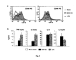
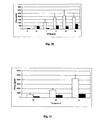
- FIG. 5 Env-SU directly activates purified human monocytes.
- Human monocytes were purified at from human PBMCs (greater than 95% purity) and then stimulated with autologous control (Mock), Env-SU or LPS, at a concentration of 1 ⁇ g / ml, for 24 hours.
- the figure 5a represents the expression of activation markers CD80 (left figure) and CD86 (right figure) analyzed by flow cytometry.
- On the abscissa is the number of cells counted and ordered the fluorescence intensity per cell ("counts").
- the resultant represents the number of cells counted for each fluorescence intensity.
- the area defined by the curves represents the number of total cells for each condition tested.
- the distribution of the cells according to the fluorescence intensity is shown by the shape of the curve.
- the white area represents the results obtained with the control control (Mock), the shaded area, contours fine lines, represents the results obtained with Env-SU and the gray area, very thick contours, represents the results obtained with the LPS .
- the figure 5b represents the secretions of TNF- ⁇ , IL-1 ⁇ , IL-6 and IL-12p40, analyzed by ELISA. In white are represented the results obtained after stimulation by the autologous control. The results obtained after stimulation with Env-SU and LPS are respectively represented in black and gray.
- the y-axis corresponds to the quantities of secreted cytokines, in ng / ml. The results represent the average of three experiments.
- FIG. 6 Env-SU activates dendritic cells derived from monocytes (MDDC).
- the MDDCs were generated from purified monocytes and then stimulated with autologous control, Env-SU, or LPS, at a concentration of 1 ⁇ g / ml, for 24 hours.
- the figure 6a represents the expression of activation markers CD80, CD86, CD40 and HLA-DR, analyzed by flow cytometry. On the abscissa is the number of cells counted and ordered the fluorescence intensity per cell ("counts").
- the upper image left represents the analysis of the CD80, the upper right image that of the CD86, the lower left image represents that of the CD40 and the lower right image that of the HLA-DR.
- the resultant represents the number of cells counted for each fluorescence intensity.
- the area defined by the curves represents the number of total cells for each condition tested.
- the distribution of the cells according to the fluorescence intensity is shown by the shape of the curve.
- the white area on the left represents the results obtained with the control control (Mock), the white area on the right, contour lines thicker, represents the results obtained with Env-SU and the shaded area represents the results obtained with the LPS .
- the figure 6b represents the secretion of TNF- ⁇ , IL-6, IL-12p40 and IL-12p70, analyzed by ELISA, in culture supernatants.
- the y-axis corresponds to the quantities of cytokines secreted, in ng / ml.
- Mock corresponds to the results obtained after stimulation with the autologous control
- Env-SU in black
- LPS in gray
- the Figure 6c represents the allogeneic proliferation of T cells by dendritic cells previously stimulated by: Env-SU - ⁇ -, LPS - ⁇ -, control CK2 - ⁇ -.
- the abscissa axis represents the number of dendritic cells (0, 1000, 5000 and 10,000, respectively).
- the y-axis represents the number of counts per minute emitted by the cells having incorporated 3 H-Thymidine.
- FIG. 7 CD14 and TLR4 are involved in the pro-inflammatory properties of Env-SU.
- PBMCs were preincubated for one hour with or without anti-CD14 neutralizing antibodies (rhCD14, ref .: AB383, R & D Systems - UK) ( figure 7a ) and anti-TLR4 ( figure 7b ), at a concentration of 20 ⁇ g / ml and 5 ⁇ g / ml.
- the cells were then stimulated for 24 hours with CK2 control, Env-SU (ENV1), LPS and SEB, at a concentration of 1 ⁇ g / ml.
- the release of TNF- ⁇ was analyzed in culture supernatants by ELISA.
- the results are shown in the histograms of the Figures 7a and 7b .
- the ordinate axis corresponds to the quantity of TNF- ⁇ released, in ng / ml.
- the black histograms correspond to the results obtained without the addition of antibodies
- the blank histograms correspond to the results obtained in the presence of anti-CD14 and anti-TLR4 antibodies, at 20 ⁇ g / ml
- the histograms in gray correspond to the results obtained in presence of anti-CD14 and anti-TLR4 antibodies, at 5 ⁇ g / ml.
- the results correspond to the average of three experiments.
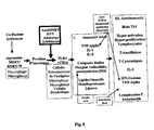
- FIG. 8 Immunological Amplification Cascade Following the activation of the TLR4 pathway. Example of therapeutic targets in this cascade.
- the figure 8 schematically represents the activation cascade resulting from the pathological expression of an MSRV / HERV-W envelope protein. This initially stimulates the TLR4 receptor, possibly associating the CD14 co-receptor. Before this interaction there is only one agonist, the MSRV-ENV protein. After this activation, the cells of the innate immunity are activated and dozens of molecular effectors (cytokines, enzymes, lipids, radical compounds or redox, ...) and activated cells are present. In the case of multiple sclerosis, a second component is activated after destruction of the white matter of the brain and presentation of myelin antigen to T-cells, namely the autoimmune component linked to T-autoreactive cells of adaptive immunity .
- cytokines cytokines, enzymes, lipids, radical compounds or redox,
- a treatment inhibiting the initial effect at a stage of the cascade where all the downstream effectors inducible by the target protein are not produced has a relevance and a potential effectiveness far superior to the approaches commonly conceived and implemented in these cases. pathologies associated with the pro-inflammatory effects of this MSRV / HERV-W envelope protein. Indeed, even if the cascade is activated, it will be "dry" upstream by this therapeutic strategy, while the upstream stimulation will continue in other therapeutic approaches "downstream”.
- these antibodies can neutralize the "ENV MSRV / HERV-W” proteins before they start the cascade described here, while other therapies targeting "downstream" molecules or cells can not to intervene only after putting this inflammatory cascade into action!
- the envelope protein (Env) is produced by a retroviral copy of the MSRV / HERV-W family, in a context of pathological activation such as, for example, after transactivation by an infectious co-factor of the family of herpesviridae [46]. -49] in a tissue territory determined by the tropism of this co-factor and by the presence of cells in the target tissue harboring at least one MSRV / HERV-W proviral copy activated by this co-factor and encoding a protein of envelope.
- This ENV protein thus produced binds to the TLR4 receptor and, depending on the context, to TLR4 co-receptors such as CD14, macrophage or microgliocyte-like cells present in brain tissue in the vicinity of the MSRV / HERV ENV producing cell. -W and / or MSRV virions. If the latter is a macrophage or a microgliocyte, the autocrine effect on the TLR4 receptors of this same cell is possible.
- the immunopathological amplification cascade creates, with the production of dozens of pro-inflammatory molecules and mediators of tissue destruction, a local inflammation in the relevant tissue, around the MSRV / HERV-W reactivation site.
- the reactivation or induced overexpression of an MSRV / HERV-W element in the vicinity of the neuronal structures of the frontal cortex induces a local inflammation which, in the context Tissue of the gray matter, will not allow a major pro-lesional activity and a specific immune recruitment. This local inflammation will not allow T lymphocyte infiltration at this level.
- the pro-inflammatory mediators produced near the neuronal cells responsible for the "intellectual" and cognitive activities provoke a focal neuronal excitotoxicity which determines a dysfunction of the associated neural networks in the affected brain space and in the time that this production lasts pro-inflammatory [17, 20, 22-26, 29, 50, 51].
- "psychic" disorders resulting from excitotoxic neuronal activations result in hallucinatory and delusional manifestations that characterize the clinicopathological accesses of Schizophrenia.
- this neuronal excitotoxicity can lead to cell death (neurotoxicity), which is objectified by the ventricular enlargement measured in MRI in the brain of patients with Schizophrenia [52].
- the myelin of the white matter is extremely sensitive to radical and proinflammatory agents that generate primary demyelination with presentation of self-antigens to the lymphocytes recruited by the patient. prior inflammation.
- lymphocyte reactivity biases are conditioned by the cytokines previously secreted by the microgliocyte / macrophage type cells (Th1 bias) which may be sufficient to generate an autoimmune response to "self" antigens presented under these conditions.
- Th1 bias microgliocyte / macrophage type cells
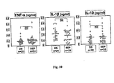
- FIG. 10 Production of ENV-SU Induced Cytokines on PBMCs of Multiple Sclerosis (MS) and Healthy Donor (DS) Patients: TNF-alpha, IL-1Beta and IL-10
- the figure 10 represents the cytokine production induced by the ENV-SU MSRV protein in blood mononuclear cells (PBMCs) taken ex vivo from patients with multiple sclerosis (MS) and from healthy donors ( DS).
- PBMCs blood mononuclear cells
- MS multiple sclerosis
- DS healthy donors
- the abscissa axis represents the cytokine assay in supernatant of stimulated PBMC cultures in ng / ml.
- Each graph compares the results for each individual tested, represented by one (round) point in each population (DS and SEP).
- the three graphs represent, from left to right, the production of tumor necrosis factor (TNF) -alpha, interleukin (IL) -lbeta and interleukin (IL) -10.
- TNF tumor necrosis factor
- IL interleukin
- IL interleukin
- FIG 11 Production of ENV-SU Induced Cytokines on PBMCs of Multiple Sclerosis (MS) and Healthy Donor (DS) Patients: IL-12p40 and IL-6
- the figure 11 represents the cytokine production induced by the ENV-SU MSRV protein in blood mononuclear cells (PBMCs) taken ex vivo from patients with multiple sclerosis (MS) and from healthy donors ( DS).
- PBMCs blood mononuclear cells
- MS multiple sclerosis
- DS healthy donors
- the abscissa axis represents the cytokine assay in supernatant of stimulated PBMC cultures in ng / ml.
- Each graph compares the results for each individual tested, represented by one (round) point in each population (DS and SEP).
- the two graphs represent, from left to right, the production of interleukin (IL) -12p40 and interleukin (IL) -6.
- IL interleukin
- IL interleukin
- Figure 12 Correlations between cytokine productions and patients' clinical parameters
- the figure 12 represents the graphical results of the correlation analysis between clinical parameters of the MS population studied (on the abscissa) and the levels of certain cytokines (on the ordinate) produced in response to the stimulation of their PBMC by the MSRV-SU ENV protein.
- the value of "r” represents the statistical calculation of points distribution with respect to the correlation line.
- the value of "p” represents the statistical probability that this correlation is obtained by chance; thus, any value of p greater than 0.05 is "insignificant” and any value less than 0.05 is significant of an existing correlation between the factors analyzed.
- the top two graphs show the parameters that were found with a significant correlation between the clinical score of MS patients, EDSS [53] measured on a gravity scale of 1 to 10, and IL-6 (left) or IL12p40 (right).
- the bottom two graphs show two examples of parameters that were not found to be significantly correlated: disease duration and IL-6 (left) or interferon gamma and EDSS clinical score (right).
- Figure 13 Spontaneous (a) or ENV-SU (b) induced production of cytokines in PBMCs of Schizophrenia (SCZ) and healthy donors (DS) patients: IL-10.
- the figure 13 represents the cytokine production induced by the ENV-SU MSRV protein in exon vivo blood mononuclear cells (PBMCs) in patients with schizophrenia (SCZ) and in healthy donors (DS) .
- the abscissa axis represents the cytokine assay in supernatant of stimulated PBMC cultures in ng / ml. Each chart compares the results for each individual tested, represented by one point in each population (DS and SEP). The two graphs represent, from left to right, the production of spontaneous interleukin (IL) -10 in culture and interleukin (IL) -10 induced after stimulation by ENV-SU.
- IL spontaneous interleukin
- IL interleukin
- Figure 14 Spontaneous (a) or ENV-SU (b) induced production of cytokines in PBMCs of patients with Schizophrenia (SCZ) and healthy donors (DS), with calculation of relative increase (c): IL-12p40 .
- the figure 14 represents the cytokine production induced by the ENV-SU MSRV protein in exon vivo blood mononuclear cells (PBMCs) in patients with schizophrenia (SCZ) and in healthy donors (DS) .
- PBMCs exon vivo blood mononuclear cells
- SCZ schizophrenia
- DS healthy donors
- the abscissa axis represents the cytokine assay in supernatant of stimulated PBMC cultures in ng / ml. Each chart compares the results for each individual tested, represented by one point in each population (DS and SEP). The three graphs represent, from left to right: a) production of spontaneous interleukin (IL) -12p40 in culture, b) interleukin (IL) -10 induced after stimulation by ENV-SU and c) the relative increase in IL12p40 production calculated according to the formula: (Rate after stimulation ENV-SU - spontaneous rate) / spontaneous rate.
- IL spontaneous interleukin
- IL interleukin
- Figure 15 Identification and selection of anti-ENV MSRV / HERV-W monoclonal antibodies inhibitors of pro-inflammatory activation of monocytes macrophages induced by ENV-SU protein, in cultures of human PBMCs from healthy donors. a) analysis with two anti-ENV antibodies and a control antibody b) verification of the specificity conditions of the analysis c) example of independent experience d) d) another example of independent experience:
- the figure 15a represents, on the ordinate, the TNF-alpha secretion (ng / ml) induced by (abscissa) the control protein "Mock” (1 microgram / ml), ENV-SU (1 microgram / ml) and LPS (1 microgram / ml) in a PBMC culture of a healthy donor.
- the white bar represents the result in the absence of antibodies
- the black bar represents the result in the presence of anti-MSRV 3B2H4 antibodies (30 microgram / ml)
- the hatched bar represents the result in the presence of MSRV13H5A5 anti-ENV antibody (30 microgram / ml)
- the shaded bar represents the result in the presence of anti-GAG MSRV 3H1H6 antibodies (30 microgram / ml).
- the figure 15b represents, on the ordinate, the TNF-alpha secretion (ng / ml) induced by (abscissa) the control protein "Mock" (1 microgram / ml), ENV-SU (1 microgram / ml) and LPS (1 microgram / ml) in a PBMC culture of the same healthy donor as in 15a.
- the white bar represents the result in the absence of antibodies
- the black bar represents the result in the presence of polymixin B (25 microgram / ml)
- the shaded bar represents the result obtained with MOCK, ENV-SU or LPS heated at 100 ° C for 30 minutes prior to addition to the PBMC culture.
- the figure 15c represents, on the ordinate, the secretion of TNF-alpha (pg / ml) induced by the control protein "Mock” illustrated by a white bar (1 microgram / ml), ENV-SU illustrated by a black bar (1 microgram / ml) and the LPS illustrated by a hatched bar (1 microgram / ml), in a PBMC culture of a healthy donor.
- the figure 15d represents, on the ordinate, the secretion of TNF-alpha (pg / ml) induced by the control protein "Mock CK2" illustrated by a white bar (1 microgram / ml) and ENV-SU illustrated by a black bar (1 microgram / ml), in a PBMC culture of a healthy donor.
- PBMCs from healthy donors were stimulated with 5 ⁇ 1 buffer (dashed and rounded curve), 1 ⁇ g / ml ENV-SU (thick line curve with squares) or 1 ⁇ g / ml LPS (curve thin line with triangles) and incubated for 2h, 24h or 48h (x-axis), before analysis of TNF- ⁇ production by ELISA (y-axis in pg / ml).
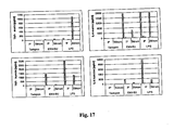
- Figure 17 Proinflammatory effects of ENV-SU in SCID-humanized.
- IP peritoneal washing
- the graphs on the left represent the determination of TNF-alpha (pg / ml).
- the top one represents the dosage of the murine cytokine and that of the bottom, the human cytokine.
- the graphs on the right represent the IL-6 assay (pg / ml).
- the top one represents the dosage of the murine cytokine and that of the bottom, the human cytokine.
- the figure 18 represents the results of a preliminary experiment of induction of EAE by the MSRV ENV protein in C57B16 mice.
- the x-axis represents the days after the injection.
- the y-axis represents the average clinical score of the animals studied.
- the squared curve represents positive control animals injected with the MOG autoantigen (myelin oligodendrocyte glycoprotein) and complete Freund's adjuvant (containing Mycobacterium tuberculosis extract). The study of this series was finalized here at 18 days.
- MOG autoantigen myelin oligodendrocyte glycoprotein
- complete Freund's adjuvant containing Mycobacterium tuberculosis extract
- the curve with the triangles represents the animals injected with the autoantigen MOG (myelin oligodendrocyte glycoprotein), the incomplete Freund's adjuvant (not containing the M. tuberculosis extract) and the ENV-SU MSRV protein.
- MOG myelin oligodendrocyte glycoprotein
- incomplete Freund's adjuvant not containing the M. tuberculosis extract
- ENV-SU MSRV protein ENV-SU MSRV protein
- the diamond curve represents the negative control animals injected with the MOG autoantigen (myelin oligodendrocyte glycoprotein) and Freund's incomplete adjuvant (not containing the M. tuberculosis extract). The study of this series has been continued here until 25 days.
- MOG autoantigen myelin oligodendrocyte glycoprotein
- Freund's incomplete adjuvant not containing the M. tuberculosis extract
- the figure 19 Represents the results of an experiment confirming the induction of EAE by the MSRV ENV protein in C57B16 mice.
- the x-axis represents the days after the injection.
- the y-axis represents the average clinical score of the animals studied.
- the squared curve represents positive control animals injected with the MOG autoantigen (myelin oligodendrocyte glycoprotein) and complete Freund's adjuvant (containing Mycobacterium tuberculosis extract). The study of this series was finalized here at 21 days.
- MOG autoantigen myelin oligodendrocyte glycoprotein
- complete Freund's adjuvant containing Mycobacterium tuberculosis extract
- the curve with the triangles represents the animals injected with the autoantigen MOG (myelin oligodendrocyte glycoprotein), the incomplete Freund's adjuvant (not containing the M. tuberculosis extract) and the ENV-SU MSRV protein.
- MOG myelin oligodendrocyte glycoprotein
- incomplete Freund's adjuvant not containing the M. tuberculosis extract
- ENV-SU MSRV protein ENV-SU MSRV protein
- the diamond curve represents the negative control animals injected with the MOG autoantigen (myelin oligodendrocyte glycoprotein) and Freund's incomplete adjuvant (not containing the M. tuberculosis extract). The study of this series was continued here up to 42 days.
- MOG autoantigen myelin oligodendrocyte glycoprotein
- Freund's incomplete adjuvant not containing the M. tuberculosis extract
- the curve with the crosses represents the negative control animals injected with the MOG autoantigen (myelin oligodendrocyte glycoprotein), incomplete Freund's adjuvant (not containing the M. tuberculosis extract) and LPS.
- MOG autoantigen myelin oligodendrocyte glycoprotein
- incomplete Freund's adjuvant not containing the M. tuberculosis extract
- LPS incomplete Freund's adjuvant
- Figure 20 24-hour assay of the autoimmune response to increasing doses of MOG autoantigen in mice of the "EAE / MOG / ENV-SU" model and in the immunized control without ENV.
- the abscissa axis represents the concentrations of MOG autoantigen (microgram / ml) in the presence of the mouse PBMCs taken during the protocol illustrated in FIG. figure 19 .
- the ordinate axis represents the interferon gamma assay secreted in vitro by autoimmune T cells present in PBMCs exposed to increasing doses of MOG antigen.
- the white bars represent the PBMCs of mice injected in vivo (at day "0" of the series illustrated in FIG. figure 19 ) the MOG autoantigen (myelin oligodendrocyte glycoprotein), Freund's incomplete adjuvant (not containing the M. tuberculosis extract) and the ENV-SU MSRV protein.
- MOG autoantigen myelin oligodendrocyte glycoprotein
- Freund's incomplete adjuvant not containing the M. tuberculosis extract
- ENV-SU MSRV protein ENV-SU MSRV protein
- the black bars represent the PBMCs of control mice injected in vivo (at day “0" of the series illustrated in FIG. figure 19 ) MOG autoantigen (myelin oligodendrocyte glycoprotein) and incomplete Freund's adjuvant (not containing M. tuberculosis extract).
- MOG autoantigen myelin oligodendrocyte glycoprotein
- incomplete Freund's adjuvant not containing M. tuberculosis extract
- Figure 21 Kinetics of autoimmune anti-MOG T lymphocyte response revealed by interferon gamma secretion in mice of the "EAE / MOG / ENV-SU" model and in the immunized control without ENV.
- the abscissa represents the post-inoculation time of the preparation "MOG and adjuvants" (in hours) from which PBMCs were taken from the mice of the series illustrated in FIG. figure 19 .
- the ordinate axis represents the interferon gamma assay secreted in vitro by autoimmune T cells present in PBMCs.
- the white bars represent the PBMCs of mice injected in vivo (at day "0" of the series illustrated in FIG. figure 19 ) the MOG autoantigen (myelin oligodendrocyte glycoprotein), Freund's incomplete adjuvant (not containing the M. tuberculosis extract) and the ENV-SU MSRV protein.
- MOG autoantigen myelin oligodendrocyte glycoprotein
- Freund's incomplete adjuvant not containing the M. tuberculosis extract
- ENV-SU MSRV protein ENV-SU MSRV protein
- the black bars represent the PBMCs of control mice injected in vivo (at day “0" of the series illustrated in FIG. figure 19 ) MOG autoantigen (myelin oligodendrocyte glycoprotein) and incomplete Freund's adjuvant (not containing M. tuberculosis extract).
- MOG autoantigen myelin oligodendrocyte glycoprotein
- incomplete Freund's adjuvant not containing M. tuberculosis extract
- the x-axis represents the days after the injection.
- the y-axis represents the average clinical score of the animals studied.
- the squared curve represents positive control animals injected with the MOG autoantigen (myelin oligodendrocyte glycoprotein) and complete Freund's adjuvant (containing Mycobacterium tuberculosis extract). The study of this series was finalized here at 28 Days.
- MOG autoantigen myelin oligodendrocyte glycoprotein
- complete Freund's adjuvant containing Mycobacterium tuberculosis extract
- the curve with the triangles represents the animals injected with the autoantigen MOG (myelin oligodendrocyte glycoprotein), the incomplete Freund's adjuvant (not containing the M. tuberculosis extract) and the ENV-SU MSRV protein.
- MOG myelin oligodendrocyte glycoprotein
- incomplete Freund's adjuvant not containing the M. tuberculosis extract
- ENV-SU MSRV protein ENV-SU MSRV protein
- the diamond curve represents the negative control animals injected with the MOG autoantigen (myelin oligodendrocyte glycoprotein) and Freund's incomplete adjuvant (not containing the M. tuberculosis extract). The study of this series was continued here until 28 Days.
- MOG autoantigen myelin oligodendrocyte glycoprotein
- Freund's incomplete adjuvant not containing the M. tuberculosis extract
- the dotted curve with the crosses represents the animals injected with the MOG autoantigen (myelin oligodendrocyte glycoprotein), the incomplete Freund's adjuvant (not containing the M. tuberculosis extract) and the ENV-SU MSRV protein. These animals additionally received a dose of 50 micrograms per kilogram, that is one microgram for a 20 gram mouse, of anti-GAG MSRV 3H1H6 control antibody. The study of this series was continued here until 28 days.
- MOG autoantigen myelin oligodendrocyte glycoprotein
- incomplete Freund's adjuvant not containing the M. tuberculosis extract
- ENV-SU MSRV protein ENV-SU MSRV protein
- the curve with the circles represents the animals injected with the autoantigen MOG (myelin oligodendrocyte glycoprotein), the incomplete Freund's adjuvant (not containing the M. tuberculosis extract) and the ENV-SU MSRV protein. These animals additionally received a dose of 50 micrograms per kilogram, that is one microgram for a 20-gram mouse, of anti-MSRV MSBV 3B2H4 control antibody. The study of this series was continued here until 28 days.
- MOG myelin oligodendrocyte glycoprotein
- incomplete Freund's adjuvant not containing the M. tuberculosis extract
- ENV-SU MSRV protein ENV-SU MSRV protein
- Figure 23 Amino acid sequences compared between the MSRV ENV protein (bottom line) and the ENV protein encoded by the HERV-W copy 7q (top line) The framed sequences are identical (conserved regions)
- Figure 24 Western blot analysis.
- the arrow shows the level of the detected bands.
- 60 Kda indicates the level of the corresponding molecular weight.
- Figure 25 Analysis of the antigenic properties of ENV-SU protein Western blot analysis.
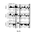
- the figures 25 a , b and c represent the analysis of the amino acid sequence of the MSRV ENV-SU protein by the "Mac Vector” analysis software, with the "Protein analysis toolbox” function.
- the regions bordered by the 3 vertical rectangles represent the three most probable antigenic regions according to a primary and secondary sequence analysis.
- the two upper graphs illustrate the hydrophilicity of the "ENV-SU" regions. Shaded areas above the "0" of the y-axis have positive hydrophilicity, the area below has negative hydrophilicity.
- the bottom graph illustrates the flexibility of the "ENV-SU" regions. Shaded areas above the "0" on the y-axis have positive flexibility, the one below has negative flexibility.
- the graph illustrates the surface probability of the "ENV-SU" regions. Shaded areas above the "0" of the y-axis have positive flexibility, the area below has a negative surface probability.
- the surface protein of the MSRV envelope corresponds to a 287 amino acid protein sequence of the envelope total protein (Env Pv14, GenBank AF331500).
- the structures and amino acid sequences of Env Pv14 and Env-SU are respectively represented in the Figures 1 (a) and 1 (b) .
- the recombinant MSRV Env-SU protein is expressed in E. coli and purified on an FPLC column. The quality and purity of the protein are confirmed by mass spectrometry and Western Blot. Casein kinase is used as an autologous negative control. This control protein was produced and purified under the same conditions as Env-SU.
- LAL Lymulus Amebocyte Lysate
- SEB Staphylococcal enterotoxin B obtained from Toxin Technology (Sarasota, FL, USA) was 95% pure.
- LPS liposaccharide
- PBMCs Human peripheral blood mononuclear cells
- Monocytes from PBMCs are purified by removal of T cells, B cells, dendritic cells, NK cells and basophils using the monocyte isolation kit, marketed by Miltenyi Biotec.
- PBMCs are first incubated with a cocktail of monoclonal antibodies and anti-human immunoglobulin conjugated to a hapten and magnetically labeled (anti-CD3, anti-CD7, anti-CD19, anti-CD45RA, anti- CD56 and anti-IgE), then with micro-beads (MACs MicroBeads) coupled with anti-hapten monoclonal antibodies.
- MACs MicroBeads micro-beads
- the magnetically labeled cells are finally removed by holding them on a column in a magnetic field.
- the purity of the Monocyte population recovered is always greater than 95%, as determined by CD14 expression by flow cytometric analysis.
- the purified monocytes are cultured for 5 days on 6-well plates containing IL-4 at 25 ng / ml and GM-CSF at 50 ng / ml in 2 ml of the culture medium.
- the complete amount of cytokines is added to the cells.
- the resulting cell preparation contains more than 90% CD1a positive dendritic cells.
- the cells are placed in 24-well plates, at a concentration of 1 ⁇ 10 6 cells per well, in 1 ml of culture medium before stimulation with Env-SU, LPS, SEB, or autologous control. They are then incubated at 37 ° C. in a humidified atmosphere containing 5% CO 2 . When indicated, the cells were pre-incubated either with 10 ⁇ g / ml of Polymyxin B (Sigma-Aldrich), 20 ⁇ g / ml and 5 ⁇ g / ml of anti-CD14 monoclonal antibody, 20 ⁇ g / ml and 5 ⁇ g / ml.
- Polymyxin B Sigma-Aldrich
- Env-SU ml of anti-TLR-4 antibody (HTA125, eBioscience) or isotype control IgG 2a (IgG2a) (eBM2a, eBioscience) prior to cell stimulation.
- IgG2a isotype control IgG 2a
- eBM2a eBioscience
- Env-SU, autologous control, LPS and SEB are boiled for 30 minutes before cell processing.
- Env-SU 13H5A5; IgG1, biomérieux
- GAG 3H1H6, IgG1, biomérieux
- the cells are incubated for 24 hours at 37 ° C., and then the culture supernatants are harvested for the analysis of the secretion of TNF- ⁇ , IL-1 ⁇ and IL-6 by ELISA.
- the stimulated monocytes and MDDCs are used as stimulators of T cells.
- Allogeneic T cells are used at 1 x 10 5 cells per well as "responder" cells in 96 well round bottom microplates.
- the stimulator cells are added to the T cells in increasing doses and the cultures are carried out in triplicate in a final volume of 200 ⁇ l of culture medium. After 5 days of incubation at 37 ° C., the proliferation of T cells is evaluated by measuring the incorporated radioactivity. To do this, in the last 18 hours of incubation, 1 ⁇ Ci of 3 H Thymidine is added to each well. The cells are then recovered on glass filter mats for the measurement of the incorporated radioactivity.
- the cells are harvested, washed in PBS and then stained for different surface markers.
- the following monoclonal antibodies (Becton-Dickinson, San Jose, CA) were used: anti-CD1a allophycocyanin (HI149-APC), anti-CD14 fluorescein isothiocyanate (MOP9-FITC), CD40 phycoerythrin (5C3-PE), CD80 phycoerythrin (L307.4-PE), CD86 phycoerythrin (IT2.2-PE) and HLA-DR chlorophyll peridine (L243-PerCP).
- the direct immunofluorescence staining of the cells is performed in ice-cold PBS supplemented with 2% FCS, with the different antibodies at the concentrations recommended by the manufacturers. After 30 minutes at 4 ° C, the cells are washed and then analyzed using a Calibur FACS (trade name) and CellQuest (trade name) software (Becton Dickinson).
- the culture supernatants are collected and stored at -20 ° C. before analysis of cytokine secretion.
- the amounts of cytokines are measured using the ELISA OptEIA (trade name) kits (Pharmigen) for IL-1beta, IL-6, IL-10, IL-12p40, IL-12p40 and TNF-alpha, according to the manufacturer's instructions. .
- Env-SU induces the production of pro-inflammatory cytokines from human PBMCs.
- PBMCs from healthy donors were incubated for 24 hours with increasing doses of the recombinant Env-SU protein and cytokine secretion of TNF- ⁇ , IL-1 ⁇ and IL-6 was assessed by ELISA.
- the amounts of secreted cytokines were compared with those obtained with autologous control, SEB (a well characterized bacterial superantigen) and LPS, well known to have pro-inflammatory properties on human PBMCs. All proteins and toxins were used at a concentration of 1 ⁇ g / ml (optimal concentration for the induction of pro-inflammatory cytokines determined by dose / response experiments).
- Env-SU induces the secretion of the three cytokines in a dose-dependent manner, even at doses as low as 10 ng / ml.
- the cytokine secretion kinetics obtained with Env-SU is closer to that of LPS than SEB.
- SEB antigen represented here by the SEB antigen: (i) no early secretion of interferon gamma (which signifies the activation of T lymphocytes, specifically recognized at the level of the "T-cell recptor" (TCR) by superantigens, ( ii) large and early secretion of IL-6 (secreted by monocytes-macrophages and not by T cells) and TNF-alpha, which are not produced under the conditions of our in vitro test by a superantigen (as shown here with the example of SEB)
- the pro-inflammatory activation pathway of the MSRV / HERV-W Env protein claimed here passes through the "Toll-like receptor 4" (TLR4), possibly with the assistance of its CD14 co-receptor, which is activated in upstream of T-cell activation, as illustrated in figure 8 .
- TLR4 Toll-like receptor 4
- This pro-inflammatory pathway (TLR4) mobilizes the "innate” component of the immune system that is mobilized well in advance of the immunity linked to the "T" lymphocytes (adaptive immunity).
- This upstream pathway can, after activation of the dendritic cells, influence downstream the activation state of adaptive immunity (Th1 or Th2 profiles), through cytokines secreted in response to the activation of an innate immunity receptor such as TLR4.
- This latter effect shows, in addition to the fact that the innate immunity pathway is activated upstream of adaptive immunity mediated by T lymphocytes, that even the resultant downstream effect on T cells does not pass through the TCR receptor.
- the TLR4 pathway excludes primary activation of T lymphocytes at this stage and therefore passes through other cells (monocytes-macrophages, dendritic cells, B lymphocytes).
- the activation pathway observed here is therefore upstream of the superantigen effect, which corroborates the kinetics that corresponds to the "LPS" referent, rather than "SEB” in the cell test used here.
- Env-SU and LPS are capable of inducing the secretion of large amounts of TNF- ⁇ , IL-6 and IL-1 ⁇ as early as 24 hours, whereas SEB only induces the secretion of TNF- ⁇ itself. after 72 hours of incubation. It is interesting to note that Env-SU and LPS reach their peak of TNF- ⁇ secretion whereas SEB induces a constant secretion of TNF- ⁇ . Compared with IL-1 ⁇ , Env-SU and LPS induce a secretion profile similar to that of TNF- ⁇ , characterized by a secretion peak around 24 hours of incubation followed by a constant decrease.
- SEB which is known to activate a large population of T cells with the same TCR V ⁇ specificity, induces no IL-1 ⁇ .
- IL-6 is secreted consistently by PBMCs stimulated by Env-SU and LPS, but not by SEB.
- IL-6 and IL-1 ⁇ are two cytokines preferentially released by activated monocytes / macrophages.
- PBMCs Humans were either treated with an LPS inhibitor, Polymixin B (PB) before stimulation, or incubated with boiled proteins and toxins.
- PB Polymixin B
- an autologous control produced and purified under the same conditions, with the same reagents and material: casein kinase CK2 human.
- the culture supernatants were harvested and analyzed for the secretion of TNF- ⁇ .
- the Env-SU and SEB-induced TNF- ⁇ is only partially inhibited by PB, whereas the effects of LPS are completely abolished.
- the autologous control protein induces no secretion of cytokines.
- the release of TNF- ⁇ is also significantly inhibited when the Env-SU proteins are boiled for 30 minutes, while the LPS activity is not affected. This is consistent with the negative results obtained in the quality control analysis performed on both purified samples of Env-SU and autologous control using the LAL assay, approved by the Food and Drug Administration.
- the effects of monoclonal antibodies were studied.
- the PBMCs were incubated for 24 hours with autologous control, Env-Su or LPS pre-incubated at 4 ° C for 1 hour with either a monoclonal antibody against Env-SU, or with a monoclonal antibody against Gag.
- the Gag protein used to develop this monoclonal antibody does not exhibit any pro-inflammatory activity and constitutes an adequate control.
- the anti-Env-SU monoclonal antibody specifically blocks the Env-SU mediated TNF- ⁇ secretion, but not that of LPS. Secretion of cytokines is unaffected by the anti-Gag monoclonal antibody.
- Env-SU has the ability to induce pro-inflammatory cytokines in PBMC cultures. It was then verified that Env-SU was able to directly activate purified monocytes. Purified monocytes were stimulated with autologous control, Env-SU or LPS for 24 hours and different activation markers, such as CD80 and CD86, were evaluated by flow cytometry. Compared to autologous control, Env-SU induces upstream regulation of both markers and the expression levels obtained are similar to those of LPS ( figure 5a ). Large amounts of TNF- ⁇ , IL-1 ⁇ , IL-6 and IL-12p40 are produced in response to Env-SU ( figure 5b ). These results show that Env-SU induces rapid and direct activation of monocytes associated with pro-inflammatory cytokine production.
- Dendritic cells are innate and adaptive immunity-binding antigen-presenting cells with the unique ability to control the activation of naive T cells.
- the ability of Env-SU to directly activate monocyte-derived dendritic cells (MDDCs) was studied. Dendritic cells were generated in vitro from highly purified monocytes and stimulated for 24 hours with autologous control, Env-SU or LPS. Env-SU is able to drastically increase the activation of CD80, CD86, CD40 and HLA-DR markers ( figure 6a ). The pro-inflammatory cytokines IL-6, TNF- ⁇ , IL-12p40 and IL12p70 are secreted at higher levels.
- Env-SU stimulated MDDCs are shown to be capable of inducing allogeneic T-cell proliferation to a greater extent than autologous control, even when the number of stimulator cells is low ( Figure 6c ). Therefore, similarly to LPS (positive control), Env-SU is able to induce the maturation of dendritic cells secreting IL-12 and is therefore able to induce specific primary immune responses.
- TLR4 blockade results in 37% inhibition of Env-SU effects and 43% inhibition of LPS effects, with 20 ⁇ g of anti-TLR4 antibodies ( figure 7b ). No inhibition effects were observed for control antibodies in both experiments.
- the CD14 and TLR4 receptors are therefore involved in Env-SU mediated pro-inflammatory properties.
- the soluble fraction of the MSRV / HERV-W envelope protein stimulates an innate immune response via the CD14 and TLR4 recognition receptors and it is shown in the present invention that the immunopathological cascade leading to inflammatory lesions can be blocked at a very early stage by administration of a therapeutic composition or drug comprising at least one antibody chosen from anti-Env-SU and / or anti-TLR4 antibodies.
- the cell test developed and developed here identified those that have the property of inhibiting the pro-inflammatory effect activating the TLR4 pathway and selecting among inhibitory antibodies, those with an inhibitory potential closest to 100%.
- the antibodies 3B2H4 and 13H5A5 are the preferred antibodies.
- the antibodies of the invention make it possible to block upstream pathogenic cascades which differ downstream in pathologies such as MS and SCZ.
- the figure 8 shows the target targeted by the antibodies of the invention in the pathogenic cascade of MS that anticipates all targets currently targeted by existing therapeutic agents.
- SCZ Schizophrenia
- the progressive loss of brain neurons in these patients is compensated by an increase in the volume of the cerebral ventricles which becomes detectable on MRI after a certain period of evolution of the disease.
- the figures 8 and 9 thus illustrate the fact that the anti-Env antibodies identified and selected by the cell test developed in the context of the present invention actually block the "primary" stimulus most upstream of this cascade, after the activation of a or multiple pathogenic copies of the MSRV / HERV-W family.
- Env-SU-induced IL-6 secretion is increased in blood mononuclear cells harvested ex vivo in MS patients and correlates with their clinical score (EDSS).
- gamma interferon which is secreted exclusively by T lymphocytes
- cytokines associated with innate immunity secreted here by monocytes / macrophages it does not correlate with the score. clinic (EDSS). This shows that, as has been demonstrated in vitro with the cellular tests, the effect revealed ex vivo correlates well an effect which is not mediated at this stage by the T lymphocytes, and therefore, does not resemble not to a superantigen effect.
- MS patients with the most advanced clinical signs are "hyper-sensitive" to retroviral factors such as Env-SU, but may also suggest a role for Env-SU in the pathogenesis of MS via pro-inflammatory cytokines and IL-6.
- Env-SU-induced IL-12p40 secretion is increased in blood mononuclear cells taken ex vivo in SCZ patients and furthermore allows to identify at the highest levels a subpopulation of patients resistant to anti-cancer therapies. psychotic and / or having particularly progressive forms of SCZ.
- Table 1 represents the various healthy donors whose code is indicated in the rows of the first column with, in each line, the quantities in ng / ml assayed for the various cytokines indicated at the top of the column with the two conditions indicated in the line. next for their respective columns (Mock and ENV-SU stimulation).
- Table 2 represents the different MS patients whose code is indicated in the rows of the first column with, in each row, the quantities in ng / ml assayed for the various cytokines indicated at the top of the column with the two conditions indicated in the line. next for their respective columns (Mock and ENV-SU stimulation).
- the two bottom rows indicate the mean and standard deviation of the measured data for each column, respectively.
- Table 1 TNF- IL-6 IL-10 IL-12p40 IL-1b Mock ENV1 Mock ENV1 Mock ENV1 Mock ENV1 ND 151003 24 500 216 3493 12 59 0 245 38 1149 ND 291003 14 1491 166 14230 14 337 0 398 32 1677 ND 011003 114 969 3727 12141 38 478 1 99 214 1712 ND 300402 18 58 172 601 20 21 9 31 69 193 ND 020702 0 139 111 1028 3.5 44 5 16 99 392 ND 180602 48 264 92 1816 16 32 20 57 110 424 ND 070801 0 124 84 1028 6 31 20 42 25 235 ND 180901 1 228 39 2328 7 60 0 113 21 651 ND 060701 30 260 345 3263 17 155 6 38 33 277 ND 110901 0 328 70 7352 14 214 11 169 9 587
- the response to Env-SU stimulation is globally increased in the SCZ patient series and some patients respond with secretion rates of cytokines significantly above the mean of healthy controls and sometimes even higher than the maximum observed in the control series (as shown in figures 13 and 14 , for IL-10 and IL-12p40). This also confirms that the response to Env-SU is significantly increased in some patients with SCZ and that Env-SU is therefore able to reveal an immunological bias involving components of innate immunity activated by the TLR4 pathway, in some patients. of those patients whose clinical status coincides.
- patients with an induced rate of IL-12p40 induced by Env-SU among the highest include all patients resistant to antipsychotic therapeutics of the tested series and forms of schizophrenia particularly progressive.
- activation of an MSRV / HERV-W provirus by different co-factors can activate expression of the MSRV / HERV-W Env protein in brain cells [56] and, depending on the nature of the co-factor and circumstances, target different areas of the brain. Under these conditions activation in the regions of the frontal cortex can cause neuronal excitotoxicity resulting in various hallucinations depending on the territories reached.
- the early inflammation produced by the Env protein at the level of the macrophages and / or microgliocytes is likely to stimulate a degradation of the myelin and therefore, after the contribution of the T lymphocytes, to induce autoimmunity against self-antigens of myelin.
- the level at which these antibodies inhibit the biological effects of the Env protein is well in advance of all the pathological mediators that are produced downstream and which are the usual targets of conventional anti-inflammatory therapies (cytokines, free radicals, redox compounds, enzymes, prostaglandins, pro-inflammatory proteins and lipids, activated T cells, and ).
- cytokines free radicals, redox compounds, enzymes, prostaglandins, pro-inflammatory proteins and lipids, activated T cells, and .
- the only agonist is the MSRV / HERV-W Env protein itself, and its inhibition of activating the TLR4 receptor blocks the initial entry of the different immunobiological cascades that result downstream, as shown in FIGS. figures 8 and 9 .
- Such therapeutic guidance provides a very important benefit to the definition of patients eligible for treatment at a given time or in a given subgroup and allows to adjust the dose and frequency therapy according to the biological results.
- Anti-Env antibodies MSRV / HERV-W are the 13H5A5 and 3B2H4 antibodies.
- the MSRV / HERV-W anti-Gag control antibody is the 3H1H6 antibody.
- Fragments are first diluted. 100 ⁇ l at the concentration of 1 ⁇ g / ⁇ l are thus prepared and then brought into contact with sodium iodide (NaI 125 NEN, 5mCi / 50 ⁇ l) adsorbed on beads (Iodobeads No. 28665 Pierce Rockford, Illinois USA) , as recommended by the manufacturer.
- sodium iodide NaI 125 NEN, 5mCi / 50 ⁇ l
- beads Iodobeads No. 28665 Pierce Rockford, Illinois USA
- the sample is then neutralized with 10 .mu.l of a solution of sodium pyrosulfite 4mg / ml (Fluka). Finally, sodium iodide is driven using 10 ⁇ l of a cold driver dosed at 250 nM / ml.
- the purification is carried out by the ion exchange column separation technique.
- This method uses an anion exchange column to fix free iodine. It is activated at first, using 2 ml of 0.9% NaCl solution (Aguettant), the migration buffer. The purification is carried out in four passages, entrained with 0.5 ml of 0.9% NaCl. The radioactivity contained in the four tubes is then counted.
- the labeling yield is correct after 10 minutes of incubation. It does not increase with time.
- the percentage obtained for all the fragments is between 70 and 80%.
- mice 7 week BALB / c mice provided by Charles River Laboratories (Wilmington, North Carolina, USA). Their good health is guaranteed by the supplier.
- the batches consist of 3 BALB / c white mice 7 weeks old for each fragment tested.
- ketamine 2% -xylamine10g / 100ml administered at the dose of 1 ⁇ l / g of weight.
- mice are injected intravenously (IV) with 0.15 mg of antibodies labeled at 700 ⁇ Ci / mg (the mice are inoculated with the fragments of one of the three antibodies tested (3B2H4, 13H5A5 and 3H1H6).
- a reading is performed at 10, 45, 90 and 210 minutes after the injection.
- the mouse is sacrificed and removed: the spleen, liver, kidneys, brain, heart, lungs and blood.
- the tail is also kept to adjust the values obtained.
- Table 4 shows that none of the fragments tested are abnormally fixed in a tissue.
- the animal models make it possible to select from this first selection, the therapeutic antibodies which do not have side effects harmful to their therapeutic use in the pathological context. Indeed, as has been shown for an antibody potentiating the neurological damage in the "EAE-MOG" example (see example: EAE-MOG and 3H1H6 antibody), some antibodies may be inadequate for therapy whatever their specificity and, even if they inhibit a pathological target, these side effects must have them eliminated or modified before therapeutic use in a particular context.
- the tools developed in the present invention thus make it possible to identify these possible harmful effects in the pathological context of use and thus contribute in an original and adapted manner to the selection and validation of the appropriate therapeutic antibodies.
- the lipopolysaccharide (LPS) of the E. coli 026: B6 strain was obtained from Sigma (St Louis, Mi).
- the recombinant Env-SU protein represents a fraction of the MSRV whole envelope protein, ENV pV14, of about 33kDa and 287 amino acids.
- Env-SU was produced in E.coli, purified by chromatography and analyzed by western blot (Protein Expert, Grenoble).
- An LAL test lymulus amebocyte lysate, Clean cell, Bouffere, France was carried out to detect the possible presence of endotoxins. The results were negative, below the detection limit of 5 IU / ml.
- the buffer used for the preservation of the protein will be used in the experiments as a negative control. It consists of Tris pH8 50mM, 0.3M NaCl, 1mM ⁇ -mercaptoethanol, 2% sucrose, 2% glycerol and 5.3mM urea.
- PBMCS were prepared from citrated total fresh blood from healthy donors (citrated whole blood bags, Valence Blood Transfusion Center, France) by density gradient on Ficoll (Amersham Biosciences, Freiburg, Germany). The dilution of 25 ml of blood with 10 ml of PBS-2% FCS (fetal calf serum), delicately deposited on 15 ml of Ficoll, is centrifuged at 2400 rpm for 20 min. at room temperature (RT). Cell rings are recovered and the PBMCs are washed three times with 50ml of PBS-2% FCS ( Figure 11 ). After counting with trypan blue, the cells are frozen at -80 ° C. in a mixture of 90% SVFd-10% DMSO and used directly in culture for cell tests or for injection in the mouse.
- FCS fetal calf serum
- mice After sacrifice of the mice by cervical dislocation, the spleens are removed and crushed on a metal filter in RPMI. The cell suspensions are centrifuged at 1200 rpm for 10 min. at 4 ° C. The cell pellet is then taken up in approximately 4 ml of physiological saline or of SVFd (for respectively injection with C57B16 mice or freezing). A trypan blue count is performed and then the cell concentration is adjusted. The cells are then frozen or used for animal models. For injection into the mice, gentamicin is added at a concentration of 0.2 mg / ml. Finally, 500 ⁇ l or 50.10 6 cells are injected IP (intraperitoneally) to each of the C57B16 mice.
- IP intraperitoneally
- the cell suspensions are washed once in 50 ml of PBS (bioMérieux, France) at 4 ° C. and centrifuged at 1400 rpm for 7 min at 4 ° C.
- the cells are then taken up in a few ml of FCS and counted.
- the cell concentration is then adjusted to 20 ⁇ 10 6 cells in general.
- 500 ⁇ l of this solution are deposited in a cryotube and then 500 ⁇ l of freezing solution (80% SVFd-20% DMSO (Sigma)) are added.
- the cells are stored in 1 ml of 90% FCS-10% DMSO solution.
- the cryotubes are placed in a freeze box containing isopropanol for a slow descent in temperature and placed at -80 ° C.
- the cell suspensions are thawed in a water bath at 37 ° C.
- the tubes are washed with alcohol before opening.
- the cells are rapidly transferred into 50 ml of RPMIc-10% SVFd, centrifuged at 1400 rpm for 7 min. at 4 ° C then two further washes are performed with RPMIc-10% FCS.
- PBMCs Cell cultures are incubated in a humid atmosphere at 5% CO 2 at 37 ° C.
- the culture medium used consists of RPMI 1640 (Gibco, Rockeville, MD) supplemented with 1% L-glutamine, 1% penicillin-streptomicin, 1% sodium pyruvate, 1% non-essential amino acids (Sigma ) as well as 10% of fetal calf serum (Biowest, Nuaille, France) decomplemented (SVFd) by heating at 56 ° C for 30 min.
- the cell suspensions (PBMCs or splenocytes) are thawed and a Trypan blue count is performed: the cell concentration is adjusted to 1.10 6 cells / ml.
- the cultures are carried out in 48-well plates (500 ⁇ l of cell suspension per well) or in 24-well plates (1 ml of cell suspension per well). After depositing the cells in plate, the different substances to be tested are added and the cells are incubated for various periods of time. The supernatants are harvested by centrifugation of the cell suspensions at 6000 rpm for 10 minutes. at TA. They are then frozen in ependorf tube at - 20 ° C.
- the concentrations of Env-SU and LPS used for the cell assays were 1 ⁇ g / ml.
- Env-SU and LPS were boiled for 30 min.
- Polymixin B (PB) was used at 25 g / ml, and pre-incubated for 45 min. at 37 ° C with the cells before buffer addition, LPS or Env-SU.
- preincubations at 4 ° C or 37 ° C of the cells with the antibodies or Env-SU, LPS or buffer with the antibodies were necessary for different periods of time.
- the anti-Env-SU (13H5A5 and 3B3H4) and anti-Gag (3H1H6) IgG monoclonal antibodies were obtained by hybridoma culture after immunization of the mice with the recombinant Env-SU or Gag proteins, respectively.
- the specificity of anti-Env-SU antibodies was verified by ELISA. Unless indicated, the concentrations of 13H5A5, 3B2H4 and 3H1H6 used for cell assays were 30 ⁇ g / ml.
- mice (Charles River, L'Arbresle, France) are bought at the age of 5 or 6 weeks and are kept one week at rest after reception. They are housed in sterile filter cages at a temperature of 24 ° C. All manipulations are performed under laminar flow host.
- the SCID mice receive an intraperitoneal injection (IP) of 50.10 6 fresh human PBMCs in 2ml of RPMI without phenol red (Eurobio, Les Ulis, France) supplemented with gentamicin at a concentration of 0, 25 mg / ml, and are again left for one week at rest.
- IP intraperitoneal injection
- 50 ⁇ l of anti-NK antibody 25 ⁇ l of pure antibody diluted in 25 microl of physiological saline
- blood is collected by RO on each mice and the serum stored at -80 ° C in order to test the level of humanization of the mice.
- the C57B16 mice receive, after one week of adaptation, an intraperitoneal injection (IP) of 50 10 6 murine fresh splenocytes in 2 ml of RPMI without phenol red added with gentamicin at a concentration of 0.25 mg / ml and are again left behind. a week at rest.
- IP intraperitoneal injection
- SCIDs as in C57B16, the liquid resorbs quickly but some losses are observed.
- the human IgG assay in the serum of SCID mice is performed by the radial immunodiffusion method following the manufacturer's instructions (The binding site, Birmingham, UK). Measuring the diameter of the precipitate 96h after depositing the serum on the gel makes it possible, thanks to a calibration curve, to connect its square to the IgG concentration of the sample tested.
- the proteins (Env-SU), toxins (LPS) or buffer are assigned to the mice by IP injection after dilution of the substances in 1ml of physiological saline (Fresenius Kabi, Bad Homburg, Germany) at the desired concentration.
- physiological saline Fresenius Kabi, Bad Homburg, Germany
- anti-Env-SU or anti-GAG antibodies they are first incubated for 3 hours at 4 ° C. with Env-SU, LPS or buffer.
- the control mice receive 1 ml of physiological saline. Samples take place a few hours (1h, 2h) or days (24, 48, 72h) later.
- mice After anesthesia of the mice with ether, the maximum of blood (approximately 1 ml) is taken retro-orbitally (RO) with a pasteur pipette. 2ml of saline are then injected into the intraperitoneal cavity (IP), and after massage of the abdomen, the maximum of liquid is removed (1 to 1.5 ml). Finally, the mice are sacrificed by cervical dislocation and the spleen is removed. The different protocols used are presented figures 12 , 13 , and 14 . All mice are observed clinically until the end of the experiment. Particularly noted are signs of inflammation and signs of nervous system damage.
- RO retro-orbitally
- the removed spleen is divided into two fragments. Part of this is slurried by grid grinding in RPMI. After centrifugation at 1200 rpm for 10 min. at 4 ° C., the cells are taken up in 50 ml of PBS at 4 ° C. and then centrifuged and frozen in a cryotube in 1 ml of freezing solution (10% DMSO-90% SVO). Another part of the spleen is frozen as it is in an ependorf at -80 ° C. The liquid withdrawn at IP is centrifuged at 6000 rpm for 10 min. at RT to remove the cell pellet and frozen at -80 ° C ependorf tube.
- the cell suspensions are thawed and the cells are taken up in 50 ml of PBS-2% SVFd-1mMEDTA and centrifuged at 1400 rpm for 7 min. at 4 ° C. Trypan blue counting is performed and the cells are plated in 96 wells (about 1.10 6 per well). After centrifugation of the plate at 4000 rpm for 1 min. at 4 ° C., the supernatants are removed and 50 ⁇ l of the surface marker antibody cocktail (dilution in PBS-2% SVFd-1mMEDTA) are added to each well. The cells are resuspended and incubated for 30 minutes. at 4 ° C.
- Antibodies (Pharmingen, San Diego, CA) and dilutions used for the labeling of murine cells are: CD3 - FITC (1/500), CD4 - PE (1/1000), CD8 - cy - chromium (1/600), CD25 - APC (1/1000), CD69 - APC (1/500, biotinylated initially).
- CD3 -cy-chromium CD4-APC
- CD8-PE CD25-PE
- CD69-FITC 2 ⁇ 1 of each antibody are used: CD3-cy-chromium, CD4-APC, CD8-PE, CD25-PE, CD69-FITC.
- Some anti-Env antibodies did not have detectable inhibitory activity in these tests (6A2B2 and 2A12A5), or moderate inhibitory activity (3C1D5) or uneven activity according to experiments with PBMCs from different donors (3H10F10). Examples of these tests are shown in the figure 15 (15a, b, c and d) which illustrate the effects of these antibodies against pro-inflammatory activation produced by the Env-SU protein in different experiments.
- an antibody such as that produced by the hybridoma 2A12A5, in the absence of inhibitory activity on the Env-SU protein, paradoxically produced a non-specific immunological stimulation in the test, even in the absence of protein Env- SU.
- the anti-MSRV / HERV-W monoclonal antibodies produced by the 13H5A5 and 3B2H4 hybridomas consistently proved to be inhibitors of the effect of Env-SU protein on PBMCs of various healthy human donors, without any pro-inflammatory paradoxical effect. significant variation between tests performed with PBMCs from different healthy donors, detectable under the conditions achieved.
- FIG. 15a An example of the inhibitory activity of the antibodies 3B2H4 and 13H5A5 and the absence of effect of the anti-GAG antibody MSRV 3H1H6 is presented in the figure 15a .
- the specificity of the inhibition conditions are validated with respect to another ligand stimulating this activation pathway (LPS) on which these antibodies have no effect.
- the figure 15b shows that the conditions of activation specificity by the Env-SU protein are validated by the absence of effect of a control protein (mock) produced and purified under identical conditions as well as by the tests confirming the absence of contamination of the Env-SU sample used by bacterial LPS (Inhibition by heating at 100 ° C which denatures proteins and not LPS, and lack of inhibition by polymixin B which inhibits the effect of LPS).
- the cell test developed and developed in the present invention made it possible to identify those which are capable of inhibiting cleavage. Pro-inflammatory effect activating the TLR4 pathway and selecting among the inhibitory antibodies, those with an inhibitory potential closest to 100%.
- the 3B2H4 and 13H5A5 antibodies are preferred. The utility of these antibodies or other antibodies that can be produced by conventional techniques within the abilities of those skilled in the art, as described above, is therefore confirmed.
- mice
- the following experiment involves 17 female C57B16 mice 6 weeks old.
- the first step is to inject 50 million human PBLs into IP.
- the mice are irradiated beforehand and receive an injection of anti-NK antibodies ( Firouzi et al., Journal of Neurovirology 2003 ).
- the mice are allowed to rest for a week, a time necessary for the stabilization of the immune system.
- the cell suspensions are pooled in a 50 ml tube, centrifuged for 10 min. at 4 ° C and at 1200 rpm. The cell pellet is then taken up in about 4 ml of physiological saline. A trypan blue count is performed and then the cell concentration is adjusted. Gentamicin is added at a concentration of 0.2 mg / ml. Finally, 500 ⁇ l, ie 50 ⁇ 10 6 cells are injected IP to each of the 17 C57B16 mice. Constitution of inoculation lots:
- the maximum amount of blood is collected retro-orbitally with a heparin piper pipette. Centrifugation of the blood at 6000 rpm / 10 min./TA. Collect and freeze plasma and cell pellet separately in ependorf tube at -80 ° C.
- the removed spleen is divided into two fragments.
- One part is suspended (in order to carry out its human and murine phenotyping by FACS): grid grinding in about 10 ml of RPMI, centrifugation at 1200 rpm / 4 ° C / 10 min, washing with about 15 ml of PBS / 4 ° C then centrifugation and freezing in cryotube in 1ml of freezing solution (DMSO 10% -SVF 90%) and another part is frozen as such in an ependorf at -80 ° C (for a possible PCR).
- mice and recover just before death are sacrificed after 15-20 days or recovered as soon as they die if it appears before.
- mice All mice are observed clinically until the end of the experiment. Particularly noted are signs of inflammation and signs of neurological damage.
- This part aims to study in vivo on an SCID-humanized model the properties of Env-SU thanks to the parameters determined by a previously realized feasibility study.
- Stimulation with Env-SU gives rise to a production of 550, 350 and then 160 pg / ml at 2h, 24h and 48h post-stimulation respectively.
- the action of LPS is slightly higher, giving rise to a production of 650, 180 then 50 pg / ml at 2h, 24h and 48h.
- This study confirmed the pro-inflammatory properties of the Env-SU protein on PBMCs, as already shown. It also allows to see that the production of TNF-alpha is maximal at 2h post-injection.
- mice After observing the pro-inflammatory effects caused by the Env-SU protein on different cell cultures, human and murine, we evaluated in vivo the pathogenicity of these same substances in SCID-hu mice.
- a group of 16 mice were grafted with IP 50.10 6 human PBMCs after receiving 50 microliters of anti-NK by retro-orbital route.
- blood from each mouse was taken to assay human IgG in their serum, in order to validate the humanization of mice.
- the half-life of IgG in SCID-hu mice is 12 days. In our case, we can therefore say that the humanization of mice has been successful.
- mice are then divided into five batches, each batch comprising three mice injected with, respectively, 0.2 ml of buffer, 50 ⁇ g of Env-SU or 50 ⁇ g of LPS, diluted in 2 ml of physiological saline (one of the mice).
- Each batch was sacrificed at 2, 24 and 48 hours after the injections, all the mice remained alive and no external signs of damage to the nervous system were visible until they were sacrificed.
- the dosage of human and murine cytokines TNF-alpha and IL-6 was carried out by ELISA.
- the results of the Figure 17 show that the production of cytokines, human or murine, follows the same trend: it is abruptly detected at 2h post-injection then it becomes zero in the following days. Overall, this kinetics is identical to that observed in vitro on PBMCs. Mice injected with buffer showed no significant production of cytokines.
- the determination of murine TNF-alpha reveals only one important peak: the mouse which has received LPS shows a level of TNF- ⁇ greater than 1000 ⁇ g / ml in its serum.
- a level of approximately 20000 ⁇ g / ml is reached in the IP liquid of the mice injected with Env or LPS.
- the concentrations found in the serum of these mice reach respectively 6200 ⁇ g / ml and more than 20000 ⁇ g / ml.
- the human cytokine assay reveals a higher detection of cytokines in the IP liquid than in the serum. Indeed, the mice having been grafted only ten days before the administration of the proteins and toxins, the PBMCs had only a short time to migrate and colonize the spleen as well as the secondary lymphoid organs (the rate of migrating cells remains relatively weak).
- Env-SU targets mainly monocytes that differentiate rapidly into macrophages in tissues. These cells are very adherent and preferentially come to "stick to the peritoneum rather than colonize secondary lymphoid organs. It therefore seems logical to find a greater production of cytokines in the place of cell transplantation, in the intraperitoneal cavity (IP).
- IP intraperitoneal cavity
- human TNF- ⁇ reveals a concentration of up to 1400 ⁇ g / ml in IP for the mouse injected with Env-SU and 3000 ⁇ g / ml in IP, as well as 1200 ⁇ g / ml in the serum for the mouse injected with LPS.
- the same trends are observed for the determination of IL-6 with 1700 ⁇ g / ml detected in IP in the mouse injected with Env-SU and 9600 ⁇ g / ml and 1400 ⁇ g / ml respectively in IP and in the serum of the mouse. injected with LPS.
- the EAE model is an animal model of multiple sclerosis based on peripheral induction of autoimmunity directed against determinants of myelin.
- This model is, to date, the reference model used for all "preclinical" validation protocols for therapeutic molecules intended to treat multiple sclerosis.
- This model is characterized by the presence of autoreactive T cells and demyelination leading to severe neurological signs.
- the adjuvant which is composed of inactivated mycobacteria allows the breaking of tolerance against the injected myelin and promotes the development of autoreactive T cells.
- the pertussis toxin promotes the opening of the blood-brain barrier but also plays a role in breaking tolerance.
- Env-SU was shown to activate the innate immune system via the TLR4 receptor and was able to induce the development of Th1-type lymphocyte responses. Env-SU could therefore play an adjuvant role in triggering autoimmune and demyelinating mechanisms associated with MS. This potential role has been studied in the EAE model.
- Myelin peptide MOG Myelin Oligodendrocyte Glycoprotein 35-55 immunograde Neosystem.
- IFA Incomplete Freund Adjuvant
- the neurological signs are then measured daily. The different stages are listed below according to the neurological signs observed.
- Stage 0 means no clinical signs
- stage 1 means soft tails
- stage 2 means gait disturbances
- stage 3 means partial paralysis of the hind limbs
- stage 4 means total paralysis of the hind limbs
- stage 5 means paralysis of the hind limbs and partial paralysis of the forelegs
- stage 6 means moribund or dead animals.
- Env-SU which activates the immune system via the TLR4 receptor, can be used as an adjunct for the development of the MS model, EAE.
- the positive control with a "classical” adjuvant validates the experiment.
- Negative Control with Incomplete Adjuvant Without Potential Autoimmune Inducer (AIF) Validates the Need to Boost the Immune System in specific ways to induce an autoimmune reaction.
- the Env protein of the retrovirus MSRV / HERV-W is therefore capable of provoking an autoimmune sensitization with an effect on the central nervous system as the "experimental" adjuvant currently used for the model. EAE.
- ACF whose active ingredient is a lysate of Mycobacterium tuberculosis
- MSRV retrovirus and its genetic analogs of the HERV family -W are clearly associated with multiple sclerosis in humans [2, 7, 8, 10, 61-63].
- the expression and circulation in biological fluids of virions carrying this envelope protein is correlated with the progression of the disease [10].
- any therapeutic agent capable of inhibiting the immunological "autoimmunity inducing" potential of the Env envelope protein of this retroviral family is particularly interesting, moreover it is has been selected for its inhibitory activity of anti-inflammatory effects as described in the present invention with in vitro cell assays.
- the monoclonal antibodies directed against the MSRV / HERV-W Env proteins are implicitly inhibitory of the "inducing autoimmunity" effects of these proteins when they are expressed on the surface of detectable virions in patients with MS [8]. , 10, 62].
- mice The spleens of all the mice were then recovered and cell suspensions were frozen.
- mice The brains of 2 mice were recovered and then frozen after transfusion with 4% PFA. (Brain of an Env 3 mouse, Brain of a LPS stage 0 mouse).
- Characteristic inflammatory lesions were visualized by histological analysis in the brain of the mouse that received the Env protein and not in that of the LPS-injected mouse.
- Env-SU may have an adjuvant role in the induction of neurological signs observed during the development of the SEP model that is EAE.
- LPS bacterial lipoplysaccharide
- mice The averages of 3 mice for Env and 2 for IFA and LPS are presented.
- the figure 20 illustrates the response as a function of the MOG (dose-effect) dose, by the interferon-gamma assay, which signals the activation of T-lymphocytes in the presence of the specific autoantigen, and clearly shows the reality of the autoimmune response induced in this model by Env protein MSRV / HERV-W, using the Env-SU fragment that specifically stimulates the TLR-4 pathway under these conditions.
- MOG dose-effect
- the response of T cells does not result from direct activation by MSRV / HERV-W Env, for example by the T-receptor (TCR) as in the case of a superantigen, but well upstream at level of the innate immunity cells (monocytes / macrophages, dendritic cells, etc.) as the numerous results of the present invention show (absence of IFN-gamma secretion, stimulation of monocytes and purified dendritic cells, stimulation of cytokines Murine "macrophage" in the SCID model that does not include functional murine lymphocytes, IL-6 kinetics parallel to that of LPS, absence of IL-6 and TNF-alpha but of interferon gamma induced under the same conditions by a reference superantigen -SEB-, etc ).
- TCR T-receptor
- an Env-SU stimulation associated with an autoantigen allows, downstream of the Env-SU initiated cascade at the level of the TLR4 receptor on the antigen-presenting cells (APC of which the monocytes / macrophages, dendritic cells, brain microgliocytes, etc.), the development of autoreactive T lymphocytes which are exclusively responsible for interferon gamma (IFN-g) released in very high quantities (see graphs) and thus auto-immunity mediated by these T cells in vivo.
- APC antigen-presenting cells
- IFN-g interferon gamma
- the positive control "MOG + CFA” corresponds here to 4/5 mice with clinical signs and the negative control "MOG + IFA” corresponds to 0/5 mice reached.
- the "anti-Gag” antibody has no inhibitory effect on the immunopathological effect induced by the Env protein, or even slightly potentiates this effect by its presence, whereas the antibody 3B2H4 has a very clear inhibitory effect that brings the observed curve closer to that of the negative "MOG + CFA” controls.
- the in vivo inhibitory effect of EAE-type "T cell-mediated autoimmune inducer-inducing” effects is therefore related to the presence of MSRV / HERV-W 3B2H4 anti-Env antibody.
- EAE multiple sclerosis
- Sequence analysis also makes it possible to evaluate antigenic regions of interest in the sequence of the ENV-SU protein referenced in SEQ ID NO: 1 corresponding to the regions defined by amino acids 122-131 (inclusive) and / or 312-316 (inclusive ) and / or 181-186 (inclusive).
- a monoclonal antibody directed against the Env envelope protein retrovirus family MSRV / HERV-W and, in particular, its MSRV prototype member, especially selected for its inhibitory properties of the proinflammatory see initiated by the TLR4 receptor in a cell test constitutes a therapeutic agent capable of inhibiting the immunopathological potential, including "autoimmune inducer" of the envelope protein ENV of this retroviral family.
- the anti-Env MSRV / HERV-W monoclonal antibodies can therefore have an inhibitory effect on inflammation, autoimmunity and neurological clinical disorders induced by such a protein of a retroviral agent associated with the disease.
- the antibodies whose properties have been verified in vitro and in vivo are new therapeutic agents of human disease, multiple sclerosis, as such or improved by biological techniques, including genetic engineering.
Landscapes
- Health & Medical Sciences (AREA)
- Chemical & Material Sciences (AREA)
- Life Sciences & Earth Sciences (AREA)
- Organic Chemistry (AREA)
- Medicinal Chemistry (AREA)
- Immunology (AREA)
- General Health & Medical Sciences (AREA)
- Proteomics, Peptides & Aminoacids (AREA)
- Biophysics (AREA)
- Biochemistry (AREA)
- Engineering & Computer Science (AREA)
- Molecular Biology (AREA)
- Genetics & Genomics (AREA)
- Bioinformatics & Cheminformatics (AREA)
- Animal Behavior & Ethology (AREA)
- Virology (AREA)
- Pharmacology & Pharmacy (AREA)
- Public Health (AREA)
- Veterinary Medicine (AREA)
- Chemical Kinetics & Catalysis (AREA)
- Nuclear Medicine, Radiotherapy & Molecular Imaging (AREA)
- General Chemical & Material Sciences (AREA)
- Neurology (AREA)
- Biomedical Technology (AREA)
- Neurosurgery (AREA)
- Psychiatry (AREA)
- Physical Education & Sports Medicine (AREA)
- Orthopedic Medicine & Surgery (AREA)
- Zoology (AREA)
- Gastroenterology & Hepatology (AREA)
- Epidemiology (AREA)
- Peptides Or Proteins (AREA)
- Medicines Containing Antibodies Or Antigens For Use As Internal Diagnostic Agents (AREA)
- Preparation Of Compounds By Using Micro-Organisms (AREA)
Priority Applications (5)
| Application Number | Priority Date | Filing Date | Title |
|---|---|---|---|
| PL05717480T PL1709082T3 (pl) | 2004-01-23 | 2005-01-24 | Kompozycja do leczenia patologii związanej z MSRV/HERV-W |
| EP10183899A EP2365002A3 (fr) | 2004-01-23 | 2005-01-24 | Composition pour le traitement d'une pathologie associée à la MSRV/HERV-W |
| SI200531857T SI1709082T1 (sl) | 2004-01-23 | 2005-01-24 | Sestavek za zdravljenje patologije, povezane z MSRV/HERV-W |
| EP16184011.1A EP3133087A1 (fr) | 2004-01-23 | 2005-01-24 | Dosage de cytokines associées à la msrv/herv-w |
| CY20141100404T CY1115295T1 (el) | 2004-01-23 | 2014-06-06 | Συνθεση για την θεραπεια παθολογιας συνδεομενης με msrv/herv-w |
Applications Claiming Priority (2)
| Application Number | Priority Date | Filing Date | Title |
|---|---|---|---|
| FR0400675A FR2865403B1 (fr) | 2004-01-23 | 2004-01-23 | Composition pour le traitement d'une pathologie associee a la msrv/herv-w |
| PCT/FR2005/000156 WO2005080437A1 (fr) | 2004-01-23 | 2005-01-24 | Composition pour le traitement d'une pathologie associee a la msrv/herv-w |
Related Child Applications (2)
| Application Number | Title | Priority Date | Filing Date |
|---|---|---|---|
| EP16184011.1A Division EP3133087A1 (fr) | 2004-01-23 | 2005-01-24 | Dosage de cytokines associées à la msrv/herv-w |
| EP10183899A Division-Into EP2365002A3 (fr) | 2004-01-23 | 2005-01-24 | Composition pour le traitement d'une pathologie associée à la MSRV/HERV-W |
Publications (2)
| Publication Number | Publication Date |
|---|---|
| EP1709082A1 EP1709082A1 (fr) | 2006-10-11 |
| EP1709082B1 true EP1709082B1 (fr) | 2014-03-12 |
Family
ID=34717397
Family Applications (3)
| Application Number | Title | Priority Date | Filing Date |
|---|---|---|---|
| EP05717480.7A Expired - Lifetime EP1709082B1 (fr) | 2004-01-23 | 2005-01-24 | Composition pour le traitement d' une pathologie associee a la msrv/herv-w |
| EP16184011.1A Withdrawn EP3133087A1 (fr) | 2004-01-23 | 2005-01-24 | Dosage de cytokines associées à la msrv/herv-w |
| EP10183899A Ceased EP2365002A3 (fr) | 2004-01-23 | 2005-01-24 | Composition pour le traitement d'une pathologie associée à la MSRV/HERV-W |
Family Applications After (2)
| Application Number | Title | Priority Date | Filing Date |
|---|---|---|---|
| EP16184011.1A Withdrawn EP3133087A1 (fr) | 2004-01-23 | 2005-01-24 | Dosage de cytokines associées à la msrv/herv-w |
| EP10183899A Ceased EP2365002A3 (fr) | 2004-01-23 | 2005-01-24 | Composition pour le traitement d'une pathologie associée à la MSRV/HERV-W |
Country Status (17)
| Country | Link |
|---|---|
| US (1) | US7666420B2 (da) |
| EP (3) | EP1709082B1 (da) |
| JP (1) | JP4991314B2 (da) |
| KR (1) | KR20070001149A (da) |
| CN (1) | CN1926153B (da) |
| AU (1) | AU2005214107A1 (da) |
| BR (1) | BRPI0507019A (da) |
| CA (1) | CA2554263C (da) |
| CY (1) | CY1115295T1 (da) |
| DK (1) | DK1709082T3 (da) |
| ES (1) | ES2466790T3 (da) |
| FR (1) | FR2865403B1 (da) |
| IL (1) | IL176829A0 (da) |
| PL (1) | PL1709082T3 (da) |
| PT (1) | PT1709082E (da) |
| SI (1) | SI1709082T1 (da) |
| WO (1) | WO2005080437A1 (da) |
Families Citing this family (11)
| Publication number | Priority date | Publication date | Assignee | Title |
|---|---|---|---|---|
| FR2897062B1 (fr) * | 2006-02-09 | 2011-11-04 | Biomerieux Sa | Domaine peptidique necessaire a l'interaction entre l'enveloppe d'un virus appartenant au groupe d'interference de herv-w et un recepteur asct |
| FR2912314B1 (fr) * | 2007-02-09 | 2012-08-03 | Geneuro Sa | Composition pharmaceutique comprenant des anticorps diriges contre l'enveloppe de herv-w. |
| PL3211005T3 (pl) | 2008-07-08 | 2021-11-29 | Geneuro Sa | Terapeutyczne zastosowanie specyficznego liganda w chorobach związanych z MSRV |
| WO2012118974A2 (en) * | 2011-03-01 | 2012-09-07 | University Of Medicine And Dentistry Of New Jersey | Detection of human endogenous retrovirus expression in cancer and normal cells |
| US10370417B2 (en) | 2011-03-01 | 2019-08-06 | Rutgers, The State University Of New Jersey | Methods for the diagnosis of cancer through detection of human endogenous retrovirus WL (HERV-WL) envelope expression |
| UA119032C2 (uk) * | 2012-10-02 | 2019-04-25 | Женеро Са | Фармацевтична композиція для лікування блокади ремієлінізації при захворюваннях, які пов'язані з експресією білка оболонки herv-w |
| CN103319600B (zh) * | 2013-06-19 | 2014-12-17 | 南京军区军事医学研究所 | 一种人源抗TLR4抗体Fab及其制备方法与应用 |
| EP2949342A1 (en) * | 2014-05-28 | 2015-12-02 | Geneuro SA | Antiretroviral drug targeting human endogenous retrovirus |
| EP3355924A1 (en) | 2015-09-29 | 2018-08-08 | The U.S.A. as represented by the Secretary, Department of Health and Human Services | Methods of treating and preventing amyotrophic lateral sclerosis |
| EP3351265A1 (en) * | 2017-01-20 | 2018-07-25 | Geneuro SA | Anti-herv-k envelope antibody and uses thereof |
| EP3916015A1 (en) * | 2020-05-28 | 2021-12-01 | Geneuro SA | Anti-herv-w envelope protein antibody for use in the treatment of psychotic diseases |
Family Cites Families (6)
| Publication number | Priority date | Publication date | Assignee | Title |
|---|---|---|---|---|
| NZ279855A (en) * | 1994-02-04 | 1998-05-27 | Bio Merieux | Msrv1 and msrv2 pathogenic and/or infectious agents associated with ms, nucleic acids encoding them and diagnostic and therapeutic applications |
| FR2780069B1 (fr) * | 1998-06-23 | 2002-06-28 | Inst Nat Sante Rech Med | Famille de sequences nucleiques et de sequences proteiques deduites presentant des motifs retroviraux endogenes humains et leurs applications |
| FR2797889A1 (fr) * | 1999-09-01 | 2001-03-02 | Bio Merieux | Procede de detection de l'expression d'une proteine d'enveloppe d'un retrovirus endogene humain et utilisations d'un gene codant pour cette proteine |
| JP2003512844A (ja) * | 1999-10-28 | 2003-04-08 | ユニヴェルシテ・ドゥ・ジュネーブ | 多発性硬化症関連スーパー抗原 |
| US20030077279A1 (en) * | 2001-10-24 | 2003-04-24 | Cedars-Sinai Medical Center | Methods for treating vascular disease by inhibiting toll-like receptor-4 |
| JP2004016021A (ja) * | 2002-06-12 | 2004-01-22 | Japan Science & Technology Corp | 抗体および阻害剤並びにそれを用いた形質転換方法および形質転換キット |
-
2004
- 2004-01-23 FR FR0400675A patent/FR2865403B1/fr not_active Expired - Lifetime
-
2005
- 2005-01-24 ES ES05717480.7T patent/ES2466790T3/es not_active Expired - Lifetime
- 2005-01-24 PT PT57174807T patent/PT1709082E/pt unknown
- 2005-01-24 EP EP05717480.7A patent/EP1709082B1/fr not_active Expired - Lifetime
- 2005-01-24 CN CN2005800064623A patent/CN1926153B/zh not_active Expired - Lifetime
- 2005-01-24 EP EP16184011.1A patent/EP3133087A1/fr not_active Withdrawn
- 2005-01-24 CA CA2554263A patent/CA2554263C/fr not_active Expired - Lifetime
- 2005-01-24 JP JP2006550240A patent/JP4991314B2/ja not_active Expired - Fee Related
- 2005-01-24 EP EP10183899A patent/EP2365002A3/fr not_active Ceased
- 2005-01-24 PL PL05717480T patent/PL1709082T3/pl unknown
- 2005-01-24 US US10/586,742 patent/US7666420B2/en not_active Expired - Lifetime
- 2005-01-24 DK DK05717480.7T patent/DK1709082T3/da active
- 2005-01-24 KR KR1020067016879A patent/KR20070001149A/ko not_active Withdrawn
- 2005-01-24 SI SI200531857T patent/SI1709082T1/sl unknown
- 2005-01-24 WO PCT/FR2005/000156 patent/WO2005080437A1/fr not_active Ceased
- 2005-01-24 AU AU2005214107A patent/AU2005214107A1/en not_active Abandoned
- 2005-01-24 BR BRPI0507019-8A patent/BRPI0507019A/pt not_active IP Right Cessation
-
2006
- 2006-07-13 IL IL176829A patent/IL176829A0/en unknown
-
2014
- 2014-06-06 CY CY20141100404T patent/CY1115295T1/el unknown
Also Published As
| Publication number | Publication date |
|---|---|
| FR2865403A1 (fr) | 2005-07-29 |
| AU2005214107A1 (en) | 2005-09-01 |
| BRPI0507019A (pt) | 2007-06-05 |
| WO2005080437A1 (fr) | 2005-09-01 |
| EP2365002A3 (fr) | 2012-05-02 |
| JP4991314B2 (ja) | 2012-08-01 |
| EP3133087A1 (fr) | 2017-02-22 |
| CN1926153A (zh) | 2007-03-07 |
| KR20070001149A (ko) | 2007-01-03 |
| US20080038279A1 (en) | 2008-02-14 |
| CA2554263C (fr) | 2014-08-05 |
| EP2365002A2 (fr) | 2011-09-14 |
| IL176829A0 (en) | 2006-10-31 |
| CN1926153B (zh) | 2011-05-04 |
| JP2008505847A (ja) | 2008-02-28 |
| SI1709082T1 (sl) | 2014-07-31 |
| US7666420B2 (en) | 2010-02-23 |
| PL1709082T3 (pl) | 2015-02-27 |
| FR2865403B1 (fr) | 2009-06-12 |
| DK1709082T3 (da) | 2014-06-16 |
| CY1115295T1 (el) | 2017-01-04 |
| PT1709082E (pt) | 2014-06-25 |
| EP1709082A1 (fr) | 2006-10-11 |
| ES2466790T3 (es) | 2014-06-11 |
| CA2554263A1 (fr) | 2005-09-01 |
Similar Documents
| Publication | Publication Date | Title |
|---|---|---|
| EP1910829B1 (en) | Prevention and treatment of synucleinopathic and amyloidogenic disease | |
| US20200223914A1 (en) | Use of anti-fam19a5 antibodies for treating cancers | |
| RS58421B2 (sr) | Prevencija i lečenje sinukleinopatske i amiloidogene bolesti | |
| EP1709082B1 (fr) | Composition pour le traitement d' une pathologie associee a la msrv/herv-w | |
| CN104602708A (zh) | 痴呆症治疗药或预防药 | |
| IL267955B1 (en) | Anti-HERV-K envelope antibody and its uses | |
| CN118647405A (zh) | 改善认知障碍的方法 | |
| Webster et al. | An aqueous orally active vaccine targeted against a RAGE/AB complex as a novel therapeutic for Alzheimer’s disease | |
| US20200385450A1 (en) | Therapeutic antibodies for treatment of neurodegeneration | |
| HUE025225T2 (en) | Prevention and treatment of synucleinopathic and amyloidogenic diseases | |
| US20240228637A9 (en) | Chemokine receptor 8 (ccr8) antibodies | |
| EP1668036B1 (fr) | Procede pour l' obtention d'anticorps humains neutralisant l'activite biologique d'une cytokine humaine | |
| KR20230142834A (ko) | 항-cd38 항체 및 이의 용도 | |
| JP2022513438A (ja) | Hivワクチン免疫原 | |
| EP1163522B1 (fr) | Detection d'une activite superantigenique induite par msrv-1 dans un echantillon biologique | |
| EP1423418B1 (fr) | Gene env mute, glycoproteine env mutee et utilisations | |
| WO2023201133A1 (en) | Cell therapy for alzheimer's disease | |
| HK40099176A (zh) | 抗cd38抗体及其用途 | |
| HK40074225A (en) | Oligodendrocyte-derived extracellular vesicles for therapy of multiple sclerosis |
Legal Events
| Date | Code | Title | Description |
|---|---|---|---|
| PUAI | Public reference made under article 153(3) epc to a published international application that has entered the european phase |
Free format text: ORIGINAL CODE: 0009012 |
|
| 17P | Request for examination filed |
Effective date: 20060807 |
|
| AK | Designated contracting states |
Kind code of ref document: A1 Designated state(s): AT BE BG CH CY CZ DE DK EE ES FI FR GB GR HU IE IS IT LI LT LU MC NL PL PT RO SE SI SK TR |
|
| RIN1 | Information on inventor provided before grant (corrected) |
Inventor name: ROLLAND, ALEXANDRE Inventor name: MARCHE, PATRICE Inventor name: PERRON, HERVE Inventor name: JOUVIN-MARCHE, EVELYNE |
|
| DAX | Request for extension of the european patent (deleted) | ||
| 17Q | First examination report despatched |
Effective date: 20080529 |
|
| GRAP | Despatch of communication of intention to grant a patent |
Free format text: ORIGINAL CODE: EPIDOSNIGR1 |
|
| RAP1 | Party data changed (applicant data changed or rights of an application transferred) |
Owner name: BIOMERIEUX Owner name: INSTITUT NATIONAL DE LA SANTE ET DE LA RECHERCHE M |
|
| RIN1 | Information on inventor provided before grant (corrected) |
Inventor name: JOUVIN-MARCHE, EVELYNE Inventor name: ROLLAND, ALEXANDRE Inventor name: PERRON, HERVE Inventor name: MARCHE, PATRICE |
|
| INTG | Intention to grant announced |
Effective date: 20131008 |
|
| GRAS | Grant fee paid |
Free format text: ORIGINAL CODE: EPIDOSNIGR3 |
|
| GRAA | (expected) grant |
Free format text: ORIGINAL CODE: 0009210 |
|
| AK | Designated contracting states |
Kind code of ref document: B1 Designated state(s): AT BE BG CH CY CZ DE DK EE ES FI FR GB GR HU IE IS IT LI LT LU MC NL PL PT RO SE SI SK TR |
|
| REG | Reference to a national code |
Ref country code: GB Ref legal event code: FG4D Free format text: NOT ENGLISH |
|
| REG | Reference to a national code |
Ref country code: CH Ref legal event code: EP |
|
| REG | Reference to a national code |
Ref country code: AT Ref legal event code: REF Ref document number: 656267 Country of ref document: AT Kind code of ref document: T Effective date: 20140315 |
|
| REG | Reference to a national code |
Ref country code: IE Ref legal event code: FG4D Free format text: LANGUAGE OF EP DOCUMENT: FRENCH |
|
| REG | Reference to a national code |
Ref country code: DE Ref legal event code: R096 Ref document number: 602005042921 Country of ref document: DE Effective date: 20140424 |
|
| REG | Reference to a national code |
Ref country code: NL Ref legal event code: T3 |
|
| REG | Reference to a national code |
Ref country code: CH Ref legal event code: NV Representative=s name: CABINET GERMAIN AND MAUREAU, CH |
|
| REG | Reference to a national code |
Ref country code: RO Ref legal event code: EPE |
|
| REG | Reference to a national code |
Ref country code: ES Ref legal event code: FG2A Ref document number: 2466790 Country of ref document: ES Kind code of ref document: T3 Effective date: 20140611 |
|
| REG | Reference to a national code |
Ref country code: DK Ref legal event code: T3 Effective date: 20140613 |
|
| REG | Reference to a national code |
Ref country code: PT Ref legal event code: SC4A Free format text: AVAILABILITY OF NATIONAL TRANSLATION Effective date: 20140612 |
|
| REG | Reference to a national code |
Ref country code: SE Ref legal event code: TRGR |
|
| REG | Reference to a national code |
Ref country code: EE Ref legal event code: FG4A Ref document number: E009345 Country of ref document: EE Effective date: 20140606 |
|
| REG | Reference to a national code |
Ref country code: GR Ref legal event code: EP Ref document number: 20140401073 Country of ref document: GR Effective date: 20140718 Ref country code: SK Ref legal event code: T3 Ref document number: E 16434 Country of ref document: SK |
|
| REG | Reference to a national code |
Ref country code: DE Ref legal event code: R097 Ref document number: 602005042921 Country of ref document: DE |
|
| PLBE | No opposition filed within time limit |
Free format text: ORIGINAL CODE: 0009261 |
|
| STAA | Information on the status of an ep patent application or granted ep patent |
Free format text: STATUS: NO OPPOSITION FILED WITHIN TIME LIMIT |
|
| 26N | No opposition filed |
Effective date: 20141215 |
|
| REG | Reference to a national code |
Ref country code: PL Ref legal event code: T3 |
|
| REG | Reference to a national code |
Ref country code: DE Ref legal event code: R097 Ref document number: 602005042921 Country of ref document: DE Effective date: 20141215 |
|
| REG | Reference to a national code |
Ref country code: HU Ref legal event code: AG4A Ref document number: E022103 Country of ref document: HU |
|
| REG | Reference to a national code |
Ref country code: FR Ref legal event code: PLFP Year of fee payment: 12 |
|
| REG | Reference to a national code |
Ref country code: FR Ref legal event code: PLFP Year of fee payment: 13 |
|
| REG | Reference to a national code |
Ref country code: FR Ref legal event code: PLFP Year of fee payment: 14 |
|
| PGFP | Annual fee paid to national office [announced via postgrant information from national office to epo] |
Ref country code: SK Payment date: 20221227 Year of fee payment: 19 Ref country code: NL Payment date: 20221220 Year of fee payment: 19 Ref country code: MC Payment date: 20221222 Year of fee payment: 19 Ref country code: LU Payment date: 20221222 Year of fee payment: 19 Ref country code: IE Payment date: 20221219 Year of fee payment: 19 Ref country code: FI Payment date: 20221220 Year of fee payment: 19 Ref country code: EE Payment date: 20221229 Year of fee payment: 19 Ref country code: DK Payment date: 20221223 Year of fee payment: 19 Ref country code: CZ Payment date: 20221228 Year of fee payment: 19 Ref country code: BG Payment date: 20221223 Year of fee payment: 19 |
|
| PGFP | Annual fee paid to national office [announced via postgrant information from national office to epo] |
Ref country code: PT Payment date: 20221215 Year of fee payment: 19 Ref country code: PL Payment date: 20221220 Year of fee payment: 19 Ref country code: IS Payment date: 20221215 Year of fee payment: 19 Ref country code: GR Payment date: 20221216 Year of fee payment: 19 |
|
| PGFP | Annual fee paid to national office [announced via postgrant information from national office to epo] |
Ref country code: RO Payment date: 20230103 Year of fee payment: 19 Ref country code: LT Payment date: 20221229 Year of fee payment: 19 Ref country code: AT Payment date: 20221220 Year of fee payment: 19 |
|
| PGFP | Annual fee paid to national office [announced via postgrant information from national office to epo] |
Ref country code: TR Payment date: 20230118 Year of fee payment: 19 Ref country code: SI Payment date: 20221221 Year of fee payment: 19 Ref country code: SE Payment date: 20230117 Year of fee payment: 19 Ref country code: HU Payment date: 20221217 Year of fee payment: 19 Ref country code: CY Payment date: 20221215 Year of fee payment: 19 Ref country code: BE Payment date: 20230117 Year of fee payment: 19 |
|
| P01 | Opt-out of the competence of the unified patent court (upc) registered |
Effective date: 20230516 |
|
| P02 | Opt-out of the competence of the unified patent court (upc) changed |
Effective date: 20230517 |
|
| PGFP | Annual fee paid to national office [announced via postgrant information from national office to epo] |
Ref country code: GB Payment date: 20231129 Year of fee payment: 20 |
|
| PGFP | Annual fee paid to national office [announced via postgrant information from national office to epo] |
Ref country code: FR Payment date: 20231129 Year of fee payment: 20 |
|
| PGFP | Annual fee paid to national office [announced via postgrant information from national office to epo] |
Ref country code: ES Payment date: 20240207 Year of fee payment: 20 |
|
| PGFP | Annual fee paid to national office [announced via postgrant information from national office to epo] |
Ref country code: DE Payment date: 20231218 Year of fee payment: 20 Ref country code: CH Payment date: 20240201 Year of fee payment: 20 |
|
| PGFP | Annual fee paid to national office [announced via postgrant information from national office to epo] |
Ref country code: IT Payment date: 20240110 Year of fee payment: 20 |
|
| REG | Reference to a national code |
Ref country code: LT Ref legal event code: MM4D Effective date: 20240124 |
|
| REG | Reference to a national code |
Ref country code: EE Ref legal event code: MM4A Ref document number: E009345 Country of ref document: EE Effective date: 20240131 |
|
| REG | Reference to a national code |
Ref country code: DK Ref legal event code: EBP Effective date: 20240131 |
|
| PG25 | Lapsed in a contracting state [announced via postgrant information from national office to epo] |
Ref country code: MC Free format text: LAPSE BECAUSE OF NON-PAYMENT OF DUE FEES Effective date: 20240131 |
|
| PG25 | Lapsed in a contracting state [announced via postgrant information from national office to epo] |
Ref country code: MC Free format text: LAPSE BECAUSE OF NON-PAYMENT OF DUE FEES Effective date: 20240131 |
|
| REG | Reference to a national code |
Ref country code: SE Ref legal event code: EUG |
|
| REG | Reference to a national code |
Ref country code: NL Ref legal event code: MM Effective date: 20240201 |
|
| REG | Reference to a national code |
Ref country code: SK Ref legal event code: MM4A Ref document number: E 16434 Country of ref document: SK Effective date: 20240124 |
|
| REG | Reference to a national code |
Ref country code: AT Ref legal event code: MM01 Ref document number: 656267 Country of ref document: AT Kind code of ref document: T Effective date: 20240124 |
|
| PG25 | Lapsed in a contracting state [announced via postgrant information from national office to epo] |
Ref country code: LU Free format text: LAPSE BECAUSE OF NON-PAYMENT OF DUE FEES Effective date: 20240124 |
|
| PG25 | Lapsed in a contracting state [announced via postgrant information from national office to epo] |
Ref country code: LU Free format text: LAPSE BECAUSE OF NON-PAYMENT OF DUE FEES Effective date: 20240124 |
|
| PG25 | Lapsed in a contracting state [announced via postgrant information from national office to epo] |
Ref country code: BG Free format text: LAPSE BECAUSE OF NON-PAYMENT OF DUE FEES Effective date: 20240124 |
|
| PG25 | Lapsed in a contracting state [announced via postgrant information from national office to epo] |
Ref country code: LT Free format text: LAPSE BECAUSE OF NON-PAYMENT OF DUE FEES Effective date: 20240124 Ref country code: FI Free format text: LAPSE BECAUSE OF NON-PAYMENT OF DUE FEES Effective date: 20240124 |
|
| PG25 | Lapsed in a contracting state [announced via postgrant information from national office to epo] |
Ref country code: GR Free format text: LAPSE BECAUSE OF NON-PAYMENT OF DUE FEES Effective date: 20240802 |
|
| PG25 | Lapsed in a contracting state [announced via postgrant information from national office to epo] |
Ref country code: PT Free format text: LAPSE BECAUSE OF NON-PAYMENT OF DUE FEES Effective date: 20240724 |
|
| PG25 | Lapsed in a contracting state [announced via postgrant information from national office to epo] |
Ref country code: BE Free format text: LAPSE BECAUSE OF NON-PAYMENT OF DUE FEES Effective date: 20240131 |
|
| PG25 | Lapsed in a contracting state [announced via postgrant information from national office to epo] |
Ref country code: NL Free format text: LAPSE BECAUSE OF NON-PAYMENT OF DUE FEES Effective date: 20240201 |
|
| PG25 | Lapsed in a contracting state [announced via postgrant information from national office to epo] |
Ref country code: EE Free format text: LAPSE BECAUSE OF NON-PAYMENT OF DUE FEES Effective date: 20240131 Ref country code: CZ Free format text: LAPSE BECAUSE OF NON-PAYMENT OF DUE FEES Effective date: 20240124 |
|
| PG25 | Lapsed in a contracting state [announced via postgrant information from national office to epo] |
Ref country code: AT Free format text: LAPSE BECAUSE OF NON-PAYMENT OF DUE FEES Effective date: 20240124 |
|
| PG25 | Lapsed in a contracting state [announced via postgrant information from national office to epo] |
Ref country code: SI Free format text: LAPSE BECAUSE OF NON-PAYMENT OF DUE FEES Effective date: 20240125 Ref country code: SK Free format text: LAPSE BECAUSE OF NON-PAYMENT OF DUE FEES Effective date: 20240124 Ref country code: HU Free format text: LAPSE BECAUSE OF NON-PAYMENT OF DUE FEES Effective date: 20240125 |
|
| PG25 | Lapsed in a contracting state [announced via postgrant information from national office to epo] |
Ref country code: SK Free format text: LAPSE BECAUSE OF NON-PAYMENT OF DUE FEES Effective date: 20240124 Ref country code: SI Free format text: LAPSE BECAUSE OF NON-PAYMENT OF DUE FEES Effective date: 20240125 Ref country code: RO Free format text: LAPSE BECAUSE OF NON-PAYMENT OF DUE FEES Effective date: 20240124 Ref country code: PT Free format text: LAPSE BECAUSE OF NON-PAYMENT OF DUE FEES Effective date: 20240724 Ref country code: NL Free format text: LAPSE BECAUSE OF NON-PAYMENT OF DUE FEES Effective date: 20240201 Ref country code: LT Free format text: LAPSE BECAUSE OF NON-PAYMENT OF DUE FEES Effective date: 20240124 Ref country code: HU Free format text: LAPSE BECAUSE OF NON-PAYMENT OF DUE FEES Effective date: 20240125 Ref country code: GR Free format text: LAPSE BECAUSE OF NON-PAYMENT OF DUE FEES Effective date: 20240802 Ref country code: FI Free format text: LAPSE BECAUSE OF NON-PAYMENT OF DUE FEES Effective date: 20240124 Ref country code: EE Free format text: LAPSE BECAUSE OF NON-PAYMENT OF DUE FEES Effective date: 20240131 Ref country code: CZ Free format text: LAPSE BECAUSE OF NON-PAYMENT OF DUE FEES Effective date: 20240124 Ref country code: BG Free format text: LAPSE BECAUSE OF NON-PAYMENT OF DUE FEES Effective date: 20240124 Ref country code: BE Free format text: LAPSE BECAUSE OF NON-PAYMENT OF DUE FEES Effective date: 20240131 Ref country code: AT Free format text: LAPSE BECAUSE OF NON-PAYMENT OF DUE FEES Effective date: 20240124 |
|
| REG | Reference to a national code |
Ref country code: BE Ref legal event code: MM Effective date: 20240131 |
|
| REG | Reference to a national code |
Ref country code: SI Ref legal event code: KO00 Effective date: 20241001 |
|
| PG25 | Lapsed in a contracting state [announced via postgrant information from national office to epo] |
Ref country code: DK Free format text: LAPSE BECAUSE OF NON-PAYMENT OF DUE FEES Effective date: 20240131 |
|
| PG25 | Lapsed in a contracting state [announced via postgrant information from national office to epo] |
Ref country code: IE Free format text: LAPSE BECAUSE OF NON-PAYMENT OF DUE FEES Effective date: 20240124 |
|
| REG | Reference to a national code |
Ref country code: DE Ref legal event code: R071 Ref document number: 602005042921 Country of ref document: DE |
|
| PG25 | Lapsed in a contracting state [announced via postgrant information from national office to epo] |
Ref country code: CY Free format text: LAPSE BECAUSE OF NON-PAYMENT OF DUE FEES Effective date: 20240124 |
|
| PG25 | Lapsed in a contracting state [announced via postgrant information from national office to epo] |
Ref country code: IE Free format text: LAPSE BECAUSE OF NON-PAYMENT OF DUE FEES Effective date: 20240124 Ref country code: DK Free format text: LAPSE BECAUSE OF NON-PAYMENT OF DUE FEES Effective date: 20240131 Ref country code: CY Free format text: LAPSE BECAUSE OF NON-PAYMENT OF DUE FEES Effective date: 20240124 |
|
| REG | Reference to a national code |
Ref country code: CH Ref legal event code: PL Ref country code: ES Ref legal event code: FD2A Effective date: 20250131 |
|
| REG | Reference to a national code |
Ref country code: GB Ref legal event code: PE20 Expiry date: 20250123 |
|
| PG25 | Lapsed in a contracting state [announced via postgrant information from national office to epo] |
Ref country code: PT Free format text: LAPSE BECAUSE OF EXPIRATION OF PROTECTION Effective date: 20250203 |
|
| PG25 | Lapsed in a contracting state [announced via postgrant information from national office to epo] |
Ref country code: ES Free format text: LAPSE BECAUSE OF EXPIRATION OF PROTECTION Effective date: 20250125 |
|
| PG25 | Lapsed in a contracting state [announced via postgrant information from national office to epo] |
Ref country code: PL Free format text: LAPSE BECAUSE OF NON-PAYMENT OF DUE FEES Effective date: 20240124 |
|
| PG25 | Lapsed in a contracting state [announced via postgrant information from national office to epo] |
Ref country code: GB Free format text: LAPSE BECAUSE OF EXPIRATION OF PROTECTION Effective date: 20250123 |
|
| PG25 | Lapsed in a contracting state [announced via postgrant information from national office to epo] |
Ref country code: PT Free format text: LAPSE BECAUSE OF NON-PAYMENT OF DUE FEES Effective date: 20240124 |
|
| PG25 | Lapsed in a contracting state [announced via postgrant information from national office to epo] |
Ref country code: SE Free format text: LAPSE BECAUSE OF NON-PAYMENT OF DUE FEES Effective date: 20240125 |