EP1718763B1 - Biomarqueurs seriques de la maladie de chagas - Google Patents
Biomarqueurs seriques de la maladie de chagas Download PDFInfo
- Publication number
- EP1718763B1 EP1718763B1 EP04813321A EP04813321A EP1718763B1 EP 1718763 B1 EP1718763 B1 EP 1718763B1 EP 04813321 A EP04813321 A EP 04813321A EP 04813321 A EP04813321 A EP 04813321A EP 1718763 B1 EP1718763 B1 EP 1718763B1
- Authority
- EP
- European Patent Office
- Prior art keywords
- kda
- terminal fragment
- biomarker
- biomarkers
- amino acid
- Prior art date
- Legal status (The legal status is an assumption and is not a legal conclusion. Google has not performed a legal analysis and makes no representation as to the accuracy of the status listed.)
- Expired - Lifetime
Links
- 239000000090 biomarker Substances 0.000 title claims abstract description 301
- 206010001935 American trypanosomiasis Diseases 0.000 title claims abstract description 145
- 241000223109 Trypanosoma cruzi Species 0.000 title claims abstract description 144
- 208000024699 Chagas disease Diseases 0.000 title claims abstract description 135
- 210000002966 serum Anatomy 0.000 title claims description 20
- 238000004949 mass spectrometry Methods 0.000 claims abstract description 31
- 238000000672 surface-enhanced laser desorption--ionisation Methods 0.000 claims abstract description 23
- 238000000034 method Methods 0.000 claims description 133
- 239000000523 sample Substances 0.000 claims description 64
- 102000005666 Apolipoprotein A-I Human genes 0.000 claims description 59
- 108010059886 Apolipoprotein A-I Proteins 0.000 claims description 59
- 239000003463 adsorbent Substances 0.000 claims description 44
- 125000000539 amino acid group Chemical group 0.000 claims description 44
- 210000004900 c-terminal fragment Anatomy 0.000 claims description 39
- 239000003153 chemical reaction reagent Substances 0.000 claims description 35
- 210000004898 n-terminal fragment Anatomy 0.000 claims description 31
- 208000030852 Parasitic disease Diseases 0.000 claims description 25
- 102000016359 Fibronectins Human genes 0.000 claims description 22
- 108010067306 Fibronectins Proteins 0.000 claims description 22
- 208000015181 infectious disease Diseases 0.000 claims description 17
- 239000000539 dimer Substances 0.000 claims description 15
- 238000003018 immunoassay Methods 0.000 claims description 15
- 208000000230 African Trypanosomiasis Diseases 0.000 claims description 13
- 208000029080 human African trypanosomiasis Diseases 0.000 claims description 13
- 201000002612 sleeping sickness Diseases 0.000 claims description 13
- 239000012472 biological sample Substances 0.000 claims description 10
- 230000001684 chronic effect Effects 0.000 claims description 10
- 238000005259 measurement Methods 0.000 claims description 10
- 239000007787 solid Substances 0.000 claims description 10
- 241000223105 Trypanosoma brucei Species 0.000 claims description 9
- 238000005341 cation exchange Methods 0.000 claims description 9
- 201000008680 babesiosis Diseases 0.000 claims description 7
- 201000004792 malaria Diseases 0.000 claims description 7
- 229910052751 metal Inorganic materials 0.000 claims description 6
- 239000002184 metal Substances 0.000 claims description 6
- 230000001154 acute effect Effects 0.000 claims description 5
- 238000005571 anion exchange chromatography Methods 0.000 claims description 5
- 208000004554 Leishmaniasis Diseases 0.000 claims description 4
- 239000013522 chelant Substances 0.000 claims description 3
- 238000001698 laser desorption ionisation Methods 0.000 claims description 2
- 102000004169 proteins and genes Human genes 0.000 abstract description 88
- 108090000623 proteins and genes Proteins 0.000 abstract description 88
- 238000012360 testing method Methods 0.000 description 38
- 108090000765 processed proteins & peptides Proteins 0.000 description 30
- 150000001875 compounds Chemical class 0.000 description 28
- 230000000875 corresponding effect Effects 0.000 description 27
- 239000012634 fragment Substances 0.000 description 26
- 102000004196 processed proteins & peptides Human genes 0.000 description 22
- 238000001228 spectrum Methods 0.000 description 22
- 238000001514 detection method Methods 0.000 description 21
- 238000004458 analytical method Methods 0.000 description 20
- 208000037265 diseases, disorders, signs and symptoms Diseases 0.000 description 20
- 201000010099 disease Diseases 0.000 description 19
- 239000011534 wash buffer Substances 0.000 description 19
- 238000000018 DNA microarray Methods 0.000 description 18
- 150000001413 amino acids Chemical class 0.000 description 18
- 238000003556 assay Methods 0.000 description 17
- 229920001184 polypeptide Polymers 0.000 description 17
- 239000000872 buffer Substances 0.000 description 16
- 230000035945 sensitivity Effects 0.000 description 15
- 210000004899 c-terminal region Anatomy 0.000 description 14
- 239000003550 marker Substances 0.000 description 14
- 239000000463 material Substances 0.000 description 14
- 238000003491 array Methods 0.000 description 13
- 238000003795 desorption Methods 0.000 description 13
- 150000002500 ions Chemical class 0.000 description 13
- 239000011347 resin Substances 0.000 description 13
- 229920005989 resin Polymers 0.000 description 13
- 210000004369 blood Anatomy 0.000 description 12
- 239000008280 blood Substances 0.000 description 12
- 238000003745 diagnosis Methods 0.000 description 12
- PCMORTLOPMLEFB-ONEGZZNKSA-N sinapic acid Chemical compound COC1=CC(\C=C\C(O)=O)=CC(OC)=C1O PCMORTLOPMLEFB-ONEGZZNKSA-N 0.000 description 12
- PCMORTLOPMLEFB-UHFFFAOYSA-N sinapinic acid Natural products COC1=CC(C=CC(O)=O)=CC(OC)=C1O PCMORTLOPMLEFB-UHFFFAOYSA-N 0.000 description 12
- 239000000758 substrate Substances 0.000 description 12
- 238000001819 mass spectrum Methods 0.000 description 11
- 239000012491 analyte Substances 0.000 description 10
- 230000000694 effects Effects 0.000 description 10
- 230000006870 function Effects 0.000 description 10
- 230000008569 process Effects 0.000 description 10
- WEVYAHXRMPXWCK-UHFFFAOYSA-N Acetonitrile Chemical compound CC#N WEVYAHXRMPXWCK-UHFFFAOYSA-N 0.000 description 9
- 102100022133 Complement C3 Human genes 0.000 description 9
- 239000011324 bead Substances 0.000 description 9
- 238000013145 classification model Methods 0.000 description 9
- 238000005194 fractionation Methods 0.000 description 9
- 238000011282 treatment Methods 0.000 description 9
- 101710151833 Movement protein TGBp3 Proteins 0.000 description 8
- 108090000631 Trypsin Proteins 0.000 description 8
- 102000004142 Trypsin Human genes 0.000 description 8
- XSQUKJJJFZCRTK-UHFFFAOYSA-N Urea Chemical compound NC(N)=O XSQUKJJJFZCRTK-UHFFFAOYSA-N 0.000 description 8
- 238000005349 anion exchange Methods 0.000 description 8
- 239000012588 trypsin Substances 0.000 description 8
- 102000004190 Enzymes Human genes 0.000 description 7
- 108090000790 Enzymes Proteins 0.000 description 7
- 101000901154 Homo sapiens Complement C3 Proteins 0.000 description 7
- 239000012148 binding buffer Substances 0.000 description 7
- 238000004422 calculation algorithm Methods 0.000 description 7
- 239000010949 copper Substances 0.000 description 7
- 238000003066 decision tree Methods 0.000 description 7
- 238000002405 diagnostic procedure Methods 0.000 description 7
- 230000029087 digestion Effects 0.000 description 7
- 239000000243 solution Substances 0.000 description 7
- 238000005406 washing Methods 0.000 description 7
- FAPWRFPIFSIZLT-UHFFFAOYSA-M Sodium chloride Chemical compound [Na+].[Cl-] FAPWRFPIFSIZLT-UHFFFAOYSA-M 0.000 description 6
- 101000800820 Strawberry mild yellow edge-associated virus Movement protein TGBp3 Proteins 0.000 description 6
- 238000007635 classification algorithm Methods 0.000 description 6
- 238000003776 cleavage reaction Methods 0.000 description 6
- 239000012071 phase Substances 0.000 description 6
- 230000007017 scission Effects 0.000 description 6
- 238000012216 screening Methods 0.000 description 6
- 238000004885 tandem mass spectrometry Methods 0.000 description 6
- 238000012549 training Methods 0.000 description 6
- 125000003275 alpha amino acid group Chemical group 0.000 description 5
- 230000002209 hydrophobic effect Effects 0.000 description 5
- 238000000816 matrix-assisted laser desorption--ionisation Methods 0.000 description 5
- 239000007790 solid phase Substances 0.000 description 5
- 238000000638 solvent extraction Methods 0.000 description 5
- QKNYBSVHEMOAJP-UHFFFAOYSA-N 2-amino-2-(hydroxymethyl)propane-1,3-diol;hydron;chloride Chemical compound Cl.OCC(N)(CO)CO QKNYBSVHEMOAJP-UHFFFAOYSA-N 0.000 description 4
- 102100021935 C-C motif chemokine 26 Human genes 0.000 description 4
- 101000897493 Homo sapiens C-C motif chemokine 26 Proteins 0.000 description 4
- 108010085169 Lysine carboxypeptidase Proteins 0.000 description 4
- 101000889047 Mus musculus C-X-C motif chemokine 17 Proteins 0.000 description 4
- 102000035195 Peptidases Human genes 0.000 description 4
- 108091005804 Peptidases Proteins 0.000 description 4
- 239000004365 Protease Substances 0.000 description 4
- DTQVDTLACAAQTR-UHFFFAOYSA-N Trifluoroacetic acid Chemical compound OC(=O)C(F)(F)F DTQVDTLACAAQTR-UHFFFAOYSA-N 0.000 description 4
- 239000004202 carbamide Substances 0.000 description 4
- 239000003795 chemical substances by application Substances 0.000 description 4
- 108090000711 cruzipain Proteins 0.000 description 4
- 238000010828 elution Methods 0.000 description 4
- 238000005516 engineering process Methods 0.000 description 4
- 230000002255 enzymatic effect Effects 0.000 description 4
- 238000013467 fragmentation Methods 0.000 description 4
- 238000006062 fragmentation reaction Methods 0.000 description 4
- 230000003993 interaction Effects 0.000 description 4
- 108020004707 nucleic acids Proteins 0.000 description 4
- 102000039446 nucleic acids Human genes 0.000 description 4
- 150000007523 nucleic acids Chemical class 0.000 description 4
- 238000012545 processing Methods 0.000 description 4
- 150000003839 salts Chemical class 0.000 description 4
- 230000003595 spectral effect Effects 0.000 description 4
- 208000024891 symptom Diseases 0.000 description 4
- 238000001269 time-of-flight mass spectrometry Methods 0.000 description 4
- XUEQYOOPNQCUKG-UHFFFAOYSA-N 2-cyano-3-[4-(2-methylprop-2-enoyloxy)phenyl]prop-2-enoic acid Chemical compound CC(=C)C(=O)OC1=CC=C(C=C(C#N)C(O)=O)C=C1 XUEQYOOPNQCUKG-UHFFFAOYSA-N 0.000 description 3
- KEJYUAMBFQZVEH-UHFFFAOYSA-N 3-cyano-2-hydroxy-3-phenylprop-2-enoic acid Chemical compound OC(=O)C(O)=C(C#N)C1=CC=CC=C1 KEJYUAMBFQZVEH-UHFFFAOYSA-N 0.000 description 3
- QTBSBXVTEAMEQO-UHFFFAOYSA-N Acetic acid Chemical compound CC(O)=O QTBSBXVTEAMEQO-UHFFFAOYSA-N 0.000 description 3
- RYGMFSIKBFXOCR-UHFFFAOYSA-N Copper Chemical compound [Cu] RYGMFSIKBFXOCR-UHFFFAOYSA-N 0.000 description 3
- 241000242711 Fasciola hepatica Species 0.000 description 3
- 125000001429 N-terminal alpha-amino-acid group Chemical group 0.000 description 3
- 239000007983 Tris buffer Substances 0.000 description 3
- HJMZMZRCABDKKV-UHFFFAOYSA-N carbonocyanidic acid Chemical compound OC(=O)C#N HJMZMZRCABDKKV-UHFFFAOYSA-N 0.000 description 3
- 238000004587 chromatography analysis Methods 0.000 description 3
- 238000004891 communication Methods 0.000 description 3
- 229920001577 copolymer Polymers 0.000 description 3
- 229910052802 copper Inorganic materials 0.000 description 3
- ARUVKPQLZAKDPS-UHFFFAOYSA-L copper(II) sulfate Chemical compound [Cu+2].[O-][S+2]([O-])([O-])[O-] ARUVKPQLZAKDPS-UHFFFAOYSA-L 0.000 description 3
- 229910000366 copper(II) sulfate Inorganic materials 0.000 description 3
- 230000007423 decrease Effects 0.000 description 3
- 150000002118 epoxides Chemical class 0.000 description 3
- 239000000499 gel Substances 0.000 description 3
- 238000001502 gel electrophoresis Methods 0.000 description 3
- 238000001727 in vivo Methods 0.000 description 3
- 239000011159 matrix material Substances 0.000 description 3
- 239000000203 mixture Substances 0.000 description 3
- 229920002113 octoxynol Polymers 0.000 description 3
- 230000003287 optical effect Effects 0.000 description 3
- 239000003960 organic solvent Substances 0.000 description 3
- 244000045947 parasite Species 0.000 description 3
- 230000001105 regulatory effect Effects 0.000 description 3
- 239000011780 sodium chloride Substances 0.000 description 3
- 230000009897 systematic effect Effects 0.000 description 3
- 230000001225 therapeutic effect Effects 0.000 description 3
- 210000001519 tissue Anatomy 0.000 description 3
- LENZDBCJOHFCAS-UHFFFAOYSA-N tris Chemical compound OCC(N)(CO)CO LENZDBCJOHFCAS-UHFFFAOYSA-N 0.000 description 3
- NWUYHJFMYQTDRP-UHFFFAOYSA-N 1,2-bis(ethenyl)benzene;1-ethenyl-2-ethylbenzene;styrene Chemical compound C=CC1=CC=CC=C1.CCC1=CC=CC=C1C=C.C=CC1=CC=CC=C1C=C NWUYHJFMYQTDRP-UHFFFAOYSA-N 0.000 description 2
- JKMHFZQWWAIEOD-UHFFFAOYSA-N 2-[4-(2-hydroxyethyl)piperazin-1-yl]ethanesulfonic acid Chemical compound OCC[NH+]1CCN(CCS([O-])(=O)=O)CC1 JKMHFZQWWAIEOD-UHFFFAOYSA-N 0.000 description 2
- HRPVXLWXLXDGHG-UHFFFAOYSA-N Acrylamide Chemical compound NC(=O)C=C HRPVXLWXLXDGHG-UHFFFAOYSA-N 0.000 description 2
- NIXOWILDQLNWCW-UHFFFAOYSA-M Acrylate Chemical compound [O-]C(=O)C=C NIXOWILDQLNWCW-UHFFFAOYSA-M 0.000 description 2
- 102000011772 Apolipoprotein C-I Human genes 0.000 description 2
- 108010076807 Apolipoprotein C-I Proteins 0.000 description 2
- 108091023037 Aptamer Proteins 0.000 description 2
- CIWBSHSKHKDKBQ-JLAZNSOCSA-N Ascorbic acid Chemical compound OC[C@H](O)[C@H]1OC(=O)C(O)=C1O CIWBSHSKHKDKBQ-JLAZNSOCSA-N 0.000 description 2
- 241000223836 Babesia Species 0.000 description 2
- 108700012434 CCL3 Proteins 0.000 description 2
- 102000000013 Chemokine CCL3 Human genes 0.000 description 2
- 108010028780 Complement C3 Proteins 0.000 description 2
- 201000000077 Cysticercosis Diseases 0.000 description 2
- 238000002965 ELISA Methods 0.000 description 2
- 206010014096 Echinococciasis Diseases 0.000 description 2
- 208000009366 Echinococcosis Diseases 0.000 description 2
- 241000239183 Filaria Species 0.000 description 2
- 101000687323 Homo sapiens Rabenosyn-5 Proteins 0.000 description 2
- KFZMGEQAYNKOFK-UHFFFAOYSA-N Isopropanol Chemical compound CC(C)O KFZMGEQAYNKOFK-UHFFFAOYSA-N 0.000 description 2
- 241000222722 Leishmania <genus> Species 0.000 description 2
- 241001465754 Metazoa Species 0.000 description 2
- 241000244206 Nematoda Species 0.000 description 2
- PXHVJJICTQNCMI-UHFFFAOYSA-N Nickel Chemical compound [Ni] PXHVJJICTQNCMI-UHFFFAOYSA-N 0.000 description 2
- 102100024910 Rabenosyn-5 Human genes 0.000 description 2
- 241000242680 Schistosoma mansoni Species 0.000 description 2
- 241000244031 Toxocara Species 0.000 description 2
- 241000223996 Toxoplasma Species 0.000 description 2
- 241000869417 Trematodes Species 0.000 description 2
- 239000003957 anion exchange resin Substances 0.000 description 2
- 230000008901 benefit Effects 0.000 description 2
- 230000015556 catabolic process Effects 0.000 description 2
- 239000003729 cation exchange resin Substances 0.000 description 2
- 230000001413 cellular effect Effects 0.000 description 2
- 230000008859 change Effects 0.000 description 2
- 239000002738 chelating agent Substances 0.000 description 2
- 230000009920 chelation Effects 0.000 description 2
- 238000000576 coating method Methods 0.000 description 2
- 238000007405 data analysis Methods 0.000 description 2
- 238000006731 degradation reaction Methods 0.000 description 2
- 230000005750 disease progression Effects 0.000 description 2
- 239000003814 drug Substances 0.000 description 2
- 229940079593 drug Drugs 0.000 description 2
- 208000006275 fascioliasis Diseases 0.000 description 2
- 239000000017 hydrogel Substances 0.000 description 2
- NBZBKCUXIYYUSX-UHFFFAOYSA-N iminodiacetic acid Chemical compound OC(=O)CNCC(O)=O NBZBKCUXIYYUSX-UHFFFAOYSA-N 0.000 description 2
- 230000003053 immunization Effects 0.000 description 2
- 238000000338 in vitro Methods 0.000 description 2
- 230000000968 intestinal effect Effects 0.000 description 2
- 238000002372 labelling Methods 0.000 description 2
- 238000012417 linear regression Methods 0.000 description 2
- 230000007246 mechanism Effects 0.000 description 2
- 239000012528 membrane Substances 0.000 description 2
- 239000000178 monomer Substances 0.000 description 2
- 239000000256 polyoxyethylene sorbitan monolaurate Substances 0.000 description 2
- 230000001581 pretranslational effect Effects 0.000 description 2
- 239000000047 product Substances 0.000 description 2
- 108020003175 receptors Proteins 0.000 description 2
- 102000005962 receptors Human genes 0.000 description 2
- 230000004044 response Effects 0.000 description 2
- 230000000717 retained effect Effects 0.000 description 2
- 238000012552 review Methods 0.000 description 2
- 238000004611 spectroscopical analysis Methods 0.000 description 2
- 239000000126 substance Substances 0.000 description 2
- 208000004441 taeniasis Diseases 0.000 description 2
- XLYOFNOQVPJJNP-UHFFFAOYSA-N water Substances O XLYOFNOQVPJJNP-UHFFFAOYSA-N 0.000 description 2
- 238000001262 western blot Methods 0.000 description 2
- KSEBMYQBYZTDHS-HWKANZROSA-M (E)-Ferulic acid Natural products COC1=CC(\C=C\C([O-])=O)=CC=C1O KSEBMYQBYZTDHS-HWKANZROSA-M 0.000 description 1
- 101710167252 10.1 kDa protein Proteins 0.000 description 1
- NVKAWKQGWWIWPM-ABEVXSGRSA-N 17-β-hydroxy-5-α-Androstan-3-one Chemical compound C1C(=O)CC[C@]2(C)[C@H]3CC[C@](C)([C@H](CC4)O)[C@@H]4[C@@H]3CC[C@H]21 NVKAWKQGWWIWPM-ABEVXSGRSA-N 0.000 description 1
- WXTMDXOMEHJXQO-UHFFFAOYSA-N 2,5-dihydroxybenzoic acid Chemical compound OC(=O)C1=CC(O)=CC=C1O WXTMDXOMEHJXQO-UHFFFAOYSA-N 0.000 description 1
- -1 3-(tri-ethoxy)silyl propyl Chemical group 0.000 description 1
- UMCMPZBLKLEWAF-BCTGSCMUSA-N 3-[(3-cholamidopropyl)dimethylammonio]propane-1-sulfonate Chemical compound C([C@H]1C[C@H]2O)[C@H](O)CC[C@]1(C)[C@@H]1[C@@H]2[C@@H]2CC[C@H]([C@@H](CCC(=O)NCCC[N+](C)(C)CCCS([O-])(=O)=O)C)[C@@]2(C)[C@@H](O)C1 UMCMPZBLKLEWAF-BCTGSCMUSA-N 0.000 description 1
- HVCOBJNICQPDBP-UHFFFAOYSA-N 3-[3-[3,5-dihydroxy-6-methyl-4-(3,4,5-trihydroxy-6-methyloxan-2-yl)oxyoxan-2-yl]oxydecanoyloxy]decanoic acid;hydrate Chemical compound O.OC1C(OC(CC(=O)OC(CCCCCCC)CC(O)=O)CCCCCCC)OC(C)C(O)C1OC1C(O)C(O)C(O)C(C)O1 HVCOBJNICQPDBP-UHFFFAOYSA-N 0.000 description 1
- 101710151906 31 kDa protein Proteins 0.000 description 1
- TXFPEBPIARQUIG-UHFFFAOYSA-N 4'-hydroxyacetophenone Chemical class CC(=O)C1=CC=C(O)C=C1 TXFPEBPIARQUIG-UHFFFAOYSA-N 0.000 description 1
- ODHCTXKNWHHXJC-VKHMYHEASA-N 5-oxo-L-proline Chemical compound OC(=O)[C@@H]1CCC(=O)N1 ODHCTXKNWHHXJC-VKHMYHEASA-N 0.000 description 1
- QTBSBXVTEAMEQO-UHFFFAOYSA-M Acetate Chemical compound CC([O-])=O QTBSBXVTEAMEQO-UHFFFAOYSA-M 0.000 description 1
- 239000004475 Arginine Substances 0.000 description 1
- CULUWZNBISUWAS-UHFFFAOYSA-N Benznidazole Chemical compound [O-][N+](=O)C1=NC=CN1CC(=O)NCC1=CC=CC=C1 CULUWZNBISUWAS-UHFFFAOYSA-N 0.000 description 1
- 125000001433 C-terminal amino-acid group Chemical group 0.000 description 1
- 101800001577 C3a anaphylatoxin Proteins 0.000 description 1
- 102000016574 Complement C3-C5 Convertases Human genes 0.000 description 1
- 108010067641 Complement C3-C5 Convertases Proteins 0.000 description 1
- 206010010356 Congenital anomaly Diseases 0.000 description 1
- 201000003808 Cystic echinococcosis Diseases 0.000 description 1
- 206010012289 Dementia Diseases 0.000 description 1
- 206010061818 Disease progression Diseases 0.000 description 1
- 208000030453 Drug-Related Side Effects and Adverse reaction Diseases 0.000 description 1
- 238000012286 ELISA Assay Methods 0.000 description 1
- 241000244160 Echinococcus Species 0.000 description 1
- LYCAIKOWRPUZTN-UHFFFAOYSA-N Ethylene glycol Chemical group OCCO LYCAIKOWRPUZTN-UHFFFAOYSA-N 0.000 description 1
- GYHNNYVSQQEPJS-UHFFFAOYSA-N Gallium Chemical compound [Ga] GYHNNYVSQQEPJS-UHFFFAOYSA-N 0.000 description 1
- 108010051815 Glutamyl endopeptidase Proteins 0.000 description 1
- 229930186217 Glycolipid Natural products 0.000 description 1
- 102000003886 Glycoproteins Human genes 0.000 description 1
- 108090000288 Glycoproteins Proteins 0.000 description 1
- 239000007995 HEPES buffer Substances 0.000 description 1
- 108010034145 Helminth Proteins Proteins 0.000 description 1
- 208000006968 Helminthiasis Diseases 0.000 description 1
- 101000777387 Homo sapiens C-C motif chemokine 3 Proteins 0.000 description 1
- 241000222732 Leishmania major Species 0.000 description 1
- 102000004895 Lipoproteins Human genes 0.000 description 1
- 108090001030 Lipoproteins Proteins 0.000 description 1
- 101710151805 Mitochondrial intermediate peptidase 1 Proteins 0.000 description 1
- ARFHIAQFJWUCFH-IZZDOVSWSA-N Nifurtimox Chemical compound CC1CS(=O)(=O)CCN1\N=C\C1=CC=C([N+]([O-])=O)O1 ARFHIAQFJWUCFH-IZZDOVSWSA-N 0.000 description 1
- 241001495084 Phylo Species 0.000 description 1
- 241000224016 Plasmodium Species 0.000 description 1
- 206010037075 Protozoal infections Diseases 0.000 description 1
- 208000010362 Protozoan Infections Diseases 0.000 description 1
- 238000010357 RNA editing Methods 0.000 description 1
- 230000026279 RNA modification Effects 0.000 description 1
- 241000217239 Schizotrypanum Species 0.000 description 1
- 238000002105 Southern blotting Methods 0.000 description 1
- 241000244174 Strongyloides Species 0.000 description 1
- 241000244177 Strongyloides stercoralis Species 0.000 description 1
- 238000000692 Student's t-test Methods 0.000 description 1
- 241000244157 Taenia solium Species 0.000 description 1
- 206010070863 Toxicity to various agents Diseases 0.000 description 1
- 101710120037 Toxin CcdB Proteins 0.000 description 1
- 241000242541 Trematoda Species 0.000 description 1
- 241000243776 Trichinella nativa Species 0.000 description 1
- 241000223104 Trypanosoma Species 0.000 description 1
- 241001442399 Trypanosoma brucei gambiense Species 0.000 description 1
- 206010044708 Trypanosomal infections Diseases 0.000 description 1
- 241000700605 Viruses Species 0.000 description 1
- 102100038968 WAP four-disulfide core domain protein 1 Human genes 0.000 description 1
- HCHKCACWOHOZIP-UHFFFAOYSA-N Zinc Chemical compound [Zn] HCHKCACWOHOZIP-UHFFFAOYSA-N 0.000 description 1
- 238000002835 absorbance Methods 0.000 description 1
- 238000009825 accumulation Methods 0.000 description 1
- 230000021736 acetylation Effects 0.000 description 1
- 238000006640 acetylation reaction Methods 0.000 description 1
- 230000009471 action Effects 0.000 description 1
- OFCNXPDARWKPPY-UHFFFAOYSA-N allopurinol Chemical compound OC1=NC=NC2=C1C=NN2 OFCNXPDARWKPPY-UHFFFAOYSA-N 0.000 description 1
- 229960003459 allopurinol Drugs 0.000 description 1
- 235000001014 amino acid Nutrition 0.000 description 1
- 238000004082 amperometric method Methods 0.000 description 1
- 238000000540 analysis of variance Methods 0.000 description 1
- 239000007864 aqueous solution Substances 0.000 description 1
- ODKSFYDXXFIFQN-UHFFFAOYSA-N arginine Natural products OC(=O)C(N)CCCNC(N)=N ODKSFYDXXFIFQN-UHFFFAOYSA-N 0.000 description 1
- 125000000637 arginyl group Chemical group N[C@@H](CCCNC(N)=N)C(=O)* 0.000 description 1
- 238000013528 artificial neural network Methods 0.000 description 1
- 229960005070 ascorbic acid Drugs 0.000 description 1
- 235000010323 ascorbic acid Nutrition 0.000 description 1
- 239000011668 ascorbic acid Substances 0.000 description 1
- 238000004630 atomic force microscopy Methods 0.000 description 1
- 229960004001 benznidazole Drugs 0.000 description 1
- 230000001588 bifunctional effect Effects 0.000 description 1
- 239000013060 biological fluid Substances 0.000 description 1
- 239000000091 biomarker candidate Substances 0.000 description 1
- 230000036770 blood supply Effects 0.000 description 1
- 150000007942 carboxylates Chemical group 0.000 description 1
- 230000000747 cardiac effect Effects 0.000 description 1
- 238000006555 catalytic reaction Methods 0.000 description 1
- 150000001768 cations Chemical class 0.000 description 1
- 210000004027 cell Anatomy 0.000 description 1
- 239000000919 ceramic Substances 0.000 description 1
- 238000006243 chemical reaction Methods 0.000 description 1
- 239000007795 chemical reaction product Substances 0.000 description 1
- 150000001851 cinnamic acid derivatives Chemical class 0.000 description 1
- 238000007621 cluster analysis Methods 0.000 description 1
- 239000011248 coating agent Substances 0.000 description 1
- 238000004590 computer program Methods 0.000 description 1
- 238000012790 confirmation Methods 0.000 description 1
- 230000001276 controlling effect Effects 0.000 description 1
- 238000001816 cooling Methods 0.000 description 1
- 230000002596 correlated effect Effects 0.000 description 1
- 230000008878 coupling Effects 0.000 description 1
- 238000010168 coupling process Methods 0.000 description 1
- 238000005859 coupling reaction Methods 0.000 description 1
- 229920006037 cross link polymer Polymers 0.000 description 1
- ZXJXZNDDNMQXFV-UHFFFAOYSA-M crystal violet Chemical compound [Cl-].C1=CC(N(C)C)=CC=C1[C+](C=1C=CC(=CC=1)N(C)C)C1=CC=C(N(C)C)C=C1 ZXJXZNDDNMQXFV-UHFFFAOYSA-M 0.000 description 1
- 210000004748 cultured cell Anatomy 0.000 description 1
- 230000003247 decreasing effect Effects 0.000 description 1
- 239000003599 detergent Substances 0.000 description 1
- 239000000104 diagnostic biomarker Substances 0.000 description 1
- 235000014113 dietary fatty acids Nutrition 0.000 description 1
- 230000001079 digestive effect Effects 0.000 description 1
- 208000035475 disorder Diseases 0.000 description 1
- 239000000975 dye Substances 0.000 description 1
- 238000002848 electrochemical method Methods 0.000 description 1
- 238000000572 ellipsometry Methods 0.000 description 1
- 238000001704 evaporation Methods 0.000 description 1
- 230000008020 evaporation Effects 0.000 description 1
- 230000007717 exclusion Effects 0.000 description 1
- 230000001747 exhibiting effect Effects 0.000 description 1
- 239000000284 extract Substances 0.000 description 1
- 229930195729 fatty acid Natural products 0.000 description 1
- 239000000194 fatty acid Substances 0.000 description 1
- 150000004665 fatty acids Chemical class 0.000 description 1
- KSEBMYQBYZTDHS-HWKANZROSA-N ferulic acid Chemical compound COC1=CC(\C=C\C(O)=O)=CC=C1O KSEBMYQBYZTDHS-HWKANZROSA-N 0.000 description 1
- 235000001785 ferulic acid Nutrition 0.000 description 1
- 229940114124 ferulic acid Drugs 0.000 description 1
- KSEBMYQBYZTDHS-UHFFFAOYSA-N ferulic acid Natural products COC1=CC(C=CC(O)=O)=CC=C1O KSEBMYQBYZTDHS-UHFFFAOYSA-N 0.000 description 1
- 238000001914 filtration Methods 0.000 description 1
- 229910052733 gallium Inorganic materials 0.000 description 1
- 229960001235 gentian violet Drugs 0.000 description 1
- 150000004676 glycans Chemical class 0.000 description 1
- 230000013595 glycosylation Effects 0.000 description 1
- 238000006206 glycosylation reaction Methods 0.000 description 1
- 210000005003 heart tissue Anatomy 0.000 description 1
- 244000000013 helminth Species 0.000 description 1
- HNDVDQJCIGZPNO-UHFFFAOYSA-N histidine Natural products OC(=O)C(N)CC1=CN=CN1 HNDVDQJCIGZPNO-UHFFFAOYSA-N 0.000 description 1
- 102000057770 human C3 Human genes 0.000 description 1
- 102000043726 human CCL3 Human genes 0.000 description 1
- 230000005661 hydrophobic surface Effects 0.000 description 1
- 230000006872 improvement Effects 0.000 description 1
- 238000011534 incubation Methods 0.000 description 1
- 230000002458 infectious effect Effects 0.000 description 1
- 230000002401 inhibitory effect Effects 0.000 description 1
- 238000005305 interferometry Methods 0.000 description 1
- 238000005342 ion exchange Methods 0.000 description 1
- 238000001616 ion spectroscopy Methods 0.000 description 1
- 238000005040 ion trap Methods 0.000 description 1
- 238000002955 isolation Methods 0.000 description 1
- 125000001449 isopropyl group Chemical group [H]C([H])([H])C([H])(*)C([H])([H])[H] 0.000 description 1
- 239000004816 latex Substances 0.000 description 1
- 229920000126 latex Polymers 0.000 description 1
- 230000029226 lipidation Effects 0.000 description 1
- 150000002632 lipids Chemical class 0.000 description 1
- 238000004020 luminiscence type Methods 0.000 description 1
- 108020004999 messenger RNA Proteins 0.000 description 1
- 239000002207 metabolite Substances 0.000 description 1
- 229910021645 metal ion Inorganic materials 0.000 description 1
- 230000011987 methylation Effects 0.000 description 1
- 238000007069 methylation reaction Methods 0.000 description 1
- 238000000386 microscopy Methods 0.000 description 1
- 238000005065 mining Methods 0.000 description 1
- 102000035118 modified proteins Human genes 0.000 description 1
- 108091005573 modified proteins Proteins 0.000 description 1
- 239000003607 modifier Substances 0.000 description 1
- 238000012544 monitoring process Methods 0.000 description 1
- 229910052759 nickel Inorganic materials 0.000 description 1
- 229960002644 nifurtimox Drugs 0.000 description 1
- 239000002773 nucleotide Substances 0.000 description 1
- 125000003729 nucleotide group Chemical group 0.000 description 1
- HMZGPNHSPWNGEP-UHFFFAOYSA-N octadecyl 2-methylprop-2-enoate Chemical compound CCCCCCCCCCCCCCCCCCOC(=O)C(C)=C HMZGPNHSPWNGEP-UHFFFAOYSA-N 0.000 description 1
- 230000003647 oxidation Effects 0.000 description 1
- 238000007254 oxidation reaction Methods 0.000 description 1
- 208000014837 parasitic helminthiasis infectious disease Diseases 0.000 description 1
- 238000010238 partial least squares regression Methods 0.000 description 1
- 244000052769 pathogen Species 0.000 description 1
- 230000001717 pathogenic effect Effects 0.000 description 1
- 239000013610 patient sample Substances 0.000 description 1
- 238000003909 pattern recognition Methods 0.000 description 1
- 239000008188 pellet Substances 0.000 description 1
- 230000026731 phosphorylation Effects 0.000 description 1
- 238000006366 phosphorylation reaction Methods 0.000 description 1
- 230000001766 physiological effect Effects 0.000 description 1
- 229920001223 polyethylene glycol Polymers 0.000 description 1
- 229920000642 polymer Polymers 0.000 description 1
- 229920000193 polymethacrylate Polymers 0.000 description 1
- 229920001282 polysaccharide Polymers 0.000 description 1
- 239000005017 polysaccharide Substances 0.000 description 1
- 230000004481 post-translational protein modification Effects 0.000 description 1
- 239000000843 powder Substances 0.000 description 1
- 230000006337 proteolytic cleavage Effects 0.000 description 1
- HUDHMIUZDXZZRC-UHFFFAOYSA-N protogonyautoxin 3 Chemical compound N=C1N(O)C(COC(=O)NS(O)(=O)=O)C2NC(=N)NC22C(O)(O)C(OS(O)(=O)=O)CN21 HUDHMIUZDXZZRC-UHFFFAOYSA-N 0.000 description 1
- 208000028172 protozoa infectious disease Diseases 0.000 description 1
- 230000005180 public health Effects 0.000 description 1
- 238000000746 purification Methods 0.000 description 1
- 229940079889 pyrrolidonecarboxylic acid Drugs 0.000 description 1
- 125000001453 quaternary ammonium group Chemical group 0.000 description 1
- 239000012465 retentate Substances 0.000 description 1
- 238000004366 reverse phase liquid chromatography Methods 0.000 description 1
- 239000012898 sample dilution Substances 0.000 description 1
- 238000007423 screening assay Methods 0.000 description 1
- 238000001338 self-assembly Methods 0.000 description 1
- 238000000926 separation method Methods 0.000 description 1
- 235000021309 simple sugar Nutrition 0.000 description 1
- 239000007974 sodium acetate buffer Substances 0.000 description 1
- 238000002415 sodium dodecyl sulfate polyacrylamide gel electrophoresis Methods 0.000 description 1
- 239000001488 sodium phosphate Substances 0.000 description 1
- 229910000162 sodium phosphate Inorganic materials 0.000 description 1
- 238000001179 sorption measurement Methods 0.000 description 1
- 238000011895 specific detection Methods 0.000 description 1
- 238000002798 spectrophotometry method Methods 0.000 description 1
- 238000012289 standard assay Methods 0.000 description 1
- 150000003431 steroids Chemical class 0.000 description 1
- 238000012706 support-vector machine Methods 0.000 description 1
- 238000002198 surface plasmon resonance spectroscopy Methods 0.000 description 1
- 238000001920 surface-enhanced laser desorption--ionisation mass spectrometry Methods 0.000 description 1
- 239000000725 suspension Substances 0.000 description 1
- 238000012353 t test Methods 0.000 description 1
- 238000002560 therapeutic procedure Methods 0.000 description 1
- 238000011285 therapeutic regimen Methods 0.000 description 1
- QURCVMIEKCOAJU-UHFFFAOYSA-N trans-isoferulic acid Natural products COC1=CC=C(C=CC(O)=O)C=C1O QURCVMIEKCOAJU-UHFFFAOYSA-N 0.000 description 1
- 230000009466 transformation Effects 0.000 description 1
- 229910001428 transition metal ion Inorganic materials 0.000 description 1
- 238000002834 transmittance Methods 0.000 description 1
- 208000003982 trichinellosis Diseases 0.000 description 1
- RYFMWSXOAZQYPI-UHFFFAOYSA-K trisodium phosphate Chemical compound [Na+].[Na+].[Na+].[O-]P([O-])([O-])=O RYFMWSXOAZQYPI-UHFFFAOYSA-K 0.000 description 1
- 230000000007 visual effect Effects 0.000 description 1
- 238000004832 voltammetry Methods 0.000 description 1
- 238000010792 warming Methods 0.000 description 1
- 239000002699 waste material Substances 0.000 description 1
- 229910052725 zinc Inorganic materials 0.000 description 1
- 239000011701 zinc Substances 0.000 description 1
Images













Classifications
-
- G—PHYSICS
- G01—MEASURING; TESTING
- G01N—INVESTIGATING OR ANALYSING MATERIALS BY DETERMINING THEIR CHEMICAL OR PHYSICAL PROPERTIES
- G01N33/00—Investigating or analysing materials by specific methods not covered by groups G01N1/00 - G01N31/00
- G01N33/48—Biological material, e.g. blood, urine; Haemocytometers
- G01N33/50—Chemical analysis of biological material, e.g. blood, urine; Testing involving biospecific ligand binding methods; Immunological testing
- G01N33/53—Immunoassay; Biospecific binding assay; Materials therefor
- G01N33/569—Immunoassay; Biospecific binding assay; Materials therefor for microorganisms, e.g. protozoa, bacteria, viruses
- G01N33/56905—Protozoa
-
- A—HUMAN NECESSITIES
- A61—MEDICAL OR VETERINARY SCIENCE; HYGIENE
- A61P—SPECIFIC THERAPEUTIC ACTIVITY OF CHEMICAL COMPOUNDS OR MEDICINAL PREPARATIONS
- A61P33/00—Antiparasitic agents
- A61P33/02—Antiprotozoals, e.g. for leishmaniasis, trichomoniasis, toxoplasmosis
-
- C—CHEMISTRY; METALLURGY
- C12—BIOCHEMISTRY; BEER; SPIRITS; WINE; VINEGAR; MICROBIOLOGY; ENZYMOLOGY; MUTATION OR GENETIC ENGINEERING
- C12Q—MEASURING OR TESTING PROCESSES INVOLVING ENZYMES, NUCLEIC ACIDS OR MICROORGANISMS; COMPOSITIONS OR TEST PAPERS THEREFOR; PROCESSES OF PREPARING SUCH COMPOSITIONS; CONDITION-RESPONSIVE CONTROL IN MICROBIOLOGICAL OR ENZYMOLOGICAL PROCESSES
- C12Q1/00—Measuring or testing processes involving enzymes, nucleic acids or microorganisms; Compositions therefor; Processes of preparing such compositions
- C12Q1/68—Measuring or testing processes involving enzymes, nucleic acids or microorganisms; Compositions therefor; Processes of preparing such compositions involving nucleic acids
- C12Q1/6876—Nucleic acid products used in the analysis of nucleic acids, e.g. primers or probes
- C12Q1/6883—Nucleic acid products used in the analysis of nucleic acids, e.g. primers or probes for diseases caused by alterations of genetic material
-
- C—CHEMISTRY; METALLURGY
- C12—BIOCHEMISTRY; BEER; SPIRITS; WINE; VINEGAR; MICROBIOLOGY; ENZYMOLOGY; MUTATION OR GENETIC ENGINEERING
- C12Q—MEASURING OR TESTING PROCESSES INVOLVING ENZYMES, NUCLEIC ACIDS OR MICROORGANISMS; COMPOSITIONS OR TEST PAPERS THEREFOR; PROCESSES OF PREPARING SUCH COMPOSITIONS; CONDITION-RESPONSIVE CONTROL IN MICROBIOLOGICAL OR ENZYMOLOGICAL PROCESSES
- C12Q1/00—Measuring or testing processes involving enzymes, nucleic acids or microorganisms; Compositions therefor; Processes of preparing such compositions
- C12Q1/68—Measuring or testing processes involving enzymes, nucleic acids or microorganisms; Compositions therefor; Processes of preparing such compositions involving nucleic acids
- C12Q1/6876—Nucleic acid products used in the analysis of nucleic acids, e.g. primers or probes
- C12Q1/6888—Nucleic acid products used in the analysis of nucleic acids, e.g. primers or probes for detection or identification of organisms
- C12Q1/6893—Nucleic acid products used in the analysis of nucleic acids, e.g. primers or probes for detection or identification of organisms for protozoa
-
- G—PHYSICS
- G01—MEASURING; TESTING
- G01N—INVESTIGATING OR ANALYSING MATERIALS BY DETERMINING THEIR CHEMICAL OR PHYSICAL PROPERTIES
- G01N33/00—Investigating or analysing materials by specific methods not covered by groups G01N1/00 - G01N31/00
- G01N33/48—Biological material, e.g. blood, urine; Haemocytometers
- G01N33/50—Chemical analysis of biological material, e.g. blood, urine; Testing involving biospecific ligand binding methods; Immunological testing
- G01N33/68—Chemical analysis of biological material, e.g. blood, urine; Testing involving biospecific ligand binding methods; Immunological testing involving proteins, peptides or amino acids
- G01N33/6803—General methods of protein analysis not limited to specific proteins or families of proteins
- G01N33/6848—Methods of protein analysis involving mass spectrometry
-
- G—PHYSICS
- G01—MEASURING; TESTING
- G01N—INVESTIGATING OR ANALYSING MATERIALS BY DETERMINING THEIR CHEMICAL OR PHYSICAL PROPERTIES
- G01N33/00—Investigating or analysing materials by specific methods not covered by groups G01N1/00 - G01N31/00
- G01N33/48—Biological material, e.g. blood, urine; Haemocytometers
- G01N33/50—Chemical analysis of biological material, e.g. blood, urine; Testing involving biospecific ligand binding methods; Immunological testing
- G01N33/68—Chemical analysis of biological material, e.g. blood, urine; Testing involving biospecific ligand binding methods; Immunological testing involving proteins, peptides or amino acids
- G01N33/6803—General methods of protein analysis not limited to specific proteins or families of proteins
- G01N33/6848—Methods of protein analysis involving mass spectrometry
- G01N33/6851—Methods of protein analysis involving laser desorption ionisation mass spectrometry
-
- G—PHYSICS
- G01—MEASURING; TESTING
- G01N—INVESTIGATING OR ANALYSING MATERIALS BY DETERMINING THEIR CHEMICAL OR PHYSICAL PROPERTIES
- G01N33/00—Investigating or analysing materials by specific methods not covered by groups G01N1/00 - G01N31/00
- G01N33/48—Biological material, e.g. blood, urine; Haemocytometers
- G01N33/50—Chemical analysis of biological material, e.g. blood, urine; Testing involving biospecific ligand binding methods; Immunological testing
- G01N33/68—Chemical analysis of biological material, e.g. blood, urine; Testing involving biospecific ligand binding methods; Immunological testing involving proteins, peptides or amino acids
- G01N33/6893—Chemical analysis of biological material, e.g. blood, urine; Testing involving biospecific ligand binding methods; Immunological testing involving proteins, peptides or amino acids related to diseases not provided for elsewhere
-
- G—PHYSICS
- G01—MEASURING; TESTING
- G01N—INVESTIGATING OR ANALYSING MATERIALS BY DETERMINING THEIR CHEMICAL OR PHYSICAL PROPERTIES
- G01N2333/00—Assays involving biological materials from specific organisms or of a specific nature
- G01N2333/435—Assays involving biological materials from specific organisms or of a specific nature from animals; from humans
- G01N2333/46—Assays involving biological materials from specific organisms or of a specific nature from animals; from humans from vertebrates
- G01N2333/47—Assays involving proteins of known structure or function as defined in the subgroups
- G01N2333/4701—Details
- G01N2333/4716—Complement proteins, e.g. anaphylatoxin, C3a, C5a
-
- G—PHYSICS
- G01—MEASURING; TESTING
- G01N—INVESTIGATING OR ANALYSING MATERIALS BY DETERMINING THEIR CHEMICAL OR PHYSICAL PROPERTIES
- G01N2333/00—Assays involving biological materials from specific organisms or of a specific nature
- G01N2333/435—Assays involving biological materials from specific organisms or of a specific nature from animals; from humans
- G01N2333/52—Assays involving cytokines
-
- G—PHYSICS
- G01—MEASURING; TESTING
- G01N—INVESTIGATING OR ANALYSING MATERIALS BY DETERMINING THEIR CHEMICAL OR PHYSICAL PROPERTIES
- G01N2333/00—Assays involving biological materials from specific organisms or of a specific nature
- G01N2333/435—Assays involving biological materials from specific organisms or of a specific nature from animals; from humans
- G01N2333/775—Apolipopeptides
-
- G—PHYSICS
- G01—MEASURING; TESTING
- G01N—INVESTIGATING OR ANALYSING MATERIALS BY DETERMINING THEIR CHEMICAL OR PHYSICAL PROPERTIES
- G01N2333/00—Assays involving biological materials from specific organisms or of a specific nature
- G01N2333/435—Assays involving biological materials from specific organisms or of a specific nature from animals; from humans
- G01N2333/78—Connective tissue peptides, e.g. collagen, elastin, laminin, fibronectin, vitronectin, cold insoluble globulin [CIG]
-
- G—PHYSICS
- G01—MEASURING; TESTING
- G01N—INVESTIGATING OR ANALYSING MATERIALS BY DETERMINING THEIR CHEMICAL OR PHYSICAL PROPERTIES
- G01N2333/00—Assays involving biological materials from specific organisms or of a specific nature
- G01N2333/90—Enzymes; Proenzymes
- G01N2333/91—Transferases (2.)
- G01N2333/912—Transferases (2.) transferring phosphorus containing groups, e.g. kinases (2.7)
- G01N2333/91205—Phosphotransferases in general
- G01N2333/9121—Phosphotransferases in general with an alcohol group as acceptor (2.7.1), e.g. general tyrosine, serine or threonine kinases
-
- Y—GENERAL TAGGING OF NEW TECHNOLOGICAL DEVELOPMENTS; GENERAL TAGGING OF CROSS-SECTIONAL TECHNOLOGIES SPANNING OVER SEVERAL SECTIONS OF THE IPC; TECHNICAL SUBJECTS COVERED BY FORMER USPC CROSS-REFERENCE ART COLLECTIONS [XRACs] AND DIGESTS
- Y02—TECHNOLOGIES OR APPLICATIONS FOR MITIGATION OR ADAPTATION AGAINST CLIMATE CHANGE
- Y02A—TECHNOLOGIES FOR ADAPTATION TO CLIMATE CHANGE
- Y02A90/00—Technologies having an indirect contribution to adaptation to climate change
- Y02A90/10—Information and communication technologies [ICT] supporting adaptation to climate change, e.g. for weather forecasting or climate simulation
Definitions
- Chagas disease is a protozoan infection caused by the flagellate Trypanosoma (Schizotrypanum) cruzi, widespread in the Americas, and endemic to Central and South America. Chagas disease can be quickly fatal, especially in children, or it can be carried asymptomatically for decades. Between 10-30% of infected people eventually develop severe cardiac or digestive chronic involvement as late manifestations of Chagas disease. These complications are usually fatal. In the Americas, approximately 16-18 million people are estimated to be infected by the parasite. This estimate does not include Mexico and Portugal, for which accurate public health data are not available.
- Chagas disease Due to recent patterns of urbanization and immigration, Chagas disease is no longer a unique problem for Latin American countries. Estimates a decade ago suggested that approximately 300,000 infected individuals were living in the city of S ⁇ o Paulo, and more than 200,000 in Rio de Janeiro and wholesome Aires. In addition, Chagasic patients with chronic and asymptomatic forms of the disease are immigrating northward to the USA and Canada, and even eastward to Europe. Several years ago it was estimated that around 100,000 infected individuals were already living in the USA, most of them having immigrated from Mexico and Central America. Many of these immigrants are unaware that they have contracted Chagas disease and continue to donate infected blood. Controlling "transfusional" Chagas disease is therefore of paramount importance in preventing infection in the USA and Canada.
- Chagas disease may also be transmitted congenitally.
- Several American families never exposed by travel to endemic areas were congenitally infected by parents or grandparents from Central or South America. Programs in Central and South America are presently engaged in attempting to screen pregnant women and newborns to reduce the rate of congenital chagas.
- the present invention provides polypeptide-based biomarkers that are differentially present in subjects with Chagas disease, and particularly that are differentially present in chronically infected subjects versus uninfected healthy individuals.
- the present invention provides methods of using the polypeptide-based biomarkers to qualify Chagas disease in a subject or in a biological sample taken from a subject, including a sample of serum, blood or other donated tissue.
- the invention provides biomarkers that represent novel fragments of proteins expressed in infected individuals by T . cruzi, the pathogen responsible for Chagas disease.
- One such protein referred to here as M110, is homologous to portions of a Leishmania major protein of unknown function (LM15-1.32). M110 and portions thereof provide useful biomarkers for Chagas disease.
- the present invention provides a method for qualifying Chagas disease status in a subject comprising:
- biomarker disclosed herein include the biomarkers of Tables 1, 2 (i.e. Tables 2A-2X), 3 and 4, as well as those set forth in the figures. Also disclosed are the following biomarkers: MIP-1a, Apo 1A, Fibronectin, C3 anaphylatoxin and M110. Also disclosed is a method which comprises measuring each of the following biomarkers: MIP-1a, Apo 1A, Fibronectin, C3 anaphylatoxin and M110. In another disclosed method, the method further comprises additionally measuring one or more of any of the biomarkers listed in Table 1, Table 2 and in the figures. In one disclosed method, highly sensitive biomarkers of molecular masses 4.4, 4.8, 7.8, 8.9, 9.3, 13.6, 16.3. 28.7, and 54.04 are utilized.
- the at least one biomarker is measured by capturing the biomarker on an adsorbent of a SELDI probe and detecting the captured biomarkers by laser desorption-ionization mass spectrometry.
- the adsorbent is a cation exchange adsorbent, whereas in other methods, the adsorbent is a metal chelation adsorbent.
- the at least one biomarker is measured by immunoassay.
- the correlating is performed by a software classification algorithm.
- the Chagas disease status is selected from chronically infected versus uninfected.
- the Chagas disease status is selected from chronically infected status versus acutely infected disease status, chronically infected asymptomatic status versus chronically affected with symptoms, or acutely infected status versus healthy uninfected status.
- the Chagas disease status is selected from Chagas versus healthy.
- the at least one biomarker is selected from the biomarkers of Table 3.
- the Chagas disease status is selected from Chagas versus non-Chagas.
- the at least one biomarker is selected from the biomarkers of Table 4.
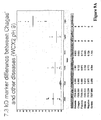
- the at least one biomarker is selected from the biomarkers of molecular weight 8.351 kDa, 9.3 kDa, 7.3 kDa, 6.04 kDa, 4.4 kDa, 4.07 kDa and 5.1 kDa, as depicted in Figures 7-9 .
- the present invention provides a kit comprising:
- the capture reagent is a cation exchange adsorbent.
- the kit additionally comprises an anion exchange chromatography adsorbent.
- the present invention provides a purified biomolecule selected from the group of biomarkers consisting of the 28.7 kDa N-terminal fragment corresponding to amino acid residues 1-258 of fibronectin, the 13.6 kDa C-terminal fragment corresponding to amino acid residues 124-243 of ApoA-I, the 24.7 kDa N-terminal fragment corresponding to amino acid residues 1-214 of Apo A-I, the 9.3 kDa C-terminal fragment corresponding to amino acid residues 161-243 of ApoA-I, the 10.1 kDa C-terminal fragment corresponding to amino acid residues 154-243 of ApoA-I, the 8.1 kDa N-terminal fragment corresponding to amino acid residues 1-68 of C3 anaphylatoxin, and the 16.3 kDa dimer corresponding to amino acid residues 1-68 of C3 anaphylatoxin.
- the present invention provides a method for qualifying Chagas disease status in a subject in comparison to the status of a different parasitic disease (i.e., a non-Chagas parasitic disease), the method comprising:
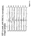
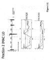
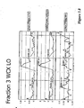
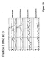
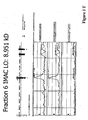
- FIGURE IA-W shows representative mass spectra displaying several biomarkers of the invention and providing their mass-to-charge ratio.
- FIGURE 2 shows the analysis of the trypsin digests of the 110 kDa Chagas disease biomarker.
- FIGURE 3A-B shows the results of multivariate analyses utilizing five biomarkers to determine Chagas disease status.
- the detection of two or more biomarkers expressed independently of each other provides higher degrees of sensitivity and specificity for Chagas disease than may be provided by the detection of any single biomarker.
- the biomarkers used for the analysis shown in Figure 3A-B are referred to in the figure by their marker IDs, by reference to Table 1.
- FIGURE 4 shows a mass spectrophotometric analysis confirming the identity of the 8.1 kDa protein which was detected in fraction 1 on IMAC-Cu and WCX arrays using SPA as the EAM.
- Panels 1 and 2 show the spectrum of proteins bound by antiC3a antibodies and control mouse IgG antibodies, respectively. The antibodies were coupled to Protein A HyperD beads.
- Panel 3 shows the spectrum from the discovery phase of the study. The blood sample was fractionated using anion exchange chromatography and Fraction 1 was profiled using a WCX array (low laser energy).
- the proteins utilized in the model are indicated by their marker IDs, by reference to Table 1, as follows: F1WH_2 (C-terminal fragment of Apo A1 (amino acids 124-243); F3WL_8 C-terminal truncation of C3 anaphylatoxin (amino acids 1-68); F41H_4, N-terminal fragment of Apo A1 (amino acids 1-214); F1IL_3 (Double-charged peak of C3 anaphylatoxin des Arg (amino acids 1-76).
- FIGURE 5 shows a graphical representation of the differential signal intensity of a 8.127 kDa peptide from Apo-1 in asymptomatic chronically infected Venezuelan patients infected with Chagas versus acutely infected pediatric Guatemalan patients with EKG signs indicative of Chagas disease.
- This biomarker is useful for qualifying chronic Chagas disease and for distinguishing between chronic and acutely infected individuals.
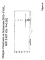
- FIGURE 6 shows a graphical representation of the differential signal intensity of a 8.937 kDa peptide in asymptomatic chronically infected Venezuelan patients infected with Chagas versus acutely infected Guatemalan patients with high EKG readings.
- This biomarker is useful for qualifying chronic Chagas disease and for distinguishing between chronic and acutely infected individuals.
- FIGURE 7 shows the differential signal intensity of a 8.351 kDa peptide in Venezuelan patients infected with Chagas compared to uninfected healthy individuals and individuals infected with different parasitic diseases, here African trypanosomiasis (sleeping sickness), malaria, and babesiosis.
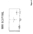
- FIGURE 8 shows the differential signal intensity of a 9.3 kDa peptide in Venezuelan patients infected with Chagas compared to individuals infected with a different parasitic diseases, here African trypanosomiasis (sleeping sickness) and malaria.
- FIGURE 9A-E demonstrates applying the methods of the present patent application to the identification of biomarkers indicating the status of other parasitic diseases, for example helminth infections, including biomarkers indicative of infection with organisms such as Fasciola hepatica, Schistosoma mansoni, Strongyloides stercoralis, Echinococcus granulosis, Trichinella nativa, Filaria, Cysticercosis and Toxocara.
- helminth infections including biomarkers indicative of infection with organisms such as Fasciola hepatica, Schistosoma mansoni, Strongyloides stercoralis, Echinococcus granulosis, Trichinella nativa, Filaria, Cysticercosis and Toxocara.
- a biomarker is an organic biomolecule which is differentially present in a sample taken from a subject of one phenotypic status (e.g ., having a disease) as compared with another phenotypic status (e.g ., not having the disease).
- a biomarker is differentially present between different phenotypic statuses if the mean or median expression level of the biomarker in the different groups is calculated to be statistically significant. Common tests for statistical significance include, among others, t-test, ANOVA, Kruskal-Wallis, Wilcoxon, Mann-Whitney and odds ratio.
- Biomarkers, alone or in combination provide measures of relative risk that a subject belongs to one phenotypic status or another. Therefore, they are useful as markers for disease (diagnostics), therapeutic effectiveness of a drug (theranostics) and drug toxicity.
- This invention provides, among other useful features, polypeptide-based biomarkers that are differentially present in subjects having Chagas disease versus healthy uninfected healthy individuals.
- the biomarkers are characterized by mass-to-charge ratio as determined by mass spectrometry, by the shape of their spectral peak in time-of-flight mass spectrometry and by their binding characteristics to adsorbent surfaces. These characteristics provide one method to determine whether a particular detected biomolecule is a biomarker of this invention. These characteristics represent inherent characteristics of the biomolecules and not process limitations in the manner in which the biomolecules are discriminated. In one aspect, this invention provides these biomarkers in isolated form.
- the biomarkers were discovered using SELDI technology employing ProteinChip arrays from Ciphergen Biosystems, Inc. (Fremont, CA) ("Ciphergen"). Serum samples were collected from subjects diagnosed with Chagas disease and subjects diagnosed as normal (non-demented). The samples were fractionated by anion exchange chromatography. Fractionated samples were applied to SELDI biochips and spectra of polypeptides in the samples were generated by time-of-flight mass spectrometry on a Ciphergen PBSII mass spectrometer. The spectra thus obtained were analyzed by Ciphergen Empress tm Data Manager Software with Biomarker Wizard and Biomarker Pattern Software from Ciphergen Biosystems, Inc.
- the mass spectra for each group were subjected to scatter plot analysis.
- a Mann-Whitney test analysis was employed to compare Chagas disease and control groups for each protein cluster in the scatter plot, and proteins were selected that differed significantly (p ⁇ 0.006, but preferably less than 0.0001) between the two groups. This method is described in more detail in the Example Section.
- the biomarkers thus discovered are presented in Tables 1-4, and additionally in Figures 5-9 .
- the "ProteinChip assay” column refers to the chromatographic fraction in which the biomarker is found, the type of biochip to which the biomarker binds and the wash conditions, as per the Example.
- F1, F2, etc. refer to "Fraction 1," "Fraction 2,” etc.
- I refers to the use of the commercially available IMAC-3 ProteinChip ("Ciphergen Biosystems, Inc.”).
- W refers to the use of the commercially available WCX2 ProteinChip ("Ciphergen Biosystems, Inc.”).
- H and L refers to the reading of SELDI-MS data at high and low intensities, respectively. This code, along with a unique number, is used to determine a Marker ID.
- all of the biomarkers disclosed herein were first discovered using ProteinChip assays which employed the use of sinapinic acid (SPA) as an Energy Absorbing Molecule (EAM), as described in greater detail below and in the Examples.
- SPA sinapinic acid
- EAM Energy Absorbing Molecule
- S which appears in the Marker ID for the biomarkers of Tables 2-4 refers to the use of SPA.
- Biomarkers may also be referred to as M### where the ### represents the biomarker's measured mass to charge ratio (M/Z), or they may be referred to by their molecular weights (e.g ., "the 110 kDa biomarker”). Where a particular biomarker has been subjected to further identification protocols, as described herein, the marker identity is indicated in the Table along with a description of its length by reference to the full-length protein (e.g ., the 13.6 kDa peak, labeled F1IH_1 in Table 1, corresponds to the C-terminal fragment of Apolipoprotein A1, amino acids 124-243).
- M/Z measured mass to charge ratio
- the marker identity is indicated in the Table along with a description of its length by reference to the full-length protein (e.g ., the 13.6 kDa peak, labeled F1IH_1 in Table 1, corresponds to the C-terminal fragment of Apolipoprotein A1, amino acids 124-243).
- the biomarkers are characterized by their mass-to-charge ratio as determined by mass spectrometry.
- the mass-to-charge ratio of each biomarker is provided in Table 1 under the column heading "M/Z".
- the mass-to-charge ratios were determined from mass spectra generated on a Ciphergen Biosystems, Inc. PBS II mass spectrometer. This instrument has a mass accuracy of about +/-0.15 percent for markers with molecular weights of approximately 20 kDa or less, and roughly 2.0% for markers of molecular weights above approximately 20 kDa. Additionally, the instrument has a mass resolution of about 400 to 1000 m/dm, where m is mass and dm is the mass spectral peak width at 0.5 peak height.
- the mass-to-charge ratio of the biomarkers was determined using Biomarker WizardTM software (Ciphergen Biosystems, Inc.). Biomarker Wizard assigns a mass-to-charge ratio to a biomarker by clustering the mass-to-charge ratios of the same peaks from all the spectra analyzed, as determined by the PBSII, taking the maximum and minimum mass-to-charge-ratio in the cluster, and dividing by two. Accordingly, the masses provided reflect these specifications.
- the biomarkers are further characterized by the shape of their spectral peak in time-of-flight mass spectrometry. Mass spectra showing the shapes of peaks corresponding to representative biomarkers are presented in FIGURE 1 .
- the biomarkers are further characterized by their binding properties on chromatographic surfaces, i.e., their ability to bind to the IMAC-2 ProteinChips versus their ability to bind to WXC2 cation exchange ProteinChips.
- biomarkers have been determined. The method by which this determination was made is described below.
- the presence of the biomarker (or nucleic acid encoding the biomarker) in a sample or subject can be determined by other methods known in the art including, but not limited to, methods such as Western blotting, Southern blotting, or PCR.
- biomarkers are characterized by mass-to-charge ratio, binding properties and spectral shape, they can be detected by mass spectrometry without knowing their specific identity.
- biomarkers whose identity is not determined can be identified by, for example, determining the amino acid sequence of the polypeptides.
- a biomarker can be peptide-mapped with a number of enzymes, such as trypsin or V8 protease, and the molecular weights of the digestion fragments can be used to search databases for sequences that match the molecular weights of the digestion fragments generated by the various enzymes.
- protein biomarkers can be sequenced using tandem MS technology. In this method, the protein is isolated by, for example, gel electrophoresis.
- a band containing the biomarker is cut out and the protein is subject to protease digestion. Individual protein fragments are separated by a first mass spectrometer. The fragment is then subjected to collision-induced cooling, which fragments the peptide and produces a polypeptide ladder. A polypeptide ladder is then analyzed by the second mass spectrometer of the tandem MS. The difference in masses of the members of the polypeptide ladder identifies the amino acids in the sequence. An entire protein can be sequenced this way, or a sequence fragment can be subjected to database mining to find identity candidates.
- the preferred biological source for detection of the biomarkers is serum.
- the biomarkers can be detected in any tissues of interest where infectious material may be found.
- stocks of blood may be tested by isolating the blood serum according to techniques well-known in the art. The serum can then be analyzed according to the techniques described herein. If the measurements taken from blood samples indicate that the individual from which the blood was taken was infected with Chagas disease, then the blood may be treated with purification agents available to one skilled in the art including, but not limited to, agents such as gentian violet, ascorbic acid, and aminoloquinolone WR6026. Alternatively, the infected blood may be discarded or destroyed and only stocks of blood which have not tested positively for Chagas disease are retained.
- the biomarkers are biomolecules. Accordingly, the biomolecules are disclosed in isolated form.
- the biomarkers can be isolated from biological fluids, such as serum. They can be isolated by any method known in the art, based on both their mass and their binding characteristics. For example, a sample comprising the biomolecules can be subject to chromatographic fractionation, as described herein, and subject to further separation by, e.g., acrylamide gel electrophoresis. Knowledge of the identity of the biomarker also allows their isolation by immunoaffinity chromatography.
- Pretranslational modified forms include allelic variants, slice variants and RNA editing forms.
- Post- translationally modified forms include forms resulting from proteolytic cleavage (e.g., fragments of a parent protein), glycosylation, phosphorylation, lipidation, oxidation, methylation, cystinylation, sulphonation and acetylation.
- proteolytic cleavage e.g., fragments of a parent protein
- modified protein cluster The collection of all modified forms of a specific protein, excluding the specific protein, itself, is referred to herein as a "modified protein cluster". Modified forms of any biomarker (including any of biomarkers listed in Tables 1-4 and the Figures herein) also may be used, themselves, as biomarkers. In certain cases the modified forms may exhibit better discriminatory power in diagnosis than the specific forms set forth herein.
- Modified forms of a biomarker can be initially detected by any methodology that can detect and distinguish the modified from the biomarker.
- a preferred method for initial detection involves first capturing the biomarker and modified forms of it, e.g., with biospecific capture reagents, and then detecting the captured proteins by mass spectrometry. More specifically, the proteins are captured using biospecific capture reagents, such as antibodies, aptamers or Affibodies that recognize the biomarker and modified forms of it. This method also will also result in the capture of protein interactors that are bound to the proteins or that are otherwise recognized by antibodies and that, themselves, can be biomarkers.
- the biospecific capture reagents are bound to a solid phase.
- the captured proteins can be detected by SELDI mass spectrometry or by eluting the proteins from the capture reagent and detecting the eluted proteins by traditional MALDI or by SELDI.
- SELDI mass spectrometry is especially attractive because it can distinguish and quantify modified forms of a protein based on mass and without the need for labeling.
- the biospecific capture reagent is bound to a solid phase, such as a bead, a plate, a membrane or a chip.
- a solid phase such as a bead, a plate, a membrane or a chip.
- Methods of coupling biomolecules, such as antibodies, to a solid phase are well known in the art. They can employ, for example, bifunctional linking agents, or the solid phase can be derivatized with a reactive group, such as an epoxide or an imidizole, that will bind the molecule on contact.
- Biospecific capture reagents against different target proteins can be mixed in the same place, or they can be attached to solid phases in different physical or addressable locations. For example, one can load multiple columns with derivatized beads, each column able to capture a single protein cluster.
- antibody-derivatized bead-based technologies such as xMAP technology of Luminex (Austin, TX) can be used to detect the protein clusters.
- the biospecific capture reagents must be specifically directed toward the members of a cluster in order to differentiate them.
- biochips can be derivatized with the capture reagents directed against protein clusters either in the same location or in physically different addressable locations.
- One advantage of capturing different clusters in different addressable locations is that the analysis becomes simpler.
- the modified form can be used as a biomarker in any of the methods disclosed herein.
- detection of the modified from can be accomplished by any specific detection methodology including affinity capture followed by mass spectrometry, or traditional immunoassay directed specifically the modified form.
- biospecific capture reagents such as antibodies
- the assay must be designed to specifically distinguish protein and modified forms of protein. This can be done, for example, by employing a sandwich assay in which one antibody captures more than one form and second, distinctly labeled antibodies, specifically bind, and provide distinct detection of, the various forms.
- Antibodies can be produced by immunizing animals with the biomolecules.
- Traditional immunoassays including, for example, sandwich immunoassays including ELISA or fluorescence-based immunoassays, as well as other enzyme immunoassays are contemplated.
- the biomarkers can be detected by any suitable method. Detection paradigms that can be employed to this end include optical methods, electrochemical methods (voltametry and amperometry techniques), atomic force microscopy, and radio frequency methods, e.g., multipolar resonance spectroscopy. Illustrative of optical methods, in addition to microscopy, both confocal and non-confocal, are detection of fluorescence, luminescence, chemiluminescence, absorbance, reflectance, transmittance, and birefringence or refractive index (e.g., surface plasmon resonance, ellipsometry, a resonant mirror method, a grating coupler waveguide method or interferometry).
- optical methods in addition to microscopy, both confocal and non-confocal, are detection of fluorescence, luminescence, chemiluminescence, absorbance, reflectance, transmittance, and birefringence or refractive index (e.g., surface plasmon resonance,
- Biochips generally comprise solid substrates and have a generally planar surface, to which a capture reagent (also called an adsorbent or affinity reagent) is attached. Frequently, the surface of a biochip comprises a plurality of addressable locations, each of which has the capture reagent bound there.
- a capture reagent also called an adsorbent or affinity reagent
- Protein biochip refers to a biochip adapted for the capture of polypeptides.
- Many protein biochips are described in the art. These include, for example, protein biochips produced by Ciphergen Biosystems, Inc. (Fremont, CA), Packard BioScience Company (Meriden CT), Zyomyx (Hayward, CA), Phylos (Lexington, MA) and Biacore (Uppsala, Sweden). Examples of such protein biochips are described in the following patents or published patent applications: U.S. Patent No. 6,225,047 ; PCT International Publication No. WO 99/51773 ; U.S. Patent No. 6,329,209 , PCT International Publication No. WO 00/56934 and U.S. Patent No. 5,242,828 .
- the biomarkers are detected by mass spectrometry, a method that employs a mass spectrometer to detect gas phase ions.
- mass spectrometers are time-of-flight, magnetic sector, quadrupole filter, ion trap, ion cyclotron resonance, electrostatic sector analyzer and hybrids of these.
- the mass spectrometer is a laser desorption/ionization mass spectrometer.
- the analytes are placed on the surface of a mass spectrometry probe, a device adapted to engage a probe interface of the mass spectrometer and to present an analyte to ionizing energy for ionization and introduction into a mass spectrometer.
- a laser desorption mass spectrometer employs laser energy, typically from an ultraviolet laser, but also from an infrared laser, to desorb analytes from a surface, to volatilize and ionize them and make them available to the ion optics of the mass spectrometer.
- a preferred mass spectrometric technique for use in the invention is "Surface Enhanced Laser Desorption and Ionization"or "SELDI,"as described, for example, in U.S. Patents No. 5,719,060 and No. 6,225,047, both to Hutches and Yip .
- This refers to a method of desorption/ionization gas phase ion spectrometry (e.g., mass spectrometry) in which an analyte (here, one or more of the biomarkers) is captured on the surface of a SELDI mass spectrometry probe.
- SELDI analyte
- SELDI affinity capture mass spectrometry
- SEAC Surface-Enhanced Affinity Capture
- This version involves the use of probes that have a material on the probe surface that captures analytes through a noncovalent affinity interaction (adsorption) between the material and the analyte.
- the material is variously called an “adsorbent,” a “capture reagent,” an “affinity reagent” or a “binding moiety.”
- Such probes can be referred to as “affinity capture probes” and as having an “adsorbent surface.”
- the capture reagent can be any material capable of binding an analyte.
- the capture reagent may be attached directly to the substrate of the selective surface, or the substrate may have a reactive surface that carries a reactive moiety that is capable of binding the capture reagent, e.g ., through a reaction forming a covalent or coordinate covalent bond.
- Epoxide and carbodiimidizole are useful reactive moieties to covalently bind polypeptide capture reagents such as antibodies or cellular receptors.
- Nitriloacetic acid and iminodiacetic acid are useful reactive moieties that function as chelating agents to bind metal ions that interact non-covalently with histidine containing peptides.
- Adsorbents are generally classified as chromatographic adsorbents and biospecific adsorbents.
- Chromatographic adsorbent refers to an adsorbent material typically used in chromatography. Chromatographic adsorbents include, for example, ion exchange materials, metal chelators (e.g ., nitriloacetic acid or iminodiacetic acid), immobilized metal chelates, hydrophobic interaction adsorbents, hydrophilic interaction adsorbents, dyes, simple biomolecules ( e.g ., nucleotides, amino acids, simple sugars and fatty acids) and mixed mode adsorbents (e.g ., hydrophobic attraction/electrostatic repulsion adsorbents).
- metal chelators e.g ., nitriloacetic acid or iminodiacetic acid
- immobilized metal chelates e.g nitriloacetic acid or iminodiacetic acid
- hydrophobic interaction adsorbents e.g ., hydrophilic interaction a
- Biospecific adsorbent refers to an adsorbent comprising a biomolecule, e.g ., a nucleic acid molecule (e.g ., an aptamer), a polypeptide, a polysaccharide, a lipid, a steroid or a conjugate of these (e.g ., a glycoprotein, a lipoprotein, a glycolipid, a nucleic acid (e.g ., DNA)-protein conjugate).
- the biospecific adsorbent can be a macromolecular structure such as a multiprotein complex, a biological membrane or a virus. Examples of biospecific adsorbents are antibodies, receptor proteins and nucleic acids.
- Biospecific adsorbents typically have higher specificity for a target analyte than chromatographic adsorbents. Further examples of adsorbents for use in SELDI can be found in U.S. Patent No. 6,225,047 .
- a "bioselective adsorbent” refers to an adsorbent that binds to an analyte with an affinity of at least 10 -8 M.
- Ciphergen Biosystems, Inc. comprise surfaces having chromatographic or biospecific adsorbents attached thereto at addressable locations.
- Ciphergen ProteinChip ® arrays include NP20 (hydrophilic); H4 and H50 (hydrophobic); SAX-2, Q-10 and LSAX-30 (anion exchange); WCX-2, CM-10 and LWCX-30 (cation exchange); IMAC-3, IMAC-30 and IMAC 40 (metal chelate); and PS-10, PS-20 (reactive surface with carboimidizole, expoxide) and PG-20 (protein G coupled through carboimidizole).
- Hydrophobic ProteinChip arrays have isopropyl or nonylphenoxy-poly(ethylene glycol)methacrylate functionalities.
- Anion exchange ProteinChip arrays have quaternary ammonium functionalities.
- Cation exchange ProteinChip arrays have carboxylate functionalities.
- Immobilized metal chelate ProteinChip arrays have have nitriloacetic acid functionalities that adsorb transition metal ions, such as copper, nickel, zinc, and gallium, by chelation.
- Preactivated ProteinChip arrays have carboimidizole or epoxide functional groups that can react with groups on proteins for covalent binding.
- a probe with an adsorbent surface is contacted with the sample for a period of time sufficient to allow biomarker or biomarkers that may be present in the sample to bind to the adsorbent.
- the substrate is washed to remove unbound material. Any suitable washing solutions can be used; preferably, aqueous solutions are employed.
- the extent to which molecules remain bound can be manipulated by adjusting the stringency of the wash. The elution characteristics of a wash solution can depend, for example, on pH, ionic strength, hydrophobicity, degree of chaotropism, detergent strength, and temperature.
- an energy absorbing molecule then is applied to the substrate with the bound biomarkers.
- the biomarkers bound to the substrates are detected in a gas phase ion spectrometer such as a time-of flight mass spectrometer.
- the biomarkers are ionized by an ionization source such as a laser, the generated ions are collected by an ion optic assembly, and then a mass analyzer disperses and analyzes the passing ions.
- the detector then translates information of the detected ions into mass-to-charge ratios. Detection of a biomarker typically will involve detection of signal intensity. Thus, both the quantity and mass of the biomarker can be determined.
- SELDI Surface-Enhanced Neat Desorption
- SEND probe Another version of SELDI is Surface-Enhanced Neat Desorption (SEND), which involves the use of probes comprising energy absorbing molecules that are chemically bound to the probe surface (“SEND probe”).
- energy absorbing molecules (EAM) denotes molecules that are capable of absorbing energy from a laser desorption/ionization source and, thereafter, contribute to desorption and ionization of analyte molecules in contact therewith.
- the EAM category includes molecules used in MALDI, frequently referred to as "matrix,” and is exemplified by cinnamic acid derivatives, sinapinic acid (SPA), cyano-hydroxy-cinnamic acid (CHCA) and dihydroxybenzoic acid, ferulic acid, and hydroxyaceto-phenone derivatives.
- SPA sinapinic acid
- CHCA cyano-hydroxy-cinnamic acid
- dihydroxybenzoic acid ferulic acid
- the energy absorbing molecule is incorporated into a linear or cross-linked polymer, e.g ., a polymethacrylate.
- the composition can be a co-polymer of ⁇ -cyano-4-methacryloyloxycinnamic acid and acrylate.
- the composition is a co-polymer of ⁇ -cyano-4-methacryloyloxycinnamic acid, acrylate and 3-(tri-ethoxy)silyl propyl methacrylate.
- the composition is a co-polymer of ⁇ -cyano-4-methacryloyloxycinnamic acid and octadecylmethacrylate ("C18 SEND").
- SEND is further described in U.S. Patent No. 6,124,137 and PCT International Publication No. WO 03/64594 (Kitagawa , "Monomers And Polymers Having Energy Absorbing Moieties Of Use In Desorption/Ionization Of Analytes," August 7, 2003).
- SEAC/SEND is a version of SELDI in which both a capture reagent and an energy absorbing molecule are attached to the sample presenting surface. SEAC/SEND probes therefore allow the capture of analytes through affinity capture and ionization/desorption without the need to apply external matrix.
- the C 18 SEND biochip is a version of SEAC/SEND, comprising a C18 moiety which functions as a capture reagent, and a CHCA moiety which functions as an energy absorbing moiety.
- SELDI Surface-Enhanced Photolabile Attachment and Release
- SEPAR involves the use of probes having moieties attached to the surface that can covalently bind an analyte, and then release the analyte through breaking a photolabile bond in the moiety after exposure to light, e.g ., to laser light ( see, U.S. Patent No. 5,719,060 ).
- SEPAR and other forms of SELDI are readily adapted to detecting a biomarker or biomarker profile, pursuant to the present invention.
- the biomarkers can be first captured on a chromatographic resin having chromatographic properties that bind the biomarkers.
- this could include a variety of methods. For example, one could capture the biomarkers on a cation exchange resin, such as CM Ceramic HyperD F resin, wash the resin, elute the biomarkers and detect by MALDI.
- this method could be preceded by fractionating the sample on an anion exchange resin before application to the cation exchange resin.
- one could fractionate on an anion exchange resin and detect by MALDI directly.
- Time-of-flight mass spectrometry Analysis of analytes by time-of-flight mass spectrometry generates a time-of-flight spectrum.
- the time-of-flight spectrum ultimately analyzed typically does not represent the signal from a single pulse of ionizing energy against a sample, but rather the sum of signals from a number of pulses. This reduces noise and increases dynamic range.
- This time-of-flight data is then subject to data processing.
- data processing typically includes TOF-to-M/Z transformation to generate a mass spectrum, baseline subtraction to eliminate instrument offsets and high frequency noise filtering to reduce high frequency noise.
- Data generated by desorption and detection of biomarkers can be analyzed with the use of a programmable digital computer.
- the computer program analyzes the data to indicate the number of biomarkers detected, and optionally the strength of the signal and the determined molecular mass for each biomarker detected.
- Data analysis can include steps of determining signal strength of a biomarker and removing data deviating from a predetermined statistical distribution. For example, the observed peaks can be normalized, by calculating the height of each peak relative to some reference.
- the reference can be background noise generated by the instrument and chemicals such as the energy absorbing molecule which is set at zero in the scale.
- the computer can transform the resulting data into various formats for display.
- the standard spectrum can be displayed, but in one useful format only the peak height and mass information are retained from the spectrum view, yielding a cleaner image and enabling biomarkers with nearly identical molecular weights to be more easily seen.
- two or more spectra are compared, conveniently highlighting unique biomarkers and biomarkers that are up-or down-regulated between samples. Using any of these formats, one can readily determine whether a particular biomarker is present in a sample.
- Peak Analysis generally involves the identification of peaks in the spectrum that represent signal from an analyte. Peak selection can be done visually, but software is available, as part of Ciphergen's ProteinChip® software package, that can automate the detection of peaks. In general, this software functions by identifying signals having a signal- to-noise ratio above a selected threshold and labeling the mass of the peak at the centroid of the peak signal. In one useful application, many spectra are compared to identify identical peaks present in some selected percentage of the mass spectra. One version of this software clusters all peaks appearing in the various spectra within a defined mass range, and assigns a mass (M/Z) to all the peaks that are near the mid-point of the mass (M/Z) cluster.
- M/Z mass
- Software used to analyze the data can include code that applies an algorithm to the analysis of the signal to determine whether the signal represents a peak in a signal that corresponds to a biomarker according to the present invention.
- the software also can subject the data regarding observed biomarker peaks to classification tree or ANN analysis, to determine whether a biomarker peak or combination of biomarker peaks is present that indicates the status of the particular clinical parameter under examination. Analysis of the data may be "keyed" to a variety of parameters that are obtained, either directly or indirectly, from the mass spectrometric analysis of the sample.
- These parameters include, but are not limited to, the presence or absence of one or more peaks, the shape of a peak or group of peaks, the height of one or more peaks, the log of the height of one or more peaks, and other arithmetic manipulations of peak height data.
- a preferred protocol for the detection of biomarkers is as follows.
- the biological sample to be tested e.g., serum
- the biological sample to be tested e.g., serum
- pre-fractionation Prior to the pre-fractionation, the serum (20 ⁇ l) is denatured using a 9 M urea/2% Chaps/50 mM Tris pH 9.0 buffer (U9 buffer). 30 ⁇ l of U9 is added to the 20 ⁇ l of serum and then this diluted serum is subjected to anion exchange fractionation.
- a preferred method of pre-fractionation involves contacting the sample with an anion exchange chromatographic material, such as Q HyperD (BioSepra, SA, a division of Ciphergen Biosystems, Inc.).
- the bound materials are then subject to stepwise pH elution using buffers at pH 9, pH 7, pH 5, pH 4 and pH 3. (See Example 1-Buffer list) as well as an organic solvent elution (The fractions in which the biomarkers are eluted are also indicated in Tables 1-4 and in the Figures by reference to the Marker IDs, e. g., F1IH_#, F2WSL_#, etc. ). Various fractions containing the biomarker are collected.
- the sample to be tested (preferably pre-fractionated) is then contacted with an affinity capture probe comprising a cation exchange adsorbent (preferably a WCX ProteinChip array (Ciphergcn Biosystems, Inc.)) or an IMAC adsorbent (preferably an IMAC3 ProteinChip array (Ciphergen Biosystems, Inc.)), again as indicated in the Marker IDs listed in Tables 1-4 and in the Figures.
- a cation exchange adsorbent preferably a WCX ProteinChip array (Ciphergcn Biosystems, Inc.)
- an IMAC adsorbent preferably an IMAC3 ProteinChip array (Ciphergen Biosystems, Inc.)
- a probe such as a pre-activated PS 10 or PS20 ProteinChip array (Ciphergen Biosystems, Inc.). These antibodies can capture the biomarkers from a sample onto the probe surface. Then the biomarkers can be detected by, e. g., laser desorption/ionization mass spectrometry.
- the biomarkers can be measured by immunoassay.
- hnmunoassay requires biospecific capture reagents, such as antibodies, to capture the biomarkers.
- Antibodies can be produced by methods well known in the art, e.g., by immunizing animals with the biomarkers. Biomarkers can be isolated from samples based on their binding characteristics. Alternatively, if the amino acid sequence of a polypeptide biomarker is known, the polypeptide can be synthesized and used to generate antibodies by methods well known in the art.
- biospecific capture reagent for the biomarker is attached to the surface of an MS probe, such as a pre- activated ProteinChip array. The biomarker is then specifically captured on the biochip through this reagent, and the captured biomarker is detected by mass spectrometry.
- the biomarkers can be used in diagnostic tests to assess Chagas disease status in a subject, e.g., to diagnose Chagas disease.
- the phrase "Chagas disease status” includes distinguishing, inter alia, chronic Chagas disease versus non-Chagas disease and, in particular, chronic asymptomatic Chagas disease versus non- infection or acute Chagas disease status versus non-infection. Based on this status, further procedures may be indicated, including additional diagnostic tests or therapeutic procedures or regimens.
- the power of a diagnostic test to correctly predict status is commonly measured as the sensitivity of the assay, the specificity of the assay or the area under a receiver operated characteristic ("ROC") curve.
- Sensitivity is the percentage of true positives that are predicted by a test to be positive, while specificity is the percentage of true negatives that are predicted by a test to be negative.
- An ROC curve provides the sensitivity of a test as a function of 1-specificity. The greater the area under the ROC curve, the more powerful the predictive value of the test.
- Other useful measures of the utility of a test are positive predictive value and negative predictive value. Positive predictive value is the percentage of actual positives who test as positive. Negative predictive value is the percentage of actual negatives that test as negative.
- the biomarkers show a statistical difference in different Chagas disease statuses of at least p ⁇ 0. 05, p ⁇ 10 -2 , p ⁇ 10 -3 , p ⁇ 10-4 or p ⁇ 10-5. Diagnostic tests that use these biomarkers alone or in combination show a sensitivity and specificity of at least 75%, at least 80%, at least 85%, at least 90%, at least 95%, at least 98% and about 100%.
- Each biomarker listed in Tables 1-4 and the Figures is differentially present in Chagas disease, and, therefore, each is individually useful in aiding in the determination of Chagas disease status.
- the method involves, first, measuring the selected biomarker in a subject sample using the methods described herein, e.g., capture on a SELDI biochip followed by detection by mass spectrometry and, second, comparing the measurement with a diagnostic amount or cut-off that distinguishes a positive Chagas disease status from a negative Chagas disease status.
- the diagnostic amount represents a measured amount of a biomarker above which or below which a subject is classified as having a particular Chagas disease status.
- the biomarker is up-regulated compared to normal during Chagas disease, then a measured amount above the diagnostic cut-off provides a diagnosis of Chagas disease.
- a measured amount below the diagnostic cutoff provides a diagnosis of Chagas disease.
- the particular diagnostic cut-off can be determined, for example, by measuring the amount of the biomarker in a statistically significant number of samples from subjects with the different Chagas disease statuses, as was done here, and drawing the cut-off to suit the diagnostician's desired levels of specificity and sensitivity.
- biomarkers While individual biomarkers are useful diagnostic biomarkers, it has been found that a combination of biomarkers can provide greater predictive value of a particular status than single biomarkers alone. Specifically, the detection of a plurality of biomarkers in a sample can increase the sensitivity and/or specificity of the test.
- Example II the protocols described in Example I below were used to generate mass spectra from 73 Venezuelan patient samples. Of these 73 samples, 39 of the samples were obtained from patients chronically infected with Chagas disease and 34 were taken from healthy individuals living in the same endemic region. The peak masses and heights were abstracted into a discovery data set. This data set was used to train a learning algorithm employing classification and regression tree analysis (CART) (Ciphergen Biomarker Patterns SoftwareTM). In particular, CART chose many subsets of the peaks at random. For each subset, CART generated a best or near best decision tree to classify a sample as Chagas disease or non-Chagas disease. Among the many decision trees generated by CART, several had excellent sensitivity and specificity in distinguishing Chagas disease from non-Chagas disease.
- CART classification and regression tree analysis
- FIGURE 3 An exemplary decision tree for qualifying the Chagas disease status of a sample taken from a subject is presented in FIGURE 3 .
- the identity of the biomarkers used is indicated in FIGURE 3 , by reference to Table 1.
- the biomarker in "Node 1" in FIGURE 3 is F1WH_2, corresponding to the Chagas disease biomarker with an estimated mass of 13.6 kD.
- the specificity and sensitivity of the multiple-biomarker analysis increases as the number of biomarkers in the decision tree is increased.
- FIGURE 4B shows that 100% specificity and greater than 94% sensitivity can be achieved using the 5 biomarkers selected in this example.
- the specifics of the decision trees in particular the cut-off values used in making branching decisions, depends on the details of the assay used to generate the discovery data set.
- the data acquisition parameters of the assay that produced the data used in the present analysis is provided in the Example.
- the operator In developing a classification algorithm from, for example, a new sample set or a different assay protocol, the operator uses a protocol that detects these biomarkers and keys the learning algorithm to include them.
- the methods further provide for specifically qualifying Chagas disease status in a subject in comparison to the status of a different parasitic disease (i.e., a non- Chagas disease), the method comprising: (a) measuring at least one biomarker in a biological sample from the subject, wherein the at least one biomarker specifically indicates the presence of Chagas disease and does not indicate the presence of a different parasitic infection; and (b) correlating the measurement with Chagas disease status in comparison to the status of a different parasitic infection.
- the biological sample is a serum sample.
- biomarkers specifically identify the presence or absence of Chagas disease as distinguished from a different parasitic infection, including a protozoa, a helminth or a malarial infection.
- the biomarkers specifically identify the presence or absence of Chagas disease as distinguished from other protozoal infections, including Leishmaniasis, African trypanosomiasis (sleeping sickness) and babesiosis.
- the biomarkers specifically identify the presence or absence of Chagas disease as distinguished from other kinetoplastidae or trypanosomal infections, including Leishmaniasis and African trypanosomiasis.
- the biomarkers specifically identify the presence or absence of an infection with T.
- the biomarkers that specifically identify the presence or absence of a Chagas disease infection as distinguished from another parasitic disease are selected from the group consisting of biomarkers of molecular weight 8.351 kDa, 9.3 kDa, 7.3 kDa, 6.04 kDa, 4.4 kDa, 4.07 kDa and 5.1 kDa, as depicted in Figures 7-9 .
- the presence of a biomarker specific for Chagas disease, or the presence of a biomarker specific for Chagas disease above (or below) a cut-off level is indicative of infection by T. cruzi and is indicative of Chagas disease in an individual.
- Biomarker amounts or patterns are characteristic of various risk states, e.g., high, medium or low.
- the risk of developing Chagas disease is determined by measuring the relevant biomarker or biomarkers and then either submitting them to a classification algorithm or comparing them with a reference amount and/or pattern of biomarkers that is associated with the particular risk level.
- Each stage of Chagas disease has a characteristic amount of a biomarker or relative amounts of a set of biomarkers (a pattern).
- the stage of Chagas disease is determined by measuring the relevant biomarker or biomarkers and then either submitting them to a classification algorithm or comparing them with a reference amount and/or pattern of biomarkers that is associated with the particular stage.
- Disease course refers to changes in disease status over time, including disease progression (worsening) and disease regression (improvement).
- the amounts or relative amounts (e.g., the pattern) of the biomarkers changes.
- certain biomarkers increase with Chagas disease progresson or regression, while other biomarkers decrease with Chagas disease progression or regression. Therefore, the trend of these markers, either increased or decreased over time toward diseased or non- diseased indicates the course of the disease.
- this method involves measuring one or more biomarkers in a subject at at least two different time points, e.g., a first time and a second time, and comparing the change in amounts, if any.
- Chagas disease The course of Chagas disease is determined based on these comparisons. Similarly, this method is useful for determining the response to treatment. If a treatment is effective, then the biomarkers will trend toward normal, while if treatment is ineffective, the biomarkers will trend toward disease indications.
- Also disclosed are methods of qualifying Chagas disease status which further comprise managing subject treatment based on the status.
- Such management includes the actions of the physician or clinician subsequent to determining Chagas disease status.
- a physician makes a diagnosis of Chagas disease
- a certain regime of treatment may be administered, such as drugs shown to be effective in the treatment of Chagas disease, such as nifurtimox, benznidazole or allopurinol.
- a negative diagnosis of Chagas disease in an individual exhibiting Chagas-associated symptoms might be followed with further testing to determine if the patient is suffering from an infection by a parasite related to T. cruzi or an illness unrelated to infection by a trypanosome. If the diagnostic test gives an inconclusive result on Chagas disease status, further tests may be called for.
- Additional disclosed methods relate to the communication of assay results or diagnoses or both to technicians, physicans or patients, for example.
- computers will be used to communicate assay results or diagnoses or both to interested parties, e.g., physicians and their patients.
- the assays will be performed or the assay results analyzed in a country or jurisdiction which differs from the country or jurisdiction to which the results or diagnoses are communicated.
- a diagnosis based on the presence or absence in a test subject of any of the biomarkers listed in Table 1-4 or in the Figures may be communicated to the subject as soon as possible after the diagnosis is obtained.
- the diagnosis may be communicated to the subject by the subject's treating physician.
- the diagnosis may be sent to a test subject by email or communicated to the subject by phone.
- a computer may be used to communicate the diagnosis by email or phone.
- the message containing results of a diagnostic test may be generated and delivered automatically to the subject using a combination of computer hardware and software which will be familiar to artisans skilled in telecommunications.
- One example of a healthcare-oriented communications system is described in U.S. Patent Number 6,283,761 ; however, the present invention is not limited to methods which utilize this particular communications system.
- all or some of the method steps, including the assaying of samples, diagnosing of diseases, and communicating of assay results or diagnoses may be carried out in diverse (e.g., foreign) jurisdictions.
- Data derived from the spectra can be used to "train” a classification model.
- a "known sample” is a sample that has been pre-classified.
- the data that are derived from the spectra and are used to form the classification model can be referred to as a "training data set”.
- the classification model can recognize patterns in data derived from spectra generated using unknown samples.
- the classification model can then be used to classify the unknown samples into classes. This can be useful, for example, in predicting whether or not a particular biological sample is associated with a certain biological condition (e.g., Chagas disease versus uninfected, or asymptomatic Chagas diseased versus acute Chagas disease).
- the training data set that is used to form the classification model may comprise raw data or pre-processed data.
- raw data can be obtained directly from time-of-flight spectra or mass spectra, and then may be optionally "pre- processed" as described above.
- Classification models can be formed using any suitable statistical classification (or "learning") method that attempts to segregate bodies of data into classes based on objective parameters present in the data.
- Classification methods may be either supervised or unsupervised. Examples of supervised and unsupervised classification processes are described in Jain, "Statistical Pattern Recognition: A Review", IEEE Transactions on Pattern Analysis and Machine Intelligence, Vol. 22, No. 1, January 2000 .
- supervised classification training data containing examples of known categories are presented to a learning mechanism, which learns one or more sets of relationships that define each of the known classes. New data may then be applied to the learning mechanism, which then classifies the new data using the learned relationships.
- supervised classification processes include linear regression processes (e. g., multiple linear regression (MLR), partial least squares (PLS) regression and principal components regression (PCR) ), binary decision trees (e.g., recursive partitioning processes such as CART-classification and regression trees), artificial neural networks such as back propagation networks, discriminant analyses (e. g., Bayesian classifier or Fischer analysis), logistic classifiers, and upport vector classifiers (support vector machines).
- LLR multiple linear regression
- PLS partial least squares
- PCR principal components regression
- binary decision trees e.g., recursive partitioning processes such as CART-classification and regression trees
- artificial neural networks such as back propagation networks
- discriminant analyses e. g., Bayesian classifier or Fischer
- a preferred supervised classification method is a recursive partitioning process.
- Recursive partitioning processes use recursive partitioning trees to classify spectra derived from unknown samples. Further details about recursive partitioning processes are provided in U. S. Patent Application No. 2002 0138208 Al to Paulse et al. , "Method for analyzing mass spectra".
- Unsupervised classification attempts to learn classifications based on similarities in the training data set, without pre-classifying the spectra from which the training data set was derived.
- Unsupervised learning methods include cluster analyses. A cluster analysis attempts to divide the data into "clusters" or groups that ideally should have members that are very similar to each other, and very dissimilar to members of other clusters. Similarity is then measured using some distance metric, which measures the distance between data items, and clusters together data items that are closer to each other.
- Clustering techniques include the MacQueen's K-means algorithm and the Kohonen's Self- Organizing Map algorithm.
- the classification models can be formed on and used on any suitable digital computer.
- Suitable digital computers include micro, mini, or large computers using any standard or specialized operating system, such as a Unix, WindowsTM or LinusTM based operating system.
- the digital computer that is used may be physically separate from the mass spectrometer that is used to create the spectra of interest, or it may be coupled to the mass spectrometer.
- the training data set and the classification models according to embodiments of the invention can be embodied by computer, code that is executed or used by a digital computer.
- the computer code can be stored on any suitable computer readable media including optical or magnetic disks, sticks, tapes, etc. , and can be written in any suitable computer programming language including C, C++, visual basic, etc.
- the learning algorithms described above are useful both for developing classification algorithms for the biomarkers already discovered, or for finding new biomarkers for Chagas disease.
- the classification algorithms form the base for diagnostic tests by providing diagnostic values (e. g., cut-off points) for biomarkers used singly or in combination.
- kits for qualifying Chagas disease status which kits are used to detect biomarkers according to the invention.
- the kit comprises a solid support, such as a chip, a microtiter plate or a bead or resin having a capture reagent attached thereon, wherein the capture reagent binds a biomarker of the invention.
- the kits of the present invention can comprise mass spectrometry probes for SELDI, such as ProteinChip® arrays.
- the kit can comprise a solid support with a reactive surface, and a container comprising the biospecific capture reagent.
- the kit can also comprise a washing solution or instructions for making a washing solution, in which the combination of the capture reagent and the washing solution allows capture of the biomarker or biomarkers on the solid support for subsequent detection by, e.g., mass spectrometry.
- the kit may include more than type of adsorbent, each present on a different solid support.
- such a kit can comprise instructions for suitable operational parameters in the form of a label or separate insert.
- the instructions may inform a consumer about how to collect the sample, how to wash the probe or the particular biomarkers to be detected.
- the kit can comprise one or more containers with biomarker samples, to be used as standard (s) for calibration.
- BIOMARKERS FOR CHAGAS DISEASE IN SCREENING ASSAYS
- the biomarkers can be used to screen for compounds that modulate the expression of the biomarkers in vitro or in vivo, which compounds in turn may be useful in treating or preventing Chagas disease in patients.
- the biomarkers can be used to monitor the response to treatments for Chagas disease.
- the biomarkers can be used in heredity studies to determine if the subject is at risk for developing Chagas disease.
- a kit could include a solid substrate having an cation exchange function, such as a protein biochip (e.g., a Ciphergen WCX2 ProteinChip array) and a sodium acetate buffer for washing the substrate, as well as instructions providing a protocol to measure the biomarkers of this invention on the chip and to use these measurements to diagnose Chagas disease.
- a protein biochip e.g., a Ciphergen WCX2 ProteinChip array
- a sodium acetate buffer for washing the substrate
- instructions providing a protocol to measure the biomarkers of this invention on the chip and to use these measurements to diagnose Chagas disease.
- Compounds suitable for therapeutic testing may be screened initially by identifying compounds which interact with one or more biomarkers listed in Tables 1-4 or in the Figures herein.
- screening might include recombinantly expressing a biomarker listed in Table 1-4 or in the Figures, purifying the biomarker, and affixing the biomarker to a substrate.
- Test compounds would then be contacted with the substrate, typically in aqueous conditions, and interactions between the test compound and the biomarker are measured, for example, by measuring elution rates as a function of salt concentration.
- Certain proteins may recognize and cleave one or more biomarkers of Tables 1-4, in which case the proteins may be detected by monitoring the digestion of one or more biomarkers in a standard assay, e. g. , by gel electrophoresis of the proteins.
- test compound to inhibit the activity (i.e., non-enzymatic or enzymatic) of one or more of the biomarkers of Table 1-4 or the Figures may be measured.
- activity i.e., non-enzymatic or enzymatic
- One of skill in the art will recognize that the techniques used to measure the activity of a particular biomarker will vary depending on the function and properties of the biomarker. For example, an enzymatic activity of a biomarker may be assayed provided that an appropriate substrate is available and provided that the concentration of the substrate or the appearance of the reaction product is readily measurable.
- the ability of potentially therapeutic test compounds to inhibit or enhance the activity of a given biomarker may be determined by measuring the rates of catalysis in the presence or absence of the test compounds.
- test compound to interfere with a non- enzymatic (e. g., structural) function or activity of one of the biomarkers of Table 1-4 or of the Figures may also be measured.
- a non- enzymatic e. g., structural
- the self-assembly of a multi-protein complex which includes one of the biomarkers described herein may be monitored by spectroscopy in the presence or absence of a test compound.
- Test compounds capable of modulating the activity or expression of any of the biomarkers described in Tables 1-4 or the Figures may be administered to patients who are suffering from or are at risk of developing Chagas disease.
- the administration of a test compound which increases the activity of a particular biomarker may diminish the symptoms of Chagas disease in a patient if the activity of the particular biomarker in vivo prevents the accumulation of harmful metabolites associated with Chagas disease.
- a test compound which decreases the activity or expression of a particular biomarker may diminish or alleviate the symptoms of Chagas disease in a patient if the increased activity or expression is responsible, at least in part, for the onset of Chagas disease or for the ability of the parasite to propogate and effectuate the disease state in a patient.
- cell extracts or expression libraries may be screened for compounds which inhibit the cleavage of the full-length proteins associated with the biomarkers listed in the Tables, including MIP-1a, M110, Apo 1A, Fibronectin, and C3 anaphylatoxin, to form truncated forms of these biomarker proteins.
- cleavage of biomarker proteins may be detected by attaching a fluorophore to the biomarker protein which remains quenched when the biomarker protein is uncleaved but which fluoresces when the protein is cleaved.
- a version of a full-length biomarker protein including MIP-1a, M110, Apo 1A, Fibronectin, and C3 anaphylatoxin, or any other biomarker described herein may be modified so as to render the amide bond between amino acids x and y uncleavable may be used to selectively bind or "trap" the cellular protease which cleaves full-length biomarker protein at that site in vivo.
- Methods for screening and identifying proteases and their targets are well-documented in the scientific literature, e. g., in Lopez-Ottin et al. (Nature Reviews, 3: 509-519 (2002 )).
- a method for treating or reducing the progression or likelihood of a disease e.g., Chagas disease, which is associated with the increased levels of proteins truncated by enzymes from T. cruzi, (i.e., cruzipain).
- combinatorial libraries may be screened for compounds which inhibit the ability of a protease from T . cruzi (i.e., cruzipain) to cleave one or more of the biomarker proteins described herein.
- Methods of screening chemical libraries for such compounds are well-known in art. See, e.g., Lopez-Otin et al. (2002).
- inhibitory compounds may be intelligently designed based on the structure of T . cruzi enzymes, including cruzipain.
- screening a test compound includes obtaining samples from test subjects before and after the subjects have been exposed to a test compound.
- the levels in the samples of one or more of the biomarkers described in Tables 1-4 and in the Figures may be measured and analyzed to determine whether the levels of the biomarkers change after exposure to a test compound.
- the samples may be analyzed by mass spectrometry, as described herein, or the samples may be analyzed by any appropriate means known to one of skill in the art.
- the levels of one or more of the biomarkers described herein may be measured directly by Western blot using radio-or fluorescently- labeled antibodies which specifically bind to the biomarkers.
- changes in the levels of mRNA encoding the one or more biomarkers may be measured and correlated with the administration of a given test compound to a subject.
- the changes in the level of expression of one or more of the biomarkers may be measured using in vitro methods and materials.
- human tissue cultured cells which express, or are capable of expressing, one or more of the biomarkers described herein may be contacted with test compounds.
- Subjects who have been treated with test compounds will be routinely examined for any physiological effects which may result from the treatment.
- the test compounds will be evaluated for their ability to decrease disease likelihood in a subject.
- test compounds will be screened for their ability to slow or stop the progression of the disease.
- GITC is a salt and will inhibit binding as other salts do.
- Recommended binding buffers 50mM Tris, HEPES or Acetate (pH 4.0-9.5)
- Recommended sample dilution 50-2000 ⁇ g/ml total protein
- Stringency modifiers the addition of salts or changes in pH will alter the stringency of the binding step (see chip user notes)
- Proteins were separated on an acrylamide gel and a band containing the biomarker was cut out of the gel. The protein in the band was destained. The gel was dried using acetonitrile and then subject to digestion in a solution of trypsin. The digest fragments were analyzed on a Ciphergen PBSII mass spectrometer. The determined masses were used to interrogate the NCBInr protein database (using ProFound software), which identified the protein having the same tryptic digest pattern.
- M110 is a novel protein with homology to a predicted protein (LM15-1.32) encoded by Leishinanii major.
- LM15-1.32 predicted protein encoded by Leishinanii major.
- FIGURE 2 A summary of the search results and sequenced fragments of M110 are shown in FIGURE 2 .
- M7.861 Another biomarker with a mass of 7.8 kD, M7.861, was identified as human MIP-1 ⁇
- the protein identity was confirmed using an ELISA assay and an antibody specific to MIP-1 ⁇ .
- Measurements of MIP-1 ⁇ levels in Chagas disease infected subjects versus non-infected subjects showed that the mean level of M1P-1 ⁇ in the serum of infected subjects (20.73 pg/ml) was significantly higher than the mean level in versus in non-infected subjects (12.22 pg/ml).
- the 13.6 kDa protein was detected in fraction 1 on IMAC-Cu and WCX arrays using SPA as the EAM.
- a tryptic digest of the 13.6 kDa protein was analyzed by mass-spectrometry in a single MS mode. Major unique peaks were further analyzed with tandem MS, and resulting CID data were submitted to Mascot Database for identification.
- the 8.1 kDa protein was detected in fraction 1 on IMAC-Cu and WCX arrays using SPA as the EAM.
- a tryptic digest of the 8.1 kDa protein was analyzed by mass-spectrometry in a single MS mode. Major unique peaks were further analyzed with tandem MS, and resulting CID data were submitted to Mascot Database for identification.
- the following ions were identified as tryptic fragments of Complement C3 anaphylatoxin: m/z Position Sequence 1095.58 42-51 FISLGEACKK 1339.59 18-28 ELRKCCEDGMR 1588.74 52-64 VFLDCCNYITELR 1716.84 51-64 KVFLDCCNYITELR
- Monoclonal anti-C3a antibody and control mouse IgG were coupled to ProteinA HyperD beads.
- the serum sample 506N containing according to the profiling study approximately equal amounts of the 8.1 kDa and 8.9 kDa proteins was incubated with beads. Unbound proteins were removed by wash with PBS, and bound proteins were eluted with 0.1 M acetic acid.
- Profiling of the eluted fractions showed that the 8.1 kDa and 8936 Da proteins specifically bound to the anti-C3a antibody, but not to control mouse IgG, thus confirming that both proteins are derivatives of human C3 anaphylatoxin ( Figure 5 ).
- C3 convertase activates Complement C3 by cleaving the alpha chain, releasing C3a anaphylatoxin.
- C3a activity is under control of the carboxypeptidase N.
- the enzyme rapidly cuts off the C-terminal arginine, thereby generating C3ades-Arg (MW 8938. 46).
- the 8.1 kda protein appears to be the product of further degradation/cleavage of C3ades-Arg.
- the systematic removal of N-terminal amino acids would result in the 8152.56 Da peptide.
- the C-terminal amino acid removal would result in the 8132.52 Da peptide.
- the 8.13 kDa biomarker corresponds to the C-terminal truncation of C3a, specifically amino acids 1-68 of the full-length protein. Most likely, this polypeptide is generated by trypsin cleavage at Arg69 followed by the removal of Arg by carboxypeptidase N. The putative C-terminal Ala68 is underlined. Table 1 includes representative fraction and chip conditions where the truncated C3 anaphylatoxin biomarker may be observed.
- the 28.7 kDa protein was detected in fraction 1 on WCX array using SPA as the EAM.
- a tryptic digest of the 28.7 kDa protein was analyzed by mass-spectrometry in a single MS mode. Major unique peaks were further analyzed with tandem MS, and resulting CID data were submitted to Mascot Database for identification.
- this polypeptide is generated by trypsin cleavage at Arg259 followed by the removal of Arg by carboxypeptidase N.
- the C-terminal Val258 is underlined in the sequence above.
- this 28.7 kDa kilodalton protein corresponds to amino acids 1-258 of full-length fibronectin (see, e.g., marker F1WH_6 in Table 1).
- the 28.7 kDa fragment may represent the product of Fibronectin digestion by cruzipain, a trypanosomal protein which binds to the fibronectin network present in heart tissue.
- the 24.7 kDa Da protein was detected in fraction 4 on IMAC-Cu and WCX arrays using SPA as the EAM.
- a tryptic digest of the 24.7 kDa protein was analyzed by mass-spectrometry in a single MS mode. Major unique peaks were further analyzed with tandem MS, and resulting CID data were submitted to Mascot Database for identification.
- the MW of the full-length ApoA-I is 25,078.62. Several fragments corresponded to the N-terminal part of ApoA-I. In contrast, the C-terminal fragments of the 13.6 kDa protein were not detected in the digest of the 24.7 kDa protein. Thus, the 24.7 kDa protein is the C-terminal truncation of ApoA-I, corresponding to amino acids 1-214 of the full-length protein (see, e.g. , the F41H_4 biomarker in Table 1). The systematic removal of amino acids from the C-terminus resulted in the sequence with the theoretical MW of 24,756 Da. This polypeptide, then, is likely generated by trypsin cleavage at Arg215 followed by the removal of Arg by carboxypeptidase N. The C-terminal Leu214 is underlined in the above Apo A-I sequence.
- the 16.3 kDa marker replicated the appearance of the 8133 Da marker identified as truncated C3a. Furthermore, the mass of the 16.3 kDa marker strongly suggested that it was a dimer of the 8133 Da marker.
- Two samples with very high (Chagas positive) and very low (Chagas negative) content of the 8133 Da protein were analyzed using a beads-based immunoassay with the monoclonal Ab against C3a. As expected, the pull-down of the 8133 Da protein was very specific.
- the 16.3 kDa was specifically pulled down from the positive sample by C3a Ab, but not by a mouse control IgG antibody, indicating that the 16.3 kDa protein is a dimer of the truncated C3 a fragment corresponding to amino acids 1-68 of the full-length protein (see, e.g., F1IH_2 and the corresponding monomer biomarker, F1IL_7, in Table 1). This dimer was found to be DTT-resistant, therefore it is not a Cys-bridged dimer.
- the 9.3 kDa and 10.1 kDa markers (e.g., F1WL_3 and F1WH_1, respectively) were co-purified through anion exchange and reverse phase chromatography. These two markers migrated together through both reducing and non-reducing SDS-PAGE. Trypsin digestion of the gel-extracted bands showed the same Apolipoprotein A-1 fragments identified for the 13.6 kDa marker, except for one peptide present in the 13.6 kDa digest, but absent in the 9.3/10.1 kDa digest.
- the latter tryptic fragment is the most N-terminal in the 13.6 kDa sequence, indicating that both 9.3 kDa and 10.1 kDa polypeptides represent further degradation of ApoA1 in the N- to C-terminal direction.
- the sequential removal of amino acid from the N-terminus of ApoA1 resulted in a theoretical molecular weights of 9306.59 Da (observed MW 9307 Da) and 10069.46 Da (observed MW 10070 Da) for the candidate biomarkers.
- the 9.3 kDa biomarker corresponds to a fragment consisting of amino acids 161-243 of full-length ApoA1
- the 10.1 kDa biomarker corresponds to a fragment consisting of amino acids 154-243 of full-length ApoA1.
- Table 2 in the "Chagas versus Healthy” column shows the results of a biomarker discovery study analyzing samples taken from approximately 40 infected Chagas patients versus roughly the same number of healthy uninfected geographically matched controls.
- the infected set included included 11 Guatemalen patients with acute Chagas disease, 12 patients from from Cuba, 10 chronically infected patients from Venezuela and 3 Chagas infected Canadian patients.
- To obtain the set of biomarkers shown in the "Chagas versus Non-Chagas” column a sample was used which included 11 Guatemalen patients with acute Chagas disease, 12 patients from from Cuba, and 10 chronically infected patients from Venezuela and 3 Chagas infected Canadian patients.
- Tables 3 and 4 collect a preferred set of biomarkers from each study with greater degrees of sensitivity and specificity, as indicated in Table 2 (typically, p-value less than 0.006; ROC greater than 0.7 or less 0.3). Note that when using combinations of biomarkers, an important consideration in increasing specificity will be to use biomarkers whose level of expression and/or intensity are independent of each other.
- This example demonstrates the use of the methods of the present invention to identify biomarkers that indicate whether the individual is acutely infected versus chronically infected with Chagas disease.
- Samples were analyzed from chronically infected Venezuelan patients and compared to samples from acutely infected Guetemalan pediatric patients (as measured by an EKG test). The results are summarized in Table 5, below, and in Figures 5 and 6 .
- biomarkers that indicate the status in an individual of the presence or absence of Chagas disease as distinguished from a different trypanosome infection or another parasitic infection.
- biomarkers were identified that indicated the presence or absence of Chagas disease as distinguished from a different trypanosome infection, such as African trypanosomiasis (sleeping sickness), a protozoal infection, such as babesiosis, and a parasitic infection, such as malaria.
- the biomarkers that specifically indicate the presence or absence of Chagas disease also were compared to uninfected individuals.
- biomarkers specific for Chagas disease were identified. For example, an 8.351 kDa biomarker and a 9.3 kDa biomarker. The presence, or the comparatively greater presence, of one or more of these biomarkers in a sample from an individual is indicative of the specific presence of a T. cruzi infection and the specific presence of Chagas disease.
- the results are depicted in Figures 7- 9 .
Landscapes
- Life Sciences & Earth Sciences (AREA)
- Health & Medical Sciences (AREA)
- Engineering & Computer Science (AREA)
- Chemical & Material Sciences (AREA)
- Molecular Biology (AREA)
- Physics & Mathematics (AREA)
- Immunology (AREA)
- Proteomics, Peptides & Aminoacids (AREA)
- Analytical Chemistry (AREA)
- Hematology (AREA)
- Urology & Nephrology (AREA)
- Biomedical Technology (AREA)
- Bioinformatics & Cheminformatics (AREA)
- Organic Chemistry (AREA)
- General Health & Medical Sciences (AREA)
- Biochemistry (AREA)
- Biotechnology (AREA)
- Microbiology (AREA)
- Pathology (AREA)
- Bioinformatics & Computational Biology (AREA)
- Medicinal Chemistry (AREA)
- Wood Science & Technology (AREA)
- Cell Biology (AREA)
- Food Science & Technology (AREA)
- General Physics & Mathematics (AREA)
- Biophysics (AREA)
- Zoology (AREA)
- Spectroscopy & Molecular Physics (AREA)
- Genetics & Genomics (AREA)
- Tropical Medicine & Parasitology (AREA)
- General Engineering & Computer Science (AREA)
- Veterinary Medicine (AREA)
- Optics & Photonics (AREA)
- Virology (AREA)
- Animal Behavior & Ethology (AREA)
- Nuclear Medicine, Radiotherapy & Molecular Imaging (AREA)
- Chemical Kinetics & Catalysis (AREA)
- Pharmacology & Pharmacy (AREA)
- Public Health (AREA)
- General Chemical & Material Sciences (AREA)
Claims (21)
- Procédé permettant de qualifier le statut d'un sujet vis-à-vis de la maladie de Chagas, comportant les étapes suivantes :a) mesurer au moins un biomarqueur dans un échantillon biologique provenant du sujet, lequel biomarqueur au nombre d'au moins un est choisi dans l'ensemble constitué par le fragment N-terminal de 28,7 kDa correspondant aux résidus d'acides aminés n° 1 à 258 de la fibronectine, le fragment C-terminal de 13,6 kDa correspondant aux résidus d'acides aminés n° 124 à 243 de l'apolipoprotéine A-1, le fragment N-terminal de 24,7 kDa correspondant aux résidus d'acides aminés n° 1 à 214 de l'apolipoprotéine A-1, le fragment C-terminal de 9,3 kDa correspondant aux résidus d'acides aminés n° 161 à 243 de l'apolipoprotéine A-1, le fragment C-terminal de 10,1 kDa correspondant aux résidus d'acides aminés n° 154 à 243 de l'apolipoprotéine A-1, le fragment N-terminal de 8,1 kDa correspondant aux résidus d'acides aminés n° 1 à 68 de l'anaphylatoxine C3, et le dimère de 16,3 kDa du fragment correspondant aux résidus d'acides aminés n° 1 à 68 de l'anaphylatoxine C3 ;b) et corréler les mesures avec le statut vis-à-vis de la maladie de Chagas.
- Procédé conforme à la revendication 1, dans lequel on mesure chacun des biomarqueurs de l'ensemble formé par le fragment N-terminal de 28,7 kDa de la fibronectine, le fragment C-terminal de 13,6 kDa de l'apolipoprotéine A-1, le fragment N-terminal de 24,7 kDa de l'apolipoprotéine A-1, le fragment C-terminal de 9,3 kDa de l'apolipoprotéine A-1, le fragment C-terminal de 10,1 kDa de l'apolipoprotéine A-1, le fragment N-terminal de 8,1 kDa de l'anaphylatoxine C3, et le dimère de 16,3 kDa dérivé de l'anaphylatoxine C3.
- Procédé conforme à la revendication 1, qui comporte en outre le fait de mesurer la protéine MIP-1a.
- Procédé conforme à l'une des revendications 1, 2 et 3, qui comporte en outre le fait de mesurer un ou plusieurs biomarqueur(s) supplémentaire(s) choisi(s) dans l'ensemble des biomarqueurs présentés dans le tableau 1 et les tableaux 2A à 2X.
- Procédé conforme à l'une des revendications 1, 2, 3 et 4, dans lequel on mesure le biomarqueur au nombre d'au moins un en piégeant ce ou ces biomarqueur(s) sur une surface adsorbante d'une sonde SELDI et en détectant le ou les biomarqueur(s) ainsi piégé(s) par spectrométrie de masse après désorption et ionisation par laser.
- Procédé conforme à l'une des revendications 1, 2, 3 et 4, dans lequel on mesure par immunotest le biomarqueur au nombre d'au moins un.
- Procédé conforme à l'une des revendications 1, 2, 3 et 4, dans lequel l'échantillon est un sérum.
- Procédé conforme à l'une des revendications 1, 2, 3 et 4, dans lequel on effectue la corrélation à l'aide d'un algorithme logiciel de classification.
- Procédé conforme à l'une des revendications 1, 2, 3 et 4, dans lequel le statut vis-à-vis de la maladie de Chagas est choisi parmi "malade chronique symptomatique", "malade chronique asymptomatique", "malade en phase aiguë" et "sujet non infecté".
- Procédé conforme à l'une des revendications 1, 2, 3 et 4, dans lequel le statut vis-à-vis de la maladie de Chagas est "sujet atteint de la maladie de Chagas" par opposition à "sujet en bonne santé".
- Procédé conforme à la revendication 1, dans lequel on mesure chacun des biomarqueurs de l'ensemble formé par le fragment N-terminal de 24,7 kDa de l'apolipoprotéine A-1, le fragment C-terminal de 13,6 kDa de l'apolipoprotéine A-1, et le fragment N-terminal de 8,1 kDa de l'anaphylatoxine C3.
- Procédé conforme à l'une des revendications 1, 2, 3 et 4, dans lequel le statut vis-à-vis de la maladie de Chagas est "sujet atteint de la maladie de Chagas" par opposition à "sujet non-atteint de la maladie de Chagas".
- Procédé conforme à la revendication 5, dans lequel l'adsorbant est un adsorbant échangeur de cations.
- Procédé conforme à la revendication 5, dans lequel l'adsorbant est un adsorbant à chélate de métal.
- Trousse comprenant :a) un support solide comportant au moins un réactif piégeur attaché dessus, lequel réactif piégeur fixe au moins un biomarqueur faisant partie d'un premier ensemble constitué par le fragment N-terminal de 28,7 kDa correspondant aux résidus d'acides aminés n° 1 à 258 de la fibronectine, le fragment C-terminal de 13,6 kDa correspondant aux résidus d'acides aminés n° 124 à 243 de l'apolipoprotéine A-1, le fragment N-terminal de 24,7 kDa correspondant aux résidus d'acides aminés n° 1 à 214 de l'apolipoprotéine A-1, le fragment C-terminal de 9,3 kDa correspondant aux résidus d'acides aminés n° 161 à 243 de l'apolipoprotéine A-1, le fragment C-terminal de 10,1 kDa correspondant aux résidus d'acides aminés n° 154 à 243 de l'apolipoprotéine A-1, le fragment N-terminal de 8,1 kDa correspondant aux résidus d'acides aminés n° 1 à 68 de l'anaphylatoxine C3, et le dimère de 16,3 kDa du fragment correspondant aux résidus d'acides aminés n° 1 à 68 de l'anaphylatoxine C3 ;b) des instructions concernant l'utilisation de ce support solide en vue de détecter ledit biomarqueur au nombre d'au moins un ;c) et un récipient contenant au moins un biomarqueur choisi dans l'ensemble formé par le fragment N-terminal de 28,7 kDa de la fibronectine, le fragment C-terminal de 13,6 kDa de l'apolipoprotéine A-1, le fragment N-terminal de 24,7 kDa de l'apolipoprotéine A-1, le fragment C-terminal de 9,3 kDa de l'apolipoprotéine A-1, le fragment C-terminal de 10,1 kDa de l'apolipoprotéine A-1, le fragment N-terminal de 8,1 kDa de l'anaphylatoxine C3, et le dimère de 16,3 kDa dérivé de l'anaphylatoxine C3.
- Trousse conforme à la revendication 15, dans laquelle le réactif piégeur est un adsorbant échangeur de cations.
- Trousse conforme à l'une des revendications 15 et 16, qui comprend en outre un adsorbant pour chromatographie par échange d'anions.
- Biomolécule purifiée, choisie dans l'ensemble de biomarqueurs constitué par le fragment N-terminal de 28,7 kDa correspondant aux résidus d'acides aminés n° 1 à 258 de la fibronectine, le fragment C-terminal de 13,6 kDa correspondant aux résidus d'acides aminés n° 124 à 243 de l'apolipoprotéine A-1, le fragment N-terminal de 24,7 kDa correspondant aux résidus d'acides aminés n° 1 à 214 de l'apolipoprotéine A-1, le fragment C-terminal de 9,3 kDa correspondant aux résidus d'acides aminés n° 161 à 243 de l'apolipoprotéine A-1, le fragment C-terminal de 10,1 kDa correspondant aux résidus d'acides aminés n° 154 à 243 de l'apolipoprotéine A-1, le fragment N-terminal de 8,1 kDa correspondant aux résidus d'acides aminés n° 1 à 68 de l'anaphylatoxine C3, et le dimère de 16,3 kDa du fragment correspondant aux résidus d'acides aminés n° 1 à 68 de l'anaphylatoxine C3.
- Procédé permettant de qualifier le statut d'un sujet vis-à-vis de la maladie de Chagas en comparaison de son statut vis-à-vis d'une infection parasitaire différente, lequel procédé comporte les étapes suivantes :a) mesurer au moins un biomarqueur dans un échantillon biologique provenant du sujet, lequel biomarqueur au nombre d'au moins un indique spécifiquement la présence de la maladie de Chagas et n'indique pas la présence d'une infection parasitaire différente, et lequel biomarqueur au nombre d'au moins un est le fragment C-terminal de 9,3 kDa correspondant aux résidus d'acides aminés n° 161 à 243 de l'apolipoprotéine A-1 ;b) et corréler les mesures avec le statut vis-à-vis de la maladie de Chagas en comparaison du statut vis-à-vis d'une infection parasitaire différente.
- Procédé conforme à la revendication 19, pour lequel ladite infection parasitaire est une infection par kinétoplastidé.
- Procédé conforme à la revendication 19, pour lequel ladite infection parasitaire est choisie dans l'ensemble constitué par les leishmanioses, la trypanosomiase africaine (maladie du sommeil), le paludisme et les babésioses.
Applications Claiming Priority (4)
| Application Number | Priority Date | Filing Date | Title |
|---|---|---|---|
| US52715303P | 2003-12-05 | 2003-12-05 | |
| US56509304P | 2004-04-22 | 2004-04-22 | |
| US62551904P | 2004-11-06 | 2004-11-06 | |
| PCT/US2004/040990 WO2005055812A2 (fr) | 2003-12-05 | 2004-12-06 | Biomarqueurs seriques de la maladie de chagas |
Publications (3)
| Publication Number | Publication Date |
|---|---|
| EP1718763A2 EP1718763A2 (fr) | 2006-11-08 |
| EP1718763A4 EP1718763A4 (fr) | 2009-02-25 |
| EP1718763B1 true EP1718763B1 (fr) | 2011-09-21 |
Family
ID=34682161
Family Applications (1)
| Application Number | Title | Priority Date | Filing Date |
|---|---|---|---|
| EP04813321A Expired - Lifetime EP1718763B1 (fr) | 2003-12-05 | 2004-12-06 | Biomarqueurs seriques de la maladie de chagas |
Country Status (6)
| Country | Link |
|---|---|
| US (2) | US8043825B2 (fr) |
| EP (1) | EP1718763B1 (fr) |
| AT (1) | ATE525478T1 (fr) |
| BR (1) | BRPI0417231A (fr) |
| CA (1) | CA2547861A1 (fr) |
| WO (1) | WO2005055812A2 (fr) |
Cited By (1)
| Publication number | Priority date | Publication date | Assignee | Title |
|---|---|---|---|---|
| CN108393195A (zh) * | 2018-02-06 | 2018-08-14 | 常州大学 | 一种湿法脱除pm2.5装置 |
Families Citing this family (6)
| Publication number | Priority date | Publication date | Assignee | Title |
|---|---|---|---|---|
| WO2007012052A1 (fr) * | 2005-07-20 | 2007-01-25 | Medical College Of Georgia Research Institute | Utilisation de profils de proteine dans le diagnostic et le traitement de maladies |
| WO2007070809A2 (fr) * | 2005-12-12 | 2007-06-21 | Mcgill University | Biomarqueurs pour babesia |
| WO2009059409A1 (fr) * | 2007-11-05 | 2009-05-14 | The Royal Institution For The Advancement Of Learning/Mcgill University | Biomarqueurs pour une leishmaniose viscérale |
| JP2009244154A (ja) * | 2008-03-31 | 2009-10-22 | Nationa Hospital Organization | 老化、および血管障害を伴う疾患の検定のための組成物、キットおよび方法 |
| US20110195488A1 (en) * | 2010-02-10 | 2011-08-11 | Selinfreund Richard H | Devices for collection and stabilization of biomarkers in liquid samples |
| WO2015006752A1 (fr) * | 2013-07-12 | 2015-01-15 | The Regents Of The University Of California | Polythérapies contre la malaria |
Family Cites Families (31)
| Publication number | Priority date | Publication date | Assignee | Title |
|---|---|---|---|---|
| US4705751A (en) * | 1983-12-28 | 1987-11-10 | Wisconsin Alumni Research Foundation | Process for preparation of multimeric plasma fibronectin |
| US5281522A (en) * | 1988-09-15 | 1994-01-25 | Adeza Biomedical Corporation | Reagents and kits for determination of fetal fibronectin in a vaginal sample |
| SE462454B (sv) | 1988-11-10 | 1990-06-25 | Pharmacia Ab | Maetyta foer anvaendning i biosensorer |
| US6283761B1 (en) | 1992-09-08 | 2001-09-04 | Raymond Anthony Joao | Apparatus and method for processing and/or for providing healthcare information and/or healthcare-related information |
| EP0700521B1 (fr) * | 1993-05-28 | 2003-06-04 | Baylor College Of Medicine | Procedes et spectrometre de masse pour la desorption et l'ionisation d'analytes |
| US5786156A (en) * | 1993-12-21 | 1998-07-28 | Perimmune Holdings, Inc. | Immunoreactive peptides of APO(A) |
| US6107045A (en) * | 1994-06-30 | 2000-08-22 | Oklahoma Medical Research Foundation | Antibodies to lipoproteins and apolipoproteins and methods of use thereof |
| ES2242191T3 (es) * | 1994-09-14 | 2005-11-01 | Baylor College Of Medicine | Vacuna contra una infeccion por estreptococos del grupo a. |
| US6156546A (en) * | 1997-05-16 | 2000-12-05 | Washington State University Research Foundation | Identification and molecular cloning of a gene encoding a fibronectin binding protein (CadF) from Campylobacter coli and Campylobacter jejuni |
| NZ516848A (en) | 1997-06-20 | 2004-03-26 | Ciphergen Biosystems Inc | Retentate chromatography apparatus with applications in biology and medicine |
| CA2309705A1 (fr) | 1998-02-16 | 1999-08-19 | Dimas Tadeu Covas | Diagnostic serologique de la maladie de chagas |
| CA2323638A1 (fr) | 1998-04-03 | 1999-10-14 | Phylos, Inc. | Systemes de proteines adressables |
| US6406921B1 (en) | 1998-07-14 | 2002-06-18 | Zyomyx, Incorporated | Protein arrays for high-throughput screening |
| EP0976763B1 (fr) * | 1998-07-30 | 2003-11-05 | Innogenetics N.V. | Antigènes und immunoessais pour la maladie de Chagas |
| WO2000056934A1 (fr) | 1999-03-24 | 2000-09-28 | Packard Bioscience Company | Reseaux matriciels poreux et continus |
| US7803765B2 (en) * | 1999-05-05 | 2010-09-28 | Phylogica Limited | Methods of constructing biodiverse gene fragment libraries and biological modulators isolated therefrom |
| US6887470B1 (en) * | 1999-09-10 | 2005-05-03 | Conjuchem, Inc. | Protection of endogenous therapeutic peptides from peptidase activity through conjugation to blood components |
| WO2001031580A2 (fr) | 1999-10-27 | 2001-05-03 | Biowulf Technologies, Llc | Procedes et dispositifs pouvant identifier des modeles dans des systemes biologiques, et procedes d'utilisation |
| US20030078192A1 (en) * | 2000-02-03 | 2003-04-24 | Winter Gregory Paul | Combinatorial protein domains |
| US6925389B2 (en) | 2000-07-18 | 2005-08-02 | Correlogic Systems, Inc., | Process for discriminating between biological states based on hidden patterns from biological data |
| AU2002245043B2 (en) | 2000-11-16 | 2006-10-26 | Bio-Rad Laboratories, Inc. | Method for analyzing mass spectra |
| US7299134B2 (en) * | 2001-02-16 | 2007-11-20 | Bio-Rad Laboratories, Inc. | Method for correlating gene expression profiles with protein expression profiles |
| US7113896B2 (en) | 2001-05-11 | 2006-09-26 | Zhen Zhang | System and methods for processing biological expression data |
| US7179605B2 (en) * | 2001-11-23 | 2007-02-20 | Nanogen Inc. | Fibronectin precursor biopolymer markers indicative of alzheimer's disease |
| US7094549B2 (en) * | 2001-11-23 | 2006-08-22 | Syn X Pharma, Inc. | Fibronectin biopolymer marker indicative of insulin resistance |
| US20020193950A1 (en) | 2002-02-25 | 2002-12-19 | Gavin Edward J. | Method for analyzing mass spectra |
| KR101107765B1 (ko) * | 2002-08-06 | 2012-01-25 | 싸이퍼젠 바이오시스템즈, 인코포레이티드 | 난소암의 검출을 위한 생물 마커의 용도 |
| MXPA05005073A (es) * | 2002-11-12 | 2005-11-17 | Becton Dickinson Co | Diagnostico de la sepsis o sirs usando perfiles de biomarcadores. |
| US7785782B2 (en) * | 2002-12-12 | 2010-08-31 | Novartis Vaccines And Diagnostics, Inc. | Device and method for in-line blood testing using biochips |
| US8346482B2 (en) * | 2003-08-22 | 2013-01-01 | Fernandez Dennis S | Integrated biosensor and simulation system for diagnosis and therapy |
| KR20070043868A (ko) * | 2004-08-02 | 2007-04-25 | 칠드런'즈 메디컬 센터 코포레이션 | 암에 대한 혈소판 바이오마커 |
-
2004
- 2004-12-06 EP EP04813321A patent/EP1718763B1/fr not_active Expired - Lifetime
- 2004-12-06 CA CA002547861A patent/CA2547861A1/fr not_active Abandoned
- 2004-12-06 US US11/006,119 patent/US8043825B2/en not_active Expired - Fee Related
- 2004-12-06 AT AT04813321T patent/ATE525478T1/de not_active IP Right Cessation
- 2004-12-06 BR BRPI0417231-0A patent/BRPI0417231A/pt not_active IP Right Cessation
- 2004-12-06 WO PCT/US2004/040990 patent/WO2005055812A2/fr not_active Ceased
-
2011
- 2011-10-25 US US13/280,830 patent/US20120172356A1/en not_active Abandoned
Cited By (2)
| Publication number | Priority date | Publication date | Assignee | Title |
|---|---|---|---|---|
| CN108393195A (zh) * | 2018-02-06 | 2018-08-14 | 常州大学 | 一种湿法脱除pm2.5装置 |
| CN108393195B (zh) * | 2018-02-06 | 2020-03-20 | 常州大学 | 一种湿法脱除pm2.5装置 |
Also Published As
| Publication number | Publication date |
|---|---|
| BRPI0417231A (pt) | 2007-04-17 |
| EP1718763A4 (fr) | 2009-02-25 |
| WO2005055812A3 (fr) | 2005-09-15 |
| US20120172356A1 (en) | 2012-07-05 |
| US20050260691A1 (en) | 2005-11-24 |
| US8043825B2 (en) | 2011-10-25 |
| WO2005055812A2 (fr) | 2005-06-23 |
| EP1718763A2 (fr) | 2006-11-08 |
| ATE525478T1 (de) | 2011-10-15 |
| CA2547861A1 (fr) | 2005-06-23 |
Similar Documents
| Publication | Publication Date | Title |
|---|---|---|
| US8173433B2 (en) | Platelet biomarkers for cancer | |
| CA2611173C (fr) | Biomarqueurs pour le cancer des ovaires | |
| US20210181207A1 (en) | Biomarkers for Ovarian Cancer: B2 Microglobulin | |
| US20120172356A1 (en) | Serum biomarkers for chagas disease | |
| US20080026410A1 (en) | Biomarkers for Bladder Cancer | |
| EP2469279A1 (fr) | Cystatin C, lysozyme et bêta-2 microglobuline en tant que biomarqueur d'une maladie des artères périphériques | |
| US20090227692A1 (en) | Biomarkers for breast cancer | |
| JP2010522882A (ja) | 卵巣癌のバイオマーカー | |
| US20070015221A1 (en) | Fragment of neurosecretory protein VGF as a biomarker for Alzheimer's disease | |
| AU2005274726A1 (en) | Biomarkers for bladder cancer | |
| EP1907838B1 (fr) | Fragment de vgf proteique neurosecretoire utilise comme biomarqueur pour la maladie d'alzheimer | |
| MX2007001375A (es) | Metodo para extraer nylon a partir de materiales de desperdicio. | |
| AU2015210343B2 (en) | BIOMARKERS FOR OVARIAN CANCER: ß-2 MICROGLOBULIN | |
| AU2012216473B2 (en) | Biomarkers for ovarian cancer: beta-2 microglobulin | |
| MXPA06006231A (en) | Serum biomarkers for chagas disease | |
| US20150153348A1 (en) | BIOMARKERS FOR OVARIAN CANCER: Beta 2 MICROGLOBULIN | |
| HK1110388B (en) | Fragment of neurosecretory protein vgf as a biomarker for alzheimer's disease |
Legal Events
| Date | Code | Title | Description |
|---|---|---|---|
| PUAI | Public reference made under article 153(3) epc to a published international application that has entered the european phase |
Free format text: ORIGINAL CODE: 0009012 |
|
| 17P | Request for examination filed |
Effective date: 20060622 |
|
| AK | Designated contracting states |
Kind code of ref document: A2 Designated state(s): AT BE BG CH CY CZ DE DK EE ES FI FR GB GR HU IE IS IT LI LT LU MC NL PL PT RO SE SI SK TR |
|
| AX | Request for extension of the european patent |
Extension state: AL BA HR LV MK YU |
|
| RIN1 | Information on inventor provided before grant (corrected) |
Inventor name: SPITHILL, TERENCE WILLIAM Inventor name: WARD, BRIAN Inventor name: NDAO, MOMAR Inventor name: PERICHON, REGIS Inventor name: LI, HONGSHAN Inventor name: PODUST, VLADIMIR Inventor name: CAFFREY, REBECCA |
|
| RAP1 | Party data changed (applicant data changed or rights of an application transferred) |
Owner name: MCGILL UNIVERSITY Owner name: VERMILLION, INC. |
|
| A4 | Supplementary search report drawn up and despatched |
Effective date: 20090128 |
|
| 17Q | First examination report despatched |
Effective date: 20090427 |
|
| GRAP | Despatch of communication of intention to grant a patent |
Free format text: ORIGINAL CODE: EPIDOSNIGR1 |
|
| GRAS | Grant fee paid |
Free format text: ORIGINAL CODE: EPIDOSNIGR3 |
|
| GRAA | (expected) grant |
Free format text: ORIGINAL CODE: 0009210 |
|
| AK | Designated contracting states |
Kind code of ref document: B1 Designated state(s): AT BE BG CH CY CZ DE DK EE ES FI FR GB GR HU IE IS IT LI LT LU MC NL PL PT RO SE SI SK TR |
|
| AX | Request for extension of the european patent |
Extension state: AL BA HR LV MK YU |
|
| REG | Reference to a national code |
Ref country code: GB Ref legal event code: FG4D |
|
| REG | Reference to a national code |
Ref country code: CH Ref legal event code: EP |
|
| REG | Reference to a national code |
Ref country code: IE Ref legal event code: FG4D |
|
| REG | Reference to a national code |
Ref country code: DE Ref legal event code: R096 Ref document number: 602004034486 Country of ref document: DE Effective date: 20111124 |
|
| REG | Reference to a national code |
Ref country code: NL Ref legal event code: VDEP Effective date: 20110921 |
|
| PG25 | Lapsed in a contracting state [announced via postgrant information from national office to epo] |
Ref country code: SE Free format text: LAPSE BECAUSE OF FAILURE TO SUBMIT A TRANSLATION OF THE DESCRIPTION OR TO PAY THE FEE WITHIN THE PRESCRIBED TIME-LIMIT Effective date: 20110921 Ref country code: LT Free format text: LAPSE BECAUSE OF FAILURE TO SUBMIT A TRANSLATION OF THE DESCRIPTION OR TO PAY THE FEE WITHIN THE PRESCRIBED TIME-LIMIT Effective date: 20110921 Ref country code: FI Free format text: LAPSE BECAUSE OF FAILURE TO SUBMIT A TRANSLATION OF THE DESCRIPTION OR TO PAY THE FEE WITHIN THE PRESCRIBED TIME-LIMIT Effective date: 20110921 |
|
| LTIE | Lt: invalidation of european patent or patent extension |
Effective date: 20110921 |
|
| PG25 | Lapsed in a contracting state [announced via postgrant information from national office to epo] |
Ref country code: AT Free format text: LAPSE BECAUSE OF FAILURE TO SUBMIT A TRANSLATION OF THE DESCRIPTION OR TO PAY THE FEE WITHIN THE PRESCRIBED TIME-LIMIT Effective date: 20110921 Ref country code: SI Free format text: LAPSE BECAUSE OF FAILURE TO SUBMIT A TRANSLATION OF THE DESCRIPTION OR TO PAY THE FEE WITHIN THE PRESCRIBED TIME-LIMIT Effective date: 20110921 Ref country code: CY Free format text: LAPSE BECAUSE OF FAILURE TO SUBMIT A TRANSLATION OF THE DESCRIPTION OR TO PAY THE FEE WITHIN THE PRESCRIBED TIME-LIMIT Effective date: 20110921 Ref country code: GR Free format text: LAPSE BECAUSE OF FAILURE TO SUBMIT A TRANSLATION OF THE DESCRIPTION OR TO PAY THE FEE WITHIN THE PRESCRIBED TIME-LIMIT Effective date: 20111222 |
|
| REG | Reference to a national code |
Ref country code: AT Ref legal event code: MK05 Ref document number: 525478 Country of ref document: AT Kind code of ref document: T Effective date: 20110921 |
|
| PG25 | Lapsed in a contracting state [announced via postgrant information from national office to epo] |
Ref country code: BE Free format text: LAPSE BECAUSE OF FAILURE TO SUBMIT A TRANSLATION OF THE DESCRIPTION OR TO PAY THE FEE WITHIN THE PRESCRIBED TIME-LIMIT Effective date: 20110921 |
|
| PG25 | Lapsed in a contracting state [announced via postgrant information from national office to epo] |
Ref country code: CZ Free format text: LAPSE BECAUSE OF FAILURE TO SUBMIT A TRANSLATION OF THE DESCRIPTION OR TO PAY THE FEE WITHIN THE PRESCRIBED TIME-LIMIT Effective date: 20110921 Ref country code: SK Free format text: LAPSE BECAUSE OF FAILURE TO SUBMIT A TRANSLATION OF THE DESCRIPTION OR TO PAY THE FEE WITHIN THE PRESCRIBED TIME-LIMIT Effective date: 20110921 Ref country code: IS Free format text: LAPSE BECAUSE OF FAILURE TO SUBMIT A TRANSLATION OF THE DESCRIPTION OR TO PAY THE FEE WITHIN THE PRESCRIBED TIME-LIMIT Effective date: 20120121 |
|
| PG25 | Lapsed in a contracting state [announced via postgrant information from national office to epo] |
Ref country code: NL Free format text: LAPSE BECAUSE OF FAILURE TO SUBMIT A TRANSLATION OF THE DESCRIPTION OR TO PAY THE FEE WITHIN THE PRESCRIBED TIME-LIMIT Effective date: 20110921 Ref country code: PT Free format text: LAPSE BECAUSE OF FAILURE TO SUBMIT A TRANSLATION OF THE DESCRIPTION OR TO PAY THE FEE WITHIN THE PRESCRIBED TIME-LIMIT Effective date: 20120123 Ref country code: EE Free format text: LAPSE BECAUSE OF FAILURE TO SUBMIT A TRANSLATION OF THE DESCRIPTION OR TO PAY THE FEE WITHIN THE PRESCRIBED TIME-LIMIT Effective date: 20110921 Ref country code: IT Free format text: LAPSE BECAUSE OF FAILURE TO SUBMIT A TRANSLATION OF THE DESCRIPTION OR TO PAY THE FEE WITHIN THE PRESCRIBED TIME-LIMIT Effective date: 20110921 Ref country code: RO Free format text: LAPSE BECAUSE OF FAILURE TO SUBMIT A TRANSLATION OF THE DESCRIPTION OR TO PAY THE FEE WITHIN THE PRESCRIBED TIME-LIMIT Effective date: 20110921 Ref country code: PL Free format text: LAPSE BECAUSE OF FAILURE TO SUBMIT A TRANSLATION OF THE DESCRIPTION OR TO PAY THE FEE WITHIN THE PRESCRIBED TIME-LIMIT Effective date: 20110921 |
|
| PLBE | No opposition filed within time limit |
Free format text: ORIGINAL CODE: 0009261 |
|
| STAA | Information on the status of an ep patent application or granted ep patent |
Free format text: STATUS: NO OPPOSITION FILED WITHIN TIME LIMIT |
|
| PG25 | Lapsed in a contracting state [announced via postgrant information from national office to epo] |
Ref country code: MC Free format text: LAPSE BECAUSE OF NON-PAYMENT OF DUE FEES Effective date: 20111231 Ref country code: DK Free format text: LAPSE BECAUSE OF FAILURE TO SUBMIT A TRANSLATION OF THE DESCRIPTION OR TO PAY THE FEE WITHIN THE PRESCRIBED TIME-LIMIT Effective date: 20110921 |
|
| REG | Reference to a national code |
Ref country code: CH Ref legal event code: PL |
|
| 26N | No opposition filed |
Effective date: 20120622 |
|
| REG | Reference to a national code |
Ref country code: IE Ref legal event code: MM4A |
|
| REG | Reference to a national code |
Ref country code: DE Ref legal event code: R097 Ref document number: 602004034486 Country of ref document: DE Effective date: 20120622 |
|
| PG25 | Lapsed in a contracting state [announced via postgrant information from national office to epo] |
Ref country code: IE Free format text: LAPSE BECAUSE OF NON-PAYMENT OF DUE FEES Effective date: 20111206 Ref country code: CH Free format text: LAPSE BECAUSE OF NON-PAYMENT OF DUE FEES Effective date: 20111231 Ref country code: LI Free format text: LAPSE BECAUSE OF NON-PAYMENT OF DUE FEES Effective date: 20111231 |
|
| PG25 | Lapsed in a contracting state [announced via postgrant information from national office to epo] |
Ref country code: ES Free format text: LAPSE BECAUSE OF FAILURE TO SUBMIT A TRANSLATION OF THE DESCRIPTION OR TO PAY THE FEE WITHIN THE PRESCRIBED TIME-LIMIT Effective date: 20120101 |
|
| PG25 | Lapsed in a contracting state [announced via postgrant information from national office to epo] |
Ref country code: LU Free format text: LAPSE BECAUSE OF NON-PAYMENT OF DUE FEES Effective date: 20111206 |
|
| PG25 | Lapsed in a contracting state [announced via postgrant information from national office to epo] |
Ref country code: BG Free format text: LAPSE BECAUSE OF FAILURE TO SUBMIT A TRANSLATION OF THE DESCRIPTION OR TO PAY THE FEE WITHIN THE PRESCRIBED TIME-LIMIT Effective date: 20111221 |
|
| PG25 | Lapsed in a contracting state [announced via postgrant information from national office to epo] |
Ref country code: TR Free format text: LAPSE BECAUSE OF FAILURE TO SUBMIT A TRANSLATION OF THE DESCRIPTION OR TO PAY THE FEE WITHIN THE PRESCRIBED TIME-LIMIT Effective date: 20110921 |
|
| PG25 | Lapsed in a contracting state [announced via postgrant information from national office to epo] |
Ref country code: HU Free format text: LAPSE BECAUSE OF FAILURE TO SUBMIT A TRANSLATION OF THE DESCRIPTION OR TO PAY THE FEE WITHIN THE PRESCRIBED TIME-LIMIT Effective date: 20110921 |
|
| PGFP | Annual fee paid to national office [announced via postgrant information from national office to epo] |
Ref country code: FR Payment date: 20140930 Year of fee payment: 11 Ref country code: DE Payment date: 20141015 Year of fee payment: 11 Ref country code: GB Payment date: 20141203 Year of fee payment: 11 |
|
| REG | Reference to a national code |
Ref country code: DE Ref legal event code: R119 Ref document number: 602004034486 Country of ref document: DE |
|
| GBPC | Gb: european patent ceased through non-payment of renewal fee |
Effective date: 20151206 |
|
| REG | Reference to a national code |
Ref country code: FR Ref legal event code: ST Effective date: 20160831 |
|
| PG25 | Lapsed in a contracting state [announced via postgrant information from national office to epo] |
Ref country code: DE Free format text: LAPSE BECAUSE OF NON-PAYMENT OF DUE FEES Effective date: 20160701 Ref country code: GB Free format text: LAPSE BECAUSE OF NON-PAYMENT OF DUE FEES Effective date: 20151206 |
|
| PG25 | Lapsed in a contracting state [announced via postgrant information from national office to epo] |
Ref country code: FR Free format text: LAPSE BECAUSE OF NON-PAYMENT OF DUE FEES Effective date: 20151231 |