EP1724351A2 - Menschliche Kallikrein-ähnliche Gene - Google Patents
Menschliche Kallikrein-ähnliche Gene Download PDFInfo
- Publication number
- EP1724351A2 EP1724351A2 EP06076156A EP06076156A EP1724351A2 EP 1724351 A2 EP1724351 A2 EP 1724351A2 EP 06076156 A EP06076156 A EP 06076156A EP 06076156 A EP06076156 A EP 06076156A EP 1724351 A2 EP1724351 A2 EP 1724351A2
- Authority
- EP
- European Patent Office
- Prior art keywords
- klk
- protein
- gene
- nucleic acid
- sequence
- Prior art date
- Legal status (The legal status is an assumption and is not a legal conclusion. Google has not performed a legal analysis and makes no representation as to the accuracy of the status listed.)
- Withdrawn
Links
Images


















Classifications
-
- C—CHEMISTRY; METALLURGY
- C12—BIOCHEMISTRY; BEER; SPIRITS; WINE; VINEGAR; MICROBIOLOGY; ENZYMOLOGY; MUTATION OR GENETIC ENGINEERING
- C12N—MICROORGANISMS OR ENZYMES; COMPOSITIONS THEREOF; PROPAGATING, PRESERVING, OR MAINTAINING MICROORGANISMS; MUTATION OR GENETIC ENGINEERING; CULTURE MEDIA
- C12N9/00—Enzymes; Proenzymes; Compositions thereof; Processes for preparing, activating, inhibiting, separating or purifying enzymes
- C12N9/14—Hydrolases (3)
- C12N9/48—Hydrolases (3) acting on peptide bonds (3.4)
- C12N9/50—Proteinases, e.g. Endopeptidases (3.4.21-3.4.25)
- C12N9/64—Proteinases, e.g. Endopeptidases (3.4.21-3.4.25) derived from animal tissue
- C12N9/6421—Proteinases, e.g. Endopeptidases (3.4.21-3.4.25) derived from animal tissue from mammals
- C12N9/6424—Serine endopeptidases (3.4.21)
- C12N9/6445—Kallikreins (3.4.21.34; 3.4.21.35)
-
- A—HUMAN NECESSITIES
- A61—MEDICAL OR VETERINARY SCIENCE; HYGIENE
- A61P—SPECIFIC THERAPEUTIC ACTIVITY OF CHEMICAL COMPOUNDS OR MEDICINAL PREPARATIONS
- A61P35/00—Antineoplastic agents
-
- C—CHEMISTRY; METALLURGY
- C07—ORGANIC CHEMISTRY
- C07K—PEPTIDES
- C07K14/00—Peptides having more than 20 amino acids; Gastrins; Somatostatins; Melanotropins; Derivatives thereof
- C07K14/435—Peptides having more than 20 amino acids; Gastrins; Somatostatins; Melanotropins; Derivatives thereof from animals; from humans
- C07K14/705—Receptors; Cell surface antigens; Cell surface determinants
-
- A—HUMAN NECESSITIES
- A01—AGRICULTURE; FORESTRY; ANIMAL HUSBANDRY; HUNTING; TRAPPING; FISHING
- A01K—ANIMAL HUSBANDRY; AVICULTURE; APICULTURE; PISCICULTURE; FISHING; REARING OR BREEDING ANIMALS, NOT OTHERWISE PROVIDED FOR; NEW BREEDS OF ANIMALS
- A01K2217/00—Genetically modified animals
- A01K2217/05—Animals comprising random inserted nucleic acids (transgenic)
-
- A—HUMAN NECESSITIES
- A01—AGRICULTURE; FORESTRY; ANIMAL HUSBANDRY; HUNTING; TRAPPING; FISHING
- A01K—ANIMAL HUSBANDRY; AVICULTURE; APICULTURE; PISCICULTURE; FISHING; REARING OR BREEDING ANIMALS, NOT OTHERWISE PROVIDED FOR; NEW BREEDS OF ANIMALS
- A01K2217/00—Genetically modified animals
- A01K2217/07—Animals genetically altered by homologous recombination
- A01K2217/075—Animals genetically altered by homologous recombination inducing loss of function, i.e. knock out
-
- A—HUMAN NECESSITIES
- A61—MEDICAL OR VETERINARY SCIENCE; HYGIENE
- A61K—PREPARATIONS FOR MEDICAL, DENTAL OR TOILETRY PURPOSES
- A61K39/00—Medicinal preparations containing antigens or antibodies
- A61K2039/505—Medicinal preparations containing antigens or antibodies comprising antibodies
-
- A—HUMAN NECESSITIES
- A61—MEDICAL OR VETERINARY SCIENCE; HYGIENE
- A61K—PREPARATIONS FOR MEDICAL, DENTAL OR TOILETRY PURPOSES
- A61K48/00—Medicinal preparations containing genetic material which is inserted into cells of the living body to treat genetic diseases; Gene therapy
-
- C—CHEMISTRY; METALLURGY
- C07—ORGANIC CHEMISTRY
- C07K—PEPTIDES
- C07K2319/00—Fusion polypeptide
Definitions
- PSA prostatic specific antigen
- KLK2 human glandular kallikrein
- KLK1 tissue (pancreatic-renal) kallikrein
- the PSA gene spans 5.8 Kb of sequence which has been published (7); the KLK2 gene has a size of 5.2 Kb and its complete structure has also been elucidated (8).
- the KLK1 gene is approximately 4.5 Kb long and the exon sequences and the exon/intron junctions of this gene have been determined (9).
- a major difference between mouse and human kallikreins is that two of the human kallikreins (KLK2 and KLK3) are expressed almost exclusively in the prostate while in animals none of the kallikreins is localized in this organ.
- Other candidate new members of the human kallikrein gene family include protease M (10) (also named Zyme (11) or neurosin (12) and the normal epithelial cell-specific gene-1 (NES1) (13). Both genes have been assigned to chromosome 19q13.3 (10,14) and show structural homology with other serine proteases and the kallikrein gene family (10-14).
- the present invention relates to an isolated nucleic acid molecule which comprises:
- the invention also contemplates a nucleic acid molecule comprising a sequence encoding a truncation of a KLK-L protein, an analog, or a homolog of a KLK-L Protein or a truncation thereof.
- KLK-L Proteins and truncations, analogs and homologs of KLK-L Proteins are also collectively referred to herein as "KLK-L Related Proteins").
- the recombinant expression vector can be used to prepare transformed host cells expressing KLK-L Related Proteins. Therefore, the invention further provides host cells containing a recombinant molecule of the invention.
- the invention also contemplates transgenic non-human mammals whose germ cells and somatic cells contain a recombinant molecule comprising a nucleic acid molecule of the invention, in particular one which encodes an analog of a KLK-L Protein, or a truncation of a KLK-L Protein.
- the invention further broadly contemplates an isolated KLK-L Protein comprising an amino acid sequence as shown in SEQ.ID.NO. 2, 3, 14, 22, 23, 44, 45, 57, 58, 59, 60, 66, or 67.
- the invention further contemplates antibodies having specificity against an epitope of a KLK-L Related Protein of the invention.
- Antibodies may be labeled with a detectable substance and used to detect proteins of the invention in tissues and cells.
- the invention also permits the construction of nucleotide probes which are unique to the nucleic acid molecules of the invention and/or to proteins of the invention. Therefore, the invention also relates to a probe comprising a nucleic acid sequence of the invention, or a nucleic acid sequence encoding a protein of the invention, or a part thereof.
- the probe may be labeled, for example, with a detectable substance and it may be used to select from a mixture of nucleotide sequences a nucleic acid molecule of the invention including nucleic acid molecules coding for a protein which displays one or more of the properties of a protein of the invention.
- the invention still further provides a method for identifying a substance which binds to a protein of the invention comprising reacting the protein with at least one substance which potentially can bind with the protein, under conditions which permit the formation of complexes between the substance and protein and detecting binding. Binding may be detected by assaying for complexes, for free substance, or for non-complexed protein.
- the invention also contemplates methods for identifying substances that bind to other intracellular proteins that interact with a KLK-L Related Protein. Methods can also be utilized which identify compounds which bind to KLK-L gene regulatory sequences (e.g. promoter sequences).
- the invention provides a method for evaluating a compound for its ability to modulate the biological activity of a KLK-L Related Protein of the invention. For example a substance which inhibits or enhances the interaction of the protein and a substance which binds to the protein may be evaluated.
- the method comprises providing a known concentration of a KLK-L Related Protein, with a substance which binds to the protein and a test compound under conditions which permit the formation of complexes between the substance and protein, and removing and/or detecting complexes.
- Compounds which modulate the biological activity of a protein of the invention may also be identified using the methods of the invention by comparing the pattern and level of expression of the protein of the invention in tissues and cells, in the presence, and in the absence of the compounds.
- the invention provides an isolated nucleic acid molecule having a sequence encoding a KLK-L Protein.
- isolated refers to a nucleic acid substantially free of cellular material or culture medium when produced by recombinant DNA techniques, or chemical reactants, or other chemicals when chemically synthesized.
- An "isolated” nucleic acid may also be free of sequences which naturally flank the nucleic acid (i.e., sequences located at the 5' and 3' ends of the nucleic acid molecule) from which the nucleic acid is derived.
- nucleic acid is intended to include DNA and RNA and can be either double stranded or single stranded.
- a nucleic acid molecule encodes a KLK-L Protein comprising an amino acid sequence as shown in SEQ.ID.NO. 2, 3, 14, 22, 23, 44, 45, 57, 58, 59, 60, 66, or 67, preferably a nucleic acid molecule comprising a nucleic acid sequence as shown in SEQ.ID.NO. 1, 13, 21, 43, 56, or 65.
- the invention includes nucleic acid sequences complementary to a nucleic acid encoding a KLK-L Protein comprising an amino acid sequence as shown in SEQ.ID.NO. 2, 3, 14, 22, 23, 44, 45, 57, 58, 59, 60, 66, or 67, preferably the nucleic acid sequences complementary to a full nucleic acid sequence shown in SEQ.ID.NO. 1, 13, 21, 43, 56, or 65.
- the invention includes nucleic acid molecules having substantial sequence identity or homology to nucleic acid sequences of the invention or encoding proteins having substantial identity or similarity to the amino acid sequence shown in in SEQ.ID.NO. 2, 3, 14, 22, 23, 44, 45, 57, 58, 59, 60, 66, or 67.
- the nucleic acids have substantial sequence identity for example at least 30%, 35%, 40%, 45%, 50%, 55%, 60%n, 65%, 70%, 75%, 80%, or 85% nucleic acid identity; more preferably 90% nucleic acid identity; and most preferably at least 95%, 96%, 97%, 98%, or 99% sequence identity.
- Identity as known in the art and used herein, is a relationship between two or more amino acid sequences or two or more nucleic acid sequences, as determined by comparing the sequences. It also refers to the degree of sequence relatedness between amino acid or nucleic acid sequences, as the case may be, as determined by the match between strings of such sequences. Identity and similarity are well known terms to skilled artisans and they can be calculated by conventional methods (for example see Computational Molecular Biology, Lesk, A.M. ed., Oxford University Press, New York, 1988 ; Biocomputing: Informatics and Genome Projects, Smith, D.W. ed., Academic Press, New York, 1993 ; Computer Analysis of Sequence Data, Part I, Griffin, A.M.
- nucleic acid molecules encoding a KLK-L Protein and having a sequence which differs from a nucleic acid sequence of the invention due to degeneracy in the genetic code are also within the scope of the invention.
- Such nucleic acids encode functionally equivalent proteins (e.g., a KLK-L Protein) but differ in sequence from the sequence of a KLK-L Protein due to degeneracy in the genetic code.
- DNA sequence polymorphisms within the nucleotide sequence of a KLK-L Protein may result in silent mutations which do not affect the amino acid sequence.
- Variations in one or more nucleotides may exist among individuals within a population due to natural allelic variation. Any and all such nucleic acid variations are within the scope of the invention.
- DNA sequence polymorphisms may also occur which lead to changes in the amino acid sequence of a KLK-L Protein. These amino acid polymorphisms are also within the scope of the present invention.
- nucleic acid molecule which hybridizes under stringent conditions, preferably high stringency conditions to a nucleic acid molecule which comprises a sequence which encodes a KLK-L Protein having an amino acid sequence shown in SEQ.ID.NO. 2, 3, 14, 22, 23, 44, 45, 57, 58, 59, 60, 66, or 67.
- Appropriate stringency conditions which promote DNA hybridization are known to those skilled in the art, or can be found in Current Protocols in Molecular Biology, John Wiley & Sons, N.Y. (1989), 6.3.1-6.3.6.
- 6.0 x sodium chloride/sodium citrate (SSC) at about 45°C, followed by a wash of 2.0 x SSC at 50°C may be employed.
- the stringency may be selected based on the conditions used in the wash step.
- the salt concentration in the wash step can be selected from a high stringency of about 0.2 x SSC at 50°C.
- the temperature in the wash step can be at high stringency conditions, at about 65°C.
- the invention includes nucleic acid molecules encoding a KLK-L Related Protein including truncations of a KLK-L Protein, and analogs of a KLK-L Protein as described herein. It will further be appreciated that variant forms of the nucleic acid molecules of the invention which arise by alternative splicing of an mRNA corresponding to a cDNA of the invention are encompassed by the invention. (See for example, splice variants of KLK-L5, SEQ.ID.NO. 58, 59, and 60.)
- An isolated nucleic acid molecule of the invention which comprises DNA can be isolated by preparing a labelled nucleic acid probe based on all or part of a nucleic acid sequence of the invention.
- the labeled nucleic acid probe is used to screen an appropriate DNA library (e.g. a cDNA or genomic DNA library).
- a cDNA library can be used to isolate a cDNA encoding a KLK-L Related Protein by screening the library with the labeled probe using standard techniques.
- a genomic DNA library can be similarly screened to isolate a genomic clone encompassing a gene encoding a KLK-L Related Protein.
- Nucleic acids isolated by screening of a cDNA or genomic DNA library can be sequenced by standard techniques.
- An isolated nucleic acid molecule of the invention which is DNA can also be isolated by selectively amplifying a nucleic acid encoding a KLK-L Related Protein using the polymerase chain reaction (PCR) methods and cDNA or genomic DNA. It is possible to design synthetic oligonucleotide primers from the nucleotide sequence of the invention for use in PCR. A nucleic acid can be amplified from cDNA or genomic DNA using these oligonucleotide primers and standard PCR amplification techniques.
- PCR polymerase chain reaction
- cDNA may be prepared from mRNA, by isolating total cellular mRNA by a variety of techniques, for example, by using the guanidinium-thiocyanate extraction procedure of Chirgwin et al., Biochemistry, 18, 5294-5299 (1979) .
- cDNA is then synthesized from the mRNA using reverse transcriptase (for example, Moloney MLV reverse transcriptase available from Gibco/BRL, Bethesda, MD, or AMV reverse transcriptase available from Seikagaku America, Inc., St. Russia, FL).
- reverse transcriptase for example, Moloney MLV reverse transcriptase available from Gibco/BRL, Bethesda, MD, or AMV reverse transcriptase available from Seikagaku America, Inc., St. Russia, FL.
- An isolated nucleic acid molecule of the invention which is RNA can be isolated by cloning a cDNA encoding a KLK-L Related Protein into an appropriate vector which allows for transcription of the cDNA to produce an RNA molecule which encodes a KLK-L Related Protein.
- a cDNA can be cloned downstream of a bacteriophage promoter, (e.g. a T7 promoter) in a vector, cDNA can be transcribed in vitro with T7 polymerase, and the resultant RNA can be isolated by conventional techniques.
- Nucleic acid molecules of the invention may be chemically synthesized using standard techniques. Methods of chemically synthesizing polydeoxynucleotides are known, including but not limited to solid-phase synthesis which, like peptide synthesis, has been fully automated in commercially available DNA synthesizers (See e.g., Itakura et al. U.S. Patent No. 4,598,049 ; Caruthers et al. U.S. Patent No. 4,458,066 ; and Itakura U.S. Patent Nos. 4,401,796 and 4,373,071 ).
- Determination of whether a particular nucleic acid molecule encodes a KLK-L Related Protein can be accomplished by expressing the cDNA in an appropriate host cell by standard techniques, and testing the expressed protein in the methods described herein.
- a cDNA encoding a KLK-L Related Protein can be sequenced by standard techniques, such as dideoxynucleotide chain termination or Maxam-Gilbert chemical sequencing, to determine the nucleic acid sequence and the predicted amino acid sequence of the encoded protein.
- the initiation codon and untranslated sequences of a KLK-L Related Protein may be determined using computer software designed for the purpose, such as PC/Gene (IntelliGenetics Inc., Calif.).
- the intron-exon structure and the transcription regulatory sequences of a gene encoding a KLK-L Related Protein may be confirmed by using a nucleic acid molecule of the invention encoding a KLK-L Related Protein to probe a genomic DNA clone library.
- Regulatory elements can be identified using standard techniques.
- the function of the elements can be confirmed by using these elements to express a reporter gene such as the lacZ gene which is operatively linked to the elements.
- These constructs may be introduced into cultured cells using conventional procedures or into non-human transgenic animal models. In addition to identifying regulatory elements in DNA, such constructs may also be used to identify nuclear proteins interacting with the elements, using techniques known in the art.
- the nucleic acid molecules isolated using the methods described herein are mutant klk-l gene alleles.
- the mutant alleles may be isolated from individuals either known or proposed to have a genotype which contributes to the symptoms of for example, cancer (e.g.. breast, testicular, brain, colon, and prostate cancer).
- Mutant alleles and mutant allele products may be used in therapeutic and diagnostic methods described herein.
- a cDNA of a mutant klk-l gene may be isolated using PCR as described herein, and the DNA sequence of the mutant allele may be compared to the normal allele to ascertain the mutation(s) responsible for the loss or alteration of function of the mutant gene product.
- a genomic library can also be constructed using DNA from an individual suspected of or known to carry a mutant allele, or a cDNA library can be constructed using RNA from tissue known, or suspected to express the mutant allele.
- a nucleic acid encoding a normal klk-l gene or any suitable fragment thereof, may then be labeled and used as a probe to identify the corresponding mutant allele in such libraries.
- Clones containing mutant sequences can be purified and subjected to sequence analysis.
- an expression library can be constructed using cDNA from RNA isolated from a tissue of an individual known or suspected to express a mutant klk-l allele. Gene products made by the putatively mutant tissue may be expressed and screened, for example using antibodies specific for a KLK-L Related Protein as described herein. Library clones identified using the antibodies can be purified and subjected to sequence analysis.
- sequence of a nucleic acid molecule of the invention, or a fragment of the molecule may be inverted relative to its normal presentation for transcription to produce an antisense nucleic acid molecule.
- An antisense nucleic acid molecule may be constructed using chemical synthesis and enzymatic ligation reactions using procedures known in the art.
- An amino acid sequence of a KLK-L Protein comprises a sequence as shown in Tables 1 to 5 or SEQ.ID.NO. 2, 3, 14, 22, 23, 44, 45, 57, 58, 59, 60, 66, or 67.
- the proteins of the present invention include truncations of a KLK-L Protein, analogs of a KLK-L Protein, and proteins having sequence identity or similarity to a KLK-L Protein, and truncations thereof as described herein (i.e.included in KLK-L Related Proteins).
- Truncated proteins may comprise peptides of between 3 and 70 amino acid residues, ranging in size from a tripeptide to a 70 mer polypeptide.
- the truncated proteins may have an amino group (-NH2), a hydrophobic group (for example, carbobenzoxyl, dansyl, or T-butyloxycarbonyl), an acetyl group, a 9-fluorenylmethoxy-carbonyl (PMOC) group, or a macromolecule including but not limited to lipid-fatty acid conjugates, polyethylene glycol, or carbohydrates at the amino terminal end.
- the truncated proteins may have a carboxyl group, an amido group, a T-butyloxycarbonyl group, or a macromolecule including but not limited to lipid-fatty acid conjugates, polyethylene glycol, or carbohydrates at the carboxy terminal end.
- the proteins of the invention may also include analogs of a KLK-L Protein, and/or truncations thereof as described herein, which may include, but are not limited to a KLK-L Protein, containing one or more amino acid substitutions, insertions, and/or deletions.
- Amino acid substitutions may be of a conserved or non-conserved nature. conserveed amino acid substitutions involve replacing one or more amino acids of a KLK-L Protein amino acid sequence with amino acids of similar charge, size, and/or hydrophobicity characteristics. When only conserved substitutions are made the resulting analog is preferably functionally equivalent to a KLK-L Protein.
- Non-conserved substitutions involve replacing one or more amino acids of the KLK-L Protein amino acid sequence with one or more amino acids which possess dissimilar charge, size, and/or hydrophobicity characteristics.
- One or more amino acid insertions may be introduced into a KLK-L Protein.
- Amino acid insertions may consist of single amino acid residues or sequential amino acids ranging from 2 to 15 amino acids in length.
- Deletions may consist of the removal of one or more amino acids, or discrete portions from a KLK-L Protein sequence.
- the deleted amino acids may or may not be contiguous.
- the lower limit length of the resulting analog with a deletion mutation is about 10 amino acids, preferably 20 to 40 amino acids.
- the proteins of the invention include proteins with sequence identity or similarity to a KLK-L Protein and/or truncations thereof as described herein.
- KLK-L Proteins include proteins whose amino acid sequences are comprised of the amino acid sequences of KLK-L Protein regions from other species that hybridize under selected hybridization conditions (see discussion of stringent hybridization conditions herein) with a probe used to obtain a KLK-L Protein. These proteins will generally have the same regions which are characteristic of a KLK-L Protein.
- a protein will have substantial sequence identity for example, about 30%, 35%, 40%, 45%, 50%, 60%, 65%, 70%, 75%, 80%, or 85% identity, preferably 90% identity, more preferably at least 95%, 96%, 97%, 98%, or 99% identity, and most preferably 98% identity with an amino acid sequence shown in Tables 1 to 5 or SEQ.ID.NO. 2, 3, 14, 22, 23, 44, 45, 57, 58, 59, 60, 66, or 67.
- a percent amino acid sequence homology, similarity or identity is calculated as the percentage of aligned amino acids that match the reference sequence using known methods as described herein.
- the invention also contemplates isoforms of the proteins of the invention.
- An isoform contains the same number and kinds of amino acids as a protein of the invention, but the isoform has a different molecular structure.
- Isoforms contemplated by the present invention preferably have the same properties as a protein of the invention as described herein.
- the present invention also includes KLK-L Related Proteins conjugated with a selected protein, or a marker protein (see below) to produce fusion proteins. Additionally, immunogenic portions of a KLK-L Protein and a KLK-L Protein Related Protein are within the scope of the invention.
- a KLK-L Related Protein of the invention may be prepared using recombinant DNA methods. Accordingly, the nucleic acid molecules of the present invention having a sequence which encodes a KLK-L Related Protein of the invention may be incorporated in a known manner into an appropriate expression vector which ensures good expression of the protein. Possible expression vectors include but are not limited to cosmids, plasmids, or modified viruses (e.g. replication defective retroviruses, adenoviruses and adeno-associated viruses), so long as the vector is compatible with the host cell used.
- the invention therefore contemplates a recombinant expression vector of the invention containing a nucleic acid molecule of the invention, and the necessary regulatory sequences for the transcription and translation of the inserted protein-sequence.
- Suitable regulatory sequences may be derived from a variety of sources, including bacterial, fungal, viral, mammalian, or insect genes (For example, see the regulatory sequences described in Goeddel, Gene Expression Technology: Methods in Enzymology 185, Academic Press, San Diego, CA (1990). Selection of appropriate regulatory sequences is dependent on the host cell chosen as discussed below, and may be readily accomplished by one of ordinary skill in the art.
- the necessary regulatory sequences may be supplied by the native KLK-L Protein and/or its flanking regions.
- the invention further provides a recombinant expression vector comprising a DNA nucleic acid molecule of the invention cloned into the expression vector in an antisense orientation. That is, the DNA molecule is linked to a regulatory sequence in a manner which allows for expression, by transcription of the DNA molecule, of an RNA molecule which is antisense to the nucleic acid sequence of a protein of the invention or a fragment thereof. Regulatory sequences linked to the antisense nucleic acid can be chosen which direct the continuous expression of the antisense RNA molecule in a variety of cell types, for instance a viral promoter and/or enhancer, or regulatory sequences can be chosen which direct tissue or cell type specific expression of antisense RNA.
- the recombinant expression vectors of the invention may also contain a marker gene which facilitates the selection of host cells transformed or transfected with a recombinant molecule of the invention.
- marker genes are genes encoding a protein such as G418 and hygromycin which confer resistance to certain drugs, ⁇ -galactosidase, chloramphenicol acetyltransferase, firefly luciferase, or an immunoglobulin or portion thereof such as the Fc portion of an immunoglobulin preferably IgG.
- the markers can be introduced on a separate vector from the nucleic acid of interest.
- the recombinant expression vectors may also contain genes which encode a fusion moiety which provides increased expression of the recombinant protein; increased solubility of the recombinant protein; and aid in the purification of the target recombinant protein by acting as a ligand in affinity purification.
- a proteolytic cleavage site may be added to the target recombinant protein to allow separation of the recombinant protein from the fusion moiety subsequent to purification of the fusion protein.
- Typical fusion expression vectors include pGEX (Amrad Corp., Melbourne, Australia), pMAL (New England Biolabs, Beverly, MA) and pRIT5 (Pharmacia, Piscataway, NJ) which fuse glutathione S-transferase (GST), maltose E binding protein, or protein A, respectively, to the recombinant protein.
- GST glutathione S-transferase
- the recombinant expression vectors may be introduced into host cells to produce a transformant host cell.
- Transformant host cells include host cells which have been transformed or transfected with a recombinant expression vector of the invention.
- the terms "transformed with”, “transfected with”, “transformation” and “transfection” encompass the introduction of a nucleic acid (e.g. a vector) into a cell by one of many standard techniques.
- Prokaryotic cells can be transformed with a nucleic acid by, for example, electroporation or calcium-chloride mediated transformation.
- a nucleic acid can be introduced into mammalian cells via conventional techniques such as calcium phosphate or calcium chloride coprecipitation, DEAE-dextran-mediated transfection, lipofectin, electroporation or microinjection. Suitable methods for transforming and transfecting host cells can be found in Sambrook et al. (Molecular Cloning: A Laboratory Manual, 2nd Edition, Cold Spring Harbor Laboratory press (1989 )), and other laboratory textbooks.
- Suitable host cells include a wide variety of prokaryotic and eukaryotic host cells.
- the proteins of the invention may be expressed in bacterial cells such as E. coli, insect cells (using baculovirus), yeast cells, or mammalian cells.
- Other suitable host cells can be found in Goeddel, Gene Expression Technology: Methods in Enzymology 185, Academic Press, San Diego, CA (1991 ).
- a host cell may also be chosen which modulates the expression of an inserted nucleic acid sequence, or modifies (e.g. glycosylation or phosphorylation) and processes (e.g. cleaves) the protein in a desired fashion.
- Host systems or cell lines may be selected which have specific and characteristic mechanisms for post-translational processing and modification of proteins.
- eukaryotic host cells including CHO, VERO, BHK, HeLA, COS, MDCK, 293, 3T3, and WI38 may be used.
- cell lines and host systems which stably express the gene product may be engineered.
- Host cells and in particular cell lines produced using the methods described herein may be particularly useful in screening and evaluating compounds that modulate the activity of a KLK-L Related Protein.
- the proteins of the invention may also be expressed in non-human transgenic animals including but not limited to mice, rats, rabbits, guinea pigs, micro-pigs, goats, sheep, pigs, non-human primates (e.g. baboons, monkeys, and chimpanzees) [see Hammer et al. (Nature 315:680-683, 1985 ), Palmiter et al. (Science 222:809-814, 1983 ), Brinster et al. (Proc Natl. Acad. Sci USA 82:44384442, 1985 ), Palmiter and Brinster (Cell. 41:343-345, 1985 ) and U.S. Patent No. 4,736,866 )].
- non-human transgenic animals including but not limited to mice, rats, rabbits, guinea pigs, micro-pigs, goats, sheep, pigs, non-human primates (e.g. baboons, monkeys, and chimpanzee
- Procedures known in the art may be used to introduce a nucleic acid molecule of the invention encoding a KLK-L Related Protein into animals to produce the founder lines of transgenic animals.
- Such procedures include pronuclear microinjection, retrovirus mediated gene transfer into germ lines, gene targeting in embryonic stem cells, electroporation of embryos, and sperm-mediated gene transfer.
- the present invention contemplates a transgenic animal that carries the KLK-L gene in all their cells, and animals which carry the transgene in some but not all their cells.
- the transgene may be integrated as a single transgene or in concatamers.
- the transgene may be selectively introduced into and activated in specific cell types (See for example, Lasko et al, 1992 Proc. Natl. Acad. Sci. USA 89: 6236 ).
- the transgene may be integrated into the chromosomal site of the endogenous gene by gene targeting.
- the transgene may be selectively introduced into a particular cell type inactivating the endogenous gene in that cell type (See Gu et al Science 265: 103-106 ).
- the expression of a recombinant KLK-L Related Protein in a transgenic animal may be assayed using standard techniques. Initial screening may be conducted by Southern Blot analysis, or PCR methods to analyze whether the transgene has been integrated. The level of mRNA expression in the tissues of transgenic animals may also be assessed using techniques including Northern blot analysis of tissue samples, in situ hybridization, and RT-PCR. Tissue may also be evaluated immunocytochemically using antibodies against KLK-L Protein.
- Proteins of the invention may also be prepared by chemical synthesis using techniques well known in the chemistry of proteins such as solid phase synthesis ( Merrifield, 1964, J. Am. Chem. Assoc. 85:2149-2154 ) or synthesis in homogenous solution ( Houbenweyl, 1987, Methods of Organic Chemistry, ed. E. Wansch, Vol. 15 I and II, Thieme, Stuttgart ).
- N-terminal or C-terminal fusion proteins comprising a KLK-L Related Protein of the invention conjugated with other molecules, such as proteins, may be prepared by fusing, through recombinant techniques, the N-terminal or C-terminal of a KLK-L Related Protein, and the sequence of a selected protein or marker protein with a desired biological function.
- the resultant fusion proteins contain KLK-L Protein fused to the selected protein or marker protein as described herein.
- proteins which may be used to prepare fusion proteins include immunoglobulins, glutathione-S-transferase (GST), hemagglutinin (HA), and truncated myc.
- KLK-L Related Proteins of the invention can be used to prepare antibodies specific for the proteins.
- Antibodies can be prepared which bind a distinct epitope in an unconserved region of the protein.
- An unconserved region of the protein is one that does not have substantial sequence homology to other proteins.
- a region from a conserved region such as a well-characterized domain can also be used to prepare an antibody to a conserved region of a KLK-L Related Protein.
- Antibodies having specificity for a KLK-L Related Protein may also be raised from fusion proteins created by expressing fusion proteins in bacteria as described herein.
- the invention can employ intact monoclonal or polyclonal antibodies, and immunologically active fragments (e.g. a Fab, (Fab) 2 fragment, or Fab expression library fragments and epitope-binding fragments thereof), an antibody heavy chain, and antibody light chain, a genetically engineered single chain Fv molecule ( Ladner et al, U.S. Pat. No. 4,946,778 ), or a chimeric antibody, for example, an antibody which contains the binding specificity of a murine antibody, but in which the remaining portions are of human origin.
- Antibodies including monoclonal and polyclonal antibodies, fragments and chimeras may be prepared using methods known to those skilled in the art.
- the nucleic acid molecules, KLK-L Related Proteins, and antibodies of the invention may be used in the prognostic and diagnostic evaluation of cancer (e.g. breast, testicular, and prostate cancer) or other conditions, and the identification of subjects with a predisposition to cancer (Section 4.1.1 and 4.1.2).
- Methods for detecting nucleic acid molecules and KLK-L Related Proteins of the invention can be used to monitor conditions including cancer, by detecting KLK-L Related Proteins and nucleic acid molecules encoding KLK-L Related Proteins. It would also be apparent to one skilled in the art that the methods described herein may be used to study the developmental expression of KLK-L Related Proteins and, accordingly, will provide further insight into the role of KLK-L Related Proteins.
- the applications of the present invention also include methods for the identification of compounds that modulate the biological activity of KLK-L or KLK-L Related Proteins (Section 4.2).
- the compounds, antibodies etc. may be used for the treatment of cancer (Section 4.3).
- a variety of methods can be employed for the diagnostic and prognostic evaluation of conditions including cancer (e.g. breast, testicular, and prostate cancer), and the identification of subjects with a predisposition to such conditions.
- Such methods may, for example, utilize nucleic acid molecules of the invention, and fragments thereof, and antibodies directed against KLK-L Related Proteins, including peptide fragments.
- the nucleic acids and antibodies may be used, for example, for: (1) the detection of the presence of KLK-L mutations, or the detection of either over- or under-expression of KLK-L mRNA relative to a non-disorder state or the qualitative or quantitative detection of alternatively spliced forms of KLK-L transcripts which may correlate with certain conditions or susceptibility toward such conditions; and (2) the detection of either an over- or an under-abundance of KLK-L Related Proteins relative to a non- disorder state or the presence of a modified (e.g., less than full length) KLK-L Protein which correlates with a disorder state, or a progression toward a disorder state.
- a modified (e.g., less than full length) KLK-L Protein which correlates with a disorder state, or a progression toward a disorder state.
- the methods described herein may be performed by utilizing pre-packaged diagnostic kits comprising at least one specific KLK-L nucleic acid or antibody described herein, which may be conveniently used, e.g., in clinical settings, to screen and diagnose patients and to screen and identify those individuals exhibiting a predisposition to developing a disorder.
- Nucleic acid-based detection techniques are described, below, in Section 4.1.1.
- Peptide detection techniques are described, below, in Section 4.1.2.
- the samples that may be analyzed using the methods of the invention include those which are known or suspected to express KLK-L or contain KLK-L Related Proteins.
- the samples may be derived from a patient or a cell culture, and include but are not limited to biological fluids, tissue extracts, freshly harvested cells, and lysates of cells which have been incubated in cell cultures.
- Oligonucleotides or longer fragments derived from any of the nucleic acid molecules of the invention may be used as targets in a microarray.
- the microarray can be used to simultaneously monitor the expression levels of large numbers of genes and to identify genetic variants, mutations, and polymorphisms.
- the information from the microarray may be used to determine gene function, to understand the genetic basis of a disorder, to diagnose a disorder, and to develop and monitor the activities of therapeutic agents.
- nucleic acid molecules of the invention allow those skilled in the art to construct nucleotide probes for use in the detection of nucleic acid sequences of the invention in samples.
- Suitable probes include nucleic acid molecules based on nucleic acid sequences encoding at least 5 sequential amino acids from regions of a KLK-L Protein, preferably they comprise 15 to 30 nucleotides.
- a nucleotide probe may be labeled with a detectable substance such as a radioactive label which provides for an adequate signal and has sufficient half-life such as 32 P, 3 H, 14 C or the like.
- detectable substances which may be used include antigens that are recognized by a specific labeled antibody, fluorescent compounds, enzymes, antibodies specific for a labeled antigen, and luminescent compounds.
- An appropriate label may be selected having regard to the rate of hybridization and binding of the probe to the nucleotide to be detected and the amount of nucleotide available for hybridization.
- Labeled probes may be hybridized to nucleic acids on solid supports such as nitrocellulose filters or nylon membranes as generally described in Sambrook et al, 1989, Molecular Cloning, A Laboratory Manual (2nd ed.).
- the nucleic acid probes may be used to detect genes, preferably in human cells, that encode KLK-L Related Proteins.
- the nucleotide probes may also be useful in the diagnosis of cancer; in monitoring the progression of cancer; or monitoring a therapeutic treatment.
- the probe may be used in hybridization techniques to detect genes that encode KLK-L Related Proteins.
- the technique generally involves contacting and incubating nucleic acids (e.g. recombinant DNA molecules, cloned genes) obtained from a sample from a patient or other cellular source with a probe of the present invention under conditions favorable for the specific annealing of the probes to complementary sequences in the nucleic acids. After incubation, the non-annealed nucleic acids are removed, and the presence of nucleic acids that have hybridized to the probe if any are detected.
- nucleic acids e.g. recombinant DNA molecules, cloned genes
- the detection of nucleic acid molecules of the invention may involve the amplification of specific gene sequences using an amplification method such as PCR, followed by the analysis of the amplified molecules using techniques known to those skilled in the art. Suitable primers can be routinely designed by one of skill in the art.
- Genomic DNA may be used in hybridization or amplification assays of biological samples to detect abnormalities involving klk- l structure, including point mutations, insertions, deletions, and chromosomal rearrangements.
- direct sequencing single stranded conformational polymorphism analyses, heteroduplex analysis, denaturing gradient gel electrophoresis, chemical mismatch cleavage, and oligonucleotide hybridization may be utilized.
- Genotyping techniques known to one skilled in the art can be used to type polymorphisms that are in close proximity to the mutations in a klk-l gene.
- the polymorphisms may be used to identify individuals in families that are likely to carry mutations. If a polymorphism exhibits linkage disequalibrium with mutations in a klk-l gene, it can also be used to screen for individuals in the general population likely to carry mutations.
- Polymorphisms which may be used include restriction fragment length polymorphisms (RFLPs), single-base polymorphisms, and simple sequence repeat polymorphisms (SSLPs).
- RFLPs restriction fragment length polymorphisms
- SSLPs simple sequence repeat polymorphisms
- a probe of the invention may be used to directly identify RFLPs.
- a probe or primer of the invention can additionally be used to isolate genomic clones such as YACs, BACs, PACs, cosmids, phage or plasmids. The DNA in the clones can be screened for SSLPs using hybridization or sequencing procedures.
- Hybridization and amplification techniques described herein may be used to assay qualitative and quantitative aspects of klk-l expression.
- RNA may be isolated from a cell type or tissue known to express klk-l and tested utilizing the hybridization (e.g. standard Northern analyses) or PCR techniques referred to herein.
- the techniques may be used to detect differences in transcript size which may be due to normal or abnormal alternative splicing.
- the techniques may be used to detect quantitative differences between levels of full length and/or alternatively splice transcripts detected in normal individuals relative to those individuals exhibiting cancer symptoms or other disease conditions.
- the primers and probes may be used in the above described methods in situ i.e directly on tissue sections (fixed and/or frozen) of patient tissue obtained from biopsies or resections.
- Antibodies specifically reactive with a KLK-L Related Protein, or derivatives, such as enzyme conjugates or labeled derivatives, may be used to detect KLK-L Related Proteins in various samples (e.g. biological materials). They may be used as diagnostic or prognostic reagents and they may be used to detect abnormalities in the level of KLK-L Related Proteins expression, or abnormalities in the structure, and/or temporal, tissue, cellular, or subcellular location of a KLK-L Related Protein. Antibodies may also be used to screen potentially therapeutic compounds in vitro to determine their effects on cancer, and other conditions. In vitro immunoassays may also be used to assess or monitor the efficacy of particular therapies. The antibodies of the invention may also be used in vitro to determine the level of KLK-L expression in cells genetically engineered to produce a KLK-L Related Protein.
- the antibodies may be used in any known immunoassays which rely on the binding interaction between an antigenic determinant of a KLK-L Related Protein and the antibodies.
- assays are radioimmunoassays, enzyme immunoassays (e.g. ELISA), immunofluorescence, immunoprecipitation, latex agglutination, hemagglutination, and histochemical tests.
- the antibodies may be used to detect and quantify KLK-L Related Proteins in a sample in order to determine its role in particular cellular events or pathological states, and to diagnose and treat such pathological states.
- the antibodies of the invention may be used in immuno-histochemical analyses, for example, at the cellular and sub-subcellular level, to detect a KLK-L Related Protein, to localize it to particular cells and tissues, and to specific subcellular locations, and to quantitate the level of expression.
- Cytochemical techniques known in the art for localizing antigens using light and electron microscopy may be used to detect a KLK-L Related Protein.
- an antibody of the invention may be labeled with a detectable substance and a KLK-L Related Protein may be localised in tissues and cells based upon the presence of the detectable substance.
- detectable substances include, but are not limited to, the following: radioisotopes (e.g., 3 H, 14 C, 35 S, 125 I, 131 I), fluorescent labels (e.g., FITC, rhodamine, lanthanide phosphors), luminescent labels such as luminol; enzymatic labels (e.g., horseradish peroxidase, beta-galactosidase, luciferase, alkaline phosphatase, acetylcholinesterase), biotinyl groups (which can be detected by marked avidin e.g., streptavidin containing a fluorescent marker or enzymatic activity that can be detected by optical or calorimetric methods), predetermined polypeptide epitopes recognized by a secondary reporter (e.g., leucine zipper pair sequences, binding sites for secondary antibodies, metal binding domains, epitope tags).
- labels are attached via spacer arms of various lengths to reduce potential
- the antibody or sample may be immobilized on a carrier or solid support which is capable of immobilizing cells, antibodies etc.
- the carrier or support may be nitrocellulose, or glass, polyacrylamides, gabbros, and magnetite.
- the support material may have any possible configuration including spherical (e.g. bead), cylindrical (e.g. inside surface of a test tube or well, or the external surface of a rod), or flat (e.g. sheet, test strip).
- Indirect methods may also be employed in which the primary antigen-antibody reaction is amplified by the introduction of a second antibody, having specificity for the antibody reactive against KLK-L Related Protein.
- the second antibody may be goat anti-rabbit gamma-globulin labeled with a detectable substance as described herein.
- a KLK-L Related Protein may be localized by radioautography.
- the results of radioautography may be quantitated by determining the density of particles in the radioautographs by various optical methods, or by counting the grains.
- the methods described herein are designed to identify substances that modulate the biological activity of a KLK-L Related Protein including substances that bind to KLK-L Related Proteins, or bind to other proteins that interact with a KLK-L Related Protein, to compounds that interfere with, or enhance the interaction of a KLK-L Related Protein and substances that bind to the KLK-L Related Protein or other proteins that interact with a KLK-L Related Protein. Methods are also utilized that identify compounds that bind to KLK-L regulatory sequences.
- the substances and compounds identified using the methods of the invention include but are not limited to peptides such as soluble peptides including Ig-tailed fusion peptides, members of random peptide libraries and combinatorial chemistry-derived molecular libraries made of D- and/or L-configuration amino acids, phosphopeptides (including members of random or partially degenerate, directed phosphopeptide libraries), antibodies [e.g. polyclonal, monoclonal, humanized, anti-idiotypic, chimeric, single chain antibodies, fragments, (e.g. Fab, F(ab) 2 , and Fab expression library fragments, and epitope-binding fragments thereof)], and small organic or inorganic molecules.
- the substance or compound may be an endogenous physiological compound or it may be a natural or synthetic compound.
- Substances which modulate a KLK-L Related Protein can be identified based on their ability to bind to a KLK-L Related Protein. Therefore, the invention also provides methods for identifying substances which bind to a KLK-L Related Protein. Substances identified using the methods of the invention may be isolated, cloned and sequenced using conventional techniques. A substance that associates with a polypeptide of the invention may be an agonist or antagonist of the biological or immunological activity of a polypeptide of the invention.
- agonist refers to a molecule that increases the amount of, or prolongs the duration of, the activity of the polypeptide.
- antagonist refers to a molecule which decreases the biological or immunological activity of the polypeptide.
- Agonists and antagonists may include proteins, nucleic acids, carbohydrates, or any other molecules that associate with a polypeptide of the invention.
- Substances which can bind with a KLK-L Related Protein may be identified by reacting a KLK-L Related Protein with a test substance which potentially binds to a KLK-L Related Protein, under conditions which permit the formation of substance-KLK-L Related Protein complexes and removing and/or detecting the complexes.
- the complexes can be detected by assaying for substance-KLK-L Related Protein complexes, for free substance, or for non-complexed KLK-L Related Protein.
- Conditions which permit the formation of substance-KLK-L Related Protein complexes may be selected having regard to factors such as the nature and amounts of the substance and the protein.
- the substance-protein complex, free substance or non-complexed proteins may be isolated by conventional isolation techniques, for example, salting out, chromatography, electrophoresis, gel filtration, fractionation, absorption, polyacrylamide gel electrophoresis, agglutination, or combinations thereof.
- isolation techniques for example, salting out, chromatography, electrophoresis, gel filtration, fractionation, absorption, polyacrylamide gel electrophoresis, agglutination, or combinations thereof.
- antibody against KLK-L Related Protein or the substance, or labeled KLK-L Related Protein, or a labeled substance may be utilized.
- the antibodies, proteins, or substances may be labeled with a detectable substance as described above.
- a KLK-L Related Protein, or the substance used in the method of the invention may be insolubilized.
- a KLK-L Related Protein, or substance may be bound to a suitable carrier such as agarose, cellulose, dextran, Sephadex, Sepharose, carboxymethyl cellulose polystyrene, filter paper, ionexchange resin, plastic film, plastic tube, glass beads, polyamine-methyl vinyl-ether-maleic acid copolymer, amino acid copolymer, ethylene-maleic acid copolymer, nylon, silk, etc.
- the carrier may be in the shape of, for example, a tube, test plate, beads, disc, sphere etc.
- the insolubilized protein or substance may be prepared by reacting the material with a suitable insoluble carrier using known chemical or physical methods, for example, cyanogen bromide coupling.
- the invention also contemplates a method for evaluating a compound for its ability to modulate the biological activity of a KLK-L Related Protein of the invention, by assaying for an agonist or antagonist (i.e. enhancer or inhibitor) of the binding of a KLK-L Related Protein with a substance which binds with a KLK-L Related Protein.
- the basic method for evaluating if a compound is an agonist or antagonist of the binding of a KLK-L Related Protein and a substance that binds to the protein is to prepare a reaction mixture containing the KLK-L Related Protein and the substance under conditions which permit the formation of substance-KLK-L Related Protein complexes, in the presence of a test compound.
- the test compound may be initially added to the mixture, or may be added subsequent to the addition of the KLK-L Related Protein and substance.
- Control reaction mixtures without the test compound or with a placebo are also prepared.
- the formation of complexes is detected and the formation of complexes in the control reaction but not in the reaction mixture indicates that the test compound interferes with the interaction of the KLK-L Related Protein and substance.
- the reactions may be carried out in the liquid phase or the KLK-L Related Protein, substance, or test compound may be immobilized as described herein.
- the ability of a compound to modulate the biological activity of a KLK-L Related Protein of the invention may be tested by determining the biological effects on cells.
- the invention also makes it possible to screen for antagonists that inhibit the effects of an agonist of the interaction of KLK-L Related Protein with a substance which is capable of binding to the KLK-L Related Protein.
- the invention may be used to assay for a compound that competes for the same binding site of a KLK-L Related Protein.
- the invention also contemplates methods for identifying compounds that bind to proteins that interact with a KLK-L Related Protein.
- Protein-protein interactions may be identified using conventional methods such as co-immunoprecipitation, crosslinking and co-purification through gradients or chromatographic columns. Methods may also be employed that result in the simultaneous identification of genes which encode proteins interacting with a KLK-L Related Protein. These methods include probing expression libraries with labeled KLK-L Related Protein.
- fusion proteins may be used in the above-described methods.
- KLK-L Related Proteins fused to a glutathione-S-transferase may be used in the methods.
- the reagents suitable for applying the methods of the invention to evaluate compounds that modulate a KLK-L Related Protein may be packaged into convenient kits providing the necessary materials packaged into suitable containers.
- the kits may also include suitable supports useful in performing the methods of the invention.
- the proteins of the invention, substances or compounds identified by the methods described herein, antibodies, and antisense nucleic acid molecules of the invention may be used for modulating the biological activity of a KLK-L Related Protein, and they may be used in the treatment of conditions such as cancer (e.g. prostate, testicular, brain, uterine, thymus, ovarian, colon, ovarian, or breast cancer).
- the substances, antibodies, peptides, and compounds may be formulated into pharmaceutical compositions for administration to subjects in a biologically compatible form suitable for administration in vivo.
- biologically compatible form suitable for administration in vivo is meant a form of the active substance to be administered in which any toxic effects are outweighed by the therapeutic effects.
- the active substances may be administered to living organisms including humans, and animals.
- Administration of a therapeutically active amount of a pharmaceutical composition of the present invention is defined as an amount effective, at dosages and for periods of time necessary to achieve the desired result.
- a therapeutically active amount of a substance may vary according to factors such as the disease state, age, sex, and weight of the individual, and the ability of antibody to elicit a desired response in the individual. Dosage procedures may be adjusted to provide the optimum therapeutic response. For example, several divided doses may be administered daily or the dose may be proportionally reduced as indicated by the exigencies of the therapeutic situation.
- the active substance may be administered in a convenient manner such as by injection (subcutaneous, intravenous, etc.), oral administration, inhalation, transdermal application, or rectal administration.
- the active substance may be coated in a material to protect the substance from the action of enzymes, acids and other natural conditions that may inactivate the substance.
- compositions described herein can be prepared by per se known methods for the preparation of pharmaceutically acceptable compositions which can be administered to subjects, such that an effective quantity of the active substance is combined in a mixture with a pharmaceutically acceptable vehicle.
- Suitable vehicles are described, for example, in Remington's Pharmaceutical Sciences ( Remington's Pharmaceutical Sciences, Mack Publishing Company, Easton, Pa., USA 1985 ).
- the compositions include, albeit not exclusively, solutions of the active substances in association with one or more pharmaceutically acceptable vehicles or diluents, and contained in buffered solutions with a suitable pH and iso-osmotic with the physiological fluids.
- nucleic acid molecules of the invention may be also useful in the treatment of conditions such as hypertension, cardiac hypertrophy, arthritis, inflammatory disorders, neurological disorders, and blood clotting disorders.
- Vectors derived from retroviruses, adenovirus, herpes or vaccinia viruses, or from various bacterial plasmids may be used to deliver nucleic acid molecules to a targeted organ, tissue, or cell population. Methods well known to those skilled in the art may be used to construct recombinant vectors which will express antisense nucleic acid molecules of the invention. (See, for example, the techniques described in Sambrook et al (supra) and Ausubel et al (supra)).
- nucleic acid molecules comprising full length cDNA sequences and/or their regulatory elements enable a skilled artisan to use sequences encoding a protein of the invention as an investigative tool in sense ( Youssoufian H and H F Lodish 1993 Mol Cell Biol 13:98-104 ) or antisense ( Eguchi et al (1991) Annu Rev Biochem 60:631-652 ) regulation of gene function.
- sense or antisense oligomers, or larger fragments can be designed from various locations along the coding or control regions.
- Genes encoding a protein of the invention can be turned off by transfecting a cell or tissue with vectors which express high levels of a desired KLK-L-encoding fragment. Such constructs can inundate cells with untranslatable sense or antisense sequences. Even in the absence of integration into the DNA, such vectors may continue to transcribe RNA molecules until all copies are disabled by endogenous nucleases.
- Modifications of gene expression can be obtained by designing antisense molecules, DNA, RNA or PNA, to the regulatory regions of a gene encoding a protein of the invention, ie, the promoters, enhancers, and introns.
- oligonucleotides are derived from the transcription initiation site, eg, between -10 and +10 regions of the leader sequence.
- the antisense molecules may also be designed so that they block translation of mRNA by preventing the transcript from binding to ribosomes. Inhibition may also be achieved using "triple helix" base-pairing methodology. Triple helix pairing compromises the ability of the double helix to open sufficiently for the binding of polymerases, transcription factors, or regulatory molecules. Therapeutic advances using triplex DNA were reviewed by Gee J E et al (In: Huber B E and B I Carr (1994) Molecular and Immunologic Approaches, Futura Publishing Co, Mt Kisco N.Y.).
- Ribozymes are enzymatic RNA molecules that catalyze the specific cleavage of RNA. Ribozymes act by sequence-specific hybridization of the ribozyme molecule to complementary target RNA, followed by endonucleolytic cleavage.
- the invention therefore contemplates engineered hammerhead motif ribozyme molecules that can specifically and efficiently catalyze endonucleolytic cleavage of sequences encoding a protein of the invention.
- Specific ribozyme cleavage sites within any potential RNA target may initially be identified by scanning the target molecule for ribozyme cleavage sites which include the following sequences, GUA, GUU and GUC. Once the sites are identified, short RNA sequences of between 15 and 20 ribonucleotides corresponding to the region of the target gene containing the cleavage site may be evaluated for secondary structural features which may render the oligonucleotide inoperable. The suitability of candidate targets may also be determined by testing accessibility to hybridization with complementary oligonucleotides using ribonuclease protection assays.
- vectors may be introduced into stem cells obtained from a patient and clonally propagated for autologous transplant into the same patient (See U.S. Pat. Nos. 5,399,493 and 5,437,994 ). Delivery by transfection and by liposome are well known in the art.
- nucleic acid molecules disclosed herein may also be used in molecular biology techniques that have not yet been developed, provided the new techniques rely on properties of nucleotide sequences that are currently known, including but not limited to such properties as the triplet genetic code and specific base pair interactions.
- the invention also provides methods for studying the function of a polypeptide of the invention.
- Cells, tissues, and non-human animals lacking in expression or partially lacking in expression of a nucleic acid molecule or gene of the invention may be developed using recombinant expression vectors of the invention having specific deletion or insertion mutations in the gene.
- a recombinant expression vector may be used to inactivate or alter the endogenous gene by homologous recombination, and thereby create a deficient cell, tissue, or animal.
- Null alleles may be generated in cells, such as embryonic stem cells by deletion mutation.
- a recombinant gene may also be engineered to contain an insertion mutation that inactivates the gene.
- Such a construct may then be introduced into a cell, such as an embryonic stem cell, by a technique such as transfection, electroporation, injection etc.
- Cells lacking an intact gene may then be identified, for example by Southern blotting, Northern Blotting, or by assaying for expression of the encoded polypeptide using the methods described herein.
- Such cells may then be fused to embryonic stem cells to generate transgenic non-human animals deficient in a polypeptide of the invention.
- Germline transmission of the mutation may be achieved, for example, by aggregating the embryonic stem cells with early stage embryos, such as 8 cell embryos, in vitro; transferring the resulting blastocysts into recipient females and; generating germline transmission of the resulting aggregation chimeras.
- Such a mutant animal may be used to define specific cell populations, developmental patterns and in vivo processes, normally dependent on gene expression.
- the invention thus provides a transgenic non-human mammal all of whose germ cells and somatic cells contain a recombinant expression vector that inactivates or alters a gene encoding a KLK-L Related Protein.
- the invention provides a transgenic non-human mammal all of whose germ cells and somatic cells contain a recombinant expression vector that inactivates or alters a gene encoding a KLK-L Related Protein resulting in a KLK-L Related Protein associated pathology.
- the invention provides a transgenic non-human mammal which doe not express a KLK-L Related Protein of the invention.
- the invention provides a transgenic non-human mammal which doe not express a KLK-L Related Protein of the invention resulting in a KLK-L Related Protein associated pathology.
- a KLK-L Related Protein pathology refers to a phenotype observed for a KLK-L Related Protein homozygous mutant.
- a transgenic non-human animal includes but is not limited to mouse, rat, rabbit, sheep, hamster, dog, guinea pig, micro-pig, pig, cat, goat, and non-human primates, preferably mouse.
- the invention also provides a transgenic non-human animal assay system which provides a model system for testing for an agent that reduces or inhibits a pathology associated with an KLK-L Related Protein, preferably a KLK-L Related Protein associated pathology, comprising:
- the agent may be useful in the treatment and prophylaxis of conditions such as cancer as discussed herein.
- the agents may also be incorporated in a pharmaceutical composition as described herein.
- the activity of the proteins, substances, compounds, antibodies, nucleic acid molecules, agents, and compositions of the invention may be confirmed in animal experimental model systems.
- Therapeutic efficacy and toxicity may be determined by standard pharmaceutical procedures in cell cultures or with experimental animals, such as by calculating the ED 50 (the dose therapeutically effective in 50% of the population) or LD 50 (the dose lethal to 50% of the population) statistics.
- the therapeutic index is the dose ratio of therapeutic to toxic effects and it can be expressed as the ED 50 /LD 50 ratio.
- Pharmaceutical compositions which exhibit large therapeutic indices are preferred.
- the Lawrence Livermore National Laboratory participates in the sequencing of the human genome project and focuses on sequencing chromosome 19. Large sequencing information on this chromosome is available at the website of the Lawrence Livermore National Laboratory (http://www- bio.llnl.gov/genome/gemnome.html).
- Putative exons of the new genes were first translated to the corresponding amino acid sequences.
- BLAST homology searching for the proteins encoded by the exons of the putative new genes were performed using the BLASTP program and the Genbank databases.
- BAC 288H1 and BAC 76F7 Two clones which were positive for the Zyme gene. These BACs were further analyzed by PCR and primers specific for PSA, NES1, KLK1 and KLK2. These analyses indicated that both BACs were positive for Zyme, PSA and KLK2 and negative for KLK1 and NES1 genes.
- HSCCE human stratum corneum chymotrypsin enzyme
- TLSP trypsin-like serine protease
- Tables 1 to 5 the preliminary exon structure and partial protein sequence for each one of the newly identified genes is shown.
- Table 6 some proteins are presented which appear, on preliminary analysis, to be homologous to the proteins encoded by the putative new genes.
- SEQ. ID. NOs. 2, 3, 14, 22, 23, 44, 45, 57, 58, 59, 60, 66, and 67 show amino acid sequences of KLK-L1-KLK-L6.
- SEQ. ID. NOs.1, 13, 21, 43, 56, and 65 show nucleic acid sequences of the genes encoding KLK-L1- KLK-L6.
- kallikreins are encoded by large multigene families and these genes tend to cluster in groups with a distance as small as 3.3 - 7.0 Kb (3).
- KLK1, KLK2, KLK3 The three established human kallikreins (KLK1, KLK2, KLK3), Zyme and NES1, as well as the stratum corneum chymotrypticn enzyme, neuropsin, and TLSP (trypsin-like serine protease) and another five new genes , KLK-L1 to KLK-L5, may constitute a large gene family. This will bring the total number of kallikrein or kallikrein-like genes in this region of chromosome 19 to thirteen.
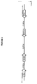
- the human stratum corneum chymotryptic enzyme (19), neuropsin (20) and trypsin-like serine protease (TLSP) (21) are three previously characterized genes which have many structural similarities with the kallikreins and other members of the serine protease family. However, they have not been mapped in the past. Their precise mapping in the region of the kallikrein gene family indicates that these three genes, along with the ones that were newly identified, or are already known, constitute a family that likely originated by duplication of an ancestral gene. The relative localization of all these genes is depicted in Figure 1.
- Kallikrein genes are a subfamily of serine proteases, traditionally characterized by their ability to liberate lysyl-bradykinin (kallidin) from kininogen (18). More recently, however, a new, structural concept has emerged to describe kallikreins. From accumulated sequence data, it is now clear that the mouse has many genes with high homology to kallikrein coding sequences (19-20). Richard and co-workers have contributed to the concept of a " kallikrein multigene family" to refer to these genes (21-22). This definition is not based much on specific enzymatic function of the gene product, but more on its sequence homology and their close linkage on mouse chromosome 7.
- KLK1 meets the functional definition of a kallikrein.
- KLK2 has trypsin-like enzymatic activity and KLK3 (PSA) has very weak chymotrypsin-like enzymatic activity.
- PSA KLK3
- KLK2 and KLK3 are not known to liberate biologically active peptides from precursors.
- members of the kallikrein family include, not only the gene for the kallikrein enzyme, but also genes encoding other homologous proteases, including the enzyme that processes the precursors of the nerve growth factor and epidermal growth factor (8). Therefore, it is important to note the clear distinction between the enzyme kallikrein and a kallikrein or a kallikrein-like gene.
- trypsinogen, PSA and NES1 have 5 coding exons of which the first has intron phase I (the intron occurs after the first nucleotide of the codon), the second has intron phase II (the intron occurs after the second nucleotide and the codon), the third has intron phase I and the fourth has intron phase 0 (the intron occurs between codons).
- the fifth exon contains the stop codon.
- the intron phases of the predicted new kallikrein-like genes follow these rules and are shown in the respective tables. Further support comes from the identification in the new genes, of the conserved amino acids of the catalytic domain of the serine proteases, as presented in Tables 1-5.
- Sequence homology searching was performed using the BLASTN alogrithm (37) on the National Center for Biotechnology Information web server (http://www ncbi.nlm.nih.gov/BLAST/) against the human EST database (dbEST). Clones with > 95% homology were obtained from the I.M.A.G.E. (38) consortium through Research Genetics Inc, Huntsville, AL and from The Institute for Genomic Research (TIGR) (http://WWW.TIGR.ORG/ tdb/tdb.html) (Table 7). Clones were propagated, purified and then sequenced from both directions with an automated sequencer, using insert-flanking vector primers.
- the breast cancer cell line BT-474 was purchased from the American Type Culture Collection (ATCC), Rockville, MD. BT-474 cells were cultured in RPMI media (Gibco BRL, Gaithersburg, MD) supplemented with glutamine (200 mmol/L), bovine insulin (10 mg/L), fetal bovine serum (10%), antibiotics and antimycotics, in plastic flasks, to near confluency. The cells were then aliquoted into 24-well tissue culture plates and cultured to 50% confluency. 24 hours before the experiments, the culture media were changed into phenol red-free media containing 10% charcoal-stripped fetal bovine serum.
- PCR was carried out in a reaction mixture containing 1 ⁇ l of cDNA, 10 mM Tris-HCl (pH 8.3), 50 mM KCl, 1.5 mM MgCl 2 , 200 ⁇ M dNTPs (deoxynucleoside triphosphates), 150 ng of primers and 2.5 units of AmpliTaq Gold DNA polymerase (Roche Molecular Systems, Branchburg, NJ, USA) on a Perkin-Elmer 9600 thermal cycler.
- the cycling conditions were 94°C for 9 minutes to activate the Taq Gold DNA polymerase, followed by 43 cycles of 94°C for 30 s, 63°C for 1 minute and a final extension at 63°C for 10 min. Equal amounts of PCR products were electrophoresed on 2% agarose gels and visualized by ethidium bromide staining. All primers for RT-PCR spanned at least 2 exons to avoid contamination by genomic DNA.
- RNA isolated from 26 different human tissues was purchased from Clontech, Palo Alto, CA.
- cDNA was prepared as described above for the tissue culture experiments and used for PCR reactions with the primers described in Table 8 (SEQ. ID. Nos 5-12). Tissue cDNAs were amplified at various dilutions. Cloning and sequencing of the PCR products
- PCR products were cloned into the pCR 2.1-TOPO vector (Invitrogen, Carlsbad, CA, USA) according to the manufacturer's instructions.
- the inserts were sequenced from both directions using vector-specific primers, by an automated DNA sequencer.
- the exon prediction strategy of the 300Kb DNA sequences around chromosome 19q13.3 - q13.4 identified a novel gene with a structure reminiscent of a serine protease.
- the major features of this gene were its homology, at the amino acid and DNA level, with other human kallikrein genes; the conservation of the catalytic triad (histidine, aspartic acid, and serine), the number of exons and the complete conservation of the intron phases.
- the coding sequence of the klk-L2 gene is shown in SEQ. ID. NO. 1 and GenBAnk Accession # AF135023.
- the exons of the gene are as follows: exon 1 (939-999); exon 2 (2263-2425); exon 3 (2847-3097); exon 4 (3181-3317); and exon 5 (4588-4740).
- the amino acid sequence of KLK-L2 proteins are shown in SEQ. ID. Nos. 2 and 3.
- the tissues that express the prostase/KLK-L1 gene were assessed by RT-PCR.
- the experiments were performed at various dilutions of the cDNAs to obtain some information about the relative levels of expression.
- RT-PCR for actin was used as a positive control and RT-PCR for the PSA cDNA was used as another positive control with tissue restricted specificity.
- Positive ESTs for prostase/KLK-L1 were used as controls for the PCR procedure.
- the PSA gene was found to be highly expressed in the prostate, as expected, and to a lower extent in mammary and salivary glands as also expected from recent literature reports (24, 25). Very low expression of PSA in the thyroid gland, trachea and testis was also found, a finding that accords with recent RT-PCR data by others (26).
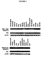
- the tissue expression of prostase/KLK-L1 is summarized in Table 9 and Figure 3. This protease is primarily expressed in the prostate, testis, adrenals, uterus, thyroid, colon, central nervous system and mammary tissues, and, at much lower levels in other tissues.
- the specificity of the RT-PCR procedure was verified for prostase/KLK-L1 by cloning the PCR products from mammary, testicular and prostate tissues and sequencing them.
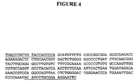
- One example with mammary tissue is shown in Figure 4. All cloned PCR products were identical in sequence to the cDNA sequence reported for the prostase/KLK-L1.
- the steroid hormone receptor-positive breast carcinoma cell line BT-474 was used as a model system to evaluate whether prostase/KLK-L1 expression is under steroid hormone regulation.
- the controls worked as expected i. e., actin positivity without hormonal regulation in all cDNAs, only estrogen up-regulation of the pS2 gene and up-regulation of the PSA gene by androgens and progestins.
- Prostase/KLK-L1 is up-regulated primarily by androgens and progestins, similarly to PSA. This up-regulation was dose-dependent and it was evident at steroid hormone levels ⁇ 10 -10 M.
- the KLK3 gene encodes for PSA, a protein that currently represents the best tumor marker available (24). Since in rodents there are so many kallikrein genes, the restriction of this family to only 3 genes in humans was somewhat surprising. More recently, new candidate kallikrein genes in humans have been discovered, including NES1 (13) and zyme/protease M/neurosin (10-12).
- the known kallikreins and the newly discovered kallikrein-like genes share the following similarities: (a) they encode serine proteases (b) they have five coding exons (c) they share significant DNA and protein homologies with each other (d) they map in the same locus on chromosome 19q 13.3-q 13.4, a region that is structurally similar to an area on mouse chromosome 7, where all the mouse kallikrein genes are localized (e) they appear to be regulated by steroid hormones. Prostase/KLK-L1 is a member of the same family since these common characteristics are also shared by the newly discovered gene.
- the established kallikreins (KLK1, KLK2, and PSA), trypsinogen and chymotrypsinogen belong to a group that has: (1) an intron just downstream from the codon for the active site histidine residue, (2) a second intron downstream from the exon containing the codon for the active site aspartic acid residue, and (3) a third intron just upstream from the exon containing the codon for the active site serine residue.
- the genomic organization of prostase/KLK-L1 gene is very similar to this group of genes.
- the lengths of the coding parts of exons 1-5 are 61,163, 263, 137 and 153 bp, respectively, which are close or identical to the lengths of the exons of the kallikrein genes and also, similar or identical to those of other newly discovered genes in the same chromosomal region like the NES1(14), zyme/protease M/neurosin (10-12) and neuoropsin (28) genes.
- the sensitive RT-PCR protocol reveals that the KLK-L1 enzyme is expressed in prostatic tissue and it is also expressed in significant amounts in other tissues, including testis, female mammary gland, adrenals, uterus, thyroid, colon, brain, lung and salivary glands (Figure 3 and Table 9).
- the specificity of the RT-PCR primers was verified by sequencing the obtained PCR products, with one example shown in Figure 4 (SEQ.ID.NO. 4).
- Tissue culture studies with the breast carcinoma cell line BT-474 further confirm not only the ability of these cells to produce prostase/KLK-L1 but also its hormonal regulation (Figure 5).
- the KLK-1L gene encodes for a serine protease that shows homology with other members of the kallikrein gene family and maps to the same chromosomal location. Many structural features of the kallikreins are conserved in prostase/KLK-L1. The precise mapping of this gene between the two known genes KLK2 and zyme is presented. It is further demonstrated that prostase/KLK-L1 is expressed in many tissues, in addition to the prostate, including the female breast. This gene is also herein referred to as 'prostase'. It has been further demonstrated, using breast carcinoma cell lines, that prostase/KLK-L1 can be produced by these cells and that its expression is significantly up-regulated by androgens and progestins. Based on information for other homologous genes in the area (PSA, zyme, and NES1), prostase/KLK-L1 may be involved in the pathogenesis and/or progression of prostate, breast and possibly other cancers.
- Sequencing data of approximately 300Kb of nucleotides on chromosome 19q13.3-q13.4 was obtained from the web site of the Lawrence Livermore National Laboratory (LLNL) ( http://www-bio.llnl.gov/genome/genome.html ). This sequence was in the form of 9 contigs of different lengths.
- a restriction analysis study of the available sequences was performed using the "WebCutter" computer program ( http://www.firstmarket. com/cutter/cut2.html ) and with the aid of the EcoR1 restriction map of this area (also available from the LLNL web site) an almost contiguous stretch of genomic sequences was constructed.
- the predicted exons of the putative new gene were subjected to homology search using the BLASTN algorithm (37) on the National Center for Biotechnology Information web server (http://www ncbi.nlm.nih.govBLAST/) against the human EST database (dbEST).
- Clones with > 95% homology were obtained from the I.M.A.G.E. consortium (38) through Research Genetics Inc, Huntsville, AL (Table 10). The clones were propagated, purified and sequenced from both directions with an automated sequencer, using insert-flanking vector primers.
- R1 & R2 Two gene-specific primers were designed (R1 & R2) (Table 11).
- Two rounds of RACE reactions (nested PCR) were performed with 5 ⁇ l Marathon ReadyTM cDNA of human testis (Clontech, Palo Alto, CA, USA) as a template.
- the reaction mix and PCR conditions were conducted according to the manufacturer's recommendations. In brief, denaturation was done for 5 min at 94°C followed by 94° C for 5 sec followed by 72°C for 2 min for 5 cycles, then 94°C for 5 sec followed by 70° C for 2 min for 5 cycles then 94°C for 5 sec followed by 65°C for 2 min for 30 cycles for the first reaction and 25 cycles for the nested PCR reaction.
- RNA isolated from 26 different human tissues was purchased from Clontech, Palo Alto, CA.
- cDNA was prepared as described below for the tissue culture experiments and used for PCR reactions with the primers described in Table 11 (SEQ. ID. Nos 9-12, 15-20). Tissue cDNAs were amplified at various dilutions.
- the breast cancer cell line BT-474 was purchased from the American Type Culture Collection (ATCC), Rockville, MD. Cells were cultured in RPMI media (Gibco BRL, Gaithersburg, MD) supplemented with glutamine (200 mmol/L), bovine insulin (10 mg/L), fetal bovine serum (10%), antibiotics and antimycotics, in plastic flasks, to near confluency. The cells were then aliquoted into 24-well tissue culture plates and cultured to 50% confluency. 24 hours before the experiments, the culture media were changed into phenol red-free media containing 10% charcoal-stripped fetal bovine serum. For stimulation experiments, various steroid hormones dissolved in 100% ethanol were added into the culture media, at a final concentration of 10 -8 M. Cells stimulated with 100% ethanol were included as controls. The cells were cultured for 24 hours, then harvested for mRNA extraction
- the cycling conditions were 94°C for 9 minutes to activate the Taq Gold DNA polymerase, followed by 43 cycles of 94°C for 30 s, 63°C for 1 minute and a final extension at 63°C for 10 min. Equal amounts of PCR products were electrophoresed on 2% agarose gels and visualized by ethidium bromide staining. All primers for RT-PCR spanned at least 2 exons to avoid contamination by genomic DNA.
- PCR products were cloned into the pCR 2.1-TOPO vector (Invitrogen, Carlsbad, CA, USA) according to the manufacturer's instructions.
- the inserts were sequenced from both directions using vector-specific primers, with an automated DNA sequencer.
- the coding sequence of the gene is shown in SEQ. ID. NO. 13 and GenBank Accession #AF135028.
- the 3' end of the gene was verified by the presence of poly A stretches that are not present in the genomic sequence at the end of two of the sequenced ESTs.
- One of the sequenced ESTs revealed the presence of an additional exon at the 5'end.
- the nucleotide sequence of this exon matches exactly with the genomic sequence.
- 5' RACE was performed but no additional sequence could be obtained. However, as is the case with other kallikreins, the presence of further up-stream untranslated exon(s) could not be excluded.
- the amino acid sequence of KLK-L2 is shown in SEQ. ID. No. 14.
- KLK-L2 gene Alignment of KLK-L2 gene and the sequences of other known kallikrein genes within the 300 Kb area of interest enabled precise localization of all genes and determination of the direction of transcription, as shown by the arrows in Figure 8.
- the PSA gene was found to be the most centromeric, separated by 12,508 base pairs (bp) from KLK2, and both genes are transcribed in the same direction (centromere to telomere).
- the prostase/KLK-L1 gene is 26,229 bp more telomeric and transcribes in the opposite direction, followed by KLK-L2.
- the distance between KLK-L1 and KLK-L2 is about 35 Kilobases (Kb).
- the zyme gene is 5,981 bp more telomeric and the latter 3 genes are all transcribed in the same direction ( Figure 8).
- the KLK-L2 gene as presented in Figure 7, is formed of 5 coding exons and 4 intervening introns, spanning an area of 9.349 bp of genomic sequence on chromosome 19q13.3-q13.4.
- the lengths of the exons are 73, 262, 257,134, and 156 bp, respectively.
- the intron/exon splice sites (mGT....AGm) and their flanking sequences are closely related to the consensus splicing sites (-mGTAAGT ...CAGm-) (32).
- the presumptive protein coding region of the KLK-L2 gene is formed of 879 bp nucleotide sequence encoding a deduced 293-amino acid polypeptide with a predicted molecular weight of 32 KDa.
- the putative signal sequence at the N-terminus is similar to other trypsin-like serine proteases (prostase and EMSP) ( Figure 9).
- the cDNA ends with a 328 bp of 3' untranslated region containing a conserved poly adenylation signal (AATAAA) located 11 bp up-stream of the poly A tail (at a position exactly the same as that of the zyme poly A tail)(11).
- AATAAA conserved poly adenylation signal
- KLK-L2 will possess a trypsin-like cleavage pattern like most of the other kallikreins (e.g., KLK1, KLK2, TLSP, neuropsin, zyme, prostase, and EMSP) but different from PSA which has a serine (S) residue in the corresponding position, and is known to have a chymotrypsin like activity (Figure 9).
- the dotted region in Figure 9 indicates an 11-amino acid loop characteristic of the classical kallikreins (PSA, KLK1, and KLK2) but not found in KLK-L2 or other members of the kallikrein-like gene family (11).
- KLK-L2 The mature 227-amino acid sequence of the predicted protein was aligned against the GenBank database and the known kallikreins using the "BLASTP” and "BLAST 2 sequence” programs.
- KLK-L2 is found to have 54% amino acid sequence identity and 68% similarity with the enamel matrix serine proteinase 1 (EMSP1) gene, 50% identity with both trypsin like serine protease (TLSP) and neuropsin genes and 47%, 46%, and 42% identity with trypsinogen, zyme, and PSA genes, respectively.
- EMP1 enamel matrix serine proteinase 1
- the KLK-L2 gene is primarily expressed in the brain, mammary gland, and testis but lower levels of expression are found in many other tissues.
- the PCR products were cloned and sequenced.
- a steroid hormone receptor positive breast cancer cell line (BT-474) was used as a model to verify whether the KLK-L2 gene is under steroid hormone regulation.
- PSA was used as a control known to be upregulated by androgens and progestins and pS2 as an estrogen upregulated control. The results indicate that KLK-L2 is up-regulated by estrogens and progestins (Figure 12).
- KLK-L2 is up-regulated (overexpressed) in ovarian tumors ( Figure 13).
- KLK-L2 for kallikrein like gene 2
- the KLK-L2 gene is composed of 5 coding exons and 4 intervening introns and, except for the second coding exon, the exon lengths are comparable to those of other members of the kallikrein gene family ( Figure 6).
- the exon-intron splice junctions were identified by comparing the genomic sequence with the EST sequence and were further confirmed by the conservation of the consensus splice sequence (-mGT&AGm-) (32), and the fully conserved intron phases, as shown in Figure 6. Furthermore, the position of the catalytic triad residues in relation to the different exons is also conserved (Figure 6).
- KLK-L2 is more functionally related to trypsin than to chymotrypsin (34).
- the wide range of tissue expression of KLK-L2 should not be surprising since, by using the more sensitive RT-PCR technique instead of Northern blot analysis, many kallikrein genes were found to be expressed in a wide variety of tissues including salivary gland, kidney, pancreas, brain, and tissues of the reproductive system (uterus, mammary gland, ovary, and testis) (34). KLK-L2 is highly expressed in the brain.
- each kallikrein enzyme has one specific physiological substrate.
- substrates which purified proteins can cleave in vitro, has led to the suggestion that they may perform a variety of functions in different tissues or physiological circumstances.
- Serine proteases encode protein cleaving enzymes that are involved in digestion, tissue remodeling, blood clotting etc., and many of the kallikrein genes are synthesized as precursor proteins that must be activated by cleavage of the propeptide.
- KLK-L2 The predicted trypsin-like cleavage specificity of KLK-L2 makes it a candidate activator of other kallikreins or it may be involved in a "cascade" of enzymatic reactions similar to those found in fibrinolysis and blood clotting (36).
- KLK-L2 a new member of the human kallikrein gene family, KLK-L2 was characterized. This gene is hormonally regulated and it is mostly expressed in the brain, mammary gland and testis. KLK-L2 may be useful as a tumor marker.
- LLNL Lawrence Livermore National Laboratory
- RNA isolated from 26 different human tissues was purchased from Clontech, Palo Alto, CA.
- cDNA was prepared as described below and used for PCR amplification.
- a primer set (L3-F1 and L3-R1) was used to identify the presence of the gene in tissues, and the reverse primer (L3-R1) was used with another primer (L3-F2) to amplify and clone the full cDNA of the gene.
- L3-F1 and L3-R1 was used to identify the presence of the gene in tissues
- L3-R1 reverse primer
- L3-F2 reverse primer
- Tissue cDNAs were amplified at various dilutions.
- PCR was carried out in a reaction mixture containing 1 ⁇ l of cDNA, 10 mM Tris-HCl (pH 8.3), 50 mM KCl, 1.5 mM MgCl 2 , 200 ⁇ M dNTPs (deoxynucleoside triphosphates), 150 ng of primers and 2.5 units of AmpliTaq Gold DNA polymerase (Roche Molecular Systems, Branchburg, NJ, USA) on a Perkin-Elmer 9600 thermal cycler. The cycling conditions were 94°C for 9 minutes, followed by 43 cycles of 94°C for 30 s, 63°C for 1 minute, and a final extension at 63°C for 10 minutes. Equal amounts of PCR products were electrophoresed on 2% agarose gels and visualized by ethidium bromide staining. All primers for RT-PCR spanned at least 2 exons to avoid contamination by genomic DNA.
- the breast cancer cell line BT-474 was purchased from the American Type Culture Collection (ATCC), Rockville, MD. Cells were cultured in RPMI media (Gibco BRL, Gaithersburg, MD) supplemented with glutamine (200 mmol/L), bovine insulin (10 mg/L), fetal bovine serum (10%), antibiotics and antimycotics, in plastic flasks, to near confluency. The cells were then aliquoted into 24-well tissue culture plates and cultured to 50% confluency. 24 hours before the experiments, the culture media were changed into phenol red-free media containing 10% charcoal-stripped fetal bovine serum. For stimulation experiments, various steroid hormones dissolved in 100% ethanol were added into the culture media, at a final concentration of 10 -8 M.
- PCR products were cloned into the pCR 2.1-TOPO vector (Invitrogen, Carlsbad, CA, USA) according to the manufacturer's instructions.
- the inserts were sequenced from both directions using vector-specific primers, with an automated DNA sequencer.
- the PCR product generated with primer set Z1S and Z1AS (Table 14, SEQ.ID.NOS. 27-42), was purified and then labeled with 32 P by the random primer method (Sambrook, supra) and used as a probe to screen a human genomic DNA BAC library, spotted in duplicate on nylon membranes, for identification of positive clones.
- the filters were hybridized in 15% formamide, 500 mM Na 2 HPO 4 , 7% SDS, 1% BSA (w/v) at 65°C overnight, then washed sequentially with 2X SSC, 1X SSC, 0.2X SSC, containing 0.1% SDS at 65°C, and then exposed to X-ray film as described (Sambrook, supra).
- PAC and BAC libraries were constructed by de Jong and associates (42). Purification of BAC and PAC DNA was done by a rapid alkaline lysis miniprep method, which is a modification of the standard Qiagen-Tip method. Positive clones were further confirmed by Southern blot analysis as described (Sambrook, supra).
- Two gene specific primers were designed. Two rounds of RACE reactions (nested PCR) were performed with 5 ⁇ l Marathon ReadyTM cDNA of human testis (Clontech) as a template. The reaction mix and PCR conditions were selected according to the manufacturer's recommendations. Positive bands were gel-purified using Qiagen Gel Purification kits according to manufacturer's recommendations.
- prostatic specific antigen PSA
- human renal kallikrein KLK1
- human glandular kallikrein KLK2
- normal epithelial cell-specific 1gene NES1
- KLK-L2 and zyme genes gene-specific primers were designed for each of these genes (Table 14) and developed polymerase chain reaction (PCR)-based amplification protocols which allowed us to generate specific PCR products with genomic DNA as a template.
- the PCR reactions were carried out as described above but by using an annealing/extension temperature of 65°C.
- ⁇ BLAST> analysis of these segments against the GenBank database (37) enabled the precise location of two classical kallikreins, namely PSA and KLK2 to be defined.
- Other newly discovered serine proteases were localized which are homologous with the kallikrein genes, namely protease M/zyme/neurosin (10, 11, 12), human stratum corneum chymotryptic enzyme ( HSSCE) (55), neuropsin (28), normal epithelial cell-specific 1 gene ( NES1) (13), trypsin-like serine protease ( TLSP) (GenBank accession # AF164623), KLK-L1 (SEQ.ID.NO. 1) and KLK-L2 (SEQ.ID.NO. 13).
- the gaps in the 300 kb genomic sequence were partially filled as follows:
- the predicted exons were subjected to sequence homology search against the human EST database (dbEST) and revealed an EST clone (GenBank accession # AA583908) which exhibited 99% homology with the putative gene.
- This EST was obtained, purified and sequenced and the sequence was aligned by BLAST software (37) against the genomic area that contains the putative gene.
- GDSGGP This highly conserved motif (GDSGGP) always occurs at the beginning of the last exon in all known kallikreins.
- PCR reactions were performed using gene-specific primers for the first and last exons of the predicted structure of the gene (L3-F2 and L3-R1) with cDNA isolated from different human tissues as putative templates.
- a positive band of the expected size was isolated from testis cDNA and fully sequenced. Its sequence was aligned by BLAST against the genomic sequence to unequivocally define the exon/intron boundaries.
- 5'RACE reaction was performed using Marathon Ready cDNA from testis as a template. This allowed identification of an additional exon that contains the start codon and 5' untranslated region.
- the full sequence of the gene is shown in SEQ. ID. NO. 21 (GenBank Accession # AF135026) and the amino acid sequences of KLK-L3 proteins are shown in SEQ. ID. Nos. 22 and 23.
- the KLK-L3 gene is formed of 5 coding exons and 4 intervening introns, although, as with other kallikreins, the presence of further upstream untranslated exon(s) could not be ruled out (14, 28). All of the exon /intron splice sites conform to the consensus sequence for eukaryotic splice sites (32). The gene further follows strictly the common structural features of the human kallikrein multigene family, as described below.
- the predicted protein-coding region of the gene is formed of 753 bp, encoding a deduced amino acid polypeptide with a predicted molecular weight of 27.5 kDa.
- a potential translation initiation codon is found at position 28 of the predicted first exon (numbers of nucleotides refer to SEQ. ID. NO. 21 or GenBAnk Accession # AF135026. This codon does not match well with the consensus Kozak sequence (33), however, it has a purine at position (-3) which occurs in 97% of vertebrate mRNAs (43), and it is almost identical to the sequence of the zyme gene flanking the start codon. It should also be noted that most kallikreins do not have the consensus G nucleotide in position (+4).
- Nucleotides 6803-6808 (AGTAAA) closely resemble a consensus polyadenylation signal (44) and are followed by a stretch of 19 poly A nucleotides not found in genomic DNA, after a space of 14 nucleotides. No other potential polyadenylation signals were discernable in the 3' untranslated region, suggesting that the above motif is indeed the polyadenylation signal. The same polyadenylation signal motif was predicted for the KLK1 and KLK2 genes.
- KLK-L3 protein sequence is unique, comparative analysis revealed that it is highly homologous to other members of the kallikrein multigene family.
- KLK-L3 shows 40% protein identity with the TLSP gene product and 38% and 33% identity with the KLK-L2 and KLK1 proteins, respectively.
- Hydrophobicity analysis revealed that the amino-terminal region is quite hydrophobic ( Figure 16), consistent with the possibility that this region may harbor a signal sequence, analogous to other serine proteases.
- Computer analysis of the aminoacid sequence of KLK-L3 predicted a cleavage site between amino acids 19 and 20 (GWA-DT).
- Figure 17 also revealed a potential cleavage site (Arg 22 ), at a site homologous to other serine proteases (lysine (K) or arginine (R) is present in most cases).
- Arg 22 a potential cleavage site
- Several evenly distributed hydrophobic regions throughout the KLK-L3 polypeptide are consistent with a globular protein, similar to other kallikreins and serine proteases.
- the dotted region in Figure 17 indicates an 11-amino acid loop characteristic of the classical kallikreins (PSA, KLK1, and KLK2) but not found in KLK-L3 or other members of the kallikrein multi-gene family (11, 41).
- the KLK-L3 gene is primarily expressed in thymus, testis, spinal cord, cerebellum, trachea, mammary gland, prostate, brain, salivary gland, ovary and skin (the latter two tissues are not shown in the figure). Lower levels of expression are seen in fetal brain, stomach, lung, thyroid, placenta, liver, small intestine, and bone marrow. No expression was seen in uterus, heart, fetal liver, adrenal gland, colon, spleen, skeletal muscle, pancreas, and kidney. In order to verify the RT-PCR specificity, representative PCR products were cloned and sequenced.
- Figure 20 shows that KLK-L3 gene is regulated by steroid hormones in the human breast cancer cell line BT-474.
- Kallikreins with verified 5'-untranslated exons include NES1 (14), zyme, and neuropsin (35). This map is also directional; it indicates that PSA and KLK2 genes are transcribed in the same direction (centromere to telomere) and that the rest of the kallikrein-like genes are transcribed in the reverse direction ( Figure 14).
- KLK1 is located approximately 31 kb centromeric to PSA (9).
- the map described extends only 24 kb centromeric to PSA, and for this reason, KLK1 was not precisely localized.
- the exact location of the KLK1 gene is still to be defined from linear chromosome 19 sequencing data. The possibility still exists that this locus is extended further, and that other kallikrein-like genes may be located upstream of PSA or downstream from TLSP.
- kallikreins are characterized by their ability to liberate lysyl-bradykinin (kallidin) from kininogen (2).
- KLK1 meets this "functional" definition.
- KLK2 and KLK3 are assigned to the same family based on the strong structural similarities of the genes and proteins and the close localization of these genes on the same chromosomal region (20). More recently, a new structural concept has emerged to describe kallikreins. Richards and co-workers introduced the concept of a "kallikrein multigene family" in mice, to refer to these genes (20, 21). This definition is not based much on the specific enzymatic function of the gene product, but more on its sequence homology and its close linkage on mouse chromosome 7.
- the chromosomal band 19q13 is nonrandomly rearranged in a variety of human solid tumors including ovarian cancers (46), and the currently available data indicate that the kallikrein gene locus is related to many malignancies.
- At least three kallikrein genes (PSA, zyme and NES1) are down regulated in breast cancer (10, 13, 47, 48), and NES1 appears to be a novel tumor suppressor gene (29).
- PSA exhibits potent antiangiogenic activity (49). It is possible that some of these kallikreins are involved in a cascade pathway, similar to the coagulation or apoptotic process, whereby pro-forms of proteolytic enzymes are activated and then act upon downstream substrates. Such activity was found for the KLK2 gene product which acts upon and activates pro PSA (50, 51).
- the expanded human kallikrein gene family has similar number of members as the rodent family of genes. Some new compelling data have raised the possibility that at least some of these genes behave as tumor suppressors (29), as negative regulators of cell growth (52), as antiangiogenic (49) and apoptotic (53) molecules. The paramount diagnostic value of some members is also well-established (24, 54). For these reasons, it is important to check all members of this family of genes as potential diagnostic or prognostic markers or as candidate therapeutic targets.
- KLK-L3 The newly identified KLK-L3 gene is expressed in many tissues, including skin, thymus, central nervous system, breast, prostate, and testis.
- the wide range of tissue expression of KLK-L3 should not be surprising since, by using the more sensitive RT-PCR technique, many kallikrein genes were found to be expressed in a wide variety of tissues.
- PSA, KLK2, prostase/KLK-L1, and KLK-L2 are now known to be expressed in breast and many other tissues (30, 54).
- KLK-L3 is regulated by steroid hormones but in a more complex fashion than PSA and KLK2 which are up-regulated by androgens and progestins (71). In the breast carcinoma cell line studied, KLK-L3 appears to be up-regulated by progestins > estrogens > androgens (Figure 20).
- LLNL Lawrence Livermore National Laboratory
- the predicted exons of the putative new gene were subjected to homology search using the BLASTN algorithm (37) on the National Center for Biotechnology Information web server (http://www ncbi.nim.nih.gov/BLAST/) against the human EST database (dbEST).
- Clones with > 95% homology were obtained from the I.M.A.G.E. consortium (38) through Research Genetics Inc, Huntsville, AL.
- the clones were propagated, purified and sequenced from both directions with an automated sequencer, using insert-flanking vector primers.
- RNA isolated from 26 different human tissues was purchased from Clontech. cDNA was prepared as described below, and used for PCR reactions with different sets of primers (Table 15, SEQ.ID.NOs. 46-55, 9-12). Tissue cDNAs were amplified at various dilutions.
- the breast cancer cell line BT-474 was purchased from the American Type Culture Collection (ATCC), Rockville, MD. Cells were cultured in RPMI media (Gibco BRL, Gaithersburg, MD) supplemented with glutamine (200 mmol/L), bovine insulin (10 mg/L), fetal bovine serum (10%), antibiotics and antimycotics, in plastic flasks, to near confluency. The cells were then aliquoted into 24-well tissue culture plates and cultured to 50% confluency. 24 hours before the experiments, the culture media were changed into phenol red-free media containing 10% charcoal-stripped fetal bovine serum. For stimulation experiments, various steroid hormones dissolved in 100% ethanol were added into the culture media, at a final concentration of 10 -8 M. Cells stimulated with 100% ethanol were included as controls. The cells were cultured for 24 hours, then harvested for mRNA extraction.
- PCR was carried out in a reaction mixture containing 1 ⁇ l of cDNA, 10 mM Tris-HCl (pH 8.3), 50 mM KCl, 1.5 mM MgCl 2 , 200 ⁇ M dNTPs (deoxynucleoside triphosphates), 150 ng of primers and 2.5 units of AmpliTaq Gold DNA polymerase (Roche Molecular Systems, Branchburg, NJ, USA) on a Perkin-Elmer 9600 thermal cycler. The cycling conditions were 94°C for 9 minutes to activate the Taq Gold DNA polymerase, followed by 43 cycles of 94°C for 30 s, 63°C for 1 minute and a final extension at 63°C for 10 min. Equal amounts of PCR products were electrophoresed on 2% agarose gels and visualized by ethidium bromide staining. All primers for RT-PCR spanned at least 2 exons to avoid contamination by genomic DNA.
- PCR products were cloned into the pCR 2.1-TOPO vector (Invitrogen, Carlsbad, CA, USA) according to the manufacturer's instructions.
- the inserts were sequenced from both directions using vector-specific primers, with an automated DNA sequencer.
- Normal breast tissues were obtained from women undergoing reduction mammoplasties.
- Breast tumor tissues were obtained from female patients at participating hospitals of the Ontario Provincial Steroid Hormone Receptor Program.
- the normal and tumor tissues were immediately frozen in liquid nitrogen after surgical resection and stored in this manner until extracted.
- the tissues were pulverized with a hammer at dry ice temperature and RNA was extracted as described above, using Trizol reagent.
- exon 2 was identified based on: a) a consensus exon/intron splice site b) preservation intron phase II after this exon, in agreement to intron phases of all other known kallikreins c) presence of the histidine residue of the catalytic triad (H 76 ) surrounded by a well-conserved peptide motif [see below] just before the end of this exon d) comparable exon length to other kallikrein genes.
- a potential first exon was also predicted from the upstream genomic sequence, based on the preserved intron phase (phase I), and the existence of an in-frame start codon that is located at a comparable distance [in relation to other kallikreins] from the end of this exon.
- a PCR reaction was performed using one reverse primer (L4-R1) together with another forward primer that is located in the predicted first exon (primer L4-X1) (Table 15 & Figure 22).
- Two main PCR bands were obtained from the tissues examined; the expected 819 bp band (predominant) and an additional minor band of about 650 bp ( Figure 24). Cloning and sequencing of these two bands revealed that the gene exists in two main forms in these tissues; the long form [SEQ. ID.
- KLK-L4 The long form of the KLK-L4 gene is presented in Figure 25 (SEQ.ID.NO. 43).
- KLK-L4 is formed of five coding exons and four intervening introns, spanning an area of 8,905 bp of genomic sequence on chromosome 19q13.3-q13.4.
- the lengths of the coding regions are 52, 187, 269,137 and 189 bp, respectively.
- the predicted protein coding region of the gene is formed of 831 bp, encoding a deduced 277-amino acid protein with a predicted molecular mass of 30.6 kDa ( Figure 25).
- the intron/exon splice sites (mGT....AGm, where m is any base) and their flanking sequences are in agreement with the consensus splice site sequence.
- a potential translation initiation codon is present at position 45 of the predicted first exon [numbers refer to SEQ. ID. No. 43].
- the cDNA extends at least 382 bp further downstream from the stop codon and a putative polyadenylation signal (TATAAA) is present at the end of this region ( Figure 25).
- the protein product is very likely to be a secreted protein.
- the dotted region in Figure 27 indicates an 11-amino acid loop characteristic of the classical kallikreins (PSA, KLK1, and KLK2) which is not found in KLK-L4 or other members of the kallikrein multi-gene family (11,13, 35).
- Amino acid sequences for KLK-L4 proteins are shown in SEQ.ID.NO. 44 and 45.
- KLK-L4 will possess a trypsin-like cleavage pattern, similarly to most of the other kallikreins [e.g., KLK1, KLK2, TLSP, neuropsin, zyme, prostase, and EMSP] but different from PSA which has a serine (S) residue in the corresponding position, and is known to have chymotrypsin like activity (Figure 27) (2,40).
- KLK-L4 Alignment of the KLK-L4 gene and the sequences of other known kallikrein genes within the 300 Kb area of interest [the human kallikrein gene family locus], enabled precise localization of all known genes and to determine the direction of transcription, as shown by the arrows in Figure 28.
- the PSA gene lies between KLK1 and KLK2 genes and is separated by 13, 319 base pairs (bp) from KLK2, and both genes are transcribed in the same direction (centromere to telomere). All other kallikrein-like genes are transcribed in the opposite direction.
- KLK-L4 is 13 kb centromeric from KLK-L6 [SEQ.ID.NO. 65], and 21 kb more telomeric to KLK-L5 [SEQ. ID. NO. 56].
- KLK-L4 Alignment of the amino acid sequence of the KLK-L4 protein (long form) against the GenBank database and the known kallikreins, using the BLAST algorithm (37), indicated that KLK-L4 has 51% amino acid sequence identity with the TLSP and zyme genes, 49% identity with KLK-L2 and 47% and 45% identity with PSA and KLK2 genes, respectively.
- Multiple alignment study shows that the typical catalytic triad of serine proteases is conserved in the KLK-L4 gene (H 108 , D 153 , and S 245 ) and, as is the case with all other kallikreins, a well conserved peptide motif is found around the amino acid residues of the catalytic triad [i.e.
- the KLK-L4 gene is primarily expressed in mammary gland, prostate, salivary gland and testis, but, as is the case with other kallikreins, lower levels of expression are found in many other tissues.
- the PCR products were cloned and sequenced.
- a steroid hormone receptor-positive breast cancer cell line (BT-474) was used as a model, to verify whether the KLK-L4 gene is under steroid hormone regulation.
- PSA was used as a control gene, known to be up-regulated by androgens and progestins and pS2 as an estrogen up-regulated control gene in the same cell line.
- Preliminary results indicate that KLK-L4 is up-regulated by progestins and androgens and to a lower extent by estrogens (Figure 30).
- KLK-L4 gene expression was undetectable in 7, lower than normal tissues in 9, comparable to the normal tissues in 1, and higher than normal tissues in 2 tumors.
- the BT-474 and T-47D cell lines had no detectable KLK-L4 mRNA, while the MCF-7 cell line was positive.
- the established kallikreins (KLK1, KLK2, and PSA), trypsinogen and chymotrypsinogen belong to a group that has: (1) an intron just downstream from the codon for the active site histidine residue, (2) a second intron downstream from the exon containing the codon for the active site aspartic acid residue, and (3) a third intron just upstream from the exon containing the codon for the active site serine residue.
- Figure 31 shows that KLK-L4 meets the above mentioned criteria; moreover, is located in close proximity to other kallikrein genes on the chromosomal locus 19q13.3-q13.4 (Figure 28).
- hK2 (encoded by the KLK2 gene) could be another useful diagnostic marker for prostate cancer (57, S8).
- NES1 appears to be a tumor suppressor gene (29).
- the protease M gene was shown to be differentially expressed in primary breast and ovarian tumors (10), and the human stratum corneum chymotryptic enzyme has been shown to be expressed at abnormally high levels in ovarian cancer (59).
- Another recently identified kallikrein-like gene, located close to KLK-L4 and tentatively named tumor-associated, differentially expressed gene-14 (TADG14) [an alternatively spliced form of neuropsin, see Figure 28] was found to be overexpressed in about 60% of ovarian cancer tissues (59).
- prostase/KLK-L1 another newly discovered kallikrein-like gene, is speculated to be linked to prostate cancer (41).
- extensive new literature suggests multiple connections of many kallikrein genes to various forms of human cancer.
- RNA sequences intervening RNA sequences (introns) from the pre-messenger RNA in eukaryotic nuclei is a major step in the regulation of gene expression (60).
- RNA splicing provides a mechanism whereby protein isoform diversity can be generated and the expression of particular proteins with specialized functions can be restricted to certain cell or tissue types during development (60).
- the sequence elements in the pre-mRNA at the 5' and 3' splice sites in metazoans have very loose consensus sequence; only the first and the last two bases (GT..AG) of the introns are highly conserved (Sambrook, supra).
- sequences cannot be the sole determinants of splice site selection, since identical, but not ordinarily active, consensus sequences can be found within both exons and introns of many eukaryotic genes. Other protein factors and sequences downstream of the splice sites are also involved.
- RNA species are transcribed from the PSA gene, in addition to the major 1.6-kb transcript (61).
- PSA transcripts have been described by Reigman et al (7).
- one of these clones lacks the 3' untranslated region and the first 373 nucleotides of the open reading frame, and has an extended exon that contains a stop codon, a pattern that is comparable with some alternative forms of the KLK-L4 cDNA, as described here ( Figure 22).
- Heuze et al. reported the cloning of a full-length cDNA corresponding to a 2.1 kb PSA mRNA.
- Neuropsin a recently identified kallikrein-like gene, was found to have two alternatively spliced forms, in addition to the major form (59, 64).
- the cloning of the KLK-L4 gene is described and the identification of a number of alternative mRNA forms. These forms may result from alternative-splicing (Sambrook, supra), retained intronic segment (7), or from the utilization of an alternative transcription initiation site (63).
- KLK-L4 and the major alternative splice variant [short KLK-L4 variant] ( Figure 24) have an identical 5' sequence required for translation, secretion and activation, it is possible to assume that both mRNAs encode for a secreted protein (61).
- LLNL Lawrence Livermore National Laboratory
- a number of computer programs were used to predict the presence of putative new genes within the contiguous genomic area of interest. The ability of these programs for predicting new genes was first examined by using the genomic sequences of the known kallikreins as testing parameters. The most reliable computer programs; GeneBuilder (gene prediction) (http://125.itba.rni.cnr.it/ ⁇ webgene/genebuilder.html). GeneBuilder (exon prediction) (http://125.itba.mi.cnr.it/-webgene/genebuilder.html), Grail 2 (http://compbio.ornl.gov), and GENEID-3 (http://apolo.imim.es/geneid.html) were selected for further use.
- the predicted exons of the putative new gene were subjected to homology search using the BLASTN algorithm (37) on the National Center for Biotechnology Information web server (http://www ncbi.nlm.nih.gov/BLAST/) against the human EST database (dbEST).
- BLASTN algorithm 3-7 on the National Center for Biotechnology Information web server (http://www ncbi.nlm.nih.gov/BLAST/) against the human EST database (dbEST).
- a clone with > 95% homology was obtained from the I.M.A.G.E. consortium (38) through Research Genetics Inc, Huntsville, AL. This clone was propagated, purified and sequenced from both directions with an automated sequencer, using insert-flanking vector primers.
- RNA isolated from 26 different human tissues was purchased from Clontech, Palo Alto, CA.
- cDNA was prepared as described below for the tissue culture experiments and used for PCR reactions.
- two primers (KLK-L5-R1 and KLK-L5-F1) (Table 17, SEQ.ID.NOs. 61-64, 9-12, and Figure 32) were designed from areas with relatively low homology.
- Tissue cDNAs were amplified at various dilutions. Due to the high degree of homology between kallikreins, and to exclude non-specific amplification, PCR products were cloned and sequenced.
- Normal breast tissues were obtained from women undergoing reduction mammoplasties.
- Breast tumor tissues were obtained from female patients at participating hospitals of the Ontario Provincial Steroid Hormone Receptor Program.
- the normal and tumor tissues were immediately frozen in liquid nitrogen after surgical resection and stored in this manner until extracted.
- the tissues were pulverized with a hammer under liquid nitrogen and RNA was extracted as described below, using Trizol reagent.
- the breast cancer cell lines BT-474 and T-47D, and the LNCaP prostate cancer cell line were purchased from the American Type Culture Collection (ATCC), Rockville, MD. Cells were cultured in RPMI media (Gibco BRL, Gaithersburg, MD) supplemented with glutamine (200 mmol/L), bovine insulin (10 mg/L), fetal bovine serum (10%), antibiotics and antimycotics, in plastic flasks, to near confluency. The cells were then aliquoted into 24-well tissue culture plates and cultured to 50% confluency. 24 hours before the experiments, the culture media were changed into phenol red-free media containing 10% charcoal-stripped fetal bovine serum. For stimulation experiments, various steroid hormones dissolved in 100% ethanol were added into the culture media at a final concentration of 10 -8 M. Cells stimulated with 100% ethanol were included as controls. The cells were cultured for 24 hours, then harvested for mRNA extraction.
- RT-PCR Reverse transcriptase polymerase chain reaction
- KLK-L5-F1 and KLK-L5-R1 Two gene-specific primers were designed (KLK-L5-F1 and KLK-L5-R1) (Table 17) and PCR was carried out in a reaction mixture containing 1 ⁇ l of cDNA, 10 mM Tris-HCl (pH 8.3), 50 mM KCl, 1.5 mM MgCl 2 , 200 ⁇ M dNTPs (deoxynucleoside triphosphates), 150 ng of primers and 2.5 units of AmpliTaq Gold DNA polymerase (Roche Molecular Systems, Branchburg, NJ, USA) on a Perkin-Elmer 9600 thermal cycler.
- the cycling conditions were 94°C for 9 minutes to activate the Taq Gold DNA polymerase, followed by 43 cycles of 94°C for 30 s, 63°C for 1 minute and a final extension step at 63°C for 10 min.
- Equal amounts of PCR products were electrophoresed on 2% agarose gels and visualized by ethidium bromide staining. All primers for RT-PCR spanned at least 2 exons to avoid contamination by genomic DNA.
- PCR products were cloned into the pCR 2.1-TOPO vector (Invitrogen, Carlsbad, CA, USA) according to the manufacturer's instructions.
- the inserts were sequenced from both directions using vector-specific primers, with an automated DNA sequencer.
- Amino acid sequences for KLK-L5 proteins are shown in SEQ. ID. NOs. 57 to 60.
- Figure 35 shows a comparative hydrophobicity analysis of the KLK-L5 and the prostate-specific antigen (PSA) proteins.
- PSA prostate-specific antigen
- the amino terminal regions of both genes are quite hydrophobic, indicating that this region of KLK-L5 is possibly harboring a signal peptide analogous to PSA.
- Figure 35 also shows the presence of several evenly distributed hydrophobic regions throughout the KLK-L5 polypeptide, which are consistent with a globular protein, similar to other serine proteases (13).
- Figure 36 shows the alignment of KLK-L5 protein with another 10 members of the same gene family.
- the dotted region in Figure 36 indicates an 11-amino acid loop characteristic of the classical human kallikreins (PSA, hK1 and hK2) but not found in KLK-L5 protein or other members of the kallikrein multigene family (11, 13, 35).
- Sequence analysis of eukaryotic serine proteases indicates the presence of twenty nine invariant amino acids (39). Twenty eight of them are conserved in the KLK-L5 polypeptide and the remaining amino acid (S156 instead of P) is not conserved among all other kallikreins (Figure 36).
- KLK-L5 will possess a trypsin-like cleavage pattern, similarly to most of the other kallikreins (e.g., hK1 hK2, TLSP, neuropsin, zyme, prostase, and EMSP) but different from PSA, which has a serine (S) residue in the corresponding position, and is known to have chymotrypsin like activity (Figure 36) (54).
- kallikreins e.g., hK1 hK2, TLSP, neuropsin, zyme, prostase, and EMSP
- the KLK-L5 protein (classical form) has 48% amino acid sequence identity and 57% overall similarity with neuropsin, 46% identity with the normal epithelial cell-specific 1 gene product (NES1) and 38% identity with both PSA and hK2 proteins. Multiple alignment shows that the typical catalytic triad of serine proteases is conserved in the KLK-L5 protein (H 62 , D 108 , and S 200 ) ( Figures 33 and 36).
- Figure 36 shows other amino acid residues that are completely conserved between kallikreins and kallikrein-like proteins.
- Figure 37 shows the phylogenetic analysis which separated the classical kallikreins (hK1, hK2, and PSA) and clustered KLK-L5 with NES1 and neuropsin proteins in a separate group away from other serine proteases, consistent with previously published studies (27, 41) and indicating that this group of genes probably arose from a common ancestral gene, by gene duplication.
- the KLK-L5 gene is primarily expressed in the salivary gland, stomach, uterus, trachea, prostate, thymus, lung, colon, brain, breast and thyroid gland, but, as is the case with other kallikreins, lower levels of expression are found in some other tissues (testis, pancreas, small intestine, spinal cord).
- the PCR products were cloned and sequenced. The three splice forms of the gene were expressed in most of these tissues. However, the relative abundance of each form was different among tissues ( Figure 38).
- BT-474 and T-47D breast cancer cell lines
- LNCaP prostate cancer cell line
- the gene was up-regulated only by androgen and progestin. Only in this cell line all 3 isoforms were expressed.
- BT-474 cells KLK-L5 was found to be up-regulated, at the mRNA level, by estrogen and androgen, and to a lesser extent by the progestin.
- the rank of potency was estrogen>androgen>progestin.
- the rank of potency for the T-47D cell line was androgen>progestin>estrogen .
- the short isoform (related protein-2) was present (Figure 39).
- KLK-L5 is down regulated in breast cancer
- kallikreins are characterized by the following common structural features: (a) All genes are formed of 5 coding exons and 4 intervening introns [ some genes may have extra 5( untranslated exon(s)] (14, 35). (b) The exon lengths are usually comparable. (c) The intron phases are always conserved (I-II-1-0) (see legend of Figure 34 for definition of intron phases). (d) These genes are clustered in the same chromosomal region, without any intervening non-kallikrein-like genes ( Figure 28).
- KLK-L5 should be considered a new member of the kallikrein multigene family.
- Preproenzymes Serine proteases and kallikreins are synthesized as "preproenzymes" that contain an N-terminal signal peptide (pre-zymogen), followed by a short activation peptide and the enzymatic domain (41, 54).
- PreproPSA has 24 additional residues that constitute the pre-region (signal peptide, 17 residues), and the propeptide (7 residues) (67).
- the signal peptide directs the protein to and across the endoplasmic reticulum (ER). In the ER, the signal peptide is removed and the resulting proPSA is transported to the plasma membrane, where it is secreted.
- a novel transcript of the tissue kallikrein gene was isolated from the colon (63).
- Neuropsin a recently identified kallkrein-like gene, was found to have two alternatively spliced forms in addition to the major form (35, 64).
- the cloning of the classical kallikrein form and two unique splice forms of the KLK-L5 gene are described. Because the classical form and the splice forms all have the same 5' sequence required for translation, secretion and activation as do other kallikreins, i.e. a 5' leader sequence, a signal peptide, and a proregion, it is reasonable to assume that all three mRNA forms should produce a secreted protein.
- Preliminary findings identifying forms of KLK-L5 predominant in certain tissues are presented in Figures 35 and 40.
- KLK-L5 a new member of the human kallikrein gene family, KLK-L5, has been characterized which maps to the human kallikrein locus (chromosome 19q13.3-q13.4). This gene has two related splice forms in addition to the main form. KLK-L5 is expressed in a variety of tissues, appears to be down-regulated in breast cancer and its expression is influenced by steroid hormones. Since a few other kallikreins are already used as valuable tumour markers, KLK-L5 may also find a similar clinical application.
- KLK-L6 another novel gene of the kallikrein multigen family referred to as KLK-L6.
- the full structure of the KLK-L6 gene is shown in Figure 41. Exons 1, 2, 3, 4, 5, 6, and 7 are at nucleic acids 1172-1281; 2561-2695; 2781-2842, 3714-3885; 5715-5968; 6466-6602; and 7258-7520.
- the nucleic acid sequence of the KLK-L6 gene is also shown in SEQ.ID.NO. 65 and amino acid sequences for the KLK-L6 protein are shown in SEQ.ID. Nos. 66 and 67. (See also GenBank Accession # AF161221).
- Figure 42 shows a comparative hydrophobicity analysis of KLK-L6 and the prostate-specific antigen (PSA).
- PSA prostate-specific antigen
- Figure 44 shows the phylogenetic analysis which separated the classical kallikreins (hK1, hK2, and PSA) and placed KLK-L6 in a separate group.
Landscapes
- Health & Medical Sciences (AREA)
- Chemical & Material Sciences (AREA)
- Life Sciences & Earth Sciences (AREA)
- Organic Chemistry (AREA)
- Genetics & Genomics (AREA)
- Medicinal Chemistry (AREA)
- General Health & Medical Sciences (AREA)
- Engineering & Computer Science (AREA)
- Zoology (AREA)
- Molecular Biology (AREA)
- Bioinformatics & Cheminformatics (AREA)
- Biomedical Technology (AREA)
- Wood Science & Technology (AREA)
- Biochemistry (AREA)
- Biotechnology (AREA)
- Chemical Kinetics & Catalysis (AREA)
- Microbiology (AREA)
- Cell Biology (AREA)
- Immunology (AREA)
- Toxicology (AREA)
- Gastroenterology & Hepatology (AREA)
- Biophysics (AREA)
- Proteomics, Peptides & Aminoacids (AREA)
- General Engineering & Computer Science (AREA)
- General Chemical & Material Sciences (AREA)
- Nuclear Medicine, Radiotherapy & Molecular Imaging (AREA)
- Pharmacology & Pharmacy (AREA)
- Animal Behavior & Ethology (AREA)
- Public Health (AREA)
- Veterinary Medicine (AREA)
- Medicines That Contain Protein Lipid Enzymes And Other Medicines (AREA)
- Peptides Or Proteins (AREA)
- Micro-Organisms Or Cultivation Processes Thereof (AREA)
Applications Claiming Priority (4)
| Application Number | Priority Date | Filing Date | Title |
|---|---|---|---|
| US12426099P | 1999-03-11 | 1999-03-11 | |
| US12738699P | 1999-04-01 | 1999-04-01 | |
| US14491999P | 1999-07-21 | 1999-07-21 | |
| EP00908878A EP1159431A2 (de) | 1999-03-11 | 2000-03-09 | Menschliche kallikrein gene |
Related Parent Applications (1)
| Application Number | Title | Priority Date | Filing Date |
|---|---|---|---|
| EP00908878A Division EP1159431A2 (de) | 1999-03-11 | 2000-03-09 | Menschliche kallikrein gene |
Publications (2)
| Publication Number | Publication Date |
|---|---|
| EP1724351A2 true EP1724351A2 (de) | 2006-11-22 |
| EP1724351A3 EP1724351A3 (de) | 2007-11-14 |
Family
ID=37106550
Family Applications (1)
| Application Number | Title | Priority Date | Filing Date |
|---|---|---|---|
| EP06076156A Withdrawn EP1724351A3 (de) | 1999-03-11 | 2000-03-09 | Menschliche Kallikrein-ähnliche Gene |
Country Status (1)
| Country | Link |
|---|---|
| EP (1) | EP1724351A3 (de) |
Family Cites Families (4)
| Publication number | Priority date | Publication date | Assignee | Title |
|---|---|---|---|---|
| US5786148A (en) * | 1996-11-05 | 1998-07-28 | Incyte Pharmaceuticals, Inc. | Polynucleotides encoding a novel prostate-specific kallikrein |
| ES2248891T3 (es) * | 1997-02-25 | 2006-03-16 | Corixa Corporation | Compuestos para inmunodiagnostico de cancer de prostata y metodos para su uso. |
| HUP0002095A3 (en) * | 1997-02-25 | 2002-02-28 | Corixa Corp Seattle | Compounds for immunotherapy of prostate cancer and methods for their use |
| DE19805633A1 (de) * | 1998-02-12 | 1999-08-19 | Basf Ag | Neue Serinprotease aus der Prostata |
-
2000
- 2000-03-09 EP EP06076156A patent/EP1724351A3/de not_active Withdrawn
Also Published As
| Publication number | Publication date |
|---|---|
| EP1724351A3 (de) | 2007-11-14 |
Similar Documents
| Publication | Publication Date | Title |
|---|---|---|
| US7507403B2 (en) | Kallikrein gene | |
| Yousef et al. | Prostase/KLK-L1 is a new member of the human kallikrein gene family, is expressed in prostate and breast tissues, and is hormonally regulated | |
| JP3981416B2 (ja) | Pca3タンパク質、pca3遺伝子、及びこれらの用途 | |
| EP1159431A2 (de) | Menschliche kallikrein gene | |
| AU2001287384A1 (en) | Kallikrein gene | |
| Yousef et al. | Molecular cloning of a novel human acid phosphatase gene (ACPT) that is highly expressed in the testis | |
| WO2002064798A1 (en) | Dna sequences differentially expressed in tumour cell lines | |
| US7022497B1 (en) | Human kallikrein-like genes | |
| WO2000053774A2 (en) | Metalloproteinases and methods of use therefor | |
| US20030103981A1 (en) | Methods of use of a prostate-associated protease in the diagnosis and treatment of prostate cancer | |
| JP2002355056A (ja) | 脈管形成を伴う障害の診断及び治療のための組成物及び方法 | |
| US20020119531A1 (en) | Prostate-associated protease antibody | |
| US20020102641A1 (en) | Oncogenic osteomalacia-related gene 1 | |
| WO1999009215A1 (en) | Prognostic compositions for prostate cancer and methods of use thereof | |
| US20050196753A1 (en) | Human coactivator-associated arginine methyltransferase 1 (hCARM1) | |
| WO2002063013A2 (en) | Human acid phosphatase gene | |
| JP2002186492A (ja) | ヒトrce1 | |
| WO2002092128A1 (en) | Compositions and methods to regulate serum phosphate | |
| US7867731B2 (en) | HX2004-6 polypeptide expressed in cancerous cells | |
| JP2003159080A (ja) | 痛みに関連する分子の同定及び使用 | |
| US20020106735A1 (en) | Novel Bcl-2 related proline rich protein (BPR) | |
| WO2002095008A2 (en) | Methods of modulating surfactant phospholipid production | |
| DIAMANDISI‘Z | Expression in the Human Testis and Testicular Cancer | |
| WO2001073062A1 (fr) | Nouveau polypeptide, antigene prostatique specifique membranaire 18, et polynucleotide codant pour ce polypeptide | |
| Regulated | KLK-L1 Prostase |
Legal Events
| Date | Code | Title | Description |
|---|---|---|---|
| PUAI | Public reference made under article 153(3) epc to a published international application that has entered the european phase |
Free format text: ORIGINAL CODE: 0009012 |
|
| 17P | Request for examination filed |
Effective date: 20060602 |
|
| AC | Divisional application: reference to earlier application |
Ref document number: 1159431 Country of ref document: EP Kind code of ref document: P |
|
| AK | Designated contracting states |
Kind code of ref document: A2 Designated state(s): AT BE CH CY DE DK ES FI FR GB GR IE IT LI LU MC NL PT SE |
|
| RIC1 | Information provided on ipc code assigned before grant |
Ipc: C12N 15/12 20060101ALI20070628BHEP Ipc: C12Q 1/68 20060101ALI20070628BHEP Ipc: A61K 38/48 20060101ALI20070628BHEP Ipc: C12N 15/57 20060101AFI20070628BHEP Ipc: C07K 16/28 20060101ALI20070628BHEP Ipc: A61K 38/00 20060101ALI20070628BHEP Ipc: C12N 15/63 20060101ALI20070628BHEP Ipc: A61K 39/395 20060101ALI20070628BHEP Ipc: C12N 9/64 20060101ALI20070628BHEP Ipc: G01N 33/566 20060101ALI20070628BHEP Ipc: C07K 16/40 20060101ALI20070628BHEP Ipc: A01K 67/027 20060101ALI20070628BHEP Ipc: G01N 33/53 20060101ALI20070628BHEP Ipc: C12N 15/62 20060101ALI20070628BHEP Ipc: C07K 14/705 20060101ALI20070628BHEP Ipc: A61K 48/00 20060101ALI20070628BHEP |
|
| PUAL | Search report despatched |
Free format text: ORIGINAL CODE: 0009013 |
|
| AK | Designated contracting states |
Kind code of ref document: A3 Designated state(s): AT BE CH CY DE DK ES FI FR GB GR IE IT LI LU MC NL PT SE |
|
| AKX | Designation fees paid |
Designated state(s): AT BE CH CY DE DK ES FI FR GB GR IE IT LI LU MC NL PT SE |
|
| 17Q | First examination report despatched |
Effective date: 20080717 |
|
| STAA | Information on the status of an ep patent application or granted ep patent |
Free format text: STATUS: THE APPLICATION IS DEEMED TO BE WITHDRAWN |
|
| 18D | Application deemed to be withdrawn |
Effective date: 20091117 |