EP1746160A2 - Antagonistes du récepteur du facteur de croissance des hepatocytes et leurs utilisations - Google Patents
Antagonistes du récepteur du facteur de croissance des hepatocytes et leurs utilisations Download PDFInfo
- Publication number
- EP1746160A2 EP1746160A2 EP06017254A EP06017254A EP1746160A2 EP 1746160 A2 EP1746160 A2 EP 1746160A2 EP 06017254 A EP06017254 A EP 06017254A EP 06017254 A EP06017254 A EP 06017254A EP 1746160 A2 EP1746160 A2 EP 1746160A2
- Authority
- EP
- European Patent Office
- Prior art keywords
- antibody
- hgf
- hgf receptor
- antagonist
- cells
- Prior art date
- Legal status (The legal status is an assumption and is not a legal conclusion. Google has not performed a legal analysis and makes no representation as to the accuracy of the status listed.)
- Withdrawn
Links
Images





Classifications
-
- C—CHEMISTRY; METALLURGY
- C07—ORGANIC CHEMISTRY
- C07K—PEPTIDES
- C07K16/00—Immunoglobulins [IG], e.g. monoclonal or polyclonal antibodies
- C07K16/18—Immunoglobulins [IG], e.g. monoclonal or polyclonal antibodies against material from animals or humans
- C07K16/28—Immunoglobulins [IG], e.g. monoclonal or polyclonal antibodies against material from animals or humans against receptors, cell surface antigens or cell surface determinants
- C07K16/2863—Immunoglobulins [IG], e.g. monoclonal or polyclonal antibodies against material from animals or humans against receptors, cell surface antigens or cell surface determinants against receptors for growth factors, growth regulators
-
- A—HUMAN NECESSITIES
- A61—MEDICAL OR VETERINARY SCIENCE; HYGIENE
- A61P—SPECIFIC THERAPEUTIC ACTIVITY OF CHEMICAL COMPOUNDS OR MEDICINAL PREPARATIONS
- A61P35/00—Antineoplastic agents
-
- B—PERFORMING OPERATIONS; TRANSPORTING
- B32—LAYERED PRODUCTS
- B32B—LAYERED PRODUCTS, i.e. PRODUCTS BUILT-UP OF STRATA OF FLAT OR NON-FLAT, e.g. CELLULAR OR HONEYCOMB, FORM
- B32B1/00—Layered products having a non-planar shape
-
- A—HUMAN NECESSITIES
- A61—MEDICAL OR VETERINARY SCIENCE; HYGIENE
- A61K—PREPARATIONS FOR MEDICAL, DENTAL OR TOILETRY PURPOSES
- A61K38/00—Medicinal preparations containing peptides
-
- C—CHEMISTRY; METALLURGY
- C07—ORGANIC CHEMISTRY
- C07K—PEPTIDES
- C07K2317/00—Immunoglobulins specific features
- C07K2317/50—Immunoglobulins specific features characterized by immunoglobulin fragments
- C07K2317/55—Fab or Fab'
-
- C—CHEMISTRY; METALLURGY
- C07—ORGANIC CHEMISTRY
- C07K—PEPTIDES
- C07K2317/00—Immunoglobulins specific features
- C07K2317/70—Immunoglobulins specific features characterized by effect upon binding to a cell or to an antigen
- C07K2317/73—Inducing cell death, e.g. apoptosis, necrosis or inhibition of cell proliferation
-
- C—CHEMISTRY; METALLURGY
- C07—ORGANIC CHEMISTRY
- C07K—PEPTIDES
- C07K2319/00—Fusion polypeptide
-
- Y—GENERAL TAGGING OF NEW TECHNOLOGICAL DEVELOPMENTS; GENERAL TAGGING OF CROSS-SECTIONAL TECHNOLOGIES SPANNING OVER SEVERAL SECTIONS OF THE IPC; TECHNICAL SUBJECTS COVERED BY FORMER USPC CROSS-REFERENCE ART COLLECTIONS [XRACs] AND DIGESTS
- Y10—TECHNICAL SUBJECTS COVERED BY FORMER USPC
- Y10T—TECHNICAL SUBJECTS COVERED BY FORMER US CLASSIFICATION
- Y10T428/00—Stock material or miscellaneous articles
- Y10T428/13—Hollow or container type article [e.g., tube, vase, etc.]
Definitions
- This application relates to hepatocyte growth factor receptor antagonists.
- the application also relates to the use of the antagonists in therapy or diagnosis of particular pathological conditions in mammals, including cancer.
- Hepatocyte growth factor functions as a growth factor for particular tissues and cell types. HGF was identified initially as a mitogen for hepatocytes [ Michalopoulos et al., Cancer Res., 44:4414-4419 (1984 ); Russel et al., J. Cell. Physiol., 119:183-192 (1984 ); Nakamura et al., Biochem. Biophys. Res. Comm., 122:1450-1459 (1984 )]. Nakamura et al., supra, reported the purification of HGF from the serum of partially hepatectomized rats.
- HGF human HGF
- delta5 HGF a naturally occurring variant lacking 5 amino acids designated "delta5 HGF”
- huHGF The mature form of huHGF, corresponding to the major form purified from human serum, is a disulfide linked heterodimer derived by proteolytic cleavage of the human pro-hormone between amino acids R494 and V495. This cleavage process generates a molecule composed of an ⁇ -subunit of 440 amino acids (M r 69 kDa) and a ⁇ -subunit of 234 amino acids (M r 34 kDa).
- the nucleotide sequence of the huHGF cDNA reveals that both the ⁇ - and the ⁇ -chains are contained in a single open reading frame coding for a pre-pro precursor protein.
- an interchain S-S bridge is formed between Cys 487 of the ⁇ -chain and Cys 604 in the ⁇ -chain [see Nakamura et al., Nature , supra ].
- the N-terminus of the ⁇ -chain is preceded by 54 amino acids, starting with a methionine group. This segment includes a characteristic hydrophobic leader (signal) sequence of 31 residues and the prosequence.
- the ⁇ -chain starts at amino acid (aa) 55, and contains four kringle domains.
- the kringle I domain extends from about aa 128 to about aa 206, the kringle 2 domain is between about aa 211 and about aa 288, the kringle 3 domain is defined as extending from about aa 303 to about aa 383, and the kringle 4 domain extends from about aa 391 to about aa 464 of the ⁇ -chain.
- huHGF shows high homology to the catalytic domain of serine proteases (38% homology to the plasminogen serine protease domain).
- huHGF appears to have no proteolytic activity, and the precise role of the ⁇ -chain remains unknown.
- HGF contains four putative glycosylation sites, which are located at positions 294 and 402 of the ⁇ -chain and at positions 566 and 653 of the ⁇ -chain.
- HGF/NK2 has been proposed by some investigators to be a competitive antagonist of mature huHGF. Hartmann et al. have reported, however, that HGF/NK2 may retain the ability to cause MDCK cells to scatter [ Hartmann et al., Proc. Natl. Acad. Sci., 89:11574-11578 (1992 )].
- HGF/NK1 Another HGF variant, designated HGF/NK1, has also been reported to act as a competitive antagonist of HGF [ Lokker et al., J. Biol. Chem., 268:17145-17150 (1993 ); Lokker et al., EMBO J., 11:2503-2510 (1992 )]. That HGF/NK1 molecule, containing the N-terminal hairpin and the first kringle domain, was found to block binding of HGF to the HGF receptor on A549 human lung carcinoma cells. It was also found, however, that certain concentrations of the HGF/NK1 induced a detectable increase in receptor tyrosine phosphorylation in the A549 cells, suggesting some agonistic activity. Accordingly, it is believed that the agonist or antagonist action of HGF/NK 1 may be dependent upon cell type.
- HGF and HGF variants are described further in U.S. Patent Nos. 5,227,158 , 5,316,921 , and 5,328,837 .
- This receptor is usually referred to as "c-Met” or “p190 MET" and typically comprises, in its native form, a 190-kDa heterodimeric (a disulfide-linked 50-kDa ⁇ -chain and a 145-kDa ⁇ -chain) membrane-spanning tyrosine kinase protein [ Park et al., Proc. Natl. Acad. Sci. USA, 84:6379-6383 (1987 )].
- Several truncated forms of the c-Met receptor have also been described [ WO 92/20792 ; Prat et al., Mol. Cell. Biol., 11:5954-5962 (1991 )].
- the binding activity of HGF to c-Met is believed to be conveyed by a functional domain located in the N-terminal portion of the HGF molecule, including the first two kringles [ Matsumoto et al., Biochem. Biophys. Res. Commun., 181:691-699 (1991 ); Hartmann et al., Proc. Natl. Acad. Sci.. 89:11574-11578 (1992 ); Lokker et al., EMBO J., 11:2503-2510 (1992 ); Lokker and Godowski, J. Biol, Chem., 268:17145-17150 (1991 )].
- the c-Met protein becomes phosphorylated on tyrosine residues of the 145-kDa ⁇ -subunit upon HGF binding.
- the monoclonal antibodies were selected following immunization of Balb/c mice with whole living GTL-16 cells (human gastric carcinoma cell line) overexpressing the Met protein.
- GTL-16 cells human gastric carcinoma cell line
- the spleen cells obtained from the immunized mice were fused with Ag8.653 myeloma cells, and hybrid supernatants were screened for binding to GTL-16 cells.
- Crepaldi et al. J. Cell Biol., 125: 313-320 (1994 ) report using monoclonal antibodies DO-24 and DN-30 [described in Prat et al., Mol. Cell. Biol. , supra ] and monoclonal antibody DQ-13 to identify subcellular distribution of HGF receptors in epithelial tissues and in MDCK cell monolayers. According to Crepaldi et al., monoclonal antibody DQ-13 was raised against a peptide corresponding to nineteen COOH-terminal amino acids (from Ser 1372 to Ser 1390 ) of the human c-Met sequence.
- HGF binds to some heparin and heparan sulfate proteoglycans which are present on cell surfaces or in extracellular matrices [ Rouslahti et al., Cell, 64:867-869 (1991 ); Lyon et al., J. Biol. Chem., 269:11216-11223 (1994 )].
- Heparan sulfate is a glycosaminoglycan similar in composition and structure to heparin and is found on many mammalian cell surfaces.
- HPSGs heparin and heparan sulfate proteoglycans
- Mizuno et al. describe some experiments which attempted to locate heparin-binding sites within the HGF molecule [ Mizuno et al., J. Biol. Chem., 269:1131-1136 (1994 )]. Mizuno et al.
- HGFs constructed variously deleted mutant HGFs [d-K1 (deletion of first kringle domain); d-K2 (deletion of second kringle domain); d-K3 (deletion of third kringle domain); d-K4 (deletion of fourth kringle domain); d-beta (deletion of beta chain); d-H (deletion of N-terminal hairpin loop); and HK1K2 (consisting of N-terminal hairpin loop and the first and second kringle domains)] and examined their respective binding to an immobilized heparin column.
- the reference reports that the d-H and d-K2 mutants exhibited decreased binding to heparin affinity columns, while the native HGF and the other constructed HGF mutants tightly bound to the heparin columns.
- HGF-SF Hepatocyte Growth Factor-Scatter Factor
- C-Met Receptor Goldberg and Rosen, eds., Birkhauser Verlag-Basel (1993), pp. 67-79 ].
- HGF-SF Hepatocyte Growth Factor-Scatter Factor
- Goldberg and Rosen, eds., Birkhauser Verlag-Basel (1993), pp. 67-79 It has been observed that levels of HGF increase in the plasma of patients with hepatic failure [Gohda et al., supra ] and in the plasma [ Lindroos et al., Hepatol., 13:734-750 (1991 )] or serum [ Asami et al., J. Biochem., 109:8-13 (1991 )] of animals with experimentally induced liver damage.
- HGF has also been shown to be a mitogen for certain cell types, including melanocytes, renal tubular cells, keratinocytes, certain endothelial cells and cells of epithelial origin [ Matsumoto et al., Biochem. Biophys, Res. Commun., 176:45-51 (1991 ); Igawa et al., Biochem. Biophys. Res, Commun., 174:831-838 (1991 ); Han et al., Biochem., 30:9768-9780 (1991 ); Rubin et al., Proc. Natl. Acad.
- HGF can also act as a "scatter factor", an activity that promotes the dissociation of epithelial and vascular endothelial cells in vitro [ Stoker et al., Nature, 327:239-242 (1987 ); Weidner et al., J. Cell Biol., 111:2097-2108 (1990 ); Naldini et al., EMBO J., 10:2867-2878 (1991 ); Giordano et al., Proc. Natl. Acad. Sci. USA, 90:649-653 (1993 )].
- HGF has recently been described as an epithelial morphogen [ Montesano et al., Cell, 67:901-908 (1991 )]. Therefore, HGF has been postulated to be important in tumor invasion [ Comoglio, Hepatocyte Growth Factor-Scatter Factor (HGF-SF) and the C-Met Receptor, Goldberg and Rosen, eds., Birkhauser Verlag-Basel (1993), pp. 131-165 ].
- HGF-SF Hepatocyte Growth Factor-Scatter Factor
- Bellusci et al., Oncogene, 9:1091-1099 (1994 ) report that HGF can promote motility and invasive properties of NBT-II bladder carcinoma cells.
- c-Met RNA has been detected in several murine myeloid progenitor tumor cell lines [ lyer et al., Cell Growth and Differentiation, 1:87-95 (1990 )]. Further, c-Met is expressed in various human solid tumors [Prat et al., Int. J, Cancer , supra ]. Overexpression of the c-Met oncogene has also been suggested to play a role in the pathogenesis and progression of thyroid tumors derived from follicular epithelium [ DiRenzo et al., Oncogene, 7:2549-2553 (1992 )]. Chronic c-Met/HGF receptor activation has also been observed in certain malignancies [ Cooper et al., EMBO J., 5:2623 (1986 ); Giordano et al., Nature, 339:155 (1989 )].
- HGF and/or c-Met In view of the role of HGF and/or c-Met in potentiating or promoting such diseases or pathological conditions, it would be useful to have a means of substantially reducing or inhibiting one or more of the biological effects of HGF and c-Met.
- the invention provides HGF receptor antagonists which are capable of specifically binding to a HGF receptor.
- Preferred HGF receptor antagonists are capable of substantially reducing or inhibiting the mitogenic, motogenic (migration or scatter) or other biological activity of HGF or HGF receptor activation, and thus are useful in the treatment of various diseases and pathological conditions such as cancer.
- the HGF receptor antagonist is an antibody.
- this antagonist is a monoclonal antibody, and more preferably, is a Fab fragment of a monoclonal antibody.
- the invention also provides hybridoma cell lines which produce HGF receptor antagonist monoclonal antibodies.
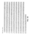
- the invention also provides HGF receptor antagonists that comprise isolated polypeptide comprising the amino acid sequences of Figure 1A (SEQ ID NO:1) and Figure 1B (SEQ ID NO:2).
- the polypeptides consisting of the amino acid sequences of Figure 1A (SEQ ID NO:1) and Figure 1B (SEQ ID NO:2) correspond to the light chain and heavy chain, respectively, of monoclonal antibody 5D5 Fab, described herein.
- the invention also provides chimeric molecules comprising HGF receptor antagonist linked or fused to another, heterologous polypeptide or polymer.
- An example of such a chimeric molecule comprises a HGF receptor antagonist amino acid sequence linked or fused to an albumin sequence or polyethylene glycol (“PEG”) sequence.
- the invention further provides an isolated nucleic acid molecule encoding HGF receptor antagonist.
- the nucleic acid molecule is RNA or DNA that encodes a HGF receptor antagonist or is complementary to a nucleic acid sequence encoding such HGF receptor antagonist, and remains stably bound to it under stringent conditions.
- the nucleic acid sequences are selected from:
- the invention also provides a replicable vector comprising the nucleic acid molecule(s) encoding the HGF receptor antagonist operably linked to control sequence(s) recognized by a host cell transfected or transformed with the vector.
- a host cell comprising the vector or the nucleic acid motecule(s) is also provided.
- a method of producing HGF receptor antagonist which comprises culturing a host cell comprising the nucleic acid molecule(s) and recovering the protein from the host cell culture is further provided.
- the invention also provides pharmaceutical compositions comprising one or more HGF receptor antagonists in a pharmaceutically-acceptable carrier.
- the pharmaceutical composition may be included in an article of manufacture or kit.
- the invention also provides methods of employing HGF receptor antagonists, including methods of inhibiting HGF receptor activation.
- the invention further provides methods for treating cancer comprising administering to a mammal diagnosed as having cancer an effective amount of a HGF receptor antagonist.
- the HGF receptor antagonist alone may be administered to the mammal, or alternatively, may be administered to the mammal in combination with other therapeutic agents such as anti-cancer agents.
- the antagonists can be used to block binding of HGF to HGF receptor(s) or substantially prevent HGF receptor activation, thereby treating pathologic conditions associated with binding of HGF to HGF receptor(s) or with the activation of HGF receptor(s).
- hepatocyte growth factor and “HGF” refer to a growth factor typically having a structure with six domams (finger, Kringle 1, Kringle 2, Kringle 3, Kringle 4 and serine protease domains) and having the property of binding to a HGF receptor, as defined below.
- the terms "hepatocyte growth factor” and “HGF” include hepatocyte growth factor from humans (“huHGF”) and any non-human mammalian species, and in particular rat HGF.
- the terms as used herein include mature, pre, pre-pro, and pro forms, purified or isolated from a natural source, chemically synthesized or recombinantly produced.
- Human HGF is encoded by the cDNA sequence published by Miyazawa et al., 1989, supra , or Nakamura et al., 1989, supra .
- the sequences reported by Miyazawa et al. and Nakamura et al. differ in 14 amino acids. The reason for the differences is not entirely clear; polymorphism or cloning artifacts are among the possibilities. Both sequences are specifically encompassed by the foregoing terms. It will be understood that natural allelic variations exist and can occur among individuals, as demonstrated by one or more amino acid differences in the amino acid sequence of each individual.
- the HGF of the invention preferably has at least about 80% sequence identity, more preferably at least about 90% sequence identity, and even more preferably, at least about 95% sequence identity with a native mammalian HGF.
- the terms "hepatocyte growth factor” and "HGF” specifically include the delta5 huHGF as disclosed by Seki et al., supra .
- HGF receptor and "c-Met” when used herein refer to a cellular receptor for HGF, which typically includes an extracellular domain, a transmembrane domain and an intracellular domain, as well as variants and fragments thereof which retain the ability to bind HGF.
- HGF receptor and "c-Met” include the polypeptide molecule that comprises the full-length, native amino acid sequence encoded by the gene variously known as p190 MET .
- the present definition specifically encompasses soluble forms of HGF receptor, and HGF receptor from natural sources, synthetically produced in vitro or obtained by genetic manipulation including methods of recombinant DNA technology.
- the HGF receptor variants or fragments preferably share at least about 65% sequence identity, and more preferably at least about 75% sequence identity with any domain of the human c-Met amino acid sequence published in Rodrigues et at., Mol. Cell. Biol., 11:2962-2970 (1991 ); Park et at., Proc. Natl. Acad. Sci., 84:6379-6383 (1987 ); or Ponzetto et al., Oncogene, 6:553-559 (1991 ).
- 5D5 Fab is used herein to refer to polypeptide comprising amino acid residues 1 to 220 of the amino acid sequence shown in Figure 1A (SEQ ID NO:1) and amino acid residues 1 to 230 of the amino acid sequence shown in Figure 1B(SEQ ID NO:2), as well as biologically active deletional, insertional, or substitutional variants thereof.
- the 5D5 Fab consists of the amino acid sequences shown in Figures 1A and 1B, which correspond to the light chain and heavy chain, respectively, of monoclonal antibody 5D5 Fab.
- the biologically active variants have at least about 80% sequence identity, more preferably at least about 90% sequence identity, and even more preferably, at least about 95% sequence identity with the sequences described above.
- the definition encompasses 5D5 Fab obtained from an antibody source, such as papain digestion of monoclonal antibody 5D5 described herein, or prepared by recombinant or synthetic methods, described for instance in Example 13 below.
- epitope tagged when used herein refers to a chimeric polypeptide comprising HGF receptor antagonist, or a portion thereof, fused to a "tag polypeptide".
- the tag polypeptide has enough residues to provide an epitope against which an antibody can be made, yet is short enough such that it does not interfere with activity of the antagonist.
- the tag polypeptide preferably also is fairly unique so that the antibody does not substantially cross-react with other epitopes.
- Suitable tag polypeptides generally have at least six amino acid residues and usually between about 8 to about 50 amino acid residues (preferably, between about 10 to about 20 residues).
- the term "salvage receptor binding epitope” refers to an epitope of the Fc region of an IgG molecule (e.g., IgG 1 , IgG 2 , IgG 3 , or IgG 4 ) that is responsible for increasing the in vivo half-life of the IgG molecule.
- isolated when used to describe the various polypeptides disclosed herein, means polypeptide that has been identified and separated and/or recovered from a component of its natural environment. Contaminant components of its natural environment are materials that would typically interfere with diagnostic or therapeutic uses for the polypeptide, and may include enzymes, hormones, and other proteinaceous or nonproteinaceous solutes.
- the polypeptide will be purified (1) to a degree sufficient to obtain at least 15 residues of N-terminal or internal amino acid sequence by use of a spinning cup sequenator, or (2) to homogeneity by SDS-PAGE under non-reducing or reducing conditions using Coomassie blue or, preferably, silver stain.
- Isolated polypeptide includes polypeptide in situ within recombinant cells, since at least one component of the HGF receptor antagonist natural environment will not be present. Ordinarily, however, isolated polypeptide will be prepared by at least one purification step. Homogeneity here means less than about 5% contamination with other source proteins and polypeptides.
- An "isolated" HGF receptor antagonist nucleic acid molecule is a nucleic acid molecule that is identified and separated from at least one contaminant nucleic acid molecule with which it is ordinarily associated in the natural source of the HGF receptor antagonist nucleic acid.
- An isolated HGF receptor antagonist nucleic acid molecule is other than in the form or setting in which it is found in nature. Isolated HGF receptor antagonist nucleic acid molecules therefore are distinguished from the HGF receptor antagonist nucleic acid molecule as it exists in natural cells.
- an isolated HGF receptor antagonist nucleic acid molecule includes HGF receptor antagonist nucleic acid molecules contained in cells that ordinarily express HGF receptor antagonist where, for example, the nucleic acid molecule is in a chromosomal location different from that of natural cells.
- control sequences refers to DNA sequences necessary for the expression of an operably linked coding sequence in a particular host organism.
- the control sequences that are suitable for prokaryotes include a promoter, optionally an operator sequence, and a ribosome binding site.
- Eukaryotic cells are known to utilize promoters, polyadenylation signals, and enhancers.
- Nucleic acid is "operably linked" when it is placed into a functional relationship with another nucleic acid sequence.
- DNA for a presequence or secretory leader is operably linked to DNA for a polypeptide if it is expressed as a preprotein that participates in the secretion of the polypeptide;
- a promoter or enhancer is operably linked to a coding sequence if it affects the transcription of the sequence; or
- a ribosome binding site is operably linked to a coding sequence if it is positioned so as to facilitate translation.
- "operably linked” means that the DNA sequences being linked are contiguous, and, in the case of a secretory leader, contiguous and in reading phase. However, enhancers do not have to be contiguous. Linking is accomplished by ligation at convenient restriction sites. If such sites do not exist, the synthetic oligonucleotide adaptors or linkers are used in accordance with conventional practice.
- amino acid and “amino acids” refer to all naturally occurring L- ⁇ -amino acids. This definition is meant to include norleucine, omithine, and homocysteine.
- the amino acids are identified by either the single-letter or three-letter designations: Asp D aspartic acid Ile I isoleucine Thr T threonine Leu L leucine Ser S serine Tyr Y tyrosine Glu E glutamic acid Phe F phenylalanine Pro P proline His H histidine Gly G glycine Lys K lysine Ala A alanine Arg R arginine Cys C cysteine Trp W tryptophan Val V valine Gln Q glutamine Met M methionine Asn N asparagine
- Nucleotide bases referred to in the Sequence Listing and Figures include “A” (adenine), “C” (cytosine), “G” (guanine), “T” (thymine) and “S” (cytosine or guanine).
- heparin is used in a broad sense and refers to a heterogeneous group of sulfated, straight-chain anionic mucopolysaccharides, often referred to as glycosaminoglycans. Although others may be present, the main sugars in heparin are: ⁇ -L-iduronic acid 2-sulfate, 2-deoxy-2-sulfamino- ⁇ -glucose 6-sulfate, ⁇ -D-glucuronic acid, 2-acetamido-2-deoxy- ⁇ -D-glucose, and L-iduronic acid. These and optionally other sugars are typically joined by glycosidic linkages.
- the molecular weight of heparin typically varies from about 6,000 to about 20,000 Da depending on the source and the method of molecular weight determination. Heparin is a native constituent of various cells and tissues, especially liver and lung, in several mammalian species.
- heparin-independent when used herein describes HGF receptor antagonists which have substantially reduced ability to bind heparin or are unable to bind heparin, or heparin-like glycosaminoglycans, including heparan sulfate and proteoglycans. Determination of whether a HGF receptor antagonist is heparin-independent can be determined by the skilled artisan without undue experimentation. Heparin-independence can be determined, for example, by assaying the antagonist for HGF blocking activity in the presence of heparin, as described in the Examples, and observing the activity of the molecule.
- agonist and “agonistic” when used herein refer to or describe a molecule which is capable of, directly or indirectly, substantially inducing, promoting or enhancing HGF biological activity or HGF receptor activation.
- antagonist and “antagonistic” when used herein refer to or describe a molecule which is capable of, directly or indirectly, substantially counteracting, reducing or inhibiting HGF biological activity or HGF receptor activation.
- HGF biological activity when used herein refers to any mitogenic, motogenic or morphogenic activities of HGF or any activities occurring as a result of HGF binding to a HGF receptor.
- HGF receptor activation refers to HGF receptor dimerization or HGF receptor-induced tyrosine kinase activity. HGF receptor activation may occur as a result of HGF binding to a HGF receptor, but may alternatively occur independent of any HGF binding to a HGF receptor.
- HGF biological activity may, for example, be determined in an in vitro or in vivo assay of hepatocyte growth promotion. Adult rat hepatocytes in primary culture have been used to test the effect of HGF on hepatocyte proliferation.
- the effect of a HGF receptor antagonist can be determined in an assay suitable for testing the ability of HGF to induce DNA synthesis of rat hepatocytes in primary cultures.
- Human hepatocytes can be cultured similarly to the methods established for preparing primary cultures of normal rat hepatocytes.
- the effect of a HGF receptor antagonist can be determined in an assay suitable for testing the ability of HGF to induce DNA synthesis in other types of cells expressing HGF receptor(s), such as mink lung cells or human mammary epithelial cells described in Examples 4 and 5.
- DNA synthesis can, for example, be assayed by measuring incorporation of 3 H-thymidine into DNA.
- the effectiveness of the HGF receptor antagonist can be determined by its ability to block proliferation and incorporation of the 3 H-thymidine into DNA.
- the effect of HGF receptor antagonists can also be tested in vivo in animal models.
- antibody is used herein in a broad sense and includes intact immunoglobulin or antibody molecules, polyclonal antibodies, multispecific antibodies (i.e., bispecific antibodies formed from at least two intact antibodies) and immunoglobulin fragments (such as Fab, F(ab') 2 , or Fv), so long as they exhibit any of the desired antagonistic properties described herein.
- Antibodies are typically proteins or polypeptides which exhibit binding specificity to a specific antigen. Native antibodies are usually heterotetrameric glycoproteins, composed of two identical light (L) chains and two identical heavy (H) chains.
- each light chain is linked to a heavy chain by one covalent disulfide bond, while the number of disulfide linkages varies between the heavy chains of different immunoglobulin isotypes.
- Each heavy and light chain also has regularly spaced intrachain disulfide bridges.
- Each heavy chain has at one end a variable domain (V H ) followed by a number of constant domains.
- Each light chain has a variable domain at one end (V L ) and a constant domain at its other end; the constant domain of the light chain is aligned with the first constant domain of the heavy chain, and the light chain variable domain is aligned with the variable domain of the heavy chain.
- the light chains of antibodies from any vertebrate species can be assigned to one of two clearly distinct types, called kappa ( ⁇ ) and lambda ( ⁇ ), based on the amino acid sequences of their constant domains.
- immunoglobulins can be assigned to different classes.
- immunoglobulins There are five major classes of immunoglobulins: IgA, IgD, IgE, IgG and IgM, and several of these may be further divided into subclasses (isotypes), e.g., IgG-1, IgG-2, IgG-3, and IgG-4; IgA-1 and IgA-2.
- the heavy chain constant domains that correspond to the different classes of immunoglobulins are called ⁇ , delta, epsilon, ⁇ , and ⁇ , respectively.
- Antibody fragments comprise a portion of an intact antibody, generally the antigen binding or variable region of the intact antibody.
- antibody fragments include Fab, Fab', F(ab')2, and Fv fragments, diabodies, single chain antibody molecules, and multispecific antibodies formed from antibody fragments.
- variable is used herein to describe certain portions of the variable domains which differ in sequence among antibodies and are used in the binding and specificity of each particular antibody for its particular antigen.
- variability is not usually evenly distributed through the variable domains of antibodies. It is typically concentrated in three segments called complementarity determining regions (CDRs) or hypervariable regions both in the light chain and the heavy chain variable domains.
- CDRs complementarity determining regions
- FR framework
- the variable domains of native heavy and light chains each comprise four FR regions, largely adopting a ⁇ -sheet configuration, connected by three CDRs, which form loops connecting, and in some cases forming part of, the ⁇ -sheet structure.
- the CDRs in each chain are held together in close proximity by the FR regions and, with the CDRs from the other chain, contribute to the formation of the antigen binding site of antibodies [see Kabat, E.A. et al., Sequences of Proteins of Immunological Interest, National Institutes of Health, Bethesda, MD (1987 )].
- the constant domains are not involved directly in binding an antibody to an antigen, but exhibit various effector functions, such as participation of the antibody in antibody-dependent cellular toxicity.
- the term "monoclonal antibody” as used herein refers to an antibody obtained from a substantially homogeneous population of antibodies, i.e., the individual antibodies comprising the population are identical except for possible naturally occurring mutations that may be present in minor amounts.
- the monoclonal antibodies herein specifically include "chimeric" antibodies in which a portion of the heavy and/or light chain is identical with or homologous to corresponding sequences in antibodies derived from a particular species or belonging to a particular antibody class or subclass, while the remainder of the chain(s) is identical with or homologous to corresponding sequences in antibodies derived from another species or belonging to another antibody class or subclass, as well as fragments of such antibodies, so long as they exhibit the desired antagonistic activity [ U.S. Patent No.
- cancer and "cancerous" when used herein refer to or describe the physiological condition in mammals that is typically characterized by unregulated cell growth.
- cancer include but are not limited to, carcinoma, lymphoma, sarcoma, blastoma and leukemia. More particular examples of such cancers include squamous cell carcinoma, lung cancer (small cell and non-small cell), gastrointestinal cancer, liver cancer, kidney cancer, pancreatic cancer, cervical cancer, bladder cancer, hepatoma, breast cancer, colon carcinoma, and head and neck cancer.
- cancer as used herein is not limited to any one specific form of the disease, it is believed that the methods of the invention will be particularly effective for cancers which are found to be accompanied by increased levels of HGF or overexpression or activation of HGF receptor in the mammal.
- treating refers to curative therapy, prophylactic therapy, and preventative therapy.
- mammal refers to any animal classified as a mammal, including humans, cows, horses, dogs and cats. In a preferred embodiment of the invention, the mammal is a human.
- HGF receptor antagonists are provided.
- HGF receptor antagonists include antibodies, polypeptides, glycoproteins, glycopeptides, glycolipids, polysaccharides, oligosaccharides, nucleic acids, bioorganic molecules, peptidomimetics, pharmacological agents and their metabolites, transcriptional and translation control sequences, and the like.
- the HGF receptor antagonists of the invention comprise HGF receptor antibodies.
- the antagonist antibodies may be polyclonal antibodies. Methods of preparing polyclonal antibodies are known to the skilled artisan. Polyclonal antibodies can be raised in a mammal, for example, by one or more injections of an immunizing agent and, if desired, an adjuvant. Typically, the immunizing agent and/or adjuvant will be injected in the mammal by multiple subcutaneous or intraperitoneal injections.
- the immunizing agent includes the c-Met polypeptide or a fusion protein thereof. It may be useful to conjugate the immunizing agent to a protein known to be immunogenic in the mammal being immunized.
- immunogenic proteins examples include but are not limited to keyhole limpet hemocyanin, serum albumin, bovine thyroglobulin, and soybean trypsin inhibitor.
- An aggregating agent such as alum may also be employed to enhance the mammal's immune response.
- adjuvants examples include Freund's complete adjuvant and MPL-TDM adjuvant (monophosphoryl Lipid A, synthetic trehalose dicorynomycolate).
- the immunization protocol may be selected by one skilled in the art without undue experimentation.
- the mammal can then be bled, and the serum assayed for HGF receptor antibody titer. If desired, the mammal can be boosted until the antibody titer increases or plateaus.
- the antagonist antibodies of the invention may, alternatively, be monoclonal antibodies.
- Antagonist monoclonal antibodies of the invention may be prepared using hybridoma methods, such as those described by Kohler and Milstein, Nature, 226:495 (1975 ).
- a hybridoma method a mouse or other appropriate host animal, is typically immunized (such as described above) with an immunizing agent to elicit lymphocytes that produce or are capable of producing antibodies that will specifically bind to the immunizing agent.
- the lymphocytes may be immunized in vitro.
- the immunizing agent includes the c-Met polypeptide or a fusion protein thereof.
- the immunizing agent may alternatively comprise a fragment or portion of HGF or a HGF receptor having one or more amino acid residues that participate in the binding of HGF to its receptor.
- the immunizing agent comprises an extracellular domain of c-Met fused to an IgG sequence, such as described in Example I.
- peripheral blood lymphocytes are used if cells of human origin are desired, or spleen cells or lymph node cells are used if non-human mammalian sources are desired.
- the lymphocytes are then fused with an immortalized cell line using a suitable fusing agent, such as polyethylene glycol, to form a hybridoma cell [ Goding, Monoclonal Antibodies: Principles and Practice, Academic Press, (1986) pp. 59-103 ].
- Immortalized cell lines are usually transformed mammalian cells, particularly myeloma cells of rodent, bovine and human origin. Usually, rat or mouse myeloma cell lines are employed.
- the hybridoma cells may be cultured in a suitable culture medium that preferably contains one or more substances that inhibit the growth or survival of the unfused, immortalized cells.
- a suitable culture medium that preferably contains one or more substances that inhibit the growth or survival of the unfused, immortalized cells.
- the culture medium for the hybridomas typically will include hypoxanthine, aminopterin, and thymidine ("HAT medium"), which substances prevent the growth of HGPRT-deficient cells.
- Preferred immortalized cell lines are those that fuse efficiently, support stable high level expression of antibody by the selected antibody-producing cells, and are sensitive to a medium such as HAT medium. More preferred immortalized cell lines are murine myeloma lines, which can be obtained, for instance, from the Salk Institute Cell Distribution Center, San Diego, California and the American Type Culture Collection, Rockville, Maryland. An example of such a murine myeloma cell line is P3X63AgU. I, described in Example I below. Human myeloma and mouse-human heteromyeloma cell lines also have been described for the production of human monoclonal antibodies [ Kozbor, J. Immunol., 133:3001-3005 (1984 ); Brodeur et al., Monoclonal Antibody Production Techniques and Applications, Marcel Dekker, Inc., New York, (1987) pp. 51-63 ].
- the culture medium in which the hybridoma cells are cultured can then be assayed for the presence of monoclonal antibodies directed against a HGF receptor.
- the binding specificity of monoclonal antibodies produced by the hybridoma cells is determined by immunoprecipitation or by an in vitro binding assay, such as radioimmunoassay (RIA) or enzyme-linked immunoabsorbent assay (ELISA).
- RIA radioimmunoassay
- ELISA enzyme-linked immunoabsorbent assay
- the binding affinity of the monoclonal antibody can, for example, be determined by the Scatchard analysis of Munson and Pollard, Anal. Biochem., 107:220-239 (1980 ).
- the clones may be subcloned by limiting dilution procedures and grown by standard methods [Goding, supra ]. Suitable culture media for this purpose include, for example, Dulbecco's Modified Eagle's Medium and RPMI-1640 medium. Alternatively, the hybridoma cells may be grown in vivo as ascites in a mammal.
- the monoclonal antibodies secreted by the subclones may be isolated or purified from the culture medium or ascites fluid by conventional immunoglobulin purification procedures such as, for example, protein A-Sepharose, hydroxylapatite chromatography, gel electrophoresis, dialysis, or affinity chromatography.
- the monoclonal antibodies may also be made by recombinant DNA methods, such as those described in U.S. Patent No. 4,816,567 .
- DNA encoding the monoclonal antibodies of the invention can be readily isolated and sequenced using conventional procedures (e.g., by using oligonucleotide probes that are capable of binding specifically to genes encoding the heavy and light chains of murine antibodies).
- the hybridoma cells of the invention serve as a preferred source of such DNA. Once isolated, the DNA may be placed into expression vectors, which are then transfected into host cells such as simian COS cells, E.
- the DNA also may be modified, for example, by substituting the coding sequence for human heavy and light chain constant domains in place of the homologous murine sequences [ U.S. Patent No. 4,816,567 ; Morrison et al., supra ] or by covalently joining to the immunoglobulin coding sequence all or part of the coding sequence for a non-immunoglobulin polypeptide.
- non-immunoglobulin polypeptide can be substituted for the constant domains of an antibody of the invention, or can be substituted for the variable domains of one antigen-combining site of an antibody of the invention to create a chimeric bivalent antibody comprising one antigen-combining site having specificity for a HGF receptor and another antigen-combining site having specificity for a different antigen, such as HER2 or CD3.
- monovalent antibodies capable of binding to a HGF receptor will be especially useful as HGF receptor antagonists. While not being bound to any particular theory, it is presently believed that activation of c-Met may proceed by a mechanism wherein huHGF binding to c-Met induces aggregation or dimerization of the receptors which in turn activates intracellular receptor kinase activity. Because monovalent antibodies will likely be unable to induce such aggregation or dimerization, the monovalent antibodies should not activate c-Met.
- Such monovalent antibodies may be directed against the HGF binding site of the receptor or may otherwise be capable of interfering with HGF, its fragments or its variants binding to the HGF receptor, such as by sterically hindering HGF, its fragments or its variants access to the receptor.
- the monovalent antibodies may be capable of sterically preventing HGF receptor dimerization.
- one method involves recombinant expression of immunoglobulin light chain and modified heavy chain.
- the heavy chain is truncated generally at any point so as to prevent heavy chain crosslinking.
- the relevant cysteine residues may be substituted with another amino acid residue or are deleted so as to prevent crosslinking.
- Recombinant expression of Fab light chain and heavy chain is described in further detail below.
- In vitro methods are also suitable for preparing monovalent antibodies.
- Digestion of antibodies to produce fragments thereof, particularly, Fab fragments can be accomplished using routine techniques known in the art. For instance, digestion can be performed using papain. Examples of papain digestion are described in WO 94/29348 published 12/22/94 and U.S. Patent No. 4,342,566 . Papain digestion is also described in Examples 6 and 7 below. Papain digestion of antibodies typically produces two identical antigen binding fragments, called Fab fragments, each with a single antigen binding site, and a residual Fc fragment. Pepsin treatment yields an F(ab') 2 fragment that has two antigen combining sites and is still capable of cross-linking antigen.
- the Fab fragments produced in the antibody digestion also contain the constant domains of the light chain and the first constant domain (CH 1 ) of the heavy chain.
- Fab' fragments differ from Fab fragments by the addition of a few residues at the carboxy terminus of the heavy chain CH 1 domain including one or more cysteines from the antibody hinge region.
- Fab'-SH is the designation herein for Fab' in which the cysteine residue(s) of the constant domains bear a free thiol group.
- F(ab') 2 antibody fragments originally were produced as pairs of Fab' fragments which have hinge cysteines between them. Other chemical couplings of antibody fragments are also known.
- the antagonists comprise Fab fragments of monoclonal antibodies specific for c-Met.
- the monoclonal antibody Fab fragments have the same biological characteristics as the monoclonal antibody Fab fragments produced by digesting either of the monoclonal antibodies secreted by the hybridoma cell lines deposited under American Type Culture Collection Accession Nos. ATCC HB-11894 or ATCC HB-11895.
- biological characteristics is used to refer to the in vitro and/or in vivo activities of the monoclonal antibody, e.g., ability to substantially reduce or inhibit binding of huHGF to c-Met or to substantially reduce or inhibit c-Met activation.
- the monovalent antibody preferably binds to substantially the same epitope as the 1A3.3.13 antibody or the 5D5.11.6 antibody disclosed herein.
- This can be determined by conducting assays described herein and in the Examples. For instance, to determine whether a monoclonal antibody has the same specificity as the 1A3.3.13 antibody specifically disclosed (i.e., the antibody having the ATCC deposit No. HB-11894) or the 5D5.11.6 antibody specifically disclosed (i.e., the antibody having the ATCC deposit No. HB-11895), one can use a competitive ELISA binding assay such as those described in the Examples.
- the monoclonal antibody Fab fragments are heparin-independent antagonists, as defined herein.
- the monoclonal antibody or fragment thereof will inhibit the binding of HGF, its fragments or its variants, or the mitogenic activity of HGF, its fragments, or its variants at least about 50%, preferably, greater than about 80%, and more preferably, greater than about 90%, as determined by an in vitro competitive binding assay or proliferation assay, such as described in the Examples below.
- chimeric or hybrid antagonist antibodies may be prepared in vitro using known methods in synthetic protein chemistry, including those involving crosslinking agents.
- immunotoxins may be constructed using a disulfide exchange reaction or by forming a thioether bond.
- suitable reagents for this purpose include iminothiolate and methyl-4-mercaptobutyrimidate.
- the antagonist antibodies of the invention may also comprise diabodies.
- diabodies refers to small antibody fragments with two antigen binding sites, which fragments comprise a heavy chain variable domain (V H ) connected to a light chain variable domain (V L ) in the same polypeptide chain (V H - V L ).
- V H heavy chain variable domain
- V L light chain variable domain
- the domains are forced to pair with the complementary domains of another chain and create two antigen binding sites.
- Diabodies are described in further detail, for example, in EP 404,097 ; WO 93/ 11161 ; and Hollinger et al., Proc. Natl. Acad. Sci., 90:6444-6448 (1993 ).
- the antagonist antibodies of the invention may further comprise humanized antibodies or human antibodies.
- Humanized forms of non-human (e.g., murine) antibodies are chimeric immunoglobulins, immunoglobulin chains or fragments thereof (such as Fv, Fab, Fab', F(ab') 2 or other antigen-binding subsequences of antibodies) which contain minimal sequence derived from non-human immunoglobulin.
- Humanized antibodies include human immunoglobulins (recipient antibody) in which residues from a complementary determining region (CDR) of the recipient are replaced by residues from a CDR of a non-human species (donor antibody) such as mouse, rat or rabbit having the desired specificity, affinity and capacity.
- CDR complementary determining region
- Fv framework residues of the human immunoglobulin are replaced by corresponding non-human residues.
- Humanized antibodies may also comprise residues which are found neither in the recipient antibody nor in the imported CDR or framework sequences.
- the humanized antibody will comprise substantially all of at least one, and typically two, variable domains, in which all or substantially all of the CDR regions correspond to those of a non-human immunoglobulin and all or substantially all of the FR regions are those of a human immunoglobulin consensus sequence.
- the humanized antibody optimally also will comprise at least a portion of an immunoglobulin constant region (Fc), typically that of a human immunoglobulin [ Jones et al., Nature, 321:522-525 (1986 ); Reichmann et al., Nature, 332:323-327 (1988 ); and Presta, Curr. Op. Struct. Biol., 2:593-596 (1992 )].
- Fc immunoglobulin constant region
- a humanized antibody has one or more amino acid residues introduced into it from a source which is non-human. These non-human amino acid residues are often referred to as "import" residues, which are typically taken from an "import” variable domain.
- Humanization can be essentially performed following the method of Winter and co-workers [ Jones et al., Nature, 321:522-525 (1986 ); Riechmann et al., Nature, 332:323-327 (1988 ); Verhoeyen et al., Science, 239:1534-1536 (1988 )], by substituting rodent CDRs or CDR sequences for the corresponding sequences of a human antibody.
- humanized antibodies are chimeric antibodies ( U.S. Patent No. 4,816,567 ), wherein substantially less than an intact human variable domain has been substituted by the corresponding sequence from a non-human species.
- humanized antibodies are typically human antibodies in which some CDR residues and possibly some FR residues are substituted by residues from analogous sites in rodent antibodies.
- variable domains both light and heavy
- the choice of human variable domains, both light and heavy, to be used in making the humanized antibodies is important in order to reduce antigenicity.
- the sequence of the variable domain of a rodent antibody is screened against the entire library of known human variable domain sequences.
- the human sequence which is closest to that of the rodent is then accepted as the human framework (FR) for the humanized antibody [ Sims et al., J. Immunol., 151:2296 (1993 ); Chothia and Lesk, J. Mol. Biol., 196:901 (1987 )].
- Another method uses a particular framework derived from the consensus sequence of all human antibodies of a particular subgroup of light or heavy chains.
- the same framework may be used for several different humanized antibodies ( Carter et al., Proc. Natl. Acad. Sci. USA, 89:4285 (1992 ); Presta et al., J. Immunol., 151:2623-2632 (1993 )].
- humanized antibodies are prepared by a process of analysis of the parental sequences and various conceptual humanized products using three dimensional models of the parental and humanized sequences.
- Three dimensional immunoglobulin models are commonly available and are familiar to those skilled in the art.
- Computer programs are available which illustrate and display probable three-dimensional conformational structures of selected candidate immunoglobulin sequences. Inspection of these displays permits analysis of the likely role of the residues in the functioning of the candidate immunoglobulin sequence, i.e., the analysis of residues that influence the ability of the candidate immunoglobulin to bind its antigen.
- FR residues can be selected and combined from the consensus and import sequence so that the desired antibody characteristic, such as increased affinity for the target antigen(s), is achieved.
- the CDR residues are directly and most substantially involved in influencing antigen binding [see, WO 94/04679 published 3 March 1994 ].
- Transgenic animals e.g., mice
- mice that are capable, upon immunization, of producing a full repertoire of human antibodies in the absence of endogenous immunoglobulin production
- J H antibody heavy chain joining region
- Human antibodies can also be produced in phage display libraries [ Hoogenboom and Winter, J. Mol. Biol., 227:381 (1991 ); Marks et al., J. Mol. Biol., 222:581 (1991 )].
- the techniques of Cote et al. and Boerner et al. are also available for the preparation of human monoclonal antibodies ( Cole et al., Monoclonal Antibodies and Cancer Therapy, Alan R. Liss, p. 77 (1985 ) and Boemer et al., J. Immunol., 147(1):86-95(1991 )].
- the present invention also provides HGF receptor antagonists comprising one or more isolated polypeptides.
- the antagonist comprises residues 1 to 220 of the amino acid sequence shown in Figure 1A (SEQ ID NO:1) and residues I to 230 of the amino acid sequence shown in Figure I B (SEQ ID NO:2).
- the antagonist comprises two isolated polypeptides which correspond to the light chain and heavy chain, respectively, of an anti-HGF receptor monoclonal antibody Fab.
- the description below relates primarily to production of the HGF receptor antagonist by culturing cells transformed or transfected with a vector containing the antagonist nucleic acid and recovering the polypeptide(s) from the cell culture. It is of course, contemplated that alternative methods, which are well known in the art, may be employed to prepare the HGF receptor antagonist polypeptide.
- the DNA encoding the HGF receptor antagonist may be obtained from any cDNA library prepared from tissue believed to possess the antagonist mRNA and to express it at a detectable level. Accordingly, human c-Met antagonist DNA can be conveniently obtained from a cDNA library prepared from human tissues. The c-Met antagonist-encoding gene may also be obtained from a genomic library or by oligonucleotide synthesis.
- Probes such as antibodies to the c-Met receptor antagonist or oligonucleotides of at least about 20-80 bases
- Screening the cDNA or genomic library with the selected probe may be conducted using standard procedures, such as described in Sambrook et al., Molecular Cloning: A Laboratory Manual (New York: Cold Spring Harbor Laboratory Press, 1989 ).
- An alternative means to isolate the gene encoding the receptor antagonist is to use PCR methodology [Sambrook et al., supra ; Dieffenbach et al., PCR Primer:A Laboratory Manual (Cold Spring Harbor Laboratory Press, 1995 )].
- One method of screening employs selected oligonucleotide sequences to screen cDNA libraries from various human tissues.
- the oligonucleotide sequences selected as probes should be of sufficient length and sufficiently unambiguous that false positives are minimized.
- the oligonucleotide is preferably labeled such that it can be detected upon hybridization to DNA in the library being screened. Methods of labeling are well known in the art, and include the use of radiolabels like 32 P-labeled ATP, biotinylation or enzyme labeling.
- Nucleic acid having all the protein coding sequence may be obtained by screening selected cDNA or genomic libraries using the amino acid sequence disclosed herein, and, if necessary, using conventional primer extension procedures as described in Sambrook et al., supra , to detect precursors and processing intermediates of mRNA that may not have been reverse-transcribed into cDNA.
- Amino acid sequence variants of the antagonist polypeptide can be prepared by introducing appropriate nucleotide changes into its DNA, or by synthesis of the desired antagonist polypeptide. Such variants represent insertions, substitutions, and/or deletions of residues within or at one or both of the ends of the amino acid sequences shown in Figures 1A and 1B for the 5D5 Fab. Any combination of insertion, substitution, and/or deletion can be made to arrive at the final construct, provided that the final construct possesses the desired antagonistic activity as defined herein. In a preferred embodiment, the variants have at least about 80% sequence identity, more preferably, at least about 90% sequence identity, and even more preferably, at least about 95% sequence identity with the sequences identified herein for the 5D5 Fab.
- Variations in the native sequence as described above can be made using any of the techniques and guidelines for conservative and non-conservative mutations set forth in U.S. Pat. No. 5,364,934 . These include oligonucleotide-mediated (site-directed) mutagenesis, alanine scanning, and PCR mutagenesis.
- the nucleic acid e.g., cDNA or genomic DNA
- the nucleic acid may be inserted into a replicable vector for further cloning (amplification of the DNA) or for expression.
- a replicable vector for further cloning (amplification of the DNA) or for expression.
- Various vectors are publicly available.
- the vector components generally include, but are not limited to, one or more of the following: a signal sequence, an origin of replication, one or more marker genes, an enhancer element, a promoter, and a transcription termination sequence, each of which is described below.
- the HGF receptor antagonist may be produced recombinantly not only directly, but also as a fusion polypeptide with a heterologous polypeptide, which may be a signal sequence or other polypeptide having a specific cleavage site at the N-terminus of the mature protein or polypeptide.
- a heterologous polypeptide which may be a signal sequence or other polypeptide having a specific cleavage site at the N-terminus of the mature protein or polypeptide.
- the signal sequence may be a component of the vector, or it may be a part of the antagonist DNA that is inserted into the vector.
- the heterologous signal sequence selected preferably is one that is recognized and processed ( i.e., cleaved by a signal peptidase) by the host cell.
- the signal sequence may be a prokaryotic signal sequence selected, for example, from the group of the alkaline phosphatase, penicillinase, lpp, or heat-stable enterotoxin II leaders.
- the signal sequence may be, e.g., the yeast invertase leader, alpha factor leader (including Saccharomyces and Kluyveromyces ⁇ -factor leaders, the latter described in U.S. Pat. No. 5,010,182 ), or acid phosphatase leader, the C. albicans glucoamylase leader ( EP 362,179 published 4 April 1990 ), or the signal described in WO 90/13646 published 15 November 1990 .
- mammalian signal sequences may be used to direct secretion of the protein, such as signal sequences from secreted polypeptides of the same or related species, as well as viral secretory leaders, for example, the herpes simplex glycoprotein D signal.
- the DNA for such precursor region is preferably ligated in reading frame to DNA encoding the antagonist.
- Both expression and cloning vectors contain a nucleic acid sequence that enables the vector to replicate in one or more selected host cells.
- this sequence is one that enables the vector to replicate independently of the host chromosomal DNA, and includes origins of replication or autonomously replicating sequences.
- origins of replication or autonomously replicating sequences are well known for a variety of bacteria, yeast, and viruses.
- the origin of replication from the plasmid pBR322 is suitable for most Gram-negative bacteria, the 2 ⁇ plasmid origin is suitable for yeast, and various viral origins (SV40, polyoma, adenovirus, VSV or BPV) are useful for cloning vectors in mammalian cells.
- the origin of replication component is not needed for mammalian expression vectors (the SV40 origin may typically be used because it contains the early promoter).
- Most expression vectors are "shuttle" vectors, i.e., they are capable of replication in at least one class of organisms but can be transfected into another organism for expression.
- a vector is cloned in E. coli and then the same vector is transfected into yeast or mammalian cells for expression even though it is not capable of replicating independently of the host cell chromosome.
- DNA may also be amplified by insertion into the host genome. This is readily accomplished using Bacillus species as hosts, for example, by including in the vector a DNA sequence that is complementary to a sequence found in Bacillus genomic DNA. Transfection of Bacillus with this vector results in homologous recombination with the genome and insertion of the receptor antagonist DNA. However, the recovery of genomic DNA encoding the antagonist is more complex than that of an exogenously replicated vector because restriction enzyme digestion is required to excise the antagonist DNA.
- Selection genes typically contain a selection gene, also termed a selectable marker. This gene encodes a protein necessary for the survival or growth of transformed host cells grown in a selective culture medium. Host cells not transformed with the vector containing the selection gene will not survive in the culture medium.
- Typical selection genes encode proteins that (a) confer resistance to antibiotics or other toxins, e.g., ampicillin, neomycin, methotrexate, or tetracycline, (b) complement auxotrophic deficiencies, or (c) supply critical nutrients not available from complex media, e.g., the gene encoding D-alanine racemase for Bacilli.
- One example of a selection scheme utilizes a drug to arrest growth of a host cell. Those cells that are successfully transformed with a heterologous gene produce a protein conferring drug resistance and thus survive the selection regimen. Examples of such dominant selection use the drugs neomycin [ Southern et al., J. Molec. Appl. Genet., 1:327 (1982 )], mycophenolic acid ( Mulligan et al., Science, 209:1422 (1980 )] or hygromycin [ Sugden et al., Mol. Cell. Biol., 5:410-413 (1985 )]. The three examples given above employ bacterial genes under eukaryotic control to convey resistance to the appropriate drug G418 or neomycin (geneticin), xgpt (mycophenolic acid), or hygromycin, respectively.
- suitable selectable markers for mammalian cells are those that enable the identification of cells competent to take up the antagonist nucleic acid, such as DHFR or thymidine kinase.
- the mammalian cell transformants are placed under selection pressure that only the transformants are uniquely adapted to survive by virtue of having taken up the marker.
- Selection pressure is imposed by culturing the transformants under conditions in which the concentration of selection agent in the medium is successively changed, thereby leading to amplification of both the selection gene and the DNA that encodes the receptor antagonist.
- Amplification is the process by which genes in greater demand for the production of a protein critical for growth are reiterated in tandem within the chromosomes of successive generations of recombinant cells.
- Other examples of amplifiable genes include metal lothionein-I and -II, adenosine deaminase, and ornithine decarboxylase.
- Cells transformed with the DHFR selection gene may first be identified by culturing all of the transformants in a culture medium that contains methotrexate (Mtx), a competitive antagonist of DHFR.
- Mtx methotrexate
- An appropriate host cell when wild-type DHFR is employed is the Chinese hamster ovary (CHO) cell line deficient in DHFR activity, prepared and propagated as described by Urlaub et al., Proc. Natl. Acad. Sci. USA, 77:4216 (1980 ).
- the transformed cells are then exposed to increased levels of methotrexate. This leads to the synthesis of multiple copies of the DHFR gene, and, concomitantly, multiple copies of other DNA comprising the expression vectors, such as the DNA encoding the receptor antagonist.
- This amplification technique can be used with any otherwise suitable host, e.g., ATCC No. CCL61 CHO-K1, notwithstanding the presence of endogenous DHFR if, for example, a mutant DHFR gene that is highly resistant to Mtx is employed ( EP 117,060 ).
- host cells transformed or co-transformed with DNA sequences encoding HGF receptor antagonist, wild-type DHFR protein, and another selectable marker such as aminoglycoside 3'-phosphotransferase (APH) can be selected by cell growth in medium containing a selection agent for the selectable marker such as an aminoglycosidic antibiotic, e.g., kanamycin, neomycin, or G418. See U.S. Patent No. 4,965,199 .
- APH aminoglycoside 3'-phosphotransferase
- a suitable selection gene for use in yeast is the trp 1 gene present in the yeast plasmid YRp7 [ Stinchcomb et al., Nature, 282:39 (1979 ); Kingsman et al., Gene, 7:141 (1979 ); Tschemper et al., Gene, 10: 157 (1980 )].
- the trp 1 gene provides a selection marker for a mutant strain of yeast lacking the ability to grow in tryptophan, for example, ATCC No. 44076 or PEP4-1 [ Jones, Genetics, 85:12 (1977 )].
- the presence of the trp I lesion in the yeast host cell genome then provides an effective environment for detecting transformation by growth in the absence of tryptophan.
- Leu 2-deficient yeast strains (ATCC 20,622 or 38,626) are complemented by known plasmids bearing the Leu 2 gene.
- vectors derived from the 1.6 ⁇ m circular plasmid pKD1 can be used for transformation of Kluyveromyces yeasts [ Bianchi et al., Curr. Genet., 12:185 (1987 )]. More recently, an expression system for large-scale production of recombinant calf chymosin was reported for K. lactis [ Van den Berg, Bio/Technology, 8:135 (1990 )]. Stable multi-copy expression vectors for secretion of mature recombinant human serum albumin by industrial strains of Kluyveromyces have also been disclosed [ Fleer et al., Bio/Technology, 9:968-975 (1991 )].
- Expression and cloning vectors usually contain a promoter that is recognized by the host organism and is operably linked to the receptor antagonist nucleic acid sequence. Promoters are untranslated sequences located upstream (5') to the start codon of a structural gene (generally within about 100 to 1000 bp) that control the transcription and translation of particular nucleic acid sequence to which they are operably linked. Such promoters typically fall into two classes, inducible and constitutive. Inducible promoters are promoters that initiate increased levels of transcription from DNA under their control in response to some change in culture conditions, e.g., the presence or absence of a nutrient or a change in temperature. At this time a large number of promoters recognized by a variety of potential host cells are well known.
- promoters are operably linked to receptor antagonist encoding DNA by removing the promoter from the source DNA by restriction enzyme digestion and inserting the isolated promoter sequence into the vector.
- Various heterologous promoters may be used to direct amplification and/or expression of the receptor antagonist DNA.
- Promoters suitable for use with prokaryotic hosts include the ⁇ -lactamase and lactose promoter systems [ Chang et al., Nature, 275:615 (1978 ); Goeddel et al., Nature, 281:544 (1979 )], alkaline phosphatase, a tryptophan (trp) promoter system [ Goeddel, Nucleic Acids Res., 8:4057 (1980 ); EP 36,776 ], and hybrid promoters such as the tac promoter [ deBoer et al., Proc. Natl. Acad. Sci. USA, 80:21-25 (1983 )].
- other known bacterial promoters are suitable.
- Promoter sequences are known for eukaryotes. Virtually all eukaryotic genes have an AT-rich region located approximately 25 to 30 bases upstream from the site where transcription is initiated. At the 3' end of most eukaryotic genes is an AATAAA sequence that may be the signal for addition of the poly A tail to the 3' end of the coding sequence. All of these sequences are suitably inserted into eukaryotic expression vectors.
- Suitable promoting sequences for use with yeast hosts include the promoters for 3-phosphoglycemte kinase [ Hitzeman et al., J. Biol. Chem., 255:2073 (1980 )] or other glycolytic enzymes [ Hess et al., J. Adv.
- yeast promoters which are inducible promoters having the additional advantage of transcription controlled by growth conditions, are the promoter regions for alcohol dehydrogenase 2, isocytochrome C, acid phosphatase, degradative enzymes associated with nitrogen metabolism, metallothionein, glyceraldehyde-3-phosphate dehydrogenase, and enzymes responsible for maltose and galactose utilization.
- Suitable vectors and promoters for use in yeast expression are further described in EP 73,657 .
- Yeast enhancers also are advantageously used with yeast promoters.
- HGF receptor antagonist transcription from vectors in mammalian host cells is controlled, for example, by promoters obtained from the genomes of viruses such as polyoma virus, fowlpox virus ( UK 2,211,504 published 5 July 1989 ), adenovirus (such as Adenovirus 2), bovine papilloma virus, avian sarcoma virus, cytomegalovirus, a retrovirus, hepatitis-B virus and most preferably Simian Virus 40 (SV40), or from heterologous mammalian promoters, e.g., the actin promoter or an inimunoglobulin promoter, from heat-shock promoters, provided such promoters are compatible with the host cell systems.
- viruses such as polyoma virus, fowlpox virus ( UK 2,211,504 published 5 July 1989 ), adenovirus (such as Adenovirus 2), bovine papilloma virus, avian sarcoma virus, cytome
- the early and late promoters of the SV40 virus are conveniently obtained as an SV40 restriction fragment that also contains the SV40 viral origin of replication [ Fiers et al., Nature, 273: 113 (1978 ); Mulligan and Berg, Science, 209:1422-1427 (1980 ); Pavlakis et al., Proc. Nati. Acad. Sci. USA, 78:7398-7402 (1981 )].
- the immediate early promoter of the human cytomegalovirus is conveniently obtained as a HindIII E restriction fragment [ Greenaway et al., Gene, 18:355-360 (1982 )].
- a system for expressing DNA in mammalian hosts using the bovine papilloma virus as a vector is disclosed in U.S.
- Patent No. 4,419,446 A modification of this system is described in U.S. Patent No. 4,601,978 [See also Gray et al., Nature, 295:503-508 (1982 ) on expressing cDNA encoding immune interferon in monkey cells; Reyes et al., Nature, 297:598-601 (1982 ) on expression of human ⁇ -interferon cDNA in mouse cells under the control of a thymidine kinase promoter from herpes simplex virus; Canaani and Berg, Proc. Natl. Acad. Sci.
- Enhancers are cis-acting elements of DNA, usually about from 10 to 300 bp, that act on a promoter to increase its transcription. Enhancers are relatively orientation and position independent, having been found 5' [ Laimins et al., Proc. Natl. Acad. Sci. USA, 78:993 (1981 ]) and 3' [ Lusky et al., Mol.
- Examples include the SV40 enhancer on the late side of the replication origin (bp 100-270), the cytomegalovirus early promoter enhancer, the polyoma enhancer on the late side of the replication origin, and adenovirus enhancers. See also Yaniv, Nature, 297:17-18 (1982 ) on enhancing elements for activation of eukaryotic promoters.
- the enhancer may be spliced into the vector at a position 5' or 3' to the HGF receptor antagonist-encoding sequence, but is preferably located at a site 5' from the promoter.
- Expression vectors used in eukaryotic host cells will also contain sequences necessary for the termination of transcription and for stabilizing the mRNA. Such sequences are commonly available from the 5' and, occasionally 3', untranslated regions of eukaryotic or viral DNAs or cDNAs. These regions contain nucleotide segments transcribed as polyadenylated fragments in the untranslated portion of the mRNA encoding HGF receptor antagonist.
- Plasmids containing one or more of the above-listed components employs standard ligation techniques. Isolated plasmids or DNA fragments are cleaved, tailored, and re-ligated in the form desired to generate the plasmids required.
- the ligation mixtures can be used to transform E. coli K12 strain 294 (ATCC 31,446) and successful transformants selected by ampicillin or tetracycline resistance where appropriate. Plasmids from the transformants are prepared, analyzed by restriction endonuclease digestion, and/or sequenced by the method of Messing et al., Nucleic Acids Res., 9:309 (1981 ) or by the method of Maxam et al., Methods in Enzymology, 65:499 (1980 ).
- transient expression involves the use of an expression vector that is able to replicate efficiently in a host cell, such that the host cell accumulates many copies of the expression vector and, in turn, synthesizes high levels of a desired polypeptide encoded by the expression vector [Sambrook et al., supra ].
- Transient expression systems comprising a suitable expression vector and a host cell, allow for the convenient positive identification of polypeptides encoded by cloned DNAs, as well as for the rapid screening of such polypeptides for desired biological or physiological properties.
- transient expression systems are particularly useful in the invention for purposes of identifying analogs and variants of the receptor antagonists.
- Suitable host cells for cloning or expressing the DNA in the vectors herein are the prokaryote, yeast, or higher eukaryote cells described above.
- Suitable prokaryotes for this purpose include but are not lim ited to eubacteria, such as Gram-negative or Gram-positive organisms, for example, Enterobacteriaceae such as Escherichia, e.g., E. coli, Enterobacter, Erwinia, Klebsiella, Proteus, Salmonella. e.g., Salmonella typhimurium, Serratia, e.g., Serratia marcescans, and Shigella, as well as Bacilli such as B. subtilis and B.
- licheniformis e.g., B. licheniformis 41 IP disclosed in DD 266,710 published 12 April 1989
- Pseudomonas such as P. aeruginosa
- Streptomyces Preferably, the host cell should secrete minimal amounts of proteolytic enzymes.
- eukaryotic microbes such as filamentous fungi or yeast are suitable cloning or expression hosts for HGF receptor antagonist-encoding vectors.
- Saccharomyces cerevisiae, or common baker's yeast is the most commonly used among lower eukaryotic host microorganisms.
- a number of other genera, species, and strains are commonly available and useful herein.
- Suitable host cells for the expression of glycosylated HGF receptor antagonist are derived from multicellular organisms. Such host cells are capable of complex processing and glycosylation activities. In principle, any higher eukaryotic cell culture is workable, whether from vertebrate or invertebrate culture. Examples of invertebrate cells include plant and insect cells.
- baculoviral strains and variants and corresponding permissive insect host cells from hosts such as Spodoptera frugiperda (caterpillar), Aedes aegypti (mosquito), Aedes albopictus (mosquito), Drosophila melanogaster (fruitfly), and Bombyx mori have been identified [See, e.g., Luckow et al., Bio/Technology, 6:47-55 (1988 ); Miller et al., in Genetic Engineering, Setlow et al., eds., Vol. 8 (Plenum Publishing, 1986), pp. 277-279 ; and Maeda et al., Nature, 315:592-594 (1985 )].
- a variety of viral strains for transfection are publicly available, e.g., the L-I variant of Autographa californica NPV and the Bm-5 strain of Bombyx mori NPV.
- Plant cell cultures of cotton, corn, potato, soybean, petunia, tomato, and tobacco can be utilized as hosts.
- plant cells are transfected by incubation with certain strains of the bacterium Agrobacterium tumefaciens, which has been previously manipulated to contain the receptor antagonist-encoding DNA.
- the DNA encoding the receptor antagonist is transferred to the plant cell host such that it is transfected, and will, under appropriate conditions, express the receptor antagonist-encoding DNA.
- regulatory and signal sequences compatible with plant cells are available, such as the nopaline synthase promoter and polyadenylation signal sequences [ Depicker et at., J. Mol, Appl.
- DNA segments isolated from the upstream region of the T-DNA 780 gene are capable of activating or increasing transcription levels of plant-expressible genes in recombinant DNA-containing plant tissue [ EP 321,196 published 21 June 1989 ].
- Propagation of vertebrate cells in culture is also well known in the art [See, e.g., Tissue Culture, Academic Press, Kruse and Patterson, editors (1973 )].
- useful mammalian host cell lines are monkey kidney CV1 line transformed by SV40 (COS-7, ATCC CRL 1651); human embryonic kidney line (293 or 293 cells subcloned for growth in suspension culture, Graham et al., J. Gen Virol., 36:59 (1977 )); baby hamster kidney cells (BHK, ATCC CCL 10); Chinese hamster ovary cells/-DHFR (CHO, Urlaub and Chasin, Proc. Natl. Acad. Sci.
- mice sertoli cells TM4, Mather, Biol. Reprod., 23:243-251 (1980 )); monkey kidney cells (CV1 ATCC CCL 70); African green monkey kidney cells (VERO-76, ATCC CRL-1587); human cervical carcinoma cells (HELA, ATCC CCL 2); canine kidney cells (MDCK, ATCC CCL 34); buffalo rat liver cells (BRL 3A, ATCC CRL 1442); human lung cells (W138, ATCC CCL 75); human liver cells (Hep G2, HB 8065); mouse mammary tumor (MMT 060562, ATCC CCL51); TRI cells ( Mather et al., Annals N.Y. Acad. Sci., 383:44-68 (1982 )); MRC 5 cells; and FS4 cells.
- CV1 ATCC CCL 70 African green monkey kidney cells (VERO-76, ATCC CRL-1587); human cervical carcinoma cells (HELA, ATCC CCL 2); canine kidney cells (MDCK, ATCC CCL 34); buffalo rat
- Host cells are transacted and preferably transformed with the above-described expression or cloning vectors for HGF receptor antagonist production and cultured in conventional nutrient media modified as appropriate for inducing promoters, selecting transformants, or amplifying the genes encoding the desired sequences.
- Transfection refers to the taking up of an expression vector by a host cell whether or not any coding sequences are in fact expressed. Numerous methods of transfection are known to the ordinarily skilled artisan, for example, CaPO 4 and electroporation. Successful transfection is generally recognized when any indication of the operation of this vector occurs within the host cell.
- Transformation means introducing DNA into an organism so that the DNA is replicable, either as an extrachromosomal element or by chromosomal integrant. Depending on the host cell used, transformation is done using standard techniques appropriate to such cells.
- the calcium treatment employing calcium chloride, as described in Sambrook et al., supra , or electroporation is generally used for prokaryotes or other cells that contain substantial cell-wall barriers.
- Infection with Agrobacterium tumefaciens is used for transformation of certain plant cells, as described by Shaw et al., Gene, 23:315 (1983 ) and WO 89/05859 published 29 June 1989 .
- plants may be transfected using ultrasound treatment as described in WO 91/00358 published 10 January 1991 .
- DNA into cells such as by nuclear microinjection, electroporation, bacterial protoplast fusion with intact cells, or polycations, e.g., polybrene, polyomithine, may also be used.
- polycations e.g., polybrene, polyomithine
- transforming mammalian cells see Keown et al., Methods in Enzymology, 185:527-537 (1990 ) and Mansour et al., Nature, 336:348-352 (1988 ).
- Prokaryotic cells used to produce HGF receptor antagonist may be cultured in suitable media as described generally in Sambrook et al., supra .
- the mammalian host cells used to produce HGF receptor antagonist may be cultured in a variety of media.
- Examples of commercially available media include Ham's F10 (Sigma), Minimal Essential Medium (“MEM”, Sigma), RPMI-1640 (Sigma), and Dulbecco's Modified Eagle's Medium (“DMEM”, Sigma).
- Any such media may be supplemented as necessary with hormones and/or other growth factors (such as insulin, transferrin, or epidermal growth factor), salts (such as sodium chloride, calcium, magnesium, and phosphate), buffers (such as HEPES), nucleosides (such as adenosine and thymidine), antibiotics (such as GentamycinTM drug), trace elements (defined as inorganic compounds usually present at final concentrations in the micromolar range), and glucose or an equivalent energy source. Any other necessary supplements may also be included at appropriate concentrations that would be known to those skilled in the art.
- the culture conditions such as temperature, pH, and the like, are those previously used with the host cell selected for expression, and will be apparent to the ordinarily skilled artisan.
- the host cells referred to in this disclosure encompass cells in culture as well as cells that are within a host animal.
- Gene amplification and/or expression may be measured in a sample directly, for example, by conventional Southern blotting, Northern blotting to quantitate the transcription of mRNA [ Thomas, Proc. Natl. Acad, Sci, USA, 77:5201-5205 (1980 )], dot blotting (DNA analysis), or in situ hybridization, using an appropriately labeled probe, based on the sequences provided herein.
- Various labels may be employed, most commonly radioisotopes, and particularly 32 P.
- other techniques may also be employed, such as using biotin-modified nucleotides for introduction into a polynucleotide.
- the biotin then serves as the site for binding to avidin or antibodies, which may be labeled with a wide variety of labels, such as radionucleotides, fluorescers or enzymes.
- labels such as radionucleotides, fluorescers or enzymes.
- antibodies may be employed that can recognize specific duplexes, including DNA duplexes, RNA duplexes, and DNA-RNA hybrid duplexes or DNA-protein duplexes.
- the antibodies in turn may be labeled and the assay may be carried out where the duplex is bound to a surface, so that upon the formation of duplex on the surface, the presence of antibody bound to the duplex can be detected.
- Gene expression may be measured by immunological methods, such as immunohistochemical staining of cells or tissue sections and assay of cell culture or body fluids, to quantitate directly the expression of gene product.
- immunohistochemical staining techniques a cell sample is prepared, typically by dehydration and fixation, followed by reaction with labeled antibodies specific for the gene product coupled, where the labels are usually visually detectable, such as enzymatic labels, fluorescent labels, or luminescent labels.
- Antibodies useful for immunohistochemical staining and/or assay of sample fluids may be either monoclonal or polyclonal, and may be prepared in any mammal. 6. Purification of HGF Receptor Antagonist Polypeptide
- HGF receptor antagonist preferably is recovered from the culture medium as a secreted polypeptide, although it also may be recovered from host cell lysates when directly produced without a secretory signal. If the receptor antagonist is membrane-bound, it can be released from the membrane using a suitable detergent solution (e.g. Triton-X 100) or its extracellular region may be released by enzymatic cleavage.
- a suitable detergent solution e.g. Triton-X 100
- the antagonist polypeptide When the antagonist is produced in a recombinant cell other than one of human origin, the antagonist polypeptide is free of proteins or polypeptides of human origin. However, it may be desired to purify the receptor antagonist from recombinant cell proteins or polypeptides to obtain preparations that are substantially homogeneous as to the receptor antagonist. As a first step, the culture medium or lysate may be centrifuged to remove particulate cell debris.
- HGF receptor antagonist thereafter is purified from contaminant soluble proteins and polypeptides, with the following procedures being exemplary of suitable purification procedures: by fractionation on an ion-exchange column; ethanol precipitation; reverse phase HPLC; chromatography on silica or on a cation-exchange resin such as DEAE; chromatofocusing; SDS-PAGE; ammonium sulfate precipitation; gel filtration using, for example, Sephadex G-75; and protein A Sepharose columns to remove contaminants such as IgG.
- a protease inhibitor such as phenyl methyl sulfonyl fluoride (PMSF) also may be useful to inhibit proteolytic degradation during purification, and antibiotics may be included to prevent the growth of adventitious contaminants.
- PMSF phenyl methyl sulfonyl fluoride
- HGF receptor antagonists are included within the scope of this invention.
- One type of covalent modification of the HGF receptor antagonist is introduced into the molecule by reacting targeted amino acid residues of the antagonist with an organic derivatizing agent that is capable of reacting with selected side chains or the N- or C- terminal residues of the antagonist.
- Derivatization with bifunctional agents is useful for crosslinking the antagonist to a water-insoluble support matrix or surface for use in a method for purifying.
- Commonly used crosslinking agents include, e.g., 1,1-bis(diazoacetyl)-2-phenylethane, glutaraldehyde, N-hydroxysuccinimide esters, for example, esters with 4-azidosalicylic acid, homobifunctional imidoesters, including disuccinimidyl esters such as 3,3'-dithiobis(succinimidylpropionate), and bifunctional maieimides such as bis-N-maleimido-1,8-octane.
- Derivatizing agents such as methyl-3-[(p-azidophenyl)dithio]propioimidate yield photoactivatable intermediates that are capable of forming crosslinks in the presence of light.
- reactive water-insoluble matrices such as cyanogen bromide-activated carbohydrates and the reactive substrates described in U.S. Patent Nos. 3,969,287 ; 3,691,016 ; 4,195,128 ; 4,247,642 ; 4,229,537 ; and 4,330,440 are employed for protein immobilization.
- Another type of covalent modification of the receptor antagonist polypeptide included within the scope of this invention comprises altering the native glycosylation pattern of the polypeptide.
- "Altering the native glycosylation pattern" is intended for purposes herein to mean deleting one or more carbohydrate moieties and/or adding one or more glycosylation sites that are not present in the native polypeptide.
- N-linked refers to the attachment of the carbohydrate moiety to the side chain of an asparagine residue.
- the tripeptide sequences asparagine-Z-serine and asparagine-Z-threonine, where Z is any amino acid except proline, are the recognition sequences for enzymatic attachment of the carbohydrate moiety to the asparagine side chain.
- Z is any amino acid except proline
- O-linked glycosylation refers to the attachment of one of the sugars N-aceylgalactosamine, galactose, or xylose to a hydroxylamino acid, most commonly serine or threonine, although 5-hydroxyproline or 5-hydroxylysine may also be used.
- Addition of glycosylation sites to the polypeptide may be accomplished by altering the amino acid sequence such that it contains one or more of the above-described tripeptide sequences (for N-linked glycosylation sites).
- the alteration may also be made by the addition of, or substitution by, one or more serine or threonine residues to the native sequence (for O-linked glycosylation sites).
- the amino acid sequence may optionally be altered through changes at the DNA level, particularly by mutating the DNA encoding the polypeptide at preselected bases such that codons are generated that will translate into the desired amino acids.
- the DNA mutation(s) may be made using methods described above and in U.S. Pat. No. 5,364,934 , supra .
- sugar(s) may be attached to (a) arginine and histidine, (b) free carboxyl groups, (c) free sulfhydryl groups such as those of cysteine, (d) free hydroxyl groups such as those of serine, threonine, or hydroxyproline, (e) aromatic residues such as those of phenylalanine, tyrosine, or tryptophan, or (f) the amide group of glutamine.
- arginine and histidine the sugar(s) may be attached to (a) arginine and histidine, (b) free carboxyl groups, (c) free sulfhydryl groups such as those of cysteine, (d) free hydroxyl groups such as those of serine, threonine, or hydroxyproline, (e) aromatic residues such as those of phenylalanine, tyrosine, or tryptophan, or (f) the amide group of glutamine.
- Removal of carbohydrate moieties present on the polypeptide may be accomplished chemically or enzymatically or by mutational substitution of codons encoding for amino acid residues that serve as targets for glycosylation.
- chemical deglycosylation by exposing the polypeptide to the compound trifluoromethanesulfonic acid, or an equivalent compound can result in the cleavage of most or all sugars except the linking sugar (N-acetylglucosamine or N-acetylgalactosamine), while leaving the polypeptide intact.
- Chemical deglycosylation is described by Hakimuddin, et al., Arch, Biochem.
- Enzymatic cleavage of carbohydrate moieties on polypeptides can be achieved by the use of a variety of endo- and exo-glycosidases as described by Thotakura et al., Meth, Enzymol., 138:350 (1987 ).
- Glycosylation at potential glycosylation sites may be prevented by the use of the compound tunicamycin as described by Duskin et al., J. Biol. Chem., 257:3105 (1982 ). Tunicamycin blocks the formation of protein-N-glycoside linkages.
- Another type of covalent modification comprises linking the HGF receptor antagonist to one of a variety of nonproteinaceous polymers, e.g., polyethylene glycol (“PEG”), polypropylene glycol, or polyoxyalkylenes, in the manner set forth in U.S. Patent Nos. 4,640,835 ; 4,496,689 ; 4,301,144 ; 4,670,417 ; 4,791,192 or 4,179,337 .
- WO 93/00109 also describes methods of linking amino acid residues in polypeptides to PEG molecules.
- the present invention also provides chimeric molecules comprising HGF receptor antagonist fused to another, heterologous polypeptide.
- the chimeric molecule comprises a fusion of the antagonist with a tag polypeptide which provides an epitope to which an anti-tag antibody can selectively bind.
- the epitope tag is generally placed at the amino- or carboxyl- terminus of the antagonist. The presence of such epitope-tagged forms of the antagonist can be detected using an antibody against the tag polypeptide. Also, provision of the epitope tag enables the antagonist to be readily purified by affinity purification using an anti-tag antibody or another type of affinity matrix that binds to the epitope tag.
- tag polypeptides and their respective antibodies are well known in the art. Examples include the flu HA tag polypeptide and its antibody 12CA5 [ Field et al., Mol. Cell. Biol., 8:2159-2165 (1988 )]; the c-myc tag and the 8F9, 3C7, 6E10, G4, B7 and 9E10 antibodies thereto [ Evan et al., Molecular and Cellular Biology, 5:3610-3616 (1985 )]; and the Herpes Simplex virus glycoprotein D (gD) tag and its antibody [ Paborsky et al., Protein Engineering, 3(6):547-553 (1990 )].
- tag polypeptides include the Flag-peptide [ Hopp et al., BioTechnology, 6:1204-1210 (1988 )]; the KT3 epitope peptide [ Martin et al., Science, 255:192-194 (1992 )]; an ⁇ -tubulin epitope peptide [ Skinner et al., J. Biol. Chem., 266:15163-15166 (1991 )]; and the T7 gene 10 protein peptide tag [ Lutz-Freyermuth et al., Proc. Natl. Acad. Sci. USA, 87:6393-6397 (1990 )].
- an antibody thereto can be generated using the techniques disclosed herein.
- epitope-tagged antagonist may be constructed and produced according to the methods described above.
- HGF receptor antagonist-tag polypeptide fusions are preferably constructed by fusing the cDNA sequence encoding the HGF receptor antagonist portion in-frame to the tag polypeptide DNA sequence and expressing the resultant DNA fusion construct in appropriate host cells.
- nucleic acid encoding the antagonist will be fused at its 3' end to nucleic acid encoding the N-terminus of the tag polypeptide, however 5' fusions are also possible.
- a polyhistidine sequence of about 5 to about 10 histidine residues may be fused at the N- terminus or the C- terminus and used as a purification handle in affinity chromatography.
- Epitope-tagged HGF receptor antagonist can be purified by affinity chromatography using the anti-tag antibody.
- the matrix to which the affinity antibody is attached may include, for instance, agarose, controlled pore glass or poly(stymnedivinyl)benzene.
- the epitope-tagged HGF receptor antagonist can then be eluted from the affinity column using techniques known in the art.
- the HGF receptor antagonist may be fused to a salvage receptor binding epitope in order to increase its serum half-life.
- a salvage receptor binding epitope may be achieved, for example, by incorporation of a salvage receptor binding epitope into a HGF receptor antagonist antibody fragment (e.g., by mutation of the appropriate region in the antibody fragment or by incorporating the epitope into a peptide tag that is then fused to the antibody fragment at either the end or in the middle, e.g., by DNA or peptide synthesis).
- a systematic method for preparing such a chimera having an increased in vivo half-life comprises several steps. The first involves identifying the sequence and conformation of a salvage receptor binding epitope of an Fc region of an IgG molecule. Once this epitope is identified, the sequence of the HGF receptor antagonist of interest is modified to include the sequence and conformation of the identified binding epitope. After the sequence is mutated, the chimera is tested to see if it has a longer in vivo half-life than that of the original antagonist. If the chimera does not have a longer half-life upon testing, its sequence is further altered to include the sequence and conformation of the identified binding epitope.
- the salvage receptor binding epitope being incorporated into the HGF receptor antagonist of interest is any suitable such epitope as defined above, and its nature will depend for example, on the type of antagonist being modified. The transfer is made such that the HGF receptor antagonist of interest still possesses antagonistic activity.
- the salvage receptor binding epitope generally constitutes a region wherein any one or more amino acid residues from one or two loops of a Fc domain and is preferably transferred to an analogous position in an HGF receptor antagonist antibody fragment.
- three or more residues from one or two loops of the Fc domain are transferred, and more preferably, the epitope is taken from the CH2 domain of the Fc region of an IgG and transferred to the CH1, CH3, or V H region, or more than one such region, of the antagonist antibody.
- the epitope is taken from the CH2 domain of the Fc region and transferred to the C L region or the V L region, or both, of the antagonist antibody fragment.
- the chimeric molecule comprises a HGF receptor antagonist fused to an immunoglobulin constant domain or another heterologous polypeptide such as albumin.
- a HGF receptor antagonist fused to an immunoglobulin constant domain or another heterologous polypeptide such as albumin.
- the chimeric molecules can be constructed in a fashion similar to chimeric antibodies in which a variable domain from an antibody of one species is substituted for the variable domain of another species. See, for example, EP 0 125 023 ; EP 173,494 ; Munro, Nature, 312:597 (13 December 1984 ); Neuberger et al., Nature, 312:604-608 (13 December 1984 ); Sharon et al., Nature, 309:364-367 (24 May 1984 ); Morrison et al., Proc. Nat'l. Acad. Sci.
- the Ig is a human immunoglobulin when the chimera is intended for in vivo therapy for humans.
- DNA encoding immunoglobulin light or heavy chain constant regions is known or readily available from cDNA libraries or can be synthesized.
- the chimera comprises a HGF receptor antagonist fused to albumin.
- Such chimeras may be constructed by inserting the entire coding region of albumin into a plasmid expression vector.
- the DNA encoding the antagonist can be inserted at the 5' end of the albumin sequence, along with an insert encoding a linker consisting of four glycine residues [ Lu et al., FEBS Letters, 356:56-59 (1994 )].
- the HGF receptor antagonist-albumin chimera can then be expressed in desired mammalian cells or yeast, for instance.
- HGF receptor antagonist is administered to a mammal diagnosed as having cancer.
- cancer as used herein is not limited to any one specific form of the disease, it is believed that the methods will be particularly effective for cancers which are found to be accompanied by increased levels of HGF, or overexpression or activation of HGF receptor in the mammal.
- the cancer is breast cancer. It is of course contemplated that the methods of the invention can be employed in combination with still other therapeutic techniques such as surgery.
- the antagonist is preferably administered to the mammal in a pharmaceutically-acceptable carrier.
- Suitable carriers and their formulations are described in Remington's Pharmaceutical Sciences, 16th ed., 1980, Mack Publishing Co., edited by Oslo et al.
- an appropriate amount of a pharmaceutically-acceptable salt is used in the formulation to render the formulation isotonic.
- the pharmaceutically-acceptable carrier include saline, Ringer's solution and dextrose solution.
- the pH of the solution is preferably from about 5 to about 8, and more preferably from about 7 to about 7.5.
- Further carriers include sustained release preparations such as semipermeable matrices of solid hydrophobic polymers containing the antagonist.
- matrices are in the form of shaped articles, e.g., films, liposomes or microparticles. It will be apparent to those persons skilled in the art that certain carriers may be more preferable depending upon, for instance, the route of administration and concentration of antagonist being administered.
- the antagonist can be administered to the mammal by injection (e.g., intravenous, intraperitoneal, subcutaneous, intramuscular), or by other methods such as infusion that ensure its delivery to the bloodstream in an effective form.
- the antagonist may also be administered by intratumoral, peritumoral, intralesional, or perilesional routes, to exert local as well as systemic therapeutic effects. Local or intravenous injection is preferred.
- Effective dosages and schedules for administering the antagonist may be determined empirically, and making such determinations is within the skill in the art. Those skilled in the art will understand that the dosage of antagonist that must be administered will vary depending on, for example, the mammal which will receive the antagonist, the route of administration, the particular type of antagonist used and other drugs being administered to the mammal. Guidance in selecting appropriate doses for antibody antagonists is found in the literature on therapeutic uses of antibodies, e.g., Handbook of Monoclonal Antibodies, Ferrone et al., eds., Noges Publications, Park Ridge, N.J., (1985) ch. 22 and pp.
- a typical daily dosage of the antagonist used alone might range from about 1 ⁇ g/kg to up to 100 mg/kg of body weight or more per day, depending on the factors mentioned above.
- the antagonist may also be administered to the mammal in combination with effective amounts of one or more other therapeutic agents or in conjunction with radiation treatment.
- Therapeutic agents contemplated include chemotherapeutics as well as immunoadjuvants and cytokines.
- Chemotherapies contemplated by the invention include chemical substances or drugs which are known in the art and are commercially available, such as Doxorubicin, 5-Fluorouracil, Cytosine arabinoside ("Ara-C"), Cyclophosphamide, Thiotepa, Busulfan, Cytoxin, Taxol, Methotrexate, Cisplatin, Melphalan, Vinblastine and Carboplatin.
- the antagonist may be administered sequentially or concurrently with the one or more other therapeutic agents.
- the amounts of antagonist and therapeutic agent depend, for example, on what type of drugs are used, the cancer being treated, and the scheduling and routes of administration but would generally be less than if each were used individually.
- the mammal's cancer and physiological condition can be monitored in various ways well known to the skilled practitioner. For instance, tumor mass may be observed physically or by standard x-ray imaging techniques.
- the antagonists of the invention also have utility in non-therapeutic applications. For instance" methods for employing the antagonists in vitro in diagnostic assays are provided.
- the antagonists may be employed in diagnostic assays to detect overexpression of HGF receptor in specific cells and tissues.
- diagnostic assay techniques known in the art may be used, such as competitive binding assays, direct or indirect sandwich assays and immunoprecipitation assays conducted in either heterogeneous or homogeneous phases [ Zola, Monoclonal Antibodies: A Manual of Techniques, CRC Press, Inc. (1987) pp. 147-158 ].
- the antagonists used in the diagnostic assays can be labeled with a detectable moiety.
- the detectable moiety should be capable of producing, either directly or indirectly, a detectable signal.
- the detectable moiety may be a radioisotope, such as 3 H, 14 C, 32 P, 35 S, or 125 I, a fluorescent or chemiluminescent compound, such as fluorescein isothiocyanate, rhodamine, or luciferin, or an enzyme, such as alkaline phosphatase, beta-galactosidase or horseradish peroxidase.
- any method known in the art for conjugating the antagonist to the detectable moiety may be employed, including those methods described by Hunter et al., Nature, 194:495 (1962 ); David et al., biochemistry, 13:1014-1021 (1974 ); Pain et al., J. Immunol. Meth., 40:219-230 (1981 ); and Nygren, J. Histochem, and Cytochem., 30:407 (1982 ).
- HGF receptor antagonist antibodies can be used to immunopurify HGF receptor(s).
- the article of manufacture comprises a container with a label.
- Suitable containers include, for example, bottles, vials, and test tubes.
- the containers may be formed from a variety of materials such as glass or plastic.
- the container holds a composition having an active agent which is effective for treating cancer or for detecting or purifying HGF receptor.
- the active agent in the composition is a HGF receptor antagonist and preferably, comprises Fab fragments of monoclonal antibodies specific for c-Met.
- the label on the container indicates that the composition is used for treating cancer or detecting or purifying HGF receptor, and may also indicate directions for either in vivo or in vitro use, such as those described above.
- the kit of the invention comprises the container described above and a second container comprising a buffer. It may further include other materials desirable from a commercial and user standpoint, including other buffers, diluents, filters, needles, syringes, and package inserts with instructions for use.
- mice obtained from Charles River Laboratories were immunized by injecting 2.5 ⁇ g/50 ⁇ l c-Met-IgG fusion protein (diluted in MPL-TDM adjuvant purchased from Ribi Immunochemical Research Inc., Hamilton, MT) five times into each hind foot pad. Injections were administered on Day 0 and Days 56, 63, 66 and 73.
- the c-Met-IgG fusion protein (including the extracellular domain of c-Met fused to a human IgGl heavy chain) was constructed essentially as described by Mark et al., J, Biol, Chem., 267:26166-26171 (1992 ) and produced in Chinese hamster ovary (CHO) cells.
- the c-Met-IgG was subsequently purified in a single step using affinity chromatography on immobilized Protein A (Bioprocessing, Inc., Princeton. NJ), using an elution scheme modified from Chamow et al., J. Immunol., 153:4268-4280 (1994 ). Culture supernatant was loaded onto a Protein A column equilibrated in 20 mM Tris, pH 7.4, 0.15M NaCl. The column was washed, first with equilibration buffer, then with equilibration buffer containing 0.5 M tetramethylammonium chloride, to remove non-specifically bound protein.
- c-Met-IgG was eluted with 20 mM Tris, pH 7.4, 3.5 M MgCl 2 . This c-Met-IgG eluate was concentrated and exchanged into 20 mM Tris, pH 7.4, 0.15 M NaCl by gel filtration on Sephadex G25 to a final concentration of about 2-4 mg/ml.
- lymph nodes were removed from the mice and a single cell suspension was prepared in DMEM media (obtained from Biowhitakker Corp.) supplemented with 1% penicillin-streptomycin.
- the lymph node cells were then fused with murine myeloma cells P3X63AgU.1 (ATCC CRL 1597) using 35% polyethylene glycol and cultured in 96-well culture plates. Hybridomas resulting from the fusion were selected in HAT medium.
- hybridoma culture supernatants were screened in an ELISA to test for the presence of monoclonal antibodies binding to the c-Met-IgG fusion protein.
- 96-well microtiter plates (Nunc) were coated by adding 50 ⁇ l of 2 ⁇ g/ml goat anti-human IgG Fc (purchased from Cappel Laboratories) to each well and incubating at 4° C overnight. The plates were then washed three times with distilled water. The wells in the microtiter plates were blocked with 200 ⁇ l of 2% bovine serum albumin and incubated at room temperature for I hour. The plates were then washed again three times with distilled water.
- mice were immunized as described in Example 1 except that the c-Met-IgG fusion protein injections were administered on Day 0 and Days 7, 14, 21, 28, 211, 273 and 279. On Day 282, lymph nodes were removed and a fusion was conducted as described in Example 1. Hybridoma supernatants were tested according to the ELISA described in Example 1. One of the positive anti-c-Met monoclonal antibodies was called 5D5.11.6 ("5D5"). Further antibody analysis showed that the 5D5 monoclonal antibody is an IgG I isotype antibody comprising a kappa light chain.
- 5D5 monoclonal antibody is an IgG I isotype antibody comprising a kappa light chain.
- An inhibition assay was conducted to examine the ability of the antibodies (described in Example 1) to block binding of HGF to c-Met-IgG fusion protein. Prior to conducting the inhibition assay, the 24 hybridoma supernatants determined positive in the ELISA in Example I were purified on Protein A-Sepharose columns to yield antibody preparations of about 1 ⁇ g/ml.
- 96-well microtiter plates were coated by adding 100 ⁇ l of 2 ⁇ g/ml goat anti-human Fc (purchased from Jackson Immunochemical, West Grove, PA) in 0.05 M sodium bicarbonate, pH 9.6, to each well and incubating overnight at 4°C. The plates were then washed with a washing buffer (0.05% Tween-20, 0.01% Thimersol in PBS). Nonspecific binding in the wells was blocked by adding 150 ⁇ l blocking buffer (0.5% BSA, 0.01% Thimersol in PBS, pH 7.4) to each well and incubating at room temperature for 2 hours with rapid agitation on an orbital shaker.
- a washing buffer 0.05% Tween-20, 0.01% Thimersol in PBS
- rhuHGF Recombinant human HGF
- rhuHGF-transfected cells were grown in a 400 L bioreactor in medium containing 2% fetal bovine serum for 8 days. Culture supernatant containing rhuHGF was concentrated and clarified, then conditioned by the addition of solid NaCl to 0.3 M. rhuHGF was then purified in a single step using cation exchange chromatography.
- concentrated culture supernatant was loaded onto a column of S-Sepharose Fast Flow equilibrated in 20 mM Tris, pH 7.5, 0.3 M NaCl. After washing out unbound protein, rhuHGF was eluted in a linear gradient from 20 mM Tris, pH 7.5, 0.3 M NaCl to 20 mM Tris, pH 7.5, 1.2 M NaCl. rhuHGF-containing fractions were pooled based on SDS-PAGE analysis. The S Sepharose Fast Flow pool was concentrated and exchanged into 20 mM Tris, pH 7.5, 0.5 M NaCl by gel filtration on Sephadex G25 to a final concentration of about 3-5 mg/ml.
- a rhuHGF stock solution was then prepared by diluting the rhuHGF in assay buffer (0.5% bovine serum albumin, 0.05% Tween-20, 0.01% Thimersol in PBS) to a concentration of 10 ⁇ g/ml.
- a stock solution of control gp120 antibody (Genentech. Inc.) was also prepared by diluting the antibody in assay buffer to a concentration of 10 ⁇ g/ml.
- rhuHGF rhuHGF, gp120 antibody or one of the 24 monoclonal antibodies was added to designated wells to yield a final concentration of 1000, 100, 10, or I ng/ml/well.
- 50 ⁇ l of 200 ng/ml biotinylated rhuHGF rhuHGF biotinylated using Biotin-X-NHS obtained from Research Organics. Inc., Cleveland OH
- HRP-streptavidin (1:2000 dilution in assay buffer) (purchased from Zymed Laboratories), was added to the wells and the plates were incubated for 30 minutes. The plates were washed again 3 times with washing buffer. 50 ⁇ l of substrate (5 mg OPD, 12.5 ml PBS, 5 ⁇ l H 2 O 2 ) was added to the wells and color was allowed to develop for 20 minutes. The reaction was stopped by adding 100 ⁇ l 4.5 N sulfuric acid to each well. Absorbance at 492 nm was quantitated in an automated microtiter plate reader.
- 1A3.3.13 One of the monoclonal antibodies, referred to as 1A3.3.13, significantly blocked binding of HGF ( Figure 2). Further antibody analysis showed that the 1A3.3.13 monoclonal antibody is an IgGl isotype antibody comprising a kappa light chain.
- Example 1 Several different antibodies produced in the fusion described in Example 1. referred to as 3D6, 6G1, and 1A3.3.13, and purified as described in Example 3, were tested and compared for their ability to induce DNA synthesis in a human mammary epithelial cell bioassay.
- Human mammary epithelial cells obtained from Clonetics Corp., No. CC-2551) were passaged in Mammary Epithelial Cell Basal Medium (Clonetics Corp., No. CC-3151). Prior to conducting the bioassay, the cells were trypsinized, washed, and resuspended in assay medium (Basal Medium supplemented with 1 mg/ml BSA, penicillin, streptomycin and L-glutamine) to a concentration of 1 x 10 5 cells/ml. Next, 100 ⁇ l of the cells were added to the wells of 96-well culture plates.
- assay medium Basal Medium supplemented with 1 mg/ml BSA, penicillin, streptomycin and L-glutamine
- rhuHGF (described in Example 3) was diluted in assay medium at concentrations of 20 ng/ml and 200 ng/ml.
- the 3D6, 6G1, 1A3.3.13, and control gp 120 antibodies were diluted in assay medium at concentrations of 200 ng/ml and 2 ⁇ g/ml. 100 ⁇ l of the rhuHGF and antibody preparations was then added to designated wells. The plates were incubated at 37°C in 5% CO 2 for 16 hours.
- Anti-c-Met antibodies 05-237 and 05-238 purchased from Upstate Biotechnology Inc.. Lake Placid, NY; see also, Prat et al., Mol. Cell. Biol. , supra , Prat et al., Int. J. Cancer , supra ) and antibody 3D6 (described in Example 4) were tested and compared for their ability to induce DNA synthesis in a mink lung bioassay.
- Mink lung cells (Mv I Lu, ATCC CCl 64) were passaged in DME/F12 (50:50) supplemented with 10% fetal bovine serum, penicillin, streptomycin and L-glutamine. Prior to conducting the bioassay, the mink lung cells were trypsinized, washed, and resuspended in assay medium (DME/F12 medium supplemented with I mg/ml BSA, penicillin, streptomycin and L-glutamine) to a concentration of 1 x 10 5 cells/ml. The bioassay was then conducted as described in Example 4.
- Antagonistic activity of 1A3.3.13 monoclonal antibody Fab fragments was determined using a thymidine incorporation assay.
- Monoclonal antibody 1A3.3.13 (described in Example 3) was digested with papain to obtain the Fab fragments. The papain digestion was performed by initially dialyzing the antibody against a 20 mM phosphate/10 mM EDTA, pH 7.0, buffer overnight. The antibody was then concentrated to approximately 10 mg/ml. Next, 0.5 ml immobilized papain (crosslinked 6% beaded agarose, obtained from Pierce Chemicals) was added to a 16 x 100 mm tube.
- the papain beads were washed 2 times with 4 ml of digestion buffer (42 mg cysteine-HCl in 12 ml phosphate buffer, pH 10). Each wash was removed using a separator. About 0.5 to 1 ml of the 1A3.3.13 antibody was added to the papain beads and then incubated in a heated shaker bath (37°C,200 rpm) overnight. 1.5 ml of binding buffer (Immunopure IgG Binding Buffer obtained from Pierce Chemicals) was added to the tube, and the supernatant was separated from the beads with a separator. The supernatant was then passed over a Protein A column equilibrated with the binding buffer.
- digestion buffer 42 mg cysteine-HCl in 12 ml phosphate buffer, pH 10
- An expression plasmid was prepared by inserting a full-length cDNA for human c-Met (described as pOK met cDNA in Rodrigues et al., supra ) into a pRK5.tk.neo vector [ de Sauvage et al., Nature, 369:533-538 (1994 ); Gorman, DNA Cloning: A New Approach, 2:143-190 (IRL Washington 1985 )].
- the resulting plasmid was linearized and transfected into the IL-3 dependent cell line, BaF3 [ Palacios et al., Cell, 41:727-734 (1985 )] by electroporation (800 microfarad, 250 V, BRL electroporator).
- transfectants were performed by culturing the cells for 2-3 weeks in the presence of 2 mg/ml G418.
- One of the selected transfectant cell lines referred to as BaF3-hmet.8, was confirmed by Western blotting to express c-Met.
- BaF3-hmet.8 also tested positive for response to HGF in a proliferation assay measuring incorporation of 3 H-thymidine. Neither the parental BaF3 cells nor any cells derived by transfection with the pRK5.tk.neo vector alone (“BaF3-neo") were found to express c-Met or respond to HGF in the proliferation assay.
- the BaF3-hmet.8 cells were passaged in RPMI medium supplemented with 10% fetal bovine serum, 5% WEHI-conditioned medium (as a source of IL-3) and 2 mM glutamine. Prior to conducting the assay, the cells were washed twice with assay medium (RPMI medium supplemented with 10% fetal bovine serum) and resuspended in assay medium to a concentration of 5 x 10 4 cells/ml. Next, 100 ⁇ l of the cells was added to each well in the 96-well culture plates.
- control gp 120 Fab fragments gp 120 monoclonal antibody digested with papain as described above
- 1A3.3.13 Fab fragments were prepared in assay medium and 100 ⁇ l was added to designated wells. The plates were incubated at 37° C in 5% CO 2 for 15 hours.
- the 5D5 monoclonal antibody (as described in Example 2) was dialyzed and digested with papain essentially as described in Example 6 except that after dialysis, the antibody was concentrated to 7 mg/ml using a Centricon 30 filter. After dialyzing the 5D5 Fab fragments against PBS overnight, the preparation of 5D5 Fab fragments was further purified by gel filtration (SuperoseTM 12, Pharmacia) to remove residual F(ab') 2 .
- the binding specificity of the 5D5 antibody was examined by incubating BaF3-hmet cells or BaF3-neo cells (each described in Example 6) with saturating concentrations of 5D5 antibody or a control IgG, followed by fluorescein-conjugated mouse anti-IgG.
- 10 ⁇ g/ml 5D5 antibody was incubated with 50 ⁇ l of 1 x 10 5 BaF3-hmet cells or BaF3-neo cells for 30 minutes at 4° C in cell sorting buffer (PBS and 1% fetal calf serum). The cells were washed twice with cell sorting buffer and spun at 1500 rpm for 5 minutes.
- the cells were then incubated with 100 ⁇ l goat (Fab')2 anti-mouse IgG Fc (Cappel) at a 1:1000 dilution for 30 minutes at 4° C.
- the cells were again washed twice with cell sorting buffer and spun at 1500 rpm for 5 minutes.
- the cells were then transferred to microtiter tubes with 250 ⁇ l of cell sorting buffer and analyzed by flow cytometry with a Becton Dickinson FACScan.
- the 5D5 antibody binds to the BaF3-hmet cells but not to the BaF3-neo cells, indicating that 5D5 antibody binds c-Met.
- Example 7 An inhibition assay was also conducted, essentially as described in Example 3, to examine the ability of 5D5 antibody (Example 2) and 5D5 Fab (described in Example 7) to block binding of HGF to c-Met-IgG fusion protein.
- 5D5 antibody Example 2
- 5D5 Fab described in Example 7
- rhuHGF, 5D5 antibody, 5D5 Fab and control gp 120 Fab were tested at concentrations ranging from 0 to 10 ⁇ g/ml, as shown in Figure 7.
- Each data point in the graph of Figure 7 is the mean of triplicates.
- the data illustrated in Figure 7 shows that both the 5D5 antibody and 5D5 Fab blocked binding of HGF to c-Met-IgG.
- Antagonistic activity of 5D5 Fab fragments was examined using a thymidine incorporation assay, as described in Example 6.
- Various concentrations (0, 0.01, 0.1, 1, 10 ⁇ g/ml) of control gp 120 Fab and 5D5 Fab (Example 7) were prepared in the assay medium and added to designated wells, either alone or in the presence of 10 ng/ml rhuHGF.
- the results are shown in Figures 8A and 8B. Data are the mean ⁇ SEM of 4 replicates in a representative experiment.
- the 5D5 Fab acts as an antagonist at concentrations as low as I ⁇ g/ml, significantly blocking BaF3-hmet cell proliferation in the presence of HGF.
- Antagonistic activity of 5D5 Fab fragments was examined in a mitogenic assay to measure induction of DNA synthesis in a human breast carcinoma cell line.
- MDA-MB-435 human breast carcinoma cells (ATCC HTB 129) (which express c-Met) were cultured in DMEM, 5% fetal bovine serum, 100 U/ml penicillin, 100 ⁇ g/ml streptomycin sulfate and 2 mM glutamine. Prior to conducting the assay, the cells were washed and resuspended in assay medium (DMEM, 0.1% BSA, 2mM glutamine).
- the cells were then plated in a 96 well plate at 5,000 cells/well and incubated at 37° C with varying concentrations ofrhuHGF (0,0.1, 1, 10, 100, 1000 ng/ml) in the absence or presence of 10 ⁇ g/ml 5D5 Fab (Example 7) overnight.
- I ⁇ Ci 3 H-thymidine was added to each well and the plates were incubated for 24 hours at 37° C.
- the cells were then harvested and the amount of radioactivity incorporated into the DNA was quantitated in a microplate scintillation counter.
- the results are shown in Figure 9. Data are the mean ⁇ SEM of 6 replicates in a representative experiment. As shown in Figure 9, the mitogenic response of the carcinoma cells to the HGF was completely blocked by 5D5 Fab.
- Microtiter plates were coated overnight at 4° C with 5 ⁇ g/ml rabbit anti-c-Met polyclonal IgG, then non-specific binding was blocked as described in Example 3. While the microtiter plates were coated, A549 cells (described in Example 1) were plated into 100 mm dishes. The next day the cells were washed twice with assay medium (MEM supplemented with 1% BSA) and then challenged for 10 minutes with NK1(1 ⁇ g/ml)[NK1 prepared as described in Lokker et al., J. Biol.
- NK1 did stimulate tyrosine phosphorylation of c-Met (indicating agonistic activity), but did not significantly inhibit the response of A549 cells to the HGF.
- 5D5 Fab had no stimulatory effect on tyrosine phosphorylation and blocked HGF responses.
- NK1 can be converted to an agonistic molecule by the presence of exogenous heparin.
- heparin did not confer agonist activity on the 5DS Fab ( Figure 11B).
- the response to HGF was enhanced by heparin, the 5D5 Fab remained an antagonist and completely blocked HGF activity (Figure 11C).
- A549 cells (described in Example 1) were added to the upper wells of a 48-well modified Boyden chamber (NEURO PROBE INC., Cabin John, MD) containing rhuHGF (10 ng/ml) and/or 5D5 Fab (Example 7; 10 ⁇ g/ml) in the lower wells.
- a barrier of polyvinylpyrrolidone-free polycarbonate filter with 8 micron pore size was employed. After incubation for 6 hours at 37° C, the cells on the upper surface of the membrane were scraped off, and the membrane was stained with DifQuikTM (Baxter Scientific Products).
- the Fab fragment was reduced with 7 mM DTT at 45° C for 1 hour and alkylated with 180 mM isopropylacetamide at 25° C for 20 minutes [ Krutzsch et al., Anal. Biochem., 209:109-116 (1993 )].
- the alkylated Fab fragment was then exchanged 3X in a Microcon-10 with 0.1 M sodium phosphate containing 10 mM DTT (in digestion buffer) and digested with I mU of pyroglumate aminopeptidase (Takara Biochemicals, Berkeley, CA) at 45° C for 3 hours in 20 ⁇ l digestion buffer.
- the deblocked Fab was then transferred to the PVDF membrane and sequenced as described above.
- N-terminal sequence data were used to design PCR primers specific for the 5' ends of the variable regions of the light and heavy chains, while 3' primers were designed to anneal to the concensus framework 4 of each chain [ Kabat et al., Sequences of Proteins of Immunological Interest, Public Health Service, National Institutes of Health, Bethesda, MD, (1991 )].
- the primers were also designed to add restriction enzyme sites for cloning.
- Reverse transcription was performed under standard conditions [ Kawasaki et al., PCR Protocols:A Guide to Methods and Applications, Innis et al., eds., Academic Press, San Diego, pp. 21-27 (1990 )] using the framework 4 specific primers and Superscript II RNase H-Reverse Transcriptase (Gibco-BRL, Gaithersburg, MD).
- PCR amplification employed Taq polymerase (Perkin Elmer-Cetus, Foster City, CA), as described in Kawasaki et al., supra , except that 2% DMSO was included in the reaction mixture.
- Amplified DNA fragments were digested with restriction enzymes SfiI and RsrII (light chain) or Mlul and Apal (heavy chain), gel purified, and cloned into a derivative of expression plasmid pAK19 [ Carter et al., Bio/Technology, 10:163-167 (1992 )].
- This vector, pXCA730 was modified by site-directed mutagenesis [ Kunkel et al., Proc. Natl. Acad. Sci., 82:488 (1985 )] to contain unique restriction sites between the STII signal sequences and the variable domains, and at the junction of the variable and constant domains of each chain.
- the light and heavy chain variable domain cDNAs were inserted upstream and in frame to human CK and CH I domains.
- FIG. 12 shows a schematic representation of plasmid p5D5 containing the discistronic operon for expression of the chimer 5D5 Fab. Expression was under the control of the E, coli alkaline phosphatase promoter, which is inducible by phosphate starvation. Each antibody chain was preceded by the E. coli heat-stable enterotoxin II signal sequence to direct sequence to the periplasmic space of E. coli.
- the murine variable domains from antibody 5D5 (V L and V H ) were precisely fused on their 3' side to human K 1 C L and C H1 constant domains, respectively.
- the cell pellet from a 10-L fermentation was harvested by continuous feed centrifugation, frozen and stored at -70° C. A portion of the pellet was suspended in extraction buffer (120 mM MES, pH 6.0, and 5 mM EDTA, 5 ml/gram of paste). The suspension was mixed thoroughly using an ultraturrax (Janke and Kunkel) for approximately 15 minutes at 4° C. Intact cells were then disrupted using 2 passes through a cell homogenizer (Microfluidizer, by Microfluidics Corp., Newton, MA) fitted with a cooling coil. The suspension was then adjusted to 0.1% (v/v) polyethyleneimine using a 5% (v/v) stock which had been adjusted to pH 6.0.
- extraction buffer 120 mM MES, pH 6.0, and 5 mM EDTA, 5 ml/gram of paste.
- the suspension was mixed thoroughly using an ultraturrax (Janke and Kunkel) for approximately 15 minutes at 4° C. Intact cells were then disrupted using 2
- Intact cells and PEI-flocculated debris were separated from the soluble fraction by centrifugation at 25,400x g for 30 minutes.
- the supernatant was adjusted to a conductivity less than 4 mS by addition of purified water and loaded onto a column (1 x 10 cm) of Bakerbond ABX, 40 micron particle size (J.T. Baker, Phillipsburg, N.J.).
- the column had been equilibrated in 50 mM MES, 5 mM EDTA, pH 6.0. All steps were done at a linear flow rate of 100 cm/hour. After loading, the column was washed with equilibration buffer until the absorbance of the column effluent was equivalent to baseline.
- Elution was performed using a 16-column volume, linear gradient from 0 to 100 mM ammonium sulfate in equilibration buffer. Column fractions were analyzed by SDS-polyacrylamide gel electrophoresis and fractions which contained the Fab were pooled. The conductivity of the pool from the ABX column was lowered to less than 4 mS and loaded onto a column (1 x 10 cm) of SP-Sepharose High Performance resin (Pharmacia Biotech, Piscataway, N.J.) that had been equilibrated in 25 mM MOPS buffer, pH 6.9. All steps were performed at a linear flow rate of 100 cm/hour.
- the column was washed with one column volume of equilibration buffer.
- the 5D5 Fab was then eluted from the column using a 16-column volume, linear gradient from 0 to 200 mM sodium acetate in equilibration buffer.
- Column fractions were analyzed by SDS-polyacrylamide gel electrophoresis and fractions which contained the Fab were pooled.
- the light chain of the 5D5 Fab included amino acid residues I to 220, as shown in Figure 1A (SEQ ID NO:1), and the heavy chain included amino acid residues 1 to 230 (wherein amino acid residue 1 comprised a glutamic acid residue), as shown in Figure 1B (SEQ ID NO:2).
- Molecular weight analysis of the 5D5 Fab showed that it had a molecular weight of approximately 45 kDa. Although not fully understood, it is believed that amino acid residue 1 of native 5D5 Fab heavy chain may be a glutamine residue.
- heparin on antagonistic activity of recombinant 5D5 Fab was examined using a thymidine incorporation assay as described in Examples 6, 9 and 11.
- BaF3-hmet cells were incubated with 0 - 10,000 ng/ml recombinant 5D5 Fab (Example 13) or recombinant control Fab alone (anti-VEGF Fab; described in Example 14), with 1 ⁇ g/ml heparin (Sigma), with 10 ng/ml rhuHGF, or with 10 ng/ml rhuHGF plus I ⁇ g/ml heparin.
Landscapes
- Health & Medical Sciences (AREA)
- Chemical & Material Sciences (AREA)
- Organic Chemistry (AREA)
- Immunology (AREA)
- Life Sciences & Earth Sciences (AREA)
- General Health & Medical Sciences (AREA)
- Medicinal Chemistry (AREA)
- Biochemistry (AREA)
- Biophysics (AREA)
- Genetics & Genomics (AREA)
- Molecular Biology (AREA)
- Proteomics, Peptides & Aminoacids (AREA)
- Chemical Kinetics & Catalysis (AREA)
- Engineering & Computer Science (AREA)
- Nuclear Medicine, Radiotherapy & Molecular Imaging (AREA)
- Pharmacology & Pharmacy (AREA)
- Animal Behavior & Ethology (AREA)
- Public Health (AREA)
- Veterinary Medicine (AREA)
- General Chemical & Material Sciences (AREA)
- Mechanical Engineering (AREA)
- Peptides Or Proteins (AREA)
- Preparation Of Compounds By Using Micro-Organisms (AREA)
- Medicines Containing Antibodies Or Antigens For Use As Internal Diagnostic Agents (AREA)
- Medicines That Contain Protein Lipid Enzymes And Other Medicines (AREA)
- Micro-Organisms Or Cultivation Processes Thereof (AREA)
- Acyclic And Carbocyclic Compounds In Medicinal Compositions (AREA)
Applications Claiming Priority (2)
| Application Number | Priority Date | Filing Date | Title |
|---|---|---|---|
| US08/460,368 US5686292A (en) | 1995-06-02 | 1995-06-02 | Hepatocyte growth factor receptor antagonist antibodies and uses thereof |
| EP96919018A EP0832224A1 (fr) | 1995-06-02 | 1996-05-31 | Antagonistes du recepteur du facteur de croissance des hepatocytes et leurs utilisations |
Related Parent Applications (1)
| Application Number | Title | Priority Date | Filing Date |
|---|---|---|---|
| EP96919018A Division EP0832224A1 (fr) | 1995-06-02 | 1996-05-31 | Antagonistes du recepteur du facteur de croissance des hepatocytes et leurs utilisations |
Publications (2)
| Publication Number | Publication Date |
|---|---|
| EP1746160A2 true EP1746160A2 (fr) | 2007-01-24 |
| EP1746160A3 EP1746160A3 (fr) | 2007-05-23 |
Family
ID=23828430
Family Applications (2)
| Application Number | Title | Priority Date | Filing Date |
|---|---|---|---|
| EP06017254A Withdrawn EP1746160A3 (fr) | 1995-06-02 | 1996-05-31 | Antagonistes du récepteur du facteur de croissance des hepatocytes et leurs utilisations |
| EP96919018A Withdrawn EP0832224A1 (fr) | 1995-06-02 | 1996-05-31 | Antagonistes du recepteur du facteur de croissance des hepatocytes et leurs utilisations |
Family Applications After (1)
| Application Number | Title | Priority Date | Filing Date |
|---|---|---|---|
| EP96919018A Withdrawn EP0832224A1 (fr) | 1995-06-02 | 1996-05-31 | Antagonistes du recepteur du facteur de croissance des hepatocytes et leurs utilisations |
Country Status (8)
| Country | Link |
|---|---|
| US (7) | US5686292A (fr) |
| EP (2) | EP1746160A3 (fr) |
| JP (1) | JPH11506327A (fr) |
| AU (1) | AU713862B2 (fr) |
| CA (1) | CA2223269C (fr) |
| IL (1) | IL118476A (fr) |
| WO (1) | WO1996038557A1 (fr) |
| ZA (1) | ZA964383B (fr) |
Cited By (2)
| Publication number | Priority date | Publication date | Assignee | Title |
|---|---|---|---|---|
| US8101727B2 (en) | 2006-03-30 | 2012-01-24 | Novartis Ag | Compositions and methods of use for antibodies of c-Met |
| WO2023034569A1 (fr) * | 2021-09-03 | 2023-03-09 | Go Therapeutics, Inc. | Anticorps anti-glyco-cmet et leurs utilisations |
Families Citing this family (234)
| Publication number | Priority date | Publication date | Assignee | Title |
|---|---|---|---|---|
| US6214344B1 (en) * | 1995-06-02 | 2001-04-10 | Genetech, Inc. | Hepatocyte growth factor receptor antagonists and uses thereof |
| US5686292A (en) * | 1995-06-02 | 1997-11-11 | Genentech, Inc. | Hepatocyte growth factor receptor antagonist antibodies and uses thereof |
| WO1997012629A1 (fr) * | 1995-10-05 | 1997-04-10 | Genentech, Inc. | Angiogenese amelioree au moyen du facteur de croissance des hepatocytes |
| JP2000515735A (ja) * | 1996-07-03 | 2000-11-28 | ジェネンテック インコーポレーテッド | 肝細胞成長因子レセプターアゴニスト |
| JP3515072B2 (ja) * | 1998-01-23 | 2004-04-05 | アレクシオン ファーマシューティカルズ, インコーポレイテッド | 成長因子模倣物質、成長因子および阻害因子の同定に用いられる方法および組成物 |
| US20020136721A1 (en) * | 1998-02-17 | 2002-09-26 | Schwall Ralph H. | Hepatocyte growth factor receptor antagonists and uses thereof |
| EP1074264B1 (fr) * | 1998-04-28 | 2008-09-17 | Toshikazu Nakamura | Inhibiteurs de neovascularisation |
| US7226991B1 (en) * | 1999-03-23 | 2007-06-05 | United States Of America, Represented By The Secretary, Department Of Health And Human Services | Phenylalanine derivatives |
| WO2000056760A1 (fr) * | 1999-03-23 | 2000-09-28 | The United States Of America, Represented By Secretary, Department Of Health And Human Services | Derives de phenylalanine |
| EP1212407A4 (fr) * | 1999-09-03 | 2003-04-02 | Human Genome Sciences Inc | Polynucleotides, polypeptides, et anticorps de la superfamille des immunoglobulines |
| AU780697B2 (en) | 1999-10-22 | 2005-04-14 | Government Of The United States Of America, As Represented By The Secretary Of The Department Of Health And Human Services, The | Inhibition of cell motility and angiogenesis by inhibitors of the GRB2 SH2-domain |
| US7871981B2 (en) * | 1999-10-22 | 2011-01-18 | The United States Of America As Represented By The Department Of Health And Human Services | Inhibition of cell motility, angiogenesis, and metastasis |
| WO2002016407A2 (fr) * | 2000-08-22 | 2002-02-28 | The Government Of The United States Of America, Represented By The Secretary, Department Of Health And Human Services | Inhibiteurs de liaison au domaine sh2 |
| US7425537B2 (en) * | 2000-08-22 | 2008-09-16 | The United States Of America As Represented By The Secretary Of The Department Of Health And Human Services | SH2 domain binding inhibitors |
| US20060057651A1 (en) | 2000-12-08 | 2006-03-16 | Bowdish Katherine S | Polypeptides and antibodies derived from chronic lymphocytic leukemia cells and uses thereof |
| US7408041B2 (en) | 2000-12-08 | 2008-08-05 | Alexion Pharmaceuticals, Inc. | Polypeptides and antibodies derived from chronic lymphocytic leukemia cells and uses thereof |
| US9249229B2 (en) | 2000-12-08 | 2016-02-02 | Alexion Pharmaceuticals, Inc. | Polypeptides and antibodies derived from chronic lymphocytic leukemia cells and uses thereof |
| US7427665B2 (en) | 2000-12-08 | 2008-09-23 | Alexion Pharmaceuticals, Inc. | Chronic lymphocytic leukemia cell line |
| US20020151508A1 (en) * | 2001-02-09 | 2002-10-17 | Schering Corporation | Methods for treating proliferative diseases |
| US20030118585A1 (en) * | 2001-10-17 | 2003-06-26 | Agy Therapeutics | Use of protein biomolecular targets in the treatment and visualization of brain tumors |
| JP2003238592A (ja) * | 2001-12-13 | 2003-08-27 | Japan Tobacco Inc | 組織及び血管の再生のための医薬及びその方法 |
| US7794693B2 (en) * | 2002-03-01 | 2010-09-14 | Bracco International B.V. | Targeting vector-phospholipid conjugates |
| US7211240B2 (en) | 2002-03-01 | 2007-05-01 | Bracco International B.V. | Multivalent constructs for therapeutic and diagnostic applications |
| US8623822B2 (en) | 2002-03-01 | 2014-01-07 | Bracco Suisse Sa | KDR and VEGF/KDR binding peptides and their use in diagnosis and therapy |
| EP1587944A4 (fr) | 2002-03-01 | 2007-03-21 | Dyax Corp | Peptides de liaison kdr et vegf/kdr et leur utilisation a des fins diagnostiques et therapeutiques |
| US7261876B2 (en) | 2002-03-01 | 2007-08-28 | Bracco International Bv | Multivalent constructs for therapeutic and diagnostic applications |
| AT413487B (de) * | 2002-08-12 | 2006-03-15 | Igeneon Krebs Immuntherapie | Verwendung von antikörpern gegen ein tumor-assoziiertes antigen |
| SI1539798T1 (sl) | 2002-09-06 | 2011-03-31 | Genentech Inc | Postopek za proteinsko ekstrakcijo |
| US20040138104A1 (en) * | 2003-01-14 | 2004-07-15 | The Government Of The United States Of America Represented By The Secretary, | Peptides |
| EP1592713A2 (fr) * | 2003-02-13 | 2005-11-09 | Pharmacia Corporation | Anticorps contre c-met dans le traitement de cancers |
| EP2284180B1 (fr) * | 2003-03-03 | 2015-09-09 | Dyax Corp. | Utilisations de peptides qui lient spécifiquement le récepteur HGF (cMET) |
| US20040208876A1 (en) * | 2003-04-18 | 2004-10-21 | Kim Kyung Jin | Monoclonal antibodies to hepatocyte growth factor |
| TWI353991B (en) | 2003-05-06 | 2011-12-11 | Syntonix Pharmaceuticals Inc | Immunoglobulin chimeric monomer-dimer hybrids |
| ITMI20031127A1 (it) * | 2003-06-05 | 2004-12-06 | Uni Degli Studi Del Piemont E Orientale Am | Anticorpi anti-hgf-r e loro uso |
| EP2093570A1 (fr) * | 2003-06-06 | 2009-08-26 | Genentech, Inc. | Modulation de l'interaction entre la chaîne HGF bêta et c-MET |
| NZ544797A (en) * | 2003-07-18 | 2011-04-29 | Amgen Fremont Inc | Specific antibodies that bind HGF and neutralise binding of HGF to met |
| HN2004000285A (es) * | 2003-08-04 | 2006-04-27 | Pfizer Prod Inc | ANTICUERPOS DIRIGIDOS A c-MET |
| US20050119163A1 (en) | 2003-09-18 | 2005-06-02 | The Government Of The United States Of America, As Represented By The Secretary, | SH2 domain binding inhibitors |
| WO2005058965A1 (fr) * | 2003-12-11 | 2005-06-30 | Genentech, Inc. | Methodes et compositions pour inhiber une dimerisation et une activation de c-met |
| AU2004309347B2 (en) | 2003-12-19 | 2010-03-04 | Genentech, Inc. | Monovalent antibody fragments useful as therapeutics |
| BRPI0507834A (pt) * | 2004-02-23 | 2007-07-10 | Sugen Inc | método para tratar o crescimento celular anormal usando inibidores de c-met e de m-tor |
| WO2006001911A2 (fr) * | 2004-05-06 | 2006-01-05 | Genentech, Inc. | Structure cristalline du facteur de croissance des hepatocytes et procedes pour l'utiliser |
| WO2005108424A1 (fr) * | 2004-05-06 | 2005-11-17 | Genentech, Inc. | Structure cristalline du complexe forme par la chaine beta du facteur de croissance des hepatocytes et le recepteur met, et procedes d'utilisation |
| PT1773885E (pt) * | 2004-08-05 | 2010-07-21 | Genentech Inc | Antagonistas anti-cmet humanizados |
| AU2006203889A1 (en) | 2005-01-05 | 2006-07-13 | Biogen Idec Ma Inc. | CRIPTO binding molecules |
| CN101184506B (zh) | 2005-03-25 | 2013-07-17 | 健泰科生物技术公司 | 用于调节超稳定化的c-met的方法和组合物 |
| ES2440953T3 (es) * | 2005-03-31 | 2014-01-31 | The General Hospital Corporation | Modulación de la actividad de HGF/HGFR para tratar un linfedema |
| ZA200710599B (en) | 2005-04-15 | 2009-08-26 | Genentech Inc | HGF Beta chain variants |
| KR20080026562A (ko) * | 2005-06-02 | 2008-03-25 | 갤럭시 바이오테크, 엘엘씨 | 항체를 이용한 뇌종양 치료 방법 |
| TW200732350A (en) * | 2005-10-21 | 2007-09-01 | Amgen Inc | Methods for generating monovalent IgG |
| CA2627446A1 (fr) * | 2005-11-21 | 2007-05-31 | Laboratoires Serono S.A. | Compositions et methodes de production de molecules hybrides de fixant a des antigenes, et leurs utilisations |
| US8075884B2 (en) | 2006-01-12 | 2011-12-13 | Alexion Pharmaceuticals, Inc. | Antibodies to OX-2/CD200 and uses thereof |
| ES2369739T3 (es) * | 2006-03-20 | 2011-12-05 | Seikagaku Corporation | Agente terapéutico para la artritis reumatoide. |
| AR059922A1 (es) * | 2006-04-01 | 2008-05-07 | Galaxy Biotech Llc | Anticuerpos monoclonales humanizados para el factor de crecimiento de hepatocitos |
| US9109015B2 (en) * | 2006-11-01 | 2015-08-18 | Biogen Ma Inc | Method of isolating biomacromolecules using low pH and divalent cations |
| CA2672951A1 (fr) | 2006-12-20 | 2008-07-03 | Bayer Healthcare Llc | Compose d'hydroxy methyle phenyle pyrazolyle uree utilise pour traiter un cancer |
| AU2014200630B2 (en) * | 2007-07-12 | 2016-10-06 | Pierre Fabre Medicament | Novel antibodies inhibiting c-met dimerization, and uses thereof |
| EP2014681A1 (fr) * | 2007-07-12 | 2009-01-14 | Pierre Fabre Medicament | Nouveaux anticorps à inhibition de dimérisation c-met, et utilisations correspondantes |
| AU2016228280B2 (en) * | 2007-07-12 | 2019-02-21 | Pierre Fabre Medicament | Novel antibodies inhibiting c-met dimerization, and uses thereof |
| US8986684B2 (en) | 2007-07-25 | 2015-03-24 | Alexion Pharmaceuticals, Inc. | Methods and compositions for treating autoimmune disease |
| MX2010002249A (es) * | 2007-08-31 | 2010-03-17 | Amgen Inc | Formulacion de proteina de estado solido. |
| AU2008304111B2 (en) * | 2007-09-27 | 2014-04-24 | Amgen Inc. | Pharmaceutical formulations |
| EP3381445B1 (fr) | 2007-11-15 | 2023-10-25 | Amgen Inc. | Formulation aqueuse d'anticorps stabilisée par des antioxydants pour administration parentérale |
| US20110092452A1 (en) * | 2008-03-05 | 2011-04-21 | The Regents Of The University Of Michigan | Compositions and methods for diagnosing and treating pancreatic cancer |
| US20090226455A1 (en) * | 2008-03-06 | 2009-09-10 | Genentech, Inc. | Combination therapy with c-met and her antagonists |
| MX2010009669A (es) * | 2008-03-06 | 2010-09-22 | Genentech Inc | Terapia de combinacion con antagonistas de c-met y egfr. |
| IL198545A0 (en) | 2008-05-14 | 2011-07-31 | Metheresis Translational Res Sa | High affinity binding site of hgfr and methods for identification of antagonists thereof |
| WO2009142738A2 (fr) * | 2008-05-21 | 2009-11-26 | Oncomed Pharmaceuticals, Inc. | Compositions et procédés utilisables en matière de diagnostic et de traitement du cancer |
| EP2143441A1 (fr) | 2008-07-08 | 2010-01-13 | Pierre Fabre Medicament | Combinaison d'antagoniste c-MET et composé d'aminihétéroaryl pour le traitement du cancer |
| CA2734172A1 (fr) * | 2008-08-29 | 2010-03-04 | Genentech, Inc. | Diagnostics et traitements de tumeurs independantes du vegf |
| PA8849001A1 (es) | 2008-11-21 | 2010-06-28 | Lilly Co Eli | Anticuerpos de c-met |
| US9469691B2 (en) | 2008-12-02 | 2016-10-18 | Pierre Fabre Medicament | Anti-cMET antibody |
| WO2010064089A1 (fr) * | 2008-12-02 | 2010-06-10 | Pierre Fabre Medicament | Nouvel anticorps anti-cmet |
| AR074439A1 (es) * | 2008-12-02 | 2011-01-19 | Pf Medicament | Anticuerpo anti-cmet (receptor c-met) |
| US8545839B2 (en) | 2008-12-02 | 2013-10-01 | Pierre Fabre Medicament | Anti-c-Met antibody |
| EP3243527B1 (fr) | 2009-02-13 | 2019-06-05 | Immunomedics, Inc. | Immunoconjugués comportant une liaison clivable au niveau intracellulaire |
| AU2010233993A1 (en) | 2009-04-07 | 2011-09-08 | Roche Glycart Ag | Bispecific anti-ErbB-1/anti-c-Met antibodies |
| JP5587975B2 (ja) | 2009-04-07 | 2014-09-10 | ロシュ グリクアート アクチェンゲゼルシャフト | 二重特異的抗−ErbB−3/抗−c−Met抗体 |
| CA2774260C (fr) | 2009-09-16 | 2018-10-09 | Immunomedics, Inc. | Anticorps anti-cea de classe i et leurs utilisations |
| MX344382B (es) | 2009-10-23 | 2016-12-14 | Amgen Inc * | Adaptador de vial y sistema. |
| KR101671378B1 (ko) * | 2009-10-30 | 2016-11-01 | 삼성전자 주식회사 | c-Met에 특이적으로 결합하는 항체 및 그의 용도 |
| KR101748707B1 (ko) * | 2009-11-27 | 2017-06-20 | 삼성전자주식회사 | c-Met에 특이적으로 결합하는 항체 및 그를 이용한 암 진단용 키트 |
| EP2506881B1 (fr) | 2009-12-02 | 2024-03-06 | Immunomedics, Inc. | Combinaison de radioimmunotherapie et conjugues anticorps-medicament pour une meilleure therapie du cancer |
| WO2011110642A2 (fr) * | 2010-03-10 | 2011-09-15 | Genmab A/S | Anticorps monoclonaux contre c-met |
| EP4450523A3 (fr) | 2010-04-02 | 2025-03-12 | Amunix Pharmaceuticals, Inc. | Protéines de fusion de liaison, conjugués protéine de fusion-médicament de liaison, conjugués xten-médicament et leurs procédés de fabrication et d'utilisation |
| JP6040148B2 (ja) | 2010-04-20 | 2016-12-07 | ゲンマブ エー/エス | ヘテロ二量体抗体Fc含有タンパク質およびその産生方法 |
| EP2569014A4 (fr) * | 2010-05-14 | 2013-11-20 | Hoffmann La Roche | Procédés de traitement |
| ES2545411T5 (es) | 2010-06-07 | 2024-04-05 | Amgen Inc | Dispositivo de administración de fármaco |
| JP2013532627A (ja) | 2010-07-01 | 2013-08-19 | 武田薬品工業株式会社 | cMET阻害剤とHGFおよび/またはcMETに対する抗体との組み合わせ |
| HRP20191305T1 (hr) | 2010-07-09 | 2019-10-18 | Bioverativ Therapeutics Inc. | Preradive jednolančane molekule i polipeptidi izrađeni koristeći iste |
| CN103391773A (zh) | 2010-09-27 | 2013-11-13 | 埃克塞里艾克西斯公司 | 用于治疗去势抵抗性前列腺癌和成骨性骨转移的met和vegf的双重抑制剂 |
| BR122014027420A2 (pt) | 2010-11-03 | 2019-08-20 | Argenx Bvba | Anticorpos anti c-met |
| PT2653540T (pt) | 2010-12-15 | 2018-02-27 | Kyowa Hakko Kirin Co Ltd | Processo de produção de proteínas |
| CA2831100C (fr) | 2011-03-31 | 2020-02-18 | Mark Dominis Holt | Adaptateur de flacon et systeme |
| CA3021845C (fr) | 2011-04-20 | 2022-03-29 | Amgen Inc. | Appareil auto-injecteur |
| AU2012250924B2 (en) | 2011-05-02 | 2017-05-25 | Immunomedics, Inc. | Ultrafiltration concentration of allotype selected antibodies for small-volume administration |
| WO2013003680A1 (fr) | 2011-06-30 | 2013-01-03 | Genentech, Inc. | Formulations d'anticorps anti-c-met |
| US9738707B2 (en) | 2011-07-15 | 2017-08-22 | Biogen Ma Inc. | Heterodimeric Fc regions, binding molecules comprising same, and methods relating thereto |
| CN103857700A (zh) | 2011-08-26 | 2014-06-11 | 梅里麦克制药股份有限公司 | 串联fc双特异性抗体 |
| US9201074B2 (en) | 2011-09-20 | 2015-12-01 | Eli Lilly And Company | Anti-c-Met antibodies |
| LT3045187T (lt) | 2011-10-14 | 2019-07-10 | Amgen Inc. | Injekcijų įrenginys ir surinkimo būdas |
| DK2771364T3 (da) | 2011-10-27 | 2019-08-19 | Genmab As | Fremstilling af heterodimere proteiner |
| WO2013064700A2 (fr) | 2011-11-03 | 2013-05-10 | Argen-X B.V. | Polypeptides chimériques et leurs procédés d'utilisation |
| MX2014005885A (es) | 2011-11-21 | 2014-09-04 | Genentech Inc | Purificacion de anticuerpos anti-c-met. |
| WO2013079973A1 (fr) | 2011-12-02 | 2013-06-06 | Di Cara Danielle Marie | Anticorps dirigés contre le récepteur de hgf et leurs utilisations |
| KR101910601B1 (ko) | 2011-12-22 | 2018-10-23 | 삼성전자주식회사 | 면역원성이 제거된 항 c-Met 인간화 항체 및 그의 용도 |
| HUE039033T2 (hu) | 2012-01-10 | 2018-12-28 | Biogen Ma Inc | Terápiás molekulák transzportjának fokozása a vér-agy gáton keresztül |
| WO2013152252A1 (fr) | 2012-04-06 | 2013-10-10 | OSI Pharmaceuticals, LLC | Polythérapie antinéoplasique |
| JP6429771B2 (ja) | 2012-06-21 | 2018-11-28 | ソレント・セラピューティクス・インコーポレイテッドSorrento Therapeutics, Inc. | c−Metに結合する抗原結合タンパク質 |
| WO2014028560A2 (fr) | 2012-08-14 | 2014-02-20 | Ibc Pharmaceuticals, Inc. | Anticorps bispécifiques redirigés contre des cellules t pour le traitement de maladies |
| ES2738310T3 (es) | 2012-10-12 | 2020-01-21 | Medimmune Ltd | Pirrolobenzodiazepinas y conjugados de las mismas |
| EP2727941A1 (fr) | 2012-11-05 | 2014-05-07 | MAB Discovery GmbH | Procédé pour la production d'anticorps multispécifiques |
| EP2914629A1 (fr) | 2012-11-05 | 2015-09-09 | MAB Discovery GmbH | Procédé de fabrication d'anticorps plurispécifiques |
| US9695228B2 (en) | 2012-11-21 | 2017-07-04 | Janssen Biotech, Inc. | EGFR and c-Met fibronectin type III domain binding molecules |
| SI3081249T1 (sl) | 2012-11-21 | 2021-03-31 | Amgen Inc. | Naprava za dajanje zdravila |
| AU2013360335B2 (en) | 2012-12-13 | 2017-12-07 | Immunomedics, Inc. | Dosages of immunoconjugates of antibodies and SN-38 for improved efficacy and decreased toxicity |
| WO2017004144A1 (fr) | 2015-07-01 | 2017-01-05 | Immunomedics, Inc. | Immunoconjugués d'anticorps sn-38 avec un lieur cl2a |
| ITTO20130012A1 (it) * | 2013-01-09 | 2014-07-10 | Metheresis Translational Res S A | Nuovi frammenti anticorpali, relative composizioni ed usi |
| CA2902505A1 (fr) | 2013-03-06 | 2014-09-12 | Merrimack Pharmaceuticals, Inc. | Anticorps bispecifiques anti-c-met a fc en tandem |
| CA2905181C (fr) | 2013-03-13 | 2020-06-02 | Medimmune Limited | Pyrrolobenzodiazepines et ses conjugues servant a fournir une therapie ciblee |
| US9168300B2 (en) | 2013-03-14 | 2015-10-27 | Oncomed Pharmaceuticals, Inc. | MET-binding agents and uses thereof |
| TWI580451B (zh) | 2013-03-15 | 2017-05-01 | 安美基公司 | 用於注射器之匣盒及使用具有自動注射器及匣盒之自動注射器設備之方法 |
| TWI828269B (zh) | 2013-03-15 | 2024-01-01 | 美商百歐維拉提夫治療公司 | 因子ix多肽調配物 |
| CN105142695B (zh) | 2013-03-15 | 2020-06-02 | 安进公司 | 身体轮廓可调适的自动注射器装置 |
| EP2968775B1 (fr) | 2013-03-15 | 2019-12-18 | Amgen Inc. | Boîte à médicament, autoinjecteur et système d'autoinjecteur |
| SG11201507878SA (en) | 2013-03-22 | 2015-10-29 | Amgen Inc | Injector and method of assembly |
| KR20160067966A (ko) | 2013-10-14 | 2016-06-14 | 얀센 바이오테크 인코포레이티드 | 시스테인 조작된 피브로넥틴 iii형 도메인 결합 분자 |
| EP3421066B1 (fr) | 2013-10-24 | 2021-03-03 | Amgen Inc. | Injecteur et procédé d'assemblage |
| EP3060281B1 (fr) | 2013-10-24 | 2019-01-30 | Amgen Inc. | Système de distribution de médicaments équipé d'un dispositif de commande sensible à la température |
| WO2015119906A1 (fr) | 2014-02-05 | 2015-08-13 | Amgen Inc. | Système d'administration de médicament doté d'un générateur de champ électromagnétique |
| JP6447933B2 (ja) | 2014-02-21 | 2019-01-09 | アイビーシー ファーマスーティカルズ,インコーポレイテッド | Trop−2発現細胞に対する免疫応答を誘発することによる疾患治療 |
| CA2935748A1 (fr) | 2014-02-25 | 2015-09-03 | Immunomedics, Inc. | Anticorps rfb4 humanises anti-cd22 |
| MX2016012285A (es) | 2014-03-24 | 2017-01-23 | Genentech Inc | Tratamiento de cáncer con antagonista de c-met y correlación de estos con la expresión de hgf. |
| JP6640113B2 (ja) | 2014-05-07 | 2020-02-05 | アムジエン・インコーポレーテツド | 衝撃低減要素を有する自動注入器 |
| CN112237662B (zh) | 2014-06-03 | 2022-10-21 | 安姆根有限公司 | 药物递送系统和使用方法 |
| EP3160504B1 (fr) | 2014-06-24 | 2020-09-16 | Immunomedics, Inc. | Thérapie par anti-histone pour nécrose vasculaire dans la glomérulonéphrite sévère |
| US10188746B2 (en) | 2014-09-10 | 2019-01-29 | Medimmune Limited | Pyrrolobenzodiazepines and conjugates thereof |
| ES2748295T3 (es) | 2014-09-16 | 2020-03-16 | Symphogen As | Anticuerpos anti-MET y composiciones |
| SI3204018T1 (sl) | 2014-10-07 | 2021-11-30 | Immunomedics, Inc. | Neoadjuvantna uporaba konjugatov protitelo-zdravilo |
| WO2016061220A2 (fr) | 2014-10-14 | 2016-04-21 | Amgen Inc. | Dispositif d'injection de médicament comportant des témoins visuels et sonores |
| US11357916B2 (en) | 2014-12-19 | 2022-06-14 | Amgen Inc. | Drug delivery device with live button or user interface field |
| EP3848072A1 (fr) | 2014-12-19 | 2021-07-14 | Amgen Inc. | Dispositif d'administration de médicament comportant un capteur de proximité |
| EP3254104B1 (fr) | 2015-02-04 | 2021-08-25 | Bristol-Myers Squibb Company | Procédés de sélection de molécules thérapeutiques |
| SG11201706293XA (en) | 2015-02-04 | 2017-09-28 | Hoffmann La Roche | Tau antisense oligomers and uses thereof |
| CA2976935C (fr) | 2015-02-17 | 2020-03-10 | Amgen Inc. | Dispositif d'administration de medicament a securisation assistee par depression et/ou retour d'informations |
| WO2016138434A1 (fr) | 2015-02-27 | 2016-09-01 | Amgen Inc. | Dispositif d'administration de médicament ayant un mécanisme de protection d'aiguille présentant un seuil réglable de résistance au mouvement de l'élément de protection d'aiguille |
| US9797907B2 (en) | 2015-04-22 | 2017-10-24 | Immunomedics, Inc. | Isolation, detection, diagnosis and/or characterization of circulating Trop-2-positive cancer cells |
| AU2016281622C1 (en) | 2015-06-25 | 2021-11-18 | Immunomedics, Inc. | Combining anti-HLA-DR or anti-Trop-2 antibodies with microtubule inhibitors, PARP inhibitors, Bruton kinase inhibitors or phosphoinositide 3-kinase inhibitors significantly improves therapeutic outcome in cancer |
| WO2017039786A1 (fr) | 2015-09-02 | 2017-03-09 | Amgen Inc. | Adaptateur d'ensemble de seringue pour une seringue |
| ES2755717T3 (es) | 2015-12-09 | 2020-04-23 | Amgen Inc | Autoinyector con tapa de señalización |
| US11154661B2 (en) | 2016-01-06 | 2021-10-26 | Amgen Inc. | Auto-injector with signaling electronics |
| US11200298B2 (en) | 2016-03-15 | 2021-12-14 | Amgen Inc. | Reducing probability of glass breakage in drug delivery devices |
| US11541168B2 (en) | 2016-04-29 | 2023-01-03 | Amgen Inc. | Drug delivery device with messaging label |
| WO2017192287A1 (fr) | 2016-05-02 | 2017-11-09 | Amgen Inc. | Adaptateur de seringue et guide pour remplir un injecteur sur le corps |
| EP3455142B1 (fr) | 2016-05-13 | 2023-08-09 | Amgen Inc. | Ensemble manchon de flacon |
| US11238150B2 (en) | 2016-05-16 | 2022-02-01 | Amgen Inc. | Data encryption in medical devices with limited computational capability |
| US11541176B2 (en) | 2016-06-03 | 2023-01-03 | Amgen Inc. | Impact testing apparatuses and methods for drug delivery devices |
| AU2017281083B2 (en) | 2016-06-21 | 2022-01-27 | Janssen Biotech, Inc. | Cysteine engineered fibronectin type III domain binding molecules |
| GB201611123D0 (en) | 2016-06-27 | 2016-08-10 | Euremab Srl | Anti met antibodiesand uses thereof |
| WO2018004842A1 (fr) | 2016-07-01 | 2018-01-04 | Amgen Inc. | Dispositif d'administration de médicament présentant un risque réduit au minimum de fracture de composant lors d'événements d'impact |
| WO2018034784A1 (fr) | 2016-08-17 | 2018-02-22 | Amgen Inc. | Dispositif d'administration de médicament avec détection de positionnement. |
| WO2018069289A1 (fr) | 2016-10-11 | 2018-04-19 | Medimmune Limited | Conjugués anticorps-médicament associés à des agents thérapeutiques à médiation immunitaire |
| US20200261643A1 (en) | 2016-10-25 | 2020-08-20 | Amgen Inc. | On-body injector |
| TWI782930B (zh) | 2016-11-16 | 2022-11-11 | 美商再生元醫藥公司 | 抗met抗體,結合met之雙特異性抗原結合分子及其使用方法 |
| HUE054500T2 (hu) | 2016-11-23 | 2021-09-28 | Lilly Co Eli | MET antitest hatóanyag konjugátumok |
| EP3554535A4 (fr) | 2016-12-14 | 2020-10-21 | Janssen Biotech, Inc. | Domaines de fibronectine de type iii se liant à pd-l1 |
| EP3554561B1 (fr) | 2016-12-14 | 2023-06-28 | Janssen Biotech, Inc. | Domaines de fibronectine de type iii à liaison au cd137 |
| BR112019012154A2 (pt) | 2016-12-14 | 2019-11-12 | Janssen Biotech, Inc. | domínios do tipo iii da fibronectina de ligação a cd8a |
| US20180230218A1 (en) | 2017-01-04 | 2018-08-16 | Immunogen, Inc. | Met antibodies and immunoconjugates and uses thereof |
| WO2018136398A1 (fr) | 2017-01-17 | 2018-07-26 | Amgen Inc. | Dispositifs d'injection et procédés d'utilisation et d'assemblage associés |
| US11369736B2 (en) | 2017-02-17 | 2022-06-28 | Amgen Inc. | Cannula insertion and retraction mechanisms |
| US11752258B2 (en) | 2017-02-17 | 2023-09-12 | Amgen Inc. | Drug delivery device with sterile fluid flowpath and related method of assembly |
| CA3050927A1 (fr) | 2017-03-06 | 2018-09-13 | Brian Stonecipher | Dispositif d'administration de medicaments dote d'une fonction de prevention d'activation |
| KR102627069B1 (ko) | 2017-03-07 | 2024-01-18 | 암겐 인코포레이티드 | 과압에 의한 바늘 삽입 |
| EP3592404A1 (fr) | 2017-03-09 | 2020-01-15 | Amgen Inc. | Mécanisme d'insertion pour dispositif d'administration de médicament |
| KR102651078B1 (ko) | 2017-03-28 | 2024-03-22 | 암겐 인코포레이티드 | 플런저 로드 및 주사기 조립 시스템 및 방법 |
| US11590294B2 (en) | 2017-06-08 | 2023-02-28 | Amgen Inc. | Syringe assembly for a drug delivery device and method of assembly |
| JP7200134B2 (ja) | 2017-06-08 | 2023-01-06 | アムジエン・インコーポレーテツド | トルク駆動式薬物送達デバイス |
| JP7195276B2 (ja) | 2017-06-22 | 2022-12-23 | アムジエン・インコーポレーテツド | デバイス起動による衝突/衝撃の低減 |
| MA49461A (fr) | 2017-06-23 | 2020-04-29 | Amgen Inc | Dispositif électronique d'administration de médicament comprenant un bouchon activé par un ensemble commutateur |
| MA49562A (fr) | 2017-07-14 | 2020-05-20 | Amgen Inc | Système d'insertion-rétractation d'aiguille présentant un système à ressort en double torsion |
| EP3655063A1 (fr) | 2017-07-21 | 2020-05-27 | Amgen Inc. | Élément d'étanchéité perméable aux gaz pour récipient à médicament et procédés d'assemblage |
| US11484648B2 (en) | 2017-07-25 | 2022-11-01 | Amgen Inc. | Drug delivery device with container access system and related method of assembly |
| MA49677A (fr) | 2017-07-25 | 2021-04-21 | Amgen Inc | Dispositif d'administration de médicament avec module d'engrenage et procédé d'assemblage associé |
| MA49838A (fr) | 2017-08-09 | 2020-06-17 | Amgen Inc | Systèm de administration de médicaments avec pression hydraulique-pneumatique de chambre |
| WO2019036181A1 (fr) | 2017-08-18 | 2019-02-21 | Amgen Inc. | Injecteur sur-corps avec patch adhésif stérile |
| US11103636B2 (en) | 2017-08-22 | 2021-08-31 | Amgen Inc. | Needle insertion mechanism for drug delivery device |
| US11761963B2 (en) | 2017-09-27 | 2023-09-19 | Alexion Pharmaceuticals, Inc. | Biomarker signature for predicting tumor response to anti-CD200 therapy |
| WO2019070472A1 (fr) | 2017-10-04 | 2019-04-11 | Amgen Inc. | Adaptateur d'écoulement destiné à un dispositif d'administration de médicament |
| CN111132711B (zh) | 2017-10-06 | 2022-07-01 | 安进公司 | 带有联锁组件的药物递送装置及相关组装方法 |
| MA50348A (fr) | 2017-10-09 | 2020-08-19 | Amgen Inc | Dispositif d'administration de médicament comprenant un ensemble d'entraînement et procédé d'assemblage associé |
| WO2019090086A1 (fr) | 2017-11-03 | 2019-05-09 | Amgen Inc. | Systèmes et approches pour stériliser un dispositif d'administration de médicament |
| US20200261648A1 (en) | 2017-11-06 | 2020-08-20 | Amgen Inc. | Drug delivery device with placement and flow sensing |
| IL273664B1 (en) | 2017-11-06 | 2026-02-01 | Amgen Inc | Full-fledged assemblies and related methods |
| IL319987A (en) | 2017-11-10 | 2025-06-01 | Amgen Inc | Plungers for drug delivery devices |
| AU2018368338B2 (en) | 2017-11-16 | 2024-07-25 | Amgen Inc. | Autoinjector with stall and end point detection |
| SG11202003004RA (en) | 2017-11-16 | 2020-04-29 | Amgen Inc | Door latch mechanism for drug delivery device |
| IT201800000535A1 (it) | 2018-01-03 | 2019-07-03 | Procedimenti per la cura del cancro. | |
| SG11202010469QA (en) | 2018-05-23 | 2020-11-27 | Adc Therapeutics Sa | Molecular adjuvant |
| US10835685B2 (en) | 2018-05-30 | 2020-11-17 | Amgen Inc. | Thermal spring release mechanism for a drug delivery device |
| US11083840B2 (en) | 2018-06-01 | 2021-08-10 | Amgen Inc. | Modular fluid path assemblies for drug delivery devices |
| WO2020014306A1 (fr) | 2018-07-10 | 2020-01-16 | Immunogen, Inc. | Anticorps anti-met, immunoconjugués et utilisations de ceux-ci |
| WO2020023220A1 (fr) | 2018-07-24 | 2020-01-30 | Amgen Inc. | Dispositifs d'administration de médicament hybrides dotés d'une partie de fixation collante à placer sur la peau et procédé de préparation associé |
| WO2020023451A1 (fr) | 2018-07-24 | 2020-01-30 | Amgen Inc. | Dispositifs d'administration pour l'administration de médicaments |
| CA3103681A1 (fr) | 2018-07-24 | 2020-01-30 | Amgen Inc. | Dispositifs d'administration pour l'administration de medicaments |
| WO2020023336A1 (fr) | 2018-07-24 | 2020-01-30 | Amgen Inc. | Dispositifs hybrides d'administration de médicament dotés d'une partie de préhension |
| EP3829692A1 (fr) | 2018-07-31 | 2021-06-09 | Amgen Inc. | Ensemble de trajet de fluide pour dispositif d'administration de médicament |
| EP3856284A1 (fr) | 2018-09-24 | 2021-08-04 | Amgen Inc. | Systèmes et procédés de dosage interventionnel |
| MA53718A (fr) | 2018-09-28 | 2022-01-05 | Amgen Inc | Ensemble d'activation d'échappement de fil de muscle pour un dispositif d'administration de médicament |
| MX2021003492A (es) | 2018-10-02 | 2021-06-18 | Amgen Inc | Sistemas de inyeccion para la administracion de farmacos con transmision de fuerza interna. |
| JP7547325B2 (ja) | 2018-10-05 | 2024-09-09 | アムジエン・インコーポレーテツド | 投薬量インジケータを有する薬物送達デバイス |
| AU2019359801B2 (en) | 2018-10-15 | 2025-01-02 | Amgen Inc. | Platform assembly process for drug delivery device |
| SG11202103800RA (en) | 2018-10-15 | 2021-05-28 | Amgen Inc | Drug delivery device having damping mechanism |
| TWI831847B (zh) | 2018-11-01 | 2024-02-11 | 美商安進公司 | 部分針頭縮回之藥物遞送裝置及其操作方法 |
| ES3010833T3 (en) | 2018-11-01 | 2025-04-04 | Amgen Inc | Drug delivery devices with partial drug delivery member retraction |
| MA54057A (fr) | 2018-11-01 | 2022-02-09 | Amgen Inc | Dispositifs d'administration de médicament à rétraction partielle d'élément d'administration de médicament |
| MX2021010114A (es) | 2019-02-21 | 2021-12-10 | Regeneron Pharma | Métodos para tratar el cáncer ocular mediante el uso de anticuerpos anti-met y moléculas de unión a antígeno bispecíficas que se unen a met. |
| AU2020263289B2 (en) | 2019-04-24 | 2025-05-22 | Amgen Inc. | Syringe sterilization verification assemblies and methods |
| CN114206107A (zh) * | 2019-07-26 | 2022-03-18 | 安必思医药公司 | 用于产生肝细胞的方法和组合物 |
| MX2022002149A (es) | 2019-08-23 | 2022-03-17 | Amgen Inc | Dispositivo de suministro de farmacos con componentes de acoplamiento de capuchon de aguja configurable y metodos relacionados. |
| JP7750828B2 (ja) | 2019-09-16 | 2025-10-07 | リジェネロン・ファーマシューティカルズ・インコーポレイテッド | 免疫pet撮像のための放射標識されたmet結合タンパク質 |
| US11781138B2 (en) | 2019-10-14 | 2023-10-10 | Aro Biotherapeutics Company | FN3 domain-siRNA conjugates and uses thereof |
| US12491255B2 (en) | 2019-10-14 | 2025-12-09 | Aro Biotherapeutics Company | EPCAM binding fibronectin type III domains |
| WO2021076546A1 (fr) | 2019-10-14 | 2021-04-22 | Aro Biotherapeutics Company | Domaines de type iii de fibronectine de liaison à cd137 |
| CN115461362A (zh) | 2020-04-14 | 2022-12-09 | 葛兰素史密斯克莱知识产权发展有限公司 | 基于ICOS抗体和PD-L1抗体TGF-β受体融合蛋白的癌症组合疗法 |
| US20230372528A1 (en) | 2020-10-16 | 2023-11-23 | University Of Georgia Research Foundation, Inc. | Glycoconjugates |
| GB202102396D0 (en) | 2021-02-19 | 2021-04-07 | Adc Therapeutics Sa | Molecular adjuvant |
| US12239710B2 (en) | 2021-04-14 | 2025-03-04 | Aro Biotherapeutics Company | FN3 domain-siRNA conjugates and uses thereof |
| IL307582A (en) | 2021-04-14 | 2023-12-01 | Aro Biotherapeutics Company | CD71-binding domains of the fibronectin type |
| EP4342497A1 (fr) | 2021-05-10 | 2024-03-27 | Kawasaki Institute of Industrial Promotion | Anticorps d'affinité de liaison vis-à-vis d'antigènes réduite |
| MX2023013640A (es) | 2021-05-21 | 2023-11-30 | Amgen Inc | Metodo de optimizacion de una receta de llenado para un contenedor de medicamento. |
| CN117980327A (zh) | 2021-11-03 | 2024-05-03 | 杭州多禧生物科技有限公司 | 抗体的特异性偶联 |
| KR20250058748A (ko) | 2022-09-01 | 2025-04-30 | 리제너론 파마슈티칼스 인코포레이티드 | 중간엽 상피 전이 인자 (met)-표적 제제를 이용한 비-소 세포성 폐암 치료 방법 |
| WO2026030152A1 (fr) | 2024-07-29 | 2026-02-05 | Amgen Inc. | Système et procédé d'évaluation de la transférabilité d'une recette de remplissage |
Family Cites Families (21)
| Publication number | Priority date | Publication date | Assignee | Title |
|---|---|---|---|---|
| US6776A (en) * | 1849-10-09 | Hydraulic pbess for cotton | ||
| US669971A (en) * | 1900-09-11 | 1901-03-19 | Josef Adler | Magazine-camera. |
| US4342566A (en) * | 1980-02-22 | 1982-08-03 | Scripps Clinic & Research Foundation | Solid phase anti-C3 assay for detection of immune complexes |
| US4816567A (en) * | 1983-04-08 | 1989-03-28 | Genentech, Inc. | Recombinant immunoglobin preparations |
| US5362716A (en) | 1989-12-27 | 1994-11-08 | The United States Of America As Represented By The Department Of Health And Human Services | Methods for stimulating hematopoietic progenitors using hepatocyte growth factor and lymphokines |
| US5871959A (en) * | 1989-12-27 | 1999-02-16 | The United States Of America As Represented By The Department Of Health And Human Services | Method of producing hepatocycte growth factor/scatter factor and related cell lines |
| US5648273A (en) * | 1989-12-27 | 1997-07-15 | The United States Of America, As Represented By The Department Of Health And Human Services | Hepatic growth factor receptor is the MET proto-oncogene |
| ATE256736T1 (de) * | 1990-09-14 | 2004-01-15 | Us Health | Ein nicht-mitogener kompetitiver hgf-antagonist |
| ATE155525T1 (de) * | 1991-05-10 | 1997-08-15 | Pharmacia & Upjohn Spa | Verkürzte formen den hepatozyten wachstumsfaktor (hgf) rezeptors |
| US5227158A (en) * | 1991-06-10 | 1993-07-13 | Genentech, Inc. | Method of stimulating hepatocyte proliferation by administration of hepatocyte growth factor and gamma-interferon |
| WO1994004679A1 (fr) * | 1991-06-14 | 1994-03-03 | Genentech, Inc. | Procede pour fabriquer des anticorps humanises |
| US5328837A (en) * | 1992-05-18 | 1994-07-12 | Genentech, Inc. | Hepatocyte growth factor protease domain variants |
| US5316921A (en) * | 1992-05-18 | 1994-05-31 | Genentech, Inc. | Single-chain hepatocyte growth factor variants |
| ES2257734T3 (es) * | 1992-05-18 | 2006-08-01 | Genentech, Inc. | Activacion de los receptores de oligomerizacion por medio de los ligandos de receptores fusionados. |
| WO1993023541A1 (fr) * | 1992-05-18 | 1993-11-25 | Genentech, Inc. | Variantes du facteur de croissance des hepatocytes |
| CA2144856A1 (fr) * | 1992-09-18 | 1994-03-31 | Donna L. Faletto | Methode de prevention des metastases tumorales |
| DE69420137T2 (de) * | 1993-06-03 | 1999-12-23 | Therapeutic Antibodies, Inc. | Herstellung von antikörperfragmenten |
| CA2142713A1 (fr) * | 1993-06-30 | 1995-01-12 | Paolo Comoglio | Inhibiteurs peptidiques de la mitose et de la motogenese |
| US5686292A (en) * | 1995-06-02 | 1997-11-11 | Genentech, Inc. | Hepatocyte growth factor receptor antagonist antibodies and uses thereof |
| US5646036A (en) * | 1995-06-02 | 1997-07-08 | Genentech, Inc. | Nucleic acids encoding hepatocyte growth factor receptor antagonist antibodies |
| US6214344B1 (en) * | 1995-06-02 | 2001-04-10 | Genetech, Inc. | Hepatocyte growth factor receptor antagonists and uses thereof |
-
1995
- 1995-06-02 US US08/460,368 patent/US5686292A/en not_active Expired - Lifetime
-
1996
- 1996-05-29 ZA ZA9604383A patent/ZA964383B/xx unknown
- 1996-05-30 IL IL11847696A patent/IL118476A/xx not_active IP Right Cessation
- 1996-05-31 JP JP8536672A patent/JPH11506327A/ja not_active Ceased
- 1996-05-31 EP EP06017254A patent/EP1746160A3/fr not_active Withdrawn
- 1996-05-31 CA CA2223269A patent/CA2223269C/fr not_active Expired - Lifetime
- 1996-05-31 US US08/952,235 patent/US6207152B1/en not_active Expired - Lifetime
- 1996-05-31 AU AU61471/96A patent/AU713862B2/en not_active Expired
- 1996-05-31 WO PCT/US1996/008094 patent/WO1996038557A1/fr not_active Ceased
- 1996-05-31 EP EP96919018A patent/EP0832224A1/fr not_active Withdrawn
-
2000
- 2000-09-26 US US09/669,971 patent/US6468529B1/en not_active Expired - Lifetime
-
2009
- 2009-02-04 US US12/365,495 patent/US20090148457A1/en not_active Abandoned
-
2011
- 2011-01-24 US US13/012,632 patent/US20110280870A1/en not_active Abandoned
-
2012
- 2012-06-08 US US13/492,709 patent/US20120321628A1/en not_active Abandoned
-
2013
- 2013-06-06 US US13/911,407 patent/US20140056921A1/en not_active Abandoned
Cited By (2)
| Publication number | Priority date | Publication date | Assignee | Title |
|---|---|---|---|---|
| US8101727B2 (en) | 2006-03-30 | 2012-01-24 | Novartis Ag | Compositions and methods of use for antibodies of c-Met |
| WO2023034569A1 (fr) * | 2021-09-03 | 2023-03-09 | Go Therapeutics, Inc. | Anticorps anti-glyco-cmet et leurs utilisations |
Also Published As
| Publication number | Publication date |
|---|---|
| ZA964383B (en) | 1997-12-01 |
| EP0832224A1 (fr) | 1998-04-01 |
| US20140056921A1 (en) | 2014-02-27 |
| JPH11506327A (ja) | 1999-06-08 |
| US5686292A (en) | 1997-11-11 |
| US6207152B1 (en) | 2001-03-27 |
| CA2223269C (fr) | 2012-10-02 |
| AU713862B2 (en) | 1999-12-09 |
| US20090148457A1 (en) | 2009-06-11 |
| AU6147196A (en) | 1996-12-18 |
| CA2223269A1 (fr) | 1996-12-05 |
| WO1996038557A1 (fr) | 1996-12-05 |
| US6468529B1 (en) | 2002-10-22 |
| IL118476A (en) | 2000-10-31 |
| US20120321628A1 (en) | 2012-12-20 |
| IL118476A0 (en) | 1996-09-12 |
| US20110280870A1 (en) | 2011-11-17 |
| EP1746160A3 (fr) | 2007-05-23 |
Similar Documents
| Publication | Publication Date | Title |
|---|---|---|
| US6468529B1 (en) | Hepatocyte growth factor receptor antagonists and uses thereof | |
| US6099841A (en) | Hepatocyte growth factor receptor agonists and uses thereof | |
| US6214344B1 (en) | Hepatocyte growth factor receptor antagonists and uses thereof | |
| US5646036A (en) | Nucleic acids encoding hepatocyte growth factor receptor antagonist antibodies | |
| CA2301964C (fr) | Anticorps agonistes envers le recepteur de thrombopoietine, et utilisation therapeutique de ces anticorps | |
| US20080044415A1 (en) | Hepatocyte growth factor receptor antagonists and uses thereof | |
| US20070098707A1 (en) | Methods and Compositions for Inhibiting C-MET Dimerization and Activation | |
| CA2331239A1 (fr) | Structure, fabrication et utilisation de l'hereguline | |
| KR20180021136A (ko) | 항-혈관 내피 성장 인자 수용체 2 (vegfr2) 항체 | |
| CA2576886A1 (fr) | Proteines de liaison au complexe tie | |
| US6737249B1 (en) | Agonist antibodies | |
| EP2480665A1 (fr) | Anticorps humanisés spécifiques d'une séquence rgd d'acides aminés d'une protéine matricielle extracellulaire et leurs utilisations | |
| AU2003201322B2 (en) | Agonist antibodies to the thrombopoietin receptor, and their therapeutic uses | |
| HK1159131A (en) | Methods and compositions for inhibiting c-met dimerization and activation |
Legal Events
| Date | Code | Title | Description |
|---|---|---|---|
| PUAI | Public reference made under article 153(3) epc to a published international application that has entered the european phase |
Free format text: ORIGINAL CODE: 0009012 |
|
| 17P | Request for examination filed |
Effective date: 20060906 |
|
| AC | Divisional application: reference to earlier application |
Ref document number: 0832224 Country of ref document: EP Kind code of ref document: P |
|
| AK | Designated contracting states |
Kind code of ref document: A2 Designated state(s): AT BE CH DE DK ES FI FR GB GR IE IT LI LU MC NL PT SE |
|
| PUAL | Search report despatched |
Free format text: ORIGINAL CODE: 0009013 |
|
| AK | Designated contracting states |
Kind code of ref document: A3 Designated state(s): AT BE CH DE DK ES FI FR GB GR IE IT LI LU MC NL PT SE |
|
| AKX | Designation fees paid |
Designated state(s): AT BE CH DE DK ES FI FR GB GR IE IT LI LU MC NL PT SE |
|
| 17Q | First examination report despatched |
Effective date: 20080122 |
|
| STAA | Information on the status of an ep patent application or granted ep patent |
Free format text: STATUS: THE APPLICATION IS DEEMED TO BE WITHDRAWN |
|
| 18D | Application deemed to be withdrawn |
Effective date: 20080802 |