EP1751299B1 - Verfahren zur auswertung von ribonukleotidsequenzen - Google Patents
Verfahren zur auswertung von ribonukleotidsequenzen Download PDFInfo
- Publication number
- EP1751299B1 EP1751299B1 EP05743440A EP05743440A EP1751299B1 EP 1751299 B1 EP1751299 B1 EP 1751299B1 EP 05743440 A EP05743440 A EP 05743440A EP 05743440 A EP05743440 A EP 05743440A EP 1751299 B1 EP1751299 B1 EP 1751299B1
- Authority
- EP
- European Patent Office
- Prior art keywords
- psm
- ribosomal
- mrna
- sequence
- fluorescent
- Prior art date
- Legal status (The legal status is an assumption and is not a legal conclusion. Google has not performed a legal analysis and makes no representation as to the accuracy of the status listed.)
- Expired - Lifetime
Links
Images

Classifications
-
- C—CHEMISTRY; METALLURGY
- C12—BIOCHEMISTRY; BEER; SPIRITS; WINE; VINEGAR; MICROBIOLOGY; ENZYMOLOGY; MUTATION OR GENETIC ENGINEERING
- C12Q—MEASURING OR TESTING PROCESSES INVOLVING ENZYMES, NUCLEIC ACIDS OR MICROORGANISMS; COMPOSITIONS OR TEST PAPERS THEREFOR; PROCESSES OF PREPARING SUCH COMPOSITIONS; CONDITION-RESPONSIVE CONTROL IN MICROBIOLOGICAL OR ENZYMOLOGICAL PROCESSES
- C12Q1/00—Measuring or testing processes involving enzymes, nucleic acids or microorganisms; Compositions therefor; Processes of preparing such compositions
- C12Q1/68—Measuring or testing processes involving enzymes, nucleic acids or microorganisms; Compositions therefor; Processes of preparing such compositions involving nucleic acids
-
- B—PERFORMING OPERATIONS; TRANSPORTING
- B82—NANOTECHNOLOGY
- B82Y—SPECIFIC USES OR APPLICATIONS OF NANOSTRUCTURES; MEASUREMENT OR ANALYSIS OF NANOSTRUCTURES; MANUFACTURE OR TREATMENT OF NANOSTRUCTURES
- B82Y10/00—Nanotechnology for information processing, storage or transmission, e.g. quantum computing or single electron logic
-
- B—PERFORMING OPERATIONS; TRANSPORTING
- B82—NANOTECHNOLOGY
- B82Y—SPECIFIC USES OR APPLICATIONS OF NANOSTRUCTURES; MEASUREMENT OR ANALYSIS OF NANOSTRUCTURES; MANUFACTURE OR TREATMENT OF NANOSTRUCTURES
- B82Y5/00—Nanobiotechnology or nanomedicine, e.g. protein engineering or drug delivery
-
- C—CHEMISTRY; METALLURGY
- C12—BIOCHEMISTRY; BEER; SPIRITS; WINE; VINEGAR; MICROBIOLOGY; ENZYMOLOGY; MUTATION OR GENETIC ENGINEERING
- C12Q—MEASURING OR TESTING PROCESSES INVOLVING ENZYMES, NUCLEIC ACIDS OR MICROORGANISMS; COMPOSITIONS OR TEST PAPERS THEREFOR; PROCESSES OF PREPARING SUCH COMPOSITIONS; CONDITION-RESPONSIVE CONTROL IN MICROBIOLOGICAL OR ENZYMOLOGICAL PROCESSES
- C12Q1/00—Measuring or testing processes involving enzymes, nucleic acids or microorganisms; Compositions therefor; Processes of preparing such compositions
- C12Q1/68—Measuring or testing processes involving enzymes, nucleic acids or microorganisms; Compositions therefor; Processes of preparing such compositions involving nucleic acids
- C12Q1/6813—Hybridisation assays
- C12Q1/6816—Hybridisation assays characterised by the detection means
- C12Q1/6818—Hybridisation assays characterised by the detection means involving interaction of two or more labels, e.g. resonant energy transfer
-
- C—CHEMISTRY; METALLURGY
- C12—BIOCHEMISTRY; BEER; SPIRITS; WINE; VINEGAR; MICROBIOLOGY; ENZYMOLOGY; MUTATION OR GENETIC ENGINEERING
- C12Q—MEASURING OR TESTING PROCESSES INVOLVING ENZYMES, NUCLEIC ACIDS OR MICROORGANISMS; COMPOSITIONS OR TEST PAPERS THEREFOR; PROCESSES OF PREPARING SUCH COMPOSITIONS; CONDITION-RESPONSIVE CONTROL IN MICROBIOLOGICAL OR ENZYMOLOGICAL PROCESSES
- C12Q1/00—Measuring or testing processes involving enzymes, nucleic acids or microorganisms; Compositions therefor; Processes of preparing such compositions
- C12Q1/68—Measuring or testing processes involving enzymes, nucleic acids or microorganisms; Compositions therefor; Processes of preparing such compositions involving nucleic acids
- C12Q1/6813—Hybridisation assays
- C12Q1/6834—Enzymatic or biochemical coupling of nucleic acids to a solid phase
-
- C—CHEMISTRY; METALLURGY
- C12—BIOCHEMISTRY; BEER; SPIRITS; WINE; VINEGAR; MICROBIOLOGY; ENZYMOLOGY; MUTATION OR GENETIC ENGINEERING
- C12Q—MEASURING OR TESTING PROCESSES INVOLVING ENZYMES, NUCLEIC ACIDS OR MICROORGANISMS; COMPOSITIONS OR TEST PAPERS THEREFOR; PROCESSES OF PREPARING SUCH COMPOSITIONS; CONDITION-RESPONSIVE CONTROL IN MICROBIOLOGICAL OR ENZYMOLOGICAL PROCESSES
- C12Q1/00—Measuring or testing processes involving enzymes, nucleic acids or microorganisms; Compositions therefor; Processes of preparing such compositions
- C12Q1/68—Measuring or testing processes involving enzymes, nucleic acids or microorganisms; Compositions therefor; Processes of preparing such compositions involving nucleic acids
- C12Q1/6869—Methods for sequencing
Definitions
- the present invention relates to methods for identifying ribonucleotide sequences by monitoring ribosomal translation, in vitro.
- RNA reading has an important value in biological and pharmaceutical industries. Identifying expression levels of multiple genes between various biological samples enables to perform genotyping, study disease pathways and obtain improved diagnosis and prognosis of diseases among other applications.
- DNA chips DNA microarrays
- a DNA chip consists of numerous addressable locations. In each location numerous copies of a specific single strand DNA molecule (probes) are attached. When a sample containing a DNA strand that is complementary to one or more of the DNA molecules on the chip, hybridization takes place. With appropriate sample labeling strategies, a pattern indicating the identity of the DNA strands and their amounts is obtained,. The chip with its large number of probes can identify, quantitate and compare the RNA sequences expressed in a set of samples (e.g. Nature Genetics, January 1999 Supplement).
- PSM protein synthesis monitoring
- WO 2004/050825 also discloses a method for identifying tRNA.
- WO2004/05082 was published on 17 June 2004 and claims the priority date of 29 November 2002. It is therefore comprised in the state of the art in the meaning of Article 54(3) EPC.
- FR-A-2 798 143 discloses a method for RNA sequencing using a "reader molecule" capable of moving along a polynucleotide and emitting a sequence of signals that is characteristic of the sequence of nucleotides (p. 1, I. 1-5).
- the reader molecule comprises a motor element (p. 2, I. 26-27) which can be of the kind that exists in ribosomes (p. 3, I. 2-4).
- the system may comprise an energy source such as ATP (p. 3, I. 27-28) and detection means adapted to measure emitted radiation from the system (p. 4, I. 35-38).
- FR-A 2798143 does not teach what the reader molecule is.
- U.S. Patent No. 5,706,498 discloses a gene database retrieval system for retrieving gene sequences having a sequence similar to a sequence data from the gene database.
- the system is capable of storing the sequence data of genes whose structures or sequences were analyzed and identified.
- the system includes a dynamic programming operation unit for determining the degree of similarity between target data and key data by utilizing the sequence data of the bases of the gene from the gene database as the target data and the sequence data of the bases as the key for retrieval, and further contains a central processing device unit for allowing access to the gene database in parallel to the operation process for determining the degree of similarity.
- U.S. Patent No. 5,706,498 merely provides a database retrieval tool in silico but does not teach or even suggest identification of mRNA molecules in cellular systems.
- U.S. Patent No. 5,856,928 discloses a system for characterizing and interpreting nucleotide and amino acid sequences. Natural numbers are assigned to represent DNA and mRNA nucleotide bases (n-numbers 0, 1, 2, 3), base pairing numbers in RNA (p-numbers 0, 1, 2, 3), and amino acids in protein (z-numbers with seventeen prime numbers and odd numbers 1, 25, 45; all smaller than 64). Gene and protein sequences may be represented, characterized and interpreted by their specific n-sums and z-sums.
- the system disclosed in US 5,856,928 is in fact a representational scheme facilitating computation and characterization of nucleotide and amino acid sequences in silico. This system cannot provide mRNA identification in cellular systems.
- RNA may be identified by utilizing the putative transcription activity.
- RNA may be identified by utilizing the putative transcription activity.
- RNA there is an unmet need to measure RNA through its natural role, namely as a template for protein production, rather than through reverse transcription followed and/or hybridization techniques.
- the present invention provides methods for identifying the ribonucleotide sequences of mRNA molecules.
- the method of the present invention is essentially different from any other method known in the art for mRNA identification as it uses the cellular translation mechanism which is carried out by ribosomes for identifying the RNA molecules being used by this mechanism as a template for protein translation.
- the method of the invention is devoid of the drawbacks characterizing the methods known in the art. Particularly, the methods of the invention do not require use of DNA chips and thus the entire stage of chip design is avoided.
- RNA identification according to the methods of the present invention does not require reverse transcription, amplification or fluorescent labeling.
- RNA identification according to the methods of the present invention is that the fundamental components of the PSM system, namely a solid substrate such as a microscope slide with ribosomes immobilized thereto and a cell-free translation system, are readily accessible and can be adjusted for any assay and biologic source.
- RNA is fed into a cell-free translation system, also termed hereinafter PSM system, where ribosomes are immobilized on a solid substrate, for example, a microscope slide.
- PSM systems disclosed in WO2004/050825 , enables to perform a PSM assay which includes monitoring protein synthesis through detection of the signal sequences produced by fluorescent markers being attached to ribosomes or fragments thereof, amino acids and tRNAs. The signal sequences are generated in the PSM system upon excitation of the fluorescent markers.
- mRNA molecules that correspond to signal sequences obtained in the PSM assay may be identified by performing database interrogation in a database, termed hereinafter "PSM database".
- the PSM database is specifically designed for the method of the invention and comprises a plurality of signal sequences or the corresponding data streams wherein each signal sequence is assigned to a particular RNA molecule.
- the method of the present invention is particularly advantageous over other methods known in the art for mRNA identification since mRNA is monitored substantially throughout its length, thus facilitating to distinguish between variants and isoforms of the same generic RNA molecule.
- RNA molecules are detected at a resolution of a single RNA molecule, thereby the number of RNA molecules processed in the PSM assay can be calculated.
- the methods of the invention provide analysis on single molecule basis, hence providing an ultimate signal to noise ratios.
- RNA identification according to the method of the present invention is carried out in real time substantially during elongation of the RNA molecules.
- the RNA sequence enables discrimination between isoforms.
- the entire identification assay carried out by the method of the present invention is completed within hours rather than days.
- the present invention provides a method for mRNA identification comprising:
- the PSM system comprises a plurality of markers.
- the method comprises introducing a plurality of RNA molecules into the PSM system.
- the solid substrate is selected from the group consisting of: glass, glass slide adapted for microscope means and a solid substrate having a mica surface.
- the method further comprises identifying the ribonucleotide sequence of the at least one RNA molecule.
- the step of identifying the ribonucleotide sequence of the at least one RNA molecule comprises:
- identifying the ribonucleotide sequence of at least one RNA molecule further comprises storing the at least one signal sequence in a PSM database.
- the step of performing PSM database interrogation comprises:
- the at least one signal sequence is composed of one or more values selected from the group consisting of: time, spatial coordinates, signal type and signal intensity.
- signals are obtained by energy transfer between the pair of interacting labeling moieties.
- the signals are selected from the group consisting of: FRET signals, quenching signals and a fluorescent signals.
- the marker comprises a label selected from the group consisting of: a fluorescent dye, a fluorescent amino acid, a fluorescent peptide or protein, a fluorescent nucleotide, a quantum dot, a luminescent substance, a donor-quencher pair and a fluorescent donor-acceptor pair.
- the second labeling moiety is a fluorescent amino acid.
- the ribosomal fragment is selected from the group consisting of: ribosomal RNA, a ribosomal protein, ribosomal protein L1, ribosomal protein L11, ribosomal protein S1 and fragments thereof.
- the ribosomal fragment is located near a ribosomal site selected from the group consisting of: ribosomal A site, ribosomal P site, ribosomal E site, peptide exit channel site, L1 arm, and L7/L12 arm.
- the present invention provides a method for RNA identification, preferably by feeding the RNA into an in-vitro protein synthesis monitoring system, also termed hereinafter "PSM system", and determining the identity of the RNA by monitoring translation activity at the PSM system.
- PSM system in-vitro protein synthesis monitoring system
- RNA and mRNA are interchangeably used herein to describe a ribonucleotide sequence that transfers genetic information to ribosomes, where it serves as a template for protein synthesis.
- a ribonucleotide sequence is a polymer of ribonucleic acids, and is a constituent of all living cells and many viruses. It consists of a long, usually single-stranded chain of alternating phosphate and ribose units with the bases adenine, guanine, cytosine, and uracil bonded to the ribose.
- the structure and base sequence of RNA are determinants of protein synthesis and the transmission of genetic information.
- in vitro refers to cellular systems rather than in silico systems.
- in vitro is used to define a cell-free system comprising ribosome(s) and components required for carrying out translation in the ribosome(s) of the system.
- in vitro as used herein may also refer to a cellular system with the proviso that such system would enable RNA identification by PSM assay in accordance with the teaching of the present invention.
- translation activity refers to any step during the transition from mRNA to an amino acid or an amino acid sequence, including, but not limited to, mRNA-tRNA recognition and pairing (also known as codon-anti codon paring), amino acid activation (or tRNA aminoacylation), attachment of an amino acid to the tRNA and addition of the amino acid to a growing peptide chain.
- the background art merely teaches how to identify RNA molecules by using its propensity for hybridization, and does not consider or suggest using its natural role as a template for protein synthesis.
- the background art suffers from severe disadvantages such as the need for elaborate chip design, ambiguous results, complex and lengthy result analysis, insensitivity to RNA isoforms, inaccurate quantitation and lengthy assay preparation.
- a comprehensive review on DNA chips is provided in the supplemental issue of Nature published January 1999. Additional publications are available from commercial suppliers such as Affymetrix, Agilent, and Amersham Pharmacia.
- the probes contained within DNA chips are broadly divided into two categories: oligonucleotides and cDNAs.
- the strands are relatively short (20-70 base pairs). They are synthesized either directly on the chip or in a solution and subsequently printed.
- cDNA strands are usually longer, often between 200-1000 base pairs, and they are produced by techniques of genetic engineering (or cloning) from living cells.
- oligo microarrays In oligo microarrays, the chip is carefully designed and the content of each spot is well defined. Because of the relatively short sequence length, several oligos are required to properly identify a gene. Identification of hybridized mRNAs is complicated in cases where one gene gives rise to several different mRNAs (as in the event of alternative splicing). Further, hybridization signals tend to fluctuate, yielding relatively low signal to noise. Due to these obstacles, the designing of an oligo microarray is a difficult, costly and time-consuming process.
- chip content commonly arises from a gene library. There is no assurance that the cDNAs on the chip cover all genes of interest, neither is there any assurance of uniqueness. Repetitive sequences, as well as sequences common to several gene families, can lead to cross-hybridization. In fact chip content is often unknown until after the assay is analyzed, as cDNA sequencing is expensive and thus performed only for spots of interest. There are serious quality control problems involved with cDNA chips, such as variation in the concentration of the probes on a slide, incorrect identity of some probes due to mis-annotation, cross-hybridization of splice-variants, differences in the probe length, pin-to-pin variation and spot size variation.
- Preparation of a DNA chip may last several weeks and even several months, depending on the type of chip, number of genes and the required precision.
- oligo chips off-the-shelf chips or oligo libraries ready for printing can be purchased. Performance of the analysis usually takes a few days. The stages typically involve harvesting total RNA from a single cell or from a plurality of cells; applying reverse transcription thereby obtaining cDNA; optionally amplifying the resulting products, either through bacterial growth or through PCR; fluorescent labeling and hybridization to the chip and finally the chip is scanned and the results analyzed.
- the present invention overcomes the disadvantages of background art by providing a method for RNA identification, preferably by feeding the RNA into an in-vitro protein synthesis system, and determining the identity of the mRNA being translated by monitoring the synthesis process.
- PSM protein synthesis monitoring
- donor and acceptor serve as a proximity gauge: when they are near, a FRET signal can be generated, and when they are not, a FRET signal cannot be generated.
- a ribosome is engineered to carry a donor fluorophore, and tRNA and/or amino acids and/or some other part of the ribosome are either engineered to carry acceptor fluorophores or else their natural fluorescent properties are utilized as acceptors.
- the PSM assay involves translation of a pool of RNA molecules in vitro or in vivo using ribosomes tRNA and amino acid molecules.
- a light source illuminates the ribosomes thus exciting the donor fluorophores and thereby the acceptor fluorophores whenever these components are in sufficient proximity to each other.
- the resulting signals are detected and optionally stored.
- the protein being synthesized is identified.
- PSM uses the FRET effect to monitor the synthesis process performed by a ribosome, and identify the protein being synthesized, substantially in real time.
- Various labeling strategies can be used for PSM.
- the ribosome is labeled with donor and one or more amino acid species are labeled with acceptor fluorophore. According to certain embodiments, all amino acid species are labeled.
- a FRET signal is generated. The resulting signal sequence corresponds to the loci of labeled amino acids within the protein sequence, and therefore is a characteristic of this protein and enables its identification.
- labeling strategies involve labeling tRNA rather than amino acids; labeling both donor and acceptor on selected parts of the ribosome; labeling with more than one type of FRET pair; labeling all amino acids and /or all tRNAs; and many other variants of the basic idea. Some of these variants are discussed below while many others will be evident to anyone skilled in the appropriate art.
- the present invention provides a method for RNA identification the method comprising:
- An optical apparatus monitors the protein synthesis system (PSM), described hereinabove and in WO2004/050825 , optionally by directing electromagnetic radiation of the required wavelength and energy onto the marked system, thereby exciting the donor fluorophores.
- PSM protein synthesis system
- the acceptor fluorophores on the tRNAs and/or amino acids and/or on the ribosome respond to this energy with the FRET signal whenever a donor and acceptor pair are in sufficient proximity, indicative of particular steps of translation activity, for example, indicating the incorporation of a particular tRNA or amino acid by said synthesis system.
- Fluorescence radiation emitted from acceptor fluorophores is detected by the optical apparatus and the event is recorded by the image acquisition device connected to a computerized analysis unit.
- the acquired image sequence is then analyzed by the software for the purpose of identifying the mRNA(s) associated with the recorded translation activity.
- Analysis includes separation of the initial signals (for example as a stream of video frames) into a set of signal sequences, wherein each signal sequence is emitted from a single ribosome. Each ribosomal signal sequence is separately analyzed to identify the mRNA that served as a template for its synthesis. Alternatively, the mRNA that served as a template for the recorded synthesis is analyzed directly by interrogating the signal sequence in a PSM databases which includes signal sequence data associated with RNA rather than with proteins.
- a signal sequence data obtained from the PSM assay is stored in the PSM database.
- the correspondence between the data in the PSM database and the RNA molecules assigned thereto depends on the labeling strategy used in the PSM assay. Several labeling strategies are detailed below.
- the resulting sequence of detection events is a characteristic of the RNA used as a template for the protein synthesis.
- This signal sequence may be described as a bit stream with zeroes and ones (as commonly used to describe data in computer science terminology). This stream may contain some uncertainty as to the number of bits in each field, as well as other elements of uncertainty.
- the signal is subsequently used to interrogate a database of signals computed in silico from a relevant database of protein or nucleotide sequences.
- the method disclosed herein uses this signal to identify the mRNA that most likely produced the signal. In the event that for a particular RNA molecule only part of the elongation and/or translation event is recorded, then the resulting signal sequence does not correspond directly to the RNA.
- the PSM database includes information relating to the labeling strategy and to various translation events and therefore can assign any signal sequence to an RNA molecule even if the signal sequence does not include record of the entire translation event.
- ribosomes are immobilized on a solid substrate.
- Numerous solid substrates are suitable for carrying out the method of the invention providing that the surface enables to immobilize ribosomes thereto and allows the immobilized ribosome to execute translation activity in the cell-free translation system.
- Solid substrates that may be utilized for immobilizing ribosomes are known in the art (see for example Ha, T, Single-molecule fluorescence resonance energy transfer, METHODS 25, 78-86 (2001 )).
- the solid surface is adapted for use under a microscope, thereby enabling to monitor the translation activity through the microscope.
- the microscope as well as any monitoring means that is utilized in the context of the present invention is adapted for detecting single molecule fluorescence.
- the detected activity is stored.
- ribosomes are immobilized at a density that permits optical resolution at the level of a single ribosome.
- the ribosomes are labeled with one or more types of donor fluorophores, and either some tRNA species, or some amino acid species, or another part of that ribosome, or a combination thereof, labeled with acceptor fluorophores that match the donor(s).
- the donor-acceptor pair is also referred herein as the first and second labeling moieties of the marker used in the PSM assay. In other preferred embodiments assignment of donors and acceptors to molecules could be switched.
- the sample containing the mixture of mRNA molecules to be assayed is introduced with the other components required for performing translation in a cell-free system including tRNAs, labeled and unlabeled; synthetases; initiation, elongation and termination factors energy sources (ATP, GTP), energy regenerating systems (creatine phosphate and creatine phosphokinase for eukaryotic systems ( Shimizu et al., Cell-free translation reconstituted with purified components. Nat Biotechnol. 2001, 19(8):751-5 ).
- the entire translation system is placed under a microscope equipped for single molecule detection, such as instruments available from Zeiss (Oberkochen, Germany) and Leica (Wetzlar, Germany), with an image acquisition device operable at a sufficient rate (10-100 frames per second), and computational units that can acquire and analyze the resulting images and data.
- a microscope equipped for single molecule detection such as instruments available from Zeiss (Oberkochen, Germany) and Leica (Wetzlar, Germany)
- an image acquisition device operable at a sufficient rate (10-100 frames per second)
- computational units that can acquire and analyze the resulting images and data.
- the marker optionally comprises at least one photoactive component.
- the emitted electromagnetic radiation is detected and can be analyzed to identify and measure RNA molecules in a sample mixture.
- the procedure can optionally be performed simultaneously for hundreds, thousands and even millions of single ribosomes.
- Amino acid labeling is preferable for measuring RNA in vitro (more preferable than it is for in-vivo applications of PSM) since proper structure, folding and functionality of the synthesized protein is not crucial. In addition, amino acid labeling is relatively straightforward.
- Double labeling more than one FRET pair is utilized thereby distinct tRNAs or amino acids being attached to distinct labeling moieties.
- two donors with distinct emission spectra are placed on one ribosome, and matching fluorescent acceptors can be attached to tRNAs or to amino acids.
- an arginine tRNA is labeled with an acceptor of one color
- a lysine tRNA is labeled with an acceptor of a different color.
- the appropriate pair of donors is attached to, for example, ribosomal proteins L1 and S1, respectively.
- This scheme is beneficial even if only two out of the 20 amino acids are labeled, since the identification of the mRNA being translated in this setting depends on the interspersion profile of one amino acid relative to the other, rather than on the timing of the FRET signals. This removes the dependence of the identification process on temporal aspects of the synthesis, such as codon bias, variable tRNA species abundance, effects of RNA secondary structure ( Pelletier J, Sonenberg N. The involvement of mRNA secondary structure in protein synthesis. Biochem Cell Biol. 1987, 65:576-581 ), ribosome pausing ( Wolin SL and Walter P., EMBO J. 1988, 7:3559-3569 ), among other aspects of synthesis.
- the two spectrally distinct acceptors are selected to respond to a single donor.
- Full sequence labeling This scheme calls for labeling the ribosome with one, two or more donors with distinct emission spectra, and labeling the entire set of amino acids and/or tRNAs with acceptors of corresponding excitation spectra. This strategy is in contrast to labeling with only one FRET pair, where one subset of amino acids is labeled and the complementary subset remains unlabeled, in order to produce an informative signal sequence.
- Such full sequence labeling allows the sequence of amino acids to be read in its entirety, obviating the need for estimating the numbers of consecutive unlabeled events (see WO2004/050825 for section entitled "Data interpretation simulation").
- full sequence labeling is independent from temporal aspects of the synthesis. Full sequence labeling also offers important additional advantages, such as the possibility of identifying mutations, and that of identifying mRNAs from a given organism based on an mRNA database of another (similar) organism, based on sequence matching.
- Identifying the ribonucleotide sequence of the at least one RNA molecule, using PSM assay according to the principles of the present invention comprises:
- the identification process may further includes, prior to database interrogation any one of the following steps:
- the signal sequence data obtained during PSM assay must be processed and compiled to enable such identification.
- the following general three steps must be taken: (i) modeling of the data obtained from the PSM process; (ii) construction of a scoring function; and (iii) performance of efficient and accurate classification of the modeled data.
- scoring function refers to an estimate of the probability that a given signal sequence obtained from a PSM assay corresponds to a particular ribonucleotide sequence or subsequence.
- This probability is in effect the probability that a specific ribonucleotide sequence or subsequence, the details of which are stored in the PSM database, would yield a signal sequence that is substantially identical to said signal sequence obtained from the PSM assay.
- a value obtained from the scoring function is not necessarily identical to the probability value. In fact, it may be difficult or even impossible to compute said probability value with sufficient precision. However, for the purpose of prioritizing and/or determining the best RNA candidates, stored in the database, that are the plausible generators of the measured signal, the value yielded by the scoring function may provide sufficient information.
- the physical and chemical events leading to the production of the PSM signal sequence are modeled, as precisely as possible, by computerized algorithms.
- Such an algorithm produces, from a ribonucleotide sequence (or a protein sequence), an expected PSM signal sequence that is stored in the PSM database.
- t i denotes a timing value
- x i and y i denote image coordinates
- s i denotes signal type or intensity (of both donor and acceptor).
- a signal sequence obtained during PSM assay is recorded and forwarded to a software module (also termed a "sequence analyzer") which transforms the sequence s into one or more data-stream of FRET on/off signals, as described in detail in WO2004/050825 .
- the following labeling strategies are used in a PSM assay of the present invention:
- the mRNA sequence is identical to the labeled sequence, and no modeling is required.
- the scoring function can be simply based upon the familiar BLAST algorithm.
- the amino acid (or tRNA) sequence is partitioned into two types - labeled versus unlabeled.
- the signals obtained are interspersed with "no signals".
- the data stream of PSM signals measured from a single ribosome consists of a list of temporal data, indicating the precise time measurement at signal detection: t1, t2, t3, and so on.
- t1, t2, t3, and so on We can call this time sequence the timing fingerprint of an mRNA.
- timing fingerprints for mRNAs which are polymers of a single codon, or a single tRNA, and thereby deduce the average and standard deviation of timing for incorporation of that particular tRNA or amino acid.
- the scoring function that should be used is based on the description above, where the comparison between a measured timing fingerprint and a candidate timing fingerprint from the database is based on evaluation of the probability that the candidate mRNA actually yielded the measured timing fingerprint, based on the statistical measurements made while constructing the database.
- a scoring function is applied.
- the scoring function provides the probability ( p -value) for a particular signal sequence stored in the PSM database to yield the signal sequence measure by the PSM assay.
- the probability of matching to the measured PSM signal sequence obtained during PSM assay is computed.
- the most probable matches are selected.
- the signal sequence obtained during PSM assay is assigned to the RNA molecules corresponding to the most probable matches.
- the degree of similarity between the signals produced during the PSM assay and the signals stored in the PSM database can be calculated using the system disclosed in US Patent No. 5,706,498 .
- This patent discloses a database retrieval system adapted for determining the degree of similarity between target data and key data.
- SNP refers to a single nucleotide polymorphism, a mutation involving a single base.
- Figure 1 describes one optional but preferred embodiment for an exemplary apparatus for data acquisition, based on a wide-field microscope equipped with TIR (Total internal reflection, a microscopy illumination method that illuminates a volume at the interface of two materials with different refractive indices) illumination and intensified CCD camera.
- TIR Total internal reflection, a microscopy illumination method that illuminates a volume at the interface of two materials with different refractive indices
- This setup is useful for in-vitro single-molecule protein synthesis monitoring application, where the ribosomes are immobilized on the microscope slide.
- the ribosome is labeled with one or two quantum dots as fluorescent donors.
- a ribosomal protein such as optionally and preferably L1 is first biotinylated using FluoReporter Biotin-XX protein labeling kit (Molecular Probes, cat# F-2610) according to manufacturer protocol. Then, the biotinylated protein is linked to QdotTM 525 Streptavidin Conjugate (QuantumDot) according to manufacturer protocol.
- a second ribosomal protein such as optionally and preferably S1, is labeled with QdotTM 605 Streptavidin Conjugate.
- a fluorescent acceptor with excitation maximum near 525nm is attached to one amino acid, in this example lysine, and a fluorescent acceptor with excitation maximum near 605nm is attached to the other amino acid, in this example arginine (see below for techniques of amino acid labeling).
- arginine the appearance of the emission from the lysine acceptor is termed “green” and the emission of the arginine acceptor is termed “red”.
- the two-color labeling scheme has an advantage over one-color labeling, even if only two amino acids are labeled as the identification of the mRNA being translated depends on the interspersion profile of one amino acid relative to the other, and not solely on the temporal sequence of events. This is explained in more detail below. Double labeling also removes the dependence on temporal aspects of synthesis that affect the synthesis rate, as discussed above.
- laser 100 is a diode-pumped doubled YAG laser (Crystalaser, Reno, NV) that can excite a wide range of dyes.
- Laser illumination 102 travels through a dichroic mirror 104 (Chroma Technology, Brattleboro, Vermont) and into a dove prism 106 such as a small Pellin Broca prism (CVI laser, http://www.cvilaser.com/) where the illumination undergoes TIR.
- the prism is optically coupled to the fused silica bottom of the sample chamber 108, so that evanescent waves illuminate up to 150 nm above the surface of the fused silica, thereby illuminating the immobilized ribosomes.
- the ribosomes are immobilized on the microscope slide.
- the slide may have a mica surface (Novascan TechniquesTM) which is transparent and flat on a molecular size scale.
- Ribosomes either labeled or unlabeled, undergo binding to mica in a few seconds, allowing the detection of single fluorescence images in aqueous buffer. A large excess of ribosomes and a short incubation period are employed for single molecule detection. Mica-bound ribosomes retain their activities, as shown in Sytnik et al., J. Mol. Biol. (1999), 285, 49-54 , where detailed protocols are provided.
- Ribosomes can also be immobilized on surface treated glass slides. Ribosomes should be immobilized at a density that permits optical resolution of single ribosomes. For example, a minimal distance of 1-2 microns could be kept between ribosomes.
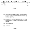
- mRNA identification is based on the interspersion profile of red and green signals.
- a dark disc 210 corresponds to a green signal (lysine) and an bright disc 220 to a red signal (arginine).
- the events are shown as they appear on temporal axis 230.
- This signal is translated into the code grgggrrgrrgg indicating the appearance order of green and red events.
- This code is used to search in an mRNA database precompiled for such searches.
- amino acid sequences 301 and 302 of two distinct mRNA templates are shown, with lysine (K) and arginine (R) underlined.
- the acceptor fluorphore is attached to one or more species of tRNA.
- Methods of labeling tRNA are discussed in detail in WO2004/050825 .
- the labeled tRNAs are known to be processed normally both by ribosome and by the cognate synthetases.
- Ribosome labeling is performed using one of several preferred methods. These include labeling with naturally fluorescent proteins, with organic dyes, and with semiconductor quantum dots. Labeling strategies included labeling ribosomal proteins such as ribosomal proteins L1, S1, S21 and others; In addition, 3' and 5' ends or 5S, 16S and 23S rRNA have been labeled ( Robbins and Hardesty, Biochemistry. 1983, 22; 22(24):5675-5679 ).
- Organic dyes can be used to label ribosomal proteins using standard protein labeling techniques. Suppliers of these dyes publish detailed protocols describing their use. General procedures label proteins through their amino groups (lysine). Other procedures target cysteines which are sometimes available for precisely located labeling. In this way, ribosomal proteins S1 and S8 were labeled by coumarin ( Bakin et al., J Mol Biol., 1991 221:441-453 ), and ribosomal proteins were tagged with fluorescin attached to a cysteine residue ( Odom et al., Biochemistry, 1990, 29:10734-10744 ).
- a novel labeling strategy uses quantum dots, which are commercially available pre-conjugated with biotin or streptavidin.
- proteins can be labeled with biotin, so the streptavidin conjugated Qdots (commercially available as Qdot TM 525/ Qdot TM 565/ Qdot TM 605/ Qdot TM 655 streptavidin conjugated quantum dots, from Quantum Dot Corporation, Hayward, Ca, USA; the numbers relate to maximal emission wavelength, nm) bind specifically to them.
- One useful method of generating a biotin-labeled protein involves creating a fusion protein between the protein of choice and biotin carboxyl carrier protein (BCCP).
- BCCP biotin carboxyl carrier protein
- the original protein sequence is fused optionally to the last 87 (or 110) codons of the E. coli BCCP.
- the fusion protein When the fusion protein is translated, it has the biotin tag attached to it and binds specifically to streptavidin (cf. Surrey et al., Proc. Natl. Acad. Sci. USA, 95: 4293-4298, 1998 ).
- Another method that is useful for labeling of ribosomal proteins is the well known strategy of fusing the protein of choice with a naturally fluorescent protein, such as green fluorescent protein, yellow/cyan/blue fluorescent proteins or any other naturally fluorescent protein.
- a naturally fluorescent protein such as green fluorescent protein, yellow/cyan/blue fluorescent proteins or any other naturally fluorescent protein.
- L1 was labeled by fusing it with a naturally fluorescent protein is described in ( Mascarenhas et al., EMBO Rep. 2001, 2:685-689 ). Numerous additional strategies for ribosome labeling were tested and others are clearly suitable for use with the present invention.
- an E. Coli translation system can be used for producing mammalian proteins.
- a mammalian translation system can be used.
- using some E. Coli tRNAs in a mammalian system can allow specific labeling of tRNAs that have appropriately modified bases.
- E. Coli tRNAs have the uridine in position 8 modified to thiouridine. This makes such tRNA types suitable for attaching a fluorescent label.
- a complete database of tRNA sequences can be found in http://www.uni-bayreuth.de/departmentslbiochemie/trna/ .
- a database of known RNA modifications can be found in http://medstat.med.utah.edu/RNAmods/ ,.
- amino-acid labeling could be the method of choice since the functionality of the protein is not crucial, and since amino acids can be labeled relatively easily.
- a second way of introducing labeled amino acids is by fluorescent labeling of acylated tRNA.
- Fluorescent labeled acylated tRNA can be purchased, for example the FluoroTectTM GreenLys in vitro Translation Labeling System available from Promega Corporation (Madison, WI). It is important in this system also that no unlabeled amino acids exist to avoid confounding the PSM signal sequence.
- One way to ensure this is by removing the cognate synthetases of labeled amino acids. This will ensure that recharging of tRNA does not occur, and only the labeled tRNA that was introduced will be used.
- a quenching strategy is used instead of FRET, as noted previously. Accordingly, instead of a fluorescent donor and acceptor, there is a fluorescent donor and an acceptor quencher that captures the donor energy without emission. Donor fluorescence is detected as long as the quencher is not sufficiently near to the donor. Use of this strategy in PSM, generates a signal of donor fluorescence intermitted by periods of quenching.
- tRNAs and some amino acids may be labeled as acceptors, either with distinct or with indistinguishable optical characteristics.
- the amino acids are labeled with fluorescent labels that are distinct from tRNA labels.
- the signal analysis system accepts both signals and uses both in order to identify more confidently the mRNA being translated. It is obvious to anyone skilled in the art of single molecule detection and analysis that this is just one example of a wide variety of methods that can be derived from this particular example.
Landscapes
- Chemical & Material Sciences (AREA)
- Life Sciences & Earth Sciences (AREA)
- Engineering & Computer Science (AREA)
- Organic Chemistry (AREA)
- Health & Medical Sciences (AREA)
- Proteomics, Peptides & Aminoacids (AREA)
- Zoology (AREA)
- Wood Science & Technology (AREA)
- Bioinformatics & Cheminformatics (AREA)
- Biotechnology (AREA)
- General Health & Medical Sciences (AREA)
- General Engineering & Computer Science (AREA)
- Molecular Biology (AREA)
- Biophysics (AREA)
- Physics & Mathematics (AREA)
- Analytical Chemistry (AREA)
- Biochemistry (AREA)
- Microbiology (AREA)
- Immunology (AREA)
- Genetics & Genomics (AREA)
- Nanotechnology (AREA)
- Crystallography & Structural Chemistry (AREA)
- Theoretical Computer Science (AREA)
- Mathematical Physics (AREA)
- Medical Informatics (AREA)
- Medicinal Chemistry (AREA)
- Pharmacology & Pharmacy (AREA)
- Measuring Or Testing Involving Enzymes Or Micro-Organisms (AREA)
- Apparatus Associated With Microorganisms And Enzymes (AREA)
Claims (17)
- Verfahren zur Identifizierung von mRNA welches umfasst:(a) Bereitstellen eines Proteinsynthese-Überwachungs- (PSM)- Systems, wobei das PSM-System umfasst:(i) Mindestens einen Marker, der durch Detektion von elektromagnetischer Strahlung detektierbar ist, wobei der mindestens eine Marker ein Paar interagierender Markerreste umfasst, wobei der erste Rest an ein Ribosom oder ein Fragment davon gebunden ist, wobei das Ribosom oder Fragment davon an einem festen Substrat befestigt sind, und wobei der zweite Rest an eine Einheit gebunden ist ausgewählt aus der Gruppe bestehen aus: dem Ribosom oder dem markierten Fragment davon, tRNA und Aminosäure, wobei der Marker fähig ist, als Antwort auf eine Translationsaktivität elektromagnetische Strahlung zu emittieren;(ii) mindestens eine Translationskomponente ausgewählt aus der Gruppe bestehend aus: Aminoacyl-tRNA-Synthetasen, Initiierungsfaktoren, Elongationsfaktoren, Terminationsfaktoren, Energiequellen und Energieregenerationsmolekülen; und(iii) Detektionsmittel, die angepasst sind, emittierte Strahlung aus dem PSM-System zu erfassen;(b) Einführen mindestens eines mRNA Moleküls in das PSM-System; und(c) Detektieren der elektromagnetischen Strahlungssignale, die als Antwort auf die Translationsaktivität erhalten werden.
- Verfahren nach Anspruch 1, umfassend Einführen von mehreren mRNA Molekülen in das PSM-System.
- Verfahren nach Anspruch 1, wobei das feste Substrat ausgewählt ist aus der Gruppe bestehend aus: Glas, Objektträger für Mikroskope und einem festen Substrat mit Glimmeroberfläche.
- Verfahren nach Anspruch 1, weiter umfassend:Identifizieren der Ribonukleotidsequenz des mindestens einen mRNA Moleküls.
- Verfahren nach Anspruch 4, wobei der Schritt des Identifizierens der Ribonukleotidsequenz des mindestens einen mRNA Moleküls umfasst:Durchführen einer PSM-Datenbank Abfrage, dabei Zuordnen mindestens einer Signalsequenz zu mindestens einer bestimmten mRNA.
- Verfahren nach Anspruch 5, wobei das Identifizieren der Ribonukleotidsequenz von mindestens einem mRNA Molekül weiter umfasst Speichern der mindestens einen Signalsequenz in einer PSM-Datenbank.
- Verfahren nach Anspruch 5, wobei der Schritt des Durchführens einer PSM-Datenbank Abfrage umfasst:Bestimmen der Wahrscheinlichkeit, dass ein mRNA Molekül in der PSM-Datenbank die mindestens eine Signalsequenz erzeugt; undAuswählen von einem oder mehreren mRNA Molekülen mit dem höchsten Punktfunktionswert, dabei Zuordnen des einen oder der mehreren mRNA Moleküle zu der mindestens einen Signalsequenz.
- Verfahren nach Anspruch 5, wobei die mindestens eine Signalsequenz aus einem oder mehreren Werten besteht, die ausgewählt sind aus der Gruppe bestehend aus: Zeit, räumliche Koordinaten, Signalart und Signalintensität.
- Verfahren nach Anspruch 1, wobei die Signale durch Energieübertragung zwischen dem Paar interagierender Markerreste erhalten werden.
- Verfahren nach Anspruch 9, wobei die Signale ausgewählt sind aus der Gruppe bestehend aus: FRET-Signalen, Quenchsignalen und Fluoreszenzsignalen.
- Verfahren nach Anspruch 1, wobei der Marker einen Marker umfasst ausgewählt aus der Gruppe bestehend aus: Einem Fluoreszenzfarbstoff, einer fluoreszierenden Aminosäure, einem fluoreszierenden Peptid oder Protein, fluoreszierenden Nukleotid, Quantum-Dot, einer Lumineszenzsubstanz, Donor-Quencher-Paar und fluoreszierenden Donor-Akzeptor-Paar.
- Verfahren nach Anspruch 1, wobei der zweite Markerrest eine fluoreszierende Aminosäure ist.
- Verfahren nach Anspruch 1, wobei das ribosomale Fragment ausgewählt ist aus der Gruppe bestehend aus: Ribosomaler RNA, ribosomalen Protein, ribosomalen Protein L1, ribosomalen Protein L 11, ribosomalen Protein S1 und Fragmenten davon.
- Verfahren nach Anspruch 13, wobei sich das ribosomale Fragment nahe einer ribosomalen Stelle befindet ausgewählt aus der Gruppe bestehend aus: Ribosomale Stelle A, ribosomale Stelle P, ribosomale Stelle E, Stelle des Peptidausgangskanals, L1 Arm und L7/L12 Arm.
- Verfahren nach Anspruch 1, wobei das PSM-System umfasst:Mehrere Marker, die mittels Detektion von elektromagnetischer Strahlung detektierbar sind, wobei jeder Marker ein Paar interagierender Markerreste umfasst, wobei der erste Rest an ein Ribosom oder ein markiertes Fragment davon gebunden ist, wobei das Ribosom oder Fragment davon an einem festen Substrat befestigt sind, und wobei der zweite Rest an eine Einheit gebunden ist ausgewählt aus der Gruppe bestehen aus: dem Ribosom oder dem Fragment davon, tRNA und Aminosäure, wobei der Marker fähig ist, als Antwort auf eine Translationsaktivität elektromagnetische Strahlung zu emittieren
- Verfahren nach Anspruch 15, wobei unterschiedliche tRNAs oder Aminosäuren an unterschiedliche Marker gebunden sind.
- Verfahren nach Anspruch 15, wobei der Marker eine von zwei unterschiedlichen Farben ist.
Applications Claiming Priority (2)
| Application Number | Priority Date | Filing Date | Title |
|---|---|---|---|
| US57421004P | 2004-05-26 | 2004-05-26 | |
| PCT/IL2005/000540 WO2005116252A2 (en) | 2004-05-26 | 2005-05-26 | Methods for evaluating ribonucleotide sequences |
Publications (3)
| Publication Number | Publication Date |
|---|---|
| EP1751299A2 EP1751299A2 (de) | 2007-02-14 |
| EP1751299A4 EP1751299A4 (de) | 2008-09-24 |
| EP1751299B1 true EP1751299B1 (de) | 2010-01-27 |
Family
ID=35451475
Family Applications (1)
| Application Number | Title | Priority Date | Filing Date |
|---|---|---|---|
| EP05743440A Expired - Lifetime EP1751299B1 (de) | 2004-05-26 | 2005-05-26 | Verfahren zur auswertung von ribonukleotidsequenzen |
Country Status (7)
| Country | Link |
|---|---|
| US (1) | US9012150B2 (de) |
| EP (1) | EP1751299B1 (de) |
| AT (1) | ATE456666T1 (de) |
| AU (1) | AU2005248167B2 (de) |
| CA (1) | CA2567749C (de) |
| DE (1) | DE602005019156D1 (de) |
| WO (1) | WO2005116252A2 (de) |
Families Citing this family (13)
| Publication number | Priority date | Publication date | Assignee | Title |
|---|---|---|---|---|
| ATE456666T1 (de) | 2004-05-26 | 2010-02-15 | Anima Cell Metrology | Verfahren zur auswertung von ribonukleotidsequenzen |
| US7777002B2 (en) * | 2006-06-29 | 2010-08-17 | The Invention Science Fund I, Llc | Methods for arbitrary peptide synthesis |
| US7923533B2 (en) * | 2006-06-29 | 2011-04-12 | The Invention Science Fund I, Llc | Methods for arbitrary peptide synthesis |
| US7910695B2 (en) | 2006-06-29 | 2011-03-22 | The Invention Science Fund I, Llc | Methods for arbitrary peptide synthesis |
| US7993873B2 (en) * | 2006-06-29 | 2011-08-09 | The Invention Science Fund I, Llc | Apparatus for arbitrary peptide synthesis |
| US7879975B2 (en) | 2006-06-29 | 2011-02-01 | The Invention Science Fund I, Llc | Methods for arbitrary peptide synthesis |
| CA2692159C (en) | 2007-06-22 | 2016-09-13 | Barry S. Cooperman | Fluorescent labeling of transfer rna and study of protein synthesis |
| WO2009047760A2 (en) | 2007-10-09 | 2009-04-16 | Anima Cell Metrology, Inc. | Systems and methods for measuring translation activity in viable cells |
| WO2010144151A2 (en) * | 2009-06-12 | 2010-12-16 | Pacific Biosciences Of California, Inc. | Single-molecule real-time analysis of protein synthesis |
| EP2288195B1 (de) | 2009-08-20 | 2019-10-23 | Samsung Electronics Co., Ltd. | Verfahren und Vorrichtung zum Betreiben einer Basisstation in einem drahtlosen Kommunikationssystem |
| US9034576B2 (en) | 2009-09-24 | 2015-05-19 | Anima Cell Metrology Inc. | Systems and methods for measuring translation of target proteins in cells |
| BE1019985A3 (nl) * | 2011-05-23 | 2013-03-05 | Willy Boermans | Chemie doen met behulp van kleurensensoren. |
| WO2017085718A1 (en) | 2015-11-17 | 2017-05-26 | Anima Biotech Inc. | FUNCTIONAL TRANSFER tRNA-APTAMER MOLECULES |
Citations (1)
| Publication number | Priority date | Publication date | Assignee | Title |
|---|---|---|---|---|
| WO2004050825A2 (en) * | 2002-11-29 | 2004-06-17 | Meishar H.Z. Yazamut Ltd. | Protein synthesis monitoring (psm) |
Family Cites Families (39)
| Publication number | Priority date | Publication date | Assignee | Title |
|---|---|---|---|---|
| US5082767A (en) * | 1989-02-27 | 1992-01-21 | Hatfield G Wesley | Codon pair utilization |
| US5856928A (en) * | 1992-03-13 | 1999-01-05 | Yan; Johnson F. | Gene and protein representation, characterization and interpretation process |
| EP0786522A2 (de) | 1992-07-17 | 1997-07-30 | Ribozyme Pharmaceuticals, Inc. | Enzymatische RNA-Moleküle zur Behandlung von stenotischen Zuständen |
| JPH0793370A (ja) | 1993-09-27 | 1995-04-07 | Hitachi Device Eng Co Ltd | 遺伝子データベース検索システム |
| DE4410655C2 (de) | 1994-03-26 | 2001-06-28 | Heiko Schwertner | Verfahren und Vorrichtung zur schnellen kontinuierlichen Sequenzanalyse von verschiedenen linearen polymeren Substanzen nach einer einheitlichen Vorgehensweise |
| US5643722A (en) * | 1994-05-11 | 1997-07-01 | Trustees Of Boston University | Methods for the detection and isolation of proteins |
| US6210941B1 (en) * | 1997-06-27 | 2001-04-03 | The Trustees Of Boston University | Methods for the detection and isolation of proteins |
| US5777079A (en) * | 1994-11-10 | 1998-07-07 | The Regents Of The University Of California | Modified green fluorescent proteins |
| US5626058A (en) * | 1995-04-24 | 1997-05-06 | Karpowich; John W. | Rotary actuator for a bicycle brake |
| US6189013B1 (en) * | 1996-12-12 | 2001-02-13 | Incyte Genomics, Inc. | Project-based full length biomolecular sequence database |
| FR2786352B1 (fr) | 1998-11-24 | 2004-10-22 | Ar2I Sa Analyses Rech S Et Inn | Appareil d'acquisition d'images par fluorescence et systeme d'imagerie comportant un tel appareil |
| US20040023874A1 (en) * | 2002-03-15 | 2004-02-05 | Burgess Catherine E. | Therapeutic polypeptides, nucleic acids encoding same, and methods of use |
| CA2311201A1 (en) * | 1999-08-05 | 2001-02-05 | Genset S.A. | Ests and encoded human proteins |
| US6306628B1 (en) * | 1999-08-25 | 2001-10-23 | Ambergen, Incorporated | Methods for the detection, analysis and isolation of Nascent proteins |
| WO2001016375A2 (en) | 1999-08-30 | 2001-03-08 | The Government Of The United States Of America, As Represented By The Secretary, Department Of Health And Human Services | High speed parallel molecular nucleic acid sequencing |
| FR2798143A1 (fr) | 1999-09-08 | 2001-03-09 | A2F | Dispositif de lecture sequentielle rapide de polynucleotides |
| US20030044972A1 (en) * | 2000-03-17 | 2003-03-06 | Zoran Ristic | Maize chloroplast protein synthesis elongation factors and methods of use for same |
| CA2314398A1 (en) * | 2000-08-10 | 2002-02-10 | Edward Shipwash | Microarrays and microsystems for amino acid analysis and protein sequencing |
| GB0111015D0 (en) * | 2001-05-04 | 2001-06-27 | Norsk Hydro As | Genetic material |
| US20050118151A1 (en) * | 2001-05-29 | 2005-06-02 | Syddansk Universitet | Proteins in diabetes proteome anlysis |
| US7065236B2 (en) | 2001-09-19 | 2006-06-20 | Tripath Imaging, Inc. | Method for quantitative video-microscopy and associated system and computer software program product |
| AU2002361649A1 (en) | 2001-12-13 | 2003-06-30 | University Of Virginia Patent Foundation | Vector mediated organelle transfection |
| US20030134423A1 (en) | 2002-01-04 | 2003-07-17 | Chu Yong Liang | Compounds for delivering substances into cells |
| US7288372B2 (en) * | 2002-01-17 | 2007-10-30 | Ambergen, Inc. | Methods for the preparation of chemically misaminoacylated tRNA via protective groups |
| WO2003064608A2 (en) * | 2002-01-25 | 2003-08-07 | The Board Of Trustees Of The Leland Stanford Junior University | Surface based translation system |
| WO2003064604A2 (en) | 2002-01-25 | 2003-08-07 | The Board Of Trustees Of The Leland Stanford Junior University | Translation profiling |
| KR20050003356A (ko) * | 2002-04-10 | 2005-01-10 | 후지 샤신 필름 가부시기가이샤 | 노광헤드 및 노광장치와 그 응용 |
| EP1565193B1 (de) * | 2002-11-06 | 2013-04-24 | Dana-Farber Cancer Institute, Inc. | Zusammensetzungen zur Krebsbehandlung unter Verwendung vom Proteasom-Hemmer PS-341 |
| JP2006506986A (ja) * | 2002-11-08 | 2006-03-02 | ザ ユニバーシティー オブ クイーンズランド | 同義コドンの最適化を用いて遺伝子発現を最適化するための方法 |
| US8442773B2 (en) * | 2002-11-29 | 2013-05-14 | Anima Cell Metrology | Protein synthesis monitoring (PSM) |
| CA2530248A1 (en) | 2003-06-25 | 2005-01-06 | Gencia Corporation | Modified vectors for organelle transfection |
| WO2005121724A2 (en) * | 2004-01-21 | 2005-12-22 | Optical Insights, Llc | Method and apparatus for multi-mode spectral imaging |
| ATE456666T1 (de) | 2004-05-26 | 2010-02-15 | Anima Cell Metrology | Verfahren zur auswertung von ribonukleotidsequenzen |
| EP1913328A4 (de) | 2005-06-24 | 2014-12-24 | Biolase Inc | Geräte zur visuellen rückkopplung für vorrichtungen zur abgabe elektromagnetischer energie |
| US20070134814A1 (en) * | 2005-12-09 | 2007-06-14 | Kajander E O | Methods and compositions for the detection of calcifying nano-particles, identification and quantification of associated proteins thereon, and correlation to disease |
| WO2008028298A1 (en) | 2006-09-06 | 2008-03-13 | University Health Network | Fluorescence quantification and image acquisition in highly turbid media |
| CA2692159C (en) * | 2007-06-22 | 2016-09-13 | Barry S. Cooperman | Fluorescent labeling of transfer rna and study of protein synthesis |
| WO2009047760A2 (en) | 2007-10-09 | 2009-04-16 | Anima Cell Metrology, Inc. | Systems and methods for measuring translation activity in viable cells |
| US9034576B2 (en) * | 2009-09-24 | 2015-05-19 | Anima Cell Metrology Inc. | Systems and methods for measuring translation of target proteins in cells |
-
2005
- 2005-05-26 AT AT05743440T patent/ATE456666T1/de not_active IP Right Cessation
- 2005-05-26 CA CA2567749A patent/CA2567749C/en not_active Expired - Lifetime
- 2005-05-26 US US11/569,476 patent/US9012150B2/en active Active
- 2005-05-26 DE DE602005019156T patent/DE602005019156D1/de not_active Expired - Lifetime
- 2005-05-26 WO PCT/IL2005/000540 patent/WO2005116252A2/en not_active Ceased
- 2005-05-26 AU AU2005248167A patent/AU2005248167B2/en not_active Expired
- 2005-05-26 EP EP05743440A patent/EP1751299B1/de not_active Expired - Lifetime
Patent Citations (1)
| Publication number | Priority date | Publication date | Assignee | Title |
|---|---|---|---|---|
| WO2004050825A2 (en) * | 2002-11-29 | 2004-06-17 | Meishar H.Z. Yazamut Ltd. | Protein synthesis monitoring (psm) |
Also Published As
| Publication number | Publication date |
|---|---|
| CA2567749C (en) | 2013-07-23 |
| AU2005248167A1 (en) | 2005-12-08 |
| EP1751299A2 (de) | 2007-02-14 |
| DE602005019156D1 (de) | 2010-03-18 |
| AU2005248167B2 (en) | 2010-11-25 |
| WO2005116252A2 (en) | 2005-12-08 |
| WO2005116252A8 (en) | 2006-07-06 |
| WO2005116252A3 (en) | 2006-03-02 |
| US20090081643A1 (en) | 2009-03-26 |
| ATE456666T1 (de) | 2010-02-15 |
| EP1751299A4 (de) | 2008-09-24 |
| US9012150B2 (en) | 2015-04-21 |
| CA2567749A1 (en) | 2005-12-08 |
Similar Documents
| Publication | Publication Date | Title |
|---|---|---|
| EP2030011B1 (de) | Systeme und verfahren zur analyse von nanoreportern | |
| EP1565566B1 (de) | Proteinsynthese-monitoring (psm) | |
| EP1751299B1 (de) | Verfahren zur auswertung von ribonukleotidsequenzen | |
| US20140371088A1 (en) | Multiplexable tag-based reporter system | |
| US20120015825A1 (en) | Analytical systems and methods with software mask | |
| US8609421B2 (en) | Single-molecule real-time analysis of protein synthesis | |
| Hyun et al. | Single molecule and single cell epigenomics | |
| WO2005047545A2 (en) | Microarray controls | |
| De Angelis | Why FRET over genomics? | |
| US8442773B2 (en) | Protein synthesis monitoring (PSM) | |
| Stevens et al. | FRET-based identification of mRNAs undergoing translation | |
| JP3944576B2 (ja) | マイクロアレイを用いたアプタマーの取得方法 | |
| US20100190151A1 (en) | Fluorescently labeled nucleoside triphosphates and analogs thereof for sequencing nucleic acids | |
| US20240100518A1 (en) | Flow cell based motion system calibration and control methods | |
| EP4506691A1 (de) | Verfahren und system zur analyse einer biologischen probe | |
| US20260074017A1 (en) | Method for indexing a cellular sample | |
| IL168822A (en) | Method and apparatus for protein synthesis monitoring based on electromagnetic radiation | |
| WO2004101811A2 (en) | Detection result storing method | |
| JP2005102614A (ja) | ゲノム物理地図作成法及び作成装置 |
Legal Events
| Date | Code | Title | Description |
|---|---|---|---|
| PUAI | Public reference made under article 153(3) epc to a published international application that has entered the european phase |
Free format text: ORIGINAL CODE: 0009012 |
|
| 17P | Request for examination filed |
Effective date: 20061115 |
|
| AK | Designated contracting states |
Kind code of ref document: A2 Designated state(s): AT BE BG CH CY CZ DE DK EE ES FI FR GB GR HU IE IS IT LI LT LU MC NL PL PT RO SE SI SK TR |
|
| RAP1 | Party data changed (applicant data changed or rights of an application transferred) |
Owner name: ANIMA CELL METROLOGY |
|
| DAX | Request for extension of the european patent (deleted) | ||
| A4 | Supplementary search report drawn up and despatched |
Effective date: 20080826 |
|
| RIC1 | Information provided on ipc code assigned before grant |
Ipc: G06F 7/00 20060101ALI20080820BHEP Ipc: G01N 33/00 20060101ALI20080820BHEP Ipc: C12Q 1/68 20060101ALI20080820BHEP Ipc: C12P 21/06 20060101AFI20060621BHEP |
|
| 17Q | First examination report despatched |
Effective date: 20090420 |
|
| GRAP | Despatch of communication of intention to grant a patent |
Free format text: ORIGINAL CODE: EPIDOSNIGR1 |
|
| RIC1 | Information provided on ipc code assigned before grant |
Ipc: C12P 21/06 20060101AFI20090813BHEP Ipc: G01N 33/00 20060101ALI20090813BHEP Ipc: G06F 7/00 20060101ALI20090813BHEP |
|
| GRAS | Grant fee paid |
Free format text: ORIGINAL CODE: EPIDOSNIGR3 |
|
| GRAA | (expected) grant |
Free format text: ORIGINAL CODE: 0009210 |
|
| AK | Designated contracting states |
Kind code of ref document: B1 Designated state(s): AT BE BG CH CY CZ DE DK EE ES FI FR GB GR HU IE IS IT LI LT LU MC NL PL PT RO SE SI SK TR |
|
| REG | Reference to a national code |
Ref country code: GB Ref legal event code: FG4D |
|
| REG | Reference to a national code |
Ref country code: CH Ref legal event code: EP |
|
| REG | Reference to a national code |
Ref country code: IE Ref legal event code: FG4D |
|
| REF | Corresponds to: |
Ref document number: 602005019156 Country of ref document: DE Date of ref document: 20100318 Kind code of ref document: P |
|
| REG | Reference to a national code |
Ref country code: NL Ref legal event code: VDEP Effective date: 20100127 |
|
| LTIE | Lt: invalidation of european patent or patent extension |
Effective date: 20100127 |
|
| PG25 | Lapsed in a contracting state [announced via postgrant information from national office to epo] |
Ref country code: AT Free format text: LAPSE BECAUSE OF FAILURE TO SUBMIT A TRANSLATION OF THE DESCRIPTION OR TO PAY THE FEE WITHIN THE PRESCRIBED TIME-LIMIT Effective date: 20100127 |
|
| PG25 | Lapsed in a contracting state [announced via postgrant information from national office to epo] |
Ref country code: ES Free format text: LAPSE BECAUSE OF FAILURE TO SUBMIT A TRANSLATION OF THE DESCRIPTION OR TO PAY THE FEE WITHIN THE PRESCRIBED TIME-LIMIT Effective date: 20100508 Ref country code: PT Free format text: LAPSE BECAUSE OF FAILURE TO SUBMIT A TRANSLATION OF THE DESCRIPTION OR TO PAY THE FEE WITHIN THE PRESCRIBED TIME-LIMIT Effective date: 20100527 Ref country code: IS Free format text: LAPSE BECAUSE OF FAILURE TO SUBMIT A TRANSLATION OF THE DESCRIPTION OR TO PAY THE FEE WITHIN THE PRESCRIBED TIME-LIMIT Effective date: 20100527 Ref country code: LT Free format text: LAPSE BECAUSE OF FAILURE TO SUBMIT A TRANSLATION OF THE DESCRIPTION OR TO PAY THE FEE WITHIN THE PRESCRIBED TIME-LIMIT Effective date: 20100127 Ref country code: NL Free format text: LAPSE BECAUSE OF FAILURE TO SUBMIT A TRANSLATION OF THE DESCRIPTION OR TO PAY THE FEE WITHIN THE PRESCRIBED TIME-LIMIT Effective date: 20100127 |
|
| PG25 | Lapsed in a contracting state [announced via postgrant information from national office to epo] |
Ref country code: FI Free format text: LAPSE BECAUSE OF FAILURE TO SUBMIT A TRANSLATION OF THE DESCRIPTION OR TO PAY THE FEE WITHIN THE PRESCRIBED TIME-LIMIT Effective date: 20100127 Ref country code: SI Free format text: LAPSE BECAUSE OF FAILURE TO SUBMIT A TRANSLATION OF THE DESCRIPTION OR TO PAY THE FEE WITHIN THE PRESCRIBED TIME-LIMIT Effective date: 20100127 Ref country code: PL Free format text: LAPSE BECAUSE OF FAILURE TO SUBMIT A TRANSLATION OF THE DESCRIPTION OR TO PAY THE FEE WITHIN THE PRESCRIBED TIME-LIMIT Effective date: 20100127 |
|
| PG25 | Lapsed in a contracting state [announced via postgrant information from national office to epo] |
Ref country code: RO Free format text: LAPSE BECAUSE OF FAILURE TO SUBMIT A TRANSLATION OF THE DESCRIPTION OR TO PAY THE FEE WITHIN THE PRESCRIBED TIME-LIMIT Effective date: 20100127 Ref country code: BE Free format text: LAPSE BECAUSE OF FAILURE TO SUBMIT A TRANSLATION OF THE DESCRIPTION OR TO PAY THE FEE WITHIN THE PRESCRIBED TIME-LIMIT Effective date: 20100127 Ref country code: CY Free format text: LAPSE BECAUSE OF FAILURE TO SUBMIT A TRANSLATION OF THE DESCRIPTION OR TO PAY THE FEE WITHIN THE PRESCRIBED TIME-LIMIT Effective date: 20100127 Ref country code: EE Free format text: LAPSE BECAUSE OF FAILURE TO SUBMIT A TRANSLATION OF THE DESCRIPTION OR TO PAY THE FEE WITHIN THE PRESCRIBED TIME-LIMIT Effective date: 20100127 Ref country code: GR Free format text: LAPSE BECAUSE OF FAILURE TO SUBMIT A TRANSLATION OF THE DESCRIPTION OR TO PAY THE FEE WITHIN THE PRESCRIBED TIME-LIMIT Effective date: 20100428 Ref country code: SE Free format text: LAPSE BECAUSE OF FAILURE TO SUBMIT A TRANSLATION OF THE DESCRIPTION OR TO PAY THE FEE WITHIN THE PRESCRIBED TIME-LIMIT Effective date: 20100127 |
|
| PG25 | Lapsed in a contracting state [announced via postgrant information from national office to epo] |
Ref country code: BG Free format text: LAPSE BECAUSE OF FAILURE TO SUBMIT A TRANSLATION OF THE DESCRIPTION OR TO PAY THE FEE WITHIN THE PRESCRIBED TIME-LIMIT Effective date: 20100427 Ref country code: CZ Free format text: LAPSE BECAUSE OF FAILURE TO SUBMIT A TRANSLATION OF THE DESCRIPTION OR TO PAY THE FEE WITHIN THE PRESCRIBED TIME-LIMIT Effective date: 20100127 Ref country code: SK Free format text: LAPSE BECAUSE OF FAILURE TO SUBMIT A TRANSLATION OF THE DESCRIPTION OR TO PAY THE FEE WITHIN THE PRESCRIBED TIME-LIMIT Effective date: 20100127 |
|
| PLBE | No opposition filed within time limit |
Free format text: ORIGINAL CODE: 0009261 |
|
| STAA | Information on the status of an ep patent application or granted ep patent |
Free format text: STATUS: NO OPPOSITION FILED WITHIN TIME LIMIT |
|
| PG25 | Lapsed in a contracting state [announced via postgrant information from national office to epo] |
Ref country code: MC Free format text: LAPSE BECAUSE OF NON-PAYMENT OF DUE FEES Effective date: 20100531 |
|
| REG | Reference to a national code |
Ref country code: CH Ref legal event code: PL |
|
| 26N | No opposition filed |
Effective date: 20101028 |
|
| PG25 | Lapsed in a contracting state [announced via postgrant information from national office to epo] |
Ref country code: DK Free format text: LAPSE BECAUSE OF FAILURE TO SUBMIT A TRANSLATION OF THE DESCRIPTION OR TO PAY THE FEE WITHIN THE PRESCRIBED TIME-LIMIT Effective date: 20100127 |
|
| PG25 | Lapsed in a contracting state [announced via postgrant information from national office to epo] |
Ref country code: CH Free format text: LAPSE BECAUSE OF NON-PAYMENT OF DUE FEES Effective date: 20100531 Ref country code: LI Free format text: LAPSE BECAUSE OF NON-PAYMENT OF DUE FEES Effective date: 20100531 |
|
| PG25 | Lapsed in a contracting state [announced via postgrant information from national office to epo] |
Ref country code: IT Free format text: LAPSE BECAUSE OF FAILURE TO SUBMIT A TRANSLATION OF THE DESCRIPTION OR TO PAY THE FEE WITHIN THE PRESCRIBED TIME-LIMIT Effective date: 20100127 |
|
| PG25 | Lapsed in a contracting state [announced via postgrant information from national office to epo] |
Ref country code: IE Free format text: LAPSE BECAUSE OF NON-PAYMENT OF DUE FEES Effective date: 20100526 |
|
| PG25 | Lapsed in a contracting state [announced via postgrant information from national office to epo] |
Ref country code: HU Free format text: LAPSE BECAUSE OF FAILURE TO SUBMIT A TRANSLATION OF THE DESCRIPTION OR TO PAY THE FEE WITHIN THE PRESCRIBED TIME-LIMIT Effective date: 20100728 Ref country code: LU Free format text: LAPSE BECAUSE OF NON-PAYMENT OF DUE FEES Effective date: 20100526 |
|
| PG25 | Lapsed in a contracting state [announced via postgrant information from national office to epo] |
Ref country code: TR Free format text: LAPSE BECAUSE OF FAILURE TO SUBMIT A TRANSLATION OF THE DESCRIPTION OR TO PAY THE FEE WITHIN THE PRESCRIBED TIME-LIMIT Effective date: 20100127 |
|
| REG | Reference to a national code |
Ref country code: FR Ref legal event code: PLFP Year of fee payment: 12 |
|
| REG | Reference to a national code |
Ref country code: FR Ref legal event code: PLFP Year of fee payment: 13 |
|
| REG | Reference to a national code |
Ref country code: FR Ref legal event code: PLFP Year of fee payment: 14 |
|
| PGFP | Annual fee paid to national office [announced via postgrant information from national office to epo] |
Ref country code: GB Payment date: 20240521 Year of fee payment: 20 |
|
| PGFP | Annual fee paid to national office [announced via postgrant information from national office to epo] |
Ref country code: DE Payment date: 20240521 Year of fee payment: 20 |
|
| PGFP | Annual fee paid to national office [announced via postgrant information from national office to epo] |
Ref country code: FR Payment date: 20240529 Year of fee payment: 20 |
|
| REG | Reference to a national code |
Ref country code: DE Ref legal event code: R071 Ref document number: 602005019156 Country of ref document: DE |
|
| REG | Reference to a national code |
Ref country code: GB Ref legal event code: PE20 Expiry date: 20250525 |
|
| PG25 | Lapsed in a contracting state [announced via postgrant information from national office to epo] |
Ref country code: GB Free format text: LAPSE BECAUSE OF EXPIRATION OF PROTECTION Effective date: 20250525 |