EP1757689A2 - Säugetiere Apoptosis-Inhibitorprotein Genfamilie, Primers, Proben und Verfahren zum Nachweis - Google Patents
Säugetiere Apoptosis-Inhibitorprotein Genfamilie, Primers, Proben und Verfahren zum Nachweis Download PDFInfo
- Publication number
- EP1757689A2 EP1757689A2 EP05025419A EP05025419A EP1757689A2 EP 1757689 A2 EP1757689 A2 EP 1757689A2 EP 05025419 A EP05025419 A EP 05025419A EP 05025419 A EP05025419 A EP 05025419A EP 1757689 A2 EP1757689 A2 EP 1757689A2
- Authority
- EP
- European Patent Office
- Prior art keywords
- xaal
- iap
- cell
- hiap
- polypeptide
- Prior art date
- Legal status (The legal status is an assumption and is not a legal conclusion. Google has not performed a legal analysis and makes no representation as to the accuracy of the status listed.)
- Withdrawn
Links
Images




















Classifications
-
- C—CHEMISTRY; METALLURGY
- C07—ORGANIC CHEMISTRY
- C07K—PEPTIDES
- C07K14/00—Peptides having more than 20 amino acids; Gastrins; Somatostatins; Melanotropins; Derivatives thereof
- C07K14/435—Peptides having more than 20 amino acids; Gastrins; Somatostatins; Melanotropins; Derivatives thereof from animals; from humans
- C07K14/46—Peptides having more than 20 amino acids; Gastrins; Somatostatins; Melanotropins; Derivatives thereof from animals; from humans from vertebrates
- C07K14/47—Peptides having more than 20 amino acids; Gastrins; Somatostatins; Melanotropins; Derivatives thereof from animals; from humans from vertebrates from mammals
- C07K14/4701—Peptides having more than 20 amino acids; Gastrins; Somatostatins; Melanotropins; Derivatives thereof from animals; from humans from vertebrates from mammals not used
- C07K14/4747—Apoptosis related proteins
-
- A—HUMAN NECESSITIES
- A61—MEDICAL OR VETERINARY SCIENCE; HYGIENE
- A61P—SPECIFIC THERAPEUTIC ACTIVITY OF CHEMICAL COMPOUNDS OR MEDICINAL PREPARATIONS
- A61P1/00—Drugs for disorders of the alimentary tract or the digestive system
-
- A—HUMAN NECESSITIES
- A61—MEDICAL OR VETERINARY SCIENCE; HYGIENE
- A61P—SPECIFIC THERAPEUTIC ACTIVITY OF CHEMICAL COMPOUNDS OR MEDICINAL PREPARATIONS
- A61P1/00—Drugs for disorders of the alimentary tract or the digestive system
- A61P1/16—Drugs for disorders of the alimentary tract or the digestive system for liver or gallbladder disorders, e.g. hepatoprotective agents, cholagogues, litholytics
-
- A—HUMAN NECESSITIES
- A61—MEDICAL OR VETERINARY SCIENCE; HYGIENE
- A61P—SPECIFIC THERAPEUTIC ACTIVITY OF CHEMICAL COMPOUNDS OR MEDICINAL PREPARATIONS
- A61P25/00—Drugs for disorders of the nervous system
-
- A—HUMAN NECESSITIES
- A61—MEDICAL OR VETERINARY SCIENCE; HYGIENE
- A61P—SPECIFIC THERAPEUTIC ACTIVITY OF CHEMICAL COMPOUNDS OR MEDICINAL PREPARATIONS
- A61P31/00—Antiinfectives, i.e. antibiotics, antiseptics, chemotherapeutics
-
- A—HUMAN NECESSITIES
- A61—MEDICAL OR VETERINARY SCIENCE; HYGIENE
- A61P—SPECIFIC THERAPEUTIC ACTIVITY OF CHEMICAL COMPOUNDS OR MEDICINAL PREPARATIONS
- A61P31/00—Antiinfectives, i.e. antibiotics, antiseptics, chemotherapeutics
- A61P31/12—Antivirals
- A61P31/14—Antivirals for RNA viruses
- A61P31/18—Antivirals for RNA viruses for HIV
-
- A—HUMAN NECESSITIES
- A61—MEDICAL OR VETERINARY SCIENCE; HYGIENE
- A61P—SPECIFIC THERAPEUTIC ACTIVITY OF CHEMICAL COMPOUNDS OR MEDICINAL PREPARATIONS
- A61P43/00—Drugs for specific purposes, not provided for in groups A61P1/00-A61P41/00
-
- A—HUMAN NECESSITIES
- A61—MEDICAL OR VETERINARY SCIENCE; HYGIENE
- A61P—SPECIFIC THERAPEUTIC ACTIVITY OF CHEMICAL COMPOUNDS OR MEDICINAL PREPARATIONS
- A61P9/00—Drugs for disorders of the cardiovascular system
-
- A—HUMAN NECESSITIES
- A61—MEDICAL OR VETERINARY SCIENCE; HYGIENE
- A61P—SPECIFIC THERAPEUTIC ACTIVITY OF CHEMICAL COMPOUNDS OR MEDICINAL PREPARATIONS
- A61P9/00—Drugs for disorders of the cardiovascular system
- A61P9/10—Drugs for disorders of the cardiovascular system for treating ischaemic or atherosclerotic diseases, e.g. antianginal drugs, coronary vasodilators, drugs for myocardial infarction, retinopathy, cerebrovascula insufficiency, renal arteriosclerosis
-
- C—CHEMISTRY; METALLURGY
- C12—BIOCHEMISTRY; BEER; SPIRITS; WINE; VINEGAR; MICROBIOLOGY; ENZYMOLOGY; MUTATION OR GENETIC ENGINEERING
- C12Q—MEASURING OR TESTING PROCESSES INVOLVING ENZYMES, NUCLEIC ACIDS OR MICROORGANISMS; COMPOSITIONS OR TEST PAPERS THEREFOR; PROCESSES OF PREPARING SUCH COMPOSITIONS; CONDITION-RESPONSIVE CONTROL IN MICROBIOLOGICAL OR ENZYMOLOGICAL PROCESSES
- C12Q1/00—Measuring or testing processes involving enzymes, nucleic acids or microorganisms; Compositions therefor; Processes of preparing such compositions
- C12Q1/68—Measuring or testing processes involving enzymes, nucleic acids or microorganisms; Compositions therefor; Processes of preparing such compositions involving nucleic acids
- C12Q1/6876—Nucleic acid products used in the analysis of nucleic acids, e.g. primers or probes
- C12Q1/6883—Nucleic acid products used in the analysis of nucleic acids, e.g. primers or probes for diseases caused by alterations of genetic material
-
- C—CHEMISTRY; METALLURGY
- C12—BIOCHEMISTRY; BEER; SPIRITS; WINE; VINEGAR; MICROBIOLOGY; ENZYMOLOGY; MUTATION OR GENETIC ENGINEERING
- C12Q—MEASURING OR TESTING PROCESSES INVOLVING ENZYMES, NUCLEIC ACIDS OR MICROORGANISMS; COMPOSITIONS OR TEST PAPERS THEREFOR; PROCESSES OF PREPARING SUCH COMPOSITIONS; CONDITION-RESPONSIVE CONTROL IN MICROBIOLOGICAL OR ENZYMOLOGICAL PROCESSES
- C12Q1/00—Measuring or testing processes involving enzymes, nucleic acids or microorganisms; Compositions therefor; Processes of preparing such compositions
- C12Q1/68—Measuring or testing processes involving enzymes, nucleic acids or microorganisms; Compositions therefor; Processes of preparing such compositions involving nucleic acids
- C12Q1/6876—Nucleic acid products used in the analysis of nucleic acids, e.g. primers or probes
- C12Q1/6883—Nucleic acid products used in the analysis of nucleic acids, e.g. primers or probes for diseases caused by alterations of genetic material
- C12Q1/6886—Nucleic acid products used in the analysis of nucleic acids, e.g. primers or probes for diseases caused by alterations of genetic material for cancer
-
- G—PHYSICS
- G01—MEASURING; TESTING
- G01N—INVESTIGATING OR ANALYSING MATERIALS BY DETERMINING THEIR CHEMICAL OR PHYSICAL PROPERTIES
- G01N33/00—Investigating or analysing materials by specific methods not covered by groups G01N1/00 - G01N31/00
- G01N33/48—Biological material, e.g. blood, urine; Haemocytometers
- G01N33/50—Chemical analysis of biological material, e.g. blood, urine; Testing involving biospecific ligand binding methods; Immunological testing
- G01N33/5005—Chemical analysis of biological material, e.g. blood, urine; Testing involving biospecific ligand binding methods; Immunological testing involving human or animal cells
- G01N33/5008—Chemical analysis of biological material, e.g. blood, urine; Testing involving biospecific ligand binding methods; Immunological testing involving human or animal cells for testing or evaluating the effect of chemical or biological compounds, e.g. drugs, cosmetics
-
- G—PHYSICS
- G01—MEASURING; TESTING
- G01N—INVESTIGATING OR ANALYSING MATERIALS BY DETERMINING THEIR CHEMICAL OR PHYSICAL PROPERTIES
- G01N33/00—Investigating or analysing materials by specific methods not covered by groups G01N1/00 - G01N31/00
- G01N33/48—Biological material, e.g. blood, urine; Haemocytometers
- G01N33/50—Chemical analysis of biological material, e.g. blood, urine; Testing involving biospecific ligand binding methods; Immunological testing
- G01N33/5005—Chemical analysis of biological material, e.g. blood, urine; Testing involving biospecific ligand binding methods; Immunological testing involving human or animal cells
- G01N33/5008—Chemical analysis of biological material, e.g. blood, urine; Testing involving biospecific ligand binding methods; Immunological testing involving human or animal cells for testing or evaluating the effect of chemical or biological compounds, e.g. drugs, cosmetics
- G01N33/502—Chemical analysis of biological material, e.g. blood, urine; Testing involving biospecific ligand binding methods; Immunological testing involving human or animal cells for testing or evaluating the effect of chemical or biological compounds, e.g. drugs, cosmetics for testing non-proliferative effects
-
- G—PHYSICS
- G01—MEASURING; TESTING
- G01N—INVESTIGATING OR ANALYSING MATERIALS BY DETERMINING THEIR CHEMICAL OR PHYSICAL PROPERTIES
- G01N33/00—Investigating or analysing materials by specific methods not covered by groups G01N1/00 - G01N31/00
- G01N33/48—Biological material, e.g. blood, urine; Haemocytometers
- G01N33/50—Chemical analysis of biological material, e.g. blood, urine; Testing involving biospecific ligand binding methods; Immunological testing
- G01N33/53—Immunoassay; Biospecific binding assay; Materials therefor
- G01N33/575—Immunoassay; Biospecific binding assay; Materials therefor for cancer
- G01N33/57505—Immunoassay; Biospecific binding assay; Materials therefor for cancer of the blood, e.g. leukaemia
-
- G—PHYSICS
- G01—MEASURING; TESTING
- G01N—INVESTIGATING OR ANALYSING MATERIALS BY DETERMINING THEIR CHEMICAL OR PHYSICAL PROPERTIES
- G01N33/00—Investigating or analysing materials by specific methods not covered by groups G01N1/00 - G01N31/00
- G01N33/48—Biological material, e.g. blood, urine; Haemocytometers
- G01N33/50—Chemical analysis of biological material, e.g. blood, urine; Testing involving biospecific ligand binding methods; Immunological testing
- G01N33/53—Immunoassay; Biospecific binding assay; Materials therefor
- G01N33/575—Immunoassay; Biospecific binding assay; Materials therefor for cancer
- G01N33/5751—Immunoassay; Biospecific binding assay; Materials therefor for cancer of the skin, e.g. melanoma
-
- G—PHYSICS
- G01—MEASURING; TESTING
- G01N—INVESTIGATING OR ANALYSING MATERIALS BY DETERMINING THEIR CHEMICAL OR PHYSICAL PROPERTIES
- G01N33/00—Investigating or analysing materials by specific methods not covered by groups G01N1/00 - G01N31/00
- G01N33/48—Biological material, e.g. blood, urine; Haemocytometers
- G01N33/50—Chemical analysis of biological material, e.g. blood, urine; Testing involving biospecific ligand binding methods; Immunological testing
- G01N33/53—Immunoassay; Biospecific binding assay; Materials therefor
- G01N33/575—Immunoassay; Biospecific binding assay; Materials therefor for cancer
- G01N33/5752—Immunoassay; Biospecific binding assay; Materials therefor for cancer of the lungs
-
- G—PHYSICS
- G01—MEASURING; TESTING
- G01N—INVESTIGATING OR ANALYSING MATERIALS BY DETERMINING THEIR CHEMICAL OR PHYSICAL PROPERTIES
- G01N33/00—Investigating or analysing materials by specific methods not covered by groups G01N1/00 - G01N31/00
- G01N33/48—Biological material, e.g. blood, urine; Haemocytometers
- G01N33/50—Chemical analysis of biological material, e.g. blood, urine; Testing involving biospecific ligand binding methods; Immunological testing
- G01N33/53—Immunoassay; Biospecific binding assay; Materials therefor
- G01N33/575—Immunoassay; Biospecific binding assay; Materials therefor for cancer
- G01N33/57535—Immunoassay; Biospecific binding assay; Materials therefor for cancer of the large intestine, e.g. colon, rectum or anus
-
- G—PHYSICS
- G01—MEASURING; TESTING
- G01N—INVESTIGATING OR ANALYSING MATERIALS BY DETERMINING THEIR CHEMICAL OR PHYSICAL PROPERTIES
- G01N33/00—Investigating or analysing materials by specific methods not covered by groups G01N1/00 - G01N31/00
- G01N33/48—Biological material, e.g. blood, urine; Haemocytometers
- G01N33/50—Chemical analysis of biological material, e.g. blood, urine; Testing involving biospecific ligand binding methods; Immunological testing
- G01N33/53—Immunoassay; Biospecific binding assay; Materials therefor
- G01N33/575—Immunoassay; Biospecific binding assay; Materials therefor for cancer
- G01N33/57557—Immunoassay; Biospecific binding assay; Materials therefor for cancer of other specific parts of the body, e.g. brain
-
- G—PHYSICS
- G01—MEASURING; TESTING
- G01N—INVESTIGATING OR ANALYSING MATERIALS BY DETERMINING THEIR CHEMICAL OR PHYSICAL PROPERTIES
- G01N33/00—Investigating or analysing materials by specific methods not covered by groups G01N1/00 - G01N31/00
- G01N33/48—Biological material, e.g. blood, urine; Haemocytometers
- G01N33/50—Chemical analysis of biological material, e.g. blood, urine; Testing involving biospecific ligand binding methods; Immunological testing
- G01N33/68—Chemical analysis of biological material, e.g. blood, urine; Testing involving biospecific ligand binding methods; Immunological testing involving proteins, peptides or amino acids
-
- G—PHYSICS
- G01—MEASURING; TESTING
- G01N—INVESTIGATING OR ANALYSING MATERIALS BY DETERMINING THEIR CHEMICAL OR PHYSICAL PROPERTIES
- G01N33/00—Investigating or analysing materials by specific methods not covered by groups G01N1/00 - G01N31/00
- G01N33/48—Biological material, e.g. blood, urine; Haemocytometers
- G01N33/50—Chemical analysis of biological material, e.g. blood, urine; Testing involving biospecific ligand binding methods; Immunological testing
- G01N33/68—Chemical analysis of biological material, e.g. blood, urine; Testing involving biospecific ligand binding methods; Immunological testing involving proteins, peptides or amino acids
- G01N33/6872—Intracellular protein regulatory factors and their receptors, e.g. including ion channels
-
- A—HUMAN NECESSITIES
- A01—AGRICULTURE; FORESTRY; ANIMAL HUSBANDRY; HUNTING; TRAPPING; FISHING
- A01K—ANIMAL HUSBANDRY; AVICULTURE; APICULTURE; PISCICULTURE; FISHING; REARING OR BREEDING ANIMALS, NOT OTHERWISE PROVIDED FOR; NEW BREEDS OF ANIMALS
- A01K2217/00—Genetically modified animals
- A01K2217/05—Animals comprising random inserted nucleic acids (transgenic)
-
- A—HUMAN NECESSITIES
- A01—AGRICULTURE; FORESTRY; ANIMAL HUSBANDRY; HUNTING; TRAPPING; FISHING
- A01K—ANIMAL HUSBANDRY; AVICULTURE; APICULTURE; PISCICULTURE; FISHING; REARING OR BREEDING ANIMALS, NOT OTHERWISE PROVIDED FOR; NEW BREEDS OF ANIMALS
- A01K2217/00—Genetically modified animals
- A01K2217/07—Animals genetically altered by homologous recombination
- A01K2217/075—Animals genetically altered by homologous recombination inducing loss of function, i.e. knock out
-
- A—HUMAN NECESSITIES
- A61—MEDICAL OR VETERINARY SCIENCE; HYGIENE
- A61K—PREPARATIONS FOR MEDICAL, DENTAL OR TOILETRY PURPOSES
- A61K38/00—Medicinal preparations containing peptides
-
- A—HUMAN NECESSITIES
- A61—MEDICAL OR VETERINARY SCIENCE; HYGIENE
- A61K—PREPARATIONS FOR MEDICAL, DENTAL OR TOILETRY PURPOSES
- A61K48/00—Medicinal preparations containing genetic material which is inserted into cells of the living body to treat genetic diseases; Gene therapy
-
- C—CHEMISTRY; METALLURGY
- C12—BIOCHEMISTRY; BEER; SPIRITS; WINE; VINEGAR; MICROBIOLOGY; ENZYMOLOGY; MUTATION OR GENETIC ENGINEERING
- C12Q—MEASURING OR TESTING PROCESSES INVOLVING ENZYMES, NUCLEIC ACIDS OR MICROORGANISMS; COMPOSITIONS OR TEST PAPERS THEREFOR; PROCESSES OF PREPARING SUCH COMPOSITIONS; CONDITION-RESPONSIVE CONTROL IN MICROBIOLOGICAL OR ENZYMOLOGICAL PROCESSES
- C12Q2600/00—Oligonucleotides characterized by their use
- C12Q2600/118—Prognosis of disease development
-
- C—CHEMISTRY; METALLURGY
- C12—BIOCHEMISTRY; BEER; SPIRITS; WINE; VINEGAR; MICROBIOLOGY; ENZYMOLOGY; MUTATION OR GENETIC ENGINEERING
- C12Q—MEASURING OR TESTING PROCESSES INVOLVING ENZYMES, NUCLEIC ACIDS OR MICROORGANISMS; COMPOSITIONS OR TEST PAPERS THEREFOR; PROCESSES OF PREPARING SUCH COMPOSITIONS; CONDITION-RESPONSIVE CONTROL IN MICROBIOLOGICAL OR ENZYMOLOGICAL PROCESSES
- C12Q2600/00—Oligonucleotides characterized by their use
- C12Q2600/136—Screening for pharmacological compounds
-
- C—CHEMISTRY; METALLURGY
- C12—BIOCHEMISTRY; BEER; SPIRITS; WINE; VINEGAR; MICROBIOLOGY; ENZYMOLOGY; MUTATION OR GENETIC ENGINEERING
- C12Q—MEASURING OR TESTING PROCESSES INVOLVING ENZYMES, NUCLEIC ACIDS OR MICROORGANISMS; COMPOSITIONS OR TEST PAPERS THEREFOR; PROCESSES OF PREPARING SUCH COMPOSITIONS; CONDITION-RESPONSIVE CONTROL IN MICROBIOLOGICAL OR ENZYMOLOGICAL PROCESSES
- C12Q2600/00—Oligonucleotides characterized by their use
- C12Q2600/156—Polymorphic or mutational markers
-
- C—CHEMISTRY; METALLURGY
- C12—BIOCHEMISTRY; BEER; SPIRITS; WINE; VINEGAR; MICROBIOLOGY; ENZYMOLOGY; MUTATION OR GENETIC ENGINEERING
- C12Q—MEASURING OR TESTING PROCESSES INVOLVING ENZYMES, NUCLEIC ACIDS OR MICROORGANISMS; COMPOSITIONS OR TEST PAPERS THEREFOR; PROCESSES OF PREPARING SUCH COMPOSITIONS; CONDITION-RESPONSIVE CONTROL IN MICROBIOLOGICAL OR ENZYMOLOGICAL PROCESSES
- C12Q2600/00—Oligonucleotides characterized by their use
- C12Q2600/158—Expression markers
-
- G—PHYSICS
- G01—MEASURING; TESTING
- G01N—INVESTIGATING OR ANALYSING MATERIALS BY DETERMINING THEIR CHEMICAL OR PHYSICAL PROPERTIES
- G01N2500/00—Screening for compounds of potential therapeutic value
-
- G—PHYSICS
- G01—MEASURING; TESTING
- G01N—INVESTIGATING OR ANALYSING MATERIALS BY DETERMINING THEIR CHEMICAL OR PHYSICAL PROPERTIES
- G01N2510/00—Detection of programmed cell death, i.e. apoptosis
Definitions
- necrosis which is usually caused by an injury that is severe enough to disrupt cellular homeostasis.
- necrosis usually caused by an injury that is severe enough to disrupt cellular homeostasis.
- necrosis usually caused by an injury that is severe enough to disrupt cellular homeostasis.
- the cell's osmotic pressure is disturbed and, consequently, the cell swells and then ruptures.
- an inflammatory response often ensues.
- Apoptosis The second general way by which cells die is referred to as apoptosis, or programmed cell death. Apoptosis often occurs so rapidly that it is difficult to detect. This may help to explain why the involvement of apoptosis in a wide spectrum of biological processes has only recently been recognized.
- the apoptosis pathway has been highly conserved throughout evolution, and plays a critical role in embryonic development, viral pathogenesis, cancer, autoimmune disorders, and neurodegenerative disease.
- inappropriate apoptosis may cause or contribute to AIDS, Alzheimer's Disease, Parkinson's Disease, Amyotrophic Lateral Sclerosis (ALS), retinitis pigmentosa and other diseases of the retina, myelodysplastic syndrome (e.g. aplastic anemia), toxin-induced liver disease, including alcoholism, and ischemic injury (e.g. myocardial infarction, stroke, and reperfusion injury).
- myelodysplastic syndrome e.g. aplastic anemia
- toxin-induced liver disease including alcoholism
- ischemic injury e.g. myocardial infarction, stroke, and reperfusion injury.
- the invention features a transgenic animal, preferably a mammal, more preferably a rodent, and most preferably a mouse, having either increased copies of at least one IAP gene inserted into the genome (mutant or wild-type), or a knockout of at least one IAP gene in the genome.
- the transgenic animals will express either an increased or a decreased amount of IAP polypeptide, depending on the construct used and the nature of the genomic alteration. For example, utilizing a nucleic acid molecule that encodes all or part of an IAP to engineer a knockout mutation in an IAP gene would generate an animal with decreased expression of either all or part of the corresponding IAP polypeptide. In contrast, inserting exogenous copies of all or part of an IAP gene into the genome, preferably under the control of active regulatory and promoter elements, would lead to increased expression or the corresponding IAP polypeptide.
- the invention features a method of detecting an IAP gene in a cell by contacting the IAP gene, or a portion thereof (which is greater than 9 nucleotides, and preferably greater than 18 nucleotides in length), with a preparation of genomic DNA from the cell.
- the IAP gene and the genomic DNA are brought into contact under conditions that allow for hybridization (and therefore, detection) of DNA sequences in the cell that are at least 50% identical to the DNA encoding HIAP-1, HIAP-2, or XIAP polypeptides.
- the invention features a recombinant mammalian polypeptide that is capable of modulating apoptosis.
- the polypeptide may include at least a ring zinc finger domain and a BIR domain as defined herein.
- the invention features (a) a substantially pure polypeptide, and (b) an oligonucleotide encoding the polypeptide.
- the polypeptide has at least two or, more preferably at least three BIR domains
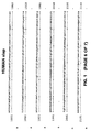
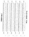
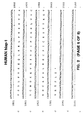
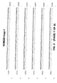
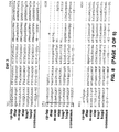
- the RZF domain has one of the IAP sequences shown in Fig. 6, and the BIR domains are comprised of BIR domains shown in Fig. 5.
- the BIR domains are at the amino terminal end of the protein relative to the RZF domain, which is at or near the carboxyl terminus of the polypeptide.
- the amplification is carried out using a reverse-transcription polymerase chain reaction, for example, the RACE method.
- the invention features an IAP gene isolated according to the method involving: (a) providing a preparation of DNA; (b) providing a detectably labelled DNA sequence having homology to a conserved region of an IAP gene; (c) contacting the preparation of DNA with the detectably-labelled DNA sequence under hybridization conditions providing detection of genes having 50% or greater nucleotide sequence identity; and (d) identifying an IAP gene by its association with the detectable label.
- the invention features an IAP gene isolated according to the method involving: (a) providing a cell sample; (b) introducing by transformation into the cell sample a candidate IAP gene; (c) expressing the candidate IAP gene within the cell sample; and (d) determining whether the cell sample exhibits an altered apoptotic response, whereby a response identifies an IAP gene.
- the invention features a method of isolating an IAP gene from a recombinant library, involving: (a) providing a recombinant library; (b) contacting the library with a detectably-labelled gene fragment produced according to the PCR method of the invention under hybridization conditions providing detection of genes having 50% or greater nucleotide sequence identity; and (c) isolating an IAP gene by its association with the detectable label.
- the invention features a method of inhibiting apoptosis by producing a cell that has integrated, into its genome, a transgene that includes the IAP gene, or a fragment thereof.
- the IAP gene may be placed under the control of a promoter providing constitutive expression of the IAP gene.
- the IAP transgene may be placed under the control of a promoter that allows expression of the gene to be regulated by environmental stimuli.
- the IAP gene may be expressed using a tissue-specific or cell type-specific promoter, or by a promoter that is activated by the introduction of an external signal or agent, such as a chemical signal or agent.
- the cell is a lymphocyte, a neuronal cell, a glial cell, or a fibroblast.
- the cell in an HIV-infected human, or in a mammal suffering from a neurodegenerative disease, an ischemic injury, a toxin-induced liver disease, or a myelodysplastic syndrome.
- the invention features a purified antibody that binds specifically to an IAP family protein.
- an antibody may be used in any standard immunodetection method for the identification of an IAP polypeptide.
- the antibody binds specifically to XIAP, HIAP-1, or HIAP-2.
- the antibody may react with other IAP polypeptides or may be specific for one or a few IAP polypeptides.
- the antibody may be a monoclonal or a polyclonal antibody.
- the antibody reacts specifically with only one of the IAP polypeptides, for example, reacts with murine and human xiap, but not with hiap-1 or hiap-2 from other mammalian species.
- Patent 4,816,397 describe various methods for producing immunoglobulines, and immunologically functional fragments thereof, which include at least the variable domains of the heavy and light chain in a single host cell.
- Cabilly et al. U.S. Patent 4,816,567) describe methods for preparing chimeric antibodies.
- the invention features a method for diagnosing a cell proliferation disease, or an increased likelihood of such a disease, using an IAP nucleic acid probe or antibody.
- the disease is a cancer.
- the disease is selected from the group consisting of promyelocytic leukemia, a HeLa-type carcinoma, chronic myelogenous leukemia (preferably using xiap or hiap-2 related probes), lymphoblastic leukemia (preferably using a xiap related probe), Burkitt's lymphoma (preferably using an hiap-1 related probe), colorectal adenocarcinoma, lung carcinoma, and melanoma (preferably using a xiap probe).
- a diagnosis is indicated by a 2-fold increase in expression or activity, more preferably, at least a 10-fold increase in expression or activity.
- a mammalian IAP may serve as an active ingredient in a therapeutic composition.
- This composition depending on the IAP or fragment included, may be used to modulate apoptosis and thereby treat any condition that is caused by a disturbance in apoptosis.
- the antibody may bind to an approximately 26 kDa cleavage product of an IAP polypeptide that includes at least one BIR domain but lacks a ring zinc finger domain.
- the negative regulator may also be an IAP antisense mRNA molecule.
- IAP gene is meant a gene encoding a polypeptide having at least one BIR domain and a ring zinc finger domain which is capable of modulating (inhibiting or enhancing) apoptosis in a cell or tissue when provided by other intracellular or extracellular delivery methods.
- the IAP gene is a gene having about 50% or greater nucleotide sequence identity to at least one of the IAP amino acid encoding sequences of Figs. 1-4 or portions thereof.
- the region of sequence over which identity is measured is a region encoding at least one BIR domain and a ring zinc finger domain.
- Mammalian IAP genes include nucleotide sequences isolated from any mammalian source.
- the mammal is a human.
- IAP gene is meant to encompass any member of the family of apoptosis inhibitory genes, which are characterized by their ability to modulate apoptosis.
- An IAP gene may encode a polypeptide that has at least 20%, preferably at least 30%, and most preferably at least 50% amino acid sequence identity with at least one of the conserved regions of one of the IAP members described herein (i.e., either the BIR or ring zinc finger domains from the human or murine xiap, hiap-1 and hiap-2).
- Representative members of the IAP gene family include, without limitation, the human and murine xiap, hiap-1, and hiap-2 genes.
- BIR domain is meant a domain having the amino acid sequence of the consensus sequence: Xaal-Xaal-Xaal-Arg-Leu-Xaal-Thr-Phe-Xaal-Xaal-Trp-Pro-Xaa2-Xaal-Xaal-Xaa2-Xaa2-Xaal-Xaal-Xaal-Xaal-Leu-Ala-Xaal-Ala-Gly-Phe-Tyr-Tyr-Xaal-Gly-Xaal-Xaal-Asp-Xaal-Val-Xaal-Cys-Phe-Xaal-Cys-Phe-Xaal-Cys-Xaal-Xaal- Xaal- Xaal-Trp-Xaal-Xaal-Xaal-Xaal-Asp-Xaal-Xaal-Xaal-Asp-Xaal-Xaal-Xaal-Xaal-His-
- ring zinc finger or “RZF” is meant a domain having the amino acid sequence of the consensus sequence: Glu-Xaal-Xaal-Xaal-Xaal-Xaal-Xaal-Xaal-Xaa2-Xaal-Xaal-Xaal-Cys- Lys-Xaa3-Cys-Met-Xaal-Xaal-Xaal-Xaal-Xaal-Xaa3-Xaal-Phe-Xaal-Pro-Cys-Gly-His-Xaal-Xaal-Xaal-Cys-Xaal-Xaal-Cys-Ala- Xaal-Xaal-Xaal-Xaal-Cys-Pro-Xaal-Cys, wherein Xaa1 is any amino acid, Xaa2 is Glu or Asp, and Xaa3 is Val or Ile (SEQ ID NO:1).
- modulating apoptosis or “altering apoptosis” is meant increasing or decreasing the number of cells that would otherwise undergo apoptosis in a given cell population.
- the cell population is selected from a group including T cells, neuronal cells, fibroblasts, or any other cell line known to undergo apoptosis in a laboratory setting (e.g., the baculovirus infected insect cells).
- the degree of modulation provided by an IAP or modulating compound in a given assay will vary, but that one skilled in the art can determine the statistically significant change in the level of apoptosis which identifies an IAP or a compound which modulates an IAP.
- inhibiting apoptosis is meant any decrease in the number of cells which undergo apoptosis relative to an untreated control.
- the decrease is at least 25%, more preferably the decrease is 50%, and most preferably the decrease is at least one-fold.
- polypeptide any chain of more than two amino acids, regardless of post-translational modification such as glycosylation or phosphorylation.
- substantially identical is meant a polypeptide or nucleic acid exhibiting at least 50%, preferably 85%, more preferably 90%, and most preferably 95% homology to a reference amino acid or nucleic acid sequence.
- the length of comparison sequences will generally be at least 16 amino acids, preferably at least 20 amino acids, more preferably at least 25 amino acids, and most preferably 35 amino acids.
- the length of comparison sequences will generally be at least 50 nucleotides, preferably at least 60 nucleotides, more preferably at least 75 nucleotides, and most preferably 110 nucleotides.
- substantially pure polypeptides include those derived from eukaryotic organisms but synthesized in E. coli or other prokaryotes.
- substantially pure DNA is meant DNA that is free of the genes which, in the naturally-occurring genome of the organism from which the DNA of the invention is derived, flank the gene.
- reporter gene is meant a gene whose expression may be assayed; such genes include, without limitation, glucuronidase (GUS), luciferase, chloramphenicol transacetylase (CAT), and ⁇ -galactosidase.
- GUS glucuronidase
- CAT chloramphenicol transacetylase
- ⁇ -galactosidase glucuronidase
- promoter minimal sequence sufficient to direct transcription. Also included in the invention are those promoter elements which are sufficient to render promoter-dependent gene expression controllable for cell type-specific, tissue-specific or inducible by external signals or agents; such elements may be located in the 5' or 3' regions of the native gene.
- operably linked is meant that a gene and one or more regulatory sequences are connected in such a way as to permit gene expression when the appropriate molecules (e.g., transcriptional activator proteins are bound to the regulatory sequences).
- conserved region any stretch of six or more contiguous amino acids exhibiting at least 30%, preferably 50%, and most preferably 70% amino acid sequence identity between two or more of the IAP family members, (e.g., between human HIAP-1, HIAP-2, and XIAP). Examples of preferred conserved regions are shown (as boxed or designated sequences) in Figures 5-7 and Tables 1 and 2, and include, without limitation, BIR domains and ring zinc finger domains.
- detectably-labelled any means for marking and identifying the presence of a molecule, e.g., an oligonucleotide probe or primer, a gene or fragment thereof, or a cDNA molecule.
- Methods for detectably-labelling a molecule are well known in the art and include, without limitation, radioactive labelling (e.g., with an isotope such as 32 P or 35 S) and nonradioactive labelling (e.g., chemiluminescent labelling, e.g., fluorescein labelling).
- antisense as used herein in reference to nucleic acids, is meant a nucleic acid sequence, regardless of length, that is complementary to the coding strand of a gene.
- IAP proteins A new class of mammalian proteins that modulate apoptosis (IAPS) and the genes that encode these proteins have been discovered.
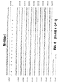
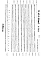
- the IAP proteins are characterized by the presence of a ring zinc finger domain (RZF; Fig. 9) and at least one BIR domain, as defined by the boxed consensus sequences shown in Figs. 7 and 8, and by the sequence domains listed in Tables 1 and 2.
- RZF ring zinc finger domain
- BIR domain BIR domain
- IAPs include those proteins lacking the ring zinc finger, as further described below.
- the IAP proteins may be used to inhibit the apoptosis that occurs as part of numerous disease processes or disorders.
- IAP polypeptides or nucleic acid encoding IAP polypeptides may be administered for the treatment or prevention of apoptosis that occurs as a part of AIDS, neurodegenerative diseases, ischemic injury, toxin-induced liver disease and myelodysplastic syndromes.
- Nucleic acid encoding the IAP polypeptide may also be provided to inhibit apoptosis.
- xiap for X-linked Inhibitor of Apoptosis Protein gene.
- the human gene has a 1.5 kb coding sequence that includes three BIR domains (Crook et al., J. Virol. 67:2168-74, 1993; Clem et al., Science 254:1388-90, 1991; Birnbaum et al., J. Virol., 68:2521-8, 1994) and a zinc finger.
- Northern blot analysis with xiap revealed message greater than 7 kb, which is expressed in various tissues, particularly liver and kidney (Fig. 12). The large size of the transcript reflects large 5' and 3' untranslated regions.
- the DNA ampliderived probe was then used to screen the human liver cDNA library for full-length hiap coding sequences.
- a third DNA was subsequently detected that includes the hiap-2 sequence but that appears to lack one exon, presumably due to alternative mRNA splicing (see boxed region in Fig. 3).
- the expression of hiap-1 and hiap-2 in human tissues as assayed by Northern blot analysis is shown in Figures 8 and 9.
- cDNA and two genomic clones were identified by screening a mouse embryo ⁇ gt11 cDNA library (Clontech, Palo Alto, CA) and a mouse FIX II genomic library with a xiap cDNA probe, respectively.
- a cDNA contig spanning 8.0 kb was constructed using 12 overlapping mouse clones. Sequence analysis revealed a coding sequence of approximately 1.5 kb.
- the mouse gene, m-xiap encodes a polypeptide with striking homology to human XIAP at and around the initiation methionine, the stop codon, the three BIR domains, and the RZF domain.
- the mouse homologue contains large 5' and 3' UTRs, which could produce a transcript as large as 7-8 kb.
- Antisera can be raised against a m-xiap fusion protein that was obtained from, for example, E. coli using a bacterial expression system. The resulting antisera can be used along with Northern blot analysis to analyze the spatial and temporal expression of m-xiap in the mouse.
- the murine homologs of hiap-1 and hiap-2 were cloned and sequenced in the same general manner as m-xiap using the human hiap-1 and hiap-2 sequences as probes. Cloning of m-hiap-1 and m-hiap-2 further demonstrate that homologs from different species may be isolated using the techniques provided herein and those generally known to artisans skilled in molecular biology.
- Standard techniques such as the polymerase chain reaction (PCR) and DNA hybridization, may be used to clone additional human IAP genes and their homologues in other species.
- Southern blots of human genomic DNA hybridized at low stringency with probes specific for xiap, hiap-1 and hiap-2 reveal bands that correspond to other known human IAP sequences as well as additional bands that do not correspond to known IAP sequences. Thus, additional IAP sequences may be readily identified using low stringency hybridization. Examples of murine and human xiap, hiap-1, and hiap-2 specific primers, which may be used to clone additional genes by RT-PCR, are shown in Table 5.
- IAPs The ability of putative IAPs to modulate apoptosis can be defined in in vitro systems in which alterations of apoptosis can be detected.
- Mammalian expression constructs carrying IAP cDNAs which are either full-length or truncated, can be introduced into cell lines such as CHO, NIH 3T3, HL60, Rat-1, or Jurkat cells.
- SF21 insect cells may be used, in which case the IAP gene is preferentially expressed using an insect heat shock promotor.
- apoptosis can be induced by standard methods, which include serum withdrawal, or application of staurosporine, menadione (which induces apoptosis via free radial formation), or anti-Fas antibodies.
- cells are cultured under the same conditions as those induced to undergo apoptosis, but either not transfected, or transfected with a vector that lacks an IAP insert.
- the ability of each IAP construct to inhibit apoptosis upon expression can be quantified by calculating the survival index of the cells, i.e., the ratio of surviving transfected cells to surviving control cells.
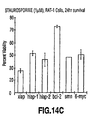
- Figs. 14A to 14D Specific examples of the results obtained by performing various apoptosis suppression assays are shown in Figs. 14A to 14D.
- CHO cell survival following transfection with one of six constructs and subsequent serum withdrawal is shown in Fig. 14A.
- the cells were transfected using Lipofectace TM with 2 ⁇ g of one of the following recombinant plasmids: pCDNA36myc-xiap (xiap), pCDNA3-6myc-hiap-1 (hiap-1), pCDNA3-6myc-hiap-2 (hiap-2), pCDNA3-bcl-2 (bcl-2), pCDNA3-HA-smn (smn), and pCDNA3-6myc (6-myc).
- Oligonucleotide primers were synthesized to allow PCR amplification and cloning of the xiap, hiap-1, and hiap-2 ORFs in pCDNA3 (Invitrogen). Each construct was modified to incorporate a synthetic myc tag encoding six repeats of the peptide sequence MEQKLISEEDL [(SEQ ID NO:_)], thus allowing detection of myc-IAP fusion proteins via monoclonal anti-myc antiserum (Egan et al., Nature 363:45-51, 1993). Triplicate samples of cell lines in 24-well dishes were washed 5 times with serum free media and maintained in serum free conditions during the course of the experiment.
- Fig. 14B The survival of CHO cells following transfection (with each one of the six constructs described above) and exposure to menadione is shown in Fig. 14B.
- the cells were plated in 24-well dishes, allowed to grow overnight, and then exposed to 20 ⁇ M menadione for 1.5 hours (Sigma Chemical Co., St. Louis, MO). Triplicate samples were harvested at the time of exposure to menadione and 24 hours afterward, and survival was assessed by trypan blue exclusion.
- Rat-1 cells were transfected and then selected in medium containing 800 ⁇ g/ml G418 for two weeks. The cell line was assessed for resistance to staurosporine-induced apoptosis (1 ⁇ M) for 5 hours. Viable cells were counted 24 hours after exposure to staurosporine by trypan blue exclusion. The percentage of viable cells shown represents the average of two experiments, ⁇ average deviation.
- expression vectors were constructed that contained either: (1) full-length IAP cDNA (as described above), (2) a portion of an IAP gene that encodes the BIR domains, but not the RZF, or (3) a portion of an IAP gene that encodes the RZF, but not the BIR domains.
- Human and murine xiap or m-xiap cDNAs were tested by transient or stable expression in HeLa, Jurkat, and CHO cell lines. Following transfection, apoptosis was induced by serum withdrawal, application of menadione, or application of an anti-Fas antibody. Cell death was then assessed, as described above, by trypan blue exclusion. As a control for transfection efficiency, the cells were co-transfected with a ⁇ -gal expression construct. Typically, approximately 20% of the cells were successfully transfected.
- Fig. 15A When CHO cells were transiently transfected, constructs containing full-length xiap or m-xiap cDNAs conferred modest protection against cell death (Fig. 15A). In contrast, the survival of CHO cells transfected with constructs encoding only the BIR domains (i.e., lacking the RZF domain; see Fig. 15A) was markedly enhanced 72 hours after serum deprivation. Furthermore, a large percentage of cells expressing the BIR domains were still viable after 96 hours, at which time no viable cells remained in the control, i.e. non-transfected, cell cultures (see "CHO" in Fig.
- Stable pools of transfected CHO cells which were maintained for several months under G418 selection, were induced to undergo apoptosis by exposure to 10 ⁇ M menadione for 2 hours.
- CHO cells tested were those that were stably transfected with: (1) full-length m-xiap cDNA (miap), (2) full-length xiap cDNA (xiap), (3) full-length bcl-2 cDNA (Bcl-2), (4) cDNA encoding the three BIR domains (but not the RZF) of m-xiap (BIR), and (5) cDNA encoding the RZF (but not BIR domains) of m-xiap (RZF).
- the assays of cell death described above indicate that the RZF may act as a negative regulator of the anti-apoptotic function of IAPs.
- One way in which the RZF, and possibly other IAP domains, may exert their regulatory influence is by altering the expression of genes, whose products function in the apoptotic pathway.
- COS cells were transiently transfected with the following four constructs, and the expressed polypeptide was localized by immunofluorescent microscopy: (1) pcDNA3-6myc-xiap, which encodes all 497 amino acids of SEQ ID NO:4, (2) pcDNA3-6myc-m-xiap, which encodes all 497 amino acids of mouse xiap (SEQ ID NO:10), (3) pcDNA3-6myc-mxiap-BIR, which encodes amino acids 1 to 341 of m-xiap (SEQ ID NO:10), and (4) pcDNA3-6myc-mxiap-RZF, which encodes amino acids 342-497 of m-xiap (SEQ ID NO:10).
- the cells were grown on multi-well tissue culture slides for 12 hours, and then fixed and permeabilized with methanol.
- the constructs used (here and in the cell death assays) were tagged with a human Myc epitope tag at the N-terminus. Therefore, a monoclonal anti-Myc antibody and a secondary goat anti-mouse antibody, which was conjugated to FITC, could be used to localize the expressed products in transiently transfected COS cells.
- Full-length XIAP and MIAP were located in the cytoplasm, with accentuated expression in the peri-nuclear zone.
- the same pattern of localization was observed when the cells expressed a construct encoding the RZF domain (but not the BIR domains). However, cells expressing the BIR domains (without the RZF) exhibited, primarily, nuclear staining.
- the protein expressed by the BIR domain construct appeared to be in various stages of transfer to the nucleus.
- XIAP is cleaved within T cells that are treated with anti-Fas antibodies (which are potent inducers of apoptosis), and its N-terminal domain is translocated to the nucleus.
- apoptosis assays are also provided in the following references.
- Assays for apoptosis in lymphocytes are disclosed by: Li et al., "Induction of apoptosis in uninfected lymphocytes by HIV-1 Tat protein", Science 268:429-431, 1995; Gibellini et al., "Tat-expressing Jurkat cells show an increased resistance to different apoptotic stimuli, including acute human immunodeficiency virus-type 1 (HIV-1) infection", Br. J. Haematol. 89:24-33, 1995; Martin et al., "HIV-1 infection of human CD4 + T cells in vitro. Differential induction of apoptosis in these cells.” J.
- Characterization of IAP genes provides information that is necessary for an IAP knockout animal model to be developed by homologous recombination.
- the model is a mammalian animal, most preferably a mouse.
- an animal model of IAP overproduction may be generated by integrating one or more IAP sequences into the genome, according to standard transgenic techniques.
- a replacement-type targeting vector which would be used to create a knockout model, can be constructed using an isogenic genomic clone, for example, from a mouse strain such as 129/Sv (Stratagene Inc., LaJolla, CA).
- the targeting vector will be introduced into a suitably-derived line of embryonic stem (ES) cells by electroporation to generate ES cell lines that carry a profoundly truncated form of an IAP.
- ES embryonic stem
- the targeted cell lines will be injected into a mouse blastula stage embryo. Heterozygous offspring will be interbred to homozygosity.
- Knockout mice would provide the means, in vivo , to screen for therapeutic compounds that modulate apoptosis via an IAP-dependent pathway.
- IAP genes may be expressed in both prokaryotic and eukaryotic cell types. If an IAP modulates apoptosis by exacerbating it, it may be desirable to express that protein under control of an inducible promotor.
- transduction and the choice of expression vehicle will depend on the host system selected. Transformation and transfection methods are described, e.g., in Ausubel et al. (supra), and expression vehicles may be chosen from those provided, e.g. in Cloning Vectors: A Laboratory Manual (P.H. Pouwels et al., 1985, Supp. 1987).
- a preferred expression system is the baculovirus system using, for example, the vector pBacPAK9, which is available from Clontech (Palo Alto, CA). If desired, this system may be used in conjunction with other protein expression techniques, for example, the myc tag approach described by Evan et al. (Mol. Cell Biol. 5:3610-3616, 1985).
- an IAP may be produced by a stably-transfected mammalian cell line.
- a number of vectors suitable for stable transfection of mammalian cells are available to the public, e.g., see Pouwels et al. (supra), as are methods for constructing such cell lines (see e.g., Ausubel et al. (supra).
- cDNA encoding an IAP is cloned into an expression vector that includes the dihydrofolate reductase (DHFR) gene.
- DHFR dihydrofolate reductase
- Antibody titres are monitored by Western blot and immunoprecipitation analyses using the thrombin-cleaved IAP fragment of the GST-IAP fusion protein. Immune sera are affinity purified using CNBr-Sepharose-coupled IAP protein. Antiserum specificity is determined using a panel of unrelated GST proteins (including GSTp53, Rb, HPV-16 E6, and E6-AP) and GST-trypsin (which was generated by PCR using known sequences).
- peptides corresponding to relatively unique hydrophilic regions of IAP may be generated and coupled to keyhole limpet hemocyanin (KLH) through an introduced C-terminal lysine.
- KLH keyhole limpet hemocyanin
- Antiserum to each of these peptides is similarly affinity purified on peptides conjugated to BSA, and specificity is tested by ELISA and Western blotting using peptide conjugates, and by Western blotting and immunoprecipitation using IAP expressed as a GST fusion protein.
- monoclonal antibodies may be prepared using the IAP proteins described above and standard hybridoma technology (see, e.g., Kohler et al., Nature 256:495, 1975; Kohler et al., Eur. J. Immunol. 6:511, 1976; Kohler et al., Eur. J. Immunol. 6:292, 1976; Hammerling et al., In Monoclonal Antibodies and T Cell Hybridomas , Elsevier, New York, NY, 1981; Ausubel et al., supra). Once produced, monoclonal antibodies are also tested for specific IAP recognition by Western blot or immunoprecipitation analysis (by the methods described in Ausubel et al., supra).
- antibodies of the invention are produced using IAP sequence that does not reside within highly conserved regions, and that appears likely to be antigenic, as analyzed by criteria such as those provided by the Peptide structure program (Genetics Computer Group Sequence Analysis Package, Program Manual for the GCG Package, Version 7, 1991) using the algorithm of Jameson and Wolf (CABIOS 4:181, 1988).
- these regions which are found between BIR1 and BIR2 of all IAPs, are: from amino acid 99 to amino acid 170 of hiap-1, from amino acid 123 to amino acid 184 of hiap-2, and from amino acid 116 to amino acid 133 of either xiap or m-xiap.
- These fragments can be generated by standard techniques, e.g.
- Isolation of IAP cDNAs also facilitates the identification of molecules that increase or decrease IAP expression.
- candidate molecules are added, in varying concentration, to the culture medium of cells expressing IAP mRNA.
- IAP expression is then measured, for example, by Northern blot analysis (Ausubel et al., supra) using an IAP cDNA, or cDNA fragment, as a hybridization probe (see also Table 5).
- the level of IAP expression in the presence of the candidate molecule is compared to the level of IAP expression in the absence of the candidate molecule, all other factors (e.g. cell type and culture conditions) being equal.
- the effect of candidate molecules on IAP-mediated apoptosis may, instead, be measured at the level of translation by using the general approach described above with standard protein detection techniques, such as Western blotting or immunoprecipitation with an IAP-specific antibody (for example, the IAP antibody described herein).
- standard protein detection techniques such as Western blotting or immunoprecipitation with an IAP-specific antibody (for example, the IAP antibody described herein).
- Another method for detecting compounds that modulate the activity of IAPs is to screen for compounds that interact physically with a given IAP polypeptide. These compounds may be detected by adapting interaction trap expression systems known in the art. These systems detect protein interactions using a transcriptional activation assay and are generally described by Gyuris et al. (Cell 75:791-803, 1993) and Field et al., Nature 340:245-246, 1989), and are commercially available from Clontech (Palo Alto, CA). In addition, PCT Publication WO 95/28497 describes an interaction trap assay in which proteins involved in apoptosis, by virtue of their interaction with Bcl-2, are detected. A similar method may be used to identify proteins and other compounds that interact with IAPs.
- Compounds or molecules that function as modulators of IAP-mediated cell death may include peptide and non-peptide molecules such as those present in cell extracts, mammalian serum, or growth medium in which mammalian cells have been cultured.
- a molecule that promotes an increase in IAP expression or IAP activity is considered particularly useful in the invention; such a molecule may be used, for example, as a therapeutic to increase cellular levels of IAP and thereby exploit the ability of IAP polypeptides to inhibit apoptosis.
- a molecule that decreases IAP activity may be used to decrease cellular proliferation. This would be advantageous in the treatment of neoplasms (see Table 3, below), or other cell proliferative diseases.
- Molecules that are found, by the methods described above, to effectively modulate IAP gene expression or polypeptide activity may be tested further in animal models. If they continue to function successfully in an in vivo setting, they may be used as therapeutics to either inhibit or enhance apoptosis, as appropriate.
- Retroviral vectors may be used as a gene transfer delivery system for a therapeutic IAP gene construct.
- Retroviral vectors are particularly well developed and have been used in clinical settings (Rosenberg et al., N. Engl. J. Med 323:370, 1990; Anderson et al., U.S. Patent No. 5,399,346).
- Non-viral approaches may also be employed for the introduction of therapeutic DNA into cells otherwise predicted to undergo apoptosis.
- IAP may be introduced into a neuron or a T cell by lipofection (Felgner et al., Proc. Natl. Acad. Sci. USA 84:7413, 1987; Ono et al., Neurosci. Lett. 117:259, 1990; Brigham et al., Am. J. Med. Sci.
- the therapeutic IAP DNA construct is preferably applied to the site of the predicted apoptosis event (for example, by injection). However, it may also be applied to tissue in the vicinity of the predicted apoptosis event or to a blood vessel supplying the cells predicted to undergo apoptosis.
- IAP cDNA expression can be directed from any suitable promoter (e.g., the human cytomegalovirus (CMV), simian virus 40 (SV40), or metallothionein promoters), and regulated by any appropriate mammalian regulatory element.
- CMV human cytomegalovirus
- SV40 simian virus 40
- metallothionein promoters e.g., metallothionein promoters
- enhancers known to preferentially direct gene expression in neural cells, T cells, or B cells may be used to direct IAP expression.
- the enhancers used could include, without limitation, those that are characterized as tissue- or cell-specific in their expression.
- regulation may be mediated by the cognate regulatory sequences or, if desired, by regulatory sequences derived from a heterologous source, including any of the promoters or regulatory elements described above.
- IAP gene therapy is accomplished by direct administration of the IAP mRNA or antisense IAP mRNA to a cell that is expected to undergo apoptosis.
- the mRNA may be produced and isolated by any standard technique, but is most readily produced by in vitro transcription using an IAP cDNA under the control of a high efficiency promoter (e.g., the T7 promoter).
- Administration of IAP mRNA to malignant cells can be carried out by any of the methods for direct nucleic acid administration described above.
- IAP protein by any gene therapy approach will result in cellular levels of IAP that are at least equivalent to the normal, cellular level of IAP in an unaffected cell.
- Treatment by any IAP-mediated gene therapy approach may be combined with more traditional therapies.
- Another therapeutic approach within the invention involves administration of recombinant IAP protein, either directly to the site of a predicted apoptosis event (for example, by injection) or systemically (for example, by any conventional recombinant protein administration technique).
- the dosage of IAP depends on a number of factors, including the size and health of the individual patient, but, generally, between 0.1 mg and 100 mg inclusive are administered per day to an adult in any pharmaceutically-acceptable formulation.
- Formulations for parenteral administration may, for example, contain excipients, sterile water, or saline, polyalkylene glycols such as polyethylene glycol, oils of vegetable origin, or hydrogenated napthalenes.
- Biocompatible, biodegradable lactide polymer, lactide/glycolide copolymer, or polyoxyethylene-polyoxypropylene copolymers may be used to control the release of the compounds.
- Other potentially useful parenteral delivery systems for IAP modulatory compounds include ethylene-vinyl acetate copolymer particles, osmotic pumps, implantable infusion systems, and liposomes.
- IAP polypeptides and nucleic acid sequences find diagnostic use in the detection or monitoring of conditions involving aberrant levels of apoptosis. For example, decrease expression of IAP may be correlated with enhanced apoptosis in humans (see XII, below). Accordingly, a decrease or increase in the level of IAP production may provide an indication of a deleterious condition.
- Levels of IAP expression may be assayed by any standard technique. For example, IAP expression in a biological sample (e.g., a biopsy) may be monitored by standard Northern blot analysis or may be aided by PCR (see, e.g., Ausubel et al., supra; PCR Technology: Principles and Applications for DNA Amplification , H.A. Ehrlich, Ed. Stockton Press, NY; Yap et al. Nucl. Acids. Res. 19:4294, 1991).
- a biological sample obtained from a patient may be analyzed for one or more mutations in the IAP sequences using a mismatch detection approach.
- these techniques involve PCR amplification of nucleic acid from the patient sample, followed by identification of the mutation (i.e., mismatch) by either altered hybridization, aberrant electrophoretic gel migration, binding or cleavage mediated by mismatch binding proteins, or direct nucleic acid sequencing. Any of these techniques may be used to facilitate mutant IAP detection, and each is well known in the art; examples of particular techniques are described, without limitation, in Orita et al., Proc. Natl. Acad. Sci. USA 86:2766-2770, 1989; Sheffield et al., Proc. Natl. Acad. Sci. USA 86:232-236, 1989).
- a tissue sample may be obtained from a patient, sectioned, and stained for the presence of IAP using an anti-IAP antibody and any standard detection system (e.g., one which includes a secondary antibody conjugated to horseradish peroxidase).
- any standard detection system e.g., one which includes a secondary antibody conjugated to horseradish peroxidase.
- a combined diagnostic method may be employed that begins with an evaluation of IAP protein production (for example, by immunological techniques or the protein truncation test (Hogerrorst et al., Nature Genetics 10:208-212, 1995) and also includes a nucleic acid-based detection technique designed to identify more subtle IAP mutations (for example, point mutations). As described above, a number of mismatch detection assays are available to those skilled in the art, and any preferred technique may be used. Mutations in IAP may be detected that either result in loss of IAP expression or loss of IAP biological activity. In a variation of this combined diagnostic method, IAP biological activity is measured as protease activity using any appropriate protease assay system (for example, those described above).
- Mismatch detection assays also provide an opportunity to diagnose an IAP-mediated predisposition to diseases caused by inappropriate apoptosis. For example, a patient heterozygous for an IAP mutation may show no clinical symptoms and yet possess a higher than normal probability of developing one or more types of neurodegenerative, myelodysplastic or ischemic diseases. Given this diagnosis, a patient may take precautions to minimize their exposure to adverse environmental factors (for example, UV exposure or chemical mutagens) and to carefully monitor their medical condition (for example, through frequent physical examinations). This type of IAP diagnostic approach may also be used to detect IAP mutations in prenatal screens.
- the IAP diagnostic assays described above may be carried out using any biological sample (for example, any biopsy sample or bodily fluid or tissue) in which IAP is normally expressed. Identification of a mutant IAP gene may also be assayed using these sources for test samples.
- a IAP mutation particularly as part of a diagnosis for predisposition to IAP-associated degenerative disease, may be tested using a DNA sample from any cell, for example, by mismatch detection techniques.
- the DNA sample is subjected to PCR amplification prior to analysis.
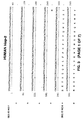
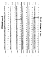
- a Human Cancer Cell Line Multiple Tissue Northern Blot (Clontech, Palo Alto, CA; #7757-1) was probed.
- This Northern blot contained approximately 2 ⁇ g of poly A + RNA per lane from eight different human cell lines: (1) promyelocytic leukemia HL-60, (2) HeLa cell S3, (3) chronic myelogenous leukemia K-562, (4) lymphoblastic leukemia MOLT-4, (5) Burkitt's lymphoma Raji, (6) colorectal adenocarcinoma SW480, (7) lung carcinoma A549, and (8) melanoma G361.
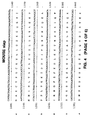
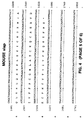
- a Human Multiple Tissue Northern Blot (Clontech, Palo Alto, CA; #7759-1) was probed. This Northern blot contained approximately 2 ⁇ g of poly A + RNA from eight different human tissues: (1) spleen, (2) thymus, (3) prostate, (4) testis, (5) ovary, (6) small intestine, (7) colon, and (8) peripheral blood leukocytes.
- the Northern blots were hybridized sequentially with: (1) a 1.6 kb probe to the xiap coding region, (2) a 375 bp hiap-2 specific probe corresponding to the 3' untranslated region, (3) a 1.3 kb probe to the coding region of hiap-1, which cross-reacts with hiap-2, (4) a 1.0 kb probe derived from the coding region of bcl-2, and (5) a probe to ⁇ -actin, which was provided by the manufacturer. Hybridization was carried out at 50°C overnight, according to the manufacturer's suggestion. The blot was washed twice with 2X SSC, 0.1% SDS at room temperature for 15 minutes and then with 2X SSC, 0.1% SDS at 50°C.
- upregulation of the anti-apoptotic IAP genes may be a widespread phenomenon, perhaps occurring much more frequently than upregulation of Bcl-2. Furthermore, upregulation may be necessary for the establishment or maintenance of the transformed state of cancerous cells.
- RNA was reverse transcribed, and amplified by PCR with the following set of oligonucleotide primers: 5'-AGTGCGGGTTTTTATTATGTG-3' (SEQ ID NO:_) and 5'-AGATGACCACAAGGAATAAACACTA-3' (SEQ ID NO:_), which selectively amplify a hiap-1 cDNA fragment.
- RT-PCR was conducted using a PerkinElmer 480 Thermocycler to carry out 35 cycles of the following program: 94°C for 1 minute, 50°C for 1.5 minutes, and 72°C for a minute.
- the PCR reaction product was electrophoresed on an agarose gel and stained with Ethidium bromide.
- Amplified cDNA fragments of the appropriate size were clearly visible in all lanes containing Burkitt's lymphoma samples, but absent in the lanes containing the normal placental tissue sample, and absent in lanes containing negative control samples, where template DNA was omitted from the reaction (Fig. 17).
- a total protein extract was prepared from Jurkat and astrocytoma cells by sonicating them (X3 for 15 seconds at 4°C) in 50 mM Tris-HCl (pH 8.0), 150 mM NaCl, 1 mM PMSF, 1 ⁇ g/ml aprotinin, and 5 mM benzamidine. Following sonication, the samples were centrifuged (14,000 RPM in a microfuge) for five minutes. Twenty ⁇ g of protein was loaded per well on a 10% SDS-polyacrylamide gel, electrophoresed, and electroblotted by standard methods to PVDF membranes.
- a 26 kDa xiap-reactive band was also observed under the following experimental conditions.
- Jurkat cells a transformed human T cell line
- an anti-Fas antibody (1 ⁇ g/ml).
- Identical cultures of Jurkat cells were exposed either to: (1) anti-Fas antibody and cycloheximide (20 ⁇ g/ml), (2) tumor necrosis factor alpha (TNF- ⁇ , at 1,000 U/ml), or (3) TNF- ⁇ and cycloheximide (20 ⁇ g/ml). All cells were harvested 6 hours after treatment began.
- anti-Fas antibody was added to an extract after the cells were harvested.
- the cells were harvested in SDS sample buffer, electrophoresed on a 12.5% SDS polyacrylamide gel, and electroblotted onto PVDF membranes using standard methods.
- the membranes were immunostained with a rabbit polyclonal anti-XIAP antibody at 1:1000 for 1 hour at room temperature. Following four 15 minute washes, a goat anti-rabbit antibody conjugated to horse-radish peroxidase was applied at room temperature for 1 hour. Unbound secondary antibody was washed away, and chemiluminescent detection of XIAP protein was performed.
- the Western blot revealed the presence of the full-length, 55 kDa XIAP protein, both in untreated and treated cells.
- a novel, approximately 26 kDa xiap-reactive band was also observed in apoptotic cell extracts, but not in the control, untreated cell extracts (Fig. 19).
- Cleavage of XIAP occurs in a variety of cell types, including other cancer cell lines such as HeLa.
- the expression of the 26 kDa XIAP cleavage product was demonstrated in HeLa cells as follows. HeLa cells were treated with either: (1) cyclohexamide (20 ⁇ g/ml), (2) anti-Fas antibody (1 ⁇ g/ml), (3) anti-Fas antibody (1 ⁇ g/ml) and cyclohexamide (20 ⁇ g/ml), (4) TNF ⁇ (1,000 U/ml), or (5) TNF ⁇ (1,000 U/ml) and cyclohexamide (20 ⁇ g/ml). All cells were harvested 18 hours after treatment began.
- the cytoplasmic fraction of the extract was processed further by centrifugation at 60,000 RPM in a TA 100.3 rotor for 30 minutes. Supernatants were removed and frozen at - 80°C. Samples of both nuclear and cytoplasmic fractions were loaded on a 12.5% SDS-polyacrylamide gel, and electroblotted onto PVDF membranes. Western blot analysis was then performed using either an anti-CPP32 antibody (Transduction Laboratories Lexington, KY; Fig. 22A) or the rabbit anti-XIAP antibody described above (Fig. 22B).
Landscapes
- Health & Medical Sciences (AREA)
- Life Sciences & Earth Sciences (AREA)
- Engineering & Computer Science (AREA)
- Chemical & Material Sciences (AREA)
- Immunology (AREA)
- Molecular Biology (AREA)
- Biomedical Technology (AREA)
- Urology & Nephrology (AREA)
- Hematology (AREA)
- General Health & Medical Sciences (AREA)
- Medicinal Chemistry (AREA)
- Analytical Chemistry (AREA)
- Biochemistry (AREA)
- Organic Chemistry (AREA)
- Pathology (AREA)
- Microbiology (AREA)
- Biotechnology (AREA)
- Physics & Mathematics (AREA)
- Cell Biology (AREA)
- Food Science & Technology (AREA)
- General Physics & Mathematics (AREA)
- Proteomics, Peptides & Aminoacids (AREA)
- Bioinformatics & Cheminformatics (AREA)
- Zoology (AREA)
- Genetics & Genomics (AREA)
- General Chemical & Material Sciences (AREA)
- Nuclear Medicine, Radiotherapy & Molecular Imaging (AREA)
- Wood Science & Technology (AREA)
- Chemical Kinetics & Catalysis (AREA)
- Pharmacology & Pharmacy (AREA)
- Animal Behavior & Ethology (AREA)
- Public Health (AREA)
- Veterinary Medicine (AREA)
- Biophysics (AREA)
- Toxicology (AREA)
- Tropical Medicine & Parasitology (AREA)
- General Engineering & Computer Science (AREA)
- Oncology (AREA)
- Gastroenterology & Hepatology (AREA)
- Communicable Diseases (AREA)
Applications Claiming Priority (3)
| Application Number | Priority Date | Filing Date | Title |
|---|---|---|---|
| US08/511,485 US5919912A (en) | 1995-08-04 | 1995-08-04 | Mammalian IAP antibodies and diagnostic kits |
| US08/576,956 US6156535A (en) | 1995-08-04 | 1995-12-22 | Mammalian IAP gene family, primers, probes, and detection methods |
| EP96931191A EP0837939B1 (de) | 1995-08-04 | 1996-08-05 | Säugetieren apoptosis inhibitorprotein genfamilie, primers, probes und verfahren zur nachweis |
Related Parent Applications (1)
| Application Number | Title | Priority Date | Filing Date |
|---|---|---|---|
| EP96931191A Division EP0837939B1 (de) | 1995-08-04 | 1996-08-05 | Säugetieren apoptosis inhibitorprotein genfamilie, primers, probes und verfahren zur nachweis |
Publications (2)
| Publication Number | Publication Date |
|---|---|
| EP1757689A2 true EP1757689A2 (de) | 2007-02-28 |
| EP1757689A3 EP1757689A3 (de) | 2007-05-23 |
Family
ID=27057240
Family Applications (2)
| Application Number | Title | Priority Date | Filing Date |
|---|---|---|---|
| EP96931191A Expired - Lifetime EP0837939B1 (de) | 1995-08-04 | 1996-08-05 | Säugetieren apoptosis inhibitorprotein genfamilie, primers, probes und verfahren zur nachweis |
| EP05025419A Withdrawn EP1757689A3 (de) | 1995-08-04 | 1996-08-05 | Säugetiere Apoptosis-Inhibitorprotein Genfamilie, Primers, Proben und Verfahren zum Nachweis |
Family Applications Before (1)
| Application Number | Title | Priority Date | Filing Date |
|---|---|---|---|
| EP96931191A Expired - Lifetime EP0837939B1 (de) | 1995-08-04 | 1996-08-05 | Säugetieren apoptosis inhibitorprotein genfamilie, primers, probes und verfahren zur nachweis |
Country Status (8)
| Country | Link |
|---|---|
| US (17) | US6156535A (de) |
| EP (2) | EP0837939B1 (de) |
| JP (1) | JP4458551B2 (de) |
| AT (1) | ATE310819T1 (de) |
| CA (1) | CA2228635C (de) |
| DE (1) | DE69635482T2 (de) |
| ES (1) | ES2253756T3 (de) |
| WO (1) | WO1997006255A2 (de) |
Families Citing this family (39)
| Publication number | Priority date | Publication date | Assignee | Title |
|---|---|---|---|---|
| US20030073159A1 (en) * | 1992-05-11 | 2003-04-17 | Human Genome Sciences, Inc. | Human inhibitor of apoptosis gene 1 |
| US5958771A (en) * | 1998-12-03 | 1999-09-28 | Isis Pharmaceuticals, Inc. | Antisense modulation of cellular inhibitor of Apoptosis-2 expression |
| US6087173A (en) * | 1999-09-09 | 2000-07-11 | Isis Pharmaceuticals Inc. | Antisense modulation of X-linked inhibitor of apoptosis expression |
| US6156535A (en) * | 1995-08-04 | 2000-12-05 | University Of Ottawa | Mammalian IAP gene family, primers, probes, and detection methods |
| AUPN727595A0 (en) * | 1995-12-22 | 1996-01-18 | Walter And Eliza Hall Institute Of Medical Research, The | Therapeutic compositions |
| GB9601108D0 (en) * | 1996-01-19 | 1996-03-20 | Univ Ottawa | Neuronal apoptosis inhibitor protein (NAIP) |
| US7785842B2 (en) * | 1996-03-26 | 2010-08-31 | Oncomedx, Inc. | Comparative analysis of extracellular RNA species |
| US8043835B1 (en) | 1996-03-26 | 2011-10-25 | Oncomedx, Inc. | Methods for detecting and monitoring cancer using extracellular RNA |
| DE69739909D1 (de) * | 1996-03-26 | 2010-07-29 | Michael S Kopreski | Methoden aus plasma oder serum extrahierte extrazelluraere rna zur diagnoseüberwachung oder evaluation von krebs verwenden |
| US6133437A (en) * | 1997-02-13 | 2000-10-17 | Apoptogen, Inc. | Modulation of IAPs for the treatment of proliferative diseases |
| WO1997040847A1 (en) * | 1996-04-26 | 1997-11-06 | University Of Ottawa | Therapeutic and drug screening methods for the treatment and prevention of neuronal disease |
| US6511828B1 (en) | 1996-05-31 | 2003-01-28 | Arch Development Corporation | Human and drosophila inhibitors of apoptosis proteins (IAPs) |
| US5840535A (en) * | 1997-06-02 | 1998-11-24 | Incyte Pharmaceuticals, Inc. | DNA encoding a zinc ring protein |
| WO1999062943A2 (en) * | 1998-06-02 | 1999-12-09 | Millenium Pharmaceuticals, Inc. | Novel molecules of the aip-related protein family and uses thereof |
| US6171821B1 (en) * | 1998-07-24 | 2001-01-09 | Apoptogen, Inc. | XIAP IRES and uses thereof |
| US6472172B1 (en) | 1998-07-31 | 2002-10-29 | Schering Aktiengesellschaft | DNA encoding a novel human inhibitor-of-apoptosis protein |
| US20080261292A1 (en) * | 1998-09-22 | 2008-10-23 | Oncomedx, Inc. | Method Enabling the Use of Extracellular Ribonucleic Acid (RNA) Extracted from Plasma or Serum to Detect, Monitor or Evaluate Cancer or Premalignant Conditions |
| CN1301769A (zh) * | 1999-12-29 | 2001-07-04 | 复旦大学 | 一种新的多肽——人的1atexin蛋白46和编码这种多肽的多核苷酸 |
| ATE428116T1 (de) * | 2000-06-16 | 2009-04-15 | Rudel Thomas Dr | Verfahren zur identifizierung wahrend der apoptose modifizierter proteine |
| EP1164374A1 (de) * | 2000-06-16 | 2001-12-19 | Max-Planck-Gesellschaft zur Förderung der Wissenschaften e.V. | Verfahren zur Identifizierung während der Apoptose modifizierter Proteine |
| US20020164576A1 (en) * | 2000-09-22 | 2002-11-07 | Pedersen Finn Skou | Methods for diagnosis and treatment of diseases associated with altered expression of Nrf2 |
| US20030044803A1 (en) * | 2000-09-22 | 2003-03-06 | Pedersen Finn Skou | Methods for diagnosis and treatment of diseases associated with altered expression of JAK1 |
| US6673917B1 (en) * | 2000-09-28 | 2004-01-06 | University Of Ottawa | Antisense IAP nucleic acids and uses thereof |
| US6901173B2 (en) * | 2001-04-25 | 2005-05-31 | Lockheed Martin Corporation | Scene-based non-uniformity correction for detector arrays |
| US20070098728A1 (en) * | 2001-09-24 | 2007-05-03 | Pedersen Finn S | Novel compositions and methods in cancer |
| US20100159464A1 (en) * | 2001-11-05 | 2010-06-24 | Oncomedx, Inc. | Method for Detection of DNA Methyltransferase RNA in Plasma and Serum |
| US7820132B2 (en) * | 2001-12-14 | 2010-10-26 | Alliance For Sustainable Energy, Llc | Hot wire production of single-wall and multi-wall carbon nanotubes |
| EP1539189A4 (de) * | 2002-03-25 | 2007-05-09 | Univ Boston | Verfahren zur verwendung von anti-apoptose-faktoren in der genexpression |
| AU2003219432B2 (en) * | 2002-03-27 | 2010-04-01 | Pharmascience Inc. | Antisense IAP nucleobase oligomers and uses thereof |
| US20050233411A9 (en) * | 2003-02-07 | 2005-10-20 | Chunying Du | Compositions and methods for cleaving IAP |
| WO2005017109A2 (en) * | 2003-06-30 | 2005-02-24 | Massachusetts Institute Of Technology | Nucleic acids and polypeptides required for cell survival in the absence of rb |
| US8012944B2 (en) * | 2003-10-30 | 2011-09-06 | Pharmascience Inc. | Method for treating cancer using IAP antisense oligomer and chemotherapeutic agent |
| US7342093B2 (en) * | 2004-07-23 | 2008-03-11 | University Of Massachusetts | Compounds that inhibit Hsp90 protein-protein interactions with IAP proteins |
| WO2006047250A2 (en) * | 2004-10-22 | 2006-05-04 | Neurologix, Inc. | Use of apoptosis inhibiting compounds in degenerative neurological disorders |
| JP5421590B2 (ja) | 2005-05-18 | 2014-02-19 | ノバルティス アーゲー | 自己免疫および/または炎症性成分を有する疾患の診断および治療のための方法 |
| CN101535300B (zh) | 2006-05-16 | 2014-05-28 | 埃格拉医疗公司 | Iap bir域结合化合物 |
| US9284350B2 (en) | 2010-02-12 | 2016-03-15 | Pharmascience Inc. | IAP BIR domain binding compounds |
| KR101215201B1 (ko) | 2010-07-21 | 2012-12-26 | 단국대학교 산학협력단 | Xiap 단백질을 포함하는 허혈성 질환을 치료 또는 예방하기 위한 조성물 |
| US20140127438A1 (en) | 2012-11-08 | 2014-05-08 | Robert L. Sherman, Jr. | Stabilized high-density polyethylene composition with improved resistance to deterioration and stabilizer system |
Family Cites Families (56)
| Publication number | Priority date | Publication date | Assignee | Title |
|---|---|---|---|---|
| US5665550A (en) | 1990-10-19 | 1997-09-09 | Board Of Trustees Of The University Of Illinois-Urbana | Genes and genetic elements associated with sensitivity to chemotherapeutic drugs |
| WO1994008003A1 (en) | 1991-06-14 | 1994-04-14 | Isis Pharmaceuticals, Inc. | ANTISENSE OLIGONUCLEOTIDE INHIBITION OF THE ras GENE |
| US5594076A (en) | 1991-09-24 | 1997-01-14 | The Pennsylvania Research Foundation | Hydrodegradable polyesters |
| US6265157B1 (en) | 1991-12-03 | 2001-07-24 | Allegheny University Of The Health Sciences | Compositions and methods for detecting altered COL1A1 gene sequences |
| WO1996035703A1 (en) | 1995-05-11 | 1996-11-14 | Human Genome Sciences, Inc. | Human inhibitor of apoptosis gene 1 |
| US20030073159A1 (en) | 1992-05-11 | 2003-04-17 | Human Genome Sciences, Inc. | Human inhibitor of apoptosis gene 1 |
| JP3562528B2 (ja) * | 1992-09-14 | 2004-09-08 | ザ ジェネラル ホスピタル コーポレイション | Ikaros:t細胞経路調節遺伝子 |
| US5958772A (en) | 1998-12-03 | 1999-09-28 | Isis Pharmaceuticals Inc. | Antisense inhibition of cellular inhibitor of apoptosis-1 expression |
| US6087173A (en) | 1999-09-09 | 2000-07-11 | Isis Pharmaceuticals Inc. | Antisense modulation of X-linked inhibitor of apoptosis expression |
| US5958771A (en) * | 1998-12-03 | 1999-09-28 | Isis Pharmaceuticals, Inc. | Antisense modulation of cellular inhibitor of Apoptosis-2 expression |
| WO1994023760A1 (en) | 1993-04-14 | 1994-10-27 | The United States Of America As Represented By The Secretary Of The Navy | Transgenic animal model for autoimmune diseases |
| US5691179A (en) * | 1993-08-26 | 1997-11-25 | Washington University | Cell death regulators |
| US5624803A (en) | 1993-10-14 | 1997-04-29 | The Regents Of The University Of California | In vivo oligonucleotide generator, and methods of testing the binding affinity of triplex forming oligonucleotides derived therefrom |
| US5801154A (en) | 1993-10-18 | 1998-09-01 | Isis Pharmaceuticals, Inc. | Antisense oligonucleotide modulation of multidrug resistance-associated protein |
| US5510239A (en) | 1993-10-18 | 1996-04-23 | Isis Pharmaceuticals, Inc. | Oligonucleotide modulation of multidrug resistance-associated protein |
| DE69534629D1 (de) * | 1994-01-18 | 2005-12-29 | Scripps Research Inst | Derivate von zinkfingerproteinen und methoden |
| AU2290195A (en) * | 1994-04-13 | 1995-11-10 | La Jolla Cancer Research Foundation | Interaction of proteins involved in a cell death pathway |
| HUT76099A (en) | 1994-05-10 | 1997-06-30 | Immulogic Pharma Corp | Compositions and treatment for multiple sclerosis |
| US6020127A (en) * | 1994-10-18 | 2000-02-01 | The University Of Ottawa | Neuronal apoptosis inhibitor protein, gene sequence and mutations causative of spinal muscular atrophy |
| IL115766A0 (en) | 1994-10-25 | 1996-01-19 | Immulogic Pharma Corp | Composition and treatment for multiple sclerosis |
| AU4695296A (en) | 1995-01-06 | 1996-07-24 | Immulogic Pharmaceutical Corporation | Compositions and methods for treating rheumatoid arthritis |
| US5770690A (en) * | 1995-06-27 | 1998-06-23 | Neurex Corporation | Bax omega protein and methods |
| US5877021A (en) | 1995-07-07 | 1999-03-02 | Ribozyme Pharmaceuticals, Inc. | B7-1 targeted ribozymes |
| US5919912A (en) | 1995-08-04 | 1999-07-06 | University Of Ottawa | Mammalian IAP antibodies and diagnostic kits |
| US6156535A (en) | 1995-08-04 | 2000-12-05 | University Of Ottawa | Mammalian IAP gene family, primers, probes, and detection methods |
| AU6692996A (en) | 1995-08-08 | 1997-03-05 | Tularik Inc. | Inhibitors of apoptosis |
| US6187557B1 (en) | 1995-08-08 | 2001-02-13 | Tularik Inc. | c-IAP1 and c-IAP2: inhibitors of apoptosis |
| US5834216A (en) | 1995-09-06 | 1998-11-10 | Arch Development Corporation | Screening methods for the identification of inducers and inhibitors of programmed cell death (apoptosis) |
| AUPN727595A0 (en) * | 1995-12-22 | 1996-01-18 | Walter And Eliza Hall Institute Of Medical Research, The | Therapeutic compositions |
| US5605022A (en) | 1995-12-26 | 1997-02-25 | Nci Building Systems, Inc. | Vented closure |
| GB9601108D0 (en) * | 1996-01-19 | 1996-03-20 | Univ Ottawa | Neuronal apoptosis inhibitor protein (NAIP) |
| US6133437A (en) | 1997-02-13 | 2000-10-17 | Apoptogen, Inc. | Modulation of IAPs for the treatment of proliferative diseases |
| EP0911411A4 (de) * | 1996-06-24 | 2004-05-06 | Takara Bio Inc | Neues enzym zur deblockierung des aminoterminalen endes |
| WO1998022131A2 (en) | 1996-11-15 | 1998-05-28 | University Of Ottawa | Modulators of ovarial apoptosis related to iap |
| US5994076A (en) | 1997-05-21 | 1999-11-30 | Clontech Laboratories, Inc. | Methods of assaying differential expression |
| US6228603B1 (en) | 1997-05-22 | 2001-05-08 | The Burnham Institute | Screening assays for agents that alter inhibitor of apoptosis (IAP) protein regulation of caspase activity |
| CA2225187A1 (en) | 1997-07-14 | 1999-01-14 | Universite D'ottawa/ University Of Ottawa | Xaf genes and polypeptides: methods and reagents for modulating apoptosis |
| US6133246A (en) | 1997-08-13 | 2000-10-17 | Isis Pharmaceuticals Inc. | Antisense oligonucleotide compositions and methods for the modulation of JNK proteins |
| AU1080899A (en) | 1997-10-14 | 1999-05-03 | Nadine A. Tatton | Methods for increasing schwann cell survival |
| US6506559B1 (en) | 1997-12-23 | 2003-01-14 | Carnegie Institute Of Washington | Genetic inhibition by double-stranded RNA |
| US6171821B1 (en) | 1998-07-24 | 2001-01-09 | Apoptogen, Inc. | XIAP IRES and uses thereof |
| US6077709A (en) | 1998-09-29 | 2000-06-20 | Isis Pharmaceuticals Inc. | Antisense modulation of Survivin expression |
| AU1105800A (en) | 1998-10-09 | 2000-05-01 | L. Sai Latha Shankar | Methods for treating multiple sclerosis |
| US6187657B1 (en) * | 1999-03-24 | 2001-02-13 | Advanced Micro Devices, Inc. | Dual material gate MOSFET technique |
| KR20000065690A (ko) | 1999-04-08 | 2000-11-15 | 박종구 | 반응 특이성 및 안정성을 개선시킨 안티센스 올리고 뉴클레오타이드, 안티센스 dna 및 그 제조방법 |
| US5998148A (en) | 1999-04-08 | 1999-12-07 | Isis Pharmaceuticals Inc. | Antisense modulation of microtubule-associated protein 4 expression |
| US6395771B1 (en) | 2000-05-31 | 2002-05-28 | Dabur Research Foundation | Paclitaxel derivatives for the treatment of cancer |
| US6673917B1 (en) | 2000-09-28 | 2004-01-06 | University Of Ottawa | Antisense IAP nucleic acids and uses thereof |
| US20020119168A1 (en) | 2001-02-20 | 2002-08-29 | Rudnic Edward M. | Therapeutic agent delivery |
| KR100397275B1 (ko) | 2001-03-08 | 2003-09-17 | 주식회사 웰진 | 단방향성 안티센스 cDNA 라이브러리 구축을 통한 신규대규모 유전자 검색 및 기능 분석 시스템 |
| WO2003076592A2 (en) | 2002-03-06 | 2003-09-18 | Rigel Pharmaceuticals, Inc. | Novel method for delivery and intracellular synthesis of sirna molecules |
| AU2003219432B2 (en) | 2002-03-27 | 2010-04-01 | Pharmascience Inc. | Antisense IAP nucleobase oligomers and uses thereof |
| EP1469070A1 (de) | 2003-04-15 | 2004-10-20 | Deutsches Krebsforschungszentrum Stiftung des öffentlichen Rechts | Livinspezifische SiRNAs zur Behandlung von therapie-resistente Tumoren |
| EP1668024A4 (de) | 2003-09-29 | 2008-07-02 | Topigen Pharma Inc | Oligonukleotidzusammensetzungen und verfahren zur behandlung von krankheiten einschliesslich entzündlicher leiden |
| US8012944B2 (en) | 2003-10-30 | 2011-09-06 | Pharmascience Inc. | Method for treating cancer using IAP antisense oligomer and chemotherapeutic agent |
| US6946917B2 (en) * | 2003-11-25 | 2005-09-20 | Texas Instruments Incorporated | Generating an oscillating signal according to a control current |
-
1995
- 1995-12-22 US US08/576,956 patent/US6156535A/en not_active Expired - Lifetime
-
1996
- 1996-08-05 DE DE69635482T patent/DE69635482T2/de not_active Expired - Lifetime
- 1996-08-05 EP EP96931191A patent/EP0837939B1/de not_active Expired - Lifetime
- 1996-08-05 WO PCT/IB1996/001022 patent/WO1997006255A2/en not_active Ceased
- 1996-08-05 EP EP05025419A patent/EP1757689A3/de not_active Withdrawn
- 1996-08-05 CA CA002228635A patent/CA2228635C/en not_active Expired - Fee Related
- 1996-08-05 ES ES96931191T patent/ES2253756T3/es not_active Expired - Lifetime
- 1996-08-05 JP JP50827797A patent/JP4458551B2/ja not_active Expired - Fee Related
- 1996-08-05 US US09/011,356 patent/US6656704B1/en not_active Expired - Lifetime
- 1996-08-05 AT AT96931191T patent/ATE310819T1/de not_active IP Right Cessation
-
1998
- 1998-12-01 US US09/201,932 patent/US6689562B1/en not_active Expired - Lifetime
- 1998-12-01 US US09/201,936 patent/US6541457B2/en not_active Expired - Fee Related
-
2000
- 2000-09-01 US US09/654,743 patent/US6977158B1/en not_active Expired - Fee Related
-
2003
- 2003-06-20 US US10/600,272 patent/US7067281B2/en not_active Expired - Fee Related
-
2005
- 2005-09-01 US US11/217,557 patent/US20060127927A1/en not_active Abandoned
- 2005-12-22 US US11/316,539 patent/US20070009971A1/en not_active Abandoned
-
2006
- 2006-01-19 US US11/334,902 patent/US20060205021A1/en not_active Abandoned
- 2006-07-25 US US11/492,113 patent/US20070026470A1/en not_active Abandoned
- 2006-07-25 US US11/492,112 patent/US20070031904A1/en not_active Abandoned
- 2006-07-25 US US11/492,111 patent/US20070031903A1/en not_active Abandoned
- 2006-07-26 US US11/492,862 patent/US20070048772A1/en not_active Abandoned
- 2006-07-26 US US11/492,868 patent/US20070026494A1/en not_active Abandoned
- 2006-07-26 US US11/492,867 patent/US20070027304A1/en not_active Abandoned
- 2006-08-03 US US11/498,244 patent/US7776552B2/en not_active Expired - Fee Related
- 2006-08-04 US US11/498,897 patent/US20070066524A1/en not_active Abandoned
Also Published As
Similar Documents
| Publication | Publication Date | Title |
|---|---|---|
| EP0837939B1 (de) | Säugetieren apoptosis inhibitorprotein genfamilie, primers, probes und verfahren zur nachweis | |
| US7087584B2 (en) | Detection and modulation of IAPs and NAIP for the diagnosis and treatment of proliferative disease | |
| US5919912A (en) | Mammalian IAP antibodies and diagnostic kits | |
| US20070202096A1 (en) | XAF genes and polypeptides: methods and reagents for modulating apoptosis | |
| EP0815231B1 (de) | Verwendung von neuronalem apoptose-inhibitorprotein | |
| CA2403947C (en) | Bir domains of mammalian iap gene family | |
| HK1103241A (en) | Mammalian apoptosis inhibitor protein gene family, primers, probes and detection methods | |
| CA2512366A1 (en) | Mammalian iap gene family, primers, probes and detection methods | |
| US20070219360A1 (en) | Detection and modulation of IAPs and NAIP for the diagnosis and treatment of proliferative disease |
Legal Events
| Date | Code | Title | Description |
|---|---|---|---|
| PUAI | Public reference made under article 153(3) epc to a published international application that has entered the european phase |
Free format text: ORIGINAL CODE: 0009012 |
|
| 17P | Request for examination filed |
Effective date: 20051122 |
|
| AC | Divisional application: reference to earlier application |
Ref document number: 0837939 Country of ref document: EP Kind code of ref document: P |
|
| AK | Designated contracting states |
Kind code of ref document: A2 Designated state(s): AT BE CH DE DK ES FI FR GB GR IE IT LI LU MC NL PT SE |
|
| AX | Request for extension of the european patent |
Extension state: AL LT LV SI |
|
| PUAL | Search report despatched |
Free format text: ORIGINAL CODE: 0009013 |
|
| AK | Designated contracting states |
Kind code of ref document: A3 Designated state(s): AT BE CH DE DK ES FI FR GB GR IE IT LI LU MC NL PT SE |
|
| AX | Request for extension of the european patent |
Extension state: AL LT LV SI |
|
| REG | Reference to a national code |
Ref country code: HK Ref legal event code: DE Ref document number: 1103241 Country of ref document: HK |
|
| AKX | Designation fees paid |
Designated state(s): AT BE CH DE DK ES FI FR GB GR IE IT LI LU MC NL PT SE |
|
| 17Q | First examination report despatched |
Effective date: 20080528 |
|
| REG | Reference to a national code |
Ref country code: HK Ref legal event code: WD Ref document number: 1103241 Country of ref document: HK |
|
| STAA | Information on the status of an ep patent application or granted ep patent |
Free format text: STATUS: THE APPLICATION IS DEEMED TO BE WITHDRAWN |
|
| 18D | Application deemed to be withdrawn |
Effective date: 20140301 |